US7967763B2 - Method for treating subcutaneous tissues - Google Patents
Method for treating subcutaneous tissuesDownload PDFInfo
- Publication number
- US7967763B2 US7967763B2US11/292,950US29295005AUS7967763B2US 7967763 B2US7967763 B2US 7967763B2US 29295005 AUS29295005 AUS 29295005AUS 7967763 B2US7967763 B2US 7967763B2
- Authority
- US
- United States
- Prior art keywords
- solution
- tissue
- ultrasound
- treatment area
- microbubbles
- Prior art date
- Legal status (The legal status is an assumption and is not a legal conclusion. Google has not performed a legal analysis and makes no representation as to the accuracy of the status listed.)
- Active, expires
Links
- 238000000034methodMethods0.000titleclaimsabstractdescription110
- 206010033675panniculitisDiseases0.000titleabstractdescription97
- 210000004304subcutaneous tissueAnatomy0.000titleabstractdescription96
- 238000002604ultrasonographyMethods0.000claimsabstractdescription278
- 238000002347injectionMethods0.000claimsabstractdescription166
- 239000007924injectionSubstances0.000claimsabstractdescription166
- 238000011282treatmentMethods0.000claimsabstractdescription78
- 208000035484CelluliteDiseases0.000claimsabstractdescription36
- 206010049752Peau d'orangeDiseases0.000claimsabstractdescription36
- 230000036232celluliteEffects0.000claimsabstractdescription36
- 239000000243solutionSubstances0.000claimsdescription327
- 239000012530fluidSubstances0.000claimsdescription31
- 210000004027cellAnatomy0.000claimsdescription28
- 210000001789adipocyteAnatomy0.000claimsdescription26
- 210000004207dermisAnatomy0.000claimsdescription15
- 239000006185dispersionSubstances0.000claimsdescription4
- 230000002427irreversible effectEffects0.000claimsdescription4
- 230000000717retained effectEffects0.000claimsdescription4
- 238000005520cutting processMethods0.000claimsdescription3
- 239000007787solidSubstances0.000claimsdescription2
- 210000001519tissueAnatomy0.000abstractdescription228
- 239000003795chemical substances by applicationSubstances0.000abstractdescription41
- 230000002708enhancing effectEffects0.000abstractdescription35
- 238000007920subcutaneous administrationMethods0.000abstractdescription33
- 206010028980NeoplasmDiseases0.000abstractdescription5
- 230000005856abnormalityEffects0.000abstractdescription4
- 206010024612LipomaDiseases0.000abstractdescription3
- 239000007789gasSubstances0.000description95
- 210000003491skinAnatomy0.000description66
- 230000000694effectsEffects0.000description29
- FAPWRFPIFSIZLT-UHFFFAOYSA-MSodium chlorideChemical compound[Na+].[Cl-]FAPWRFPIFSIZLT-UHFFFAOYSA-M0.000description22
- 210000002615epidermisAnatomy0.000description22
- 210000000577adipose tissueAnatomy0.000description16
- 210000004003subcutaneous fatAnatomy0.000description16
- 230000006378damageEffects0.000description14
- 239000000815hypotonic solutionSubstances0.000description14
- 238000007443liposuctionMethods0.000description14
- 239000011780sodium chlorideSubstances0.000description14
- NNJVILVZKWQKPM-UHFFFAOYSA-NLidocaineChemical compoundCCN(CC)CC(=O)NC1=C(C)C=CC=C1CNNJVILVZKWQKPM-UHFFFAOYSA-N0.000description13
- 229960004194lidocaineDrugs0.000description13
- 238000001816coolingMethods0.000description12
- 238000004891communicationMethods0.000description11
- 239000007788liquidSubstances0.000description10
- 230000007246mechanismEffects0.000description10
- UCTWMZQNUQWSLP-VIFPVBQESA-N(R)-adrenalineChemical compoundCNC[C@H](O)C1=CC=C(O)C(O)=C1UCTWMZQNUQWSLP-VIFPVBQESA-N0.000description9
- 229930182837(R)-adrenalineNatural products0.000description9
- 210000002808connective tissueAnatomy0.000description9
- 229960005139epinephrineDrugs0.000description9
- 230000001965increasing effectEffects0.000description9
- QYSGYZVSCZSLHT-UHFFFAOYSA-NoctafluoropropaneChemical compoundFC(F)(F)C(F)(F)C(F)(F)FQYSGYZVSCZSLHT-UHFFFAOYSA-N0.000description9
- 238000000554physical therapyMethods0.000description9
- 230000037390scarringEffects0.000description9
- 208000027418Wounds and injuryDiseases0.000description8
- 230000003444anaesthetic effectEffects0.000description8
- 239000002961echo contrast mediaSubstances0.000description8
- 208000014674injuryDiseases0.000description8
- 230000035515penetrationEffects0.000description7
- 229960004065perflutrenDrugs0.000description7
- 239000004094surface-active agentSubstances0.000description7
- 230000008961swellingEffects0.000description7
- 208000002193PainDiseases0.000description6
- 238000013459approachMethods0.000description6
- 230000000740bleeding effectEffects0.000description6
- 210000004369bloodAnatomy0.000description6
- 239000008280bloodSubstances0.000description6
- 230000030833cell deathEffects0.000description6
- 210000000170cell membraneAnatomy0.000description6
- 230000008878couplingEffects0.000description6
- 238000010168coupling processMethods0.000description6
- 238000005859coupling reactionMethods0.000description6
- 238000002059diagnostic imagingMethods0.000description6
- 230000002500effect on skinEffects0.000description6
- 239000000499gelSubstances0.000description6
- 210000003205muscleAnatomy0.000description6
- 239000002101nanobubbleSubstances0.000description6
- KAVGMUDTWQVPDF-UHFFFAOYSA-NperflubutaneChemical compoundFC(F)(F)C(F)(F)C(F)(F)C(F)(F)FKAVGMUDTWQVPDF-UHFFFAOYSA-N0.000description6
- 238000002560therapeutic procedureMethods0.000description6
- ZLMJMSJWJFRBEC-UHFFFAOYSA-NPotassiumChemical compound[K]ZLMJMSJWJFRBEC-UHFFFAOYSA-N0.000description5
- 238000002679ablationMethods0.000description5
- -1adipocytesSubstances0.000description5
- 230000006399behaviorEffects0.000description5
- 230000008901benefitEffects0.000description5
- 210000001217buttockAnatomy0.000description5
- 230000001413cellular effectEffects0.000description5
- 238000003384imaging methodMethods0.000description5
- 239000003589local anesthetic agentSubstances0.000description5
- 238000013507mappingMethods0.000description5
- 230000036407painEffects0.000description5
- 229950003332perflubutaneDrugs0.000description5
- NJCBUSHGCBERSK-UHFFFAOYSA-NperfluoropentaneChemical compoundFC(F)(F)C(F)(F)C(F)(F)C(F)(F)C(F)(F)FNJCBUSHGCBERSK-UHFFFAOYSA-N0.000description5
- 229910052700potassiumInorganic materials0.000description5
- 239000011591potassiumSubstances0.000description5
- 230000000699topical effectEffects0.000description5
- 108010088751AlbuminsProteins0.000description4
- 102000009027AlbuminsHuman genes0.000description4
- IJGRMHOSHXDMSA-UHFFFAOYSA-NAtomic nitrogenChemical compoundN#NIJGRMHOSHXDMSA-UHFFFAOYSA-N0.000description4
- CURLTUGMZLYLDI-UHFFFAOYSA-NCarbon dioxideChemical compoundO=C=OCURLTUGMZLYLDI-UHFFFAOYSA-N0.000description4
- UIIMBOGNXHQVGW-UHFFFAOYSA-MSodium bicarbonateChemical compound[Na+].OC([O-])=OUIIMBOGNXHQVGW-UHFFFAOYSA-M0.000description4
- 230000005779cell damageEffects0.000description4
- 208000037887cell injuryDiseases0.000description4
- 230000008602contractionEffects0.000description4
- 239000002872contrast mediaSubstances0.000description4
- 239000006071creamSubstances0.000description4
- IPCSVZSSVZVIGE-UHFFFAOYSA-Nhexadecanoic acidChemical compoundCCCCCCCCCCCCCCCC(O)=OIPCSVZSSVZVIGE-UHFFFAOYSA-N0.000description4
- 210000002414legAnatomy0.000description4
- 239000000203mixtureSubstances0.000description4
- ZJIJAJXFLBMLCK-UHFFFAOYSA-NperfluorohexaneChemical compoundFC(F)(F)C(F)(F)C(F)(F)C(F)(F)C(F)(F)C(F)(F)FZJIJAJXFLBMLCK-UHFFFAOYSA-N0.000description4
- 239000002243precursorSubstances0.000description4
- TXEYQDLBPFQVAA-UHFFFAOYSA-NtetrafluoromethaneChemical compoundFC(F)(F)FTXEYQDLBPFQVAA-UHFFFAOYSA-N0.000description4
- 230000003685thermal hair damageEffects0.000description4
- XLYOFNOQVPJJNP-UHFFFAOYSA-NwaterSubstancesOXLYOFNOQVPJJNP-UHFFFAOYSA-N0.000description4
- 102000008186CollagenHuman genes0.000description3
- 108010035532CollagenProteins0.000description3
- 206010021118HypotoniaDiseases0.000description3
- 208000007379Muscle HypotoniaDiseases0.000description3
- 210000000617armAnatomy0.000description3
- 230000015572biosynthetic processEffects0.000description3
- 210000000481breastAnatomy0.000description3
- 230000000747cardiac effectEffects0.000description3
- 229920001436collagenPolymers0.000description3
- 208000037265diseases, disorders, signs and symptomsDiseases0.000description3
- 229950010592dodecafluoropentaneDrugs0.000description3
- 239000003814drugSubstances0.000description3
- 230000002900effect on cellEffects0.000description3
- 210000003722extracellular fluidAnatomy0.000description3
- 239000000835fiberSubstances0.000description3
- 238000010438heat treatmentMethods0.000description3
- 210000001624hipAnatomy0.000description3
- 230000008595infiltrationEffects0.000description3
- 238000001764infiltrationMethods0.000description3
- 238000001802infusionMethods0.000description3
- 230000003834intracellular effectEffects0.000description3
- 229920002521macromoleculePolymers0.000description3
- 239000012528membraneSubstances0.000description3
- 239000004005microsphereSubstances0.000description3
- 230000007170pathologyEffects0.000description3
- 229960004624perflexaneDrugs0.000description3
- 150000003904phospholipidsChemical class0.000description3
- 229920003023plasticPolymers0.000description3
- 239000004033plasticSubstances0.000description3
- 230000008439repair processEffects0.000description3
- 239000000126substanceSubstances0.000description3
- SFZCNBIFKDRMGX-UHFFFAOYSA-Nsulfur hexafluorideChemical compoundFS(F)(F)(F)(F)FSFZCNBIFKDRMGX-UHFFFAOYSA-N0.000description3
- 229960000909sulfur hexafluorideDrugs0.000description3
- 229960003853ultrasound contrast mediaDrugs0.000description3
- 239000005526vasoconstrictor agentSubstances0.000description3
- 229940088594vitaminDrugs0.000description3
- 239000011782vitaminSubstances0.000description3
- 229930003231vitaminNatural products0.000description3
- HIPAIKSXHJHWJX-PZRMXXKTSA-N(2S,3R,4S,5R,6R)-6-ethyloxane-2,3,4,5-tetrolChemical compoundCC[C@H]1O[C@H](O)[C@H](O)[C@@H](O)[C@H]1OHIPAIKSXHJHWJX-PZRMXXKTSA-N0.000description2
- KIUKXJAPPMFGSW-DNGZLQJQSA-N(2S,3S,4S,5R,6R)-6-[(2S,3R,4R,5S,6R)-3-Acetamido-2-[(2S,3S,4R,5R,6R)-6-[(2R,3R,4R,5S,6R)-3-acetamido-2,5-dihydroxy-6-(hydroxymethyl)oxan-4-yl]oxy-2-carboxy-4,5-dihydroxyoxan-3-yl]oxy-5-hydroxy-6-(hydroxymethyl)oxan-4-yl]oxy-3,4,5-trihydroxyoxane-2-carboxylic acidChemical compoundCC(=O)N[C@H]1[C@H](O)O[C@H](CO)[C@@H](O)[C@@H]1O[C@H]1[C@H](O)[C@@H](O)[C@H](O[C@H]2[C@@H]([C@@H](O[C@H]3[C@@H]([C@@H](O)[C@H](O)[C@H](O3)C(O)=O)O)[C@H](O)[C@@H](CO)O2)NC(C)=O)[C@@H](C(O)=O)O1KIUKXJAPPMFGSW-DNGZLQJQSA-N0.000description2
- JVTAAEKCZFNVCJ-REOHCLBHSA-NL-lactic acidChemical compoundC[C@H](O)C(O)=OJVTAAEKCZFNVCJ-REOHCLBHSA-N0.000description2
- GQPLMRYTRLFLPF-UHFFFAOYSA-NNitrous OxideChemical compound[O-][N+]#NGQPLMRYTRLFLPF-UHFFFAOYSA-N0.000description2
- 235000021314Palmitic acidNutrition0.000description2
- 241001116459SequoiaSpecies0.000description2
- 239000000654additiveSubstances0.000description2
- QVGXLLKOCUKJST-UHFFFAOYSA-Natomic oxygenChemical compound[O]QVGXLLKOCUKJST-UHFFFAOYSA-N0.000description2
- 210000004204blood vesselAnatomy0.000description2
- 239000001569carbon dioxideSubstances0.000description2
- 229910002092carbon dioxideInorganic materials0.000description2
- 230000006037cell lysisEffects0.000description2
- 230000008859changeEffects0.000description2
- 238000000576coating methodMethods0.000description2
- 239000012141concentrateSubstances0.000description2
- 239000002537cosmeticSubstances0.000description2
- 208000035475disorderDiseases0.000description2
- 238000009826distributionMethods0.000description2
- 229940079593drugDrugs0.000description2
- 238000004520electroporationMethods0.000description2
- 230000001605fetal effectEffects0.000description2
- 210000003780hair follicleAnatomy0.000description2
- 239000001307heliumSubstances0.000description2
- 229910052734heliumInorganic materials0.000description2
- SWQJXJOGLNCZEY-UHFFFAOYSA-Nhelium atomChemical compound[He]SWQJXJOGLNCZEY-UHFFFAOYSA-N0.000description2
- 229920002674hyaluronanPolymers0.000description2
- 229960003160hyaluronic acidDrugs0.000description2
- 229940102223injectable solutionDrugs0.000description2
- 238000003780insertionMethods0.000description2
- 230000037431insertionEffects0.000description2
- 230000003993interactionEffects0.000description2
- 238000002690local anesthesiaMethods0.000description2
- 230000033001locomotionEffects0.000description2
- 230000001926lymphatic effectEffects0.000description2
- 238000004519manufacturing processMethods0.000description2
- 239000000463materialSubstances0.000description2
- 238000000694mesotherapyMethods0.000description2
- 230000002503metabolic effectEffects0.000description2
- 230000004089microcirculationEffects0.000description2
- WQEPLUUGTLDZJY-UHFFFAOYSA-Nn-Pentadecanoic acidNatural productsCCCCCCCCCCCCCCC(O)=OWQEPLUUGTLDZJY-UHFFFAOYSA-N0.000description2
- 210000003739neckAnatomy0.000description2
- 210000005036nerveAnatomy0.000description2
- 229910052757nitrogenInorganic materials0.000description2
- 230000010355oscillationEffects0.000description2
- 239000001301oxygenSubstances0.000description2
- 229910052760oxygenInorganic materials0.000description2
- 230000037325pain toleranceEffects0.000description2
- 230000002093peripheral effectEffects0.000description2
- 230000035699permeabilityEffects0.000description2
- 229920001432poly(L-lactide)Polymers0.000description2
- 239000011148porous materialSubstances0.000description2
- 108090000623proteins and genesProteins0.000description2
- 230000009467reductionEffects0.000description2
- 230000004044responseEffects0.000description2
- 230000002441reversible effectEffects0.000description2
- 239000000523sampleSubstances0.000description2
- 235000017557sodium bicarbonateNutrition0.000description2
- 229910000030sodium bicarbonateInorganic materials0.000description2
- 210000000434stratum corneumAnatomy0.000description2
- 230000001225therapeutic effectEffects0.000description2
- 238000012876topographyMethods0.000description2
- 230000007704transitionEffects0.000description2
- 210000000689upper legAnatomy0.000description2
- 235000013343vitaminNutrition0.000description2
- 230000004584weight gainEffects0.000description2
- 235000019786weight gainNutrition0.000description2
- 230000037303wrinklesEffects0.000description2
- 108010056388AlbunexProteins0.000description1
- 206010002091AnaesthesiaDiseases0.000description1
- 208000025978Athletic injuryDiseases0.000description1
- 206010060999Benign neoplasmDiseases0.000description1
- 241001474374BlenniusSpecies0.000description1
- 208000000094Chronic PainDiseases0.000description1
- 241000218158ClematisSpecies0.000description1
- 208000034656ContusionsDiseases0.000description1
- 229920001651CyanoacrylatePolymers0.000description1
- 241000196324EmbryophytaSpecies0.000description1
- 241000195955Equisetum hyemaleSpecies0.000description1
- 108010008908FS 069Proteins0.000description1
- 240000008669Hedera helixSpecies0.000description1
- 208000032843HemorrhageDiseases0.000description1
- 206010049287Lipodystrophy acquiredDiseases0.000description1
- 241001465754MetazoaSpecies0.000description1
- MWCLLHOVUTZFKS-UHFFFAOYSA-NMethyl cyanoacrylateChemical compoundCOC(=O)C(=C)C#NMWCLLHOVUTZFKS-UHFFFAOYSA-N0.000description1
- 208000008589ObesityDiseases0.000description1
- 206010033307OverweightDiseases0.000description1
- 208000034189SclerosisDiseases0.000description1
- 229920002385Sodium hyaluronatePolymers0.000description1
- 206010052428WoundDiseases0.000description1
- 210000001015abdomenAnatomy0.000description1
- 230000003187abdominal effectEffects0.000description1
- 230000002159abnormal effectEffects0.000description1
- 230000009102absorptionEffects0.000description1
- 238000010521absorption reactionMethods0.000description1
- 230000009471actionEffects0.000description1
- 230000004913activationEffects0.000description1
- 238000013019agitationMethods0.000description1
- 230000004075alterationEffects0.000description1
- FQPFAHBPWDRTLU-UHFFFAOYSA-NaminophyllineChemical compoundNCCN.O=C1N(C)C(=O)N(C)C2=C1NC=N2.O=C1N(C)C(=O)N(C)C2=C1NC=N2FQPFAHBPWDRTLU-UHFFFAOYSA-N0.000description1
- 229960003556aminophyllineDrugs0.000description1
- 230000037005anaesthesiaEffects0.000description1
- 210000003484anatomyAnatomy0.000description1
- 229940035674anestheticsDrugs0.000description1
- 230000003510anti-fibrotic effectEffects0.000description1
- 229940121363anti-inflammatory agentDrugs0.000description1
- 239000002260anti-inflammatory agentSubstances0.000description1
- 230000003110anti-inflammatory effectEffects0.000description1
- 230000006907apoptotic processEffects0.000description1
- 238000000429assemblyMethods0.000description1
- 230000000712assemblyEffects0.000description1
- 239000012298atmosphereSubstances0.000description1
- 230000009286beneficial effectEffects0.000description1
- 208000034158bleedingDiseases0.000description1
- 210000000746body regionAnatomy0.000description1
- 210000004556brainAnatomy0.000description1
- SIEYLFHKZGLBNX-UHFFFAOYSA-Nbupivacaine hydrochloride (anhydrous)Chemical compound[Cl-].CCCC[NH+]1CCCCC1C(=O)NC1=C(C)C=CC=C1CSIEYLFHKZGLBNX-UHFFFAOYSA-N0.000description1
- 239000002775capsuleSubstances0.000description1
- UBAZGMLMVVQSCD-UHFFFAOYSA-Ncarbon dioxide;molecular oxygenChemical compoundO=O.O=C=OUBAZGMLMVVQSCD-UHFFFAOYSA-N0.000description1
- 230000022534cell killingEffects0.000description1
- 210000000038chestAnatomy0.000description1
- 230000004087circulationEffects0.000description1
- 239000011248coating agentSubstances0.000description1
- 230000000052comparative effectEffects0.000description1
- 230000006835compressionEffects0.000description1
- 238000007906compressionMethods0.000description1
- 229940039231contrast mediaDrugs0.000description1
- 238000002316cosmetic surgeryMethods0.000description1
- 230000009089cytolysisEffects0.000description1
- 230000007547defectEffects0.000description1
- 230000001419dependent effectEffects0.000description1
- 230000001066destructive effectEffects0.000description1
- 201000010099diseaseDiseases0.000description1
- 238000002224dissectionMethods0.000description1
- 238000002592echocardiographyMethods0.000description1
- 230000005684electric fieldEffects0.000description1
- 230000005672electromagnetic fieldEffects0.000description1
- 238000005538encapsulationMethods0.000description1
- 238000011156evaluationMethods0.000description1
- 230000003090exacerbative effectEffects0.000description1
- 230000001747exhibiting effectEffects0.000description1
- 239000000284extractSubstances0.000description1
- 238000000605extractionMethods0.000description1
- 235000020650eye health related herbal supplementsNutrition0.000description1
- 230000001815facial effectEffects0.000description1
- 239000003193general anesthetic agentSubstances0.000description1
- 239000011521glassSubstances0.000description1
- 230000005484gravityEffects0.000description1
- 208000006454hepatitisDiseases0.000description1
- 231100000283hepatitisToxicity0.000description1
- 230000003118histopathologic effectEffects0.000description1
- 230000002209hydrophobic effectEffects0.000description1
- 239000000819hypertonic solutionSubstances0.000description1
- 229940021223hypertonic solutionDrugs0.000description1
- 230000002631hypothermal effectEffects0.000description1
- 210000000987immune systemAnatomy0.000description1
- 230000001771impaired effectEffects0.000description1
- 208000015181infectious diseaseDiseases0.000description1
- 230000001788irregularEffects0.000description1
- 210000003127kneeAnatomy0.000description1
- 230000002045lasting effectEffects0.000description1
- 230000000670limiting effectEffects0.000description1
- 150000002632lipidsChemical class0.000description1
- 208000006132lipodystrophyDiseases0.000description1
- 229960005015local anestheticsDrugs0.000description1
- 239000006210lotionSubstances0.000description1
- 210000002751lymphAnatomy0.000description1
- 210000004324lymphatic systemAnatomy0.000description1
- 229940106885marcaineDrugs0.000description1
- 238000005259measurementMethods0.000description1
- 229940075473medical gasesDrugs0.000description1
- 238000002483medicationMethods0.000description1
- 208000030159metabolic diseaseDiseases0.000description1
- 230000004060metabolic processEffects0.000description1
- 229910052751metalInorganic materials0.000description1
- 239000002184metalSubstances0.000description1
- 238000001471micro-filtrationMethods0.000description1
- 230000003278mimic effectEffects0.000description1
- 239000011259mixed solutionSubstances0.000description1
- 238000012544monitoring processMethods0.000description1
- 230000017074necrotic cell deathEffects0.000description1
- 239000001272nitrous oxideSubstances0.000description1
- 231100001160nonlethalToxicity0.000description1
- 230000037311normal skinEffects0.000description1
- 229940053973novocaineDrugs0.000description1
- 235000020824obesityNutrition0.000description1
- 230000003647oxidationEffects0.000description1
- 238000007254oxidation reactionMethods0.000description1
- 150000002926oxygenChemical class0.000description1
- 230000036961partial effectEffects0.000description1
- 210000004197pelvisAnatomy0.000description1
- 230000000149penetrating effectEffects0.000description1
- 230000008447perceptionEffects0.000description1
- WTWWXOGTJWMJHI-UHFFFAOYSA-NperflubronChemical compoundFC(F)(F)C(F)(F)C(F)(F)C(F)(F)C(F)(F)C(F)(F)C(F)(F)C(F)(F)BrWTWWXOGTJWMJHI-UHFFFAOYSA-N0.000description1
- 239000002504physiological saline solutionSubstances0.000description1
- 239000000419plant extractSubstances0.000description1
- MFDFERRIHVXMIY-UHFFFAOYSA-NprocaineChemical compoundCCN(CC)CCOC(=O)C1=CC=C(N)C=C1MFDFERRIHVXMIY-UHFFFAOYSA-N0.000description1
- 239000000047productSubstances0.000description1
- 230000000750progressive effectEffects0.000description1
- 230000035755proliferationEffects0.000description1
- 230000001902propagating effectEffects0.000description1
- 238000011321prophylaxisMethods0.000description1
- 210000002307prostateAnatomy0.000description1
- 230000002685pulmonary effectEffects0.000description1
- 230000009103reabsorptionEffects0.000description1
- 238000002278reconstructive surgeryMethods0.000description1
- 230000002829reductive effectEffects0.000description1
- 239000004065semiconductorSubstances0.000description1
- 229940010747sodium hyaluronateDrugs0.000description1
- YWIVKILSMZOHHF-QJZPQSOGSA-Nsodium;(2s,3s,4s,5r,6r)-6-[(2s,3r,4r,5s,6r)-3-acetamido-2-[(2s,3s,4r,5r,6r)-6-[(2r,3r,4r,5s,6r)-3-acetamido-2,5-dihydroxy-6-(hydroxymethyl)oxan-4-yl]oxy-2-carboxy-4,5-dihydroxyoxan-3-yl]oxy-5-hydroxy-6-(hydroxymethyl)oxan-4-yl]oxy-3,4,5-trihydroxyoxane-2-Chemical compound[Na+].CC(=O)N[C@H]1[C@H](O)O[C@H](CO)[C@@H](O)[C@@H]1O[C@H]1[C@H](O)[C@@H](O)[C@H](O[C@H]2[C@@H]([C@@H](O[C@H]3[C@@H]([C@@H](O)[C@H](O)[C@H](O3)C(O)=O)O)[C@H](O)[C@@H](CO)O2)NC(C)=O)[C@@H](C(O)=O)O1YWIVKILSMZOHHF-QJZPQSOGSA-N0.000description1
- 210000004872soft tissueAnatomy0.000description1
- 230000000087stabilizing effectEffects0.000description1
- 210000002784stomachAnatomy0.000description1
- 238000010254subcutaneous injectionMethods0.000description1
- 239000007929subcutaneous injectionSubstances0.000description1
- 239000000758substrateSubstances0.000description1
- 230000002459sustained effectEffects0.000description1
- 230000008685targetingEffects0.000description1
- 239000003860topical agentSubstances0.000description1
- 210000005010torsoAnatomy0.000description1
- 239000003053toxinSubstances0.000description1
- 231100000765toxinToxicity0.000description1
- 108700012359toxinsProteins0.000description1
- 238000012549trainingMethods0.000description1
- 238000001890transfectionMethods0.000description1
- 238000012546transferMethods0.000description1
- 230000001052transient effectEffects0.000description1
- 238000013519translationMethods0.000description1
- 230000000472traumatic effectEffects0.000description1
- 238000001132ultrasonic dispersionMethods0.000description1
- 230000002792vascularEffects0.000description1
- 210000005166vasculatureAnatomy0.000description1
- 230000000007visual effectEffects0.000description1
- 235000019195vitamin supplementNutrition0.000description1
- 239000000341volatile oilSubstances0.000description1
- 238000010792warmingMethods0.000description1
Images


Classifications
- A—HUMAN NECESSITIES
- A61—MEDICAL OR VETERINARY SCIENCE; HYGIENE
- A61N—ELECTROTHERAPY; MAGNETOTHERAPY; RADIATION THERAPY; ULTRASOUND THERAPY
- A61N7/00—Ultrasound therapy
- A—HUMAN NECESSITIES
- A61—MEDICAL OR VETERINARY SCIENCE; HYGIENE
- A61K—PREPARATIONS FOR MEDICAL, DENTAL OR TOILETRY PURPOSES
- A61K41/00—Medicinal preparations obtained by treating materials with wave energy or particle radiation ; Therapies using these preparations
- A61K41/0028—Disruption, e.g. by heat or ultrasounds, sonophysical or sonochemical activation, e.g. thermosensitive or heat-sensitive liposomes, disruption of calculi with a medicinal preparation and ultrasounds
- A—HUMAN NECESSITIES
- A61—MEDICAL OR VETERINARY SCIENCE; HYGIENE
- A61M—DEVICES FOR INTRODUCING MEDIA INTO, OR ONTO, THE BODY; DEVICES FOR TRANSDUCING BODY MEDIA OR FOR TAKING MEDIA FROM THE BODY; DEVICES FOR PRODUCING OR ENDING SLEEP OR STUPOR
- A61M37/00—Other apparatus for introducing media into the body; Percutany, i.e. introducing medicines into the body by diffusion through the skin
- A61M37/0092—Other apparatus for introducing media into the body; Percutany, i.e. introducing medicines into the body by diffusion through the skin using ultrasonic, sonic or infrasonic vibrations, e.g. phonophoresis
- A—HUMAN NECESSITIES
- A61—MEDICAL OR VETERINARY SCIENCE; HYGIENE
- A61N—ELECTROTHERAPY; MAGNETOTHERAPY; RADIATION THERAPY; ULTRASOUND THERAPY
- A61N1/00—Electrotherapy; Circuits therefor
- A61N1/02—Details
- A61N1/04—Electrodes
- A61N1/0404—Electrodes for external use
- A61N1/0408—Use-related aspects
- A61N1/0412—Specially adapted for transcutaneous electroporation, e.g. including drug reservoirs
- A—HUMAN NECESSITIES
- A61—MEDICAL OR VETERINARY SCIENCE; HYGIENE
- A61N—ELECTROTHERAPY; MAGNETOTHERAPY; RADIATION THERAPY; ULTRASOUND THERAPY
- A61N1/00—Electrotherapy; Circuits therefor
- A61N1/02—Details
- A61N1/04—Electrodes
- A61N1/0404—Electrodes for external use
- A61N1/0472—Structure-related aspects
- A61N1/0492—Patch electrodes
- A—HUMAN NECESSITIES
- A61—MEDICAL OR VETERINARY SCIENCE; HYGIENE
- A61B—DIAGNOSIS; SURGERY; IDENTIFICATION
- A61B17/00—Surgical instruments, devices or methods
- A61B2017/00743—Type of operation; Specification of treatment sites
- A61B2017/00747—Dermatology
- A—HUMAN NECESSITIES
- A61—MEDICAL OR VETERINARY SCIENCE; HYGIENE
- A61B—DIAGNOSIS; SURGERY; IDENTIFICATION
- A61B17/00—Surgical instruments, devices or methods
- A61B17/22—Implements for squeezing-off ulcers or the like on inner organs of the body; Implements for scraping-out cavities of body organs, e.g. bones; for invasive removal or destruction of calculus using mechanical vibrations; for removing obstructions in blood vessels, not otherwise provided for
- A61B17/22004—Implements for squeezing-off ulcers or the like on inner organs of the body; Implements for scraping-out cavities of body organs, e.g. bones; for invasive removal or destruction of calculus using mechanical vibrations; for removing obstructions in blood vessels, not otherwise provided for using mechanical vibrations, e.g. ultrasonic shock waves
- A61B2017/22005—Effects, e.g. on tissue
- A61B2017/22007—Cavitation or pseudocavitation, i.e. creation of gas bubbles generating a secondary shock wave when collapsing
- A61B2017/22008—Cavitation or pseudocavitation, i.e. creation of gas bubbles generating a secondary shock wave when collapsing used or promoted
- A—HUMAN NECESSITIES
- A61—MEDICAL OR VETERINARY SCIENCE; HYGIENE
- A61K—PREPARATIONS FOR MEDICAL, DENTAL OR TOILETRY PURPOSES
- A61K9/00—Medicinal preparations characterised by special physical form
- A61K9/0002—Galenical forms characterised by the drug release technique; Application systems commanded by energy
- A61K9/0009—Galenical forms characterised by the drug release technique; Application systems commanded by energy involving or responsive to electricity, magnetism or acoustic waves; Galenical aspects of sonophoresis, iontophoresis, electroporation or electroosmosis
- A—HUMAN NECESSITIES
- A61—MEDICAL OR VETERINARY SCIENCE; HYGIENE
- A61M—DEVICES FOR INTRODUCING MEDIA INTO, OR ONTO, THE BODY; DEVICES FOR TRANSDUCING BODY MEDIA OR FOR TAKING MEDIA FROM THE BODY; DEVICES FOR PRODUCING OR ENDING SLEEP OR STUPOR
- A61M37/00—Other apparatus for introducing media into the body; Percutany, i.e. introducing medicines into the body by diffusion through the skin
- A61M37/0015—Other apparatus for introducing media into the body; Percutany, i.e. introducing medicines into the body by diffusion through the skin by using microneedles
- A61M2037/003—Other apparatus for introducing media into the body; Percutany, i.e. introducing medicines into the body by diffusion through the skin by using microneedles having a lumen
- A—HUMAN NECESSITIES
- A61—MEDICAL OR VETERINARY SCIENCE; HYGIENE
- A61N—ELECTROTHERAPY; MAGNETOTHERAPY; RADIATION THERAPY; ULTRASOUND THERAPY
- A61N1/00—Electrotherapy; Circuits therefor
- A61N1/18—Applying electric currents by contact electrodes
- A61N1/32—Applying electric currents by contact electrodes alternating or intermittent currents
- A61N1/327—Applying electric currents by contact electrodes alternating or intermittent currents for enhancing the absorption properties of tissue, e.g. by electroporation
- A—HUMAN NECESSITIES
- A61—MEDICAL OR VETERINARY SCIENCE; HYGIENE
- A61N—ELECTROTHERAPY; MAGNETOTHERAPY; RADIATION THERAPY; ULTRASOUND THERAPY
- A61N7/00—Ultrasound therapy
- A61N2007/0004—Applications of ultrasound therapy
- A61N2007/0008—Destruction of fat cells
- A—HUMAN NECESSITIES
- A61—MEDICAL OR VETERINARY SCIENCE; HYGIENE
- A61N—ELECTROTHERAPY; MAGNETOTHERAPY; RADIATION THERAPY; ULTRASOUND THERAPY
- A61N7/00—Ultrasound therapy
- A61N2007/0039—Ultrasound therapy using microbubbles
Definitions
- the present inventionrelates generally to the use of externally applied energy to biologic tissues wherein destruction or disruption of the biologic tissue is provided. More specifically, the present invention relates to the use of acoustic waves to produce the destruction or disruption of biologic tissues and cells. Even more specifically, the present invention relates to methods and apparatus that include ultrasound wave application to a tissue infiltrated with a solution for the treatment of subcutaneous structures in a mammalian body, for example, for the treatment of disorders such as excess adipose tissue, fatty deposits or tumors, cellulite, and scarring.
- Gynoid lipodystrophyis a localized metabolic disorder of the subcutaneous tissue which leads to an alteration in the topography of the cutaneous surface (skin), or a dimpling effect caused by increased fluid retention and/or proliferation of adipose tissue in certain subdermal regions.
- This conditioncommonly known as cellulite, affects over 90% of post-pubescent women, and some men.
- Cellulitecommonly appears on the hips, buttocks and legs, but is not necessarily caused by being overweight, as is a common perception.
- Celluliteis formed in the subcutaneous level of tissue below the epidermis and dermis layers. In this region, fat cells are arranged in chambers surrounded by bands of connective tissue called septae.
- mesotherapyis the injection of various treatment solutions through the skin that has been widely used in Europe for conditions ranging from sports injuries to chronic pain, to cosmetic procedures to treat wrinkles and cellulite.
- the treatmentconsists of the injection or transfer of various agents through the skin to provide increased circulation and the potential for fat oxidation, such as aminophylline, hyaluronic acid, novocaine, plant extracts and other vitamins.
- the treatment entitled Acthydermemploys a roller system that electroporates the stratum corneum to open small channels in the dermis, followed by the application of various mesotherapy agents, such as vitamins, antifibrotics, lypolitics, anti-inflammatories and the like.
- the “VelaSmooth” system(Syneron, Inc., Yokneam Illit, Israel) employs bipolar radiofrequency energy in conjunction with suction to increase metabolism in adipose tissue, and the “Thermacool” device (Thermage, Inc., Hayward, Calif.) utilizes radiofrequency energy to shrink the subdermal fibrous septae to treat wrinkles and other skin defects.
- Other energy based therapiessuch as electrolipophoresis, using several pairs of needles to apply a low frequency interstitial electromagnetic field to aid circulatory drainage have also been developed.
- non-invasive ultrasoundis used in the “Dermosonic” device (Symedex Medical, Minneapolis, Minn.) to promote reabsorption and drainage of retained fluids and toxins.
- subcisionAnother approach to the treatment of skin irregularities such as scarring and dimpling is a technique called subcision.
- This techniqueinvolves the insertion of a relatively large gauge needle subdermally in the region of dimpling or scarring, and then mechanically manipulating the needle below the skin to break up the fibrous septae in the subdermal region.
- a local anestheticis injected into the targeted region, and an 18 gauge needle is inserted 10-20 mm below the cutaneous surface.
- the needleis then directed parallel to the epidermis to create a dissection plane beneath the skin to essentially tear through, or “free up” the tightened septae causing the dimpling or scarring.
- Pressureis then applied to control bleeding acutely, and then by the use of compressive clothing following the procedure. While clinically effective in some patients, pain, bruising, bleeding and scarring can result.
- the known artalso describes a laterally deployed cutting mechanism for subcision, and a technique employing an ultrasonically assisted subcision technique.
- liposuctiontarget adipose tissue in the subdermal and deep fat regions of the body. These techniques may include also removing the fat cells once they are disrupted, or leaving them to be resorbed by the body's immune/lymphatic system.
- Traditional liposuctionincludes the use of a surgical cannula placed at the site of the fat to be removed, and then the use of an infusion of fluids and mechanical motion of the cannula to break up the fatty tissue, and suction to “vacuum” the disrupted fatty tissue directly out of the patient.
- Liposonix(Mentor Corporation, Santa Barbara, Calif.) utilizes an ultrasonic transducer on the handpiece of the suction cannula to assist in tissue disruption (by cavitation of the tissue at the targeted site).
- Liposonix(Bothell, Wash.) and Ultrashape (TelAviv, Israel) employ the use of focused ultrasound to destroy adipose tissue noninvasively.
- cryogenic coolinghas been proposed for destroying adipose tissue.
- a variation on the traditional liposuction technique known as tumescent liposuctionwas introduced in 1985 and is currently considered by some to be the standard of care in the United States.
- tumescent fluidsmay help to ease the pain of the mechanical disruption, while also swelling the tissues making them more susceptible to mechanical removal.
- Various combinations of fluidsmay be employed in the tumescent solution including a local anesthetic such as lidocaine, a vasoconstrictive agent such as epinephrine, saline, potassium and the like.
- a local anestheticsuch as lidocaine
- a vasoconstrictive agentsuch as epinephrine
- salinesaline
- potassium and the likethe benefits of such an approach are detailed in the articles, “Laboratory and Histopathologic Comparative Study of Internal Ultrasound-Assisted Lipoplasty and Tumescent Lipoplasty” Plastic and Reconstructive Surgery, Sep.
- Medical ultrasound apparatus and methodsare generally of two different types.
- One type of medical ultrasound wave generating device known in the artis that which provides high intensity focused ultrasound or high acoustic pressure ultrasound for tissue treatment, for example for tumor destruction.
- High intensity or high acoustic pressure ultrasoundis capable of providing direct tissue destruction.
- High intensity or high acoustic pressure ultrasoundis most commonly focused at a point in order to concentrate the energy from the generated acoustic waves in a relatively small focus of tissue.
- another type of medical ultrasoundis a lower intensity and less focused type of ultrasound that is used for diagnostic imaging and physical therapy applications.
- Low acoustic pressure ultrasoundis commonly used, for example, for cardiac imaging and fetal imaging.
- Low acoustic pressure ultrasoundmay be used for tissue warming, without tissue disruption, in physical therapy applications.
- Low acoustic pressure ultrasound, using power ranges for diagnostic imaginggenerally will not cause any significant tissue disruption when used for limited periods of time in the absence of certain enhancing agents.
- Such techniquesmay utilize a high intensity ultrasound wave that is focused on a tissue within the body, thereby causing a localized destruction or injury to cells.
- the focusing of the high intensity ultrasoundmay be achieved utilizing, for example, a concave transducer or an acoustic lens.
- Use of high intensity focused ultrasound to disrupt fat, sometimes in combination with removal of the fat by liposuction,has been described in the known prior art.
- Such use of high intensity focused ultrasoundshould be distinguished from the low acoustic pressure ultrasound.
- the present inventionprovides a new and improved apparatus and method for treating biologic tissues.
- the inventionprovides for the exposure of tissue infiltrated with a solution including gaseous bodies of microbubbles to low acoustic power ultrasound waves.
- Microbubblesbeing compressible, alternately contract and expand in an ultrasound field. These expansions and contractions may be generally equal and symmetrical at lower ultrasound pressures. This behavior is referred to by some skilled in the art as moderately oscillating. As the ultrasound driving pressure increases, more complex phenomenon occurs, for example, with bubble expansion larger than contraction. Furthermore, there may be relatively slow expansion followed by rapid collapse. This behavior is referred to by some as strongly collapsing. It is associated with the production of harmonic signals. The transition from the moderately oscillating to the strongly collapsing state may be abrupt, wherein the microbubble implodes and releases energy to tissue in the proximity of the microbubble.
- microbubble cavitationThe implosion of microbubbles when exposed to ultrasound is referred to herein as cavitation, and is one factor producing observed subcutaneous cavitational bioeffects.
- the present inventionmakes use of the microbubble cavitation effect to disrupt subcutaneous tissues.
- Subcutaneous cavitational bioeffects, including, for example, microbubble cavitation effectsis advantageously used for the disruption of superficial and/or deep fat and/or septae, for example, for the treatment of cellulite and focal fat deposits.
- Cavitationcan be induced in tissue by using high intensity focused ultrasound (HIFU) without injection of extraneous microbubbles.
- HIFUhigh intensity focused ultrasound
- HIFUis disadvantageous in that it produces a significant amount of heat and thermal damage to tissues.
- ultrasound enhanced thermal ablationthe target tissue is heated past 100 degrees Celsius wherein intercellular and extracellular fluids boil and create steam.
- one embodiment of the present inventionadvantageously includes a method of infiltrating exogenous microbubbles into the target tissue, and then applying low acoustic pressure ultrasound to the infiltrated tissue to be treated to cavitate the bubbles and destroy the target tissue without direct thermal injury to the tissue.
- low acoustic pressure ultrasoundmay somewhat heat the tissue, the tissue is not heated sufficiently to cause direct tissue disruption or to enhance the ablation.
- Exogenous microbubblescavitate in the presence of ultrasound signals in the range of today's physical therapy and diagnostic ultrasound machines.
- One such commercially available ultrasound machineis the Sonicator 730 available from Mettler Electronics Corp., located in Anaheim, Calif.
- the Sonicator 730is a 1.0 MHz and 3.3 MHz therapeutic ultrasound unit which comes with a choice of up to four applicators and operates in either continuous or pulsed modes.
- the Sonicator 730has a maximum intensity of 2.2 W/cm 2 with all applicators.
- Other examples of commercially available low acoustic pressure ultrasound diagnostic imaging machinesare the Acuson Aspen and Acuson Sequoia available from Siemens, AG located in Munich, Germany and in Malvern, Pa., U.S.A.
- At least one aspect of the present inventionincludes exposing a solution including exogenous gaseous bodies to acoustic waves or ultrasound.
- Microbubbles infiltrated into the tissue by way of direct injection and exposed to ultrasound waveswill serve as a nidus for cavitation and tissue disruption from subcutaneous cavitational bioeffects. These microbubbles may exist in different forms as described in more detail elsewhere herein.
- the present inventionmakes use of the microbubble cavitation effect to destroy subcutaneous tissues.
- the microbubble cavitationis advantageous for the disruption of superficial and/or deep fat and/or septae, for example, for the treatment of cellulite and focal fat deposits.
- manipulation of the targeted tissuemay be enhanced by the injection or application of an enhancing agent, such as hypotonic saline, potassium, lidocaine, a surfactant, and the like to cause cellular swelling and/or to change the intracellular environment and/or cellular membrane so as to make it more susceptible to energy applied to disrupt the tissue and/or subcutaneous cavitational bioeffects. Swelling may not only make the cells more susceptible to energy, but also to the forces of cavitation.
- energy applied at the cellular levelcauses reversible or irreversible changes in the cellular membrane, for example, sonoporation or electroporation.
- targeted tissuesuch as connective tissue, collagen, adipose tissue or the like
- an enhancing agentsuch as microbubbles, agitated saline, commercially available ultrasound contrast agent or the like to increase subcutaneous cavitational bioeffects.
- targeted tissuemay include fat or subcutaneous tissue of many kinds.
- the targeted tissuemay include lipomas, localized facial fat deposits, such as double-chins, and excess fat found on arms legs, waist and buttocks of a patient.
- targeted tissuemay include other tissues determined by a clinician to be appropriate targets for the methods of the present invention.
- a further aspect of the inventionis to provide methods and apparatus for treating skin irregularities and other related disorders, for example, cellulite, by utilizing any of the energy approaches of the present invention in conjunction with application of a treatment enhancing agent to the treatment site, such as a lidocaine, a surfactant, epinephrine, hypotonic saline, potassium, agitated saline, microbubbles, commercially available ultrasound contrast agents, microspheres, or the like.
- the tissue to be treatedmay be injected anywhere between the dermal layer and the deep fat layer.
- the tissue to be treatedmay be injected anywhere between the superficial fat layer and the muscle layer.
- the tissue to be treatedmay be injected anywhere between the dermal layer and the muscle layer.
- an assembly for treating subcutaneous tissueincludes an acoustic wave generator for producing acoustic waves having a frequency in the range of about 0.25 MHz to about 10 MHz and a peak negative pressure less than 10.0 MPa.
- the acoustic wave generatorproduces acoustic waves having a peak negative pressure less than 5.0 MPa.
- the assemblyincludes an acoustic wave transducer, a source of gas, a solution agitator, and a solution injection member.
- the assemblyincludes a planar transducer having a focal zone that extends from a near zone in a tissue depth extending from about 1 mm to about 15 mm below the epidermis to a far zone in a tissue depth of between about 15 mm and about 30 mm below the epidermis.
- the assemblyincludes a cooling module.
- a method of treating subcutaneous tissueincludes disposing microbubbles to the subcutaneous tissue to be treated.
- the methodfurther includes providing a source of ultrasonic waves and applying the ultrasound waves to the microbubbles, resulting in cavitation of at least some of the microbubbles.
- Subcutaneous cavitational bioeffectscause disruption of at least some of the tissue and destroy at least some cells or septae in the target region.
- the peak negative pressure of the ultrasonic waves appliedis less than 10.0 MPa (megapascals). In at least one embodiment, the peak negative pressure of the ultrasonic waves applied is less than 5.0 MPa (megapascals).
- a method for the disruption of subcutaneous tissueincludes infiltrating the subcutaneous tissue with a solution including microbubbles, providing a source of acoustic waves in the range of diagnostic ultrasound waves, and applying the acoustic waves in the range of diagnostic ultrasound waves to at least some of the microbubbles.
- the energy transmitted from rupture of at least some of the microbubblesinjures at least some of the subcutaneous tissue.
- the injury to at least some of the subcutaneous tissuecauses cellular death of at least some of the subcutaneous tissue.
- the injury to at least some of the subcutaneous tissuemay include injury to at least some fat cells.
- the injury to at least some of the subcutaneous tissuemay includes injury to at least some septae.
- the methodincludes making the microbubbles or gaseous bodies.
- the methodmay include providing an apparatus for generating a solution including microbubbles.
- the solution injectedis hypotonic relative to normal tissue.
- the solutionmay include a perfluorocarbon or the solution may include lidocaine.
- the solutionis a tumescent solution.
- a methodfor the disruption of subcutaneous tissue.
- the methodincludes infiltrating subcutaneous tissue to be treated with a solution including a plurality of gas bodies.
- a source of low acoustic pressure ultrasoundThere is provided a source of low acoustic pressure ultrasound.
- the methodincludes applying the acoustic waves to at least some of the gas bodies, wherein energy transmitted from the source of acoustic waves ruptures at least some of the gas bodies causing disruption to at least some of the subcutaneous tissue.
- at least some of the subcutaneous tissue disruptedis fat cells.
- at least some of the subcutaneous tissue disruptedis septae.
- the gas bodiesmay include gas bubble precursors.
- the gas bodiesmay include microbubbles or nanobubbles.
- the gas bodiesmay be encapsulated.
- the acoustic waveshave an amplitude that is less than the amplitude of an acoustic wave required to produce disruption of tissue in the absence of gas bodies.
- a first pattern of non-cavitational acoustic wavesis applied to condition the gas bodies into a prescribed size range, or disperse the solution in the tissue, before applying a second pattern of acoustic waves sufficient to cavitate or rupture at least some of the gas bodies.
- the step of infiltrating the subcutaneous tissue with a solution including a plurality of gas bodies, and applying the acoustic waves to at least some of the gas bodiesis repeated in at least one additional region of subcutaneous tissue.
- the tissueis treated at various depths below the dermis.
- a still further aspect of the inventionis a method for the disruption of a biologic tissue.
- the methodincludes infusing a hypotonic solution into the tissue and allowing the tissue to remain in proximity to the solution for a sufficient time to swell the target cells.
- the tissueis then exposed to low power ultrasound waves, wherein a plurality of cells included in the tissue suffer rupture of a cell membrane.
- the hypotonic solutionis left in the target tissue to be treated for about 5 seconds to 5 minutes before applying the ultrasound to the tissue.
- the hypotonic solutionis left in the target tissue to be treated for about 5 minutes to 20 minutes before applying the ultrasound to the tissue.
- the hypotonic solutionis left in the target tissue to be treated for about 20 to 40 minutes before applying the ultrasound to the tissue.
- the hypotonic solutionis left in the target tissue to be treated for about 40 to 60 minutes before applying the ultrasound to the tissue.
- Yet one further aspect of the inventionis a method for the disruption of subcutaneous tissue.
- the methodincludes infiltrating the subcutaneous tissue with a solution.
- a source of acoustic waves in the range of low acoustic pressure ultrasoundis provided.
- a first lower amplitude ultrasound wave in the range of diagnostic ultrasound wavesis applied to the injected tissue to disperse the solution in the tissue.
- the first lower amplitude ultrasound waveis then discontinued.
- a second higher amplitude ultrasound wave in the range of low acoustic pressure ultrasound wavesis then applied to the tissue, wherein the second higher amplitude ultrasound wave is sufficient to cause a cavitation effect in the presence of the enhancing agent without the ultrasound wave having enough energy to disrupt cells in the tissue in the absence of the enhancing agent.
- One aspect of the inventionis a method of treating subcutaneous tissue, including, providing a source of low acoustic pressure ultrasound waves and providing an enhancing agent.
- the methodincludes disposing the enhancing agent to the subcutaneous tissue to be treated and applying the low acoustic pressure ultrasound waves to the enhancing agent in the subcutaneous tissue, wherein energy released from the enhancing agent produces subcutaneous cavitational bioeffects, wherein at least some of the subcutaneous tissue is disrupted.
- Another aspect of the inventionis a method for the disruption of subcutaneous tissue, including, infiltrating the subcutaneous tissue with a solution including microbubbles and providing a source of unfocused acoustic waves in the power range of low acoustic pressure ultrasound waves.
- the methodincludes applying the unfocused acoustic waves to at least some of the microbubbles, wherein energy transmitted from rupture of the at least some of the microbubbles injures at least some of the subcutaneous tissue.
- Yet another aspect of the inventionis a method for the disruption of subcutaneous tissue, including, infiltrating the subcutaneous tissue with a solution including a plurality of gas bodies and providing a source of acoustic waves having a frequency in the range of about 0.25 MHz to about 10 MHz and having a peak negative pressure less than 10.0 MPa.
- the methodincludes applying the acoustic waves to at least some of the gas bodies, wherein energy transmitted from the source of acoustic waves ruptures the at least some of the gas bodies causing subcutaneous cavitational bioeffects and disruption to at least some of the subcutaneous tissue.
- Still another aspect of the inventionis a method for the disruption of a biologic tissue, including, injecting a solution in proximity to the tissue to be disrupted and allowing the solution to remain in proximity to the tissue to be disrupted.
- the methodincludes exposing the solution to low acoustic pressure ultrasound, wherein a plurality of cells included in the tissue suffer cell lysis from subcutaneous cavitational bioeffects.
- the methodmay also include allowing the patient's body to reabsorb the dead cells.
- Yet a further aspect of the inventionis a method for the disruption of subcutaneous tissue, including infiltrating the subcutaneous tissue with a solution including an enhancing agent and applying a first lower amplitude ultrasound wave in a pressure range of low acoustic pressure ultrasound waves to disperse the solution in the subcutaneous tissue.
- the methodincludes applying a second higher amplitude ultrasound wave in the pressure range of low acoustic pressure ultrasound waves to the tissue, wherein the second higher amplitude ultrasound wave has sufficient power to cause cavitation in the solution without having sufficient power to cause a direct cavitational effect on cells in the tissue.
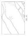
- FIG. 1is a cross section view through a portion of normal subcutaneous tissue.
- FIG. 2is schematic cross section illustrating septae and fat of normal subcutaneous tissue.
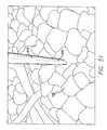
- FIG. 3is a schematic cross section illustrating septae and chambers of adipose tissue (fat) of normal subcutaneous tissue.
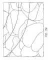
- FIG. 4is a schematic cross section illustrating septae and chambers of adipose tissue (fat) of abnormal subcutaneous tissue having an abnormality commonly referred to as cellulite.
- FIG. 5is a schematic representation of a focused ultrasound wave.
- FIG. 6is a schematic representation of an unfocused ultrasound wave.
- FIG. 7is a schematic representation of a near zone and a far zone of an ultrasound wave.
- FIG. 8is a schematic representation of a focused ultrasound wave.
- FIG. 9is a schematic representation of a focused ultrasound wave.
- FIG. 10is a schematic view of an oscillation of a gaseous bubble in the presence of an ultrasound wave.
- FIG. 11is a schematic view illustrating a gaseous body undergoing cavitation phenomenon.
- FIG. 12is a schematic view illustrating one embodiment of an assembly of the present invention.
- FIG. 13is a schematic view illustrating another embodiment of an assembly of the present invention.
- FIG. 14is a schematic view illustrating yet one other embodiment of an assembly of the present invention.
- FIG. 15is a schematic view illustrating one further embodiment of an assembly of the present invention.
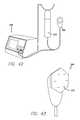
- FIG. 16is a perspective view of an assembly of the present invention.
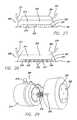
- FIG. 17is a perspective view of a handpiece of the present invention.
- FIG. 18is a perspective view of a handpiece of the assembly of FIG. 16 in a first configuration.
- FIG. 19is a perspective view of a handpiece of the assembly of FIG. 16 in a second configuration.
- FIG. 20is a perspective view of a handpiece of the assembly of FIG. 16 applied to skin of a patient to be treated.
- FIG. 21illustrates a handpiece configured to produce a solution including gaseous bodies.
- FIG. 22illustrates a handpiece configured to produce a solution including gaseous bodies.
- FIG. 23illustrates a handpiece configured to produce a solution including gaseous bodies.
- FIG. 24is a perspective view illustrating an injection member including a pad and hypodermic needles of one embodiment of an assembly of the invention.
- FIG. 25is an enlarged view through a portion of the injection member of FIG. 24 .
- FIG. 26is a cross section view showing another embodiment of an injection member including a pad and hypodermic needles inserted into subcutaneous tissue.
- FIG. 27is an illustration of a handpiece including a suction member.
- FIG. 28is an illustration of the handpiece of FIG. 27 , wherein some of the needles have penetrated epidermis of a patient to be treated.
- FIG. 29is a perspective view of a handpiece including at least one spring.
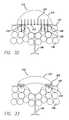
- FIG. 30is a perspective view of the handpiece of FIG. 29 in a first configuration.
- FIG. 31is a perspective view of the handpiece of FIG. 29 in a second configuration.
- FIG. 32is a schematic cross section view through an embodiment of an injection member having some needles deployed through the skin of a patient.
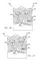
- FIG. 33is a schematic cross section view through an embodiment of an injection member having peripheral needles deployed through the skin of a patient.
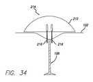
- FIG. 34is a schematic cross section view through an embodiment of an injection member having central needles deployed through the skin of a patient.
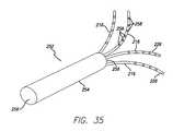
- FIG. 35is a perspective view of a fanned needle array.
- FIG. 36is a cross section schematic view showing a needle advancing towards a portion of subcutaneous tissue to be treated with a microbubble solution.
- FIG. 37is a cross section schematic view showing a needle advancing into the subcutaneous tissue of FIG. 36 .
- FIG. 38is a cross section schematic view showing the microbubbles solution being injected into the subcutaneous tissue to be treated of FIG. 36 .
- FIG. 39is a cross section schematic view showing the needle being withdrawn from the subcutaneous tissue of FIG. 36 , leaving microbubbles disposed in a superficial subcutaneous fat layer.
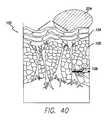
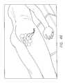
- FIG. 40is a cross section schematic view showing the application of ultrasound waves to the subcutaneous tissue of FIG. 36 , causing cavitation of the microbubbles disposed in the superficial subcutaneous fat layer and disruption of fat and septae.
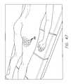
- FIG. 41is a graph illustrating an algorithm of a method of treatment of subcutaneous tissue using two different amplitudes of ultrasound.
- FIGS. 42-58illustrate a method of treatment of subcutaneous tissue.
- FIG. 59is a flow chart of an exemplary treatment algorithm.
- FIG. 60Ais a longitudinal cross sectional view through a needle having concentric lumens.
- FIG. 60Bis a horizontal cross sectional view through a needle having concentric lumens.
- FIG. 60Cis a longitudinal cross sectional view through a needle having side by side lumens.
- FIG. 60Dis a horizontal cross sectional view through a needle having side by side lumens.
- FIG. 61Ais a schematic cross-sectional side view illustrating the fanned needle array positioned in subcutaneous tissue.
- FIG. 61Bis a schematic partial cross-sectional top view illustrating the fanned needle array positioned in subcutaneous tissue.
- FIGS. 1 to 60are provided for purposes of illustration and by way of example, embodiments of the present invention are illustrated in FIGS. 1 to 60 .
- the present inventionrelates to a method and apparatus for treating subcutaneous tissue.
- the present inventionincludes an apparatus for treating soft tissue.
- the present inventionincludes a method for treating tissue.
- the present inventionfurther includes a method and apparatus for treating a subcutaneous fat layer including fat cells and septae.
- the present inventionfurther includes a method and apparatus for treating cellulite.
- the present inventionmay be useful for a temporary reduction in the appearance of cellulite or the permanent reduction of cellulite.
- the inventionmay also be used as an adjunct to liposuction.
- the inventionfurther provides for a subcutaneous infusion and ultrasonic dispersion of fluid to temporarily improve the appearance of cellulite.
- the inventionmay also be advantageous for a removal of benign neoplasms, for example, lipomas.
- the present inventionis directed to methods and apparatus for targeting and disrupting subcutaneous structures, such as collagen, connective tissue, adipose tissue (fat cells) and the like (collectively “target tissue” or “subcutaneous structures”) in order to improve the aesthetic appearance of the targeted region.
- Targeted regionsmay consist of any surface or contour of the human form that it is desirable to enhance, including the face, chin, neck, chest, breasts, arms, torso, abdominal region (including pelvic region), thighs, buttocks, knees and legs.
- the target tissuemay include the connective tissue or septae of the region, or the underlying tissues that may exacerbate the unwanted body contour, such as subdermal and deeper fat deposits or layers.
- Skin irregularitiesrefer to conditions that decrease a person's satisfaction with their outward appearance, such as cellulite, scarring, or fat deposits or excess fat in certain regions, such as neck, chin, breasts, hips, buttocks, abdomen, arms and the like.
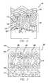
- FIGS. 1-3a cross section of a portion of a normal subcutaneous tissue 100 region is shown, including the epidermis 102 , dermis 104 , subcutaneous fat 106 , fibrous septae 108 , and deeper fat layers 110 .
- the subcutaneous tissuealso includes vascularity, microcirculation and lymph drainage.
- the dermisinterfaces with the fatty subcutaneous connective tissue that attaches to the dermal layers via substantially vertical septae 108 (collagenous fibers).
- the subcutaneous fatty tissue 106is compartmentalized into chambers 112 of adipose tissue (fat) separated by the fibers of the septae.
- FIGS. 1-3show a fairly normal skin cross section, not exhibiting skin irregularities. As shown in FIG. 4 , the subcutaneous fat layer is swollen and septae tightened, leading to the irregular skin surface characteristic of cellulite.
- a reserve or deeper fat layer 110is disposed beneath the subcutaneous fat 106 and may also contribute to a skin irregularity.
- the deeper fat layeris also included herein as one of the subcutaneous tissues that may be treated by at least one embodiment of the present invention.
- treatment of subcutaneous tissueincludes energy assisted subcision of the subcutaneous tissue 100 .
- treatment of subcutaneous tissueincludes disruption of the fibrous septae 108 .
- treatment of subcutaneous tissueincludes disruption of the subcutaneous fat cells 112 to lessen the outward pressure on the skin surface 102 that contributes to dimpling.
- treatment of subcutaneous tissueincludes disruption of a deeper fat layer 110 for overall surface contouring.
- the energy modalityis an acoustic wave.
- the energy modalityis ultrasound.
- Acoustic waves, including ultrasoundare energy that is transmitted through a medium, for example solution or biologic tissue.
- the inventionmay also include a solution including gas, for example, microbubbles.
- One category of medical ultrasound waveis high acoustic pressure ultrasound.
- Another category of medical ultrasound waveis low acoustic pressure ultrasound.
- the Mechanical Indexis a standard measure of the acoustic output in a diagnostic ultrasound system, defined as the peak negative pressure, or rarefactional pressure, of an ultrasound wave propagating in a uniform medium, divided by the square root of the centre frequency of the transmitted ultrasound pulse.
- the uniform mediumis assumed to have an attenuation of 0.3 dB/cm/MHz (attenuation coefficient divided by ultrasound frequency).
- the MImay determine the interaction of microbubbles with ultrasound, including the likelihood of bubble rupture. Some skilled in the art have suggested the bubble rupture is more likely with increasing MI.
- the mechanical index (MI)is intended to offer a rough guide to the likelihood of the occurrence of cavitational bioeffects.
- High acoustic pressure ultrasound systemsgenerally have a MI greater than 10.
- Low acoustic pressure systemsgenerally have a MI lower than 5.
- diagnostic ultrasound systemsare limited by law to a Mechanical Index not to exceed 1.9.
- Isppaspatial peak, peak average intensity
- the intensity of an ultrasound beamis greater at the center of its cross section than at the periphery. Similarly, the intensity varies over a given pulse of ultrasound energy. Isppa is measured at the location where intensity is maximum averaged over the pulse duration. Isppa for high acoustic pressure or HIFU applications ranges from approximately 1500 W/cm2. to 9000 W/cm2. Diagnostic ultrasound equipment, for instance, will generally have, and an Isppa less than 700 W/cm2.
- ultrasound wavescan be characterized by the amplitude of their peak negative pressure.
- High acoustic pressure or HIFU applicationsemploy waves with peak amplitudes in excess of 10 MPa.
- Low acoustic pressure ultrasoundincludes ultrasound waves will generally have peak negative pressures in the range of 0.01 to 5.0 MPa.
- Diagnostic ultrasound equipmentfor example, will generally have a peak amplitude less than 3.0 MPa
- Both high and low acoustic pressure ultrasound systemsgenerally operate within the frequency range of 250 KHz-10.0 MHz. Diagnostic imaging typically uses frequencies of about 1.0 MHz to about 5.0 MHz.
- One known low acoustic pressure ultrasound probemay produce ultrasound having a frequency as high as 7.5 MHz. Some low acoustic pressure ultrasound probes may produce ultrasound frequencies as high as 10.0 MHz.
- Physical therapy ultrasound systemsgenerally operate at frequencies of either 1.0 MHz or 3.3 MHz.
- High acoustic pressure ultrasound or high intensity focused ultrasoundhas been used for tissue disruption, for example for direct tumor destruction. This high intensity focused ultrasound is generally used for tissue disruption rather than for diagnostic purposes. High intensity focused ultrasound using high acoustic pressure ultrasound is most commonly focused at a point in order to concentrate the energy from the generated acoustic waves in a relatively small focus of tissue.
- high intensity focused ultrasoundalso referred to herein as high acoustic pressure ultrasound
- the tissue located at the focus point of the arraycan be heated to supraphysiologic temperatures in order to cause thermal necrosis of the tissue.
- the ablative zonecan be increased by exploiting a phenomenon known as cavitation.
- High intensity focused ultrasound (HIFU)can cause cavitation even in the absence of an exogenous enhancing agent, for example, an exogenous solution including microbubbles.
- Low acoustic pressure ultrasoundis generally used to provide a low intensity generally unfocused ultrasound wave ( FIG. 6 ) to the tissues.
- the ultrasound wave in low acoustic pressure ultrasoundis generally transmitted to the patient by a planar transducer.
- the low intensity generally unfocused ultrasound waveis described by some skilled in the art as including a focal zone 114 , a near zone 116 , and a far zone 118 .
- FIG. 7Low acoustic pressure ultrasound is readily available in the medical arts, for example, for cardiac imaging and fetal monitoring. Both diagnostic ultrasound systems and physical therapy systems fall within the range of low acoustic pressure systems.
- unfocused ultrasound waves using low acoustic pressure ultrasound systemshave not been utilized by clinicians for purposes of tissue disruption.
- the ultrasound wave usedis low acoustic pressure ultrasound.
- Cavitationis a physical phenomenon resulting from pressure changes in a fluid. Cavitation can occur in the absence of an exogenous microbubble solution when high intensity focused ultrasound (HIFU) or high acoustic pressure ultrasound is applied to tissues. Cavitation has been noted during the direct application of high intensity focused ultrasound into human tissue, for example, blood, brain, and prostate tissues. Cavitation involves the formation and collapse of microbubbles in a fluid due to pressure in the fluid reaching certain levels. In a fluid, when ultrasound waves are introduced at high power, the negative pressure part of the wave will reach a point that induces cavitation bubble formation as a result of the fluid reaching the vapor pressure level. Endogenous microbubbles may therefore be formed in tissues that are exposed to HIFU or high acoustic pressure ultrasound. However, HIFU is disadvantageous in that it produces a significant amount of heat and thermal damage to tissues.
- enhancing agentrefers to at least one of an exogenous gas, liquid, mixture, solution, chemical, or material that enhances the disruptive cavitational bioeffects of an ultrasound wave on tissue.
- an enhancing agentis an enhancing solution.
- the enhancing solutioncontains exogenous gaseous bodies, for example, microbubbles.
- Other enhancing agentsare described in more detail herein.
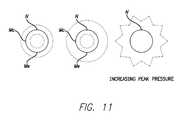
- Nis a microbubble in its normal state
- Cis the microbubble in a compressed state
- Eis the microbubble in an expanded state.
- the dashed line “D”shows the diameter of the microbubble in the normal state “N.”
- FIG. 11shows a series of oscillating microbubbles exposed to increasing levels of peak negative pressures.
- the inner dashed line “Mc”shows maximum compression
- the outer dashed line “Me”shows maximum expansion.
- Microbubbles(either endogenous or exogenous), being compressible, alternately contract and expand in an ultrasound field. These expansions and contractions may be generally equal and symmetrical at lower ultrasound pressures. This behavior is referred to by some skilled in the art as moderately oscillating. As the ultrasound driving pressure increases, more complex phenomenon occurs, for example, with bubble expansion larger than contraction. Furthermore, there may be relatively slow expansion followed by rapid collapse. This behavior is referred to by some as strongly collapsing. It is associated with the production of harmonic signals. The transition from the moderately oscillating to the strongly collapsing state may be abrupt, wherein the microbubble implodes and releases energy to tissue in the proximity of the microbubble. The energy released by bubble implosion, when bubbles are exposed to ultrasound, is one factor producing observed subcutaneous cavitational bioeffects.
- the present inventionmay advantageously introduce exogenous microbubbles to the target tissue, and then use low power ultrasonic energy to cavitate the microbubbles and destroy the target tissue by subcutaneous cavitational bioeffects without the generation of enough heat for direct thermal injury to the tissue being treated.
- Microbubbles infiltrated into the tissue by way of direct injectionwill also serve as a nidus for cavitation and tissue disruption.
- the present inventionmakes use of the microbubble cavitational bioeffects to destroy subcutaneous tissues without significant thermal effects.
- the subcutaneous cavitational bioeffects produced by the present inventionare advantageous for the disruption of superficial and/or deep fat and/or septae, for example, for the treatment of cellulite and focal fat deposits.
- exogenous gas, gaseous bodies, including microbubbles and nanobubbleshas several advantages in addition to being a non-thermal ablative technique.
- Ultrasound waves in the range of those produced by commercially available physical therapy ultrasound machines and diagnostic ultrasound machinesare provided in a preferred embodiment of the present invention.
- the safety of these low intensity machines and more specifically the safety of the ultrasound waves they produceis well established. Normally, these low intensity ultrasound machines only cause disruptive cavitation effects in the presence of microbubbles.
- cavitational bioeffectsoccur in animals exposed to ultrasound waves when ultrasonic contrast agents are present (Miller and Gies 1998, Skyba et al. 1998).
- the implosion of microbubbles when exposed to ultrasoundis one factor producing observed subcutaneous cavitational bioeffects.
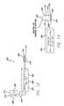
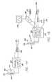
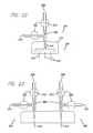
- the assembly 200includes an acoustic wave system, for example, an ultrasound system that includes an ultrasound generator 202 configured to drive the electronics and a transdermal transducer 204 configured to interface the ultrasound waves through the patient's skin.
- the assembly 200further includes an agitation system 208 configured to agitate and/or mix a solution 210 and an injection member 214 configured to inject the solution subdermally.
- the assemblyfurther provides a variety of solutions known in the art.
- the solutionmay include, for example, saline, normal saline, hypotonic saline, a hypotonic solution, a hypertonic solution, lidocaine, epinephrine, a tumescent solution, and/or microbubble solution.
- the acoustic wave generator 202 and the acoustic wave transducer 204may include an ultrasound generator and an ultrasound transducer.
- the apparatusmay further include a source of gas 206 .
- the apparatusmay further include a solution agitator 208 for providing a solution 210 including gaseous bodies 212 , for example, microbubbles, or other enhancing agents.
- the inventionfurther includes a solution injection member 214 .
- the assemblymay also include a container 220 for storing the solution 210 , for example a reservoir for storing the solution therein.
- the reservoirmay be an IV bag known in the art.
- the assemblymay also include a source of gas 206 .
- the source of gasmay be room air or a various other biocompatible gases.
- the source of gasis gas enclosed in a container or a tank of gas.
- the source of gasis atmospheric room air that is drawn into the apparatus by the agitator including an opening in fluid communication with the atmosphere.
- the solution and the gas 212flow into the agitator 208 where an injectable solution including gaseous bodies 212 is produced. The injectable solution including gaseous bodies then flows to the injection member 214 and is available for injection into the tissue 100 to be treated.
- the physicianmay prepare and hang their selected solution, and the assembly 200 mixes, injects & insonates according to a pre-programmed or a user defined algorithm.
- the algorithmmay be programmed into a controller 228 .
- the controllermay be included in a unitary assembly with the other components, or may be a separate unit configured to communicate with the other components of the assembly.
- the controllerincludes a processor and memory.
- the controllermay also include inputs 236 , for example, electrical switches, buttons, or keypad.
- the controllermay also include outputs 238 , for example, LED lights, an LCD screen, gauges, or other screens and output indicators known in the art.
- the inputs 236 and outputs 238may be separate from the controller but in electrical communication with the controller.
- the assemblyis configured to first mix and agitate the solution 210 in the agitator 208 .
- the assemblyis configured to thereafter inject the solution into the patient using the injection member 214 .
- the assemblyis also configured to insonate the injected tissue 100 using the ultrasound transducer 204 .
- At least one hypodermic needle 216is disposed in the solution injection member 214 .
- the solution injection membermay be configured with retractable hypodermic needles 216 .
- the solution injection memberis configured with a pad 232 ( FIGS. 24-25 ).
- the apparatusmay further include a tissue cooling module 218 .
- the acoustic wave generator 202is a low acoustic pressure ultrasound wave generator.
- Various acoustic wave generators 202 and acoustic waves transducers 204are well known in the art and commercially available.
- the ultrasound generator and the ultrasound transducerare of the type commercially available for use in diagnostic ultrasound.
- the ultrasound generator and the ultrasound transducerare of the type commercially available for use in physical therapy.
- One example of a commercially available low acoustic pressure ultrasound machineis the Sonicator 730 available from Mettler Electronics Corp., located in Anaheim, Calif.
- the Sonicator 730is a 1.0 MHz and 3.3 MHz therapeutic ultrasound unit which comes with a choice of up to four applicators and operates in either continuous or pulsed modes.
- the Sonicator 730has a maximum intensity of 2.2 W/cm2 with all applicators.
- Other examples of commercially available low acoustic pressure ultrasound machinesare the Acuson Aspen and Acuson Sequoia available from Siemens, AG located in Munich, Germany and in Malvern, Pa., U.S.A.
- Yet another example of a low acoustic pressure ultrasound machineis the Sonoporator available from G. Heinemann Ultraschall, Germany.
- the peak negative pressure wave generated by the ultrasound generator 202is less than 10.0 MPa. In at least one embodiment, the peak negative pressure wave generated by the ultrasound generator is less than 5.0 MPa. In another embodiment of the present invention, the peak negative pressure wave generated by the ultrasound generator is less than 4.0 MPa. In one further embodiment of the present invention, the peak negative pressure wave generated by the ultrasound generator is less than 3.0 MPa.
- the MI of the ultrasound generator 202is in the range of 0.01 to 5.0. In one embodiment of the present invention, the MI is in the range of 0.1 to 4.0. In yet one further embodiment of the present invention, the MI is in the range of 0.5 to 3.0. In yet another embodiment of the present invention, the MI is in the range of 0.7 to 2.5.
- the ultrasound generator 202operates at a frequency range of about 100 KHz to about 10.0 MHz. In another embodiment of the invention, the ultrasound generator operates at a frequency range of about 250 KHz to about 5.0 MHz. In yet another embodiment of the invention, the ultrasound generator operates at a frequency of range of about 500 KHz to about 3.0 MHz. In a further embodiment of the invention, the ultrasound generator operates at a frequency range of about 750 KHz to about 2.0 MHz.
- the transducer 204is adapted for the external application of ultrasound waves through the epidermis 102 of the patient.
- the transduceris adapted for the penetration of the ultrasound energy into the subcutaneous tissue 100 .
- the transduceris adapted for the penetration of the ultrasound energy into the subcutaneous fat 106 .
- the transduceris adapted for the penetration of the ultrasound energy into the fibrous septae 108 .
- the transduceris adapted for the penetration of the ultrasound energy into the deeper fat layer 110 .
- the transduceremits poorly focused or unfocused ultrasound waves.
- the poorly focused or unfocused wavesare emitted by a planar transducer.
- HIFUwhich emits discretely focused or point focused ultrasound waves.
- HIFU wavesare emitted from hemispherical transducers, an array of transducers, for example, transducers arranged on a hemispherical substrate, or a planar transducer whose waves are focused to mimic a hemispherical transducer by the use of an acoustic lens.
- the transducer 204is included in a handpiece 264 having a base 266 that is polygonal in shape.
- the basehas between three to six sides.
- the polygonal shape of the baseis advantageous in mapping out and treating large tissue regions in a patient. A plurality of contiguous areas on the patient's epidermis 102 , each area corresponding to the size and shape of the polygonal base, may be mapped out on the patient over the region to be treated. Each polygonal area can then be sequentially treated, thereby efficiently treating all or a portion of the mapped out treatment region.
- a polygonal shaped handpiece baseis not meant to be restrictive, and in other embodiments the base may be other shapes, for example, round, triangular, square, polygonal, cylindrical, spherical, or pyramidal.
- a plurality of interchangeable handpieces, some various shaped basesmay be provided.
- Various shaped basesmay be advantageous in getting the best interface between the base and the patient's skin, wherein the ultrasound waves are more effectively applied to the patient.
- the transducer 204is preferably interfaced with the patient's epidermis 102 , wherein the loss of acoustic wave is minimized as it passes into the patient.
- the transducermay be interfaced with the patient's epidermis in any number of ways known in the art.
- commercially available ultrasound gelis utilized between the transducer head and the epidermis as well known in the art.
- the acoustic waveis interfaced between the transducer and the epidermis utilizing degassed water. Use of degassed water may be advantageous in reducing patient discomfort compared to ultrasound gels.
- a topical anesthetic gel or cream known in the artmay be applied to the patient's skin before the procedure to minimize discomfort.
- the ultrasound gelmay include a topical anesthetic. In the event of using a topical anesthetic, it may not be necessary to include lidocaine as part of the enhancing solution.
- the inventionincludes a source of gas 206 for inclusion in a solution 210 including gaseous bodies 212 .
- the source of gasis room air.
- the room airmay be supplied contained, as a part of the system, or it may be drawn in by the system from uncontained atmospheric room air.
- the source of gasmay be a tank of gas or a reservoir of gas.
- the source of gasmay be a disposable gas cartridge.
- the disposable gas cartridgemay include an impeller.
- the disposable gas cartridgemay include gas under pressure. In one embodiment, the gas under pressure in the gas cartridge is released into the solution to create the solution including microbubbles.
- the gasmay include oxygen, super-saturated oxygen, nitrogen, carbon dioxide, helium, or other biocompatible gases.
- gases that may be usedinclude gases used in presently known encapsulated echogenic contrast agents, including those described elsewhere herein, for example, air, perfluorocarbons, perfluorobutane, perfluorohexane, octafluoropropane, dodecafluoropentane, perfluoropropane, sulphur hexafluoride.
- a pressurized solution including bubblesis provided. The bubbles are dissolved in the solution, because the solution is under pressure. After the solution is injected into the patient's tissue the pressure of the solution is lowered, wherein the gaseous bodies come out of solution and may form microbubbles in the tissue to be treated.
- the source of gas 206 and gaseous bodies 212 as used hereinincludes microbubble precursors that may be liquid at room temperature, prior to injection into the patient, and become gaseous after injection into the patient, at physiologic temperatures.
- a gaseous precursor that is liquid at room temperature and gaseous at physiologic temperatureis methylactate.
- a gaseous precursormay be a liquid when pressurized prior to injection and become a gas under atmospheric pressure or physiologic biological tissue pressures.
- Other suitable materials for gaseous bodiesare disclosed in U.S. Pat. No. 6,302,863 to Tankovich, filed Jun.
- the present inventionalso includes a variety of solutions 210 that are biocompatible with subcutaneous injection into the subcutaneous tissue 100 of a patient.
- the solutionis a tumescent solution.
- Tumescent solutionsare specially adapted to provide for the application of local anesthesia and are well known in the art.
- Tumescent solutionsmay include a variety of medicated solutions.
- a tumescent solutionis a solution that includes 1000 milliliters of normal saline with 2% lidocaine, 30 ml. (600 mg) of epinephrine, and one mole (12.5 ml or 12.5 mg.) of sodium bicarbonate.
- At least one other example of a tumescent solutionis a solution that includes 1000 milliliters of normal saline, 50 ml of 1% lidocaine, and 1 cc. of 1:1000 epinephrine. These additives are commercially available. Tumescent solutions may decrease bleeding at the treatment site and provide for local anesthetic effects that decrease pain during and after the procedure.
- the solutionis a normal saline solution.
- the solutionis a hypotonic solution.
- the hypotonic solutionmay be beneficial in making the fat cells more susceptible to disruption by ultrasound.
- the solutionis a solution including microbubbles or nanobubbles.
- the solutionmay be agitated between two syringes one or more times to produce a solution including microbubbles.
- Several solutions including microbubbles or nanobubblesare commercially available, as described in detail elsewhere herein.
- the inventionmay also include a container 220 for the solution 210 , for example a reservoir for storing the solution therein.
- the tumescent solution and/or the hypotonic solutionmay be mixed in the agitator 208 .
- the tumescent solution and/or the hypotonic solutionmay be mixed with gas in the agitator 208 to provide a solution including gaseous bodies 212 .
- treatment enhancing agentsmay be added to the agitator 208 and/or injected into the tissue 100 to be treated in conjunction with the application of the various energy modalities.
- the enhancing agent includeddepends on the desired effects, some of which are detailed below.
- enhancing agentsmay be transmitted transdermally, or via injection into the tissue to be treated.
- Treatment enhancing agentsinclude, anesthetics such as lidocaine, a surfactant, vasoconstrictive agents such as epinephrine, hypotonic saline, potassium, agitated saline, microbubbles, commercially available ultrasound contrast agents, microspheres, adipocytes, fat, autologous tissues (e.g.
- Treatment enhancing agentsmay be delivered prior to, during or following the application of acoustic waves to the subcutaneous tissue.
- the present inventionincludes a solution agitator 208 for providing a mixed solution 210 .
- the solutionmay be agitated with gas to produce a solution including gaseous bodies 212 .
- the gaseous bodiesmay include microbubbles or nanobubbles as discussed in greater detail herein.
- the gaseous bodiesin their simplest form may be made from room air.
- gases known in the artfor example, carbon dioxide, nitrous oxide, helium, perfluorocarbons, perfluorobutane, perfluorohexane, octafluoropropane, dodecafluoropentane, perfluoropropane, and sulphur hexafluoride.
- Liquids that become gases after injectionmay also be introduced into the solution in the agitator.
- examples of such chemicalsinclude methylactate and other agents described in U.S. Pat. No. 6,302,863 to Tankovich, the entirety of which is included herein by reference.
- Various other enhancing agents, as described elsewhere herein,may also be disposed into the agitator for inclusion in the solution.
- the container 220 of solution 210 and the source of gas 206are connected with the solution agitator 208 .
- the solution and gasare delivered into the solution agitator, for example, by one or more pumps 222 ( FIG. 12 and FIGS. 21-23 ) or by gravity flow, and mixed together within the solution agitator.
- the flow of the gas or enhancing agents into the solutionmay be controlled by the control module 228 .
- the solution agitatormay be as simple as two syringes connected together. Gas or enhancing agents in one syringe may be mixed with the solution in the other syringe to produce the solution for injection.
- the solutionmay be re-agitated one or more times, from one syringe to the other, during the procedure to reconstitute the bubbles.
- Solution agitators 208 and microbubble generatorsare known in the art.
- An example of a microbubble generatoris described in U.S. Ser. No. 10/798,876 filed Mar. 11, 2004 and entitled “APPARATUS, SYSTEM AND METHOD FOR GENERATING BUBBLES ON DEMAND,” the entirety of which is included herein by reference.
- the solution agitator 208may be included in the solution injection member 214 , wherein the microbubbles are produced in the handpiece 264 .
- solution 210 and gas 212are forced by a pump 222 through a filter 260 , wherein a solution including gaseous bodies is produced in the solution injection member.
- the filtermay include, for example, a micro-filtration paper or a mesh.
- the filtermay include a fenestrated member made from, for example, a fiber, metal, plastic, hydrophobic sintered plastic, or glass.
- a solution 210is pumped across a filter without the introduction of additional gas.
- a filter when dry and exposed to gascontains many small partially trapped spaces of the gas.
- the filterWhen the filter is placed in a circuit of flowing solution, the partially trapped spaces of gas are released in to the flowing solution, forming a plurality of bubbles in the solution that correspond in size to the originating space of the trapped gas.
- the filtermay be configured to limit the size of microbubbles that are injected through the needles 216 .
- the agitorincludes at least one metered orifice 262 .
- the solution 210 and gas 212are mixed through the at least one metered orifice 262 , wherein a solution including gaseous bodies is produced in the solution injection member 214 .
- the ratio of the liquid orifice to the gas orificeis in the range of about 100:1 to about 1000:1.
- the liquid linehas an inner diameter of about 0.100 inch and the gas orifice diameter is about 0.001 inch.
- the solution and the gasintersect proximal to one or more injection needles 216 , wherein the solution including gaseous bodies enters one or more of the needles.
- At least one of the needlesreceive microbubbles from one metered orifice and at least one other needle receives microbubbles from at least one other metered orifice.
- a plurality of gas orificesmay be arranged around a liquid orifice.
- the injection pressure through the metered orificewill depend on several factors.
- One factoris the diameter of the orifice.
- Another factoris the viscosity of the fluid and gas.
- Another factoris the ratio of bubbles to solution desired.
- the pressure range with which the solution is fed into the injectoris between 25 psi to 100 psi, and the pressure range with which the gas is fed through the orifice is between 50 psi and 125 psi.
- the pressure with which the solution is fed into the injectoris between 50 psi, and the pressure with which the gas is fed through the orifice is 75 psi.
- the gaseous bodies 212are formed at the distal end 226 of at least one of the needles 216 .
- the needlesmay have at least two lumens 225 .
- the lumensmay be configured side by side ( FIGS. 60C-60D ) or concentrically ( FIGS. 60A-60B ).
- Solutionis forced out of one lumen and gas is forced out of another lumen of at least one needle to form a solution including gaseous bodies.
- the solution including gaseous bodiesis formed within the subcutaneous tissue of the patient as the gas and solution are injected simultaneously. The gas and solution are agitated together either immediately prior to exiting the needle tip, or as they enter the subcutaneous tissue adjacent to each other.
- the solution 210 to be injectedfor example, the solution including the gaseous bodies 212 , is delivered to the solution injection member 214 .
- at least a portion of the injector mechanismmay be built into the housing for the solution agitator 208 .
- power to the solution injection memberis included within the solution injection member. In another embodiment, power to the solution injection member is located externally to the solution injection member. For example, power to the solution injection member may be supplied by the controller 228 . In at least one embodiment, algorithms controlling the injection volume, depth, timing, and synchronization of injection with the application of ultrasound may be included in memory and/or a processor included within the solution injection member. In at least another embodiment, algorithms controlling the injection volume, depth, timing, and synchronization of injection with the application of ultrasound may be included in memory and/or a processor located externally to the solution injection member, for example, in the controller.
- the solution injection memberincludes at least one hypodermic needle 216 .
- the hypodermic needlehas a proximal end 224 connected to the solution injection member and a distal end 226 configured for penetrating into the subcutaneous tissue 100 ( FIG. 1 ).
- the distal ends of the needlesmay be beveled (not shown) as known in the art for less traumatic penetration into the skin.
- the needlesmay include microneedles.
- the needlesmay be pyramid shaped (not shown).
- the solution injection memberincludes a plurality of hypodermic needles.
- the hypodermic needlehas a tubular channel having a central lumen 234 configured for flow of the solution through the needle and into the tissue.
- the solution injection memberincludes an actuation element (not shown) for moving the hypodermic needle from a position inside the solution injection member to a position wherein the needle may penetrate through the epidermis 102 and into the subcutaneous tissue to be treated.
- the needlesare configured to penetrate at least into the subcutaneous fat 106 . In yet one other embodiment, the needles are configured to penetrate into the deep fat layer 110 .
- the assemblyincludes a solution injection member 214 having a reservoir or pad 232 capable of conforming to the skin surface of a patient.
- the padcontains at least one hypodermic needle 216 extending therefrom.
- the hypodermic needleis configured to inject solution 210 into the subcutaneous tissue 100 .
- a central lumen 234 of the needleis in fluid communication with the solution in the agitator 208 .
- the distal end 226 of the needle 216may be deployed manually out of the injection member.
- the distal end 226 of at least one needle 216may be deployed out of the injection member 214 automatically by the control mechanism 228 .
- the base 266 of the handpiece 264may include a suction member 268 for sucking the patient's skin 102 up towards the injection member 214 and/or the transducer 204 using subatmospheric pressures.
- the suction memberincludes a suction tube 270 that may connect to a mechanical pump (not shown), hand pump (not shown) or other source of subatmospheric pressure.
- the suction memberis configured to remove room air located between the skin of the patient and the transducer.
- the handpiece 264further includes a second tube 271 configured for delivering a volume of ultrasound coupling fluid 276 between the skin 102 and the transducer 204 .
- the ultrasound coupling fluidis a degassed liquid.
- the coupling fluidmay be removed by the suction tube 270 or by a third tube (not shown).
- the coupling fluidis delivered under the base 266 of the handpiece, by a plurality of openings (not shown) in the base that are in fluid communication with the second tube.
- the fluidmay be removed from under the base by a plurality of openings (not shown) in the base that are in fluid communication with the suction tube.
- the coupling fluidis advantageous is providing an interface with consistent acoustic properties between the transducer and the skin of the patient.
- the coupling fluidis also advantageous in providing a flow of acoustically efficient interface fluid through the treatment area while the suction is applied, since the peaks of cellulite may create valleys of air pockets that preferably are filled with fluid to better transmit ultrasound.
- the skinis sucked up towards needles 216 that are deployed out of the handpiece 264 in the second configuration 246 before the suction is applied to the skin 102 . Thereafter, suction is applied to the skin and the skin is sucked up towards the base 266 of the handpiece, wherein the needles penetrate through at least the epidermis 102 of the patient to be treated.
- the handpieceis placed on the patient in the first configuration 244 , wherein the distal ends 226 of the needles are inside the handpiece. Suction is then applied to pull the skin up against the base of the handpiece. Thereafter, the needles may be deployed into the second configuration, wherein the needles penetrate through at least the epidermis 102 of the patient to be treated.
- the solution injection member 214is configured to deploy at least one needle 216 out of the injection member and through openings 205 in the transducer 204 as the handpiece 264 is pressed against the patient's skin.
- the injection membermay be configured to fit into a recess of the transducer housing.
- the handpiece 264includes at least one spring 272 ( FIG. 29 ) positioned between the injection member and the transducer.
- the needlesare disposed completely inside of the handpiece ( FIG. 30 ).
- the distal end of the needle 226is disposed through openings in the transducer, out of the handpiece ( FIG. 31 ).
- a safety lock 278is provided to prevent the needles from being deployed into the second configuration unintentionally.
- the springis compressed and the needles are deployed from the first configuration to the second configuration.
- the springforces the injection member away from the transducer, wherein the needles retract back into the handpiece from the second configuration to the first configuration.
- a bladder(not shown) may be distended with air or fluid to deploy the needles through the dermal layers of the patient's skin.
- the handpiecealso includes at least one connector 274 configured for electrical and/or fluid connection to the ultrasound generator, the source of solution, and/or the source of gas.
- the solution including gaseous bodiesmay be produced in the handpiece as described in detail elsewhere herein.
- an O-ringmay be included in the handpiece between the transducer and the injection member.
- the distal end of the needlemay be deployed automatically out of the injection member. Movement of the needles between the first configuration and the second configuration may be controlled by the controller 228 . In at least one embodiment, a motor may be included in the injection member for automatic deployment of the needles between the first configuration and the second configuration.
- the injection member 214may be configured to retain at least one needle 216 in the first configuration 244 , within the housing, while deploying the distal end 226 of at least one other needles 216 in the second configuration 246 , out of the housing. In one embodiment illustrated in FIG. 32 , the injection member may be configured to retain at least one needle 216 in the first configuration 244 , within the housing, while deploying the distal end 226 of at least one other needles 216 in the second configuration 246 , out of the housing. In one embodiment illustrated in FIG.
- the injection memberis configured with at least two needles disposed peripherally and a paucity of needles disposed centrally, wherein a greater concentration of microbubbles may be delivered to the peripheral tissues, for example the subcutaneous fat 106 .
- the injection memberis configured with at least two needles disposed centrally and a paucity of needles disposed peripherally, wherein a greater concentration of microbubbles may be delivered to the central tissues, for example the septae 108 .
- the solution injection member 214is mounted behind the acoustic wave transducer 204 .
- the acoustic wave transducermay be configured with a plurality of holes, wherein the needles included in the solution injection member are configured and aligned to pass through at least some of the holes in the transducer.
- the injection needles diametermay range in size from 40 gauge to 7 gauge. In one embodiment the injection needles include size 30 gauge. In another embodiment the injection needles include size 28 gauge. In one further embodiment the injection needles include size 25 gauge. In one additional embodiment the injection needles include size 22 gauge. In yet another embodiment the injection needles include size 20 gauge. In still one other embodiment the injection needles include size 18 gauge.
- the needlesmay all be of one length or may be of different lengths. In one embodiment, the length of the needles are between 2.0 mm long and 10.0 cm long. In one embodiment, the length of the needles are less than 5 mm long. In another embodiment, the length of the needles are in the range of 5.0 mm to 2.0 cm.
- the length of the needlesare in the range of 1.0 cm to 3 cm. In yet another embodiment, the length of the needles are in the range of 2.0 cm to 5 cm. In still another embodiment, the length of the needles are in the range of 3.0 cm to 10.0 cm.
- the injection needlesinclude microneedles.
- the diameter of the microneedlesmay be in the range of 20 microns to 500 microns.
- the length of the microneedlesmay be in the range of 100 microns to 2000 microns.
- the needlesare long enough to reach from the epidermis to the deep fat layer.
- the needlesare long enough to reach from the epidermis to the muscle layer.
- further anesthesiamay be applied to the area to be treated using topical anesthetic creams or gels, local hypothermia, or regional blocks. Topical anethetic may be the only anesthetic necessary and may take the place of any lidocaine used as an enhancing agent.
- the needles 216may be long enough to extend into the subcutaneous tissue 100 a distance of 0.2 mm to 40 mm from the skin surface, depending on the target tissue to be treated.
- the needleis long enough to allow the distal end 226 of the needle to extend at least through stratum corneum.
- stratum corneumFor example, to treat cellulite a depth of penetration from 1.0 mm-5.0 mm may be desired, and for deeper subcutaneous fat, a depth of 3.0 mm-40 mm.
- One or more hypodermic needlemay be moved to various depths manually or automatically by the controller 228 .
- the needlesare long enough to reach from the epidermis to the deep fat layer.
- the needlesare long enough to reach from the epidermis to the muscle layer.
- the present apparatusis configured to provide staged depths of injection from the deeper tissue layer to the more superficial tissue layer with application of acoustic waves between each stage of injection.
- One or more hypodermic needlemay be moved to various depths manually or automatically by the controller 228 wherein the tissue can be treated with the ultrasound in staged depths as described further below.
- the apparatus 200is configured to allow activation of the ultrasound wave generator 202 at various times after injection of the solution 210 by the injection member 214 , as described in more detail below.
- the controller 228may be used to synchronize the timing of the ultrasound application following the injection of the solution into the tissue to be treated.
- the injection memberis further configured with an on switch to start at least the injection of the solution into the tissue to be treated.
- the injection membermay be configured with a stop switch to stop the injection and/or withdraw the needles 216 from the patient.
- the apparatus 200is configured to provide two different amplitudes of ultrasound waves.
- the subcutaneous tissue 100 to be treatedis first infiltrated with the solution 210 including gaseous bodies 212 using the injection member 214 .
- the ultrasound wave generator 202is configured to generate a first low amplitude ultrasound wave that is applied through the transducer 204 to disperse the solution in the tissue to be treated.
- the ultrasound wave generatoris also configured to generate thereafter a second higher amplitude ultrasound wave that is applied through the transducer to the tissue to be treated, therein providing cavitation of the bubbles 212 in the injected solution and disruption of the tissues, for example by cell injury or cell death.
- a fanned needle array 252may be provided for insertion through the skin to the targeted subcutaneous tissue 100 .
- the fanned needle arraymay include a tubular member 254 , for example a catheter, trocar, or needle having a central channel 256 .
- multiple needles 216are disposed in the central channel.
- the multiple needlesinclude microneedles in the size range of 20 microns to 500 microns.
- multiple needles 216are disposed in the central channel and configured to be extended out from the central channel or retracted into the central channel. The epidermis and dermis may be penetrated by the tubular member.
- the central tubular membermay have a beveled tip to facilitate penetration into the subcutaneous tissue.
- the plurality of needlesare configured to splay out circumferentially from the central channel of the central tubular member, wherein the microneedles may pierce multiple septae.
- the microneedleshave channels or lumens through which to provide distribution of the solution to the subcutaneous tissue to be treated.
- the needlesmay include needles having at least two lumens 225 ( FIGS. 60A-60D ), wherein a solution including bubbles is created at the distal end 226 of the needle as the solution 210 and gas 212 exit the needle.
- the needle lumensmay be concentric or side by side.
- the circumferential splaying out of the microneedlesis advantageous in disrupting the septae and providing a more even distribution of the solution.
- the fanned needle arrayis also advantageous because a larger area of tissue may be more evenly injected through at least one puncture wound through the skin. This may reduce the risk of infection and the amount of pain experienced by the patient during the injection.
- the needles 216may be employed through the skin 102 , 104 through the main tubular member 254 , and “fan out” in an orientation substantially horizontal (parallel) to the skin surface.
- the subcutaneous structuressuch as the fibrous septae may be disrupted.
- the microneedlesmay be configured with additional outlet ports 258 along the length of the microneedles.
- the needlesinclude sharp cutting elements.
- the fanned needle array 252can efficiently disrupt multiple septae 108 in one rotation or retraction.
- some of the needlesmay be solid without central channels and at least one other may have a channel for injection.
- at least one needle in the needle arrayis further configured for delivery of electrical energy through the needle to heat tissue or disrupt tissue.
- at least a portion of the needleis electrically insulated, wherein the electrical energy is dispersed to the tissue over only a portion of the needle.
- the inventionmay also be combined with subcision procedures and apparatus disclosed in U.S. Pat. No. 6,916,328 to Brett, filed Jul. 22, 2002, and entitled “PERCUTANEOUS CELLULITE REMOVAL SYSTEM”, the entirety of which is incorporated herein by reference. Combination with subcision may be particularly advantageous in areas of severe cellulite pathology.
- the apparatus 200includes a cooling module 218 .
- Injection into the skin of a patientmay commonly be associated with the side effect of discomfort, swelling, bleeding, scarring or other undesired effects.
- the disruption of subcutaneous tissues 100 treated by the present inventionmay also result in some side effects common to many cosmetic or dermatologic treatment.
- the use of a cooling modulereduces the side effects of the treatment with the invention. Cooling of the tissues reduces bleeding, swelling, and discomfort.
- the cooling modulemay include any of the many known methods of cooling tissue known in the art. As shown in FIG. 12 , in at least one embodiment a portion of the cooling module may be included with the transducer 204 or the injection member 214 .
- cooling moduleis to assist in treatment or prophylaxis of discomfort, swelling, scarring and other undesired effects associated with treatments of the present invention.
- the cooling moduleis included in the assembly as a separate module.
- the transducer 204 and the injector member 214may be separate elements which attach together for use.
- the transducermay be reusable and at least a portion of the injector member may be disposable.
- the transducermay be reusable and at least a portion of the injector member may be reusable.
- the transducermay be disposable and at least a portion of the injector member may be disposable.
- the transducermay be disposable and at least a portion of the injector member may be reusable.
- a combination transducer and injection memberis provided in a unitary handpiece. The unitary handpiece may attach to a common fitting that communicates with the other parts of the assembly described herein.
- the unitary handpiecemay be provided in different sizes that are configured to treat different subcutaneous abnormalities or different severities of subcutaneous abnormalities.
- One unitary handpiecemay be quickly exchanged for a different size unitary handpiece.
- One unitary handpiecemay have a more dense pattern of needles 216 than another. For example, a more severe area of cellulite may be treated with the handpiece having the more dense pattern of needles.
- the apparatus 200is advantageously configured to integrate and coordinate the generation of the microbubble solution, the injection of the microbubble solution into the tissue to be treated, the dispersion of the microbubble solution in the tissue to be treated, and the insonation of the microbubble solution in the tissue to be treated.
- the timing of the insonation after the injection of the microbubbles solution into the tissue to be treatedmay be one factor in getting an even and consistent effect of the treatment of subcutaneous tissues.
- the depth of treatmentmay be another factor in getting consistent bioeffects of the treatment. Otherwise, the patient may have an outcome of a lumpy skin contour.
- a contemporaneously prepared microbubble solution for injectionmay be yet another factor in getting a consistent and evenly distributed result.
- the apparatusis advantageously configured to control these factors and others wherein the apparatus produces consistent and uniform subcutaneous cavitational bioeffects in the tissue that is treated.
- a security chip(not shown) may be provided in the handpiece to prevent re-use of the handpiece on other patients, thereby preventing the spread of disease, for example, hepatitis or aids.
- the security chipmay also be included to prevent counterfeit handpieces from being distributed and used on patients.
- volume of injected solution per skin surface area of a location to be treatedmay be a volume of injected solution per skin surface area of a location to be treated.
- the volume of injectionis in the range of about 0.1 cc/sq cm of skin surface area in the location to be treated to about 2.0 cc/sq cm of skin surface area in the location to be treated.
- the volume of injectionis in the range of about 0.25 cc/sq cm of skin surface area in the location to be treated to about 1.5 cc/sq cm of skin surface area in the location to be treated.
- the volume of injectionis in the range of about 0.5 c/sq cm of skin surface area in the location to be treated to about 1.0 cc/sq cm of skin surface area in the location to be treated.
- the above volumes to be injectedare exemplary only, and may be varied depending on the pain tolerance of the individual patient treated and the depth of the fat layer in the location to be treated.
- the rate of injection of the solution into the tissue to be treatedmay be in the rate of injection of the solution into the tissue to be treated.
- the rate of injection of the solutionis in the range of about 0.01 cc/second to about 1.0 cc/second.
- the rate of injection of the solutionis in the range of about 0.02 cc/second to about 0.5 cc/second.
- the rate of injection of the solutionis in the range of about 0.05 cc/second to about 0.2 cc/second.
- the above rates of injectionare exemplary only, and may be varied depending on the pain tolerance of the individual patient treated and the pathology of the fat layer in the location to be treated.
- the components of the assembly 200include an acoustic wave generator 202 , an acoustic wave transducer 204 , a solution agitator 208 , and a solution injection member 214 .
- all system componentsmay be disposed into a single unit, or the assembly may include separate components.
- one or more of the components of the assemblyis discrete.
- Disposable canistersprovide the supply of gas 206 and a container 220 providing solution 210 are included in the system.
- An electrical control unit 228 including input, output, memory, and processoris provided for the assembly.
- the control unitprovides for flow of solution and gas into the agitator 208 that then agitates the solution-gas mixture.
- the control unitthen further directs the injector 214 to injects the solution including gaseous bodies into the tissue.
- the control unitthen further controls the ultrasound generator 202 and transducer 204 to insonate the tissue to be treated according to a pre-programmed or user defined algorithm.
- the control unit 228drives the ultrasound generator 202 and transducer 204 only.
- the gas supply 206 , the solution 210 , the agitator, 208 , and the injector 214are all contained in a single use, disposable, pre-filled unitary disposable module 230 .
- the transduceris reusable.
- the transduceris disposable and built into the unitary disposable module.
- the hypodermic needles 216may pass through openings in the transducer.
- the unitary disposable moduleincludes a solution agitator and a solution injection member that are self contained and self powered.
- the control unitpowers and/or controls the injection and insonation algorithm for the unitary disposable module.
- a dedicated ultrasound generator 202 and a separate unit including a solution agitator 208 and a solution injection member 214are provided.
- the ultrasound unitmay be, for example, substantially similar to one presently used by those skilled in the art for physical therapy applications. Such ultrasound units are commercially available.
- a tumescent solution 210 injection systemsubstantially similar to one known in the art and commercially available may also be provided.
- the transducer 204 and the injection member 214may be connected together, wherein the injection of solution and the insonation may occur generally simultaneously. As with other embodiments described herein, the transducer and/or injection member may either by reusable or disposable.
- a source of gas 206 and a container 220 for the solutionis provided.
- the apparatus 200includes a first unit having an acoustic wave generator 202 and a transducer 204 .
- a second unit including a solution agitator 208 and an injection member 214is also provided.
- the injection memberis disposable and the transducer is reusable.
- the injection membermay be reusable.
- the transduceris disposable.
- a third unitincludes the controller 228 , having a memory and a processor. The controller is in electrical communication with at least one of the acoustic wave generator, the solution agitator, at least a portion of the solution injection member, or the transducer.
- the controllermay further include input 236 means, for example electrical buttons, switches, or keyboard.
- the apparatusmay further include output 238 means, for example, an LCD screen, LED lights, or other output means known in the art.
- the apparatusmay further include fourth unit having a tissue cooling module 218 .
- the apparatus 200includes a first module 240 including an acoustic wave generator 202 and a controller 228 , having memory and processor.
- the controlleris advantageously configured to integrate and coordinate the generation of the microbubble solution, the injection of the microbubble solution into the tissue to be treated, the dispersion of the microbubble solution in the tissue to be treated, and the insonation of the microbubble solution in the tissue to be treated.
- the first modulefurther includes input 236 and output 238 in electrical communication with the controller. Algorithms and settings may be chosen using the input, that includes an on-off switch.
- the outputprovides at least visual feedback as to the settings and may inform the clinician regarding the steps in the treatment algorithm that are presently taking place.
- a second unitary module 242includes a handpiece 264 having a solution agitator 208 , acoustic wave transducer 204 , and a solution injection member 214 .
- the solution injection memberincludes a plurality of needles 216 configured to move distal ends of the needles 226 between a first configuration 244 ( FIG. 18 ) inside the injection member housing 215 and a second configuration 246 ( FIG. 19 ) outside the injection member housing.
- the transduceris configured with openings that permit the passage of the needles through the transducer.
- the second moduleis also in electrical communication with the controller, for example by wires. Furthermore, the second module is in fluid communication, for example by tubes, with a container 220 configured for retaining the solution 210 therein.
- Atmospheric room airis taken into the solution agitator through an opening, port, or valve 248 .
- the solution agitatormixes the solution with the room air, thereby creating a solution including microbubbles 212 which may be immediately injected into the tissue to be treated through the needles.
- the timing of the injection, the dispersion, and the insonation of the solution into the tissue to be treatedis advantageously automatically coordinated by the controller or may be set manually by the clinician.
- the housing and/or transducermay include a polygonal shaped base 266 having a surface advantageously shaped for mapping out contiguous surface areas on the patient, wherein uniform coverage of the tissue area to be treated can be planned ( FIG. 20 ). Other embodiments may have alternatively shaped handpieces.
- At least one further embodiment of the inventionprovides a method for delivering a localized disruptive effect of low intensity ultrasound to the tissue to be treated.
- the methodmay be carried out using the apparatus 200 of the present invention described herein.
- the methodmay be carried out using commercially available acoustic wave generating apparatus.
- the solutions usedmay be commercially available solutions, for example, solutions including microbubble solutions, nanobubbles solutions, tumescent solutions, saline solutions, and hypotonic solutions.
- the solution to be injectedmay be prepared by the clinician by generally available and know methods. Injecting and infiltrating the solution may be done by hypodermic needles and syringes, or by other methods known in the art.
- Low acoustic pressure ultrasoundfor example physical therapy ultrasound and diagnostic ultrasound, is only capable of producing destructive effects when the ultrasound wave is applied to a tissue region that has been infiltrated with a solution 210 including gaseous bodies 212 .
- This characteristic of low pressure ultrasound systems only causing tissue disruption in the presence of gaseous bodiesmay be advantageously applied to localize the disruptive effects of ultrasound, wherein only the tissue to be treated has been locally injected with microbubbles.
- the inventionincludes a method of disrupting subcutaneous tissue.
- the methodincludes disposing microbubbles 212 to the subcutaneous tissue 100 to be treated.
- the microbubblesmay be included in a solution 210 .
- the solutionmay be injected into the subcutaneous fat 106 through at least one hypodermic needle 216 .
- the needlemay then be withdrawn (in direction of arrow) leaving the microbubbles disposed in the subcutaneous tissue for a period of time.
- an ultrasound transducer 204may be provided to apply ultrasound waves to the tissue to be treated and the microbubbles. This induces the microbubbles to cavitate, resulting in subcutaneous cavitational bioeffects, wherein the subcutaneous fat and/or the fibrous septae in proximity to the microbubbles are disrupted.
- Microbubblesmay be encapsulated (in a gel type coating) or free (created by agitating a solution such as saline). Microbubbles are also sold commercially as echocardiographic contrast agents, several types of which are described in more detail below.
- the outer shell and gasmay be chosen to prolong the life of the microbubbles in blood or tissue. Another way to prolong the life of a gaseous body in tissue is to increase the viscosity of the carrier solution. For example, a solution of sodium hyaluronate will maintain gaseous bodies in solution much longer than a solution of saline.
- microbubbles 212may be generated in solution 210 in the apparatus 200 of the present invention or in other ways known in the art.
- microbubblesmay be generated as disclosed in U.S. Ser. No. 10/798,876 filed Mar. 11, 2004 and entitled “APPARATUS, SYSTEM AND METHOD FOR GENERATING BUBBLES ON DEMAND,” the entirety of which is included herein by reference.
- Gas bodies 212may be created in a solution 210 by agitating the solution in the presence of a gas.
- the simplest microbubblesmay be formed by agitating a solution and room air back and forth between two syringes connected together, for example by a tube or stopcock.
- the solution to be injected in the present inventionmay include microbubbles.
- the solution to be injected in the present inventionmay not include microbubbles.
- the solutionmay be a tumescent solution.
- the solutionmay be a saline solution.
- the solutionmay be a hypotonic solution.
- the solutionmay be a hypotonic tumescent solution.
- the solutionmay be a hypotonic saline solution.
- gasesother than room air may be used to create the microbubbles.
- the gas bodies or microbubblesmay include various gases, for example, oxygen, carbon dioxide, nitrogen, and/or other suitable gases described elsewhere herein.
- Free gas bubbles 212represent the simplest form of ultrasound contrast media.
- the bubblesmay pre-exist in solution, or they may be created via cavitation during or following injection.
- Intravascular injection of physiological saline including gas bubblesfor example, room air gas bubbles, has been used as a contrast medium in echocardiography since the late nineteen sixties.
- the utility of free air gas bubblesis highly limited in intravascular diagnostic imaging due to the rapid absorption of room air or oxygen in blood. These bubbles are also too large to pass the pulmonary vasculature.
- gas bubbles larger than 10 ⁇ mmay transiently obstruct the capillaries and act as gas emboli.
- Commercial ultrasound contrast media having various stabilizing coatings or shellshave been developed to produce encapsulated gas microbubble contrast media.
- room air bubblesmay be used in the solution 210 because they may be absorbed less rapidly in fat than in blood.
- microbubblesToday, almost all commercially available ultrasound contrast media for human applications include encapsulated microbubbles.
- Commercially available microbubblestypically are less than 10 ⁇ m in diameter.
- the known commercially available microbubblesare for use with diagnostic imaging, for example, cardiac imaging, and during clinical use most commercially available microbubbles are confined to the vascular space.
- Microbubblesmay produce up to 25 dB (more than 300-fold) increase in diagnostic echo strength.
- the main mechanisms for signal enhancementare backscattering, bubble resonance and bubble rupture. These mechanisms are highly dependent on the acoustic power of the transmitted ultrasound, which is reflected by, among other things, the mechanical index (MI).
- MImechanical index
- First generation microbubblesinclude free microbubbles containing air without any encapsulation shell.
- Echovist (SHU 454)is a commercially available microbubble containing air without a capsule.
- Second generation microbubbles having a short half-life ( ⁇ 5 min) in bloodinclude Albunex which contains air encapsulated by an albumin shell, and Levovist (SHU 508 A) which contains air encapsulate by a palmitic acid shell.
- Third generation microbubbles having a longer half-life (>5 min) in bloodinclude Aerosomes (Definity, MRX115, DMP115) (ImaRx Pharmaceutical) which contains perfluoropropane encapsulate by a phospholipids shell, Echogen (QW3600) which contains dodecafluoropentane encapsulate by a surfactant shell, Optison (FSO 69) (Molecular Biosystems) which contains octafluoropropane encapsulate by an albumin shell, PESDA which contains perfluorobutane encapsulate by an albumin shell, Quantison which contains air encapsulate by an albumin shell, QW7437 which contains perfluorocarbon encapsulate by a surfactant shell, Imavist (Imagent, AFO150) (Alliance Pharmaceuticals), which contains perfluorohexane encapsulate by a surfactant shell, Sonovue (BR1) (Bracco Imaging) which contains
- microbubblesare BR14 which contains perfluorobutane encapsulated by a phospholipids shell, Levovist (SHU 508 A) which contains air encapsulated by a palmitic acid shell, Sonavist (SHU 563 A) which contains air encapsulated by a cyanoacrylate shell, and Sonazoid (NC100100) which contains perfluorocarbon encapsulated by a surfactant shell.
- At least one factor in the amount of energy transmitted to the tissuemay be the concentration of bubbles in the injected solution.
- the concentration of microbubbles in tissuemay therefore also determine the magnitude of bioeffects.
- the range of concentration of bubbles in the injected solutionis 1.0 ⁇ 10 3 bubbles per mL to 10.0 ⁇ 10 10 bubbles per mL (milliliter).
- the range of concentration of bubbles in the injected solutionis 1.0 ⁇ 10 4 bubbles per mL to 10.0 ⁇ 10 9 bubbles per mL.
- the range of concentration of bubbles in the injected solutionis 1.0 ⁇ 10 5 bubbles per mL to 5.0 ⁇ 10 9 bubbles per mL.
- the range of concentration of bubbles in the injected solutionis 1.0 ⁇ 10 6 bubbles per mL to 10.0 ⁇ 10 8 bubbles per mL.
- the bubblehave median diameters in the range of about 100 nanometers to about 20 ⁇ m (micrometers). In one additional embodiment, the bubble have median diameters in the range of about 500 nanometers to about 150 ⁇ m. In at least one other embodiment, the bubble have median diameters in the range of about 1.0 ⁇ m to about 10.0 ⁇ m.
- Still another factor in the amount of energy transmitted to the tissue 100 and the bioeffects on the tissuemay be the length of time that the injected solution and/or bubbles are in the tissue before the ultrasound is applied to the tissue.
- the injected solution 210is infiltrated into the tissue about 10 minutes to about 30 minutes before the application of the acoustic waves.
- the injected solutionis infiltrated into the tissue about 1 minute to about 10 minutes before the application of the acoustic waves.
- the injected solutionis infiltrated into the tissue about 1 second to about 1 minute before the application of the acoustic waves.
- the injected solutionis infiltrated into the tissue about 50 milliseconds to about 1000 milliseconds before the application of the acoustic waves.
- the ultrasoundis applied to the tissue to be treated about simultaneously with the injection of the solution.
- the duration of ultrasound exposuremay also determine the bioeffects of the ultrasound waves on the tissue.
- ultrasoundis applied to the tissue to be treated 100 for a duration of about 10 seconds.
- ultrasoundis applied for a duration of about 30 seconds.
- ultrasoundis applied for a duration of about 1 minute.
- ultrasoundis applied for a duration of about 2 minutes.
- ultrasoundis applied for a duration of about 5 minutes.
- ultrasoundis applied for a duration of between about 5 minutes and 20 minutes.
- ultrasoundis applied for a duration of between about 20 minutes and one hour.
- the frequency, mechanical index, and pressure peaksare in the ranges produced by the assembly of the present invention described herein.
- the ultrasound waves provideddo not have sufficient intensity to directly cause a significant amount of fat cell or connective tissue disruption in the absence of microbubbles. Rather, the type of ultrasound waves provided in the invention cause subcutaneous cavitational bioeffects by a mechanism that includes cavitation and implosion of the injected exogenous gaseous bodies 212 which therein causes a disruption of the tissue that is in proximity to the gaseous bodies. This is in distinction to high power focused ultrasound that may cause direct disruption and cavitation of tissues in the absence of exogenous gaseous bodies, for example, microbubbles.
- the present inventionis therefore advantageously safer to use in a clinical setting than prior known high acoustic pressure ultrasound or HIFU systems that directly disrupt and destroy tissues.
- Tumescent solutionsare specially adapted solutions 210 that provide for the application of local anesthesia, for example, during liposuction procedures. Tumescent solutions are well known in the art. Tumescent solutions employ a variety of medicated solutions.
- the tumescent solutionincludes 1000 milliliters of normal saline with 2% lidocaine, 30 ml. (600 mg) of epinephrine, and one mole (12.5 ml or 12.5 mg.) of sodium bicarbonate.
- the tumescent solutionis a solution that includes 1000 milliliters of normal saline, 50 ml of 1% lidocaine, and 1 cc. of 1:1000 epinephrine.
- the tumescent solutionmay be mixed in the agitator 208 .
- a pre-mixed or commercially available tumescent solutionmay be used. Tumescent solutions may decrease bleeding at the treatment site and may provide for local anesthetic effects that decrease pain during and after the procedure.
- gaseous bodiesmay also be included in the tumescent solution.
- Various other enhancing agents as described elsewhere hereinmay be included in the solution 210 , for example, the tumescent solution that is to be injected in the tissue 100 to be treated.
- the solution 210 to be injectedis a hypotonic solution.
- Non-lethal hypotoniais known to enhance ultrasound induced cell killing even at intensities of 0.5 W/cm2.
- Hypotoniamay effect the behavior of the ultrasound interaction with the tissue 100 based on the fact that low density liquids promote cavitation formation.
- hypotoniamay effect cells directly. Increases in cell volume resulting from swelling of the cells in a hypotonic environment may result in tension of the cell membrane. This effect on the cell membrane could account for an increase in immediate cell death when exposed to ultrasound waves and also to a poor ability of the cells to repair and survive after exposure to ultrasound or to the bioeffects of cavitating microbubbles.
- Treatment agentsmay include macromolecules that decrease the cells ability to repair itself or that cause cell death in the tissue that is treated. Examples of such types of treatment agents include lidocaine, and non-ablative heating of cells to be treated.
- treatment agentsinclude, local anesthetics such as marcaine, vasoconstrictive agents such as epinephrine, hypotonic saline, potassium, agitated saline, microbubbles, commercially available ultrasound contrast agents, microspheres, adipocytes, fat, autologous tissues (e.g. lysed fat cells to produce clean adipocytes to form a tissue graft to minimize hostile response from the body), PLLA, hydroxyappetite. Treatment agents may be delivered prior to, during or following the treatment of the present invention.
- Cavitational effects in a more superficial tissue layermay shield a deeper tissue layer from the full power and intensity of an ultrasound wave applied first to the skin.
- the shielding effect of superficial cavitationmay result in insufficient acoustic wave power to simultaneously cavitate the deeper tissue layer or may result in inconsistent tissue disruption in the deeper layers.
- treatment at various subcutaneous tissue 100 depthsis performed in stages. Each injection may be followed by an application of acoustic waves to the tissue to be treated.
- the acoustic waves appliedare low acoustic pressure ultrasound waves.
- the acoustic waves appliedare in the power range of diagnostic ultrasound.
- each subsequent stage of injectionis performed at a depth about 0.5 mm to 2.0 cm more superficial than the previous stage of injection. In one embodiment, each subsequent stage of injection is performed at a depth about 0.5 mm more superficial than the previous stage of injection. In another embodiment, each subsequent stage of injection is performed at a depth about 1.0 mm more superficial than the previous stage of injection.
- each subsequent stage of injectionis performed at a depth about 2 mm more superficial than the previous stage of injection. In another embodiment, each subsequent stage of injection is performed at a depth about 5 mm more superficial than the previous stage of injection. In yet another embodiment, each subsequent stage of injection is performed at a depth about 1.0 cm more superficial than the previous stage of injection. In yet one further embodiment, each subsequent stage of injection is performed at a depth about 1.5 cm more superficial than the previous stage of injection. In one further embodiment, each subsequent stage of injection is performed at a depth about 2.0 cm more superficial than the previous stage of injection. In yet one other embodiment, infiltrating the subcutaneous tissue is performed in stages at depths of about 30 mm, about 25 mm, and about 20 mm.
- infiltrating the subcutaneous tissueis performed in stages at depths of about 15 mm, about 10 mm, about 5 mm and about 2 mm.
- one series of ultrasound wavesmay be applied to the tissue after all depths have been injected, rather than the ultrasound waves being applied between injections.
- the tissue to be treatedmay be injected between the dermal layer and the deep fat layer. In another embodiment, the tissue to be treated may be injected between the superficial fat layer and the muscle layer. In yet one other embodiment, the tissue to be treated may be injected between the dermal layer and the muscle layer. In one embodiment, the tissue to be treated may be injected at depths of about 2 mm to 4.0 cm. In one embodiment, the tissue to be treated may be injected at depths of about 0.5 mm. In at least one embodiment, the tissue to be treated may be injected at depths of about 1.0 mm. In yet one additional embodiment, the tissue to be treated may be injected at depths of about 1.5 mm. In one embodiment, the tissue is injected and treated at a depth of about 2 mm.
- the tissueis injected and treated at a depth of about 5 mm. In yet another embodiment, the tissue is injected and treated at a depth of about 1.0 cm. In yet one further embodiment, the tissue is injected and treated at a depth of about 1.5 cm. In one further embodiment, the tissue is injected and treated at a depth of about 2.0 cm. In one further embodiment, the tissue is injected and treated at a depth of about 2.5 cm. In one further embodiment, the tissue is injected and treated at a depth of about 3.0 cm. In one further embodiment, the tissue is injected and treated at a depth of about 3.5 cm. In one further embodiment, the tissue is injected and treated at a depth of about 4.0 cm. In one embodiment, a single depth of injection or tissue infiltration is performed. In at least one other embodiment, more than one depth of injection or infiltration is performed.
- the time lapse between the injection of the solution and the application of the ultrasound wavesmay be in the range of about zero seconds to about one hour.
- An automatic controller 228may be used to synchronize the timing of the ultrasound application following the injection of the solution.
- the application of the ultrasound wavesmay be about simultaneous with the injection of the solution.
- the injectionmay be performed less than about 5 seconds before the application of the ultrasound wave.
- the injectionis performed about 5 seconds to about 20 seconds before the application of the ultrasound wave.
- the injectionis performed about 20 seconds to about 60 seconds before the application of the ultrasound wave.
- the injectionis performed about one minute to about five minutes before the application of the ultrasound wave.
- the injectionis performed about 5 minutes to about 15 minutes before the application of the ultrasound wave. In yet one more embodiment, the injection is performed about 15 minutes to about 30 minutes before the application of the ultrasound wave. In yet another embodiment, the injection is performed about 30 minutes to about 60 minutes before the application of the ultrasound wave.
- one further embodiment of the inventionis a method for the disruption of subcutaneous tissue 100 using two different amplitudes of ultrasound waves.
- the tissue to be treatedis first infiltrated with a solution 210 , for example, a hypotonic solution, a solution including gaseous bodies 212 , a solution including microbubbles, or a tumescent solution.
- a first lower amplitude ultrasound wave in the range of low acoustic pressure ultrasound wavesis applied to the patient's skin over the area to be treated to disperse the solution in the subcutaneous tissues.
- the first lower amplitude ultrasound waveis not of sufficient intensity to cause subcutaneous cavitational bioeffects.
- the first lower amplitude ultrasound wavehas a MI (Mechanical Index) in the range of 0.01 to 0.7. In at least another embodiment, the first lower amplitude ultrasound wave has an MI in the range of 0.05 to 0.5. In yet one other embodiment, the first lower amplitude ultrasound wave has an MI in the range of 0.075 to 0.25.
- MIMagnetic Index
- a second higher amplitude ultrasound waveAfter discontinuing application of the first lower amplitude ultrasound wave to tissue to be treated, a second higher amplitude ultrasound wave, remaining in the range of low acoustic pressure ultrasound is then applied to the tissue to be treated.
- the second higher amplitude ultrasound wavehas an amplitude that is sufficient to cause subcutaneous cavitational bioeffects in the presence of the solution but insufficient to cause a direct cavitation effect on cells in the tissue if the solution was not present.
- the second higher amplitude ultrasound wavehas an amplitude that is sufficient to cause a cavitation effect in the presence of a microbubble solution but not of a high enough amplitude to cause a direct cavitation effect on cells in the tissue.
- the second higher amplitude ultrasound wavehas an MI in the range of 0.7 to 5.0. In another embodiment, the second higher amplitude ultrasound wave has an MI in the range of 1.0 to 4.0. In yet one further embodiment, the second higher amplitude ultrasound wave has an MI in the range of 1.5 to 3.0.
- the needles 216are inserted into the subcutaneous tissue 100 at the max depth of tissue to be treated.
- the solution 210for example, the solution including microbubbles 212 is injected and dispersed in the tissue with low level ultrasound.
- the injection of the solution and the dispersing with the ultrasoundmay be done about simultaneously.
- the needlemay then be withdrawn, while applying a second higher amplitude of ultrasound sufficient to produce cavitation in the solution.
- the second higher amplitude of ultrasoundmay be applied about simultaneous with the withdrawal of the needle or at a time following the withdrawal of the needle.
- the steps of injecting-dispersing and withdrawing-cavitatingmay then be repeated at other depths or in other regions to be treated.
- the steps of injecting-dispersing and withdrawing-cavitatingmay be repeated from deeper areas of fat to more superficially located areas of fat. This progressive injecting-dispersing and withdrawing-cavitating, from deep to superficial will ensure full cavitation of all of the bubbles, preventing any shallow “sonic shielding” that could occur if a shallow layer of bubbles explodes before the deeper layer. It may be useful with encapsulated bubbles, or other solutions discussed herein.
- the duration of dispersing the solution in the tissue with energy before applying the cavitating ultrasound wavesis about 1 second to 5 seconds. In another embodiment, the duration of dispersing the solution in the tissue with energy before applying the cavitating ultrasound waves is about 5 seconds to 30 seconds. In one further embodiment, the duration of dispersing the solution in the tissue with energy before applying the cavitating ultrasound waves is about 30 seconds to 60 seconds. In still another embodiment, the duration of dispersing the solution in the tissue with energy before applying the cavitating ultrasound waves is about 1 minute to 5 minutes.
- the disrupted tissuemay be left in the patient, for example, to be absorbed by the patient's body.
- the disrupted tissuemay be removed from the patient's body, for example, by liposuction.
- the methodincludes providing an apparatus.
- the apparatus providedincludes a controller with input, output, processor, and memory.
- the apparatus providedmay also include an ultrasound generator, a transducer, a solution stored within a container for the solution, an injection member, and a solution agitator.
- the source of gasis atmospheric room air.
- the transducer, solution agitator, and the injection memberare all included in a unitary handpiece.
- the solutionis in fluid communication with the handpiece by at least one tube.
- the controlleris in electrical communication with the handpiece by at least one wire.
- the handpiecehas a polygonal shaped base, configured to contact the skin of a patient to be treated, and further includes a plurality of needles.
- the handpiecemay include on-off switches.
- the methodmay also include mapping out the area to be treated on the patient's skin.
- the mapping outmay include mapping out contiguous polygonal areas matching the size and shape of the handpiece base.
- the methodincludes advancing the needles into the subcutaneous tissue to be treated, agitating the solution and gas together to create a solution including gaseous bodies, and injecting the solution including gaseous bodies into the subcutaneous tissue to be treated, for example, the subcutaneous fat and septae.
- the ultrasound waveis applied to the solution including the gaseous bodies that has been infiltrated into the tissue to be treated.
- the ultrasound wavesare applied simultaneously with the injection of the solution including the gaseous bodies.
- the ultrasound wavesinduce cavitation resulting in subcutaneous cavitational bioeffects, wherein at least some of the subcutaneous tissue is disrupted.
- the subcutaneous tissue that is disruptedmay include subcutaneous fat or septae, wherein the appearance of cellulite on the patient is reduced or eliminated.
- the methodincludes a clinician establishing a treatment plan after evaluation of a patient to be treated.
- An enhancing agent solutionis dispensed to the desired tissue location to be treated.
- energymay be applied to the tissue to be treated to disperse the enhancing agent solution.
- Energyis then applied to the tissue to be treated to cavitate the enhancing agent, therein resulting in lysis of at least some cells in the tissue treated.
- the sessionis completed and the lysed cells are reabsorbed by the patient's body.
- the treatmentmay be repeated at later points in time.
- the treatment handpieceis relocated to treat addition locations that the clinician desires to treat, and the treatment loop resumes.
- the dispensing solutionis redispensed at a more superficial level of the same tissue location that was treated, and the treatment loop resumes.
- the inventionmay be combined with other methods or apparatus for treating tissues.
- the inventionmay also include use of skin tightening procedures, for example, ThermageTM available from Thermage Corporation located in Hayward, Calif., Cutera TitanTM available from Cutera, Inc. located in Brisbane, Calif., or AlumaTM available from Lumenis, Inc. located in Santa Clara, Calif.
Landscapes
- Health & Medical Sciences (AREA)
- Engineering & Computer Science (AREA)
- Life Sciences & Earth Sciences (AREA)
- Animal Behavior & Ethology (AREA)
- General Health & Medical Sciences (AREA)
- Public Health (AREA)
- Veterinary Medicine (AREA)
- Biomedical Technology (AREA)
- Nuclear Medicine, Radiotherapy & Molecular Imaging (AREA)
- Radiology & Medical Imaging (AREA)
- Bioinformatics & Cheminformatics (AREA)
- Biophysics (AREA)
- Chemical & Material Sciences (AREA)
- Medicinal Chemistry (AREA)
- Pharmacology & Pharmacy (AREA)
- Epidemiology (AREA)
- Dermatology (AREA)
- Medical Informatics (AREA)
- Anesthesiology (AREA)
- Heart & Thoracic Surgery (AREA)
- Hematology (AREA)
- Surgical Instruments (AREA)
Abstract
Description
Claims (16)
Priority Applications (19)
| Application Number | Priority Date | Filing Date | Title |
|---|---|---|---|
| US11/292,950US7967763B2 (en) | 2005-09-07 | 2005-12-02 | Method for treating subcutaneous tissues |
| US11/334,794US7588547B2 (en) | 2005-09-07 | 2006-01-17 | Methods and system for treating subcutaneous tissues |
| US11/334,805US7601128B2 (en) | 2005-09-07 | 2006-01-17 | Apparatus for treating subcutaneous tissues |
| EP06802625AEP1940300A4 (en) | 2005-09-07 | 2006-08-30 | Method for treating subcutaneous tissues |
| AU2006287678AAU2006287678A1 (en) | 2005-09-07 | 2006-08-30 | Method for treating subcutaneous tissues |
| CA002621354ACA2621354A1 (en) | 2005-09-07 | 2006-08-30 | Method for treating subcutaneous tissues |
| JP2008530099AJP2009506870A (en) | 2005-09-07 | 2006-08-30 | How to treat subcutaneous tissue |
| PCT/US2006/033855WO2007030367A2 (en) | 2005-09-07 | 2006-08-30 | Method for treating subcutaneous tissues |
| US11/771,960US20080200864A1 (en) | 2005-12-02 | 2007-06-29 | Devices and methods for selectively lysing cells |
| US11/771,932US9248317B2 (en) | 2005-12-02 | 2007-06-29 | Devices and methods for selectively lysing cells |
| US11/771,945US20080200863A1 (en) | 2005-12-02 | 2007-06-29 | Devices and methods for selectively lysing cells |
| US11/771,972US20080014627A1 (en) | 2005-12-02 | 2007-06-29 | Devices and methods for selectively lysing cells |
| US11/771,951US20080197517A1 (en) | 2005-12-02 | 2007-06-29 | Devices and methods for selectively lysing cells |
| US11/771,966US20080195036A1 (en) | 2005-12-02 | 2007-06-29 | Devices and methods for selectively lysing cells |
| US12/467,240US20090275899A1 (en) | 2005-09-07 | 2009-05-15 | Apparatus for treating subcutaneous tissues |
| US12/467,234US8348867B2 (en) | 2005-09-07 | 2009-05-15 | Method for treating subcutaneous tissues |
| US12/559,448US8366643B2 (en) | 2005-09-07 | 2009-09-14 | System and method for treating subcutaneous tissues |
| US13/799,377US9079001B2 (en) | 2005-12-02 | 2013-03-13 | Devices and methods for selectively lysing cells |
| US14/536,375US9272124B2 (en) | 2005-12-02 | 2014-11-07 | Systems and devices for selective cell lysis and methods of using same |
Applications Claiming Priority (2)
| Application Number | Priority Date | Filing Date | Title |
|---|---|---|---|
| US71539805P | 2005-09-07 | 2005-09-07 | |
| US11/292,950US7967763B2 (en) | 2005-09-07 | 2005-12-02 | Method for treating subcutaneous tissues |
Related Parent Applications (1)
| Application Number | Title | Priority Date | Filing Date |
|---|---|---|---|
| US11/334,794Continuation-In-PartUS7588547B2 (en) | 2005-09-07 | 2006-01-17 | Methods and system for treating subcutaneous tissues |
Related Child Applications (9)
| Application Number | Title | Priority Date | Filing Date |
|---|---|---|---|
| US11/334,805ContinuationUS7601128B2 (en) | 2005-09-07 | 2006-01-17 | Apparatus for treating subcutaneous tissues |
| US11/334,794Continuation-In-PartUS7588547B2 (en) | 2005-09-07 | 2006-01-17 | Methods and system for treating subcutaneous tissues |
| US11/334,794ContinuationUS7588547B2 (en) | 2005-09-07 | 2006-01-17 | Methods and system for treating subcutaneous tissues |
| US11/771,932Continuation-In-PartUS9248317B2 (en) | 2005-12-02 | 2007-06-29 | Devices and methods for selectively lysing cells |
| US11/771,960Continuation-In-PartUS20080200864A1 (en) | 2005-12-02 | 2007-06-29 | Devices and methods for selectively lysing cells |
| US11/771,966Continuation-In-PartUS20080195036A1 (en) | 2005-12-02 | 2007-06-29 | Devices and methods for selectively lysing cells |
| US11/771,945Continuation-In-PartUS20080200863A1 (en) | 2005-12-02 | 2007-06-29 | Devices and methods for selectively lysing cells |
| US11/771,972Continuation-In-PartUS20080014627A1 (en) | 2005-12-02 | 2007-06-29 | Devices and methods for selectively lysing cells |
| US11/771,951Continuation-In-PartUS20080197517A1 (en) | 2005-12-02 | 2007-06-29 | Devices and methods for selectively lysing cells |
Publications (2)
| Publication Number | Publication Date |
|---|---|
| US20070055179A1 US20070055179A1 (en) | 2007-03-08 |
| US7967763B2true US7967763B2 (en) | 2011-06-28 |
Family
ID=37830884
Family Applications (6)
| Application Number | Title | Priority Date | Filing Date |
|---|---|---|---|
| US11/292,950Active2029-02-16US7967763B2 (en) | 2005-09-07 | 2005-12-02 | Method for treating subcutaneous tissues |
| US11/334,794ActiveUS7588547B2 (en) | 2005-09-07 | 2006-01-17 | Methods and system for treating subcutaneous tissues |
| US11/334,805ActiveUS7601128B2 (en) | 2005-09-07 | 2006-01-17 | Apparatus for treating subcutaneous tissues |
| US12/467,240AbandonedUS20090275899A1 (en) | 2005-09-07 | 2009-05-15 | Apparatus for treating subcutaneous tissues |
| US12/467,234Expired - Fee RelatedUS8348867B2 (en) | 2005-09-07 | 2009-05-15 | Method for treating subcutaneous tissues |
| US12/559,448Expired - Fee RelatedUS8366643B2 (en) | 2005-09-07 | 2009-09-14 | System and method for treating subcutaneous tissues |
Family Applications After (5)
| Application Number | Title | Priority Date | Filing Date |
|---|---|---|---|
| US11/334,794ActiveUS7588547B2 (en) | 2005-09-07 | 2006-01-17 | Methods and system for treating subcutaneous tissues |
| US11/334,805ActiveUS7601128B2 (en) | 2005-09-07 | 2006-01-17 | Apparatus for treating subcutaneous tissues |
| US12/467,240AbandonedUS20090275899A1 (en) | 2005-09-07 | 2009-05-15 | Apparatus for treating subcutaneous tissues |
| US12/467,234Expired - Fee RelatedUS8348867B2 (en) | 2005-09-07 | 2009-05-15 | Method for treating subcutaneous tissues |
| US12/559,448Expired - Fee RelatedUS8366643B2 (en) | 2005-09-07 | 2009-09-14 | System and method for treating subcutaneous tissues |
Country Status (6)
| Country | Link |
|---|---|
| US (6) | US7967763B2 (en) |
| EP (1) | EP1940300A4 (en) |
| JP (1) | JP2009506870A (en) |
| AU (1) | AU2006287678A1 (en) |
| CA (1) | CA2621354A1 (en) |
| WO (1) | WO2007030367A2 (en) |
Cited By (49)
| Publication number | Priority date | Publication date | Assignee | Title |
|---|---|---|---|---|
| US20100041989A1 (en)* | 2006-09-29 | 2010-02-18 | The Trustees Of The University Of Pennsylvania | Use of ultrasound as an antivascular agent |
| US20100228207A1 (en)* | 2005-09-07 | 2010-09-09 | Cabochon Aesthetics, Inc. | Fluid-jet dissection system and method for reducing the appearance of cellulite |
| US20110238051A1 (en)* | 2010-01-25 | 2011-09-29 | Zeltiq Aesthetics, Inc. | Home-use applicators for non-invasively removing heat from subcutaneous lipid-rich cells via phase change coolants, and associated devices, systems and methods |
| US20120116221A1 (en)* | 2009-04-09 | 2012-05-10 | The Trustees Of The University Of Pennsylvania | Methods and systems for image-guided treatment of blood vessels |
| US20130066309A1 (en)* | 2006-11-09 | 2013-03-14 | Zeltiq Aesthetics, Inc. | Tissue treatment methods |
| US20130197427A1 (en)* | 2005-12-02 | 2013-08-01 | Cabochon Aesthetics, Inc. | Devices and methods for selectively lysing cells |
| US8894678B2 (en) | 2009-08-07 | 2014-11-25 | Ulthera, Inc. | Cellulite treatment methods |
| US9011473B2 (en) | 2005-09-07 | 2015-04-21 | Ulthera, Inc. | Dissection handpiece and method for reducing the appearance of cellulite |
| US9039722B2 (en) | 2007-10-09 | 2015-05-26 | Ulthera, Inc. | Dissection handpiece with aspiration means for reducing the appearance of cellulite |
| US9248317B2 (en) | 2005-12-02 | 2016-02-02 | Ulthera, Inc. | Devices and methods for selectively lysing cells |
| US9358064B2 (en) | 2009-08-07 | 2016-06-07 | Ulthera, Inc. | Handpiece and methods for performing subcutaneous surgery |
| US9375345B2 (en) | 2006-09-26 | 2016-06-28 | Zeltiq Aesthetics, Inc. | Cooling device having a plurality of controllable cooling elements to provide a predetermined cooling profile |
| US9408745B2 (en) | 2007-08-21 | 2016-08-09 | Zeltiq Aesthetics, Inc. | Monitoring the cooling of subcutaneous lipid-rich cells, such as the cooling of adipose tissue |
| US9486274B2 (en) | 2005-09-07 | 2016-11-08 | Ulthera, Inc. | Dissection handpiece and method for reducing the appearance of cellulite |
| US9545523B2 (en) | 2013-03-14 | 2017-01-17 | Zeltiq Aesthetics, Inc. | Multi-modality treatment systems, methods and apparatus for altering subcutaneous lipid-rich tissue |
| USD777338S1 (en) | 2014-03-20 | 2017-01-24 | Zeltiq Aesthetics, Inc. | Cryotherapy applicator for cooling tissue |
| US9655770B2 (en) | 2007-07-13 | 2017-05-23 | Zeltiq Aesthetics, Inc. | System for treating lipid-rich regions |
| US9737434B2 (en) | 2008-12-17 | 2017-08-22 | Zeltiq Aestehtics, Inc. | Systems and methods with interrupt/resume capabilities for treating subcutaneous lipid-rich cells |
| US9844460B2 (en) | 2013-03-14 | 2017-12-19 | Zeltiq Aesthetics, Inc. | Treatment systems with fluid mixing systems and fluid-cooled applicators and methods of using the same |
| US9861520B2 (en) | 2009-04-30 | 2018-01-09 | Zeltiq Aesthetics, Inc. | Device, system and method of removing heat from subcutaneous lipid-rich cells |
| US9861421B2 (en) | 2014-01-31 | 2018-01-09 | Zeltiq Aesthetics, Inc. | Compositions, treatment systems and methods for improved cooling of lipid-rich tissue |
| US10092346B2 (en) | 2010-07-20 | 2018-10-09 | Zeltiq Aesthetics, Inc. | Combined modality treatment systems, methods and apparatus for body contouring applications |
| US10117892B2 (en) | 2013-08-29 | 2018-11-06 | Allergan, Inc. | Devices and methods for reducing the appearance of cellulite |
| US10383787B2 (en) | 2007-05-18 | 2019-08-20 | Zeltiq Aesthetics, Inc. | Treatment apparatus for removing heat from subcutaneous lipid-rich cells and massaging tissue |
| US10524956B2 (en) | 2016-01-07 | 2020-01-07 | Zeltiq Aesthetics, Inc. | Temperature-dependent adhesion between applicator and skin during cooling of tissue |
| US10548659B2 (en) | 2006-01-17 | 2020-02-04 | Ulthera, Inc. | High pressure pre-burst for improved fluid delivery |
| US10555831B2 (en) | 2016-05-10 | 2020-02-11 | Zeltiq Aesthetics, Inc. | Hydrogel substances and methods of cryotherapy |
| US10568759B2 (en) | 2014-08-19 | 2020-02-25 | Zeltiq Aesthetics, Inc. | Treatment systems, small volume applicators, and methods for treating submental tissue |
| US10675176B1 (en) | 2014-03-19 | 2020-06-09 | Zeltiq Aesthetics, Inc. | Treatment systems, devices, and methods for cooling targeted tissue |
| US10682297B2 (en) | 2016-05-10 | 2020-06-16 | Zeltiq Aesthetics, Inc. | Liposomes, emulsions, and methods for cryotherapy |
| US10765552B2 (en) | 2016-02-18 | 2020-09-08 | Zeltiq Aesthetics, Inc. | Cooling cup applicators with contoured heads and liner assemblies |
| US10780298B2 (en) | 2013-08-22 | 2020-09-22 | The Regents Of The University Of Michigan | Histotripsy using very short monopolar ultrasound pulses |
| US10935174B2 (en) | 2014-08-19 | 2021-03-02 | Zeltiq Aesthetics, Inc. | Stress relief couplings for cryotherapy apparatuses |
| US10952891B1 (en) | 2014-05-13 | 2021-03-23 | Zeltiq Aesthetics, Inc. | Treatment systems with adjustable gap applicators and methods for cooling tissue |
| US11058399B2 (en) | 2012-10-05 | 2021-07-13 | The Regents Of The University Of Michigan | Bubble-induced color doppler feedback during histotripsy |
| US11076879B2 (en) | 2017-04-26 | 2021-08-03 | Zeltiq Aesthetics, Inc. | Shallow surface cryotherapy applicators and related technology |
| US11096708B2 (en) | 2009-08-07 | 2021-08-24 | Ulthera, Inc. | Devices and methods for performing subcutaneous surgery |
| US11135454B2 (en) | 2015-06-24 | 2021-10-05 | The Regents Of The University Of Michigan | Histotripsy therapy systems and methods for the treatment of brain tissue |
| US11154418B2 (en) | 2015-10-19 | 2021-10-26 | Zeltiq Aesthetics, Inc. | Vascular treatment systems, cooling devices, and methods for cooling vascular structures |
| US11364042B2 (en) | 2005-09-22 | 2022-06-21 | The Regents Of The University Of Michigan | Histotripsy for thrombolysis |
| US11382790B2 (en) | 2016-05-10 | 2022-07-12 | Zeltiq Aesthetics, Inc. | Skin freezing systems for treating acne and skin conditions |
| US11432900B2 (en) | 2013-07-03 | 2022-09-06 | Histosonics, Inc. | Articulating arm limiter for cavitational ultrasound therapy system |
| US11446175B2 (en) | 2018-07-31 | 2022-09-20 | Zeltiq Aesthetics, Inc. | Methods, devices, and systems for improving skin characteristics |
| US11648424B2 (en) | 2018-11-28 | 2023-05-16 | Histosonics Inc. | Histotripsy systems and methods |
| US11813485B2 (en) | 2020-01-28 | 2023-11-14 | The Regents Of The University Of Michigan | Systems and methods for histotripsy immunosensitization |
| US11986421B2 (en) | 2006-09-26 | 2024-05-21 | Zeltiq Aesthetics, Inc. | Cooling devices with flexible sensors |
| US12070411B2 (en) | 2006-04-28 | 2024-08-27 | Zeltiq Aesthetics, Inc. | Cryoprotectant for use with a treatment device for improved cooling of subcutaneous lipid-rich cells |
| US12318636B2 (en) | 2022-10-28 | 2025-06-03 | Histosonics, Inc. | Histotripsy systems and methods |
| US12343568B2 (en) | 2020-08-27 | 2025-07-01 | The Regents Of The University Of Michigan | Ultrasound transducer with transmit-receive capability for histotripsy |
Families Citing this family (202)
| Publication number | Priority date | Publication date | Assignee | Title |
|---|---|---|---|---|
| US6104959A (en) | 1997-07-31 | 2000-08-15 | Microwave Medical Corp. | Method and apparatus for treating subcutaneous histological features |
| WO2004000098A2 (en) | 2002-06-19 | 2003-12-31 | Palomar Medical Technologies, Inc. | Method and apparatus for treatment of cutaneous and subcutaneous conditions |
| JP4373699B2 (en)* | 2003-04-28 | 2009-11-25 | 株式会社東芝 | Ultrasonic diagnostic equipment |
| US8571648B2 (en) | 2004-05-07 | 2013-10-29 | Aesthera | Apparatus and method to apply substances to tissue |
| WO2006044996A2 (en)* | 2004-10-15 | 2006-04-27 | The Trustees Of Columbia University In The City Of New York | System and method for automated boundary detection of body structures |
| WO2006044997A2 (en)* | 2004-10-15 | 2006-04-27 | The Trustees Of Columbia University In The City Of New York | System and method for localized measurement and imaging of viscosity of tissues |
| US7856985B2 (en) | 2005-04-22 | 2010-12-28 | Cynosure, Inc. | Method of treatment body tissue using a non-uniform laser beam |
| US10687785B2 (en) | 2005-05-12 | 2020-06-23 | The Trustees Of Columbia Univeristy In The City Of New York | System and method for electromechanical activation of arrhythmias |
| US20070016039A1 (en)* | 2005-06-21 | 2007-01-18 | Insightec-Image Guided Treatment Ltd. | Controlled, non-linear focused ultrasound treatment |
| US7967763B2 (en)* | 2005-09-07 | 2011-06-28 | Cabochon Aesthetics, Inc. | Method for treating subcutaneous tissues |
| EP1937151A4 (en)* | 2005-09-19 | 2011-07-06 | Univ Columbia | SYSTEMS AND METHOD FOR OPENING THE BLOOD-BRAIN BARRIER OF A PERSON WITH ULTRASOUND |
| US20070078290A1 (en)* | 2005-09-30 | 2007-04-05 | Esenaliev Rinat O | Ultrasound-based treatment methods for therapeutic treatment of skin and subcutaneous tissues |
| US20080200864A1 (en)* | 2005-12-02 | 2008-08-21 | Cabochon Aesthetics, Inc. | Devices and methods for selectively lysing cells |
| US20080014627A1 (en)* | 2005-12-02 | 2008-01-17 | Cabochon Aesthetics, Inc. | Devices and methods for selectively lysing cells |
| US20080200863A1 (en)* | 2005-12-02 | 2008-08-21 | Cabochon Aesthetics, Inc. | Devices and methods for selectively lysing cells |
| US20080195036A1 (en)* | 2005-12-02 | 2008-08-14 | Cabochon Aesthetics, Inc. | Devices and methods for selectively lysing cells |
| US8133191B2 (en)* | 2006-02-16 | 2012-03-13 | Syneron Medical Ltd. | Method and apparatus for treatment of adipose tissue |
| US20070213705A1 (en)* | 2006-03-08 | 2007-09-13 | Schmid Peter M | Insulated needle and system |
| US20070299539A1 (en)* | 2006-04-12 | 2007-12-27 | Shadi Othman | Ultrasound accelerated tissue engineering process |
| US8235901B2 (en)* | 2006-04-26 | 2012-08-07 | Insightec, Ltd. | Focused ultrasound system with far field tail suppression |
| WO2007143715A2 (en)* | 2006-06-07 | 2007-12-13 | Trinity Wound Institute, Llc | Oxygen therapy with ultrasound |
| US7586957B2 (en) | 2006-08-02 | 2009-09-08 | Cynosure, Inc | Picosecond laser apparatus and methods for its operation and use |
| WO2008027520A2 (en)* | 2006-08-30 | 2008-03-06 | The Trustees Of Columbia University In The City Of New York | Systems and methods for composite elastography and wave imaging |
| WO2008074005A1 (en)* | 2006-12-13 | 2008-06-19 | Palomar Medical Technologies, Inc. | Cosmetic and biomedical applications of ultrasonic energy and methods of generation thereof |
| US20080154180A1 (en)* | 2006-12-15 | 2008-06-26 | Walter Ray Seidel | Ultra-Lipodissolve Method |
| WO2008131306A1 (en) | 2007-04-19 | 2008-10-30 | The Foundry, Inc. | Systems and methods for creating an effect using microwave energy to specified tissue |
| EP3391844A1 (en) | 2007-04-19 | 2018-10-24 | Miramar Labs, Inc. | Apparatus for reducing sweat production |
| JP6100613B2 (en)* | 2007-04-19 | 2017-03-22 | ミラマー ラブズ, インコーポレイテッド | Method and apparatus for reducing sweat production |
| BRPI0810066A2 (en) | 2007-04-19 | 2015-05-05 | The Foundry Inc | Systems and methods for creating an effect using microwave energy for specific tissue |
| EP2767308B1 (en) | 2007-04-19 | 2016-04-13 | Miramar Labs, Inc. | Devices, and systems for non-invasive delivery of microwave therapy |
| GB2460386B (en)* | 2007-05-03 | 2012-01-11 | Vibrant Medical Ltd | Vibration pad cover |
| US20150174388A1 (en) | 2007-05-07 | 2015-06-25 | Guided Therapy Systems, Llc | Methods and Systems for Ultrasound Assisted Delivery of a Medicant to Tissue |
| WO2008141221A1 (en)* | 2007-05-09 | 2008-11-20 | Old Dominion University Research Foundation | Suction electrode-based medical instrument and system including the medical instrument for therapeutic electrotherapy |
| US20080294072A1 (en)* | 2007-05-24 | 2008-11-27 | Crutchfield Dermatology | Mesotherapy with ultrasound |
| US20090012434A1 (en)* | 2007-07-03 | 2009-01-08 | Anderson Robert S | Apparatus, method, and system to treat a volume of skin |
| US9814422B2 (en) | 2007-08-06 | 2017-11-14 | The Regents Of The University Of California | Compositions for solubilizing cells and/or tissue |
| US8389582B2 (en) | 2007-08-06 | 2013-03-05 | Samir Mitragotri | Composition for solubilizing tissue comprising 3-(decyl dimethyl ammonio) propane sulfonate and tetraethylene glycol dodecyl ether |
| US8609041B2 (en) | 2007-08-06 | 2013-12-17 | Samir Mitragotri | Apparatus for solubilizing tissue |
| US8642664B2 (en) | 2007-08-06 | 2014-02-04 | Samir Mitragotri | Composition for solubilizing tissue and cells comprising N-tetradecyl-N,N-dimethyl-3-ammonio-1-propanesulfonate and polyoxyethylene (10) cetyl ether |
| US9909098B2 (en)* | 2007-08-06 | 2018-03-06 | The Regents Of The University Of California | Methods of tissue-based diagnosis |
| CN101801445B (en) | 2007-08-14 | 2013-04-17 | 弗雷德哈钦森癌症研究中心 | Needle Array Assembly for Delivery of Therapeutic Drugs |
| US20090069795A1 (en)* | 2007-09-10 | 2009-03-12 | Anderson Robert S | Apparatus and method for selective treatment of tissue |
| US20090093723A1 (en)* | 2007-10-05 | 2009-04-09 | Cabochon Aesthetics, Inc. | Ultrasound device including dispenser |
| US20090093864A1 (en)* | 2007-10-08 | 2009-04-09 | Anderson Robert S | Methods and devices for applying energy to tissue |
| US20090093738A1 (en)* | 2007-10-09 | 2009-04-09 | Cabochon Aesthetics, Inc. | Device and method for monitoring a treatment area |
| EP2209424A1 (en)* | 2007-10-09 | 2010-07-28 | Cabochon Aesthetics, Inc. | Ultrasound apparatus with treatment lens |
| JP4388115B2 (en)* | 2007-10-15 | 2009-12-24 | 田 邦 彦 満 | Ozone treatment device for acne disease |
| US20090157152A1 (en)* | 2007-10-19 | 2009-06-18 | Shiseido Company, Ltd. | Cosmetic method for improving skin condition of face and neck, and apparatus thereof |
| EP2762199B1 (en) | 2007-12-12 | 2018-03-14 | Miramar Labs, Inc. | Systems, apparatus, methods and procedures for the noninvasive treatment of tissue using microwave energy |
| KR101826243B1 (en) | 2007-12-12 | 2018-02-06 | 미라마 랩스 인코포레이티드 | Systems, apparatus, methods and procedures for the noninvasive treatment of tissue using microwave energy |
| US20090171266A1 (en)* | 2008-01-01 | 2009-07-02 | Dagan Harris | Combination therapy |
| EP2271276A4 (en) | 2008-04-17 | 2013-01-23 | Miramar Labs Inc | Systems, apparatus, methods and procedures for the noninvasive treatment of tissue using microwave energy |
| US20090287205A1 (en)* | 2008-05-16 | 2009-11-19 | Boston Scientific Scimed, Inc. | Systems and methods for preventing tissue popping caused by bubble expansion during tissue ablation |
| US20090312693A1 (en)* | 2008-06-13 | 2009-12-17 | Vytronus, Inc. | System and method for delivering energy to tissue |
| US20100010484A1 (en)* | 2008-07-14 | 2010-01-14 | Primaeva Medical, Inc. | Devices and methods for percutaneous energy delivery |
| US20100009424A1 (en)* | 2008-07-14 | 2010-01-14 | Natasha Forde | Sonoporation systems and methods |
| WO2010014977A1 (en)* | 2008-08-01 | 2010-02-04 | The Trustees Of Columbia University In The City Of New York | Systems and methods for matching and imaging tissue characteristics |
| US8512715B2 (en)* | 2008-08-14 | 2013-08-20 | The Cleveland Clinic Foundation | Apparatus and method for treating a neuromuscular defect |
| WO2010030819A1 (en) | 2008-09-10 | 2010-03-18 | The Trustees Of Columbia University In The City Of New York | Systems and methods for opening a tissue |
| US10980925B1 (en) | 2008-10-14 | 2021-04-20 | A Hoyos Llc | High definition liposculpture |
| US9888933B1 (en)* | 2008-10-14 | 2018-02-13 | Alfredo Ernesto Hoyos Ariza | Vaser assisted high definition liposculpture |
| US20100106063A1 (en)* | 2008-10-29 | 2010-04-29 | Cabochon Aesthetics, Inc. | Ultrasound Enhancing Target for Treating Subcutaneous Tissue |
| SI3187219T1 (en) | 2008-12-02 | 2020-09-30 | Allergan, Inc. | Injection device |
| US9375529B2 (en) | 2009-09-02 | 2016-06-28 | Becton, Dickinson And Company | Extended use medical device |
| ES2568605T3 (en) | 2009-02-13 | 2016-05-03 | The Regents Of The University Of California | Composition and method for tissue-based diagnosis |
| US8167280B2 (en)* | 2009-03-23 | 2012-05-01 | Cabochon Aesthetics, Inc. | Bubble generator having disposable bubble cartridges |
| WO2010115196A1 (en)* | 2009-04-03 | 2010-10-07 | Candela Corporation | Skin resurfacing at 1930 nm |
| US20100256596A1 (en)* | 2009-04-07 | 2010-10-07 | Cabochon Aesthetics, Inc. | Fiber growth promoting implants for reducing the appearance of cellulite |
| WO2010119363A1 (en)* | 2009-04-15 | 2010-10-21 | Koninklijke Philips Electronics, N.V. | Tumor treatment using ultrasound cavitation |
| WO2010127369A1 (en)* | 2009-05-01 | 2010-11-04 | Rational Biotechnology Inc. | Eit (electrical impedance tomography) guided sonoporation, ultrasound tissue ablation and their use thereof |
| WO2010144545A1 (en)* | 2009-06-09 | 2010-12-16 | Vascular Technology, Inc. | Soft tissue dissector |
| WO2011013118A2 (en)* | 2009-07-27 | 2011-02-03 | Slatkine Michael | Methods and devices for tissue ablation |
| AU2010280361B2 (en) | 2009-08-04 | 2015-10-08 | Pollogen Ltd. | Cosmetic skin rejuvination |
| EP3272303B1 (en) | 2009-08-07 | 2020-01-01 | Ulthera, Inc. | Dissection handpiece for reducing the appearance of cellulite |
| AU2010279258B2 (en) | 2009-08-07 | 2015-01-22 | Ulthera, Inc. | Dissection handpiece and method for reducing the appearance of cellulite |
| EP2480144B1 (en) | 2009-09-21 | 2024-03-06 | The Trustees of Columbia University in the City of New York | Systems for opening of a tissue barrier |
| US9174065B2 (en)* | 2009-10-12 | 2015-11-03 | Kona Medical, Inc. | Energetic modulation of nerves |
| US9119951B2 (en) | 2009-10-12 | 2015-09-01 | Kona Medical, Inc. | Energetic modulation of nerves |
| US20160059044A1 (en) | 2009-10-12 | 2016-03-03 | Kona Medical, Inc. | Energy delivery to intraparenchymal regions of the kidney to treat hypertension |
| US8986211B2 (en) | 2009-10-12 | 2015-03-24 | Kona Medical, Inc. | Energetic modulation of nerves |
| US8986231B2 (en) | 2009-10-12 | 2015-03-24 | Kona Medical, Inc. | Energetic modulation of nerves |
| US8469904B2 (en) | 2009-10-12 | 2013-06-25 | Kona Medical, Inc. | Energetic modulation of nerves |
| US11998266B2 (en) | 2009-10-12 | 2024-06-04 | Otsuka Medical Devices Co., Ltd | Intravascular energy delivery |
| US20110118600A1 (en) | 2009-11-16 | 2011-05-19 | Michael Gertner | External Autonomic Modulation |
| US20110092880A1 (en)* | 2009-10-12 | 2011-04-21 | Michael Gertner | Energetic modulation of nerves |
| US8295912B2 (en) | 2009-10-12 | 2012-10-23 | Kona Medical, Inc. | Method and system to inhibit a function of a nerve traveling with an artery |
| US11590346B2 (en) | 2009-11-16 | 2023-02-28 | Pollogen Ltd. | Apparatus and method for cosmetic treatment of human mucosal tissue |
| EP2501352B1 (en) | 2009-11-16 | 2016-07-13 | Pollogen Ltd | Device for non-invasive fat reduction comprising an applicator head with massage contacts and with electrodes |
| AU2011207601B2 (en) | 2010-01-19 | 2014-10-02 | The Board Of Regents Of The University Of Texas System | Apparatuses and systems for generating high-frequency shockwaves, and methods of use |
| WO2011107927A1 (en)* | 2010-03-01 | 2011-09-09 | Yoni Iger | Method and apparatus for the removal of non desired biological components from external surfaces of tissues |
| WO2011109735A2 (en)* | 2010-03-05 | 2011-09-09 | Cornell University | Ultrasound-assisted convection enhanced delivery of compounds in vivo with a transducer cannula assembly |
| US8876740B2 (en)* | 2010-04-12 | 2014-11-04 | University Of Washington | Methods and systems for non-invasive treatment of tissue using high intensity focused ultrasound therapy |
| KR101999730B1 (en)* | 2010-05-07 | 2019-07-12 | 더 제너럴 하스피탈 코포레이션 | Method and apparatus for tissue grafting and copying |
| US20120158100A1 (en)* | 2010-06-21 | 2012-06-21 | Kevin Schomacker | Driving Microneedle Arrays into Skin and Delivering RF Energy |
| US8192429B2 (en) | 2010-06-29 | 2012-06-05 | Theravant, Inc. | Abnormality eradication through resonance |
| US9693728B2 (en)* | 2010-06-29 | 2017-07-04 | Lucidux, Llc | Systems and methods for measuring mechanical properties of deformable materials |
| US20120046601A1 (en)* | 2010-08-23 | 2012-02-23 | Sing George L | Methods for delivering novel cell and cell-based compositions |
| US20120165785A1 (en)* | 2010-12-23 | 2012-06-28 | Schatz Richard A | Catheter System for a Needle Injector with an Automatic Needle/Barrier Extension |
| US9662458B2 (en) | 2010-12-23 | 2017-05-30 | Richard A. Schatz | Injection needle insertion barrier |
| US9409006B2 (en)* | 2011-04-10 | 2016-08-09 | David Hirshberg | Fat removal device and obesity treatment |
| WO2012162664A1 (en) | 2011-05-26 | 2012-11-29 | The Trustees Of Columbia University In The City Of New York | Systems and methods for opening of a tissue barrier in primates |
| AR087170A1 (en) | 2011-07-15 | 2014-02-26 | Univ Texas | APPARATUS FOR GENERATING THERAPEUTIC SHOCK WAVES AND ITS APPLICATIONS |
| CA2843587C (en) | 2011-08-01 | 2020-03-24 | Alcyone Lifesciences, Inc. | Microfluidic drug delivery devices |
| JP2014502896A (en)* | 2011-08-01 | 2014-02-06 | エコマイン カンパニー リミテッド | Optical treatment equipment for scalp and hair |
| US9314301B2 (en) | 2011-08-01 | 2016-04-19 | Miramar Labs, Inc. | Applicator and tissue interface module for dermatological device |
| SG10201508662SA (en) | 2011-10-28 | 2015-11-27 | Presage Biosciences Inc | Methods for drug delivery |
| WO2013136327A1 (en)* | 2012-03-15 | 2013-09-19 | Steadymed Ltd. | Enhanced infusion-site pain-reduction for drug-delivery devices |
| EP2839552A4 (en) | 2012-04-18 | 2015-12-30 | Cynosure Inc | PICOSECOND LASER APPARATUS AND METHOD OF PROCESSING TARGET TISSUES USING THE SAME |
| KR101984021B1 (en)* | 2012-05-01 | 2019-05-30 | 마사노리 사에키 | Perspiration alleviation device |
| EP2852352B1 (en)* | 2012-05-04 | 2021-09-01 | Roger Khouri | Surgical tools |
| WO2014059170A1 (en) | 2012-10-10 | 2014-04-17 | The Trustees Of Columbia University In The City Of New York | Systems and methods for mechanical mapping of cardiac rhythm |
| ES2869328T3 (en) | 2012-12-18 | 2021-10-25 | Alcyone Lifesciences Inc | Devices and methods for reducing or preventing reflux in an administration system |
| RU2511068C1 (en)* | 2013-02-18 | 2014-04-10 | Федеральное государственное бюджетное учреждение "Дальневосточный научный центр физиологии и патологии дыхания" Сибирского отделения Российской академии медицинских наук (ФГБУ "ДНЦ ФПД" СО РАМН) | Method of treating obesity |
| US10835767B2 (en) | 2013-03-08 | 2020-11-17 | Board Of Regents, The University Of Texas System | Rapid pulse electrohydraulic (EH) shockwave generator apparatus and methods for medical and cosmetic treatments |
| US10624865B2 (en) | 2013-03-14 | 2020-04-21 | Pathak Holdings Llc | Methods, compositions, and devices for drug/live cell microarrays |
| WO2014160387A2 (en)* | 2013-03-14 | 2014-10-02 | Pathak Holdings, Llc | Compositions, methods and devices for local drug delivery |
| US9072678B2 (en) | 2013-03-14 | 2015-07-07 | Pathak Holdings Llc | Methods for local drug delivery by microinjection |
| US9789073B2 (en)* | 2013-03-14 | 2017-10-17 | Pathak Holdings, Llc | Compositions, methods and devices for local drug delivery |
| US10285757B2 (en) | 2013-03-15 | 2019-05-14 | Cynosure, Llc | Picosecond optical radiation systems and methods of use |
| EP2968885A4 (en)* | 2013-03-15 | 2017-03-01 | Plum Systems Co. | Apparatus and method for tissue rejuvenation |
| EP2996597A4 (en)* | 2013-03-21 | 2017-03-01 | Weinberg Medical Physics LLC | Apparatus and method for spatially selective interventional neuroparticles |
| JP2016518932A (en)* | 2013-05-06 | 2016-06-30 | ミューファーマ ピーティーワイ リミテッド | Non-invasive drug applicator |
| US20140350516A1 (en) | 2013-05-23 | 2014-11-27 | Allergan, Inc. | Mechanical syringe accessory |
| US20140350518A1 (en) | 2013-05-23 | 2014-11-27 | Allergan, Inc. | Syringe extrusion accessory |
| US9247921B2 (en) | 2013-06-07 | 2016-02-02 | The Trustees Of Columbia University In The City Of New York | Systems and methods of high frame rate streaming for treatment monitoring |
| CA3120114A1 (en) | 2013-06-17 | 2014-12-24 | Alcyone Lifesciences, Inc. | Methods and devices for protecting catheter tips and stereotactic fixtures for microcatheters |
| WO2015003091A1 (en)* | 2013-07-03 | 2015-01-08 | Aesthetic Medical Devices, Llc | Device to optimize cosmetic injectable procedures |
| US10413359B2 (en)* | 2013-07-18 | 2019-09-17 | International Business Machines Corporation | Laser-assisted transdermal delivery of nanoparticulates and hydrogels |
| US10779885B2 (en) | 2013-07-24 | 2020-09-22 | Miradry. Inc. | Apparatus and methods for the treatment of tissue using microwave energy |
| CA2920014C (en) | 2013-07-31 | 2021-11-09 | Alcyone Lifesciences, Inc. | Systems and methods for drug delivery, treatment, and monitoring |
| US10322178B2 (en) | 2013-08-09 | 2019-06-18 | The Trustees Of Columbia University In The City Of New York | Systems and methods for targeted drug delivery |
| US10028723B2 (en) | 2013-09-03 | 2018-07-24 | The Trustees Of Columbia University In The City Of New York | Systems and methods for real-time, transcranial monitoring of blood-brain barrier opening |
| PL3082634T3 (en) | 2013-12-18 | 2021-10-18 | Novoxel Ltd. | Devices for tissue vaporization |
| WO2015151516A1 (en)* | 2014-04-03 | 2015-10-08 | 凸版印刷株式会社 | Puncture injection instrument |
| CN106413798A (en) | 2014-05-06 | 2017-02-15 | 穆医药有限公司 | Non-invasive agent applicator |
| US10029048B2 (en) | 2014-05-13 | 2018-07-24 | Allergan, Inc. | High force injection devices |
| JP2017528239A (en) | 2014-09-15 | 2017-09-28 | ノヴォクセル リミテッド | Method and apparatus for thermal transpiration and thermal compression of tissue |
| US10226585B2 (en) | 2014-10-01 | 2019-03-12 | Allergan, Inc. | Devices for injection and dosing |
| EP3206597B1 (en)* | 2014-10-17 | 2019-04-24 | University of Washington | Broadly focused ultrasonic propulsion probes systems |
| US10925579B2 (en) | 2014-11-05 | 2021-02-23 | Otsuka Medical Devices Co., Ltd. | Systems and methods for real-time tracking of a target tissue using imaging before and during therapy delivery |
| EP3218043A4 (en) | 2014-11-12 | 2018-07-11 | Mupharma Pty Ltd | Non-invasive agent applicator |
| WO2016112361A1 (en)* | 2015-01-08 | 2016-07-14 | University Of Washington | Systems and methods for inhibiting heterotopic ossification |
| US10806396B2 (en) | 2015-01-26 | 2020-10-20 | Alcyone Lifesciences, Inc. | Drug delivery methods with tracer |
| WO2016128976A1 (en)* | 2015-02-10 | 2016-08-18 | Shemi Daniela | Efficient subcutaneous tissue treatment |
| EP3265167B1 (en)* | 2015-03-03 | 2025-07-16 | Guided Therapy Systems, L.L.C. | Systems for material transport across an impermeable or semi-permeable membrane via artificially created microchannels |
| EP3268063A4 (en) | 2015-03-10 | 2018-10-31 | Allergan Pharmaceuticals Holdings (Ireland) Unlimited Company | Multiple needle injector |
| WO2016164821A1 (en)* | 2015-04-08 | 2016-10-13 | Schoellhammer Carl Magnus | Systems, apparatus, and assembling method thereof for administering a substance |
| EP3294158B1 (en)* | 2015-05-08 | 2020-05-06 | KCI Licensing, Inc. | Wound debridement by irrigation with ultrasonically activated microbubbles |
| WO2016183307A1 (en) | 2015-05-12 | 2016-11-17 | Soliton, Inc. | Methods of treating cellulite and subcutaneous adipose tissue |
| EP4215135B8 (en) | 2015-06-17 | 2025-01-01 | Stryker European Operations Holdings LLC | Surgical instrument with ultrasonic tip for fibrous tissue removal |
| US10188777B2 (en) | 2015-08-20 | 2019-01-29 | Aurastem Llc | Liposuction device and system and use thereof |
| JP2019502473A (en) | 2016-01-04 | 2019-01-31 | アルキオーネ・ライフサイエンシズ・インコーポレイテッドAlcyone Lifesciences, Inc. | Method and apparatus for treating stroke |
| EP3432985A4 (en)* | 2016-03-23 | 2019-11-20 | Soliton, Inc. | SYSTEM AND METHOD FOR PULSED ACOUSTIC WAVE SKIN CLEANING |
| CN109310827B (en) | 2016-04-08 | 2021-09-07 | 阿勒根公司 | Suction and Injection Devices |
| AU2017278615B2 (en) | 2016-06-06 | 2022-06-16 | Sofwave Medical Ltd. | Ultrasound transducer and system |
| TW202448390A (en) | 2016-07-21 | 2024-12-16 | 美商席利通公司 | Rapid pulse electrohydraulic (eh) shockwave generator apparatus with improved electrode lifetime |
| KR102313881B1 (en) | 2016-09-23 | 2021-10-18 | 이노비오 파마수티컬즈, 인크. | Method and apparatus for minimally invasive in vivo transfection of adipose tissue using electroporation |
| EP3525883A4 (en) | 2016-10-16 | 2020-12-02 | Vibrato Medical, Inc. | EXTRACORPORAL THERAPEUTIC ULTRASOUND TO PROMOTE ANGIOGENESIS |
| DK3534852T3 (en) | 2016-11-02 | 2022-03-28 | Miraki Innovation Think Tank Llc | Apparatus for generating a slurry |
| US11324673B2 (en) | 2016-11-18 | 2022-05-10 | Miraki Innovation Think Tank Llc | Cosmetic appearance of skin |
| EP3582686A4 (en) | 2017-02-19 | 2020-12-02 | Soliton, Inc. | SELECTIVE LASER INDUCED OPTICAL BREAKTHROUGH IN A BIOLOGICAL MEDIUM |
| USD867582S1 (en) | 2017-03-24 | 2019-11-19 | Allergan, Inc. | Syringe device |
| MX2019011996A (en) | 2017-04-05 | 2020-01-20 | Miraki Innovation Think Tank Llc | Cold slurry containment. |
| WO2018187573A1 (en) | 2017-04-05 | 2018-10-11 | Arctic Fox Biomedical, Inc. | Point of delivery cold slurry generation |
| JP6970279B2 (en) | 2017-04-28 | 2021-11-24 | オーラステム エルエルシーAurastem Llc | Micro Liposuction Needle Device and Its Use |
| US10500342B2 (en) | 2017-08-21 | 2019-12-10 | Miraki Innovation Think Tank Llc | Cold slurry syringe |
| EP3735285A4 (en)* | 2018-01-07 | 2021-10-27 | Avraham Amir | HIGH DUTY MICRON NEEDLES AND COMPOSITIONS FOR SKIN AUGMENTATION |
| WO2019165426A1 (en) | 2018-02-26 | 2019-08-29 | Cynosure, Inc. | Q-switched cavity dumped sub-nanosecond laser |
| US20230093814A1 (en)* | 2018-04-18 | 2023-03-30 | Vibrato Medical, Inc. | Extracorporeal therapeutic ultrasound for promoting angiogenesis |
| US11980388B2 (en) | 2018-07-23 | 2024-05-14 | Revelle Aesthetics, Inc. | Cellulite treatment apparatus |
| WO2020023412A1 (en) | 2018-07-23 | 2020-01-30 | Nc8, Inc. | Cellulite treatment system and methods |
| MX2021000901A (en) | 2018-07-23 | 2021-06-18 | Revelle Aesthetics Inc | Cellulite treatment system and methods. |
| EP3829713B1 (en)* | 2018-08-02 | 2025-10-01 | Sofwave Medical Ltd. | System for fat tissue treatment |
| US11540894B2 (en) | 2018-10-04 | 2023-01-03 | Lipocosm, Llc | Rigiditying brace |
| US12121077B2 (en) | 2018-10-04 | 2024-10-22 | Lipocosm, Llc | Brassiere with rigidifying cups |
| US11504260B2 (en) | 2018-10-04 | 2022-11-22 | Lipocosm, Llc | Rigidifying brace |
| USD883767S1 (en) | 2018-10-10 | 2020-05-12 | A Hoyos Llc | Handle |
| US12097162B2 (en) | 2019-04-03 | 2024-09-24 | Soliton, Inc. | Systems, devices, and methods of treating tissue and cellulite by non-invasive acoustic subcision |
| CN110042055B (en)* | 2019-04-29 | 2022-05-13 | 新乡医学院 | An operating table for cell ultrasound |
| KR102096204B1 (en)* | 2019-05-31 | 2020-04-02 | 최명석 | Localized fat destroying method and hypotonic solution for destroying localized fat |
| US12296203B2 (en)* | 2019-06-19 | 2025-05-13 | Theraclion Sa | Method for treating a patient with HIFU |
| ES2793798B2 (en)* | 2020-02-13 | 2022-04-29 | Julia Jose Manuel Saenz | ULTRASOUND EMITTING DEVICE FOR THE APPLICATION OF SELECTIVE TREATMENTS ON ADIPOSE TISSUE IN PROCESSES OF BODY REMODELING / REJUVENATION |
| IT202000013984A1 (en)* | 2020-06-11 | 2021-12-11 | Biotec Italia S R L | DEVICE AND EQUIPMENT FOR SKIN REGENERATIVE TREATMENT AND LIPOLYSIS |
| AU2021305336B2 (en) | 2020-07-10 | 2025-02-06 | C° Change Surgical Llc | Injectable slush feed supply |
| FR3116450A1 (en)* | 2020-11-26 | 2022-05-27 | L'oreal | Device for cleaning keratin materials generating gas bubbles |
| JP2023551826A (en) | 2020-11-26 | 2023-12-13 | ロレアル | Method for cleansing human keratinous material, device and kit for carrying out said method |
| EP4255241A4 (en) | 2020-12-07 | 2024-06-05 | Lipocosm, LLC | External vacuum tissue expander with non-adhesive rim |
| US12042458B2 (en) | 2020-12-07 | 2024-07-23 | Lipocosm, Llc | External vacuum tissue expander with slidable rim |
| CN116803216A (en) | 2020-12-31 | 2023-09-22 | 苏维夫医疗有限公司 | Cooling of multiple ultrasonic exciters mounted on multiple printed circuit boards |
| US11717638B2 (en) | 2021-02-09 | 2023-08-08 | Thuy Doan | Method for anesthetizing the lips |
| FR3124397B1 (en)* | 2021-06-24 | 2023-08-04 | Oreal | Device for fluid injection therapy |
| WO2022231011A1 (en)* | 2021-04-28 | 2022-11-03 | L'oreal | Device for fluid injection therapy |
| CN113520585B (en)* | 2021-07-16 | 2022-06-17 | 河南科技大学第一附属医院 | Isolation irrigation piece, ablation electrode tip and tumor radiofrequency ablation treatment device |
| FR3135604A1 (en) | 2022-05-18 | 2023-11-24 | L'oreal | Device for treating human keratin materials |
| FR3135603A1 (en) | 2022-05-18 | 2023-11-24 | L'oreal | Hair treatment device and method |
| CN119730748A (en) | 2022-05-18 | 2025-03-28 | 欧莱雅 | Device for treating human keratin materials |
| FR3135605A1 (en) | 2022-05-18 | 2023-11-24 | L'oreal | Process for cleaning human keratin materials |
| WO2025029588A1 (en)* | 2023-07-28 | 2025-02-06 | Sonothera, Inc. | Methods and systems for improved delivery via ultrasound |
| FR3152357A1 (en) | 2023-08-31 | 2025-03-07 | L'oreal | Human keratin material processing system |
| FR3153751A1 (en) | 2023-10-04 | 2025-04-11 | L'oreal | Process for exfoliating human skin |
| WO2025073860A1 (en) | 2023-10-04 | 2025-04-10 | L'oreal | Process for exfoliating human skin |
| FR3153729A1 (en) | 2023-10-04 | 2025-04-11 | L'oreal | Process for the preparation and treatment of keratin materials |
Citations (139)
| Publication number | Priority date | Publication date | Assignee | Title |
|---|---|---|---|---|
| US2961382A (en) | 1957-07-25 | 1960-11-22 | Ortho Pharma Corp | Urokinase-a plasmiogen activator and methods of obtaining the same |
| GB1216813A (en) | 1969-02-21 | 1970-12-23 | Shozo Narusawa | Transcutaneous injection device |
| US4276885A (en) | 1979-05-04 | 1981-07-07 | Rasor Associates, Inc | Ultrasonic image enhancement |
| US4466442A (en) | 1981-10-16 | 1984-08-21 | Schering Aktiengesellschaft | Carrier liquid solutions for the production of gas microbubbles, preparation thereof, and use thereof as contrast medium for ultrasonic diagnostics |
| US4549533A (en) | 1984-01-30 | 1985-10-29 | University Of Illinois | Apparatus and method for generating and directing ultrasound |
| US4657756A (en) | 1980-11-17 | 1987-04-14 | Schering Aktiengesellschaft | Microbubble precursors and apparatus for their production and use |
| EP0224934A2 (en) | 1985-12-05 | 1987-06-10 | Steven B. Feinstein | Contrast agent, process for its preparation and its use for ultrasonic imaging |
| US4681119A (en) | 1980-11-17 | 1987-07-21 | Schering Aktiengesellschaft | Method of production and use of microbubble precursors |
| US4689986A (en) | 1985-03-13 | 1987-09-01 | The University Of Michigan | Variable frequency gas-bubble-manipulating apparatus and method |
| CA1232837A (en) | 1983-04-15 | 1988-02-16 | Jurgen Hilmann | Ultrasound contrast agent containing microparticles and gas micro-bubbles |
| US4751921A (en) | 1985-10-21 | 1988-06-21 | University Of Iowa Research Foundation | Bone cement syringe |
| CA1239092A (en) | 1983-04-15 | 1988-07-12 | Jurgen Hilmann | Ultrasound contrast agents containing microparticles and gas micro-bubbles |
| US4762915A (en) | 1985-01-18 | 1988-08-09 | Liposome Technology, Inc. | Protein-liposome conjugates |
| EP0278074A2 (en) | 1986-11-26 | 1988-08-17 | Shunro Tachibana | Apparatus for the endermic application of medicines |
| US4797285A (en) | 1985-12-06 | 1989-01-10 | Yissum Research And Development Company Of The Hebrew University Of Jerusalem | Lipsome/anthraquinone drug composition and method |
| US4844882A (en) | 1987-12-29 | 1989-07-04 | Molecular Biosystems, Inc. | Concentrated stabilized microbubble-type ultrasonic imaging agent |
| EP0327490A1 (en) | 1988-02-05 | 1989-08-09 | Schering Aktiengesellschaft | Ultrasonic imaging agents, process for their preparation and their diagnostic and therapeutical use |
| US4886491A (en) | 1988-02-29 | 1989-12-12 | Tulio Parisi | Liposuction procedure with ultrasonic probe |
| US4900540A (en) | 1983-06-20 | 1990-02-13 | Trustees Of The University Of Massachusetts | Lipisomes containing gas for ultrasound detection |
| US4920954A (en) | 1988-08-05 | 1990-05-01 | Sonic Needle Corporation | Ultrasonic device for applying cavitation forces |
| US4936281A (en) | 1989-04-13 | 1990-06-26 | Everest Medical Corporation | Ultrasonically enhanced RF ablation catheter |
| EP0384831A3 (en) | 1989-02-21 | 1990-12-27 | Technomed International | Apparatus for selective destruction of cells including soft tissues and bones inside a living being by implosing of gas bubbles |
| US5040537A (en) | 1987-11-24 | 1991-08-20 | Hitachi, Ltd. | Method and apparatus for the measurement and medical treatment using an ultrasonic wave |
| US5069664A (en) | 1990-01-25 | 1991-12-03 | Inter Therapy, Inc. | Intravascular ultrasonic angioplasty probe |
| US5088499A (en) | 1989-12-22 | 1992-02-18 | Unger Evan C | Liposomes as contrast agents for ultrasonic imaging and methods for preparing the same |
| US5143063A (en) | 1988-02-09 | 1992-09-01 | Fellner Donald G | Method of removing adipose tissue from the body |
| US5149319A (en) | 1990-09-11 | 1992-09-22 | Unger Evan C | Methods for providing localized therapeutic heat to biological tissues and fluids |
| US5158071A (en) | 1988-07-01 | 1992-10-27 | Hitachi, Ltd. | Ultrasonic apparatus for therapeutical use |
| US5209720A (en) | 1989-12-22 | 1993-05-11 | Unger Evan C | Methods for providing localized therapeutic heat to biological tissues and fluids using gas filled liposomes |
| US5216130A (en) | 1990-05-17 | 1993-06-01 | Albany Medical College | Complex for in-vivo target localization |
| US5215680A (en)* | 1990-07-10 | 1993-06-01 | Cavitation-Control Technology, Inc. | Method for the production of medical-grade lipid-coated microbubbles, paramagnetic labeling of such microbubbles and therapeutic uses of microbubbles |
| US5315998A (en) | 1991-03-22 | 1994-05-31 | Katsuro Tachibana | Booster for therapy of diseases with ultrasound and pharmaceutical liquid composition containing the same |
| US5316000A (en) | 1991-03-05 | 1994-05-31 | Technomed International (Societe Anonyme) | Use of at least one composite piezoelectric transducer in the manufacture of an ultrasonic therapy apparatus for applying therapy, in a body zone, in particular to concretions, to tissue, or to bones, of a living being and method of ultrasonic therapy |
| US5380411A (en) | 1987-12-02 | 1995-01-10 | Schering Aktiengesellschaft | Ultrasound or shock wave work process and preparation for carrying out same |
| US5419761A (en) | 1993-08-03 | 1995-05-30 | Misonix, Inc. | Liposuction apparatus and associated method |
| US5476368A (en) | 1992-08-20 | 1995-12-19 | Ryder International Corporation | Sterile fluid pump diaphragm construction |
| US5507790A (en)* | 1994-03-21 | 1996-04-16 | Weiss; William V. | Method of non-invasive reduction of human site-specific subcutaneous fat tissue deposits by accelerated lipolysis metabolism |
| US5569242A (en) | 1994-05-06 | 1996-10-29 | Lax; Ronald G. | Method and apparatus for controlled contraction of soft tissue |
| US5573497A (en) | 1994-11-30 | 1996-11-12 | Technomed Medical Systems And Institut National | High-intensity ultrasound therapy method and apparatus with controlled cavitation effect and reduced side lobes |
| US5590657A (en) | 1995-11-06 | 1997-01-07 | The Regents Of The University Of Michigan | Phased array ultrasound system and method for cardiac ablation |
| US5601526A (en) | 1991-12-20 | 1997-02-11 | Technomed Medical Systems | Ultrasound therapy apparatus delivering ultrasound waves having thermal and cavitation effects |
| WO1997029701A1 (en) | 1996-02-15 | 1997-08-21 | Biosense Inc. | Catheter based surgery |
| US5681026A (en) | 1993-10-07 | 1997-10-28 | Lpg Systems | Solenoid valve and massage apparatus employing such a solenoid valve |
| US5695460A (en) | 1994-09-09 | 1997-12-09 | Coraje, Inc. | Enhancement of ultrasound thrombolysis |
| US5733572A (en) | 1989-12-22 | 1998-03-31 | Imarx Pharmaceutical Corp. | Gas and gaseous precursor filled microspheres as topical and subcutaneous delivery vehicles |
| US5755753A (en) | 1995-05-05 | 1998-05-26 | Thermage, Inc. | Method for controlled contraction of collagen tissue |
| US5827204A (en) | 1996-11-26 | 1998-10-27 | Grandia; Willem | Medical noninvasive operations using focused modulated high power ultrasound |
| GB2327614A (en) | 1997-07-30 | 1999-02-03 | Univ Dundee | Multiple Hypodermic Needle Arrangement |
| US5871524A (en) | 1995-05-05 | 1999-02-16 | Thermage, Inc. | Apparatus for controlled contraction of collagen tissue |
| US5885232A (en) | 1994-08-05 | 1999-03-23 | Lpg Systems | Roller massaging apparatus with suction function |
| US5884631A (en)* | 1997-04-17 | 1999-03-23 | Silberg; Barry | Body contouring technique and apparatus |
| US5948011A (en) | 1995-05-05 | 1999-09-07 | Thermage, Inc. | Method for controlled contraction of collagen tissue via non-continuous energy delivery |
| US5961475A (en) | 1996-08-09 | 1999-10-05 | Lpg Systems | Massage apparatus with sucking and mobilising action on skin tissue |
| EP0953432A1 (en) | 1998-04-28 | 1999-11-03 | Academisch Ziekenhuis Utrecht | Method and device for interconnecting two objects |
| US6039048A (en) | 1998-04-08 | 2000-03-21 | Silberg; Barry | External ultrasound treatment of connective tissue |
| US6047215A (en) | 1998-03-06 | 2000-04-04 | Sonique Surgical Systems, Inc. | Method and apparatus for electromagnetically assisted liposuction |
| US6071239A (en) | 1997-10-27 | 2000-06-06 | Cribbs; Robert W. | Method and apparatus for lipolytic therapy using ultrasound energy |
| WO2000036982A1 (en) | 1998-12-22 | 2000-06-29 | The Regents Of The University Of Michigan | Method and assembly for performing ultrasound surgery using cavitation |
| US6113558A (en) | 1997-09-29 | 2000-09-05 | Angiosonics Inc. | Pulsed mode lysis method |
| US6128958A (en) | 1997-09-11 | 2000-10-10 | The Regents Of The University Of Michigan | Phased array system architecture |
| US6203540B1 (en) | 1998-05-28 | 2001-03-20 | Pearl I, Llc | Ultrasound and laser face-lift and bulbous lysing device |
| US6237604B1 (en) | 1999-09-07 | 2001-05-29 | Scimed Life Systems, Inc. | Systems and methods for preventing automatic identification of re-used single use devices |
| US6277116B1 (en) | 1994-05-06 | 2001-08-21 | Vidaderm | Systems and methods for shrinking collagen in the dermis |
| US6302863B1 (en) | 1998-06-16 | 2001-10-16 | Nikolai Tankovich | Method for removal of lipids via a perfluorocarbon tumescent solution |
| US6350276B1 (en) | 1996-01-05 | 2002-02-26 | Thermage, Inc. | Tissue remodeling apparatus containing cooling fluid |
| US6375634B1 (en) | 1997-11-19 | 2002-04-23 | Oncology Innovations, Inc. | Apparatus and method to encapsulate, kill and remove malignancies, including selectively increasing absorption of x-rays and increasing free-radical damage to residual tumors targeted by ionizing and non-ionizing radiation therapy |
| US20020082528A1 (en) | 2000-12-27 | 2002-06-27 | Insight Therapeutics Ltd. | Systems and methods for ultrasound assisted lipolysis |
| US6413255B1 (en) | 1999-03-09 | 2002-07-02 | Thermage, Inc. | Apparatus and method for treatment of tissue |
| US20020099356A1 (en) | 2001-01-19 | 2002-07-25 | Unger Evan C. | Transmembrane transport apparatus and method |
| US6425912B1 (en) | 1995-05-05 | 2002-07-30 | Thermage, Inc. | Method and apparatus for modifying skin surface and soft tissue structure |
| US6430446B1 (en) | 1995-05-05 | 2002-08-06 | Thermage, Inc. | Apparatus for tissue remodeling |
| US20020120261A1 (en) | 2001-02-28 | 2002-08-29 | Morris David L. | Tissue surface treatment apparatus and method |
| US6450979B1 (en) | 1998-02-05 | 2002-09-17 | Miwa Science Laboratory Inc. | Ultrasonic wave irradiation apparatus |
| US20020134733A1 (en) | 1995-05-05 | 2002-09-26 | Kerfoot William B. | Microporous diffusion apparatus |
| US6464680B1 (en)* | 1998-07-29 | 2002-10-15 | Pharmasonics, Inc. | Ultrasonic enhancement of drug injection |
| US20020169394A1 (en) | 1993-11-15 | 2002-11-14 | Eppstein Jonathan A. | Integrated tissue poration, fluid harvesting and analysis device, and method therefor |
| US20020185557A1 (en) | 2001-06-06 | 2002-12-12 | Integrates Sensing Systems | Micromachined lysing device and method for performing cell lysis |
| US20020193831A1 (en) | 2001-04-26 | 2002-12-19 | Smith Edward Dewey | Method and apparatus for the treatment of cosmetic skin conditions |
| US6500141B1 (en) | 1998-01-08 | 2002-12-31 | Karl Storz Gmbh & Co. Kg | Apparatus and method for treating body tissue, in particular soft surface tissue with ultrasound |
| US6514220B2 (en) | 2001-01-25 | 2003-02-04 | Walnut Technologies | Non focussed method of exciting and controlling acoustic fields in animal body parts |
| US6544201B1 (en) | 1997-09-11 | 2003-04-08 | Lpg Systems | Massage apparatus operating by suction and mobilization of the skin tissue |
| WO2003030984A1 (en) | 2001-10-05 | 2003-04-17 | Becton, Dickinson And Company | Microdevice and method of delivering or withdrawing a substance through the skin of an animal |
| US20030083536A1 (en) | 2001-10-29 | 2003-05-01 | Ultrashape Inc. | Non-invasive ultrasonic body contouring |
| US6572839B2 (en) | 2000-03-09 | 2003-06-03 | Hitachi, Ltd. | Sensitizer for tumor treatment |
| US6575930B1 (en)* | 1999-03-12 | 2003-06-10 | Medrad, Inc. | Agitation devices and dispensing systems incorporating such agitation devices |
| WO2003047689A1 (en) | 2001-11-29 | 2003-06-12 | Microlin Llc | Apparatus and methods for fluid delivery using electroactive needles and implantable electrochemical delivery devices |
| US6582442B2 (en) | 2000-02-28 | 2003-06-24 | Dynatronics Corporation | Method and system for performing microabrasion |
| US20030139740A1 (en) | 2002-01-22 | 2003-07-24 | Syneron Medical Ltd. | System and method for treating skin |
| US6605079B2 (en) | 2001-03-02 | 2003-08-12 | Erchonia Patent Holdings, Llc | Method for performing lipoplasty using external laser radiation |
| US20030153905A1 (en) | 2002-01-25 | 2003-08-14 | Edwards Stuart Denzil | Selective ablation system |
| US6607498B2 (en) | 2001-01-03 | 2003-08-19 | Uitra Shape, Inc. | Method and apparatus for non-invasive body contouring by lysing adipose tissue |
| US20030171670A1 (en) | 2000-06-28 | 2003-09-11 | Lothar Gumb | Device for the injection of medical preparations with CT/MRI monitoring |
| US20030187371A1 (en) | 2002-03-27 | 2003-10-02 | Insightec-Txsonics Ltd. | Systems and methods for enhanced focused ultrasound ablation using microbubbles |
| US6638767B2 (en) | 1996-05-01 | 2003-10-28 | Imarx Pharmaceutical Corporation | Methods for delivering compounds into a cell |
| US6645162B2 (en) | 2000-12-27 | 2003-11-11 | Insightec - Txsonics Ltd. | Systems and methods for ultrasound assisted lipolysis |
| US6662054B2 (en) | 2002-03-26 | 2003-12-09 | Syneron Medical Ltd. | Method and system for treating skin |
| US20030228254A1 (en) | 1996-10-28 | 2003-12-11 | Amersham Health As | Contrast agents |
| WO2004000116A1 (en) | 2002-06-25 | 2003-12-31 | Ultrashape Inc. | Devices and methodologies useful in body aesthetics |
| US6685657B2 (en) | 1998-11-20 | 2004-02-03 | Joie P. Jones | Methods for selectively dissolving and removing materials using ultra-high frequency ultrasound |
| US6695781B2 (en) | 1999-10-05 | 2004-02-24 | Omnisonics Medical Technologies, Inc. | Ultrasonic medical device for tissue remodeling |
| US6695808B2 (en) | 2000-03-23 | 2004-02-24 | Scimed Life Systems, Inc. | Pressure sensor for therapeutic delivery device and method |
| US20040039312A1 (en) | 2002-02-20 | 2004-02-26 | Liposonix, Inc. | Ultrasonic treatment and imaging of adipose tissue |
| US6725095B2 (en) | 2000-04-13 | 2004-04-20 | Celsion Corporation | Thermotherapy method for treatment and prevention of cancer in male and female patients and cosmetic ablation of tissue |
| US20040106867A1 (en) | 2001-01-03 | 2004-06-03 | Yoram Eshel | Non-invasive ultrasonic body contouring |
| US20040122483A1 (en) | 2001-12-18 | 2004-06-24 | N.E.S.S. Neuromuscular Electrical Stimulation Systems Ltd. | Neuroprosthesis system for improved lower-limb function |
| US20040120861A1 (en) | 2002-10-11 | 2004-06-24 | Affymetrix, Inc. | System and method for high-throughput processing of biological probe arrays |
| US20040158150A1 (en) | 1999-10-05 | 2004-08-12 | Omnisonics Medical Technologies, Inc. | Apparatus and method for an ultrasonic medical device for tissue remodeling |
| US20040162546A1 (en) | 2003-02-19 | 2004-08-19 | Liang Marc D. | Minimally invasive fat cavitation method |
| WO2004069153A2 (en) | 2003-01-27 | 2004-08-19 | Medrad, Inc. | Apparatus, system and method for generating bubbles on demand |
| US6795727B2 (en) | 2001-10-17 | 2004-09-21 | Pedro Giammarusti | Devices and methods for promoting transcutaneous movement of substances |
| US20040186425A1 (en) | 1997-12-04 | 2004-09-23 | Michel Schneider | Automatic liquid injection system and method |
| US20040200909A1 (en) | 1999-05-28 | 2004-10-14 | Cepheid | Apparatus and method for cell disruption |
| US20040215110A1 (en) | 2003-04-24 | 2004-10-28 | Syneron Medical Ltd. | Method and device for adipose tissue treatment |
| US6826429B2 (en) | 2001-01-19 | 2004-11-30 | Dynatronics Corporation | Interferential current treatment apparatus |
| US20040253148A1 (en) | 2003-06-16 | 2004-12-16 | Leaton John R. | Multiple probe expansion (MPX™) accessory device for manual, semi-automated and automated liquid handling equipment federally sponsored research |
| US20050015024A1 (en) | 2002-03-06 | 2005-01-20 | Eilaz Babaev | Ultrasonic method and device for lypolytic therapy |
| US20050049543A1 (en) | 2002-08-16 | 2005-03-03 | Anderson Robert S. | System and method for treating tissue |
| US20050055018A1 (en) | 2003-09-08 | 2005-03-10 | Michael Kreindel | Method and device for sub-dermal tissue treatment |
| US6882884B1 (en) | 2000-10-13 | 2005-04-19 | Soundskin, L.L.C. | Process for the stimulation of production of extracellular dermal proteins in human tissue |
| US20050085748A1 (en) | 2003-09-08 | 2005-04-21 | Culp William C. | Ultrasound apparatus and method for augmented clot lysis |
| US6889090B2 (en) | 2001-11-20 | 2005-05-03 | Syneron Medical Ltd. | System and method for skin treatment using electrical current |
| US20050102009A1 (en) | 2003-07-31 | 2005-05-12 | Peter Costantino | Ultrasound treatment and imaging system |
| US6896659B2 (en)* | 1998-02-06 | 2005-05-24 | Point Biomedical Corporation | Method for ultrasound triggered drug delivery using hollow microbubbles with controlled fragility |
| US6916328B2 (en) | 2001-11-15 | 2005-07-12 | Expanding Concepts, L.L.C | Percutaneous cellulite removal system |
| US20050163711A1 (en) | 2003-06-13 | 2005-07-28 | Becton, Dickinson And Company, Inc. | Intra-dermal delivery of biologically active agents |
| US6931277B1 (en) | 1999-06-09 | 2005-08-16 | The Procter & Gamble Company | Intracutaneous microneedle array apparatus |
| US20050191252A1 (en) | 2004-03-01 | 2005-09-01 | Yukio Mitsui | Skin beautification cosmetic system using iontophoresis device, ultrasonic facial stimulator, and cosmetic additive |
| WO2005105181A1 (en) | 2004-05-04 | 2005-11-10 | James Keenan | Medical device for generating transient bubbles |
| US6971994B1 (en) | 1999-07-05 | 2005-12-06 | Michael John Radley Young | Method and apparatus for focussing ultrasonic energy |
| US20060074313A1 (en) | 2004-10-06 | 2006-04-06 | Guided Therapy Systems, L.L.C. | Method and system for treating cellulite |
| US20060094988A1 (en) | 2004-10-28 | 2006-05-04 | Tosaya Carol A | Ultrasonic apparatus and method for treating obesity or fat-deposits or for delivering cosmetic or other bodily therapy |
| US20060264809A1 (en) | 2005-04-12 | 2006-11-23 | Hansmann Douglas R | Ultrasound catheter with cavitation promoting surface |
| US20060264926A1 (en) | 2002-11-08 | 2006-11-23 | Kochamba Gary S | Cutaneous stabilization by vacuum for delivery of micro-needle array |
| US20070031482A1 (en) | 2005-08-02 | 2007-02-08 | Ceramoptec Industries, Ind. | PDT treatment method for cellulites and cosmetic use |
| US20070043295A1 (en) | 2005-08-05 | 2007-02-22 | Siemens Medical Solutions Usa, Inc. | Contrast agent manipulation with medical ultrasound imaging |
| US20070055179A1 (en) | 2005-09-07 | 2007-03-08 | Deem Mark E | Method for treating subcutaneous tissues |
| US20070060989A1 (en) | 2005-09-07 | 2007-03-15 | Deem Mark E | Apparatus and method for disrupting subcutaneous structures |
| US20070129708A1 (en) | 2005-02-01 | 2007-06-07 | Edwards Eric S | Devices, systems and methods for medicament delivery |
| US20080200845A1 (en)* | 2005-06-07 | 2008-08-21 | Koninklijke Philips Electronics, N.V. | Method and Apparatus for Ultrasound Drug Delivery and Thermal Therapy with Phase-Convertible Fluids |
Family Cites Families (107)
| Publication number | Priority date | Publication date | Assignee | Title |
|---|---|---|---|---|
| US475921A (en)* | 1892-05-31 | Benny bernstein | ||
| US2370529A (en)* | 1942-04-21 | 1945-02-27 | Fuller Katherine | Golf ball teeing device |
| US2490409A (en)* | 1947-10-03 | 1949-12-06 | Paul H Brown | Golf practice target element |
| US3129944A (en)* | 1961-08-24 | 1964-04-21 | Fuller Brush Co | Golf mat composed of a plurality of parallel brush strips |
| JPS5215591Y2 (en) | 1971-10-04 | 1977-04-08 | ||
| CA1083040A (en) | 1976-02-09 | 1980-08-05 | Mostafa Fahim | Topical application of medication by ultrasound with coupling agent |
| CH665127A5 (en) | 1983-12-02 | 1988-04-29 | Debiopharm Sa | COUPLING PART FOR INJECTION SYRINGES. |
| US4684479A (en)* | 1985-08-14 | 1987-08-04 | Arrigo Joseph S D | Surfactant mixtures, stable gas-in-liquid emulsions, and methods for the production of such emulsions from said mixtures |
| US4948587A (en) | 1986-07-08 | 1990-08-14 | Massachusetts Institute Of Technology | Ultrasound enhancement of transbuccal drug delivery |
| US4720075A (en)* | 1986-07-28 | 1988-01-19 | Buell Industries, Inc. | Shock isolating mount |
| US4815462A (en) | 1987-04-06 | 1989-03-28 | Clark Vickie J | Lipectomy device |
| US5178433A (en)* | 1987-06-12 | 1993-01-12 | Frank Wagner | Vehicle body mount |
| DE3741199A1 (en) | 1987-12-02 | 1989-08-17 | Schering Ag | USE OF ULTRASONIC CONTRASTING AGENTS FOR ULTRASONIC LITHOTRIPSY |
| DE3812816A1 (en) | 1988-04-16 | 1989-11-02 | Lawaczeck Ruediger Dipl Phys P | METHOD FOR SOLUBILIZING LIPOSOMES AND / OR BIOLOGICAL MEMBRANES AND THE USE THEREOF |
| US4844470A (en)* | 1988-06-06 | 1989-07-04 | Ste-Mak, Inc. | Golf mat |
| US4957656A (en) | 1988-09-14 | 1990-09-18 | Molecular Biosystems, Inc. | Continuous sonication method for preparing protein encapsulated microbubbles |
| DE3838530A1 (en) | 1988-11-14 | 1990-05-17 | Hilti Ag | Packaging for two-component compounds |
| JPH02180275A (en) | 1988-12-29 | 1990-07-13 | Toshiro Tachibana | Medicine injection tool having ultrasonic wave oscillation element |
| US5131600A (en)* | 1989-02-13 | 1992-07-21 | The Dow Chemical Company | Alkanol amine grinding aids |
| US5050537A (en)* | 1990-05-01 | 1991-09-24 | Fox Harvey Z | Automatic animal feeding system |
| EP0513244A1 (en) | 1990-11-30 | 1992-11-19 | Michele Dr. Zocchi | Method and apparatus for treating human fat tissue |
| US5425580A (en) | 1990-12-28 | 1995-06-20 | Byk Gulden Lomberg Chemische Fabrik Gmbh | Dosage form for micro-bubble echo contrast agents |
| US5170604A (en)* | 1991-08-21 | 1992-12-15 | Tamer Industries, Inc. | Wall panel system and fastener therefor |
| AU5457394A (en)* | 1992-11-02 | 1994-05-24 | Drexel University | Surfactant-stabilized microbubble mixtures, process for preparing and methods of using the same |
| US5605673A (en)* | 1993-07-30 | 1997-02-25 | Alliance Pharmaceutical Corp. | Stabilized microbubble compositions for ultrasound |
| GB9322014D0 (en)* | 1993-10-26 | 1993-12-15 | Co Ordinated Drug Dev | Improvements in and relating to carrier particles for use in dry powder inhalers |
| US5649547A (en)* | 1994-03-24 | 1997-07-22 | Biopsys Medical, Inc. | Methods and devices for automated biopsy and collection of soft tissue |
| US6397098B1 (en)* | 1994-09-21 | 2002-05-28 | Medrad, Inc. | Data communication and control for medical imaging systems |
| US7422574B2 (en)* | 1995-05-19 | 2008-09-09 | Applied Tissue Technologies, Llc | Microseeding device for gene delivery by microneedle injection |
| US5980517A (en)* | 1995-08-15 | 1999-11-09 | Rita Medical Systems, Inc. | Cell necrosis apparatus |
| US7452358B2 (en) | 1996-01-05 | 2008-11-18 | Thermage, Inc. | RF electrode assembly for handpiece |
| US7473251B2 (en) | 1996-01-05 | 2009-01-06 | Thermage, Inc. | Methods for creating tissue effect utilizing electromagnetic energy and a reverse thermal gradient |
| US5778894A (en) | 1996-04-18 | 1998-07-14 | Elizabeth Arden Co. | Method for reducing human body cellulite by treatment with pulsed electromagnetic energy |
| US5911700A (en) | 1997-03-11 | 1999-06-15 | Microaire Surgical Instruments | Power assisted liposuction and lipoinjection equipment |
| US5885323A (en) | 1997-04-25 | 1999-03-23 | Ltv Steel Company, Inc. | Foamy slag process using multi-circuit lance |
| US5937906A (en)* | 1997-05-06 | 1999-08-17 | Kozyuk; Oleg V. | Method and apparatus for conducting sonochemical reactions and processes using hydrodynamic cavitation |
| US20080027328A1 (en)* | 1997-12-29 | 2008-01-31 | Julia Therapeutics, Llc | Multi-focal treatment of skin with acoustic energy |
| CA2326120C (en) | 1998-03-27 | 2015-01-13 | The General Hospital Corporation | Method and apparatus for the selective targeting of lipid-rich tissues |
| US6974450B2 (en) | 1999-12-30 | 2005-12-13 | Pearl Technology Holdings, Llc | Face-lifting device |
| US7494488B2 (en) | 1998-05-28 | 2009-02-24 | Pearl Technology Holdings, Llc | Facial tissue strengthening and tightening device and methods |
| US6212433B1 (en)* | 1998-07-28 | 2001-04-03 | Radiotherapeutics Corporation | Method for treating tumors near the surface of an organ |
| US20030009153A1 (en)* | 1998-07-29 | 2003-01-09 | Pharmasonics, Inc. | Ultrasonic enhancement of drug injection |
| US6443914B1 (en)* | 1998-08-10 | 2002-09-03 | Lysonix, Inc. | Apparatus and method for preventing and treating cellulite |
| EP1875960A3 (en)* | 1998-10-28 | 2008-01-30 | Covaris, Inc. | Controlling sonic treatment |
| US6319230B1 (en)* | 1999-05-07 | 2001-11-20 | Scimed Life Systems, Inc. | Lateral needle injection apparatus and method |
| US6366206B1 (en)* | 1999-06-02 | 2002-04-02 | Ball Semiconductor, Inc. | Method and apparatus for attaching tags to medical and non-medical devices |
| US6743211B1 (en)* | 1999-11-23 | 2004-06-01 | Georgia Tech Research Corporation | Devices and methods for enhanced microneedle penetration of biological barriers |
| KR100302144B1 (en) | 1999-08-05 | 2001-11-01 | 고한준 | connector-type optical transceiver using SOI optical wave-guide |
| US6430466B1 (en)* | 1999-08-23 | 2002-08-06 | General Electric Company | System for controlling clamp pressure in an automatic molding machine |
| JP2001076582A (en)* | 1999-09-01 | 2001-03-23 | Matsushita Electric Ind Co Ltd | Electronics |
| US6391020B1 (en)* | 1999-10-06 | 2002-05-21 | The Regents Of The Univerity Of Michigan | Photodisruptive laser nucleation and ultrasonically-driven cavitation of tissues and materials |
| AU1678000A (en) | 1999-12-15 | 2001-06-25 | Tensor Technologies, Llc | Massage device |
| US6585687B1 (en)* | 2000-03-27 | 2003-07-01 | Cordis Corporation | Inflatable balloon catheter body construction |
| US6537242B1 (en)* | 2000-06-06 | 2003-03-25 | Becton, Dickinson And Company | Method and apparatus for enhancing penetration of a member for the intradermal sampling or administration of a substance |
| JP4454114B2 (en)* | 2000-06-30 | 2010-04-21 | 株式会社日立メディコ | Ultrasonic therapy device |
| US6795728B2 (en) | 2001-08-17 | 2004-09-21 | Minnesota Medical Physics, Llc | Apparatus and method for reducing subcutaneous fat deposits by electroporation |
| US6892099B2 (en) | 2001-02-08 | 2005-05-10 | Minnesota Medical Physics, Llc | Apparatus and method for reducing subcutaneous fat deposits, virtual face lift and body sculpturing by electroporation |
| GB2366286A (en)* | 2000-08-29 | 2002-03-06 | Almedica Europ Ltd | Blister pack |
| US6910671B1 (en) | 2000-09-06 | 2005-06-28 | Illinois Tool Works Inc. | Shock mount assembly with polymeric thimble tube |
| DE20016945U1 (en)* | 2000-09-30 | 2002-02-14 | B. Braun Melsungen Ag, 34212 Melsungen | withdrawal spike |
| US7087040B2 (en)* | 2001-02-28 | 2006-08-08 | Rex Medical, L.P. | Apparatus for delivering ablation fluid to treat lesions |
| ES2257539T3 (en)* | 2001-02-28 | 2006-08-01 | Rex Medical, L.P. | APPLIANCE TO SUPPLY ABLATIVE FLUID FOR THE TREATMENT OF INJURIES. |
| US6743215B2 (en) | 2001-04-06 | 2004-06-01 | Mattioli Engineering Ltd. | Method and apparatus for skin absorption enhancement and cellulite reduction |
| US7083580B2 (en) | 2001-04-06 | 2006-08-01 | Mattioli Engineering Ltd. | Method and apparatus for skin absorption enhancement and transdermal drug delivery |
| US6687537B2 (en) | 2001-04-06 | 2004-02-03 | Mattioli Engineering Ltd. | Method and apparatus for skin absorption enhancement and cellulite reduction |
| ITFI20010133A1 (en) | 2001-07-13 | 2003-01-13 | El En Spa | ANTI-CELLULITE EQUIPMENT WITH COMPOSITE TECHNIQUES |
| JP4450622B2 (en) | 2001-09-28 | 2010-04-14 | アンジオ ダイナミクス インコーポレイテッド | Impedance-controlled tissue peeling device and method |
| US6920883B2 (en)* | 2001-11-08 | 2005-07-26 | Arthrocare Corporation | Methods and apparatus for skin treatment |
| US20030130628A1 (en) | 2002-01-08 | 2003-07-10 | Duffy David M. | Method and apparatus for the treatment of scar tissue and wrinkles |
| AU2003230256B2 (en)* | 2002-05-06 | 2009-06-04 | Becton, Dickinson And Company | Method and device for controlling drug pharmacokinetics |
| US20030212350A1 (en) | 2002-05-13 | 2003-11-13 | Mark Tadlock | Apparatus and method for treating scar tissue |
| US7740600B2 (en) | 2002-08-02 | 2010-06-22 | Candela Corporation | Apparatus and method for inhibiting pain signals transmitted during a skin related medical treatment |
| US20040073144A1 (en) | 2002-10-11 | 2004-04-15 | Carava Alma Delia | Devices and methods for applying negative pressure to body surfaces |
| US20040083536A1 (en)* | 2002-10-30 | 2004-05-06 | I-Chang Hung | Elastic health belt |
| US7483738B2 (en)* | 2002-11-29 | 2009-01-27 | Power Paper Ltd. | Combination stimulating and exothermic heating device and method of use thereof |
| US6918907B2 (en)* | 2003-03-13 | 2005-07-19 | Boston Scientific Scimed, Inc. | Surface electrode multiple mode operation |
| WO2004089188A2 (en)* | 2003-03-31 | 2004-10-21 | Liposonix, Inc. | Vortex transducer |
| US9149322B2 (en) | 2003-03-31 | 2015-10-06 | Edward Wells Knowlton | Method for treatment of tissue |
| US20040251566A1 (en)* | 2003-06-13 | 2004-12-16 | Kozyuk Oleg V. | Device and method for generating microbubbles in a liquid using hydrodynamic cavitation |
| US20050050018A1 (en)* | 2003-09-03 | 2005-03-03 | International Business Machines Corporation | Data structure supporting session timer and variable aging function including self adjustable 2MSL |
| US7588557B2 (en)* | 2003-09-24 | 2009-09-15 | Granit-Medical Innovations, Llc | Medical instrument for fluid injection and related method |
| US7857773B2 (en)* | 2003-12-30 | 2010-12-28 | Medicis Technologies Corporation | Apparatus and methods for the destruction of adipose tissue |
| KR20060113930A (en) | 2003-12-30 | 2006-11-03 | 리포소닉스 인코포레이티드 | Systems and devices for the destruction of adipose tissue |
| US9254213B2 (en)* | 2004-01-09 | 2016-02-09 | Rubicon Medical, Inc. | Stent delivery device |
| US7351246B2 (en)* | 2004-01-20 | 2008-04-01 | Epley John M | Minimally invasive, sustained, intra-tympanic drug delivery system |
| CN1976941B (en) | 2004-04-23 | 2012-09-12 | 苏普雷斯塔有限责任公司 | Alkylation process of phosphorus-containing compounds |
| US7524318B2 (en)* | 2004-10-28 | 2009-04-28 | Boston Scientific Scimed, Inc. | Ablation probe with flared electrodes |
| WO2006052779A2 (en) | 2004-11-05 | 2006-05-18 | Cellutite, Llc | Apparatus and system for treating cellulite |
| US20060122509A1 (en)* | 2004-11-24 | 2006-06-08 | Liposonix, Inc. | System and methods for destroying adipose tissue |
| US20060206040A1 (en) | 2005-03-09 | 2006-09-14 | Greenberg Ronald A | aparatus and method of body contouring and skin conditioning using a mobile suction device |
| US7857775B2 (en) | 2005-03-15 | 2010-12-28 | Syneron Medical Ltd. | Method for soft tissue treatment |
| US20070005091A1 (en) | 2005-04-21 | 2007-01-04 | Zadini Filiberto P | Infra-epidermic subcision device for blunt dissection of sub-epidermic tissues |
| KR100652795B1 (en)* | 2005-06-30 | 2006-12-01 | 주식회사 하이닉스반도체 | Storage node contact formation method of semiconductor device using nitrogen-containing metal mask hard mask |
| US7591996B2 (en)* | 2005-08-17 | 2009-09-22 | University Of Washington | Ultrasound target vessel occlusion using microbubbles |
| US8057408B2 (en)* | 2005-09-22 | 2011-11-15 | The Regents Of The University Of Michigan | Pulsed cavitational ultrasound therapy |
| US8257338B2 (en)* | 2006-10-27 | 2012-09-04 | Artenga, Inc. | Medical microbubble generation |
| US20080014627A1 (en)* | 2005-12-02 | 2008-01-17 | Cabochon Aesthetics, Inc. | Devices and methods for selectively lysing cells |
| WO2007099460A2 (en) | 2006-01-17 | 2007-09-07 | Endymion Medical Ltd. | Electrosurgical methods and devices employing phase-controlled radiofrequency energy |
| US8133191B2 (en) | 2006-02-16 | 2012-03-13 | Syneron Medical Ltd. | Method and apparatus for treatment of adipose tissue |
| US9107798B2 (en) | 2006-03-09 | 2015-08-18 | Slender Medical Ltd. | Method and system for lipolysis and body contouring |
| US20070282318A1 (en) | 2006-05-16 | 2007-12-06 | Spooner Gregory J | Subcutaneous thermolipolysis using radiofrequency energy |
| US20080109023A1 (en) | 2006-11-03 | 2008-05-08 | Greer Steven E | Method and apparatus for altering the appearance of a patient's skin |
| EP2139560B1 (en) | 2007-03-19 | 2012-05-30 | Syneron Medical Ltd. | Device for soft tissue destruction |
| US20090012434A1 (en) | 2007-07-03 | 2009-01-08 | Anderson Robert S | Apparatus, method, and system to treat a volume of skin |
| US8103355B2 (en) | 2007-07-16 | 2012-01-24 | Invasix Ltd | Method and device for minimally invasive skin and fat treatment |
| US8568339B2 (en) | 2007-08-16 | 2013-10-29 | Ultrashape Ltd. | Single element ultrasound transducer with multiple driving circuits |
| US20090125013A1 (en) | 2007-11-13 | 2009-05-14 | Roza Sypniewski | Fat retraction apparatus and method for using same |
- 2005
- 2005-12-02USUS11/292,950patent/US7967763B2/enactiveActive
- 2006
- 2006-01-17USUS11/334,794patent/US7588547B2/enactiveActive
- 2006-01-17USUS11/334,805patent/US7601128B2/enactiveActive
- 2006-08-30WOPCT/US2006/033855patent/WO2007030367A2/enactiveApplication Filing
- 2006-08-30AUAU2006287678Apatent/AU2006287678A1/ennot_activeAbandoned
- 2006-08-30CACA002621354Apatent/CA2621354A1/ennot_activeAbandoned
- 2006-08-30EPEP06802625Apatent/EP1940300A4/ennot_activeWithdrawn
- 2006-08-30JPJP2008530099Apatent/JP2009506870A/enactivePending
- 2009
- 2009-05-15USUS12/467,240patent/US20090275899A1/ennot_activeAbandoned
- 2009-05-15USUS12/467,234patent/US8348867B2/ennot_activeExpired - Fee Related
- 2009-09-14USUS12/559,448patent/US8366643B2/ennot_activeExpired - Fee Related
Patent Citations (165)
| Publication number | Priority date | Publication date | Assignee | Title |
|---|---|---|---|---|
| US2961382A (en) | 1957-07-25 | 1960-11-22 | Ortho Pharma Corp | Urokinase-a plasmiogen activator and methods of obtaining the same |
| GB1216813A (en) | 1969-02-21 | 1970-12-23 | Shozo Narusawa | Transcutaneous injection device |
| US4276885A (en) | 1979-05-04 | 1981-07-07 | Rasor Associates, Inc | Ultrasonic image enhancement |
| US4657756A (en) | 1980-11-17 | 1987-04-14 | Schering Aktiengesellschaft | Microbubble precursors and apparatus for their production and use |
| US4681119A (en) | 1980-11-17 | 1987-07-21 | Schering Aktiengesellschaft | Method of production and use of microbubble precursors |
| US4466442A (en) | 1981-10-16 | 1984-08-21 | Schering Aktiengesellschaft | Carrier liquid solutions for the production of gas microbubbles, preparation thereof, and use thereof as contrast medium for ultrasonic diagnostics |
| CA1232837A (en) | 1983-04-15 | 1988-02-16 | Jurgen Hilmann | Ultrasound contrast agent containing microparticles and gas micro-bubbles |
| CA1239092A (en) | 1983-04-15 | 1988-07-12 | Jurgen Hilmann | Ultrasound contrast agents containing microparticles and gas micro-bubbles |
| US4900540A (en) | 1983-06-20 | 1990-02-13 | Trustees Of The University Of Massachusetts | Lipisomes containing gas for ultrasound detection |
| US4549533A (en) | 1984-01-30 | 1985-10-29 | University Of Illinois | Apparatus and method for generating and directing ultrasound |
| US4762915A (en) | 1985-01-18 | 1988-08-09 | Liposome Technology, Inc. | Protein-liposome conjugates |
| US4689986A (en) | 1985-03-13 | 1987-09-01 | The University Of Michigan | Variable frequency gas-bubble-manipulating apparatus and method |
| US4751921A (en) | 1985-10-21 | 1988-06-21 | University Of Iowa Research Foundation | Bone cement syringe |
| US4774958A (en) | 1985-12-05 | 1988-10-04 | Feinstein Steven B | Ultrasonic imaging agent and method of preparation |
| EP0224934A2 (en) | 1985-12-05 | 1987-06-10 | Steven B. Feinstein | Contrast agent, process for its preparation and its use for ultrasonic imaging |
| US4797285A (en) | 1985-12-06 | 1989-01-10 | Yissum Research And Development Company Of The Hebrew University Of Jerusalem | Lipsome/anthraquinone drug composition and method |
| EP0278074A2 (en) | 1986-11-26 | 1988-08-17 | Shunro Tachibana | Apparatus for the endermic application of medicines |
| US5040537A (en) | 1987-11-24 | 1991-08-20 | Hitachi, Ltd. | Method and apparatus for the measurement and medical treatment using an ultrasonic wave |
| US5380411A (en) | 1987-12-02 | 1995-01-10 | Schering Aktiengesellschaft | Ultrasound or shock wave work process and preparation for carrying out same |
| US4844882A (en) | 1987-12-29 | 1989-07-04 | Molecular Biosystems, Inc. | Concentrated stabilized microbubble-type ultrasonic imaging agent |
| EP0327490A1 (en) | 1988-02-05 | 1989-08-09 | Schering Aktiengesellschaft | Ultrasonic imaging agents, process for their preparation and their diagnostic and therapeutical use |
| US5143063A (en) | 1988-02-09 | 1992-09-01 | Fellner Donald G | Method of removing adipose tissue from the body |
| US4886491A (en) | 1988-02-29 | 1989-12-12 | Tulio Parisi | Liposuction procedure with ultrasonic probe |
| US5158071A (en) | 1988-07-01 | 1992-10-27 | Hitachi, Ltd. | Ultrasonic apparatus for therapeutical use |
| US4920954A (en) | 1988-08-05 | 1990-05-01 | Sonic Needle Corporation | Ultrasonic device for applying cavitation forces |
| EP0384831A3 (en) | 1989-02-21 | 1990-12-27 | Technomed International | Apparatus for selective destruction of cells including soft tissues and bones inside a living being by implosing of gas bubbles |
| US5219401A (en) | 1989-02-21 | 1993-06-15 | Technomed Int'l | Apparatus for selective destruction of cells by implosion of gas bubbles |
| US4936281A (en) | 1989-04-13 | 1990-06-26 | Everest Medical Corporation | Ultrasonically enhanced RF ablation catheter |
| US5088499A (en) | 1989-12-22 | 1992-02-18 | Unger Evan C | Liposomes as contrast agents for ultrasonic imaging and methods for preparing the same |
| US5733572A (en) | 1989-12-22 | 1998-03-31 | Imarx Pharmaceutical Corp. | Gas and gaseous precursor filled microspheres as topical and subcutaneous delivery vehicles |
| US5209720A (en) | 1989-12-22 | 1993-05-11 | Unger Evan C | Methods for providing localized therapeutic heat to biological tissues and fluids using gas filled liposomes |
| US5069664A (en) | 1990-01-25 | 1991-12-03 | Inter Therapy, Inc. | Intravascular ultrasonic angioplasty probe |
| US5216130A (en) | 1990-05-17 | 1993-06-01 | Albany Medical College | Complex for in-vivo target localization |
| US5215680A (en)* | 1990-07-10 | 1993-06-01 | Cavitation-Control Technology, Inc. | Method for the production of medical-grade lipid-coated microbubbles, paramagnetic labeling of such microbubbles and therapeutic uses of microbubbles |
| US5149319A (en) | 1990-09-11 | 1992-09-22 | Unger Evan C | Methods for providing localized therapeutic heat to biological tissues and fluids |
| US5316000A (en) | 1991-03-05 | 1994-05-31 | Technomed International (Societe Anonyme) | Use of at least one composite piezoelectric transducer in the manufacture of an ultrasonic therapy apparatus for applying therapy, in a body zone, in particular to concretions, to tissue, or to bones, of a living being and method of ultrasonic therapy |
| US6585678B1 (en) | 1991-03-22 | 2003-07-01 | Ekos Corporation | Booster for therapy of disease with ultrasound and pharmaceutical IDLIQU composition containing the same |
| USRE36939E (en) | 1991-03-22 | 2000-10-31 | Ekos Corporation | Composition for therapy of diseases with ultrasonic and pharmaceutical liquid composition containing the same |
| US5315998A (en) | 1991-03-22 | 1994-05-31 | Katsuro Tachibana | Booster for therapy of diseases with ultrasound and pharmaceutical liquid composition containing the same |
| US5601526A (en) | 1991-12-20 | 1997-02-11 | Technomed Medical Systems | Ultrasound therapy apparatus delivering ultrasound waves having thermal and cavitation effects |
| US5476368A (en) | 1992-08-20 | 1995-12-19 | Ryder International Corporation | Sterile fluid pump diaphragm construction |
| US5419761A (en) | 1993-08-03 | 1995-05-30 | Misonix, Inc. | Liposuction apparatus and associated method |
| US5681026A (en) | 1993-10-07 | 1997-10-28 | Lpg Systems | Solenoid valve and massage apparatus employing such a solenoid valve |
| US20020169394A1 (en) | 1993-11-15 | 2002-11-14 | Eppstein Jonathan A. | Integrated tissue poration, fluid harvesting and analysis device, and method therefor |
| US5507790A (en)* | 1994-03-21 | 1996-04-16 | Weiss; William V. | Method of non-invasive reduction of human site-specific subcutaneous fat tissue deposits by accelerated lipolysis metabolism |
| US5569242A (en) | 1994-05-06 | 1996-10-29 | Lax; Ronald G. | Method and apparatus for controlled contraction of soft tissue |
| US6277116B1 (en) | 1994-05-06 | 2001-08-21 | Vidaderm | Systems and methods for shrinking collagen in the dermis |
| US5885232A (en) | 1994-08-05 | 1999-03-23 | Lpg Systems | Roller massaging apparatus with suction function |
| US5695460A (en) | 1994-09-09 | 1997-12-09 | Coraje, Inc. | Enhancement of ultrasound thrombolysis |
| US5573497A (en) | 1994-11-30 | 1996-11-12 | Technomed Medical Systems And Institut National | High-intensity ultrasound therapy method and apparatus with controlled cavitation effect and reduced side lobes |
| US5871524A (en) | 1995-05-05 | 1999-02-16 | Thermage, Inc. | Apparatus for controlled contraction of collagen tissue |
| US6381497B1 (en) | 1995-05-05 | 2002-04-30 | Thermage, Inc. | Method for smoothing contour irregularity of skin surface by controlled contraction of collagen tissue |
| US6381498B1 (en) | 1995-05-05 | 2002-04-30 | Thermage, Inc. | Method and apparatus for controlled contraction of collagen tissue |
| US5919219A (en) | 1995-05-05 | 1999-07-06 | Thermage, Inc. | Method for controlled contraction of collagen tissue using RF energy |
| US5948011A (en) | 1995-05-05 | 1999-09-07 | Thermage, Inc. | Method for controlled contraction of collagen tissue via non-continuous energy delivery |
| US6405090B1 (en) | 1995-05-05 | 2002-06-11 | Thermage, Inc. | Method and apparatus for tightening skin by controlled contraction of collagen tissue |
| US6377854B1 (en) | 1995-05-05 | 2002-04-23 | Thermage, Inc. | Method for controlled contraction of collagen in fibrous septae in subcutaneous fat layers |
| US6387380B1 (en) | 1995-05-05 | 2002-05-14 | Thermage, Inc. | Apparatus for controlled contraction of collagen tissue |
| US6311090B1 (en) | 1995-05-05 | 2001-10-30 | Thermage, Inc. | Method and apparatus for controlled contraction of collagen tissue |
| US6470216B1 (en) | 1995-05-05 | 2002-10-22 | Thermage, Inc. | Method for smoothing contour irregularities of skin surface |
| US6461378B1 (en) | 1995-05-05 | 2002-10-08 | Thermage, Inc. | Apparatus for smoothing contour irregularities of skin surface |
| US20020134733A1 (en) | 1995-05-05 | 2002-09-26 | Kerfoot William B. | Microporous diffusion apparatus |
| US6453202B1 (en) | 1995-05-05 | 2002-09-17 | Thermage, Inc. | Method and apparatus for controlled contraction of collagen tissue |
| US6377855B1 (en) | 1995-05-05 | 2002-04-23 | Thermage, Inc. | Method and apparatus for controlled contraction of collagen tissue |
| US6438424B1 (en) | 1995-05-05 | 2002-08-20 | Thermage, Inc. | Apparatus for tissue remodeling |
| US6430446B1 (en) | 1995-05-05 | 2002-08-06 | Thermage, Inc. | Apparatus for tissue remodeling |
| US6241753B1 (en) | 1995-05-05 | 2001-06-05 | Thermage, Inc. | Method for scar collagen formation and contraction |
| US5755753A (en) | 1995-05-05 | 1998-05-26 | Thermage, Inc. | Method for controlled contraction of collagen tissue |
| US6425912B1 (en) | 1995-05-05 | 2002-07-30 | Thermage, Inc. | Method and apparatus for modifying skin surface and soft tissue structure |
| US5590657A (en) | 1995-11-06 | 1997-01-07 | The Regents Of The University Of Michigan | Phased array ultrasound system and method for cardiac ablation |
| US6749624B2 (en) | 1996-01-05 | 2004-06-15 | Edward W. Knowlton | Fluid delivery apparatus |
| US6350276B1 (en) | 1996-01-05 | 2002-02-26 | Thermage, Inc. | Tissue remodeling apparatus containing cooling fluid |
| WO1997029701A1 (en) | 1996-02-15 | 1997-08-21 | Biosense Inc. | Catheter based surgery |
| US6638767B2 (en) | 1996-05-01 | 2003-10-28 | Imarx Pharmaceutical Corporation | Methods for delivering compounds into a cell |
| US5961475A (en) | 1996-08-09 | 1999-10-05 | Lpg Systems | Massage apparatus with sucking and mobilising action on skin tissue |
| US20030228254A1 (en) | 1996-10-28 | 2003-12-11 | Amersham Health As | Contrast agents |
| US5827204A (en) | 1996-11-26 | 1998-10-27 | Grandia; Willem | Medical noninvasive operations using focused modulated high power ultrasound |
| US5884631A (en)* | 1997-04-17 | 1999-03-23 | Silberg; Barry | Body contouring technique and apparatus |
| US6730061B1 (en)* | 1997-07-30 | 2004-05-04 | Alfred Cuschieri | Multiple hypodermic needle arrangement |
| GB2327614A (en) | 1997-07-30 | 1999-02-03 | Univ Dundee | Multiple Hypodermic Needle Arrangement |
| US6544201B1 (en) | 1997-09-11 | 2003-04-08 | Lpg Systems | Massage apparatus operating by suction and mobilization of the skin tissue |
| US6128958A (en) | 1997-09-11 | 2000-10-10 | The Regents Of The University Of Michigan | Phased array system architecture |
| US6113558A (en) | 1997-09-29 | 2000-09-05 | Angiosonics Inc. | Pulsed mode lysis method |
| US20020111569A1 (en) | 1997-09-29 | 2002-08-15 | Uri Rosenschein | Lysis method and apparatus |
| US6071239A (en) | 1997-10-27 | 2000-06-06 | Cribbs; Robert W. | Method and apparatus for lipolytic therapy using ultrasound energy |
| US6375634B1 (en) | 1997-11-19 | 2002-04-23 | Oncology Innovations, Inc. | Apparatus and method to encapsulate, kill and remove malignancies, including selectively increasing absorption of x-rays and increasing free-radical damage to residual tumors targeted by ionizing and non-ionizing radiation therapy |
| US20040186425A1 (en) | 1997-12-04 | 2004-09-23 | Michel Schneider | Automatic liquid injection system and method |
| US6500141B1 (en) | 1998-01-08 | 2002-12-31 | Karl Storz Gmbh & Co. Kg | Apparatus and method for treating body tissue, in particular soft surface tissue with ultrasound |
| US6450979B1 (en) | 1998-02-05 | 2002-09-17 | Miwa Science Laboratory Inc. | Ultrasonic wave irradiation apparatus |
| US6896659B2 (en)* | 1998-02-06 | 2005-05-24 | Point Biomedical Corporation | Method for ultrasound triggered drug delivery using hollow microbubbles with controlled fragility |
| US6047215A (en) | 1998-03-06 | 2000-04-04 | Sonique Surgical Systems, Inc. | Method and apparatus for electromagnetically assisted liposuction |
| US6039048A (en) | 1998-04-08 | 2000-03-21 | Silberg; Barry | External ultrasound treatment of connective tissue |
| EP0953432A1 (en) | 1998-04-28 | 1999-11-03 | Academisch Ziekenhuis Utrecht | Method and device for interconnecting two objects |
| US6203540B1 (en) | 1998-05-28 | 2001-03-20 | Pearl I, Llc | Ultrasound and laser face-lift and bulbous lysing device |
| US6302863B1 (en) | 1998-06-16 | 2001-10-16 | Nikolai Tankovich | Method for removal of lipids via a perfluorocarbon tumescent solution |
| US6315756B1 (en) | 1998-06-16 | 2001-11-13 | Nikolai Tankovich | Tumescent solution |
| US6464680B1 (en)* | 1998-07-29 | 2002-10-15 | Pharmasonics, Inc. | Ultrasonic enhancement of drug injection |
| US6685657B2 (en) | 1998-11-20 | 2004-02-03 | Joie P. Jones | Methods for selectively dissolving and removing materials using ultra-high frequency ultrasound |
| WO2000036982A1 (en) | 1998-12-22 | 2000-06-29 | The Regents Of The University Of Michigan | Method and assembly for performing ultrasound surgery using cavitation |
| US6309355B1 (en)* | 1998-12-22 | 2001-10-30 | The Regents Of The University Of Michigan | Method and assembly for performing ultrasound surgery using cavitation |
| US6413216B1 (en)* | 1998-12-22 | 2002-07-02 | The Regents Of The University Of Michigan | Method and assembly for performing ultrasound surgery using cavitation |
| US6413255B1 (en) | 1999-03-09 | 2002-07-02 | Thermage, Inc. | Apparatus and method for treatment of tissue |
| US6575930B1 (en)* | 1999-03-12 | 2003-06-10 | Medrad, Inc. | Agitation devices and dispensing systems incorporating such agitation devices |
| US20040200909A1 (en) | 1999-05-28 | 2004-10-14 | Cepheid | Apparatus and method for cell disruption |
| US6931277B1 (en) | 1999-06-09 | 2005-08-16 | The Procter & Gamble Company | Intracutaneous microneedle array apparatus |
| US6971994B1 (en) | 1999-07-05 | 2005-12-06 | Michael John Radley Young | Method and apparatus for focussing ultrasonic energy |
| US6237604B1 (en) | 1999-09-07 | 2001-05-29 | Scimed Life Systems, Inc. | Systems and methods for preventing automatic identification of re-used single use devices |
| US20040158150A1 (en) | 1999-10-05 | 2004-08-12 | Omnisonics Medical Technologies, Inc. | Apparatus and method for an ultrasonic medical device for tissue remodeling |
| US6695781B2 (en) | 1999-10-05 | 2004-02-24 | Omnisonics Medical Technologies, Inc. | Ultrasonic medical device for tissue remodeling |
| US6582442B2 (en) | 2000-02-28 | 2003-06-24 | Dynatronics Corporation | Method and system for performing microabrasion |
| US6572839B2 (en) | 2000-03-09 | 2003-06-03 | Hitachi, Ltd. | Sensitizer for tumor treatment |
| US6695808B2 (en) | 2000-03-23 | 2004-02-24 | Scimed Life Systems, Inc. | Pressure sensor for therapeutic delivery device and method |
| US6725095B2 (en) | 2000-04-13 | 2004-04-20 | Celsion Corporation | Thermotherapy method for treatment and prevention of cancer in male and female patients and cosmetic ablation of tissue |
| US20030171670A1 (en) | 2000-06-28 | 2003-09-11 | Lothar Gumb | Device for the injection of medical preparations with CT/MRI monitoring |
| US6882884B1 (en) | 2000-10-13 | 2005-04-19 | Soundskin, L.L.C. | Process for the stimulation of production of extracellular dermal proteins in human tissue |
| US6626854B2 (en) | 2000-12-27 | 2003-09-30 | Insightec - Txsonics Ltd. | Systems and methods for ultrasound assisted lipolysis |
| US6645162B2 (en) | 2000-12-27 | 2003-11-11 | Insightec - Txsonics Ltd. | Systems and methods for ultrasound assisted lipolysis |
| US20020082528A1 (en) | 2000-12-27 | 2002-06-27 | Insight Therapeutics Ltd. | Systems and methods for ultrasound assisted lipolysis |
| US20040106867A1 (en) | 2001-01-03 | 2004-06-03 | Yoram Eshel | Non-invasive ultrasonic body contouring |
| US6607498B2 (en) | 2001-01-03 | 2003-08-19 | Uitra Shape, Inc. | Method and apparatus for non-invasive body contouring by lysing adipose tissue |
| US6826429B2 (en) | 2001-01-19 | 2004-11-30 | Dynatronics Corporation | Interferential current treatment apparatus |
| US20020099356A1 (en) | 2001-01-19 | 2002-07-25 | Unger Evan C. | Transmembrane transport apparatus and method |
| US6514220B2 (en) | 2001-01-25 | 2003-02-04 | Walnut Technologies | Non focussed method of exciting and controlling acoustic fields in animal body parts |
| US20020120261A1 (en) | 2001-02-28 | 2002-08-29 | Morris David L. | Tissue surface treatment apparatus and method |
| US6605079B2 (en) | 2001-03-02 | 2003-08-12 | Erchonia Patent Holdings, Llc | Method for performing lipoplasty using external laser radiation |
| US20020193831A1 (en) | 2001-04-26 | 2002-12-19 | Smith Edward Dewey | Method and apparatus for the treatment of cosmetic skin conditions |
| US20020185557A1 (en) | 2001-06-06 | 2002-12-12 | Integrates Sensing Systems | Micromachined lysing device and method for performing cell lysis |
| WO2003030984A1 (en) | 2001-10-05 | 2003-04-17 | Becton, Dickinson And Company | Microdevice and method of delivering or withdrawing a substance through the skin of an animal |
| US6795727B2 (en) | 2001-10-17 | 2004-09-21 | Pedro Giammarusti | Devices and methods for promoting transcutaneous movement of substances |
| US20030083536A1 (en) | 2001-10-29 | 2003-05-01 | Ultrashape Inc. | Non-invasive ultrasonic body contouring |
| US6916328B2 (en) | 2001-11-15 | 2005-07-12 | Expanding Concepts, L.L.C | Percutaneous cellulite removal system |
| US6889090B2 (en) | 2001-11-20 | 2005-05-03 | Syneron Medical Ltd. | System and method for skin treatment using electrical current |
| WO2003047689A1 (en) | 2001-11-29 | 2003-06-12 | Microlin Llc | Apparatus and methods for fluid delivery using electroactive needles and implantable electrochemical delivery devices |
| US20040122483A1 (en) | 2001-12-18 | 2004-06-24 | N.E.S.S. Neuromuscular Electrical Stimulation Systems Ltd. | Neuroprosthesis system for improved lower-limb function |
| US20030139740A1 (en) | 2002-01-22 | 2003-07-24 | Syneron Medical Ltd. | System and method for treating skin |
| US20040220512A1 (en) | 2002-01-22 | 2004-11-04 | Syneron Medical Ltd. | System and method for treating skin |
| US20030153905A1 (en) | 2002-01-25 | 2003-08-14 | Edwards Stuart Denzil | Selective ablation system |
| US20040253183A1 (en) | 2002-01-25 | 2004-12-16 | Uber Arthur E. | Apparatus, system and method for generating bubbles on demand |
| US20040039312A1 (en) | 2002-02-20 | 2004-02-26 | Liposonix, Inc. | Ultrasonic treatment and imaging of adipose tissue |
| US20050015024A1 (en) | 2002-03-06 | 2005-01-20 | Eilaz Babaev | Ultrasonic method and device for lypolytic therapy |
| US6662054B2 (en) | 2002-03-26 | 2003-12-09 | Syneron Medical Ltd. | Method and system for treating skin |
| US20030187371A1 (en) | 2002-03-27 | 2003-10-02 | Insightec-Txsonics Ltd. | Systems and methods for enhanced focused ultrasound ablation using microbubbles |
| WO2004000116A1 (en) | 2002-06-25 | 2003-12-31 | Ultrashape Inc. | Devices and methodologies useful in body aesthetics |
| US20050049543A1 (en) | 2002-08-16 | 2005-03-03 | Anderson Robert S. | System and method for treating tissue |
| US20040120861A1 (en) | 2002-10-11 | 2004-06-24 | Affymetrix, Inc. | System and method for high-throughput processing of biological probe arrays |
| US20060264926A1 (en) | 2002-11-08 | 2006-11-23 | Kochamba Gary S | Cutaneous stabilization by vacuum for delivery of micro-needle array |
| WO2004069153A2 (en) | 2003-01-27 | 2004-08-19 | Medrad, Inc. | Apparatus, system and method for generating bubbles on demand |
| US20040162546A1 (en) | 2003-02-19 | 2004-08-19 | Liang Marc D. | Minimally invasive fat cavitation method |
| US20040215110A1 (en) | 2003-04-24 | 2004-10-28 | Syneron Medical Ltd. | Method and device for adipose tissue treatment |
| US20050163711A1 (en) | 2003-06-13 | 2005-07-28 | Becton, Dickinson And Company, Inc. | Intra-dermal delivery of biologically active agents |
| US20040253148A1 (en) | 2003-06-16 | 2004-12-16 | Leaton John R. | Multiple probe expansion (MPX™) accessory device for manual, semi-automated and automated liquid handling equipment federally sponsored research |
| US20050102009A1 (en) | 2003-07-31 | 2005-05-12 | Peter Costantino | Ultrasound treatment and imaging system |
| US20050055018A1 (en) | 2003-09-08 | 2005-03-10 | Michael Kreindel | Method and device for sub-dermal tissue treatment |
| US20050085748A1 (en) | 2003-09-08 | 2005-04-21 | Culp William C. | Ultrasound apparatus and method for augmented clot lysis |
| US20050191252A1 (en) | 2004-03-01 | 2005-09-01 | Yukio Mitsui | Skin beautification cosmetic system using iontophoresis device, ultrasonic facial stimulator, and cosmetic additive |
| WO2005105181A1 (en) | 2004-05-04 | 2005-11-10 | James Keenan | Medical device for generating transient bubbles |
| US20060074313A1 (en) | 2004-10-06 | 2006-04-06 | Guided Therapy Systems, L.L.C. | Method and system for treating cellulite |
| US20060094988A1 (en) | 2004-10-28 | 2006-05-04 | Tosaya Carol A | Ultrasonic apparatus and method for treating obesity or fat-deposits or for delivering cosmetic or other bodily therapy |
| US20070129708A1 (en) | 2005-02-01 | 2007-06-07 | Edwards Eric S | Devices, systems and methods for medicament delivery |
| US20060264809A1 (en) | 2005-04-12 | 2006-11-23 | Hansmann Douglas R | Ultrasound catheter with cavitation promoting surface |
| US20080200845A1 (en)* | 2005-06-07 | 2008-08-21 | Koninklijke Philips Electronics, N.V. | Method and Apparatus for Ultrasound Drug Delivery and Thermal Therapy with Phase-Convertible Fluids |
| US20070031482A1 (en) | 2005-08-02 | 2007-02-08 | Ceramoptec Industries, Ind. | PDT treatment method for cellulites and cosmetic use |
| US20070043295A1 (en) | 2005-08-05 | 2007-02-22 | Siemens Medical Solutions Usa, Inc. | Contrast agent manipulation with medical ultrasound imaging |
| US20070055179A1 (en) | 2005-09-07 | 2007-03-08 | Deem Mark E | Method for treating subcutaneous tissues |
| US20070060989A1 (en) | 2005-09-07 | 2007-03-15 | Deem Mark E | Apparatus and method for disrupting subcutaneous structures |
Non-Patent Citations (34)
| Title |
|---|
| Albrecht, T., et al., Guidelines for the Use of Contrast Agents in Ultrasound, Ultraschall in Med 2004, Jan. 2004, pp. 249-256, vol. 25. |
| Bindal, Dr. V. V., et al., Environmental Health Criteria for Ultrasound, International Programme on Chemical Safety, 1982, pp. 1-153, World Health Organization. |
| Brown, Ph.D., S., Director of Plastic Surgery Research, UT Southwestern Medical Center, Dallas, USA, What Happens After Treatment With the UltraShape Device, UltraShape Ltd., Tel Aviv, Israel. |
| Cartensen, E.L., Allerton Conference for Ultrasonics in Biophysics and Bioengineering: Cavitation, Ultrasound in Med. & Biol., 1987, pp. 687-688, vol. 13, Pergamon Journals, Ltd. |
| Chang, Peter P., et al., Thresholds for Inertial Cavitation in Albunex Suspensions Under Pulsed Ultrasound Conditions, IEEE Transactions on Ultrasonics, Ferroelectrics, and Frequency Control, Jan. 2001, pp. 161-170, vol. 48, No. 1. |
| Chen, Wen-Shiang, Ultrasound Contrast Agent Behavior near the Fragmentation Threshold, 2000 IEEE Ultrasonics Symposium, 2000, pp. 1935-1938. |
| Dijkmans, P.A., et al., Microbubbles and Ultrasound: From Diagnosis to Therapy, Eur J Echocardiography, 2004, pp. 245-256, vol. 5, Elsevier Ltd., The Netherlands. |
| Ferik, L.B., et al., Enhanced Ultrasound-Induced Apoptosis and Cell Lysis by a Hypnotic Medium, International Journal of Radiation Biology, Feb. 2004, pp. 165-175, vol. 2, Taylor & Francis Ltd., United Kingdom. |
| Feril, Jr., Loreto B., et al., Biological Effects of Low Intensity Ultrasound: The Mechanism Involved, and its Implications on Therapy and on Biosafety of Ultrasound, J. Radiat. Res., 2004, pp. 479-489, vol. 45. |
| Forsberg, Ph.D., F., et al., On the Usefulness of the Mechanical Index Displayed on Clinical Ultrasound Scanners for Predicting Contrast Microbubble Destruction, J Ultrasound Med, 2005, pp. 443-450, vol. 24, American Institute Ultrasound in Medicine. |
| Hanscom, D.R., Infringement Search Report prepared for K. Angela Macfarlane, Esq., Chief Technology Counsel, The Foundry, Nov. 15, 2005. |
| Hexsel, M.D., Doris Maria, et al., Subcision: a Treatment for Cellulite, International journal of Dermatology 2000, pp. 539-544, vol. 39. |
| Holland, Christy K., et al., In Vitro Detection of Cavitation Induced by a Diagnostic Ultrasound System, IEEE Transactions on Ultrasonics, Ferroelectrics, and Frequency Control, Jan. 1992, pp. 95-101, vol. 39, No. 1. |
| Internet Web Site-www.icin.nl/read/project-21, The Interuniversity Cardiology Institute of the Netherlands, 3 pgs., visited Dec. 22, 2005. |
| Internet Web Site—www.icin.nl/read/project—21, The Interuniversity Cardiology Institute of the Netherlands, 3 pgs., visited Dec. 22, 2005. |
| Internet Web Site-www.turnwoodinternational.com/Cellulite.htm, Acthyderm Treating Cellulite, Aug. 5, 2005, 4 pgs., visited Jan. 12, 2006. |
| Internet Web Site—www.turnwoodinternational.com/Cellulite.htm, Acthyderm Treating Cellulite, Aug. 5, 2005, 4 pgs., visited Jan. 12, 2006. |
| Lawrence, M.D., N., et al., The Efficacy of External Ultrasound-Assisted Liposuction: A Randomized Controlled Trial, Dermatol Surg, Apr. 2000, pp. 329-332, vol. 26, Blackwell Science, Inc. |
| Letters to the Editor re On the Thermal Motions of Small Bubbles, Ultrasound in Med. & Biol., 1984, pp. L377-L379, Pergamon Press Ltd., U.S.A. |
| Michaelson, Solomon M., et al., Fundamental and Applied Aspects of Nonionizing Radiation, Rochester International Conference on Environmental Toxicity, 75h, 1974, pp. 275-299, Plenum Press, New York and London. |
| Miller, Douglas L., A Review of the Ultrasonic Bioeffects of Microsonation, Gas-Body Activiation, andRelated Cavitation-Like Phenomena, Ultrasound in Med. & Biol., 1987, pp. 443-470, vol. 13, Pergamon Journals Ltd. |
| Miller, Douglas L., et al., Further Investigations of ATP Release From Human Erythrocytes Exposed to Ultrasonically Activated Gas-Filled Pores, Ultrasound in Med. & Biol., 1983, pp. 297-307, vol. 9, No. 3, Pergamon Press Ltd., Great Britain. |
| Miller, Douglas L., et al., On the Oscillation Mode of Gas-filled Micropores, J. Acoust. Soc. Am., 1985, pp. 946-953, vol. 77 (3). |
| Miller, Douglas L., Gas Body Activation, Ultrasonics, Nov. 1984, pp. 261-269, vol. 22, No. 6, Butterworth & Co. Ltd. |
| Miller, Douglas L., Microstreaming Shear as a Mechanism of Cell Death in Elodea Leaves Exposed to Ultrasound, Ultrasound in Med. & Biol., 1985, pp. 285-292, vol. 11, No. 2, Pergamon Press, U.S.A. |
| Miller, Morton W., et al., A Review of In Vitro Bioeffects of Inertial Ultrasonic Cavitation From a Mechanistic Perspective, Ultrasound in Med. & Biol., 1996, pp. 1131-1154, vol. 22, No. 9. |
| Notification of Transmittal of the International Search Report and the Written Opinion of the International Searching Authority, or the Declaration, Patent Cooperation Treaty, European Patent Office, Jan. 26, 2010 (8 pages). |
| Nyborg, Dr. Wesley L., Physical Mechanisms for Biological Effects of Ultrasound, HEW Publicaton (FDA) 78-8062, Sep. 1977, pp. 1-59, U.S. Department of Health, Education, and Welfare, Rockville, Maryland. |
| Patent Search, CTX System Microbubble Cavitation, Nov. 11, 2005. |
| Report, Carstensen, E.L., Biological Effects of Acoustic Cavitation, University of Rochester, Rochester, New York, May 13-16, 1985. |
| Rohrich, M.D., R.J., et al., Comparative Lipoplasty Analysis of In Vivo-Treated Adipose Tissue, Plastic and Reconstructive Surgery, May 2000, pp. 2152-2158, vol. 105, No. 6. |
| Scheinfeld, M.D., J.D. FAAD, N.S., Liposuction Techniques: External Ultrasound-Assisted, eMedicine.com, Inc., 2005. |
| Villarraga, M.D., H.R., et al., Destruction of Contrast Microbubbles During Ultrasound Imaging at Conventional Power Output, Journal of the American Society of Echocardiography, Oct. 1997, pp. 783-791. |
| Vivino, Alfred A., et al., Stable Cavitation at low Ultrasonic Intensities Induces Cell Death and Inhibits 3 H-TdR Incorporation by Con-A-Stimulated Murine LymphocytesIn Vitro, Ultrasound in Med. & Biol., 1985, pp. 751-759, vol. 11, No. 5, Pergamon Press Ltd. |
Cited By (99)
| Publication number | Priority date | Publication date | Assignee | Title |
|---|---|---|---|---|
| US9486274B2 (en) | 2005-09-07 | 2016-11-08 | Ulthera, Inc. | Dissection handpiece and method for reducing the appearance of cellulite |
| US20100228207A1 (en)* | 2005-09-07 | 2010-09-09 | Cabochon Aesthetics, Inc. | Fluid-jet dissection system and method for reducing the appearance of cellulite |
| US9364246B2 (en) | 2005-09-07 | 2016-06-14 | Ulthera, Inc. | Dissection handpiece and method for reducing the appearance of cellulite |
| US9358033B2 (en)* | 2005-09-07 | 2016-06-07 | Ulthera, Inc. | Fluid-jet dissection system and method for reducing the appearance of cellulite |
| US9005229B2 (en) | 2005-09-07 | 2015-04-14 | Ulthera, Inc. | Dissection handpiece and method for reducing the appearance of cellulite |
| US9011473B2 (en) | 2005-09-07 | 2015-04-21 | Ulthera, Inc. | Dissection handpiece and method for reducing the appearance of cellulite |
| US9179928B2 (en) | 2005-09-07 | 2015-11-10 | Ulthera, Inc. | Dissection handpiece and method for reducing the appearance of cellulite |
| US12303152B2 (en) | 2005-09-22 | 2025-05-20 | The Regents Of The University Of Michigan | Histotripsy for thrombolysis |
| US11364042B2 (en) | 2005-09-22 | 2022-06-21 | The Regents Of The University Of Michigan | Histotripsy for thrombolysis |
| US12150661B2 (en) | 2005-09-22 | 2024-11-26 | The Regents Of The University Of Michigan | Histotripsy for thrombolysis |
| US11701134B2 (en) | 2005-09-22 | 2023-07-18 | The Regents Of The University Of Michigan | Histotripsy for thrombolysis |
| US20130197427A1 (en)* | 2005-12-02 | 2013-08-01 | Cabochon Aesthetics, Inc. | Devices and methods for selectively lysing cells |
| US9272124B2 (en)* | 2005-12-02 | 2016-03-01 | Ulthera, Inc. | Systems and devices for selective cell lysis and methods of using same |
| US9248317B2 (en) | 2005-12-02 | 2016-02-02 | Ulthera, Inc. | Devices and methods for selectively lysing cells |
| US20150112245A1 (en)* | 2005-12-02 | 2015-04-23 | Ulthera, Inc. | Systems and devices for selective cell lysis and methods of using same |
| US9079001B2 (en)* | 2005-12-02 | 2015-07-14 | Ulthera, Inc. | Devices and methods for selectively lysing cells |
| US10548659B2 (en) | 2006-01-17 | 2020-02-04 | Ulthera, Inc. | High pressure pre-burst for improved fluid delivery |
| US12070411B2 (en) | 2006-04-28 | 2024-08-27 | Zeltiq Aesthetics, Inc. | Cryoprotectant for use with a treatment device for improved cooling of subcutaneous lipid-rich cells |
| US11179269B2 (en) | 2006-09-26 | 2021-11-23 | Zeltiq Aesthetics, Inc. | Cooling device having a plurality of controllable cooling elements to provide a predetermined cooling profile |
| US9375345B2 (en) | 2006-09-26 | 2016-06-28 | Zeltiq Aesthetics, Inc. | Cooling device having a plurality of controllable cooling elements to provide a predetermined cooling profile |
| US11395760B2 (en) | 2006-09-26 | 2022-07-26 | Zeltiq Aesthetics, Inc. | Tissue treatment methods |
| US11986421B2 (en) | 2006-09-26 | 2024-05-21 | Zeltiq Aesthetics, Inc. | Cooling devices with flexible sensors |
| US11219549B2 (en) | 2006-09-26 | 2022-01-11 | Zeltiq Aesthetics, Inc. | Cooling device having a plurality of controllable cooling elements to provide a predetermined cooling profile |
| US10292859B2 (en) | 2006-09-26 | 2019-05-21 | Zeltiq Aesthetics, Inc. | Cooling device having a plurality of controllable cooling elements to provide a predetermined cooling profile |
| US20100041989A1 (en)* | 2006-09-29 | 2010-02-18 | The Trustees Of The University Of Pennsylvania | Use of ultrasound as an antivascular agent |
| US20130066309A1 (en)* | 2006-11-09 | 2013-03-14 | Zeltiq Aesthetics, Inc. | Tissue treatment methods |
| US11291606B2 (en) | 2007-05-18 | 2022-04-05 | Zeltiq Aesthetics, Inc. | Treatment apparatus for removing heat from subcutaneous lipid-rich cells and massaging tissue |
| US10383787B2 (en) | 2007-05-18 | 2019-08-20 | Zeltiq Aesthetics, Inc. | Treatment apparatus for removing heat from subcutaneous lipid-rich cells and massaging tissue |
| US9655770B2 (en) | 2007-07-13 | 2017-05-23 | Zeltiq Aesthetics, Inc. | System for treating lipid-rich regions |
| US9408745B2 (en) | 2007-08-21 | 2016-08-09 | Zeltiq Aesthetics, Inc. | Monitoring the cooling of subcutaneous lipid-rich cells, such as the cooling of adipose tissue |
| US10675178B2 (en) | 2007-08-21 | 2020-06-09 | Zeltiq Aesthetics, Inc. | Monitoring the cooling of subcutaneous lipid-rich cells, such as the cooling of adipose tissue |
| US11583438B1 (en) | 2007-08-21 | 2023-02-21 | Zeltiq Aesthetics, Inc. | Monitoring the cooling of subcutaneous lipid-rich cells, such as the cooling of adipose tissue |
| US10220122B2 (en) | 2007-10-09 | 2019-03-05 | Ulthera, Inc. | System for tissue dissection and aspiration |
| US9039722B2 (en) | 2007-10-09 | 2015-05-26 | Ulthera, Inc. | Dissection handpiece with aspiration means for reducing the appearance of cellulite |
| US9737434B2 (en) | 2008-12-17 | 2017-08-22 | Zeltiq Aestehtics, Inc. | Systems and methods with interrupt/resume capabilities for treating subcutaneous lipid-rich cells |
| US20120116221A1 (en)* | 2009-04-09 | 2012-05-10 | The Trustees Of The University Of Pennsylvania | Methods and systems for image-guided treatment of blood vessels |
| US11224536B2 (en) | 2009-04-30 | 2022-01-18 | Zeltiq Aesthetics, Inc. | Device, system and method of removing heat from subcutaneous lipid-rich cells |
| US9861520B2 (en) | 2009-04-30 | 2018-01-09 | Zeltiq Aesthetics, Inc. | Device, system and method of removing heat from subcutaneous lipid-rich cells |
| US11452634B2 (en) | 2009-04-30 | 2022-09-27 | Zeltiq Aesthetics, Inc. | Device, system and method of removing heat from subcutaneous lipid-rich cells |
| US9757145B2 (en) | 2009-08-07 | 2017-09-12 | Ulthera, Inc. | Dissection handpiece and method for reducing the appearance of cellulite |
| US10485573B2 (en) | 2009-08-07 | 2019-11-26 | Ulthera, Inc. | Handpieces for tissue treatment |
| US8900262B2 (en) | 2009-08-07 | 2014-12-02 | Ulthera, Inc. | Device for dissection of subcutaneous tissue |
| US9510849B2 (en) | 2009-08-07 | 2016-12-06 | Ulthera, Inc. | Devices and methods for performing subcutaneous surgery |
| US10271866B2 (en) | 2009-08-07 | 2019-04-30 | Ulthera, Inc. | Modular systems for treating tissue |
| US11096708B2 (en) | 2009-08-07 | 2021-08-24 | Ulthera, Inc. | Devices and methods for performing subcutaneous surgery |
| US8894678B2 (en) | 2009-08-07 | 2014-11-25 | Ulthera, Inc. | Cellulite treatment methods |
| US8906054B2 (en) | 2009-08-07 | 2014-12-09 | Ulthera, Inc. | Apparatus for reducing the appearance of cellulite |
| US8920452B2 (en) | 2009-08-07 | 2014-12-30 | Ulthera, Inc. | Methods of tissue release to reduce the appearance of cellulite |
| US10531888B2 (en) | 2009-08-07 | 2020-01-14 | Ulthera, Inc. | Methods for efficiently reducing the appearance of cellulite |
| US8979881B2 (en) | 2009-08-07 | 2015-03-17 | Ulthera, Inc. | Methods and handpiece for use in tissue dissection |
| US9358064B2 (en) | 2009-08-07 | 2016-06-07 | Ulthera, Inc. | Handpiece and methods for performing subcutaneous surgery |
| US9044259B2 (en) | 2009-08-07 | 2015-06-02 | Ulthera, Inc. | Methods for dissection of subcutaneous tissue |
| US11337725B2 (en) | 2009-08-07 | 2022-05-24 | Ulthera, Inc. | Handpieces for tissue treatment |
| US8900261B2 (en) | 2009-08-07 | 2014-12-02 | Ulthera, Inc. | Tissue treatment system for reducing the appearance of cellulite |
| US9078688B2 (en) | 2009-08-07 | 2015-07-14 | Ulthera, Inc. | Handpiece for use in tissue dissection |
| US9844461B2 (en) | 2010-01-25 | 2017-12-19 | Zeltiq Aesthetics, Inc. | Home-use applicators for non-invasively removing heat from subcutaneous lipid-rich cells via phase change coolants |
| US9314368B2 (en) | 2010-01-25 | 2016-04-19 | Zeltiq Aesthetics, Inc. | Home-use applicators for non-invasively removing heat from subcutaneous lipid-rich cells via phase change coolants, and associates devices, systems and methods |
| US20110238051A1 (en)* | 2010-01-25 | 2011-09-29 | Zeltiq Aesthetics, Inc. | Home-use applicators for non-invasively removing heat from subcutaneous lipid-rich cells via phase change coolants, and associated devices, systems and methods |
| US10603066B2 (en) | 2010-05-25 | 2020-03-31 | Ulthera, Inc. | Fluid-jet dissection system and method for reducing the appearance of cellulite |
| US10092346B2 (en) | 2010-07-20 | 2018-10-09 | Zeltiq Aesthetics, Inc. | Combined modality treatment systems, methods and apparatus for body contouring applications |
| US11213618B2 (en) | 2010-12-22 | 2022-01-04 | Ulthera, Inc. | System for tissue dissection and aspiration |
| US11058399B2 (en) | 2012-10-05 | 2021-07-13 | The Regents Of The University Of Michigan | Bubble-induced color doppler feedback during histotripsy |
| US9545523B2 (en) | 2013-03-14 | 2017-01-17 | Zeltiq Aesthetics, Inc. | Multi-modality treatment systems, methods and apparatus for altering subcutaneous lipid-rich tissue |
| US9844460B2 (en) | 2013-03-14 | 2017-12-19 | Zeltiq Aesthetics, Inc. | Treatment systems with fluid mixing systems and fluid-cooled applicators and methods of using the same |
| US11432900B2 (en) | 2013-07-03 | 2022-09-06 | Histosonics, Inc. | Articulating arm limiter for cavitational ultrasound therapy system |
| US12350525B2 (en) | 2013-08-22 | 2025-07-08 | The Regents Of The University Of Michigan | Histotripsy using very short ultrasound pulses |
| US10780298B2 (en) | 2013-08-22 | 2020-09-22 | The Regents Of The University Of Michigan | Histotripsy using very short monopolar ultrasound pulses |
| US11819712B2 (en) | 2013-08-22 | 2023-11-21 | The Regents Of The University Of Michigan | Histotripsy using very short ultrasound pulses |
| US10117892B2 (en) | 2013-08-29 | 2018-11-06 | Allergan, Inc. | Devices and methods for reducing the appearance of cellulite |
| US10912599B2 (en) | 2014-01-31 | 2021-02-09 | Zeltiq Aesthetics, Inc. | Compositions, treatment systems and methods for improved cooling of lipid-rich tissue |
| US10806500B2 (en) | 2014-01-31 | 2020-10-20 | Zeltiq Aesthetics, Inc. | Treatment systems, methods, and apparatuses for improving the appearance of skin and providing other treatments |
| US10575890B2 (en) | 2014-01-31 | 2020-03-03 | Zeltiq Aesthetics, Inc. | Treatment systems and methods for affecting glands and other targeted structures |
| US10201380B2 (en) | 2014-01-31 | 2019-02-12 | Zeltiq Aesthetics, Inc. | Treatment systems, methods, and apparatuses for improving the appearance of skin and providing other treatments |
| US11819257B2 (en) | 2014-01-31 | 2023-11-21 | Zeltiq Aesthetics, Inc. | Compositions, treatment systems and methods for improved cooling of lipid-rich tissue |
| US9861421B2 (en) | 2014-01-31 | 2018-01-09 | Zeltiq Aesthetics, Inc. | Compositions, treatment systems and methods for improved cooling of lipid-rich tissue |
| US10675176B1 (en) | 2014-03-19 | 2020-06-09 | Zeltiq Aesthetics, Inc. | Treatment systems, devices, and methods for cooling targeted tissue |
| USD777338S1 (en) | 2014-03-20 | 2017-01-24 | Zeltiq Aesthetics, Inc. | Cryotherapy applicator for cooling tissue |
| US10952891B1 (en) | 2014-05-13 | 2021-03-23 | Zeltiq Aesthetics, Inc. | Treatment systems with adjustable gap applicators and methods for cooling tissue |
| US10568759B2 (en) | 2014-08-19 | 2020-02-25 | Zeltiq Aesthetics, Inc. | Treatment systems, small volume applicators, and methods for treating submental tissue |
| US10935174B2 (en) | 2014-08-19 | 2021-03-02 | Zeltiq Aesthetics, Inc. | Stress relief couplings for cryotherapy apparatuses |
| US11135454B2 (en) | 2015-06-24 | 2021-10-05 | The Regents Of The University Of Michigan | Histotripsy therapy systems and methods for the treatment of brain tissue |
| US12220602B2 (en) | 2015-06-24 | 2025-02-11 | The Regents Of The University Of Michigan | Histotripsy therapy systems and methods for the treatment of brain tissue |
| US11154418B2 (en) | 2015-10-19 | 2021-10-26 | Zeltiq Aesthetics, Inc. | Vascular treatment systems, cooling devices, and methods for cooling vascular structures |
| US10524956B2 (en) | 2016-01-07 | 2020-01-07 | Zeltiq Aesthetics, Inc. | Temperature-dependent adhesion between applicator and skin during cooling of tissue |
| US10765552B2 (en) | 2016-02-18 | 2020-09-08 | Zeltiq Aesthetics, Inc. | Cooling cup applicators with contoured heads and liner assemblies |
| US11382790B2 (en) | 2016-05-10 | 2022-07-12 | Zeltiq Aesthetics, Inc. | Skin freezing systems for treating acne and skin conditions |
| US10682297B2 (en) | 2016-05-10 | 2020-06-16 | Zeltiq Aesthetics, Inc. | Liposomes, emulsions, and methods for cryotherapy |
| US10555831B2 (en) | 2016-05-10 | 2020-02-11 | Zeltiq Aesthetics, Inc. | Hydrogel substances and methods of cryotherapy |
| US11076879B2 (en) | 2017-04-26 | 2021-08-03 | Zeltiq Aesthetics, Inc. | Shallow surface cryotherapy applicators and related technology |
| US11446175B2 (en) | 2018-07-31 | 2022-09-20 | Zeltiq Aesthetics, Inc. | Methods, devices, and systems for improving skin characteristics |
| US12102557B2 (en) | 2018-07-31 | 2024-10-01 | Zeltiq Aesthetics, Inc. | Methods, devices, and systems for improving skin characteristics |
| US11980778B2 (en) | 2018-11-28 | 2024-05-14 | Histosonics, Inc. | Histotripsy systems and methods |
| US11813484B2 (en) | 2018-11-28 | 2023-11-14 | Histosonics, Inc. | Histotripsy systems and methods |
| US11648424B2 (en) | 2018-11-28 | 2023-05-16 | Histosonics Inc. | Histotripsy systems and methods |
| US12420118B2 (en) | 2018-11-28 | 2025-09-23 | Histosonics, Inc. | Histotripsy systems and methods |
| US11813485B2 (en) | 2020-01-28 | 2023-11-14 | The Regents Of The University Of Michigan | Systems and methods for histotripsy immunosensitization |
| US12343568B2 (en) | 2020-08-27 | 2025-07-01 | The Regents Of The University Of Michigan | Ultrasound transducer with transmit-receive capability for histotripsy |
| US12390665B1 (en) | 2022-10-28 | 2025-08-19 | Histosonics, Inc. | Histotripsy systems and methods |
| US12318636B2 (en) | 2022-10-28 | 2025-06-03 | Histosonics, Inc. | Histotripsy systems and methods |
Also Published As
| Publication number | Publication date |
|---|---|
| US8366643B2 (en) | 2013-02-05 |
| EP1940300A2 (en) | 2008-07-09 |
| EP1940300A4 (en) | 2010-02-24 |
| US20070055180A1 (en) | 2007-03-08 |
| WO2007030367A2 (en) | 2007-03-15 |
| US20090275899A1 (en) | 2009-11-05 |
| JP2009506870A (en) | 2009-02-19 |
| US20070055181A1 (en) | 2007-03-08 |
| AU2006287678A1 (en) | 2007-03-15 |
| CA2621354A1 (en) | 2007-03-15 |
| US20070055179A1 (en) | 2007-03-08 |
| US7601128B2 (en) | 2009-10-13 |
| US8348867B2 (en) | 2013-01-08 |
| WO2007030367A3 (en) | 2007-06-21 |
| US7588547B2 (en) | 2009-09-15 |
| US20100010420A1 (en) | 2010-01-14 |
| US20090275879A1 (en) | 2009-11-05 |
Similar Documents
| Publication | Publication Date | Title |
|---|---|---|
| US7967763B2 (en) | Method for treating subcutaneous tissues | |
| US20070060989A1 (en) | Apparatus and method for disrupting subcutaneous structures | |
| US10603066B2 (en) | Fluid-jet dissection system and method for reducing the appearance of cellulite | |
| US6096000A (en) | Apparatus for transport of fluids across, into or from biological tissues | |
| US9272124B2 (en) | Systems and devices for selective cell lysis and methods of using same | |
| US9248317B2 (en) | Devices and methods for selectively lysing cells | |
| EP2173870A1 (en) | Devices and methods for selectively lysing cells | |
| WO2009005980A2 (en) | Devices and methods for selectively lysing cells | |
| WO2009005982A1 (en) | Devices and methods for selectively lysing cells | |
| WO2009005995A1 (en) | Devices and methods for selectively lysing cells | |
| WO2009006001A1 (en) | Devices and methods for selectively lysing cells | |
| Soni et al. | Medical Applications |
Legal Events
| Date | Code | Title | Description |
|---|---|---|---|
| AS | Assignment | Owner name:THE FOUNDRY, INC., CALIFORNIA Free format text:ASSIGNMENT OF ASSIGNORS INTEREST;ASSIGNORS:DEEM, MARK;GIFFORD, HANSON;SUTTON, DOUGLAS;REEL/FRAME:017532/0414 Effective date:20060127 | |
| AS | Assignment | Owner name:CABOCHON AESTHETICS, INC., CALIFORNIA Free format text:ASSIGNMENT OF ASSIGNORS INTEREST;ASSIGNOR:THE FOUNDRY, INC.;REEL/FRAME:022961/0788 Effective date:20090710 | |
| AS | Assignment | Owner name:CABOCHON AESTHETICS, INC., CALIFORNIA Free format text:ASSIGNMENT OF ASSIGNORS INTEREST;ASSIGNOR:THE FOUNDRY, LLC;REEL/FRAME:025139/0799 Effective date:20100827 | |
| STCF | Information on status: patent grant | Free format text:PATENTED CASE | |
| FEPP | Fee payment procedure | Free format text:PAT HOLDER NO LONGER CLAIMS SMALL ENTITY STATUS, ENTITY STATUS SET TO UNDISCOUNTED (ORIGINAL EVENT CODE: STOL); ENTITY STATUS OF PATENT OWNER: LARGE ENTITY Free format text:PAYER NUMBER DE-ASSIGNED (ORIGINAL EVENT CODE: RMPN); ENTITY STATUS OF PATENT OWNER: LARGE ENTITY Free format text:PAYOR NUMBER ASSIGNED (ORIGINAL EVENT CODE: ASPN); ENTITY STATUS OF PATENT OWNER: LARGE ENTITY | |
| AS | Assignment | Owner name:ULTHERA, INC., ARIZONA Free format text:MERGER;ASSIGNOR:CABOCHON AESTHETICS, INC.;REEL/FRAME:032795/0126 Effective date:20140203 | |
| FPAY | Fee payment | Year of fee payment:4 | |
| MAFP | Maintenance fee payment | Free format text:PAYMENT OF MAINTENANCE FEE, 8TH YEAR, LARGE ENTITY (ORIGINAL EVENT CODE: M1552); ENTITY STATUS OF PATENT OWNER: LARGE ENTITY Year of fee payment:8 | |
| MAFP | Maintenance fee payment | Free format text:PAYMENT OF MAINTENANCE FEE, 12TH YEAR, LARGE ENTITY (ORIGINAL EVENT CODE: M1553); ENTITY STATUS OF PATENT OWNER: LARGE ENTITY Year of fee payment:12 |