US7930014B2 - Vascular image co-registration - Google Patents
Vascular image co-registrationDownload PDFInfo
- Publication number
- US7930014B2 US7930014B2US11/329,609US32960906AUS7930014B2US 7930014 B2US7930014 B2US 7930014B2US 32960906 AUS32960906 AUS 32960906AUS 7930014 B2US7930014 B2US 7930014B2
- Authority
- US
- United States
- Prior art keywords
- image
- radiopaque marker
- imaging probe
- data
- intravascular
- Prior art date
- Legal status (The legal status is an assumption and is not a legal conclusion. Google has not performed a legal analysis and makes no representation as to the accuracy of the status listed.)
- Ceased, expires
Links
- RPZUBXWEQBPUJR-JAMMHHFISA-NC(C1)[C@H]2C1CCCC2Chemical compoundC(C1)[C@H]2C1CCCC2RPZUBXWEQBPUJR-JAMMHHFISA-N0.000description1
Images


Classifications
- A—HUMAN NECESSITIES
- A61—MEDICAL OR VETERINARY SCIENCE; HYGIENE
- A61B—DIAGNOSIS; SURGERY; IDENTIFICATION
- A61B5/00—Measuring for diagnostic purposes; Identification of persons
- A61B5/06—Devices, other than using radiation, for detecting or locating foreign bodies ; Determining position of diagnostic devices within or on the body of the patient
- A—HUMAN NECESSITIES
- A61—MEDICAL OR VETERINARY SCIENCE; HYGIENE
- A61B—DIAGNOSIS; SURGERY; IDENTIFICATION
- A61B6/00—Apparatus or devices for radiation diagnosis; Apparatus or devices for radiation diagnosis combined with radiation therapy equipment
- A61B6/50—Apparatus or devices for radiation diagnosis; Apparatus or devices for radiation diagnosis combined with radiation therapy equipment specially adapted for specific body parts; specially adapted for specific clinical applications
- A61B6/504—Apparatus or devices for radiation diagnosis; Apparatus or devices for radiation diagnosis combined with radiation therapy equipment specially adapted for specific body parts; specially adapted for specific clinical applications for diagnosis of blood vessels, e.g. by angiography
- A—HUMAN NECESSITIES
- A61—MEDICAL OR VETERINARY SCIENCE; HYGIENE
- A61B—DIAGNOSIS; SURGERY; IDENTIFICATION
- A61B34/00—Computer-aided surgery; Manipulators or robots specially adapted for use in surgery
- A61B34/20—Surgical navigation systems; Devices for tracking or guiding surgical instruments, e.g. for frameless stereotaxis
- A—HUMAN NECESSITIES
- A61—MEDICAL OR VETERINARY SCIENCE; HYGIENE
- A61B—DIAGNOSIS; SURGERY; IDENTIFICATION
- A61B6/00—Apparatus or devices for radiation diagnosis; Apparatus or devices for radiation diagnosis combined with radiation therapy equipment
- A61B6/12—Arrangements for detecting or locating foreign bodies
- A—HUMAN NECESSITIES
- A61—MEDICAL OR VETERINARY SCIENCE; HYGIENE
- A61B—DIAGNOSIS; SURGERY; IDENTIFICATION
- A61B6/00—Apparatus or devices for radiation diagnosis; Apparatus or devices for radiation diagnosis combined with radiation therapy equipment
- A61B6/44—Constructional features of apparatus for radiation diagnosis
- A61B6/4494—Means for identifying the diagnostic device
- A—HUMAN NECESSITIES
- A61—MEDICAL OR VETERINARY SCIENCE; HYGIENE
- A61B—DIAGNOSIS; SURGERY; IDENTIFICATION
- A61B6/00—Apparatus or devices for radiation diagnosis; Apparatus or devices for radiation diagnosis combined with radiation therapy equipment
- A61B6/46—Arrangements for interfacing with the operator or the patient
- A61B6/461—Displaying means of special interest
- A61B6/463—Displaying means of special interest characterised by displaying multiple images or images and diagnostic data on one display
- A—HUMAN NECESSITIES
- A61—MEDICAL OR VETERINARY SCIENCE; HYGIENE
- A61B—DIAGNOSIS; SURGERY; IDENTIFICATION
- A61B6/00—Apparatus or devices for radiation diagnosis; Apparatus or devices for radiation diagnosis combined with radiation therapy equipment
- A61B6/48—Diagnostic techniques
- A61B6/486—Diagnostic techniques involving generating temporal series of image data
- A61B6/487—Diagnostic techniques involving generating temporal series of image data involving fluoroscopy
- A—HUMAN NECESSITIES
- A61—MEDICAL OR VETERINARY SCIENCE; HYGIENE
- A61B—DIAGNOSIS; SURGERY; IDENTIFICATION
- A61B6/00—Apparatus or devices for radiation diagnosis; Apparatus or devices for radiation diagnosis combined with radiation therapy equipment
- A61B6/52—Devices using data or image processing specially adapted for radiation diagnosis
- A61B6/5211—Devices using data or image processing specially adapted for radiation diagnosis involving processing of medical diagnostic data
- A61B6/5229—Devices using data or image processing specially adapted for radiation diagnosis involving processing of medical diagnostic data combining image data of a patient, e.g. combining a functional image with an anatomical image
- A61B6/5247—Devices using data or image processing specially adapted for radiation diagnosis involving processing of medical diagnostic data combining image data of a patient, e.g. combining a functional image with an anatomical image combining images from an ionising-radiation diagnostic technique and a non-ionising radiation diagnostic technique, e.g. X-ray and ultrasound
- A—HUMAN NECESSITIES
- A61—MEDICAL OR VETERINARY SCIENCE; HYGIENE
- A61B—DIAGNOSIS; SURGERY; IDENTIFICATION
- A61B8/00—Diagnosis using ultrasonic, sonic or infrasonic waves
- A61B8/12—Diagnosis using ultrasonic, sonic or infrasonic waves in body cavities or body tracts, e.g. by using catheters
- A—HUMAN NECESSITIES
- A61—MEDICAL OR VETERINARY SCIENCE; HYGIENE
- A61B—DIAGNOSIS; SURGERY; IDENTIFICATION
- A61B8/00—Diagnosis using ultrasonic, sonic or infrasonic waves
- A61B8/46—Ultrasonic, sonic or infrasonic diagnostic devices with special arrangements for interfacing with the operator or the patient
- A61B8/461—Displaying means of special interest
- A61B8/463—Displaying means of special interest characterised by displaying multiple images or images and diagnostic data on one display
- A—HUMAN NECESSITIES
- A61—MEDICAL OR VETERINARY SCIENCE; HYGIENE
- A61B—DIAGNOSIS; SURGERY; IDENTIFICATION
- A61B8/00—Diagnosis using ultrasonic, sonic or infrasonic waves
- A61B8/54—Control of the diagnostic device
- A61B8/543—Control of the diagnostic device involving acquisition triggered by a physiological signal
- A—HUMAN NECESSITIES
- A61—MEDICAL OR VETERINARY SCIENCE; HYGIENE
- A61B—DIAGNOSIS; SURGERY; IDENTIFICATION
- A61B5/00—Measuring for diagnostic purposes; Identification of persons
- A61B5/02—Detecting, measuring or recording for evaluating the cardiovascular system, e.g. pulse, heart rate, blood pressure or blood flow
- A61B5/021—Measuring pressure in heart or blood vessels
- A61B5/0215—Measuring pressure in heart or blood vessels by means inserted into the body
- A—HUMAN NECESSITIES
- A61—MEDICAL OR VETERINARY SCIENCE; HYGIENE
- A61B—DIAGNOSIS; SURGERY; IDENTIFICATION
- A61B6/00—Apparatus or devices for radiation diagnosis; Apparatus or devices for radiation diagnosis combined with radiation therapy equipment
- A61B6/44—Constructional features of apparatus for radiation diagnosis
- A61B6/4429—Constructional features of apparatus for radiation diagnosis related to the mounting of source units and detector units
- A61B6/4435—Constructional features of apparatus for radiation diagnosis related to the mounting of source units and detector units the source unit and the detector unit being coupled by a rigid structure
- A61B6/4441—Constructional features of apparatus for radiation diagnosis related to the mounting of source units and detector units the source unit and the detector unit being coupled by a rigid structure the rigid structure being a C-arm or U-arm
- A—HUMAN NECESSITIES
- A61—MEDICAL OR VETERINARY SCIENCE; HYGIENE
- A61B—DIAGNOSIS; SURGERY; IDENTIFICATION
- A61B8/00—Diagnosis using ultrasonic, sonic or infrasonic waves
- A61B8/44—Constructional features of the ultrasonic, sonic or infrasonic diagnostic device
- A61B8/4483—Constructional features of the ultrasonic, sonic or infrasonic diagnostic device characterised by features of the ultrasound transducer
- A61B8/4494—Constructional features of the ultrasonic, sonic or infrasonic diagnostic device characterised by features of the ultrasound transducer characterised by the arrangement of the transducer elements
Definitions
- the present inventiongenerally relates to imaging blood vessels. More particularly, the present invention is directed to methods and systems for generating composite displays relating a first image rendered from a first type of data and a second image rendered from a second type of data.
- a particular example of such composite displaycomprises an angiogram displayed along-side an IVUS image.
- vascular diseasesincluding vessel lumen narrowing, usually due to atherosclerotic plaque, can lead to reduced blood flow to a heart muscle, angina (chest pain) and myocardial infarction—a heart attack.
- interventional treatments of cardiovascular diseaseare presently available to identify and treat such narrowing of a vessel lumen. Examples of such treatments include balloon angioplasty and/or deployment of stents.
- Diagnostic imagingis utilized to identify the extent and/or type of blockages within vessels prior to and/or during the treatment of such blockages. Diagnostic imaging enables doctors to ensure proper treatment of diseased vessels and verify the efficacy of such treatment.
- a first manner of diagnostic imaginginvolves generating a radiological image of a stream flowing through a blood vessel's lumen from outside the vessel lumen.
- the purpose of generating an image of such flowis to identify blockages within diseased blood vessels that restrict blood flow.
- the extent of a vessel's lumenis traditionally imaged using angiography, which involves rendering a two-dimensional view of one or more vessels within a portion of a patient's vasculature through which radiopaque contrast media has been injected.
- the two-dimensional angiographic imagecan also be viewed real time by fluoroscopy.
- the imagesare potentially captured in various digital media, or in cine angiography (cine).
- cine angiographythough rendering higher quality images of blood vessel lumens, exposes patients to high levels of ionizing radiation.
- Fluoroscopygenerally using substantially less intense radiation than angiography, is used by physicians primarily to visually guide diagnostic and therapeutic catheters or guidewires, including one or more radiopaque markers, through vessels.
- the radiation intensity during fluoroscopyis typically one-tenth the intensity of radiation to which a patient is exposed during cine angiography.
- Many cathetershave radiopaque markers that are viewable on a fluoroscope, thereby enabling a physician to track the location/path of such catheters as they are inserted within and/or withdrawn from patients.
- the platinum spring coil of guidewiresalso serves as a radiopaque marker. The lower radiation intensity of fluoroscopy allows a greater duration of use during a diagnostic/treatment procedure.
- the first manner of imaginghas a number of drawbacks.
- limited flow of contrast media near vessel walls and extreme variations in vessel cross-sectionscan result in incomplete filling of the vessel with a sufficient concentration of contrast media.
- the diameters of vessel segmentscan be misrepresented in an angiographic image.
- a left main coronary artery cross-sectionis often underestimated by angiography. This can be problematic when attempting to judge the significance of a blockage within the vessel or when choosing the size of the treatment balloon or stent. An under-sized balloon or stent will not provide as effective treatment as a properly sized device.
- a vessel's cross-sectionis determined by a two-dimensional view which may not accurately represent an actual extent of blood vessel narrowing.
- angiographyis ineffective in determining the target diameter of a vessel with disease along its entire length. For example, since vessels tend to taper in diameter along their length, a uniformly narrowed vessel may appear normal in an angiographic image.
- angiographydoes not facilitate differentiating between different types of tissue found in atherosclerotic plaque. For example, in coronary arteries prone to producing a heart attack, necrotic tissue is thought to be more prevalent than purely fibrous tissue. Thus, while providing a good way to identify severe blockages, angiography is not always the best diagnostic imaging tool due to the incomplete nature of the angiographic image data.
- the second manner of intravascular imagingcomprises imaging the vessel itself using a catheter-mounted intravascular probe.
- Intravascular imaging of blood vesselsprovides a variety of information about the vessel including: the cross-section of the lumen, the thickness of deposits on a vessel wall, the diameter of the non-diseased portion of a vessel, the length of diseased sections, and the makeup of the atherosclerotic plaque on the wall of the vessel.
- IVUSintravascular ultrasound
- MRImagnetic resonance imaging
- OCToptical coherence tomography
- thermography catheters and palpography cathetershave also been demonstrated to generate vessel image data via intravascular probes.
- Other catheter modalitiesinclude infrared or near-infrared imaging.
- these intravascular catheter-mounted probesare moved along a vessel in the region where imaging is desired.
- sets of image dataare obtained that, correspond to a series of “slices” or cross-sections of the vessel, the lumen, and surrounding tissue.
- the cathetersinclude radiopaque markers. Such markers are generally positioned near a distal catheter tip. Therefore, the approximate location of the imaging probe can be discerned by observing the catheterization procedure on either a fluoroscope or angiographic image.
- imaging cathetersare connected to a dedicated console, including specialized signal processing hardware and software, and display. The raw image data is received by the console, processed to render an image including features of concern, and rendered on the dedicated display device.
- IVUS images used to diagnose/treat vascular diseasegenerally comprise sets of cross-sectional image “slices” of a vessel.
- a grayscale cross-sectional slice imageis rendered, at each of a set of positions along the vessel based upon the intensity of ultrasound echoes received by an imaging probe.
- Calcium or stent struts, which produce relatively strong echoes,are seen as a lighter shade of gray.
- Blood or vessel laminae, which produce weaker echoes,are seen as a darker shade of gray.
- Atherosclerotic tissueis identified as being the portion of a cross-sectional image between an internal elastic lamina (IEL) and an external elastic lamina (EEL).
- IELinternal elastic lamina
- EELexternal elastic lamina
- Advanced IVUS imageshave also been described which perform tissue characterization and denote different types of tissue with a color code.
- One such modalityis described in Vince, U.S. Pat. No. 6,200,268.
- the other catheters mentioned abovedisplay a series of cross-sectional images from which additional information can be obtained.
- Catheter-mounted probesand in particular, IVUS probes can be configured to render a variety of two and three-dimensional images.
- a longitudinal planar imagecan be constructed from a plane which cuts through a “stack” of cross-section “slices”.
- three-dimensional “fly-through” imagescan be constructed from information in a series of cross-sectional slices of a vessel. Though these three-dimensional images can be visually spectacular, the two dimensional angiography image remains the primary basis for determining the location of a catheter in a vessel, and the “schematic” reference through which the physician plans and carries out a treatment procedure.
- an angiographic image provided on a different display monitor than a corresponding IVUS imagepresents challenges to a obtaining a comprehensive understanding of a state of a diseased vessel.
- a physicianidentifies specific structures (e.g. feeder vessels) in cross-sectional images in order to determine a location on a vessel presented on an angiography display that needs to be treated.
- Coordinating images rendered by two distinct display devicescan become cumbersome as the physician refers back and forth between two different screens on two distinct display devices.
- a known visualization displaysimultaneously provides an angiogram, an IVUS transverse plane view, and an IVUS longitudinal plane view.
- a red dotis placed upon the angiogram corresponding to a currently displayed IVUS transverse plane view.
- a blue lineis placed upon the angiogram corresponding to a currently displayed longitudinal plane view.
- the reference dot and lineare only as valuable as the accuracy of the process that registers their positions on the angiogram.
- a system and methodinclude a single display simultaneously providing a first view of a patient including an angiogram image and a second view including an intravascular image rendered from information provided by an imaging probe mounted on a distal end of a flexible elongate member.
- a cursorhaving a position derived from image information provided by a radiopaque marker proximate the imaging probe, is displayed within the angiogram image to correlate the position of the imaging probe to a presently displayed intravascular image and thus provide an easily discernable identification of a position within a patient corresponding to a currently displayed intravascular image.
- the resulting composite displaysimultaneously provides: an intravascular image that includes information about a vessel that is not available from an angiogram and a current location within a vessel of a source of intravascular image data from which the intravascular image is rendered.
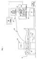
- FIG. 1is a schematic illustration of a system for implementing catheter image co-registration
- FIG. 2depicts an illustrative angiogram image
- FIG. 3depicts an illustrative fluoroscopic image of a radiopaque marker mounted upon a catheter
- FIG. 4depicts an illustrative enhanced radiological image along-side a cross-sectional IVUS image
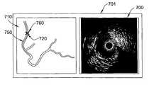
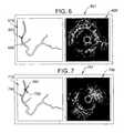
- FIG. 5depicts an illustrative enhanced radiological image along-side a cross-sectional IVUS image wherein the radiological image further includes a calculated path within a vessel of interest;
- FIG. 6depicts an illustrative enhanced radiological image along-side a cross-sectional IVUS image wherein the radiological image further includes a calculated path within a vessel of interest with a marker positioned at a different location than the view of FIG. 5 ;
- FIG. 7depicts an illustrative enhanced radiological image along-side a cross-sectional IVUS image wherein the radiological image further includes a calculated path within a vessel of interest and a reference mark providing a point of synchronization/calibration of a marker position;
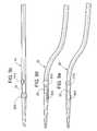
- FIG. 8depicts an illustrative catheter distal end including a single cylindrical radiopaque marker band
- FIG. 9 adepicts a radiopaque marker band 900 , suitable for use in an exemplary embodiment, that partially encircles the catheter shaft;
- FIG. 9 bdepicts an imaging catheter having two of the radiopaque marker bands of the type depicted in FIG. 9 a wherein the two bands are skewed by a quarter rotation along the axis of the catheter;
- FIG. 9 cdepicts the imaging catheter of 9 b from a view that looks directly on the full surface of the distal marker band 920 ;
- FIG. 9 ddepicts the imaging catheter of 9 c at a view wherein the catheter is axially rotated 90 degrees from the position depicted in FIG. 9 c;
- FIG. 9 edepicts the imaging catheter at a different rotational position from FIG. 9 c and FIG. 9 d;
- FIG. 10depicts an illustrative display for co-registration of radiological and hemodynamic image information
- FIG. 11is a flowchart summarizing a set of steps for rendering and displaying a co-registered view during a data acquisition procedure.
- FIG. 12is a flowchart summarizing a set of steps for rendering and displaying a co-registered view during playback of previously acquired image data.
- a method and systemare described by way of example herein below including image data acquisition equipment and data/image processors that generate views on a single display that simultaneously provides positional information and intravascular images associated with a imaging probe (e.g., an IVUS transducer probe) mounted upon a flexible elongate member (e.g, a catheter, guidewire, etc.).
- a imaging probee.g., an IVUS transducer probe
- a flexible elongate membere.g, a catheter, guidewire, etc.
- FIG. 1an exemplary system is schematically depicted for carrying out the present invention in the form of co-registration of angiogram/fluoroscopy and intravascular ultrasound images.
- the radiological and ultrasound image data acquisition sub-systemsare generally well known in the art.
- a patient 10is positioned upon an angiographic table 12 .
- the angiographic table 12is arranged to provide sufficient space for the positioning of an angiography/fluoroscopy unit c-arm 14 in an operative position in relation to the patient 10 on the table 12 .
- Radiological image data acquired by the angiography/fluoroscopy c-arm 14passes to an angiography/fluoroscopy processor 18 via transmission cable 16 .
- the angiography/fluoroscopy processor 18converts the received radiological image data received via the cable 16 into angiographic/fluoroscopic image data.
- the angiographic/fluoroscopic (“radiological”) image datais initially stored within the processor 18 .
- an imaging catheter 20is inserted within the patient 10 so that its distal end, including a diagnostic probe 22 (in particular an IVUS probe), is in the vicinity of a desired imaging location of a blood vessel.
- a diagnostic probe 22in particular an IVUS probe
- a radiopaque material located near the probe 22provides indicia of a current location of the probe 22 in a radiological image.
- the diagnostic probe 22generates ultrasound waves, receives ultrasound echoes representative of a region proximate the diagnostic probe 22 , and converts the ultrasound echoes to corresponding electrical signals.
- the corresponding electrical signalsare transmitted along the length of the imaging catheter 20 to a proximal connector 24 .
- IVUS versions of the probe 22come in a variety of configurations including single and multiple transducer element arrangements.
- an array of transducersis potentially arranged: linearly along a lengthwise axis of the imaging catheter 20 , curvilinearly about the lengthwise axis of the catheter 20 , circumferentially around the lengthwise axis, etc.
- the proximal connector 24 of the catheter 20is communicatively coupled to a catheter image processor 26 .
- the catheter image processor 26converts the signals received via the proximal connector 24 into, for example, cross-sectional images of vessel segments. Additionally, the catheter image processor 26 generates longitudinal cross-sectional images corresponding to slices of a blood vessel taken along the blood vessel's length.
- the IVUS image data rendered by the catheter image processor 26is initially stored within the processor 26 .
- the type of diagnostic imaging data acquired by the diagnostic probe 22 and processed by the catheter image processor 26varies in accordance with alternative embodiments of the invention.
- the diagnostic probe 22is equipped with one or more sensors (e.g., Doppler and/or pressure) for providing hemodynamic information (e.g., blood flow velocity and pressure)—also referred to as functional flow measurements.
- hemodynamic informatione.g., blood flow velocity and pressure
- functional flow measurementsare processed by the catheter image processor 26 .
- imageis intended to be broadly interpreted to encompass a variety of ways of representing vascular information including blood pressure, blood flow velocity/volume, blood vessel cross-sectional composition, shear stress throughout the blood, shear stress at the blood/blood vessel wall interface, etc.
- a co-registration processor 30receives IVUS image data from the catheter image processor 26 via line 32 and radiological image data from the radiological image processor 18 via line 34 . Alternatively, the communications between the sensors and the processors are carried out via wireless media.
- the co-registration processor 30renders a co-registration image including both radiological and IVUS image frames derived from the received image data.
- indiciae.g., a radiopaque marker artifact
- the co-registration processor 30initially buffers angiogram image data received via line 34 from the radiological image processor 18 in a first portion 36 of image data memory 40 .
- IVUS and radiopaque marker image data received via lines 32 and 34is stored within a second portion 38 and a third portion 42 , respectively, of the image data memory 40 .
- the individually rendered frames of stored image dataare appropriately tagged (e.g., time stamp, sequence number, etc.) to correlate IVUS image frames and corresponding radiological (radiopaque marker) image data frames.
- the hemodynamic datais stored within the second portion 38 .
- markerscan be placed on the surface of the patient or within the vicinity of the patient within the field of view of the angiogram/fluoroscope imaging device. The locations of these markers are then used to position the radiopaque marker artifact upon the angiographic image in an accurate location.
- the co-registration processor 30renders a co-registration image from the data previously stored within the first portion 36 , second portion 38 and third portion 42 of the image data memory 40 .
- a particular IVUS image frame/sliceis selected from the second portion 38 .
- the co-registration processor 30identifies fluoroscopic image data within the third portion 42 corresponding to the selected IVUS image data from the second portion 38 .
- the co-registration processor 30superimposes the fluoroscopic image data from the third portion 42 upon the angiogram image frame retrieved from the first portion 36 .
- the co-registered radiological and IVUS image framesare simultaneously displayed, along-side one another, upon a graphical display device 50 .
- the co-registered image data frames driving the display device 50are also stored upon a long-term storage device 60 for later review in a session separate from a procedure that acquired the radiological and IVUS image data stored in the image data memory 40 .
- a pullback deviceis incorporated that draws the catheter 20 from the patient at a controlled/measured manner.
- Such devicesare well known in the art. Incorporation of such devices facilitates calculating a current position of the probe 22 within a field of view at points in time when fluoroscopy is not active.
- the angiography/fluoroscopy processor 18captures an angiographic “roadmap” image 200 in a desired projection (patient/vessel orientation) and magnification.
- the image 200is initially captured by an angiography procedure performed prior to tracking the IVUS catheter to the region of interest within a patient's vasculature.
- Performing the angiography procedure without the catheter 20 in the vesselprovides maximal contrast flow, better vessel filling and therefore a better overall angiogram image.
- side branchessuch as side branch 210 and other vasculature landmarks can be displayed and seen clearly on the radiological image portion of a co-registered image displayed upon the graphical display device 50 .
- the catheter 20is tracked to its starting position (e.g., a position where an IVUS pullback procedure begins). Typically the catheter 20 is tracked over a previously advanced guidewire (not shown). Thereafter, a fluoroscopic image is obtained. In the image, the catheter radiopaque marker 300 is visualized, but the vessel lumen is not, due to the absence of contrast flow. However, a set of locating markers present in both the angiogram and fluoroscopy images enable proper positioning (superimposing) of the marker image within the previously obtained angiogram image. Other ways of properly positioning the radiopaque marker image within the field of view of the angiogram image will be known to those skilled in the art in view of the teachings herein.
- the marker artifactcan be automatically adjusted (both size and position) on the superimposed image frames to correspond to the approximate position of the transducers.
- the result of overlaying/superimposing the radiopaque marker artifact upon the angiogram imageis depicted, by way of example in an exemplary co-registration image depicted in FIG. 4 .
- the exemplary co-registration display 401depicts a selected cross-sectional IVUS image 400 of a vessel.
- a radiological image 410is simultaneously displayed along-side the IVUS image 400 on the display 50 .
- the radiological image 410includes a marker artifact 420 , generated from radiological image data rendered by a fluoroscope image frame, superimposed on an angiogram background rendered from the first portion 36 of the memory 40 .
- the fluoroscope image framecorresponds to the current location of the diagnostic probe 22 within a vessel under observation.
- Precise matching of the field of view represented in both the angiogram and fluoroscope imagesallows identification of the current position of the IVUS probe corresponding to the displayed IVUS image 400 in the right pane of the co-registered images displayed in FIG. 4 .
- the composite radiological image 410is obtained in one step.
- the original roadmap angiogram imageis obtained with the catheter already in its starting position.
- the angiogram imageis reused as the IVUS probe is withdrawn from the vessel.
- the systemalso takes heart motion into account when generating/acquiring the radiological and IVUS image data.
- heart motionis much less a factor and good overlay correlation exists between the angiogram and fluoroscope fields of view.
- the peak R-waveis selected because it represents end-diastole, during which the heart has the least amount of motion, and thus, a more consistent condition from which to obtain the radiological image data.
- the peak R-waveis also an easy point in the EKG for the system to detect.
- the cross-sectional image 400 from the IVUS catheteris displayed in tandem with the enhanced radiological image 410 including both the angiogram background and the superimposed marker artifact 420 .
- the enhanced radiological image 410 and the cross-sectional IVUS image 400are displayed close to (e.g., along side) each other on the display 50 , so that the operator can concentrate on the information in the cross-sectional image 400 while virtually simultaneously observing the status of the enhanced radiological image 410 .
- the simultaneous display of both the composite/enhanced radiological image and the cross-sectional imageallows instant awareness of both disease state of a vessel segment and the location of the vessel segment within a patient.
- Such comprehensive informationis not readily discernable in a three dimensional flythrough image or a stacked longitudinal image. Neither flythrough nor stacked images alone allows for the simultaneous appreciation of 1) all of the information in a cross-section, 2) a feel for the shape of the vessel and 3) the location of the cross-section along the length of the vessel.
- the above-described “co-registration” of enhanced angiographic (including the marker artifact) and intravascular cross-sectional images/informationdelivers all three of these items in a presentation that is straight forward to an operator with even average visual and spatial abilities.
- the co-registration displayis presented, by way of example, either on an IVUS console display, or the co-registration display is presented on one or more angiographic monitors, either in the room where the procedure is occurring or in a remote location.
- one monitor over the table in the procedure roomallows the attending physician to view the procedure, while at the same time a second consulting physician who has not scrubbed for the case is also able to view the case via a second monitor containing the co-registration display from a separate control room. Control room viewing is also possible without having to wear leaded covering.
- a single angiogram imageis, by way of example, obtained/generated and stored in the first portion 36 of the memory 40 for a given procedure/patient position. If the field of view changes or the patient's position changes, then an updated background angiogram image is generated and stored in the first portion 36 .
- the background angiogram imageis live or continuously updated, for example, at each additional step in which angiography is performed.
- the projection of the angiogram roadmap/background image portion of the enhanced radiological image 410is preferably in an orientation and magnification that best displays the entire vessel to be viewed, taking into account the foreshortening that is present in a tortuous/winding vessel.
- two roadmap imagesor even two enhanced radiological images 410
- Such multiple viewsare provided in the context of biplane angiography.
- Establishing a position for the marker artifact within the field of the enhanced radiological image, based at least in part upon a radiopaque marker on the imaging catheter 20is achievable in a variety of ways. Examples, described further herein below include: user-specified points (by clicking at a position near the marker to establish a point); image pattern recognition (automatic identification of a marker's unique signature within a field of view); and combinations of manual and automated calculations of a path.
- Enhancing the background/roadmap angiogram image to render the image 410is achieved in a number of different ways.
- the marker artifact 420(derived from a fluoroscope image of a radiopaque marker near the probe 22 mounted on the distal end of the catheter 20 ) is superimposed upon/overlays the angiogram/roadmap background of the enhanced radiological image 410 .
- the live/marker artifact portion of the image 410requires that fluoroscopy be performed the entire time of catheter movement (e.g. pullback)
- the marker artifactis displayed on the image 410 only during those periods when the fluoroscope is active. When the fluoroscope is inactive, only the background angiogram is presented on the enhanced image 410 of the display 50 .
- the co-registration processor 30calculates an approximate location of the radiopaque marker based upon its last registered position and other indicators of catheter movement (e.g., pullback distance sensors/meters). The approximate location is utilized in place of the radiopaque marker image to render a marker artifact 520 on an enhanced radiological image 510 displayed along-side a corresponding IVUS cross-sectional image 500 within a display 501 .
- the marker artifact 520 's positionis calculated by software/hardware within the co-registration processor 30 from sensor data indicative of a current/changed location of the radiopaque marker within the current image field provided by the current background angiogram image.
- a visual characteristice.g., color, symbol, intensity, etc.
- both the displacement and angular orientation of the markerare determined to render accurate approximations of the current position of the diagnostic probe 22 within a vessel as it acquires data for generating the image 500 .
- a calculated path 550 / 650is determined by the co-registration processor 30 within displays 501 / 601 .
- a marker artifact 520 / 620is placed on top of the calculated path 550 / 650 .
- the marker artifact 520 / 620is superimposed on the angiogram image at a location calculated from non-visual position data (e.g., pull-back distance, spatial position sensors, angular orientation sensors, etc.).
- the cursorcan be placed by the system at a distance from the initial location along the calculated path 550 / 650 that represents the product of the pullback rate and the time period. Furthermore, each subsequent time that a fluoroscope is activated and an image of the radiopaque marker is acquired and presented to the co-registration processor 30 , an error between the actual radiopaque marker location and a current calculated marker artifact 520 / 620 location is eliminated by replacing the calculated position by a position calculated by the radiopaque marker image.
- the error between the corrected position and the calculated location of the marker artifact 520 / 620is determined.
- the error/total travel distance ratiois used as a scaling factor to recalculate and adjust all previously calculated/rendered/presented marker artifact overlay positions on the rendered/stored copies of the enhanced radiological image 510 / 610 for the entire preceding period in which the fluoroscope has been inactive.
- a re-calculationcan also update a shape of the calculated path 550 / 560 curve.
- the calculated path 550 / 650is shown as a curve that matches the tortuosity of a vessel through which the probe 22 passes—represented by a center line through the displayed vessel.
- the catheter paths within vesselstake a straighter and shorter path than the centerline of a blood vessel when pulled through such vessel. If, however, the catheter is being translated by pushing, instead of pulling, the calculated path 550 / 650 more closely matches the curvature of the vessel, or even exaggerates the tortuosity of the vessel by taking a longer path.
- a multiplication coefficient(e.g., 1.05 for pushing, 0.95 for pulling) can be introduced when calculating a path based upon this general observation of the path taken by a probe as it is pushed/pulled through a vessel.
- the pathcan alternatively be calculated from two different angiographic images taken at different projections (planes). This allows a three dimensional angiographic image, from which a true centerline can be calculated.
- the operatorcreates a reference mark 760 at one or more points on a calculated path 750 .
- the reference mark 760serves a variety of potential uses.
- the reference mark 760potentially serves as a benchmark (location synchronization point) for updating position of a marker artifact 720 within the enhanced radiological image 710 .
- the co-registration processor 30waits for manual input of the reference mark 760 location information prior to proceeding with calculations.
- the usercreates the reference mark 760 which coincides with a marker artifact 720 rendered from image data provided by a fluoroscope of a field of view containing a radiopaque marker.
- the reference mark 760which potentially persists beyond its initial entry period, is distinguished from the marker artifact 720 which follows the current/estimated position of the probe 22 .
- the reference markis used to highlight/mark actual positions of the probe 22 (rendered by a fluoroscope image of a radiopaque marker) as opposed to estimated points on a calculated point (e.g. points on a path e.g., 550 / 560 ) from merely calculated position estimates upon the paths 550 / 560 .
- the reference mark 760is used to highlight a particular point of interest during a diagnostic/treatment procedure.
- a bookmarkis placed within a series of cross-sectional images associated with the IVUS image 700 portion of the display 701 . The bookmark allows quick access to a particular archived image frame corresponding to the reference mark 760 in the display 701 .
- a user interface associated with the displayed images provided in FIGS. 4-7includes a “slider” control that allows an operator to track through a series of stored frames representing sequentially acquired data along a traversed path within a vessel.
- the slider controlcan be a set of arrows on a keyboard, a bar/cursor displayed upon an enhanced radiological image that can be manipulated by an operator, during playback, using a mouse or other user interface device to traverse a vessel segment, etc.
- a display similar to FIG. 7is rendered by the co-registration processor 30 during playback of a previous data acquisition session.
- a cursor similar to the reference mark 760is displayed during playback on the enhanced radiological image 710 .
- a userselects and drags the cursor along a path similar to the calculated path 750 .
- the co-registration processor 30acquires and presents corresponding co-registered images. The user sequentially proceeds through the stored images using, by way of example, arrow keys, mouse buttons, etc.
- a single radiopaque marker band 800is attached to the catheter 820 near an IVUS probe.
- the radiopaque band 800includes a proximal edge 802 and a distal edge 804 .
- the band 800is cylindrical, with the diameter at the proximal edge 802 equal to the diameter at the distal edge 804 .
- the band 800has a known length.
- the processor 26Upon connection of the proximal connector 24 of the catheter 20 into an outlet on the catheter image processor 26 (or an interposed patient interface module which is communicatively connected to the processor 26 ), the processor 26 receives identification information from the catheter 20 via EPROM, RFID, optical reader or any other appropriate method for identifying the catheter 20 .
- the catheter length and diameter dimensions (or dimension ratio)are included in the received identification information.
- image field informationsuch as magnification and/or projection angle
- the co-registration processor 30By identifying four points at the corners of an approximate four-sided polygon of the marker band image, the co-registration processor 30 automatically calculates foreshortening of a vessel in an enhanced radiological image view and the true length of a segment of a calculated path.
- FIGS. 9 a - ea catheter 920 carries two marker bands having a known linear separation distance that facilitates making the calculations described herein above with reference to FIG. 8 .
- FIG. 9 ashows a radiopaque marker band 900 , suitable for use in an exemplary embodiment, that partially encircles the catheter shaft; In the exemplary embodiment, the marker band 900 extends about 180° (one half) of the perimeter of the catheter shaft.
- the bandis potentially made, for example, of 100% Platinum, or 90% Platinum/10% Irridium, Tantalum, Gold or any other radiopaque materials or combinations/amalgams thereof.
- FIG. 9 bshows an imaging catheter 20 having two of the radiopaque marker bands 910 and 920 of the type depicted in FIG. 9 a .
- the proximal band 910is skewed 90° (a quarter of the circumference of the catheter 20 ) in relation to the distal band 920 .
- the bands 910 / 920are shown equally spaced on opposite sides of the diagnostic probe 22 .
- This catheter 20also has a guidewire lumen 930 for passing a guidewire, for example a 0.014′′ guidewire.
- the guidewireexits out the distal guidewire port.
- the proximal end of the guidewirecan exit a proximal port either within the blood vessel (short lumen rapid exchange catheter), within a guiding catheter (long lumen rapid exchange catheter) or outside of the patient (over-the-wire catheter).
- FIG. 9 cshows the imaging catheter 20 from a view that looks directly on the full surface of the distal marker band 920 . Exactly one half of the proximal marker band 910 , skewed by 90 degrees, is seen. An angiography image of the two marker bands, when viewed as shown in FIG. 9 c reveals band 920 having a thickness that is twice the thickness of the image of the band 910 . Furthermore, an image length “L” of the marker bands 910 / 920 depends on angular position of the portion of the catheter 20 in the image containing the bands 910 / 920 . In a perfect side view, the length L is equal to the actual length of the marker band. Offset O is equal to the difference between the thickness of band 920 and the thickness of band 910 .
- FIG. 9 dan image is taken at a view wherein the catheter 20 is axially rotated 90 degrees from the position depicted in FIG. 9 c .
- the thickness of band 920is half the thickness of band 910 .
- the position of the relative positions of the bands 910 / 920 in relation to the axis of the catheter 20is used to determine the actual angular orientation of the catheter 20 since the offset alone is not enough to establish a current rotational position of the catheter 20 .
- FIG. 9 eis an image of the catheter 20 and bands 910 / 910 at a different rotational position from FIG. 9 c and FIG. 9 d .
- the orientation of the cathetercan be determined by comparing the relative thicknesses (e.g., the offset, a ratio) of the thickness of images of the bands 910 and 920 .
- co-registration processor 30facilitates performing a variety of additional tasks. For example, during a catheter pullback, a commenting functionality incorporated into the processor 30 enables a user to select a “bookmark” button. In response, the co-registration processor 30 attaches a note/comment to a specific cross-section and/or location along a calculated path on an enhanced radiological image.
- an alternative version of co-registration image schemeincorporates biplane angiography instead of standard, single view angiography images.
- biplane angiographytwo radiological projections are simultaneously presented to a user—e.g., two views skewed by 90 degrees on a common axis of rotation.
- two enhanced radiological imagesare presented along-side a cross-sectional image.
- marker artifact (cursor) positionis determined by calculations in relation to a known pullback rate, two cursor positions are determined—one on each of the two enhanced radiological images.
- the foreshortening of the vessel seen on one biplane imageis less than the other.
- the opposite biplane imagewould have less foreshortening at other periods where a marker artifact is based upon calculations rather than actual fluoroscope images.
- the errorsare calculated independently in the two different biplane images, and corresponding scaling factors are generated for the correction.
- a derived 3-dimensional roadmapis created based on information of the two images from different planes. In this case, the two different planes are the 90° biplane images Locating a marker artifact on a derived 3-D image is calculated from locations of marker artifacts one each of two orthogonal biplane images.
- an enhanced radiological imagecan be combined with a longitudinal stack instead of a cross sectional slice—in fact, the enhanced radiological, transverse cross-sectional, and longitudinal cross-sectional images can be displayed together.
- the enhanced radiological imageis presented along-side an IVUS image including both grayscale and color image artifacts that characterizing tissue and deposits within a vessel.
- the longitudinal IVUS grayscale image and/or the color (Virtual Histology) imageare overlaid on the 2-D angiographic image or derived 3-D image.
- an exemplary co-registration display 1001 rendered by the co-registration processor 30includes an enhanced radiological image 1010 displayed along-side functional flow measurement values presented in a graph 1000 .
- functional flow reserveFFR
- the enhanced radiological image 1010comprises a marker artifact 1020 superimposed upon an angiogram image.
- the marker artifact 1020indicates the point at which the presently displayed functional flow measurements are being presented based upon measurements previously acquired by sensors/transducers on the probe 22 mounted at the distal end of a flexible elongate member such as a guidewire or the catheter 20 .
- the co-registration imagefurther includes an IVUS cross-sectional image (not depicted) corresponding to the vessel segment indicated by the marker artifact 1020 on the enhanced radiological image 1010 .
- the displayalso includes a variety of additional text information associated with the section of the vessel identified by the marker artifact 1020 .
- Vessel dimensions 1030specify an approximate diameter and lumen area of a particular cross section indicated by the marker artifact 1020 's current position on the enhanced radiological image 1010 .
- IVUS information 1040specify a plaque burden percentage and a total plaque area for a current cross-sectional slice indicated by the marker artifact 1020 .
- An FFR information 1050specifies a current FFR value associated with the current location of the marker artifact 1020 . It is noted that the marker artifact 1020 approximates the location of a probe (e.g., probe 22 ) at the time data was acquired to render the presently displayed data values.
- the location of the marker artifact 1020is derived from image data provided by a radiopaque element/marker located near a probe mounted upon a flexible elongate member such as probe 22 mounted on a guidewire or catheter 20 .
- the marker artifact 1020operates as a slider control that enables a user to sequentially traverse a set of stored data records containing information of the type displayed in FIG. 10 . Furthermore, in the particular example, an FFR value associated with a particular location designated by the marker artifact 1020 is displayed near the marker artifact 1020 . Also, a second slider 1060 is also provided that is linked to the position of marker artifact 1020 and thus moves in synchronism with the marker artifact 1020 . Moving either the slider 1060 or the marker artifact 1020 causes movement of the other.
- interventional ultrasound imagingsuch as Intracardiac Echocardiography
- a steerable catheter with a linear, curvilinear, circumferential or other ultrasonic array at the distal endis placed into or in proximity to the chambers of the heart, and its location is incorporated into an enhanced ultrasound image.
- an angiogram imageis generated and stored within the first portion 36 of image data memory 40 .
- a single angiogram imagecan be used to support co-registered display of multiple acquired data sets from the probe 22 as the probe 22 passes within a length of a blood vessel.
- a visual artifacte.g., marker artifact 420
- a visual artifacthaving a position determined at least in part upon a radiopaque marker positioned near the probe 22 on the imaging catheter 20 , is superimposed on the angiogram image.
- the visual artifactprogresses along the angiogram image of the blood vessel thereby providing an approximate location of the probe 22 associated with currently displayed data rendered according to information provided by the probe 22 .
- an initial calculated path(e.g., path 550 ) is generated by the co-registration processor 30 .
- This estimation of the pathcan be generated according to any of a variety of methods including: automated two-dimensional and three-dimensional path calculations; manual path specification; and user assisted automated path calculations (a combination of automated path calculation with user-specified over-rides).
- the calculated pathis superimposed upon the angiogram image generated during step 1100 and represents the projected path of the probe 22 when pullback is commenced of the probe 22 .
- the operation of the co-registration systemis determined by whether the fluoroscope has been activated (providing a live image of a radiopaque marker mounted proximate the probe 22 ). If the fluoroscope is active, then control passes to step 1115 wherein a fluoroscope image (see, e.g., FIG. 3 ) of the radiopaque marker is acquired, timestamped and stored. Thereafter, at step 1120 image data associated with the probe 22 is acquired, timestamped and stored.
- the image datacomprises an IVUS image generated by an ultrasound transducer probe mounted upon the imaging catheter 20 .
- the co-registration processor 30superimposes/overlays a marker artifact on the previously stored angiogram image to render the aforementioned enhanced radiological image.
- the marker artifactderives is position, at least in part, from the previously acquired and stored radiopaque marker position data.
- the enhanced radiological (e.g., angiogram) imageis thereafter stored with the timestamp associated with the radiopaque marker position data during step 1130 .
- the co-registration processor 30renders and simultaneously presents on a display/monitor the previously generated enhanced angiogram image and a corresponding probe (IVUS) image.
- the enhanced angiogram image and the corresponding probe imageare displayed along-side one another on the display/monitor. Selection of a corresponding image is based upon a timestamp associated with the selected IVUS probe image.
- the respective timestamps of the radiological and probe components of the co-registered displayneed not be identical.
- a closest match criterionis applied to the selection process. Control then returns to step 1110 for another iteration of the co-registration imaging process.
- the co-registration processor 30acquires/registers a pullback rate for the pullback mechanism.
- image data associated with the probe 22is acquired, timestamped and stored.
- the image datacomprises an IVUS image generated by an ultrasound transducer probe mounted upon the imaging catheter 20 .
- the processor 30determines a time that has elapsed since the previous calculation of the artifact marker position.
- step 1165the co-registration processor 30 generates an estimate of a present position of the probe 22 and a corresponding marker artifact position on the enhanced radiological image.
- the pullback rate and the elapsed time between a previous marker artifact position determination and the present position determinationare used to generate a present position estimate for the marker artifact.
- the co-registration processor 30superimposes/overlays a marker artifact on the angiogram at the new calculated position based upon the calculated path and the distance calculation rendered during step 1165 .
- the enhanced radiological (e.g., angiogram) imageis stored with the timestamp associated with the calculated marker artifact position data.
- the resulting enhanced radiological imageis utilized to render and present a co-registered display including both the enhanced angiogram image and a corresponding (based upon timestamp) previously stored probe image. Control thereafter returns to step 1110 .
- step 1200the co-registration processor 30 initially displays an enhanced radiological image including, for example, an angiogram image, a calculated path, and a cursor/slider mark positioned on the calculated path indicating a location associated with a presently provided image derived from data acquired by the probe 22 at the indicated location on the enhanced radiological image.
- an enhanced radiological imageincluding, for example, an angiogram image, a calculated path, and a cursor/slider mark positioned on the calculated path indicating a location associated with a presently provided image derived from data acquired by the probe 22 at the indicated location on the enhanced radiological image.
- a userpositions the cursor/slider mark on the calculated path.
- Such repositioningcan occur in any of a number of ways.
- the userdrags and drops the cursor/slider using a mouse.
- a keyboard inputcan advance/backup the cursor/slider through a series of previously designated/bookmarked points along the calculated path displayed within the enhanced angiogram image provided during step 1200 .
- keyscan be used to advance the cursor/slider on a record-by-record basis through a set of stored records associated with the progression of the probe 22 along the calculated path.
- Still other modes of selecting a position of interest on the calculated path and its associated probe 22 (e.g., IVUS) imagewill be contemplated by those skilled in the art in view of the description provided herein.
- the co-registration processor 30accesses a corresponding record within the set of records derived from the data provided by the probe 22 .
- data setsinclude cross-sectional IVUS images or alternatively FFR values at specified positions along a blood vessel.
- a co-registered viewis presented wherein the enhanced radiological image, including the calculated path and cursor/slider (derived at least partially from positional information provided by a radiopaque marker during data acquisition), is displayed along-side an image (e.g., an IVUS cross-section) derived from data provided by the probe 22 at a position indicated by the current cursor/slider position within the enhanced radiological image.
- the steps depicted in FIG. 12are repeated in response to a detected change in the position of the cursor/slider to update the display to show the new position of the cursor/slider and the corresponding image (e.g. cross-sectional IVUS image) derived from data provided by the probe 22 at the designated cursor/slider position.
Landscapes
- Health & Medical Sciences (AREA)
- Life Sciences & Earth Sciences (AREA)
- Engineering & Computer Science (AREA)
- Medical Informatics (AREA)
- Surgery (AREA)
- Veterinary Medicine (AREA)
- Animal Behavior & Ethology (AREA)
- Public Health (AREA)
- General Health & Medical Sciences (AREA)
- Biomedical Technology (AREA)
- Heart & Thoracic Surgery (AREA)
- Molecular Biology (AREA)
- Biophysics (AREA)
- Physics & Mathematics (AREA)
- Pathology (AREA)
- Nuclear Medicine, Radiotherapy & Molecular Imaging (AREA)
- Radiology & Medical Imaging (AREA)
- Optics & Photonics (AREA)
- High Energy & Nuclear Physics (AREA)
- Human Computer Interaction (AREA)
- Dentistry (AREA)
- Oral & Maxillofacial Surgery (AREA)
- Vascular Medicine (AREA)
- Physiology (AREA)
- Computer Vision & Pattern Recognition (AREA)
- Robotics (AREA)
- Apparatus For Radiation Diagnosis (AREA)
- Ultra Sonic Daignosis Equipment (AREA)
Abstract
Description
This application claims priority of Huennekens et al. U.S. provisional application Ser. No. 60/642,893 filed on Jan. 11, 2005, entitled “Catheter Image Co- Registration,” and Walker et al. U.S. provisional application Ser. No. 60/694,014 filed on Jun. 24, 2005, entitled “Three-Dimensional Co-Registration For Intravascular Diagnosis and Therapy”, the contents of both of the above-identified provisional applications are expressly incorporated herein by reference in their entirety including the contents and teachings of any references contained therein.
The present invention generally relates to imaging blood vessels. More particularly, the present invention is directed to methods and systems for generating composite displays relating a first image rendered from a first type of data and a second image rendered from a second type of data. A particular example of such composite display comprises an angiogram displayed along-side an IVUS image.
In coronary arteries, vascular diseases including vessel lumen narrowing, usually due to atherosclerotic plaque, can lead to reduced blood flow to a heart muscle, angina (chest pain) and myocardial infarction—a heart attack. A variety of interventional treatments of cardiovascular disease are presently available to identify and treat such narrowing of a vessel lumen. Examples of such treatments include balloon angioplasty and/or deployment of stents. Diagnostic imaging is utilized to identify the extent and/or type of blockages within vessels prior to and/or during the treatment of such blockages. Diagnostic imaging enables doctors to ensure proper treatment of diseased vessels and verify the efficacy of such treatment.
In general, two distinct manners exist for generating diagnostic images for the identification and treatment of cardiovascular disease within a vasculature. A first manner of diagnostic imaging involves generating a radiological image of a stream flowing through a blood vessel's lumen from outside the vessel lumen. The purpose of generating an image of such flow is to identify blockages within diseased blood vessels that restrict blood flow. The extent of a vessel's lumen is traditionally imaged using angiography, which involves rendering a two-dimensional view of one or more vessels within a portion of a patient's vasculature through which radiopaque contrast media has been injected. The two-dimensional angiographic image can also be viewed real time by fluoroscopy. During such procedures, the images are potentially captured in various digital media, or in cine angiography (cine). Cine angiography, though rendering higher quality images of blood vessel lumens, exposes patients to high levels of ionizing radiation.
Fluoroscopy, generally using substantially less intense radiation than angiography, is used by physicians primarily to visually guide diagnostic and therapeutic catheters or guidewires, including one or more radiopaque markers, through vessels. The radiation intensity during fluoroscopy is typically one-tenth the intensity of radiation to which a patient is exposed during cine angiography. Many catheters have radiopaque markers that are viewable on a fluoroscope, thereby enabling a physician to track the location/path of such catheters as they are inserted within and/or withdrawn from patients. The platinum spring coil of guidewires also serves as a radiopaque marker. The lower radiation intensity of fluoroscopy allows a greater duration of use during a diagnostic/treatment procedure. However, due to its greater time of use, the total radiation exposure from fluoroscopy during an interventional treatment procedure can greatly exceed the radiation exposure during a typical cine angiography procedure. Thus, it is incumbent upon a physician to minimize the duration of time that a fluoroscope is used during a diagnostic and/or interventional treatment procedure.
The first manner of imaging, described above, has a number of drawbacks. For example, limited flow of contrast media near vessel walls and extreme variations in vessel cross-sections can result in incomplete filling of the vessel with a sufficient concentration of contrast media. As a consequence, the diameters of vessel segments can be misrepresented in an angiographic image. For example, a left main coronary artery cross-section is often underestimated by angiography. This can be problematic when attempting to judge the significance of a blockage within the vessel or when choosing the size of the treatment balloon or stent. An under-sized balloon or stent will not provide as effective treatment as a properly sized device. Furthermore, in angiography, a vessel's cross-section is determined by a two-dimensional view which may not accurately represent an actual extent of blood vessel narrowing.
Furthermore, to achieve an optimum treatment result, it is important to correctly determine a true target diameter of a native blood vessel—the diameter of a non-diseased blood vessel. However, angiography is ineffective in determining the target diameter of a vessel with disease along its entire length. For example, since vessels tend to taper in diameter along their length, a uniformly narrowed vessel may appear normal in an angiographic image.
Finally, angiography does not facilitate differentiating between different types of tissue found in atherosclerotic plaque. For example, in coronary arteries prone to producing a heart attack, necrotic tissue is thought to be more prevalent than purely fibrous tissue. Thus, while providing a good way to identify severe blockages, angiography is not always the best diagnostic imaging tool due to the incomplete nature of the angiographic image data.
The second manner of intravascular imaging comprises imaging the vessel itself using a catheter-mounted intravascular probe. Intravascular imaging of blood vessels provides a variety of information about the vessel including: the cross-section of the lumen, the thickness of deposits on a vessel wall, the diameter of the non-diseased portion of a vessel, the length of diseased sections, and the makeup of the atherosclerotic plaque on the wall of the vessel.
Several types of catheter systems have been designed to track through a vasculature to image atherosclerotic plaque deposits on vessel walls. These advanced imaging modalities include, but are not limited to, intravascular ultrasound (IVUS) catheters, magnetic resonance imaging (MRI) catheters and optical coherence tomography (OCT) catheters. In addition, thermography catheters and palpography catheters have also been demonstrated to generate vessel image data via intravascular probes. Other catheter modalities that have been proposed include infrared or near-infrared imaging.
In operation, these intravascular catheter-mounted probes are moved along a vessel in the region where imaging is desired. As the probe passes through an area of interest, sets of image data are obtained that, correspond to a series of “slices” or cross-sections of the vessel, the lumen, and surrounding tissue. As noted above, the catheters include radiopaque markers. Such markers are generally positioned near a distal catheter tip. Therefore, the approximate location of the imaging probe can be discerned by observing the catheterization procedure on either a fluoroscope or angiographic image. Typically imaging catheters are connected to a dedicated console, including specialized signal processing hardware and software, and display. The raw image data is received by the console, processed to render an image including features of concern, and rendered on the dedicated display device.
For example, IVUS images used to diagnose/treat vascular disease generally comprise sets of cross-sectional image “slices” of a vessel. A grayscale cross-sectional slice image is rendered, at each of a set of positions along the vessel based upon the intensity of ultrasound echoes received by an imaging probe. Calcium or stent struts, which produce relatively strong echoes, are seen as a lighter shade of gray. Blood or vessel laminae, which produce weaker echoes, are seen as a darker shade of gray.
Atherosclerotic tissue is identified as being the portion of a cross-sectional image between an internal elastic lamina (IEL) and an external elastic lamina (EEL). The ability to see the vessel lumen, and calculate its dimensions, allows the diameters and cross-sectional area of the vessel to be determined more reliably than the limited two-dimensional angiography. Because IVUS does not rely upon dispersing a contrast agent, IVUS is especially useful in generating images of the left main coronary artery as described above. Furthermore, the ability to view the EEL, and calculate its dimensions, allows an IVUS image to render a more reliable determination than angiography, of the correct diameter and length of the balloon or stent to use when restoring proper blood flow to a blocked/diseased vessel. Advanced IVUS images have also been described which perform tissue characterization and denote different types of tissue with a color code. One such modality is described in Vince, U.S. Pat. No. 6,200,268. Like IVUS, the other catheters mentioned above display a series of cross-sectional images from which additional information can be obtained.
Catheter-mounted probes, and in particular, IVUS probes can be configured to render a variety of two and three-dimensional images. In addition to the two-dimensional transverse cross-sectional images discussed above, a longitudinal planar image can be constructed from a plane which cuts through a “stack” of cross-section “slices”. In addition, three-dimensional “fly-through” images can be constructed from information in a series of cross-sectional slices of a vessel. Though these three-dimensional images can be visually impressive, the two dimensional angiography image remains the primary basis for determining the location of a catheter in a vessel, and the “schematic” reference through which the physician plans and carries out a treatment procedure.
In creating the “stack” or “flythrough” images, some assumptions are made by image data processing software in terms of the orientation of each slice to the next. In many cases the compound images, rendered from a series of transverse cross-sectional slices, are rendered in the form of a straight vessel segment. In reality, vessels can curve significantly. In segment visualizations that render straight segments, spatial orientation of each cross-sectional slice in relation to other slices is not measured. In addition, the rotational orientation of a catheter-mounted probe is generally not known due to twisting of the catheter as it passes through a vessel. Therefore, the angular relation between adjacent slices is not generally known. In many cases, these limitations do not significantly effect treatment of a diseased vessel because the typical treatment modalities (balloons, stents) are not circumferentially specific. A balloon, for example, dilates a vessel 360° around a lumen.
In view of the advantages provided by the two above described methods of imaging vessels, many catheter labs use both methods simultaneously to diagnose and treat a patient. However, an angiographic image provided on a different display monitor than a corresponding IVUS image (or the other image rendered by a catheter-mounted probe), presents challenges to a obtaining a comprehensive understanding of a state of a diseased vessel. For example, a physician identifies specific structures (e.g. feeder vessels) in cross-sectional images in order to determine a location on a vessel presented on an angiography display that needs to be treated. Coordinating images rendered by two distinct display devices can become cumbersome as the physician refers back and forth between two different screens on two distinct display devices. In addition, when a video loop of IVUS images is recorded, to be played back later on a machine, a corresponding angiographic image is not recorded in sync with it. Therefore, during playback, the specific cross-section being viewed needs to be compared to the vessel angiography, which is usually on a separate file.
A known visualization display simultaneously provides an angiogram, an IVUS transverse plane view, and an IVUS longitudinal plane view. A red dot is placed upon the angiogram corresponding to a currently displayed IVUS transverse plane view. A blue line is placed upon the angiogram corresponding to a currently displayed longitudinal plane view. The reference dot and line are only as valuable as the accuracy of the process that registers their positions on the angiogram.
In order to provide a better overall view of vascular systems, in accordance with the present invention, a system and method include a single display simultaneously providing a first view of a patient including an angiogram image and a second view including an intravascular image rendered from information provided by an imaging probe mounted on a distal end of a flexible elongate member. A cursor, having a position derived from image information provided by a radiopaque marker proximate the imaging probe, is displayed within the angiogram image to correlate the position of the imaging probe to a presently displayed intravascular image and thus provide an easily discernable identification of a position within a patient corresponding to a currently displayed intravascular image. The resulting composite display simultaneously provides: an intravascular image that includes information about a vessel that is not available from an angiogram and a current location within a vessel of a source of intravascular image data from which the intravascular image is rendered.
While the claims set forth the features of the present invention with particularity, the invention, together with its objects and advantages, may be best understood from the following detailed description taken in conjunction with the accompanying drawing of which:
In accordance with embodiments of the present invention, a method and system are described by way of example herein below including image data acquisition equipment and data/image processors that generate views on a single display that simultaneously provides positional information and intravascular images associated with a imaging probe (e.g., an IVUS transducer probe) mounted upon a flexible elongate member (e.g, a catheter, guidewire, etc.).
Turning initially toFIG. 1 , an exemplary system is schematically depicted for carrying out the present invention in the form of co-registration of angiogram/fluoroscopy and intravascular ultrasound images. The radiological and ultrasound image data acquisition sub-systems are generally well known in the art. With regard to the radiological image data, apatient 10 is positioned upon an angiographic table12. The angiographic table12 is arranged to provide sufficient space for the positioning of an angiography/fluoroscopy unit c-arm 14 in an operative position in relation to the patient10 on the table12. Radiological image data acquired by the angiography/fluoroscopy c-arm 14 passes to an angiography/fluoroscopy processor 18 viatransmission cable 16. The angiography/fluoroscopy processor 18 converts the received radiological image data received via thecable 16 into angiographic/fluoroscopic image data. The angiographic/fluoroscopic (“radiological”) image data is initially stored within theprocessor 18.
With regard to portions of the system associated with acquiring ultrasound image data, animaging catheter 20, and in particular an IVUS catheter, is inserted within thepatient 10 so that its distal end, including a diagnostic probe22 (in particular an IVUS probe), is in the vicinity of a desired imaging location of a blood vessel. While not specifically identified inFIG. 1 , a radiopaque material located near theprobe 22 provides indicia of a current location of theprobe 22 in a radiological image. By way of example, thediagnostic probe 22 generates ultrasound waves, receives ultrasound echoes representative of a region proximate thediagnostic probe 22, and converts the ultrasound echoes to corresponding electrical signals. The corresponding electrical signals are transmitted along the length of theimaging catheter 20 to aproximal connector 24. IVUS versions of theprobe 22 come in a variety of configurations including single and multiple transducer element arrangements. In the case of multiple transducer element arrangements, an array of transducers is potentially arranged: linearly along a lengthwise axis of theimaging catheter 20, curvilinearly about the lengthwise axis of thecatheter 20, circumferentially around the lengthwise axis, etc.
Theproximal connector 24 of thecatheter 20 is communicatively coupled to acatheter image processor 26. Thecatheter image processor 26 converts the signals received via theproximal connector 24 into, for example, cross-sectional images of vessel segments. Additionally, thecatheter image processor 26 generates longitudinal cross-sectional images corresponding to slices of a blood vessel taken along the blood vessel's length. The IVUS image data rendered by thecatheter image processor 26 is initially stored within theprocessor 26.
The type of diagnostic imaging data acquired by thediagnostic probe 22 and processed by thecatheter image processor 26 varies in accordance with alternative embodiments of the invention. In accordance with a particular alternative embodiment, thediagnostic probe 22 is equipped with one or more sensors (e.g., Doppler and/or pressure) for providing hemodynamic information (e.g., blood flow velocity and pressure)—also referred to as functional flow measurements. In such alternative embodiments functional flow measurements are processed by thecatheter image processor 26. It is thus noted that the term “image” is intended to be broadly interpreted to encompass a variety of ways of representing vascular information including blood pressure, blood flow velocity/volume, blood vessel cross-sectional composition, shear stress throughout the blood, shear stress at the blood/blood vessel wall interface, etc. In the case of acquiring hemodynamic data for particular portions of a blood vessel, effective diagnosis relies upon the ability to visualize a current location of thediagnostic probe 22 within a vasculature while simultaneously observing functional flow metrics indicative of cardiovascular disease. Co-registration of hemodynamic and radiological images facilitates precise treatment of diseased vessels. Alternatively, instead of catheter mounted sensors, the sensors can be mounted on a guidewire, for example a guidewire with a diameter of 0.018″ or less. Thus, in accordance with embodiments of the present invention, not only are a variety of probe types used, but also a variety of flexible elongate members to which such probes are mounted at a distal end (e.g., catheter, guidewire, etc.).
Aco-registration processor 30 receives IVUS image data from thecatheter image processor 26 vialine 32 and radiological image data from theradiological image processor 18 vialine 34. Alternatively, the communications between the sensors and the processors are carried out via wireless media. Theco-registration processor 30 renders a co-registration image including both radiological and IVUS image frames derived from the received image data. In accordance with an embodiment of the present invention, indicia (e.g., a radiopaque marker artifact) are provided on the radiological images of a location corresponding to simultaneously displayed IVUS image data. Theco-registration processor 30 initially buffers angiogram image data received vialine 34 from theradiological image processor 18 in afirst portion 36 ofimage data memory 40. Thereafter, during the course of a catheterization procedure IVUS and radiopaque marker image data received vialines second portion 38 and athird portion 42, respectively, of theimage data memory 40. The individually rendered frames of stored image data are appropriately tagged (e.g., time stamp, sequence number, etc.) to correlate IVUS image frames and corresponding radiological (radiopaque marker) image data frames. In an embodiment wherein hemodynamic data is acquired rather than IVUS data, the hemodynamic data is stored within thesecond portion 38.
In addition, additional markers can be placed on the surface of the patient or within the vicinity of the patient within the field of view of the angiogram/fluoroscope imaging device. The locations of these markers are then used to position the radiopaque marker artifact upon the angiographic image in an accurate location.
Theco-registration processor 30 renders a co-registration image from the data previously stored within thefirst portion 36,second portion 38 andthird portion 42 of theimage data memory 40. By way of example, a particular IVUS image frame/slice is selected from thesecond portion 38. Theco-registration processor 30 identifies fluoroscopic image data within thethird portion 42 corresponding to the selected IVUS image data from thesecond portion 38. Thereafter, theco-registration processor 30 superimposes the fluoroscopic image data from thethird portion 42 upon the angiogram image frame retrieved from thefirst portion 36. Thereafter, the co-registered radiological and IVUS image frames are simultaneously displayed, along-side one another, upon agraphical display device 50. The co-registered image data frames driving thedisplay device 50 are also stored upon a long-term storage device 60 for later review in a session separate from a procedure that acquired the radiological and IVUS image data stored in theimage data memory 40.
While not shown inFIG. 1 , a pullback device is incorporated that draws thecatheter 20 from the patient at a controlled/measured manner. Such devices are well known in the art. Incorporation of such devices facilitates calculating a current position of theprobe 22 within a field of view at points in time when fluoroscopy is not active.
Turning toFIG. 2 , the angiography/fluoroscopy processor 18 captures an angiographic “roadmap”image 200 in a desired projection (patient/vessel orientation) and magnification. By way of example, theimage 200 is initially captured by an angiography procedure performed prior to tracking the IVUS catheter to the region of interest within a patient's vasculature. Performing the angiography procedure without thecatheter 20 in the vessel provides maximal contrast flow, better vessel filling and therefore a better overall angiogram image. Thus, side branches such asside branch 210 and other vasculature landmarks can be displayed and seen clearly on the radiological image portion of a co-registered image displayed upon thegraphical display device 50.
Turning toFIG. 3 , thecatheter 20 is tracked to its starting position (e.g., a position where an IVUS pullback procedure begins). Typically thecatheter 20 is tracked over a previously advanced guidewire (not shown). Thereafter, a fluoroscopic image is obtained. In the image, the catheterradiopaque marker 300 is visualized, but the vessel lumen is not, due to the absence of contrast flow. However, a set of locating markers present in both the angiogram and fluoroscopy images enable proper positioning (superimposing) of the marker image within the previously obtained angiogram image. Other ways of properly positioning the radiopaque marker image within the field of view of the angiogram image will be known to those skilled in the art in view of the teachings herein. Furthermore, the marker artifact can be automatically adjusted (both size and position) on the superimposed image frames to correspond to the approximate position of the transducers. The result of overlaying/superimposing the radiopaque marker artifact upon the angiogram image is depicted, by way of example in an exemplary co-registration image depicted inFIG. 4 .
Turning toFIG. 4 the exemplary co-registration display401 (including the correlated radiological and IVUS images) depicts a selectedcross-sectional IVUS image 400 of a vessel. Aradiological image 410 is simultaneously displayed along-side theIVUS image 400 on thedisplay 50. Theradiological image 410 includes amarker artifact 420, generated from radiological image data rendered by a fluoroscope image frame, superimposed on an angiogram background rendered from thefirst portion 36 of thememory 40. The fluoroscope image frame corresponds to the current location of thediagnostic probe 22 within a vessel under observation. Precise matching of the field of view represented in both the angiogram and fluoroscope images (i.e., precise projection and magnification of the two images) allows identification of the current position of the IVUS probe corresponding to the displayedIVUS image 400 in the right pane of the co-registered images displayed inFIG. 4 .
Alternatively, the compositeradiological image 410 is obtained in one step. In such case, the original roadmap angiogram image is obtained with the catheter already in its starting position. However, once obtained, the angiogram image is reused as the IVUS probe is withdrawn from the vessel.
The system also takes heart motion into account when generating/acquiring the radiological and IVUS image data. By way of example, by acquiring the image data for both the angiogram (background) and the radiopaque marker only during the peak R-wave of the EKG, heart motion is much less a factor and good overlay correlation exists between the angiogram and fluoroscope fields of view. The peak R-wave is selected because it represents end-diastole, during which the heart has the least amount of motion, and thus, a more consistent condition from which to obtain the radiological image data. The peak R-wave is also an easy point in the EKG for the system to detect.
With continued reference toFIG. 4 , in an exemplary embodiment when theIVUS catheter 20 begins to image, thecross-sectional image 400 from the IVUS catheter is displayed in tandem with the enhancedradiological image 410 including both the angiogram background and the superimposedmarker artifact 420. The enhancedradiological image 410 and thecross-sectional IVUS image 400 are displayed close to (e.g., along side) each other on thedisplay 50, so that the operator can concentrate on the information in thecross-sectional image 400 while virtually simultaneously observing the status of the enhancedradiological image 410.
The simultaneous display of both the composite/enhanced radiological image and the cross-sectional image allows instant awareness of both disease state of a vessel segment and the location of the vessel segment within a patient. Such comprehensive information is not readily discernable in a three dimensional flythrough image or a stacked longitudinal image. Neither flythrough nor stacked images alone allows for the simultaneous appreciation of 1) all of the information in a cross-section, 2) a feel for the shape of the vessel and 3) the location of the cross-section along the length of the vessel. The above-described “co-registration” of enhanced angiographic (including the marker artifact) and intravascular cross-sectional images/information delivers all three of these items in a presentation that is straight forward to an operator with even average visual and spatial abilities. The co-registration display is presented, by way of example, either on an IVUS console display, or the co-registration display is presented on one or more angiographic monitors, either in the room where the procedure is occurring or in a remote location. For example, one monitor over the table in the procedure room allows the attending physician to view the procedure, while at the same time a second consulting physician who has not scrubbed for the case is also able to view the case via a second monitor containing the co-registration display from a separate control room. Control room viewing is also possible without having to wear leaded covering.
With regard to the persistence of the background angiogram (“roadmap”) image portion of the enhancedradiological image 410, a single angiogram image is, by way of example, obtained/generated and stored in thefirst portion 36 of thememory 40 for a given procedure/patient position. If the field of view changes or the patient's position changes, then an updated background angiogram image is generated and stored in thefirst portion 36. Alternatively, the background angiogram image is live or continuously updated, for example, at each additional step in which angiography is performed. The projection of the angiogram roadmap/background image portion of the enhancedradiological image 410 is preferably in an orientation and magnification that best displays the entire vessel to be viewed, taking into account the foreshortening that is present in a tortuous/winding vessel. Alternatively, two roadmap images (or even two enhanced radiological images410) can be used/displayed in place of the oneimage 410. Such multiple views are provided in the context of biplane angiography.
Establishing a position for the marker artifact within the field of the enhanced radiological image, based at least in part upon a radiopaque marker on theimaging catheter 20 is achievable in a variety of ways. Examples, described further herein below include: user-specified points (by clicking at a position near the marker to establish a point); image pattern recognition (automatic identification of a marker's unique signature within a field of view); and combinations of manual and automated calculations of a path.
Enhancing the background/roadmap angiogram image to render theimage 410 is achieved in a number of different ways. As mentioned above, in an illustrative embodiment, the marker artifact420 (derived from a fluoroscope image of a radiopaque marker near theprobe 22 mounted on the distal end of the catheter20) is superimposed upon/overlays the angiogram/roadmap background of the enhancedradiological image 410. Because the live/marker artifact portion of theimage 410 requires that fluoroscopy be performed the entire time of catheter movement (e.g. pullback), in an alternative embodiment, the marker artifact is displayed on theimage 410 only during those periods when the fluoroscope is active. When the fluoroscope is inactive, only the background angiogram is presented on theenhanced image 410 of thedisplay 50.
Turning toFIGS. 5 and 6 , in embodiments of the invention, when the fluoroscope is inactive, theco-registration processor 30 calculates an approximate location of the radiopaque marker based upon its last registered position and other indicators of catheter movement (e.g., pullback distance sensors/meters). The approximate location is utilized in place of the radiopaque marker image to render amarker artifact 520 on an enhancedradiological image 510 displayed along-side a corresponding IVUScross-sectional image 500 within adisplay 501. By way of a particular illustrative example, during periods in which a fluoroscope is inactive, themarker artifact 520's position is calculated by software/hardware within theco-registration processor 30 from sensor data indicative of a current/changed location of the radiopaque marker within the current image field provided by the current background angiogram image. In an embodiment of the invention, a visual characteristic (e.g., color, symbol, intensity, etc.) of themarker artifact 520 is used to distinguish when the fluoroscope is active/inactive and thus indicate whether the marker artifact position is actual/calculated. Furthermore, in more advanced systems, both the displacement and angular orientation of the marker (and thus the diagnostic probe22) are determined to render accurate approximations of the current position of thediagnostic probe 22 within a vessel as it acquires data for generating theimage 500.
With continued reference toFIGS. 5 and 6 , acalculated path 550/650 is determined by theco-registration processor 30 withindisplays 501/601. Amarker artifact 520/620 is placed on top of thecalculated path 550/650. Themarker artifact 520/620 is superimposed on the angiogram image at a location calculated from non-visual position data (e.g., pull-back distance, spatial position sensors, angular orientation sensors, etc.). For example, if the initial location of a radiopaque marker within the enhancedradiological image 510/610 is known and the catheter is pulled by an automatic pullback system at a specific rate for a known amount of time, the cursor can be placed by the system at a distance from the initial location along thecalculated path 550/650 that represents the product of the pullback rate and the time period. Furthermore, each subsequent time that a fluoroscope is activated and an image of the radiopaque marker is acquired and presented to theco-registration processor 30, an error between the actual radiopaque marker location and a currentcalculated marker artifact 520/620 location is eliminated by replacing the calculated position by a position calculated by the radiopaque marker image. The error between the corrected position and the calculated location of themarker artifact 520/620 is determined. In an exemplary embodiment, the error/total travel distance ratio is used as a scaling factor to recalculate and adjust all previously calculated/rendered/presented marker artifact overlay positions on the rendered/stored copies of the enhancedradiological image 510/610 for the entire preceding period in which the fluoroscope has been inactive.
Similarly, a re-calculation can also update a shape of thecalculated path 550/560 curve. As seen inFIGS. 5 and 6 , thecalculated path 550/650 is shown as a curve that matches the tortuosity of a vessel through which theprobe 22 passes—represented by a center line through the displayed vessel. Alternatively, the catheter paths within vessels take a straighter and shorter path than the centerline of a blood vessel when pulled through such vessel. If, however, the catheter is being translated by pushing, instead of pulling, thecalculated path 550/650 more closely matches the curvature of the vessel, or even exaggerates the tortuosity of the vessel by taking a longer path. A multiplication coefficient (e.g., 1.05 for pushing, 0.95 for pulling) can be introduced when calculating a path based upon this general observation of the path taken by a probe as it is pushed/pulled through a vessel. The path can alternatively be calculated from two different angiographic images taken at different projections (planes). This allows a three dimensional angiographic image, from which a true centerline can be calculated.
In accordance with yet another embodiment, represented by theco-registered IVUS image 700 and enhancedradiological image 710 in adisplay 701 presented inFIG. 7 , the operator creates areference mark 760 at one or more points on acalculated path 750. Thereference mark 760 serves a variety of potential uses. By way of example, thereference mark 760 potentially serves as a benchmark (location synchronization point) for updating position of amarker artifact 720 within the enhancedradiological image 710. In the embodiment represented byFIG. 7 , theco-registration processor 30 waits for manual input of thereference mark 760 location information prior to proceeding with calculations. The user creates thereference mark 760 which coincides with amarker artifact 720 rendered from image data provided by a fluoroscope of a field of view containing a radiopaque marker. Thereference mark 760, which potentially persists beyond its initial entry period, is distinguished from themarker artifact 720 which follows the current/estimated position of theprobe 22. Furthermore, in an exemplary embodiment the reference mark is used to highlight/mark actual positions of the probe22 (rendered by a fluoroscope image of a radiopaque marker) as opposed to estimated points on a calculated point (e.g. points on a path e.g.,550/560) from merely calculated position estimates upon thepaths 550/560. In yet other embodiments, thereference mark 760 is used to highlight a particular point of interest during a diagnostic/treatment procedure. A bookmark is placed within a series of cross-sectional images associated with theIVUS image 700 portion of thedisplay 701. The bookmark allows quick access to a particular archived image frame corresponding to thereference mark 760 in thedisplay 701.
In accordance with embodiments of the present invention, a user interface associated with the displayed images provided inFIGS. 4-7 includes a “slider” control that allows an operator to track through a series of stored frames representing sequentially acquired data along a traversed path within a vessel. The slider control can be a set of arrows on a keyboard, a bar/cursor displayed upon an enhanced radiological image that can be manipulated by an operator, during playback, using a mouse or other user interface device to traverse a vessel segment, etc. By way of example, a display similar toFIG. 7 is rendered by theco-registration processor 30 during playback of a previous data acquisition session. A cursor similar to thereference mark 760 is displayed during playback on the enhancedradiological image 710. A user selects and drags the cursor along a path similar to thecalculated path 750. As the user drags and drops the cursor along the path, theco-registration processor 30 acquires and presents corresponding co-registered images. The user sequentially proceeds through the stored images using, by way of example, arrow keys, mouse buttons, etc.
It is noted that various catheter marking schemes are contemplated that improve/optimize theco-registration processor 30's calculations of a position of the marker artifact (representing a position within a vessel corresponding to a currently displayed IVUS cross-section image) when the fluoroscope is inactive. Turning toFIG. 8 , a singleradiopaque marker band 800 is attached to thecatheter 820 near an IVUS probe. Theradiopaque band 800 includes aproximal edge 802 and adistal edge 804. Theband 800 is cylindrical, with the diameter at theproximal edge 802 equal to the diameter at thedistal edge 804. In addition, theband 800 has a known length.
Upon connection of theproximal connector 24 of thecatheter 20 into an outlet on the catheter image processor26 (or an interposed patient interface module which is communicatively connected to the processor26), theprocessor 26 receives identification information from thecatheter 20 via EPROM, RFID, optical reader or any other appropriate method for identifying thecatheter 20. In an illustrative embodiment, the catheter length and diameter dimensions (or dimension ratio) are included in the received identification information. In addition, image field information such as magnification and/or projection angle) from theradiological image processor 18 is provided to theco-registration processor 30. By identifying four points at the corners of an approximate four-sided polygon of the marker band image, theco-registration processor 30 automatically calculates foreshortening of a vessel in an enhanced radiological image view and the true length of a segment of a calculated path.
Turning briefly toFIGS. 9 a-e, acatheter 920 carries two marker bands having a known linear separation distance that facilitates making the calculations described herein above with reference toFIG. 8 .FIG. 9 ashows aradiopaque marker band 900, suitable for use in an exemplary embodiment, that partially encircles the catheter shaft; In the exemplary embodiment, themarker band 900 extends about 180° (one half) of the perimeter of the catheter shaft. The band is potentially made, for example, of 100% Platinum, or 90% Platinum/10% Irridium, Tantalum, Gold or any other radiopaque materials or combinations/amalgams thereof.
InFIG. 9 dan image is taken at a view wherein thecatheter 20 is axially rotated 90 degrees from the position depicted inFIG. 9 c. The thickness ofband 920 is half the thickness ofband 910. Also, the position of the relative positions of thebands 910/920 in relation to the axis of thecatheter 20 is used to determine the actual angular orientation of thecatheter 20 since the offset alone is not enough to establish a current rotational position of thecatheter 20.
Other controls associated with theco-registration processor 30 facilitate performing a variety of additional tasks. For example, during a catheter pullback, a commenting functionality incorporated into theprocessor 30 enables a user to select a “bookmark” button. In response, theco-registration processor 30 attaches a note/comment to a specific cross-section and/or location along a calculated path on an enhanced radiological image.
As mentioned above, an alternative version of co-registration image scheme incorporates biplane angiography instead of standard, single view angiography images. In biplane angiography, two radiological projections are simultaneously presented to a user—e.g., two views skewed by 90 degrees on a common axis of rotation. In such systems, two enhanced radiological images are presented along-side a cross-sectional image. During an inactive fluoroscopy period, when marker artifact (cursor) position is determined by calculations in relation to a known pullback rate, two cursor positions are determined—one on each of the two enhanced radiological images. It is expected that at certain periods during which fluoroscopy is inactive, the foreshortening of the vessel seen on one biplane image is less than the other. Depending on the 3-dimensional vessel tortuosity, it is expected that the opposite biplane image would have less foreshortening at other periods where a marker artifact is based upon calculations rather than actual fluoroscope images. The errors are calculated independently in the two different biplane images, and corresponding scaling factors are generated for the correction. As previously mentioned, a derived 3-dimensional roadmap is created based on information of the two images from different planes. In this case, the two different planes are the 90° biplane images Locating a marker artifact on a derived 3-D image is calculated from locations of marker artifacts one each of two orthogonal biplane images.
All of the descriptions hereinabove associated with illustrative embodiments using an IVUS catheter are applicable to a variety of alternative types of imaging catheters. Similarly, an enhanced radiological image can be combined with a longitudinal stack instead of a cross sectional slice—in fact, the enhanced radiological, transverse cross-sectional, and longitudinal cross-sectional images can be displayed together. In yet other embodiments, the enhanced radiological image is presented along-side an IVUS image including both grayscale and color image artifacts that characterizing tissue and deposits within a vessel. Additionally, the longitudinal IVUS grayscale image and/or the color (Virtual Histology) image are overlaid on the 2-D angiographic image or derived 3-D image.
The above-described examples of co-registration have primarily addressed IVUS examples. However, as mentioned above, co-registration is alternatively incorporated into functional flow measurement systems that provide hemodynamic image information such as blood flow velocity and pressure. Turning briefly toFIG. 10 , anexemplary co-registration display 1001 rendered by theco-registration processor 30 includes an enhancedradiological image 1010 displayed along-side functional flow measurement values presented in agraph 1000. InFIG. 10 functional flow reserve (FFR) is depicted in thegraph 1000 as a function of displacement along a length of a blood vessel. The enhancedradiological image 1010 comprises amarker artifact 1020 superimposed upon an angiogram image. Themarker artifact 1020 indicates the point at which the presently displayed functional flow measurements are being presented based upon measurements previously acquired by sensors/transducers on theprobe 22 mounted at the distal end of a flexible elongate member such as a guidewire or thecatheter 20. In yet another illustrative embodiment, the co-registration image further includes an IVUS cross-sectional image (not depicted) corresponding to the vessel segment indicated by themarker artifact 1020 on the enhancedradiological image 1010.
The display also includes a variety of additional text information associated with the section of the vessel identified by themarker artifact 1020.Vessel dimensions 1030 specify an approximate diameter and lumen area of a particular cross section indicated by themarker artifact 1020's current position on the enhancedradiological image 1010. Additionally,IVUS information 1040 specify a plaque burden percentage and a total plaque area for a current cross-sectional slice indicated by themarker artifact 1020. AnFFR information 1050 specifies a current FFR value associated with the current location of themarker artifact 1020. It is noted that themarker artifact 1020 approximates the location of a probe (e.g., probe22) at the time data was acquired to render the presently displayed data values. In accordance with an exemplary embodiment of the present invention, the location of themarker artifact 1020 is derived from image data provided by a radiopaque element/marker located near a probe mounted upon a flexible elongate member such asprobe 22 mounted on a guidewire orcatheter 20.
By way of example, themarker artifact 1020 operates as a slider control that enables a user to sequentially traverse a set of stored data records containing information of the type displayed inFIG. 10 . Furthermore, in the particular example, an FFR value associated with a particular location designated by themarker artifact 1020 is displayed near themarker artifact 1020. Also, asecond slider 1060 is also provided that is linked to the position ofmarker artifact 1020 and thus moves in synchronism with themarker artifact 1020. Moving either theslider 1060 or themarker artifact 1020 causes movement of the other.
Other types of interventional ultrasound imaging, such as Intracardiac Echocardiography are also envisioned that utilize this co-registration system. For example a steerable catheter with a linear, curvilinear, circumferential or other ultrasonic array at the distal end is placed into or in proximity to the chambers of the heart, and its location is incorporated into an enhanced ultrasound image.
Having described exemplary systems embodying the present invention, attention is directed toFIG. 11 that summarizes a set of exemplary steps associated with the operation of the above-described systems. Initially, duringstep 1100 an angiogram image is generated and stored within thefirst portion 36 ofimage data memory 40. A single angiogram image can be used to support co-registered display of multiple acquired data sets from theprobe 22 as theprobe 22 passes within a length of a blood vessel. A visual artifact (e.g., marker artifact420) having a position determined at least in part upon a radiopaque marker positioned near theprobe 22 on theimaging catheter 20, is superimposed on the angiogram image. As theprobe 22 passes within the blood vessel the visual artifact progresses along the angiogram image of the blood vessel thereby providing an approximate location of theprobe 22 associated with currently displayed data rendered according to information provided by theprobe 22.
Thereafter, duringstep 1105 an initial calculated path (e.g., path550) is generated by theco-registration processor 30. This estimation of the path can be generated according to any of a variety of methods including: automated two-dimensional and three-dimensional path calculations; manual path specification; and user assisted automated path calculations (a combination of automated path calculation with user-specified over-rides). The calculated path is superimposed upon the angiogram image generated duringstep 1100 and represents the projected path of theprobe 22 when pullback is commenced of theprobe 22.
In an exemplary embodiment, the operation of the co-registration system is determined by whether the fluoroscope has been activated (providing a live image of a radiopaque marker mounted proximate the probe22). If the fluoroscope is active, then control passes to step1115 wherein a fluoroscope image (see, e.g.,FIG. 3 ) of the radiopaque marker is acquired, timestamped and stored. Thereafter, atstep 1120 image data associated with theprobe 22 is acquired, timestamped and stored. In the illustrative example, the image data comprises an IVUS image generated by an ultrasound transducer probe mounted upon theimaging catheter 20.
Atstep 1125 theco-registration processor 30 superimposes/overlays a marker artifact on the previously stored angiogram image to render the aforementioned enhanced radiological image. The marker artifact derives is position, at least in part, from the previously acquired and stored radiopaque marker position data. The enhanced radiological (e.g., angiogram) image is thereafter stored with the timestamp associated with the radiopaque marker position data duringstep 1130.
Thereafter, atstep 1135 theco-registration processor 30 renders and simultaneously presents on a display/monitor the previously generated enhanced angiogram image and a corresponding probe (IVUS) image. The enhanced angiogram image and the corresponding probe image are displayed along-side one another on the display/monitor. Selection of a corresponding image is based upon a timestamp associated with the selected IVUS probe image. The respective timestamps of the radiological and probe components of the co-registered display need not be identical. In an embodiment of the invention a closest match criterion is applied to the selection process. Control then returns to step1110 for another iteration of the co-registration imaging process.
Alternatively, if the fluoroscope is inactive during a period wherein a pullback mechanism is drawing theprobe 22 through a segment of a vessel of interest, then control passes fromstep 1110 to step1150. At1150 theco-registration processor 30 acquires/registers a pullback rate for the pullback mechanism. Atstep 1155 image data associated with theprobe 22 is acquired, timestamped and stored. In the illustrative example, the image data comprises an IVUS image generated by an ultrasound transducer probe mounted upon theimaging catheter 20. Duringstep 1160 theprocessor 30 determines a time that has elapsed since the previous calculation of the artifact marker position. In cases where the elapsed time is a constant, this step need not be repeated once the elapsed time constant has been determined. Duringstep 1165 theco-registration processor 30 generates an estimate of a present position of theprobe 22 and a corresponding marker artifact position on the enhanced radiological image. By way of example, the pullback rate and the elapsed time between a previous marker artifact position determination and the present position determination are used to generate a present position estimate for the marker artifact.
Thereafter, duringstep 1170 theco-registration processor 30 superimposes/overlays a marker artifact on the angiogram at the new calculated position based upon the calculated path and the distance calculation rendered duringstep 1165. Duringstep 1175 the enhanced radiological (e.g., angiogram) image is stored with the timestamp associated with the calculated marker artifact position data. Thereafter, atstep 1180 the resulting enhanced radiological image is utilized to render and present a co-registered display including both the enhanced angiogram image and a corresponding (based upon timestamp) previously stored probe image. Control thereafter returns to step1110.
The above-described steps are associated with providing a co-registered display as an intravascular probe mounted upon a flexible elongate member (e.g., a catheter, guidewire, etc.) progresses along a length of blood vessel. Co-registered displays are also rendered in a playback mode. Turning toFIG. 12 , duringstep 1200 theco-registration processor 30 initially displays an enhanced radiological image including, for example, an angiogram image, a calculated path, and a cursor/slider mark positioned on the calculated path indicating a location associated with a presently provided image derived from data acquired by theprobe 22 at the indicated location on the enhanced radiological image.
During step1205 a user positions the cursor/slider mark on the calculated path. Such repositioning can occur in any of a number of ways. By way of example, the user drags and drops the cursor/slider using a mouse. Alternatively, a keyboard input can advance/backup the cursor/slider through a series of previously designated/bookmarked points along the calculated path displayed within the enhanced angiogram image provided duringstep 1200. Yet other keys can be used to advance the cursor/slider on a record-by-record basis through a set of stored records associated with the progression of theprobe 22 along the calculated path. Still other modes of selecting a position of interest on the calculated path and its associated probe22 (e.g., IVUS) image will be contemplated by those skilled in the art in view of the description provided herein.
Duringstep 1210 in response to a particular position/timestamp associated with a current position of the cursor/slider on the enhanced radiological image, theco-registration processor 30 accesses a corresponding record within the set of records derived from the data provided by theprobe 22. By way of example, such data sets include cross-sectional IVUS images or alternatively FFR values at specified positions along a blood vessel. Thereafter, during step1215 a co-registered view is presented wherein the enhanced radiological image, including the calculated path and cursor/slider (derived at least partially from positional information provided by a radiopaque marker during data acquisition), is displayed along-side an image (e.g., an IVUS cross-section) derived from data provided by theprobe 22 at a position indicated by the current cursor/slider position within the enhanced radiological image. The steps depicted inFIG. 12 are repeated in response to a detected change in the position of the cursor/slider to update the display to show the new position of the cursor/slider and the corresponding image (e.g. cross-sectional IVUS image) derived from data provided by theprobe 22 at the designated cursor/slider position.
The structures, techniques, and benefits discussed above are merely exemplary embodiments of the invention. In view of the many possible embodiments to which the principles of this invention may be applied, it should be recognized that the embodiments described herein with respect to the drawing figures are meant to be illustrative only and should not be taken as limiting the scope of invention. For example, while separate processors are shown to carry out particular aspects of the invention, in alternative embodiments the functionality of the multiple processors can be incorporated into a single processor or even distributed among even more processors. Therefore, the invention as described herein contemplates all such embodiments as may come within the scope of the following claims and equivalents thereof.
Claims (45)
1. A system for acquisition and co-registered display of intravascular information, comprising:
an imaging flexible elongate member having a proximal end and a distal end;
an imaging probe located near the distal end of the flexible elongate member, and configured to obtain information for generating an image of a vessel;
a radiopaque marker located near the imaging probe;
a first memory for storing angiogram image data;
a second memory for storing intravascular image data derived from information obtained by the imaging probe;
a third memory for storing radiopaque marker image data, distinct from the angiogram image data, the radiopaque marker image data being derived from information obtained from a fluoroscopic imaging device;
a display processor configured to retrieve and combine data from the first memory, the second memory and the third memory, and further configured to render a composite image including:
an enhanced radiological image derived from the angiogram image data comprising a superimposition of at least a portion of the angiogram data and the radiopaque marker image data and providing a location of the radiopaque marker based upon an actual location during active fluoroscopy and an estimated location during inactive fluoroscopy,
an intravascular image element corresponding to the intravascular image data, wherein the enhanced radiological image and the intravascular image element are displayed proximate each other; and
a cursor, displayed upon the enhanced radiological image, indicative of a location of the imaging probe while acquiring data for the intravascular image element presently displayed on the composite image, said cursor having a position that is based at least in part on third data derived from the radiopaque marker image data stored in the third memory;
wherein the display processor is further configured to calculate an error function based on a difference between the estimated location of the radiopaque marker and the actual location of the radiopaque marker and wherein the display processor further configured to utilize the error function to correct the estimated location of the radiopaque marker for the preceding period of inactive fluoroscopy.
2. The system ofclaim 1 wherein the flexible elongate member is a catheter.
3. The system ofclaim 1 wherein the imaging probe comprises an ultrasound device.
4. The system ofclaim 3 wherein the ultrasound device is a side-firing intravascular ultrasound transducer assembly.
5. The system ofclaim 4 wherein the side-firing intravascular ultrasound transducer assembly comprises an array of transducer elements.
6. The system ofclaim 5 wherein the array of transducer elements are linearly arranged along a lengthwise axis of the flexible elongate member.
7. The system ofclaim 5 wherein the array of transducer elements are curvilinearly arranged about a lengthwise axis of the flexible elongate member.
8. The system ofclaim 5 wherein the array of transducer elements are circumferentially arranged about a lengthwise axis of the flexible elongate member.
9. The system ofclaim 3 wherein the ultrasound device comprises a Doppler transducer.
10. The system ofclaim 9 wherein the flexible elongate member comprises a guidewire.
11. The system ofclaim 1 wherein the flexible elongate member is a guidewire and the imaging probe comprises a pressure sensor.
12. The system ofclaim 1 wherein the radiopaque marker comprises a cylindrical marker band.
13. The system ofclaim 1 wherein the radiopaque marker comprises at least one partially complete cylindrical marker band.
14. The system ofclaim 13 wherein the radiopaque marker comprises two semi-cylindrical marker bands.
15. The system ofclaim 14 wherein the two semi-cylindrical marker bands are skewed in relation to one another along a lengthwise axis of the flexible elongate member.
16. The system ofclaim 15 wherein the display processor further comprises an orientation determination function for determining a relative orientation of the imaging probe within the vessel based upon at least a relative size and position of the two semi-cylindrical marker bands in relation to one another.
17. The system ofclaim 1 wherein the third data is derived from user-specified points.
18. The system ofclaim 1 wherein the third data is derived by automated processes that determine a position of the radiopaque marker within a field of view.
19. The system ofclaim 18 wherein the automated processes utilize image pattern recognition to determine the position.
20. The system ofclaim 1 wherein the third data is derived from a combination of manual user input and automated calculations.
21. The system ofclaim 20 wherein the automated calculations include determination of a predicted path of the imaging probe.
22. The system ofclaim 1 wherein the display processor further comprises a bookmark function enabling a user to designate particular images of interest in a stored set of images containing at least the intravascular image element.
23. The system ofclaim 1 wherein the enhanced radiological image includes a calculated path of the imaging probe.
24. The system ofclaim 1 wherein the display processor further comprises-a slider function associated with the cursor that enables a user to reposition the cursor to a point of interest on the enhanced radiological image through a user interface control, and in response displays a particular instance of the intravascular image element associated with the point of interest.
25. The system ofclaim 1 , wherein the estimated location is based at least in part on a calculated path of the imaging probe and wherein the calculated path is updated based on the error function that is calculated based on a difference between the estimated location of the radiopaque marker and the actual location of the radiopaque marker.
26. A method for acquiring and displaying intravascular information in a system including an imaging flexible elongate member having a proximal end and a distal end, an imaging probe located near the distal end of the flexible elongate member, and configured to obtain information for generating an image of a vessel, and a radiopaque marker located near the imaging probe, the method comprising the steps of:
storing angiogram image data in a first memory;
storing intravascular image data derived from information obtained by the imaging probe in a second memory;
storing radiopaque marker image data, distinct from the angiogram image data, in a third memory, the radiopaque marker image data being derived from information obtained from a fluoroscopic imaging device;
combining, by a display processor, data retrieved from the first memory, the second memory and the third memory to render a composite image including:
an enhanced radiological image derived from the angiogram image data comprising a superimposition of at least a portion of the angiogram data and the radiopaque marker data and providing a location of the radiopaque marker based upon an actual location during active fluoroscopy and an estimated location during inactive fluoroscopy, and
an intravascular image element corresponding to the intravascular image data, wherein the enhanced radiological image and the intravascular image element are displayed proximate each other; and
displaying a cursor upon the enhanced radiological image, indicative of a location of the imaging probe while acquiring data for the intravascular image element presently displayed on the composite image, said cursor having a position that is based at least in part on third data derived from the radiopaque marker image data previously stored in the third memory;
wherein the estimated location is based on a calculated path of the imaging probe, and wherein an error function is calculated based on a difference between the estimated location of the radiopaque marker and the actual location of the radiopaque marker when active fluoroscopy is resumed after inactive fluoroscopy and wherein the error function is utilized to correct the calculated path.
27. The method ofclaim 26 wherein the flexible elongate member is a catheter.
28. The method ofclaim 26 wherein the imaging probe comprises an ultrasound device.
29. The method ofclaim 28 wherein the ultrasound device comprises a Doppler transducer.
30. The method ofclaim 26 wherein the flexible elongate member is a guidewire and the imaging probe comprises a pressure sensor.
31. The method ofclaim 26 wherein the radiopaque marker comprises two semi-cylindrical marker bands that are skewed in relation to one another along a lengthwise axis of the flexible elongate member and wherein the method comprises determining an orientation of the imaging probe based upon at least a relative size and position of the two semi-cylindrical marker bands in relation to one another.
32. The method ofclaim 26 wherein the third data is derived from user-specified points.
33. The method ofclaim 26 wherein the third data is derived by automated processes that determine a position of the radiopaque marker within a field of view.
34. The method ofclaim 26 wherein the third data is derived from a combination of manual user input and automated calculations.
35. The method ofclaim 34 wherein the automated calculations determine a predicted path of the imaging probe.
36. The method ofclaim 26 further comprising storing a user-designated set of particular images of interest in a stored set of images containing at least the intravascular image element.
37. The method ofclaim 26 further comprising incorporating a calculated path of the imaging probe within the enhanced radiological image.
38. The method ofclaim 26 further comprising providing a slider function associated with the cursor that enables a user to reposition the cursor to a point of interest on the enhanced radiological image through a user interface control, and in response display a particular instance of the intravascular image element associated with the point of interest.
39. The method ofclaim 26 , wherein the calculated path is calculated using a first multiplication coefficient if the imaging probe is being pulled through the vessel and a second multiplication coefficient if the imaging probe is being pushed through the vessel.
40. A system for acquisition and co-registered display of intravascular information, comprising:
an imaging flexible elongate member having a proximal end and a distal end;
an imaging probe located near the distal end of the flexible elongate member, and configured to obtain information for generating an image of a vessel;
a radiopaque marker located near the imaging probe;
a first memory portion for storing angiogram image data;
a second memory portion for storing intravascular image data derived from information obtained by the imaging probe;
a third memory portion for storing radiopaque marker image data, the radiopaque marker image data being derived from information obtained from a fluoroscopic imaging device;
a display processor configured to retrieve and combine data from the first memory portion, the second memory portion and the third memory portion, and further configured to render a composite image including:
an enhanced radiological image derived from the angiogram image data comprising a superimposition of at least a portion of the angiogram data and the radiopaque marker image data and providing a location of the radiopaque marker based upon an actual location during active fluoroscopy and an estimated location during inactive fluoroscopy, wherein an error function is calculated based on a difference between the estimated location of the radiopaque marker and the actual location of the radiopaque marker when active fluoroscopy is resumed after inactive fluoroscopy and wherein the error function is utilized to correct the estimated location of the radiopaque marker for the preceding period of inactive fluoroscopy,
an intravascular image element corresponding to the intravascular image data, wherein the enhanced radiological image and the intravascular image element are displayed proximate each other; and
a cursor, displayed upon the enhanced radiological image, indicative of a location of the imaging probe while acquiring data for the intravascular image element presently displayed on the composite image, said cursor having a position that is based at least in part on third data derived from the radiopaque marker image data stored in the third memory portion.
41. The method ofclaim 40 , wherein the calculated path is calculated using a first multiplication coefficient if the imaging probe is being pulled through the vessel and a second multiplication coefficient if the imaging probe is being pushed through the vessel.
42. The system ofclaim 40 , wherein the imaging flexible elongate member is a catheter.
43. The system ofclaim 40 , wherein the imaging flexible elongate member is a guidewire.
44. The system ofclaim 40 , wherein the imaging probe includes an ultrasound transducer.
45. The system ofclaim 40 , wherein the imaging probe includes a pressure sensor.
Priority Applications (3)
| Application Number | Priority Date | Filing Date | Title |
|---|---|---|---|
| US11/329,609US7930014B2 (en) | 2005-01-11 | 2006-01-11 | Vascular image co-registration |
| US13/865,803USRE45534E1 (en) | 2005-01-11 | 2013-04-18 | Vascular image co-registration |
| US14/727,617USRE46562E1 (en) | 2005-01-11 | 2015-06-01 | Vascular image co-registration |
Applications Claiming Priority (3)
| Application Number | Priority Date | Filing Date | Title |
|---|---|---|---|
| US64289305P | 2005-01-11 | 2005-01-11 | |
| US69401405P | 2005-06-24 | 2005-06-24 | |
| US11/329,609US7930014B2 (en) | 2005-01-11 | 2006-01-11 | Vascular image co-registration |
Related Parent Applications (1)
| Application Number | Title | Priority Date | Filing Date |
|---|---|---|---|
| US13/865,803ContinuationUSRE45534E1 (en) | 2005-01-11 | 2013-04-18 | Vascular image co-registration |
Related Child Applications (2)
| Application Number | Title | Priority Date | Filing Date |
|---|---|---|---|
| US13/865,803ReissueUSRE45534E1 (en) | 2005-01-11 | 2013-04-18 | Vascular image co-registration |
| US14/727,617ReissueUSRE46562E1 (en) | 2005-01-11 | 2015-06-01 | Vascular image co-registration |
Publications (2)
| Publication Number | Publication Date |
|---|---|
| US20060241465A1 US20060241465A1 (en) | 2006-10-26 |
| US7930014B2true US7930014B2 (en) | 2011-04-19 |
Family
ID=36678153
Family Applications (3)
| Application Number | Title | Priority Date | Filing Date |
|---|---|---|---|
| US11/329,609CeasedUS7930014B2 (en) | 2005-01-11 | 2006-01-11 | Vascular image co-registration |
| US13/865,803Expired - Fee RelatedUSRE45534E1 (en) | 2005-01-11 | 2013-04-18 | Vascular image co-registration |
| US14/727,617Expired - Fee RelatedUSRE46562E1 (en) | 2005-01-11 | 2015-06-01 | Vascular image co-registration |
Family Applications After (2)
| Application Number | Title | Priority Date | Filing Date |
|---|---|---|---|
| US13/865,803Expired - Fee RelatedUSRE45534E1 (en) | 2005-01-11 | 2013-04-18 | Vascular image co-registration |
| US14/727,617Expired - Fee RelatedUSRE46562E1 (en) | 2005-01-11 | 2015-06-01 | Vascular image co-registration |
Country Status (4)
| Country | Link |
|---|---|
| US (3) | US7930014B2 (en) |
| EP (2) | EP2712553A3 (en) |
| JP (3) | JP5345782B2 (en) |
| WO (1) | WO2006076409A2 (en) |
Cited By (185)
| Publication number | Priority date | Publication date | Assignee | Title |
|---|---|---|---|---|
| US20080009731A1 (en)* | 2006-06-07 | 2008-01-10 | Siemens Aktiengesellschaft | Radiotherapy device |
| US20080154137A1 (en)* | 2006-11-22 | 2008-06-26 | Celine Pruvot | Method, system, and computer product for separating coronary lumen, coronary vessel wall and calcified plaque in an intravascular ultrasound view |
| US20080240338A1 (en)* | 2007-03-26 | 2008-10-02 | Siemens Aktiengesellschaft | Evaluation method for mapping the myocardium of a patient |
| US20100016718A1 (en)* | 2008-07-16 | 2010-01-21 | Siemens Medical Solutions Usa, Inc. | Shear Wave Imaging |
| US20100160773A1 (en)* | 2007-03-08 | 2010-06-24 | Sync-Rx, Ltd. | Automatic quantitative vessel analysis at the location of an automatically-detected tool |
| US20110071404A1 (en)* | 2009-09-23 | 2011-03-24 | Lightlab Imaging, Inc. | Lumen Morphology and Vascular Resistance Measurements Data Collection Systems, Apparatus and Methods |
| US20120087558A1 (en)* | 2009-03-27 | 2012-04-12 | Koninklijke Philips Electronics N.V. | Medical imaging |
| WO2012170448A1 (en)* | 2011-06-05 | 2012-12-13 | The Research Foundation Of State University Of New York | System and method for simulating deployment configuration of an expandable device |
| EP2540214A1 (en) | 2011-06-28 | 2013-01-02 | Christie Digital Systems USA, Inc. | Apparatus for detection of catheter location for intravenous access |
| US8629888B1 (en) | 2012-12-21 | 2014-01-14 | Volcano Corporation | Method and apparatus for performing virtual pullback of an intravascular imaging device |
| US8700130B2 (en) | 2007-03-08 | 2014-04-15 | Sync-Rx, Ltd. | Stepwise advancement of a medical tool |
| US20140180032A1 (en)* | 2012-12-21 | 2014-06-26 | Volcano Corporation | System and Method for Multi-Site Intravascular Measurement |
| WO2014106186A1 (en) | 2012-12-31 | 2014-07-03 | Volcano Corporation | Devices, systems, and methods for assessment of vessels |
| US8798712B2 (en)* | 2010-06-13 | 2014-08-05 | Angiometrix Corporation | Methods and systems for determining vascular bodily lumen information and guiding medical devices |
| US20140228687A1 (en)* | 2013-02-08 | 2014-08-14 | Samsung Electronics Co., Ltd. | Diagnosis aiding apparatus and method to provide diagnosis information and diagnosis system thereof |
| US8831321B1 (en) | 2011-11-07 | 2014-09-09 | Lightlab Imaging, Inc. | Side branch detection methods, systems and devices |
| WO2014138555A1 (en) | 2013-03-07 | 2014-09-12 | Bernhard Sturm | Multimodal segmentation in intravascular images |
| US8837798B2 (en) | 2011-12-27 | 2014-09-16 | Industrial Technology Research Institute | Signal and image analysis method and ultrasound imaging system |
| US8855744B2 (en) | 2008-11-18 | 2014-10-07 | Sync-Rx, Ltd. | Displaying a device within an endoluminal image stack |
| US20150016704A1 (en)* | 2012-02-03 | 2015-01-15 | Koninklijke Philips N.V. | Imaging apparatus for imaging an object |
| WO2015010027A1 (en) | 2013-07-19 | 2015-01-22 | Volcano Corporation | Devices, systems, and methods for assessment of vessels |
| US20150054929A1 (en)* | 2013-03-06 | 2015-02-26 | Olympus Medical Systems Corp. | Endoscope system |
| US9091628B2 (en) | 2012-12-21 | 2015-07-28 | L-3 Communications Security And Detection Systems, Inc. | 3D mapping with two orthogonal imaging views |
| US9095313B2 (en) | 2008-11-18 | 2015-08-04 | Sync-Rx, Ltd. | Accounting for non-uniform longitudinal motion during movement of an endoluminal imaging probe |
| US9101286B2 (en) | 2008-11-18 | 2015-08-11 | Sync-Rx, Ltd. | Apparatus and methods for determining a dimension of a portion of a stack of endoluminal data points |
| US9144394B2 (en) | 2008-11-18 | 2015-09-29 | Sync-Rx, Ltd. | Apparatus and methods for determining a plurality of local calibration factors for an image |
| US9233015B2 (en) | 2012-06-15 | 2016-01-12 | Trivascular, Inc. | Endovascular delivery system with an improved radiopaque marker scheme |
| WO2016005944A1 (en) | 2014-07-11 | 2016-01-14 | Koninklijke Philips N.V. | Devices, systems, and methods for treatment of vessels |
| WO2016008809A1 (en) | 2014-07-15 | 2016-01-21 | Koninklijke Philips N.V. | Devices, systems, and methods and associated display screens for assessment of vessels with multiple sensing components |
| US9286673B2 (en) | 2012-10-05 | 2016-03-15 | Volcano Corporation | Systems for correcting distortions in a medical image and methods of use thereof |
| US9292918B2 (en) | 2012-10-05 | 2016-03-22 | Volcano Corporation | Methods and systems for transforming luminal images |
| US9301687B2 (en) | 2013-03-13 | 2016-04-05 | Volcano Corporation | System and method for OCT depth calibration |
| US9305334B2 (en) | 2007-03-08 | 2016-04-05 | Sync-Rx, Ltd. | Luminal background cleaning |
| US9307926B2 (en) | 2012-10-05 | 2016-04-12 | Volcano Corporation | Automatic stent detection |
| US9324141B2 (en) | 2012-10-05 | 2016-04-26 | Volcano Corporation | Removal of A-scan streaking artifact |
| WO2016075590A1 (en) | 2014-11-14 | 2016-05-19 | Koninklijke Philips N.V. | Percutaneous coronary intervention (pci) planning interface and associated devices, systems, and methods |
| US9351698B2 (en) | 2013-03-12 | 2016-05-31 | Lightlab Imaging, Inc. | Vascular data processing and image registration systems, methods, and apparatuses |
| US9360630B2 (en) | 2011-08-31 | 2016-06-07 | Volcano Corporation | Optical-electrical rotary joint and methods of use |
| US20160157787A1 (en)* | 2014-12-08 | 2016-06-09 | Volcano Corporation | Automated identification and classification of intravascular lesions |
| US20160157802A1 (en)* | 2014-12-08 | 2016-06-09 | Volcano Corporation | Devices, systems, and methods for vessel assessment and intervention recommendation |
| US9367965B2 (en) | 2012-10-05 | 2016-06-14 | Volcano Corporation | Systems and methods for generating images of tissue |
| WO2016092421A1 (en)* | 2014-12-08 | 2016-06-16 | Koninklijke Philips N.V. | Interactive cardiac test data and associated devices, systems, and methods |
| WO2016092390A1 (en) | 2014-12-08 | 2016-06-16 | Koninklijke Philips N.V. | Interactive physiologic data and intravascular imaging data and associated devices, systems, and methods |
| US9375164B2 (en) | 2007-03-08 | 2016-06-28 | Sync-Rx, Ltd. | Co-use of endoluminal data and extraluminal imaging |
| US9383263B2 (en) | 2012-12-21 | 2016-07-05 | Volcano Corporation | Systems and methods for narrowing a wavelength emission of light |
| US20160199017A1 (en)* | 2013-09-26 | 2016-07-14 | Terumo Kabushiki Kaisha | Information processing device, imaging system, information processing method and program |
| US20160206267A1 (en)* | 2013-09-26 | 2016-07-21 | Terumo Kabushiki Kaisha | Image processing apparatus, image display system, imaging system, image processing method, and program |
| US9429713B2 (en) | 2014-04-17 | 2016-08-30 | Boston Scientific Scimed, Inc. | Self-cleaning optical connector |
| US9439793B2 (en) | 2013-03-12 | 2016-09-13 | Cook Medical Technologies Llc | Extension for iliac branch delivery device and methods of using the same |
| US9478940B2 (en) | 2012-10-05 | 2016-10-25 | Volcano Corporation | Systems and methods for amplifying light |
| US9486143B2 (en) | 2012-12-21 | 2016-11-08 | Volcano Corporation | Intravascular forward imaging device |
| US20160331351A1 (en)* | 2015-05-15 | 2016-11-17 | Siemens Medical Solutions Usa, Inc. | Registration for multi-modality medical imaging fusion with narrow field of view |
| US20160350913A1 (en)* | 2015-06-01 | 2016-12-01 | Toshiba Medical Systems Corporation | Image processing apparatus, x-ray diagnostic apparatus, and image processing method |
| US9522073B2 (en) | 2012-02-08 | 2016-12-20 | Cook Medical Technologies Llc | Orientation markers for endovascular delivery system |
| WO2016207762A1 (en) | 2015-06-25 | 2016-12-29 | Koninklijke Philips N.V. | Interactive intravascular procedure training and associated devices, systems, and methods |
| US20170061627A1 (en)* | 2015-08-24 | 2017-03-02 | Boston Scientific Neuromodulation Corporation | Systems and methods for determining orientation of an implanted lead |
| US9596993B2 (en) | 2007-07-12 | 2017-03-21 | Volcano Corporation | Automatic calibration systems and methods of use |
| US9612105B2 (en) | 2012-12-21 | 2017-04-04 | Volcano Corporation | Polarization sensitive optical coherence tomography system |
| US9622706B2 (en) | 2007-07-12 | 2017-04-18 | Volcano Corporation | Catheter for in vivo imaging |
| US9629571B2 (en) | 2007-03-08 | 2017-04-25 | Sync-Rx, Ltd. | Co-use of endoluminal data and extraluminal imaging |
| US9709379B2 (en) | 2012-12-20 | 2017-07-18 | Volcano Corporation | Optical coherence tomography system that is reconfigurable between different imaging modes |
| JP2017519544A (en)* | 2014-05-06 | 2017-07-20 | コーニンクレッカ フィリップス エヌ ヴェKoninklijke Philips N.V. | Device, system and method for blood vessel evaluation |
| EP3199107A1 (en) | 2016-01-26 | 2017-08-02 | Terumo Kabushiki Kaisha | Image display device, control method thereof, and detection method of radiopaque marker |
| US9730613B2 (en) | 2012-12-20 | 2017-08-15 | Volcano Corporation | Locating intravascular images |
| US9775523B2 (en) | 2013-10-14 | 2017-10-03 | Boston Scientific Scimed, Inc. | Pressure sensing guidewire and methods for calculating fractional flow reserve |
| US9782129B2 (en) | 2014-08-01 | 2017-10-10 | Boston Scientific Scimed, Inc. | Pressure sensing guidewires |
| US20170296293A1 (en)* | 2015-03-17 | 2017-10-19 | Siu Wai Jacky Mak | Method, system and apparatus for tracking surgical imaging devices |
| US9795307B2 (en) | 2014-12-05 | 2017-10-24 | Boston Scientific Scimed, Inc. | Pressure sensing guidewires |
| US9858668B2 (en) | 2012-10-05 | 2018-01-02 | Volcano Corporation | Guidewire artifact removal in images |
| US9855384B2 (en) | 2007-03-08 | 2018-01-02 | Sync-Rx, Ltd. | Automatic enhancement of an image stream of a moving organ and displaying as a movie |
| US9867530B2 (en) | 2006-08-14 | 2018-01-16 | Volcano Corporation | Telescopic side port catheter device with imaging system and method for accessing side branch occlusions |
| US9888969B2 (en) | 2007-03-08 | 2018-02-13 | Sync-Rx Ltd. | Automatic quantitative vessel analysis |
| EP3298959A2 (en) | 2012-08-03 | 2018-03-28 | Volcano Corporation | Devices, systems, and methods for assessing a vessel |
| US9955878B2 (en) | 2014-02-03 | 2018-05-01 | Volcano Corporation | Intravascular devices, systems, and methods having a core wire with embedded conductors |
| US9974509B2 (en) | 2008-11-18 | 2018-05-22 | Sync-Rx Ltd. | Image super enhancement |
| US9980698B2 (en) | 2009-05-28 | 2018-05-29 | Koninklijke Philips N.V. | Re-calibration of pre-recorded images during interventions using a needle device |
| US9996921B2 (en) | 2015-05-17 | 2018-06-12 | LIGHTLAB IMAGING, lNC. | Detection of metal stent struts |
| US10028666B2 (en) | 2013-03-15 | 2018-07-24 | Boston Scientific Scimed, Inc. | Pressure sensing guidewire |
| US10058284B2 (en) | 2012-12-21 | 2018-08-28 | Volcano Corporation | Simultaneous imaging, monitoring, and therapy |
| US10064595B2 (en) | 2012-04-24 | 2018-09-04 | Siemens Healthcare Gmbh | System for coregistration of optical coherence tomography and angiographic X-ray image data |
| US10070827B2 (en) | 2012-10-05 | 2018-09-11 | Volcano Corporation | Automatic image playback |
| US10109058B2 (en) | 2015-05-17 | 2018-10-23 | Lightlab Imaging, Inc. | Intravascular imaging system interfaces and stent detection methods |
| US10105107B2 (en) | 2015-01-08 | 2018-10-23 | St. Jude Medical International Holding S.À R.L. | Medical system having combined and synergized data output from multiple independent inputs |
| US10130501B2 (en) | 2013-03-12 | 2018-11-20 | Cook Medical Technologies Llc | Delivery device with an extension sheath and methods of using the same |
| US10166003B2 (en) | 2012-12-21 | 2019-01-01 | Volcano Corporation | Ultrasound imaging with variable line density |
| US10172582B2 (en) | 2015-11-18 | 2019-01-08 | Lightlab Imaging, Inc. | X-ray image feature detection and registration systems and methods |
| US10191220B2 (en) | 2012-12-21 | 2019-01-29 | Volcano Corporation | Power-efficient optical circuit |
| US10213182B2 (en) | 2014-03-26 | 2019-02-26 | Volcano Corporation | Devices, systems, and methods for assessing a vessel utilizing angled flow-sensing elements |
| US10219780B2 (en) | 2007-07-12 | 2019-03-05 | Volcano Corporation | OCT-IVUS catheter for concurrent luminal imaging |
| US10222956B2 (en) | 2015-05-17 | 2019-03-05 | Lightlab Imaging, Inc. | Intravascular imaging user interface systems and methods |
| US10219887B2 (en) | 2013-03-14 | 2019-03-05 | Volcano Corporation | Filters with echogenic characteristics |
| US10226597B2 (en) | 2013-03-07 | 2019-03-12 | Volcano Corporation | Guidewire with centering mechanism |
| US10238367B2 (en) | 2012-12-13 | 2019-03-26 | Volcano Corporation | Devices, systems, and methods for targeted cannulation |
| US10278594B2 (en) | 2014-06-04 | 2019-05-07 | Boston Scientific Scimed, Inc. | Pressure sensing guidewire systems with reduced pressure offsets |
| US10292677B2 (en) | 2013-03-14 | 2019-05-21 | Volcano Corporation | Endoluminal filter having enhanced echogenic properties |
| US10332228B2 (en) | 2012-12-21 | 2019-06-25 | Volcano Corporation | System and method for graphical processing of medical data |
| US10346976B2 (en)* | 2006-11-10 | 2019-07-09 | Covidien Lp | Adaptive navigation technique for navigating a catheter through a body channel or cavity |
| US10362962B2 (en) | 2008-11-18 | 2019-07-30 | Synx-Rx, Ltd. | Accounting for skipped imaging locations during movement of an endoluminal imaging probe |
| US10413317B2 (en) | 2012-12-21 | 2019-09-17 | Volcano Corporation | System and method for catheter steering and operation |
| US10420530B2 (en) | 2012-12-21 | 2019-09-24 | Volcano Corporation | System and method for multipath processing of image signals |
| US10426590B2 (en) | 2013-03-14 | 2019-10-01 | Volcano Corporation | Filters with echogenic characteristics |
| US10441754B2 (en) | 2014-03-26 | 2019-10-15 | Volcano Corporation | Intravascular devices, systems, and methods having a core wire formed of multiple materials |
| US10453190B2 (en) | 2015-11-23 | 2019-10-22 | Lightlab Imaging, Inc. | Detection of and validation of shadows in intravascular images |
| US10499820B2 (en) | 2013-05-22 | 2019-12-10 | Boston Scientific Scimed, Inc. | Pressure sensing guidewire systems including an optical connector cable |
| US10499813B2 (en) | 2014-09-12 | 2019-12-10 | Lightlab Imaging, Inc. | Methods, systems and apparatus for temporal calibration of an intravascular imaging system |
| US10568586B2 (en) | 2012-10-05 | 2020-02-25 | Volcano Corporation | Systems for indicating parameters in an imaging data set and methods of use |
| US10568585B2 (en) | 2014-11-14 | 2020-02-25 | Philips Image Guided Therapy Corporation | Percutaneous coronary intervention (PCI) planning interface with pressure data and vessel data and associated devices, systems, and methods |
| WO2020043781A1 (en) | 2018-08-31 | 2020-03-05 | Koninklijke Philips N.V. | Intravascular device movement speed guidance and associated devices, systems, and methods |
| US10582860B2 (en) | 2012-08-27 | 2020-03-10 | Boston Scientific Scimed, Inc. | Pressure-sensing medical devices and medical device systems |
| US10593037B2 (en) | 2016-04-14 | 2020-03-17 | Lightlab Imaging, Inc. | Method, apparatus, and system to identify branches of a blood vessel |
| US10595820B2 (en) | 2012-12-20 | 2020-03-24 | Philips Image Guided Therapy Corporation | Smooth transition catheters |
| US10631754B2 (en) | 2016-05-16 | 2020-04-28 | Lightlab Imaging, Inc. | Intravascular absorbable stent detection and diagnostic methods and systems |
| WO2020084031A1 (en) | 2018-10-26 | 2020-04-30 | Koninklijke Philips N.V. | Intraluminal ultrasound directional guidance and associated devices, systems, and methods |
| WO2020084037A1 (en) | 2018-10-26 | 2020-04-30 | Koninklijke Philips N.V. | Speed determination for intraluminal ultrasound imaging and associated devices, systems, and methods |
| WO2020084039A1 (en) | 2018-10-26 | 2020-04-30 | Koninklijke Philips N.V. | Intraluminal ultrasound navigation guidance and associated devices, systems, and methods |
| US10642953B2 (en) | 2012-12-26 | 2020-05-05 | Philips Image Guided Therapy Corporation | Data labeling and indexing in a multi-modality medical imaging system |
| US10638939B2 (en) | 2013-03-12 | 2020-05-05 | Philips Image Guided Therapy Corporation | Systems and methods for diagnosing coronary microvascular disease |
| US10646198B2 (en) | 2015-05-17 | 2020-05-12 | Lightlab Imaging, Inc. | Intravascular imaging and guide catheter detection methods and systems |
| US10702162B2 (en) | 2010-11-09 | 2020-07-07 | Opsens Inc. | Guidewire with internal pressure sensor |
| US10716528B2 (en) | 2007-03-08 | 2020-07-21 | Sync-Rx, Ltd. | Automatic display of previously-acquired endoluminal images |
| US10724082B2 (en) | 2012-10-22 | 2020-07-28 | Bio-Rad Laboratories, Inc. | Methods for analyzing DNA |
| US10748289B2 (en) | 2012-06-26 | 2020-08-18 | Sync-Rx, Ltd | Coregistration of endoluminal data points with values of a luminal-flow-related index |
| US10758207B2 (en) | 2013-03-13 | 2020-09-01 | Philips Image Guided Therapy Corporation | Systems and methods for producing an image from a rotational intravascular ultrasound device |
| US10799209B2 (en) | 2012-12-26 | 2020-10-13 | Philips Image Guided Therapy Corporation | Measurement navigation in a multi-modality medical imaging system |
| US10835182B2 (en) | 2013-08-14 | 2020-11-17 | Boston Scientific Scimed, Inc. | Medical device systems including an optical fiber with a tapered core |
| US10932679B2 (en) | 2014-03-18 | 2021-03-02 | Boston Scientific Scimed, Inc. | Pressure sensing guidewires and methods of use |
| US10939826B2 (en) | 2012-12-20 | 2021-03-09 | Philips Image Guided Therapy Corporation | Aspirating and removing biological material |
| US10942022B2 (en) | 2012-12-20 | 2021-03-09 | Philips Image Guided Therapy Corporation | Manual calibration of imaging system |
| WO2021058317A1 (en) | 2019-09-23 | 2021-04-01 | Koninklijke Philips N.V. | Co-registration of intravascular and extravascular imaging for extravascular image with intravascular tissue morphology |
| US10993694B2 (en) | 2012-12-21 | 2021-05-04 | Philips Image Guided Therapy Corporation | Rotational ultrasound imaging catheter with extended catheter body telescope |
| US10993628B2 (en) | 2013-10-25 | 2021-05-04 | Philips Image Guided Therapy Corporation | Devices, systems, and methods for vessel assessment |
| WO2021089810A1 (en) | 2019-11-06 | 2021-05-14 | Philips Image Guided Therapy Corporation | Co-registration of intravascular data and multi-segment vasculature, and associated devices, systems, and methods |
| US11026591B2 (en) | 2013-03-13 | 2021-06-08 | Philips Image Guided Therapy Corporation | Intravascular pressure sensor calibration |
| US11040140B2 (en) | 2010-12-31 | 2021-06-22 | Philips Image Guided Therapy Corporation | Deep vein thrombosis therapeutic methods |
| US11058307B2 (en) | 2016-02-23 | 2021-07-13 | Boston Scientific Scimed, Inc. | Pressure sensing guidewire systems including an optical connector cable |
| WO2021140042A1 (en) | 2020-01-06 | 2021-07-15 | Koninklijke Philips N.V. | Intraluminal imaging based detection and visualization of intraluminal treatment anomalies |
| US11064964B2 (en) | 2007-03-08 | 2021-07-20 | Sync-Rx, Ltd | Determining a characteristic of a lumen by measuring velocity of a contrast agent |
| US11064903B2 (en) | 2008-11-18 | 2021-07-20 | Sync-Rx, Ltd | Apparatus and methods for mapping a sequence of images to a roadmap image |
| US11076765B2 (en) | 2013-07-26 | 2021-08-03 | Boston Scientific Scimed, Inc. | FFR sensor head design that minimizes stress induced pressure offsets |
| WO2021180501A1 (en) | 2020-03-10 | 2021-09-16 | Koninklijke Philips N.V. | Intraluminal image visualization with adaptive scaling and associated systems, methods, and devices |
| WO2021185604A1 (en) | 2020-03-17 | 2021-09-23 | Koninklijke Philips N.V. | Self expanding stent system with imaging |
| EP3884868A1 (en) | 2020-03-26 | 2021-09-29 | Pie Medical Imaging BV | Method and system for registering intra-object data with extra-object data |
| US11141063B2 (en) | 2010-12-23 | 2021-10-12 | Philips Image Guided Therapy Corporation | Integrated system architectures and methods of use |
| US11154313B2 (en) | 2013-03-12 | 2021-10-26 | The Volcano Corporation | Vibrating guidewire torquer and methods of use |
| WO2021213927A1 (en) | 2020-04-21 | 2021-10-28 | Philips Image Guided Therapy Corporation | Automated control of intraluminal data acquisition and associated devices, systems, and methods |
| US11197651B2 (en) | 2007-03-08 | 2021-12-14 | Sync-Rx, Ltd. | Identification and presentation of device-to-vessel relative motion |
| US11224395B2 (en) | 2012-10-05 | 2022-01-18 | Koninklijke Philips N.V. | Medical imaging system and method for providing an enhanced X-ray image |
| US11246661B2 (en) | 2015-09-29 | 2022-02-15 | Imperial Innovations Limited | Devices systems and methods for coronary intervention assessment, planning, and treatment based on desired outcome |
| US11272845B2 (en) | 2012-10-05 | 2022-03-15 | Philips Image Guided Therapy Corporation | System and method for instant and automatic border detection |
| US11287961B2 (en) | 2015-07-25 | 2022-03-29 | Lightlab Imaging, Inc. | Intravascular data visualization and interface systems and methods |
| WO2022069254A1 (en) | 2020-09-29 | 2022-04-07 | Koninklijke Philips N.V. | Co-registration of intravascular data with angiography-based roadmap image at arbitrary angle, and associated systems, devices, and methods |
| WO2022069327A2 (en) | 2020-09-29 | 2022-04-07 | Philips Image Guided Therapy Corporation | Computed tomography-based pathway for co-registration of intravascular data and blood vessel metrics with computed tomography-based three-dimensional model |
| WO2022069303A2 (en) | 2020-09-29 | 2022-04-07 | Philips Image Guided Therapy Corporation | Mapping between computed tomography and angiography for co-registration of intravascular data and blood vessel metrics with computed tomography-based three-dimensional model |
| US11311196B2 (en) | 2018-02-23 | 2022-04-26 | Boston Scientific Scimed, Inc. | Methods for assessing a vessel with sequential physiological measurements |
| US11350898B2 (en)* | 2018-11-30 | 2022-06-07 | Siemens Healthcare Gmbh | Apparatus and method for controlling an x-ray device during an intervention procedure |
| US11406498B2 (en) | 2012-12-20 | 2022-08-09 | Philips Image Guided Therapy Corporation | Implant delivery system and implants |
| WO2022238392A1 (en) | 2021-05-13 | 2022-11-17 | Koninklijke Philips N.V. | Coregistration of intraluminal data to guidewire in extraluminal image obtained without contrast |
| WO2022238274A1 (en) | 2021-05-13 | 2022-11-17 | Koninklijke Philips N.V. | Automatic measurement of body lumen length between bookmarked intraluminal data based on coregistration of intraluminal data to extraluminal image |
| WO2022238092A1 (en) | 2021-05-13 | 2022-11-17 | Koninklijke Philips N.V. | Intraluminal treatment guidance from prior extraluminal imaging, intraluminal data, and coregistration |
| WO2022238058A1 (en) | 2021-05-13 | 2022-11-17 | Koninklijke Philips N.V. | Preview of intraluminal ultrasound image along longitudinal view of body lumen |
| WO2022238229A1 (en) | 2021-05-13 | 2022-11-17 | Koninklijke Philips N.V. | Coregistration reliability with extraluminal image and intraluminal data |
| WO2022238276A1 (en) | 2021-05-13 | 2022-11-17 | Koninklijke Philips N.V. | Pathway modification for coregistration of extraluminal image and intraluminal data |
| US11559213B2 (en) | 2018-04-06 | 2023-01-24 | Boston Scientific Scimed, Inc. | Medical device with pressure sensor |
| US11564581B2 (en) | 2017-08-03 | 2023-01-31 | Boston Scientific Scimed, Inc. | Methods for assessing fractional flow reserve |
| US11583340B2 (en)* | 2010-08-12 | 2023-02-21 | Heartflow, Inc. | Method and system for image processing to determine blood flow |
| US11666232B2 (en) | 2018-04-18 | 2023-06-06 | Boston Scientific Scimed, Inc. | Methods for assessing a vessel with sequential physiological measurements |
| WO2023104841A1 (en) | 2021-12-11 | 2023-06-15 | Koninklijke Philips N.V. | Registration of intraluminal physiological data to longitudinal image of body lumen using extraluminal imaging data |
| WO2023110607A1 (en) | 2021-12-17 | 2023-06-22 | Koninklijke Philips N.V. | Control of laser atherectomy by co-registered intravascular imaging |
| WO2023110555A1 (en) | 2021-12-17 | 2023-06-22 | Koninklijke Philips N.V. | Systems, devices, and methods for coregistration of intravascular data to enhanced stent deployment x-ray images |
| WO2023110556A1 (en) | 2021-12-16 | 2023-06-22 | Koninklijke Philips N.V. | Plaque burden indication on longitudinal intraluminal image and x-ray image |
| WO2023117721A1 (en) | 2021-12-22 | 2023-06-29 | Koninklijke Philips N.V. | Intraluminal imaging for reference image frame and target image frame confirmation with deep breathing |
| WO2023138914A1 (en) | 2022-01-24 | 2023-07-27 | Koninklijke Philips N.V. | Pulse wave velocity determination using co-registration between intravascular data and extraluminal image, and associated systems, devices, and methods |
| US11813104B2 (en) | 2017-10-06 | 2023-11-14 | Emory University | Methods and systems for determining hemodynamic information for one or more arterial segments |
| US11850073B2 (en) | 2018-03-23 | 2023-12-26 | Boston Scientific Scimed, Inc. | Medical device with pressure sensor |
| US11923067B2 (en) | 2012-12-12 | 2024-03-05 | Lightlab Imaging, Inc. | Method and apparatus for automated determination of stent landing zones based on a maximum diameter of a segmented blood vessel data obtained by intravascular device |
| WO2024120659A1 (en) | 2022-12-07 | 2024-06-13 | Koninklijke Philips N.V. | Registration of intraluminal physiological data to longitudinal image of body lumen using extraluminal imaging data |
| US12067225B2 (en) | 2018-05-03 | 2024-08-20 | Canon U.S.A., Inc. | Devices, systems, and methods to emphasize regions of interest across multiple imaging modalities |
| US12082912B2 (en) | 2009-09-23 | 2024-09-10 | Lightlab Imaging, Inc. | Lumen morphology and vascular resistance measurements data collection systems apparatus and methods |
| US12087000B2 (en) | 2021-03-05 | 2024-09-10 | Boston Scientific Scimed, Inc. | Systems and methods for vascular image co-registration |
| US12176094B2 (en) | 2009-03-17 | 2024-12-24 | The Board Of Trustees Of Leland Stanford Junior University | Systems and methods for processing electronic images |
| US12343198B2 (en) | 2013-03-14 | 2025-07-01 | Philips Image Guided Therapy Corporation | Delivery catheter having imaging capabilities |
| WO2025190824A1 (en) | 2024-03-15 | 2025-09-18 | Koninklijke Philips N.V. | Renal nerve bundle co-registration with x-ray image for renal denervation treatment guidance |
| WO2025190981A1 (en) | 2024-03-15 | 2025-09-18 | Koninklijke Philips N.V. | Renal denervation treatment guidance using hemodynamic co-registration and associated systems, devices, methods |
| WO2025190755A1 (en) | 2024-03-15 | 2025-09-18 | Koninklijke Philips N.V. | Multi-factor renal denervation index for patient suitability and/or expected responsiveness to renal denervation treatment |
| US12440188B2 (en) | 2019-10-24 | 2025-10-14 | Philips Image Guided Therapy Corporation | Graphical longitudinal display for intraluminal ultrasound imaging and associated devices, systems, and methods |
Families Citing this family (148)
| Publication number | Priority date | Publication date | Assignee | Title |
|---|---|---|---|---|
| US8177762B2 (en) | 1998-12-07 | 2012-05-15 | C. R. Bard, Inc. | Septum including at least one identifiable feature, access ports including same, and related methods |
| US20080051660A1 (en)* | 2004-01-16 | 2008-02-28 | The University Of Houston System | Methods and apparatuses for medical imaging |
| US20060242143A1 (en)* | 2005-02-17 | 2006-10-26 | Esham Matthew P | System for processing medical image representative data from multiple clinical imaging devices |
| US9474888B2 (en) | 2005-03-04 | 2016-10-25 | C. R. Bard, Inc. | Implantable access port including a sandwiched radiopaque insert |
| JP5484674B2 (en) | 2005-03-04 | 2014-05-07 | シー・アール・バード・インコーポレーテッド | Access port and identification method |
| US7947022B2 (en) | 2005-03-04 | 2011-05-24 | C. R. Bard, Inc. | Access port identification systems and methods |
| US8029482B2 (en) | 2005-03-04 | 2011-10-04 | C. R. Bard, Inc. | Systems and methods for radiographically identifying an access port |
| US10307581B2 (en) | 2005-04-27 | 2019-06-04 | C. R. Bard, Inc. | Reinforced septum for an implantable medical device |
| EP3884989B1 (en) | 2005-04-27 | 2022-07-13 | C. R. Bard, Inc. | Vascular access port |
| EP1874393B1 (en) | 2005-04-27 | 2017-09-06 | C.R.Bard, Inc. | Infusion apparatuses |
| CN100445488C (en)* | 2005-08-01 | 2008-12-24 | 邱则有 | A cavity component for cast-in-place concrete molding |
| JP4835245B2 (en)* | 2006-04-19 | 2011-12-14 | 株式会社島津製作所 | Cardiac diagnostic imaging equipment |
| US8068920B2 (en)* | 2006-10-03 | 2011-11-29 | Vincent A Gaudiani | Transcoronary sinus pacing system, LV summit pacing, early mitral closure pacing, and methods therefor |
| US20080147086A1 (en)* | 2006-10-05 | 2008-06-19 | Marcus Pfister | Integrating 3D images into interventional procedures |
| US9265912B2 (en) | 2006-11-08 | 2016-02-23 | C. R. Bard, Inc. | Indicia informative of characteristics of insertable medical devices |
| US7935060B2 (en)* | 2006-11-08 | 2011-05-03 | Lightlab Imaging, Inc. | Opto-acoustic imaging devices and methods |
| US9642986B2 (en) | 2006-11-08 | 2017-05-09 | C. R. Bard, Inc. | Resource information key for an insertable medical device |
| US7890155B2 (en)* | 2007-01-04 | 2011-02-15 | Siemens Medical Solutions Usa, Inc. | Feature emphasis and contextual cutaways for image visualization |
| CN107126182B (en) | 2007-01-19 | 2020-06-16 | 桑尼布鲁克健康科学中心 | Scanning mechanism for imaging probe |
| US20220031270A1 (en)* | 2007-03-08 | 2022-02-03 | Sync-Rx, Ltd | Identification an dpresentation of device-tovessel relative motion |
| WO2012014212A2 (en) | 2010-07-29 | 2012-02-02 | Sync-Rx, Ltd. | Co-use of endoluminal data and extraluminal imaging |
| JP5305609B2 (en)* | 2007-04-04 | 2013-10-02 | 株式会社東芝 | X-ray imaging apparatus and fluoroscopic road map image creation program |
| EP2036494A3 (en)* | 2007-05-07 | 2009-04-15 | Olympus Medical Systems Corp. | Medical guiding system |
| US9579496B2 (en) | 2007-11-07 | 2017-02-28 | C. R. Bard, Inc. | Radiopaque and septum-based indicators for a multi-lumen implantable port |
| FR2926384B1 (en)* | 2008-01-10 | 2010-01-15 | Gen Electric | METHOD FOR PROCESSING INTERVENTIONAL RADIOLOGY IMAGES AND ASSOCIATED IMAGING SYSTEM. |
| CN102046071B (en) | 2008-06-02 | 2013-11-06 | 光学实验室成像公司 | Quantitative methods for obtaining tissue characteristics from optical coherence tomography images |
| CN102047140B (en)* | 2008-06-05 | 2015-06-03 | 皇家飞利浦电子股份有限公司 | Extended field of view ultrasonic imaging with guided EFOV scanning |
| EP2138095A1 (en)* | 2008-06-25 | 2009-12-30 | BrainLAB AG | Method for determining the position of a medical instrument in a body |
| US20100063400A1 (en) | 2008-09-05 | 2010-03-11 | Anne Lindsay Hall | Method and apparatus for catheter guidance using a combination of ultrasound and x-ray imaging |
| EP2160978A1 (en) | 2008-09-05 | 2010-03-10 | General Electric Company | Method and apparatus for catheter guidance using a combination of ultrasound and x-ray imaging |
| AU2009305771B2 (en) | 2008-10-14 | 2013-08-15 | Lightlab Imaging, Inc. | Methods for stent strut detection and related measurement and display using optical coherence tomography |
| ES2906416T3 (en) | 2008-10-31 | 2022-04-18 | Bard Inc C R | Systems and methods to identify an access road |
| US8932271B2 (en) | 2008-11-13 | 2015-01-13 | C. R. Bard, Inc. | Implantable medical devices including septum-based indicators |
| US11890443B2 (en) | 2008-11-13 | 2024-02-06 | C. R. Bard, Inc. | Implantable medical devices including septum-based indicators |
| US20240164656A1 (en)* | 2008-11-18 | 2024-05-23 | Sync-Rx, Ltd | Co-use of endoluminal data and extraluminal imaging |
| US8317713B2 (en)* | 2009-01-09 | 2012-11-27 | Volcano Corporation | Ultrasound catheter with rotatable transducer |
| US20110178395A1 (en)* | 2009-04-08 | 2011-07-21 | Carl Zeiss Surgical Gmbh | Imaging method and system |
| US9019305B2 (en)* | 2009-04-10 | 2015-04-28 | Siemens Medical Solutions Usa, Inc. | Method of visualization of contrast intensity change over time in a DSA image |
| US20100305442A1 (en)* | 2009-05-29 | 2010-12-02 | Boston Scientific Scimed, Inc. | Systems and methods for implementing a data management system for catheter-based imaging systems |
| US8909323B2 (en)* | 2009-08-06 | 2014-12-09 | Siemens Medical Solutions Usa, Inc. | System for processing angiography and ultrasound image data |
| CA2765410C (en) | 2009-09-23 | 2015-05-05 | Lightlab Imaging, Inc. | Apparatus, systems, and methods of in-vivo blood clearing in a lumen |
| DE102009043069A1 (en) | 2009-09-25 | 2011-04-07 | Siemens Aktiengesellschaft | Visualization method and imaging system |
| JP5730879B2 (en) | 2009-09-25 | 2015-06-10 | ヴォルケイノウ・コーポレーション | Device and system for determining the likelihood of a patient having a clinical or clinical asymptomatic event based on identified physiological parameters |
| US8295912B2 (en)* | 2009-10-12 | 2012-10-23 | Kona Medical, Inc. | Method and system to inhibit a function of a nerve traveling with an artery |
| ES2695907T3 (en) | 2009-11-17 | 2019-01-11 | Bard Inc C R | Overmolded access port that includes anchoring and identification features |
| EP2509507B1 (en)* | 2009-12-09 | 2018-04-04 | Koninklijke Philips N.V. | Combination of ultrasound and x-ray systems |
| DE102010012621A1 (en)* | 2010-03-24 | 2011-09-29 | Siemens Aktiengesellschaft | Method and device for automatically adapting a reference image |
| US8532360B2 (en)* | 2010-04-20 | 2013-09-10 | Atheropoint Llc | Imaging based symptomatic classification using a combination of trace transform, fuzzy technique and multitude of features |
| US8805043B1 (en) | 2010-04-02 | 2014-08-12 | Jasjit S. Suri | System and method for creating and using intelligent databases for assisting in intima-media thickness (IMT) |
| US8708914B2 (en) | 2010-06-07 | 2014-04-29 | Atheropoint, LLC | Validation embedded segmentation method for vascular ultrasound images |
| US8639008B2 (en) | 2010-04-20 | 2014-01-28 | Athero Point, LLC | Mobile architecture using cloud for data mining application |
| US8485975B2 (en) | 2010-06-07 | 2013-07-16 | Atheropoint Llc | Multi-resolution edge flow approach to vascular ultrasound for intima-media thickness (IMT) measurement |
| US8313437B1 (en) | 2010-06-07 | 2012-11-20 | Suri Jasjit S | Vascular ultrasound intima-media thickness (IMT) measurement system |
| US20120101369A1 (en)* | 2010-06-13 | 2012-04-26 | Angiometrix Corporation | Methods and systems for determining vascular bodily lumen information and guiding medical devices |
| JP5641792B2 (en)* | 2010-06-24 | 2014-12-17 | 株式会社東芝 | MEDICAL IMAGE DIAGNOSIS DEVICE AND METHOD FOR CONTROLLING MEDICAL IMAGE DIAGNOSIS DEVICE |
| US8565859B2 (en)* | 2010-06-29 | 2013-10-22 | Siemens Aktiengesellschaft | Method and system for image based device tracking for co-registration of angiography and intravascular ultrasound images |
| AU2011272764B2 (en)* | 2010-06-30 | 2015-11-19 | Muffin Incorporated | Percutaneous, ultrasound-guided introduction of medical devices |
| CN107669295B (en) | 2010-09-10 | 2021-04-20 | 阿西斯特医疗系统有限公司 | Apparatus and method for medical image search |
| US8634896B2 (en) | 2010-09-20 | 2014-01-21 | Apn Health, Llc | 3D model creation of anatomic structures using single-plane fluoroscopy |
| WO2012042413A1 (en)* | 2010-09-30 | 2012-04-05 | Koninklijke Philips Electronics N.V. | Detection of bifurcations using traceable imaging device and imaging tool |
| EP2642927A1 (en)* | 2010-11-24 | 2013-10-02 | Boston Scientific Scimed, Inc. | Systems and methods for concurrently displaying a plurality of images using an intra vascular ultrasound imaging system |
| EP2468207A1 (en) | 2010-12-21 | 2012-06-27 | Renishaw (Ireland) Limited | Method and apparatus for analysing images |
| USD682416S1 (en) | 2010-12-30 | 2013-05-14 | C. R. Bard, Inc. | Implantable access port |
| USD676955S1 (en) | 2010-12-30 | 2013-02-26 | C. R. Bard, Inc. | Implantable access port |
| US8761469B2 (en)* | 2011-01-03 | 2014-06-24 | Volcano Corporation | Artifact management in rotational imaging |
| US9107639B2 (en) | 2011-03-15 | 2015-08-18 | Medicinsk Bildteknik Sverige Ab | System for synchronously visualizing a representation of first and second input data |
| US10186056B2 (en)* | 2011-03-21 | 2019-01-22 | General Electric Company | System and method for estimating vascular flow using CT imaging |
| CN103959043B (en) | 2011-05-31 | 2016-11-02 | 光学实验室成像公司 | Multimodal imaging systems, devices and methods |
| EP2762079A4 (en)* | 2011-09-30 | 2015-04-29 | Hitachi Medical Corp | Diagnostic x-ray imaging equipment and x-ray image display method |
| US8548778B1 (en) | 2012-05-14 | 2013-10-01 | Heartflow, Inc. | Method and system for providing information from a patient-specific model of blood flow |
| CA2874415A1 (en)* | 2012-05-21 | 2013-11-28 | Sync-Rx, Ltd. | Co-use of endoluminal data and extraluminal imaging |
| CN104411248B (en)* | 2012-06-28 | 2017-09-26 | 皇家飞利浦有限公司 | It is used for the C-arm trajectory planning that optimized image is gathered in endo-surgical |
| WO2014027692A1 (en) | 2012-08-16 | 2014-02-20 | 株式会社東芝 | Image-processing device, diagnostic medical image apparatus and blood pressure monitor |
| US20140100454A1 (en) | 2012-10-05 | 2014-04-10 | Volcano Corporation | Methods and systems for establishing parameters for three-dimensional imaging |
| EP2919658B1 (en) | 2012-11-19 | 2024-03-20 | Lightlab Imaging, Inc. | Interface devices, systems and methods for multimodal probes |
| CN104780833B (en)* | 2012-11-29 | 2018-03-02 | 东芝医疗系统株式会社 | Blood flow function testing device and X-ray diagnostic device |
| WO2014084377A1 (en)* | 2012-11-29 | 2014-06-05 | 株式会社 東芝 | Circulatory function examination device and x-ray diagnostic device |
| JP2016502881A (en)* | 2012-12-21 | 2016-02-01 | ポール ホセイト, | System and method for cleaning triggered imaging |
| JP2016511644A (en)* | 2012-12-21 | 2016-04-21 | ジェイソン スペンサー, | Catheter orientation marker |
| WO2014105717A1 (en) | 2012-12-28 | 2014-07-03 | Volcano Corporation | Synthetic aperture image reconstruction system in a patient interface module (pim) |
| JP6591895B2 (en)* | 2013-01-08 | 2019-10-16 | ボルケーノ コーポレイション | Acoustic tomography method |
| US9173591B2 (en) | 2013-03-08 | 2015-11-03 | Lightlab Imaging, Inc. | Stent visualization and malapposition detection systems, devices, and methods |
| US20140275996A1 (en)* | 2013-03-12 | 2014-09-18 | Volcano Corporation | Systems and methods for constructing an image of a body structure |
| CN111652917B (en)* | 2013-03-12 | 2024-07-09 | 光学实验室成像公司 | Vascular data processing and image registration system, method and device |
| WO2014164992A1 (en)* | 2013-03-13 | 2014-10-09 | Miller David G | Coregistered intravascular and angiographic images |
| WO2014143816A1 (en)* | 2013-03-15 | 2014-09-18 | Volcano Corporation | Devices, systems, and methods for preservation of arteriovenous access sites |
| US9833221B2 (en)* | 2013-03-15 | 2017-12-05 | Lightlab Imaging, Inc. | Apparatus and method of image registration |
| US9271663B2 (en)* | 2013-03-15 | 2016-03-01 | Hansen Medical, Inc. | Flexible instrument localization from both remote and elongation sensors |
| WO2014151870A1 (en)* | 2013-03-15 | 2014-09-25 | Volcano Corporation | Pressure wire detection and communication protocol for use with medical measurement systems |
| US9986967B2 (en)* | 2013-03-15 | 2018-06-05 | Volcano Corporation | Distal protection systems and methods with pressure and ultrasound features |
| CN105246411B (en)* | 2013-04-03 | 2019-10-18 | 皇家飞利浦有限公司 | Intervene x-ray system |
| WO2014167511A1 (en)* | 2013-04-12 | 2014-10-16 | Koninklijke Philips N.V. | Shape sensed ultrasound probe for fractional flow reserve simulation |
| US10779775B2 (en) | 2013-06-26 | 2020-09-22 | Corindus, Inc. | X-ray marker guided automated guide wire or working catheter advancement |
| US11229490B2 (en) | 2013-06-26 | 2022-01-25 | Corindus, Inc. | System and method for monitoring of guide catheter seating |
| EP2873371B1 (en)* | 2013-11-13 | 2022-12-21 | Pie Medical Imaging BV | Method and system for registering intravascular images |
| US20150157197A1 (en)* | 2013-12-09 | 2015-06-11 | Omer Aslam Ilahi | Endoscopic image overlay |
| US10575822B2 (en) | 2014-01-10 | 2020-03-03 | Philips Image Guided Therapy Corporation | Detecting endoleaks associated with aneurysm repair |
| DE102014200326A1 (en)* | 2014-01-10 | 2015-07-16 | Siemens Aktiengesellschaft | A method of supporting navigation of a medical instrument |
| US9539090B2 (en) | 2014-01-16 | 2017-01-10 | Cook Medical Technologies Llc | Transaortic valve access device |
| EP3176753B1 (en)* | 2014-04-10 | 2018-10-24 | Sync-RX, Ltd. | Image analysis in the presence of a medical device |
| WO2015167997A1 (en)* | 2014-04-30 | 2015-11-05 | Stryker Corporation | Implant delivery system and method of use |
| US9754082B2 (en) | 2014-05-30 | 2017-09-05 | Heartflow, Inc. | Systems and methods for reporting blood flow characteristics |
| US9848799B2 (en)* | 2014-06-25 | 2017-12-26 | Biosense Webster (Israel) Ltd | Real-time generation of MRI slices |
| US10542954B2 (en)* | 2014-07-14 | 2020-01-28 | Volcano Corporation | Devices, systems, and methods for improved accuracy model of vessel anatomy |
| JP6682526B2 (en) | 2014-07-24 | 2020-04-15 | ライトラボ・イメージング・インコーポレーテッド | Method of operating a processor-based automated system for assessing endovascular stent deployment and processor-based automated system for assessing intravascular stent deployment |
| WO2016038493A1 (en)* | 2014-09-11 | 2016-03-17 | Koninklijke Philips N.V. | Bedside controller for assessment of vessels and associated devices, systems, and methods |
| WO2016070041A1 (en)* | 2014-10-31 | 2016-05-06 | Zelina Fluency, Inc. | Electronic health record hanging protocol and display for an integrated clinical course |
| WO2016092398A1 (en)* | 2014-12-08 | 2016-06-16 | Koninklijke Philips N.V. | Device and method to recommend diagnostic procedure based on co-registered angiographic image and physiological information measured by intravascular device |
| EP3229663B1 (en) | 2014-12-12 | 2021-02-24 | Lightlab Imaging, Inc. | Method to detect and display endovascular features |
| EP3285653B1 (en)* | 2015-04-20 | 2019-12-18 | Koninklijke Philips N.V. | Dual lumen diagnostic catheter |
| US10140712B2 (en) | 2015-05-17 | 2018-11-27 | Lightlab Imaging, Inc. | Detection of stent struts relative to side branches |
| EP3361932B1 (en)* | 2015-10-14 | 2022-01-19 | Koninklijke Philips N.V. | System for characterizing a vessel wall |
| EP3871589B1 (en) | 2015-11-18 | 2025-03-05 | Lightlab Imaging, Inc. | Detection of stent struts relative to side branches |
| US10492754B2 (en)* | 2015-11-20 | 2019-12-03 | International Business Machines Corporation | Real-time cloud-based virtual fractional flow reserve estimation |
| CN108472096B (en) | 2015-12-31 | 2021-11-16 | 史赛克公司 | System and method for performing a procedure on a patient at a target site defined by a virtual object |
| WO2017130927A1 (en)* | 2016-01-26 | 2017-08-03 | テルモ株式会社 | Image display device and method for controlling same |
| US10806516B2 (en)* | 2016-06-20 | 2020-10-20 | General Electric Company | Virtual 4D stent implantation path assessment |
| CN109310387B (en)* | 2016-06-22 | 2023-07-04 | Sync-Rx有限公司 | Estimating an intraluminal path of an intraluminal device along a lumen |
| WO2018023336A1 (en)* | 2016-08-01 | 2018-02-08 | 深圳迈瑞生物医疗电子股份有限公司 | Method and system for displaying ultrasonic elastic measurement |
| JP7431578B2 (en) | 2016-09-28 | 2024-02-15 | ライトラボ・イメージング・インコーポレーテッド | How to use the stent planning system and vessel representation |
| WO2018065849A1 (en) | 2016-10-03 | 2018-04-12 | Koninklijke Philips N.V. | Radiopaque arrangement of electronic components in intra-cardiac echocardiography (ice) catheter |
| US10842589B2 (en) | 2017-03-21 | 2020-11-24 | Canon U.S.A., Inc. | Method for displaying an anatomical image of a coronary artery on a graphical user interface |
| EP3384850A1 (en)* | 2017-04-05 | 2018-10-10 | Koninklijke Philips N.V. | Method and apparatus for physiological functional parameter determination |
| JP7054411B2 (en) | 2017-07-26 | 2022-04-13 | キヤノン ユーエスエイ,インコーポレイテッド | Methods for assessing cardiac motion using angiographic images |
| EP3679583A1 (en)* | 2017-09-07 | 2020-07-15 | Koninklijke Philips N.V. | Automatic normalization of intravascular devices |
| EP3461416A1 (en)* | 2017-09-28 | 2019-04-03 | Koninklijke Philips N.V. | Guiding an intravascular us catheter |
| US11571129B2 (en) | 2017-10-03 | 2023-02-07 | Canon U.S.A., Inc. | Detecting and displaying stent expansion |
| US10621748B2 (en) | 2017-10-03 | 2020-04-14 | Canon U.S.A., Inc. | Detecting and displaying stent expansion |
| WO2019189518A1 (en)* | 2018-03-29 | 2019-10-03 | テルモ株式会社 | Image processing device and image display method |
| US11382516B2 (en) | 2018-06-08 | 2022-07-12 | Canon U.S.A., Inc. | Apparatuses, methods, and storage mediums for lumen and artifacts detection in one or more images, such as in optical coherence tomography images |
| US11577075B1 (en) | 2018-10-12 | 2023-02-14 | Vincent A. Gaudiani | Transcoronary sinus pacing of his bundle |
| US11648397B1 (en) | 2018-10-12 | 2023-05-16 | Vincent Gaudiani | Transcoronary sinus pacing of posteroseptal left ventricular base |
| JP2020110513A (en)* | 2019-01-17 | 2020-07-27 | 株式会社日立製作所 | Radiation imaging apparatus, image processing method, and image processing program |
| WO2020159984A1 (en) | 2019-01-30 | 2020-08-06 | Canon U.S.A., Inc. | Apparatuses, systems, methods and storage mediums for performance of co-registration |
| US20200375576A1 (en)* | 2019-06-01 | 2020-12-03 | Philips Image Guided Therapy Corporation | Co-registration systems and methods fo renhancing the quality of intravascular images |
| WO2021003401A1 (en) | 2019-07-03 | 2021-01-07 | Stryker Corporation | Obstacle avoidance techniques for surgical navigation |
| US12109056B2 (en) | 2019-09-17 | 2024-10-08 | Canon U.S.A., Inc. | Constructing or reconstructing 3D structure(s) |
| US12127874B2 (en) | 2019-09-19 | 2024-10-29 | Lightlab Imaging, Inc. | Systems and methods of combined imaging |
| GB2588102B (en)* | 2019-10-04 | 2023-09-13 | Darkvision Tech Ltd | Surface extraction for ultrasonic images using path energy |
| EP3900635A1 (en)* | 2020-04-23 | 2021-10-27 | Koninklijke Philips N.V. | Vascular system visualization |
| JP7610929B2 (en)* | 2020-05-29 | 2025-01-09 | 富士フイルム株式会社 | Ultrasound imaging device and medical support system |
| WO2022054141A1 (en)* | 2020-09-08 | 2022-03-17 | 朝日インテック株式会社 | Catheter and recanalization catheter system |
| US20230371928A1 (en)* | 2020-09-24 | 2023-11-23 | Koninklijke Philips N.V. | Appearance control for medical images |
| JP7686525B2 (en)* | 2021-09-30 | 2025-06-02 | テルモ株式会社 | COMPUTER PROGRAM, INFORMATION PROCESSING METHOD, AND INFORMATION PROCESSING APPARATUS |
| WO2023104599A1 (en) | 2021-12-11 | 2023-06-15 | Koninklijke Philips N.V. | Automatic segmentation and treatment planning for a vessel with coregistration of physiology data and extraluminal data |
| EP4452080A1 (en)* | 2021-12-31 | 2024-10-30 | Boston Scientific Scimed, Inc. | Systems and methods for vascular image co-registration |
| JP1739823S (en) | 2022-12-16 | 2023-03-24 | ||
| WO2024203044A1 (en)* | 2023-03-31 | 2024-10-03 | テルモ株式会社 | Image processing device, image processing system, image display method, and image processing program |
Citations (96)
| Publication number | Priority date | Publication date | Assignee | Title |
|---|---|---|---|---|
| US4173228A (en) | 1977-05-16 | 1979-11-06 | Applied Medical Devices | Catheter locating device |
| US4821731A (en) | 1986-04-25 | 1989-04-18 | Intra-Sonix, Inc. | Acoustic image system and method |
| US4838879A (en) | 1986-05-08 | 1989-06-13 | Terumo Kabushiki Kaisha | Catheter |
| US4875165A (en) | 1987-11-27 | 1989-10-17 | University Of Chicago | Method for determination of 3-D structure in biplane angiography |
| US4938220A (en) | 1986-08-01 | 1990-07-03 | Advanced Cardiovascular Systems, Inc. | Catheter with split tip marker and method of manufacture |
| US5042486A (en) | 1989-09-29 | 1991-08-27 | Siemens Aktiengesellschaft | Catheter locatable with non-ionizing field and method for locating same |
| US5109859A (en) | 1989-10-04 | 1992-05-05 | Beth Israel Hospital Association | Ultrasound guided laser angioplasty |
| US5159931A (en) | 1988-11-25 | 1992-11-03 | Riccardo Pini | Apparatus for obtaining a three-dimensional reconstruction of anatomic structures through the acquisition of echographic images |
| US5203777A (en) | 1992-03-19 | 1993-04-20 | Lee Peter Y | Radiopaque marker system for a tubular device |
| US5207226A (en)* | 1991-01-25 | 1993-05-04 | Regents Of The University Of Minnesota | Device and method for measurement of blood flow |
| US5357550A (en)* | 1991-09-09 | 1994-10-18 | Kabushiki Kaisha Toshiba | Apparatus for diagnosing vascular systems in organism |
| US5386828A (en) | 1991-12-23 | 1995-02-07 | Sims Deltec, Inc. | Guide wire apparatus with location sensing member |
| US5429617A (en) | 1993-12-13 | 1995-07-04 | The Spectranetics Corporation | Radiopaque tip marker for alignment of a catheter within a body |
| US5485840A (en) | 1994-03-15 | 1996-01-23 | Bauman; Robert P. | Method of precise guidance for directional atherectomy using ultrasound |
| US5540229A (en) | 1993-09-29 | 1996-07-30 | U.S. Philips Cororation | System and method for viewing three-dimensional echographic data |
| US5592939A (en) | 1995-06-14 | 1997-01-14 | Martinelli; Michael A. | Method and system for navigating a catheter probe |
| US5690113A (en) | 1996-06-14 | 1997-11-25 | Acuson Corporation | Method and apparatus for two dimensional ultrasonic imaging |
| US5699446A (en) | 1993-05-13 | 1997-12-16 | Ge Medical Systems S.A. | Method for the acquisition of images of a body by the rotational positioning of a radiology device, notably an angiography device |
| US5699805A (en)* | 1996-06-20 | 1997-12-23 | Mayo Foundation For Medical Education And Research | Longitudinal multiplane ultrasound transducer underfluid catheter system |
| US5709206A (en) | 1995-11-27 | 1998-01-20 | Teboul; Michel | Imaging system for breast sonography |
| US5729129A (en) | 1995-06-07 | 1998-03-17 | Biosense, Inc. | Magnetic location system with feedback adjustment of magnetic field generator |
| US5744953A (en) | 1996-08-29 | 1998-04-28 | Ascension Technology Corporation | Magnetic motion tracker with transmitter placed on tracked object |
| US5752513A (en) | 1995-06-07 | 1998-05-19 | Biosense, Inc. | Method and apparatus for determining position of object |
| US5771895A (en) | 1996-02-12 | 1998-06-30 | Slager; Cornelis J. | Catheter for obtaining three-dimensional reconstruction of a vascular lumen and wall |
| US5824042A (en) | 1996-04-05 | 1998-10-20 | Medtronic, Inc. | Endoluminal prostheses having position indicating markers |
| US5830145A (en) | 1996-09-20 | 1998-11-03 | Cardiovascular Imaging Systems, Inc. | Enhanced accuracy of three-dimensional intraluminal ultrasound (ILUS) image reconstruction |
| US5840025A (en) | 1993-07-20 | 1998-11-24 | Biosense, Inc. | Apparatus and method for treating cardiac arrhythmias |
| US5872861A (en) | 1993-07-22 | 1999-02-16 | U.S. Philips Corporation | Digital image processing method for automatic detection of stenoses |
| US5876344A (en) | 1997-12-09 | 1999-03-02 | Endosonics Corporation | Modular imaging/treatment catheter assembly and method |
| US5899860A (en) | 1996-09-12 | 1999-05-04 | Siemens Elema Ab | Method and device for determining the position of a catheter inside the body of a patient |
| US5921978A (en) | 1997-06-20 | 1999-07-13 | Ep Technologies, Inc. | Catheter tip steering plane marker |
| US5954647A (en) | 1995-02-14 | 1999-09-21 | University Of Florida Research Foundation, Inc. | Marker system and related stereotactic procedure |
| US5957844A (en) | 1996-12-03 | 1999-09-28 | Surgical Navigation Specialist Inc. | Apparatus and method for visualizing ultrasonic images |
| US5993390A (en) | 1998-09-18 | 1999-11-30 | Hewlett- Packard Company | Segmented 3-D cardiac ultrasound imaging method and apparatus |
| US6014473A (en) | 1996-02-29 | 2000-01-11 | Acuson Corporation | Multiple ultrasound image registration system, method and transducer |
| US6016439A (en) | 1996-10-15 | 2000-01-18 | Biosense, Inc. | Method and apparatus for synthetic viewpoint imaging |
| US6024763A (en) | 1994-06-08 | 2000-02-15 | Medtronic, Inc. | Apparatus and methods for deployment release of intraluminal prostheses |
| US6035226A (en) | 1998-05-22 | 2000-03-07 | Scimed Life Systems, Inc. | Systems and methods for assessing stability of an operative instrument inside a body region |
| US6036682A (en) | 1997-12-02 | 2000-03-14 | Scimed Life Systems, Inc. | Catheter having a plurality of integral radiopaque bands |
| US6083167A (en) | 1998-02-10 | 2000-07-04 | Emory University | Systems and methods for providing radiation therapy and catheter guides |
| US6095976A (en) | 1997-06-19 | 2000-08-01 | Medinol Ltd. | Method for enhancing an image derived from reflected ultrasound signals produced by an ultrasound transmitter and detector inserted in a bodily lumen |
| US6104944A (en) | 1997-11-17 | 2000-08-15 | Martinelli; Michael A. | System and method for navigating a multiple electrode catheter |
| US6148095A (en)* | 1997-09-08 | 2000-11-14 | University Of Iowa Research Foundation | Apparatus and method for determining three-dimensional representations of tortuous vessels |
| US6159225A (en) | 1995-10-13 | 2000-12-12 | Transvascular, Inc. | Device for interstitial transvascular intervention and revascularization |
| US6166740A (en) | 1994-04-15 | 2000-12-26 | Hewlett Packard Company | Method and system for viewing three-dimensional data for a tracked structure |
| US6190353B1 (en) | 1995-10-13 | 2001-02-20 | Transvascular, Inc. | Methods and apparatus for bypassing arterial obstructions and/or performing other transvascular procedures |
| US6216029B1 (en) | 1995-07-16 | 2001-04-10 | Ultraguide Ltd. | Free-hand aiming of a needle guide |
| US6233476B1 (en) | 1999-05-18 | 2001-05-15 | Mediguide Ltd. | Medical positioning system |
| US6246898B1 (en) | 1995-03-28 | 2001-06-12 | Sonometrics Corporation | Method for carrying out a medical procedure using a three-dimensional tracking and imaging system |
| US6248075B1 (en) | 1997-09-26 | 2001-06-19 | Ep Technologies, Inc. | Method and apparatus for fixing the anatomical orientation of a displayed ultrasound generated image |
| US6275724B1 (en) | 1998-03-27 | 2001-08-14 | Intravascular Research Limited | Medical ultrasonic imaging |
| US6285903B1 (en) | 1998-06-30 | 2001-09-04 | Eclipse Surgical Technologies, Inc. | Intracorporeal device with radiopaque marker |
| US6298261B1 (en) | 1997-11-15 | 2001-10-02 | Roke Manor Research Limited | Catheter tracking system |
| US6314310B1 (en) | 1997-02-14 | 2001-11-06 | Biosense, Inc. | X-ray guided surgical location system with extended mapping volume |
| US20010041842A1 (en) | 1993-02-01 | 2001-11-15 | Eberle Michael J. | Ultrasound transducer assembly |
| US20020019644A1 (en) | 1999-07-12 | 2002-02-14 | Hastings Roger N. | Magnetically guided atherectomy |
| US6351513B1 (en) | 2000-06-30 | 2002-02-26 | Siemens Corporate Research, Inc. | Fluoroscopy based 3-D neural navigation based on co-registration of other modalities with 3-D angiography reconstruction data |
| US6374134B1 (en) | 1992-08-14 | 2002-04-16 | British Telecommunications Public Limited Company | Simultaneous display during surgical navigation |
| US20020049375A1 (en) | 1999-05-18 | 2002-04-25 | Mediguide Ltd. | Method and apparatus for real time quantitative three-dimensional image reconstruction of a moving organ and intra-body navigation |
| US6389104B1 (en) | 2000-06-30 | 2002-05-14 | Siemens Corporate Research, Inc. | Fluoroscopy based 3-D neural navigation based on 3-D angiography reconstruction data |
| US6405072B1 (en) | 1991-01-28 | 2002-06-11 | Sherwood Services Ag | Apparatus and method for determining a location of an anatomical target with reference to a medical apparatus |
| US20020099428A1 (en) | 2001-01-25 | 2002-07-25 | Leon Kaufman | Position-controlled heat delivery catheter |
| US20020115931A1 (en)* | 2001-02-21 | 2002-08-22 | Strauss H. William | Localizing intravascular lesions on anatomic images |
| US6464645B1 (en)* | 1997-01-31 | 2002-10-15 | Acuson Corporation | Ultrasonic transducer assembly controller |
| US6501848B1 (en) | 1996-06-19 | 2002-12-31 | University Technology Corporation | Method and apparatus for three-dimensional reconstruction of coronary vessels from angiographic images and analytical techniques applied thereto |
| US6515657B1 (en) | 2000-02-11 | 2003-02-04 | Claudio I. Zanelli | Ultrasonic imager |
| US6546271B1 (en) | 1999-10-01 | 2003-04-08 | Bioscience, Inc. | Vascular reconstruction |
| US6574498B1 (en) | 1999-09-16 | 2003-06-03 | Super Dimension Ltd. | Linking of an intra-body tracking system to external reference coordinates |
| US6577889B2 (en) | 2000-10-17 | 2003-06-10 | Kabushiki Kaisha Toshiba | Radiographic image diagnosis apparatus capable of displaying a projection image in a similar position and direction as a fluoroscopic image |
| US6612992B1 (en) | 2000-03-02 | 2003-09-02 | Acuson Corp | Medical diagnostic ultrasound catheter and method for position determination |
| US6638222B2 (en) | 2000-02-29 | 2003-10-28 | Scimed Life Systems, Inc. | RF ablation and ultrasound catheter for crossing chronic total occlusions |
| US6650927B1 (en) | 2000-08-18 | 2003-11-18 | Biosense, Inc. | Rendering of diagnostic imaging data on a three-dimensional map |
| US20030220555A1 (en) | 2002-03-11 | 2003-11-27 | Benno Heigl | Method and apparatus for image presentation of a medical instrument introduced into an examination region of a patent |
| US20030231789A1 (en) | 2002-06-18 | 2003-12-18 | Scimed Life Systems, Inc. | Computer generated representation of the imaging pattern of an imaging device |
| US6673018B2 (en) | 2001-08-31 | 2004-01-06 | Ge Medical Systems Global Technology Company Llc | Ultrasonic monitoring system and method |
| US6718054B1 (en) | 1999-06-23 | 2004-04-06 | Massachusetts Institute Of Technology | MRA segmentation using active contour models |
| US6719700B1 (en) | 2002-12-13 | 2004-04-13 | Scimed Life Systems, Inc. | Ultrasound ranging for localization of imaging transducer |
| US20040114146A1 (en) | 2002-12-13 | 2004-06-17 | Scimed Life Systems, Inc. | Method and apparatus for orienting a medical image |
| US20040138548A1 (en) | 2003-01-13 | 2004-07-15 | Mediguide Ltd. | Method and system for registering a medical situation associated with a first coordinate system, in second coordinate system using an MPS system |
| US6775404B1 (en) | 1999-03-18 | 2004-08-10 | University Of Washington | Apparatus and method for interactive 3D registration of ultrasound and magnetic resonance images based on a magnetic position sensor |
| US6785571B2 (en) | 2001-03-30 | 2004-08-31 | Neil David Glossop | Device and method for registering a position sensor in an anatomical body |
| WO2004075756A1 (en) | 2003-02-25 | 2004-09-10 | Philips Intellectual Property & Standards Gmbh | Intravascular imaging |
| US6805132B2 (en) | 2002-08-06 | 2004-10-19 | Scimed Life Systems, Inc. | Performing ultrasound ranging in the presence of ultrasound interference |
| US6831644B2 (en) | 2001-06-29 | 2004-12-14 | Ge Medical Systems Global Technology Company Llc | Method and device for displaying the deployment of an endovascular prosthesis |
| US20040254463A1 (en)* | 2003-05-30 | 2004-12-16 | The Regents Of The University Of California | Radial reflection diffraction tomography |
| US20050096647A1 (en) | 2003-09-12 | 2005-05-05 | Minnow Medical, Inc. | Selectable eccentric remodeling and/or ablation of atherosclerotic material |
| CA2449080A1 (en) | 2003-11-13 | 2005-05-13 | Centre Hospitalier De L'universite De Montreal - Chum | Apparatus and method for intravascular ultrasound image segmentation: a fast-marching method |
| US6895267B2 (en) | 2001-10-24 | 2005-05-17 | Scimed Life Systems, Inc. | Systems and methods for guiding and locating functional elements on medical devices positioned in a body |
| US6896657B2 (en) | 2003-05-23 | 2005-05-24 | Scimed Life Systems, Inc. | Method and system for registering ultrasound image in three-dimensional coordinate system |
| US20050113685A1 (en) | 2003-11-21 | 2005-05-26 | Michael Maschke | Medical system for examination or treatment |
| US6923768B2 (en) | 2002-03-11 | 2005-08-02 | Siemens Aktiengesellschaft | Method and apparatus for acquiring and displaying a medical instrument introduced into a cavity organ of a patient to be examined or treated |
| US20050203369A1 (en) | 2004-03-01 | 2005-09-15 | Scimed Life Systems, Inc. | Method of catheter tracking using image information |
| US6970734B2 (en) | 2002-12-02 | 2005-11-29 | Boston Scientific Scimed, Inc. | Flexible marker bands |
| US6970733B2 (en) | 1997-08-01 | 2005-11-29 | Scimed Life Systems, Inc. | System and method for electrode localization using ultrasound |
| US20060036167A1 (en)* | 2004-07-03 | 2006-02-16 | Shina Systems Ltd. | Vascular image processing |
| US7052463B2 (en)* | 2002-09-25 | 2006-05-30 | Koninklijke Philips Electronics, N.V. | Method and apparatus for cooling a contacting surface of an ultrasound probe |
Family Cites Families (17)
| Publication number | Priority date | Publication date | Assignee | Title |
|---|---|---|---|---|
| JPH01204650A (en)* | 1988-02-09 | 1989-08-17 | Toshiba Corp | X-ray image diagnosis device |
| JP3033211B2 (en)* | 1991-01-31 | 2000-04-17 | 株式会社島津製作所 | X-ray diagnostic imaging device |
| JP2970884B2 (en)* | 1991-05-01 | 1999-11-02 | オリンパス光学工業株式会社 | Probe device for vascular elasticity measurement |
| JP3144849B2 (en)* | 1991-09-30 | 2001-03-12 | 株式会社東芝 | Cardiovascular diagnostic device |
| US5619995A (en)* | 1991-11-12 | 1997-04-15 | Lobodzinski; Suave M. | Motion video transformation system and method |
| JP3668822B2 (en)* | 1996-11-11 | 2005-07-06 | 株式会社島津製作所 | Medical image processing device |
| US5857974A (en)* | 1997-01-08 | 1999-01-12 | Endosonics Corporation | High resolution intravascular ultrasound transducer assembly having a flexible substrate |
| JP4248050B2 (en)* | 1998-09-08 | 2009-04-02 | 株式会社東芝 | X-ray computed tomography system |
| US6190653B1 (en)* | 1998-09-18 | 2001-02-20 | The United States Of America As Represented By The Secretary Of Agriculture | Chemical attractants for moths |
| US6645147B1 (en)* | 1998-11-25 | 2003-11-11 | Acuson Corporation | Diagnostic medical ultrasound image and system for contrast agent imaging |
| US6471656B1 (en)* | 1999-06-25 | 2002-10-29 | Florence Medical Ltd | Method and system for pressure based measurements of CFR and additional clinical hemodynamic parameters |
| US6200268B1 (en) | 1999-09-10 | 2001-03-13 | The Cleveland Clinic Foundation | Vascular plaque characterization |
| AU1013001A (en)* | 1999-10-26 | 2001-05-08 | Cedara Software Corp. | Catheter with radiopaque markers for 3d position tracking |
| JP4838449B2 (en)* | 2001-07-16 | 2011-12-14 | 日立アロカメディカル株式会社 | Ultrasonic diagnostic equipment |
| US6663570B2 (en)* | 2002-02-27 | 2003-12-16 | Volcano Therapeutics, Inc. | Connector for interfacing intravascular sensors to a physiology monitor |
| US7697972B2 (en)* | 2002-11-19 | 2010-04-13 | Medtronic Navigation, Inc. | Navigation system for cardiac therapies |
| EP1467317B1 (en)* | 2003-04-11 | 2005-07-20 | MedCom Gesellschaft für medizinische Bildverarbeitung mbH | Combining first and second image data of an object |
- 2006
- 2006-01-11USUS11/329,609patent/US7930014B2/ennot_activeCeased
- 2006-01-11JPJP2007550581Apatent/JP5345782B2/ennot_activeExpired - Fee Related
- 2006-01-11EPEP13188470.2Apatent/EP2712553A3/ennot_activeWithdrawn
- 2006-01-11EPEP06718058.8Apatent/EP1835855B1/ennot_activeNot-in-force
- 2006-01-11WOPCT/US2006/000942patent/WO2006076409A2/enactiveApplication Filing
- 2013
- 2013-01-30JPJP2013015279Apatent/JP5886219B2/ennot_activeExpired - Fee Related
- 2013-04-18USUS13/865,803patent/USRE45534E1/ennot_activeExpired - Fee Related
- 2014
- 2014-11-04JPJP2014223907Apatent/JP6134695B2/ennot_activeExpired - Fee Related
- 2015
- 2015-06-01USUS14/727,617patent/USRE46562E1/ennot_activeExpired - Fee Related
Patent Citations (102)
| Publication number | Priority date | Publication date | Assignee | Title |
|---|---|---|---|---|
| US4173228A (en) | 1977-05-16 | 1979-11-06 | Applied Medical Devices | Catheter locating device |
| US4821731A (en) | 1986-04-25 | 1989-04-18 | Intra-Sonix, Inc. | Acoustic image system and method |
| US4838879A (en) | 1986-05-08 | 1989-06-13 | Terumo Kabushiki Kaisha | Catheter |
| US4938220A (en) | 1986-08-01 | 1990-07-03 | Advanced Cardiovascular Systems, Inc. | Catheter with split tip marker and method of manufacture |
| US4875165A (en) | 1987-11-27 | 1989-10-17 | University Of Chicago | Method for determination of 3-D structure in biplane angiography |
| US5159931A (en) | 1988-11-25 | 1992-11-03 | Riccardo Pini | Apparatus for obtaining a three-dimensional reconstruction of anatomic structures through the acquisition of echographic images |
| US5042486A (en) | 1989-09-29 | 1991-08-27 | Siemens Aktiengesellschaft | Catheter locatable with non-ionizing field and method for locating same |
| US5109859A (en) | 1989-10-04 | 1992-05-05 | Beth Israel Hospital Association | Ultrasound guided laser angioplasty |
| US5207226A (en)* | 1991-01-25 | 1993-05-04 | Regents Of The University Of Minnesota | Device and method for measurement of blood flow |
| US6405072B1 (en) | 1991-01-28 | 2002-06-11 | Sherwood Services Ag | Apparatus and method for determining a location of an anatomical target with reference to a medical apparatus |
| US5357550A (en)* | 1991-09-09 | 1994-10-18 | Kabushiki Kaisha Toshiba | Apparatus for diagnosing vascular systems in organism |
| US5386828A (en) | 1991-12-23 | 1995-02-07 | Sims Deltec, Inc. | Guide wire apparatus with location sensing member |
| US5203777A (en) | 1992-03-19 | 1993-04-20 | Lee Peter Y | Radiopaque marker system for a tubular device |
| US6374134B1 (en) | 1992-08-14 | 2002-04-16 | British Telecommunications Public Limited Company | Simultaneous display during surgical navigation |
| US20010041842A1 (en) | 1993-02-01 | 2001-11-15 | Eberle Michael J. | Ultrasound transducer assembly |
| US5699446A (en) | 1993-05-13 | 1997-12-16 | Ge Medical Systems S.A. | Method for the acquisition of images of a body by the rotational positioning of a radiology device, notably an angiography device |
| US5840025A (en) | 1993-07-20 | 1998-11-24 | Biosense, Inc. | Apparatus and method for treating cardiac arrhythmias |
| US5872861A (en) | 1993-07-22 | 1999-02-16 | U.S. Philips Corporation | Digital image processing method for automatic detection of stenoses |
| US5540229A (en) | 1993-09-29 | 1996-07-30 | U.S. Philips Cororation | System and method for viewing three-dimensional echographic data |
| US5429617A (en) | 1993-12-13 | 1995-07-04 | The Spectranetics Corporation | Radiopaque tip marker for alignment of a catheter within a body |
| US5485840A (en) | 1994-03-15 | 1996-01-23 | Bauman; Robert P. | Method of precise guidance for directional atherectomy using ultrasound |
| US6166740A (en) | 1994-04-15 | 2000-12-26 | Hewlett Packard Company | Method and system for viewing three-dimensional data for a tracked structure |
| US6024763A (en) | 1994-06-08 | 2000-02-15 | Medtronic, Inc. | Apparatus and methods for deployment release of intraluminal prostheses |
| US5954647A (en) | 1995-02-14 | 1999-09-21 | University Of Florida Research Foundation, Inc. | Marker system and related stereotactic procedure |
| US6246898B1 (en) | 1995-03-28 | 2001-06-12 | Sonometrics Corporation | Method for carrying out a medical procedure using a three-dimensional tracking and imaging system |
| US5729129A (en) | 1995-06-07 | 1998-03-17 | Biosense, Inc. | Magnetic location system with feedback adjustment of magnetic field generator |
| US5752513A (en) | 1995-06-07 | 1998-05-19 | Biosense, Inc. | Method and apparatus for determining position of object |
| US5592939A (en) | 1995-06-14 | 1997-01-14 | Martinelli; Michael A. | Method and system for navigating a catheter probe |
| US6216029B1 (en) | 1995-07-16 | 2001-04-10 | Ultraguide Ltd. | Free-hand aiming of a needle guide |
| US6190353B1 (en) | 1995-10-13 | 2001-02-20 | Transvascular, Inc. | Methods and apparatus for bypassing arterial obstructions and/or performing other transvascular procedures |
| US6159225A (en) | 1995-10-13 | 2000-12-12 | Transvascular, Inc. | Device for interstitial transvascular intervention and revascularization |
| US5709206A (en) | 1995-11-27 | 1998-01-20 | Teboul; Michel | Imaging system for breast sonography |
| US5771895A (en) | 1996-02-12 | 1998-06-30 | Slager; Cornelis J. | Catheter for obtaining three-dimensional reconstruction of a vascular lumen and wall |
| US6360027B1 (en) | 1996-02-29 | 2002-03-19 | Acuson Corporation | Multiple ultrasound image registration system, method and transducer |
| US6201900B1 (en) | 1996-02-29 | 2001-03-13 | Acuson Corporation | Multiple ultrasound image registration system, method and transducer |
| US6102865A (en) | 1996-02-29 | 2000-08-15 | Acuson Corporation | Multiple ultrasound image registration system, method and transducer |
| US6014473A (en) | 1996-02-29 | 2000-01-11 | Acuson Corporation | Multiple ultrasound image registration system, method and transducer |
| US6132376A (en) | 1996-02-29 | 2000-10-17 | Acuson Corporation | Multiple ultrasonic image registration system, method and transducer |
| US5824042A (en) | 1996-04-05 | 1998-10-20 | Medtronic, Inc. | Endoluminal prostheses having position indicating markers |
| US5690113A (en) | 1996-06-14 | 1997-11-25 | Acuson Corporation | Method and apparatus for two dimensional ultrasonic imaging |
| US6501848B1 (en) | 1996-06-19 | 2002-12-31 | University Technology Corporation | Method and apparatus for three-dimensional reconstruction of coronary vessels from angiographic images and analytical techniques applied thereto |
| US5699805A (en)* | 1996-06-20 | 1997-12-23 | Mayo Foundation For Medical Education And Research | Longitudinal multiplane ultrasound transducer underfluid catheter system |
| US5744953A (en) | 1996-08-29 | 1998-04-28 | Ascension Technology Corporation | Magnetic motion tracker with transmitter placed on tracked object |
| US5899860A (en) | 1996-09-12 | 1999-05-04 | Siemens Elema Ab | Method and device for determining the position of a catheter inside the body of a patient |
| US5830145A (en) | 1996-09-20 | 1998-11-03 | Cardiovascular Imaging Systems, Inc. | Enhanced accuracy of three-dimensional intraluminal ultrasound (ILUS) image reconstruction |
| US6016439A (en) | 1996-10-15 | 2000-01-18 | Biosense, Inc. | Method and apparatus for synthetic viewpoint imaging |
| US5957844A (en) | 1996-12-03 | 1999-09-28 | Surgical Navigation Specialist Inc. | Apparatus and method for visualizing ultrasonic images |
| US6464645B1 (en)* | 1997-01-31 | 2002-10-15 | Acuson Corporation | Ultrasonic transducer assembly controller |
| US6314310B1 (en) | 1997-02-14 | 2001-11-06 | Biosense, Inc. | X-ray guided surgical location system with extended mapping volume |
| US6095976A (en) | 1997-06-19 | 2000-08-01 | Medinol Ltd. | Method for enhancing an image derived from reflected ultrasound signals produced by an ultrasound transmitter and detector inserted in a bodily lumen |
| US6152878A (en) | 1997-06-19 | 2000-11-28 | Medinol Ltd. | Intravascular ultrasound enhanced image and signal processing |
| US5921978A (en) | 1997-06-20 | 1999-07-13 | Ep Technologies, Inc. | Catheter tip steering plane marker |
| US6970733B2 (en) | 1997-08-01 | 2005-11-29 | Scimed Life Systems, Inc. | System and method for electrode localization using ultrasound |
| US6148095A (en)* | 1997-09-08 | 2000-11-14 | University Of Iowa Research Foundation | Apparatus and method for determining three-dimensional representations of tortuous vessels |
| US6248075B1 (en) | 1997-09-26 | 2001-06-19 | Ep Technologies, Inc. | Method and apparatus for fixing the anatomical orientation of a displayed ultrasound generated image |
| US6298261B1 (en) | 1997-11-15 | 2001-10-02 | Roke Manor Research Limited | Catheter tracking system |
| US6104944A (en) | 1997-11-17 | 2000-08-15 | Martinelli; Michael A. | System and method for navigating a multiple electrode catheter |
| US6036682A (en) | 1997-12-02 | 2000-03-14 | Scimed Life Systems, Inc. | Catheter having a plurality of integral radiopaque bands |
| US5876344A (en) | 1997-12-09 | 1999-03-02 | Endosonics Corporation | Modular imaging/treatment catheter assembly and method |
| US6273858B1 (en)* | 1998-02-10 | 2001-08-14 | Emory University | Systems and methods for providing radiation therapy and catheter guides |
| US6083167A (en) | 1998-02-10 | 2000-07-04 | Emory University | Systems and methods for providing radiation therapy and catheter guides |
| US6275724B1 (en) | 1998-03-27 | 2001-08-14 | Intravascular Research Limited | Medical ultrasonic imaging |
| US6035226A (en) | 1998-05-22 | 2000-03-07 | Scimed Life Systems, Inc. | Systems and methods for assessing stability of an operative instrument inside a body region |
| US6285903B1 (en) | 1998-06-30 | 2001-09-04 | Eclipse Surgical Technologies, Inc. | Intracorporeal device with radiopaque marker |
| US5993390A (en) | 1998-09-18 | 1999-11-30 | Hewlett- Packard Company | Segmented 3-D cardiac ultrasound imaging method and apparatus |
| US6775404B1 (en) | 1999-03-18 | 2004-08-10 | University Of Washington | Apparatus and method for interactive 3D registration of ultrasound and magnetic resonance images based on a magnetic position sensor |
| US20020049375A1 (en) | 1999-05-18 | 2002-04-25 | Mediguide Ltd. | Method and apparatus for real time quantitative three-dimensional image reconstruction of a moving organ and intra-body navigation |
| US6233476B1 (en) | 1999-05-18 | 2001-05-15 | Mediguide Ltd. | Medical positioning system |
| US6718054B1 (en) | 1999-06-23 | 2004-04-06 | Massachusetts Institute Of Technology | MRA segmentation using active contour models |
| US20020019644A1 (en) | 1999-07-12 | 2002-02-14 | Hastings Roger N. | Magnetically guided atherectomy |
| US6574498B1 (en) | 1999-09-16 | 2003-06-03 | Super Dimension Ltd. | Linking of an intra-body tracking system to external reference coordinates |
| US6546271B1 (en) | 1999-10-01 | 2003-04-08 | Bioscience, Inc. | Vascular reconstruction |
| US6515657B1 (en) | 2000-02-11 | 2003-02-04 | Claudio I. Zanelli | Ultrasonic imager |
| US6638222B2 (en) | 2000-02-29 | 2003-10-28 | Scimed Life Systems, Inc. | RF ablation and ultrasound catheter for crossing chronic total occlusions |
| US6612992B1 (en) | 2000-03-02 | 2003-09-02 | Acuson Corp | Medical diagnostic ultrasound catheter and method for position determination |
| US6389104B1 (en) | 2000-06-30 | 2002-05-14 | Siemens Corporate Research, Inc. | Fluoroscopy based 3-D neural navigation based on 3-D angiography reconstruction data |
| US6351513B1 (en) | 2000-06-30 | 2002-02-26 | Siemens Corporate Research, Inc. | Fluoroscopy based 3-D neural navigation based on co-registration of other modalities with 3-D angiography reconstruction data |
| US6650927B1 (en) | 2000-08-18 | 2003-11-18 | Biosense, Inc. | Rendering of diagnostic imaging data on a three-dimensional map |
| US6577889B2 (en) | 2000-10-17 | 2003-06-10 | Kabushiki Kaisha Toshiba | Radiographic image diagnosis apparatus capable of displaying a projection image in a similar position and direction as a fluoroscopic image |
| US20020099428A1 (en) | 2001-01-25 | 2002-07-25 | Leon Kaufman | Position-controlled heat delivery catheter |
| US20020115931A1 (en)* | 2001-02-21 | 2002-08-22 | Strauss H. William | Localizing intravascular lesions on anatomic images |
| US6785571B2 (en) | 2001-03-30 | 2004-08-31 | Neil David Glossop | Device and method for registering a position sensor in an anatomical body |
| US6831644B2 (en) | 2001-06-29 | 2004-12-14 | Ge Medical Systems Global Technology Company Llc | Method and device for displaying the deployment of an endovascular prosthesis |
| US6673018B2 (en) | 2001-08-31 | 2004-01-06 | Ge Medical Systems Global Technology Company Llc | Ultrasonic monitoring system and method |
| US6895267B2 (en) | 2001-10-24 | 2005-05-17 | Scimed Life Systems, Inc. | Systems and methods for guiding and locating functional elements on medical devices positioned in a body |
| US20030220555A1 (en) | 2002-03-11 | 2003-11-27 | Benno Heigl | Method and apparatus for image presentation of a medical instrument introduced into an examination region of a patent |
| US6923768B2 (en) | 2002-03-11 | 2005-08-02 | Siemens Aktiengesellschaft | Method and apparatus for acquiring and displaying a medical instrument introduced into a cavity organ of a patient to be examined or treated |
| US20030231789A1 (en) | 2002-06-18 | 2003-12-18 | Scimed Life Systems, Inc. | Computer generated representation of the imaging pattern of an imaging device |
| US6805132B2 (en) | 2002-08-06 | 2004-10-19 | Scimed Life Systems, Inc. | Performing ultrasound ranging in the presence of ultrasound interference |
| US7052463B2 (en)* | 2002-09-25 | 2006-05-30 | Koninklijke Philips Electronics, N.V. | Method and apparatus for cooling a contacting surface of an ultrasound probe |
| US6970734B2 (en) | 2002-12-02 | 2005-11-29 | Boston Scientific Scimed, Inc. | Flexible marker bands |
| US6719700B1 (en) | 2002-12-13 | 2004-04-13 | Scimed Life Systems, Inc. | Ultrasound ranging for localization of imaging transducer |
| US20040114146A1 (en) | 2002-12-13 | 2004-06-17 | Scimed Life Systems, Inc. | Method and apparatus for orienting a medical image |
| US20040138548A1 (en) | 2003-01-13 | 2004-07-15 | Mediguide Ltd. | Method and system for registering a medical situation associated with a first coordinate system, in second coordinate system using an MPS system |
| WO2004075756A1 (en) | 2003-02-25 | 2004-09-10 | Philips Intellectual Property & Standards Gmbh | Intravascular imaging |
| US6896657B2 (en) | 2003-05-23 | 2005-05-24 | Scimed Life Systems, Inc. | Method and system for registering ultrasound image in three-dimensional coordinate system |
| US20040254463A1 (en)* | 2003-05-30 | 2004-12-16 | The Regents Of The University Of California | Radial reflection diffraction tomography |
| US20050096647A1 (en) | 2003-09-12 | 2005-05-05 | Minnow Medical, Inc. | Selectable eccentric remodeling and/or ablation of atherosclerotic material |
| CA2449080A1 (en) | 2003-11-13 | 2005-05-13 | Centre Hospitalier De L'universite De Montreal - Chum | Apparatus and method for intravascular ultrasound image segmentation: a fast-marching method |
| US20050113685A1 (en) | 2003-11-21 | 2005-05-26 | Michael Maschke | Medical system for examination or treatment |
| US20050203369A1 (en) | 2004-03-01 | 2005-09-15 | Scimed Life Systems, Inc. | Method of catheter tracking using image information |
| US20060036167A1 (en)* | 2004-07-03 | 2006-02-16 | Shina Systems Ltd. | Vascular image processing |
Non-Patent Citations (39)
| Title |
|---|
| Cavaye, D., Tabbara, M., Kopchok, G., Laas, T., White, R., "Three Dimensional Vascular Ultrasound Imaging", The American Surgeon, 1991, pp. 751-755, vol. 57, No. 12, Lippincott, Philadelphia, U.S.A. |
| Chen, S., Carroll, J., "3-D Reconstruction of Coronary Arterial Tree to Optimize Angiographic Visualization", IEEE Transactions on Medical Imaging, 2000, pp. 318-336, vol. 19, No. 4, Institute of Electrical and Electronics Engineers, New York, U.S.A. |
| Chen, S., Carroll, J., Messenger, J., "Quantitative Analysis of Reconstructed 3-D Coronary Arterial Tree and Intracoronary Devices", IEEE Transactions on Medical Imaging, 2002, pp. 724-740, vol. 21, No. 7, Institute of Electrical and Electronics Engineers, New York, U.S.A. |
| Chen, S., Metz, C., "Improved Determination of Biplane Imaging Geometry from Two Projection Images and Its Application to Three-Dimensional Reconstruction of Coronary Trees", Medical Physics, 1997, pp. 633-654, vol. 24, No. 5, American Institute of Physics, New York, U.S.A. |
| Cothren, R., Shekhar, R., Tuzcu, E., Nissen, S., Cornhill, J., Vince, D., "Three-Dimensional Reconstruction of the Coronary Artery Wall by Image Fusion of Intravascular ultrasound and Bi-Plane Angiography", International Journal of Cardiac Imaging, 2000, pp. 69-85, vol. 16, No. 2, Nijhoff, Boston, U.S.A. |
| Dictionary of Cancer Terms, http://www.cancer.gov/Templates/db-alpha.aspx?CdrID=46530.* |
| Dorland's Medical Dictionary, www.mercksource.com/pp/us/cns/cns-hl-dorlands-split.jsp?pg=/ppdocs/us/common/dorlands/dorland/one/000005012.htm.* |
| Evans, J., Ng, K., Wiet, S., Vonesh, M., Burns, W., Radvany, M., Kane, B., Davidson, C., Roth, S., Kramer, B., Meyers, S., McPherson, D., "Accurate Three-Dimensional Reconstruction of Intravascular Ultrasound Data", Circulation, 1996, pp. 567-576, vol. 93, No. 3, American Heart Association, Dallas, U.S.A. |
| Falk, V., Mourgues, F., Adhami, L., Jacobs, S., Thiele, H., Nitzsche, S., Mohr, F., Coste-Maniere, E., "Cardio Navigation: Planning, Simulation, and Augmented Reality in Robotic Assisted Endoscopic Bypass Grafting", The Annals of Thoracic Surgery, 2005, pp. 2040-2048, vol. 79, No. 6, Little, Brown & Co., Boston, U.S.A. |
| Fencil, L., Doi, K., Hoffman, K., "Accurate Analysis of Blood Vessel Sizes and Stenotic Lesions Using Stereoscopic DSA System", Investigative Radiology, 1988, pp. 33-41, vol. 23, No. 1, Lippincott, Philadelphia, U.S.A. |
| Fujita, H., Doi, K., Fencil, L., Chua, K., "Image Feature Analysis and Computer-Aided Diagnosis in Digital Radiography. 2. Computerized Determination of Vessel Sizes in Digital Subtraction Angiography", Medical Physics, 1987, pp. 549-556, vol. 14, No. 4, American Institute of Physics, New York, U.S.A. |
| Godbout, B., De Guise, J., Soulez, G., Cloutier, G., "3D Elastic Registration of Vessel Structures from IVUS data on Biplane Angiography", Academic Radiology, 2005, pp. 10-16, vol. 12, No. 1, Association of University Radiologists, Reston, U.S.A. |
| Guggenheim, N., Doriot, P., Dorsaz, P., Descouts, P., Rutishauser, W., "Spatial Reconstruction of Coronary Arteries from Angiographic Images", Physics in Medicine and Biology, 1991, pp. 99-110, vol. 36, No. 1, Institute of Physics, London, England. |
| Hoffmann, K., Sen, A., Lan, L., Chua, K., Esthappan, J., Mazzucco, M., "A System for Determination of 3D Vessel Tree Centerlines from Biplane Images", The International Journal of Cardiac Imaging, 2000, pp. 315-330, vol. 16, No. 5, Nijhoff, Boston, U.S.A. |
| International Search Report for PCT/US06/00942 dated Sep. 20, 2007. |
| Jiang, H., Chen, W., Wang, G., Liu, H., "Localization Error Analysis for Stereo X-ray Image Guidance with Probability Method", Medical Engineering & Physics, 2001, pp. 573-581, vol. 23, No. 8, Butterworth-Heinemann, Oxford, England. |
| Legget, M., Leotta, D., Bolson, E., McDonald, J., Martin, R., Li, X., Otto, C., Sheehan, F., "System for Quantitative Three-Dimensional Echocardiography of the Left Ventricle Based on a Magnetic-Field Position and Orientation Sensing System", IEEE Transactions on Biomedical Engineering, 1998, pp. 494-504, vol. 45, No. 4, Institute of Electrical and Electronics Engineers, New York, U.S.A. |
| Leotta, D., "An Efficient Calibration Method for Freehand 3-D Ultrasound Imaging Systems", Ultrasound in Medicine & Biology, 2004, pp. 999-1008, vol. 30, No. 7, Elsevier, New York, U.S.A. |
| Liu, I., Sun, Y., "Fully Automatic Reconstruction of Three-Dimensional Vascular Tree Structures from Two Orthogonal Views Using Computational Algorithms and Production Rules", Optical Engineering, 1992, pp. 2197-2207, vol. 31, No. 10, The Society of Photo-optical Instrumentation Engineers, Redondo Beach, U.S.A. |
| Medical Dictionary, The Free Dictionary; http://medical-dictionary.thefreedictionary.com/angiogram.* |
| MedTerms.com, http://www.medterms.com/script/main/art.asp?articlekey=2256.* |
| Meyer, S., Wolf, P., "Registration of Three-Dimensional Cardiac Catheter Models to Single-Plane Fluoroscopic Images", IEEE Transactions on Biomedical Engineering, 1999, pp. 1471-1479, vol. 46, No. 12, Institute of Electrical and Electronics Engineers, New York, U.S.A. |
| Movassaghi, B., Grass, V., Viergever, M., Niessen, W., "A Quantitative Analysis of 3-D Coronary Modeling from Two or More Projection Images", IEEE Transactions on Medical Imaging, 2004, pp. 1517-1531, vol. 23, No. 12, Institute of Electrical and Electronics Engineers, New York, U.S.A. |
| No author, "Radiation Safety Manual for the Fluoroscopist," Internet source, 2000, Saint Luke's Hospital of Kansas City, Kansas City, U.S.A. |
| Prause, G., DeJong, S., McKay, C., Sonka, M., "Accurate 3-D Reconstruction of Tortuous Coronary Vessels Using Biplane Angiography and Intravascular Ultrasound" in SPIE Medical imaging 1997. Physiology and function from multidimensional images : Feb. 23-25, 1997, Newport Beach, California, 1997, pp. 225-234, vol. 3033, Ed.Hofman, E., SPIE, Bellingham, U.S.A. |
| Prause, G., DeJong, S., McKay, C., Sonka, M., "Semi-Automated Segmentation and 3-D Reconstruction of Coronary Trees: Biplane Angiography and Intravascular Ultrasound Data Fusion" in SPIE Medical imaging 1996. Physiology and function from multidimensional images : Feb. 11-13, 1996, Newport Beach, California, 1996, pp. 82-92, vol. 2709, Ed.Hofman, E., SPIE, Bellingham, U.S.A. |
| Rotger, D., Radeva, P., Mauri, J., Fernandez-Nofrerias, E., "Internal and External Coronary Vessel Images Registration"in Topics in Artificial Intelligence, 2002, pp. 408-418, Eds. Escrig Monferrer M. and Toledo Lobo, F., Springer-Verlag, Berlin, Germany. |
| Sheehan, H., Hodgson, J., "Intravascular Ultrasound: Advantages and Indications," International Journal of Cardiac Imaging, 1995, pp. 9-14, vol. 11, No. Suppl 1, Kluwer Academic Publishers, Boston, U.S.A. |
| Sherknies, D., Meunier, J., Mongrain, R., Tardif, J., "Three-Dimensional Trajectory Assessment of an IVUS Transducer from Single-Plane Cineangiograms: A Phantom Study", IEEE Transactions on Biomedical Engineering, 2005, pp. 543-548, vol. 52, No. 3, Institute of Electrical and Electronics Engineers, New York, U.S.A. |
| Takemura, A., Harauchi, H., Suzuki, M., Hoffmann, K., Inamura, K., Umeda, T., "An Algorithm for Mapping the Catheter Tip Position on a Fluorograph to the Three-Dimensional Position in Magnetic Resonance Angiography Volume Data", Physics in Medicine and Biology, 2003, pp. 2697-2711, vol. 48, No. 16, Institute of Physics, London, England. |
| The On-line Medical Dictionary, http://cancerweb.ncl.ac.uk/cgi-bin/omd?angiogram.* |
| Van Walsum, T., Baert, S., Niessen, W., "Guide Wire Reconstruction and Visualization in 3DRA Using Monoplane Fluoroscopic Imaging", IEEE Transactions on Medical Imaging, 2005, pp. 612-623, vol. 24, No. 5, Institute of Electrical and Electronics Engineers, New York, U.S.A. |
| Wahle, A., Lopez, J., Pennington, E., Meeks, S., Braddy, K., Fox, J., Brennan, T., Buatti, J., Rossen, J., Sonka, M., "Effects of Vessel Geometry and Catheter Position on Dose Delivery in Intracoronary Brachytherapy", IEEE Transactions on Biomedical Engineering, 2003, pp. 1286-1295, vol. 50, No. 11, Institute of Electrical and Electronics Engineers, New York, U.S.A. |
| Wahle, A., Mitchell, S., Ramaswamy, S., Chandran, K., Sonka, M., "Four-Dimensional Coronary Morphology and Computational Hemodynamics" in SPIE Medical imaging 2001 : Image processing : Feb. 19-22, 2001, San Diego, USA, 2001, pp. 743-754, vol. 4322, Eds. Sonka, M. and Hanson, K., SPIE, Bellingham, U.S.A. |
| Wahle, A., Olszewski, M., Sonka, M., "Interactive Virtual Endoscopy in Coronary Arteries Based on Multimidality Fusion", IEEE Transactions on Medical Imaging, 2004, pp. 1391-1403, vol. 23, No. 11, Institute of Electrical and Electronics Engineers, New York, U.S.A. |
| Wahle, A., Prause, G., Von Birgelen, C., Erbel, R., Sonka, M., "Automated Calculation of the Axial Orientation of Intravascular Ultrasound Images by Fusion with Biplane Angiography" in SPIE Medical imaging 1999. Image processing : Feb. 22-25, 1999, San Diego, California, 1999, pp. 1094-1104, vol. 3661, Ed. Hanson, K., SPIE, Bellingham, U.S.A. |
| Weichert, F., Wawro, M., Muller, H., Wilke, C., "Registration of Biplane Angiography and Intravascular Ultrasound for 3D Vessel Reconstruction", Methods of Information in Medicine, 2004, pp. 398-402, vol. 43, No. 4, F.K. Schattauer, Stuttgart, Germany. |
| Weichert, F., Wawro, M., Wilke, C., "A 3D Computer Graphics Approach to Brachytherapy Planning", The International Journal of Cardiovascular Imaging, 2004, pp. 173-182, vol. 20, No. 3, Kluwer Academic Publishers, Boston, U.S.A. |
| Written Opinion of the International Searching Authority for PCT/US06/00942 dated Sep. 20, 2007. |
Cited By (316)
| Publication number | Priority date | Publication date | Assignee | Title |
|---|---|---|---|---|
| US20080009731A1 (en)* | 2006-06-07 | 2008-01-10 | Siemens Aktiengesellschaft | Radiotherapy device |
| US9867530B2 (en) | 2006-08-14 | 2018-01-16 | Volcano Corporation | Telescopic side port catheter device with imaging system and method for accessing side branch occlusions |
| US11631174B2 (en) | 2006-11-10 | 2023-04-18 | Covidien Lp | Adaptive navigation technique for navigating a catheter through a body channel or cavity |
| US11024026B2 (en) | 2006-11-10 | 2021-06-01 | Covidien Lp | Adaptive navigation technique for navigating a catheter through a body channel or cavity |
| US10346976B2 (en)* | 2006-11-10 | 2019-07-09 | Covidien Lp | Adaptive navigation technique for navigating a catheter through a body channel or cavity |
| US20080154137A1 (en)* | 2006-11-22 | 2008-06-26 | Celine Pruvot | Method, system, and computer product for separating coronary lumen, coronary vessel wall and calcified plaque in an intravascular ultrasound view |
| US11179038B2 (en) | 2007-03-08 | 2021-11-23 | Sync-Rx, Ltd | Automatic stabilization of a frames of image stream of a moving organ having intracardiac or intravascular tool in the organ that is displayed in movie format |
| US9008754B2 (en)* | 2007-03-08 | 2015-04-14 | Sync-Rx, Ltd. | Automatic correction and utilization of a vascular roadmap comprising a tool |
| US20100228076A1 (en)* | 2007-03-08 | 2010-09-09 | Sync-Rx, Ltd | Controlled actuation and deployment of a medical device |
| US20100290693A1 (en)* | 2007-03-08 | 2010-11-18 | Sync-Rx, Ltd. | Location-sensitive cursor control and its use for vessel analysis |
| US20100220917A1 (en)* | 2007-03-08 | 2010-09-02 | Sync-Rx, Ltd. | Automatic generation of a vascular skeleton |
| US12053317B2 (en) | 2007-03-08 | 2024-08-06 | Sync-Rx Ltd. | Determining a characteristic of a lumen by measuring velocity of a contrast agent |
| US9629571B2 (en) | 2007-03-08 | 2017-04-25 | Sync-Rx, Ltd. | Co-use of endoluminal data and extraluminal imaging |
| US9375164B2 (en) | 2007-03-08 | 2016-06-28 | Sync-Rx, Ltd. | Co-use of endoluminal data and extraluminal imaging |
| US20120230565A1 (en)* | 2007-03-08 | 2012-09-13 | Sync-Rx, Ltd. | Automatic quantitative vessel analysis |
| US8290228B2 (en) | 2007-03-08 | 2012-10-16 | Sync-Rx, Ltd. | Location-sensitive cursor control and its use for vessel analysis |
| US9717415B2 (en) | 2007-03-08 | 2017-08-01 | Sync-Rx, Ltd. | Automatic quantitative vessel analysis at the location of an automatically-detected tool |
| US10716528B2 (en) | 2007-03-08 | 2020-07-21 | Sync-Rx, Ltd. | Automatic display of previously-acquired endoluminal images |
| US8463007B2 (en) | 2007-03-08 | 2013-06-11 | Sync-Rx, Ltd. | Automatic generation of a vascular skeleton |
| US8542900B2 (en) | 2007-03-08 | 2013-09-24 | Sync-Rx Ltd. | Automatic reduction of interfering elements from an image stream of a moving organ |
| US10499814B2 (en) | 2007-03-08 | 2019-12-10 | Sync-Rx, Ltd. | Automatic generation and utilization of a vascular roadmap |
| US8670603B2 (en) | 2007-03-08 | 2014-03-11 | Sync-Rx, Ltd. | Apparatus and methods for masking a portion of a moving image stream |
| US8693756B2 (en) | 2007-03-08 | 2014-04-08 | Sync-Rx, Ltd. | Automatic reduction of interfering elements from an image stream of a moving organ |
| US8700130B2 (en) | 2007-03-08 | 2014-04-15 | Sync-Rx, Ltd. | Stepwise advancement of a medical tool |
| US20140112566A1 (en)* | 2007-03-08 | 2014-04-24 | Sync-Rx, Ltd. | Automatic angiogram detection |
| US20140112539A1 (en)* | 2007-03-08 | 2014-04-24 | Sync-Rx, Ltd. | Apparatus and methods for reducing visibility of a periphery of an image stream |
| US20100191102A1 (en)* | 2007-03-08 | 2010-07-29 | Sync-Rx, Ltd. | Automatic correction and utilization of a vascular roadmap comprising a tool |
| US9308052B2 (en) | 2007-03-08 | 2016-04-12 | Sync-Rx, Ltd. | Pre-deployment positioning of an implantable device within a moving organ |
| US9305334B2 (en) | 2007-03-08 | 2016-04-05 | Sync-Rx, Ltd. | Luminal background cleaning |
| US8781193B2 (en)* | 2007-03-08 | 2014-07-15 | Sync-Rx, Ltd. | Automatic quantitative vessel analysis |
| US9855384B2 (en) | 2007-03-08 | 2018-01-02 | Sync-Rx, Ltd. | Automatic enhancement of an image stream of a moving organ and displaying as a movie |
| US11064964B2 (en) | 2007-03-08 | 2021-07-20 | Sync-Rx, Ltd | Determining a characteristic of a lumen by measuring velocity of a contrast agent |
| US20100171819A1 (en)* | 2007-03-08 | 2010-07-08 | Sync-Rx, Ltd. | Automatic reduction of interfering elements from an image stream of a moving organ |
| US9888969B2 (en) | 2007-03-08 | 2018-02-13 | Sync-Rx Ltd. | Automatic quantitative vessel analysis |
| US10307061B2 (en) | 2007-03-08 | 2019-06-04 | Sync-Rx, Ltd. | Automatic tracking of a tool upon a vascular roadmap |
| US10226178B2 (en) | 2007-03-08 | 2019-03-12 | Sync-Rx Ltd. | Automatic reduction of visibility of portions of an image |
| US9968256B2 (en) | 2007-03-08 | 2018-05-15 | Sync-Rx Ltd. | Automatic identification of a tool |
| US9216065B2 (en) | 2007-03-08 | 2015-12-22 | Sync-Rx, Ltd. | Forming and displaying a composite image |
| US11197651B2 (en) | 2007-03-08 | 2021-12-14 | Sync-Rx, Ltd. | Identification and presentation of device-to-vessel relative motion |
| US20100160773A1 (en)* | 2007-03-08 | 2010-06-24 | Sync-Rx, Ltd. | Automatic quantitative vessel analysis at the location of an automatically-detected tool |
| US9014453B2 (en)* | 2007-03-08 | 2015-04-21 | Sync-Rx, Ltd. | Automatic angiogram detection |
| US9008367B2 (en)* | 2007-03-08 | 2015-04-14 | Sync-Rx, Ltd. | Apparatus and methods for reducing visibility of a periphery of an image stream |
| US8023707B2 (en)* | 2007-03-26 | 2011-09-20 | Siemens Aktiengesellschaft | Evaluation method for mapping the myocardium of a patient |
| US20080240338A1 (en)* | 2007-03-26 | 2008-10-02 | Siemens Aktiengesellschaft | Evaluation method for mapping the myocardium of a patient |
| US10219780B2 (en) | 2007-07-12 | 2019-03-05 | Volcano Corporation | OCT-IVUS catheter for concurrent luminal imaging |
| US9596993B2 (en) | 2007-07-12 | 2017-03-21 | Volcano Corporation | Automatic calibration systems and methods of use |
| US9622706B2 (en) | 2007-07-12 | 2017-04-18 | Volcano Corporation | Catheter for in vivo imaging |
| US11350906B2 (en) | 2007-07-12 | 2022-06-07 | Philips Image Guided Therapy Corporation | OCT-IVUS catheter for concurrent luminal imaging |
| US8187187B2 (en)* | 2008-07-16 | 2012-05-29 | Siemens Medical Solutions Usa, Inc. | Shear wave imaging |
| US20100016718A1 (en)* | 2008-07-16 | 2010-01-21 | Siemens Medical Solutions Usa, Inc. | Shear Wave Imaging |
| US10362962B2 (en) | 2008-11-18 | 2019-07-30 | Synx-Rx, Ltd. | Accounting for skipped imaging locations during movement of an endoluminal imaging probe |
| US9101286B2 (en) | 2008-11-18 | 2015-08-11 | Sync-Rx, Ltd. | Apparatus and methods for determining a dimension of a portion of a stack of endoluminal data points |
| US9974509B2 (en) | 2008-11-18 | 2018-05-22 | Sync-Rx Ltd. | Image super enhancement |
| US8855744B2 (en) | 2008-11-18 | 2014-10-07 | Sync-Rx, Ltd. | Displaying a device within an endoluminal image stack |
| US11064903B2 (en) | 2008-11-18 | 2021-07-20 | Sync-Rx, Ltd | Apparatus and methods for mapping a sequence of images to a roadmap image |
| US9144394B2 (en) | 2008-11-18 | 2015-09-29 | Sync-Rx, Ltd. | Apparatus and methods for determining a plurality of local calibration factors for an image |
| US11883149B2 (en) | 2008-11-18 | 2024-01-30 | Sync-Rx Ltd. | Apparatus and methods for mapping a sequence of images to a roadmap image |
| US9095313B2 (en) | 2008-11-18 | 2015-08-04 | Sync-Rx, Ltd. | Accounting for non-uniform longitudinal motion during movement of an endoluminal imaging probe |
| US12176094B2 (en) | 2009-03-17 | 2024-12-24 | The Board Of Trustees Of Leland Stanford Junior University | Systems and methods for processing electronic images |
| US20120087558A1 (en)* | 2009-03-27 | 2012-04-12 | Koninklijke Philips Electronics N.V. | Medical imaging |
| US9196046B2 (en)* | 2009-03-27 | 2015-11-24 | Koninklijke Philips N.V. | Medical imaging |
| US9980698B2 (en) | 2009-05-28 | 2018-05-29 | Koninklijke Philips N.V. | Re-calibration of pre-recorded images during interventions using a needle device |
| US20110071404A1 (en)* | 2009-09-23 | 2011-03-24 | Lightlab Imaging, Inc. | Lumen Morphology and Vascular Resistance Measurements Data Collection Systems, Apparatus and Methods |
| US12285240B2 (en) | 2009-09-23 | 2025-04-29 | Lightlab Imaging, Inc. | Lumen morphology and vascular resistance measurements data collection systems apparatus and methods |
| US12402796B2 (en) | 2009-09-23 | 2025-09-02 | Lightlab Imaging, Inc. | Lumen morphology and vascular resistance measurements data collection systems apparatus and methods |
| US9138147B2 (en) | 2009-09-23 | 2015-09-22 | Lightlab Imaging, Inc. | Lumen morphology image reconstruction based on the scan line data of OCT |
| US12274532B2 (en) | 2009-09-23 | 2025-04-15 | Lightlab Imaging, Inc. | Lumen morphology and vascular resistance measurements data collection systems apparatus and methods |
| US12121325B2 (en) | 2009-09-23 | 2024-10-22 | Lightlab Imaging, Inc. | Lumen morphology and vascular resistance measurements data collection systems apparatus and methods |
| US9572495B2 (en) | 2009-09-23 | 2017-02-21 | Lightlab Imaging, Inc. | Optical coherence tomography lumen morphology and vascular resistance measurements methods for blood vessel evaluations |
| US12138017B2 (en) | 2009-09-23 | 2024-11-12 | Lightlab Imaging, Inc. | Lumen morphology and vascular resistance measurements data collection systems apparatus and methods |
| US12426789B2 (en) | 2009-09-23 | 2025-09-30 | Lightlab Imaging, Inc. | Blood vessel lumen morphology and minimum lumen area measurements data collection by intravascular imaging systems for stenosis or stent planning |
| US12082912B2 (en) | 2009-09-23 | 2024-09-10 | Lightlab Imaging, Inc. | Lumen morphology and vascular resistance measurements data collection systems apparatus and methods |
| US8798712B2 (en)* | 2010-06-13 | 2014-08-05 | Angiometrix Corporation | Methods and systems for determining vascular bodily lumen information and guiding medical devices |
| US9675276B2 (en) | 2010-06-13 | 2017-06-13 | Angiometrix Corporation | Methods and systems for determining vascular bodily lumen information and guiding medical devices |
| US8825151B2 (en) | 2010-06-13 | 2014-09-02 | Angiometrix Corporation | Methods and systems for determining vascular bodily lumen information and guiding medical devices |
| US11793575B2 (en) | 2010-08-12 | 2023-10-24 | Heartflow, Inc. | Method and system for image processing to determine blood flow |
| US12029494B2 (en) | 2010-08-12 | 2024-07-09 | Heartflow, Inc. | Method and system for image processing to determine blood flow |
| US12016635B2 (en) | 2010-08-12 | 2024-06-25 | Heartflow, Inc. | Method and system for image processing to determine blood flow |
| US11583340B2 (en)* | 2010-08-12 | 2023-02-21 | Heartflow, Inc. | Method and system for image processing to determine blood flow |
| US12357387B2 (en) | 2010-08-12 | 2025-07-15 | Heartflow, Inc. | Method and system for image processing to determine blood flow |
| US10702162B2 (en) | 2010-11-09 | 2020-07-07 | Opsens Inc. | Guidewire with internal pressure sensor |
| US11786130B2 (en) | 2010-11-09 | 2023-10-17 | Opsens Inc. | Guidewire with internal pressure sensor |
| US10750949B2 (en) | 2010-11-09 | 2020-08-25 | Opsens Inc. | Guidewire with internal pressure sensor |
| US11141063B2 (en) | 2010-12-23 | 2021-10-12 | Philips Image Guided Therapy Corporation | Integrated system architectures and methods of use |
| US11040140B2 (en) | 2010-12-31 | 2021-06-22 | Philips Image Guided Therapy Corporation | Deep vein thrombosis therapeutic methods |
| US9504588B2 (en) | 2011-06-05 | 2016-11-29 | The Research Foundation For The State University Of New York | System and method for simulating deployment configuration of an expandable device |
| WO2012170448A1 (en)* | 2011-06-05 | 2012-12-13 | The Research Foundation Of State University Of New York | System and method for simulating deployment configuration of an expandable device |
| US9247906B2 (en) | 2011-06-28 | 2016-02-02 | Christie Digital Systems Usa, Inc. | Method and apparatus for detection of catheter location for intravenous access |
| EP2540214A1 (en) | 2011-06-28 | 2013-01-02 | Christie Digital Systems USA, Inc. | Apparatus for detection of catheter location for intravenous access |
| US9360630B2 (en) | 2011-08-31 | 2016-06-07 | Volcano Corporation | Optical-electrical rotary joint and methods of use |
| US8831321B1 (en) | 2011-11-07 | 2014-09-09 | Lightlab Imaging, Inc. | Side branch detection methods, systems and devices |
| US8837798B2 (en) | 2011-12-27 | 2014-09-16 | Industrial Technology Research Institute | Signal and image analysis method and ultrasound imaging system |
| US9684972B2 (en)* | 2012-02-03 | 2017-06-20 | Koninklijke Philips N.V. | Imaging apparatus for imaging an object |
| US20150016704A1 (en)* | 2012-02-03 | 2015-01-15 | Koninklijke Philips N.V. | Imaging apparatus for imaging an object |
| US9522073B2 (en) | 2012-02-08 | 2016-12-20 | Cook Medical Technologies Llc | Orientation markers for endovascular delivery system |
| US10064595B2 (en) | 2012-04-24 | 2018-09-04 | Siemens Healthcare Gmbh | System for coregistration of optical coherence tomography and angiographic X-ray image data |
| US10034787B2 (en) | 2012-06-15 | 2018-07-31 | Trivascular, Inc. | Endovascular delivery system with an improved radiopaque marker scheme |
| US11013626B2 (en) | 2012-06-15 | 2021-05-25 | Trivascular, Inc. | Endovascular delivery system with an improved radiopaque marker scheme |
| US9233015B2 (en) | 2012-06-15 | 2016-01-12 | Trivascular, Inc. | Endovascular delivery system with an improved radiopaque marker scheme |
| US10748289B2 (en) | 2012-06-26 | 2020-08-18 | Sync-Rx, Ltd | Coregistration of endoluminal data points with values of a luminal-flow-related index |
| US10984531B2 (en) | 2012-06-26 | 2021-04-20 | Sync-Rx, Ltd. | Determining a luminal-flow-related index using blood velocity determination |
| EP3298959A2 (en) | 2012-08-03 | 2018-03-28 | Volcano Corporation | Devices, systems, and methods for assessing a vessel |
| EP3903672A1 (en) | 2012-08-03 | 2021-11-03 | Philips Image Guided Therapy Corporation | Devices, systems, and methods for assessing a vessel |
| US10582860B2 (en) | 2012-08-27 | 2020-03-10 | Boston Scientific Scimed, Inc. | Pressure-sensing medical devices and medical device systems |
| US9478940B2 (en) | 2012-10-05 | 2016-10-25 | Volcano Corporation | Systems and methods for amplifying light |
| US11864870B2 (en) | 2012-10-05 | 2024-01-09 | Philips Image Guided Therapy Corporation | System and method for instant and automatic border detection |
| US9292918B2 (en) | 2012-10-05 | 2016-03-22 | Volcano Corporation | Methods and systems for transforming luminal images |
| US9286673B2 (en) | 2012-10-05 | 2016-03-15 | Volcano Corporation | Systems for correcting distortions in a medical image and methods of use thereof |
| US10568586B2 (en) | 2012-10-05 | 2020-02-25 | Volcano Corporation | Systems for indicating parameters in an imaging data set and methods of use |
| US9858668B2 (en) | 2012-10-05 | 2018-01-02 | Volcano Corporation | Guidewire artifact removal in images |
| US9367965B2 (en) | 2012-10-05 | 2016-06-14 | Volcano Corporation | Systems and methods for generating images of tissue |
| US11510632B2 (en) | 2012-10-05 | 2022-11-29 | Philips Image Guided Therapy Corporation | Systems for indicating parameters in an imaging data set and methods of use |
| US11224395B2 (en) | 2012-10-05 | 2022-01-18 | Koninklijke Philips N.V. | Medical imaging system and method for providing an enhanced X-ray image |
| US9307926B2 (en) | 2012-10-05 | 2016-04-12 | Volcano Corporation | Automatic stent detection |
| US9324141B2 (en) | 2012-10-05 | 2016-04-26 | Volcano Corporation | Removal of A-scan streaking artifact |
| US11272845B2 (en) | 2012-10-05 | 2022-03-15 | Philips Image Guided Therapy Corporation | System and method for instant and automatic border detection |
| US11890117B2 (en) | 2012-10-05 | 2024-02-06 | Philips Image Guided Therapy Corporation | Systems for indicating parameters in an imaging data set and methods of use |
| US10070827B2 (en) | 2012-10-05 | 2018-09-11 | Volcano Corporation | Automatic image playback |
| US10724082B2 (en) | 2012-10-22 | 2020-07-28 | Bio-Rad Laboratories, Inc. | Methods for analyzing DNA |
| US11923067B2 (en) | 2012-12-12 | 2024-03-05 | Lightlab Imaging, Inc. | Method and apparatus for automated determination of stent landing zones based on a maximum diameter of a segmented blood vessel data obtained by intravascular device |
| US10238367B2 (en) | 2012-12-13 | 2019-03-26 | Volcano Corporation | Devices, systems, and methods for targeted cannulation |
| US10595820B2 (en) | 2012-12-20 | 2020-03-24 | Philips Image Guided Therapy Corporation | Smooth transition catheters |
| US10939826B2 (en) | 2012-12-20 | 2021-03-09 | Philips Image Guided Therapy Corporation | Aspirating and removing biological material |
| US9730613B2 (en) | 2012-12-20 | 2017-08-15 | Volcano Corporation | Locating intravascular images |
| US11141131B2 (en) | 2012-12-20 | 2021-10-12 | Philips Image Guided Therapy Corporation | Smooth transition catheters |
| US10942022B2 (en) | 2012-12-20 | 2021-03-09 | Philips Image Guided Therapy Corporation | Manual calibration of imaging system |
| US11406498B2 (en) | 2012-12-20 | 2022-08-09 | Philips Image Guided Therapy Corporation | Implant delivery system and implants |
| US11892289B2 (en) | 2012-12-20 | 2024-02-06 | Philips Image Guided Therapy Corporation | Manual calibration of imaging system |
| US9709379B2 (en) | 2012-12-20 | 2017-07-18 | Volcano Corporation | Optical coherence tomography system that is reconfigurable between different imaging modes |
| US10413317B2 (en) | 2012-12-21 | 2019-09-17 | Volcano Corporation | System and method for catheter steering and operation |
| US10058284B2 (en) | 2012-12-21 | 2018-08-28 | Volcano Corporation | Simultaneous imaging, monitoring, and therapy |
| US8913084B2 (en) | 2012-12-21 | 2014-12-16 | Volcano Corporation | Method and apparatus for performing virtual pullback of an intravascular imaging device |
| US10191220B2 (en) | 2012-12-21 | 2019-01-29 | Volcano Corporation | Power-efficient optical circuit |
| US9486143B2 (en) | 2012-12-21 | 2016-11-08 | Volcano Corporation | Intravascular forward imaging device |
| US9612105B2 (en) | 2012-12-21 | 2017-04-04 | Volcano Corporation | Polarization sensitive optical coherence tomography system |
| US10166003B2 (en) | 2012-12-21 | 2019-01-01 | Volcano Corporation | Ultrasound imaging with variable line density |
| US9383263B2 (en) | 2012-12-21 | 2016-07-05 | Volcano Corporation | Systems and methods for narrowing a wavelength emission of light |
| US11786213B2 (en) | 2012-12-21 | 2023-10-17 | Philips Image Guided Therapy Corporation | System and method for multipath processing of image signals |
| US9091628B2 (en) | 2012-12-21 | 2015-07-28 | L-3 Communications Security And Detection Systems, Inc. | 3D mapping with two orthogonal imaging views |
| US10993694B2 (en) | 2012-12-21 | 2021-05-04 | Philips Image Guided Therapy Corporation | Rotational ultrasound imaging catheter with extended catheter body telescope |
| US11253225B2 (en) | 2012-12-21 | 2022-02-22 | Philips Image Guided Therapy Corporation | System and method for multipath processing of image signals |
| US8629888B1 (en) | 2012-12-21 | 2014-01-14 | Volcano Corporation | Method and apparatus for performing virtual pullback of an intravascular imaging device |
| US10332228B2 (en) | 2012-12-21 | 2019-06-25 | Volcano Corporation | System and method for graphical processing of medical data |
| US20140180032A1 (en)* | 2012-12-21 | 2014-06-26 | Volcano Corporation | System and Method for Multi-Site Intravascular Measurement |
| US10420530B2 (en) | 2012-12-21 | 2019-09-24 | Volcano Corporation | System and method for multipath processing of image signals |
| WO2014099935A1 (en)* | 2012-12-21 | 2014-06-26 | Volcano Corporation | System and method for multi-site intravascular measurement |
| US10642953B2 (en) | 2012-12-26 | 2020-05-05 | Philips Image Guided Therapy Corporation | Data labeling and indexing in a multi-modality medical imaging system |
| US10799209B2 (en) | 2012-12-26 | 2020-10-13 | Philips Image Guided Therapy Corporation | Measurement navigation in a multi-modality medical imaging system |
| WO2014106186A1 (en) | 2012-12-31 | 2014-07-03 | Volcano Corporation | Devices, systems, and methods for assessment of vessels |
| US10076301B2 (en) | 2012-12-31 | 2018-09-18 | Volcano Corporation | Devices, systems, and methods for assessment of vessels |
| US10123778B2 (en)* | 2013-02-08 | 2018-11-13 | Samsung Electronics Co., Ltd. | Diagnosis aiding apparatus and method to provide diagnosis information and diagnosis system thereof |
| US20140228687A1 (en)* | 2013-02-08 | 2014-08-14 | Samsung Electronics Co., Ltd. | Diagnosis aiding apparatus and method to provide diagnosis information and diagnosis system thereof |
| US20150054929A1 (en)* | 2013-03-06 | 2015-02-26 | Olympus Medical Systems Corp. | Endoscope system |
| US10226597B2 (en) | 2013-03-07 | 2019-03-12 | Volcano Corporation | Guidewire with centering mechanism |
| US9770172B2 (en) | 2013-03-07 | 2017-09-26 | Volcano Corporation | Multimodal segmentation in intravascular images |
| WO2014138555A1 (en) | 2013-03-07 | 2014-09-12 | Bernhard Sturm | Multimodal segmentation in intravascular images |
| US10130501B2 (en) | 2013-03-12 | 2018-11-20 | Cook Medical Technologies Llc | Delivery device with an extension sheath and methods of using the same |
| US9351698B2 (en) | 2013-03-12 | 2016-05-31 | Lightlab Imaging, Inc. | Vascular data processing and image registration systems, methods, and apparatuses |
| US10638939B2 (en) | 2013-03-12 | 2020-05-05 | Philips Image Guided Therapy Corporation | Systems and methods for diagnosing coronary microvascular disease |
| US9907527B2 (en) | 2013-03-12 | 2018-03-06 | Lightlab Imaging, Inc. | Vascular data processing and image registration systems, methods, and apparatuses |
| US10687777B2 (en) | 2013-03-12 | 2020-06-23 | Lightlab Imaging, Inc. | Vascular data processing and image registration systems, methods, and apparatuses |
| US9439793B2 (en) | 2013-03-12 | 2016-09-13 | Cook Medical Technologies Llc | Extension for iliac branch delivery device and methods of using the same |
| US11154313B2 (en) | 2013-03-12 | 2021-10-26 | The Volcano Corporation | Vibrating guidewire torquer and methods of use |
| US9301687B2 (en) | 2013-03-13 | 2016-04-05 | Volcano Corporation | System and method for OCT depth calibration |
| US11026591B2 (en) | 2013-03-13 | 2021-06-08 | Philips Image Guided Therapy Corporation | Intravascular pressure sensor calibration |
| US10758207B2 (en) | 2013-03-13 | 2020-09-01 | Philips Image Guided Therapy Corporation | Systems and methods for producing an image from a rotational intravascular ultrasound device |
| US10292677B2 (en) | 2013-03-14 | 2019-05-21 | Volcano Corporation | Endoluminal filter having enhanced echogenic properties |
| US10426590B2 (en) | 2013-03-14 | 2019-10-01 | Volcano Corporation | Filters with echogenic characteristics |
| US12343198B2 (en) | 2013-03-14 | 2025-07-01 | Philips Image Guided Therapy Corporation | Delivery catheter having imaging capabilities |
| US10219887B2 (en) | 2013-03-14 | 2019-03-05 | Volcano Corporation | Filters with echogenic characteristics |
| US10028666B2 (en) | 2013-03-15 | 2018-07-24 | Boston Scientific Scimed, Inc. | Pressure sensing guidewire |
| US10499820B2 (en) | 2013-05-22 | 2019-12-10 | Boston Scientific Scimed, Inc. | Pressure sensing guidewire systems including an optical connector cable |
| US20220225884A1 (en)* | 2013-07-19 | 2022-07-21 | Philips Image Guided Therapy Corporation | Devices, systems, and methods for assessment of vessels |
| US12251203B2 (en)* | 2013-07-19 | 2025-03-18 | Philips Image Guided Therapy Corporation | Devices, systems, and methods for assessment of vessels |
| US10226189B2 (en)* | 2013-07-19 | 2019-03-12 | Volcano Corporation | Devices, systems, and methods for assessment of vessels |
| WO2015010027A1 (en) | 2013-07-19 | 2015-01-22 | Volcano Corporation | Devices, systems, and methods for assessment of vessels |
| US20150025330A1 (en)* | 2013-07-19 | 2015-01-22 | Volcano Corporation | Devices, Systems, and Methods for Assessment of Vessels |
| US11298030B2 (en)* | 2013-07-19 | 2022-04-12 | Philips Image Guided Therapy Corporation | Devices systems and methods for assessment of vessels |
| US11857296B2 (en)* | 2013-07-19 | 2024-01-02 | Philips Image Guided Therapy Corporation | Devices, systems, and methods for assessment of vessels |
| US20240122490A1 (en)* | 2013-07-19 | 2024-04-18 | Philips Image Guided Therapy Corporation | Devices, systems, and methods for assessment of vessels |
| US11076765B2 (en) | 2013-07-26 | 2021-08-03 | Boston Scientific Scimed, Inc. | FFR sensor head design that minimizes stress induced pressure offsets |
| US10835182B2 (en) | 2013-08-14 | 2020-11-17 | Boston Scientific Scimed, Inc. | Medical device systems including an optical fiber with a tapered core |
| US10524652B2 (en) | 2013-09-26 | 2020-01-07 | Terumo Kabushiki Kaisha | Information processing device, imaging system, information processing method and program |
| US20160199017A1 (en)* | 2013-09-26 | 2016-07-14 | Terumo Kabushiki Kaisha | Information processing device, imaging system, information processing method and program |
| US20160206267A1 (en)* | 2013-09-26 | 2016-07-21 | Terumo Kabushiki Kaisha | Image processing apparatus, image display system, imaging system, image processing method, and program |
| US9775523B2 (en) | 2013-10-14 | 2017-10-03 | Boston Scientific Scimed, Inc. | Pressure sensing guidewire and methods for calculating fractional flow reserve |
| US10499817B2 (en) | 2013-10-14 | 2019-12-10 | Boston Scientific Scimed, Inc. | Pressure sensing guidewire and methods for calculating fractional flow reserve |
| US10993628B2 (en) | 2013-10-25 | 2021-05-04 | Philips Image Guided Therapy Corporation | Devices, systems, and methods for vessel assessment |
| US12109009B2 (en) | 2014-02-03 | 2024-10-08 | Philips Image Guided Therapy Corporation | Intravascular devices, systems, and methods having a core wire with embedded conductors |
| US10492697B2 (en) | 2014-02-03 | 2019-12-03 | Volcano Corporation | Intravascular devices, systems, and methods having a core wire with embedded conductors |
| US11324410B2 (en) | 2014-02-03 | 2022-05-10 | Philips Image Guided Therapy Corporation | Intravascular devices, systems, and methods having a core wire with embedded conductors |
| US9955878B2 (en) | 2014-02-03 | 2018-05-01 | Volcano Corporation | Intravascular devices, systems, and methods having a core wire with embedded conductors |
| US10932679B2 (en) | 2014-03-18 | 2021-03-02 | Boston Scientific Scimed, Inc. | Pressure sensing guidewires and methods of use |
| US10213182B2 (en) | 2014-03-26 | 2019-02-26 | Volcano Corporation | Devices, systems, and methods for assessing a vessel utilizing angled flow-sensing elements |
| US10441754B2 (en) | 2014-03-26 | 2019-10-15 | Volcano Corporation | Intravascular devices, systems, and methods having a core wire formed of multiple materials |
| US9563023B2 (en) | 2014-04-17 | 2017-02-07 | Boston Scientific Scimed, Inc. | Self-cleaning optical connector |
| US9429713B2 (en) | 2014-04-17 | 2016-08-30 | Boston Scientific Scimed, Inc. | Self-cleaning optical connector |
| US11744544B2 (en) | 2014-05-06 | 2023-09-05 | Philips Image Guided Therapy Corporation | Devices, systems, and methods for vessel assessment |
| JP2017519544A (en)* | 2014-05-06 | 2017-07-20 | コーニンクレッカ フィリップス エヌ ヴェKoninklijke Philips N.V. | Device, system and method for blood vessel evaluation |
| US12109065B2 (en) | 2014-05-06 | 2024-10-08 | Philips Image Guided Therapy Corporation | Devices, systems, and methods for vessel assessment |
| US10278594B2 (en) | 2014-06-04 | 2019-05-07 | Boston Scientific Scimed, Inc. | Pressure sensing guidewire systems with reduced pressure offsets |
| WO2016005944A1 (en) | 2014-07-11 | 2016-01-14 | Koninklijke Philips N.V. | Devices, systems, and methods for treatment of vessels |
| US10098702B2 (en) | 2014-07-11 | 2018-10-16 | Volcano Corporation | Devices, systems, and methods for treatment of vessels |
| US10531841B2 (en) | 2014-07-15 | 2020-01-14 | Volcano Corporation | Devices, systems, and methods and associated display screens for assessment of vessels with multiple sensing components |
| WO2016008809A1 (en) | 2014-07-15 | 2016-01-21 | Koninklijke Philips N.V. | Devices, systems, and methods and associated display screens for assessment of vessels with multiple sensing components |
| US12115007B2 (en) | 2014-07-15 | 2024-10-15 | Philips Image Guided Therapy Corporation | Devices, systems, and methods and associated display screens for assessment of vessels with multiple sensing components |
| US9782129B2 (en) | 2014-08-01 | 2017-10-10 | Boston Scientific Scimed, Inc. | Pressure sensing guidewires |
| US10499813B2 (en) | 2014-09-12 | 2019-12-10 | Lightlab Imaging, Inc. | Methods, systems and apparatus for temporal calibration of an intravascular imaging system |
| US11688502B2 (en) | 2014-11-14 | 2023-06-27 | Philips Image Guided Therapy Corporation | Percutaneous coronary intervention (PCI) planning interface and associated devices, systems, and methods |
| US10667775B2 (en) | 2014-11-14 | 2020-06-02 | Philips Image Guided Therapy Corporation | Percutaneous coronary intervention (PCI) planning interface and associated devices, systems, and methods |
| WO2016075590A1 (en) | 2014-11-14 | 2016-05-19 | Koninklijke Philips N.V. | Percutaneous coronary intervention (pci) planning interface and associated devices, systems, and methods |
| US10568585B2 (en) | 2014-11-14 | 2020-02-25 | Philips Image Guided Therapy Corporation | Percutaneous coronary intervention (PCI) planning interface with pressure data and vessel data and associated devices, systems, and methods |
| US11205507B2 (en) | 2014-11-14 | 2021-12-21 | Philips Image Guided Therapy Corporation | Percutaneous coronary intervention (PCI) planning interface and associated devices, systems, and methods |
| US12087427B2 (en) | 2014-11-14 | 2024-09-10 | Philips Image Guided Therapy Corporation | Percutaneous coronary intervention (PCI) planning interface and associated devices, systems, and methods |
| US11160513B2 (en) | 2014-11-14 | 2021-11-02 | Koninklijike Philips N.V. | Percutaneous coronary intervention (PCI) planning interface with pressure data and vessel data and associated devices, systems, and methods |
| US12178619B2 (en) | 2014-11-14 | 2024-12-31 | Philips Image Guided Therapy Corporation | Percutaneous coronary intervention (PCI) planning interface with pressure data and vessel data and associated devices, systems, and methods |
| US12237067B2 (en) | 2014-11-14 | 2025-02-25 | Philips Image Guided Therapy Corporation | Percutaneous coronary intervention (PCI) planning interface and associated devices, systems, and methods |
| US11779286B2 (en) | 2014-11-14 | 2023-10-10 | Philips Image Guided Therapy Corporation | Percutaneous coronary intervention (PCI) planning interface with pressure data and vessel data and associated devices, systems, and methods |
| US9795307B2 (en) | 2014-12-05 | 2017-10-24 | Boston Scientific Scimed, Inc. | Pressure sensing guidewires |
| WO2016092390A1 (en) | 2014-12-08 | 2016-06-16 | Koninklijke Philips N.V. | Interactive physiologic data and intravascular imaging data and associated devices, systems, and methods |
| US11309071B2 (en)* | 2014-12-08 | 2022-04-19 | Philips Image Guided Therapy Corporation | Devices, systems, and methods for vessel assessment and intervention recommendation |
| US10758190B2 (en) | 2014-12-08 | 2020-09-01 | Philips Image Guided Therapy Corporation | Interactive cardiac test data and associated devices, systems, and methods |
| WO2016092421A1 (en)* | 2014-12-08 | 2016-06-16 | Koninklijke Philips N.V. | Interactive cardiac test data and associated devices, systems, and methods |
| US11123019B2 (en)* | 2014-12-08 | 2021-09-21 | Philips Image Guided Therapy Corporation | Automated identification and classification of intravascular lesions |
| US20220000427A1 (en)* | 2014-12-08 | 2022-01-06 | Philips Image Guided Therapy Corporation | Automated identification and classification of intravascular lesions |
| US20160157787A1 (en)* | 2014-12-08 | 2016-06-09 | Volcano Corporation | Automated identification and classification of intravascular lesions |
| US12243631B2 (en) | 2014-12-08 | 2025-03-04 | Philips Image Guided Therapy Corporation | Devices, systems, and methods for vessel assessment and intervention recommendation |
| WO2016092403A1 (en) | 2014-12-08 | 2016-06-16 | Koninklijke Philips N.V. | Automated identification and classification of intravascular lesions |
| US20160157802A1 (en)* | 2014-12-08 | 2016-06-09 | Volcano Corporation | Devices, systems, and methods for vessel assessment and intervention recommendation |
| US10105107B2 (en) | 2015-01-08 | 2018-10-23 | St. Jude Medical International Holding S.À R.L. | Medical system having combined and synergized data output from multiple independent inputs |
| US20170296293A1 (en)* | 2015-03-17 | 2017-10-19 | Siu Wai Jacky Mak | Method, system and apparatus for tracking surgical imaging devices |
| US11172823B2 (en)* | 2015-03-17 | 2021-11-16 | Synaptive Medical Inc. | Method, system and apparatus for tracking surgical imaging devices |
| US10675006B2 (en)* | 2015-05-15 | 2020-06-09 | Siemens Medical Solutions Usa, Inc. | Registration for multi-modality medical imaging fusion with narrow field of view |
| US20160331351A1 (en)* | 2015-05-15 | 2016-11-17 | Siemens Medical Solutions Usa, Inc. | Registration for multi-modality medical imaging fusion with narrow field of view |
| US9996921B2 (en) | 2015-05-17 | 2018-06-12 | LIGHTLAB IMAGING, lNC. | Detection of metal stent struts |
| US11532087B2 (en) | 2015-05-17 | 2022-12-20 | Lightlab Imaging, Inc. | Stent detection methods and imaging system interfaces |
| US10222956B2 (en) | 2015-05-17 | 2019-03-05 | Lightlab Imaging, Inc. | Intravascular imaging user interface systems and methods |
| US10902599B2 (en) | 2015-05-17 | 2021-01-26 | Lightlab Imaging, Inc. | Stent detection methods and imaging system interfaces |
| US10713786B2 (en) | 2015-05-17 | 2020-07-14 | Lightlab Imaging, Inc. | Detection of metal stent struts |
| US11367186B2 (en) | 2015-05-17 | 2022-06-21 | Lightlab Imaging, Inc. | Detection of metal stent struts |
| US10109058B2 (en) | 2015-05-17 | 2018-10-23 | Lightlab Imaging, Inc. | Intravascular imaging system interfaces and stent detection methods |
| US10646198B2 (en) | 2015-05-17 | 2020-05-12 | Lightlab Imaging, Inc. | Intravascular imaging and guide catheter detection methods and systems |
| US10307125B2 (en)* | 2015-06-01 | 2019-06-04 | Toshiba Medical Systems Corporation | Image processing apparatus, X-ray diagnostic apparatus, and image processing method |
| US20160350913A1 (en)* | 2015-06-01 | 2016-12-01 | Toshiba Medical Systems Corporation | Image processing apparatus, x-ray diagnostic apparatus, and image processing method |
| US10943504B2 (en) | 2015-06-25 | 2021-03-09 | Koninklijke Philips N.V. | Interactive intravascular procedure training and associated devices, systems, and methods |
| WO2016207762A1 (en) | 2015-06-25 | 2016-12-29 | Koninklijke Philips N.V. | Interactive intravascular procedure training and associated devices, systems, and methods |
| US11287961B2 (en) | 2015-07-25 | 2022-03-29 | Lightlab Imaging, Inc. | Intravascular data visualization and interface systems and methods |
| US11768593B2 (en) | 2015-07-25 | 2023-09-26 | Lightlab Imaging, Inc. | Intravascular data visualization and interface systems and methods |
| US20170061627A1 (en)* | 2015-08-24 | 2017-03-02 | Boston Scientific Neuromodulation Corporation | Systems and methods for determining orientation of an implanted lead |
| US10067659B2 (en)* | 2015-08-24 | 2018-09-04 | Boston Scientific Neuromodulation Corporation | Systems and methods for determining orientation of an implanted lead |
| US11246661B2 (en) | 2015-09-29 | 2022-02-15 | Imperial Innovations Limited | Devices systems and methods for coronary intervention assessment, planning, and treatment based on desired outcome |
| US10172582B2 (en) | 2015-11-18 | 2019-01-08 | Lightlab Imaging, Inc. | X-ray image feature detection and registration systems and methods |
| US11633167B2 (en) | 2015-11-18 | 2023-04-25 | Lightlab Imaging, Inc. | X-ray image feature detection and registration systems and methods |
| US12161500B2 (en) | 2015-11-18 | 2024-12-10 | Lightlab Imaging, Inc. | X-ray image feature detection and registration systems and methods |
| US10342502B2 (en) | 2015-11-18 | 2019-07-09 | Lightlab Imaging, Inc. | X-ray image feature detection and registration systems and methods |
| US10327726B2 (en) | 2015-11-18 | 2019-06-25 | Lightlab Imaging, Inc. | X-ray image feature detection and registration systems and methods |
| US11020078B2 (en) | 2015-11-18 | 2021-06-01 | Lightlab Imaging, Inc. | X-ray image feature detection and registration systems and methods |
| US11850089B2 (en) | 2015-11-19 | 2023-12-26 | Lightlab Imaging, Inc. | Intravascular imaging and guide catheter detection methods and systems |
| US10453190B2 (en) | 2015-11-23 | 2019-10-22 | Lightlab Imaging, Inc. | Detection of and validation of shadows in intravascular images |
| EP3199107A1 (en) | 2016-01-26 | 2017-08-02 | Terumo Kabushiki Kaisha | Image display device, control method thereof, and detection method of radiopaque marker |
| US10733753B2 (en) | 2016-01-26 | 2020-08-04 | Terumo Kabushiki Kaisha | Image display device and its control method, x-ray opaque marker detection method |
| US11058307B2 (en) | 2016-02-23 | 2021-07-13 | Boston Scientific Scimed, Inc. | Pressure sensing guidewire systems including an optical connector cable |
| US11475560B2 (en) | 2016-04-14 | 2022-10-18 | Lightlab Imaging, Inc. | Method, apparatus, and system to identify branches of a blood vessel |
| US10593037B2 (en) | 2016-04-14 | 2020-03-17 | Lightlab Imaging, Inc. | Method, apparatus, and system to identify branches of a blood vessel |
| US10631754B2 (en) | 2016-05-16 | 2020-04-28 | Lightlab Imaging, Inc. | Intravascular absorbable stent detection and diagnostic methods and systems |
| US11564581B2 (en) | 2017-08-03 | 2023-01-31 | Boston Scientific Scimed, Inc. | Methods for assessing fractional flow reserve |
| US11813104B2 (en) | 2017-10-06 | 2023-11-14 | Emory University | Methods and systems for determining hemodynamic information for one or more arterial segments |
| US12369800B2 (en) | 2018-02-23 | 2025-07-29 | Boston Scientific Scimed, Inc. | Methods for assessing a vessel with sequential physiological measurements |
| US11311196B2 (en) | 2018-02-23 | 2022-04-26 | Boston Scientific Scimed, Inc. | Methods for assessing a vessel with sequential physiological measurements |
| US11850073B2 (en) | 2018-03-23 | 2023-12-26 | Boston Scientific Scimed, Inc. | Medical device with pressure sensor |
| US11559213B2 (en) | 2018-04-06 | 2023-01-24 | Boston Scientific Scimed, Inc. | Medical device with pressure sensor |
| US11666232B2 (en) | 2018-04-18 | 2023-06-06 | Boston Scientific Scimed, Inc. | Methods for assessing a vessel with sequential physiological measurements |
| US12268482B2 (en) | 2018-04-18 | 2025-04-08 | Boston Scientific Scimed, Inc. | Methods for assessing a vessel with sequential physiological measurements |
| US12067225B2 (en) | 2018-05-03 | 2024-08-20 | Canon U.S.A., Inc. | Devices, systems, and methods to emphasize regions of interest across multiple imaging modalities |
| WO2020043781A1 (en) | 2018-08-31 | 2020-03-05 | Koninklijke Philips N.V. | Intravascular device movement speed guidance and associated devices, systems, and methods |
| WO2020084031A1 (en) | 2018-10-26 | 2020-04-30 | Koninklijke Philips N.V. | Intraluminal ultrasound directional guidance and associated devices, systems, and methods |
| EP4272654A2 (en) | 2018-10-26 | 2023-11-08 | Koninklijke Philips N.V. | Speed determination for intraluminal ultrasound imaging and associated devices, systems, and methods |
| WO2020084037A1 (en) | 2018-10-26 | 2020-04-30 | Koninklijke Philips N.V. | Speed determination for intraluminal ultrasound imaging and associated devices, systems, and methods |
| WO2020084039A1 (en) | 2018-10-26 | 2020-04-30 | Koninklijke Philips N.V. | Intraluminal ultrasound navigation guidance and associated devices, systems, and methods |
| US12232907B2 (en) | 2018-10-26 | 2025-02-25 | Philips Image Guided Therapy Corporation | Intraluminal ultrasound navigation guidance and associated devices, systems, and methods |
| US11707254B2 (en) | 2018-10-26 | 2023-07-25 | Philips Image Guided Therapy Corporation | Speed determination for intraluminal ultrasound imaging and associated devices, systems, and methods |
| US11350898B2 (en)* | 2018-11-30 | 2022-06-07 | Siemens Healthcare Gmbh | Apparatus and method for controlling an x-ray device during an intervention procedure |
| WO2021058317A1 (en) | 2019-09-23 | 2021-04-01 | Koninklijke Philips N.V. | Co-registration of intravascular and extravascular imaging for extravascular image with intravascular tissue morphology |
| US12440188B2 (en) | 2019-10-24 | 2025-10-14 | Philips Image Guided Therapy Corporation | Graphical longitudinal display for intraluminal ultrasound imaging and associated devices, systems, and methods |
| WO2021089810A1 (en) | 2019-11-06 | 2021-05-14 | Philips Image Guided Therapy Corporation | Co-registration of intravascular data and multi-segment vasculature, and associated devices, systems, and methods |
| WO2021140042A1 (en) | 2020-01-06 | 2021-07-15 | Koninklijke Philips N.V. | Intraluminal imaging based detection and visualization of intraluminal treatment anomalies |
| US12205239B2 (en) | 2020-03-10 | 2025-01-21 | Philips Image Guided Therapy Corporation | Intraluminal image visualization with adaptive scaling and associated systems, methods, and devices |
| WO2021180501A1 (en) | 2020-03-10 | 2021-09-16 | Koninklijke Philips N.V. | Intraluminal image visualization with adaptive scaling and associated systems, methods, and devices |
| WO2021185604A1 (en) | 2020-03-17 | 2021-09-23 | Koninklijke Philips N.V. | Self expanding stent system with imaging |
| US20230118551A1 (en)* | 2020-03-17 | 2023-04-20 | Koninklijke Philips N.V. | Self expanding stent system with imaging |
| EP3884868A1 (en) | 2020-03-26 | 2021-09-29 | Pie Medical Imaging BV | Method and system for registering intra-object data with extra-object data |
| US11911203B2 (en) | 2020-03-26 | 2024-02-27 | Pie Medical Imaging B.V. | Method and system for registering intra-object data with extra-object data |
| US12070359B2 (en) | 2020-04-21 | 2024-08-27 | Philips Image Guided Therapy Corporation | Automated control of intraluminal data acquisition and associated devices, systems, and methods |
| WO2021213927A1 (en) | 2020-04-21 | 2021-10-28 | Philips Image Guided Therapy Corporation | Automated control of intraluminal data acquisition and associated devices, systems, and methods |
| WO2022069254A1 (en) | 2020-09-29 | 2022-04-07 | Koninklijke Philips N.V. | Co-registration of intravascular data with angiography-based roadmap image at arbitrary angle, and associated systems, devices, and methods |
| US12394074B2 (en) | 2020-09-29 | 2025-08-19 | Philips Image Guided Therapy Corporation | Computed tomography-based pathway for co-registration of intravascular data and blood vessel metrics with computed tomography-based three-dimensional model |
| WO2022069327A2 (en) | 2020-09-29 | 2022-04-07 | Philips Image Guided Therapy Corporation | Computed tomography-based pathway for co-registration of intravascular data and blood vessel metrics with computed tomography-based three-dimensional model |
| WO2022069303A2 (en) | 2020-09-29 | 2022-04-07 | Philips Image Guided Therapy Corporation | Mapping between computed tomography and angiography for co-registration of intravascular data and blood vessel metrics with computed tomography-based three-dimensional model |
| US12087000B2 (en) | 2021-03-05 | 2024-09-10 | Boston Scientific Scimed, Inc. | Systems and methods for vascular image co-registration |
| WO2022238229A1 (en) | 2021-05-13 | 2022-11-17 | Koninklijke Philips N.V. | Coregistration reliability with extraluminal image and intraluminal data |
| WO2022238276A1 (en) | 2021-05-13 | 2022-11-17 | Koninklijke Philips N.V. | Pathway modification for coregistration of extraluminal image and intraluminal data |
| WO2022238058A1 (en) | 2021-05-13 | 2022-11-17 | Koninklijke Philips N.V. | Preview of intraluminal ultrasound image along longitudinal view of body lumen |
| WO2022238092A1 (en) | 2021-05-13 | 2022-11-17 | Koninklijke Philips N.V. | Intraluminal treatment guidance from prior extraluminal imaging, intraluminal data, and coregistration |
| WO2022238274A1 (en) | 2021-05-13 | 2022-11-17 | Koninklijke Philips N.V. | Automatic measurement of body lumen length between bookmarked intraluminal data based on coregistration of intraluminal data to extraluminal image |
| WO2022238392A1 (en) | 2021-05-13 | 2022-11-17 | Koninklijke Philips N.V. | Coregistration of intraluminal data to guidewire in extraluminal image obtained without contrast |
| WO2023104841A1 (en) | 2021-12-11 | 2023-06-15 | Koninklijke Philips N.V. | Registration of intraluminal physiological data to longitudinal image of body lumen using extraluminal imaging data |
| WO2023110556A1 (en) | 2021-12-16 | 2023-06-22 | Koninklijke Philips N.V. | Plaque burden indication on longitudinal intraluminal image and x-ray image |
| WO2023110555A1 (en) | 2021-12-17 | 2023-06-22 | Koninklijke Philips N.V. | Systems, devices, and methods for coregistration of intravascular data to enhanced stent deployment x-ray images |
| WO2023110607A1 (en) | 2021-12-17 | 2023-06-22 | Koninklijke Philips N.V. | Control of laser atherectomy by co-registered intravascular imaging |
| WO2023117721A1 (en) | 2021-12-22 | 2023-06-29 | Koninklijke Philips N.V. | Intraluminal imaging for reference image frame and target image frame confirmation with deep breathing |
| US12419607B2 (en) | 2021-12-22 | 2025-09-23 | Philips Image Guided Therapy Corporation | Intraluminal imaging for reference image frame and target image frame confirmation with deep breathing |
| WO2023138914A1 (en) | 2022-01-24 | 2023-07-27 | Koninklijke Philips N.V. | Pulse wave velocity determination using co-registration between intravascular data and extraluminal image, and associated systems, devices, and methods |
| WO2024120659A1 (en) | 2022-12-07 | 2024-06-13 | Koninklijke Philips N.V. | Registration of intraluminal physiological data to longitudinal image of body lumen using extraluminal imaging data |
| WO2025190824A1 (en) | 2024-03-15 | 2025-09-18 | Koninklijke Philips N.V. | Renal nerve bundle co-registration with x-ray image for renal denervation treatment guidance |
| WO2025190981A1 (en) | 2024-03-15 | 2025-09-18 | Koninklijke Philips N.V. | Renal denervation treatment guidance using hemodynamic co-registration and associated systems, devices, methods |
| WO2025190755A1 (en) | 2024-03-15 | 2025-09-18 | Koninklijke Philips N.V. | Multi-factor renal denervation index for patient suitability and/or expected responsiveness to renal denervation treatment |
Also Published As
| Publication number | Publication date |
|---|---|
| JP2013116332A (en) | 2013-06-13 |
| JP2015062680A (en) | 2015-04-09 |
| USRE46562E1 (en) | 2017-10-03 |
| JP2008526387A (en) | 2008-07-24 |
| JP5886219B2 (en) | 2016-03-16 |
| EP1835855A4 (en) | 2010-12-01 |
| JP6134695B2 (en) | 2017-05-24 |
| EP2712553A3 (en) | 2014-09-17 |
| WO2006076409A3 (en) | 2007-11-22 |
| USRE45534E1 (en) | 2015-06-02 |
| US20060241465A1 (en) | 2006-10-26 |
| EP1835855B1 (en) | 2017-04-05 |
| EP2712553A2 (en) | 2014-04-02 |
| WO2006076409A2 (en) | 2006-07-20 |
| EP1835855A2 (en) | 2007-09-26 |
| JP5345782B2 (en) | 2013-11-20 |
Similar Documents
| Publication | Publication Date | Title |
|---|---|---|
| USRE46562E1 (en) | Vascular image co-registration | |
| US20200345321A1 (en) | Automatic display of previously-acquired endoluminal images | |
| US9375164B2 (en) | Co-use of endoluminal data and extraluminal imaging | |
| US9629571B2 (en) | Co-use of endoluminal data and extraluminal imaging | |
| US20200375576A1 (en) | Co-registration systems and methods fo renhancing the quality of intravascular images | |
| US9974509B2 (en) | Image super enhancement | |
| EP2599033B1 (en) | Co-use of endoluminal data and extraluminal imaging | |
| JP4698589B2 (en) | Apparatus and method for displaying ultrasound image of blood vessel | |
| US8457375B2 (en) | Visualization method and imaging system | |
| JP4993982B2 (en) | Catheter apparatus and treatment apparatus | |
| JP2008526387A5 (en) | ||
| US20020049375A1 (en) | Method and apparatus for real time quantitative three-dimensional image reconstruction of a moving organ and intra-body navigation | |
| US20140275996A1 (en) | Systems and methods for constructing an image of a body structure | |
| CN107126227A (en) | Method and system is put in X-ray/intravascular imaging altogether | |
| JP2025128320A (en) | Systems and methods for co-registration of vascular images | |
| US20230309835A1 (en) | Systems and methods for vascular image co-registration |
Legal Events
| Date | Code | Title | Description |
|---|---|---|---|
| AS | Assignment | Owner name:VOLCANO CORPORATION, CALIFORNIA Free format text:ASSIGNMENT OF ASSIGNORS INTEREST;ASSIGNORS:HUENNEKENS, R. SCOTT;FRY, STEPHEN M.;WALKER, BLAIR D.;AND OTHERS;SIGNING DATES FROM 20060118 TO 20060427;REEL/FRAME:017552/0446 Owner name:VOLCANO CORPORATION, CALIFORNIA Free format text:ASSIGNMENT OF ASSIGNORS INTEREST;ASSIGNORS:HUENNEKENS, R. SCOTT;FRY, STEPHEN M.;WALKER, BLAIR D.;AND OTHERS;REEL/FRAME:017552/0446;SIGNING DATES FROM 20060118 TO 20060427 | |
| STCF | Information on status: patent grant | Free format text:PATENTED CASE | |
| RF | Reissue application filed | Effective date:20130418 | |
| FPAY | Fee payment | Year of fee payment:4 | |
| RF | Reissue application filed | Effective date:20150601 | |
| MAFP | Maintenance fee payment | Free format text:PAYMENT OF MAINTENANCE FEE, 8TH YEAR, LARGE ENTITY (ORIGINAL EVENT CODE: M1552); ENTITY STATUS OF PATENT OWNER: LARGE ENTITY Year of fee payment:8 |