US7927324B2 - Aspirator and method for pneumostoma management - Google Patents
Aspirator and method for pneumostoma managementDownload PDFInfo
- Publication number
- US7927324B2 US7927324B2US12/388,462US38846209AUS7927324B2US 7927324 B2US7927324 B2US 7927324B2US 38846209 AUS38846209 AUS 38846209AUS 7927324 B2US7927324 B2US 7927324B2
- Authority
- US
- United States
- Prior art keywords
- pneumostoma
- aspirator
- tube
- lumen
- patient
- Prior art date
- Legal status (The legal status is an assumption and is not a legal conclusion. Google has not performed a legal analysis and makes no representation as to the accuracy of the status listed.)
- Expired - Fee Related
Links
- 238000000034methodMethods0.000titleclaimsabstractdescription62
- 210000004072lungAnatomy0.000claimsdescription88
- 210000000038chestAnatomy0.000claimsdescription70
- 230000008878couplingEffects0.000claimsdescription24
- 238000010168coupling processMethods0.000claimsdescription24
- 238000005859coupling reactionMethods0.000claimsdescription24
- 210000000779thoracic wallAnatomy0.000claimsdescription17
- 210000003281pleural cavityAnatomy0.000claimsdescription14
- 239000012530fluidSubstances0.000abstractdescription11
- 230000002262irrigationEffects0.000abstractdescription10
- 238000003973irrigationMethods0.000abstractdescription10
- 239000007788liquidSubstances0.000abstractdescription6
- 239000007787solidSubstances0.000abstractdescription4
- 238000007726management methodMethods0.000description44
- 208000006545Chronic Obstructive Pulmonary DiseaseDiseases0.000description39
- 230000002209hydrophobic effectEffects0.000description26
- 239000000463materialSubstances0.000description21
- 210000004379membraneAnatomy0.000description18
- 239000012528membraneSubstances0.000description18
- 210000001519tissueAnatomy0.000description16
- 238000011282treatmentMethods0.000description16
- 230000006378damageEffects0.000description15
- 238000003780insertionMethods0.000description14
- 239000007789gasSubstances0.000description13
- 230000037431insertionEffects0.000description13
- 206010014561EmphysemaDiseases0.000description12
- 239000000853adhesiveSubstances0.000description12
- 230000001070adhesive effectEffects0.000description12
- -1polyethylenesPolymers0.000description11
- 238000001356surgical procedureMethods0.000description10
- 206010013975DyspnoeasDiseases0.000description9
- 208000027418Wounds and injuryDiseases0.000description9
- 208000014674injuryDiseases0.000description9
- 230000009278visceral effectEffects0.000description9
- 208000000059DyspneaDiseases0.000description8
- 230000001936parietal effectEffects0.000description8
- 238000009423ventilationMethods0.000description8
- 201000010099diseaseDiseases0.000description6
- 208000037265diseases, disorders, signs and symptomsDiseases0.000description6
- 238000004519manufacturing processMethods0.000description6
- 230000037361pathwayEffects0.000description6
- 230000003872anastomosisEffects0.000description5
- 230000008901benefitEffects0.000description5
- 239000003292glueSubstances0.000description5
- 230000007794irritationEffects0.000description5
- 230000013011matingEffects0.000description5
- 230000007246mechanismEffects0.000description5
- 238000007789sealingMethods0.000description5
- 208000024891symptomDiseases0.000description5
- CURLTUGMZLYLDI-UHFFFAOYSA-NCarbon dioxideChemical compoundO=C=OCURLTUGMZLYLDI-UHFFFAOYSA-N0.000description4
- 239000004743PolypropyleneSubstances0.000description4
- 238000011038discontinuous diafiltration by volume reductionMethods0.000description4
- 239000003814drugSubstances0.000description4
- 230000001771impaired effectEffects0.000description4
- 229920001155polypropylenePolymers0.000description4
- 229920001343polytetrafluoroethylenePolymers0.000description4
- 239000004810polytetrafluoroethyleneSubstances0.000description4
- 230000002829reductive effectEffects0.000description4
- 239000000243solutionSubstances0.000description4
- XLYOFNOQVPJJNP-UHFFFAOYSA-NwaterSubstancesOXLYOFNOQVPJJNP-UHFFFAOYSA-N0.000description4
- 229920000544Gore-TexPolymers0.000description3
- 229920002614Polyether block amidePolymers0.000description3
- 238000013459approachMethods0.000description3
- 210000000621bronchiAnatomy0.000description3
- 210000003123bronchioleAnatomy0.000description3
- 206010006451bronchitisDiseases0.000description3
- 229920001577copolymerPolymers0.000description3
- 230000004927fusionEffects0.000description3
- 210000003097mucusAnatomy0.000description3
- 201000003144pneumothoraxDiseases0.000description3
- 229920000642polymerPolymers0.000description3
- 208000013220shortness of breathDiseases0.000description3
- 239000000126substanceSubstances0.000description3
- 229940124597therapeutic agentDrugs0.000description3
- 229920001169thermoplasticPolymers0.000description3
- 206010006458Bronchitis chronicDiseases0.000description2
- 206010011224CoughDiseases0.000description2
- VGGSQFUCUMXWEO-UHFFFAOYSA-NEtheneChemical compoundC=CVGGSQFUCUMXWEO-UHFFFAOYSA-N0.000description2
- 239000005977EthyleneSubstances0.000description2
- 102000009123FibrinHuman genes0.000description2
- 108010073385FibrinProteins0.000description2
- BWGVNKXGVNDBDI-UHFFFAOYSA-NFibrin monomerChemical compoundCNC(=O)CNC(=O)CNBWGVNKXGVNDBDI-UHFFFAOYSA-N0.000description2
- 239000004677NylonSubstances0.000description2
- 239000004698PolyethyleneSubstances0.000description2
- 206010036790Productive coughDiseases0.000description2
- FAPWRFPIFSIZLT-UHFFFAOYSA-MSodium chlorideChemical compound[Na+].[Cl-]FAPWRFPIFSIZLT-UHFFFAOYSA-M0.000description2
- PPBRXRYQALVLMV-UHFFFAOYSA-NStyreneChemical compoundC=CC1=CC=CC=C1PPBRXRYQALVLMV-UHFFFAOYSA-N0.000description2
- 230000001154acute effectEffects0.000description2
- 230000006978adaptationEffects0.000description2
- 210000000577adipose tissueAnatomy0.000description2
- 230000000844anti-bacterial effectEffects0.000description2
- 229920000249biocompatible polymerPolymers0.000description2
- 230000015572biosynthetic processEffects0.000description2
- 229910002092carbon dioxideInorganic materials0.000description2
- 239000001569carbon dioxideSubstances0.000description2
- 239000003795chemical substances by applicationSubstances0.000description2
- 208000007451chronic bronchitisDiseases0.000description2
- 208000013116chronic coughDiseases0.000description2
- 230000001684chronic effectEffects0.000description2
- 238000004140cleaningMethods0.000description2
- 210000002808connective tissueAnatomy0.000description2
- 230000000694effectsEffects0.000description2
- 229950003499fibrinDrugs0.000description2
- 230000035876healingEffects0.000description2
- 208000015181infectious diseaseDiseases0.000description2
- 230000003601intercostal effectEffects0.000description2
- 230000004199lung functionEffects0.000description2
- 239000000203mixtureSubstances0.000description2
- 229920001778nylonPolymers0.000description2
- 244000052769pathogenSpecies0.000description2
- 229920000573polyethylenePolymers0.000description2
- 229920001296polysiloxanePolymers0.000description2
- 239000004800polyvinyl chlorideSubstances0.000description2
- 229920000915polyvinyl chloridePolymers0.000description2
- 239000011148porous materialSubstances0.000description2
- 230000001681protective effectEffects0.000description2
- SQGYOTSLMSWVJD-UHFFFAOYSA-Nsilver(1+) nitrateChemical compound[Ag+].[O-]N(=O)=OSQGYOTSLMSWVJD-UHFFFAOYSA-N0.000description2
- 239000011780sodium chlorideSubstances0.000description2
- 208000024794sputumDiseases0.000description2
- 210000003802sputumAnatomy0.000description2
- 229910001220stainless steelInorganic materials0.000description2
- 239000010935stainless steelSubstances0.000description2
- BFKJFAAPBSQJPD-UHFFFAOYSA-NtetrafluoroetheneChemical groupFC(F)=C(F)FBFKJFAAPBSQJPD-UHFFFAOYSA-N0.000description2
- 239000004416thermosoftening plasticSubstances0.000description2
- 210000000115thoracic cavityAnatomy0.000description2
- 210000003437tracheaAnatomy0.000description2
- 238000002054transplantationMethods0.000description2
- NIXOWILDQLNWCW-UHFFFAOYSA-MAcrylateChemical compound[O-]C(=O)C=CNIXOWILDQLNWCW-UHFFFAOYSA-M0.000description1
- 208000000884Airway ObstructionDiseases0.000description1
- 108010006654BleomycinProteins0.000description1
- 102000008186CollagenHuman genes0.000description1
- 108010035532CollagenProteins0.000description1
- 241000186216CorynebacteriumSpecies0.000description1
- 229920001651CyanoacrylatePolymers0.000description1
- 102000004127CytokinesHuman genes0.000description1
- 108090000695CytokinesProteins0.000description1
- 206010016654FibrosisDiseases0.000description1
- 206010020591HypercapniaDiseases0.000description1
- 102000014150InterferonsHuman genes0.000description1
- 108010050904InterferonsProteins0.000description1
- 101000686985Mouse mammary tumor virus (strain C3H) Protein PR73Proteins0.000description1
- 229930012538PaclitaxelNatural products0.000description1
- 239000004696Poly ether ether ketoneSubstances0.000description1
- 229920002319Poly(methyl acrylate)Polymers0.000description1
- 239000004952PolyamideSubstances0.000description1
- 239000004962Polyamide-imideSubstances0.000description1
- 239000004695Polyether sulfoneSubstances0.000description1
- 239000004697PolyetherimideSubstances0.000description1
- 239000004372Polyvinyl alcoholSubstances0.000description1
- 241000191967Staphylococcus aureusSpecies0.000description1
- 229920006362Teflon®Polymers0.000description1
- RTAQQCXQSZGOHL-UHFFFAOYSA-NTitaniumChemical compound[Ti]RTAQQCXQSZGOHL-UHFFFAOYSA-N0.000description1
- 102000004887Transforming Growth Factor betaHuman genes0.000description1
- 108090001012Transforming Growth Factor betaProteins0.000description1
- 238000005299abrasionMethods0.000description1
- 238000010521absorption reactionMethods0.000description1
- 238000009825accumulationMethods0.000description1
- 238000004026adhesive bondingMethods0.000description1
- 229920003232aliphatic polyesterPolymers0.000description1
- 239000013566allergenSubstances0.000description1
- 210000003484anatomyAnatomy0.000description1
- 239000003242anti bacterial agentSubstances0.000description1
- 230000000845anti-microbial effectEffects0.000description1
- 230000000840anti-viral effectEffects0.000description1
- 229940088710antibiotic agentDrugs0.000description1
- 239000004599antimicrobialSubstances0.000description1
- 239000002246antineoplastic agentSubstances0.000description1
- 229940041181antineoplastic drugDrugs0.000description1
- QVGXLLKOCUKJST-UHFFFAOYSA-Natomic oxygenChemical compound[O]QVGXLLKOCUKJST-UHFFFAOYSA-N0.000description1
- 229960001561bleomycinDrugs0.000description1
- OYVAGSVQBOHSSS-UAPAGMARSA-Obleomycin A2Chemical compoundN([C@H](C(=O)N[C@H](C)[C@@H](O)[C@H](C)C(=O)N[C@@H]([C@H](O)C)C(=O)NCCC=1SC=C(N=1)C=1SC=C(N=1)C(=O)NCCC[S+](C)C)[C@@H](O[C@H]1[C@H]([C@@H](O)[C@H](O)[C@H](CO)O1)O[C@@H]1[C@H]([C@@H](OC(N)=O)[C@H](O)[C@@H](CO)O1)O)C=1N=CNC=1)C(=O)C1=NC([C@H](CC(N)=O)NC[C@H](N)C(N)=O)=NC(N)=C1COYVAGSVQBOHSSS-UAPAGMARSA-O0.000description1
- 229940124630bronchodilatorDrugs0.000description1
- 239000000168bronchodilator agentSubstances0.000description1
- 150000004649carbonic acid derivativesChemical class0.000description1
- 230000015556catabolic processEffects0.000description1
- 239000000919ceramicSubstances0.000description1
- 235000019504cigarettesNutrition0.000description1
- DQLATGHUWYMOKM-UHFFFAOYSA-LcisplatinChemical compoundN[Pt](N)(Cl)ClDQLATGHUWYMOKM-UHFFFAOYSA-L0.000description1
- 229960004316cisplatinDrugs0.000description1
- 229920001436collagenPolymers0.000description1
- 238000004891communicationMethods0.000description1
- 239000002537cosmeticSubstances0.000description1
- 238000000315cryotherapyMethods0.000description1
- NLCKLZIHJQEMCU-UHFFFAOYSA-Ncyano prop-2-enoateChemical classC=CC(=O)OC#NNLCKLZIHJQEMCU-UHFFFAOYSA-N0.000description1
- 230000006866deteriorationEffects0.000description1
- 238000003745diagnosisMethods0.000description1
- 229960003722doxycyclineDrugs0.000description1
- XQTWDDCIUJNLTR-CVHRZJFOSA-Ndoxycycline monohydrateChemical compoundO.O=C1C2=C(O)C=CC=C2[C@H](C)[C@@H]2C1=C(O)[C@]1(O)C(=O)C(C(N)=O)=C(O)[C@@H](N(C)C)[C@@H]1[C@H]2OXQTWDDCIUJNLTR-CVHRZJFOSA-N0.000description1
- 229940079593drugDrugs0.000description1
- 229920001971elastomerPolymers0.000description1
- 239000000806elastomerSubstances0.000description1
- CGPRUXZTHGTMKW-UHFFFAOYSA-Nethene;ethyl prop-2-enoateChemical compoundC=C.CCOC(=O)C=CCGPRUXZTHGTMKW-UHFFFAOYSA-N0.000description1
- 230000005713exacerbationEffects0.000description1
- 239000000284extractSubstances0.000description1
- 238000001125extrusionMethods0.000description1
- 230000004761fibrosisEffects0.000description1
- 230000003176fibrotic effectEffects0.000description1
- 239000006260foamSubstances0.000description1
- 239000011521glassSubstances0.000description1
- PCHJSUWPFVWCPO-UHFFFAOYSA-NgoldChemical compound[Au]PCHJSUWPFVWCPO-UHFFFAOYSA-N0.000description1
- 239000010931goldSubstances0.000description1
- 229910052737goldInorganic materials0.000description1
- 229920001903high density polyethylenePolymers0.000description1
- 239000004700high-density polyethyleneSubstances0.000description1
- 239000000416hydrocolloidSubstances0.000description1
- 229920001600hydrophobic polymerPolymers0.000description1
- 230000028709inflammatory responseEffects0.000description1
- 238000001746injection mouldingMethods0.000description1
- 229910010272inorganic materialInorganic materials0.000description1
- 239000011147inorganic materialSubstances0.000description1
- 229910052500inorganic mineralInorganic materials0.000description1
- 230000003434inspiratory effectEffects0.000description1
- 230000003993interactionEffects0.000description1
- 229940079322interferonDrugs0.000description1
- 239000002085irritantSubstances0.000description1
- 231100000021irritantToxicity0.000description1
- 238000005304joiningMethods0.000description1
- 230000000670limiting effectEffects0.000description1
- 239000011344liquid materialSubstances0.000description1
- 230000007774longtermEffects0.000description1
- 239000000314lubricantSubstances0.000description1
- 238000002483medicationMethods0.000description1
- 229960000901mepacrineDrugs0.000description1
- 229910052751metalInorganic materials0.000description1
- 239000002184metalSubstances0.000description1
- 150000002739metalsChemical class0.000description1
- 239000011707mineralSubstances0.000description1
- KKZJGLLVHKMTCM-UHFFFAOYSA-NmitoxantroneChemical compoundO=C1C2=C(O)C=CC(O)=C2C(=O)C2=C1C(NCCNCCO)=CC=C2NCCNCCOKKZJGLLVHKMTCM-UHFFFAOYSA-N0.000description1
- 229960001156mitoxantroneDrugs0.000description1
- 238000012986modificationMethods0.000description1
- 230000004048modificationEffects0.000description1
- 230000000510mucolytic effectEffects0.000description1
- 230000001473noxious effectEffects0.000description1
- 230000000414obstructive effectEffects0.000description1
- 229910052760oxygenInorganic materials0.000description1
- 239000001301oxygenSubstances0.000description1
- 238000002640oxygen therapyMethods0.000description1
- 229960001592paclitaxelDrugs0.000description1
- 239000002245particleSubstances0.000description1
- VPRUMANMDWQMNF-UHFFFAOYSA-Nphenylethane boronic acidChemical compoundOB(O)CCC1=CC=CC=C1VPRUMANMDWQMNF-UHFFFAOYSA-N0.000description1
- 230000035479physiological effects, processes and functionsEffects0.000description1
- 229920003023plasticPolymers0.000description1
- 239000004033plasticSubstances0.000description1
- 210000004224pleuraAnatomy0.000description1
- 229920001643poly(ether ketone)Polymers0.000description1
- 229920003229poly(methyl methacrylate)Polymers0.000description1
- 229920002627poly(phosphazenes)Polymers0.000description1
- 229920000058polyacrylatePolymers0.000description1
- 229920002647polyamidePolymers0.000description1
- 229920002312polyamide-imidePolymers0.000description1
- 229920001707polybutylene terephthalatePolymers0.000description1
- 229920001692polycarbonate urethanePolymers0.000description1
- 229920000728polyesterPolymers0.000description1
- 229920006393polyether sulfonePolymers0.000description1
- 229920002530polyetherether ketonePolymers0.000description1
- 229920001601polyetherimidePolymers0.000description1
- 229920000120polyethyl acrylatePolymers0.000description1
- 229920001470polyketonePolymers0.000description1
- 239000002861polymer materialSubstances0.000description1
- 230000000379polymerizing effectEffects0.000description1
- 239000004926polymethyl methacrylateSubstances0.000description1
- 229920000098polyolefinPolymers0.000description1
- 229920002635polyurethanePolymers0.000description1
- 239000004814polyurethaneSubstances0.000description1
- 229920002451polyvinyl alcoholPolymers0.000description1
- 229920002620polyvinyl fluoridePolymers0.000description1
- 230000002028prematureEffects0.000description1
- 238000012545processingMethods0.000description1
- 208000037821progressive diseaseDiseases0.000description1
- 230000000750progressive effectEffects0.000description1
- 230000001012protectorEffects0.000description1
- 108090000623proteins and genesProteins0.000description1
- 102000004169proteins and genesHuman genes0.000description1
- 239000002510pyrogenSubstances0.000description1
- GPKJTRJOBQGKQK-UHFFFAOYSA-NquinacrineChemical compoundC1=C(OC)C=C2C(NC(C)CCCN(CC)CC)=C(C=CC(Cl)=C3)C3=NC2=C1GPKJTRJOBQGKQK-UHFFFAOYSA-N0.000description1
- 230000005855radiationEffects0.000description1
- 230000002285radioactive effectEffects0.000description1
- 238000001959radiotherapyMethods0.000description1
- 230000009467reductionEffects0.000description1
- 238000002271resectionMethods0.000description1
- 230000029058respiratory gaseous exchangeEffects0.000description1
- 230000004044responseEffects0.000description1
- 230000000717retained effectEffects0.000description1
- 230000028327secretionEffects0.000description1
- 229910001961silver nitrateInorganic materials0.000description1
- 239000002002slurrySubstances0.000description1
- 230000000391smoking effectEffects0.000description1
- 239000011343solid materialSubstances0.000description1
- 238000013125spirometryMethods0.000description1
- 239000008223sterile waterSubstances0.000description1
- 231100000617superantigenToxicity0.000description1
- 239000000454talcSubstances0.000description1
- 229910052623talcInorganic materials0.000description1
- RCINICONZNJXQF-MZXODVADSA-NtaxolChemical compoundO([C@@H]1[C@@]2(C[C@@H](C(C)=C(C2(C)C)[C@H](C([C@]2(C)[C@@H](O)C[C@H]3OC[C@]3([C@H]21)OC(C)=O)=O)OC(=O)C)OC(=O)[C@H](O)[C@@H](NC(=O)C=1C=CC=CC=1)C=1C=CC=CC=1)O)C(=O)C1=CC=CC=C1RCINICONZNJXQF-MZXODVADSA-N0.000description1
- 238000012360testing methodMethods0.000description1
- 125000000383tetramethylene groupChemical group[H]C([H])([*:1])C([H])([H])C([H])([H])C([H])([H])[*:2]0.000description1
- ZRKFYGHZFMAOKI-QMGMOQQFSA-NtgfbetaChemical compoundC([C@H](NC(=O)[C@H](C(C)C)NC(=O)CNC(=O)[C@H](CCC(O)=O)NC(=O)[C@H](CCCNC(N)=N)NC(=O)[C@H](CC(N)=O)NC(=O)[C@H](CC(C)C)NC(=O)[C@H]([C@@H](C)O)NC(=O)[C@H](CCC(O)=O)NC(=O)[C@H]([C@@H](C)O)NC(=O)[C@H](CC(C)C)NC(=O)CNC(=O)[C@H](C)NC(=O)[C@H](CO)NC(=O)[C@H](CCC(N)=O)NC(=O)[C@@H](NC(=O)[C@H](C)NC(=O)[C@H](C)NC(=O)[C@@H](NC(=O)[C@H](CC(C)C)NC(=O)[C@@H](N)CCSC)C(C)C)[C@@H](C)CC)C(=O)N[C@@H]([C@@H](C)O)C(=O)N[C@@H](C(C)C)C(=O)N[C@@H](CC=1C=CC=CC=1)C(=O)N[C@@H](C)C(=O)N1[C@@H](CCC1)C(=O)N[C@@H]([C@@H](C)O)C(=O)N[C@@H](CC(N)=O)C(=O)N[C@@H](CCC(O)=O)C(=O)N[C@@H](C)C(=O)N[C@@H](CC=1C=CC=CC=1)C(=O)N[C@@H](CCCNC(N)=N)C(=O)N[C@@H](C)C(=O)N[C@@H](CC(C)C)C(=O)N1[C@@H](CCC1)C(=O)N1[C@@H](CCC1)C(=O)N[C@@H](CCCNC(N)=N)C(=O)N[C@@H](CCC(O)=O)C(=O)N[C@@H](CCCNC(N)=N)C(=O)N[C@@H](CO)C(=O)N[C@@H](CCCNC(N)=N)C(=O)N[C@@H](CC(C)C)C(=O)N[C@@H](CC(C)C)C(O)=O)C1=CC=C(O)C=C1ZRKFYGHZFMAOKI-QMGMOQQFSA-N0.000description1
- 229920002725thermoplastic elastomerPolymers0.000description1
- 239000010409thin filmSubstances0.000description1
- 239000010936titaniumSubstances0.000description1
- 229910052719titaniumInorganic materials0.000description1
- 230000000472traumatic effectEffects0.000description1
- 238000013022ventingMethods0.000description1
- 238000003466weldingMethods0.000description1
- 230000029663wound healingEffects0.000description1
Images


Classifications
- A—HUMAN NECESSITIES
- A61—MEDICAL OR VETERINARY SCIENCE; HYGIENE
- A61M—DEVICES FOR INTRODUCING MEDIA INTO, OR ONTO, THE BODY; DEVICES FOR TRANSDUCING BODY MEDIA OR FOR TAKING MEDIA FROM THE BODY; DEVICES FOR PRODUCING OR ENDING SLEEP OR STUPOR
- A61M27/00—Drainage appliance for wounds or the like, i.e. wound drains, implanted drains
- A—HUMAN NECESSITIES
- A61—MEDICAL OR VETERINARY SCIENCE; HYGIENE
- A61K—PREPARATIONS FOR MEDICAL, DENTAL OR TOILETRY PURPOSES
- A61K9/00—Medicinal preparations characterised by special physical form
- A61K9/0012—Galenical forms characterised by the site of application
- A61K9/007—Pulmonary tract; Aromatherapy
- A—HUMAN NECESSITIES
- A61—MEDICAL OR VETERINARY SCIENCE; HYGIENE
- A61M—DEVICES FOR INTRODUCING MEDIA INTO, OR ONTO, THE BODY; DEVICES FOR TRANSDUCING BODY MEDIA OR FOR TAKING MEDIA FROM THE BODY; DEVICES FOR PRODUCING OR ENDING SLEEP OR STUPOR
- A61M1/00—Suction or pumping devices for medical purposes; Devices for carrying-off, for treatment of, or for carrying-over, body-liquids; Drainage systems
- A61M1/04—Artificial pneumothorax apparatus
- A—HUMAN NECESSITIES
- A61—MEDICAL OR VETERINARY SCIENCE; HYGIENE
- A61M—DEVICES FOR INTRODUCING MEDIA INTO, OR ONTO, THE BODY; DEVICES FOR TRANSDUCING BODY MEDIA OR FOR TAKING MEDIA FROM THE BODY; DEVICES FOR PRODUCING OR ENDING SLEEP OR STUPOR
- A61M13/00—Insufflators for therapeutic or disinfectant purposes, i.e. devices for blowing a gas, powder or vapour into the body
- A—HUMAN NECESSITIES
- A61—MEDICAL OR VETERINARY SCIENCE; HYGIENE
- A61M—DEVICES FOR INTRODUCING MEDIA INTO, OR ONTO, THE BODY; DEVICES FOR TRANSDUCING BODY MEDIA OR FOR TAKING MEDIA FROM THE BODY; DEVICES FOR PRODUCING OR ENDING SLEEP OR STUPOR
- A61M16/00—Devices for influencing the respiratory system of patients by gas treatment, e.g. ventilators; Tracheal tubes
- A61M16/08—Bellows; Connecting tubes ; Water traps; Patient circuits
- A61M16/0816—Joints or connectors
- A61M16/0833—T- or Y-type connectors, e.g. Y-piece
- A—HUMAN NECESSITIES
- A61—MEDICAL OR VETERINARY SCIENCE; HYGIENE
- A61M—DEVICES FOR INTRODUCING MEDIA INTO, OR ONTO, THE BODY; DEVICES FOR TRANSDUCING BODY MEDIA OR FOR TAKING MEDIA FROM THE BODY; DEVICES FOR PRODUCING OR ENDING SLEEP OR STUPOR
- A61M16/00—Devices for influencing the respiratory system of patients by gas treatment, e.g. ventilators; Tracheal tubes
- A61M16/20—Valves specially adapted to medical respiratory devices
- A61M16/201—Controlled valves
- A61M16/202—Controlled valves electrically actuated
- A—HUMAN NECESSITIES
- A61—MEDICAL OR VETERINARY SCIENCE; HYGIENE
- A61M—DEVICES FOR INTRODUCING MEDIA INTO, OR ONTO, THE BODY; DEVICES FOR TRANSDUCING BODY MEDIA OR FOR TAKING MEDIA FROM THE BODY; DEVICES FOR PRODUCING OR ENDING SLEEP OR STUPOR
- A61M25/00—Catheters; Hollow probes
- A61M25/01—Introducing, guiding, advancing, emplacing or holding catheters
- A61M25/02—Holding devices, e.g. on the body
- A61M25/04—Holding devices, e.g. on the body in the body, e.g. expansible
- A—HUMAN NECESSITIES
- A61—MEDICAL OR VETERINARY SCIENCE; HYGIENE
- A61M—DEVICES FOR INTRODUCING MEDIA INTO, OR ONTO, THE BODY; DEVICES FOR TRANSDUCING BODY MEDIA OR FOR TAKING MEDIA FROM THE BODY; DEVICES FOR PRODUCING OR ENDING SLEEP OR STUPOR
- A61M25/00—Catheters; Hollow probes
- A61M25/10—Balloon catheters
- A—HUMAN NECESSITIES
- A61—MEDICAL OR VETERINARY SCIENCE; HYGIENE
- A61M—DEVICES FOR INTRODUCING MEDIA INTO, OR ONTO, THE BODY; DEVICES FOR TRANSDUCING BODY MEDIA OR FOR TAKING MEDIA FROM THE BODY; DEVICES FOR PRODUCING OR ENDING SLEEP OR STUPOR
- A61M39/00—Tubes, tube connectors, tube couplings, valves, access sites or the like, specially adapted for medical use
- A61M39/02—Access sites
- A61M39/0247—Semi-permanent or permanent transcutaneous or percutaneous access sites to the inside of the body
- A—HUMAN NECESSITIES
- A61—MEDICAL OR VETERINARY SCIENCE; HYGIENE
- A61B—DIAGNOSIS; SURGERY; IDENTIFICATION
- A61B17/00—Surgical instruments, devices or methods
- A61B2017/00743—Type of operation; Specification of treatment sites
- A61B2017/00809—Lung operations
- A—HUMAN NECESSITIES
- A61—MEDICAL OR VETERINARY SCIENCE; HYGIENE
- A61M—DEVICES FOR INTRODUCING MEDIA INTO, OR ONTO, THE BODY; DEVICES FOR TRANSDUCING BODY MEDIA OR FOR TAKING MEDIA FROM THE BODY; DEVICES FOR PRODUCING OR ENDING SLEEP OR STUPOR
- A61M11/00—Sprayers or atomisers specially adapted for therapeutic purposes
- A61M11/005—Sprayers or atomisers specially adapted for therapeutic purposes using ultrasonics
- A—HUMAN NECESSITIES
- A61—MEDICAL OR VETERINARY SCIENCE; HYGIENE
- A61M—DEVICES FOR INTRODUCING MEDIA INTO, OR ONTO, THE BODY; DEVICES FOR TRANSDUCING BODY MEDIA OR FOR TAKING MEDIA FROM THE BODY; DEVICES FOR PRODUCING OR ENDING SLEEP OR STUPOR
- A61M11/00—Sprayers or atomisers specially adapted for therapeutic purposes
- A61M11/04—Sprayers or atomisers specially adapted for therapeutic purposes operated by the vapour pressure of the liquid to be sprayed or atomised
- A61M11/041—Sprayers or atomisers specially adapted for therapeutic purposes operated by the vapour pressure of the liquid to be sprayed or atomised using heaters
- A61M11/042—Sprayers or atomisers specially adapted for therapeutic purposes operated by the vapour pressure of the liquid to be sprayed or atomised using heaters electrical
- A—HUMAN NECESSITIES
- A61—MEDICAL OR VETERINARY SCIENCE; HYGIENE
- A61M—DEVICES FOR INTRODUCING MEDIA INTO, OR ONTO, THE BODY; DEVICES FOR TRANSDUCING BODY MEDIA OR FOR TAKING MEDIA FROM THE BODY; DEVICES FOR PRODUCING OR ENDING SLEEP OR STUPOR
- A61M15/00—Inhalators
- A61M15/0085—Inhalators using ultrasonics
- A—HUMAN NECESSITIES
- A61—MEDICAL OR VETERINARY SCIENCE; HYGIENE
- A61M—DEVICES FOR INTRODUCING MEDIA INTO, OR ONTO, THE BODY; DEVICES FOR TRANSDUCING BODY MEDIA OR FOR TAKING MEDIA FROM THE BODY; DEVICES FOR PRODUCING OR ENDING SLEEP OR STUPOR
- A61M15/00—Inhalators
- A61M15/009—Inhalators using medicine packages with incorporated spraying means, e.g. aerosol cans
- A—HUMAN NECESSITIES
- A61—MEDICAL OR VETERINARY SCIENCE; HYGIENE
- A61M—DEVICES FOR INTRODUCING MEDIA INTO, OR ONTO, THE BODY; DEVICES FOR TRANSDUCING BODY MEDIA OR FOR TAKING MEDIA FROM THE BODY; DEVICES FOR PRODUCING OR ENDING SLEEP OR STUPOR
- A61M15/00—Inhalators
- A61M15/02—Inhalators with activated or ionised fluids, e.g. electrohydrodynamic [EHD] or electrostatic devices; Ozone-inhalators with radioactive tagged particles
- A—HUMAN NECESSITIES
- A61—MEDICAL OR VETERINARY SCIENCE; HYGIENE
- A61M—DEVICES FOR INTRODUCING MEDIA INTO, OR ONTO, THE BODY; DEVICES FOR TRANSDUCING BODY MEDIA OR FOR TAKING MEDIA FROM THE BODY; DEVICES FOR PRODUCING OR ENDING SLEEP OR STUPOR
- A61M39/00—Tubes, tube connectors, tube couplings, valves, access sites or the like, specially adapted for medical use
- A61M39/02—Access sites
- A61M39/0247—Semi-permanent or permanent transcutaneous or percutaneous access sites to the inside of the body
- A61M2039/0252—Semi-permanent or permanent transcutaneous or percutaneous access sites to the inside of the body for access to the lungs
- A—HUMAN NECESSITIES
- A61—MEDICAL OR VETERINARY SCIENCE; HYGIENE
- A61M—DEVICES FOR INTRODUCING MEDIA INTO, OR ONTO, THE BODY; DEVICES FOR TRANSDUCING BODY MEDIA OR FOR TAKING MEDIA FROM THE BODY; DEVICES FOR PRODUCING OR ENDING SLEEP OR STUPOR
- A61M39/00—Tubes, tube connectors, tube couplings, valves, access sites or the like, specially adapted for medical use
- A61M39/02—Access sites
- A61M39/0247—Semi-permanent or permanent transcutaneous or percutaneous access sites to the inside of the body
- A61M2039/0276—Semi-permanent or permanent transcutaneous or percutaneous access sites to the inside of the body for introducing or removing fluids into or out of the body
- A—HUMAN NECESSITIES
- A61—MEDICAL OR VETERINARY SCIENCE; HYGIENE
- A61M—DEVICES FOR INTRODUCING MEDIA INTO, OR ONTO, THE BODY; DEVICES FOR TRANSDUCING BODY MEDIA OR FOR TAKING MEDIA FROM THE BODY; DEVICES FOR PRODUCING OR ENDING SLEEP OR STUPOR
- A61M2202/00—Special media to be introduced, removed or treated
- A61M2202/02—Gases
- A61M2202/0208—Oxygen
- A—HUMAN NECESSITIES
- A61—MEDICAL OR VETERINARY SCIENCE; HYGIENE
- A61M—DEVICES FOR INTRODUCING MEDIA INTO, OR ONTO, THE BODY; DEVICES FOR TRANSDUCING BODY MEDIA OR FOR TAKING MEDIA FROM THE BODY; DEVICES FOR PRODUCING OR ENDING SLEEP OR STUPOR
- A61M2202/00—Special media to be introduced, removed or treated
- A61M2202/02—Gases
- A61M2202/025—Helium
- A—HUMAN NECESSITIES
- A61—MEDICAL OR VETERINARY SCIENCE; HYGIENE
- A61M—DEVICES FOR INTRODUCING MEDIA INTO, OR ONTO, THE BODY; DEVICES FOR TRANSDUCING BODY MEDIA OR FOR TAKING MEDIA FROM THE BODY; DEVICES FOR PRODUCING OR ENDING SLEEP OR STUPOR
- A61M2202/00—Special media to be introduced, removed or treated
- A61M2202/06—Solids
- A61M2202/064—Powder
- A—HUMAN NECESSITIES
- A61—MEDICAL OR VETERINARY SCIENCE; HYGIENE
- A61M—DEVICES FOR INTRODUCING MEDIA INTO, OR ONTO, THE BODY; DEVICES FOR TRANSDUCING BODY MEDIA OR FOR TAKING MEDIA FROM THE BODY; DEVICES FOR PRODUCING OR ENDING SLEEP OR STUPOR
- A61M2205/00—General characteristics of the apparatus
- A61M2205/07—General characteristics of the apparatus having air pumping means
- A61M2205/071—General characteristics of the apparatus having air pumping means hand operated
- A61M2205/075—Bulb type
- A—HUMAN NECESSITIES
- A61—MEDICAL OR VETERINARY SCIENCE; HYGIENE
- A61M—DEVICES FOR INTRODUCING MEDIA INTO, OR ONTO, THE BODY; DEVICES FOR TRANSDUCING BODY MEDIA OR FOR TAKING MEDIA FROM THE BODY; DEVICES FOR PRODUCING OR ENDING SLEEP OR STUPOR
- A61M2205/00—General characteristics of the apparatus
- A61M2205/75—General characteristics of the apparatus with filters
- A61M2205/7518—General characteristics of the apparatus with filters bacterial
- A—HUMAN NECESSITIES
- A61—MEDICAL OR VETERINARY SCIENCE; HYGIENE
- A61M—DEVICES FOR INTRODUCING MEDIA INTO, OR ONTO, THE BODY; DEVICES FOR TRANSDUCING BODY MEDIA OR FOR TAKING MEDIA FROM THE BODY; DEVICES FOR PRODUCING OR ENDING SLEEP OR STUPOR
- A61M2205/00—General characteristics of the apparatus
- A61M2205/75—General characteristics of the apparatus with filters
- A61M2205/7536—General characteristics of the apparatus with filters allowing gas passage, but preventing liquid passage, e.g. liquophobic, hydrophobic, water-repellent membranes
- A—HUMAN NECESSITIES
- A61—MEDICAL OR VETERINARY SCIENCE; HYGIENE
- A61M—DEVICES FOR INTRODUCING MEDIA INTO, OR ONTO, THE BODY; DEVICES FOR TRANSDUCING BODY MEDIA OR FOR TAKING MEDIA FROM THE BODY; DEVICES FOR PRODUCING OR ENDING SLEEP OR STUPOR
- A61M39/00—Tubes, tube connectors, tube couplings, valves, access sites or the like, specially adapted for medical use
- A61M39/02—Access sites
Definitions
- COPDChronic Obstructive Pulmonary Disease
- COPDChronic Obstructive Pulmonary Disease
- the symptoms of COPDcan range from the chronic cough and sputum production of chronic bronchitis to the severe disabling shortness of breath of emphysema.
- chronic cough and sputum productionare the first signs that they are at risk for developing the airflow obstruction and shortness of breath characteristic of COPD.
- Acute infections or certain weather conditionsmay temporarily worsen symptoms (exacerbations), occasionally where hospitalization may be required.
- shortness of breathmay be the first indication of the disease.
- the diagnosis of COPDis confirmed by the presence of airway obstruction on testing with spirometry. Ultimately, severe emphysema may lead to severe dyspnea, severe limitation of daily activities, illness and death.
- LVRSLung Volume Reduction Surgery
- Applicantshave developed a method for treating COPD in which an artificial passageway is made through the chest wall into the lung.
- An anastomosisis formed between the artificial passageway and the lung by creating a pleurodesis between the visceral and parietal membranes surrounding the passageway as it enters the lung.
- the pleurodesisprevents air from entering the pleural cavity and causing a pneumothorax (deflation of the lung due to air pressure in the pleural cavity).
- the pleurodesisis stabilized by a fibrotic healing response between the membranes.
- the artificial passageway through the chest wallalso becomes epithelialized. The result is a stable artificial aperture through the chest wall which communicates with the parenchymal tissue of the lung.
- a pneumostomaprovides an extra pathway that allows air to exit the lung while bypassing the natural airways which have been impaired by COPD and emphysema.
- the pneumostomaallows the stale air trapped in the lung to escape from the lung thereby shrinking the lung (reducing hyperinflation).
- the ventilation bypassreduces breathing effort (reducing dyspnea), allows more fresh air to be drawn in through the natural airways and increases the effectiveness of all of the tissues of the lung for gas exchange.
- Increasing the effectiveness of gas exchangeallows for increased absorption of oxygen into the bloodstream and also increased removal of carbon dioxide. Reducing the amount of carbon dioxide retained in the lung reduces hypercapnia which also reduces dyspnea.
- the pneumostomathereby achieves the advantages of lung volume reduction surgery without surgically removing or sealing off a portion of the lung.
- mucus/discharge and/or foreign mattermay accumulate in the pneumostoma or a medical device implanted in the pneumostoma.
- An aspirator and methods of use in accordance with embodiments of the present inventionare desirable and useful to remove the mucus/discharge and/or foreign matter in order to maintain the patency of the pneumostoma and prevent infection.
- the present inventionprovides a pneumostoma aspirator and methods for removing the mucus/discharge and/or foreign matter from a pneumostoma. Some embodiments, of the present invention may also be used to irrigate the pneumostoma instead of or in addition to providing suction.
- the present inventionprovides a pneumostoma management system which includes a partially-implantable pneumostoma vent, a chest mount and a pneumostoma aspirator.
- the pneumostoma aspiratorattaches to the pneumostoma management device to safely and effectively apply suction to a pneumostoma.
- the present inventionprovides pneumostoma management system which includes a partially-implantable pneumostoma management device which can be placed into a pneumostoma to prevent the entry of foreign substances into the lung, control air flow through the pneumostoma and collect any materials that may exit the lung and a pneumostoma aspirator which attaches to the pneumostoma management device to safely and effectively apply suction to a pneumostoma.
- the present inventionprovides a pneumostoma management system which includes a partially-implantable pneumostoma vent, a chest mount and pneumostoma aspirator.
- the chest mountis secured to the skin of the patient.
- the partially-implantable pneumostoma ventis placed into a pneumostoma through an aperture in the chest mount.
- the pneumostoma aspiratorattaches to the chest mount in the absence of the pneumostoma vent.
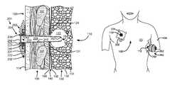
- FIG. 1Ashows the chest of a patient indicating alternative locations for a pneumostoma that may be managed using the device and methods of the present invention.
- FIG. 1Bshows a sectional view of the chest illustrating the relationship between the pneumostoma, lung and natural airways.
- FIG. 1Cshows a detailed sectional view of a pneumostoma.
- FIG. 2Ashows a perspective view of components of a pneumostoma management device according to an embodiment of the present invention.
- FIG. 2Bshows a sectional view of the pneumostoma management device of FIG. 2A partially implanted in a pneumostoma.
- FIG. 2Cshows a perspective view of a pneumostoma aspirator designed to operate with the pneumostoma management device of FIGS. 2A and 2B according to an embodiment of the present invention.
- FIG. 2Dshows a sectional view of the pneumostoma aspirator of FIG. 2C mated with the pneumostoma management device of FIGS. 2A and 2B according to an embodiment of the present invention.
- FIG. 2Eshows a positioning of a pneumostoma management device and pneumostoma aspirator relative to the chest of a patient.
- FIG. 2Fshows a method of using a pneumostoma aspirator in accordance with an embodiment of the present invention, in the form of instructions for use.
- FIG. 3Ashows a perspective view of an alternative pneumostoma aspirator according to an embodiment of the present invention.
- FIG. 3Bshows a sectional view of the pneumostoma aspirator of FIG. 3A .
- FIG. 4Ashows a perspective view of an alternative pneumostoma aspirator according to an embodiment of the present invention.
- FIG. 4Bshows a sectional view of the pneumostoma aspirator of FIG. 3A .
- FIG. 4Cshows a sectional view of an alternative pneumostoma aspirator according to an embodiment of the present invention.
- FIG. 4Dshows a sectional view of an alternative pneumostoma aspirator according to an embodiment of the present invention.
- FIG. 5shows a perspective view of an alternative pneumostoma aspirator according to an embodiment of the present invention.
- FIG. 6shows a perspective view of a motorized alternative pneumostoma aspirator according to an embodiment of the present invention.
- FIG. 1Ashows the chest of a patient identifying alternative locations for creating a pneumostoma that may be managed using the system of the present invention.
- a first pneumostoma 110is shown on the front of the chest 100 over the right lung 101 (shown in dashed lines).
- the pneumostomais preferably positioned over the third intercostal space on the mid-clavicular line.
- the pneumostoma 110is located on the front of the chest between the third and fourth ribs.
- the pneumostoma 110is preferably located between two ribs, in alternative procedures a pneumostoma can also be prepared using a minithoracotomy with a rib resection.
- FIG. 1Aa second pneumostoma 112 is illustrated in a lateral position entering the left lung 103 (shown in dashed lines).
- the pneumostoma 112is preferably positioned over the fourth or fifth intercostal space under the left arm 104 .
- one pneumostoma per lungis created; however, more or less than one pneumostoma per lung may be created depending upon the needs of the patient. In most humans, the lobes of the lung are not completely separate and air may pass between the lobes.
- a pneumostomais surgically created by forming an artificial channel through the chest wall and joining that channel with an opening through the visceral membrane of the lung into parenchymal tissue of the lung to form an anastomosis.
- the anastomosisis joined and sealed by sealing the channel from the pleural cavity using adhesives, mechanical sealing and/or pleurodesis.
- Methods for forming the channel, opening, anastomosis and pleurodesisare disclosed in applicant's pending and issued patents and applications including U.S. patent application Ser. No. 10/881,408 entitled “Methods and Devices to Accelerate Wound Healing in Thoracic Anastomosis Applications” and U.S. patent application Ser. No. 12/030,006 entitled “Variable Parietal/Visceral Pleural Coupling” which are incorporated herein by reference in their entirety.
- FIG. 1Bshows a sectional view of chest 100 illustrating the position of the pneumostoma 110 .
- the parenchymal tissue 132 of the lung 130is comprised principally of alveoli 134 .
- the alveoli 134are the thin walled air-filled sacs in which gas exchange takes place. Air flows into the lungs through the natural airways including the trachea 136 , carina 137 , and bronchi 138 . Inside the lungs, the bronchi branch into a multiplicity of smaller vessels referred to as bronchioles (not shown). Typically, there are more than one million bronchioles in each lung. Each bronchiole connects a cluster of alveoli to the natural airways.
- pneumostoma 110comprises a channel through the thoracic wall 106 of the chest 100 between two ribs 107 .
- Pneumostoma 110opens at an aperture 126 through the skin 114 of chest 100 .
- FIG. 1Cshows a detailed sectional view of the pneumostoma 110 .
- pneumostoma 110comprises a channel 120 through the thoracic wall 106 of the chest 100 between the ribs 107 .
- the channel 120is joined to cavity 122 in the parenchymal tissue 132 of lung 130 .
- An adhesion or pleurodesis 124surrounds the channel 120 where it enters the lung 130 .
- the thoracic wall 106is lined with the parietal membrane 108 .
- the surface of the lung 130is covered with a continuous sac called the visceral membrane 138 .
- the parietal membrane 108 and visceral membrane 138are often referred to collectively as the pleural membranes.
- the pleural cavityusually only contains a thin film of fluid that serves as a lubricant between the lungs and the chest wall.
- pleurodesis 124the pleural membranes are fused and/or adhered to one another eliminating the space between the pleural membranes in that region.
- Pleurodesis 124is the fusion or adhesion of the parietal membrane 108 and visceral membrane 138 .
- a pleurodesismay be a complete pleurodesis in which the entire pleural cavity 140 is removed by fusion of the visceral membrane 138 with the parietal membrane 108 over the entire surface of the lung 130 .
- the pleurodesisis preferably localized to the region surrounding the channel 120 .
- the pleurodesis 124 surrounding the channel 120prevents air from entering the pleural cavity 140 . If air is permitted to enter pleural cavity 140 , a pneumothorax will result and the lung may collapse.
- Pleurodesis 124can be created between the visceral pleura of the lung and the inner wall of the thoracic cavity using chemical methods including introducing into the pleural space irritants such as iodopovidone or silver nitrate, antibiotics (e.g. Doxycycline or Quinacrine), anticancer drugs (e.g. Bleomycin, Mitoxantrone or Cisplatin), cytokines (e.g. interferon alpha-2 ⁇ and Transforming growth factor- ⁇ ); pyrogens (e.g. Corynebacterium parvum, Staphylococcus aureus superantigen or OK432); connective tissue proteins (e.g. fibrin or collagen) and minerals (e.g.
- irritantssuch as iodopovidone or silver nitrate, antibiotics (e.g. Doxycycline or Quinacrine), anticancer drugs (e.g. Bleomycin, Mitoxantrone or Cisplatin
- a pleurodesiscan also be created using surgical methods including pleurectomy.
- the pleural spacemay be mechanically abraded during thoracoscopy or thoracotomy. This procedure is called dry abrasion pleurodesis.
- a pleurodesismay also be created using radiotherapy methods, including radioactive gold or external radiation. These methods cause an inflammatory response and or fibrosis, healing, and fusion of the pleural membranes.
- a sealcan be created in an acute manner between the pleural membranes using biocompatible glues, meshes or mechanical means such as clamps, staples, clips and/or sutures. The adhesive or mechanical seal may develop into pleurodesis over time.
- a range of biocompatible gluesare available that may be used on the lung, including light-activatable glues, fibrin glues, cyanoacrylates and two part polymerizing glues.
- Applicant's copending U.S. patent application Ser. No. 12/030,006 entitled “VARIABLE PARIETAL/VISCERAL PLEURAL COUPLING”discloses methods such as pleurodesis for coupling a channel through the chest wall to the inner volume of the lung without causing a pneumothorax and is incorporated herein by reference for all purposes.
- pneumostoma 110When formed, pneumostoma 110 provides an extra pathway for exhaled air to exit the lung 130 reducing residual volume and intra-thoracic pressure without the air passing through the major natural airways such as the bronchi 138 and trachea.
- Collateral ventilationis particularly prevalent in an emphysemous lung because of the deterioration of lung tissue caused by COPD.
- Collateral ventilationis the term given to leakage of air through the connective tissue between the alveoli 134 .
- Collateral ventilationmay include leakage of air through pathways that include the interalveolar pores of Kohn, bronchiole-alveolar communications of Lambert, and interbronchiolar pathways of Martin. This air typically becomes trapped in the lung and contributes to hyperinflation.
- the pneumostomaallows stale air trapped in the parenchymal tissue 132 to escape from the lung 130 . This reduces the residual volume and intra-thoracic pressure. The lower intra-thoracic pressure reduces the dynamic collapse of airways during exhalation. By allowing the airways to remain patent during exhalation, labored breathing (dyspnea) and residual volume (hyperinflation) are both reduced.
- Pneumostoma 110not only provides an extra pathway that allows air to exit the lung 130 but also allows more fresh air to be drawn in through the natural airways. This increases the effectiveness of all of the tissues of the lung 130 and improves gas exchange. Pneumostoma 110 thus achieves many of the advantages sought by lung volume reduction surgery without surgically removing a portion of the lung or sealing off a portion of the lung.
- a pneumostoma management systemin accordance with embodiments of the present invention is desirable to maintain the patency of the pneumostoma and control flow of materials between the exterior of the patient and the parenchymal tissue of the lung via a pneumostoma.
- the pneumostoma management systemincludes a pneumostoma management device and a pneumostoma aspirator as described herein.
- Pneumostoma Management SystemIncluding A Pneumostoma Aspirator
- a pneumostomamay be created to treat the symptoms of chronic obstructive pulmonary disease.
- a patientis typically provided with a pneumostoma management system to protect the pneumostoma and keeps the pneumostoma open on a day-to-day basis.
- a pneumostoma management device(“PMD”) comprises a tube which is inserted into the pneumostoma and an external component which is secured to the skin of the patient to keep the tube in place. Gases escape from the lung through the tube and are vented external to the patient.
- the pneumostoma management devicemay, in some, but not all cases, include a filter which only permits gases to enter or exit the tube.
- the pneumostoma management devicemay, in some, but not all cases, include a one-way valve which allows gases to exit the lung but not enter the lung through the tube. Additional details and variations of pneumostoma management devices are described in applicant's pending and issued patents and applications including those related cases incorporated by reference above.
- FIGS. 2A through 2Dillustrate views of a pneumostoma management system including a pneumostoma management device (“PMD”) 201 and a pneumostoma aspirator 260 in accordance with an embodiment of the present invention.
- FIGS. 2C and 2Dshow pneumostoma aspirator 260 and its interaction with the PMD 201 .
- PMD 201includes a chest mount 202 which may be mounted to the skin of the patient and a pneumostoma vent 204 which is fitted to the chest mount 202 .
- pneumostoma vent 204is mounted through an aperture 224 in chest mount 202 .
- Chest mount 202has a first coupling that engages a second coupling of the pneumostoma vent to releasably secure the pneumostoma vent 204 to the chest mount 202 .
- the join between the two components of PMD 201is engineered to ensure that pneumostoma vent 204 cannot be over-inserted into the lung if it separates from chest mount 202 .
- pneumostoma vent 204includes a tube 240 sized and configured to fit within the channel of a pneumostoma 110 .
- Tube 240is stiff enough that it may be inserted into a pneumostoma without collapsing. Over time, a pneumostoma may constrict and it is one function of PMD 201 to preserve the patency of the channel of the pneumostoma by resisting the natural tendency of the pneumostoma to constrict.
- Tube 240 of pneumostoma vent 204preferably comprises an atraumatic tip 252 at the distal end.
- proximalrefers to the end or side of a device closest to the hand operating the device
- distalrefers to the end or side of a device furthest from the hand operating the device.
- Tip 252may be rounded, beveled or curved in order to reduce irritation or damage to the tissues of the pneumostoma or lung during insertion or while in position. Opening 254 in tip 252 allows the entry of gases from the cavity of the pneumostoma 110 into lumen 258 of tube 240 .
- Tube 240is optionally provided with one or more side openings (not shown) positioned near tip 252 and/or along the length of tube 240 to facilitate the flow of gas and/or mucous/discharge into lumen 258 .
- Tube 240 of pneumostoma vent 204is sufficiently long that it can pass through the thoracic wall and into the cavity of a pneumostoma inside the lung. Pneumostoma vent 204 is not however so long that it penetrates so far into the lung that it might cause injury.
- the material and thickness of tube 240 of pneumostoma vent 204is selected such that tube 240 is soft enough that it will deform rather than cause injury to the pneumostoma or lung.
- Pneumostoma vent 204has an opening 254 in tip 252 of tube 240 .
- the length of tube 240 required for a pneumostoma vent 204varies significantly between different pneumostomas. A longer tube 240 is usually required in patients with larger amounts of body fat on the chest.
- a longer tube 240is usually required where the pneumostoma is placed in the lateral position 112 rather than the frontal position 110 . Because of the variation in pneumostomas, pneumostoma vents 204 are manufactured having tubes 240 in a range of sizes and a patient is provided with a pneumostoma vent 204 having a tube 240 of appropriate length for the patient's pneumostoma.
- Pneumostoma vent 204includes a cap 242 and a hydrophobic filter 248 over the opening 255 in the proximal end of tube 240 .
- Hydrophobic filter 248is positioned over the proximal opening 255 into lumen 258 .
- Hydrophobic filter 248is positioned and mounted such that material moving between lumen 258 and the exterior of pneumostoma vent 204 must pass through hydrophobic filter 248 .
- Hydrophobic filter 248is preferably designed such to fit into a recess in cap 242 . As shown in FIG. 2B , cap 242 comprises a recess 238 into which hydrophobic filter 248 may be fit.
- Hydrophobic filter 248may alternatively be fitted into cap 242 using a joint such as a threaded coupling or adhesive or, in some cases, formed integrally with cap 242 .
- Hydrophobic filter 248may be made from a material such as medical grade GOR-TEX (W. L. Gore & Associates, Inc., Flagstaff, Ariz.).
- a snap ring 243locks cap 242 and hydrophobic filter 248 onto the proximal end of tube 240 .
- Hydrophobic filter 248serves several purposes. In general, hydrophobic filter 248 controls the passage of solid or liquid material between the lumen 258 and the exterior of cap 242 . For example, hydrophobic filter 248 prevents the flow of water into the lumen 258 through proximal opening 255 . Thus, a patient using PMD 201 may shower without water entering the lung through the pneumostoma. Hydrophobic filter 248 may also be selected so as to prevent the entry of microbes, pollen and other allergens and pathogens into the lumen 258 . Hydrophobic filter 248 also prevents the exit of liquid and particulate discharge from lumen 258 to the exterior of pneumostoma vent 204 . This is desirable to prevent contact between liquid and particulate discharge and clothing for example.
- Chest mount 202connects to the proximal end of pneumostoma vent 204 .
- chest mount 202comprises a flange 222 and an aperture 224 .
- the aperture 224is adapted and configured to receive the pneumostoma vent 204 .
- Chest mount 202is designed to have a smooth surface and a low profile so it is comfortable for the patient to wear. Chest mount 202 should be designed so as not to snag on the patient's clothing or to restrict motion of the patient's arm (if placed in a lateral pneumostoma 112 ).
- Flange 222is significantly wider than pneumostoma vent 204 .
- Flange 222thus comprises a contact surface 232 which contacts the skin of the patient surrounding the pneumostoma and positions the aperture 224 over the opening of the pneumostoma.
- Flange 222is designed such that it is sufficiently flexible that it can conform to the surface of the chest.
- Contact surface 232is also provided with a pad of biocompatible adhesive 234 , such as a hydrocolloid adhesive, for securing flange 222 to the skin of the patient.
- the adhesive 234may be protected by a protector sheet that is removed prior to use of flange 222 .
- Adhesive 234should be selected so as to secure flange 222 to the chest of the patient in the correct position relative to the pneumostoma without causing undue irritation to the skin of the patient.
- the adhesiveneed not create an air tight seal between flange 222 and the skin of the patient. Suitable adhesive pads are available commercially from Avery Dennison (Painesville, Ohio).
- FIG. 2Ashows a perspective view of chest mount 202 after insertion of pneumostoma vent 204 .
- Flange 222is generally circular but is provided with one or more tabs 236 to facilitate application and removal of flange 222 from the skin of the patient.
- chest mount 202comprises an aperture 224 through which tube 240 of pneumostoma vent 204 may be inserted.
- Flange 222is slightly convex on the upper surface 235 .
- Flange 222includes a recess 226 into which cap 242 of pneumostoma vent 204 may be press fit.
- Flange 222is thick enough in the region of aperture 224 to receive the cap 242 of pneumostoma vent 204 so that the cap of pneumostoma vent 204 is flush with the upper surface 235 of flange 222 .
- Recess 226forms a coupling adapted to releasably secure the cap 242 of pneumostoma vent 204 into flange 222 .
- recess 226has a lip 227 to releasably secure the cap 242 of pneumostoma vent 204 into flange 222 .
- Cap 242is attached to the proximal end of tube 240 .
- Hydrophobic filter 248is sandwiched between cap 242 and tube 240 .
- An opening 244 in cap 242communicates with the lumen 258 of tube 240 via hydrophobic filter 248 .
- cap 242comprises a lip 246 which releasably engages lip 227 of recess 226 of flange 222 to secure pneumostoma vent 204 within the recess 226 of flange 222 .
- Lip 246forms a coupling element of pneumostoma vent 204 that cooperates with recess 226 to releasably secure pneumostoma vent 204 into chest mount 202 with tube 240 positioned through aperture 224 .
- an aperture plate 228is embedded in the conformable polymer of flange 222 .
- the aperture plate 228defines aperture 224 of chest mount 202 .
- Aperture plate 228is made of a stiffer, less compliant material than flange 222 in order that the dimensions of aperture 224 are tightly controlled.
- Aperture plate 228is stiff enough that the size and shape of aperture 224 remains stable even under any reasonably possible application of force to chest mount 202 . It should be noted that the outer diameter of each of snap ring 243 , hydrophobic filter 248 , flange 241 and cap 242 is larger than the diameter of aperture 224 of aperture plate 228 .
- all the components of the chest mount 202such as flange 222 and aperture plate 224 are significantly larger than the aperture of a pneumostoma thus precluding passage of any component of the chest mount 202 into a pneumostoma even in the unlikely event of damage to the device.
- FIGS. 2C and 2Dshow a pneumostoma aspirator adapted for use with PMD 201 of FIGS. 2A and 2B as part of pneumostoma management system.
- FIG. 2Cshows a perspective view of pneumostoma aspirator 260 .
- FIG. 2Dshows a sectional view through pneumostoma aspirator 260 of FIG. 2C when mounted in a chest mount 202 .
- pneumostoma aspirator 260includes a bulb 262 a coupling 264 and a tube 266 .
- Tube 266has an opening 261 in the distal end.
- Opening 261is adapted to allow entry of gases as well as solid and liquid discharge during operation of aspirator.
- Tube 266may be provided with additional openings is the side of tube 266 .
- Pneumostoma aspiratoris configured such that tube 266 may be inserted through aperture 224 of chest mount 202 into a pneumostoma.
- Tube 266is sufficiently long to enter the pneumostoma but is not so long that it might cause injury to the pneumostoma.
- Coupling 264is designed such that it is too large to pass through aperture 224 of the aperture plate 228 of chest mount 202 thereby preventing further insertion of tube 266 into a pneumostoma.
- Coupling 264may also be provided with a feature such as a lip 265 for releasably engaging lip 227 of recess 228 of chest mount 202 .
- Bulb 262is made of a flexible material such that it may be squeezed to reduce the volume of the bulb and when released will return to its previous volume. The re-expansion of bulb 262 may be utilized to apply suction to the pneumostoma to remove fluid and/or discharge. In some embodiments, the reduction in volume of bulb 262 may be used to push the contents of bulb 262 into a pneumostoma, for example an irrigating fluid such as sterile saline or water.
- an irrigating fluidsuch as sterile saline or water.
- pneumostoma aspirator 260optionally comprises a one-way valve 268 through which air may pass out of bulb 262 .
- pneumostoma aspirator 260may also include a one-way valve 269 configured to allow material to flow from tube 266 into bulb 262 .
- Valve 268allows air to escape bulb 262 when it is compressed.
- valves 268 and 269prevent air flow into the pneumostoma.
- device 260may be designed to provide suction alone instead of suction and irrigation.
- an aperturemay be provided in bulb 262 in place of valve 268 .
- the apertureis configured to allow air to escape when bulb 262 is squeezed.
- the aperturemay then be covered with a digit so that air may not enter the aperture when bulb 262 expand and is instead drawn from the pneumostoma through tube 266 .
- a range of pneumostoma aspiratorsmay be manufactured each having a size appropriate for a different pneumostoma.
- pneumostoma aspirator 260may be designed to use some components in common with pneumostoma vent 204 .
- the range of tubes 240 of the pneumostoma vent 204may be used as tube 266 of pneumostoma aspirator 260 .
- the cap 264may also be a shared component.
- the only additional components required for pneumostoma aspirator 260are bulb 262 and (optionally) valves 268 and 269 .
- the pneumostoma aspirator 260may be made in only one size where the single size of tube 266 is short enough so as not to cause injury even in a small pneumostoma.
- FIG. 2Eillustrates the positioning of pneumostoma aspirator 260 over pneumostoma 112 of FIG. 1A .
- the chest mount 202remains attached for up to a week thereby avoiding irritation of the skin caused by daily attachment and removal of a mount.
- Chest mountmay be positioned by the patient by manual alignment of the aperture 224 of chest mount 202 with the aperture of the pneumostoma 112 .
- chest mount 202is first positioned over a pneumostoma and secured with adhesive to the skin of the patient.
- a pneumostoma vent or an alignment toolmay be used to align the chest mount.
- Pneumostoma aspirator 260is then inserted through the aperture in the chest mount until it engages the chest mount 202 . As shown in FIG. 2E the pneumostoma aspirator 260 is inserted through chest mount 202 after pneumostoma vent 204 has been removed. Pneumostoma aspirator 260 is then used to apply suction to pneumostoma 112 by manual operation of bulb 262 either by the patient, caregiver or medical practitioner. The application of suction draws discharge from the pneumostoma into the aperture 261 at the distal end of the aspirator 260 . Where suction is applied care should be taken to remove any discharge collected to prevent reentry of discharge into the pneumostoma 112 .
- FIG. 2Fillustrates a method for using a pneumostoma aspirator according to an embodiment of the present invention.
- the methodis illustrated in the form of Instructions For Use 270 .
- Instructions For Useare provided to patients with a medical device such as a pneumostoma aspirator.
- the instructions from useinclude instructions to perform the following steps.
- the tube of the pneumostoma aspiratoris inserted into the pneumostoma.
- the tubeis pushed into the pneumostoma until the flange of the aspirator engages the chest of the patient and prevents further insertion.
- the aspiratoris actuated to collect discharge in the tube.
- the bulbis squeezed and then allowed to expand sucking air and discharge into the tube.
- the plunger on a syringeis pulled back sucking air and discharge into the tube.
- the aspiratoris withdrawn from the pneumostoma. The discharge may then be eliminated.
- the instructionswill be slightly different where the aspirator is designed to operate with a chest mount already in place. In such case, the flange is already engaged with the chest and insertion of the aspirator is limited by engagement of the aspirator with the flange of the chest mount.
- pneumostoma aspirator 260may alternatively or additionally be used to apply irrigation to pneumostoma 112 by manual operation of bulb 262 either by the patient, caregiver or medical practitioner.
- a sterile but inert solutionmay be used.
- sterile saline or sterile watermay be used.
- an antibacterial or mucolytic solutionmay be used.
- the cleaning solutionmay also include a small concentration of an agent for maintaining the patency of the pneumostoma for example, Paclitaxel.
- the cleaning solutionshould be formulated carefully to avoid injury or irritation to the lung.
- the pneumostoma aspiratorcan be used to push the irrigation fluid through the aperture 261 in the distal end of the aspirator and into the pneumostoma.
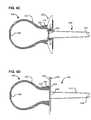
- FIGS. 3A and 3Bshow an alternative pneumostoma aspirator 310 designed to apply suction to a pneumostoma.
- FIG. 3Ashows a perspective view of pneumostoma aspirator 310 .
- FIG. 3Bshows a sectional view of the pneumostoma aspirator 310 .
- pneumostoma aspirator 310includes a flexible bulb 312 attached to a flange 314 which is attached to a tube 316 .
- Tube 316has an opening 320 in the distal end. Opening 320 is adapted to allow entry of gases as well as solid and liquid discharge during operation of aspirator 310 .
- Tube 316may also be provided with additional openings in the side of tube 316 .
- Tube 316 of aspirator 310preferably has an atraumatic tip 317 at the distal end to prevent injury and/or irritation to the pneumostoma during insertion.
- Flange 314 attached to tube 316is significantly larger than the diameter of tube 316 .
- Flange 314is too large to enter a pneumostoma and thus acts as a stop to prevent further insertion of tube 316 when flange 314 makes contact with the skin of the patient's chest.
- the contact surface 315 of flange 314may also be used to make a temporary seal surrounding the pneumostoma so that when applying suction to the pneumostoma there is reduced leakage of air/fluid around tube 316 .
- Contact surface 315may be provided with surface features (for example ridges) to enhance the formation of a temporary seal between flange 314 and the skin of the chest.
- Tube 316extends far enough past flange 314 so that it can pass through the thoracic wall into the pneumostoma. Tube 316 is not, however, so long that it may cause injury to the pneumostoma or lung. The maximum desirable length of tube 316 varies significantly between different pneumostomas. A longer tube 316 may be desirable in patients with larger amounts of body fat on the chest. A longer tube 316 may also be desirable where the pneumostoma is placed in the lateral position 112 rather than the frontal position 110 . Because of the variation in pneumostomas, pneumostoma aspirators 310 may be manufactured having tubes 316 in a range of sizes.
- a patientcan thus be provided with a pneumostoma aspirator 310 having a tube 316 of appropriate length for the patient's pneumostoma.
- Tube 316may be from 30 mm to 120 mm in length and from 5 mm to 20 mm in diameter depending on the size of a pneumostoma.
- a typical tube 240may be between 40 mm and 80 mm in length and between 8 mm and 12 mm in diameter.
- a pneumostoma aspiratoris made with a tube 316 of a single length (such as 120 mm) and tube 316 is then cut to the length appropriate for a particular patient.
- a pneumostoma aspiratoris made with a tube 316 of a single short length (such as 30 mm) which can be used in any pneumostoma without causing injury.
- bulb 312 , flange 314 and tube 316 of pneumostoma aspirator 310are made in one piece. They may alternatively be formed separately and then joined by welding, gluing or otherwise bonding/connecting.
- Suction irrigation device 310may also comprise valves 318 and 319 . Valves 318 and 319 are flow control devices (for example flapper valves) which allow flow in on one direction only.
- valves 318 and 319may be present and configured such that, when bulb 312 is compressed air leaves bulb 312 only via valve 318 , and when bulb 312 is released air enters bulb 312 only via tube 316 .
- valves 318 and 319are too large to fit through tube 316 and thus cannot be aspirated into the lung even in the event of damage to pneumostoma aspirator 310 .
- valves 318 and 319are press fit into recesses in bulb 312 .
- Valve 319(if present) is smaller than valve 318 so that valve 319 can be inserted into bulb 312 through the aperture for mounting valve 318 .
- Pneumostoma aspiratormay be used in accordance with the instructions for use of FIG. 2F . However, the patient/caregiver should be instructed to remove the pneumostoma management device from the pneumostoma prior to step 272 . Also, after step 278 , the patient/caregiver should be instructed to promptly place a new pneumostoma management device into the pneumostoma.
- FIGS. 4A and 4Bshow an alternative pneumostoma aspirator 410 designed to apply suction to a pneumostoma.
- the pneumostoma aspirator 410operates in conjunction with a pneumostoma management device located within a pneumostoma e.g. PMD 200 of FIGS. 2A-2E .
- FIG. 4Ashows a perspective view of aspirator 410 .
- FIG. 4Bshows a sectional view of aspirator 410 .
- aspirator 410includes a flexible bulb 412 attached to a coupling 414 which has two releases 416 .
- Aspirator 410also has a one-way valve 418 for releasing air from bulb 412 .
- Coupling 414is designed to releasably attach to cap 242 of pneumostoma vent 204 .
- Releases 416are release mechanisms which may be operated to release coupling 414 from cap 242 in order to reuse aspirator 410 .
- aspirator 410is a single-use device and coupling 414 permanently attaches to cap 242 —releases 416 are therefore absent.
- FIG. 4Ashows aspirator 410 aligned for attachment to cap 242 of pneumostoma vent 204 .
- FIG. 4Bshows aspirator 410 after it has been attached to cap 242 .
- the aspirator 410 of FIGS. 4A and 4Bmay be used at the time of removal of pneumostoma vent 204 in order to remove discharge from the pneumostoma prior to replacement of pneumostoma vent 204 .
- Valve 418is a flow control device (for example flapper valve) which allows flow in one direction only.
- an aspirator 420is designed to mate with chest mount 202 instead of or in addition to pneumostoma vent 204 .
- bulb 422has a mating section 424 , having a mating surface 426 designed to mate and make a temporary seal with the exterior surface of chest mount 202 .
- aspirator 420is pushed against chest mount 202 to make a temporary seal.
- Bulb 422is then compressed, expelling air through one-way valve 428 .
- Bulb 422is then released such that it expands and withdraws air and discharge into/through aperture 254 in the distal end of pneumostoma vent 204 .
- the dischargecollects inside tube 240 of pneumostoma vent 204 .
- pneumostoma vent 204(containing the discharge) may be removed and disposed of.
- a new pneumostoma vent 204may then be inserted in chest mount 202 .
- bulb 422is held in contact with chest mount 202 in order to make a temporary seal during aspiration of the pneumostoma.
- FIG. 4Dillustrates another embodiment having an aspirator 430 designed to mate with a PMD 440 in which tube 442 is formed in one piece with (or permanently attached to) a flange 444 .
- PMD 440has a hydrophobic filter 446 press fit into the proximal end of tube 442 and has a biocompatible adhesive 448 on the contact surface 449 of flange 444 for releasably securing flange 444 to the skin of the patient's chest.
- aspirator 430includes a bulb 432 which has a mating section 434 , having a mating surface 436 designed to mate and make a temporary seal with the exterior surface of flange 444 .
- aspirator 430is pushed against flange 444 to make a temporary seal.
- Bulb 432is then compressed, expelling air through one-way valve 438 .
- Bulb 432is then released such that it expands and withdraws air and discharge into/through aperture 454 in the distal end of tube 442 of PMD 440 .
- the dischargecollects inside tube 442 of PMD 440 .
- PMD 440(containing the discharge) may be removed. A new PMD 440 may then be inserted into the pneumostoma.
- a flexible bulb(with or without one or more valves) has been provided as the mechanism by which irrigation fluid may be provided or suction applied.
- a different mechanismmay be provided to produce the negative pressure required to extract the fluid/air discharge from the pneumostoma.
- Such mechanismsinclude vacuum bottles, pumps, fans and syringes. (Or positive pressure for irrigation).
- the mechanismhave safety features to prevent over insertion of any component into the pneumostoma or the application of positive or negative pressure sufficient to cause injury to the lung.
- the safety featuresare particularly desirable in devices intended for use by the patient rather than a trained medical professional.
- FIG. 5illustrates an alternative embodiment of a pneumostoma aspirator 500 in which the positive and/or negative pressure is applied using a syringe 502 .
- Syringe 502includes a plunger 504 which can be pushed and pulled by a rod 506 within barrel 508 .
- Rod 506passes though cap 510 to ring 512 which may be manually operated to move plunger 504 .
- Rings 514are connected to cap 510 which is connected to barrel 508 .
- Positive pressurecan be used to push irrigating fluid through aperture 520 of nozzle 516 into a pneumostoma.
- Negative pressurecan be used to withdraw fluid, discharge and/or air through aperture 520 of nozzle 516 from the pneumostoma into barrel 508 .
- a safety valve 518is provided which opens in the event that the positive or negative pressure is outside of a preset safe range.
- nozzle 516comprises a coupling 521 for engaging a chest mount 202 as shown in FIGS. 2A-2E .
- a tube 522extends distal of coupling 521 for insertion in the pneumostoma, but coupling 521 limits the depth of insertion of tube 522 .
- nozzle 516may comprise a flange for preventing over-insertion or mating devices for coupling the syringe to part of a PMD including for example, a chest mount 202 or pneumostoma vent 204 as described above.
- syringe 502may be used, with appropriate adaptations, in place of the bulb in the embodiments of FIGS. 2C-E , 3 A-B and 4 A-D.
- Pneumostoma aspirator 500may be a sterilizable reusable device made, for example, from stainless steel and/or glass components.
- Pneumostoma aspirator 500may alternatively be a disposable device made, for example, from medical grade plastics.
- FIG. 6illustrates an alternative embodiment of a pneumostoma aspirator 600 in which the positive and/or negative pressure is applied using a motorized device 602 .
- Motorized device 602includes a motor 604 which turns a fan/pump 606 .
- the power for motor 604may be provided by batteries 630 in battery housing 632 .
- Batteries 630may be rechargeable batteries.
- Fan/pump 606draws air in through tube 608 and expels it through tube 610 and vent 612 .
- Tube 608terminates inside removable reservoir 614 .
- Thus operation of fan/pump 606creates negative pressure inside reservoir 614 .
- Tube 616connects reservoir 614 to tube 618 which is adapted to enter a pneumostoma.
- Tube 618has an a traumatic tip 617 to facilitate insertion into a pneumostoma and one or more apertures 619 in the tip through which air and discharge may enter tube 618 to be sucked via tube 616 into reservoir 614 .
- tube 608has a valve/filter 620 which prevents entry of discharge into tube 610 and fan/pump 606 .
- Vent 612may also be provided with a replaceable filter (for example a HEPA filter) to prevent the venting of any pathogens which may be in the gases extracted from the lung.
- Fan/pump 606is selected so that it is self-limiting as to the maximum negative pressure it is capable of producing in reservoir 614 .
- the maximum negative pressureis selected to be at a level which will not damage the pneumostoma or lung.
- a safety valvemay additionally or alternatively be provided which opens in the event that the pressure is outside of a preset safe range.
- Reservoir 614is preferably translucent so that accumulation of discharge may be observed.
- Tube 618is connected with a flange 622 which limits the depth of insertion of tube 618 into a pneumostoma.
- tube 618may comprise a coupling for engaging a chest mount 202 as shown in FIGS. 2A-2E .
- Tube 618extends distal of flange 622 for a distance selected so as not to damage a pneumostoma. Different lengths of tube 618 may be supplied depending on the size of pneumostoma in a particular patient.
- a coupling 624(for example, a threaded joint or slip-on fitting) may allow the tube 618 and flange 622 to be removed and replaced.
- tube 618is pushed into the pneumostoma until flange 622 engages the chest of the patient to prevent further insertion.
- the patientor medical provider
- button 626which actuates motor 604 .
- Motor 604drives fan/pump 606 which extracts air from reservoir 614 . Air is sucked through tube 618 via tube 616 into reservoir 614 . Solid and liquid discharge may also be sucked through the aperture(s) in the tip of tube 618 and thence into reservoir 614 .
- the discharge 634accumulates in reservoir 614 . Gases removed from the pneumostoma are vented through vent 612 .
- Reservoir 614may be then detached from motorized device 602 , emptied, cleaned and re-attached.
- reservoir 614may be disposable, in which case, the reservoir is detached, disposed of and replaced with a new reservoir.
- tube 618may be detached and cleaned or detached and replaced.
- Motorized device 602is preferable a reusable device.
- pneumostoma aspiratorhas been illustrated with a flange 622 , it should be noted that alternative structures may be connected with motorized device 602 so that it may be coupled to a pneumostoma vent or chest mount as previously shown.
- motorized device 602may be used, with appropriate adaptations, in place of the bulb in the embodiments of FIGS. 2C-E , 3 A-B and 4 A-D.
- the pneumostoma management device and the pneumostoma aspiratorare formed from biocompatible polymers or biocompatible metals.
- a patientwill typically wear the PMD at all times and thus the materials, particularly of tubes entering the pneumostoma, should meet high standards for biocompatibility.
- preferred materials for manufacturing the suction irrigation device and the PMDare biocompatible thermoplastic elastomers that are readily utilized in injection molding and extrusion processing.
- other suitable similarly biocompatible thermoplastic or thermoplastic polymer materialscan be used without departing from the scope of the invention.
- Biocompatible polymersmay be selected from the group consisting of polyethylenes (HDPE), polyvinyl chloride, polyacrylates (polyethyl acrylate and polymethyl acrylate, polymethyl methacrylate, polymethyl-coethyl acrylate, ethylene/ethyl acrylate), polycarbonate urethane (BIONATEG), polysiloxanes (silicones), polytetrafluoroethylene (PTFE, GORE-TEX®, ethylene/chlorotrifluoroethylene copolymer, aliphatic polyesters, ethylene/tetrafluoroethylene copolymer), polyketones (polyaryletheretherketone, polyetheretherketone, polyetheretherketoneketone, polyetherketoneetherketoneketone polyetherketone), polyether block amides (PEBAX, PEBA), polyamides (polyamideimide, PA-11, PA-12, PA-46, PA-66), polyetherimide, polyether sulfone, poly(
- PMD 201Reference to appropriate polymers that can be used for manufacturing PMD 201 can be found in the following documents: PCT Publication WO 02/02158, entitled “Bio-Compatible Polymeric Materials;” PCT Publication WO 02/00275, entitled “Bio-Compatible Polymeric Materials;” and, PCT Publication WO 02/00270, entitled “Bio-Compatible Polymeric Materials” all of which are incorporated herein by reference.
- Other suitable materials for the manufacture of the PMDinclude medical grade inorganic materials such stainless steel, titanium, ceramics and coated materials.
- Hydrophobic filter 248should be sufficiently porous to allow air to exit through the filter.
- Materials for hydrophobic filtersare available commercially and filters can be fabricated from any suitable hydrophobic polymer, such as tetrafluoroethylene, PTFE, polyolefins, microglass, polyethylene and polypropylene or a mixture thereof.
- the hydrophobic filteris a laminated tetrafluoroethylene e.g. TEFLON®, (E.I. du Pont de Nemours Co.) or GORE-TEX® (W.L. Gore, Inc.) of a controlled pore size.
- the hydrophobic filtermay comprise a felted polypropylene; PTFE/polypropylene filter media.
- Hydrophobic filter 248may additionally comprise an antimicrobial, an anti-bacterial, and/or an anti-viral material or agent.
Landscapes
- Health & Medical Sciences (AREA)
- Life Sciences & Earth Sciences (AREA)
- Heart & Thoracic Surgery (AREA)
- Animal Behavior & Ethology (AREA)
- Veterinary Medicine (AREA)
- Public Health (AREA)
- General Health & Medical Sciences (AREA)
- Engineering & Computer Science (AREA)
- Biomedical Technology (AREA)
- Hematology (AREA)
- Anesthesiology (AREA)
- Pulmonology (AREA)
- Biophysics (AREA)
- Emergency Medicine (AREA)
- Vascular Medicine (AREA)
- Epidemiology (AREA)
- Gastroenterology & Hepatology (AREA)
- Child & Adolescent Psychology (AREA)
- Chemical & Material Sciences (AREA)
- Medicinal Chemistry (AREA)
- Pharmacology & Pharmacy (AREA)
- Otolaryngology (AREA)
- Media Introduction/Drainage Providing Device (AREA)
- Prostheses (AREA)
- Surgical Instruments (AREA)
- External Artificial Organs (AREA)
- Orthopedics, Nursing, And Contraception (AREA)
- Pharmaceuticals Containing Other Organic And Inorganic Compounds (AREA)
- Infusion, Injection, And Reservoir Apparatuses (AREA)
- Medicinal Preparation (AREA)
Abstract
Description
This application claims priority to all of the following applications including: U.S. Provisional Application No. 61/029,830, filed Feb. 19, 2008, entitled “ENHANCED PNEUMOSTOMA MANAGEMENT DEVICE AND METHODS FOR TREATMENT OF CHRONIC OBSTRUCTIVE PULMONARY DISEASE”;
U.S. Provisional Application No. 61/032,877, filed Feb. 29, 2008, entitled “PNEUMOSTOMA MANAGEMENT SYSTEM AND METHODS FOR TREATMENT OF CHRONIC OBSTRUCTIVE PULMONARY DISEASE”;
U.S. Provisional Application No. 61/038,371, filed Mar. 20, 2008, entitled “SURGICAL PROCEDURE AND INSTRUMENT TO CREATE A PNEUMOSTOMA AND TREAT CHRONIC OBSTRUCTIVE PULMONARY DISEASE”;
U.S. Provisional Application No. 61/082,892, filed Jul. 23, 2008, entitled “PNEUMOSTOMA MANAGEMENT SYSTEM HAVING A COSMETIC AND/OR PROTECTIVE COVER”;
U.S. Provisional Application No. 61/083,573, filed Jul. 25, 2008, entitled “DEVICES AND METHODS FOR DELIVERY OF A THERAPEUTIC AGENT THROUGH A PNEUMOSTOMA”;
U.S. Provisional Application No. 61/084,559, filed Jul. 29, 2008, entitled “ASPIRATOR FOR PNEUMOSTOMA MANAGEMENT”;
U.S. Provisional Application No. 61/088,118, filed Aug. 12, 2008, entitled “FLEXIBLE PNEUMOSTOMA MANAGEMENT SYSTEM AND METHODS FOR TREATMENT OF CHRONIC OBSTRUCTIVE PULMONARY DISEASE”;
U.S. Provisional Application No. 61/143,298, filed Jan. 8, 2009, entitled “METHODS AND APPARATUS FOR THE CRYOTHERAPY CREATION OR RE-CREATION OF PNEUMOSTOMY”; and
U.S. Provisional Application No. 61/151,581, filed Feb. 11, 2009, entitled “SURGICAL INSTRUMENTS AND PROCEDURES TO CREATE A PNEUMOSTOMA AND TREAT CHRONIC OBSTRUCTIVE PULMONARY DISEASE”.
All of the afore-mentioned applications are incorporated herein by reference in their entireties.
This application is related to all of the above provisional applications and all the patent applications that claim priority thereto including:
This application is related to all of the following applications including U.S. patent application Ser. No. 12/388,465, filed Feb. 18, 2009, entitled “ENHANCED PNEUMOSTOMA MANAGEMENT DEVICE AND METHODS FOR TREATMENT OF CHRONIC OBSTRUCTIVE PULMONARY DISEASE”;
U.S. patent application Ser. No. 12/388,447, filed Feb. 18, 2009, entitled “PNEUMOSTOMA MANAGEMENT SYSTEM AND METHODS FOR TREATMENT OF CHRONIC OBSTRUCTIVE PULMONARY DISEASE”;
U.S. patent application Ser. No. 12/388,451, filed Feb. 18, 2009, entitled “PNEUMOSTOMA MANAGEMENT METHOD FOR TREATMENT OF CHRONIC OBSTRUCTIVE PULMONARY DISEASE”;
U.S. patent application Ser. No. 12/388,435, filed Feb. 18, 2009, entitled “TWO-PHASE SURGICAL PROCEDURE FOR CREATING A PNEUMOSTOMA TO TREAT CHRONIC OBSTRUCTIVE PULMONARY DISEASE”;
U.S. patent application Ser. No. 12/388,438, filed Feb. 18, 2009, entitled “ACCELERATED TWO-PHASE SURGICAL PROCEDURE FOR CREATING A PNEUMOSTOMA TO TREAT CHRONIC OBSTRUCTIVE PULMONARY DISEASE”;
U.S. patent application Ser. No. 12/388,441, filed Feb. 18, 2009, entitled “SINGLE-PHASE SURGICAL PROCEDURE FOR CREATING A PNEUMOSTOMA TO TREAT CHRONIC OBSTRUCTIVE PULMONARY DISEASE”;
U.S. patent application Ser. No. 12/388,446, filed Feb. 18, 2009, entitled “PERCUTANEOUS SINGLE-PHASE SURGICAL PROCEDURE FOR CREATING A PNEUMSOTOMA TO TREAT CHRONIC OBSTRUCTIVE PULMONARY DISEASE”;
U.S. patent application Ser. No. 12/388,460, filed Feb. 13, 2009, entitled “PNEUMOSTOMA MANAGEMENT SYSTEM HAVING A COSTMETIC AND/OR PROTECTIVE COVER”;
U.S. patent application Ser. No. 12/388,455, filed Feb. 18, 2009, entitled “DEVICES AND METHODS FOR DELIVERY OF A THERAPEUTIC AGENT THROUGH A PNEUMOSTOMA”;
U.S. patent application Ser. No. 12/388,461, filed Feb. 18, 2009, entitled “ASPIRATOR FOR PNEUMOSTOMA MANAGEMENT”;
U.S. patent application Ser. No. 12/388,458, filed Feb. 18, 2009, entitled “FLEXIBLE PNEUMOSTOMA MANAGEMENT SYSTEM AND METHODS FOR TREATMENT OF CHRONIC OBSTRUCTIVE PULMONARY DISEASE”;
U.S. patent application Ser. No. 12/388,459, filed Feb. 18, 2009, entitled “METHODS AND DEVICES FOR FOLLOW-UP CARE AND TREATMENT OF A PNEUMOSTOMA”;
U.S. patent application Ser. No. 12/388,453, filed Feb. 18, 2009, entitled “SURGICAL INSTRUMENTS FOR CREATING A PNEUMOSTOMA AND TREATING CHRONIC OBSTRUCTIVE PULMONARY DISEASE”;
U.S. patent application Ser. No. 12/388,466, filed Feb. 18, 2009, entitled “ONE-PIECE PNEUMOSTOMA MANAGEMENT SYSTEM AND METHODS FOR TREATMENT OF CHRONIC OBSTRUCTIVE PULMONARY DISEASE”;
U.S. patent application Ser. No. 12/388,467, filed Feb. 18, 2009, entitled “PNEUMOSTOMA MANAGEMENT SYSTEM WITH SECRETION MANAGEMENT FEATURES FOR TREATMENT OF CHRONIC OBSTRUCTIVE PULMONARY DISEASE”;
U.S. patent application Ser. No. 12/388,468, filed Feb. 18, 2009, entitled “MULTI-LAYER PNEUMOSTOMA MANAGEMENT SYSTEM AND METHODS FOR TREATMENT OF CHRONIC OBSTRUCTIVE PULJMONARY DISEASE”;
U.S. patent application Ser. No. 12/388,469, filed Feb. 18, 2009, entitled “VARIABLE LENGTH PNEUMOSTOMA MANAGEMENT SYSTEM FOR TREATMENT OF CHRONIC OBSTRUCTIVE PULMONARY DISEASE”; and
U.S. patent application Ser. No. 12/388,470, filed Feb. 18, 2009, entitled “SELF-SEALING DEVICE AND METHOD FOR DELIVERY OF A THERAPEUTIC AGENT THROUGH A PNEUMOSTOMA”.
All of the afore-mentioned applications are incorporated herein by reference in their entireties. This patent application also incorporates by reference all patents, applications, and articles discussed and/or cited herein.
In the United States alone, approximately 14 million people suffer from some form of Chronic Obstructive Pulmonary Disease (COPD). However an additional ten million adults have evidence of impaired lung function indicating that COPD may be significantly underdiagnosed. The cost of COPD to the nation in 2002 was estimated to be $32.1 billion. Medicare expenses for COPD beneficiaries were nearly 2.5 times that of the expenditures for all other patients. Direct medical services accounted for $18.0 billion, and indirect cost of morbidity and premature mortality was $14.1 billion. COPD is the fourth leading cause of death in the U.S. and is projected to be the third leading cause of death for both males and females by the year 2020.
Chronic Obstructive Pulmonary Disease (COPD) is a progressive disease of the airways that is characterized by a gradual loss of lung function. In the United States, the term COPD includes chronic bronchitis, chronic obstructive bronchitis, and emphysema, or combinations of these conditions. In emphysema the alveoli walls of the lung tissue are progressively weakened and lose their elastic recoil. The breakdown of lung tissue causes progressive loss of elastic recoil and the loss of radial support of the airways which traps residual air in the lung. This increases the work of exhaling and leads to hyperinflation of the lung. When the lungs become hyperinflated, forced expiration cannot reduce the residual volume of the lungs because the force exerted to empty the lungs collapses the small airways and blocks air from being exhaled. As the disease progresses, the inspiratory capacity and air exchange surface area of the lungs is reduced until air exchange becomes seriously impaired and the individual can only take short shallow labored breaths (dyspnea).
The symptoms of COPD can range from the chronic cough and sputum production of chronic bronchitis to the severe disabling shortness of breath of emphysema. In some individuals, chronic cough and sputum production are the first signs that they are at risk for developing the airflow obstruction and shortness of breath characteristic of COPD. With continued exposure to cigarettes or noxious particles, the disease progresses and individuals with COPD increasingly lose their ability to breathe. Acute infections or certain weather conditions may temporarily worsen symptoms (exacerbations), occasionally where hospitalization may be required. In others, shortness of breath may be the first indication of the disease. The diagnosis of COPD is confirmed by the presence of airway obstruction on testing with spirometry. Ultimately, severe emphysema may lead to severe dyspnea, severe limitation of daily activities, illness and death.
There is no cure for COPD or pulmonary emphysema, only various treatments for ameliorating the symptoms. The goal of current treatments is to help people live with the disease more comfortably and to prevent the progression of the disease. The current options include: self-care (e.g., quitting smoking), medications (such as bronchodilators which do not address emphysema physiology), long-term oxygen therapy, and surgery (lung transplantation and lung volume reduction surgery). Lung Volume Reduction Surgery (LVRS) is an invasive procedure primarily for patients who have a localized (heterogeneous) version of emphysema; in which, the most diseased area of the lung is surgically removed to allow the remaining tissue to work more efficiently. Patients with diffuse emphysema cannot be treated with LVRS, and typically only have lung transplantation as an end-stage option. However, many patients are not candidates for such a taxing procedure.
A number of less-invasive surgical methods have been proposed for ameliorating the symptoms of COPD. In one approach new windows are opened inside the lung to allow air to more easily escape from the diseased tissue into the natural airways. These windows are kept open with permanently implanted stents. Other approaches attempt to seal off and shrink portions of the hyperinflated lung using chemical treatments and/or implantable plugs. However, these proposals remain significantly invasive and are still in clinical trails. None of the surgical approaches to treatment of COPD has been widely adopted. Therefore, a large unmet need remains for a medical procedure that can sufficiently alleviate the debilitating effects of COPD and emphysema and is accepted by physicians and patients.
In view of the disadvantages of the state of the art, Applicants have developed a method for treating COPD in which an artificial passageway is made through the chest wall into the lung. An anastomosis is formed between the artificial passageway and the lung by creating a pleurodesis between the visceral and parietal membranes surrounding the passageway as it enters the lung. The pleurodesis prevents air from entering the pleural cavity and causing a pneumothorax (deflation of the lung due to air pressure in the pleural cavity). The pleurodesis is stabilized by a fibrotic healing response between the membranes. The artificial passageway through the chest wall also becomes epithelialized. The result is a stable artificial aperture through the chest wall which communicates with the parenchymal tissue of the lung.
The stable artificial aperture into the lung through the chest is referred to herein as a pneumostoma. A pneumostoma provides an extra pathway that allows air to exit the lung while bypassing the natural airways which have been impaired by COPD and emphysema. By providing this ventilation bypass, the pneumostoma allows the stale air trapped in the lung to escape from the lung thereby shrinking the lung (reducing hyperinflation). By shrinking the lung, the ventilation bypass reduces breathing effort (reducing dyspnea), allows more fresh air to be drawn in through the natural airways and increases the effectiveness of all of the tissues of the lung for gas exchange. Increasing the effectiveness of gas exchange allows for increased absorption of oxygen into the bloodstream and also increased removal of carbon dioxide. Reducing the amount of carbon dioxide retained in the lung reduces hypercapnia which also reduces dyspnea. The pneumostoma thereby achieves the advantages of lung volume reduction surgery without surgically removing or sealing off a portion of the lung.
In some situations, mucus/discharge and/or foreign matter may accumulate in the pneumostoma or a medical device implanted in the pneumostoma. An aspirator and methods of use in accordance with embodiments of the present invention are desirable and useful to remove the mucus/discharge and/or foreign matter in order to maintain the patency of the pneumostoma and prevent infection.
In accordance with a general embodiment the present invention provides a pneumostoma aspirator and methods for removing the mucus/discharge and/or foreign matter from a pneumostoma. Some embodiments, of the present invention may also be used to irrigate the pneumostoma instead of or in addition to providing suction.
In accordance with one embodiment, the present invention provides a pneumostoma management system which includes a partially-implantable pneumostoma vent, a chest mount and a pneumostoma aspirator. The pneumostoma aspirator attaches to the pneumostoma management device to safely and effectively apply suction to a pneumostoma.
In accordance with one embodiment, the present invention provides pneumostoma management system which includes a partially-implantable pneumostoma management device which can be placed into a pneumostoma to prevent the entry of foreign substances into the lung, control air flow through the pneumostoma and collect any materials that may exit the lung and a pneumostoma aspirator which attaches to the pneumostoma management device to safely and effectively apply suction to a pneumostoma.
In accordance with one embodiment, the present invention provides a pneumostoma management system which includes a partially-implantable pneumostoma vent, a chest mount and pneumostoma aspirator. The chest mount is secured to the skin of the patient. The partially-implantable pneumostoma vent is placed into a pneumostoma through an aperture in the chest mount. The pneumostoma aspirator attaches to the chest mount in the absence of the pneumostoma vent.
Thus, various systems, components and methods are provided for managing a pneumostoma and thereby treating COPD. Other objects, features and advantages of the invention will be apparent from drawings and detailed description to follow.
The above and further features, advantages and benefits of the present invention will be apparent upon consideration of the present description taken in conjunction with the accompanying drawings.
The following description is of the best modes presently contemplated for practicing various embodiments of the present invention. The description is not to be taken in a limiting sense but is made merely for the purpose of describing the general principles of the invention. The scope of the invention should be ascertained with reference to the claims. In the description of the invention that follows, like numerals or reference designators will be used to refer to like parts or elements throughout. In addition, the first digit of a reference number identifies the drawing in which the reference number first appears.
Pneumostoma Formation and Anatomy
InFIG. 1A asecond pneumostoma 112 is illustrated in a lateral position entering the left lung103 (shown in dashed lines). Thepneumostoma 112 is preferably positioned over the fourth or fifth intercostal space under theleft arm 104. In general, one pneumostoma per lung is created; however, more or less than one pneumostoma per lung may be created depending upon the needs of the patient. In most humans, the lobes of the lung are not completely separate and air may pass between the lobes.
A pneumostoma is surgically created by forming an artificial channel through the chest wall and joining that channel with an opening through the visceral membrane of the lung into parenchymal tissue of the lung to form an anastomosis. The anastomosis is joined and sealed by sealing the channel from the pleural cavity using adhesives, mechanical sealing and/or pleurodesis. Methods for forming the channel, opening, anastomosis and pleurodesis are disclosed in applicant's pending and issued patents and applications including U.S. patent application Ser. No. 10/881,408 entitled “Methods and Devices to Accelerate Wound Healing in Thoracic Anastomosis Applications” and U.S. patent application Ser. No. 12/030,006 entitled “Variable Parietal/Visceral Pleural Coupling” which are incorporated herein by reference in their entirety.
An important feature of the pneumostoma is the seal or adhesion surrounding thechannel 120 where it enters thelung 130 which may comprise apleurodesis 124.Pleurodesis 124 is the fusion or adhesion of theparietal membrane 108 andvisceral membrane 138. A pleurodesis may be a complete pleurodesis in which the entirepleural cavity 140 is removed by fusion of thevisceral membrane 138 with theparietal membrane 108 over the entire surface of thelung 130. However, as shown inFIG. 1C , the pleurodesis is preferably localized to the region surrounding thechannel 120. Thepleurodesis 124 surrounding thechannel 120 prevents air from entering thepleural cavity 140. If air is permitted to enterpleural cavity 140, a pneumothorax will result and the lung may collapse.
When formed,pneumostoma 110 provides an extra pathway for exhaled air to exit thelung 130 reducing residual volume and intra-thoracic pressure without the air passing through the major natural airways such as thebronchi 138 and trachea. Collateral ventilation is particularly prevalent in an emphysemous lung because of the deterioration of lung tissue caused by COPD. Collateral ventilation is the term given to leakage of air through the connective tissue between the alveoli134. Collateral ventilation may include leakage of air through pathways that include the interalveolar pores of Kohn, bronchiole-alveolar communications of Lambert, and interbronchiolar pathways of Martin. This air typically becomes trapped in the lung and contributes to hyperinflation. In lungs that have been damaged by COPD and emphysema, the resistance to flow in collateral channels (not shown) of theparenchymal tissue 132 is reduced allowing collateral ventilation to increase. Air fromalveoli 134 ofparenchymal tissue 132 that passes into collateral pathways oflung 130 is collected incavity 122 ofpneumostoma 110.Pneumostoma 110 thus makes use of collateral ventilation to collect air incavity 122 and vent the air outside the body viachannel 120 reducing residual volume and intra-thoracic pressure and bypassing the natural airways which have been impaired by COPD and emphysema.
By providing this ventilation bypass, the pneumostoma allows stale air trapped in theparenchymal tissue 132 to escape from thelung 130. This reduces the residual volume and intra-thoracic pressure. The lower intra-thoracic pressure reduces the dynamic collapse of airways during exhalation. By allowing the airways to remain patent during exhalation, labored breathing (dyspnea) and residual volume (hyperinflation) are both reduced.Pneumostoma 110 not only provides an extra pathway that allows air to exit thelung 130 but also allows more fresh air to be drawn in through the natural airways. This increases the effectiveness of all of the tissues of thelung 130 and improves gas exchange.Pneumostoma 110 thus achieves many of the advantages sought by lung volume reduction surgery without surgically removing a portion of the lung or sealing off a portion of the lung.
Applicants have found that a pneumostoma management system in accordance with embodiments of the present invention is desirable to maintain the patency of the pneumostoma and control flow of materials between the exterior of the patient and the parenchymal tissue of the lung via a pneumostoma. In accordance with embodiments of the present invention, the pneumostoma management system includes a pneumostoma management device and a pneumostoma aspirator as described herein.
Pneumostoma Management System Including A Pneumostoma Aspirator
As described above, a pneumostoma may be created to treat the symptoms of chronic obstructive pulmonary disease. A patient is typically provided with a pneumostoma management system to protect the pneumostoma and keeps the pneumostoma open on a day-to-day basis. In general terms a pneumostoma management device (“PMD”) comprises a tube which is inserted into the pneumostoma and an external component which is secured to the skin of the patient to keep the tube in place. Gases escape from the lung through the tube and are vented external to the patient. The pneumostoma management device may, in some, but not all cases, include a filter which only permits gases to enter or exit the tube. The pneumostoma management device may, in some, but not all cases, include a one-way valve which allows gases to exit the lung but not enter the lung through the tube. Additional details and variations of pneumostoma management devices are described in applicant's pending and issued patents and applications including those related cases incorporated by reference above.
As shown inFIG. 2A andFIG. 2B ,pneumostoma vent 204 includes atube 240 sized and configured to fit within the channel of apneumostoma 110.Tube 240 is stiff enough that it may be inserted into a pneumostoma without collapsing. Over time, a pneumostoma may constrict and it is one function ofPMD 201 to preserve the patency of the channel of the pneumostoma by resisting the natural tendency of the pneumostoma to constrict.Tube 240 ofpneumostoma vent 204 preferably comprises anatraumatic tip 252 at the distal end. (This application uses the terms proximal and distal regarding the components of the pneumostoma management system in the conventional manner. Thus, proximal refers to the end or side of a device closest to the hand operating the device, whereas distal refers to the end or side of a device furthest from the hand operating the device.)Tip 252 may be rounded, beveled or curved in order to reduce irritation or damage to the tissues of the pneumostoma or lung during insertion or while in position. Opening254 intip 252 allows the entry of gases from the cavity of thepneumostoma 110 intolumen 258 of tube240.Tube 240 is optionally provided with one or more side openings (not shown) positioned neartip 252 and/or along the length oftube 240 to facilitate the flow of gas and/or mucous/discharge intolumen 258.
Referring now toFIG. 2A which shows a perspective view ofchest mount 202 after insertion ofpneumostoma vent 204.Flange 222 is generally circular but is provided with one ormore tabs 236 to facilitate application and removal offlange 222 from the skin of the patient. As shown inFIG. 2A ,chest mount 202 comprises anaperture 224 through whichtube 240 ofpneumostoma vent 204 may be inserted.Flange 222 is slightly convex on theupper surface 235.Flange 222 includes arecess 226 into which cap242 ofpneumostoma vent 204 may be press fit.Flange 222 is thick enough in the region ofaperture 224 to receive thecap 242 ofpneumostoma vent 204 so that the cap ofpneumostoma vent 204 is flush with theupper surface 235 offlange 222. Recess226 forms a coupling adapted to releasably secure thecap 242 ofpneumostoma vent 204 intoflange 222. As shown inFIG. 2B ,recess 226 has alip 227 to releasably secure thecap 242 ofpneumostoma vent 204 intoflange 222. However, other couplings may be used to releasablysecure pneumostoma vent 204 to chest mount202 including clips, pins, snaps, catches, threaded joints, temporary adhesive and the like.Cap 242 is attached to the proximal end oftube 240.Hydrophobic filter 248 is sandwiched betweencap 242 andtube 240. Anopening 244 incap 242 communicates with thelumen 258 oftube 240 viahydrophobic filter 248. As shown inFIGS. 2A and 2B ,cap 242 comprises alip 246 which releasably engageslip 227 ofrecess 226 offlange 222 to securepneumostoma vent 204 within therecess 226 offlange 222.Lip 246 forms a coupling element ofpneumostoma vent 204 that cooperates withrecess 226 to releasablysecure pneumostoma vent 204 intochest mount 202 withtube 240 positioned throughaperture 224.
In a preferred embodiment, anaperture plate 228 is embedded in the conformable polymer offlange 222. Theaperture plate 228 definesaperture 224 ofchest mount 202.Aperture plate 228 is made of a stiffer, less compliant material thanflange 222 in order that the dimensions ofaperture 224 are tightly controlled.Aperture plate 228 is stiff enough that the size and shape ofaperture 224 remains stable even under any reasonably possible application of force tochest mount 202. It should be noted that the outer diameter of each ofsnap ring 243,hydrophobic filter 248, flange241 andcap 242 is larger than the diameter ofaperture 224 ofaperture plate 228. Thus,snap ring 243,hydrophobic filter 248, flange241 andcap 242 cannot pass throughaperture 224 into thepneumostoma 110.Distal tip 252 oftube 240 and the body oftube 240 are small enough to pass throughaperture 224 however, flange241 and/orcap 242 serve to limit the passage oftube 240 throughaperture 224. These safety features prevent unsafe entry of any of the components ofpneumostoma vent 204 into pneumostoma even in the unlikely event of damage to the device. Likewise all the components of thechest mount 202 such asflange 222 andaperture plate 224 are significantly larger than the aperture of a pneumostoma thus precluding passage of any component of thechest mount 202 into a pneumostoma even in the unlikely event of damage to the device.
Referring now toFIGS. 2C and 2D which show a pneumostoma aspirator adapted for use withPMD 201 ofFIGS. 2A and 2B as part of pneumostoma management system.FIG. 2C shows a perspective view ofpneumostoma aspirator 260.FIG. 2D shows a sectional view throughpneumostoma aspirator 260 ofFIG. 2C when mounted in achest mount 202. As shown inFIGS. 2C and 2D ,pneumostoma aspirator 260 includes a bulb262 acoupling 264 and atube 266.Tube 266 has anopening 261 in the distal end.Opening 261 is adapted to allow entry of gases as well as solid and liquid discharge during operation of aspirator.Tube 266 may be provided with additional openings is the side oftube 266. Pneumostoma aspirator is configured such thattube 266 may be inserted throughaperture 224 ofchest mount 202 into a pneumostoma.Tube 266 is sufficiently long to enter the pneumostoma but is not so long that it might cause injury to the pneumostoma. Coupling264 is designed such that it is too large to pass throughaperture 224 of theaperture plate 228 ofchest mount 202 thereby preventing further insertion oftube 266 into a pneumostoma. Coupling264 may also be provided with a feature such as alip 265 for releasablyengaging lip 227 ofrecess 228 ofchest mount 202.Bulb 262 is made of a flexible material such that it may be squeezed to reduce the volume of the bulb and when released will return to its previous volume. The re-expansion ofbulb 262 may be utilized to apply suction to the pneumostoma to remove fluid and/or discharge. In some embodiments, the reduction in volume ofbulb 262 may be used to push the contents ofbulb 262 into a pneumostoma, for example an irrigating fluid such as sterile saline or water.
As shown inFIG. 2D ,pneumostoma aspirator 260 optionally comprises a one-way valve 268 through which air may pass out ofbulb 262. In some embodiments,pneumostoma aspirator 260 may also include a one-way valve 269 configured to allow material to flow fromtube 266 intobulb 262.Valve 268 allows air to escapebulb 262 when it is compressed. Thus,valves way device 260 may be designed to provide suction alone instead of suction and irrigation. In some embodiments, it may be desirable to preventpneumostoma aspirator 260 from expelling material into the pneumostoma. In a simple embodiment, an aperture may be provided inbulb 262 in place ofvalve 268. The aperture is configured to allow air to escape whenbulb 262 is squeezed. The aperture may then be covered with a digit so that air may not enter the aperture whenbulb 262 expand and is instead drawn from the pneumostoma throughtube 266.
A range of pneumostoma aspirators may be manufactured each having a size appropriate for a different pneumostoma. To simplify manufacture,pneumostoma aspirator 260 may be designed to use some components in common withpneumostoma vent 204. For example, the range oftubes 240 of thepneumostoma vent 204 may be used astube 266 ofpneumostoma aspirator 260. In some embodiments, thecap 264 may also be a shared component. Thus the only additional components required forpneumostoma aspirator 260 arebulb 262 and (optionally)valves pneumostoma aspirator 260 may be made in only one size where the single size oftube 266 is short enough so as not to cause injury even in a small pneumostoma.
In some embodiments,pneumostoma aspirator 260 may alternatively or additionally be used to apply irrigation topneumostoma 112 by manual operation ofbulb 262 either by the patient, caregiver or medical practitioner. For irrigation, a sterile but inert solution may be used. For example, sterile saline or sterile water may be used. Alternatively, an antibacterial or mucolytic solution may be used. The cleaning solution may also include a small concentration of an agent for maintaining the patency of the pneumostoma for example, Paclitaxel. The cleaning solution should be formulated carefully to avoid injury or irritation to the lung. The pneumostoma aspirator can be used to push the irrigation fluid through theaperture 261 in the distal end of the aspirator and into the pneumostoma.
Alternative Pneumostoma Aspirators
As shown inFIGS. 3A and 3B ,bulb 312,flange 314 andtube 316 ofpneumostoma aspirator 310 are made in one piece. They may alternatively be formed separately and then joined by welding, gluing or otherwise bonding/connecting.Suction irrigation device 310 may also comprisevalves Valves pneumostoma aspirator 310 is a suction device,valves bulb 312 is compressed air leavesbulb 312 only viavalve 318, and whenbulb 312 is released air entersbulb 312 only viatube 316. In this way materials are drawn out of the pneumostoma. Note that, for safety reasons, the components ofvalves tube 316 and thus cannot be aspirated into the lung even in the event of damage topneumostoma aspirator 310. For ease of assembly,valves bulb 312. Valve319 (if present) is smaller thanvalve 318 so thatvalve 319 can be inserted intobulb 312 through the aperture for mountingvalve 318.
Pneumostoma aspirator may be used in accordance with the instructions for use ofFIG. 2F . However, the patient/caregiver should be instructed to remove the pneumostoma management device from the pneumostoma prior to step272. Also, afterstep 278, the patient/caregiver should be instructed to promptly place a new pneumostoma management device into the pneumostoma.
Theaspirator 410 ofFIGS. 4A and 4B may be used at the time of removal ofpneumostoma vent 204 in order to remove discharge from the pneumostoma prior to replacement ofpneumostoma vent 204. As shown inFIG. 4B , when coupling414 attaches to cap242, sufficient seal is made that suction can be applied topneumostoma vent 204 byaspirator 410.Valve 418 is a flow control device (for example flapper valve) which allows flow in one direction only. Thus, whenbulb 412 is compressed, air leavesbulb 412 viavalve 418, and whenbulb 412 is released, air entersbulb 412 only viapneumostoma vent 204. In this way, materials are drawn out of the pneumostoma throughaperture 254 and intopneumostoma vent 204. In the embodiment ofFIGS. 4A ,4B, the discharge will accumulate intube 240 ofpneumostoma vent 204 because it cannot pass through thehydrophobic filter 248. In alternative embodiments,hydrophobic filter 248 may be absent or removed and discharge may be withdrawn directly intobulb 412. After applying suction topneumostoma vent 204, aspirator410 (or another device) can be used to remove the pneumostoma vent204 (containing the discharge). Thusaspirator 410 may serve as a combination of aspirator and pneumostoma vent removal tool. Anew pneumostoma vent 204 may be inserted inchest mount 202 after removal of the old pneumostoma vent.
In alternative embodiments, anaspirator 420 is designed to mate withchest mount 202 instead of or in addition topneumostoma vent 204. For example, as shown inFIG. 4C ,bulb 422 has amating section 424, having amating surface 426 designed to mate and make a temporary seal with the exterior surface ofchest mount 202. In use,aspirator 420 is pushed againstchest mount 202 to make a temporary seal.Bulb 422 is then compressed, expelling air through one-way valve 428.Bulb 422 is then released such that it expands and withdraws air and discharge into/throughaperture 254 in the distal end ofpneumostoma vent 204. The discharge collects insidetube 240 ofpneumostoma vent 204. After applying suction topneumostoma vent 204, pneumostoma vent204 (containing the discharge) may be removed and disposed of. Anew pneumostoma vent 204 may then be inserted inchest mount 202. In the embodiment shown inFIG. 4C ,bulb 422 is held in contact withchest mount 202 in order to make a temporary seal during aspiration of the pneumostoma.
In the previous embodiments, a flexible bulb (with or without one or more valves) has been provided as the mechanism by which irrigation fluid may be provided or suction applied. In alternative designs a different mechanism may be provided to produce the negative pressure required to extract the fluid/air discharge from the pneumostoma. Such mechanisms include vacuum bottles, pumps, fans and syringes. (Or positive pressure for irrigation). In each case it is desirable that the mechanism have safety features to prevent over insertion of any component into the pneumostoma or the application of positive or negative pressure sufficient to cause injury to the lung. The safety features are particularly desirable in devices intended for use by the patient rather than a trained medical professional.
Note that the end oftube 608 has a valve/filter 620 which prevents entry of discharge intotube 610 and fan/pump 606. Vent612 may also be provided with a replaceable filter (for example a HEPA filter) to prevent the venting of any pathogens which may be in the gases extracted from the lung. Fan/pump 606 is selected so that it is self-limiting as to the maximum negative pressure it is capable of producing inreservoir 614. The maximum negative pressure is selected to be at a level which will not damage the pneumostoma or lung. A safety valve may additionally or alternatively be provided which opens in the event that the pressure is outside of a preset safe range.Reservoir 614 is preferably translucent so that accumulation of discharge may be observed.
In operation,tube 618 is pushed into the pneumostoma untilflange 622 engages the chest of the patient to prevent further insertion. The patient (or medical provider) then pushesbutton 626 which actuatesmotor 604.Motor 604 drives fan/pump 606 which extracts air fromreservoir 614. Air is sucked throughtube 618 viatube 616 intoreservoir 614. Solid and liquid discharge may also be sucked through the aperture(s) in the tip oftube 618 and thence intoreservoir 614. Thedischarge 634 accumulates inreservoir 614. Gases removed from the pneumostoma are vented throughvent 612. After sufficient discharge has been removed from the pneumostoma, the pneumostoma aspirator is removed from the pneumostoma.Reservoir 614 may be then detached frommotorized device 602, emptied, cleaned and re-attached. Alternatively,reservoir 614 may be disposable, in which case, the reservoir is detached, disposed of and replaced with a new reservoir. Likewisetube 618 may be detached and cleaned or detached and replaced.Motorized device 602 is preferable a reusable device.
Although pneumostoma aspirator has been illustrated with aflange 622, it should be noted that alternative structures may be connected withmotorized device 602 so that it may be coupled to a pneumostoma vent or chest mount as previously shown. For example,motorized device 602 may be used, with appropriate adaptations, in place of the bulb in the embodiments ofFIGS. 2C-E ,3A-B and4A-D.
Materials
In preferred embodiments, the pneumostoma management device and the pneumostoma aspirator are formed from biocompatible polymers or biocompatible metals. A patient will typically wear the PMD at all times and thus the materials, particularly of tubes entering the pneumostoma, should meet high standards for biocompatibility. In general preferred materials for manufacturing the suction irrigation device and the PMD are biocompatible thermoplastic elastomers that are readily utilized in injection molding and extrusion processing. As will be appreciated, other suitable similarly biocompatible thermoplastic or thermoplastic polymer materials can be used without departing from the scope of the invention. Biocompatible polymers may be selected from the group consisting of polyethylenes (HDPE), polyvinyl chloride, polyacrylates (polyethyl acrylate and polymethyl acrylate, polymethyl methacrylate, polymethyl-coethyl acrylate, ethylene/ethyl acrylate), polycarbonate urethane (BIONATEG), polysiloxanes (silicones), polytetrafluoroethylene (PTFE, GORE-TEX®, ethylene/chlorotrifluoroethylene copolymer, aliphatic polyesters, ethylene/tetrafluoroethylene copolymer), polyketones (polyaryletheretherketone, polyetheretherketone, polyetheretherketoneketone, polyetherketoneetherketoneketone polyetherketone), polyether block amides (PEBAX, PEBA), polyamides (polyamideimide, PA-11, PA-12, PA-46, PA-66), polyetherimide, polyether sulfone, poly(iso)butylene, polyvinyl chloride, polyvinyl fluoride, polyvinyl alcohol, polyurethane, polybutylene terephthalate, polyphosphazenes, nylon, polypropylene, polybutester, nylon and polyester, polymer foams (from carbonates, styrene, for example) as well as the copolymers and blends of the classes listed and/or the class of thermoplastics and elastomers in general. Reference to appropriate polymers that can be used for manufacturingPMD 201 can be found in the following documents: PCT Publication WO 02/02158, entitled “Bio-Compatible Polymeric Materials;” PCT Publication WO 02/00275, entitled “Bio-Compatible Polymeric Materials;” and, PCT Publication WO 02/00270, entitled “Bio-Compatible Polymeric Materials” all of which are incorporated herein by reference. Other suitable materials for the manufacture of the PMD include medical grade inorganic materials such stainless steel, titanium, ceramics and coated materials.
The foregoing description of preferred embodiments of the present invention has been provided for the purposes of illustration and description. It is not intended to be exhaustive or to limit the invention to the precise forms disclosed. Many embodiments were chosen and described in order to best explain the principles of the invention and its practical application, thereby enabling others skilled in the art to understand the invention for various embodiments and with various modifications that are suited to the particular use contemplated. Embodiments of the present invention may use some or all of the features shown in the various disclosed embodiments where such features are not structurally or functionally incompatible. It is intended that the scope of the invention be defined by the claims and their equivalents.
Claims (20)
1. A method of treating a pneumostoma wherein the method comprises:
(a) receiving a pneumostoma aspirator comprising,
a tube having a distal aperture and a lumen wherein the tube is adapted to be inserted into the pneumostoma,
a flange connected to the tube at a fixed distance from the distal aperture,
a suction device connected to the tube, and
(b) inserting the tube into the pneumostoma;
(c) engaging a chest of a patient with the flange;
(d) operating the suction device to apply negative pressure to the lumen to draw discharge into the lumen from the pneumostoma through the distal aperture; and
(e) removing the pneumostoma aspirator and discharge from the pneumostoma.
2. The method ofclaim 1 , wherein the suction device is a manually-operated suction device and wherein step (d) comprises manually operating the suction device to apply negative pressure to the lumen to draw discharge into the lumen from the pneumostoma through the distal aperture.
3. The method ofclaim 1 , wherein the suction device is a flexible bulb and wherein step (d) comprises:
(d1) compressing the flexible bulb; and
(d2) allowing the flexible bulb to re-expand to apply negative pressure to the lumen to draw discharge into the lumen from the pneumostoma through the distal aperture.
4. The method ofclaim 1 , wherein the suction device is a syringe and wherein step (d) comprises:
(d1) withdrawing a plunger of the syringe to apply negative pressure to the lumen to draw discharge into the lumen from the pneumostoma through the distal aperture.
5. The method ofclaim 1 performed on a pneumostoma having a pneumostoma management device therein, the method further comprising:
(f) removing the pneumostoma management device from the pneumostoma prior to step (b).
6. The method ofclaim 1 , further comprising:
(f) inserting a pneumostoma management device into the pneumostoma promptly after step (e).
7. The method ofclaim 1 performed on a pneumostoma having a pneumostoma management device therein, the method further comprising:
(f1) removing the pneumostoma management device from the pneumostoma prior to step (b); and
(f2) inserting a pneumostoma management device into the pneumostoma promptly after step (e).
8. A method for treating a pneumostoma, which comprises a surgically-created aperture which passes through a chest wall of a patient and into a lung of a patient without communicating with a pleural cavity of the patient and which enables gasses to exit the lung through the chest wall of the patient, wherein the method comprises:
(a) providing a pneumostoma management device which includes,
a tube having a lumen and a distal aperture and adapted for placement in a pneumostoma, and
a flange connected to the tube at a fixed distance from the distal aperture;
(b) providing a pneumostoma aspirator which includes a suction device adapted to provide negative pressure;
(c) inserting the tube of the pneumostoma management device into the pneumostoma;
(d) securing the flange of the pneumostoma management device to a chest of a patient;
(e) coupling the pneumostoma aspirator to the pneumostoma management device; and
(f) operating the suction device to apply negative pressure to the lumen of the tube thereby drawing discharge into the lumen from the pneumostoma through the distal aperture in the tube.
9. The method ofclaim 8 , wherein the suction device is a manually-operated suction device and wherein step (f) comprises manually operating the suction device to apply negative pressure to the lumen of the tube thereby drawing discharge into the lumen from the pneumostoma through the distal aperture in the tube.
10. The method ofclaim 8 , wherein the suction device includes a flexible bulb and wherein step (f) comprises:
(f1) compressing the flexible bulb; and
(f2) allowing the flexible bulb to re-expand to apply negative pressure to the lumen of the tube thereby drawing discharge into the lumen from the pneumostoma through the distal aperture in the tube.
11. The method ofclaim 8 , wherein the suction device includes a syringe and wherein step (f) includes withdrawing a plunger of the syringe to apply negative pressure to the lumen of the tube thereby drawing discharge into the lumen from the pneumostoma through the distal opening in the tube.
12. The method ofclaim 8 , further comprising:
(g) uncoupling the pneumostoma aspirator from the pneumostoma management device after step (f).
13. The method ofclaim 8 , further comprising:
(g) removing the pneumostoma management device from the pneumostoma after step (f).
14. The method ofclaim 8 , further comprising:
(g) removing the pneumostoma management device from the pneumostoma after step (f); and
(h) inserting another pneumostoma management device into the pneumostoma promptly after step (g).
15. A method for treating a pneumostoma, which comprises a surgically-created aperture which passes through a chest wall of a patient and into a lung of a patient without communicating with a pleural cavity of the patient and which enables gasses to exit the lung through the chest wall of the patient, the pneumostoma having a pneumostoma management device positioned therein, wherein the method comprises:
(a) removing the pneumostoma management device from the pneumostoma;
(b) inserting a pneumostoma aspirator into the pneumostoma;
(c) operating the pneumostoma aspirator to remove discharge from the pneumostoma into the pneumostoma aspirator; and
(d) removing the pneumostoma aspirator from the pneumostoma.
16. The method ofclaim 15 , further comprising:
(e) inserting another pneumostoma management device into the pneumostoma promptly after step (d).
17. The method ofclaim 15 , wherein the pneumostoma aspirator is a manually-operated pneumostoma aspirator and wherein step (c) comprises manually operating the pneumostoma aspirator to remove discharge from the pneumostoma into the pneumostoma aspirator.
18. The method ofclaim 15 , wherein the pneumostoma aspirator includes a flexible bulb and wherein step (f) comprises:
(c1) compressing the flexible bulb of the pneumostoma aspirator; and
(c2) allowing the flexible bulb of the pneumostoma aspirator to re-expand to remove discharge from the pneumostoma into the pneumostoma aspirator.
19. The method ofclaim 15 , wherein the pneumostoma aspirator includes a syringe and wherein step (c) includes withdrawing a plunger of the syringe of the pneumostoma aspirator to remove discharge from the pneumostoma into the pneumostoma aspirator.
20. The method ofclaim 1 , performed on a pneumostoma, which comprises a surgically-created aperture which passes through a chest wall of a patient and into a lung of a patient without communicating with a pleural cavity of the patient and which enables gasses to exit the lung through the chest wall of the patient.
Priority Applications (1)
| Application Number | Priority Date | Filing Date | Title |
|---|---|---|---|
| US12/388,462US7927324B2 (en) | 2008-02-19 | 2009-02-18 | Aspirator and method for pneumostoma management |
Applications Claiming Priority (10)
| Application Number | Priority Date | Filing Date | Title |
|---|---|---|---|
| US2983008P | 2008-02-19 | 2008-02-19 | |
| US3287708P | 2008-02-29 | 2008-02-29 | |
| US3837108P | 2008-03-20 | 2008-03-20 | |
| US8289208P | 2008-07-23 | 2008-07-23 | |
| US8357308P | 2008-07-25 | 2008-07-25 | |
| US8455908P | 2008-07-29 | 2008-07-29 | |
| US8811808P | 2008-08-12 | 2008-08-12 | |
| US14329809P | 2009-01-08 | 2009-01-08 | |
| US15158109P | 2009-02-11 | 2009-02-11 | |
| US12/388,462US7927324B2 (en) | 2008-02-19 | 2009-02-18 | Aspirator and method for pneumostoma management |
Publications (2)
| Publication Number | Publication Date |
|---|---|
| US20090209936A1 US20090209936A1 (en) | 2009-08-20 |
| US7927324B2true US7927324B2 (en) | 2011-04-19 |
Family
ID=40953964
Family Applications (23)
| Application Number | Title | Priority Date | Filing Date |
|---|---|---|---|
| US12/388,465Expired - Fee RelatedUS7909803B2 (en) | 2008-02-19 | 2009-02-18 | Enhanced pneumostoma management device and methods for treatment of chronic obstructive pulmonary disease |
| US12/388,441Expired - Fee RelatedUS8491602B2 (en) | 2008-02-19 | 2009-02-18 | Single-phase surgical procedure for creating a pneumostoma to treat chronic obstructive pulmonary disease |
| US12/388,467Expired - Fee RelatedUS8347880B2 (en) | 2008-02-19 | 2009-02-18 | Pneumostoma management system with secretion management features for treatment of chronic obstructive pulmonary disease |
| US12/388,458Expired - Fee RelatedUS8430094B2 (en) | 2008-02-19 | 2009-02-18 | Flexible pneumostoma management system and methods for treatment of chronic obstructive pulmonary disease |
| US12/388,469Expired - Fee RelatedUS8474449B2 (en) | 2008-02-19 | 2009-02-18 | Variable length pneumostoma management system for treatment of chronic obstructive pulmonary disease |
| US12/388,470Expired - Fee RelatedUS8021320B2 (en) | 2008-02-19 | 2009-02-18 | Self-sealing device and method for delivery of a therapeutic agent through a pneumostoma |
| US12/388,466Expired - Fee RelatedUS8453638B2 (en) | 2008-02-19 | 2009-02-18 | One-piece pneumostoma management system and methods for treatment of chronic obstructive pulmonary disease |
| US12/388,462Expired - Fee RelatedUS7927324B2 (en) | 2008-02-19 | 2009-02-18 | Aspirator and method for pneumostoma management |
| US12/388,451AbandonedUS20090205645A1 (en) | 2008-02-19 | 2009-02-18 | Pneumostoma management method for the treatment of chronic obstructive pulmonary disease |
| US12/388,435AbandonedUS20090209856A1 (en) | 2008-02-19 | 2009-02-18 | Two-phase surgical procedure for creating a pneumostoma to treat chronic obstructive pulmonary disease |
| US12/388,459AbandonedUS20090205665A1 (en) | 2008-02-19 | 2009-02-18 | Methods and devices for follow-up care and treatment of a pneumostoma |
| US12/388,447Expired - Fee RelatedUS8453637B2 (en) | 2008-02-19 | 2009-02-18 | Pneumostoma management system for treatment of chronic obstructive pulmonary disease |
| US12/388,453Expired - Fee RelatedUS8252003B2 (en) | 2008-02-19 | 2009-02-18 | Surgical instruments for creating a pneumostoma and treating chronic obstructive pulmonary disease |
| US12/388,468Expired - Fee RelatedUS8365722B2 (en) | 2008-02-19 | 2009-02-18 | Multi-layer pneumostoma management system and methods for treatment of chronic obstructive pulmonary disease |
| US12/388,460Expired - Fee RelatedUS8464708B2 (en) | 2008-02-19 | 2009-02-18 | Pneumostoma management system having a cosmetic and/or protective cover |
| US12/388,446AbandonedUS20090209909A1 (en) | 2008-02-19 | 2009-02-18 | Percutaneous single-phase surgical procedure for creating a pneumostoma to treat chronic obstructive pulmonary disease |
| US12/388,438AbandonedUS20090205643A1 (en) | 2008-02-19 | 2009-02-18 | Accelerated two-phase surgical procedure for creating a pneumostoma to treat chronic obstructive pulmonary disease |
| US12/388,461Expired - Fee RelatedUS8348906B2 (en) | 2008-02-19 | 2009-02-18 | Aspirator for pneumostoma management |
| US12/388,455AbandonedUS20090205658A1 (en) | 2008-02-19 | 2009-02-18 | Devices and methods for delivery of a therapeutic agent through a pneumostoma |
| US13/012,962Expired - Fee RelatedUS8231581B2 (en) | 2008-02-19 | 2011-01-25 | Enhanced pneumostoma management device and methods for treatment of chronic obstructive pulmonary disease |
| US13/213,279Expired - Fee RelatedUS8273051B2 (en) | 2008-02-19 | 2011-08-19 | Self-sealing device and method for delivery of a therapeutic agent through a pneumostoma |
| US13/543,588Expired - Fee RelatedUS8506577B2 (en) | 2008-02-19 | 2012-07-06 | Two-phase surgical procedure for creating a pneumostoma to treat chronic obstructive pulmonary disease |
| US13/851,545AbandonedUS20130218134A1 (en) | 2008-02-19 | 2013-03-27 | Flexible pneumostoma management system and methods for treatment of chronic obstructive pulmonary disease |
Family Applications Before (7)
| Application Number | Title | Priority Date | Filing Date |
|---|---|---|---|
| US12/388,465Expired - Fee RelatedUS7909803B2 (en) | 2008-02-19 | 2009-02-18 | Enhanced pneumostoma management device and methods for treatment of chronic obstructive pulmonary disease |
| US12/388,441Expired - Fee RelatedUS8491602B2 (en) | 2008-02-19 | 2009-02-18 | Single-phase surgical procedure for creating a pneumostoma to treat chronic obstructive pulmonary disease |
| US12/388,467Expired - Fee RelatedUS8347880B2 (en) | 2008-02-19 | 2009-02-18 | Pneumostoma management system with secretion management features for treatment of chronic obstructive pulmonary disease |
| US12/388,458Expired - Fee RelatedUS8430094B2 (en) | 2008-02-19 | 2009-02-18 | Flexible pneumostoma management system and methods for treatment of chronic obstructive pulmonary disease |
| US12/388,469Expired - Fee RelatedUS8474449B2 (en) | 2008-02-19 | 2009-02-18 | Variable length pneumostoma management system for treatment of chronic obstructive pulmonary disease |
| US12/388,470Expired - Fee RelatedUS8021320B2 (en) | 2008-02-19 | 2009-02-18 | Self-sealing device and method for delivery of a therapeutic agent through a pneumostoma |
| US12/388,466Expired - Fee RelatedUS8453638B2 (en) | 2008-02-19 | 2009-02-18 | One-piece pneumostoma management system and methods for treatment of chronic obstructive pulmonary disease |
Family Applications After (15)
| Application Number | Title | Priority Date | Filing Date |
|---|---|---|---|
| US12/388,451AbandonedUS20090205645A1 (en) | 2008-02-19 | 2009-02-18 | Pneumostoma management method for the treatment of chronic obstructive pulmonary disease |
| US12/388,435AbandonedUS20090209856A1 (en) | 2008-02-19 | 2009-02-18 | Two-phase surgical procedure for creating a pneumostoma to treat chronic obstructive pulmonary disease |
| US12/388,459AbandonedUS20090205665A1 (en) | 2008-02-19 | 2009-02-18 | Methods and devices for follow-up care and treatment of a pneumostoma |
| US12/388,447Expired - Fee RelatedUS8453637B2 (en) | 2008-02-19 | 2009-02-18 | Pneumostoma management system for treatment of chronic obstructive pulmonary disease |
| US12/388,453Expired - Fee RelatedUS8252003B2 (en) | 2008-02-19 | 2009-02-18 | Surgical instruments for creating a pneumostoma and treating chronic obstructive pulmonary disease |
| US12/388,468Expired - Fee RelatedUS8365722B2 (en) | 2008-02-19 | 2009-02-18 | Multi-layer pneumostoma management system and methods for treatment of chronic obstructive pulmonary disease |
| US12/388,460Expired - Fee RelatedUS8464708B2 (en) | 2008-02-19 | 2009-02-18 | Pneumostoma management system having a cosmetic and/or protective cover |
| US12/388,446AbandonedUS20090209909A1 (en) | 2008-02-19 | 2009-02-18 | Percutaneous single-phase surgical procedure for creating a pneumostoma to treat chronic obstructive pulmonary disease |
| US12/388,438AbandonedUS20090205643A1 (en) | 2008-02-19 | 2009-02-18 | Accelerated two-phase surgical procedure for creating a pneumostoma to treat chronic obstructive pulmonary disease |
| US12/388,461Expired - Fee RelatedUS8348906B2 (en) | 2008-02-19 | 2009-02-18 | Aspirator for pneumostoma management |
| US12/388,455AbandonedUS20090205658A1 (en) | 2008-02-19 | 2009-02-18 | Devices and methods for delivery of a therapeutic agent through a pneumostoma |
| US13/012,962Expired - Fee RelatedUS8231581B2 (en) | 2008-02-19 | 2011-01-25 | Enhanced pneumostoma management device and methods for treatment of chronic obstructive pulmonary disease |
| US13/213,279Expired - Fee RelatedUS8273051B2 (en) | 2008-02-19 | 2011-08-19 | Self-sealing device and method for delivery of a therapeutic agent through a pneumostoma |
| US13/543,588Expired - Fee RelatedUS8506577B2 (en) | 2008-02-19 | 2012-07-06 | Two-phase surgical procedure for creating a pneumostoma to treat chronic obstructive pulmonary disease |
| US13/851,545AbandonedUS20130218134A1 (en) | 2008-02-19 | 2013-03-27 | Flexible pneumostoma management system and methods for treatment of chronic obstructive pulmonary disease |
Country Status (8)
| Country | Link |
|---|---|
| US (23) | US7909803B2 (en) |
| EP (3) | EP2242527A4 (en) |
| JP (2) | JP2011512233A (en) |
| CN (1) | CN102006904A (en) |
| AU (1) | AU2009215579A1 (en) |
| BR (1) | BRPI0908784A2 (en) |
| CA (1) | CA2752159A1 (en) |
| WO (4) | WO2009105432A2 (en) |
Families Citing this family (144)
| Publication number | Priority date | Publication date | Assignee | Title |
|---|---|---|---|---|
| US6572588B1 (en) | 2000-03-10 | 2003-06-03 | Venetec International, Inc. | Medical anchoring system |
| US7682332B2 (en) | 2003-07-15 | 2010-03-23 | Portaero, Inc. | Methods to accelerate wound healing in thoracic anastomosis applications |
| US20050192206A1 (en)* | 2004-02-27 | 2005-09-01 | O'brien Janese C. | Methods and instructions for installing and removing a fabric conditioning article in a dryer |
| US20080077038A1 (en)* | 2004-11-02 | 2008-03-27 | Children's Hospital Of Philadelphia | Respiratory Volume/Flow Gating, Monitoring, and Spirometry System for Mri |
| US9138560B2 (en) | 2006-01-12 | 2015-09-22 | Venetec International, Inc. | Universal catheter securement device |
| BRPI0709500A2 (en) | 2006-04-10 | 2011-07-26 | Aeiomed Inc | apparatus for providing positive airway pressure for the treatment of sleep apnea, chronic pulmonary obstruction and snoring, and method for providing positive air pressure for the treatment of sleep apnea, chronic pulmonary obstruction and snoring |
| US8603029B2 (en) | 2007-10-10 | 2013-12-10 | Hospi Corporation | Apparatuses and methods for medication administration |
| WO2009105432A2 (en) | 2008-02-19 | 2009-08-27 | Portaero, Inc. | Devices and methods for delivery of a therapeutic agent through a pneumostoma |
| US8336540B2 (en) | 2008-02-19 | 2012-12-25 | Portaero, Inc. | Pneumostoma management device and method for treatment of chronic obstructive pulmonary disease |
| US8475389B2 (en) | 2008-02-19 | 2013-07-02 | Portaero, Inc. | Methods and devices for assessment of pneumostoma function |
| WO2009153926A1 (en)* | 2008-06-18 | 2009-12-23 | 株式会社ニコン | Template manufacturing method, template inspecting method and inspecting apparatus, nanoimprint apparatus, nanoimprint system, and device manufacturing method |
| US9480821B2 (en) | 2008-06-30 | 2016-11-01 | Venetec International, Inc. | Anchoring system for a medical article |
| US9775980B2 (en)* | 2008-09-23 | 2017-10-03 | Hospi Corporation | Valved enteral administration assembly |
| US8347881B2 (en) | 2009-01-08 | 2013-01-08 | Portaero, Inc. | Pneumostoma management device with integrated patency sensor and method |
| US8518053B2 (en) | 2009-02-11 | 2013-08-27 | Portaero, Inc. | Surgical instruments for creating a pneumostoma and treating chronic obstructive pulmonary disease |
| US8882678B2 (en) | 2009-03-13 | 2014-11-11 | Atrium Medical Corporation | Pleural drainage system and method of use |
| WO2010107597A1 (en)* | 2009-03-19 | 2010-09-23 | Illinois Tool Works Inc. | One-way check valve |
| EP2445564A1 (en)* | 2009-06-26 | 2012-05-02 | Koninklijke Philips Electronics N.V. | A hyperpolarized contrast agent dispenser for magnetic resonance imaging |
| US9179891B2 (en)* | 2009-09-18 | 2015-11-10 | B-K Medicals Aps | Ultrasound probe |
| US9271640B2 (en) | 2009-11-10 | 2016-03-01 | Illumigyn Ltd. | Optical speculum |
| US8638995B2 (en)* | 2009-11-10 | 2014-01-28 | Illumigyn Ltd. | Optical speculum |
| US9877644B2 (en) | 2009-11-10 | 2018-01-30 | Illumigyn Ltd. | Optical speculum |
| US20120302909A1 (en)* | 2009-11-11 | 2012-11-29 | Mayse Martin L | Methods and systems for screening subjects |
| US10537714B2 (en) | 2009-11-11 | 2020-01-21 | Venetec International, Inc. | Stabilizing device for an extension set |
| CN102712112B (en)* | 2010-01-20 | 2015-11-25 | 凯希特许有限公司 | There is the foam wound insert of higher and lower density regions, wound dressing and method |
| AU2011247918B2 (en)* | 2010-05-07 | 2015-03-26 | Carefusion 2200, Inc. | Improved catheter design for use in treating pleural diseases |
| US9044267B2 (en) | 2010-06-11 | 2015-06-02 | Entourage Medical Technologies, Inc. | System and method for transapical access and closure |
| US9161778B2 (en) | 2010-06-11 | 2015-10-20 | Entourage Medical Technologies, Inc. | System and method for transapical access and closure |
| US9421032B2 (en)* | 2010-06-16 | 2016-08-23 | Covidien Lp | Seal port with blood collector |
| JP3193069U (en) | 2010-09-02 | 2014-09-18 | シプノーズ リミテッド | Nasal administration device |
| US9730690B2 (en) | 2010-09-20 | 2017-08-15 | Entourage Medical Technologies, Inc. | Method for providing surgical access |
| US8777900B2 (en)* | 2010-12-14 | 2014-07-15 | Kimberly-Clark Worldwide, Inc. | Ambulatory enteral feeding system |
| WO2012094230A2 (en)* | 2011-01-03 | 2012-07-12 | Somnetics Global Pte. Ltd. | Positive airway pressure therapy apparatus and methods |
| TWM420333U (en)* | 2011-07-01 | 2012-01-11 | Avita Corp | Body fluid collecting apparatus |
| US10813630B2 (en) | 2011-08-09 | 2020-10-27 | Corquest Medical, Inc. | Closure system for atrial wall |
| US10314594B2 (en) | 2012-12-14 | 2019-06-11 | Corquest Medical, Inc. | Assembly and method for left atrial appendage occlusion |
| US10307167B2 (en) | 2012-12-14 | 2019-06-04 | Corquest Medical, Inc. | Assembly and method for left atrial appendage occlusion |
| US8496632B2 (en)* | 2011-08-25 | 2013-07-30 | Ethicon Endo-Surgery, Inc. | Surgical access device with adjustable cannula |
| US8439881B2 (en)* | 2011-08-25 | 2013-05-14 | Ethicon Endo-Surgery, Inc. | Surgical access device with adjustable cannula |
| US8486045B2 (en)* | 2011-08-25 | 2013-07-16 | Ethicon Endo-Surgery, Inc. | Surgical access device with adjustable cannula |
| US8496633B2 (en)* | 2011-08-25 | 2013-07-30 | Ethicon Endo-Surgery, Inc. | Surgical access device with adjustable cannula |
| US8491545B2 (en)* | 2011-08-25 | 2013-07-23 | Ethicon Endo-Surgery, Inc. | Surgical access device with adjustable cannula |
| US9561134B2 (en)* | 2011-09-14 | 2017-02-07 | Matthew T. Scholz | Positive pressure medical dressings with valve and kits containing same |
| EP2583642A1 (en)* | 2011-10-21 | 2013-04-24 | Universitätsspital Basel | Implantable stoma ring |
| WO2013111057A1 (en)* | 2012-01-24 | 2013-08-01 | Kimberly-Clark Worldwide, Inc. | Drainage catheter system with cuff |
| US9078990B1 (en)* | 2012-02-03 | 2015-07-14 | Andrew Thomas Obst | Devices and methods for treatment of fistulas and complex wounds |
| EP2814390B1 (en) | 2012-02-16 | 2020-07-15 | Board of Regents of the University of Nebraska | System for monitoring pleural fluid |
| US9832980B2 (en) | 2012-02-22 | 2017-12-05 | Carter J. Kovarik | Selectively bendable remote gripping tool |
| US9901245B2 (en) | 2012-02-22 | 2018-02-27 | Carter J. Kovarik | Selectively bendable remote gripping tool |
| US9592066B2 (en) | 2012-02-22 | 2017-03-14 | Carter J. Kovarik | Selectively bendable remote gripping tool |
| US10226266B2 (en) | 2012-02-22 | 2019-03-12 | Carter J. Kovarik | Selectively bendable remote gripping tool |
| USD780547S1 (en) | 2013-08-08 | 2017-03-07 | Carter J. Kovarik | Pick up device with flexible shaft portion |
| US11083475B2 (en) | 2012-02-22 | 2021-08-10 | Carter J. Kovarik | Medical device to remove an obstruction from a body lumen, vessel or organ |
| US12343027B2 (en) | 2012-02-22 | 2025-07-01 | Carter J. Kovarik | Medical instruments for performing a minimally-invasive procedure |
| US9233015B2 (en) | 2012-06-15 | 2016-01-12 | Trivascular, Inc. | Endovascular delivery system with an improved radiopaque marker scheme |
| US9402968B2 (en) | 2012-07-11 | 2016-08-02 | Mercy Medical Research Institute, Inc. | Colorectal delivery device |
| JP2014028023A (en)* | 2012-07-31 | 2014-02-13 | Terumo Corp | Tube |
| US10058332B2 (en) | 2012-08-01 | 2018-08-28 | Terumo Kabushiki Kaisha | Method for treatment of chronic obstructive pulmonary disease |
| EP2695581B1 (en) | 2012-08-07 | 2019-03-13 | Critical Innovations, LLC | Device for simultaneously documenting and treating tension pneumothorax and/or hemothorax |
| WO2014031860A1 (en)* | 2012-08-22 | 2014-02-27 | Clemson University Research Foundation | Percutaneous tube stabilization device |
| GB2510321B (en)* | 2012-11-12 | 2018-01-31 | Biosurgical S L | Agitation apparatus |
| US20140142689A1 (en) | 2012-11-21 | 2014-05-22 | Didier De Canniere | Device and method of treating heart valve malfunction |
| SMT201700286T1 (en) | 2012-12-04 | 2017-07-18 | Ino Therapeutics Llc | Cannula for minimizing dilution of dosing during nitric oxide delivery |
| US9795756B2 (en) | 2012-12-04 | 2017-10-24 | Mallinckrodt Hospital Products IP Limited | Cannula for minimizing dilution of dosing during nitric oxide delivery |
| US10039898B2 (en)* | 2013-01-08 | 2018-08-07 | Biosense Webster (Israel) Ltd. | Catheter sheath introducer with directional retention damper |
| DE102013203116A1 (en)* | 2013-02-26 | 2014-08-28 | Robert Bosch Gmbh | Abrasive device |
| JP2016512757A (en) | 2013-03-15 | 2016-05-09 | ケイ. アルーワリア,プラバート | Content inflation delivery system |
| JP2016510676A (en) | 2013-03-15 | 2016-04-11 | ヴェネテック・インターナショナル,インコーポレーテッド | Fixing device with integrated strap and dressing |
| US8920381B2 (en)* | 2013-04-12 | 2014-12-30 | Medtronic Minimed, Inc. | Infusion set with improved bore configuration |
| CN103263703A (en)* | 2013-05-03 | 2013-08-28 | 江苏亚华生物科技工程有限公司 | Tension pneumothorax puncture component |
| US9144635B1 (en) | 2013-07-01 | 2015-09-29 | Ibiz Innovations, Llc | Easily disinfected bulb-syringe |
| AU2014342300A1 (en) | 2013-10-29 | 2016-05-19 | Entourage Medical Technologies, Inc. | System for providing surgical access |
| US9566443B2 (en) | 2013-11-26 | 2017-02-14 | Corquest Medical, Inc. | System for treating heart valve malfunction including mitral regurgitation |
| US10046147B2 (en) | 2013-12-26 | 2018-08-14 | Critical Innovations, LLC | Percutaneous access pathway system and method |
| CN104874084B (en)* | 2014-02-28 | 2019-07-05 | 北京谊安医疗系统股份有限公司 | Outlet valve and ventilator with it |
| US20150273156A1 (en)* | 2014-03-26 | 2015-10-01 | Boston Scientific Scimed, Inc. | Devices for systemic drug delivery and related methods of use |
| US10443939B2 (en)* | 2014-03-26 | 2019-10-15 | Phc Holdings Corporation | Exhalation measuring device and method for controlling exhalation measuring device |
| US9693854B2 (en)* | 2014-03-31 | 2017-07-04 | Boston Scientific Scimed, Inc. | Devices for forming collateral channels and related methods of use |
| US11602286B2 (en)* | 2014-03-31 | 2023-03-14 | Gyrus Acmi, Inc. | Simulated valve device for airway |
| CN106413588B (en) | 2014-03-31 | 2020-04-21 | 斯波瑞申有限公司 | Anchorage mechanisms and systems for endoluminal devices |
| AU2015253052A1 (en)* | 2014-04-30 | 2016-08-25 | Soffio Medical Inc. | Methods and devices for treating a hyper-inflated lung |
| CN106413637B (en)* | 2014-05-28 | 2019-04-26 | 科洛普拉斯特公司 | ostomy sheet |
| RU2695720C2 (en)* | 2014-05-28 | 2019-07-25 | Колопласт А/С | Stoma plate |
| US20150343139A1 (en)* | 2014-05-30 | 2015-12-03 | Boston Scientific Scimed, Inc. | Implantable pumps and related methods of use |
| US10206686B2 (en) | 2014-06-10 | 2019-02-19 | Ethicon Llc | Bronchus sealants and methods of sealing bronchial tubes |
| TWI549702B (en)* | 2014-07-22 | 2016-09-21 | 國立臺灣大學 | Medical supply composed of biodegradable adhesive material |
| US9636505B2 (en) | 2014-11-24 | 2017-05-02 | AtaCor Medical, Inc. | Cardiac pacing sensing and control |
| US10743960B2 (en) | 2014-09-04 | 2020-08-18 | AtaCor Medical, Inc. | Cardiac arrhythmia treatment devices and delivery |
| DK3188793T3 (en) | 2014-09-04 | 2020-04-06 | Atacor Medical Inc | PACEMAKER CABLE CONTACT |
| US10328268B2 (en) | 2014-09-04 | 2019-06-25 | AtaCor Medical, Inc. | Cardiac pacing |
| TWI552781B (en)* | 2014-09-30 | 2016-10-11 | Liquid suction and discharge filter | |
| CA2964473A1 (en) | 2014-11-03 | 2016-05-12 | Fistula Solution Corporation | Containment devices for treatment of intestinal fistulas and complex wounds |
| US11426302B2 (en) | 2014-11-03 | 2022-08-30 | Fistula Solution Corporation | Containment devices for treatment of intestinal fistulas and complex wounds |
| US11097109B2 (en) | 2014-11-24 | 2021-08-24 | AtaCor Medical, Inc. | Cardiac pacing sensing and control |
| US10842626B2 (en) | 2014-12-09 | 2020-11-24 | Didier De Canniere | Intracardiac device to correct mitral regurgitation |
| US20160166320A1 (en)* | 2014-12-11 | 2016-06-16 | Boston Scientific Scimed, Inc. | Medical device and methods of use |
| WO2016146135A1 (en)* | 2015-03-16 | 2016-09-22 | Coloplast A/S | Ostomy device |
| US10758712B2 (en)* | 2015-04-06 | 2020-09-01 | Santacruz Technology Llc | Medical device for treating abscesses |
| US10471235B2 (en)* | 2015-05-01 | 2019-11-12 | The Board Of Trustees Of The Leland Stanford Junior University | Systems and methods for protecting umbilical stumps |
| US9717293B2 (en) | 2015-06-11 | 2017-08-01 | Michelle P. LIM | Magnetic fastener for a garment |
| AU2016323425B2 (en) | 2015-09-15 | 2020-10-22 | Averto Medical, Inc. | Devices and methods for anchoring a sheath in a tissue cavity |
| US10632284B2 (en)* | 2015-10-02 | 2020-04-28 | Rabie Stephan | Catheter port |
| US20190060538A1 (en)* | 2015-10-30 | 2019-02-28 | Soffio Medical Inc. | Methods and devices for access to hyper-inflated lung |
| JP6857182B2 (en)* | 2015-12-02 | 2021-04-14 | アトス メディカル アクティエボラーグ | A device for securely mounting and mounting a tubular device on a flexible wall |
| BE1023204B1 (en)* | 2016-03-18 | 2016-12-20 | Nougimmo Sprl | Assembly to handle a container |
| US10286121B2 (en)* | 2016-04-11 | 2019-05-14 | Ulus Landon HUNT | Device for drawing and expelling a liquid or powder |
| EP3532866A1 (en) | 2016-10-28 | 2019-09-04 | PPG Industries Ohio, Inc. | Coatings for increasing near-infrared detection distances |
| CN113729889B (en)* | 2016-11-22 | 2025-03-07 | 阿尔弗雷德医疗集团 | Surgical system and method of use |
| US10987128B2 (en)* | 2017-03-22 | 2021-04-27 | Covidien Lp | Cannula assembly |
| US11083490B2 (en) | 2017-09-21 | 2021-08-10 | Covidien Lp | Systems and methods for large tissue specimen removal |
| US10814119B2 (en) | 2017-09-22 | 2020-10-27 | Critical Innovations, LLC | Percutaneous access pathway system |
| US11419658B2 (en)* | 2017-11-06 | 2022-08-23 | Uptake Medical Technology Inc. | Method for treating emphysema with condensable thermal vapor |
| EP3485826A1 (en)* | 2017-11-20 | 2019-05-22 | Universität Regensburg - Universitätsklinikum | Needle assembly for relieving a pneumothorax |
| FR3077013B1 (en)* | 2018-01-24 | 2023-12-15 | Aptar France Sas | FLUID PRODUCT DISTRIBUTION DEVICE. |
| US11344356B2 (en) | 2018-02-28 | 2022-05-31 | Medtronic Cryocath Lp | Apparatus and method for targeted bronchial denervation by cryo-ablation |
| CN108452422A (en)* | 2018-03-22 | 2018-08-28 | 四川省肿瘤医院 | A kind of autogenous cutting humidification cover |
| CA3100063A1 (en)* | 2018-04-17 | 2019-10-24 | The Board Of Trustees Of The Leland Stanford Junior University | Airway visualization system |
| AU2019295617B2 (en) | 2018-06-25 | 2025-02-06 | Novonate, Inc. | Systems and methods for securing catheters |
| CN109157718A (en)* | 2018-07-26 | 2019-01-08 | 合肥康居人智能科技有限公司 | A kind of oxygen-absorption atomizing integral type oxygenerator atomization bite |
| KR102011399B1 (en)* | 2018-10-10 | 2019-08-16 | 주식회사 코러스트 | Ultrasound apparatus of body cavity insertable type with separable sealing cover |
| KR20210087991A (en) | 2018-11-13 | 2021-07-13 | 피피지 인더스트리즈 오하이오 인코포레이티드 | How to detect hidden patterns |
| CN114126516A (en)* | 2018-11-30 | 2022-03-01 | 快管医疗有限责任公司 | Method and device for the treatment of tension pneumothorax using a rapidly deployable thoracic port |
| US11561329B2 (en) | 2019-01-07 | 2023-01-24 | Ppg Industries Ohio, Inc. | Near infrared control coating, articles formed therefrom, and methods of making the same |
| US11207097B2 (en) | 2019-02-13 | 2021-12-28 | Andrew Thomas Obst | Fluid management device for medical tubes and drainage incisions |
| WO2020185643A1 (en)* | 2019-03-14 | 2020-09-17 | Medivators Inc. | Endoscope cleaning and flushing accessory |
| EP4527449A3 (en) | 2019-05-29 | 2025-04-30 | Atacor Medical, Inc. | Implantable electrical leads and associated delivery systems |
| USD907768S1 (en) | 2019-06-21 | 2021-01-12 | Novonate, Inc. | Catheter securing device |
| US11523932B2 (en) | 2019-06-26 | 2022-12-13 | Andrew Thomas Obst | Enteric fistula, rectovaginal fistula, and ostomy effluent containment system, and devices and methods thereof |
| US12369896B2 (en) | 2019-09-03 | 2025-07-29 | Bard Peripheral Vascular, Inc. | Implant and introducer device for use in a lung access procedure to aid in preventing pneumothorax |
| RU2757523C2 (en)* | 2020-01-17 | 2021-10-18 | Владимир Иванович Попов | Device for the treatment of open pneumothorax |
| JP7401094B2 (en)* | 2020-01-24 | 2023-12-19 | 直之 石北 | Thoracic drainage catheter and thoracic drainage system |
| AT523662B1 (en)* | 2020-03-16 | 2023-07-15 | Aurovir Pharma Gmbh | Gold-containing medicines used to treat lung infections |
| US11638606B2 (en) | 2020-04-15 | 2023-05-02 | Bard Peripheral Vascular, Inc. | Bipolar electrosurgical pleura sealing device, system, and method of operating same |
| US11666771B2 (en) | 2020-05-29 | 2023-06-06 | AtaCor Medical, Inc. | Implantable electrical leads and associated delivery systems |
| CA3181249A1 (en)* | 2020-06-04 | 2021-12-09 | Quick Tube Medical, Llc | Method and apparatus for treating tension pneumothorax using a rapid deployment chest port |
| RU2751651C1 (en)* | 2020-07-17 | 2021-07-15 | Федеральное государственное автономное образовательное учреждение высшего образования «Национальный исследовательский Томский государственный университет» | Hardware and software complex for physiotherapeutic training and prevention of respiratory diseases based on artificial lung ventilation apparatus |
| US11666370B2 (en) | 2020-07-27 | 2023-06-06 | Medtronic, Inc. | Apparatus and method for targeted temporary bronchial nerve modulation by cryo-ablation for prevention and treatment of acute respiratory distress syndromes |
| USD981567S1 (en)* | 2020-12-18 | 2023-03-21 | Hamilton Medical Ag | Part of an artificial lung ventilation apparatus |
| EP4294300A4 (en) | 2021-02-17 | 2025-01-08 | Medtronic Cryocath LP | Method and apparatus for determining bronchial denervation |
| DE102021113605B4 (en)* | 2021-05-26 | 2023-06-22 | Universität Rostock, Körperschaft des öffentlichen Rechts | Cleaning element for cleaning tissues and/or implants |
| US20220387768A1 (en)* | 2021-06-04 | 2022-12-08 | Medtronic Vascular, Inc. | Balloon catheter and methods of manufacturing the same |
| US20230012749A1 (en)* | 2021-07-13 | 2023-01-19 | Moldoveanu Designs LLC | Chest wound system |
| US11986586B2 (en) | 2021-09-17 | 2024-05-21 | Naoyuki Ishikita | Catheter for chest drainage and chest drainage system |
| EP4166178A1 (en)* | 2021-10-15 | 2023-04-19 | innotrach ApS | Tracheotomy apparatus and tracheostomy system |
Citations (262)
| Publication number | Priority date | Publication date | Assignee | Title |
|---|---|---|---|---|
| US733152A (en) | 1902-08-30 | 1903-07-07 | Murdoch Chisholm | Empyema drainage device. |
| US953922A (en) | 1907-06-17 | 1910-04-05 | John B Rogers | Tracheal cannula or tube. |
| US2206687A (en) | 1937-11-29 | 1940-07-02 | Martha F Mckesson | Pleural diagnosis and treatment |
| US2867213A (en) | 1957-06-12 | 1959-01-06 | Jr Paul A Thomas | Flutter valve for drainage of the pleural cavity |
| US2873742A (en) | 1954-07-14 | 1959-02-17 | Research Corp | Surgical instruments |
| US2991787A (en) | 1957-04-11 | 1961-07-11 | Sierra Eng Co | Tracheotomy instrument |
| US3253594A (en) | 1963-07-30 | 1966-05-31 | Frank E Matthews | Peritoneal cannula |
| US3384087A (en) | 1962-11-12 | 1968-05-21 | Brummelkamp Reindert | Tracheotome |
| US3463159A (en) | 1965-02-16 | 1969-08-26 | Henry J Heimlich | Instrument for drainage of the chest |
| US3511243A (en) | 1967-07-18 | 1970-05-12 | Frederic J Toy | Apparatus for providing a breathing conduit communicating with the trachea at the base of the neck |
| US3556103A (en) | 1968-03-15 | 1971-01-19 | Edward J Calhoun | Tracheotomy instrument |
| US3638649A (en) | 1969-07-07 | 1972-02-01 | Univ Minnesota | Implantable prosthetic pass-through device |
| US3682166A (en) | 1970-07-29 | 1972-08-08 | Harvey Barry Jacobs | Emergency percutaneous trans-tracheal high flow oxygen catheter-type resuscitator for restoration of breathing in non-breathing patients |
| US3688773A (en) | 1970-04-16 | 1972-09-05 | Sol Weiss | Device for performing a tracheostomy and other surgical procedures |
| US3777757A (en)* | 1971-01-08 | 1973-12-11 | R Gray | Sucking wound plug and chest aspirator |
| US3788326A (en) | 1970-07-29 | 1974-01-29 | H Jacobs | Distally perforated catheter for use in ventilating system |
| US3817250A (en) | 1972-10-24 | 1974-06-18 | Int Medical Devices Inc | Instrument for performing a tracheostomy and other surgical procedures |
| US3908704A (en) | 1958-06-12 | 1975-09-30 | Penntube Plastics Company | Corrugated tubing |
| US3916903A (en) | 1973-07-20 | 1975-11-04 | Reta M H Pozzi | Cricothyroid puncture apparatus |
| US4153058A (en) | 1977-07-05 | 1979-05-08 | Nehme Alexander E | Pleural decompression catheter |
| US4291694A (en) | 1979-10-29 | 1981-09-29 | Chai S | Apparatus and method for performing a thoracic operation |
| US4439189A (en) | 1981-06-18 | 1984-03-27 | Bentley Laboratories, Inc. | Pleural drainage system |
| US4465062A (en) | 1982-05-14 | 1984-08-14 | Gina Versaggi | Noninvasive seal for a sucking chest wound |
| US4502482A (en) | 1983-05-23 | 1985-03-05 | Deluccia Victor C | Endotracheal tube complex |
| US4583977A (en) | 1984-08-15 | 1986-04-22 | Vsesojuzny Nauchno-Issledovatelsky Institut Meditsiuskikh Polimerov | Device for lengthy fixation of a tube introduced into the patient's body |
| US4664660A (en) | 1985-04-01 | 1987-05-12 | Becton, Dickinson And Company | Chest drainage apparatus with ambient air sealing |
| EP0260543A1 (en) | 1986-09-16 | 1988-03-23 | Becton, Dickinson and Company | Chest drainage apparatus |
| US4799494A (en) | 1986-10-22 | 1989-01-24 | Wang Ko P | Percutaneous aspiration lung biopsy needle assembly |
| US4813929A (en) | 1987-02-19 | 1989-03-21 | Neal Semrad | Chest tube device and method of inserting device |
| US4826495A (en) | 1986-09-04 | 1989-05-02 | Coloplast A/S | Coupling means for fastening disposable ostomy bags and bag system therefore |
| US4828553A (en) | 1984-12-07 | 1989-05-09 | Coloplast A/S | Annular coupling system for ostomy equipment |
| US4869717A (en) | 1988-04-25 | 1989-09-26 | Adair Edwin Lloyd | Gas insufflation needle with instrument port |
| US4872869A (en) | 1982-06-09 | 1989-10-10 | Smith & Nephew (Latin America), Inc. | Low profile ostomy device |
| US4889534A (en) | 1986-12-04 | 1989-12-26 | Hollister Incorporated | Ostomy appliance with three-lement coupling ring assembly |
| US4931045A (en) | 1986-12-09 | 1990-06-05 | E. R. Squibb And Sons, Inc. | Low profile ostomy bag coupling |
| US4944724A (en) | 1987-06-05 | 1990-07-31 | Uresil Corporation | Apparatus for locating body cavities having signaling indicator |
| US4959054A (en) | 1988-11-23 | 1990-09-25 | Clemson University | Pharmaceutically protected percutaneous devices |
| US4976688A (en) | 1989-02-03 | 1990-12-11 | Rosenblum Jeffrey L | Position-adjustable thoracic catheter |
| US5004456A (en) | 1989-03-10 | 1991-04-02 | Arrow International Investment Corporation | In-dwelling catheter |
| US5060645A (en) | 1990-08-14 | 1991-10-29 | Russell David N | Tracheostomy tube assembly |
| US5078689A (en)* | 1990-05-14 | 1992-01-07 | Keller Alan M | Device for removing body fluids |
| US5137509A (en) | 1991-04-17 | 1992-08-11 | Dexide, Inc. | Surgical insufflation instrument |
| US5139485A (en) | 1991-05-03 | 1992-08-18 | Ethicon, Inc. | Verress needle with enhanced acoustical means |
| US5218957A (en) | 1990-10-19 | 1993-06-15 | Ballard Medical Products | Multi-layered transtracheal catheter |
| US5230350A (en) | 1991-05-29 | 1993-07-27 | Tabex Industries, Inc. | Moisture barrier for indwelling catheters and the like |
| US5230332A (en) | 1990-10-22 | 1993-07-27 | Ballard Medical Products | Methods and apparatus for a micro-tracheal catheter hub assembly |
| US5261708A (en) | 1989-11-17 | 1993-11-16 | E. R. Squibb & Sons, Inc. | Ostomy coupling |
| US5263939A (en) | 1992-10-09 | 1993-11-23 | Surgin Surgical Instrumentation, Inc. | Retainer for laparoscopic cannula |
| US5312331A (en) | 1993-04-15 | 1994-05-17 | Knoepfler Dennis J | Method and apparatus for talc pleurodesis |
| US5315992A (en) | 1993-03-10 | 1994-05-31 | Dalton William J | Triple cuff endobronchial tube with selective multiple outlets served by a single airflow passage |
| US5336206A (en) | 1989-08-15 | 1994-08-09 | United States Surgical Corporation | Trocar penetration depth indicator and guide tube positioning device |
| US5354283A (en) | 1994-01-07 | 1994-10-11 | Little Rapids Corporation | Trocar retention apparatus |
| US5356386A (en) | 1987-06-05 | 1994-10-18 | Uresil Corporation | Apparatus for locating body cavities |
| US5366478A (en) | 1993-07-27 | 1994-11-22 | Ethicon, Inc. | Endoscopic surgical sealing device |
| US5370625A (en) | 1989-08-15 | 1994-12-06 | United States Surgical Corporation | Trocar guide tube positioning device |
| US5376376A (en) | 1992-01-13 | 1994-12-27 | Li; Shu-Tung | Resorbable vascular wound dressings |
| US5389077A (en) | 1993-03-03 | 1995-02-14 | Uresil Corporation | Minimally invasive body cavity penetrating instruments |
| US5401262A (en) | 1990-07-20 | 1995-03-28 | Atrium Medical Corporation | Fluid recovery system |
| US5403264A (en) | 1992-09-04 | 1995-04-04 | Ethicon, Inc. | Endoscopic closure inspection device |
| US5431633A (en) | 1994-05-06 | 1995-07-11 | Global Fury, Inc. | Noninvasive device for preventing pressure build-up in pleural space and reducing possibility of development of a tension pneumothorax from an open pneumothorax |
| US5478333A (en) | 1994-03-04 | 1995-12-26 | Asherman, Jr.; Richard E. | Medical dressing for treating open chest injuries |
| US5484401A (en) | 1992-11-04 | 1996-01-16 | Denver Biomaterials, Inc. | Treatment method for pleural effusion |
| US5496297A (en) | 1993-02-22 | 1996-03-05 | Coloplast A/S | Ostomy coupling |
| US5501677A (en) | 1993-06-25 | 1996-03-26 | Jensen; Ole R. | Two-piece ostomy appliance and low-profile coupling ring assembly |
| US5501678A (en) | 1991-08-30 | 1996-03-26 | Coloplast A/S | Adapter for use in connection with ostomy equipment |
| US5588424A (en) | 1995-06-28 | 1996-12-31 | The Cleveland Clinic Foundation | Bronchial blocker endotracheal apparatus |
| US5616131A (en) | 1992-09-23 | 1997-04-01 | Lasersurge, Inc. | Apparatus and method for anchoring surgical instrumentation |
| US5660175A (en) | 1995-08-21 | 1997-08-26 | Dayal; Bimal | Endotracheal device |
| US5662629A (en) | 1995-04-13 | 1997-09-02 | E.R. Squibb & Sons, Inc. | Ostomy coupling |
| US5728066A (en) | 1995-12-13 | 1998-03-17 | Daneshvar; Yousef | Injection systems and methods |
| US5730735A (en) | 1996-03-12 | 1998-03-24 | Hollister Incorporated | Convex ostomy faceplate with floating flange and finger recess |
| US5738661A (en) | 1995-06-16 | 1998-04-14 | Larice; Gennaro | Medical device for holding a feeding tube and use thereof |
| US5779649A (en)* | 1996-12-17 | 1998-07-14 | Pabban Development, Inc. | Surgical suction wand with filter |
| US5807341A (en) | 1996-12-11 | 1998-09-15 | Team Medical Llc | Medical catheter dressing device |
| US5843053A (en) | 1996-08-20 | 1998-12-01 | Bristol-Myers Squibb Company | Ostomy coupling |
| US5897531A (en) | 1994-01-07 | 1999-04-27 | Amirana; Omar | Adhesive surgical retaining device |
| US5931821A (en) | 1996-03-05 | 1999-08-03 | Tyco Group S.A.R.L. | Chest drainage unit with controlled automatic excess negativity relief feature |
| US5954636A (en) | 1997-07-15 | 1999-09-21 | Schwartz; Roy E. | Pediatric endotracheal tube with bronchial blocker and method for selectively blocking respiratory airflow to a pediatric patient's lung |
| US5971962A (en) | 1997-03-11 | 1999-10-26 | Sun Medical Technology Research | Skin button |
| US5972026A (en) | 1997-04-07 | 1999-10-26 | Broncus Technologies, Inc. | Bronchial stenter having diametrically adjustable electrodes |
| US6059816A (en) | 1996-02-29 | 2000-05-09 | Moenning; Stephen P. | Apparatus and method for protecting a port site opening in the wall of a body cavity utilizing a sealing member and a biologically active compound |
| US6083255A (en) | 1997-04-07 | 2000-07-04 | Broncus Technologies, Inc. | Bronchial stenter |
| WO2000076577A1 (en) | 1999-06-15 | 2000-12-21 | Bard Access Systems, Inc., A Subsidiary Of C.R. Bard, Inc. | Graft-catheter vascular access system |
| US6174323B1 (en) | 1998-06-05 | 2001-01-16 | Broncus Technologies, Inc. | Method and assembly for lung volume reduction |
| US6197010B1 (en) | 1998-06-11 | 2001-03-06 | Hollister Incorporated | Ostomy appliance faceplate with concealed coupling ring flange |
| US6200333B1 (en) | 1997-04-07 | 2001-03-13 | Broncus Technologies, Inc. | Bronchial stenter |
| WO2001045568A1 (en) | 1999-12-20 | 2001-06-28 | Dexterity Surgical, Inc. | Adjustable surgical wound protector and retractor |
| US6258100B1 (en) | 1999-08-24 | 2001-07-10 | Spiration, Inc. | Method of reducing lung size |
| US6273907B1 (en) | 1997-04-07 | 2001-08-14 | Broncus Technologies, Inc. | Bronchial stenter |
| US6283988B1 (en) | 1997-04-07 | 2001-09-04 | Broncus Technologies, Inc. | Bronchial stenter having expandable electrodes |
| US6287290B1 (en) | 1999-07-02 | 2001-09-11 | Pulmonx | Methods, systems, and kits for lung volume reduction |
| US6293930B1 (en) | 1998-06-11 | 2001-09-25 | Hollister Incorporated | Low-profile ostomy faceplate with recessed coupling ring |
| US20010025132A1 (en) | 2000-03-23 | 2001-09-27 | Spiration, Inc. | Tissue resection device, system and method |
| US20010041932A1 (en) | 1998-12-08 | 2001-11-15 | Hans Scholz | Flanged graft for end-to-side anastomosis |
| US20010041906A1 (en) | 1999-10-20 | 2001-11-15 | Spiration, Inc. | Apparatus and method for expanding the thorax |
| US6322536B1 (en) | 1998-03-06 | 2001-11-27 | Cornell Research Foundation, Inc. | Minimally invasive gene therapy delivery and method |
| US6330882B1 (en) | 1998-03-06 | 2001-12-18 | Ronald French | Emergency apparatus for evacuating air from the body cavity |
| US6334441B1 (en) | 1998-11-23 | 2002-01-01 | Mallinckrodt Medical, Inc. | Phonation valve for breathing tube |
| US6358269B1 (en) | 1998-11-02 | 2002-03-19 | Ralph Aye | Method of treating peripheral bronchopleural fistulas |
| US20020042564A1 (en) | 1999-08-05 | 2002-04-11 | Cooper Joel D. | Devices for creating collateral channels in the lungs |
| US6398775B1 (en) | 1999-10-21 | 2002-06-04 | Pulmonx | Apparatus and method for isolated lung access |
| US6411852B1 (en) | 1997-04-07 | 2002-06-25 | Broncus Technologies, Inc. | Modification of airways by application of energy |
| US20020087153A1 (en) | 1999-08-05 | 2002-07-04 | Broncus Technologies, Inc. | Devices for creating collateral channels |
| US6416554B1 (en) | 1999-08-24 | 2002-07-09 | Spiration, Inc. | Lung reduction apparatus and method |
| US6432100B1 (en) | 1998-11-11 | 2002-08-13 | Ing Klaus Affeld | Apparatus and method for generation of a protective sleeve against infections for an artificial lead |
| US20020111620A1 (en) | 2001-02-14 | 2002-08-15 | Broncus Technologies, Inc. | Devices and methods for maintaining collateral channels in tissue |
| US20020111619A1 (en) | 1999-08-05 | 2002-08-15 | Broncus Technologies, Inc. | Devices for creating collateral channels |
| US20020112729A1 (en) | 2001-02-21 | 2002-08-22 | Spiration, Inc. | Intra-bronchial obstructing device that controls biological interaction with the patient |
| US20020120177A1 (en) | 1995-09-20 | 2002-08-29 | Medtronic, Inc. | Method and apparatus for temporarily immobilizing a local area of tissue |
| US6443156B1 (en) | 2000-08-02 | 2002-09-03 | Laura E. Niklason | Separable double lumen endotracheal tube |
| US6468292B1 (en) | 1995-05-19 | 2002-10-22 | General Surgical Innovations, Inc. | Skin seal with inflatable membrane |
| US20020165618A1 (en) | 2001-01-18 | 2002-11-07 | The Brigham And Women's Hospital, Inc. | Prosthetic device for respiratory patients |
| RU2192185C1 (en) | 2001-05-04 | 2002-11-10 | Фатихов Рашит Габдуллович | Pleural cavity drainage apparatus |
| US6488673B1 (en) | 1997-04-07 | 2002-12-03 | Broncus Technologies, Inc. | Method of increasing gas exchange of a lung |
| US6491706B1 (en) | 2001-07-10 | 2002-12-10 | Spiration, Inc. | Constriction device including fixation structure |
| US20030013935A1 (en) | 2001-07-10 | 2003-01-16 | Spiration, Inc. | Constriction device viewable under X ray fluoroscopy |
| US20030018344A1 (en) | 2001-07-19 | 2003-01-23 | Olympus Optical Co., Ltd. | Medical device and method of embolizing bronchus or bronchiole |
| US6514290B1 (en) | 2000-03-31 | 2003-02-04 | Broncus Technologies, Inc. | Lung elastic recoil restoring or tissue compressing device and method |
| US6517519B1 (en) | 1999-08-13 | 2003-02-11 | The Johns Hopkins University | Device and method for rapid chest tube insertion |
| US6520183B2 (en) | 2001-06-11 | 2003-02-18 | Memorial Sloan-Kettering Cancer Center | Double endobronchial catheter for one lung isolation anesthesia and surgery |
| US6527761B1 (en) | 2000-10-27 | 2003-03-04 | Pulmonx, Inc. | Methods and devices for obstructing and aspirating lung tissue segments |
| US20030050648A1 (en) | 2001-09-11 | 2003-03-13 | Spiration, Inc. | Removable lung reduction devices, systems, and methods |
| US20030051733A1 (en) | 2001-09-10 | 2003-03-20 | Pulmonx | Method and apparatus for endobronchial diagnosis |
| US20030055331A1 (en) | 2001-09-11 | 2003-03-20 | Pulmonx | Methods of endobronchial diagnosis using imaging |
| US20030065339A1 (en) | 2001-10-02 | 2003-04-03 | Spiration, Inc. | Constriction device including reinforced suture holes |
| US6550475B1 (en) | 1998-03-11 | 2003-04-22 | Oldfield Family Holdings Pty. Limited | Endotracheal tube for selective bronchial occlusion |
| US20030078469A1 (en) | 2001-10-18 | 2003-04-24 | Spiration, Inc. | Constriction device including tear resistant structures |
| US20030083671A1 (en) | 2001-10-25 | 2003-05-01 | Spiration, Inc. | Bronchial obstruction device deployment system and method |
| US6569121B1 (en) | 2000-10-12 | 2003-05-27 | Benjamin Warren Purow | Sheath device with dressing for prevention of pneumothorax |
| US6585639B1 (en) | 2000-10-27 | 2003-07-01 | Pulmonx | Sheath and method for reconfiguring lung viewing scope |
| US20030130593A1 (en) | 2002-01-04 | 2003-07-10 | Spiration, Inc. | System and method for capturing body tissue samples |
| US20030127090A1 (en) | 2001-11-14 | 2003-07-10 | Emphasys Medical, Inc. | Active pump bronchial implant devices and methods of use thereof |
| US6599311B1 (en) | 1998-06-05 | 2003-07-29 | Broncus Technologies, Inc. | Method and assembly for lung volume reduction |
| US20030149446A1 (en) | 2002-02-01 | 2003-08-07 | Spiration, Inc. | Device and method of isolating deleterious body tissue located within healthy body tissue |
| US20030154988A1 (en) | 2002-02-21 | 2003-08-21 | Spiration, Inc. | Intra-bronchial device that provides a medicant intra-bronchially to the patient |
| US20030158515A1 (en) | 2002-02-21 | 2003-08-21 | Spiration, Inc. | Device and method for intra-bronchial provision of a therapeutic agent |
| US6609521B1 (en) | 2001-04-09 | 2003-08-26 | Regents Of The University Of Minnesota | Endotracheal tube |
| US20030181356A1 (en) | 2002-03-11 | 2003-09-25 | Edward Ingenito | Compositions and methods for treating emphysema |
| US20030181922A1 (en) | 2002-03-20 | 2003-09-25 | Spiration, Inc. | Removable anchored lung volume reduction devices and methods |
| US20030186904A1 (en) | 2001-01-08 | 2003-10-02 | Human Genome Sciences, Inc. | Keratinocyte growth factor-2 |
| US6632243B1 (en) | 1997-09-16 | 2003-10-14 | Emphasys Medical Inc. | Body fluid flow control device |
| US20030195511A1 (en) | 2002-04-16 | 2003-10-16 | Spiration, Inc. | Device for and method of removing deleterious body tissue from a site within a patient |
| US20030195385A1 (en) | 2002-04-16 | 2003-10-16 | Spiration, Inc. | Removable anchored lung volume reduction devices and methods |
| US6634363B1 (en) | 1997-04-07 | 2003-10-21 | Broncus Technologies, Inc. | Methods of treating lungs having reversible obstructive pulmonary disease |
| US6634360B1 (en) | 1998-09-02 | 2003-10-21 | Flodin Bjoern | Device for supplying inhalation gas to and removing exhalation gas from a patient |
| US6638253B2 (en) | 2001-07-17 | 2003-10-28 | Eugene Michael Breznock | Method and apparatus for chest drainage |
| US20030212337A1 (en) | 2002-05-09 | 2003-11-13 | Spiration, Inc. | Automated provision of information related to air evacuation from a chest cavity |
| US20030212412A1 (en) | 2002-05-09 | 2003-11-13 | Spiration, Inc. | Intra-bronchial obstructing device that permits mucus transport |
| US20030216730A1 (en) | 2002-05-14 | 2003-11-20 | Spiration, Inc. | Apparatus and method for resecting and removing selected body tissue from a site inside a patient |
| US20030216769A1 (en) | 2002-05-17 | 2003-11-20 | Dillard David H. | Removable anchored lung volume reduction devices and methods |
| US6659961B2 (en) | 1996-10-25 | 2003-12-09 | Gavin J. B. Robinson | Method and apparatus for measuring pulmonary blood flow by pulmonary exchange of oxygen and an inert gas with the blood |
| US20030228344A1 (en) | 2002-03-08 | 2003-12-11 | Fields Antony J. | Methods and devices for inducing collapse in lung regions fed by collateral pathways |
| US20030233099A1 (en) | 2000-10-17 | 2003-12-18 | Broncus Technologies, Inc. | Modification of airways by application of energy |
| US20040010209A1 (en) | 2002-07-15 | 2004-01-15 | Spiration, Inc. | Device and method for measuring the diameter of an air passageway |
| US20040010289A1 (en) | 2000-10-17 | 2004-01-15 | Broncus Technologies, Inc. | Control system and process for application of energy to airway walls and other mediums |
| US6679264B1 (en) | 2000-03-04 | 2004-01-20 | Emphasys Medical, Inc. | Methods and devices for use in performing pulmonary procedures |
| US6682506B1 (en) | 1998-12-22 | 2004-01-27 | Francis Navarro | Device for maintaining at least a tube |
| US20040024356A1 (en) | 2002-07-31 | 2004-02-05 | Don Tanaka | Long term oxygen therapy system |
| US20040040555A1 (en) | 2002-08-28 | 2004-03-04 | Don Tanaka | Collateral ventilation bypass trap system |
| US20040047855A1 (en) | 2002-06-17 | 2004-03-11 | Bistech, Inc., A Delaware Corporation | Compositions and methods for reducing lung volume |
| US20040055606A1 (en) | 2001-03-02 | 2004-03-25 | Emphasys Medical, Inc. | Bronchial flow control devices with membrane seal |
| US20040059263A1 (en) | 2002-09-24 | 2004-03-25 | Spiration, Inc. | Device and method for measuring the diameter of an air passageway |
| US20040073241A1 (en) | 2002-10-11 | 2004-04-15 | Spiration, Inc. | Implantable tissue constriction device and method for suppressing leakage of fluid from resectioned body tissue |
| US20040073155A1 (en) | 2000-01-14 | 2004-04-15 | Broncus Technologies, Inc. | Methods and devices for maintaining patency of surgically created channels in tissue |
| US20040078026A1 (en) | 2002-10-21 | 2004-04-22 | Wagner Robert B. | Method and apparatus for pleural drainage |
| US20040078054A1 (en) | 1998-06-05 | 2004-04-22 | Broncus Technologies, Inc. | Method for lung volume reduction |
| US20040087831A1 (en) | 2002-10-31 | 2004-05-06 | Koen Michels | Anatomical space access tools and methods |
| US6736797B1 (en) | 1998-06-19 | 2004-05-18 | Unomedical A/S | Subcutaneous infusion set |
| US20040144387A1 (en) | 2003-01-28 | 2004-07-29 | David Amar | Double-balloon endobronchial catheter for one lung isolation anesthesia and surgery |
| US6770070B1 (en) | 2000-03-17 | 2004-08-03 | Rita Medical Systems, Inc. | Lung treatment apparatus and method |
| US6770063B2 (en) | 2002-04-23 | 2004-08-03 | Uresil, L.P. | Thoracic vent kit |
| US20040173218A1 (en) | 2001-07-18 | 2004-09-09 | Hideo Yamada | Medical treating instrument |
| US20040199128A1 (en) | 2000-03-31 | 2004-10-07 | Medtronic, Inc. | Catheter for target specific drug delivery |
| US20040200484A1 (en) | 2003-04-08 | 2004-10-14 | Springmeyer Steven C. | Bronchoscopic lung volume reduction method |
| US20040210248A1 (en) | 2003-03-12 | 2004-10-21 | Spiration, Inc. | Apparatus, method and assembly for delivery of intra-bronchial devices |
| US20040211434A1 (en) | 2002-07-05 | 2004-10-28 | Broncus Technologies, Inc. | Extrapleural airway device and method |
| US20040225254A1 (en) | 2003-05-07 | 2004-11-11 | Don Tanaka | Localized pleurodesis chemical delivery |
| US20040231674A1 (en) | 2003-05-20 | 2004-11-25 | Don Tanaka | Intra/extra-thoracic collateral ventilation bypass system |
| US20040237966A1 (en) | 2003-05-29 | 2004-12-02 | Don Tanaka | Methods and devices to assist pulmonary decompression |
| US20040244803A1 (en) | 2003-06-05 | 2004-12-09 | Don Tanaka | Intra-thoracic collateral ventilation bypass system |
| US20040244802A1 (en) | 2003-06-03 | 2004-12-09 | Don Tanaka | Lung reduction system |
| US6837906B2 (en) | 2001-08-03 | 2005-01-04 | Ensure Medical, Inc. | Lung assist apparatus and methods for use |
| US20050005936A1 (en) | 2003-06-18 | 2005-01-13 | Wondka Anthony David | Methods, systems and devices for improving ventilation in a lung area |
| US6846292B2 (en) | 2002-02-19 | 2005-01-25 | Mohamed Bakry | Pleural biopsy and brushing needle |
| US20050022809A1 (en) | 2003-04-25 | 2005-02-03 | Wondka Anthony David | Methods, systems and devices for desufflating a lung area |
| US20050025816A1 (en) | 2003-07-15 | 2005-02-03 | Don Tanaka | Methods and devices to accelerate wound healing in thoracic anastomosis applications |
| US20050043752A1 (en) | 2001-09-04 | 2005-02-24 | Broncus Technologies, Inc. | Methods and devices for maintaining patency of surgically created channels in a body organ |
| US20050043751A1 (en) | 2001-09-04 | 2005-02-24 | Broncus Technologies, Inc. | Methods and devices for maintaining patency of surgically created channels in a body organ |
| US20050056292A1 (en) | 1999-08-05 | 2005-03-17 | Cooper Joel D. | Devices for maintaining patency of surgically created channels in tissue |
| US20050060042A1 (en) | 2001-09-04 | 2005-03-17 | Broncus Technologies, Inc. | Methods and devices for maintaining surgically created channels in a body organ |
| US20050060041A1 (en) | 2001-09-04 | 2005-03-17 | Broncus Technologies, Inc. | Methods and devices for maintaining surgically created channels in a body organ |
| US20050060044A1 (en) | 1999-08-05 | 2005-03-17 | Ed Roschak | Methods and devices for maintaining patency of surgically created channels in a body organ |
| US20050061322A1 (en) | 2003-01-20 | 2005-03-24 | Pulmonx | Method and arrangement for reducing the volume of a lung |
| US20050066976A1 (en) | 2003-08-18 | 2005-03-31 | Wondka Anthony D. | Method and device for non-invasive ventilation with nasal interface |
| US20050107783A1 (en) | 1999-08-05 | 2005-05-19 | Broncus Technologies, Inc. | Devices for applying energy to tissue |
| US20050103340A1 (en) | 2003-08-20 | 2005-05-19 | Wondka Anthony D. | Methods, systems & devices for endobronchial ventilation and drug delivery |
| US6901927B2 (en) | 2000-03-04 | 2005-06-07 | Emphasys Medical, Inc. | Methods and devices for use in performing pulmonary procedures |
| US20050137611A1 (en) | 2001-09-04 | 2005-06-23 | Broncus Technologies, Inc. | Methods and devices for maintaining surgically created channels in a body organ |
| US20050137712A1 (en) | 2002-04-19 | 2005-06-23 | Michael Biggs | Devices for maintaining surgically created openings |
| US20050137715A1 (en) | 1999-08-05 | 2005-06-23 | Broncus Technologies, Inc. | Methods and devices for maintaining patency of surgically created channels in a body organ |
| US20050145253A1 (en) | 2001-10-11 | 2005-07-07 | Emphasys Medical, Inc., A Delaware Corporation | Bronchial flow control devices and methods of use |
| US6916310B2 (en) | 2003-05-30 | 2005-07-12 | Codman & Shurtleff, Inc. | Percutaneous access device |
| US20050161040A1 (en) | 2003-07-03 | 2005-07-28 | Don Tanaka | Collateral ventilation bypass trap system |
| US20050171396A1 (en) | 2003-10-20 | 2005-08-04 | Cyberheart, Inc. | Method for non-invasive lung treatment |
| US20050177144A1 (en) | 1999-08-05 | 2005-08-11 | Broncus Technologies, Inc. | Methods and devices for maintaining patency of surgically created channels in a body organ |
| US20050178389A1 (en) | 2004-01-27 | 2005-08-18 | Shaw David P. | Disease indications for selective endobronchial lung region isolation |
| US20050178385A1 (en) | 2002-06-11 | 2005-08-18 | Raffaele Dellaca' | System and method for the automatic detection of the expiratory flow limitation |
| US20050205097A1 (en) | 2004-03-17 | 2005-09-22 | Cook Critical Care Incorporated | Medical devices and methods of selectively and alternately isolating bronchi or lungs |
| US20050244401A1 (en) | 2002-06-17 | 2005-11-03 | Ingenito Edward P | Compositions and methods for reducing lung volume |
| US20050282748A1 (en) | 2004-06-16 | 2005-12-22 | Glen Gong | Glue compositions for lung volume reduction |
| US20050281802A1 (en) | 2004-06-16 | 2005-12-22 | Glen Gong | Lung volume reduction using glue composition |
| US20050281801A1 (en) | 2004-06-16 | 2005-12-22 | Glen Gong | Glue composition for lung volume reduction |
| US20050281797A1 (en) | 2004-06-16 | 2005-12-22 | Glen Gong | Lung volume reduction using glue compositions |
| US20050288549A1 (en) | 2004-06-14 | 2005-12-29 | Pneumrx, Inc. | Guided access to lung tissues |
| US20050288684A1 (en) | 2004-06-16 | 2005-12-29 | Aronson Nathan A | Method of reducing collateral flow in a portion of a lung |
| US20060025815A1 (en) | 2004-07-08 | 2006-02-02 | Mcgurk Erin | Lung device with sealing features |
| US20060047291A1 (en) | 2004-08-20 | 2006-03-02 | Uptake Medical Corporation | Non-foreign occlusion of an airway and lung collapse |
| US7011094B2 (en) | 2001-03-02 | 2006-03-14 | Emphasys Medical, Inc. | Bronchial flow control devices and methods of use |
| US7014628B2 (en) | 1995-07-07 | 2006-03-21 | Bousquet Gerald G | Transcutaneous access device |
| US7022088B2 (en) | 1999-08-05 | 2006-04-04 | Broncus Technologies, Inc. | Devices for applying energy to tissue |
| US20060079838A1 (en) | 2004-10-08 | 2006-04-13 | Walker Steven C | Movable Balloon anchor for medical devices |
| US7036509B2 (en) | 2003-12-04 | 2006-05-02 | Emphasys Medical, Inc. | Multiple seal port anesthesia adapter |
| US20060107961A1 (en) | 2004-11-19 | 2006-05-25 | Don Tanaka | Localized pleurodesis evacuation device |
| US20060116749A1 (en) | 2003-07-18 | 2006-06-01 | Broncus Technologies, Inc. | Devices for maintaining patency of surgically created channels in tissue |
| US20060118126A1 (en) | 2004-11-19 | 2006-06-08 | Don Tanaka | Methods and devices for controlling collateral ventilation |
| US20060118125A1 (en) | 2004-11-19 | 2006-06-08 | Don Tanaka | Pulmonary drug delivery |
| US20060124126A1 (en) | 2004-12-10 | 2006-06-15 | Don Tanaka | Collateral ventilation device with chest tube/evacuation features |
| US20060135947A1 (en) | 2000-10-27 | 2006-06-22 | Pulmonx | Occlusal stent and methods for its use |
| US20060135984A1 (en) | 2003-07-18 | 2006-06-22 | Broncus Technologies, Inc. | Devices for maintaining patency of surgically created channels in tissue |
| US20060130830A1 (en) | 2004-09-07 | 2006-06-22 | Uptake Medical Corporation | Intra-bronchial implants for improved attachment |
| US20060161233A1 (en) | 2004-11-16 | 2006-07-20 | Uptake Medical Corp. | Device and method for lung treatment |
| US20060162731A1 (en) | 2004-11-16 | 2006-07-27 | Pulmonx | Pulmonary occlusal stent delivery catheter, loading system and methods of use |
| US20060212046A1 (en) | 2005-03-17 | 2006-09-21 | Pearce Joseph R | Device to deploy a resilient sleeve to constrict on body tissue |
| US20060235432A1 (en) | 2002-02-21 | 2006-10-19 | Devore Lauri J | Intra-bronchial obstructing device that controls biological interaction with the patient |
| US7135010B2 (en) | 2003-09-30 | 2006-11-14 | Damage Control Surgical Technologies, Inc. | Method and apparatus for rapid deployment chest drainage |
| US20060264772A1 (en) | 2001-09-10 | 2006-11-23 | Pulmonx | Minimally invasive determination of collateral ventilation in lungs |
| US20060280772A1 (en) | 2001-09-04 | 2006-12-14 | Broncus Technologies, Inc. | Methods and devices for maintaining surgically created channels in a body organ |
| US20060280773A1 (en) | 1999-08-05 | 2006-12-14 | Broncus Technologies, Inc. | Methods and devices for maintaining patency of surgically created channels in a body organ |
| US20070005083A1 (en) | 1997-04-30 | 2007-01-04 | Sabaratham Sabanathan | Occlusion device |
| US20070027434A1 (en) | 2003-06-02 | 2007-02-01 | Ole Pedersen | Adhesive face plate for an ostomy appliance, an ostomy appliance and a method for cleaning a face plate |
| US7172581B2 (en) | 2001-09-24 | 2007-02-06 | Coloplast A/S | Ostomy appliance with a removable, washable and reusable sealing member |
| US20070043350A1 (en) | 2005-08-17 | 2007-02-22 | Pulmonx | Selective lung tissue ablation |
| US20070055175A1 (en) | 2005-05-25 | 2007-03-08 | Pulmosonix Pty Ltd | Devices and methods for tissue analysis |
| US20070051372A1 (en) | 2005-08-23 | 2007-03-08 | Don Tanaka | Collateral ventilation bypass system with retention features |
| US7192420B2 (en) | 2003-08-08 | 2007-03-20 | Whiteford Bruce W | Ostomy adapter with multiple adhesives for reliable sealing |
| US7195016B2 (en) | 2004-01-07 | 2007-03-27 | E. Benson Hood Laboratories | Transtracheal oxygen stent |
| US20070088300A1 (en) | 2005-10-11 | 2007-04-19 | Bristol-Myers Squibb Company | Ostomy coupling |
| US20070128174A1 (en) | 2005-09-21 | 2007-06-07 | Kleinsek Donald A | Methods and compositions for organ and tissue functionality |
| US20070142742A1 (en) | 2005-07-13 | 2007-06-21 | Pulmonx | Methods and systems for segmental lung diagnostics |
| US20070163598A1 (en) | 2006-01-17 | 2007-07-19 | Asia Chang | Variable resistance pulmonary ventilation bypass valve |
| US20070186933A1 (en) | 2006-01-17 | 2007-08-16 | Pulmonx | Systems and methods for delivering flow restrictive element to airway in lungs |
| US20070186932A1 (en) | 2006-01-06 | 2007-08-16 | Pulmonx | Collateral pathway treatment using agent entrained by aspiration flow current |
| US20070299424A1 (en) | 2006-05-16 | 2007-12-27 | Sarah Cumming | Steerable catheter using flat pull wires and method of making same |
| US20080281433A1 (en) | 2007-05-11 | 2008-11-13 | Portaero, Inc. | Methods and devices to create a chemically and/or mechanically localized pleurodesis |
| US20080281295A1 (en) | 2007-05-11 | 2008-11-13 | Portaero, Inc. | Visceral pleura ring connector |
| US20080281151A1 (en) | 2007-05-11 | 2008-11-13 | Portaero, Inc. | Pulmonary pleural stabilizer |
| US20080287878A1 (en) | 2007-05-15 | 2008-11-20 | Portaero, Inc. | Pulmonary visceral pleura anastomosis reinforcement |
| US20080283065A1 (en) | 2007-05-15 | 2008-11-20 | Portaero, Inc. | Methods and devices to maintain patency of a lumen in parenchymal tissue of the lung |
| US20080287973A1 (en) | 2007-05-17 | 2008-11-20 | Portaero, Inc. | Variable parietal/visceral pleural coupling |
| US20080295829A1 (en) | 2007-05-30 | 2008-12-04 | Portaero, Inc. | Bridge element for lung implant |
| US20090205647A1 (en) | 2008-02-19 | 2009-08-20 | Portaero, Inc. | Pneumostoma management system having a cosmetic and/or protective cover |
| US20090205641A1 (en) | 2008-02-19 | 2009-08-20 | Portaero, Inc. | Pneumostoma management device and method for treatment of chronic obstructive pulmonary disease |
Family Cites Families (116)
| Publication number | Priority date | Publication date | Assignee | Title |
|---|---|---|---|---|
| US753922A (en)* | 1903-05-23 | 1904-03-08 | John Scheidler | Fence-post. |
| US1596754A (en)* | 1923-10-30 | 1926-08-17 | Judson D Moschelle | Reenforced tubing |
| US2599521A (en)* | 1949-06-02 | 1952-06-03 | Robert A Berman | Respiratory device |
| NL122709C (en)* | 1961-06-13 | |||
| FR1322998A (en)* | 1962-02-23 | 1963-04-05 | Improvements to aerosol production devices | |
| US3294355A (en) | 1965-02-23 | 1966-12-27 | Louis Rolnick | Wall hangers |
| US3707146A (en)* | 1967-03-07 | 1972-12-26 | Prod Res & Chem Corp | Means to inject a plastic into a cavity to produce a replica thereof |
| US3766920A (en)* | 1971-07-21 | 1973-10-23 | Ezem Co | Enemata administering device |
| US3924637A (en)* | 1974-12-23 | 1975-12-09 | Arthur H Swanson | Tracheotomy tube |
| US4312992A (en)* | 1976-12-03 | 1982-01-26 | International Business Machines Corporation | Substituted heterofulvalenes |
| US4188946A (en)* | 1977-10-07 | 1980-02-19 | Rayburn Robert L | Controllable partial rebreathing anesthesia circuit and respiratory assist device |
| DE2750447C2 (en)* | 1977-11-11 | 1986-04-17 | Max-Planck-Gesellschaft zur Förderung der Wissenschaften e.V., 3400 Göttingen | Device for measuring certain properties of particles suspended in a particle suspension |
| US4395260A (en)* | 1981-06-01 | 1983-07-26 | Sorenson Research Co., Inc. | Drip chamber |
| AU562370B2 (en)* | 1982-10-02 | 1987-06-11 | Smith & Nephew Associated Companies Plc | Moisture vapour permeable adhesive surgical dressing |
| US4527559A (en)* | 1982-10-18 | 1985-07-09 | Roxburg Dwight W | Endotracheal tube anchoring mechanism |
| FR2538705B1 (en)* | 1982-12-29 | 1987-12-31 | Vincent Michel | MEDICAL APPARATUS FOR THE EXSUFFLATION OF TIRES |
| US4513739A (en)* | 1983-02-15 | 1985-04-30 | Howmedica, Inc. | Wound dressing |
| JPS6129720U (en) | 1984-07-28 | 1986-02-22 | 高砂医科工業株式会社 | Intestinal anastomosis aid |
| US4717385A (en)* | 1985-04-12 | 1988-01-05 | The Beth Israel Hospital Association | Surgical tube anchoring device and method for using same |
| US5199424A (en) | 1987-06-26 | 1993-04-06 | Sullivan Colin E | Device for monitoring breathing during sleep and control of CPAP treatment that is patient controlled |
| US5006401A (en)* | 1988-11-23 | 1991-04-09 | E. R. Squibb & Sons, Inc. | Composite compression and support dressing |
| US4905694A (en) | 1989-04-04 | 1990-03-06 | Ethicon, Inc. | Intracorporeal temporary wound closure |
| US5141516A (en) | 1989-07-26 | 1992-08-25 | Detweiler Mark B | Dissolvable anastomosis stent and method for using the same |
| US5281204A (en)* | 1989-12-26 | 1994-01-25 | Nissho Corporation | Device for forming an inserting hole and method of using and making the same |
| US5178138A (en)* | 1990-09-11 | 1993-01-12 | Walstrom Dennis R | Drug delivery device |
| US5073169A (en) | 1990-10-02 | 1991-12-17 | Steve Raiken | Trocar support |
| US6461296B1 (en)* | 1998-06-26 | 2002-10-08 | 2000 Injectx, Inc. | Method and apparatus for delivery of genes, enzymes and biological agents to tissue cells |
| US5207652A (en)* | 1991-10-23 | 1993-05-04 | Bioderm | Medical apparatus fixation and infection control device |
| FR2683150B1 (en)* | 1991-10-30 | 1994-01-21 | Robert Bezicot | ARTIFICIAL NOSE FOR TRACHEOTOMIZED SUBJECT, WITH A SURGICAL ORIFICE ENDING THE TRACHEA WITH THE SKIN. |
| USD340987S (en)* | 1991-11-26 | 1993-11-02 | Upstate Design and Marketing, Inc. | Adhesive bandage |
| USD340989S (en)* | 1991-11-26 | 1993-11-02 | Upstate Design and Marketing, Inc. | Adhesive bandage |
| US5947914A (en)* | 1995-02-21 | 1999-09-07 | Augustine Medical, Inc. | Wound covering |
| GR930100244A (en)* | 1992-06-30 | 1994-02-28 | Ethicon Inc | Flexible endoscopic surgical port |
| GB9214716D0 (en)* | 1992-07-10 | 1992-08-19 | Emmark Technology | Tracheostomy filter |
| US5357946A (en)* | 1992-10-19 | 1994-10-25 | Sherwood Medical Company | Ventilator manifold with accessory access port and adaptors therefore |
| US5344410A (en) | 1992-11-17 | 1994-09-06 | Kolkin Yakov G | Device for draining the pleural cavity |
| US5407433A (en) | 1993-02-10 | 1995-04-18 | Origin Medsystems, Inc. | Gas-tight seal accommodating surgical instruments with a wide range of diameters |
| US5318523A (en)* | 1993-07-13 | 1994-06-07 | Lu Jieh Shan | Drug-feeder for children |
| US5685859A (en)* | 1994-06-02 | 1997-11-11 | Nikomed Aps | Device for fixating a drainage tube and a drainage tube assembly |
| US6056744A (en) | 1994-06-24 | 2000-05-02 | Conway Stuart Medical, Inc. | Sphincter treatment apparatus |
| JP3394327B2 (en)* | 1994-07-11 | 2003-04-07 | テルモ株式会社 | Tube inner surface treatment method |
| US5695504A (en) | 1995-02-24 | 1997-12-09 | Heartport, Inc. | Devices and methods for performing a vascular anastomosis |
| JPH0928666A (en)* | 1995-07-21 | 1997-02-04 | Olympus Optical Co Ltd | Trocar |
| JP3229809B2 (en)* | 1995-08-31 | 2001-11-19 | 三洋電機株式会社 | Semiconductor device |
| JP3024069U (en)* | 1995-09-13 | 1996-05-17 | 富士システムズ株式会社 | Thoracoscopic insertion aid |
| US5735661A (en)* | 1995-10-31 | 1998-04-07 | Nynex Science & Technology, Inc. | Transporter for storing and carrying multiple articles, such as coin collection boxes |
| AU7280796A (en)* | 1995-12-13 | 1997-07-03 | Nikomed Aps | A device for fixating a drainage tube |
| US5736661A (en)* | 1996-03-12 | 1998-04-07 | Armstrong; Paul R. | System and method for tuning an instrument to a meantone temperament |
| US5755709A (en)* | 1996-04-25 | 1998-05-26 | Cuppy; Michael J. | Catheter system for percutaneously introducing a liquid |
| US6355034B2 (en)* | 1996-09-20 | 2002-03-12 | Ioan Cosmescu | Multifunctional telescopic monopolar/bipolar surgical device and method therefor |
| WO1998027878A1 (en)* | 1996-12-20 | 1998-07-02 | Fundação E.J. Zerbini | A prosthesis for thoracostomy and method for its implantation |
| AU6261198A (en)* | 1997-01-24 | 1998-08-18 | University Of Massachusetts | Auxiliary thoracic endoscope for minimally invasive surgery |
| US5743053A (en)* | 1997-03-25 | 1998-04-28 | Somerville; Ronald E. | Protective barrier for arresting the fall of a person on a stairway |
| US5961090A (en) | 1997-04-03 | 1999-10-05 | Parkin; Emanuel | Mounting device |
| US5891159A (en)* | 1997-05-02 | 1999-04-06 | Cardiothoratic Systems, Inc. | Automatic purse string suture device |
| US6222090B1 (en)* | 1997-05-05 | 2001-04-24 | Shower-Seal, Inc. | Waterproof injection port cover |
| US20060239930A1 (en)* | 1997-08-04 | 2006-10-26 | Herbert Lamche | Process for nebulizing aqueous compositions containing highly concentrated insulin |
| CA2301865C (en) | 1997-08-26 | 2007-10-23 | Ben Milam Meager | A fastening device for creating a waterproof seal between fabrics or other materials |
| US20040199202A1 (en)* | 1997-11-12 | 2004-10-07 | Genesis Technologies Llc | Biological passageway occlusion removal |
| EP1039864B1 (en)* | 1997-11-14 | 2006-12-27 | Boston Scientific Limited | Multi-sheath delivery catheter |
| US6014972A (en)* | 1997-12-11 | 2000-01-18 | Thayer Medical Corporation | Dry drug particle delivery system and method for ventilator circuits |
| US6651670B2 (en)* | 1998-02-13 | 2003-11-25 | Ventrica, Inc. | Delivering a conduit into a heart wall to place a coronary vessel in communication with a heart chamber and removing tissue from the vessel or heart wall to facilitate such communication |
| US6106495A (en)* | 1998-05-20 | 2000-08-22 | Howmedica Inc. | Methods and apparatus for delivering antibiotic powders into the femoral canal for the reduction of orthopaedic sepsis during total hip arthroplasty |
| JP2000060862A (en)* | 1998-08-20 | 2000-02-29 | Sumitomo Bakelite Co Ltd | Pallial tube for insertion of thoracoscope with cuff |
| US6135111A (en)* | 1998-08-31 | 2000-10-24 | Vital Signs Inc. | Tracheostomy tube with removable inner cannula |
| JP2000197706A (en)* | 1999-01-06 | 2000-07-18 | Hideo Horii | Supporsitor insertion tool |
| US6028255A (en)* | 1999-02-16 | 2000-02-22 | Myronyk; Charles E. | Detachable neck for a guitar |
| EP1191977A1 (en) | 1999-06-18 | 2002-04-03 | Powerlung Inc | Pulmonary exercise device |
| US6179804B1 (en)* | 1999-08-18 | 2001-01-30 | Oxypatch, Llc | Treatment apparatus for wounds |
| JP4006612B2 (en)* | 1999-09-27 | 2007-11-14 | ソニー株式会社 | DATA PROCESSING DEVICE AND DATA PROCESSING METHOD, RECORDING MEDIUM, DIGITAL BROADCAST RECEIVING DEVICE, AND RECORDING / REPRODUCING METHOD IN DIGITAL BROADCAST RECEIVING DEVICE |
| CA2343453A1 (en)* | 2000-04-06 | 2001-10-06 | Wake Forest University | Inhaler dispensing system adapters for laryngectomized subjects and associated methods |
| US6274787B1 (en)* | 2000-04-30 | 2001-08-14 | Eric Downing | Transparent, span-over-the-wound bandage |
| US6459917B1 (en)* | 2000-05-22 | 2002-10-01 | Ashok Gowda | Apparatus for access to interstitial fluid, blood, or blood plasma components |
| US6719749B1 (en)* | 2000-06-01 | 2004-04-13 | Medical Components, Inc. | Multilumen catheter assembly and methods for making and inserting the same |
| US6363932B1 (en)* | 2000-07-06 | 2002-04-02 | Clinical Technologies, Inc. | Aerosol enhancement device |
| HK1056125A1 (en)* | 2000-07-15 | 2004-02-06 | Glaxo Group Limited | Medicament dispenser |
| US6692449B1 (en)* | 2000-12-15 | 2004-02-17 | Northwestern University | Methods and system for assessing limb position sense during movement |
| US9132253B2 (en)* | 2001-02-23 | 2015-09-15 | Lawrence A. Lynn | Asthma resuscitation system and method |
| US6969373B2 (en)* | 2001-04-13 | 2005-11-29 | Tricardia, Llc | Syringe system |
| US6871228B2 (en)* | 2001-06-29 | 2005-03-22 | International Business Machines Corporation | Methods and apparatus in distributed remote logging system for remote adhoc data analysis customized with multilevel hierarchical logger tree |
| US7364565B2 (en) | 2001-07-27 | 2008-04-29 | Ramot At Tel Aviv University Ltd. | Controlled enzymatic removal and retrieval of cells |
| US6840242B1 (en)* | 2002-01-23 | 2005-01-11 | Mccoy Stephen Craig | Tracheostomy aspiration suction tube |
| US20030154576A1 (en) | 2002-02-20 | 2003-08-21 | Eddia Mirharooni | Eddia cuff link |
| US7041083B2 (en)* | 2002-02-26 | 2006-05-09 | Scimed Life Systems, Inc. | Medical catheter assembly including a removable inner sleeve and method of using the same |
| US7001367B2 (en)* | 2002-04-16 | 2006-02-21 | Arkinstall William W | Valved ostomy drainage device |
| ES2385556T3 (en) | 2002-04-26 | 2012-07-26 | Teleflex Medical Incorporated | Floating shutter set for a trocar |
| AU2003239407B2 (en)* | 2002-05-09 | 2008-10-30 | Covidien Lp | Adjustable balloon anchoring trocar |
| US6809230B2 (en)* | 2002-06-04 | 2004-10-26 | Betty Hancock | Waterproof venipuncture site cover |
| DE10225281C1 (en)* | 2002-06-07 | 2003-11-06 | Dornier Gmbh | Structure element for a portable container, as a working space, has outer reinforced plastics claddings with rib spacers, to take insulation layers between them for a light weight with high mechanical strength |
| US7134273B2 (en)* | 2002-09-04 | 2006-11-14 | Ford Global Technologies, Llc | Exhaust emission control and diagnostics |
| WO2004030600A2 (en)* | 2002-09-30 | 2004-04-15 | Damage Control Surgical Technologies, Inc. | Rapid deployment chest drainage |
| US20050066041A1 (en)* | 2003-09-19 | 2005-03-24 | Chin Kwan Wu | Setting up a name resolution system for home-to-home communications |
| CA2546331A1 (en) | 2003-11-17 | 2005-05-26 | Spirojet Medical Ltd. | Spirometer |
| US20060142790A1 (en)* | 2004-03-23 | 2006-06-29 | Michael Gertner | Methods and devices to facilitate connections between body lumens |
| AU2005253930B2 (en) | 2004-05-11 | 2011-04-28 | Oregon Health And Science University | Interfacial stent and method of maintaining patency of surgical fenestrations |
| US7448385B2 (en)* | 2004-06-07 | 2008-11-11 | Purepharm Inc. | Nasal adaptation of an oral inhaler device |
| WO2006014431A2 (en)* | 2004-07-07 | 2006-02-09 | Egret Medical Products, Inc. | Suction catheter assembly |
| CA2570261C (en)* | 2004-07-08 | 2014-06-10 | Pneumrx, Inc. | Pleural effusion treatment device, method and material |
| US7460941B2 (en)* | 2004-09-29 | 2008-12-02 | Caterpillar Inc. | Slope-limited retarding control for a propelled machine |
| US20060079845A1 (en)* | 2004-10-08 | 2006-04-13 | Eben Howard And Pamela A. Howard | Movable inflatable anchor for medical devices |
| US7832394B2 (en)* | 2004-12-22 | 2010-11-16 | Schechter Alan M | Apparatus for dispensing pressurized contents |
| US20060137681A1 (en)* | 2004-12-28 | 2006-06-29 | Ric Investments, Llc. | Actuator for a metered dose inhaler |
| US7268096B2 (en)* | 2005-07-21 | 2007-09-11 | Chevron Phillips Chemical Company Lp | Diimine metal complexes, methods of synthesis, and methods of using in oligomerization and polymerization |
| US20070038177A1 (en) | 2005-08-12 | 2007-02-15 | Sinha Anil K | Self securing chest tubes and methods |
| AR055498A1 (en) | 2005-10-07 | 2007-08-22 | Pablo C Luchetti | SURGICAL DEVICE FOR INCISION AND SKIN CLOSURE |
| DE102005050902A1 (en)* | 2005-10-21 | 2007-05-03 | Khs Ag | Device for aligning containers and labeling machine with such a device |
| EP1815821A1 (en) | 2006-02-02 | 2007-08-08 | Dr. Karel Volenec - ELLA - CS | Stent delivery system |
| WO2007123945A2 (en)* | 2006-04-21 | 2007-11-01 | Aradigm Corporation | Mechanical single dose intrapulmonary drug delivery devices |
| JP5191005B2 (en)* | 2006-05-18 | 2013-04-24 | ブリーズ テクノロジーズ, インコーポレイテッド | Method and device for tracheostomy |
| US8911750B2 (en)* | 2007-04-03 | 2014-12-16 | Larry W. Tsai | Lung volume reduction therapy using crosslinked biopolymers |
| US8181651B2 (en)* | 2007-07-24 | 2012-05-22 | Passy-Muir, Inc. | Hand powered suction device with mucus trap and suction catheter for tracheostomy tubes |
| AU2008311789A1 (en)* | 2007-10-18 | 2009-04-23 | Convatec Technologies Inc. | Aspiration system for removing liquid discharged by the human body, and liquid sensor therefor |
| AU2008326154B2 (en)* | 2007-10-30 | 2013-12-12 | Uti Limited Partnership | Method and system for sustained-release of sclerosing agent |
| US8475389B2 (en) | 2008-02-19 | 2013-07-02 | Portaero, Inc. | Methods and devices for assessment of pneumostoma function |
| US8347881B2 (en) | 2009-01-08 | 2013-01-08 | Portaero, Inc. | Pneumostoma management device with integrated patency sensor and method |
| US8518053B2 (en)* | 2009-02-11 | 2013-08-27 | Portaero, Inc. | Surgical instruments for creating a pneumostoma and treating chronic obstructive pulmonary disease |
- 2009
- 2009-02-17WOPCT/US2009/034322patent/WO2009105432A2/enactiveApplication Filing
- 2009-02-17EPEP09711701Apatent/EP2242527A4/ennot_activeWithdrawn
- 2009-02-18USUS12/388,465patent/US7909803B2/ennot_activeExpired - Fee Related
- 2009-02-18WOPCT/US2009/034374patent/WO2009105455A2/enactiveApplication Filing
- 2009-02-18USUS12/388,441patent/US8491602B2/ennot_activeExpired - Fee Related
- 2009-02-18USUS12/388,467patent/US8347880B2/ennot_activeExpired - Fee Related
- 2009-02-18BRBRPI0908784Apatent/BRPI0908784A2/ennot_activeIP Right Cessation
- 2009-02-18USUS12/388,458patent/US8430094B2/ennot_activeExpired - Fee Related
- 2009-02-18AUAU2009215579Apatent/AU2009215579A1/ennot_activeAbandoned
- 2009-02-18USUS12/388,469patent/US8474449B2/ennot_activeExpired - Fee Related
- 2009-02-18USUS12/388,470patent/US8021320B2/ennot_activeExpired - Fee Related
- 2009-02-18USUS12/388,466patent/US8453638B2/ennot_activeExpired - Fee Related
- 2009-02-18EPEP09712130Apatent/EP2242530A4/ennot_activeWithdrawn
- 2009-02-18USUS12/388,462patent/US7927324B2/ennot_activeExpired - Fee Related
- 2009-02-18USUS12/388,451patent/US20090205645A1/ennot_activeAbandoned
- 2009-02-18USUS12/388,435patent/US20090209856A1/ennot_activeAbandoned
- 2009-02-18USUS12/388,459patent/US20090205665A1/ennot_activeAbandoned
- 2009-02-18CNCN2009801137738Apatent/CN102006904A/enactivePending
- 2009-02-18USUS12/388,447patent/US8453637B2/ennot_activeExpired - Fee Related
- 2009-02-18USUS12/388,453patent/US8252003B2/ennot_activeExpired - Fee Related
- 2009-02-18USUS12/388,468patent/US8365722B2/ennot_activeExpired - Fee Related
- 2009-02-18USUS12/388,460patent/US8464708B2/ennot_activeExpired - Fee Related
- 2009-02-18JPJP2010547719Apatent/JP2011512233A/enactivePending
- 2009-02-18JPJP2010547718Apatent/JP2011512232A/enactivePending
- 2009-02-18USUS12/388,446patent/US20090209909A1/ennot_activeAbandoned
- 2009-02-18USUS12/388,438patent/US20090205643A1/ennot_activeAbandoned
- 2009-02-18USUS12/388,461patent/US8348906B2/ennot_activeExpired - Fee Related
- 2009-02-18USUS12/388,455patent/US20090205658A1/ennot_activeAbandoned
- 2009-02-18EPEP09712329Apatent/EP2242529A4/ennot_activeWithdrawn
- 2009-02-18CACA2752159Apatent/CA2752159A1/ennot_activeAbandoned
- 2009-02-18WOPCT/US2009/034406patent/WO2009105473A2/enactiveApplication Filing
- 2009-02-18WOPCT/US2009/034380patent/WO2009105458A2/enactiveApplication Filing
- 2011
- 2011-01-25USUS13/012,962patent/US8231581B2/ennot_activeExpired - Fee Related
- 2011-08-19USUS13/213,279patent/US8273051B2/ennot_activeExpired - Fee Related
- 2012
- 2012-07-06USUS13/543,588patent/US8506577B2/ennot_activeExpired - Fee Related
- 2013
- 2013-03-27USUS13/851,545patent/US20130218134A1/ennot_activeAbandoned
Patent Citations (378)
| Publication number | Priority date | Publication date | Assignee | Title |
|---|---|---|---|---|
| US733152A (en) | 1902-08-30 | 1903-07-07 | Murdoch Chisholm | Empyema drainage device. |
| US953922A (en) | 1907-06-17 | 1910-04-05 | John B Rogers | Tracheal cannula or tube. |
| US2206687A (en) | 1937-11-29 | 1940-07-02 | Martha F Mckesson | Pleural diagnosis and treatment |
| US2873742A (en) | 1954-07-14 | 1959-02-17 | Research Corp | Surgical instruments |
| US2991787A (en) | 1957-04-11 | 1961-07-11 | Sierra Eng Co | Tracheotomy instrument |
| US2867213A (en) | 1957-06-12 | 1959-01-06 | Jr Paul A Thomas | Flutter valve for drainage of the pleural cavity |
| US3908704A (en) | 1958-06-12 | 1975-09-30 | Penntube Plastics Company | Corrugated tubing |
| US3384087A (en) | 1962-11-12 | 1968-05-21 | Brummelkamp Reindert | Tracheotome |
| US3253594A (en) | 1963-07-30 | 1966-05-31 | Frank E Matthews | Peritoneal cannula |
| US3463159A (en) | 1965-02-16 | 1969-08-26 | Henry J Heimlich | Instrument for drainage of the chest |
| US3511243A (en) | 1967-07-18 | 1970-05-12 | Frederic J Toy | Apparatus for providing a breathing conduit communicating with the trachea at the base of the neck |
| US3556103A (en) | 1968-03-15 | 1971-01-19 | Edward J Calhoun | Tracheotomy instrument |
| US3638649A (en) | 1969-07-07 | 1972-02-01 | Univ Minnesota | Implantable prosthetic pass-through device |
| US3688773A (en) | 1970-04-16 | 1972-09-05 | Sol Weiss | Device for performing a tracheostomy and other surgical procedures |
| US3682166A (en) | 1970-07-29 | 1972-08-08 | Harvey Barry Jacobs | Emergency percutaneous trans-tracheal high flow oxygen catheter-type resuscitator for restoration of breathing in non-breathing patients |
| US3788326A (en) | 1970-07-29 | 1974-01-29 | H Jacobs | Distally perforated catheter for use in ventilating system |
| US3777757A (en)* | 1971-01-08 | 1973-12-11 | R Gray | Sucking wound plug and chest aspirator |
| US3817250A (en) | 1972-10-24 | 1974-06-18 | Int Medical Devices Inc | Instrument for performing a tracheostomy and other surgical procedures |
| US3916903A (en) | 1973-07-20 | 1975-11-04 | Reta M H Pozzi | Cricothyroid puncture apparatus |
| US4153058A (en) | 1977-07-05 | 1979-05-08 | Nehme Alexander E | Pleural decompression catheter |
| US4291694A (en) | 1979-10-29 | 1981-09-29 | Chai S | Apparatus and method for performing a thoracic operation |
| US4439189A (en) | 1981-06-18 | 1984-03-27 | Bentley Laboratories, Inc. | Pleural drainage system |
| US4465062A (en) | 1982-05-14 | 1984-08-14 | Gina Versaggi | Noninvasive seal for a sucking chest wound |
| US4872869A (en) | 1982-06-09 | 1989-10-10 | Smith & Nephew (Latin America), Inc. | Low profile ostomy device |
| US4502482A (en) | 1983-05-23 | 1985-03-05 | Deluccia Victor C | Endotracheal tube complex |
| US4583977A (en) | 1984-08-15 | 1986-04-22 | Vsesojuzny Nauchno-Issledovatelsky Institut Meditsiuskikh Polimerov | Device for lengthy fixation of a tube introduced into the patient's body |
| US4828553A (en) | 1984-12-07 | 1989-05-09 | Coloplast A/S | Annular coupling system for ostomy equipment |
| US4664660A (en) | 1985-04-01 | 1987-05-12 | Becton, Dickinson And Company | Chest drainage apparatus with ambient air sealing |
| US4826495A (en) | 1986-09-04 | 1989-05-02 | Coloplast A/S | Coupling means for fastening disposable ostomy bags and bag system therefore |
| EP0260543A1 (en) | 1986-09-16 | 1988-03-23 | Becton, Dickinson and Company | Chest drainage apparatus |
| US4799494A (en) | 1986-10-22 | 1989-01-24 | Wang Ko P | Percutaneous aspiration lung biopsy needle assembly |
| US4889534A (en) | 1986-12-04 | 1989-12-26 | Hollister Incorporated | Ostomy appliance with three-lement coupling ring assembly |
| US4931045A (en) | 1986-12-09 | 1990-06-05 | E. R. Squibb And Sons, Inc. | Low profile ostomy bag coupling |
| US4813929A (en) | 1987-02-19 | 1989-03-21 | Neal Semrad | Chest tube device and method of inserting device |
| US5356386A (en) | 1987-06-05 | 1994-10-18 | Uresil Corporation | Apparatus for locating body cavities |
| US4944724A (en) | 1987-06-05 | 1990-07-31 | Uresil Corporation | Apparatus for locating body cavities having signaling indicator |
| US4869717A (en) | 1988-04-25 | 1989-09-26 | Adair Edwin Lloyd | Gas insufflation needle with instrument port |
| US4959054A (en) | 1988-11-23 | 1990-09-25 | Clemson University | Pharmaceutically protected percutaneous devices |
| US4976688A (en) | 1989-02-03 | 1990-12-11 | Rosenblum Jeffrey L | Position-adjustable thoracic catheter |
| US5004456A (en) | 1989-03-10 | 1991-04-02 | Arrow International Investment Corporation | In-dwelling catheter |
| US5336206A (en) | 1989-08-15 | 1994-08-09 | United States Surgical Corporation | Trocar penetration depth indicator and guide tube positioning device |
| US5370625A (en) | 1989-08-15 | 1994-12-06 | United States Surgical Corporation | Trocar guide tube positioning device |
| US5261708A (en) | 1989-11-17 | 1993-11-16 | E. R. Squibb & Sons, Inc. | Ostomy coupling |
| US5078689A (en)* | 1990-05-14 | 1992-01-07 | Keller Alan M | Device for removing body fluids |
| US5401262A (en) | 1990-07-20 | 1995-03-28 | Atrium Medical Corporation | Fluid recovery system |
| US5060645A (en) | 1990-08-14 | 1991-10-29 | Russell David N | Tracheostomy tube assembly |
| US5218957A (en) | 1990-10-19 | 1993-06-15 | Ballard Medical Products | Multi-layered transtracheal catheter |
| US5230332A (en) | 1990-10-22 | 1993-07-27 | Ballard Medical Products | Methods and apparatus for a micro-tracheal catheter hub assembly |
| US5137509A (en) | 1991-04-17 | 1992-08-11 | Dexide, Inc. | Surgical insufflation instrument |
| US5139485A (en) | 1991-05-03 | 1992-08-18 | Ethicon, Inc. | Verress needle with enhanced acoustical means |
| US5230350A (en) | 1991-05-29 | 1993-07-27 | Tabex Industries, Inc. | Moisture barrier for indwelling catheters and the like |
| US5501678A (en) | 1991-08-30 | 1996-03-26 | Coloplast A/S | Adapter for use in connection with ostomy equipment |
| US5376376A (en) | 1992-01-13 | 1994-12-27 | Li; Shu-Tung | Resorbable vascular wound dressings |
| US5403264A (en) | 1992-09-04 | 1995-04-04 | Ethicon, Inc. | Endoscopic closure inspection device |
| US5616131A (en) | 1992-09-23 | 1997-04-01 | Lasersurge, Inc. | Apparatus and method for anchoring surgical instrumentation |
| US5263939A (en) | 1992-10-09 | 1993-11-23 | Surgin Surgical Instrumentation, Inc. | Retainer for laparoscopic cannula |
| US5484401A (en) | 1992-11-04 | 1996-01-16 | Denver Biomaterials, Inc. | Treatment method for pleural effusion |
| US5496297A (en) | 1993-02-22 | 1996-03-05 | Coloplast A/S | Ostomy coupling |
| US5389077A (en) | 1993-03-03 | 1995-02-14 | Uresil Corporation | Minimally invasive body cavity penetrating instruments |
| US5315992A (en) | 1993-03-10 | 1994-05-31 | Dalton William J | Triple cuff endobronchial tube with selective multiple outlets served by a single airflow passage |
| US5312331A (en) | 1993-04-15 | 1994-05-17 | Knoepfler Dennis J | Method and apparatus for talc pleurodesis |
| US5501677A (en) | 1993-06-25 | 1996-03-26 | Jensen; Ole R. | Two-piece ostomy appliance and low-profile coupling ring assembly |
| US5366478A (en) | 1993-07-27 | 1994-11-22 | Ethicon, Inc. | Endoscopic surgical sealing device |
| US5354283A (en) | 1994-01-07 | 1994-10-11 | Little Rapids Corporation | Trocar retention apparatus |
| US5897531A (en) | 1994-01-07 | 1999-04-27 | Amirana; Omar | Adhesive surgical retaining device |
| US5478333A (en) | 1994-03-04 | 1995-12-26 | Asherman, Jr.; Richard E. | Medical dressing for treating open chest injuries |
| US5431633A (en) | 1994-05-06 | 1995-07-11 | Global Fury, Inc. | Noninvasive device for preventing pressure build-up in pleural space and reducing possibility of development of a tension pneumothorax from an open pneumothorax |
| US5662629A (en) | 1995-04-13 | 1997-09-02 | E.R. Squibb & Sons, Inc. | Ostomy coupling |
| US5830200A (en) | 1995-04-13 | 1998-11-03 | E. R. Squibb & Sons, Inc. | Ostomy coupling |
| US6468292B1 (en) | 1995-05-19 | 2002-10-22 | General Surgical Innovations, Inc. | Skin seal with inflatable membrane |
| US5738661A (en) | 1995-06-16 | 1998-04-14 | Larice; Gennaro | Medical device for holding a feeding tube and use thereof |
| US5588424A (en) | 1995-06-28 | 1996-12-31 | The Cleveland Clinic Foundation | Bronchial blocker endotracheal apparatus |
| US7014628B2 (en) | 1995-07-07 | 2006-03-21 | Bousquet Gerald G | Transcutaneous access device |
| US5660175A (en) | 1995-08-21 | 1997-08-26 | Dayal; Bimal | Endotracheal device |
| US20020120177A1 (en) | 1995-09-20 | 2002-08-29 | Medtronic, Inc. | Method and apparatus for temporarily immobilizing a local area of tissue |
| US5728066A (en) | 1995-12-13 | 1998-03-17 | Daneshvar; Yousef | Injection systems and methods |
| US6059816A (en) | 1996-02-29 | 2000-05-09 | Moenning; Stephen P. | Apparatus and method for protecting a port site opening in the wall of a body cavity utilizing a sealing member and a biologically active compound |
| US5931821A (en) | 1996-03-05 | 1999-08-03 | Tyco Group S.A.R.L. | Chest drainage unit with controlled automatic excess negativity relief feature |
| US5730735A (en) | 1996-03-12 | 1998-03-24 | Hollister Incorporated | Convex ostomy faceplate with floating flange and finger recess |
| US5843053A (en) | 1996-08-20 | 1998-12-01 | Bristol-Myers Squibb Company | Ostomy coupling |
| US6659961B2 (en) | 1996-10-25 | 2003-12-09 | Gavin J. B. Robinson | Method and apparatus for measuring pulmonary blood flow by pulmonary exchange of oxygen and an inert gas with the blood |
| US5807341A (en) | 1996-12-11 | 1998-09-15 | Team Medical Llc | Medical catheter dressing device |
| US5779649A (en)* | 1996-12-17 | 1998-07-14 | Pabban Development, Inc. | Surgical suction wand with filter |
| US5971962A (en) | 1997-03-11 | 1999-10-26 | Sun Medical Technology Research | Skin button |
| US6083255A (en) | 1997-04-07 | 2000-07-04 | Broncus Technologies, Inc. | Bronchial stenter |
| US6634363B1 (en) | 1997-04-07 | 2003-10-21 | Broncus Technologies, Inc. | Methods of treating lungs having reversible obstructive pulmonary disease |
| US6200333B1 (en) | 1997-04-07 | 2001-03-13 | Broncus Technologies, Inc. | Bronchial stenter |
| US6411852B1 (en) | 1997-04-07 | 2002-06-25 | Broncus Technologies, Inc. | Modification of airways by application of energy |
| US6488673B1 (en) | 1997-04-07 | 2002-12-03 | Broncus Technologies, Inc. | Method of increasing gas exchange of a lung |
| US6273907B1 (en) | 1997-04-07 | 2001-08-14 | Broncus Technologies, Inc. | Bronchial stenter |
| US6283988B1 (en) | 1997-04-07 | 2001-09-04 | Broncus Technologies, Inc. | Bronchial stenter having expandable electrodes |
| US6283989B1 (en) | 1997-04-07 | 2001-09-04 | Broncus Technolgies, Inc. | Method of treating a bronchial tube with a bronchial stenter having diametrically adjustable electrodes |
| US6299633B1 (en) | 1997-04-07 | 2001-10-09 | Broncus Technologies, Inc. | Bronchial stenter |
| US5972026A (en) | 1997-04-07 | 1999-10-26 | Broncus Technologies, Inc. | Bronchial stenter having diametrically adjustable electrodes |
| US20070005083A1 (en) | 1997-04-30 | 2007-01-04 | Sabaratham Sabanathan | Occlusion device |
| US5954636A (en) | 1997-07-15 | 1999-09-21 | Schwartz; Roy E. | Pediatric endotracheal tube with bronchial blocker and method for selectively blocking respiratory airflow to a pediatric patient's lung |
| US7033387B2 (en) | 1997-09-16 | 2006-04-25 | Emphasys Medical, Inc. | Body fluid flow control device |
| US6632243B1 (en) | 1997-09-16 | 2003-10-14 | Emphasys Medical Inc. | Body fluid flow control device |
| US6330882B1 (en) | 1998-03-06 | 2001-12-18 | Ronald French | Emergency apparatus for evacuating air from the body cavity |
| US6322536B1 (en) | 1998-03-06 | 2001-11-27 | Cornell Research Foundation, Inc. | Minimally invasive gene therapy delivery and method |
| US6550475B1 (en) | 1998-03-11 | 2003-04-22 | Oldfield Family Holdings Pty. Limited | Endotracheal tube for selective bronchial occlusion |
| US6997189B2 (en) | 1998-06-05 | 2006-02-14 | Broncus Technologies, Inc. | Method for lung volume reduction |
| US6174323B1 (en) | 1998-06-05 | 2001-01-16 | Broncus Technologies, Inc. | Method and assembly for lung volume reduction |
| US6599311B1 (en) | 1998-06-05 | 2003-07-29 | Broncus Technologies, Inc. | Method and assembly for lung volume reduction |
| US20040078054A1 (en) | 1998-06-05 | 2004-04-22 | Broncus Technologies, Inc. | Method for lung volume reduction |
| US20040031494A1 (en) | 1998-06-10 | 2004-02-19 | Broncus Technologies, Inc. | Methods of treating asthma |
| US6293930B1 (en) | 1998-06-11 | 2001-09-25 | Hollister Incorporated | Low-profile ostomy faceplate with recessed coupling ring |
| US6197010B1 (en) | 1998-06-11 | 2001-03-06 | Hollister Incorporated | Ostomy appliance faceplate with concealed coupling ring flange |
| US6736797B1 (en) | 1998-06-19 | 2004-05-18 | Unomedical A/S | Subcutaneous infusion set |
| US6634360B1 (en) | 1998-09-02 | 2003-10-21 | Flodin Bjoern | Device for supplying inhalation gas to and removing exhalation gas from a patient |
| US6358269B1 (en) | 1998-11-02 | 2002-03-19 | Ralph Aye | Method of treating peripheral bronchopleural fistulas |
| US6432100B1 (en) | 1998-11-11 | 2002-08-13 | Ing Klaus Affeld | Apparatus and method for generation of a protective sleeve against infections for an artificial lead |
| US6334441B1 (en) | 1998-11-23 | 2002-01-01 | Mallinckrodt Medical, Inc. | Phonation valve for breathing tube |
| US20010041932A1 (en) | 1998-12-08 | 2001-11-15 | Hans Scholz | Flanged graft for end-to-side anastomosis |
| US6682506B1 (en) | 1998-12-22 | 2004-01-27 | Francis Navarro | Device for maintaining at least a tube |
| WO2000076577A1 (en) | 1999-06-15 | 2000-12-21 | Bard Access Systems, Inc., A Subsidiary Of C.R. Bard, Inc. | Graft-catheter vascular access system |
| US6709401B2 (en) | 1999-07-02 | 2004-03-23 | Pulmonx | Methods, systems, and kits for lung volume reduction |
| US7186259B2 (en) | 1999-07-02 | 2007-03-06 | Pulmonx | Methods, systems, and kits for lung volume reduction |
| US6878141B1 (en) | 1999-07-02 | 2005-04-12 | Pulmonx | Methods systems and kits for lung volume reduction |
| US20050203483A1 (en) | 1999-07-02 | 2005-09-15 | Pulmonx | Methods, systems, and kits for lung volume reduction |
| US6287290B1 (en) | 1999-07-02 | 2001-09-11 | Pulmonx | Methods, systems, and kits for lung volume reduction |
| US20050015106A1 (en) | 1999-07-02 | 2005-01-20 | Pulmonx | Methods, systems, and kits for lung volume reduction |
| US20040158228A1 (en) | 1999-07-02 | 2004-08-12 | Pulmonx | Methods, systems, and kits for lung volume reduction |
| US20020062120A1 (en) | 1999-07-02 | 2002-05-23 | Pulmonx | Methods, systems, and kits for lung volume reduction |
| US7141046B2 (en) | 1999-07-02 | 2006-11-28 | Pulmonx | Methods, systems, and kits for lung volume reduction |
| US20020042564A1 (en) | 1999-08-05 | 2002-04-11 | Cooper Joel D. | Devices for creating collateral channels in the lungs |
| US6692494B1 (en) | 1999-08-05 | 2004-02-17 | Broncus Technologies, Inc. | Methods and devices for creating collateral channels in the lungs |
| US20050096529A1 (en) | 1999-08-05 | 2005-05-05 | Broncus Technologies, Inc. | Methods for treating chronic obstructive pulmonary disease |
| US6629951B2 (en) | 1999-08-05 | 2003-10-07 | Broncus Technologies, Inc. | Devices for creating collateral in the lungs |
| US20050085801A1 (en) | 1999-08-05 | 2005-04-21 | Broncus Technologies, Inc. | Methods of treating chronic obstructive pulmonary disease |
| US20050107783A1 (en) | 1999-08-05 | 2005-05-19 | Broncus Technologies, Inc. | Devices for applying energy to tissue |
| US6712812B2 (en) | 1999-08-05 | 2004-03-30 | Broncus Technologies, Inc. | Devices for creating collateral channels |
| US20050060044A1 (en) | 1999-08-05 | 2005-03-17 | Ed Roschak | Methods and devices for maintaining patency of surgically created channels in a body organ |
| US20050056292A1 (en) | 1999-08-05 | 2005-03-17 | Cooper Joel D. | Devices for maintaining patency of surgically created channels in tissue |
| US20050049615A1 (en) | 1999-08-05 | 2005-03-03 | Broncus Technologies, Inc. | Methods for treating chronic obstructive pulmonary disease |
| US20060276807A1 (en) | 1999-08-05 | 2006-12-07 | Broncus Technologies, Inc. | Methods for treating chronic obstructive pulmonary disease |
| US20040073201A1 (en) | 1999-08-05 | 2004-04-15 | Broncus Technologies, Inc. | Methods for treating chronic obstructive pulmonary disease |
| US20060280773A1 (en) | 1999-08-05 | 2006-12-14 | Broncus Technologies, Inc. | Methods and devices for maintaining patency of surgically created channels in a body organ |
| US20050137715A1 (en) | 1999-08-05 | 2005-06-23 | Broncus Technologies, Inc. | Methods and devices for maintaining patency of surgically created channels in a body organ |
| US20020087153A1 (en) | 1999-08-05 | 2002-07-04 | Broncus Technologies, Inc. | Devices for creating collateral channels |
| US20050177144A1 (en) | 1999-08-05 | 2005-08-11 | Broncus Technologies, Inc. | Methods and devices for maintaining patency of surgically created channels in a body organ |
| US6749606B2 (en) | 1999-08-05 | 2004-06-15 | Thomas Keast | Devices for creating collateral channels |
| US7022088B2 (en) | 1999-08-05 | 2006-04-04 | Broncus Technologies, Inc. | Devices for applying energy to tissue |
| US20040220556A1 (en) | 1999-08-05 | 2004-11-04 | Broncus Technologies, Inc. | Devices and methods for maintaining collateral channels in tissue |
| US20060142672A1 (en) | 1999-08-05 | 2006-06-29 | Broncus Technologies, Inc. | Devices for applying energy to tissue |
| US20020111619A1 (en) | 1999-08-05 | 2002-08-15 | Broncus Technologies, Inc. | Devices for creating collateral channels |
| US6517519B1 (en) | 1999-08-13 | 2003-02-11 | The Johns Hopkins University | Device and method for rapid chest tube insertion |
| US6416554B1 (en) | 1999-08-24 | 2002-07-09 | Spiration, Inc. | Lung reduction apparatus and method |
| US6293951B1 (en) | 1999-08-24 | 2001-09-25 | Spiration, Inc. | Lung reduction device, system, and method |
| US6258100B1 (en) | 1999-08-24 | 2001-07-10 | Spiration, Inc. | Method of reducing lung size |
| US20020188171A1 (en) | 1999-08-24 | 2002-12-12 | Spiration, Inc. | Lung reduction apparatus and method |
| US20010041906A1 (en) | 1999-10-20 | 2001-11-15 | Spiration, Inc. | Apparatus and method for expanding the thorax |
| US6569166B2 (en) | 1999-10-20 | 2003-05-27 | Spiration, Inc. | Method for expanding the thorax |
| US6402754B1 (en) | 1999-10-20 | 2002-06-11 | Spiration, Inc. | Apparatus for expanding the thorax |
| US6398775B1 (en) | 1999-10-21 | 2002-06-04 | Pulmonx | Apparatus and method for isolated lung access |
| US20020077593A1 (en) | 1999-10-21 | 2002-06-20 | Pulmonx | Apparatus and method for isolated lung access |
| WO2001045568A1 (en) | 1999-12-20 | 2001-06-28 | Dexterity Surgical, Inc. | Adjustable surgical wound protector and retractor |
| US20040073155A1 (en) | 2000-01-14 | 2004-04-15 | Broncus Technologies, Inc. | Methods and devices for maintaining patency of surgically created channels in tissue |
| US6694979B2 (en) | 2000-03-04 | 2004-02-24 | Emphasys Medical, Inc. | Methods and devices for use in performing pulmonary procedures |
| US7165548B2 (en) | 2000-03-04 | 2007-01-23 | Emphasys Medical, Inc. | Methods and devices for use in performing pulmonary procedures |
| US20040016435A1 (en) | 2000-03-04 | 2004-01-29 | Deem Mark E. | Methods and devices for use in performing pulmonary procedures |
| US6840243B2 (en) | 2000-03-04 | 2005-01-11 | Emphasys Medical, Inc. | Methods and devices for use in performing pulmonary procedures |
| US6904909B2 (en) | 2000-03-04 | 2005-06-14 | Emphasys Medical, Inc. | Methods and devices for use in performing pulmonary procedures |
| US6901927B2 (en) | 2000-03-04 | 2005-06-07 | Emphasys Medical, Inc. | Methods and devices for use in performing pulmonary procedures |
| US6679264B1 (en) | 2000-03-04 | 2004-01-20 | Emphasys Medical, Inc. | Methods and devices for use in performing pulmonary procedures |
| US6770070B1 (en) | 2000-03-17 | 2004-08-03 | Rita Medical Systems, Inc. | Lung treatment apparatus and method |
| US20030083542A1 (en) | 2000-03-23 | 2003-05-01 | Spiration, Inc. | Tissue resection device, system and method |
| US20050043745A1 (en) | 2000-03-23 | 2005-02-24 | Alferness Clifton A. | Tissue resection device, system, and method |
| US20010025132A1 (en) | 2000-03-23 | 2001-09-27 | Spiration, Inc. | Tissue resection device, system and method |
| US6328689B1 (en) | 2000-03-23 | 2001-12-11 | Spiration, Inc., | Lung constriction apparatus and method |
| US6485407B2 (en) | 2000-03-23 | 2002-11-26 | Spiration, Inc. | Tissue resection device, system and method |
| US6790172B2 (en) | 2000-03-23 | 2004-09-14 | Spiration, Inc. | Tissue resection device, system and method |
| US20040199128A1 (en) | 2000-03-31 | 2004-10-07 | Medtronic, Inc. | Catheter for target specific drug delivery |
| US6514290B1 (en) | 2000-03-31 | 2003-02-04 | Broncus Technologies, Inc. | Lung elastic recoil restoring or tissue compressing device and method |
| US6443156B1 (en) | 2000-08-02 | 2002-09-03 | Laura E. Niklason | Separable double lumen endotracheal tube |
| US6569121B1 (en) | 2000-10-12 | 2003-05-27 | Benjamin Warren Purow | Sheath device with dressing for prevention of pneumothorax |
| US7244245B2 (en) | 2000-10-12 | 2007-07-17 | Benjamin Purow | Sheath device with dressing for prevention of pneumothorax. |
| US20040010289A1 (en) | 2000-10-17 | 2004-01-15 | Broncus Technologies, Inc. | Control system and process for application of energy to airway walls and other mediums |
| US20030233099A1 (en) | 2000-10-17 | 2003-12-18 | Broncus Technologies, Inc. | Modification of airways by application of energy |
| US6997918B2 (en) | 2000-10-27 | 2006-02-14 | Pulmonx | Methods and devices for obstructing and aspirating lung tissue segments |
| US20060095002A1 (en) | 2000-10-27 | 2006-05-04 | Pulmonx | Methods and devices for obstructing and aspirating lung tissue segments |
| US20060135947A1 (en) | 2000-10-27 | 2006-06-22 | Pulmonx | Occlusal stent and methods for its use |
| US6527761B1 (en) | 2000-10-27 | 2003-03-04 | Pulmonx, Inc. | Methods and devices for obstructing and aspirating lung tissue segments |
| US20040073191A1 (en) | 2000-10-27 | 2004-04-15 | Pulmonx | Methods and devices for obstructing and aspirating lung tissue segments |
| US6585639B1 (en) | 2000-10-27 | 2003-07-01 | Pulmonx | Sheath and method for reconfiguring lung viewing scope |
| US20030186904A1 (en) | 2001-01-08 | 2003-10-02 | Human Genome Sciences, Inc. | Keratinocyte growth factor-2 |
| US6653525B2 (en) | 2001-01-18 | 2003-11-25 | The Brigham And Women's Hospital, Inc. | Prosthetic device for respiratory patients |
| US20020165618A1 (en) | 2001-01-18 | 2002-11-07 | The Brigham And Women's Hospital, Inc. | Prosthetic device for respiratory patients |
| US7175644B2 (en) | 2001-02-14 | 2007-02-13 | Broncus Technologies, Inc. | Devices and methods for maintaining collateral channels in tissue |
| US20070123922A1 (en) | 2001-02-14 | 2007-05-31 | Broncus Technologies, Inc. | Devices and methods for maintaining collateral channels in tissue |
| US20020111620A1 (en) | 2001-02-14 | 2002-08-15 | Broncus Technologies, Inc. | Devices and methods for maintaining collateral channels in tissue |
| US20020112729A1 (en) | 2001-02-21 | 2002-08-22 | Spiration, Inc. | Intra-bronchial obstructing device that controls biological interaction with the patient |
| US7011094B2 (en) | 2001-03-02 | 2006-03-14 | Emphasys Medical, Inc. | Bronchial flow control devices and methods of use |
| US20040055606A1 (en) | 2001-03-02 | 2004-03-25 | Emphasys Medical, Inc. | Bronchial flow control devices with membrane seal |
| US6609521B1 (en) | 2001-04-09 | 2003-08-26 | Regents Of The University Of Minnesota | Endotracheal tube |
| RU2192185C1 (en) | 2001-05-04 | 2002-11-10 | Фатихов Рашит Габдуллович | Pleural cavity drainage apparatus |
| US6520183B2 (en) | 2001-06-11 | 2003-02-18 | Memorial Sloan-Kettering Cancer Center | Double endobronchial catheter for one lung isolation anesthesia and surgery |
| US20030069488A1 (en) | 2001-07-10 | 2003-04-10 | Spiration, Inc. | Constriction device including fixation structure |
| US6860847B2 (en) | 2001-07-10 | 2005-03-01 | Spiration, Inc. | Constriction device viewable under X ray fluoroscopy |
| US20050131276A1 (en) | 2001-07-10 | 2005-06-16 | Alferness Clifton A. | Constriction device viewable under X ray fluoroscopy |
| US6491706B1 (en) | 2001-07-10 | 2002-12-10 | Spiration, Inc. | Constriction device including fixation structure |
| US7182772B2 (en) | 2001-07-10 | 2007-02-27 | Spiration, Inc. | Constriction device including fixation structure |
| US20030013935A1 (en) | 2001-07-10 | 2003-01-16 | Spiration, Inc. | Constriction device viewable under X ray fluoroscopy |
| US6638253B2 (en) | 2001-07-17 | 2003-10-28 | Eugene Michael Breznock | Method and apparatus for chest drainage |
| US20040173218A1 (en) | 2001-07-18 | 2004-09-09 | Hideo Yamada | Medical treating instrument |
| US20030018344A1 (en) | 2001-07-19 | 2003-01-23 | Olympus Optical Co., Ltd. | Medical device and method of embolizing bronchus or bronchiole |
| US6905518B2 (en) | 2001-08-03 | 2005-06-14 | Gateway Medical, Inc. | Lung assist apparatus and methods for use |
| US6837906B2 (en) | 2001-08-03 | 2005-01-04 | Ensure Medical, Inc. | Lung assist apparatus and methods for use |
| US20050137611A1 (en) | 2001-09-04 | 2005-06-23 | Broncus Technologies, Inc. | Methods and devices for maintaining surgically created channels in a body organ |
| US20050043751A1 (en) | 2001-09-04 | 2005-02-24 | Broncus Technologies, Inc. | Methods and devices for maintaining patency of surgically created channels in a body organ |
| US20050043752A1 (en) | 2001-09-04 | 2005-02-24 | Broncus Technologies, Inc. | Methods and devices for maintaining patency of surgically created channels in a body organ |
| US20050060042A1 (en) | 2001-09-04 | 2005-03-17 | Broncus Technologies, Inc. | Methods and devices for maintaining surgically created channels in a body organ |
| US20060280772A1 (en) | 2001-09-04 | 2006-12-14 | Broncus Technologies, Inc. | Methods and devices for maintaining surgically created channels in a body organ |
| US20050060041A1 (en) | 2001-09-04 | 2005-03-17 | Broncus Technologies, Inc. | Methods and devices for maintaining surgically created channels in a body organ |
| US20030051733A1 (en) | 2001-09-10 | 2003-03-20 | Pulmonx | Method and apparatus for endobronchial diagnosis |
| US20060264772A1 (en) | 2001-09-10 | 2006-11-23 | Pulmonx | Minimally invasive determination of collateral ventilation in lungs |
| US20030050648A1 (en) | 2001-09-11 | 2003-03-13 | Spiration, Inc. | Removable lung reduction devices, systems, and methods |
| US20050033310A1 (en) | 2001-09-11 | 2005-02-10 | Alferness Clifton A. | Intra-bronchial valve devices |
| US20040211412A1 (en) | 2001-09-11 | 2004-10-28 | Alferness Clifton A. | Removable lung reduction devices, systems, and method |
| US20040243140A1 (en) | 2001-09-11 | 2004-12-02 | Alferness Clifton A. | Collapsible intra-bronchial valve devices |
| US20030055331A1 (en) | 2001-09-11 | 2003-03-20 | Pulmonx | Methods of endobronchial diagnosis using imaging |
| US20040206349A1 (en) | 2001-09-11 | 2004-10-21 | Alferness Clifton A. | Removable lung reduction devices, systems, and methods |
| US7172581B2 (en) | 2001-09-24 | 2007-02-06 | Coloplast A/S | Ostomy appliance with a removable, washable and reusable sealing member |
| US6632239B2 (en) | 2001-10-02 | 2003-10-14 | Spiration, Inc. | Constriction device including reinforced suture holes |
| US20030065339A1 (en) | 2001-10-02 | 2003-04-03 | Spiration, Inc. | Constriction device including reinforced suture holes |
| US20060212051A1 (en) | 2001-10-02 | 2006-09-21 | Leslie Snyder | Constriction device including reinforced suture holes |
| US20040097983A1 (en) | 2001-10-02 | 2004-05-20 | Leslie Snyder | Constriction device including reinforced suture holes |
| US20050166925A1 (en) | 2001-10-11 | 2005-08-04 | Emphasys Medical, Inc., A California Corporation | Bronchial flow control devices and methods of use |
| US20050145253A1 (en) | 2001-10-11 | 2005-07-07 | Emphasys Medical, Inc., A Delaware Corporation | Bronchial flow control devices and methods of use |
| US6941950B2 (en) | 2001-10-11 | 2005-09-13 | Emphasys Medical, Inc. | Bronchial flow control devices and methods of use |
| US20030163024A1 (en) | 2001-10-18 | 2003-08-28 | Spiration, Inc. | Constriction device including tear resistant structures |
| US20040220446A1 (en) | 2001-10-18 | 2004-11-04 | Corcoran Dean T | Constriction device including tear resistant structures |
| US6843767B2 (en) | 2001-10-18 | 2005-01-18 | Spiration, Inc. | Constriction device including tear resistant structures |
| US20030078469A1 (en) | 2001-10-18 | 2003-04-24 | Spiration, Inc. | Constriction device including tear resistant structures |
| US6589161B2 (en) | 2001-10-18 | 2003-07-08 | Spiration, Inc. | Constriction device including tear resistant structures |
| US20070185531A1 (en) | 2001-10-25 | 2007-08-09 | Spiration, Inc. | Apparatus and method for deployment of a bronchial obstruction device |
| US20030083671A1 (en) | 2001-10-25 | 2003-05-01 | Spiration, Inc. | Bronchial obstruction device deployment system and method |
| US20030183235A1 (en) | 2001-10-25 | 2003-10-02 | Spiration, Inc. | Bronchial obstruction device deployment system and method |
| US6592594B2 (en) | 2001-10-25 | 2003-07-15 | Spiration, Inc. | Bronchial obstruction device deployment system and method |
| US20030127090A1 (en) | 2001-11-14 | 2003-07-10 | Emphasys Medical, Inc. | Active pump bronchial implant devices and methods of use thereof |
| US7232414B2 (en) | 2002-01-04 | 2007-06-19 | Spiration, Inc. | System and method for capturing body tissue samples |
| US20030130593A1 (en) | 2002-01-04 | 2003-07-10 | Spiration, Inc. | System and method for capturing body tissue samples |
| US6695791B2 (en) | 2002-01-04 | 2004-02-24 | Spiration, Inc. | System and method for capturing body tissue samples |
| US20030149446A1 (en) | 2002-02-01 | 2003-08-07 | Spiration, Inc. | Device and method of isolating deleterious body tissue located within healthy body tissue |
| US6827086B2 (en) | 2002-02-01 | 2004-12-07 | Spiration, Inc. | Device and method of isolating deleterious body tissue located within healthy body tissue |
| US6846292B2 (en) | 2002-02-19 | 2005-01-25 | Mohamed Bakry | Pleural biopsy and brushing needle |
| US20030158515A1 (en) | 2002-02-21 | 2003-08-21 | Spiration, Inc. | Device and method for intra-bronchial provision of a therapeutic agent |
| US6929637B2 (en) | 2002-02-21 | 2005-08-16 | Spiration, Inc. | Device and method for intra-bronchial provision of a therapeutic agent |
| US20030154988A1 (en) | 2002-02-21 | 2003-08-21 | Spiration, Inc. | Intra-bronchial device that provides a medicant intra-bronchially to the patient |
| US20060235432A1 (en) | 2002-02-21 | 2006-10-19 | Devore Lauri J | Intra-bronchial obstructing device that controls biological interaction with the patient |
| US20060206147A1 (en) | 2002-02-21 | 2006-09-14 | Devore Lauri J | Intra-bronchial obstruction device that provides a medicant intra-bronchially to the patient |
| US20060283462A1 (en) | 2002-03-08 | 2006-12-21 | Fields Antony J | Methods and devices for inducing collapse in lung regions fed by collateral pathways |
| US20030228344A1 (en) | 2002-03-08 | 2003-12-11 | Fields Antony J. | Methods and devices for inducing collapse in lung regions fed by collateral pathways |
| US20030181356A1 (en) | 2002-03-11 | 2003-09-25 | Edward Ingenito | Compositions and methods for treating emphysema |
| US20030181922A1 (en) | 2002-03-20 | 2003-09-25 | Spiration, Inc. | Removable anchored lung volume reduction devices and methods |
| US20030195385A1 (en) | 2002-04-16 | 2003-10-16 | Spiration, Inc. | Removable anchored lung volume reduction devices and methods |
| US20060235467A1 (en) | 2002-04-16 | 2006-10-19 | Devore Lauri J | Removable anchored lung volume reduction device and methods |
| US20030195511A1 (en) | 2002-04-16 | 2003-10-16 | Spiration, Inc. | Device for and method of removing deleterious body tissue from a site within a patient |
| US20050137518A1 (en) | 2002-04-19 | 2005-06-23 | Broncus Technologies, Inc. | Devices for maintaining surgically created openings |
| US20050137712A1 (en) | 2002-04-19 | 2005-06-23 | Michael Biggs | Devices for maintaining surgically created openings |
| US20050192526A1 (en) | 2002-04-19 | 2005-09-01 | Michael Biggs | Devices for maintaining surgically created openings |
| US6770063B2 (en) | 2002-04-23 | 2004-08-03 | Uresil, L.P. | Thoracic vent kit |
| US20030212337A1 (en) | 2002-05-09 | 2003-11-13 | Spiration, Inc. | Automated provision of information related to air evacuation from a chest cavity |
| US20030212412A1 (en) | 2002-05-09 | 2003-11-13 | Spiration, Inc. | Intra-bronchial obstructing device that permits mucus transport |
| US7207946B2 (en) | 2002-05-09 | 2007-04-24 | Spiration, Inc. | Automated provision of information related to air evacuation from a chest cavity |
| US20030216730A1 (en) | 2002-05-14 | 2003-11-20 | Spiration, Inc. | Apparatus and method for resecting and removing selected body tissue from a site inside a patient |
| US6852108B2 (en) | 2002-05-14 | 2005-02-08 | Spiration, Inc. | Apparatus and method for resecting and removing selected body tissue from a site inside a patient |
| US20040167636A1 (en) | 2002-05-17 | 2004-08-26 | Dillard David H. | Methods of achieving lung volume reduction with removable anchored devices |
| US20030216769A1 (en) | 2002-05-17 | 2003-11-20 | Dillard David H. | Removable anchored lung volume reduction devices and methods |
| US20040143282A1 (en) | 2002-05-17 | 2004-07-22 | Dillard David H. | Methods of achieving lung volume reduction with removable anchored devices |
| US20050033344A1 (en) | 2002-05-17 | 2005-02-10 | Dillard David H. | One-way valve devices for anchored implantation in a lung |
| US20050178385A1 (en) | 2002-06-11 | 2005-08-18 | Raffaele Dellaca' | System and method for the automatic detection of the expiratory flow limitation |
| US20050244401A1 (en) | 2002-06-17 | 2005-11-03 | Ingenito Edward P | Compositions and methods for reducing lung volume |
| US20040047855A1 (en) | 2002-06-17 | 2004-03-11 | Bistech, Inc., A Delaware Corporation | Compositions and methods for reducing lung volume |
| US20040211434A1 (en) | 2002-07-05 | 2004-10-28 | Broncus Technologies, Inc. | Extrapleural airway device and method |
| US20040010209A1 (en) | 2002-07-15 | 2004-01-15 | Spiration, Inc. | Device and method for measuring the diameter of an air passageway |
| US20060076023A1 (en) | 2002-07-26 | 2006-04-13 | Emphasys Medical, Inc., A Delaware Corporation | Bronchial flow control devices and methods of use |
| US20040024356A1 (en) | 2002-07-31 | 2004-02-05 | Don Tanaka | Long term oxygen therapy system |
| US7086398B2 (en) | 2002-07-31 | 2006-08-08 | Cordis Corporation | Long term oxygen therapy system |
| US20040040555A1 (en) | 2002-08-28 | 2004-03-04 | Don Tanaka | Collateral ventilation bypass trap system |
| US6886558B2 (en) | 2002-08-28 | 2005-05-03 | Cordis Corporation | Collateral ventilation bypass trap system |
| US20040059263A1 (en) | 2002-09-24 | 2004-03-25 | Spiration, Inc. | Device and method for measuring the diameter of an air passageway |
| US20040073241A1 (en) | 2002-10-11 | 2004-04-15 | Spiration, Inc. | Implantable tissue constriction device and method for suppressing leakage of fluid from resectioned body tissue |
| US20040078026A1 (en) | 2002-10-21 | 2004-04-22 | Wagner Robert B. | Method and apparatus for pleural drainage |
| US6849061B2 (en) | 2002-10-21 | 2005-02-01 | Robert B. Wagner | Method and apparatus for pleural drainage |
| US20040087831A1 (en) | 2002-10-31 | 2004-05-06 | Koen Michels | Anatomical space access tools and methods |
| US20050061322A1 (en) | 2003-01-20 | 2005-03-24 | Pulmonx | Method and arrangement for reducing the volume of a lung |
| US20040144387A1 (en) | 2003-01-28 | 2004-07-29 | David Amar | Double-balloon endobronchial catheter for one lung isolation anesthesia and surgery |
| US20040210248A1 (en) | 2003-03-12 | 2004-10-21 | Spiration, Inc. | Apparatus, method and assembly for delivery of intra-bronchial devices |
| US20040200484A1 (en) | 2003-04-08 | 2004-10-14 | Springmeyer Steven C. | Bronchoscopic lung volume reduction method |
| US7100616B2 (en) | 2003-04-08 | 2006-09-05 | Spiration, Inc. | Bronchoscopic lung volume reduction method |
| US20050022809A1 (en) | 2003-04-25 | 2005-02-03 | Wondka Anthony David | Methods, systems and devices for desufflating a lung area |
| US20040225254A1 (en) | 2003-05-07 | 2004-11-11 | Don Tanaka | Localized pleurodesis chemical delivery |
| US7426929B2 (en) | 2003-05-20 | 2008-09-23 | Portaero, Inc. | Intra/extra-thoracic collateral ventilation bypass system and method |
| US20040231674A1 (en) | 2003-05-20 | 2004-11-25 | Don Tanaka | Intra/extra-thoracic collateral ventilation bypass system |
| US7533667B2 (en) | 2003-05-29 | 2009-05-19 | Portaero, Inc. | Methods and devices to assist pulmonary decompression |
| US20040237966A1 (en) | 2003-05-29 | 2004-12-02 | Don Tanaka | Methods and devices to assist pulmonary decompression |
| US6916310B2 (en) | 2003-05-30 | 2005-07-12 | Codman & Shurtleff, Inc. | Percutaneous access device |
| US20070027434A1 (en) | 2003-06-02 | 2007-02-01 | Ole Pedersen | Adhesive face plate for an ostomy appliance, an ostomy appliance and a method for cleaning a face plate |
| US7252086B2 (en) | 2003-06-03 | 2007-08-07 | Cordis Corporation | Lung reduction system |
| US20040244802A1 (en) | 2003-06-03 | 2004-12-09 | Don Tanaka | Lung reduction system |
| US7377278B2 (en) | 2003-06-05 | 2008-05-27 | Portaero, Inc. | Intra-thoracic collateral ventilation bypass system and method |
| US20040244803A1 (en) | 2003-06-05 | 2004-12-09 | Don Tanaka | Intra-thoracic collateral ventilation bypass system |
| US20050005936A1 (en) | 2003-06-18 | 2005-01-13 | Wondka Anthony David | Methods, systems and devices for improving ventilation in a lung area |
| US7195017B2 (en) | 2003-07-03 | 2007-03-27 | Cordis Corporation | Collateral ventilation bypass trap system |
| US20050161040A1 (en) | 2003-07-03 | 2005-07-28 | Don Tanaka | Collateral ventilation bypass trap system |
| US20050025816A1 (en) | 2003-07-15 | 2005-02-03 | Don Tanaka | Methods and devices to accelerate wound healing in thoracic anastomosis applications |
| US20060135984A1 (en) | 2003-07-18 | 2006-06-22 | Broncus Technologies, Inc. | Devices for maintaining patency of surgically created channels in tissue |
| US20060116749A1 (en) | 2003-07-18 | 2006-06-01 | Broncus Technologies, Inc. | Devices for maintaining patency of surgically created channels in tissue |
| US7192420B2 (en) | 2003-08-08 | 2007-03-20 | Whiteford Bruce W | Ostomy adapter with multiple adhesives for reliable sealing |
| US20050066976A1 (en) | 2003-08-18 | 2005-03-31 | Wondka Anthony D. | Method and device for non-invasive ventilation with nasal interface |
| US20050103340A1 (en) | 2003-08-20 | 2005-05-19 | Wondka Anthony D. | Methods, systems & devices for endobronchial ventilation and drug delivery |
| US7135010B2 (en) | 2003-09-30 | 2006-11-14 | Damage Control Surgical Technologies, Inc. | Method and apparatus for rapid deployment chest drainage |
| US20050171396A1 (en) | 2003-10-20 | 2005-08-04 | Cyberheart, Inc. | Method for non-invasive lung treatment |
| US7036509B2 (en) | 2003-12-04 | 2006-05-02 | Emphasys Medical, Inc. | Multiple seal port anesthesia adapter |
| US7195016B2 (en) | 2004-01-07 | 2007-03-27 | E. Benson Hood Laboratories | Transtracheal oxygen stent |
| US20050178389A1 (en) | 2004-01-27 | 2005-08-18 | Shaw David P. | Disease indications for selective endobronchial lung region isolation |
| US20050205097A1 (en) | 2004-03-17 | 2005-09-22 | Cook Critical Care Incorporated | Medical devices and methods of selectively and alternately isolating bronchi or lungs |
| US20050288550A1 (en) | 2004-06-14 | 2005-12-29 | Pneumrx, Inc. | Lung access device |
| US20050288549A1 (en) | 2004-06-14 | 2005-12-29 | Pneumrx, Inc. | Guided access to lung tissues |
| US20050281797A1 (en) | 2004-06-16 | 2005-12-22 | Glen Gong | Lung volume reduction using glue compositions |
| US20050281801A1 (en) | 2004-06-16 | 2005-12-22 | Glen Gong | Glue composition for lung volume reduction |
| US20060009748A1 (en) | 2004-06-16 | 2006-01-12 | Mathis Mark L | Method of compressing a portion of a lung |
| US20050281802A1 (en) | 2004-06-16 | 2005-12-22 | Glen Gong | Lung volume reduction using glue composition |
| US20050282748A1 (en) | 2004-06-16 | 2005-12-22 | Glen Gong | Glue compositions for lung volume reduction |
| US20050288702A1 (en) | 2004-06-16 | 2005-12-29 | Mcgurk Erin | Intra-bronchial lung volume reduction system |
| US20060004400A1 (en) | 2004-06-16 | 2006-01-05 | Mcgurk Erin | Method of treating a lung |
| US20050288684A1 (en) | 2004-06-16 | 2005-12-29 | Aronson Nathan A | Method of reducing collateral flow in a portion of a lung |
| US20060025815A1 (en) | 2004-07-08 | 2006-02-02 | Mcgurk Erin | Lung device with sealing features |
| US20060047291A1 (en) | 2004-08-20 | 2006-03-02 | Uptake Medical Corporation | Non-foreign occlusion of an airway and lung collapse |
| US20060130830A1 (en) | 2004-09-07 | 2006-06-22 | Uptake Medical Corporation | Intra-bronchial implants for improved attachment |
| US20060079838A1 (en) | 2004-10-08 | 2006-04-13 | Walker Steven C | Movable Balloon anchor for medical devices |
| US20060162731A1 (en) | 2004-11-16 | 2006-07-27 | Pulmonx | Pulmonary occlusal stent delivery catheter, loading system and methods of use |
| US20060161233A1 (en) | 2004-11-16 | 2006-07-20 | Uptake Medical Corp. | Device and method for lung treatment |
| US7398782B2 (en) | 2004-11-19 | 2008-07-15 | Portaero, Inc. | Method for pulmonary drug delivery |
| US20060107961A1 (en) | 2004-11-19 | 2006-05-25 | Don Tanaka | Localized pleurodesis evacuation device |
| US20060118125A1 (en) | 2004-11-19 | 2006-06-08 | Don Tanaka | Pulmonary drug delivery |
| US20060118126A1 (en) | 2004-11-19 | 2006-06-08 | Don Tanaka | Methods and devices for controlling collateral ventilation |
| US20060124126A1 (en) | 2004-12-10 | 2006-06-15 | Don Tanaka | Collateral ventilation device with chest tube/evacuation features |
| US20060212046A1 (en) | 2005-03-17 | 2006-09-21 | Pearce Joseph R | Device to deploy a resilient sleeve to constrict on body tissue |
| US20070055175A1 (en) | 2005-05-25 | 2007-03-08 | Pulmosonix Pty Ltd | Devices and methods for tissue analysis |
| US20070142742A1 (en) | 2005-07-13 | 2007-06-21 | Pulmonx | Methods and systems for segmental lung diagnostics |
| US20070043350A1 (en) | 2005-08-17 | 2007-02-22 | Pulmonx | Selective lung tissue ablation |
| US20070051372A1 (en) | 2005-08-23 | 2007-03-08 | Don Tanaka | Collateral ventilation bypass system with retention features |
| US20070128174A1 (en) | 2005-09-21 | 2007-06-07 | Kleinsek Donald A | Methods and compositions for organ and tissue functionality |
| US20070088300A1 (en) | 2005-10-11 | 2007-04-19 | Bristol-Myers Squibb Company | Ostomy coupling |
| US20070186932A1 (en) | 2006-01-06 | 2007-08-16 | Pulmonx | Collateral pathway treatment using agent entrained by aspiration flow current |
| US20070163598A1 (en) | 2006-01-17 | 2007-07-19 | Asia Chang | Variable resistance pulmonary ventilation bypass valve |
| US20070186933A1 (en) | 2006-01-17 | 2007-08-16 | Pulmonx | Systems and methods for delivering flow restrictive element to airway in lungs |
| US7406963B2 (en) | 2006-01-17 | 2008-08-05 | Portaero, Inc. | Variable resistance pulmonary ventilation bypass valve and method |
| US20070299424A1 (en) | 2006-05-16 | 2007-12-27 | Sarah Cumming | Steerable catheter using flat pull wires and method of making same |
| US20080281295A1 (en) | 2007-05-11 | 2008-11-13 | Portaero, Inc. | Visceral pleura ring connector |
| US20080281151A1 (en) | 2007-05-11 | 2008-11-13 | Portaero, Inc. | Pulmonary pleural stabilizer |
| US20080281433A1 (en) | 2007-05-11 | 2008-11-13 | Portaero, Inc. | Methods and devices to create a chemically and/or mechanically localized pleurodesis |
| US20080287878A1 (en) | 2007-05-15 | 2008-11-20 | Portaero, Inc. | Pulmonary visceral pleura anastomosis reinforcement |
| US20080283065A1 (en) | 2007-05-15 | 2008-11-20 | Portaero, Inc. | Methods and devices to maintain patency of a lumen in parenchymal tissue of the lung |
| US20080287973A1 (en) | 2007-05-17 | 2008-11-20 | Portaero, Inc. | Variable parietal/visceral pleural coupling |
| US20080295829A1 (en) | 2007-05-30 | 2008-12-04 | Portaero, Inc. | Bridge element for lung implant |
| US20090205644A1 (en) | 2008-02-19 | 2009-08-20 | Portaero, Inc. | Pneumostoma management system for treatment of chronic obstructive pulmonary disease |
| US20090209971A1 (en) | 2008-02-19 | 2009-08-20 | Portaero, Inc. | Surgical instruments for creating a pneumostoma and treating chronic obstructive pulmonary disease |
| US20090205651A1 (en) | 2008-02-19 | 2009-08-20 | Portaero, Inc. | One-piece pneumostoma management system and methods for treatment of chronic obstructive pulmonary disease |
| US20090205641A1 (en) | 2008-02-19 | 2009-08-20 | Portaero, Inc. | Pneumostoma management device and method for treatment of chronic obstructive pulmonary disease |
| US20090205646A1 (en) | 2008-02-19 | 2009-08-20 | Portaero, Inc. | Flexible pneumostoma management system and methods for treatment of chronic obstructive pulmonary disease |
| US20090209856A1 (en) | 2008-02-19 | 2009-08-20 | Portaero, Inc. | Two-phase surgical procedure for creating a pneumostoma to treat chronic obstructive pulmonary disease |
| US20090209917A1 (en) | 2008-02-19 | 2009-08-20 | Portaero, Inc. | Aspirator for pneumostoma management |
| US20090205658A1 (en) | 2008-02-19 | 2009-08-20 | Portaero, Inc. | Devices and methods for delivery of a therapeutic agent through a pneumostoma |
| US20090205647A1 (en) | 2008-02-19 | 2009-08-20 | Portaero, Inc. | Pneumostoma management system having a cosmetic and/or protective cover |
| US20090209936A1 (en) | 2008-02-19 | 2009-08-20 | Portaero, Inc. | Aspirator and method for pneumostoma management |
| US20090205643A1 (en) | 2008-02-19 | 2009-08-20 | Portaero, Inc. | Accelerated two-phase surgical procedure for creating a pneumostoma to treat chronic obstructive pulmonary disease |
| US20090205645A1 (en) | 2008-02-19 | 2009-08-20 | Portaero, Inc. | Pneumostoma management method for the treatment of chronic obstructive pulmonary disease |
| US20090209970A1 (en) | 2008-02-19 | 2009-08-20 | Portaero, Inc. | Single-phase surgical procedure for creating a pneumostoma to treat chronic obstructive pulmonary disease |
| US20090209924A1 (en) | 2008-02-19 | 2009-08-20 | Portaero, Inc. | Enhanced pneumostoma management device and methods for treatment of chronic obstructive pulmonary disease |
| US20090209909A1 (en) | 2008-02-19 | 2009-08-20 | Portaero, Inc. | Percutaneous single-phase surgical procedure for creating a pneumostoma to treat chronic obstructive pulmonary disease |
| US20090205665A1 (en) | 2008-02-19 | 2009-08-20 | Portaero, Inc. | Methods and devices for follow-up care and treatment of a pneumostoma |
| US20090205649A1 (en) | 2008-02-19 | 2009-08-20 | Portaero, Inc. | Multi-layer pneumostoma management system and methods for treatment of chronic obstructive pulmonary disease |
| US20090209906A1 (en) | 2008-02-19 | 2009-08-20 | Portaero, Inc. | Self-sealing device and method for delivery of a therapeutic agent through a pneumostoma |
| US20090205648A1 (en) | 2008-02-19 | 2009-08-20 | Portaero, Inc. | Pneumostoma management system with secretion management features for treatment of chronic obstructive pulmonary disease |
| US20090205650A1 (en) | 2008-02-19 | 2009-08-20 | Portaero, Inc. | Variable length pneumostoma management system for treatment of chronic obstructive pulmonary disease |
Non-Patent Citations (81)
| Title |
|---|
| Aljuri et al., "Validation and pilot clinical study of a new bronchoscopic method to measure collateral ventilation before endobronchial lung volume reduction", J Appl Physio 106: 774-783, 2009. |
| Al-Salem et al., "Computed tomography-guided percutaneous needle aspiration of lung abscesses in neonates and children", Pediatr Surg Int (1997) 12: 417-419, copyright Springer-Verlag 1997. |
| Ball, Jr et al., "Percutaneous Drainage of Chest Abscesses in Children", Radiology 1989; 171: 431-434. |
| Becker et al., "Lung Volumes before and after Lung Volume Reduction Surgery: Quantitative CT Analysis", Am J Respir Crit Care Med 1998; 157: 1593-1599. |
| Brenner et al., "Innovative Approaches to Lung Volume Reduction for Emphysema", Chest 2004; 126: 238-248. |
| Brutinel et al., "A two-year experience with the neodymium-YAG laser in endobronchial obstruction", Chest 1987; 91: 159-165. |
| Celli et al. "Standards for the diagnosis and treatment of patients with COPD: a summary of the ATS/ERS position paper", European Respiratory Journal 2004; 23; 932-946. |
| Cetti et al., "Collateral ventilation", Thorax 2006; 61: 371-373. |
| Chino et al., "Ventilation of Excised Human Lungs Via Spiracles through the Pleura", Thematic Poster Session (Abstract p. A546) Session: 12:45 pm-4:15 pm, Mechanics of the Lung and Respiratory System. |
| Choong et al., "Feasibility and safety of airway bypass stent placement and influence of topical mitomycin C on stent patency", The Journal of Thoracic and Cardiovascular Surgery 2005; 129: 632-638. |
| Choong et al., "Transpleural ventilation of explanted human lungs", Thorax 2007; 62: 623-630; originally published online Apr. 5, 2007. |
| Cope, J. Hallam, "Monaldi Procedure", Presented at the annual meeting of the California Tuberculosis and Health Association and the California Trudeau Society, Mar. 30-Apr. 1, 1950, San Diego; retrieved from California Medicine Dec. 1950; vol. 73, No. 6: 563-564. |
| Dumon, J. F., "A Dedicated Tracheobronchial Stent", Chest 1990; 97: 328-332. |
| Eloesser, "An Operation for Tuberculous Empyema", Chest 1935; 1: 8-23. |
| Fein, Alan M, "Lung Volume Reduction Surgery: Answering the Crucial Questions", Chest 1998; 113: 277-282. |
| Fernandes et al., "Airway Hyperresponsiveness: From Molecules to Bedside Invited Review: Do inflammatory mediators influence the contribution of airway smooth muscle contraction to airway hyperresponsiveness in asthma?", Journal Appl Physiol 2003; 95; 844-853. |
| Fessler, Henry E., "Collateral Ventilation, the Bane of Bronchoscopic Volume Reduction", Am J Respir Crit Care Med 2005; 171: 423-425. |
| Frawley et al., "Airway Pressure Release Ventilation: Theory and Practice", AACN Clinical Issues 2001; vol. 12, No. 2: 234-246. |
| Freitag et al., "Theoretical and experimental basis for the development of a dynamic airway stent", European Respiratory Journal 1994; 7: 2038-2045. |
| Ghaye et al., "Imaging guided thoracic interventions", European Respiratory Journal 2001; 17: 507-528. |
| Golding et al., "External drainage of large bullae in severe generalized emphysema", Journal of Thoracic and Cardiovascular Surgery Jun. 1968; vol. 55, No. 6: 891-894. |
| Goldstraw et al., "The Surgical Treatment of Emphysema: The Brompton Approach", Chest Surgery Clinics of North America Nov. 1995; vol. 5, No. 4: 777-797. |
| Habashi, Nader M., "Other approaches to open-lung ventilation: Airway pressure release ventilation", Crit Care Med 2005, vol. 33, No. 3 (Suppl): S228-S240. |
| Harada et al., "Re-expansion of Refractory Atelectasis Using a Bronchofiberscope with a Balloon Cuff", Chest 1983; 84: 725-728. |
| Head et al., "Intracavitary Suction (Monaldi) in the Treatment of Emphysematous Bullae and Blebs", Journal of Thoracic Surgery Dec. 1949; vol. 18, No. 6: 761-776. |
| Heimlich, Henry J., "Respiratory Rehabilitation with Transtracheal Oxygen System", Ann Otol Rhinol Laryngol Nov./Dec. 1982; 91: 643-647. |
| Hogg et al., "Chronic obstructive pulmonary disease c2: Pathology and biochemistry of emphysema", Thorax 2002; 57: 830-834. |
| Hogg et al., "The Resistance of Collateral Channels in Excised Human Lungs", Journal of Clinical Investigation 1969; 48: 421-431. |
| International Search Report for PCT/US/2009/034374 dated Aug. 28, 2009; 13 pages. |
| International Search Report for PCT/US/2009/034380 dated Sep. 24, 2009; 12 pages. |
| International Search Report for PCT/US2009/034322 dated Oct. 5, 2009; 14 pages. |
| International Search Report for PCT/US2009/034406 dated Dec. 2, 2009; 16 pages. |
| Joannette, Albert, "Drainage of Tuberculous Cavities by Aspiration (Monaldi Method)", The Canadian Medical Association Journal Jan. 1941; 46-48. |
| Korpela et al., "Bioabsorbable Self-reinforced Poly-L-Lactide, Metallic, and Silicone Stents in the Management of Experimental Tracheal Stenosis", Chest 1999; 115: 490-495. |
| Lausberg et al., "Bronchial Fenestration Improves Expiratory Flow in Emphysematous Human Lungs", Annals of Thoracic Surgery 2003; 75: 393-398. |
| Lorenzo et al., "Lung Abscesses in Children: Diagnostic and Therapeutic Needle Aspiration", Radiology Oct. 1985; 157: 79-80. |
| Macarthur et al., "Intracavity suction and drainage in the treatment of emphysematous bullae", Thorax 1977; 32: 668-672. |
| Macklem, Peter T., "Collateral Ventilation", The New England Journal of Medicine Jan. 5, 1978; 298(1): 49-50. |
| Matson et al., "Evaluation of Various Surgical Procedures in the Treatment of Pulmonary Tuberculosis", Chest 1946; 12: 40-47. |
| Mccoy, Robert, "Oxygen-Conserving Techniques and Devices", Respiratory Care Jan. 2000, vol. 45, No. 1: 95-104. |
| Meyers et al., "Chronic obstructive pulmonary disease 10: Bullectomy, lung volume reduction surgery, and transplantation for patients with chronic obstructive pulmonary disease", Thorax 2003; 58: 634-638. |
| Mineo et al., "Awake Nonresectional Lung Volume Reduction Surgery", Annals of Surgery 2006; 243: 131-136. |
| Monaldi, V., "Endocavitary Aspiration in the Treatment of Lung Abscess", Chest 1956; 29: 193-201. |
| Monaldi, V., "Endocavitary Aspiration in the Treatment of Pathological Cavities of the Lung", Proceedings of the International Conference on Tuberculosis, Scandinavian Journal of Respiratory Diseases Supplementum 1968; 65: 113-121. |
| Monaldi, V., "Endocavitary Aspiration: Its Practical Application", Tubercle 1947: 223-228. |
| Parker et al., "Percutaneous small bore catheter drainage in the management of lung abscesses", Chest 1987; 92: 213-218. |
| Petty, Thomas L., "The history of COPD", International Journal of COPD 2006; 1(1): 3-14. |
| Polkey, M. J., "Bronchoscopic lung volume reduction", European Respiratory Review 2006; 15(100): 99-103. |
| Polkey, M. J., "Surgical procedures in emphysema: any impact on dynamic hyperinflation?" European Respiratory Review 2006; 15(100): 96-98. |
| Rendina et al., "Feasibility and safety of the airway bypass procedure for patients with emphysema", The Journal of Thoracic and Cardiovascular Surgery 2003; 125: 1294-1299. |
| Rockey, Edward E., "Tube Pneumonostomy for Thoracotomy Reject Crippling Bulbous Emphysema", New York State Journal of Medicine Mar. 1, 1973: 664-671. |
| Rousseau et al., "Self-expandable Prostheses in the Tracheobronchial Tree", Thoracic Radiology 1993; 188: 199-203. |
| Russi et al., "Lung volume reduction surgery: what can we learn from the National Emphysema Treatment Trial?" European Respiratory Journal 2003; 22: 571-573. |
| Saad et al., "Surgical treatment of bullae for Bulbous emphysema: a simple drainage", J. Pneumologia 2000; 26(3): 1-11, retrieved from <http://www.scielo.br/scielo.php?script=arttext&pid=S0102-35862000000300003&Ing=e...> May 2, 2007. |
| Saad et al., "Surgical treatment of bullae for Bulbous emphysema: a simple drainage", J. Pneumologia 2000; 26(3): 1-11, retrieved from May 2, 2007. |
| Shah et al., "Surgical Treatment of Bulbous Emphysema: Experience with the Brompton Technique", Annals of Thoracic Surgery 1994; 58: 1452-1456. |
| Shah, Pallav, "Surgical and Non-surgical Volume Reduction for COPD", Presented at the Clinical Consensus on COPD, Mar. 2-3, 2007, Novotel London West, 56 pages; see p. 55 of 56. |
| Shim et al., "Percutaneous Drainage of Lung Abscess", Lung 1990; 168: 201-207. |
| Snell et al., "The Potential for Bronchoscopic Lung Volume Reduction Using Bronchial Prosteses: A Pilot Study", Chest 2003; 124: 1073-1080. |
| Snell, Gregory I., "Airway Bypass Stenting for Severe Emphysema", retrieved from , Aug. 6, 2007, 4 pages. |
| Snell, Gregory I., "Airway Bypass Stenting for Severe Emphysema", retrieved from <http://www.ctsnet.org/sections/thoracic/newtechnology/acticle-4.html>, Aug. 6, 2007, 4 pages. |
| Springmeyer, Steven C., "Development of a Bronchial Valve for Treatment of Severe Emphysema", retrieved from , Jul. 16, 2007, 6 pages. |
| Springmeyer, Steven C., "Development of a Bronchial Valve for Treatment of Severe Emphysema", retrieved from <http://www.ctsnet.org/sections/thoracic/newtechnology/article-10.html>, Jul. 16, 2007, 6 pages. |
| Stewart et al., "Decompression of Giant Bulla in Acute Pneumonia: Surgical Palliation Prior to Definitive Management", Ann Thoracic Surg 2006; 82: 2308-2309. |
| Sugarmann et al., "Mesh insertion as an aid for pleurodesis", Journal of Cardiovascular Surgery 1996; 37 |
| Swallow et al., "Quadriceps strength predicts mortality in patients with moderate to severe chronic obstructive pulmonary disease", Thorax 2007; 62: 115-120. |
| Symbas et al., "Nontuberculous Pleural Empyema in Adults, The Role of a Modified Eloesser Procedure in Its Management", The Annals of Thoracic Surgery 1971; 12: 69-78. |
| Takizawa et al., "Computed tomography-guided drainage for large pulmonary bullae", Interactive Cardiovascular and Thoracic Surgery 2004; 3: 283-285. |
| Terry et al., "Collateral Ventilation in Man", The New England Journal of Medicine 1978; 298(1): 10-15. |
| Thourani et al., "Twenty-six Years of Experience With the Modified Eloesser Flap", Ann Thorac Surg 2003; 76: 401-406. |
| Toma et al., "Brave new world for interventional bronchoscopy", Thorax 2005; 60: 180-181. |
| U.S. Department of Health and Human Services; National Institutes of Health National Heart, Lung, and Blood Institute; "Chronic Obstructive Pulmonary Disease", NIH Publication No. 03-5229 Mar. 2003: 1-6. |
| Ugama et al., "Drainage of Giant Bulla with Balloon Catheter Using Chemical Irritant and Fibrin Glue", Chest 1988; 94(6): 1289-1290. |
| Vainrub et al., "Percutaneous Drainage of Lung Abscess", American Review of Respiratory Disease 1978; 117: 153-160. |
| Venn et al., "Intracavity drainage for Bulbous, emphysematous lung disease: experience with the Brompton technique", Thorax 1988; 43: 998-1002. |
| Wood et al., "A multicenter trial of an intrabronchial valve for treatment of severe emphysema", The Journal of Thoracic and Cardiovascular Surgery 2007; 133: 65-73.e2. |
| Woodring et al., "Pneumothorax ex vacuo", Chest 1996, 110: 1102-1124. |
| Woolcock et al., "Mechanical factors influencing collateral ventilation in human, dog, and pig lungs", Journal of Applied Physiology 1971, 30: 99-115. |
| Yellin et al., "Percutaneous Tube Drainage: The Treatment of Choice for Refractory Lung Abscess", The Annals of Thoracic Surgery 1985; 39: 266-270. |
| Yim et al., "Early results of endoscopic lung volume reduction for emphysema", The Journal of Thoracic and Cardiovascular Surgery 2004; 127: 1564-1573. |
| Yim et al., "Minimally invasive thoracic surgery: where do we stand now?" Hong Kong Medical Journal 1995; 1: 115-122. |
Also Published As
Similar Documents
| Publication | Publication Date | Title |
|---|---|---|
| US7927324B2 (en) | Aspirator and method for pneumostoma management | |
| EP1658867B1 (en) | Localized pleurodesis evacuation device | |
| US20080188809A1 (en) | Device and method for creating a localized pleurodesis and treating a lung through the localized pleurodesis | |
| EP1484074A1 (en) | Intra-thoracic collateral ventilation bypass system | |
| EP1481701A1 (en) | Device for assisting pulmonary decompression | |
| EP1669095A1 (en) | Collateral ventilation device with chest tube/evacuation features | |
| US8336540B2 (en) | Pneumostoma management device and method for treatment of chronic obstructive pulmonary disease | |
| US20080281151A1 (en) | Pulmonary pleural stabilizer |
Legal Events
| Date | Code | Title | Description |
|---|---|---|---|
| AS | Assignment | Owner name:PORTAERO, INC., CALIFORNIA Free format text:ASSIGNMENT OF ASSIGNORS INTEREST;ASSIGNORS:TANAKA, DON;WIESMAN, JOSHUA P.;PLOUGH, DAVID C.;REEL/FRAME:022296/0462 Effective date:20090217 | |
| REMI | Maintenance fee reminder mailed | ||
| LAPS | Lapse for failure to pay maintenance fees | ||
| STCH | Information on status: patent discontinuation | Free format text:PATENT EXPIRED DUE TO NONPAYMENT OF MAINTENANCE FEES UNDER 37 CFR 1.362 | |
| FP | Lapsed due to failure to pay maintenance fee | Effective date:20150419 |