US7905840B2 - Surgical access system and related methods - Google Patents
Surgical access system and related methodsDownload PDFInfo
- Publication number
- US7905840B2 US7905840B2US10/967,668US96766804AUS7905840B2US 7905840 B2US7905840 B2US 7905840B2US 96766804 AUS96766804 AUS 96766804AUS 7905840 B2US7905840 B2US 7905840B2
- Authority
- US
- United States
- Prior art keywords
- target site
- corridor
- surgical
- dilation member
- finger
- Prior art date
- Legal status (The legal status is an assumption and is not a legal conclusion. Google has not performed a legal analysis and makes no representation as to the accuracy of the status listed.)
- Active, expires
Links
- 238000000034methodMethods0.000titleclaimsdescription65
- 210000002097psoas muscleAnatomy0.000claimsabstractdescription34
- 210000000574retroperitoneal spaceAnatomy0.000claimsabstractdescription30
- 238000013459approachMethods0.000claimsabstractdescription19
- 210000005036nerveAnatomy0.000claimsdescription58
- 238000012544monitoring processMethods0.000claimsdescription35
- 230000000638stimulationEffects0.000claimsdescription33
- 230000010339dilationEffects0.000claimsdescription25
- 230000004044responseEffects0.000claimsdescription14
- 210000004705lumbosacral regionAnatomy0.000claimsdescription6
- 210000000577adipose tissueAnatomy0.000claimsdescription3
- 210000003200peritoneal cavityAnatomy0.000claimsdescription3
- 238000010408sweepingMethods0.000claims8
- 206010011985Decubitus ulcerDiseases0.000claims2
- 210000001519tissueAnatomy0.000abstractdescription59
- 230000001537neural effectEffects0.000abstractdescription36
- 238000001356surgical procedureMethods0.000description20
- 239000004606Fillers/ExtendersSubstances0.000description18
- 238000002567electromyographyMethods0.000description14
- 230000000916dilatatory effectEffects0.000description12
- 239000007943implantSubstances0.000description11
- 210000003205muscleAnatomy0.000description10
- 238000002224dissectionMethods0.000description6
- 230000007246mechanismEffects0.000description6
- 230000000153supplemental effectEffects0.000description6
- 238000004891communicationMethods0.000description5
- 230000004927fusionEffects0.000description5
- 230000001965increasing effectEffects0.000description5
- 210000005166vasculatureAnatomy0.000description5
- 238000010276constructionMethods0.000description4
- 230000006735deficitEffects0.000description4
- 230000008569processEffects0.000description4
- 238000012800visualizationMethods0.000description4
- 210000003484anatomyAnatomy0.000description3
- 230000004888barrier functionEffects0.000description3
- 230000008901benefitEffects0.000description3
- 210000000988bone and boneAnatomy0.000description3
- 238000002594fluoroscopyMethods0.000description3
- 230000006870functionEffects0.000description3
- 238000003384imaging methodMethods0.000description3
- 238000003780insertionMethods0.000description3
- 230000037431insertionEffects0.000description3
- 230000001681protective effectEffects0.000description3
- 230000000007visual effectEffects0.000description3
- 150000001875compoundsChemical class0.000description2
- 230000008878couplingEffects0.000description2
- 238000010168coupling processMethods0.000description2
- 238000005859coupling reactionMethods0.000description2
- 230000006378damageEffects0.000description2
- 238000011161developmentMethods0.000description2
- 238000006073displacement reactionMethods0.000description2
- 230000000694effectsEffects0.000description2
- 230000000763evoking effectEffects0.000description2
- 230000036541healthEffects0.000description2
- 210000002988lumbosacral plexusAnatomy0.000description2
- 239000000463materialSubstances0.000description2
- 239000004417polycarbonateSubstances0.000description2
- 229920000515polycarbonatePolymers0.000description2
- 238000002360preparation methodMethods0.000description2
- 238000012545processingMethods0.000description2
- 241000410537Epipleoneura laminaSpecies0.000description1
- 206010028347Muscle twitchingDiseases0.000description1
- 208000008558OsteophyteDiseases0.000description1
- 230000003213activating effectEffects0.000description1
- 230000000712assemblyEffects0.000description1
- 238000000429assemblyMethods0.000description1
- 230000000740bleeding effectEffects0.000description1
- 239000000919ceramicSubstances0.000description1
- 210000000038chestAnatomy0.000description1
- 238000012790confirmationMethods0.000description1
- 230000036461convulsionEffects0.000description1
- 230000003247decreasing effectEffects0.000description1
- 238000001514detection methodMethods0.000description1
- 238000010586diagramMethods0.000description1
- 230000009977dual effectEffects0.000description1
- 239000000835fiberSubstances0.000description1
- 230000006872improvementEffects0.000description1
- 208000014674injuryDiseases0.000description1
- 230000003902lesionEffects0.000description1
- 239000002184metalSubstances0.000description1
- 230000005012migrationEffects0.000description1
- 238000013508migrationMethods0.000description1
- 230000000921morphogenic effectEffects0.000description1
- 230000003387muscularEffects0.000description1
- 210000001087myotubuleAnatomy0.000description1
- 230000007170pathologyEffects0.000description1
- 210000000578peripheral nerveAnatomy0.000description1
- 210000004303peritoneumAnatomy0.000description1
- 230000001817pituitary effectEffects0.000description1
- 239000004033plasticSubstances0.000description1
- 230000002980postoperative effectEffects0.000description1
- 102000004169proteins and genesHuman genes0.000description1
- 108090000623proteins and genesProteins0.000description1
- 238000009877renderingMethods0.000description1
- 239000000523sampleSubstances0.000description1
- 206010039722scoliosisDiseases0.000description1
- 230000004936stimulating effectEffects0.000description1
- 238000007920subcutaneous administrationMethods0.000description1
- 230000008733traumaEffects0.000description1
- 210000001835visceraAnatomy0.000description1
- 230000009278visceral effectEffects0.000description1
- 239000011800void materialSubstances0.000description1
- 229910052724xenonInorganic materials0.000description1
- FHNFHKCVQCLJFQ-UHFFFAOYSA-Nxenon atomChemical compound[Xe]FHNFHKCVQCLJFQ-UHFFFAOYSA-N0.000description1
Images


Classifications
- A—HUMAN NECESSITIES
- A61—MEDICAL OR VETERINARY SCIENCE; HYGIENE
- A61B—DIAGNOSIS; SURGERY; IDENTIFICATION
- A61B1/00—Instruments for performing medical examinations of the interior of cavities or tubes of the body by visual or photographical inspection, e.g. endoscopes; Illuminating arrangements therefor
- A61B1/32—Devices for opening or enlarging the visual field, e.g. of a tube of the body
- A—HUMAN NECESSITIES
- A61—MEDICAL OR VETERINARY SCIENCE; HYGIENE
- A61B—DIAGNOSIS; SURGERY; IDENTIFICATION
- A61B17/00—Surgical instruments, devices or methods
- A61B17/02—Surgical instruments, devices or methods for holding wounds open, e.g. retractors; Tractors
- A—HUMAN NECESSITIES
- A61—MEDICAL OR VETERINARY SCIENCE; HYGIENE
- A61B—DIAGNOSIS; SURGERY; IDENTIFICATION
- A61B5/00—Measuring for diagnostic purposes; Identification of persons
- A61B5/24—Detecting, measuring or recording bioelectric or biomagnetic signals of the body or parts thereof
- A—HUMAN NECESSITIES
- A61—MEDICAL OR VETERINARY SCIENCE; HYGIENE
- A61B—DIAGNOSIS; SURGERY; IDENTIFICATION
- A61B17/00—Surgical instruments, devices or methods
- A61B17/02—Surgical instruments, devices or methods for holding wounds open, e.g. retractors; Tractors
- A61B17/025—Joint distractors
- A61B2017/0256—Joint distractors for the spine
Definitions
- PCT/US02/30617entitled “System and Methods for Performing Surgical Procedures and Assessments,” filed on Sep. 25, 2002; PCT App. Ser. No. PCT/US02/35047, entitled “System and Methods for Performing Percutaneous Pedicle Integrity Assessments,” filed on Oct. 30, 2002; and PCT App. Ser. No. PCT/US03/02056, entitled “System and Methods for Determining Nerve Direction to a Surgical Instrument,” filed Jan. 15, 2003 (collectively “NeuroVision PCT Applications”).
- the present inventionrelates generally to systems and methods for performing surgical procedures and, more particularly, for accessing a surgical target site in order to perform surgical procedures.
- Open surgical techniquesare generally undesirable in that they typically require large incisions and high amounts of tissue displacement to gain access to the surgical target site, which produces concomitantly high amounts of pain, lengthened hospitalization (increasing health care costs), and high morbidity in the patient population.
- Less-invasive surgical techniquesare gaining favor due to the fact that they involve accessing the surgical target site via incisions of substantially smaller size with greatly reduced tissue displacement requirements. This, in turn, reduces the pain, morbidity and cost associated with such procedures.
- the access systems developed to datefail in various respects to meet all the needs of the surgeon population.
- One drawback associated with prior art surgical access systemsrelates to the ease with which the operative corridor can be created, as well as maintained over time, depending upon the particular surgical target site. For example, when accessing surgical target sites located beneath or behind musculature or other relatively strong tissue (such as, by way of example only, the psoas muscle adjacent to the spine), it has been found that advancing an operative corridor-establishing instrument directly through such tissues can be challenging and/or lead to unwanted or undesirable effects (such as stressing or tearing the tissues). While certain efforts have been undertaken to reduce the trauma to tissue while creating an operative corridor, such as (by way of example only) the sequential dilation system of U.S. Pat. No.
- Posterior-access proceduresinvolve traversing a shorter distance within the patient to establish the operative corridor, albeit at the price of oftentimes having to reduce or cut away part of the posterior bony structures (i.e. lamina, facets, spinous process) in order to reach the target site (which typically comprises the disc space).
- Anterior-access proceduresare relatively simple for surgeons in that they do not involve reducing or cutting away bony structures to reach the surgical target site. However, they are nonetheless disadvantageous in that they require traversing through a much greater distance within the patient to establish the operative corridor, oftentimes requiring an additional surgeon to assist with moving the various internal organs out of the way to create the operative corridor.
- the present inventionis directed at eliminating, or at least minimizing the effects of, the above-identified drawbacks in the prior art.
- the present inventionaccomplishes this goal by providing a novel access system and related methods which involve detecting the existence of (and optionally the distance and/or direction to) neural structures before, during, and after the establishment of an operative corridor through (or near) any of a variety of tissues having such neural structures which, if contacted or impinged, may otherwise result in neural impairment for the patient. It is expressly noted that, although described herein largely in terms of use in spinal surgery, the access system of the present invention is suitable for use in any number of additional surgical procedures wherein tissue having significant neural structures must be passed through (or near) in order to establish an operative corridor.
- the access systemcomprises a tissue distraction assembly and a tissue retraction assembly, both of which may be equipped with one or more electrodes for use in detecting the existence of (and optionally the distance and/or direction to) neural structures.
- the tissue distraction assembly(in conjunction with one or more elements of the tissue retraction assembly) is capable of, as an initial step, distracting a region of tissue between the skin of the patient and the surgical target site.
- the tissue retraction assemblyis capable of, as a secondary step, being introduced into this distracted region to thereby define and establish the operative corridor. Once established, any of a variety of surgical instruments, devices, or implants may be passed through and/or manipulated within the operative corridor depending upon the given surgical procedure.
- the electrode(s)are capable of, during both tissue distraction and retraction, detecting the existence of (and optionally the distance and/or direction to) neural structures such that the operative corridor may be established through (or near) any of a variety of tissues having such neural structures which, if contacted or impinged, may otherwise result in neural impairment for the patient.
- the access system of the present inventionmay be used to traverse tissue that would ordinarily be deemed unsafe or undesirable, thereby broadening the number of manners in which a given surgical target site may be accessed.
- the tissue distraction assemblymay include any number of components capable of performing the necessary distraction.
- the tissue distraction assemblymay include a K-wire, an initial dilator of split construction, and one or more dilators of traditional (that is, non-split) construction for performing the necessary tissue distraction to receive the remainder of the tissue retractor assembly thereafter.
- One or more electrodesmay be provided on one or more of the K-wire and dilator(s) to detect the presence of (and optionally the distance and/or direction to) neural structures during tissue distraction.
- the tissue retraction assemblymay include any number of components capable of performing the necessary retraction.
- the tissue retraction assemblymay include one or more retractor blades extending from a handle assembly.
- the handle assemblymay be manipulated to open the retractor assembly; that is, allowing the retractor blades to separate from one another (simultaneously or sequentially) to create an operative corridor to the surgical target site.
- thisis accomplished by maintaining a posterior retractor blade in a fixed position relative to the surgical target site (so as to avoid having it impinge upon any exiting nerve roots near the posterior elements of the spine) while the additional retractor blades (i.e. cephalad-most and caudal-most blades) are moved or otherwise translated away from the posterior retractor blade (and each other) so as to create the operative corridor in a fashion that doesn't infringe upon the region of the exiting nerve roots.
- the retractor bladesmay be optionally dimensioned to receive and direct a rigid shim element to augment the structural stability of the retractor blades and thereby ensure the operative corridor, once established, will not decrease or become more restricted, such as may result if distal ends of the retractor blades were permitted to “slide” or otherwise move in response to the force exerted by the displaced tissue.
- only the posterior retractor bladeis equipped with such a rigid shim element.
- this shim elementmay be advanced into the disc space after the posterior retractor blade is positioned, but before the retractor is opened into the fully retracted position.
- the rigid shim elementis preferably oriented within the disc space such that is distracts the adjacent vertebral bodies, which serves to restore disc height. It also preferably advances a sufficient distance within the disc space (preferably past the midline), which serves the dual purpose of preventing post-operative scoliosis and forming a protective barrier (preventing the migration of tissue (such as nerve roots) into the operative field and the inadvertent advancement of instruments outside the operative field).
- the retractor bladesmay optionally be equipped with a mechanism for transporting or emitting light at or near the surgical target site to aid the surgeon's ability to visualize the surgical target site, instruments and/or implants during the given surgical procedure.
- this mechanismmay comprise, but need not be limited to, coupling one or more light sources to the retractor blades such that the terminal ends are capable of emitting light at or near the surgical target site.
- this mechanismmay comprise, but need not be limited to, constructing the retractor blades of suitable material (such as clear polycarbonate) and configuration such that light may be transmitted generally distally through the walls of the retractor blade light to shine light at or near the surgical target site.
- Thismay be performed by providing the retractor blades having light-transmission characteristics (such as with clear polycarbonate construction) and transmitting the light almost entirely within the walls of the retractor blade (such as by frosting or otherwise rendering opaque portions of the exterior and/or interior) until it exits a portion along the interior (or medially-facing) surface of the retractor blade to shine at or near the surgical target site.
- the exit portionmay be optimally configured such that the light is directed towards the approximate center of the surgical target site and may be provided along the entire inner periphery of the retractor blade or one or more portions therealong.
- a minimally invasive lateral lumber surgerymay be performed using various embodiments of the surgical access system.
- the surgical methodmay be accomplished by guiding at least a portion of the tissue distraction assembly to the surgical target site using a lateral, retroperitoneal approach.
- the access systemis used to access the lumbar spine via a direct lateral, retroperitoneal approach.
- blunt finger dissectionmay be used to safely enter the retroperitoneal space posteriorly and sweep the peritoneal cavity anteriorly.
- a distal end of the K-wire, and possibly other components of the tissue distraction assemblyare then escorted through the retroperitoneal space to the psoas muscle utilizing finger dissection.
- the initial dilatoris guided through the retroperitoneal space by a finger in contact with the distal end, so the potential of peritoneal disruption may be reduced.
- FIG. 1is a perspective view of a tissue retraction assembly (in use) forming part of a surgical access system according to the present invention
- FIGS. 2-3are perspective views illustrating the front and back of a shim element for use with a posterior retractor blade of the retractor according to the retractor of the present invention
- FIGS. 4-5are perspective views illustrating the front and back of a narrow retractor extender for use with one of a cephalad and caudal retractor blade according to the retractor of the present invention
- FIGS. 6-7are perspective views illustrating the front and back of a wide retractor extender for use with one of a cephalad and caudal retractor blade according to the retractor of the present invention
- FIG. 8is a perspective, partially exploded view of the retractor assembly of the present invention, without the retractor blades;
- FIG. 9is a perspective view illustrating the components and use of an initial distraction assembly (i.e. K-wire, an initial dilating cannula with handle, and a split-dilator housed within the initial dilating cannula) forming part of the surgical access system according to the present invention, for use in distracting to a surgical target site (i.e. annulus);
- an initial distraction assemblyi.e. K-wire, an initial dilating cannula with handle, and a split-dilator housed within the initial dilating cannula
- a surgical target sitei.e. annulus
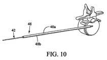
- FIG. 10is a perspective view illustrating the K-wire and split-dilator of the initial distraction assembly with the initial dilating cannula and handle removed;
- FIG. 11is a posterior view of the vertebral target site illustrating the split-dilator of the present invention in use distracting in a generally cephalad-caudal fashion according to one aspect of the present invention
- FIG. 12is a side view illustrating the use of a secondary distraction assembly (comprising a plurality of dilating cannulae over the K-wire) to further distract tissue between the skin of the patient and the surgical target site according to the present invention
- FIG. 13is a side view of a retractor assembly according to the present invention, comprising a handle assembly having three (3) retractor blades extending there from (posterior, cephalad-most, and caudal-most) disposed over the secondary distraction assembly of FIG. 12 (shown in a first, closed position);
- FIG. 14is a side view of a retractor assembly according to the present invention, comprising a handle assembly having three (3) retractor blades extending there from (posterior, cephalad-most, and caudal-most) with the secondary distraction assembly of FIG. 12 removed and shim element introduced;
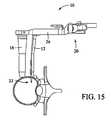
- FIGS. 15-16are perspective and top views, respectively, of the retractor assembly in a second, opened (i.e. retracted) position to thereby create an operative corridor to a surgical target site according to the present invention
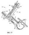
- FIGS. 17-18are perspective and side views, respectively, of the retractor assembly in the second, opened (i.e. retracted) position (with the secondary distraction assembly removed) and with the retractor extenders of FIGS. 4-5 and 6 - 7 coupled to the retractor according to the present invention.
- FIG. 19is a perspective view of an exemplary nerve monitoring system capable of performing nerve monitoring before, during and after the creating of an operative corridor to a surgical target site using the surgical access system in accordance with the present invention
- FIG. 20is a block diagram of the nerve monitoring system shown in FIG. 19 ;
- FIGS. 21-22are screen displays illustrating exemplary features and information communicated to a user during the use of the nerve monitoring system of FIG. 19 .
- FIGS. 23-50illustrate a method for accessing a surgical target site in the spine using a substantially lateral, retroperitoneal approach.
- the present inventioninvolves accessing a surgical target site in a fashion less invasive than traditional “open” surgeries and doing so in a manner that provides access in spite of the neural structures required to be passed through (or near) in order to establish an operative corridor to the surgical target site.
- the surgical access system of the present inventionaccomplishes this by providing a tissue distraction assembly and a tissue retraction assembly, both of which may be equipped with one or more electrodes for use in detecting the existence of (and optionally the distance and/or direction to) neural structures.
- the surgical access systemmay be used access a surgical target site on the spine via a substantially lateral, retroperitoneal approach (as shown, for example, in FIGS. 23-50 ).
- Electrodesare preferably provided for use with a nerve surveillance system such as, by way of example, the type shown and described in the co-pending and commonly assigned NeuroVision PCT Applications referenced above, the entire contents of which are expressly incorporated by reference as if set forth herein in their entirety.
- this nerve surveillance systemis capable of detecting the existence of (and optionally the distance and/or direction to) neural structures during the distraction and retraction of tissue by detecting the presence of nerves by applying a stimulation signal to such instruments and monitoring the evoked EMG signals from the myotomes associated with the nerves being passed by the distraction and retraction systems of the present invention.
- the system as a wholemay be used to form an operative corridor through (or near) any of a variety of tissues having such neural structures, particularly those which, if contacted or impinged, may otherwise result in neural impairment for the patient.
- the access system of the present inventionmay be used to traverse tissue that would ordinarily be deemed unsafe or undesirable, thereby broadening the number of manners in which a given surgical target site may be accessed.
- the tissue distraction assembly of the present invention(comprising a K-wire, an initial dilator, and a split-dilator disposed within the initial dilator) is employed to distract the tissues extending between the skin of the patient and a given surgical target site (preferably along the posterior region of the target intervertebral disc).
- a secondary distraction assemblyi.e. a plurality of sequentially dilating cannulae
- the resulting void or distracted region within the patientis of sufficient size to accommodate a tissue retraction assembly of the present invention.
- the tissue retraction assembly(comprising a plurality of retractor blades extending from a handle assembly) may be advanced relative to the secondary distraction assembly such that the retractor blades, in a first, closed position, are advanced over the exterior of the secondary distraction assembly.
- the handle assemblymay be operated to move the retractor blades into a second, open or “retracted” position to create an operative corridor to the surgical target site.
- a posterior shim element(which is preferably slideably engaged with the posterior retractor blade) may be advanced such that a distal shim extension in positioned within the posterior region of the disc space. If done before retraction, this helps ensure that the posterior retractor blade will not move posteriorly during the retraction process, even though the other retractor blades (i.e. cephalad-most and caudal-most) are able to move and thereby create an operative corridor. Fixing the posterior retractor blade in this fashion serves several important functions. First, the distal end of the shim element serves to distract the adjacent vertebral bodies, thereby restoring disc height.
- the posterior retractor bladealso rigidly couples the posterior retractor blade in fixed relation relative to the vertebral bodies.
- the posterior shim elementalso helps ensure that surgical instruments employed within the operative corridor are incapable of being advanced outside the operative corridor, preventing inadvertent contact with the exiting nerve roots during the surgery.
- the cephalad-most and caudal-most retractor bladesmay be locked in position and, thereafter, retractor extenders advanced therealong to prevent the ingress or egress of instruments or biological structures (i.e. nerves, vasculature, etc. . . . ) into or out of the operative corridor.
- any of a variety of surgical instruments, devices, or implantsmay be passed through and/or manipulated within the operative corridor depending upon the given surgical procedure.
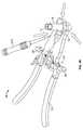
- FIG. 1illustrates a tissue retraction assembly 10 forming part of a surgical access system according to the present invention.
- the retraction assembly 10includes a plurality of retractor blades extending from a handle assembly 20 .
- the handle assembly 20is provided with a first retractor blade 12 , a second retractor blade 16 , and a third retractor blade 18 .
- the retractor assembly 10is shown in a fully retracted or “open” configuration, with the retractor blades 12 , 16 , 18 positioned a distance from one another so as to form an operative corridor 15 there between and extending to a surgical target site (e.g. an annulus of an intervertebral disc).
- a surgical target sitee.g. an annulus of an intervertebral disc
- retractor assembly 10 of the present inventionmay find use in any number of different surgical approaches, including generally posterior, generally postero-lateral, generally anterior and generally antero-lateral.
- the retractor blades 12 , 16 , 18may be equipped with various additional features or components.
- posterior retractor blade 12may be equipped with a shim element 22 (shown more clearly in FIGS. 2-3 ).
- Shim element 22serves to distract the adjacent vertebral bodies (thereby restoring disc height), helps secure the retractor assembly 10 relative to the surgical target site, and forms a protective barrier to prevent the ingress or egress of instruments or biological structures (i.e. nerves, vasculature, etc. . . . ) into or out of the operative corridor.
- Each of the remaining retractor bladesmay be equipped with a retractor extender, such as the narrow retractor extender 24 shown in FIGS. 4-5 or the wide retractor extender 25 shown in FIGS. 6-7 .
- the retractor extenders 24 / 25extend from the cephalad-most and caudal-most retractor blades 16 , 18 to form a protective barrier to prevent the ingress or egress of instruments or biological structures (i.e. nerves, vasculature, etc. . . . ) into or out of the operative corridor 15 .
- any or all of the retractor blades 12 , 16 , 18 , the shim element 22 and/or the retractor extenders 24 / 25may be provided with one or more electrodes 39 (preferably at their distal regions) equipped for use with a nerve surveillance system, such as, by way of example, the type shown and described in the NeuroVision PCT Applications.
- Each of the shim element 22 and/or the retractor extenders 24 / 25may also be equipped with a mechanism to selectively and releasably engage with the respective retractor blades 12 , 16 , 18 .
- thismay be accomplished by configuring the shim element 22 and/or the retractor extenders 24 / 25 with a tab element 27 capable of engaging with corresponding rachet-like grooves (shown at 29 in FIG. 1 ) along the inner-facing surfaces of the retractor blades 12 , 16 , 18 .
- a tab element 27capable of engaging with corresponding rachet-like grooves (shown at 29 in FIG. 1 ) along the inner-facing surfaces of the retractor blades 12 , 16 , 18 .
- Each of the shim element 22 and/or the retractor extenders 24 / 25is provided with a pair of engagement elements 37 having, by way of example only, a generally dove-tailed cross-sectional shape.
- the engagement elements 37are dimensioned to engage with receiving portions on the respective retractor blades 12 , 16 , 18 .
- each of the shim element 22 and/or the retractor extenders 24 / 25are provided with an elongate slot 43 for engagement with an insertion tool (not shown).
- Each tab member 27is also equipped with an enlarged tooth element 49 which engages within corresponding grooves 29 provided along the inner surface of the retractor blades 12 , 16 , 18 .
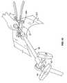
- the handle assembly 20may be coupled to any number of mechanisms for rigidly registering the handle assembly 20 in fixed relation to the operative site, such as through the use of an articulating arm mounted to the operating table.
- the handle assembly 20includes first and second arm members 26 , 28 hingedly coupled via coupling mechanism shown generally at 30 .
- the cephalad-most retractor blade 16is rigidly coupled (generally perpendicularly) to the end of the first arm member 26 .
- the caudal-most retractor blade 18is rigidly coupled (generally perpendicularly) to the end of the second arm member 28 .
- the posterior retractor blade 12is rigidly coupled (generally perpendicularly to) a translating member 17 , which is coupled to the handle assembly 20 via a linkage assembly shown generally at 14 .
- the linkage assembly 14includes a roller member 34 having a pair of manual knob members 36 which, when rotated via manual actuation by a user, causes teeth 35 on the roller member 34 to engage within ratchet-like grooves 37 in the translating member 17 .
- manual operation of the knobs 36causes the translating member 17 to move relative to the first and second arm members 26 , 28 .
- the arms 26 , 28may be simultaneously opened such that the cephalad-most and caudal-most retractor blades 16 , 18 move away from one another.
- the dimension and/or shape of the operative corridor 15may be tailored depending upon the degree to which the translating member 17 is manipulated relative to the arms 26 , 28 . That is, the operative corridor 15 may be tailored to provide any number of suitable cross-sectional shapes, including but not limited to a generally circular cross-section, a generally ellipsoidal cross-section, and/or an oval cross-section.
- Optional light emitting devices 39may be coupled to one or more of the retractor blades 12 , 16 , 18 to direct light down the operative corridor 15 .
- FIG. 9illustrates an initial distraction assembly 40 forming part of the surgical access system according to the present invention.
- the initial distraction assembly 40includes a K-wire 42 , an initial dilating cannula 44 with handle 46 , and a split-dilator 48 housed within the initial dilating cannula 44 .
- the K-wire 42 and split-dilator 48are disposed within the initial dilating cannula 44 and the entire assembly 40 advanced through the tissue towards the surgical target site (i.e. annulus).
- the surgical target sitei.e. annulus
- One exemplary method for advancing an initial dilator towards a spinal target siteis described in more detail later in connection with FIGS. 23-50 . Again, this is preferably accomplished while employing the nerve detection and/or direction features described above.
- the initial dilating assembly 40is advanced such that the distal ends of the split-dilator 48 and initial dilator 44 are positioned within the disc space ( FIG. 9 )
- the initial dilator 44 and handle 46are removed ( FIG. 10 ) to thereby leave the split-dilator 48 and K-wire 42 in place.
- the split-dilator 48is thereafter split such that the respective halves 48 a , 48 b are separated from one another to distract tissue in a generally cephalad-caudal fashion relative to the target site.
- the split dilator 48may thereafter be relaxed (allowing the dilator halves 48 a , 48 b to come together) and rotated such that the dilator halves 48 a , 48 b are disposed in the anterior-posterior plane. Once rotated in this manner, the dilator halves 48 a , 48 b are again separated to distract tissue in a generally anterior-posterior fashion.
- Each dilator halve 48 a , 48 bmay be, according to the present invention, provided with one or more electrodes (preferably at their distal regions) equipped for use with a nerve surveillance system, such as, by way of example, the type shown and described in the NeuroVision PCT Applications.
- a secondary distractionmay be optionally undertaken, such as via a sequential dilation system 50 as shown in FIG. 12 .
- the sequential dilation system 50may include the K-wire 42 , the initial dilator 44 , and one or more supplemental dilators 52 , 54 for the purpose of further dilating the tissue down to the surgical target site.
- each component of the secondary distraction assembly 50namely, the K-wire 42 , the initial dilator 44 , and the supplemental dilators 52 , 54 may be, according to the present invention, provided with one or more electrodes (preferably at their distal regions) equipped for use with a nerve surveillance system, such as, by way of example, the type shown and described in the NeuroVision PCT Applications.
- the retraction assembly 10 of the present inventionis thereafter advanced along the exterior of the sequential dilation system 50 .
- Thisis accomplished by maintaining the retractor blades 12 , 16 , 18 in a first, closed position (with the retractor blades 12 - 16 in generally abutting relation to one another).
- the sequential dilation assembly 50may be removed and the shim element 22 engaged with the posterior retractor blade 12 such that the distal end thereof extends into the disc space as shown in FIG. 14 .
- the handle assembly 20may be operated to move the retractor blades 16 , 18 into a second, open or “retracted” position as shown generally in FIGS. 15-16 .
- the posterior retractor blade 12is allowed to stay in the same general position during this process, such that the cephalad-most and caudal-most retractor blades 14 , 16 move away from the posterior retractor blade 12 .
- the narrow and wide retractor extenders 24 , 25may be engaged with the caudal-most retractor blade 18 and cephalad-most retractor blade 16 , respectively, as shown in FIGS. 17-18 .
- any number of distraction components and/or retraction componentsmay be equipped to detect the presence of (and optionally the distance and/or direction to) neural structures during the steps tissue distraction and/or retraction. This is accomplished by employing the following steps: (1) one or more stimulation electrodes are provided on the various distraction and/or retraction components; (2) a stimulation source (e.g. voltage or current) is coupled to the stimulation electrodes; (3) a stimulation signal is emitted from the stimulation electrodes as the various components are advanced towards or maintained at or near the surgical target site; and (4) the patient is monitored to determine if the stimulation signal causes muscles associated with nerves or neural structures within the tissue to innervate. If the nerves innervate, this may indicate that neural structures may be in close proximity to the distraction and/or retraction components.
- a stimulation sourcee.g. voltage or current
- Neural monitoringmay be accomplished via any number of suitable fashions, including but not limited to observing visual twitches in muscle groups associated with the neural structures likely to found in the tissue, as well as any number of monitoring systems, including but not limited to any commercially available “traditional” electromyography (EMG) system (that is, typically operated by a neurophysiologist). Such monitoring may also be carried out via the surgeon-driven EMG monitoring system shown and described in the following commonly owned and co-pending NeuroVision PCT Applications referenced above.
- the access system of the present inventionmay advantageously be used to traverse tissue that would ordinarily be deemed unsafe or undesirable, thereby broadening the number of manners in which a given surgical target site may be accessed.
- the surgical access systemmay be advantageously used to traverse tissue through the retroperitoneal space and the psoas muscle during a substantially lateral, retroperitoneal approach to the lumbar spine, as shown in FIGS. 23-50 .
- FIGS. 19-20illustrate, by way of example only, a monitoring system 120 of the type disclosed in the NeuroVision PCT Applications suitable for use with the surgical access system 10 of the present invention.
- the monitoring system 120includes a control unit 122 , a patient module 124 , and an EMG harness 126 and return electrode 128 coupled to the patient module 124 , and a cable 132 for establishing electrical communication between the patient module 124 and the surgical access system of the present invention (retractor assembly 10 of FIG. 1 and distraction assemblies 40 , 50 of FIGS. 9-12 ).
- this electrical communicationcan be achieved by providing, by way of example only, a hand-held stimulation controller 152 capable of selectively providing a stimulation signal (due to the operation of manually operated buttons on the hand-held stimulation controller 152 ) to one or more connectors 156 a , 156 b , 156 c .
- the connectors 156 a , 156 b , 156 care suitable to establish electrical communication between the hand-held stimulation controller 152 and (by way of example only) the stimulation electrodes on the K-wire 42 , the dilators 44 , 48 , 52 , 54 , the retractor blades 12 , 16 , 18 and/or the shim members 22 , 24 , 25 (collectively “surgical access instruments”).
- these surgical access instrumentsmust be connected to the connectors 156 a , 156 b and/or 156 c , at which point the user may selectively initiate a stimulation signal (preferably, a current signal) from the control unit 122 to a particular surgical access instruments. Stimulating the electrode(s) on these surgical access instruments before, during and/or after establishing operative corridor will cause nerves that come into close or relative proximity to the surgical access instruments to depolarize, producing a response in a myotome associated with the innervated nerve.
- a stimulation signalpreferably, a current signal
- the control unit 122includes a touch screen display 140 and a base 142 , which collectively contain the essential processing capabilities (software and/or hardware) for controlling the monitoring system 120 .
- the control unit 122may include an audio unit 118 that emits sounds according to a location of a surgical element with respect to a nerve.
- the patient module 124is connected to the control unit 122 via a data cable 144 , which establishes the electrical connections and communications (digital and/or analog) between the control unit 122 and patient module 124 .
- the main functions of the control unit 122include receiving user commands via the touch screen display 140 , activating stimulation electrodes on the surgical access instruments, processing signal data according to defined algorithms, displaying received parameters and processed data, and monitoring system status and report fault conditions.
- the touch screen display 140is preferably equipped with a graphical user interface (GUI) capable of communicating information to the user and receiving instructions from the user.
- GUIgraphical user interface
- the display 140 and/or base 142may contain patient module interface circuitry (hardware and/or software) that commands the stimulation sources, receives digitized signals and other information from the patient module 124 , processes the EMG responses to extract characteristic information for each muscle group, and displays the processed data to the operator via the display 140 .
- the monitoring system 120is capable of determining nerve direction relative to one or more of the K-wire 42 , the dilators 44 , 48 , 52 , 54 , the retractor blades 12 , 16 , 18 and/or the shim elements 22 , 24 , 25 before, during and/or following the creation of an operative corridor to a surgical target site. Monitoring system 120 accomplishes this by having the control unit 122 and patient module 124 cooperate to send electrical stimulation signals to one or more of the stimulation electrodes provided on these instruments. Depending upon the location of the surgical access system 10 within a patient (and more particularly, to any neural structures), the stimulation signals may cause nerves adjacent to or in the general proximity of the surgical access system 10 to depolarize.
- the nerve direction feature of the system 120is based on assessing the evoked response of the various muscle myotomes monitored by the system 120 via the EMG harness 126 .
- the surgical access system 10is capable of detecting the presence of (and optionally the distant and/or direction to) such nerves. This provides the ability to actively negotiate around or past such nerves to safely and reproducibly form the operative corridor to a particular surgical target site, as well as monitor to ensure that no neural structures migrate into contact with the surgical access system 10 after the operative corridor has been established.
- the surgical access system 10may be particularly suited for establishing an operative corridor to an intervertebral target site in a postero-lateral, trans-psoas fashion so as to avoid the bony posterior elements of the spinal column.
- one such operative corridor to an intervertebral target sitemay be established through the retroperitoneal space and the psoas muscle during a substantially lateral, retroperitoneal approach to the lumbar spine, as shown in FIGS. 23-50 .
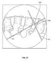
- FIGS. 21-22are exemplary screen displays (to be shown on the display 140 ) illustrating one embodiment of the nerve direction feature of the monitoring system shown and described with reference to FIGS. 19-20 . These screen displays are intended to communicate a variety of information to the surgeon in an easy-to-interpret fashion.
- This informationmay include, but is not necessarily limited to, a display of the function 180 (in this case “DIRECTION”), a graphical representation of a patient 181 , the myotome levels being monitored 182 , the nerve or group associated with a displayed myotome 183 , the name of the instrument being used 184 (in this case, a dilator 46 , 48 ), the size of the instrument being used 185 , the stimulation threshold current 186 , a graphical representation of the instrument being used 187 (in this case, a cross-sectional view of a dilator 44 , 48 ) to provide a reference point from which to illustrate relative direction of the instrument to the nerve, the stimulation current being applied to the stimulation electrodes 188 , instructions for the user 189 (in this case, “ADVANCE” and/or “HOLD”), and (in FIG.
- a display of the function 180in this case “DIRECTION”
- a graphical representation of a patient 181the myotome levels being monitored 182
- This informationmay be communicated in any number of suitable fashions, including but not limited to the use of visual indicia (such as alpha-numeric characters, light-emitting elements, and/or graphics) and audio communications (such as a speaker element).
- visual indiciasuch as alpha-numeric characters, light-emitting elements, and/or graphics
- audio communicationssuch as a speaker element.
- a dilating cannulasuch as at 184
- the present inventionis deemed to include providing similar information on the display 140 during the use of any or all of the various instruments forming the surgical access system 10 of the present invention, including the initial distraction assembly 40 (i.e. the K-wire 42 and dilators 44 , 48 ) and/or the retractor blades 12 , 16 , 18 and/or the shim elements 22 , 24 , 25 .
- some embodiments of the surgical access system 10may be particularly suited for establishing an operative corridor to a surgical target site in the spine.
- Such an operative corridormay be established through the retroperitoneal space and the psoas muscle during a direct lateral, retroperitoneal approach to the spine.
- a surgeonmay have direct visualization of the patient's anatomy without the cumbersome requirements associated with using endoscopes or operating coaxial through narrow tubes.
- the potential of damaging nerves while advancing instruments through the psoas musclemay be substantially reduced.
- the surgeonWhen accessing a spinal target site via the substantially lateral, retroperitoneal approach described in connection with FIGS. 23-50 , the surgeon should consider several anatomical reference points, such as the iliac crest, the twelfth rib, and the lateral border of the erector spinae muscle groups.
- blunt finger dissectionis used to pass between these muscle groups and access the retroperitoneal space.
- Such a techniqueoffers simple access to the retroperitoneal space while minimizing the potential of visceral lesion.
- the fingermay be used to escort one or more dilators through the retroperitoneal space, thus reducing the potential of peritoneal disruption.
- each dilatoris preferably advanced through the psoas muscle between the middle and anterior third of the muscle so that the nerves of the lumbar plexus are located posterior and outside the operative corridor.
- a monitoring system 120 of the type disclosed in the NeuroVision PCT Applicationsmay be used to avoid damage to any peripheral nerves embedded throughout the psoas muscle as the dilator is advanced through the muscle to the surgical target site in the spine.
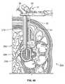
- a patient 200is positioned on a surgical table 250 in preparation of spinal surgery.
- a cushion 252is positioned between the patient's lateral side and the surgical table 250 to arrange the patient 200 in such a way as to increase the distance between the patient's iliac crest 202 and rib cage 204 .
- a flexion of the surgical table 250may be used to accomplish the desired arrangement. Such an arrangement helps to open the invertebral disc space 206 at or near the surgical target site.
- an articulating arm assembly 60is coupled to the surgical table 250 to maintain the access system 10 in a substantially fixed position relative to the surgical target site when the operative corridor has been established.
- the articulating arm assembly 60is mounted to a bedrail 254 of the surgical table 250 .
- a fluoroscopy system 260is disposed proximal to the surgical table 250 to provide the surgeon with visualization of the surgical target area.
- This fluoroscopy system 260includes a display monitor 262 that is positioned such that the surgeon may view the monitor 262 during the operation.
- a monitoring system 120 of the type disclosed in the NeuroVision PCT Applicationsmay be positioned near the surgical table 250 so that the surgeon may view a display 140 of the monitoring system 120 during the operation.
- one or more instrumentsare positioned laterally over an area of the patient 200 and then viewed using the lateral fluoroscopy.
- the instrumentsare used to identify a lateral incision location 208 that is substantially lateral to the surgical target site (e.g., the invertebral disc space 206 ).
- a first markis made on the patient 200 at the lateral incision location 208 .
- a second markis made on the patient at a posteriolateral incision location 209 near the lateral incision location 208 .
- the posteriolateral incision location 209is approximately at the lateral border of the erector spine muscle.
- the posteriolateral incision location 209is within a finger's length of the lateral incision location 208 .
- an incisionis made at the posteriolateral incision location 209 , and the subcutaneous layers 210 are dissected until reaching the muscular masses 212 .
- a dissection instrumentsuch as blunt dissection scissors 270 , is used to spread the muscle fibers 212 until the retroperitoneal space 215 is reached.
- the surgeonuses great caution to avoid perforation of the peritoneum 214 .
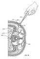
- a guide member 275is inserted through the posteriolateral incision 209 into the retroperitoneal space 215 .
- the guide memberis a finger 275 of the surgeon, which is preferably covered with a surgical glove for hygienic purposes.
- the guide member 275may be an instrument or tool configured to extend and maneuver in the retroperitoneal space as described herein.
- the finger 275may sweep a portion of the retroperitoneal space 215 and then palpate down to the psoas muscle 220 . This motion of the finger 275 in the retroperitoneal space 215 may loosen some fatty tissue before a dilator is advanced therethrough.
- the finger 275is swept away from the psoas muscle 220 toward the lateral incision location 208 .
- a scalpel 272 or other like instrumentis used to make and incision at this location 208 .
- the incisionshould be of a sufficient size to receive a distal end 41 an initial dilator 40 .
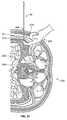
- the finger 275is used to direct the distal end 41 of the initial dilator 40 through the retroperitoneal space 215 toward the psoas muscle 220 .
- the initial dilator 40includes at least a K-wire 42 and may also include a split-dilator 48 slideably passed over the K-wire 42 (see, for example, FIG. 10 ).
- the distal end 41is introduced through the lateral incision location 208 and directed to the finger 275 in the retroperitoneal space 215 .
- FIG. 34the distal end 41 is introduced through the lateral incision location 208 and directed to the finger 275 in the retroperitoneal space 215 .
- the finger 275engages the initial dilator 40 proximal to the distal end 41 and guides the distal end 41 to the psoas muscle 220 .
- the potential for breaching or disrupting the peritonealis reduced.
- the location of the distal end 41 relative to the target sitemay be verified using an imaging system, such as an image intensifier.
- the distal end 41 of the initial dilator 40is advanced in a substantially lateral direction through the psoas muscle 220 toward the invertebral disc space 206 at or near the surgical target site.
- the fibers of the psoas muscle 220are split using blunt dissection and NeuroVision neurophysiologic monitoring of the type disclosed in the NeuroVision PCT Applications.
- a stimulation connector 156 of the NeuroVision monitoring system 120(see FIG. 19 ) is coupled to the initial dilator 40 to provide a stimulation signal 157 as the dilator 40 is advanced through the psoas muscle 220 . It should be understood that the stimulation signal 157 is depicted in FIG. 36 for illustrative purposes and is generally not visible.
- Descending nerves of the lumbar plexusnormally lie in the posterior one-third of the psoas muscle 220 .
- the NeuroVision monitoring system 120 of the type disclosed in the NeuroVision PCT Applicationsassists with the safe passage by these nerves and/or confirmation of the nerves' posterior location.
- the NeuroVision monitoring system 120will continuously search for the stimulus threshold that elicits an EMG response on the myotomes monitored and then reports such thresholds on a display 140 as shown in FIG. 37 .
- the stimulus necessary to elicit an EMG responsewill vary with distance from the nerve.
- experiencehas shown that threshold values greater than 10 mA indicate a distance that allows for safe passage through the psoas muscle 220 and continued nerve safety.
- a K-wire 42 of the initial dilator 40is introduced into the targeted disc space 206 after the dilator 40 is passed through the psoas muscle 220 .
- the position of the distal end 41 of the dilator 40is confirmed using fluoroscopic imaging before the K-wire 42 is introduced into the disc space 206 .
- depth markings 45FIG. 39
- the appropriate length blades 12 , 16 , and 18may be secured to the handle portion 20 by tightening fasteners with a driver instrument 274 .
- the sequential dilation system 50(previously described in connection with FIG. 12 ), including one or more supplemental dilators 52 , 54 , may be guided over the initial dilator 40 for the purpose of further dilating the tissue down to the surgical target site.
- the NeuroVision monitoring system 120 of the type disclosed in the NeuroVision PCT Applicationsis used with the supplemental dilators 52 , 54 to provide safe passage through the psoas muscle 220 .
- the initial dilator 40 and the supplemental dilators 52 , 54are advanced through the lateral incision location 208 to the targeted disc space 206 in a substantially lateral direction to create a distraction corridor.
- the retractor blades 12 , 16 , 18 of the access system 10are introduced over the supplemental dilator 54 (or the initial dilator 40 if the sequential dilation system 50 is not employed) toward the disc space 206 ;
- the NeuroVision monitoring system 120 of the type disclosed in the NeuroVision PCT Applicationsmay be used with the blades 12 , 16 , 18 to provide safe passage through the psoas muscle 220 .
- the posterior shim element 22 and/or the retractor extenders 24 , 25are engaged with the retractor blades 12 , 16 , 18 (as previously described in connection with FIGS. 1-7 ).
- fluoroscopic imagingmay be used to confirm the position of the blades 12 , 16 , 18 proximal to the disc space 206 .
- the articulating arm assembly 60is coupled to the handle member 20 of the access system 10 . As previous described in connection with FIG. 25 , the articulating arm assembly 60 is also coupled to the surgical table 250 so as to maintain the access system 10 in a substantially fixed position. Handles 62 and 64 may be turned to substantially fix the position of articulating arm assembly 60 .
- handle extenders 31 and 33may be squeeze to spread the blades 12 , 16 , 18 and knob members 36 may be turned to selectively adjust the posterior retractor blade 12 (previously described in connection with FIGS. 13-18 ). Such movement by the blades 12 , 16 , 18 retracts the distraction corridor so as to form an operative corridor 15 .
- FIG. 45shows a lateral view of the operative corridor 15 down to the targeted disc space 206 in the patient's spine.
- Light emitting devices 39may be coupled to one or more of the retractor blades 12 , 16 , 18 to direct light down the operative corridor 15 .
- the light emitting devices 39are coupled to a xenon arthroscopy light source. The surgeon may use direct visualization and/or a NeuroVision probe of the type disclosed in the NeuroVision PCT Applications to confirm that the operative corridor 15 is neurologically clear.
- the operative corridor 15may be inserted through the operative corridor 15 to prepare the targeted disc space 206 .
- the operative corridor 15has a 15-20 mm annulotomy to provide ample space for the various instruments.
- the operative corridor 15may have other configurations, depending on the surgical task to be performed.
- the disc space 206is undergoing a discectomy and insertion of a spinal implant.
- at least one preparation tool 276such as a disc cutter, pituitary, scraper, curette, or the like is inserted through the operative corridor 15 to prepare the disc space 206 .
- one or more sizers 277are inserted to the disc space 206 to provide appropriate disc height restoration.
- a broach 278may be used in the disc space 206 to remove osteophytes and to facilitate implant insertion.
- an appropriately sized implant 282is advanced into the disc space 206 with an inserter tool 280 .
- the implant 282is releasably secured to the inserter tool 280 such that the surgeon may release the implant when it is properly positioned in the disc space 206 .
- the implantmay comprise a material that facilitates bone fusion (such as allograft or autograft), and autograft or graft extenders may be used in the disc space 206 after the implant is inserted.
- the access system 10is carefully removed from the operative corridor 15 .
- Direct visualizationmay be used to confirm the absence of significant bleeding in the disc space 206 or the psoas muscle 220 .
- the skin around the operative corridormay be closed using a suturing method, such as a subcuticular suture.
- certain methods of using the access system 10can safely and effectively establish a minimally invasive operative corridor through the retroperitoneal space 215 and the psoas muscle 220 via a direct lateral, retroperitoneal approach to the spine.
- a minimally invasive operative corridorthrough the retroperitoneal space 215 and the psoas muscle 220 via a direct lateral, retroperitoneal approach to the spine.
- Such a methodallows the surgeon to directly visualize the patient's anatomy without the cumbersome requirements associated with using endoscopes or operating coaxial through narrow, artificial tube.
- the potential of damaging nerves while advancing dilators and other instruments through the psoas muscle 220may be substantially reduced.
- the present inventionaccomplishes the goal of gaining access a surgical target site in a fashion less invasive than traditional “open” surgeries and, moreover, does so in a manner that provides the ability to access such a surgical target site regardless of the neural structures required to be passed through (or near) in order to establish an operative corridor to the surgical target site.
- the present inventionfurthermore provides the ability to perform neural monitoring in the tissue or regions adjacent the surgical target site during any procedures performed after the operative corridor has been established.
- the surgical access system of the present inventioncan be used in any of a wide variety of surgical or medical applications, above and beyond the spinal applications discussed herein.
- Such spinal applicationsmay include any procedure wherein instruments, devices, implants and/or compounds are to be introduced into or adjacent the surgical target site, including but not limited to discectomy, fusion (including PLIF, ALIF, TLIF and any fusion effectuated via a lateral or far-lateral approach and involving, by way of example, the introduction of bone products (such as allograft or autograft) and/or devices having ceramic, metal and/or plastic construction (such as mesh) and/or compounds such as bone morphogenic protein), total disc replacement, etc. . . . ).
- discectomyincluding PLIF, ALIF, TLIF and any fusion effectuated via a lateral or far-lateral approach and involving, by way of example, the introduction of bone products (such as allograft or autograft) and/or devices having ceramic, metal and/or plastic construction (such as mesh) and/or compounds such as bone morphogenic protein), total disc replacement, etc. . . . ).
- fusionincluding PLIF, ALIF,
- the surgical access system of the present inventionopens the possibility of accessing an increased number of surgical target sites in a “less invasive” fashion by eliminating or greatly reducing the threat of contacting nerves or neural structures while establishing an operative corridor through or near tissues containing such nerves or neural structures.
- the surgical access system of the present inventionrepresents a significant advancement capable of improving patient care (via reduced pain due to “less-invasive” access and reduced or eliminated risk of neural contact before, during, and after the establishment of the operative corridor) and lowering health care costs (via reduced hospitalization based on “less-invasive” access and increased number of suitable surgical target sites based on neural monitoring).
Landscapes
- Health & Medical Sciences (AREA)
- Life Sciences & Earth Sciences (AREA)
- Surgery (AREA)
- General Health & Medical Sciences (AREA)
- Public Health (AREA)
- Veterinary Medicine (AREA)
- Animal Behavior & Ethology (AREA)
- Molecular Biology (AREA)
- Medical Informatics (AREA)
- Engineering & Computer Science (AREA)
- Biomedical Technology (AREA)
- Heart & Thoracic Surgery (AREA)
- Nuclear Medicine, Radiotherapy & Molecular Imaging (AREA)
- Biophysics (AREA)
- Pathology (AREA)
- Physics & Mathematics (AREA)
- Radiology & Medical Imaging (AREA)
- Optics & Photonics (AREA)
- Surgical Instruments (AREA)
- Neurology (AREA)
- Measurement And Recording Of Electrical Phenomena And Electrical Characteristics Of The Living Body (AREA)
Abstract
Description
Claims (30)
Priority Applications (8)
| Application Number | Priority Date | Filing Date | Title |
|---|---|---|---|
| US10/967,668US7905840B2 (en) | 2003-10-17 | 2004-10-18 | Surgical access system and related methods |
| US12/364,507US9615866B1 (en) | 2004-10-18 | 2009-02-02 | Surgical fixation system and related methods |
| US12/635,869US8303515B2 (en) | 2003-09-25 | 2009-12-11 | Surgical access system and related methods |
| US12/983,627US8591432B2 (en) | 2003-09-25 | 2011-01-03 | Surgical access system and related methods |
| US13/434,845US9795367B1 (en) | 2003-10-17 | 2012-03-29 | Surgical access system and related methods |
| US14/066,098US9314152B2 (en) | 2003-09-25 | 2013-10-29 | Surgical access system and related methods |
| US15/071,540US10653308B2 (en) | 2003-10-17 | 2016-03-16 | Surgical access system and related methods |
| US16/848,556US20200237207A1 (en) | 2003-10-17 | 2020-04-14 | Surgical Access System and Related Methods |
Applications Claiming Priority (2)
| Application Number | Priority Date | Filing Date | Title |
|---|---|---|---|
| US51259403P | 2003-10-17 | 2003-10-17 | |
| US10/967,668US7905840B2 (en) | 2003-10-17 | 2004-10-18 | Surgical access system and related methods |
Related Parent Applications (1)
| Application Number | Title | Priority Date | Filing Date |
|---|---|---|---|
| PCT/US2004/031768Continuation-In-PartWO2005030318A1 (en) | 2003-09-25 | 2004-09-27 | Surgical access system and related methods |
Related Child Applications (1)
| Application Number | Title | Priority Date | Filing Date |
|---|---|---|---|
| US12/635,869ContinuationUS8303515B2 (en) | 2003-09-25 | 2009-12-11 | Surgical access system and related methods |
Publications (2)
| Publication Number | Publication Date |
|---|---|
| US20050149035A1 US20050149035A1 (en) | 2005-07-07 |
| US7905840B2true US7905840B2 (en) | 2011-03-15 |
Family
ID=34713643
Family Applications (6)
| Application Number | Title | Priority Date | Filing Date |
|---|---|---|---|
| US10/967,668Active2029-09-25US7905840B2 (en) | 2003-09-25 | 2004-10-18 | Surgical access system and related methods |
| US12/635,869Expired - Fee RelatedUS8303515B2 (en) | 2003-09-25 | 2009-12-11 | Surgical access system and related methods |
| US12/983,627Expired - LifetimeUS8591432B2 (en) | 2003-09-25 | 2011-01-03 | Surgical access system and related methods |
| US14/066,098Expired - LifetimeUS9314152B2 (en) | 2003-09-25 | 2013-10-29 | Surgical access system and related methods |
| US15/071,540Active2027-08-19US10653308B2 (en) | 2003-10-17 | 2016-03-16 | Surgical access system and related methods |
| US16/848,556AbandonedUS20200237207A1 (en) | 2003-10-17 | 2020-04-14 | Surgical Access System and Related Methods |
Family Applications After (5)
| Application Number | Title | Priority Date | Filing Date |
|---|---|---|---|
| US12/635,869Expired - Fee RelatedUS8303515B2 (en) | 2003-09-25 | 2009-12-11 | Surgical access system and related methods |
| US12/983,627Expired - LifetimeUS8591432B2 (en) | 2003-09-25 | 2011-01-03 | Surgical access system and related methods |
| US14/066,098Expired - LifetimeUS9314152B2 (en) | 2003-09-25 | 2013-10-29 | Surgical access system and related methods |
| US15/071,540Active2027-08-19US10653308B2 (en) | 2003-10-17 | 2016-03-16 | Surgical access system and related methods |
| US16/848,556AbandonedUS20200237207A1 (en) | 2003-10-17 | 2020-04-14 | Surgical Access System and Related Methods |
Country Status (1)
| Country | Link |
|---|---|
| US (6) | US7905840B2 (en) |
Cited By (125)
| Publication number | Priority date | Publication date | Assignee | Title |
|---|---|---|---|---|
| US20070100212A1 (en)* | 2004-10-08 | 2007-05-03 | Nuvasive, Inc. | Surgical access system and related methods |
| US20090054804A1 (en)* | 2007-04-03 | 2009-02-26 | Nuvasive Inc. | Neurophysiologic monitoring system |
| US20090105788A1 (en)* | 2007-10-18 | 2009-04-23 | Innovative Surgical Solutions, Llc | Minimally invasive nerve monitoring device and method |
| US20090125072A1 (en)* | 2007-11-13 | 2009-05-14 | Neubardt Seth L | Surgical bone screw construction |
| US20090177112A1 (en)* | 2005-02-02 | 2009-07-09 | James Gharib | System and Methods for Performing Neurophysiologic Assessments During Spine Surgery |
| US20100094115A1 (en)* | 2005-01-31 | 2010-04-15 | Pond Jr John D | Electrically insulated surgical needle assembly |
| US20100145391A1 (en)* | 2008-12-05 | 2010-06-10 | Kleiner Jeffrey | Apparatus and method of spinal implant and fusion |
| US20110071536A1 (en)* | 2009-09-18 | 2011-03-24 | Kleiner Jeffrey | Bone graft delivery device and method of using the same |
| US20110130793A1 (en)* | 2009-11-10 | 2011-06-02 | Nuvasive Inc. | Method and apparatus for performing spinal surgery |
| US20110230782A1 (en)* | 2007-10-18 | 2011-09-22 | Innovative Surgical Solutions, Llc | Neural monitoring sensor |
| US20110230783A1 (en)* | 2007-10-18 | 2011-09-22 | Innovative Surgical Solutions, Llc | Neural event detection |
| US20110237974A1 (en)* | 2007-10-18 | 2011-09-29 | Innovative Surgical Solutions, Llc | Neural monitoring system |
| US8088163B1 (en) | 2008-02-06 | 2012-01-03 | Kleiner Jeffrey B | Tools and methods for spinal fusion |
| USD656610S1 (en) | 2009-02-06 | 2012-03-27 | Kleiner Jeffrey B | Spinal distraction instrument |
| DE112011102801T5 (en) | 2010-08-23 | 2013-05-29 | Nuvasive, Inc. | Surgical access system and associated procedures |
| US8538539B2 (en) | 2004-10-07 | 2013-09-17 | Nu Vasive, Inc. | System and methods for assessing the neuromuscular pathway prior to nerve testing |
| US8636655B1 (en) | 2010-01-19 | 2014-01-28 | Ronald Childs | Tissue retraction system and related methods |
| US8685031B2 (en) | 2009-09-18 | 2014-04-01 | Spinal Surgical Strategies, Llc | Bone graft delivery system |
| US8758344B2 (en) | 1988-06-13 | 2014-06-24 | Warsaw Orthopedic, Inc. | Spinal implant and instruments |
| US8855822B2 (en) | 2012-03-23 | 2014-10-07 | Innovative Surgical Solutions, Llc | Robotic surgical system with mechanomyography feedback |
| US8892259B2 (en) | 2012-09-26 | 2014-11-18 | Innovative Surgical Solutions, LLC. | Robotic surgical system with mechanomyography feedback |
| US20140372955A1 (en)* | 2010-12-17 | 2014-12-18 | Orca Health, Inc. | Visual selection of an anatomical element for requesting information about a medical condition |
| USD723682S1 (en) | 2013-05-03 | 2015-03-03 | Spinal Surgical Strategies, Llc | Bone graft delivery tool |
| US8983593B2 (en) | 2011-11-10 | 2015-03-17 | Innovative Surgical Solutions, Llc | Method of assessing neural function |
| US9039630B2 (en) | 2012-08-22 | 2015-05-26 | Innovative Surgical Solutions, Llc | Method of detecting a sacral nerve |
| US9060877B2 (en) | 2009-09-18 | 2015-06-23 | Spinal Surgical Strategies, Llc | Fusion cage with combined biological delivery system |
| US9066701B1 (en) | 2012-02-06 | 2015-06-30 | Nuvasive, Inc. | Systems and methods for performing neurophysiologic monitoring during spine surgery |
| US9084550B1 (en) | 2007-10-18 | 2015-07-21 | Innovative Surgical Solutions, Llc | Minimally invasive nerve monitoring device and method |
| US9138217B2 (en) | 2009-11-11 | 2015-09-22 | Nu Vasive, Inc. | Surgical access system and related methods |
| US9173694B2 (en) | 2009-09-18 | 2015-11-03 | Spinal Surgical Strategies, Llc | Fusion cage with combined biological delivery system |
| US9186193B2 (en) | 2009-09-18 | 2015-11-17 | Spinal Surgical Strategies, Llc | Fusion cage with combined biological delivery system |
| US9247943B1 (en) | 2009-02-06 | 2016-02-02 | Kleiner Intellectual Property, Llc | Devices and methods for preparing an intervertebral workspace |
| USD750249S1 (en) | 2014-10-20 | 2016-02-23 | Spinal Surgical Strategies, Llc | Expandable fusion cage |
| US9278214B2 (en) | 2007-04-30 | 2016-03-08 | Warsaw Orhtopedic, Inc. | Deformity correction using neural integrity monitoring |
| US9301711B2 (en) | 2011-11-10 | 2016-04-05 | Innovative Surgical Solutions, Llc | System and method for assessing neural health |
| US9307972B2 (en) | 2011-05-10 | 2016-04-12 | Nuvasive, Inc. | Method and apparatus for performing spinal fusion surgery |
| CN105748115A (en)* | 2016-02-17 | 2016-07-13 | 常海春 | Rear skull opening drainage device for neurosurgery department |
| USD761957S1 (en) | 2014-11-11 | 2016-07-19 | Nuvasive, Inc. | Combined intradiscal insertion tool and intradiscal shim |
| US9392953B1 (en) | 2010-09-17 | 2016-07-19 | Nuvasive, Inc. | Neurophysiologic monitoring |
| US9622684B2 (en) | 2013-09-20 | 2017-04-18 | Innovative Surgical Solutions, Llc | Neural locating system |
| US9629729B2 (en) | 2009-09-18 | 2017-04-25 | Spinal Surgical Strategies, Llc | Biological delivery system with adaptable fusion cage interface |
| US9655505B1 (en) | 2012-02-06 | 2017-05-23 | Nuvasive, Inc. | Systems and methods for performing neurophysiologic monitoring during spine surgery |
| US9675389B2 (en) | 2009-12-07 | 2017-06-13 | Samy Abdou | Devices and methods for minimally invasive spinal stabilization and instrumentation |
| US9693762B2 (en)* | 2014-03-03 | 2017-07-04 | Alphatec Spine, Inc. | Soft tissue retractor |
| US9743921B2 (en)* | 2014-09-25 | 2017-08-29 | Warsaw Orthopedic, Inc. | Spinal implant system and method |
| US9750490B2 (en) | 2002-06-26 | 2017-09-05 | Nuvasive, Inc. | Surgical access system and related methods |
| US9757067B1 (en) | 2012-11-09 | 2017-09-12 | Nuvasive, Inc. | Systems and methods for performing neurophysiologic monitoring during spine surgery |
| USD797290S1 (en) | 2015-10-19 | 2017-09-12 | Spinal Surgical Strategies, Llc | Bone graft delivery tool |
| US9757072B1 (en) | 2013-02-11 | 2017-09-12 | Nuvasive, Inc. | Waveform marker placement algorithm for use in neurophysiologic monitoring |
| US9788822B2 (en) | 2003-09-25 | 2017-10-17 | Nuvasive, Inc. | Surgical access system and related methods |
| US9795371B2 (en) | 2003-01-16 | 2017-10-24 | Nuvasive, Inc. | Surgical access system and related methods |
| US9795367B1 (en) | 2003-10-17 | 2017-10-24 | Nuvasive, Inc. | Surgical access system and related methods |
| US9795370B2 (en) | 2014-08-13 | 2017-10-24 | Nuvasive, Inc. | Minimally disruptive retractor and associated methods for spinal surgery |
| US9820729B2 (en) | 2002-10-08 | 2017-11-21 | Nuvasive, Inc. | Surgical access system and related methods |
| US9931077B2 (en) | 2001-07-11 | 2018-04-03 | Nuvasive, Inc. | System and methods for determining nerve proximity, direction and pathology during surgery |
| US9949651B2 (en) | 2011-11-01 | 2018-04-24 | DePuy Synthes Products, Inc. | Intraoperative neurophysiological monitoring system |
| US9949840B1 (en) | 2011-04-01 | 2018-04-24 | William D. Smith | Systems and methods for performing spine surgery |
| US10166018B2 (en) | 2011-08-19 | 2019-01-01 | Nuvasive, Inc. | Surgical retractor system and methods of use |
| US10213192B2 (en) | 2015-07-15 | 2019-02-26 | Warsaw Orthopedic, Inc. | Surgical instrument and method of use |
| US10219798B2 (en) | 2015-07-15 | 2019-03-05 | Warsaw Orthopedic, Inc. | Surgical instrument and method of use |
| US10245159B1 (en) | 2009-09-18 | 2019-04-02 | Spinal Surgical Strategies, Llc | Bone graft delivery system and method for using same |
| US10258228B2 (en) | 2014-08-08 | 2019-04-16 | K2M, Inc. | Retraction devices, systems, and methods for minimally invasive spinal surgery |
| US10278686B2 (en) | 2010-09-20 | 2019-05-07 | DePuy Synthes Products, Inc. | Spinal access retractor |
| US10321833B2 (en) | 2016-10-05 | 2019-06-18 | Innovative Surgical Solutions. | Neural locating method |
| USD853560S1 (en) | 2008-10-09 | 2019-07-09 | Nuvasive, Inc. | Spinal implant insertion device |
| US10376209B2 (en) | 2013-09-20 | 2019-08-13 | Innovative Surgical Solutions, Llc | Neural locating method |
| US10376208B2 (en) | 2013-09-20 | 2019-08-13 | Innovative Surgical Solutions, Llc | Nerve mapping system |
| US10420480B1 (en) | 2014-09-16 | 2019-09-24 | Nuvasive, Inc. | Systems and methods for performing neurophysiologic monitoring |
| US10449002B2 (en) | 2013-09-20 | 2019-10-22 | Innovative Surgical Solutions, Llc | Method of mapping a nerve |
| US10478096B2 (en) | 2013-08-13 | 2019-11-19 | Innovative Surgical Solutions. | Neural event detection |
| US10478097B2 (en) | 2013-08-13 | 2019-11-19 | Innovative Surgical Solutions | Neural event detection |
| US10507120B2 (en) | 2001-09-25 | 2019-12-17 | Nuvasive, Inc. | Systems and methods for performing surgical procedures and assessments |
| US10512413B2 (en) | 2014-08-26 | 2019-12-24 | Avent, Inc. | Method and system for identification of source of chronic pain and treatment |
| US10548740B1 (en) | 2016-10-25 | 2020-02-04 | Samy Abdou | Devices and methods for vertebral bone realignment |
| US10575961B1 (en) | 2011-09-23 | 2020-03-03 | Samy Abdou | Spinal fixation devices and methods of use |
| US10653308B2 (en) | 2003-10-17 | 2020-05-19 | Nuvasive, Inc. | Surgical access system and related methods |
| US10687797B2 (en) | 2008-12-18 | 2020-06-23 | Howmedica Osteonics Corp. | Lateral access system for the lumbar spine |
| US10695105B2 (en) | 2012-08-28 | 2020-06-30 | Samy Abdou | Spinal fixation devices and methods of use |
| US10799226B2 (en) | 2015-07-15 | 2020-10-13 | Warsaw Orthopedic, Inc. | Surgical adaptor and method |
| US10842642B2 (en) | 2009-04-16 | 2020-11-24 | Nuvasive, Inc. | Methods and apparatus of performing spine surgery |
| US10857003B1 (en) | 2015-10-14 | 2020-12-08 | Samy Abdou | Devices and methods for vertebral stabilization |
| US10870002B2 (en) | 2018-10-12 | 2020-12-22 | DePuy Synthes Products, Inc. | Neuromuscular sensing device with multi-sensor array |
| US10869616B2 (en) | 2018-06-01 | 2020-12-22 | DePuy Synthes Products, Inc. | Neural event detection |
| US10918498B2 (en) | 2004-11-24 | 2021-02-16 | Samy Abdou | Devices and methods for inter-vertebral orthopedic device placement |
| US10973648B1 (en) | 2016-10-25 | 2021-04-13 | Samy Abdou | Devices and methods for vertebral bone realignment |
| US10973656B2 (en) | 2009-09-18 | 2021-04-13 | Spinal Surgical Strategies, Inc. | Bone graft delivery system and method for using same |
| US10980527B2 (en) | 2011-08-31 | 2021-04-20 | Nuvasive, Inc. | Systems and methods for performing spine surgery |
| US11006982B2 (en) | 2012-02-22 | 2021-05-18 | Samy Abdou | Spinous process fixation devices and methods of use |
| US20210244398A1 (en)* | 2016-03-09 | 2021-08-12 | Spinal Elements, Inc. | Retractor |
| US11166709B2 (en) | 2016-08-23 | 2021-11-09 | Stryker European Operations Holdings Llc | Instrumentation and methods for the implantation of spinal implants |
| US11173040B2 (en) | 2012-10-22 | 2021-11-16 | Cogent Spine, LLC | Devices and methods for spinal stabilization and instrumentation |
| US11179248B2 (en) | 2018-10-02 | 2021-11-23 | Samy Abdou | Devices and methods for spinal implantation |
| US11191532B2 (en) | 2018-03-30 | 2021-12-07 | Stryker European Operations Holdings Llc | Lateral access retractor and core insertion |
| US11219501B2 (en) | 2019-12-30 | 2022-01-11 | Cilag Gmbh International | Visualization systems using structured light |
| US11259793B2 (en) | 2018-07-16 | 2022-03-01 | Cilag Gmbh International | Operative communication of light |
| US11259737B2 (en) | 2012-11-06 | 2022-03-01 | Nuvasive, Inc. | Systems and methods for performing neurophysiologic monitoring during spine surgery |
| US11266391B2 (en) | 2019-02-11 | 2022-03-08 | Warsaw Orthopedic, Inc. | Surgical retractor and method |
| US11284963B2 (en) | 2019-12-30 | 2022-03-29 | Cilag Gmbh International | Method of using imaging devices in surgery |
| US11311282B1 (en) | 2020-10-15 | 2022-04-26 | Charles R. Watts | Cervical retractor and method |
| USD956223S1 (en) | 2020-05-12 | 2022-06-28 | Innovasis, Inc. | Surgical retractor |
| USD956225S1 (en) | 2020-05-12 | 2022-06-28 | Innovasis, Inc. | Surgical retractor |
| USD956224S1 (en) | 2020-05-12 | 2022-06-28 | Innovasis, Inc. | Surgical retractor |
| US11399777B2 (en) | 2019-09-27 | 2022-08-02 | DePuy Synthes Products, Inc. | Intraoperative neural monitoring system and method |
| US11413029B2 (en) | 2018-10-24 | 2022-08-16 | Stryker European Operations Holdings Llc | Anterior to psoas instrumentation |
| US11432810B2 (en) | 2020-05-12 | 2022-09-06 | Innovasis, Inc. | Systems and methods for surgical retraction |
| US11457910B1 (en) | 2021-07-30 | 2022-10-04 | Nuvasive, Inc. | Surgical shim |
| US11464504B2 (en) | 2017-08-17 | 2022-10-11 | Stryker European Operations Holdings Llc | Lateral access bridges, shims and lighting including rod lighting |
| US11564674B2 (en) | 2019-11-27 | 2023-01-31 | K2M, Inc. | Lateral access system and method of use |
| US11648060B2 (en) | 2019-12-30 | 2023-05-16 | Cilag Gmbh International | Surgical system for overlaying surgical instrument data onto a virtual three dimensional construct of an organ |
| US11666455B2 (en) | 2009-09-18 | 2023-06-06 | Spinal Surgical Strategies, Inc., A Nevada Corporation | Bone graft delivery devices, systems and kits |
| US11701098B2 (en) | 2019-02-11 | 2023-07-18 | Warsaw Orthopedic, Inc. | Surgical retractor system and method |
| US11744667B2 (en) | 2019-12-30 | 2023-09-05 | Cilag Gmbh International | Adaptive visualization by a surgical system |
| US11759283B2 (en) | 2019-12-30 | 2023-09-19 | Cilag Gmbh International | Surgical systems for generating three dimensional constructs of anatomical organs and coupling identified anatomical structures thereto |
| US11776144B2 (en) | 2019-12-30 | 2023-10-03 | Cilag Gmbh International | System and method for determining, adjusting, and managing resection margin about a subject tissue |
| US11771464B2 (en) | 2018-11-13 | 2023-10-03 | Stryker European Operations Limited | Probes for surgical access system |
| US11832996B2 (en) | 2019-12-30 | 2023-12-05 | Cilag Gmbh International | Analyzing surgical trends by a surgical system |
| US11850104B2 (en) | 2019-12-30 | 2023-12-26 | Cilag Gmbh International | Surgical imaging system |
| US11877860B2 (en) | 2012-11-06 | 2024-01-23 | Nuvasive, Inc. | Systems and methods for performing neurophysiologic monitoring during spine surgery |
| US12002571B2 (en) | 2019-12-30 | 2024-06-04 | Cilag Gmbh International | Dynamic surgical visualization systems |
| US12053223B2 (en) | 2019-12-30 | 2024-08-06 | Cilag Gmbh International | Adaptive surgical system control according to surgical smoke particulate characteristics |
| US12207881B2 (en) | 2019-12-30 | 2025-01-28 | Cilag Gmbh International | Surgical systems correlating visualization data and powered surgical instrument data |
| US12232713B2 (en) | 2014-09-10 | 2025-02-25 | Spinal Elements, Inc. | Retractor |
| US12257013B2 (en) | 2019-03-15 | 2025-03-25 | Cilag Gmbh International | Robotic surgical systems with mechanisms for scaling camera magnification according to proximity of surgical tool to tissue |
| US12279972B2 (en) | 2008-05-22 | 2025-04-22 | Spinal Surgical Strategies, Inc. | Spinal fusion cage system with inserter |
| US12343069B2 (en) | 2017-02-01 | 2025-07-01 | Avent, Inc. | EMG guidance for probe placement, nearby tissue preservation, and lesion confirmation |
Families Citing this family (158)
| Publication number | Priority date | Publication date | Assignee | Title |
|---|---|---|---|---|
| US20080039859A1 (en) | 1997-01-02 | 2008-02-14 | Zucherman James F | Spine distraction implant and method |
| EP1146816B1 (en) | 1998-12-23 | 2005-10-12 | Nuvasive Inc. | Nerve surveillance cannulae systems |
| WO2001037728A1 (en) | 1999-11-24 | 2001-05-31 | Nuvasive, Inc. | Electromyography system |
| US7664544B2 (en) | 2002-10-30 | 2010-02-16 | Nuvasive, Inc. | System and methods for performing percutaneous pedicle integrity assessments |
| US8147421B2 (en) | 2003-01-15 | 2012-04-03 | Nuvasive, Inc. | System and methods for determining nerve direction to a surgical instrument |
| US7819801B2 (en) | 2003-02-27 | 2010-10-26 | Nuvasive, Inc. | Surgical access system and related methods |
| US20040225228A1 (en) | 2003-05-08 | 2004-11-11 | Ferree Bret A. | Neurophysiological apparatus and procedures |
| EP1675508B1 (en) | 2003-08-05 | 2016-04-20 | NuVasive, Inc. | System for performing dynamic pedicle integrity assessments |
| US7955355B2 (en) | 2003-09-24 | 2011-06-07 | Stryker Spine | Methods and devices for improving percutaneous access in minimally invasive surgeries |
| US8002798B2 (en) | 2003-09-24 | 2011-08-23 | Stryker Spine | System and method for spinal implant placement |
| US8313430B1 (en) | 2006-01-11 | 2012-11-20 | Nuvasive, Inc. | Surgical access system and related methods |
| US7588575B2 (en) | 2003-10-21 | 2009-09-15 | Innovative Spinal Technologies | Extension for use with stabilization systems for internal structures |
| CA2597944A1 (en)* | 2004-08-15 | 2006-02-23 | Kevin Seex | Distraction and retraction assemblies |
| US20060052812A1 (en)* | 2004-09-07 | 2006-03-09 | Michael Winer | Tool for preparing a surgical site for an access device |
| US7666189B2 (en)* | 2004-09-29 | 2010-02-23 | Synthes Usa, Llc | Less invasive surgical system and methods |
| US7887538B2 (en) | 2005-10-15 | 2011-02-15 | Baxano, Inc. | Methods and apparatus for tissue modification |
| US7938830B2 (en) | 2004-10-15 | 2011-05-10 | Baxano, Inc. | Powered tissue modification devices and methods |
| US9101386B2 (en) | 2004-10-15 | 2015-08-11 | Amendia, Inc. | Devices and methods for treating tissue |
| US8257356B2 (en) | 2004-10-15 | 2012-09-04 | Baxano, Inc. | Guidewire exchange systems to treat spinal stenosis |
| US7738969B2 (en) | 2004-10-15 | 2010-06-15 | Baxano, Inc. | Devices and methods for selective surgical removal of tissue |
| US9247952B2 (en) | 2004-10-15 | 2016-02-02 | Amendia, Inc. | Devices and methods for tissue access |
| US8221397B2 (en) | 2004-10-15 | 2012-07-17 | Baxano, Inc. | Devices and methods for tissue modification |
| US7963915B2 (en) | 2004-10-15 | 2011-06-21 | Baxano, Inc. | Devices and methods for tissue access |
| US8048080B2 (en) | 2004-10-15 | 2011-11-01 | Baxano, Inc. | Flexible tissue rasp |
| US20110190772A1 (en) | 2004-10-15 | 2011-08-04 | Vahid Saadat | Powered tissue modification devices and methods |
| JP5243034B2 (en) | 2004-10-15 | 2013-07-24 | バクサノ,インク. | Tissue removal device |
| US20100331883A1 (en) | 2004-10-15 | 2010-12-30 | Schmitz Gregory P | Access and tissue modification systems and methods |
| US8430881B2 (en) | 2004-10-15 | 2013-04-30 | Baxano, Inc. | Mechanical tissue modification devices and methods |
| US8613745B2 (en) | 2004-10-15 | 2013-12-24 | Baxano Surgical, Inc. | Methods, systems and devices for carpal tunnel release |
| US8062300B2 (en) | 2006-05-04 | 2011-11-22 | Baxano, Inc. | Tissue removal with at least partially flexible devices |
| US7857813B2 (en) | 2006-08-29 | 2010-12-28 | Baxano, Inc. | Tissue access guidewire system and method |
| US7578819B2 (en) | 2005-05-16 | 2009-08-25 | Baxano, Inc. | Spinal access and neural localization |
| US7959577B2 (en) | 2007-09-06 | 2011-06-14 | Baxano, Inc. | Method, system, and apparatus for neural localization |
| US7569061B2 (en) | 2004-11-16 | 2009-08-04 | Innovative Spinal Technologies, Inc. | Off-axis anchor guidance system |
| US7785253B1 (en) | 2005-01-31 | 2010-08-31 | Nuvasive, Inc. | Surgical access system and related methods |
| WO2006084194A2 (en)* | 2005-02-02 | 2006-08-10 | Nuvasive, Inc. | System and methods for monitoring during anterior surgery |
| US7993342B2 (en)* | 2005-02-17 | 2011-08-09 | Kyphon Sarl | Percutaneous spinal implants and methods |
| US8096995B2 (en) | 2005-02-17 | 2012-01-17 | Kyphon Sarl | Percutaneous spinal implants and methods |
| US8034080B2 (en)* | 2005-02-17 | 2011-10-11 | Kyphon Sarl | Percutaneous spinal implants and methods |
| US8029567B2 (en) | 2005-02-17 | 2011-10-04 | Kyphon Sarl | Percutaneous spinal implants and methods |
| US7927354B2 (en) | 2005-02-17 | 2011-04-19 | Kyphon Sarl | Percutaneous spinal implants and methods |
| US8096994B2 (en) | 2005-02-17 | 2012-01-17 | Kyphon Sarl | Percutaneous spinal implants and methods |
| US7763078B2 (en)* | 2005-03-28 | 2010-07-27 | Warsaw Orthopedic, Inc. | Spinal device including lateral approach |
| US7749269B2 (en) | 2005-03-28 | 2010-07-06 | Warsaw Orthopedic, Inc. | Spinal system and method including lateral approach |
| US8328851B2 (en) | 2005-07-28 | 2012-12-11 | Nuvasive, Inc. | Total disc replacement system and related methods |
| US8105331B2 (en)* | 2005-10-03 | 2012-01-31 | Globus Medical, Inc. | Spinal surgery distractor with an integrated retractor |
| US8062298B2 (en) | 2005-10-15 | 2011-11-22 | Baxano, Inc. | Flexible tissue removal devices and methods |
| US8092456B2 (en) | 2005-10-15 | 2012-01-10 | Baxano, Inc. | Multiple pathways for spinal nerve root decompression from a single access point |
| US20080051812A1 (en)* | 2006-08-01 | 2008-02-28 | Baxano, Inc. | Multi-Wire Tissue Cutter |
| US8366712B2 (en) | 2005-10-15 | 2013-02-05 | Baxano, Inc. | Multiple pathways for spinal nerve root decompression from a single access point |
| US8251902B2 (en) | 2005-10-17 | 2012-08-28 | Lanx, Inc. | Pedicle guided retractor system |
| US8480576B2 (en) | 2005-12-07 | 2013-07-09 | Faheem A. Sandhu | Access system for minimally invasive spinal surgery |
| US7985179B2 (en) | 2006-01-23 | 2011-07-26 | Pioneer Surgical Technology | Retraction apparatus and method of use |
| EP1981422B1 (en)* | 2006-02-06 | 2018-10-24 | Stryker European Holdings I, LLC | Rod contouring apparatus for percutaneous pedicle screw extension |
| EP1829488A1 (en)* | 2006-03-01 | 2007-09-05 | Ansabere Surgical S.L. | Improved retractor for surgical use |
| US8876687B2 (en)* | 2006-03-08 | 2014-11-04 | Zimmer Spine, Inc. | Surgical retractor and retractor assembly |
| US7892174B2 (en)* | 2006-07-19 | 2011-02-22 | Zimmer Spine, Inc. | Surgical access system and method of using the same |
| US8262569B2 (en)* | 2006-07-19 | 2012-09-11 | Zimmer Spine, Inc. | Surgical access system and method of using the same |
| US7981144B2 (en)* | 2006-09-21 | 2011-07-19 | Integrity Intellect, Inc. | Implant equipped for nerve location and method of use |
| US8057481B2 (en)* | 2006-11-03 | 2011-11-15 | Innovative Spine, Llc | System and method for providing surgical access to a spine |
| US8840621B2 (en) | 2006-11-03 | 2014-09-23 | Innovative Spine, Inc. | Spinal access systems and methods |
| US20080132766A1 (en)* | 2006-12-05 | 2008-06-05 | Zimmer Spine, Inc. | Surgical Access System And Method Of Using Same |
| US7955392B2 (en) | 2006-12-14 | 2011-06-07 | Warsaw Orthopedic, Inc. | Interspinous process devices and methods |
| US8374673B2 (en) | 2007-01-25 | 2013-02-12 | Warsaw Orthopedic, Inc. | Integrated surgical navigational and neuromonitoring system having automated surgical assistance and control |
| US7987001B2 (en) | 2007-01-25 | 2011-07-26 | Warsaw Orthopedic, Inc. | Surgical navigational and neuromonitoring instrument |
| BRPI0818608A2 (en) | 2007-10-05 | 2015-04-22 | Synthes Gmbh | Sequential directional dilatation system for dilating from a nerve of a patient's anatomy, and method for forming an access opening through a psoas muscle to a patient's spine using a dilatation system |
| US8192436B2 (en) | 2007-12-07 | 2012-06-05 | Baxano, Inc. | Tissue modification devices |
| US7811138B2 (en)* | 2008-02-29 | 2010-10-12 | Pioneer Surgical Technology, Inc. | Electrical connector for surgical systems |
| JP5443474B2 (en)* | 2008-05-07 | 2014-03-19 | ジンテス ゲゼルシャフト ミット ベシュレンクテル ハフツング | Method and apparatus for lateral access to intervertebral disc space |
| DK2271933T3 (en)* | 2008-05-07 | 2013-04-02 | Univ Strathclyde | Cell characterization system and method |
| US8262570B2 (en)* | 2008-05-30 | 2012-09-11 | Pioneer Surgical Technology, Inc. | Retraction apparatus and method of use |
| US8398641B2 (en) | 2008-07-01 | 2013-03-19 | Baxano, Inc. | Tissue modification devices and methods |
| US8409206B2 (en) | 2008-07-01 | 2013-04-02 | Baxano, Inc. | Tissue modification devices and methods |
| US9314253B2 (en) | 2008-07-01 | 2016-04-19 | Amendia, Inc. | Tissue modification devices and methods |
| AU2009271047B2 (en) | 2008-07-14 | 2014-04-17 | Baxano Surgical, Inc. | Tissue modification devices |
| US8114131B2 (en) | 2008-11-05 | 2012-02-14 | Kyphon Sarl | Extension limiting devices and methods of use for the spine |
| US20110144687A1 (en)* | 2009-12-10 | 2011-06-16 | Kleiner Jeffrey | Lateral Based Retractor System |
| US8864654B2 (en) | 2010-04-20 | 2014-10-21 | Jeffrey B. Kleiner | Method and apparatus for performing retro peritoneal dissection |
| US9717403B2 (en) | 2008-12-05 | 2017-08-01 | Jeffrey B. Kleiner | Method and apparatus for performing retro peritoneal dissection |
| AU2009329873A1 (en) | 2008-12-26 | 2011-11-03 | Scott Spann | Minimally-invasive retroperitoneal lateral approach for spinal surgery |
| US20100228095A1 (en)* | 2009-03-06 | 2010-09-09 | Lanx, Inc. | Surgical retractor |
| US20110196208A1 (en)* | 2009-03-06 | 2011-08-11 | Lanx, Inc. | Asymetrical surgical retractor |
| EP2405823A4 (en) | 2009-03-13 | 2012-07-04 | Baxano Inc | Flexible neural localization devices and methods |
| US9339221B1 (en) | 2009-03-24 | 2016-05-17 | Vioptix, Inc. | Diagnosing intestinal ischemia based on oxygen saturation measurements |
| CN102448379B (en) | 2009-04-03 | 2014-11-19 | 米切尔·A·哈登布鲁克 | Surgical retractor system |
| US8287597B1 (en)* | 2009-04-16 | 2012-10-16 | Nuvasive, Inc. | Method and apparatus for performing spine surgery |
| US9351845B1 (en) | 2009-04-16 | 2016-05-31 | Nuvasive, Inc. | Method and apparatus for performing spine surgery |
| US20100286701A1 (en)* | 2009-05-08 | 2010-11-11 | Kyphon Sarl | Distraction tool for distracting an interspinous space |
| EP2429388A2 (en)* | 2009-05-11 | 2012-03-21 | Timothy Taylor Davis | Neurologic monitoring system and method |
| US8394102B2 (en) | 2009-06-25 | 2013-03-12 | Baxano, Inc. | Surgical tools for treatment of spinal stenosis |
| US9445766B1 (en) | 2009-07-08 | 2016-09-20 | Vioptix, Inc. | Methods for locating a blood vessel |
| US8792951B1 (en) | 2010-02-23 | 2014-07-29 | Vioptix, Inc. | Bone oxygenation measurement |
| US8147526B2 (en) | 2010-02-26 | 2012-04-03 | Kyphon Sarl | Interspinous process spacer diagnostic parallel balloon catheter and methods of use |
| US8449463B2 (en) | 2010-10-08 | 2013-05-28 | K2M, Inc. | Lateral access system and method of use |
| WO2012083101A1 (en) | 2010-12-17 | 2012-06-21 | Synthes Usa, Llc | Methods and systems for minimally invasive posterior arch expansion |
| US10357239B2 (en) | 2011-03-08 | 2019-07-23 | Pioneer Surgical Technology, Inc. | Apparatus and method for enlarging an incision |
| US9579095B2 (en) | 2011-03-08 | 2017-02-28 | Pioneer Surgical Technology, Inc. | Apparatus and method for enlarging an incision |
| US8702600B2 (en) | 2011-03-08 | 2014-04-22 | Pioneer Surgical Technology, Inc. | Apparatus and method for enlarging an incision |
| US9700425B1 (en)* | 2011-03-20 | 2017-07-11 | Nuvasive, Inc. | Vertebral body replacement and insertion methods |
| EP2750611B1 (en) | 2011-08-31 | 2016-11-23 | Lanx, Inc. | Lateral retractor system |
| US9861349B2 (en) | 2011-09-29 | 2018-01-09 | Proa Medical, Inc. | Speculum for obstetrical and gynecological exams and related procedures |
| US9198765B1 (en) | 2011-10-31 | 2015-12-01 | Nuvasive, Inc. | Expandable spinal fusion implants and related methods |
| US9808232B2 (en) | 2011-11-01 | 2017-11-07 | DePuy Synthes Products, Inc. | Dilation system |
| US8795167B2 (en) | 2011-11-15 | 2014-08-05 | Baxano Surgical, Inc. | Spinal therapy lateral approach access instruments |
| US9271711B2 (en) | 2012-03-30 | 2016-03-01 | DePuy Synthes Products, Inc. | Methods and devices for tissue retraction |
| US9265490B2 (en)* | 2012-04-16 | 2016-02-23 | DePuy Synthes Products, Inc. | Detachable dilator blade |
| US9084591B2 (en) | 2012-10-23 | 2015-07-21 | Neurostructures, Inc. | Retractor |
| US9486330B2 (en) | 2013-03-01 | 2016-11-08 | Bones And Spine Surgery Inc. | Minimally invasive method and surgical tools for trans-PSOAS approach |
| US9408596B2 (en) | 2013-03-11 | 2016-08-09 | Spinal Elements, Inc. | Method of using a surgical tissue retractor |
| CA2846149C (en) | 2013-03-14 | 2018-03-20 | Stryker Spine | Systems and methods for percutaneous spinal fusion |
| US9827020B2 (en) | 2013-03-14 | 2017-11-28 | Stryker European Holdings I, Llc | Percutaneous spinal cross link system and method |
| US8727975B1 (en)* | 2013-05-10 | 2014-05-20 | Spine Wave, Inc. | Retractor for use in spinal surgery |
| US10716536B2 (en) | 2013-07-17 | 2020-07-21 | Tissue Differentiation Intelligence, Llc | Identifying anatomical structures |
| US9744050B1 (en) | 2013-12-06 | 2017-08-29 | Stryker European Holdings I, Llc | Compression and distraction system for percutaneous posterior spinal fusion |
| US10159579B1 (en) | 2013-12-06 | 2018-12-25 | Stryker European Holdings I, Llc | Tubular instruments for percutaneous posterior spinal fusion systems and methods |
| US9408716B1 (en) | 2013-12-06 | 2016-08-09 | Stryker European Holdings I, Llc | Percutaneous posterior spinal fusion implant construction and method |
| US10278786B2 (en)* | 2014-02-18 | 2019-05-07 | Globus Medical, Inc. | Retracting tissue |
| US9788856B2 (en) | 2014-03-11 | 2017-10-17 | Stryker European Holdings I, Llc | Endoscopic surgical systems and methods |
| US9730684B2 (en) | 2014-07-07 | 2017-08-15 | Warsaw Orthopedic, Inc. | Multiple spinal surgical pathways systems and methods |
| CA3008161C (en) | 2014-12-09 | 2023-09-26 | John A. Heflin | Spine alignment system |
| US9408598B1 (en)* | 2015-05-18 | 2016-08-09 | Altus Partners, Llc | Systems and methods for accessing an intervertebral disc space in a body of a patient |
| US10201342B2 (en)* | 2015-06-01 | 2019-02-12 | Alphatec Spine, Inc. | Radio transparent retractor system and method of using radio transparent retractor system |
| US10149674B2 (en) | 2015-08-12 | 2018-12-11 | K2M, Inc. | Orthopedic surgical system including surgical access systems, distraction systems, and methods of using same |
| US10499894B2 (en) | 2015-08-12 | 2019-12-10 | K2M, Inc. | Orthopedic surgical system including surgical access systems, distraction systems, and methods of using same |
| US12150636B2 (en) | 2015-09-04 | 2024-11-26 | Medos International Sárl | Surgical instrument connectors and related methods |
| US10987129B2 (en) | 2015-09-04 | 2021-04-27 | Medos International Sarl | Multi-shield spinal access system |
| US11439380B2 (en) | 2015-09-04 | 2022-09-13 | Medos International Sarl | Surgical instrument connectors and related methods |
| US11672562B2 (en) | 2015-09-04 | 2023-06-13 | Medos International Sarl | Multi-shield spinal access system |
| CN113143355A (en) | 2015-09-04 | 2021-07-23 | 美多斯国际有限公司 | Multi-shield spinal access system |
| US11744447B2 (en) | 2015-09-04 | 2023-09-05 | Medos International | Surgical visualization systems and related methods |
| US11986341B1 (en) | 2016-05-26 | 2024-05-21 | Tissue Differentiation Intelligence, Llc | Methods for accessing spinal column using B-mode imaging to determine a trajectory without penetrating the the patient's anatomy |
| US11701086B1 (en) | 2016-06-21 | 2023-07-18 | Tissue Differentiation Intelligence, Llc | Methods and systems for improved nerve detection |
| AU2017235887B2 (en) | 2016-09-26 | 2021-09-30 | K2M, Inc. | Retraction system and method of use |
| US10898175B2 (en) | 2016-10-04 | 2021-01-26 | Jgmg Bengochea, Llc | Retractor extension clip systems |
| AU2017340607B2 (en) | 2016-10-05 | 2022-10-27 | Nuvasive, Inc. | Surgical navigation system and related methods |
| WO2018140208A1 (en) | 2017-01-24 | 2018-08-02 | Medtronic Advanced Energy Llc | Modular lighted surgical retractor |
| USD846119S1 (en) | 2017-01-24 | 2019-04-16 | Medtronic Advanced Energy Llc | Lighted surgical retractor base |
| US10716553B2 (en) | 2017-04-19 | 2020-07-21 | Pantheon Spinal, Llc | Spine surgery retractor system and related methods |
| US10869660B2 (en)* | 2019-02-01 | 2020-12-22 | Bret Michael Berry | Malleable retractor for spinal surgery |
| US10624623B1 (en) | 2019-02-12 | 2020-04-21 | Edward Rustamzadeh | Lateral retractor system for minimizing muscle damage in spinal surgery |
| US10363023B1 (en) | 2019-02-12 | 2019-07-30 | Edward Rustamzadeh | Lateral retractor system for minimizing muscle damage in spinal surgery |
| US11246582B2 (en) | 2019-02-12 | 2022-02-15 | Edward Rustamzadeh | Dual-motion rotation and retraction system for minimizing muscle damage in spinal surgery |
| US10925593B2 (en) | 2019-02-12 | 2021-02-23 | Edward Rustamzadeh | Lateral retractor system for minimizing muscle damage in spinal surgery |
| US12016543B2 (en) | 2019-02-12 | 2024-06-25 | Edward Rustamzadeh | Lateral retractor system for minimizing muscle damage in spinal surgery |
| US10631842B1 (en)* | 2019-02-12 | 2020-04-28 | Edward Rustamzadeh | Lateral retraction system for minimizing muscle damage in spinal surgery |
| US11602337B2 (en) | 2019-04-24 | 2023-03-14 | Nuvasive, Inc. | Transmission assembly |
| EP3986286B1 (en) | 2019-06-18 | 2024-10-16 | Nuvasive, Inc. | Tissue retraction system |
| US11547395B2 (en) | 2019-06-18 | 2023-01-10 | Nuvasive, Inc. | Tissue retraction system |
| US11612440B2 (en) | 2019-09-05 | 2023-03-28 | Nuvasive, Inc. | Surgical instrument tracking devices and related methods |
| US11627952B2 (en) | 2020-06-29 | 2023-04-18 | Surgalign Spine Technologies, Inc. | Surgical retractor |
| AU2021338193A1 (en) | 2020-09-02 | 2023-03-02 | Nuvasive, Inc. | Surgical systems |
| CN112515715B (en)* | 2020-11-17 | 2022-03-11 | 河南科技大学第一附属医院 | A kind of asymmetric muscle retraction method and device |
| JP2024503601A (en)* | 2020-12-29 | 2024-01-26 | アルファテック スパイン, インコーポレイテッド | Retractor blade device and related method |
| AU2022242717A1 (en) | 2021-03-22 | 2023-09-07 | Nuvasive, Inc. | Multi-user surgical cart |
| USD1002842S1 (en) | 2021-06-29 | 2023-10-24 | Edward Rustamzadeh | Combination surgical spinal dilator and retractor system |
| CN114767187B (en)* | 2022-06-23 | 2022-09-23 | 中南大学湘雅医院 | A sternum fixation device for cardiac surgery |
| US12390206B2 (en) | 2022-11-30 | 2025-08-19 | Thompson Surgical Instruments, Inc. | Compressor/distractor with tower traversal |
| CN120036854B (en)* | 2025-03-21 | 2025-09-23 | 北京力达康科技有限公司 | A 3D-printed titanium lumbar intervertebral retractor for minimally invasive transforaminal approach |
Citations (188)
| Publication number | Priority date | Publication date | Assignee | Title |
|---|---|---|---|---|
| US208227A (en) | 1878-09-24 | Improvement in vaginal speculums | ||
| US972983A (en) | 1909-05-17 | 1910-10-18 | Lester R Lantz | Dilator. |
| US1328624A (en) | 1917-08-13 | 1920-01-20 | Frank B Graham | Dilator |
| US1548184A (en) | 1923-04-11 | 1925-08-04 | Will J Cameron | Holder and control for pulp testers |
| US2704064A (en) | 1952-09-10 | 1955-03-15 | Meditron Company | Neurosurgical stimulator |
| US2736002A (en) | 1956-02-21 | oriel | ||
| US2808826A (en) | 1956-01-19 | 1957-10-08 | Teca Corp | Electro-diagnostic apparatus and a circuit therefor |
| US3364929A (en) | 1964-12-21 | 1968-01-23 | Burroughs Wellcome Co | Method for administering muscle relaxant drug |
| US3664329A (en) | 1970-03-09 | 1972-05-23 | Concept | Nerve locator/stimulator |
| US3682162A (en) | 1968-12-13 | 1972-08-08 | Wellcome Found | Combined electrode and hypodermic syringe needle |
| US3785368A (en) | 1971-08-23 | 1974-01-15 | Carthy T Mc | Abnormal nerve pressure locus detector and method |
| US3830226A (en) | 1973-06-15 | 1974-08-20 | Concept | Variable output nerve locator |
| US3957036A (en) | 1975-02-03 | 1976-05-18 | Baylor College Of Medicine | Method and apparatus for recording activity in intact nerves |
| US4099519A (en) | 1977-01-14 | 1978-07-11 | Warren Fred E | Diagnostic device |
| US4164214A (en) | 1977-07-25 | 1979-08-14 | The Regents Of The University Of California | Method and apparatus for measuring the sensitivity of teeth |
| US4207897A (en) | 1976-07-21 | 1980-06-17 | Spembly Limited | Cryosurgical probe |
| US4224949A (en) | 1977-11-17 | 1980-09-30 | Cornell Research Foundation, Inc. | Method and electrical resistance probe for detection of estrus in bovine |
| US4226228A (en) | 1978-11-02 | 1980-10-07 | Shin Hee J | Multiple joint retractor with light |
| US4235242A (en) | 1979-04-02 | 1980-11-25 | Med General, Inc. | Electronic circuit permitting simultaneous use of stimulating and monitoring equipment |
| US4285347A (en) | 1979-07-25 | 1981-08-25 | Cordis Corporation | Stabilized directional neural electrode lead |
| US4291705A (en) | 1979-09-10 | 1981-09-29 | The Regents Of The University Of California | Neuromuscular block monitor |
| US4461300A (en) | 1982-01-18 | 1984-07-24 | Sutter Biomedical, Inc. | Bone and tissue healing device including a special electrode assembly and method |
| US4515168A (en) | 1983-07-22 | 1985-05-07 | Chester Martin H | Clamp-on nerve stimulator and locator |
| US4519403A (en) | 1983-04-29 | 1985-05-28 | Medtronic, Inc. | Balloon lead and inflator |
| US4545374A (en) | 1982-09-03 | 1985-10-08 | Jacobson Robert E | Method and instruments for performing a percutaneous lumbar diskectomy |
| US4561445A (en) | 1983-05-25 | 1985-12-31 | Joseph J. Berke | Elongated needle electrode and method of making same |
| US4562832A (en) | 1984-01-21 | 1986-01-07 | Wilder Joseph R | Medical instrument and light pipe illumination assembly |
| US4573448A (en) | 1983-10-05 | 1986-03-04 | Pilling Co. | Method for decompressing herniated intervertebral discs |
| US4592369A (en) | 1982-07-12 | 1986-06-03 | National Research Development Corp. | Method and apparatus for use in temporal analysis of waveforms |
| US4595018A (en) | 1983-06-10 | 1986-06-17 | Instrumentarium Corp. | Method of further developing the measuring of a neuro-muscular junction |
| US4611597A (en) | 1982-11-03 | 1986-09-16 | Werner Kraus | Implantable device for the stimulation of bone growth |
| US4633889A (en) | 1984-12-12 | 1987-01-06 | Andrew Talalla | Stimulation of cauda-equina spinal nerves |
| US4658835A (en) | 1985-07-25 | 1987-04-21 | Cordis Corporation | Neural stimulating lead with fixation canopy formation |
| US4744371A (en) | 1987-04-27 | 1988-05-17 | Cordis Leads, Inc. | Multi-conductor lead assembly for temporary use |
| US4759377A (en) | 1986-11-26 | 1988-07-26 | Regents Of The University Of Minnesota | Apparatus and method for mechanical stimulation of nerves |
| US4784150A (en) | 1986-11-04 | 1988-11-15 | Research Corporation | Surgical retractor and blood flow monitor |
| US4807642A (en) | 1985-08-16 | 1989-02-28 | Brown David A | Electromyographic repetitive strain injury monitor |
| US4892105A (en) | 1986-03-28 | 1990-01-09 | The Cleveland Clinic Foundation | Electrical stimulus probe |
| US4913134A (en) | 1987-07-24 | 1990-04-03 | Biotechnology, Inc. | Spinal fixation system |
| US4926865A (en) | 1987-10-01 | 1990-05-22 | Oman Paul S | Microcomputer-based nerve and muscle stimulator |
| US4962766A (en) | 1989-07-19 | 1990-10-16 | Herzon Garrett D | Nerve locator and stimulator |
| US4964411A (en) | 1989-07-13 | 1990-10-23 | Empi, Inc. | Evoked EMG signal processing |
| US5007902A (en) | 1988-03-09 | 1991-04-16 | B. Braun Melsungen Ag | Catheter set for plexus anesthesia |
| US5058602A (en) | 1988-09-30 | 1991-10-22 | Brody Stanley R | Paraspinal electromyography scanning |
| US5081990A (en) | 1990-05-11 | 1992-01-21 | New York University | Catheter for spinal epidural injection of drugs and measurement of evoked potentials |
| US5092344A (en) | 1990-11-19 | 1992-03-03 | Lee Tzium Shou | Remote indicator for stimulator |
| US5127403A (en) | 1988-07-05 | 1992-07-07 | Cardiac Control Systems, Inc. | Pacemaker catheter utilizing bipolar electrodes spaced in accordance to the length of a heart depolarization signal |
| US5161533A (en) | 1991-09-19 | 1992-11-10 | Xomed-Treace Inc. | Break-apart needle electrode system for monitoring facial EMG |
| US5171279A (en) | 1992-03-17 | 1992-12-15 | Danek Medical | Method for subcutaneous suprafascial pedicular internal fixation |
| US5196015A (en) | 1992-04-30 | 1993-03-23 | Neubardt Seth L | Procedure for spinal pedicle screw insertion |
| US5195541A (en) | 1991-10-18 | 1993-03-23 | Obenchain Theodore G | Method of performing laparoscopic lumbar discectomy |
| USRE34390E (en) | 1980-12-31 | 1993-09-28 | Nicolet Instrument Corporation | Apparatus and method for topographic display of multichannel EEG data |
| US5255691A (en) | 1991-11-13 | 1993-10-26 | Medtronic, Inc. | Percutaneous epidural lead introducing system and method |
| US5282468A (en) | 1990-06-07 | 1994-02-01 | Medtronic, Inc. | Implantable neural electrode |
| US5284153A (en) | 1992-04-14 | 1994-02-08 | Brigham And Women's Hospital | Method for locating a nerve and for protecting nerves from injury during surgery |
| US5299563A (en) | 1992-07-31 | 1994-04-05 | Seton Joseph Z | Method of using a surgical retractor |
| US5312417A (en) | 1992-07-29 | 1994-05-17 | Wilk Peter J | Laparoscopic cannula assembly and associated method |
| US5313956A (en) | 1990-12-04 | 1994-05-24 | Dorsograf Ab | Apparatus for measuring the transport time of nerve signals |
| US5327902A (en) | 1993-05-14 | 1994-07-12 | Lemmen Roger D | Apparatus for use in nerve conduction studies |
| US5333618A (en) | 1993-06-30 | 1994-08-02 | Gregory Lekhtman | Portable self-contained instrument for the measurement of nerve resistance of a patient |
| US5375067A (en) | 1992-12-11 | 1994-12-20 | Nicolet Instrument Corporation | Method and apparatus for adjustment of acquisition parameters in a data acquisition system such as a digital oscilloscope |
| US5383876A (en) | 1992-11-13 | 1995-01-24 | American Cardiac Ablation Co., Inc. | Fluid cooled electrosurgical probe for cutting and cauterizing tissue |
| US5450845A (en) | 1993-01-11 | 1995-09-19 | Axelgaard; Jens | Medical electrode system |
| US5474558A (en) | 1992-04-30 | 1995-12-12 | Neubardt; Seth L. | Procedure and system for spinal pedicle screw insertion |
| US5480440A (en) | 1991-08-15 | 1996-01-02 | Smith & Nephew Richards, Inc. | Open surgical technique for vertebral fixation with subcutaneous fixators positioned between the skin and the lumbar fascia of a patient |
| US5482038A (en) | 1994-06-28 | 1996-01-09 | Cadwell Industries, Inc. | Needle electrode assembly |
| US5484437A (en) | 1988-06-13 | 1996-01-16 | Michelson; Gary K. | Apparatus and method of inserting spinal implants |
| US5509893A (en) | 1991-06-06 | 1996-04-23 | Meditech International Pty Ltd. | Speculum |
| US5540235A (en) | 1994-06-30 | 1996-07-30 | Wilson; John R. | Adaptor for neurophysiological monitoring with a personal computer |
| US5549656A (en) | 1993-08-16 | 1996-08-27 | Med Serve Group, Inc. | Combination neuromuscular stimulator and electromyograph system |
| US5560372A (en) | 1994-02-02 | 1996-10-01 | Cory; Philip C. | Non-invasive, peripheral nerve mapping device and method of use |
| US5566678A (en) | 1993-09-10 | 1996-10-22 | Cadwell Industries, Inc. | Digital EEG noise synthesizer |
| US5571149A (en) | 1991-05-21 | 1996-11-05 | E.P., Inc. | Non-intrusive analgesic neuroaugmentive and iontophoretic delivery apparatus and management system |
| US5579781A (en) | 1994-10-13 | 1996-12-03 | Cooke; Thomas H. | Wireless transmitter for needle electrodes as used in electromyography |
| US5593429A (en) | 1994-06-28 | 1997-01-14 | Cadwell Industries, Inc. | Needle electrode with depth of penetration limiter |
| US5599279A (en) | 1994-03-16 | 1997-02-04 | Gus J. Slotman | Surgical instruments and method useful for endoscopic spinal procedures |
| US5630813A (en) | 1994-12-08 | 1997-05-20 | Kieturakis; Maciej J. | Electro-cauterizing dissector and method for facilitating breast implant procedure |
| US5667508A (en) | 1996-05-01 | 1997-09-16 | Fastenetix, Llc | Unitary locking cap for use with a pedicle screw |
| US5671752A (en) | 1995-03-31 | 1997-09-30 | Universite De Montreal/The Royal Insitution For The Advancement Of Learning (Mcgill University) | Diaphragm electromyography analysis method and system |
| US5707359A (en) | 1995-11-14 | 1998-01-13 | Bufalini; Bruno | Expanding trocar assembly |
| US5711307A (en) | 1995-04-13 | 1998-01-27 | Liberty Mutual Insurance Company | Method and apparatus for detecting myoelectric activity from the surface of the skin |
| US5728046A (en) | 1995-06-23 | 1998-03-17 | Aesculap Ag | Surgical retractor |
| US5741253A (en) | 1988-06-13 | 1998-04-21 | Michelson; Gary Karlin | Method for inserting spinal implants |
| US5759159A (en) | 1996-09-25 | 1998-06-02 | Ormco Corporation | Method and apparatus for apical detection with complex impedance measurement |
| US5772661A (en) | 1988-06-13 | 1998-06-30 | Michelson; Gary Karlin | Methods and instrumentation for the surgical correction of human thoracic and lumbar spinal disease from the antero-lateral aspect of the spine |
| US5776144A (en) | 1996-05-10 | 1998-07-07 | Implex Gmbh Spezialhorgerate | Device for positioning and fixing of therapeutic, surgical, or diagnostic instruments |
| US5775331A (en) | 1995-06-07 | 1998-07-07 | Uromed Corporation | Apparatus and method for locating a nerve |
| US5779642A (en) | 1996-01-16 | 1998-07-14 | Nightengale; Christopher | Interrogation device and method |
| US5785658A (en) | 1992-09-14 | 1998-07-28 | Sexant Medical Corporation | In vivo tissue analysis methods and apparatus |
| US5792044A (en) | 1996-03-22 | 1998-08-11 | Danek Medical, Inc. | Devices and methods for percutaneous surgery |
| US5797854A (en) | 1995-08-01 | 1998-08-25 | Hedgecock; James L. | Method and apparatus for testing and measuring current perception threshold and motor nerve junction performance |
| US5814073A (en) | 1996-12-13 | 1998-09-29 | Bonutti; Peter M. | Method and apparatus for positioning a suture anchor |
| US5830151A (en) | 1995-04-10 | 1998-11-03 | Innovative Design Associates | Apparatus for locating and anesthetizing peripheral nerves a method therefor |
| US5851191A (en) | 1997-07-01 | 1998-12-22 | Neurometrix, Inc. | Apparatus and methods for assessment of neuromuscular function |
| US5853373A (en) | 1996-08-05 | 1998-12-29 | Becton, Dickinson And Company | Bi-level charge pulse apparatus to facilitate nerve location during peripheral nerve block procedures |
| US5862314A (en) | 1996-11-01 | 1999-01-19 | Micron Electronics, Inc. | System and method for remapping defective memory locations |
| US5860973A (en) | 1995-02-27 | 1999-01-19 | Michelson; Gary Karlin | Translateral spinal implant |
| US5872314A (en) | 1997-07-25 | 1999-02-16 | Clinton; Robert P. | Method and apparatus for measuring characteristics of meat |
| US5888196A (en) | 1990-03-02 | 1999-03-30 | General Surgical Innovations, Inc. | Mechanically expandable arthroscopic retractors |
| US5891147A (en)* | 1996-06-25 | 1999-04-06 | Sdgi Holdings, Inc. | Minimally invasive spinal surgical methods & instruments |
| DE29908259U1 (en) | 1999-05-07 | 1999-07-15 | Aesculap AG & Co. KG, 78532 Tuttlingen | Rotating surgical tool |
| US5928139A (en) | 1998-04-24 | 1999-07-27 | Koros; Tibor B. | Retractor with adjustable length blades and light pipe guides |
| US5928158A (en) | 1997-03-25 | 1999-07-27 | Aristides; Arellano | Medical instrument with nerve sensor |
| US5935131A (en) | 1990-06-28 | 1999-08-10 | Bonutti; Peter M. | Apparatus and method for tissue removal |
| US5938688A (en) | 1997-10-22 | 1999-08-17 | Cornell Research Foundation, Inc. | Deep brain stimulation method |
| US5944658A (en) | 1997-09-23 | 1999-08-31 | Koros; Tibor B. | Lumbar spinal fusion retractor and distractor system |
| US6004262A (en) | 1998-05-04 | 1999-12-21 | Ad-Tech Medical Instrument Corp. | Visually-positioned electrical monitoring apparatus |
| EP0972538A2 (en) | 1998-07-13 | 2000-01-19 | Medtronic, Inc. | System for providing medical electrical stimulation to a portion of the nervous system |
| US6027456A (en) | 1998-07-10 | 2000-02-22 | Advanced Neuromodulation Systems, Inc. | Apparatus and method for positioning spinal cord stimulation leads |
| US6038477A (en) | 1998-12-23 | 2000-03-14 | Axon Engineering, Inc. | Multiple channel nerve stimulator with channel isolation |
| US6038469A (en) | 1994-10-07 | 2000-03-14 | Ortivus Ab | Myocardial ischemia and infarction analysis and monitoring method and apparatus |
| US6050992A (en) | 1997-05-19 | 2000-04-18 | Radiotherapeutics Corporation | Apparatus and method for treating tissue with multiple electrodes |
| US6074343A (en) | 1999-04-16 | 2000-06-13 | Nathanson; Michael | Surgical tissue retractor |
| US6104957A (en) | 1998-08-21 | 2000-08-15 | Alo; Kenneth M. | Epidural nerve root stimulation with lead placement method |
| US6120503A (en) | 1994-03-28 | 2000-09-19 | Michelson; Gary Karlin | Apparatus instrumentation, and method for spinal fixation |
| US6126660A (en) | 1998-07-29 | 2000-10-03 | Sofamor Danek Holdings, Inc. | Spinal compression and distraction devices and surgical methods |
| US6132387A (en) | 1997-07-01 | 2000-10-17 | Neurometrix, Inc. | Neuromuscular electrode |
| US6132386A (en) | 1997-07-01 | 2000-10-17 | Neurometrix, Inc. | Methods for the assessment of neuromuscular function by F-wave latency |
| US6135965A (en) | 1996-12-02 | 2000-10-24 | Board Of Regents, The University Of Texas System | Spectroscopic detection of cervical pre-cancer using radial basis function networks |
| US6139493A (en) | 1998-07-08 | 2000-10-31 | Koros; Tibor B. | Retractor with adjustable length blades and light pipe guides |
| US6146335A (en) | 1997-07-01 | 2000-11-14 | Neurometrix, Inc. | Apparatus for methods for the assessment of neuromuscular function of the lower extremity |
| US6152871A (en) | 1996-03-22 | 2000-11-28 | Sdgi Holdings, Inc. | Apparatus for percutaneous surgery |
| US6161047A (en) | 1998-04-30 | 2000-12-12 | Medtronic Inc. | Apparatus and method for expanding a stimulation lead body in situ |
| US6181961B1 (en) | 1997-12-16 | 2001-01-30 | Richard L. Prass | Method and apparatus for an automatic setup of a multi-channel nerve integrity monitoring system |
| US6206826B1 (en) | 1997-12-18 | 2001-03-27 | Sdgi Holdings, Inc. | Devices and methods for percutaneous surgery |
| US6224549B1 (en) | 1999-04-20 | 2001-05-01 | Nicolet Biomedical, Inc. | Medical signal monitoring and display |
| US6259945B1 (en) | 1999-04-30 | 2001-07-10 | Uromed Corporation | Method and device for locating a nerve |
| US6266558B1 (en) | 1998-12-01 | 2001-07-24 | Neurometrix, Inc. | Apparatus and method for nerve conduction measurements with automatic setting of stimulus intensity |
| US6273905B1 (en) | 1999-03-23 | 2001-08-14 | Jackson Streeter | Method for treating spinal cord transection |
| US6292701B1 (en) | 1998-08-12 | 2001-09-18 | Medtronic Xomed, Inc. | Bipolar electrical stimulus probe with planar electrodes |
| FR2795624B1 (en) | 1999-07-01 | 2001-09-28 | Vanacker Gerard | METHOD FOR DRILLING THE VERTEBRAL PEDICLE, PARTICULARLY FOR THE PLACEMENT OF A PEDICULAR SCREW, AN INSTRUMENT FOR THE IMPLEMENTATION OF SUCH A PROCESS |
| US6306100B1 (en) | 1997-12-16 | 2001-10-23 | Richard L. Prass | Intraoperative neurophysiological monitoring system |
| US6308712B1 (en)* | 2000-06-23 | 2001-10-30 | Fredrick C. Shaw | Immobilizing apparatus having a sterile insert |
| US6312392B1 (en) | 2000-04-06 | 2001-11-06 | Garrett D. Herzon | Bipolar handheld nerve locator and evaluator |
| US20010039949A1 (en) | 1999-05-04 | 2001-11-15 | Loubser Paul G. | Superglottic and peri-laryngeal apparatus for supraglottic airway insertion |
| US6334068B1 (en) | 1999-09-14 | 2001-12-25 | Medtronic Xomed, Inc. | Intraoperative neuroelectrophysiological monitor |
| US20010056280A1 (en) | 1992-01-07 | 2001-12-27 | Underwood Ronald A. | Systems and methods for electrosurgical spine surgery |
| US20020007129A1 (en) | 2000-06-08 | 2002-01-17 | Marino James F. | Nerve movement and status detection system and method |
| US20020010392A1 (en) | 1993-03-11 | 2002-01-24 | Desai Jawahar M. | Apparatus and method for cardiac ablation |
| US20020072686A1 (en) | 2000-05-18 | 2002-06-13 | Nuvasive, Inc. | Tissue discrimination and applications in medical procedures |
| US6425901B1 (en) | 1995-12-07 | 2002-07-30 | Loma Linda University Medical Center | Vascular wound closure system |
| US20020123780A1 (en) | 2000-09-26 | 2002-09-05 | Case Western Reserve University | Waveforms for selective stimulation of central nervous system neurons |
| US6451015B1 (en) | 1998-11-18 | 2002-09-17 | Sherwood Services Ag | Method and system for menu-driven two-dimensional display lesion generator |
| US6466817B1 (en) | 1999-11-24 | 2002-10-15 | Nuvasive, Inc. | Nerve proximity and status detection system and method |
| US6468205B1 (en)* | 1996-03-20 | 2002-10-22 | General Surgical Innovations, Inc. | Method and apparatus for combined dissection and retraction |
| US6468207B1 (en) | 2000-02-04 | 2002-10-22 | Lone Star Medical Products, Inc. | Deep tissue surgical retractor apparatus and method of retracting tissue |
| US20020161415A1 (en) | 2001-04-26 | 2002-10-31 | Ehud Cohen | Actuation and control of limbs through motor nerve stimulation |
| US20020193843A1 (en) | 2000-09-26 | 2002-12-19 | Hill Michael R.S. | Method and system for spinal cord stimulation prior to and during a medical procedure |
| US20030032966A1 (en) | 1999-10-20 | 2003-02-13 | Foley Kevin T. | Methods and instrumentation for distraction of a disc space |
| US6524320B2 (en) | 2001-05-15 | 2003-02-25 | Endius Incorporated | Cannula for receiving surgical instruments |
| US6564078B1 (en) | 1998-12-23 | 2003-05-13 | Nuvasive, Inc. | Nerve surveillance cannula systems |
| US20030105503A1 (en) | 2001-06-08 | 2003-06-05 | Nuvasive, Inc. | Relative nerve movement and status detection system and method |
| US6579244B2 (en) | 2001-10-24 | 2003-06-17 | Cutting Edge Surgical, Inc. | Intraosteal ultrasound during surgical implantation |
| US20030149341A1 (en) | 2002-02-06 | 2003-08-07 | Clifton Guy L. | Retractor and/or distractor for anterior cervical fusion |
| US6620157B1 (en) | 2000-12-28 | 2003-09-16 | Senorx, Inc. | High frequency power source |
| US20030225405A1 (en) | 2002-05-30 | 2003-12-04 | Millennium Medical Technologies, Inc. | Fixator with outrigger |
| US20030236544A1 (en)* | 1991-05-29 | 2003-12-25 | Lunsford John P. | Method and inflatable chamber apparatus for separating layers of tissue |
| US6719692B2 (en) | 1999-05-07 | 2004-04-13 | Aesculap Ag & Co. Kg | Rotating surgical tool |
| US20040199084A1 (en) | 1999-11-24 | 2004-10-07 | Nuvasive, Inc. | Electromyography system |
| US6810281B2 (en) | 2000-12-21 | 2004-10-26 | Endovia Medical, Inc. | Medical mapping system |
| US20040225228A1 (en) | 2003-05-08 | 2004-11-11 | Ferree Bret A. | Neurophysiological apparatus and procedures |
| US6819956B2 (en) | 1998-08-05 | 2004-11-16 | Dilorenzo Daniel J. | Optimal method and apparatus for neural modulation for the treatment of neurological disease, particularly movement disorders |
| US20050004623A1 (en) | 2002-10-30 | 2005-01-06 | Patrick Miles | System and methods for performing percutaneous pedicle integrity assessments |
| US20050004593A1 (en) | 2001-10-30 | 2005-01-06 | Depuy Spine, Inc. | Non cannulated dilators |
| US6847849B2 (en) | 2000-11-15 | 2005-01-25 | Medtronic, Inc. | Minimally invasive apparatus for implanting a sacral stimulation lead |
| US20050033380A1 (en) | 2003-08-04 | 2005-02-10 | Philip Tanner | Method and device for stimulating the brain |
| US6855105B2 (en) | 2001-07-11 | 2005-02-15 | Jackson, Iii Avery M. | Endoscopic pedicle probe |
| US20050075578A1 (en) | 2001-09-25 | 2005-04-07 | James Gharib | System and methods for performing surgical procedures and assessments |
| US6902569B2 (en) | 2000-08-17 | 2005-06-07 | Image-Guided Neurologics, Inc. | Trajectory guide with instrument immobilizer |
| US20050149035A1 (en) | 2003-10-17 | 2005-07-07 | Nuvasive, Inc. | Surgical access system and related methods |
| WO2005013805A3 (en) | 2003-08-05 | 2005-07-28 | Nuvasive Inc | Systemand methods for performing dynamic pedicle integrity assessments |
| US6926728B2 (en) | 2001-07-18 | 2005-08-09 | St. Francis Medical Technologies, Inc. | Curved dilator and method |
| US6929606B2 (en) | 2001-01-29 | 2005-08-16 | Depuy Spine, Inc. | Retractor and method for spinal pedicle screw placement |
| US20050182454A1 (en) | 2001-07-11 | 2005-08-18 | Nuvasive, Inc. | System and methods for determining nerve proximity, direction, and pathology during surgery |
| US20050192575A1 (en) | 2004-02-20 | 2005-09-01 | Pacheco Hector O. | Method of improving pedicle screw placement in spinal surgery |
| US6945933B2 (en) | 2002-06-26 | 2005-09-20 | Sdgi Holdings, Inc. | Instruments and methods for minimally invasive tissue retraction and surgery |
| US20060052828A1 (en) | 2004-09-08 | 2006-03-09 | Kim Daniel H | Methods for stimulating a nerve root ganglion |
| US20060069315A1 (en) | 2003-09-25 | 2006-03-30 | Patrick Miles | Surgical access system and related methods |
| WO2006042241A2 (en) | 2004-10-08 | 2006-04-20 | Nuvasive, Inc. | Surgical access system and related methods |
| US7047082B1 (en) | 1999-09-16 | 2006-05-16 | Micronet Medical, Inc. | Neurostimulating lead |
| US7079883B2 (en) | 1998-12-23 | 2006-07-18 | Nuvaslve, Inc. | Nerve surveillance cannulae systems |
| US7089059B1 (en) | 2000-11-03 | 2006-08-08 | Pless Benjamin D | Predicting susceptibility to neurological dysfunction based on measured neural electrophysiology |
| US20070016097A1 (en) | 2003-01-15 | 2007-01-18 | Nuvasive, Inc. | System and methods for determining nerve direction to a surgical instrument |
| US7261688B2 (en) | 2002-04-05 | 2007-08-28 | Warsaw Orthopedic, Inc. | Devices and methods for percutaneous tissue retraction and surgery |
| US20080058606A1 (en) | 2002-10-08 | 2008-03-06 | Nuvasive, Inc. | Surgical access system and related methods |
| US20080097164A1 (en) | 2003-01-16 | 2008-04-24 | Nuvasive, Inc. | Surgical access system and related methods |
| US20090124860A1 (en) | 2003-02-27 | 2009-05-14 | Nuvasive, Inc. | Surgical access system and related methods |
| US7582058B1 (en) | 2002-06-26 | 2009-09-01 | Nuvasive, Inc. | Surgical access system and related methods |
Family Cites Families (249)
| Publication number | Priority date | Publication date | Assignee | Title |
|---|---|---|---|---|
| US509226A (en) | 1893-11-21 | Surgical instrument | ||
| US1003232A (en) | 1910-10-15 | 1911-09-12 | Ferdinando Cerbo | Urethroscope. |
| US1044348A (en) | 1912-06-29 | 1912-11-12 | Ferdinando Cerbo | Urethroscope. |
| US1919120A (en) | 1932-02-10 | 1933-07-18 | Bernard A O'connor | Surgical instrument |
| US2594086A (en) | 1950-04-29 | 1952-04-22 | David P Smith | Table supported abdominal retractor |
| US2704061A (en) | 1953-10-27 | 1955-03-15 | Lloyd E Amspacker | Ring spreading device |
| US2840082A (en) | 1957-04-04 | 1958-06-24 | Joseph E Salvatore | Elastic band expanding device |
| CH531879A (en) | 1971-04-13 | 1972-12-31 | Micro Mega Sa | Dental handpiece for connection to a micromotor |
| US3740839A (en) | 1971-06-29 | 1973-06-26 | Raychem Corp | Cryogenic connection method and means |
| USD245789S (en) | 1975-11-05 | 1977-09-13 | Shea John J | Bone drill |
| US4226288A (en) | 1978-05-05 | 1980-10-07 | California Institute Of Technology | Side hole drilling in boreholes |
| DE3025785C2 (en) | 1980-07-08 | 1984-08-16 | Storz, Karl, 7200 Tuttlingen | Dilator, method for its use and device for carrying out the method |
| US4512351A (en) | 1982-11-19 | 1985-04-23 | Cordis Corporation | Percutaneous lead introducing system and method |
| US4545373A (en) | 1983-05-04 | 1985-10-08 | Christoudias George C | Silastic tube relay rod |
| US4917274A (en) | 1983-09-27 | 1990-04-17 | Maurice Asa | Miniscule droplet dispenser tip |
| DE3509787C2 (en)* | 1984-04-04 | 1987-03-19 | Aesculap-Werke Ag Vormals Jetter & Scheerer, 7200 Tuttlingen | Surgical instrument for spreading wound edges |
| US4595013A (en) | 1984-08-17 | 1986-06-17 | Neurologics, Inc. | Electrode harness |
| USD295445S (en) | 1985-09-19 | 1988-04-26 | Freeman Jerre M | Combined punctum plug dilator and inserter for treating dry eye or other ophthalmic ailments |
| USD300561S (en) | 1985-12-04 | 1989-04-04 | Maurice Asa | Fluid dispenser tip |
| US4753223A (en) | 1986-11-07 | 1988-06-28 | Bremer Paul W | System for controlling shape and direction of a catheter, cannula, electrode, endoscope or similar article |
| CH672589A5 (en)* | 1987-07-09 | 1989-12-15 | Sulzer Ag | |
| US4817587A (en) | 1987-08-31 | 1989-04-04 | Janese Woodrow W | Ring para-spinal retractor |
| US5487739A (en) | 1987-11-17 | 1996-01-30 | Brown University Research Foundation | Implantable therapy systems and methods |
| US4862891A (en) | 1988-03-14 | 1989-09-05 | Canyon Medical Products | Device for sequential percutaneous dilation |
| EP0703757B1 (en) | 1988-06-13 | 2003-08-27 | Karlin Technology, Inc. | Apparatus for inserting spinal implants |
| US6770074B2 (en) | 1988-06-13 | 2004-08-03 | Gary Karlin Michelson | Apparatus for use in inserting spinal implants |
| US7491205B1 (en)* | 1988-06-13 | 2009-02-17 | Warsaw Orthopedic, Inc. | Instrumentation for the surgical correction of human thoracic and lumbar spinal disease from the lateral aspect of the spine |
| US5609635A (en)* | 1988-06-28 | 1997-03-11 | Michelson; Gary K. | Lordotic interbody spinal fusion implants |
| US5052373A (en) | 1988-07-29 | 1991-10-01 | Michelson Gary K | Spinal retractor |
| US4950257A (en) | 1988-09-15 | 1990-08-21 | Mallinckrodt, Inc. | Catheter introducer with flexible tip |
| FR2638652B1 (en)* | 1988-11-08 | 1991-01-25 | Salomon Sa | FIXING FOR THE HOLDING, ON A CROSS-COUNTRY SKI, FROM THE FRONT OF A SHOE |
| US4945896A (en) | 1989-01-24 | 1990-08-07 | Gade George F | Surgical retractor assembly having tissue viability sensor embedded therein |
| US5045054A (en) | 1990-02-06 | 1991-09-03 | Advanced Osseous Technologies Inc. | Apparatus for implantation and extraction of osteal prostheses |
| US5458638A (en) | 1989-07-06 | 1995-10-17 | Spine-Tech, Inc. | Non-threaded spinal implant |
| US5331975A (en) | 1990-03-02 | 1994-07-26 | Bonutti Peter M | Fluid operated retractors |
| US5514153A (en) | 1990-03-02 | 1996-05-07 | General Surgical Innovations, Inc. | Method of dissecting tissue layers |
| US5295994A (en) | 1991-11-15 | 1994-03-22 | Bonutti Peter M | Active cannulas |
| USD340521S (en) | 1990-04-09 | 1993-10-19 | Colgate-Palmolive Company | Periodontal instrument |
| US5190561A (en) | 1991-01-23 | 1993-03-02 | Surgical Innovations, Inc. | Tissue and organ extractor |
| US5192327A (en)* | 1991-03-22 | 1993-03-09 | Brantigan John W | Surgical prosthetic implant for vertebrae |
| US5178133A (en) | 1991-03-26 | 1993-01-12 | Pena Louis T | Laparoscopic retractor and sheath |
| US5215100A (en) | 1991-04-29 | 1993-06-01 | Occupational Preventive Diagnostic, Inc. | Nerve condition monitoring system and electrode supporting structure |
| US5231974A (en) | 1991-05-31 | 1993-08-03 | Giglio Steven R | Self retaining retractor |
| US5329846A (en) | 1991-08-12 | 1994-07-19 | Bonutti Peter M | Tissue press and system |
| US5269797A (en)* | 1991-09-12 | 1993-12-14 | Meditron Devices, Inc. | Cervical discectomy instruments |
| US5313962A (en) | 1991-10-18 | 1994-05-24 | Obenchain Theodore G | Method of performing laparoscopic lumbar discectomy |
| US5395317A (en) | 1991-10-30 | 1995-03-07 | Smith & Nephew Dyonics, Inc. | Unilateral biportal percutaneous surgical procedure |
| US5762629A (en) | 1991-10-30 | 1998-06-09 | Smith & Nephew, Inc. | Oval cannula assembly and method of use |
| ES2041221B1 (en) | 1992-04-24 | 1994-05-16 | Alacreu Jose Vicente Barbera | PROCEDURE FOR THE PROSTHETIC VERTEBRAL SUBSTITUTION IN THE SURGERY OF MALIGNANT TUMORS AND PROTESIS FOR THE PRACTICE OF SUCH PROCEDURE. |
| US5261918A (en) | 1992-04-27 | 1993-11-16 | Edward Weck Incorporated | Sheathed surgical instrument and applicator kit |
| US5342384A (en) | 1992-08-13 | 1994-08-30 | Brigham & Women's Hospital | Surgical dilator |
| JPH08502438A (en) | 1993-02-22 | 1996-03-19 | ヴァリーラブ・インコーポレーテッド | Laparoscopic distraction tension retractor device and method |
| US5375594A (en) | 1993-03-29 | 1994-12-27 | Cueva; Roberto A. | Removable medical electrode system |
| US5425772A (en) | 1993-09-20 | 1995-06-20 | Brantigan; John W. | Prosthetic implant for intervertebral spinal fusion |
| BE1007549A3 (en) | 1993-09-21 | 1995-08-01 | Beckers Louis Francois Charles | Implant. |
| CN1156255C (en) | 1993-10-01 | 2004-07-07 | 美商-艾克罗米德公司 | Spinal implant |
| US5433739A (en) | 1993-11-02 | 1995-07-18 | Sluijter; Menno E. | Method and apparatus for heating an intervertebral disc for relief of back pain |
| US6093207A (en) | 1994-03-18 | 2000-07-25 | Pisharodi; Madhavan | Middle expanded, removable intervertebral disk stabilizer disk |
| US5653762A (en) | 1994-03-18 | 1997-08-05 | Pisharodi; Madhavan | Method of stabilizing adjacent vertebrae with rotating, lockable, middle-expanded intervertebral disk stabilizer |
| US5893890A (en) | 1994-03-18 | 1999-04-13 | Perumala Corporation | Rotating, locking intervertebral disk stabilizer and applicator |
| US5616130A (en) | 1994-06-20 | 1997-04-01 | Nima Enterprises, Inc. | Needleless injection site |
| US5503617A (en) | 1994-07-19 | 1996-04-02 | Jako; Geza J. | Retractor and method for direct access endoscopic surgery |
| US5681265A (en) | 1994-09-02 | 1997-10-28 | Yufu Seiki Co., Ltd. | Cylindrical anal retractor |
| US6033405A (en) | 1994-09-15 | 2000-03-07 | Surgical Dynamics, Inc. | Apparatus and method for implant insertion |
| US5562736A (en) | 1994-10-17 | 1996-10-08 | Raymedica, Inc. | Method for surgical implantation of a prosthetic spinal disc nucleus |
| US5766252A (en) | 1995-01-24 | 1998-06-16 | Osteonics Corp. | Interbody spinal prosthetic implant and method |
| US5569290A (en)* | 1995-01-30 | 1996-10-29 | Paul C. McAfee | Method of and apparatus for laparoscopic or endoscopic spinal surgery using an unsealed anteriorly inserted transparent trochar |
| EP0758874B1 (en) | 1995-03-08 | 2001-08-22 | Synthes AG, Chur | Intervertebral implant |
| US6206922B1 (en) | 1995-03-27 | 2001-03-27 | Sdgi Holdings, Inc. | Methods and instruments for interbody fusion |
| US5782919A (en) | 1995-03-27 | 1998-07-21 | Sdgi Holdings, Inc. | Interbody fusion device and method for restoration of normal spinal anatomy |
| US5688223A (en) | 1995-11-08 | 1997-11-18 | Minnesota Scientific, Inc. | Retractor support with adjustable retractor blades |
| US5814084A (en) | 1996-01-16 | 1998-09-29 | University Of Florida Tissue Bank, Inc. | Diaphysial cortical dowel |
| CA2198036C (en) | 1996-02-20 | 2000-12-05 | Charles S. Taylor | Access platform for internal mammary dissection |
| US5865845A (en) | 1996-03-05 | 1999-02-02 | Thalgott; John S. | Prosthetic intervertebral disc |
| US5800550A (en) | 1996-03-13 | 1998-09-01 | Sertich; Mario M. | Interbody fusion cage |
| CA2199462C (en) | 1996-03-14 | 2006-01-03 | Charles J. Winslow | Method and instrumentation for implant insertion |
| DE19780707C2 (en) | 1996-03-22 | 2002-09-12 | Sdgi Holdings Inc | Percutaneous surgery device |
| US7198598B2 (en) | 1996-03-22 | 2007-04-03 | Warsaw Orthopedic, Inc. | Devices and methods for percutaneous surgery |
| US6679833B2 (en) | 1996-03-22 | 2004-01-20 | Sdgi Holdings, Inc. | Devices and methods for percutaneous surgery |
| US5782774A (en) | 1996-04-17 | 1998-07-21 | Imagyn Medical Technologies California, Inc. | Apparatus and method of bioelectrical impedance analysis of blood flow |
| DE69735146T2 (en)* | 1996-05-09 | 2006-09-28 | Olympus Corporation | Surgical tool for holding a cavity |
| DE19622827B4 (en) | 1996-06-07 | 2009-04-23 | Ulrich, Heinrich | Implant for insertion between vertebrae as a placeholder |
| US5993474A (en) | 1996-06-11 | 1999-11-30 | Asahi Kogaku Kogyo Kabushiki Kaisha | Treatment accessory for endoscope |
| US6620155B2 (en) | 1996-07-16 | 2003-09-16 | Arthrocare Corp. | System and methods for electrosurgical tissue contraction within the spine |
| US5895426A (en) | 1996-09-06 | 1999-04-20 | Osteotech, Inc. | Fusion implant device and method of use |
| FR2753368B1 (en) | 1996-09-13 | 1999-01-08 | Chauvin Jean Luc | EXPANSIONAL OSTEOSYNTHESIS CAGE |
| US5851208A (en) | 1996-10-15 | 1998-12-22 | Linvatec Corporation | Rotatable surgical burr |
| US6063088A (en) | 1997-03-24 | 2000-05-16 | United States Surgical Corporation | Method and instrumentation for implant insertion |
| US5968098A (en) | 1996-10-22 | 1999-10-19 | Surgical Dynamics, Inc. | Apparatus for fusing adjacent bone structures |
| US5720751A (en) | 1996-11-27 | 1998-02-24 | Jackson; Roger P. | Tools for use in seating spinal rods in open ended implants |
| AU734856B2 (en) | 1996-11-27 | 2001-06-21 | Cook Medical Technologies Llc | Radio frequency dilator sheath |
| US5728159A (en) | 1997-01-02 | 1998-03-17 | Musculoskeletal Transplant Foundation | Serrated bone graft |
| US6447484B1 (en) | 1997-01-09 | 2002-09-10 | Medtronic, Inc. | Flexible disc obturator for a cannula assembly |
| US6039761A (en) | 1997-02-12 | 2000-03-21 | Li Medical Technologies, Inc. | Intervertebral spacer and tool and method for emplacement thereof |
| AU6171798A (en) | 1997-02-13 | 1998-09-08 | Boston Scientific Ireland Limited, Barbados Head Office | Method and apparatus for minimally invasive pelvic surgery |
| US5897593A (en) | 1997-03-06 | 1999-04-27 | Sulzer Spine-Tech Inc. | Lordotic spinal implant |
| US6004312A (en) | 1997-04-15 | 1999-12-21 | Paraspinal Diagnostic Corporation | Computerized EMG diagnostic system |
| AU744371B2 (en) | 1997-04-25 | 2002-02-21 | Stryker European Holdings I, Llc | Two-part intersomatic implant |
| US6042582A (en) | 1997-05-20 | 2000-03-28 | Ray; Charles D. | Instrumentation and method for facilitating insertion of spinal implant |
| CN1179562C (en) | 1997-06-18 | 2004-12-08 | 松下电器产业株式会社 | Device for recovering and displaying information such as broadcast programs |
| US6080105A (en) | 1997-06-26 | 2000-06-27 | Spears; Robert A. | Illuminated dental and surgical retractor and kit including plurality of blades and blades recharging base |
| US5976146A (en) | 1997-07-11 | 1999-11-02 | Olympus Optical Co., Ltd. | Surgical operation system and method of securing working space for surgical operation in body |
| US5910315A (en) | 1997-07-18 | 1999-06-08 | Stevenson; Sharon | Allograft tissue material for filling spinal fusion cages or related surgical spaces |
| US6241771B1 (en) | 1997-08-13 | 2001-06-05 | Cambridge Scientific, Inc. | Resorbable interbody spinal fusion devices |
| FR2767675B1 (en) | 1997-08-26 | 1999-12-03 | Materiel Orthopedique En Abreg | INTERSOMATIC IMPLANT AND ANCILLARY OF PREPARATION SUITABLE FOR ALLOWING ITS POSITION |
| US6004326A (en) | 1997-09-10 | 1999-12-21 | United States Surgical | Method and instrumentation for implant insertion |
| ATE247442T1 (en) | 1997-09-30 | 2003-09-15 | Ct Pulse Orthopedics Ltd | TUBULAR SUPPORT BODY FOR BRIDGING TWO VERTEBRATES |
| JP3429685B2 (en) | 1997-10-06 | 2003-07-22 | オリンパス光学工業株式会社 | Endoscope guide tube |
| FR2770124B1 (en) | 1997-10-23 | 1999-12-10 | Materiel Orthopedique En Abreg | SURGICAL INSTRUMENTATION FOR SHRINKAGE AND SPREADING OF SOFT TISSUES AND VESSELS FOR AN ANTERIOR APPROACH OF THE RACHIS |
| US5908435A (en) | 1997-10-23 | 1999-06-01 | Samuels; Shaun L. W. | Expandable lumen device and method of use |
| EP1867293A2 (en) | 1997-10-27 | 2007-12-19 | St. Francis Medical Technologies, Inc. | Spine distraction implant |
| US6036638A (en) | 1997-11-04 | 2000-03-14 | Nwawka; Chudi C. | Vaginal sleeve |
| US5954769A (en) | 1997-12-05 | 1999-09-21 | Rosenlicht; Joel L. | Surgical drill positioning guide |
| US6348058B1 (en) | 1997-12-12 | 2002-02-19 | Surgical Navigation Technologies, Inc. | Image guided spinal surgery guide, system, and method for use thereof |
| US6159215A (en) | 1997-12-19 | 2000-12-12 | Depuy Acromed, Inc. | Insertion instruments and method for delivering a vertebral body spacer |
| US6986788B2 (en) | 1998-01-30 | 2006-01-17 | Synthes (U.S.A.) | Intervertebral allograft spacer |
| US5931777A (en) | 1998-03-11 | 1999-08-03 | Sava; Gerard A. | Tissue retractor and method for use |
| US6224631B1 (en) | 1998-03-20 | 2001-05-01 | Sulzer Spine-Tech Inc. | Intervertebral implant with reduced contact area and method |
| US6241729B1 (en) | 1998-04-09 | 2001-06-05 | Sdgi Holdings, Inc. | Method and instrumentation for posterior interbody fusion |
| US6008433A (en) | 1998-04-23 | 1999-12-28 | Stone; Kevin R. | Osteotomy wedge device, kit and methods for realignment of a varus angulated knee |
| DE69920896T2 (en) | 1998-04-23 | 2005-11-17 | Boston Scientific Ltd., St. Michael | MEDICAL DEVICE WHICH ENABLES ACCESS TO THE BODY |
| US6024696A (en) | 1998-04-24 | 2000-02-15 | Hoftman; Moshe | Side wall support speculum |
| US6010520A (en) | 1998-05-01 | 2000-01-04 | Pattison; C. Phillip | Double tapered esophageal dilator |
| US6241769B1 (en) | 1998-05-06 | 2001-06-05 | Cortek, Inc. | Implant for spinal fusion |
| US6251140B1 (en) | 1998-05-27 | 2001-06-26 | Nuvasive, Inc. | Interlocking spinal inserts |
| US6126689A (en) | 1998-06-15 | 2000-10-03 | Expanding Concepts, L.L.C. | Collapsible and expandable interbody fusion device |
| AU4716599A (en) | 1998-06-24 | 2000-01-10 | University Of South Florida | Lateral wall retractor vaginal speculum |
| CA2237877A1 (en) | 1998-06-26 | 1999-12-26 | Anthony Paolitto | Chest retractor for performing cardiac surgery |
| US6224545B1 (en) | 1998-07-24 | 2001-05-01 | Core Surgical, Inc. | Surgical retractor and method for use |
| DK1100417T3 (en) | 1998-08-03 | 2004-08-02 | Synthes Ag | Intervertebral allograft spacer |
| US6187000B1 (en) | 1998-08-20 | 2001-02-13 | Endius Incorporated | Cannula for receiving surgical instruments |
| US5885210A (en) | 1998-09-21 | 1999-03-23 | Cox; Victor M. | Surgical retractor |
| US6159211A (en) | 1998-10-22 | 2000-12-12 | Depuy Acromed, Inc. | Stackable cage system for corpectomy/vertebrectomy |
| US6174311B1 (en) | 1998-10-28 | 2001-01-16 | Sdgi Holdings, Inc. | Interbody fusion grafts and instrumentation |
| FR2785518B3 (en) | 1998-11-10 | 2002-02-22 | Euros Sa | SPREADING DEVICE FOR PREVIOUSLY ACCESSING A PART OF THE RACHIS |
| DE29820525U1 (en) | 1998-11-17 | 2000-04-06 | B. Braun Melsungen Ag, 34212 Melsungen | Catheter set for plexus anesthesia |
| ES2228165T3 (en) | 1998-12-09 | 2005-04-01 | Cook Incorporated | HOLLOW NEEDLE, CURVED, SUPERELASTIC, FOR MEDICAL USE. |
| US6200347B1 (en) | 1999-01-05 | 2001-03-13 | Lifenet | Composite bone graft, method of making and using same |
| US6024697A (en) | 1999-01-11 | 2000-02-15 | Pisarik; Paul | Multi-bladed speculum for dilating a body cavity |
| US6547823B2 (en) | 1999-01-22 | 2003-04-15 | Osteotech, Inc. | Intervertebral implant |
| US6500116B1 (en) | 1999-01-24 | 2002-12-31 | Genzyme Corporation | Surgical retractor having improved blades |
| ATE464847T1 (en) | 1999-01-25 | 2010-05-15 | Warsaw Orthopedic Inc | INSTRUMENT FOR CREATION OF AN INTERVERBEL SPACE FOR ACCOMMODATION OF AN IMPLANT |
| DE19903762C1 (en) | 1999-01-30 | 2000-11-16 | Aesculap Ag & Co Kg | Surgical instrument for inserting intervertebral implants |
| US6648895B2 (en) | 2000-02-04 | 2003-11-18 | Sdgi Holdings, Inc. | Methods and instrumentation for vertebral interbody fusion |
| US6245108B1 (en) | 1999-02-25 | 2001-06-12 | Spineco | Spinal fusion implant |
| US6113638A (en) | 1999-02-26 | 2000-09-05 | Williams; Lytton A. | Method and apparatus for intervertebral implant anchorage |
| CA2363254C (en) | 1999-03-07 | 2009-05-05 | Discure Ltd. | Method and apparatus for computerized surgery |
| US6159179A (en) | 1999-03-12 | 2000-12-12 | Simonson; Robert E. | Cannula and sizing and insertion method |
| US6395007B1 (en) | 1999-03-16 | 2002-05-28 | American Osteomedix, Inc. | Apparatus and method for fixation of osteoporotic bone |
| US6331157B2 (en) | 1999-04-15 | 2001-12-18 | Heartport, Inc. | Apparatus and methods for off-pump cardiac surgery |
| US6442814B1 (en) | 1999-04-23 | 2002-09-03 | Spinal Concepts, Inc. | Apparatus for manufacturing a bone dowel |
| US6055456A (en) | 1999-04-29 | 2000-04-25 | Medtronic, Inc. | Single and multi-polar implantable lead for sacral nerve electrical stimulation |
| US6196969B1 (en) | 1999-05-21 | 2001-03-06 | Lab Engineering & Manufacturing, Inc. | Tissue retractor adapted for the attachment of an auxiliary element |
| US6491724B1 (en) | 1999-08-13 | 2002-12-10 | Bret Ferree | Spinal fusion cage with lordosis correction |
| US6277149B1 (en) | 1999-06-08 | 2001-08-21 | Osteotech, Inc. | Ramp-shaped intervertebral implant |
| FR2795945B1 (en) | 1999-07-09 | 2001-10-26 | Scient X | ANATOMICAL INTERSOMATIC IMPLANT AND GRIPPER FOR SUCH AN IMPLANT |
| US7824445B2 (en) | 1999-07-26 | 2010-11-02 | Ladislau Biro | Corpectomy vertebral body replacement implant system |
| US6454806B1 (en) | 1999-07-26 | 2002-09-24 | Advanced Prosthetic Technologies, Inc. | Spinal surgical prosthesis |
| IES990659A2 (en) | 1999-07-30 | 2001-02-21 | Gaya Ltd | A surgical device |
| US6830570B1 (en) | 1999-10-21 | 2004-12-14 | Sdgi Holdings, Inc. | Devices and techniques for a posterior lateral disc space approach |
| TW447286U (en) | 1999-12-10 | 2001-07-21 | Lin Jr Yi | Intervertebral restorer |
| US6319257B1 (en) | 1999-12-20 | 2001-11-20 | Kinamed, Inc. | Inserter assembly |
| DE19962547A1 (en) | 1999-12-23 | 2001-09-13 | Pag Parkhaus Ag | Multi-storey car park |
| US6575979B1 (en) | 2000-02-16 | 2003-06-10 | Axiamed, Inc. | Method and apparatus for providing posterior or anterior trans-sacral access to spinal vertebrae |
| ATE390100T1 (en) | 2000-02-22 | 2008-04-15 | Warsaw Orthopedic Inc | SPINAL IMPLANT AND INTRODUCTION DEVICE |
| US7181289B2 (en) | 2000-03-20 | 2007-02-20 | Pflueger D Russell | Epidural nerve root access catheter and treatment methods |
| US6416465B2 (en)* | 2000-04-14 | 2002-07-09 | Salvador A. Brau | Surgical retractor and related surgical approach to access the anterior lumbar region |
| US6851430B2 (en)* | 2000-05-01 | 2005-02-08 | Paul M. Tsou | Method and apparatus for endoscopic spinal surgery |
| US6478800B1 (en) | 2000-05-08 | 2002-11-12 | Depuy Acromed, Inc. | Medical installation tool |
| EP1284707A4 (en) | 2000-05-30 | 2003-06-25 | Paul S Lin | Implant for placement between cervical vertebrae |
| WO2001097680A2 (en) | 2000-06-22 | 2001-12-27 | Nuvasive, Inc. | Polar coordinate surgical guideframe |
| US7018416B2 (en) | 2000-07-06 | 2006-03-28 | Zimmer Spine, Inc. | Bone implants and methods |
| FR2811543B1 (en) | 2000-07-12 | 2003-07-04 | Spine Next Sa | INTERSOMATIC IMPLANT |
| US6626905B1 (en) | 2000-08-02 | 2003-09-30 | Sulzer Spine-Tech Inc. | Posterior oblique lumbar arthrodesis |
| US6871099B1 (en)* | 2000-08-18 | 2005-03-22 | Advanced Bionics Corporation | Fully implantable microstimulator for spinal cord stimulation as a therapy for chronic pain |
| DE10048790A1 (en) | 2000-10-02 | 2002-04-25 | Aesculap Ag & Co Kg | Device for creating percutaneous access |
| USD472634S1 (en) | 2000-10-27 | 2003-04-01 | Lifenet | Bone implant |
| AU2002225831A1 (en) | 2000-11-03 | 2002-05-15 | Osteotech, Inc. | Spinal intervertebral implant and method of making |
| CA2429572A1 (en) | 2000-11-24 | 2002-07-18 | Ckm Diagnostics, Inc. | Nerve stimulator output control needle with depth determination capability and method of use |
| US6599240B2 (en) | 2000-12-20 | 2003-07-29 | Genzyme Corporation | Segmented arm assembly for use with a surgical retractor and instruments and methods related thereto |
| US6520955B2 (en) | 2000-12-28 | 2003-02-18 | Michael Reynard | Phacophotolysis method and apparatus |
| NO312274B1 (en) | 2001-01-11 | 2002-04-22 | Medinnova Sf | Device for locating fossa ovalis |
| US6468311B2 (en) | 2001-01-22 | 2002-10-22 | Sdgi Holdings, Inc. | Modular interbody fusion implant |
| WO2002060330A1 (en) | 2001-01-29 | 2002-08-08 | Stephen Ritland | Retractor and method for spinal pedicle screw placement |
| US6595998B2 (en) | 2001-03-08 | 2003-07-22 | Spinewave, Inc. | Tissue distraction device |
| JP4181410B2 (en) | 2001-03-09 | 2008-11-12 | ボストン サイエンティフィック リミテッド | System and method for implanting an implant |
| US6989031B2 (en) | 2001-04-02 | 2006-01-24 | Sdgi Holdings, Inc. | Hemi-interbody spinal implant manufactured from a major long bone ring or a bone composite |
| US6512958B1 (en) | 2001-04-26 | 2003-01-28 | Medtronic, Inc. | Percutaneous medical probe and flexible guide wire |
| US6440142B1 (en) | 2001-04-27 | 2002-08-27 | Third Millennium Engineering, Llc | Femoral ring loader |
| CA2458595C (en) | 2001-10-11 | 2007-12-04 | Peter M. Wilson | Bronchial flow control devices and methods of use |
| WO2003032801A2 (en) | 2001-10-18 | 2003-04-24 | Third Millennium Engineering Llc | Artificial intervertebral disc having a spider spring force restoring element |
| US6829508B2 (en) | 2001-10-19 | 2004-12-07 | Alfred E. Mann Foundation For Scientific Research | Electrically sensing and stimulating system for placement of a nerve stimulator or sensor |
| JP4340153B2 (en) | 2001-10-30 | 2009-10-07 | ヌバシブ, インコーポレイテッド | System and method for performing percutaneous stem integrity assessment |
| US7008431B2 (en)* | 2001-10-30 | 2006-03-07 | Depuy Spine, Inc. | Configured and sized cannula |
| US6979353B2 (en) | 2001-12-03 | 2005-12-27 | Howmedica Osteonics Corp. | Apparatus for fusing adjacent bone structures |
| US6855167B2 (en) | 2001-12-05 | 2005-02-15 | Osteotech, Inc. | Spinal intervertebral implant, interconnections for such implant and processes for making |
| US7611522B2 (en) | 2003-06-02 | 2009-11-03 | Nuvasive, Inc. | Gravity dependent pedicle screw tap hole guide and data processing device |
| US7717959B2 (en)* | 2002-03-30 | 2010-05-18 | Lytton William | Intervertebral device and method of use |
| AU2003231267A1 (en) | 2002-05-03 | 2004-02-16 | Kapp Surgical Instrument, Inc. | Surgical retractors and method of operation |
| USD472650S1 (en) | 2002-05-29 | 2003-04-01 | Ecmd, Inc. | Newel |
| US7473222B2 (en) | 2002-06-26 | 2009-01-06 | Warsaw Orthopedic, Inc. | Instruments and methods for minimally invasive tissue retraction and surgery |
| KR101008813B1 (en) | 2002-08-02 | 2011-01-14 | 워쏘우 오르쏘페딕 인코포레이티드 | Surgery room lighting systems and technologies |
| US7232463B2 (en) | 2002-10-23 | 2007-06-19 | U.S. Spinal Technologies, Llc | Intervertebral cage designs |
| AU2003295934B2 (en) | 2002-11-23 | 2009-02-26 | Frey, George A. | Distraction and retraction system for spinal surgery |
| US6761739B2 (en) | 2002-11-25 | 2004-07-13 | Musculoskeletal Transplant Foundation | Cortical and cancellous allograft spacer |
| US6869398B2 (en)* | 2003-01-06 | 2005-03-22 | Theodore G. Obenchain | Four-blade surgical speculum |
| AU2003214884A1 (en) | 2003-01-15 | 2004-08-13 | Nuvasive, Inc. | Systems and methods for determining direction to a nerve |
| US7713304B2 (en) | 2003-07-31 | 2010-05-11 | Globus Medical, Inc. | Transforaminal prosthetic spinal disc replacement |
| US7481766B2 (en) | 2003-08-14 | 2009-01-27 | Synthes (U.S.A.) | Multiple-blade retractor |
| US7226451B2 (en) | 2003-08-26 | 2007-06-05 | Shluzas Alan E | Minimally invasive access device and method |
| US7811292B2 (en) | 2004-03-02 | 2010-10-12 | Aesculap Implant Systems, Inc. | Surgical instrument for implants |
| US7918891B1 (en) | 2004-03-29 | 2011-04-05 | Nuvasive Inc. | Systems and methods for spinal fusion |
| USD503801S1 (en) | 2004-04-05 | 2005-04-05 | Roger P. Jackson | Interbody spacer for spinal implantation |
| US6942698B1 (en) | 2004-04-23 | 2005-09-13 | Roger P. Jackson | Spinal fusion interbody spacer |
| US7150714B2 (en) | 2004-06-14 | 2006-12-19 | Ebi, L.P. | Minimally invasive surgical spinal exposure system |
| EP1814472B1 (en) | 2004-09-08 | 2018-10-24 | NuVasive, Inc. | Systems for performing spinal fixation |
| WO2006066217A2 (en) | 2004-12-16 | 2006-06-22 | Alcoa Inc. | Weight redistribution in freight trucks |
| US7643884B2 (en) | 2005-01-31 | 2010-01-05 | Warsaw Orthopedic, Inc. | Electrically insulated surgical needle assembly |
| USD530423S1 (en) | 2005-03-29 | 2006-10-17 | Nuvasive, Inc. | Intervertebral implant |
| US7491208B2 (en) | 2005-04-28 | 2009-02-17 | Warsaw Orthopedic, Inc. | Instrument and method for guiding surgical implants and instruments during surgery |
| US8740783B2 (en)* | 2005-07-20 | 2014-06-03 | Nuvasive, Inc. | System and methods for performing neurophysiologic assessments with pressure monitoring |
| US7695475B2 (en) | 2005-08-26 | 2010-04-13 | Warsaw Orthopedic, Inc. | Instruments for minimally invasive stabilization of bony structures |
| US8911364B2 (en) | 2005-09-08 | 2014-12-16 | DePuy Synthes Products, LLC | Spine retractor and distractor device |
| WO2007035925A2 (en) | 2005-09-22 | 2007-03-29 | Nuvasive, Inc. | System and methods for performing pedicle integrity assessments of the thoracic spine |
| US7618459B2 (en) | 2005-09-26 | 2009-11-17 | Infinity Orthopedics Ltd. | Universal spinal disc implant system |
| JP5167494B2 (en) | 2005-10-10 | 2013-03-21 | ドナ・ジーン・カーヴァー | Artificial intervertebral disc replacement system and method |
| JP4903815B2 (en) | 2006-01-23 | 2012-03-28 | アライドテレシスホールディングス株式会社 | Method and system for improving traffic distribution over a communications network |
| US20070208228A1 (en) | 2006-03-01 | 2007-09-06 | Nicholas Pavento | Surgical retractors and methods of minimally invasive surgery |
| WO2007121271A2 (en) | 2006-04-11 | 2007-10-25 | Synthes (U.S.A) | Minimally invasive fixation system |
| EP2023811B1 (en) | 2006-05-17 | 2018-08-08 | NuVasive, Inc. | Surgical trajectory monitoring system |
| US8162952B2 (en) | 2006-09-26 | 2012-04-24 | Ebi, Llc | Percutaneous instrument assembly |
| US8636654B2 (en) | 2006-12-18 | 2014-01-28 | Warsaw Orthopedic, Inc. | Retractors facilitating imaging during surgery |
| US20080288077A1 (en) | 2006-12-28 | 2008-11-20 | Spinal Kinetics, Inc. | Prosthetic Disc Assembly Having Natural Biomechanical Movement |
| US8673005B1 (en) | 2007-03-07 | 2014-03-18 | Nuvasive, Inc. | System and methods for spinal fusion |
| US8202216B2 (en) | 2007-03-08 | 2012-06-19 | Warsaw Orthopedic, Inc. | Tissue retractor |
| US8057547B2 (en) | 2007-06-12 | 2011-11-15 | Kinetic Spine Technologies Inc. | Articulating intervertebral disc prosthesis |
| DE112008002851B4 (en) | 2007-10-24 | 2018-06-21 | Nuvasive, Inc. | Surgical pathway monitoring system and related procedures |
| JP4596282B2 (en) | 2008-04-24 | 2010-12-08 | Smc株式会社 | Automatic reduction ratio switching device |
| BRPI0822953A2 (en) | 2008-09-04 | 2015-06-23 | Synthes Gmbh | Adjustable intervertebral implant |
| US8128701B2 (en) | 2008-10-24 | 2012-03-06 | Ulrich Gmbh & Co. Kg | Implant for use between spiral vertebrae |
| US8292962B2 (en) | 2009-03-04 | 2012-10-23 | Warsaw Orthopedic, Inc. | Spinal nucleus replacement implants |
| US9387090B2 (en) | 2009-03-12 | 2016-07-12 | Nuvasive, Inc. | Vertebral body replacement |
| US20110046448A1 (en) | 2009-08-03 | 2011-02-24 | Anthony Paolitto | Cardiac tissue retractor with associated valve cusp depressor |
| DE112010004338B4 (en) | 2009-11-10 | 2019-06-27 | Nuvasive, Inc. | DEVICE FOR IMPLEMENTING SPINE SURGERY |
| WO2011059498A1 (en) | 2009-11-11 | 2011-05-19 | Nuvasive, Inc. | Surgical access system and related methods |
| EP2547292B1 (en)* | 2010-03-16 | 2019-04-24 | Pinnacle Spine Group, LLC | Ntervertebral implants and graft delivery systems |
| CN109171845B (en) | 2010-08-23 | 2021-04-13 | 纽文思公司 | Retractor Assembly |
| WO2012103254A2 (en) | 2011-01-25 | 2012-08-02 | Nuvasive, Inc. | Spinal implants for rotationally adjusting vertebrae |
| CN103987326B (en) | 2011-08-19 | 2016-06-08 | 诺威适有限公司 | Surgical retractor system and method of use |
- 2004
- 2004-10-18USUS10/967,668patent/US7905840B2/enactiveActive
- 2009
- 2009-12-11USUS12/635,869patent/US8303515B2/ennot_activeExpired - Fee Related
- 2011
- 2011-01-03USUS12/983,627patent/US8591432B2/ennot_activeExpired - Lifetime
- 2013
- 2013-10-29USUS14/066,098patent/US9314152B2/ennot_activeExpired - Lifetime
- 2016
- 2016-03-16USUS15/071,540patent/US10653308B2/enactiveActive
- 2020
- 2020-04-14USUS16/848,556patent/US20200237207A1/ennot_activeAbandoned
Patent Citations (215)
| Publication number | Priority date | Publication date | Assignee | Title |
|---|---|---|---|---|
| US208227A (en) | 1878-09-24 | Improvement in vaginal speculums | ||
| US2736002A (en) | 1956-02-21 | oriel | ||
| US972983A (en) | 1909-05-17 | 1910-10-18 | Lester R Lantz | Dilator. |
| US1328624A (en) | 1917-08-13 | 1920-01-20 | Frank B Graham | Dilator |
| US1548184A (en) | 1923-04-11 | 1925-08-04 | Will J Cameron | Holder and control for pulp testers |
| US2704064A (en) | 1952-09-10 | 1955-03-15 | Meditron Company | Neurosurgical stimulator |
| US2808826A (en) | 1956-01-19 | 1957-10-08 | Teca Corp | Electro-diagnostic apparatus and a circuit therefor |
| US3364929A (en) | 1964-12-21 | 1968-01-23 | Burroughs Wellcome Co | Method for administering muscle relaxant drug |
| US3682162A (en) | 1968-12-13 | 1972-08-08 | Wellcome Found | Combined electrode and hypodermic syringe needle |
| US3664329A (en) | 1970-03-09 | 1972-05-23 | Concept | Nerve locator/stimulator |
| US3785368A (en) | 1971-08-23 | 1974-01-15 | Carthy T Mc | Abnormal nerve pressure locus detector and method |
| US3830226A (en) | 1973-06-15 | 1974-08-20 | Concept | Variable output nerve locator |
| US3957036A (en) | 1975-02-03 | 1976-05-18 | Baylor College Of Medicine | Method and apparatus for recording activity in intact nerves |
| US4207897A (en) | 1976-07-21 | 1980-06-17 | Spembly Limited | Cryosurgical probe |
| US4099519A (en) | 1977-01-14 | 1978-07-11 | Warren Fred E | Diagnostic device |
| US4164214A (en) | 1977-07-25 | 1979-08-14 | The Regents Of The University Of California | Method and apparatus for measuring the sensitivity of teeth |
| US4224949A (en) | 1977-11-17 | 1980-09-30 | Cornell Research Foundation, Inc. | Method and electrical resistance probe for detection of estrus in bovine |
| US4226228A (en) | 1978-11-02 | 1980-10-07 | Shin Hee J | Multiple joint retractor with light |
| US4235242A (en) | 1979-04-02 | 1980-11-25 | Med General, Inc. | Electronic circuit permitting simultaneous use of stimulating and monitoring equipment |
| US4285347A (en) | 1979-07-25 | 1981-08-25 | Cordis Corporation | Stabilized directional neural electrode lead |
| US4291705A (en) | 1979-09-10 | 1981-09-29 | The Regents Of The University Of California | Neuromuscular block monitor |
| USRE34390E (en) | 1980-12-31 | 1993-09-28 | Nicolet Instrument Corporation | Apparatus and method for topographic display of multichannel EEG data |
| US4461300A (en) | 1982-01-18 | 1984-07-24 | Sutter Biomedical, Inc. | Bone and tissue healing device including a special electrode assembly and method |
| US4592369A (en) | 1982-07-12 | 1986-06-03 | National Research Development Corp. | Method and apparatus for use in temporal analysis of waveforms |
| US4545374A (en) | 1982-09-03 | 1985-10-08 | Jacobson Robert E | Method and instruments for performing a percutaneous lumbar diskectomy |
| US4611597A (en) | 1982-11-03 | 1986-09-16 | Werner Kraus | Implantable device for the stimulation of bone growth |
| US4519403A (en) | 1983-04-29 | 1985-05-28 | Medtronic, Inc. | Balloon lead and inflator |
| US4561445A (en) | 1983-05-25 | 1985-12-31 | Joseph J. Berke | Elongated needle electrode and method of making same |
| US4595018A (en) | 1983-06-10 | 1986-06-17 | Instrumentarium Corp. | Method of further developing the measuring of a neuro-muscular junction |
| US4515168A (en) | 1983-07-22 | 1985-05-07 | Chester Martin H | Clamp-on nerve stimulator and locator |
| US4573448A (en) | 1983-10-05 | 1986-03-04 | Pilling Co. | Method for decompressing herniated intervertebral discs |
| US4562832A (en) | 1984-01-21 | 1986-01-07 | Wilder Joseph R | Medical instrument and light pipe illumination assembly |
| US4633889A (en) | 1984-12-12 | 1987-01-06 | Andrew Talalla | Stimulation of cauda-equina spinal nerves |
| US4658835A (en) | 1985-07-25 | 1987-04-21 | Cordis Corporation | Neural stimulating lead with fixation canopy formation |
| US4807642A (en) | 1985-08-16 | 1989-02-28 | Brown David A | Electromyographic repetitive strain injury monitor |
| US4892105A (en) | 1986-03-28 | 1990-01-09 | The Cleveland Clinic Foundation | Electrical stimulus probe |
| US4784150A (en) | 1986-11-04 | 1988-11-15 | Research Corporation | Surgical retractor and blood flow monitor |
| US4759377A (en) | 1986-11-26 | 1988-07-26 | Regents Of The University Of Minnesota | Apparatus and method for mechanical stimulation of nerves |
| US4744371A (en) | 1987-04-27 | 1988-05-17 | Cordis Leads, Inc. | Multi-conductor lead assembly for temporary use |
| US4913134A (en) | 1987-07-24 | 1990-04-03 | Biotechnology, Inc. | Spinal fixation system |
| US4926865A (en) | 1987-10-01 | 1990-05-22 | Oman Paul S | Microcomputer-based nerve and muscle stimulator |
| US5007902A (en) | 1988-03-09 | 1991-04-16 | B. Braun Melsungen Ag | Catheter set for plexus anesthesia |
| US5772661A (en) | 1988-06-13 | 1998-06-30 | Michelson; Gary Karlin | Methods and instrumentation for the surgical correction of human thoracic and lumbar spinal disease from the antero-lateral aspect of the spine |
| US5484437A (en) | 1988-06-13 | 1996-01-16 | Michelson; Gary K. | Apparatus and method of inserting spinal implants |
| US5741253A (en) | 1988-06-13 | 1998-04-21 | Michelson; Gary Karlin | Method for inserting spinal implants |
| US5127403A (en) | 1988-07-05 | 1992-07-07 | Cardiac Control Systems, Inc. | Pacemaker catheter utilizing bipolar electrodes spaced in accordance to the length of a heart depolarization signal |
| US5058602A (en) | 1988-09-30 | 1991-10-22 | Brody Stanley R | Paraspinal electromyography scanning |
| US4964411A (en) | 1989-07-13 | 1990-10-23 | Empi, Inc. | Evoked EMG signal processing |
| US4962766A (en) | 1989-07-19 | 1990-10-16 | Herzon Garrett D | Nerve locator and stimulator |
| US5888196A (en) | 1990-03-02 | 1999-03-30 | General Surgical Innovations, Inc. | Mechanically expandable arthroscopic retractors |
| US5081990A (en) | 1990-05-11 | 1992-01-21 | New York University | Catheter for spinal epidural injection of drugs and measurement of evoked potentials |
| US5282468A (en) | 1990-06-07 | 1994-02-01 | Medtronic, Inc. | Implantable neural electrode |
| US5935131A (en) | 1990-06-28 | 1999-08-10 | Bonutti; Peter M. | Apparatus and method for tissue removal |
| US5092344A (en) | 1990-11-19 | 1992-03-03 | Lee Tzium Shou | Remote indicator for stimulator |
| US5313956A (en) | 1990-12-04 | 1994-05-24 | Dorsograf Ab | Apparatus for measuring the transport time of nerve signals |
| US5571149A (en) | 1991-05-21 | 1996-11-05 | E.P., Inc. | Non-intrusive analgesic neuroaugmentive and iontophoretic delivery apparatus and management system |
| US20030236544A1 (en)* | 1991-05-29 | 2003-12-25 | Lunsford John P. | Method and inflatable chamber apparatus for separating layers of tissue |
| US5509893A (en) | 1991-06-06 | 1996-04-23 | Meditech International Pty Ltd. | Speculum |
| US5480440A (en) | 1991-08-15 | 1996-01-02 | Smith & Nephew Richards, Inc. | Open surgical technique for vertebral fixation with subcutaneous fixators positioned between the skin and the lumbar fascia of a patient |
| US5161533A (en) | 1991-09-19 | 1992-11-10 | Xomed-Treace Inc. | Break-apart needle electrode system for monitoring facial EMG |
| US5195541A (en) | 1991-10-18 | 1993-03-23 | Obenchain Theodore G | Method of performing laparoscopic lumbar discectomy |
| US5255691A (en) | 1991-11-13 | 1993-10-26 | Medtronic, Inc. | Percutaneous epidural lead introducing system and method |
| US20010056280A1 (en) | 1992-01-07 | 2001-12-27 | Underwood Ronald A. | Systems and methods for electrosurgical spine surgery |
| US5171279A (en) | 1992-03-17 | 1992-12-15 | Danek Medical | Method for subcutaneous suprafascial pedicular internal fixation |
| US5284153A (en) | 1992-04-14 | 1994-02-08 | Brigham And Women's Hospital | Method for locating a nerve and for protecting nerves from injury during surgery |
| US5284154A (en) | 1992-04-14 | 1994-02-08 | Brigham And Women's Hospital | Apparatus for locating a nerve and for protecting nerves from injury during surgery |
| US5196015A (en) | 1992-04-30 | 1993-03-23 | Neubardt Seth L | Procedure for spinal pedicle screw insertion |
| US5474558A (en) | 1992-04-30 | 1995-12-12 | Neubardt; Seth L. | Procedure and system for spinal pedicle screw insertion |
| US5312417A (en) | 1992-07-29 | 1994-05-17 | Wilk Peter J | Laparoscopic cannula assembly and associated method |
| US5299563A (en) | 1992-07-31 | 1994-04-05 | Seton Joseph Z | Method of using a surgical retractor |
| US5785658A (en) | 1992-09-14 | 1998-07-28 | Sexant Medical Corporation | In vivo tissue analysis methods and apparatus |
| US5383876A (en) | 1992-11-13 | 1995-01-24 | American Cardiac Ablation Co., Inc. | Fluid cooled electrosurgical probe for cutting and cauterizing tissue |
| US5375067A (en) | 1992-12-11 | 1994-12-20 | Nicolet Instrument Corporation | Method and apparatus for adjustment of acquisition parameters in a data acquisition system such as a digital oscilloscope |
| US5450845A (en) | 1993-01-11 | 1995-09-19 | Axelgaard; Jens | Medical electrode system |
| US20020010392A1 (en) | 1993-03-11 | 2002-01-24 | Desai Jawahar M. | Apparatus and method for cardiac ablation |
| US5327902A (en) | 1993-05-14 | 1994-07-12 | Lemmen Roger D | Apparatus for use in nerve conduction studies |
| US5333618A (en) | 1993-06-30 | 1994-08-02 | Gregory Lekhtman | Portable self-contained instrument for the measurement of nerve resistance of a patient |
| US5549656A (en) | 1993-08-16 | 1996-08-27 | Med Serve Group, Inc. | Combination neuromuscular stimulator and electromyograph system |
| US5566678B1 (en) | 1993-09-10 | 1999-11-30 | Cadwell Ind Inc | Digital eeg noise synthesizer |
| US5566678A (en) | 1993-09-10 | 1996-10-22 | Cadwell Industries, Inc. | Digital EEG noise synthesizer |
| US5560372A (en) | 1994-02-02 | 1996-10-01 | Cory; Philip C. | Non-invasive, peripheral nerve mapping device and method of use |
| US5599279A (en) | 1994-03-16 | 1997-02-04 | Gus J. Slotman | Surgical instruments and method useful for endoscopic spinal procedures |
| US6120503A (en) | 1994-03-28 | 2000-09-19 | Michelson; Gary Karlin | Apparatus instrumentation, and method for spinal fixation |
| US5593429A (en) | 1994-06-28 | 1997-01-14 | Cadwell Industries, Inc. | Needle electrode with depth of penetration limiter |
| US5482038A (en) | 1994-06-28 | 1996-01-09 | Cadwell Industries, Inc. | Needle electrode assembly |
| US5540235A (en) | 1994-06-30 | 1996-07-30 | Wilson; John R. | Adaptor for neurophysiological monitoring with a personal computer |
| US6038469A (en) | 1994-10-07 | 2000-03-14 | Ortivus Ab | Myocardial ischemia and infarction analysis and monitoring method and apparatus |
| US5579781A (en) | 1994-10-13 | 1996-12-03 | Cooke; Thomas H. | Wireless transmitter for needle electrodes as used in electromyography |
| US5630813A (en) | 1994-12-08 | 1997-05-20 | Kieturakis; Maciej J. | Electro-cauterizing dissector and method for facilitating breast implant procedure |
| US5860973A (en) | 1995-02-27 | 1999-01-19 | Michelson; Gary Karlin | Translateral spinal implant |
| US5671752A (en) | 1995-03-31 | 1997-09-30 | Universite De Montreal/The Royal Insitution For The Advancement Of Learning (Mcgill University) | Diaphragm electromyography analysis method and system |
| US5830151A (en) | 1995-04-10 | 1998-11-03 | Innovative Design Associates | Apparatus for locating and anesthetizing peripheral nerves a method therefor |
| US5711307A (en) | 1995-04-13 | 1998-01-27 | Liberty Mutual Insurance Company | Method and apparatus for detecting myoelectric activity from the surface of the skin |
| US5775331A (en) | 1995-06-07 | 1998-07-07 | Uromed Corporation | Apparatus and method for locating a nerve |
| US5728046A (en) | 1995-06-23 | 1998-03-17 | Aesculap Ag | Surgical retractor |
| US5797854A (en) | 1995-08-01 | 1998-08-25 | Hedgecock; James L. | Method and apparatus for testing and measuring current perception threshold and motor nerve junction performance |
| US5707359A (en) | 1995-11-14 | 1998-01-13 | Bufalini; Bruno | Expanding trocar assembly |
| US6425901B1 (en) | 1995-12-07 | 2002-07-30 | Loma Linda University Medical Center | Vascular wound closure system |
| US5779642A (en) | 1996-01-16 | 1998-07-14 | Nightengale; Christopher | Interrogation device and method |
| US5885219A (en) | 1996-01-16 | 1999-03-23 | Nightengale; Christopher | Interrogation device and method |
| US6468205B1 (en)* | 1996-03-20 | 2002-10-22 | General Surgical Innovations, Inc. | Method and apparatus for combined dissection and retraction |
| US5902231A (en) | 1996-03-22 | 1999-05-11 | Sdgi Holdings, Inc. | Devices and methods for percutaneous surgery |
| US6425859B1 (en) | 1996-03-22 | 2002-07-30 | Sdgi Holdings, Inc. | Cannula and a retractor for percutaneous surgery |
| US5792044A (en) | 1996-03-22 | 1998-08-11 | Danek Medical, Inc. | Devices and methods for percutaneous surgery |
| US6152871A (en) | 1996-03-22 | 2000-11-28 | Sdgi Holdings, Inc. | Apparatus for percutaneous surgery |
| US5667508A (en) | 1996-05-01 | 1997-09-16 | Fastenetix, Llc | Unitary locking cap for use with a pedicle screw |
| US5776144A (en) | 1996-05-10 | 1998-07-07 | Implex Gmbh Spezialhorgerate | Device for positioning and fixing of therapeutic, surgical, or diagnostic instruments |
| US5891147A (en)* | 1996-06-25 | 1999-04-06 | Sdgi Holdings, Inc. | Minimally invasive spinal surgical methods & instruments |
| US6325764B1 (en) | 1996-08-05 | 2001-12-04 | Becton, Dickinson And Company | Bi-level charge pulse apparatus to facilitate nerve location during peripheral nerve block procedures |
| US5853373A (en) | 1996-08-05 | 1998-12-29 | Becton, Dickinson And Company | Bi-level charge pulse apparatus to facilitate nerve location during peripheral nerve block procedures |
| US5759159A (en) | 1996-09-25 | 1998-06-02 | Ormco Corporation | Method and apparatus for apical detection with complex impedance measurement |
| US5862314A (en) | 1996-11-01 | 1999-01-19 | Micron Electronics, Inc. | System and method for remapping defective memory locations |
| US6135965A (en) | 1996-12-02 | 2000-10-24 | Board Of Regents, The University Of Texas System | Spectroscopic detection of cervical pre-cancer using radial basis function networks |
| US5814073A (en) | 1996-12-13 | 1998-09-29 | Bonutti; Peter M. | Method and apparatus for positioning a suture anchor |
| US5928158A (en) | 1997-03-25 | 1999-07-27 | Aristides; Arellano | Medical instrument with nerve sensor |
| US6050992A (en) | 1997-05-19 | 2000-04-18 | Radiotherapeutics Corporation | Apparatus and method for treating tissue with multiple electrodes |
| US5851191A (en) | 1997-07-01 | 1998-12-22 | Neurometrix, Inc. | Apparatus and methods for assessment of neuromuscular function |
| US6146335A (en) | 1997-07-01 | 2000-11-14 | Neurometrix, Inc. | Apparatus for methods for the assessment of neuromuscular function of the lower extremity |
| US5976094A (en) | 1997-07-01 | 1999-11-02 | Neurometrix, Inc. | Apparatus and methods for assessment of neuromuscular function |
| US6132386A (en) | 1997-07-01 | 2000-10-17 | Neurometrix, Inc. | Methods for the assessment of neuromuscular function by F-wave latency |
| US6132387A (en) | 1997-07-01 | 2000-10-17 | Neurometrix, Inc. | Neuromuscular electrode |
| US5872314A (en) | 1997-07-25 | 1999-02-16 | Clinton; Robert P. | Method and apparatus for measuring characteristics of meat |
| US5944658A (en) | 1997-09-23 | 1999-08-31 | Koros; Tibor B. | Lumbar spinal fusion retractor and distractor system |
| US5938688A (en) | 1997-10-22 | 1999-08-17 | Cornell Research Foundation, Inc. | Deep brain stimulation method |
| US6306100B1 (en) | 1997-12-16 | 2001-10-23 | Richard L. Prass | Intraoperative neurophysiological monitoring system |
| US6181961B1 (en) | 1997-12-16 | 2001-01-30 | Richard L. Prass | Method and apparatus for an automatic setup of a multi-channel nerve integrity monitoring system |
| US6206826B1 (en) | 1997-12-18 | 2001-03-27 | Sdgi Holdings, Inc. | Devices and methods for percutaneous surgery |
| US5928139A (en) | 1998-04-24 | 1999-07-27 | Koros; Tibor B. | Retractor with adjustable length blades and light pipe guides |
| US6161047A (en) | 1998-04-30 | 2000-12-12 | Medtronic Inc. | Apparatus and method for expanding a stimulation lead body in situ |
| US6004262A (en) | 1998-05-04 | 1999-12-21 | Ad-Tech Medical Instrument Corp. | Visually-positioned electrical monitoring apparatus |
| US6139493A (en) | 1998-07-08 | 2000-10-31 | Koros; Tibor B. | Retractor with adjustable length blades and light pipe guides |
| US6027456A (en) | 1998-07-10 | 2000-02-22 | Advanced Neuromodulation Systems, Inc. | Apparatus and method for positioning spinal cord stimulation leads |
| US6104960A (en) | 1998-07-13 | 2000-08-15 | Medtronic, Inc. | System and method for providing medical electrical stimulation to a portion of the nervous system |
| EP0972538A2 (en) | 1998-07-13 | 2000-01-19 | Medtronic, Inc. | System for providing medical electrical stimulation to a portion of the nervous system |
| US6126660A (en) | 1998-07-29 | 2000-10-03 | Sofamor Danek Holdings, Inc. | Spinal compression and distraction devices and surgical methods |
| US6819956B2 (en) | 1998-08-05 | 2004-11-16 | Dilorenzo Daniel J. | Optimal method and apparatus for neural modulation for the treatment of neurological disease, particularly movement disorders |
| US6292701B1 (en) | 1998-08-12 | 2001-09-18 | Medtronic Xomed, Inc. | Bipolar electrical stimulus probe with planar electrodes |
| US6104957A (en) | 1998-08-21 | 2000-08-15 | Alo; Kenneth M. | Epidural nerve root stimulation with lead placement method |
| US6451015B1 (en) | 1998-11-18 | 2002-09-17 | Sherwood Services Ag | Method and system for menu-driven two-dimensional display lesion generator |
| US6266558B1 (en) | 1998-12-01 | 2001-07-24 | Neurometrix, Inc. | Apparatus and method for nerve conduction measurements with automatic setting of stimulus intensity |
| US7079883B2 (en) | 1998-12-23 | 2006-07-18 | Nuvaslve, Inc. | Nerve surveillance cannulae systems |
| US6038477A (en) | 1998-12-23 | 2000-03-14 | Axon Engineering, Inc. | Multiple channel nerve stimulator with channel isolation |
| US6564078B1 (en) | 1998-12-23 | 2003-05-13 | Nuvasive, Inc. | Nerve surveillance cannula systems |
| US6273905B1 (en) | 1999-03-23 | 2001-08-14 | Jackson Streeter | Method for treating spinal cord transection |
| US6074343A (en) | 1999-04-16 | 2000-06-13 | Nathanson; Michael | Surgical tissue retractor |
| US6224549B1 (en) | 1999-04-20 | 2001-05-01 | Nicolet Biomedical, Inc. | Medical signal monitoring and display |
| US6259945B1 (en) | 1999-04-30 | 2001-07-10 | Uromed Corporation | Method and device for locating a nerve |
| US6535759B1 (en) | 1999-04-30 | 2003-03-18 | Blue Torch Medical Technologies, Inc. | Method and device for locating and mapping nerves |
| US20010039949A1 (en) | 1999-05-04 | 2001-11-15 | Loubser Paul G. | Superglottic and peri-laryngeal apparatus for supraglottic airway insertion |
| DE29908259U1 (en) | 1999-05-07 | 1999-07-15 | Aesculap AG & Co. KG, 78532 Tuttlingen | Rotating surgical tool |
| US6719692B2 (en) | 1999-05-07 | 2004-04-13 | Aesculap Ag & Co. Kg | Rotating surgical tool |
| FR2795624B1 (en) | 1999-07-01 | 2001-09-28 | Vanacker Gerard | METHOD FOR DRILLING THE VERTEBRAL PEDICLE, PARTICULARLY FOR THE PLACEMENT OF A PEDICULAR SCREW, AN INSTRUMENT FOR THE IMPLEMENTATION OF SUCH A PROCESS |
| US6796985B2 (en) | 1999-07-01 | 2004-09-28 | Spinevision S.A. | Method for drilling bone, in particular for setting a pedicle screw, equipment, instrument and control device for implementing said method |
| US6334068B1 (en) | 1999-09-14 | 2001-12-25 | Medtronic Xomed, Inc. | Intraoperative neuroelectrophysiological monitor |
| US7047082B1 (en) | 1999-09-16 | 2006-05-16 | Micronet Medical, Inc. | Neurostimulating lead |
| US20030032966A1 (en) | 1999-10-20 | 2003-02-13 | Foley Kevin T. | Methods and instrumentation for distraction of a disc space |
| US7177677B2 (en) | 1999-11-24 | 2007-02-13 | Nuvasive, Inc. | Nerve proximity and status detection system and method |
| US6466817B1 (en) | 1999-11-24 | 2002-10-15 | Nuvasive, Inc. | Nerve proximity and status detection system and method |
| US20080065178A1 (en) | 1999-11-24 | 2008-03-13 | Nuvasive, Inc. | Electromyography system |
| US20040199084A1 (en) | 1999-11-24 | 2004-10-07 | Nuvasive, Inc. | Electromyography system |
| US20080064977A1 (en) | 1999-11-24 | 2008-03-13 | Nuvasive, Inc. | Electromyography system |
| US20080064976A1 (en) | 1999-11-24 | 2008-03-13 | Nuvasive, Inc. | Electromyography system |
| US20070293782A1 (en) | 1999-11-24 | 2007-12-20 | Nu Vasive, Inc. | Electromyography system |
| US20080071191A1 (en) | 1999-11-24 | 2008-03-20 | Nuvasive, Inc. | Electromyography system |
| US6468207B1 (en) | 2000-02-04 | 2002-10-22 | Lone Star Medical Products, Inc. | Deep tissue surgical retractor apparatus and method of retracting tissue |
| US6312392B1 (en) | 2000-04-06 | 2001-11-06 | Garrett D. Herzon | Bipolar handheld nerve locator and evaluator |
| US20020072686A1 (en) | 2000-05-18 | 2002-06-13 | Nuvasive, Inc. | Tissue discrimination and applications in medical procedures |
| US7050848B2 (en) | 2000-05-18 | 2006-05-23 | Nuvasive, Inc. | Tissue discrimination and applications in medical procedures |
| US6760616B2 (en) | 2000-05-18 | 2004-07-06 | Nu Vasive, Inc. | Tissue discrimination and applications in medical procedures |
| US20060224078A1 (en) | 2000-05-18 | 2006-10-05 | Nuvasive, Inc. | Tissue discrimination and applications in medical procedures |
| US20020007129A1 (en) | 2000-06-08 | 2002-01-17 | Marino James F. | Nerve movement and status detection system and method |
| US6500128B2 (en) | 2000-06-08 | 2002-12-31 | Nuvasive, Inc. | Nerve movement and status detection system and method |
| US6308712B1 (en)* | 2000-06-23 | 2001-10-30 | Fredrick C. Shaw | Immobilizing apparatus having a sterile insert |
| US6902569B2 (en) | 2000-08-17 | 2005-06-07 | Image-Guided Neurologics, Inc. | Trajectory guide with instrument immobilizer |
| US20020193843A1 (en) | 2000-09-26 | 2002-12-19 | Hill Michael R.S. | Method and system for spinal cord stimulation prior to and during a medical procedure |
| US20020123780A1 (en) | 2000-09-26 | 2002-09-05 | Case Western Reserve University | Waveforms for selective stimulation of central nervous system neurons |
| US7089059B1 (en) | 2000-11-03 | 2006-08-08 | Pless Benjamin D | Predicting susceptibility to neurological dysfunction based on measured neural electrophysiology |
| US6847849B2 (en) | 2000-11-15 | 2005-01-25 | Medtronic, Inc. | Minimally invasive apparatus for implanting a sacral stimulation lead |
| US6810281B2 (en) | 2000-12-21 | 2004-10-26 | Endovia Medical, Inc. | Medical mapping system |
| US6620157B1 (en) | 2000-12-28 | 2003-09-16 | Senorx, Inc. | High frequency power source |
| US6929606B2 (en) | 2001-01-29 | 2005-08-16 | Depuy Spine, Inc. | Retractor and method for spinal pedicle screw placement |
| US20020161415A1 (en) | 2001-04-26 | 2002-10-31 | Ehud Cohen | Actuation and control of limbs through motor nerve stimulation |
| US6524320B2 (en) | 2001-05-15 | 2003-02-25 | Endius Incorporated | Cannula for receiving surgical instruments |
| US20030105503A1 (en) | 2001-06-08 | 2003-06-05 | Nuvasive, Inc. | Relative nerve movement and status detection system and method |
| US6855105B2 (en) | 2001-07-11 | 2005-02-15 | Jackson, Iii Avery M. | Endoscopic pedicle probe |
| US20050182454A1 (en) | 2001-07-11 | 2005-08-18 | Nuvasive, Inc. | System and methods for determining nerve proximity, direction, and pathology during surgery |
| US6926728B2 (en) | 2001-07-18 | 2005-08-09 | St. Francis Medical Technologies, Inc. | Curved dilator and method |
| US20050075578A1 (en) | 2001-09-25 | 2005-04-07 | James Gharib | System and methods for performing surgical procedures and assessments |
| US20090204016A1 (en) | 2001-09-25 | 2009-08-13 | Nuvasive, Inc. | System And Methods For Performing Surgical Procedures and Assessments |
| US20090192403A1 (en) | 2001-09-25 | 2009-07-30 | Nuvasive, Inc. | System And Methods For Performing Surgical Procedures and Assessments |
| US6579244B2 (en) | 2001-10-24 | 2003-06-17 | Cutting Edge Surgical, Inc. | Intraosteal ultrasound during surgical implantation |
| US6849047B2 (en) | 2001-10-24 | 2005-02-01 | Cutting Edge Surgical, Inc. | Intraosteal ultrasound during surgical implantation |
| US20050004593A1 (en) | 2001-10-30 | 2005-01-06 | Depuy Spine, Inc. | Non cannulated dilators |
| US20030149341A1 (en) | 2002-02-06 | 2003-08-07 | Clifton Guy L. | Retractor and/or distractor for anterior cervical fusion |
| US7261688B2 (en) | 2002-04-05 | 2007-08-28 | Warsaw Orthopedic, Inc. | Devices and methods for percutaneous tissue retraction and surgery |
| US20030225405A1 (en) | 2002-05-30 | 2003-12-04 | Millennium Medical Technologies, Inc. | Fixator with outrigger |
| US6945933B2 (en) | 2002-06-26 | 2005-09-20 | Sdgi Holdings, Inc. | Instruments and methods for minimally invasive tissue retraction and surgery |
| US7582058B1 (en) | 2002-06-26 | 2009-09-01 | Nuvasive, Inc. | Surgical access system and related methods |
| US20080058606A1 (en) | 2002-10-08 | 2008-03-06 | Nuvasive, Inc. | Surgical access system and related methods |
| US20050004623A1 (en) | 2002-10-30 | 2005-01-06 | Patrick Miles | System and methods for performing percutaneous pedicle integrity assessments |
| US20070016097A1 (en) | 2003-01-15 | 2007-01-18 | Nuvasive, Inc. | System and methods for determining nerve direction to a surgical instrument |
| US20080097164A1 (en) | 2003-01-16 | 2008-04-24 | Nuvasive, Inc. | Surgical access system and related methods |
| US20090124860A1 (en) | 2003-02-27 | 2009-05-14 | Nuvasive, Inc. | Surgical access system and related methods |
| US20090138050A1 (en) | 2003-05-08 | 2009-05-28 | Nuvasive Inc. | Neurophysiological apparatus and procedures |
| US20040225228A1 (en) | 2003-05-08 | 2004-11-11 | Ferree Bret A. | Neurophysiological apparatus and procedures |
| US20050033380A1 (en) | 2003-08-04 | 2005-02-10 | Philip Tanner | Method and device for stimulating the brain |
| US20060025703A1 (en) | 2003-08-05 | 2006-02-02 | Nuvasive, Inc. | System and methods for performing dynamic pedicle integrity assessments |
| WO2005013805A3 (en) | 2003-08-05 | 2005-07-28 | Nuvasive Inc | Systemand methods for performing dynamic pedicle integrity assessments |
| US20060069315A1 (en) | 2003-09-25 | 2006-03-30 | Patrick Miles | Surgical access system and related methods |
| US20070198062A1 (en) | 2003-09-25 | 2007-08-23 | Nuvasive, Inc. | Surgical access system and related methods |
| US7207949B2 (en) | 2003-09-25 | 2007-04-24 | Nuvasive, Inc. | Surgical access system and related methods |
| US20050149035A1 (en) | 2003-10-17 | 2005-07-07 | Nuvasive, Inc. | Surgical access system and related methods |
| US20050192575A1 (en) | 2004-02-20 | 2005-09-01 | Pacheco Hector O. | Method of improving pedicle screw placement in spinal surgery |
| US20060052828A1 (en) | 2004-09-08 | 2006-03-09 | Kim Daniel H | Methods for stimulating a nerve root ganglion |
| WO2006042241A2 (en) | 2004-10-08 | 2006-04-20 | Nuvasive, Inc. | Surgical access system and related methods |
Non-Patent Citations (98)
| Title |
|---|
| "Brackmann II EMG System", Medical Electronics (1999),4 pages. |
| "Electromyography System", International Search Report, International Application No. PCT/US00/32329,(Apr. 27, 2001),9 pages. |
| "MetRx System MicroEndoscopic Discectomy: An Evolution in Minimally Invasive Spine Surgery," Sofamor Danek, 1999, 6 pages. |
| "Nerve Proximity and Status Detection System and Method", International Search Report, International Application No. PCT/US01/18606,(Oct. 18, 2001),6 pages. |
| "Neurovision SE Nerve Locator/Monitor", RLN Systems, Inc. Operators Manual, (1999),22 pages. |
| "NuVasiveTM Receives Clearance to Market Two Key Elem Minimally Invasive Spine Surgery System," Nov. 27, 2001, 20 pages. |
| "Relative Nerve Movement and Status Detection System and Method", International Search Report, International Application No. PCT/US01/18579,(Jan. 15, 2002),6 pages. |
| "Sofamor Danek MED Microendoscopic Discectomy System Brochure" including Rapp "New endoscopic lumbar technique improves access preserves tissue" Reprinted with permission from: Orthopedics Today, 1998, 18(1): 2 pages. |
| "System and Method for Determining Nerve Proximity, Direction, and Pathology During Surgery", International Search Report, International Application No. PCT/US02/22247,(Mar. 27, 2003),4 pages. |
| "System and Methods for Determining Nerve Direction to a Surgical Instrument", International Search Report, International Application No. PCT/US03/02056,(Aug. 12, 2003),5 pages. |
| "Systems and Methods for Performing Percutaneous Pedicle Integrity Assessments", International Search Report, International Application No. PCT/US02/35047,(Aug. 11, 2003),5 pages. |
| "Systems and Methods for Performing Surgery Procedures and Assessments", International Search Report, International Application No. PCT/US02/30617,(Jun. 5, 2003),4 pages. |
| "The Brackmann II EMG Monitoring System", Medical Electronics Co. Operator's Manual Version 1.1, (1995),50 pages. |
| "The Nicolet Viking IV", Nicolet Biomedical Products, (1999),6 pages. |
| Anatomy of the Lumbar Spine in MED TM MicroEndoscopic Discectomy (1997 Ludann Grand Rapids MI), 14 pgs. |
| Anderson, D. G., et al., "Pedicle screws with high electrical resistance: a potential source of error with stimulus-evoked EMG", Spine. 27(14):, Department of Orthopaedic Surgery, University of Virginia,(Jul. 15, 2002),1577-1581. |
| Axon 501(k) Notification: Epoch 2000 Neurological Workstation, Dec. 3, 1997, 464 pages. |
| Bergey et al., "Endoscopic Lateral Transpsoas Approach to the Lumbar Spine," Spine, 2004, 29(15): 1681-1688. |
| Bose, Bikash, et al., "Neurophysiologic Monitoring of Spinal Nerve Root Function During Instrumented Posterior Lumbar Spine Surgery", Spine, 27(13), (2002),1444-1450. |
| Calancir, Blair, et al., "Stimulus-Evoked EMG Monitoring During Transpedicular Lumbosacral Spine Instrumentation", Spine, 19(24), (1994),2780-2786. |
| Clements, David, et al., "Evoked and Spontaneous Electromyography to Evaluate Lumbosacral Pedicle Screw Placement", Spine, 21(5), (1996),600-604. |
| Crock, H. V., M.D.., "Anterior Lumbar Interbody Fusion ," Clinical Orthopaedics and Related Research, No. One Hundred Sixty Five, 1982, pp. 157-163. 13 pages. |
| Danesh-Clough, T., "The use of evoked EMG in detecting misplaced thoracolumbar pedicle screws", Spine. 26(12), Orthopaedic Department, Dunedin Hospital,(Jun. 15, 2001),1313-1316. |
| Darden, B. V., et al., "A comparison of impedance and electromyogram measurements in detecting the presence of pedicle wall breakthrough", Spine. 23(2). Charlotte Spine Center, North Carolina,(Jan. 15, 1998),256-262. |
| Defendant's Disclosure of Asserted Claims and Preliminary Infringement Contentions Regarding USP 7207949; 7470236 and 7582058, Aug. 31, 2009, 21 pages. |
| Dezawa et al., "Retroperitoneal Laparoscopic Lateral Approach to the Lumbar Spine: A New Approach, Technique, and Clinical Trial," Journal of Spinal Disorders, 2000, 13(2): 138-143. |
| Dirksmeier et al., "Microendoscopic and Open Laminotomy and Discectomy in Lumbar Disc Disease" Seminars in Spine Surgery, 1999, 11(2): 138-146. |
| Ebraheim, N. A., et al., "Anatomic relations between the lumbar pedicle and the adjacent neural structures", Spine. 22(20), Department of Orthopaedic Surgery, Medical College of Ohio,(Oct. 15, 1997),2338-2341. |
| Foley and Smith, "Microendoscopic Discectomy," Techniques in Neurosurgery, 1997, 3(4):301-307. |
| Ford, Douglas, "Electrical Characteristics of Peripheral Nerve Stimulators Implications for Nerve Localization", Regional Anesthesia, 9, (1984),73-77. |
| Gardocki, "Tubular diskectomy minimizes collateral damage: A logical progression moves spine surgery forward," AAOS Now, 2009, 5 pages. |
| Glassman, Steven, "A Prospective Analysis of Intraoperative Electromyographic Monitoring of Pedicle Screw Placement With Computed Tomographic Scan Confirmation", Spine , 20(12), (1995),1375-1379. |
| Greenblatt, Gordon, "Needle Nerve Stimulator-Locator: Nerve Blocks with a New Instrument for Locating Nerves", Anesthesia & Analgesia, 41(5), (1962),599-602. |
| Haig, A. J., et al., "The relation among spinal geometry on MRI, paraspinal electromyographic abnormalities, and age in persons referred for electrodiagnostic testing of low back symptoms", Spine. 27(17), Department of Physical Medicine and Rehabilitation, University of Michigan,(Sep. 1, 2002),1918-1925. |
| Holland, N. R., et al., "Higher electrical stimulus intensities are required to activate chronically compressed nerve roots. Implications for intraoperative electromyographic pedicle screw testing", Spine. 23(2), Department of Neurology, Johns Hopkins University School of Medicine,(Jan. 15, 1998),224-227. |
| Holland, Neil, "Intraoperative Electromyography During Thoracolumbar Spinal Surgery", Spine. 23(17), (1998),1915-1922. |
| Hovorka et al., "Five years' experience of retroperitoneal lumbar and thoracolumbar surgery," Eur Spine J., 2000, 9(1): S30-S34. |
| Isley et al., "Recent Advances in Intraoperative Neuromonitoring of Spinal Cord Function: Pedicle Screw Stimulation Techniques," American Journal of Electroneurodagnostic Technology, Jun. 1997, 37(2): 93-126. |
| Japanese Patent Office JP Patent Application No. 2006-528306 Office Action with English Translation, Jun. 10, 2009, 4 pages. |
| Journee, H. L., et al., "System for Intra-Operative Monitoring of the Cortical Integrity of the Pedicle During Screw Placement in Low-Back Surgery: Design and Clinical Results", Sensory and neuromuscular diagnostic instrumentation and data analysis, 18th Annual International Conference on Engineering in Medicine and Biology Society, 1(31), (Oct. 1996),144-145. |
| Kossman et al., "The use of a retractor system (SynFrame) for open, minimal invasive reconstruction of the anterior column of the thoracic and lumbar spine," Eur Spine J., 2001, 10: 396-402. |
| Lenke, Lawrence, "Triggered Electromyographic Threshold for Accuracy of Pedicle Screw Placement", Spine, 20 (14), (1995),1585-1591. |
| Maguire, J., et al., "Evaluation of Intrapedicular Screw Position Using Intraoperative Evoked Electromyography", Spine, 20(9), (1995),1068-1074. |
| Marina, "New Technology for Guided Navigation with Real Time Nerve Surveillance for Minimally Invasive Spine Discectomy & Arthrodesis," Spineline, 2000, p. 39. |
| Martin, David, et al., "Initiation of Erection and Semen Release by Rectal Probe Electrostimulation (RPE)", The Williams & Wilkins Co., (1983),637-642. |
| Mathews et al., "Laparoscopic Discectomy with Anterior Lumbar Interbody Fusion," Spine, 1995, 20(16): 1797-1802. |
| Mayer and Wiechert, "Microsurgical Anterior Approaches to the Lumbar Spine for Interbody Fusion and Total Disc Replacement," Neurosurgery, 2002, 51(2): 159-165. |
| Mayer, "A New Microsurgical Technique for Minimally Invasive Anterior Lumbar Interbody Fusion," Spine, 1997, 22(6): 691-699. |
| Mayer, "The ALIF Concept," Eur Spine J., 2000, 9(1): S35-S43. |
| McAfee et al., "Minimally Invasive Anterior Retroperitoneal Approach to the Lumbar Spine: Emphasis on the Lateral BAK," Spine, 1998, 23(13): 1476-1484. |
| Medtronic Sofamor Danek "METRx System Surgical Technique," 2004, 22 pages. |
| Medtronic Sofamor Danek "METRx(TM) MicroDisectomy System," Medtronic Sofamor Danek USA, 2000, 21 pgs. |
| Medtronic Sofamor Danek "METRx™ MicroDisectomy System," Medtronic Sofamor Danek USA, 2000, 21 pgs. |
| Medtronic Sofamor Danek "Union(TM) / Union-L(TM) Anterior & Lateral Impacted Fusion Devices: Clear choice of stabilization," Medtronic Sofamor Danek, 2000, 4 pages. |
| Medtronic Sofamor Danek "Union(TM) / Union-L(TM) Anterior & Lateral Impacted Fusion Devices: Surgical Technique" Medtronic Sofamor Danek, 2001, 20 pages. |
| Medtronic Sofamor Danek "Union™ / Union-L™ Anterior & Lateral Impacted Fusion Devices: Clear choice of stabilization," Medtronic Sofamor Danek, 2000, 4 pages. |
| Medtronic Sofamor Danek "Union™ / Union-L™ Anterior & Lateral Impacted Fusion Devices: Surgical Technique" Medtronic Sofamor Danek, 2001, 20 pages. |
| METRx Delivered Order Form, 1999, 13 pages. |
| Minahan, R. E., et al., "The effect of neuromuscular blockade on pedicle screw stimulation thresholds", Spine. 25(19), Department of Neurology, Johns Hopkins University, School of Medicine,(Oct. 1, 2000),2526-2530. |
| NuVasive "INS-1 Screw Test," 2001, 10 pages. |
| NuVasive 510(k) Premarket Notification: Neurovision JJB System (Device Description), Aug. 20, 2007, 8 pages. |
| NuVasive 510(k) Premarket Notification: Spinal System (Summary), Apr. 12, 2004, 10 pages. |
| NuVasive 510(k) Summary NIM Monitor, Sep. 4, 1998, 4 pages. |
| NuVasive correspondence re 510(k) Premarket Notification INS-1 Intraoperative Nerve Surveillance System: Section IV Device Description, pp. 12-51 (prior to Sep. 25, 2003). |
| NuVasive letter re 510(k) Premarket Notification INS-1 Intraoperative Nerve Surveillance System, Aug. 24, 2000, 81 pages. |
| NuVasive letter re 510k Guided Arthroscopy System, Oct. 5, 1999, 6 pages. |
| NuVasive letter re 510k INS-1 Intraoperative Nerve Surveillance System, Nov. 13, 2000, 7 pages. |
| NuVasive letter re 510k Neuro Vision JJB System, Oct. 16, 2001, 5 pages. |
| NuVasive letter re: 510(k) for Neurovision JJB System (Summary), Sep. 25, 2001, 28 pages. |
| NuVasive letter re: 510(k) Premarket Notification: Guided Spinal Arthroscopy System (Device Description), Feb. 1, 1999, 40 pages. |
| NuVasive letter re: 510(k) Premarket Notification: Neurovision JJB System (Device Description), Jun. 24, 2005, 16 pages. |
| NuVasive letter re: Special 510(k) Premarket Notification: Neurovision JJB System (Device Description), Jul. 3, 2003, 18 pages. |
| NuVasive letter re: Special 510(k) Premarket Notification: Neurovision JJB System (Device Description), Mar. 1, 2004, 16 pages. |
| NuVasive letter re: Special 510(k) Premarket Notification: Neurovision JJB System (Device Description), May 26, 2005, 17 pages. |
| NuVasive letter re: Special 510(k) Premarket Notification: Neurovision JJB System (Device Description), Sep. 14, 2006, 17 pages. |
| NuVasive Triad(TM) Cortical Bone Allograft, 1 page (prior to Sep. 25, 2003). |
| NuVasive Triad(TM) Tri-Columnar Spinal EndoArthrodesis(TM) via Minimally Invasive Guidance, 1 page (prior to Sep. 25, 2003). |
| NuVasive Triad™ Cortical Bone Allograft, 1 page (prior to Sep. 25, 2003). |
| NuVasive Triad™ Tri-Columnar Spinal EndoArthrodesis™ via Minimally Invasive Guidance, 1 page (prior to Sep. 25, 2003). |
| NuVasive Vector(TM) Cannulae, 1 page (prior to Sep. 25, 2003). |
| NuVasive Vector™ Cannulae, 1 page (prior to Sep. 25, 2003). |
| NuVasive Vertebral Body Access System, 1 page (prior to Sep. 25, 2003). |
| Pither, Charles, et al., ""The Use of Peripheral Nerve Stimulators for Regional Anesthesia: Review of Experimental Characteristics, Technique, and Clinical Applications"", Regional Anesthesia (1985),10:47-53. |
| Plaintiffs' Preliminary Invalidity Contentions re US Patents 7207949; 7470236 and 7582058, Sep. 18, 2009, 19 pages. |
| Plaintiffs' Preliminary Invalidity Contentions-Appendices, Sep. 18, 2009, 191 pages. |
| Plaintiffs' Supplemental Preliminary Invalidity Contentions re US Patents 7207949, 7470236, and 7582058, Sep. 29, 2009, 21 pages. |
| Plaintiffs' Supplemental Preliminary Invalidity Contentions-Appendices, Sep. 29, 2009, 294 pages. |
| Raj, P. , et al., "Infraclavicular Brachial Plexus Block-A New Approach", Anesthesia and Analgesia, (52)6, (1973),897-904. |
| Raj, P. , et al., "The Use of Peripheral Nerve Stimulators for Regional Anesthesia", Clinical Issues in Regional Anesthesia, 1 (4), (1985),1-6. |
| Raj, P. , et al., "Use of the nerve Stimulator of Peripheral Blocks", Regional Anesthesia , (Apr.-Jun. 1980),14-21. |
| Rao, et al. "Dynamic retraction of the psoas muscle to expose the lumbar spine using the retroperitoneal approach," J. Neurosurg Spine, 2006, 5: 468-470. |
| Raymond, Stephen, et al., "The Nerve Seeker: A System for Automated Nerve Localization", Regional Anesthesia, 17(3), (1992),151-162. |
| Rose et al., "Persistently Electrified Pedicle Stimulation Instruments in Spinal Instrumentation: Techniques and Protocol Development," Spine, 1997, 22(3): 334-343. |
| Schick et al., "Microendoscopic lumbar discectomy versus open surgery: an intraoperative EMG study," Eur Spine J, 2002, 11: 20-26. |
| Shafik, Ahmed, "Cavernous Nerve Simulation through an Extrapelvic Subpubic Approach: Role in Pencil Erection", Eur. Urol, 26, (1994),98-102. |
| Smith and Foley "MetRx System MicroEndoscopic Discectomy: Surgical Technique" Medtronic Sofamor Danek, 2000, 24 pages. |
| Toleikis, J., et al., "The Usefulness of Electrical Stimulation for Assessing Pedicle Screw Replacements", Journal of Spinal Disorder, 13(4), (2000),283-289. |
| Wolfla et al., "Retroperitoneal lateral lumbar interbody fusion with titanium threaded fusion cages," J. Neurosurg (Spine 1), 2002, 96: 50-55. |
Cited By (272)
| Publication number | Priority date | Publication date | Assignee | Title |
|---|---|---|---|---|
| US8758344B2 (en) | 1988-06-13 | 2014-06-24 | Warsaw Orthopedic, Inc. | Spinal implant and instruments |
| US9931077B2 (en) | 2001-07-11 | 2018-04-03 | Nuvasive, Inc. | System and methods for determining nerve proximity, direction and pathology during surgery |
| US10507120B2 (en) | 2001-09-25 | 2019-12-17 | Nuvasive, Inc. | Systems and methods for performing surgical procedures and assessments |
| US9750490B2 (en) | 2002-06-26 | 2017-09-05 | Nuvasive, Inc. | Surgical access system and related methods |
| US9833227B2 (en) | 2002-06-26 | 2017-12-05 | Nuvasive, Inc. | Surgical access system and related methods |
| US9826968B2 (en) | 2002-06-26 | 2017-11-28 | Nuvasive, Inc. | Surgical access system and related methods |
| US9848861B2 (en) | 2002-06-26 | 2017-12-26 | Nuvasive, Inc. | Surgical access system and related methods |
| US10251633B2 (en) | 2002-06-26 | 2019-04-09 | Nuvasive, Inc. | Surgical access system and related methods |
| US10695044B2 (en) | 2002-10-08 | 2020-06-30 | Nuvasive, Inc. | Surgical access system and related methods |
| US9820729B2 (en) | 2002-10-08 | 2017-11-21 | Nuvasive, Inc. | Surgical access system and related methods |
| US10357238B2 (en) | 2003-01-16 | 2019-07-23 | Nuvasive, Inc. | Surgical access system and related methods |
| US9795371B2 (en) | 2003-01-16 | 2017-10-24 | Nuvasive, Inc. | Surgical access system and related methods |
| US11219440B2 (en) | 2003-01-16 | 2022-01-11 | Nuvasive, Inc. | Surgical access system and related methods |
| US11064934B2 (en) | 2003-09-25 | 2021-07-20 | Nuvasive, Inc. | Surgical access system and related methods |
| US9788822B2 (en) | 2003-09-25 | 2017-10-17 | Nuvasive, Inc. | Surgical access system and related methods |
| US10357233B2 (en) | 2003-09-25 | 2019-07-23 | Nuvasive, Inc. | Surgical access system and related methods |
| US9974531B2 (en) | 2003-09-25 | 2018-05-22 | Nuvasive, Inc. | Surgical access system and related methods |
| US10653308B2 (en) | 2003-10-17 | 2020-05-19 | Nuvasive, Inc. | Surgical access system and related methods |
| US9795367B1 (en) | 2003-10-17 | 2017-10-24 | Nuvasive, Inc. | Surgical access system and related methods |
| US8538539B2 (en) | 2004-10-07 | 2013-09-17 | Nu Vasive, Inc. | System and methods for assessing the neuromuscular pathway prior to nerve testing |
| US8989866B2 (en) | 2004-10-07 | 2015-03-24 | Nuvasive, Inc. | System and methods for assessing the neuromuscular pathway prior to nerve testing |
| US9486199B2 (en) | 2004-10-08 | 2016-11-08 | Nuvasive, Inc. | Surgical access system and related methods |
| US8876904B2 (en) | 2004-10-08 | 2014-11-04 | Nuvasive, Inc. | Surgical access system and related methods |
| US20070100212A1 (en)* | 2004-10-08 | 2007-05-03 | Nuvasive, Inc. | Surgical access system and related methods |
| US10918498B2 (en) | 2004-11-24 | 2021-02-16 | Samy Abdou | Devices and methods for inter-vertebral orthopedic device placement |
| US11096799B2 (en) | 2004-11-24 | 2021-08-24 | Samy Abdou | Devices and methods for inter-vertebral orthopedic device placement |
| US11992423B2 (en) | 2004-11-24 | 2024-05-28 | Samy Abdou | Devices and methods for inter-vertebral orthopedic device placement |
| US8425430B2 (en) | 2005-01-31 | 2013-04-23 | Warsaw Orthopedic, Inc. | Electrically insulated surgical needle assembly |
| US20100094115A1 (en)* | 2005-01-31 | 2010-04-15 | Pond Jr John D | Electrically insulated surgical needle assembly |
| US10362957B2 (en) | 2005-02-02 | 2019-07-30 | Nuvasive, Inc. | System and methods for performing neurophysiologic assessments during spine surgery |
| US11793447B2 (en) | 2005-02-02 | 2023-10-24 | Nuvasive, Inc. | System and methods for performing neurophysiologic assessments during spine surgery |
| US11033218B2 (en) | 2005-02-02 | 2021-06-15 | Nuvasive, Inc. | System and methods for performing neurophysiologic assessments during spine surgery |
| US20090177112A1 (en)* | 2005-02-02 | 2009-07-09 | James Gharib | System and Methods for Performing Neurophysiologic Assessments During Spine Surgery |
| US9700228B2 (en) | 2005-02-02 | 2017-07-11 | Nuvasive, Inc. | System and methods for performing neurophysiologic assessments during spine surgery |
| US8255045B2 (en) | 2007-04-03 | 2012-08-28 | Nuvasive, Inc. | Neurophysiologic monitoring system |
| US9295396B2 (en) | 2007-04-03 | 2016-03-29 | Nuvasive, Inc. | Neurophysiologic monitoring system |
| US20090054804A1 (en)* | 2007-04-03 | 2009-02-26 | Nuvasive Inc. | Neurophysiologic monitoring system |
| US9278214B2 (en) | 2007-04-30 | 2016-03-08 | Warsaw Orhtopedic, Inc. | Deformity correction using neural integrity monitoring |
| US10524718B2 (en) | 2007-04-30 | 2020-01-07 | Warsaw Orthopedic, Inc. | Deformity correction using neural integrity monitoring |
| US20110237974A1 (en)* | 2007-10-18 | 2011-09-29 | Innovative Surgical Solutions, Llc | Neural monitoring system |
| US8882679B2 (en) | 2007-10-18 | 2014-11-11 | Innovative Surgical Solutions, Llc | Neural monitoring system |
| US20110230782A1 (en)* | 2007-10-18 | 2011-09-22 | Innovative Surgical Solutions, Llc | Neural monitoring sensor |
| US8942797B2 (en) | 2007-10-18 | 2015-01-27 | Innovative Surgical Solutions, Llc | Neural monitoring system |
| US20090105788A1 (en)* | 2007-10-18 | 2009-04-23 | Innovative Surgical Solutions, Llc | Minimally invasive nerve monitoring device and method |
| US8343065B2 (en) | 2007-10-18 | 2013-01-01 | Innovative Surgical Solutions, Llc | Neural event detection |
| US8343079B2 (en) | 2007-10-18 | 2013-01-01 | Innovative Surgical Solutions, Llc | Neural monitoring sensor |
| US9084550B1 (en) | 2007-10-18 | 2015-07-21 | Innovative Surgical Solutions, Llc | Minimally invasive nerve monitoring device and method |
| US20110230783A1 (en)* | 2007-10-18 | 2011-09-22 | Innovative Surgical Solutions, Llc | Neural event detection |
| US20090125072A1 (en)* | 2007-11-13 | 2009-05-14 | Neubardt Seth L | Surgical bone screw construction |
| US8348983B2 (en) | 2007-11-13 | 2013-01-08 | Warsaw Orthopedic, Inc. | Surgical bone screw construction |
| USD696399S1 (en) | 2008-02-06 | 2013-12-24 | Kleiner Intellectual Property, Llc | Spinal distraction instrument |
| US9439782B2 (en) | 2008-02-06 | 2016-09-13 | Jeffrey B. Kleiner | Spinal fusion cage system with inserter |
| US8088163B1 (en) | 2008-02-06 | 2012-01-03 | Kleiner Jeffrey B | Tools and methods for spinal fusion |
| US10179054B2 (en) | 2008-02-06 | 2019-01-15 | Jeffrey B. Kleiner | Spinal fusion cage system with inserter |
| US8277510B2 (en) | 2008-02-06 | 2012-10-02 | Kleiner Intellectual Property, Llc | Tools and methods for spinal fusion |
| US8292960B2 (en) | 2008-02-06 | 2012-10-23 | Kleiner Intellectual Property, Llc | Spinal fusion cage with removable planar elements |
| USD700322S1 (en) | 2008-02-06 | 2014-02-25 | Jeffrey B. Kleiner | Intervertebral surgical tool |
| US8715355B2 (en) | 2008-02-06 | 2014-05-06 | Nuvasive, Inc. | Spinal fusion cage with removable planar elements |
| US8808305B2 (en) | 2008-02-06 | 2014-08-19 | Jeffrey B. Kleiner | Spinal fusion cage system with inserter |
| US11129730B2 (en) | 2008-02-06 | 2021-09-28 | Spinal Surgical Strategies, Inc., a Nevada corpora | Spinal fusion cage system with inserter |
| US12279972B2 (en) | 2008-05-22 | 2025-04-22 | Spinal Surgical Strategies, Inc. | Spinal fusion cage system with inserter |
| USD853560S1 (en) | 2008-10-09 | 2019-07-09 | Nuvasive, Inc. | Spinal implant insertion device |
| US8870882B2 (en) | 2008-12-05 | 2014-10-28 | Jeffrey KLEINER | Apparatus and method of spinal implant and fusion |
| US9427264B2 (en) | 2008-12-05 | 2016-08-30 | Jeffrey KLEINER | Apparatus and method of spinal implant and fusion |
| US20100145391A1 (en)* | 2008-12-05 | 2010-06-10 | Kleiner Jeffrey | Apparatus and method of spinal implant and fusion |
| US8366748B2 (en) | 2008-12-05 | 2013-02-05 | Kleiner Jeffrey | Apparatus and method of spinal implant and fusion |
| US9861496B2 (en) | 2008-12-05 | 2018-01-09 | Jeffrey B. Kleiner | Apparatus and method of spinal implant and fusion |
| US11925342B2 (en) | 2008-12-18 | 2024-03-12 | Howmedica Osteonics Corp. | Lateral access system for the lumbar spine |
| US10687797B2 (en) | 2008-12-18 | 2020-06-23 | Howmedica Osteonics Corp. | Lateral access system for the lumbar spine |
| USD667542S1 (en) | 2009-02-06 | 2012-09-18 | Kleiner Jeffrey B | Spinal distraction instrument |
| US10201355B2 (en) | 2009-02-06 | 2019-02-12 | Kleiner Intellectual Property, Llc | Angled surgical tool for removing tissue from within an intervertebral space |
| US9247943B1 (en) | 2009-02-06 | 2016-02-02 | Kleiner Intellectual Property, Llc | Devices and methods for preparing an intervertebral workspace |
| US9826988B2 (en) | 2009-02-06 | 2017-11-28 | Kleiner Intellectual Property, Llc | Devices and methods for preparing an intervertebral workspace |
| USD656610S1 (en) | 2009-02-06 | 2012-03-27 | Kleiner Jeffrey B | Spinal distraction instrument |
| US11446157B2 (en) | 2009-04-16 | 2022-09-20 | Nuvasive, Inc. | Methods and apparatus of performing spine surgery |
| US12016782B2 (en) | 2009-04-16 | 2024-06-25 | Nuvasive Inc. | Methods and apparatus of performing spine surgery |
| US10842642B2 (en) | 2009-04-16 | 2020-11-24 | Nuvasive, Inc. | Methods and apparatus of performing spine surgery |
| US8709088B2 (en) | 2009-09-18 | 2014-04-29 | Spinal Surgical Strategies, Llc | Fusion cage with combined biological delivery system |
| US12053393B2 (en) | 2009-09-18 | 2024-08-06 | Spinal Surgical Strategies, Inc. | Bone graft delivery system and method for use |
| US10245159B1 (en) | 2009-09-18 | 2019-04-02 | Spinal Surgical Strategies, Llc | Bone graft delivery system and method for using same |
| US10973656B2 (en) | 2009-09-18 | 2021-04-13 | Spinal Surgical Strategies, Inc. | Bone graft delivery system and method for using same |
| US8685031B2 (en) | 2009-09-18 | 2014-04-01 | Spinal Surgical Strategies, Llc | Bone graft delivery system |
| US10195053B2 (en) | 2009-09-18 | 2019-02-05 | Spinal Surgical Strategies, Llc | Bone graft delivery system and method for using same |
| US9173694B2 (en) | 2009-09-18 | 2015-11-03 | Spinal Surgical Strategies, Llc | Fusion cage with combined biological delivery system |
| US12167971B2 (en) | 2009-09-18 | 2024-12-17 | Spinal Surgical Strategies, Inc. | Bone graft delivery devices, systems and kits |
| US9060877B2 (en) | 2009-09-18 | 2015-06-23 | Spinal Surgical Strategies, Llc | Fusion cage with combined biological delivery system |
| US20110071536A1 (en)* | 2009-09-18 | 2011-03-24 | Kleiner Jeffrey | Bone graft delivery device and method of using the same |
| US8906028B2 (en) | 2009-09-18 | 2014-12-09 | Spinal Surgical Strategies, Llc | Bone graft delivery device and method of using the same |
| US11660208B2 (en) | 2009-09-18 | 2023-05-30 | Spinal Surgical Strategies, Inc. | Bone graft delivery system and method for using same |
| US9186193B2 (en) | 2009-09-18 | 2015-11-17 | Spinal Surgical Strategies, Llc | Fusion cage with combined biological delivery system |
| US11666455B2 (en) | 2009-09-18 | 2023-06-06 | Spinal Surgical Strategies, Inc., A Nevada Corporation | Bone graft delivery devices, systems and kits |
| US9629729B2 (en) | 2009-09-18 | 2017-04-25 | Spinal Surgical Strategies, Llc | Biological delivery system with adaptable fusion cage interface |
| US9554833B2 (en) | 2009-11-10 | 2017-01-31 | Nuvasive, Inc. | Method and apparatus for performing spinal surgery |
| US9050146B2 (en) | 2009-11-10 | 2015-06-09 | Nuvasive, Inc. | Method and apparatus for performing spinal surgery |
| US12011197B2 (en) | 2009-11-10 | 2024-06-18 | Nuvasive, Inc. | Method and apparatus for performing spinal surgery |
| US12029453B2 (en) | 2009-11-10 | 2024-07-09 | Nuvasive Inc. | Method and apparatus for performing spinal surgery |
| US8435269B2 (en) | 2009-11-10 | 2013-05-07 | Nuvasive, Inc. | Method and apparatus for performing spinal fusion surgery |
| US11911078B2 (en) | 2009-11-10 | 2024-02-27 | Nuvasive, Inc. | Method and apparatus for performing spinal surgery |
| US20110130793A1 (en)* | 2009-11-10 | 2011-06-02 | Nuvasive Inc. | Method and apparatus for performing spinal surgery |
| US10980576B2 (en) | 2009-11-10 | 2021-04-20 | Nuvasive, Inc. | Method and apparatus for performing spinal surgery |
| US10172652B2 (en) | 2009-11-10 | 2019-01-08 | Nuvasive, Inc. | Method and apparatus for performing spinal surgery |
| US8535320B2 (en) | 2009-11-10 | 2013-09-17 | Nuvasive, Inc. | Method and apparatus for performing spinal surgery |
| US8357184B2 (en) | 2009-11-10 | 2013-01-22 | Nuvasive, Inc. | Method and apparatus for performing spinal surgery |
| US9138217B2 (en) | 2009-11-11 | 2015-09-22 | Nu Vasive, Inc. | Surgical access system and related methods |
| US11918486B2 (en) | 2009-12-07 | 2024-03-05 | Samy Abdou | Devices and methods for minimally invasive spinal stabilization and instrumentation |
| US10857004B2 (en) | 2009-12-07 | 2020-12-08 | Samy Abdou | Devices and methods for minimally invasive spinal stabilization and instrumentation |
| US10543107B2 (en) | 2009-12-07 | 2020-01-28 | Samy Abdou | Devices and methods for minimally invasive spinal stabilization and instrumentation |
| US10610380B2 (en) | 2009-12-07 | 2020-04-07 | Samy Abdou | Devices and methods for minimally invasive spinal stabilization and instrumentation |
| US10945861B2 (en) | 2009-12-07 | 2021-03-16 | Samy Abdou | Devices and methods for minimally invasive spinal stabilization and instrumentation |
| US9675389B2 (en) | 2009-12-07 | 2017-06-13 | Samy Abdou | Devices and methods for minimally invasive spinal stabilization and instrumentation |
| US8636655B1 (en) | 2010-01-19 | 2014-01-28 | Ronald Childs | Tissue retraction system and related methods |
| US9924859B2 (en) | 2010-08-23 | 2018-03-27 | Nuvasive, Inc. | Surgical access system and related methods |
| DE112011102801B4 (en) | 2010-08-23 | 2024-05-02 | Nuvasive, Inc. | Surgical access system and associated procedures |
| US10172515B2 (en) | 2010-08-23 | 2019-01-08 | Nuvasive, Inc. | Surgical access system and related methods |
| US11457907B2 (en) | 2010-08-23 | 2022-10-04 | Nuvasive, Inc. | Surgical access system and related methods |
| US12426866B2 (en) | 2010-08-23 | 2025-09-30 | Nuvasive, Inc. | Surgical access system and related methods |
| US9486133B2 (en) | 2010-08-23 | 2016-11-08 | Nuvasive, Inc. | Surgical access system and related methods |
| DE112011102801T5 (en) | 2010-08-23 | 2013-05-29 | Nuvasive, Inc. | Surgical access system and associated procedures |
| US10980525B2 (en) | 2010-08-23 | 2021-04-20 | Nuvasive, Inc. | Surgical access system and related methods |
| US9392953B1 (en) | 2010-09-17 | 2016-07-19 | Nuvasive, Inc. | Neurophysiologic monitoring |
| US12023016B2 (en) | 2010-09-20 | 2024-07-02 | DePuy Synthes Products, Inc. | Spine access retractor |
| US10278686B2 (en) | 2010-09-20 | 2019-05-07 | DePuy Synthes Products, Inc. | Spinal access retractor |
| US11103227B2 (en) | 2010-09-20 | 2021-08-31 | DePuy Synthes Products, Inc. | Spinal access retractor |
| US20140372955A1 (en)* | 2010-12-17 | 2014-12-18 | Orca Health, Inc. | Visual selection of an anatomical element for requesting information about a medical condition |
| US9949840B1 (en) | 2011-04-01 | 2018-04-24 | William D. Smith | Systems and methods for performing spine surgery |
| US11759196B2 (en) | 2011-05-10 | 2023-09-19 | Nuvasive, Inc. | Method and apparatus for performing spinal fusion surgery |
| US10231724B1 (en) | 2011-05-10 | 2019-03-19 | Nuvasive, Inc. | Method and apparatus for performing spinal fusion surgery |
| US12035903B2 (en) | 2011-05-10 | 2024-07-16 | Nuvasive, Inc. | Method and apparatus for performing spinal fusion surgery |
| US9307972B2 (en) | 2011-05-10 | 2016-04-12 | Nuvasive, Inc. | Method and apparatus for performing spinal fusion surgery |
| US11154288B1 (en) | 2011-05-10 | 2021-10-26 | Nuvasive, Inc. | Method and apparatus for performing spinal fusion surgery |
| US11793504B2 (en) | 2011-08-19 | 2023-10-24 | Nuvasive, Inc. | Surgical retractor system and methods of use |
| US10952715B2 (en) | 2011-08-19 | 2021-03-23 | Nuvasive, Inc. | Surgical retractor system and methods of use |
| US10166018B2 (en) | 2011-08-19 | 2019-01-01 | Nuvasive, Inc. | Surgical retractor system and methods of use |
| US12256916B2 (en) | 2011-08-19 | 2025-03-25 | Nuvasive, Inc. | Surgical retractor system and methods of use |
| US10980527B2 (en) | 2011-08-31 | 2021-04-20 | Nuvasive, Inc. | Systems and methods for performing spine surgery |
| US11969162B2 (en) | 2011-08-31 | 2024-04-30 | Nuvasive, Inc. | Systems and methods for performing spine surgery |
| US12167973B2 (en) | 2011-09-23 | 2024-12-17 | Samy Abdou | Spinal fixation devices and methods of use |
| US10575961B1 (en) | 2011-09-23 | 2020-03-03 | Samy Abdou | Spinal fixation devices and methods of use |
| US11517449B2 (en) | 2011-09-23 | 2022-12-06 | Samy Abdou | Spinal fixation devices and methods of use |
| US11324608B2 (en) | 2011-09-23 | 2022-05-10 | Samy Abdou | Spinal fixation devices and methods of use |
| US9949651B2 (en) | 2011-11-01 | 2018-04-24 | DePuy Synthes Products, Inc. | Intraoperative neurophysiological monitoring system |
| US10980438B2 (en) | 2011-11-01 | 2021-04-20 | DePuy Synthes Products, LLC | Intraoperative neurophysiological monitoring system |
| US9301711B2 (en) | 2011-11-10 | 2016-04-05 | Innovative Surgical Solutions, Llc | System and method for assessing neural health |
| US8983593B2 (en) | 2011-11-10 | 2015-03-17 | Innovative Surgical Solutions, Llc | Method of assessing neural function |
| US9066701B1 (en) | 2012-02-06 | 2015-06-30 | Nuvasive, Inc. | Systems and methods for performing neurophysiologic monitoring during spine surgery |
| US9655505B1 (en) | 2012-02-06 | 2017-05-23 | Nuvasive, Inc. | Systems and methods for performing neurophysiologic monitoring during spine surgery |
| US11006982B2 (en) | 2012-02-22 | 2021-05-18 | Samy Abdou | Spinous process fixation devices and methods of use |
| US11839413B2 (en) | 2012-02-22 | 2023-12-12 | Samy Abdou | Spinous process fixation devices and methods of use |
| US8855822B2 (en) | 2012-03-23 | 2014-10-07 | Innovative Surgical Solutions, Llc | Robotic surgical system with mechanomyography feedback |
| US9039630B2 (en) | 2012-08-22 | 2015-05-26 | Innovative Surgical Solutions, Llc | Method of detecting a sacral nerve |
| US11559336B2 (en) | 2012-08-28 | 2023-01-24 | Samy Abdou | Spinal fixation devices and methods of use |
| US10695105B2 (en) | 2012-08-28 | 2020-06-30 | Samy Abdou | Spinal fixation devices and methods of use |
| US8892259B2 (en) | 2012-09-26 | 2014-11-18 | Innovative Surgical Solutions, LLC. | Robotic surgical system with mechanomyography feedback |
| US11173040B2 (en) | 2012-10-22 | 2021-11-16 | Cogent Spine, LLC | Devices and methods for spinal stabilization and instrumentation |
| US11918483B2 (en) | 2012-10-22 | 2024-03-05 | Cogent Spine Llc | Devices and methods for spinal stabilization and instrumentation |
| US11877860B2 (en) | 2012-11-06 | 2024-01-23 | Nuvasive, Inc. | Systems and methods for performing neurophysiologic monitoring during spine surgery |
| US11259737B2 (en) | 2012-11-06 | 2022-03-01 | Nuvasive, Inc. | Systems and methods for performing neurophysiologic monitoring during spine surgery |
| US9757067B1 (en) | 2012-11-09 | 2017-09-12 | Nuvasive, Inc. | Systems and methods for performing neurophysiologic monitoring during spine surgery |
| US9757072B1 (en) | 2013-02-11 | 2017-09-12 | Nuvasive, Inc. | Waveform marker placement algorithm for use in neurophysiologic monitoring |
| USD723682S1 (en) | 2013-05-03 | 2015-03-03 | Spinal Surgical Strategies, Llc | Bone graft delivery tool |
| US10478096B2 (en) | 2013-08-13 | 2019-11-19 | Innovative Surgical Solutions. | Neural event detection |
| US10478097B2 (en) | 2013-08-13 | 2019-11-19 | Innovative Surgical Solutions | Neural event detection |
| US10449002B2 (en) | 2013-09-20 | 2019-10-22 | Innovative Surgical Solutions, Llc | Method of mapping a nerve |
| US10376209B2 (en) | 2013-09-20 | 2019-08-13 | Innovative Surgical Solutions, Llc | Neural locating method |
| US10376208B2 (en) | 2013-09-20 | 2019-08-13 | Innovative Surgical Solutions, Llc | Nerve mapping system |
| US9622684B2 (en) | 2013-09-20 | 2017-04-18 | Innovative Surgical Solutions, Llc | Neural locating system |
| US11134935B2 (en) | 2014-03-03 | 2021-10-05 | Alphatec Spine, Inc. | Soft tissue retractor |
| US11826030B2 (en) | 2014-03-03 | 2023-11-28 | Alphatec Spine, Inc. | Soft tissue retractor |
| US9693762B2 (en)* | 2014-03-03 | 2017-07-04 | Alphatec Spine, Inc. | Soft tissue retractor |
| US10258228B2 (en) | 2014-08-08 | 2019-04-16 | K2M, Inc. | Retraction devices, systems, and methods for minimally invasive spinal surgery |
| US10874296B2 (en) | 2014-08-08 | 2020-12-29 | K2M, Inc. | Retraction devices, systems, and methods for minimally invasive spinal surgery |
| US12108947B2 (en) | 2014-08-13 | 2024-10-08 | Nuvasive, Inc. | Minimally disruptive retractor and associated methods for spinal surgery |
| US11399816B2 (en) | 2014-08-13 | 2022-08-02 | Nuvasive, Inc. | Minimally disruptive retractor and associated methods for spinal surgery |
| US10660628B2 (en) | 2014-08-13 | 2020-05-26 | Nuvasive, Inc. | Minimally disruptive retractor and associated methods for spinal surgery |
| US9795370B2 (en) | 2014-08-13 | 2017-10-24 | Nuvasive, Inc. | Minimally disruptive retractor and associated methods for spinal surgery |
| US9962147B2 (en) | 2014-08-13 | 2018-05-08 | Nuvasive, Inc. | Minimally disruptive retractor and associated methods for spinal surgery |
| US11826154B2 (en) | 2014-08-26 | 2023-11-28 | Avent, Inc. | Method and system for identification of source of chronic pain and treatment |
| US10512413B2 (en) | 2014-08-26 | 2019-12-24 | Avent, Inc. | Method and system for identification of source of chronic pain and treatment |
| US12232713B2 (en) | 2014-09-10 | 2025-02-25 | Spinal Elements, Inc. | Retractor |
| US10420480B1 (en) | 2014-09-16 | 2019-09-24 | Nuvasive, Inc. | Systems and methods for performing neurophysiologic monitoring |
| US11471086B2 (en) | 2014-09-16 | 2022-10-18 | Nuvasive, Inc. | Systems and methods for performing neurophysiologic monitoring |
| US9743921B2 (en)* | 2014-09-25 | 2017-08-29 | Warsaw Orthopedic, Inc. | Spinal implant system and method |
| USD750249S1 (en) | 2014-10-20 | 2016-02-23 | Spinal Surgical Strategies, Llc | Expandable fusion cage |
| USD779664S1 (en) | 2014-11-11 | 2017-02-21 | Nuvasive, Inc. | Intradiscal shim |
| USD761957S1 (en) | 2014-11-11 | 2016-07-19 | Nuvasive, Inc. | Combined intradiscal insertion tool and intradiscal shim |
| USD831823S1 (en) | 2014-11-11 | 2018-10-23 | Nuvasive, Inc. | Intradiscal shim |
| US10799226B2 (en) | 2015-07-15 | 2020-10-13 | Warsaw Orthopedic, Inc. | Surgical adaptor and method |
| US11622757B2 (en) | 2015-07-15 | 2023-04-11 | Warsaw Orthopedics Inc. | Surgical adaptor and method |
| US10219798B2 (en) | 2015-07-15 | 2019-03-05 | Warsaw Orthopedic, Inc. | Surgical instrument and method of use |
| US10213192B2 (en) | 2015-07-15 | 2019-02-26 | Warsaw Orthopedic, Inc. | Surgical instrument and method of use |
| US11246718B2 (en) | 2015-10-14 | 2022-02-15 | Samy Abdou | Devices and methods for vertebral stabilization |
| US10857003B1 (en) | 2015-10-14 | 2020-12-08 | Samy Abdou | Devices and methods for vertebral stabilization |
| USD797290S1 (en) | 2015-10-19 | 2017-09-12 | Spinal Surgical Strategies, Llc | Bone graft delivery tool |
| CN105748115B (en)* | 2016-02-17 | 2019-01-04 | 常海春 | Cranium struts drainage device after a kind of neurosurgery |
| CN105748115A (en)* | 2016-02-17 | 2016-07-13 | 常海春 | Rear skull opening drainage device for neurosurgery department |
| US20210244398A1 (en)* | 2016-03-09 | 2021-08-12 | Spinal Elements, Inc. | Retractor |
| US12226087B2 (en)* | 2016-03-09 | 2025-02-18 | Spinal Elements, Inc. | Retractor |
| US12226088B2 (en) | 2016-03-09 | 2025-02-18 | Spinal Elements, Inc. | Retractor |
| US12133643B2 (en) | 2016-08-23 | 2024-11-05 | Stryker European Operations Holdings Llc | Instrumentation and methods for the implantation of spinal implants |
| US11166709B2 (en) | 2016-08-23 | 2021-11-09 | Stryker European Operations Holdings Llc | Instrumentation and methods for the implantation of spinal implants |
| US12109042B2 (en) | 2016-10-05 | 2024-10-08 | Innovative Surgical Solutions, Llc | Neural locating system and method |
| US10321833B2 (en) | 2016-10-05 | 2019-06-18 | Innovative Surgical Solutions. | Neural locating method |
| US11311222B2 (en) | 2016-10-05 | 2022-04-26 | Innovative Surgical Solutions | Neural locating system |
| US11752008B1 (en) | 2016-10-25 | 2023-09-12 | Samy Abdou | Devices and methods for vertebral bone realignment |
| US11259935B1 (en) | 2016-10-25 | 2022-03-01 | Samy Abdou | Devices and methods for vertebral bone realignment |
| US10548740B1 (en) | 2016-10-25 | 2020-02-04 | Samy Abdou | Devices and methods for vertebral bone realignment |
| US11058548B1 (en) | 2016-10-25 | 2021-07-13 | Samy Abdou | Devices and methods for vertebral bone realignment |
| US10973648B1 (en) | 2016-10-25 | 2021-04-13 | Samy Abdou | Devices and methods for vertebral bone realignment |
| US10744000B1 (en) | 2016-10-25 | 2020-08-18 | Samy Abdou | Devices and methods for vertebral bone realignment |
| US12343069B2 (en) | 2017-02-01 | 2025-07-01 | Avent, Inc. | EMG guidance for probe placement, nearby tissue preservation, and lesion confirmation |
| US12290252B2 (en) | 2017-08-17 | 2025-05-06 | Stryker European Operations Holdings Llc | Lateral access bridges, shims and lighting including rod lighting |
| US11464504B2 (en) | 2017-08-17 | 2022-10-11 | Stryker European Operations Holdings Llc | Lateral access bridges, shims and lighting including rod lighting |
| US11191532B2 (en) | 2018-03-30 | 2021-12-07 | Stryker European Operations Holdings Llc | Lateral access retractor and core insertion |
| US11911016B2 (en) | 2018-03-30 | 2024-02-27 | Stryker European Operations Holdings Llc | Lateral access retractor and core insertion |
| US10869616B2 (en) | 2018-06-01 | 2020-12-22 | DePuy Synthes Products, Inc. | Neural event detection |
| US11369366B2 (en) | 2018-07-16 | 2022-06-28 | Cilag Gmbh International | Surgical visualization and monitoring |
| US11304692B2 (en) | 2018-07-16 | 2022-04-19 | Cilag Gmbh International | Singular EMR source emitter assembly |
| US11559298B2 (en) | 2018-07-16 | 2023-01-24 | Cilag Gmbh International | Surgical visualization of multiple targets |
| US12025703B2 (en) | 2018-07-16 | 2024-07-02 | Cilag Gmbh International | Robotic systems with separate photoacoustic receivers |
| US12078724B2 (en) | 2018-07-16 | 2024-09-03 | Cilag Gmbh International | Surgical visualization and monitoring |
| US12092738B2 (en) | 2018-07-16 | 2024-09-17 | Cilag Gmbh International | Surgical visualization system for generating and updating a three-dimensional digital representation from structured light imaging data |
| US12181579B2 (en) | 2018-07-16 | 2024-12-31 | Cilag GmbH Intemational | Controlling an emitter assembly pulse sequence |
| US11564678B2 (en) | 2018-07-16 | 2023-01-31 | Cilag Gmbh International | Force sensor through structured light deflection |
| US11754712B2 (en) | 2018-07-16 | 2023-09-12 | Cilag Gmbh International | Combination emitter and camera assembly |
| US11571205B2 (en) | 2018-07-16 | 2023-02-07 | Cilag Gmbh International | Surgical visualization feedback system |
| US11419604B2 (en) | 2018-07-16 | 2022-08-23 | Cilag Gmbh International | Robotic systems with separate photoacoustic receivers |
| US11259793B2 (en) | 2018-07-16 | 2022-03-01 | Cilag Gmbh International | Operative communication of light |
| US11179248B2 (en) | 2018-10-02 | 2021-11-23 | Samy Abdou | Devices and methods for spinal implantation |
| US12090320B2 (en) | 2018-10-12 | 2024-09-17 | DePuy Synthes Products, Inc. | Neuromuscular sensing device with multi-sensor array |
| US10870002B2 (en) | 2018-10-12 | 2020-12-22 | DePuy Synthes Products, Inc. | Neuromuscular sensing device with multi-sensor array |
| US11413029B2 (en) | 2018-10-24 | 2022-08-16 | Stryker European Operations Holdings Llc | Anterior to psoas instrumentation |
| US12256917B2 (en) | 2018-10-24 | 2025-03-25 | Stryker European Operations Holdings Llc | Anterior to psoas instrumentation |
| US12419666B2 (en) | 2018-11-13 | 2025-09-23 | Stryker European Operations Limited | Probes for surgical access system |
| US11771464B2 (en) | 2018-11-13 | 2023-10-03 | Stryker European Operations Limited | Probes for surgical access system |
| US11266391B2 (en) | 2019-02-11 | 2022-03-08 | Warsaw Orthopedic, Inc. | Surgical retractor and method |
| US11701098B2 (en) | 2019-02-11 | 2023-07-18 | Warsaw Orthopedic, Inc. | Surgical retractor system and method |
| USD954260S1 (en) | 2019-02-11 | 2022-06-07 | Warsaw Orthopedic, Inc. | Surgical retractor |
| US12257013B2 (en) | 2019-03-15 | 2025-03-25 | Cilag Gmbh International | Robotic surgical systems with mechanisms for scaling camera magnification according to proximity of surgical tool to tissue |
| US12303301B2 (en) | 2019-09-27 | 2025-05-20 | DePuy Synthes Products, Inc. | Intraoperative neural monitoring system and method |
| US11399777B2 (en) | 2019-09-27 | 2022-08-02 | DePuy Synthes Products, Inc. | Intraoperative neural monitoring system and method |
| US11564674B2 (en) | 2019-11-27 | 2023-01-31 | K2M, Inc. | Lateral access system and method of use |
| US11776144B2 (en) | 2019-12-30 | 2023-10-03 | Cilag Gmbh International | System and method for determining, adjusting, and managing resection margin about a subject tissue |
| US11744667B2 (en) | 2019-12-30 | 2023-09-05 | Cilag Gmbh International | Adaptive visualization by a surgical system |
| US11284963B2 (en) | 2019-12-30 | 2022-03-29 | Cilag Gmbh International | Method of using imaging devices in surgery |
| US11850104B2 (en) | 2019-12-30 | 2023-12-26 | Cilag Gmbh International | Surgical imaging system |
| US11813120B2 (en) | 2019-12-30 | 2023-11-14 | Cilag Gmbh International | Surgical systems for generating three dimensional constructs of anatomical organs and coupling identified anatomical structures thereto |
| US12053223B2 (en) | 2019-12-30 | 2024-08-06 | Cilag Gmbh International | Adaptive surgical system control according to surgical smoke particulate characteristics |
| US11896442B2 (en) | 2019-12-30 | 2024-02-13 | Cilag Gmbh International | Surgical systems for proposing and corroborating organ portion removals |
| US12002571B2 (en) | 2019-12-30 | 2024-06-04 | Cilag Gmbh International | Dynamic surgical visualization systems |
| US11908146B2 (en) | 2019-12-30 | 2024-02-20 | Cilag Gmbh International | System and method for determining, adjusting, and managing resection margin about a subject tissue |
| US11219501B2 (en) | 2019-12-30 | 2022-01-11 | Cilag Gmbh International | Visualization systems using structured light |
| US12096910B2 (en) | 2019-12-30 | 2024-09-24 | Cilag Gmbh International | Surgical hub for use with a surgical system in a surgical procedure |
| US11759284B2 (en) | 2019-12-30 | 2023-09-19 | Cilag Gmbh International | Surgical systems for generating three dimensional constructs of anatomical organs and coupling identified anatomical structures thereto |
| US11864956B2 (en) | 2019-12-30 | 2024-01-09 | Cilag Gmbh International | Surgical systems for generating three dimensional constructs of anatomical organs and coupling identified anatomical structures thereto |
| US11759283B2 (en) | 2019-12-30 | 2023-09-19 | Cilag Gmbh International | Surgical systems for generating three dimensional constructs of anatomical organs and coupling identified anatomical structures thereto |
| US11925310B2 (en) | 2019-12-30 | 2024-03-12 | Cilag Gmbh International | Method of using imaging devices in surgery |
| US11832996B2 (en) | 2019-12-30 | 2023-12-05 | Cilag Gmbh International | Analyzing surgical trends by a surgical system |
| US11864729B2 (en) | 2019-12-30 | 2024-01-09 | Cilag Gmbh International | Method of using imaging devices in surgery |
| US11925309B2 (en) | 2019-12-30 | 2024-03-12 | Cilag Gmbh International | Method of using imaging devices in surgery |
| US12207881B2 (en) | 2019-12-30 | 2025-01-28 | Cilag Gmbh International | Surgical systems correlating visualization data and powered surgical instrument data |
| US11937770B2 (en) | 2019-12-30 | 2024-03-26 | Cilag Gmbh International | Method of using imaging devices in surgery |
| US11648060B2 (en) | 2019-12-30 | 2023-05-16 | Cilag Gmbh International | Surgical system for overlaying surgical instrument data onto a virtual three dimensional construct of an organ |
| US11589731B2 (en) | 2019-12-30 | 2023-02-28 | Cilag Gmbh International | Visualization systems using structured light |
| US11882993B2 (en) | 2019-12-30 | 2024-01-30 | Cilag Gmbh International | Method of using imaging devices in surgery |
| USD956225S1 (en) | 2020-05-12 | 2022-06-28 | Innovasis, Inc. | Surgical retractor |
| USD956224S1 (en) | 2020-05-12 | 2022-06-28 | Innovasis, Inc. | Surgical retractor |
| USD956223S1 (en) | 2020-05-12 | 2022-06-28 | Innovasis, Inc. | Surgical retractor |
| US11432810B2 (en) | 2020-05-12 | 2022-09-06 | Innovasis, Inc. | Systems and methods for surgical retraction |
| US11890000B2 (en) | 2020-10-15 | 2024-02-06 | Charles R. Watts | Cervical retractor and method |
| US11311282B1 (en) | 2020-10-15 | 2022-04-26 | Charles R. Watts | Cervical retractor and method |
| US12161313B2 (en) | 2021-07-30 | 2024-12-10 | Nuvasive, Inc. | Surgical shim |
| US11457910B1 (en) | 2021-07-30 | 2022-10-04 | Nuvasive, Inc. | Surgical shim |
Also Published As
| Publication number | Publication date |
|---|---|
| US20200237207A1 (en) | 2020-07-30 |
| US20050149035A1 (en) | 2005-07-07 |
| US10653308B2 (en) | 2020-05-19 |
| US8591432B2 (en) | 2013-11-26 |
| US9314152B2 (en) | 2016-04-19 |
| US20140058206A1 (en) | 2014-02-27 |
| US20110098535A1 (en) | 2011-04-28 |
| US20100130827A1 (en) | 2010-05-27 |
| US20160192921A1 (en) | 2016-07-07 |
| US8303515B2 (en) | 2012-11-06 |
Similar Documents
| Publication | Publication Date | Title |
|---|---|---|
| US20200237207A1 (en) | Surgical Access System and Related Methods | |
| US20210298660A1 (en) | Surgical access system and related methods | |
| AU2016200573B2 (en) | Surgical access system and related methods |
Legal Events
| Date | Code | Title | Description |
|---|---|---|---|
| AS | Assignment | Owner name:NUVASIVE, INC., CALIFORNIA Free format text:ASSIGNMENT OF ASSIGNORS INTEREST;ASSIGNORS:PIMENTA, LUIZ;MILES, PATRICK;MARTINELLI, SCOT;AND OTHERS;REEL/FRAME:016361/0321;SIGNING DATES FROM 20050208 TO 20050308 Owner name:NUVASIVE, INC., CALIFORNIA Free format text:ASSIGNMENT OF ASSIGNORS INTEREST;ASSIGNORS:PIMENTA, LUIZ;MILES, PATRICK;MARTINELLI, SCOT;AND OTHERS;SIGNING DATES FROM 20050208 TO 20050308;REEL/FRAME:016361/0321 | |
| STCF | Information on status: patent grant | Free format text:PATENTED CASE | |
| FEPP | Fee payment procedure | Free format text:PAT HOLDER NO LONGER CLAIMS SMALL ENTITY STATUS, ENTITY STATUS SET TO UNDISCOUNTED (ORIGINAL EVENT CODE: STOL); ENTITY STATUS OF PATENT OWNER: LARGE ENTITY | |
| RR | Request for reexamination filed | Effective date:20120209 | |
| FPAY | Fee payment | Year of fee payment:4 | |
| AS | Assignment | Owner name:BANK OF AMERICA, N.A., AS ADMINISTRATIVE AGENT, CALIFORNIA Free format text:NOTICE OF GRANT OF SECURITY INTEREST IN PATENTS;ASSIGNORS:NUVASIVE, INC.;IMPULSE MONITORING, INC.;REEL/FRAME:040634/0404 Effective date:20160208 Owner name:BANK OF AMERICA, N.A., AS ADMINISTRATIVE AGENT, CA Free format text:NOTICE OF GRANT OF SECURITY INTEREST IN PATENTS;ASSIGNORS:NUVASIVE, INC.;IMPULSE MONITORING, INC.;REEL/FRAME:040634/0404 Effective date:20160208 | |
| AS | Assignment | Owner name:BANK OF AMERICA, N.A., AS ADMINISTRATIVE AGENT, TE Free format text:NOTICE OF GRANT OF SECURITY INTEREST IN PATENTS;ASSIGNORS:NUVASIVE, INC.;BIOTRONIC NATIONAL, LLC;NUVASIVE CLINICAL SERVICES MONITORING, INC.;AND OTHERS;REEL/FRAME:042490/0236 Effective date:20170425 Owner name:BANK OF AMERICA, N.A., AS ADMINISTRATIVE AGENT, TEXAS Free format text:NOTICE OF GRANT OF SECURITY INTEREST IN PATENTS;ASSIGNORS:NUVASIVE, INC.;BIOTRONIC NATIONAL, LLC;NUVASIVE CLINICAL SERVICES MONITORING, INC.;AND OTHERS;REEL/FRAME:042490/0236 Effective date:20170425 | |
| LIMR | Reexamination decision: claims changed and/or cancelled | Kind code of ref document:C1 Free format text:REEXAMINATION CERTIFICATE; CLAIMS 1-10, 12-15 AND 17-29 ARE CANCELLED. CLAIMS 11, 16 AND 30 WERE NOT REEXAMINED. Filing date:20120209 Effective date:20180320 | |
| MAFP | Maintenance fee payment | Free format text:PAYMENT OF MAINTENANCE FEE, 8TH YEAR, LARGE ENTITY (ORIGINAL EVENT CODE: M1552); ENTITY STATUS OF PATENT OWNER: LARGE ENTITY Year of fee payment:8 | |
| AS | Assignment | Owner name:BANK OF AMERICA, N.A., AS ADMINISTRATIVE AGENT, NORTH CAROLINA Free format text:SECURITY INTEREST;ASSIGNORS:NUVASIVE, INC.;NUVASIVE CLINICAL SERVICES MONITORING, INC.;NUVASIVE CLINICAL SERVICES, INC.;AND OTHERS;REEL/FRAME:052918/0595 Effective date:20200224 | |
| MAFP | Maintenance fee payment | Free format text:PAYMENT OF MAINTENANCE FEE, 12TH YEAR, LARGE ENTITY (ORIGINAL EVENT CODE: M1553); ENTITY STATUS OF PATENT OWNER: LARGE ENTITY Year of fee payment:12 |