US7889896B2 - Patient worklist management in digital radiography review workstations - Google Patents
Patient worklist management in digital radiography review workstationsDownload PDFInfo
- Publication number
- US7889896B2 US7889896B2US11/208,271US20827105AUS7889896B2US 7889896 B2US7889896 B2US 7889896B2US 20827105 AUS20827105 AUS 20827105AUS 7889896 B2US7889896 B2US 7889896B2
- Authority
- US
- United States
- Prior art keywords
- cad
- metric
- worklist
- computed
- sorting
- Prior art date
- Legal status (The legal status is an assumption and is not a legal conclusion. Google has not performed a legal analysis and makes no representation as to the accuracy of the status listed.)
- Active, expires
Links
- 238000012552reviewMethods0.000titleclaimsabstractdescription41
- 238000002601radiographyMethods0.000titledescription3
- 238000002059diagnostic imagingMethods0.000claimsabstractdescription32
- 238000012545processingMethods0.000claimsabstractdescription21
- 206010042635SuspiciousnessDiseases0.000claimsabstractdescription13
- 238000009607mammographyMethods0.000claimsdescription14
- 210000000481breastAnatomy0.000claimsdescription12
- 238000000034methodMethods0.000claimsdescription12
- 230000005856abnormalityEffects0.000claimsdescription7
- 238000004590computer programMethods0.000claimsdescription6
- 206010056342Pulmonary massDiseases0.000claimsdescription5
- 230000002308calcificationEffects0.000claimsdescription4
- 238000001514detection methodMethods0.000description15
- 238000012986modificationMethods0.000description9
- 230000004048modificationEffects0.000description9
- 238000007726management methodMethods0.000description8
- 238000012913prioritisationMethods0.000description6
- 238000002591computed tomographyMethods0.000description4
- 238000003745diagnosisMethods0.000description4
- 238000003384imaging methodMethods0.000description3
- 238000012216screeningMethods0.000description3
- 238000012549trainingMethods0.000description3
- 206010006187Breast cancerDiseases0.000description2
- 208000026310Breast neoplasmDiseases0.000description2
- 208000004434CalcinosisDiseases0.000description2
- 230000001174ascending effectEffects0.000description2
- 230000008901benefitEffects0.000description2
- 230000008859changeEffects0.000description2
- 238000004891communicationMethods0.000description2
- 239000002131composite materialSubstances0.000description2
- 238000010586diagramMethods0.000description2
- 238000005516engineering processMethods0.000description2
- 230000008569processEffects0.000description2
- 230000007103staminaEffects0.000description2
- 238000012546transferMethods0.000description2
- 230000004075alterationEffects0.000description1
- 210000000038chestAnatomy0.000description1
- 210000001072colonAnatomy0.000description1
- 238000013170computed tomography imagingMethods0.000description1
- 238000004195computer-aided diagnosisMethods0.000description1
- 230000003247decreasing effectEffects0.000description1
- 229940079593drugDrugs0.000description1
- 239000003814drugSubstances0.000description1
- 230000000694effectsEffects0.000description1
- 238000009472formulationMethods0.000description1
- 230000006870functionEffects0.000description1
- 238000007689inspectionMethods0.000description1
- 238000009434installationMethods0.000description1
- 210000004072lungAnatomy0.000description1
- 238000002595magnetic resonance imagingMethods0.000description1
- 238000012423maintenanceMethods0.000description1
- 239000003550markerSubstances0.000description1
- 230000003340mental effectEffects0.000description1
- 239000000203mixtureSubstances0.000description1
- 238000012831peritoneal equilibrium testMethods0.000description1
- 238000013439planningMethods0.000description1
- 238000012636positron electron tomographyMethods0.000description1
- 238000012877positron emission topographyMethods0.000description1
- 238000012797qualificationMethods0.000description1
- 238000002603single-photon emission computed tomographyMethods0.000description1
- 230000003068static effectEffects0.000description1
- 238000001931thermographyMethods0.000description1
- 238000002604ultrasonographyMethods0.000description1
- 238000002609virtual colonoscopyMethods0.000description1
Images


Classifications
- G—PHYSICS
- G16—INFORMATION AND COMMUNICATION TECHNOLOGY [ICT] SPECIALLY ADAPTED FOR SPECIFIC APPLICATION FIELDS
- G16H—HEALTHCARE INFORMATICS, i.e. INFORMATION AND COMMUNICATION TECHNOLOGY [ICT] SPECIALLY ADAPTED FOR THE HANDLING OR PROCESSING OF MEDICAL OR HEALTHCARE DATA
- G16H50/00—ICT specially adapted for medical diagnosis, medical simulation or medical data mining; ICT specially adapted for detecting, monitoring or modelling epidemics or pandemics
- G16H50/20—ICT specially adapted for medical diagnosis, medical simulation or medical data mining; ICT specially adapted for detecting, monitoring or modelling epidemics or pandemics for computer-aided diagnosis, e.g. based on medical expert systems
- G—PHYSICS
- G16—INFORMATION AND COMMUNICATION TECHNOLOGY [ICT] SPECIALLY ADAPTED FOR SPECIFIC APPLICATION FIELDS
- G16H—HEALTHCARE INFORMATICS, i.e. INFORMATION AND COMMUNICATION TECHNOLOGY [ICT] SPECIALLY ADAPTED FOR THE HANDLING OR PROCESSING OF MEDICAL OR HEALTHCARE DATA
- G16H30/00—ICT specially adapted for the handling or processing of medical images
- G16H30/20—ICT specially adapted for the handling or processing of medical images for handling medical images, e.g. DICOM, HL7 or PACS
- G—PHYSICS
- G16—INFORMATION AND COMMUNICATION TECHNOLOGY [ICT] SPECIALLY ADAPTED FOR SPECIFIC APPLICATION FIELDS
- G16H—HEALTHCARE INFORMATICS, i.e. INFORMATION AND COMMUNICATION TECHNOLOGY [ICT] SPECIALLY ADAPTED FOR THE HANDLING OR PROCESSING OF MEDICAL OR HEALTHCARE DATA
- G16H40/00—ICT specially adapted for the management or administration of healthcare resources or facilities; ICT specially adapted for the management or operation of medical equipment or devices
- G16H40/20—ICT specially adapted for the management or administration of healthcare resources or facilities; ICT specially adapted for the management or operation of medical equipment or devices for the management or administration of healthcare resources or facilities, e.g. managing hospital staff or surgery rooms
- G—PHYSICS
- G16—INFORMATION AND COMMUNICATION TECHNOLOGY [ICT] SPECIALLY ADAPTED FOR SPECIFIC APPLICATION FIELDS
- G16H—HEALTHCARE INFORMATICS, i.e. INFORMATION AND COMMUNICATION TECHNOLOGY [ICT] SPECIALLY ADAPTED FOR THE HANDLING OR PROCESSING OF MEDICAL OR HEALTHCARE DATA
- G16H40/00—ICT specially adapted for the management or administration of healthcare resources or facilities; ICT specially adapted for the management or operation of medical equipment or devices
- G16H40/60—ICT specially adapted for the management or administration of healthcare resources or facilities; ICT specially adapted for the management or operation of medical equipment or devices for the operation of medical equipment or devices
- G16H40/63—ICT specially adapted for the management or administration of healthcare resources or facilities; ICT specially adapted for the management or operation of medical equipment or devices for the operation of medical equipment or devices for local operation
Definitions
- This patent specificationrelates to medical imaging. More particularly, this patent specification relates to management of patient worklists in digital radiography review workstations.
- radiologistgenerically refers to a medical professional that analyzes medical images and makes clinical determinations therefrom, it being understood that such person might be titled differently, or might have differing qualifications, depending on the country or locality of their particular medical environment.
- the medical imaging equipment industrycontinues to develop more technology to provide more image information and/or more decision support information to the radiologist for detecting and/or diagnosing a particular condition.
- this additional informationcan sometimes frustrate the radiologist, already pressured by workload and cost considerations, by adding another layer of complexity to the process, and/or by presenting the additional information in awkward or non-intuitive user interfaces.
- a system, method, and associated computer program productsare provided for facilitating management of a patient worklist in a radiology environment, the patient worklist identifying a plurality of medical imaging cases to be reviewed at a radiology review workstation. For each case, a set of CAD-computed metrics is received, the CAD-computed metrics being derived from an application of a CAD processing algorithm to that case. According to a preferred embodiment, the cases in the patient worklist are sorted according to at least one of the CAD-computed metrics. The reviewing radiologist is provided with greater insight into, and control over, patient workflow at the radiology review workstation.
- case sorting criteriainclude, but are not limited to: a number of CAD markers per case metric; a maximum suspiciousness metric; and an anatomical complexity metric.
- the sorting criteriacan optionally include other case-related metrics such as clinical metrics (e.g., weight, family history) and demographic metrics (e.g., race, HMO type, etc.).
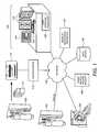
- FIG. 1illustrates a conceptual diagram of a medical imaging environment including a review workstation according to a preferred embodiment
- FIGS. 2A and 2Billustrate review workstations according to preferred embodiments displaying mammogram image information and lung-CT image information, respectively;
- FIG. 3illustrates patient worklist management according to a preferred embodiment
- FIG. 4illustrates a patient worklist display according to a preferred embodiment
- FIG. 5illustrates a patient worklist priority rule modification display according to a preferred embodiment.
- FIG. 1illustrates a conceptual diagram of a medical imaging environment for which one or more of the preferred embodiments is particularly suited.
- a network 110which may be a HIS/RIS (Hospital Information System/Radiology Information System) network, to which is coupled a film mammogram acquisition device 102 , a digital mammogram acquisition device 104 , a computed tomography (CT) acquisition device 106 , and a generalized “other” medical imaging device 108 .
- HIS/RISHospital Information System/Radiology Information System
- a computer-aided detection (CAD) processor 112 coupled to the network 110receives digital medical images from one or more of the devices 104 - 108 , and/or from a digitizer 116 that digitizes x-ray mammogram films 114 generated by the film mammogram acquisition device 102 .
- the CAD processor 112processes the medical images according to a CAD processing algorithm. The medical images are then viewed in conjunction with the associated CAD results at a radiology review workstation 120 .
- the various medical images and related informationare communicated according to the DICOM (Digital Imaging and Communications in Medicine) standard and the network 110 supports the TCP/IP protocol, which is used as the transport protocol for the DICOM standard.
- a PACS (Picture Archiving and Communication System) archive 118is also coupled to the network 110 , generally representing a repository for medical information associated with the medical imaging environment, including both current and archived images, current and archived CAD results, radiology reports for completed cases, and so forth.
- Computer-aided detectiongenerally refers to the use of computers to analyze medical images to detect anatomical abnormalities therein.
- computer-aided detectionsometimes used interchangeably with the term computer-aided detection are the terms computer-aided diagnosis, computer-assisted diagnosis, or computer-assisted detection.
- CAD detectionrefers to a location in a medical image that a CAD system, in accordance with a CAD processing algorithm operating on the medical image, has identified as warranting some type of attention by a radiologist.
- a CAD algorithmusually identifies a preliminary set of candidate detections in a medical image and then selects which ones, if any, will qualify as actual CAD detections based on a variety of computed features associated with the candidate detections.
- the CAD resultsi.e., the body of information associated with the operation of the CAD algorithm on the medical image, are most often communicated in the form of annotation maps comprising graphical annotations (CAD markers) overlaid on a diagnostic-quality or reduced-resolution version of the medical image, one CAD marker for each CAD detection.
- CAD resultsare mainly used by radiologists as “secondary reads” or secondary diagnosis tools.
- the radiologistWhen analyzing a medical image, the radiologist usually makes his or her own analytical determinations before looking at the CAD results, which either verify those determinations or trigger further inspection of the image.
- Some CAD implementationshave used CAD results in a “concurrent reading” context in which the radiologists look at the CAD results at the same time that they look at the images.
- Mammography CAD systemsIn the field of x-ray mammography, thousands of mammography CAD systems are now installed worldwide, and are used to assist radiologists in the interpretation of millions of mammograms per year. Mammography CAD systems are described, for example, in U.S. Pat. Nos. 5,729,620, 5,815,591, 5,917,929, 6,075,879, 6,266,435, 6,434,262, and U.S. Pat. No. 6,901,156, each of which is incorporated by reference herein. Mammography CAD algorithms analyze digital or digitized images of standard mammographic views (e.g. CC, MLO) for characteristics commonly associated with breast cancer, such as calcifications, masses, and architectural distortions.
- standard mammographic viewse.g. CC, MLO
- the preferred embodiments described hereinare readily applicable for a variety of present or prospective non-mammography medical imaging modalities such as CT, MRI, PET, SPECT, ultrasound, x-ray tomosynthesis, thermography, electrical conductivity-based modalities, and other modalities.
- CTcomputed tomography
- MRImagnetic resonance imaging
- PETPET
- SPECTnuclear emission computed tomography
- thermographythermography
- electrical conductivity-based modalitiesand other modalities.
- at least one CAD systemhas been commercialized for assisting radiologists in the detection of suspicious lung nodules, such systems being referenced herein as lung-CT CAD systems. Examples of lung-CT CAD systems are described in U.S. Pat. No. 5,881,124 and in the commonly assigned U.S. Pat. No. 6,925,200, each of which is incorporated by reference herein.
- the preferred embodiments described hereinare seamlessly layered upon an existing CAD workflow, in which the digital or digitized medical images are processed by the CAD processor 112 , and in which the medical images and their related CAD results are subsequently displayed at the review workstation 120 to a viewer, who makes a clinical determination therefrom.
- the clinical determinationcan be in relation to screening, diagnosis, follow-up, or any of a variety of other activities.
- the preferred embodiments hereinare particularly advantageous in a screening context for which speed, case throughput, and viewer stamina are important factors.
- the review workstation 120comprises a multi-modality workstation adapted and configured for a mammography environment.
- a Sectra IDS5/mx.net dedicated mammography workstationcan be used that allows for third-party plug-ins, including plug-ins providing the CAD user interfacing functionalities described herein.
- Review workstation 120comprises a diagnostic display 122 , an administrative display 124 , user input devices 126 (e.g., keyboard, mouse, trackball, pointers, etc), and a user interface processor 128 .
- Administrative display 124is used for input and output of a wide variety of information that may be associated with a particular set of medical images (e.g., listings, tables, plots, text descriptions, etc), as well as for system installation, maintenance, updating, and related tasks.
- a worklist management processor 130configured and adapted to implement the patient worklist processing functionalities described herein. It is to be appreciated, however, that such patient worklist processing can be performed by any combination of the user interface processor 128 , the CAD processor 112 , and the worklist management processor 130 , or by any other processor or combination of processors (such as HIS/RIS scheduling processors) coupled to the network 110 , without departing from the scope of the preferred embodiments.
- the medical imaging environment of FIG. 1is presented by way of example only and is not intended to limit the scope of the preferred embodiments to this particular scenario.
- different combinations of the devices of FIG. 1can be placed adjacently to each other or integrated into the same hardware boxes without departing from the scope of the preferred embodiments.
- the network 110can be a wide-area network with the different nodes being distributed throughout a city, a country, or the world.
- some or all of the transfer of digital informationcan be achieved by physical transfer of disks, memory sticks, or other digital media devices without departing from the scope of the preferred embodiments.
- a person skilled in the artwould be able to construct such plug-ins or other software packages capable of achieving the described user interfaces and processing functionalities without undue experimentation, using publicly available programming tools and software development platforms.
- FIGS. 2A and 2Billustrate radiology review workstations according to a preferred embodiment displaying mammogram image information and CT-lung image information, respectively.
- the diagnostic display 122presents mammogram images 203 annotated according to mammography CAD detections 204 .
- a diagnostic display 251presents lung-CT images 253 annotated according to lung-CT CAD detections 254 .
- the administrative display 124includes a patient worklist display 202 , as well as a patient worklist priority rule modification display 206 .
- an administrative display 250includes a patient worklist display 252 , as well as a patient worklist priority rule modification display 256 .
- the patient worklist displays 202 and 252are similar to each other and the patient worklist priority rule modification displays 206 and 256 are similar to each other, although many of the CAD-computed metrics, clinical metrics, and/or demographic metrics may differ.
- FIG. 3illustrates patient worklist management according to a preferred embodiment.
- CAD results for a plurality of medical imaging casesare received.
- patient worklist priority rulesare accessed, the patient worklist priority rules defining a sorting hierarchy including at least one CAD-computed metric.
- the patient worklist priority rulesmay comprise a default set when no customizations have been received, or may comprise a customized set when customizations have been received.
- CAD-computed metricrefers to (i) features computed by a CAD processing algorithm while operating on a medical imaging case to detect anatomical abnormalities therein, as well as (ii) various logical or mathematical formulations based on such features.
- CAD-computed metriccan further refer to one or more features extracted from a medical imaging case for the particular purpose of providing a basis for worklist prioritization.
- the patient worklistis sorted according to the patient worklist priority rules and CAD-computed metrics.
- the patient worklistis displayed to the radiologist.
- modifications to the patient worklist priority rulesare received from the radiologist, preferably using a graphical user interface as described hereinbelow, followed again by the sorting and display steps 306 and 308 . For newly received medical imaging cases and their associated CAD results, entries are dynamically entered into the patient worklist according to the current sorting hierarchy.
- FIG. 4illustrates a patient worklist display 402 according to a preferred embodiment for a mammography review workstation, showing the results of but one of a wide variety of different sorting hierarchies that are within the scope of the present teachings.
- Patient worklist display 402comprises an ordered listing of cases sorted by a number of CAD markers field 406 as a primary sort key, descending, and by a case acquisition date/time field 410 as a secondary sort key, ascending, such that older cases have priority over newer cases for a common number of CAD markers.
- Column heading buttons 404are provided as shown in FIG. 4 .
- the usermay dynamically change the patient worklist priority rules by pressing one of the column heading buttons 404 , the most recently-pressed button representing the primary sort key, the previously-pressed button representing the secondary sort key, and so on.
- the directional order for the primary sort keycan be toggled between ascending and descending by serially pressing the column heading button 404 associated with the current primary sort key. Any of a variety of differing graphical user interface schemes for achieving similar customization functions as shown in FIG. 4 are within the scope of the preferred embodiments.
- FIG. 5illustrates a patient worklist priority rule modification display 502 according to a preferred embodiment for a mammography review workstation, which is preferably overlayable on the administrative display 206 / 256 in a Windows®-like fashion.
- a predetermined list of sorting criteria 504is provided including at least one CAD-computed metric.
- the sorting criteria 504are strictly limited to CAD-computed metrics such as, but not limited to: a total number of CAD markers metric, a breast density metric, a breast size metric, a maximum overall suspiciousness metric, a maximum calcification suspiciousness metric, and a maximum mass suspiciousness metric.
- the sorting criteria 504can include other case-related metrics such as clinical metrics (e.g., weight, family history) and/or demographic metrics (e.g., race, HMO type, etc.). While differing CAD algorithms will often have different nomenclatures and scalings, there can often be an “ultimate” or “overall” scalar score assigned to a case, termed herein a composite CAD score, which can be based on the medical image data in optional combination with clinical data and/or demographic data. In another preferred embodiment, the sorting criteria 504 further includes the composite CAD score.
- clinical metricse.g., weight, family history
- demographic metricse.g., race, HMO type, etc.
- the sorting criteria 504further includes the composite CAD score.
- Patient worklist priority rule modification display 502further comprises a priority rule display 506 listing the currently-selected sorting hierarchy. Sorting criteria can be added and removed by add/remove buttons 508 , with the priority (i.e., primary sort key, secondary sort key, tertiary sort key) being changeable using priority change buttons 510 .
- the usercan start by choosing from a variety of pre-programmed priority rules (e.g., clinic default priority rules, saved customization choices made by other radiologists or radiologist groups, etc.) using the pull-down menu 512 .
- the patient worklist display 402 of FIG. 4 and the patient worklist priority rule modification display 502 of FIG. 5are readily applicable for any imaging modality by changing the available fields and sorting criteria 504 as needed.
- the CAD-computed metrics in sorting criteria 504may comprise a number of detected lung nodules per case metric and a lung nodule size metric.
- One particularly useful sorting criteria usually applicable for most modalitiesis an anatomical complexity metric (e.g., breast density for mammography, lung complexity for lung-CT, colon length for virtual colonoscopy, etc.), with radiologist review effort usually increasing for cases of increased anatomical complexity.
- the radiologistis provided with increased insight into their patient workflow and increased control over their patient workflow. In turn, this can increase radiologist efficiency, stamina, and even attitude toward that day's workload.
- a first radiologist who is a self-considered “morning person”may elect to sort the patient worklist by decreasing breast density so that they will review these more difficult cases while fresh and alert in the morning.
- a second radiologist who is an “evening person”may do the opposite (i.e., sort by increasing breast density).
- a rich variety of custom worklist prioritization scenariosis provided that can allow each user (or group of users) to best match the patient review sequence to their personal habits, mental or physical biorhythms, experience levels, etc.
- An additional advantage of worklist managementis that there is very little added complexity, from both a HIS/RIS/PACS perspective and from an end user perspective. This is because, preferably, the same CAD processing algorithm is used for both abnormality detection and for worklist management. Accordingly, very little additional CAD processing hardware or CAD processing time is needed, because most of the CAD-computed features useful for worklist prioritization are already computed as part of the abnormality detection computations. Likewise, the amount of additional training needed by the radiologist to understand and manage the worklists is generally small, because they are already familiar with the CAD-computed worklist prioritization features from their normal training in CAD-assisted case viewing.
- an aggregate patient worklist for an entire clinicis sorted according to one or more CAD-generated parameters for overview by a head radiologist, hospital administrator etc.
- case routingmay be implemented based on the generated patient worklist, e.g., with mostly-dense breasts being routed to a first radiologist (or first group of radiologists) for review, and with mostly-fatty breasts being routed to a second radiologist (or second group of radiologists) for review.
- Such case routingmay be particularly advantageous in a radiologist training context, where cases can be routed based on trainee status.
- More complex routing scenariosin which expert systems might be used, for example
- static or dynamic routing models having one or more CAD-generated routing criteriaare also within the scope of the preferred embodiments.
- the described CAD-based patient worklist prioritizationis readily applied in a distributed or tele-PACS environment, each remote radiologist being provided with advantageous insight into and/or control over their patient worklist.
- Judicious case routing based on the CAD-generated parameterscan also be implemented in this context, e.g., by routing cases with high maximum suspiciousness to a local group of radiologists, and by sending other cases to overseas radiologists.
- the sorting order for a particular criterionmay be something other than monotonically-increasing or monotonically-decreasing—for example, “structured” or even “purposefully random”—without departing from the scope of the preferred embodiments.
- a particular radiologistmight wish for their workload to purposefully alternate between marked cases (i.e., cases having one or more CAD markers) and unmarked cases, to keep their attention fresh or otherwise temporally balanced. Therefore, reference to the details of the preferred embodiments are not intended to limit their scope, which is limited only by the scope of the claims set forth below.
Landscapes
- Health & Medical Sciences (AREA)
- Engineering & Computer Science (AREA)
- Medical Informatics (AREA)
- Public Health (AREA)
- Primary Health Care (AREA)
- Epidemiology (AREA)
- General Health & Medical Sciences (AREA)
- Biomedical Technology (AREA)
- Business, Economics & Management (AREA)
- General Business, Economics & Management (AREA)
- Nuclear Medicine, Radiotherapy & Molecular Imaging (AREA)
- Radiology & Medical Imaging (AREA)
- Data Mining & Analysis (AREA)
- Databases & Information Systems (AREA)
- Pathology (AREA)
- Apparatus For Radiation Diagnosis (AREA)
- Measuring And Recording Apparatus For Diagnosis (AREA)
Abstract
Description
Claims (19)
Priority Applications (1)
| Application Number | Priority Date | Filing Date | Title |
|---|---|---|---|
| US11/208,271US7889896B2 (en) | 2005-08-18 | 2005-08-18 | Patient worklist management in digital radiography review workstations |
Applications Claiming Priority (1)
| Application Number | Priority Date | Filing Date | Title |
|---|---|---|---|
| US11/208,271US7889896B2 (en) | 2005-08-18 | 2005-08-18 | Patient worklist management in digital radiography review workstations |
Publications (2)
| Publication Number | Publication Date |
|---|---|
| US20070041623A1 US20070041623A1 (en) | 2007-02-22 |
| US7889896B2true US7889896B2 (en) | 2011-02-15 |
Family
ID=37767382
Family Applications (1)
| Application Number | Title | Priority Date | Filing Date |
|---|---|---|---|
| US11/208,271Active2027-09-04US7889896B2 (en) | 2005-08-18 | 2005-08-18 | Patient worklist management in digital radiography review workstations |
Country Status (1)
| Country | Link |
|---|---|
| US (1) | US7889896B2 (en) |
Cited By (19)
| Publication number | Priority date | Publication date | Assignee | Title |
|---|---|---|---|---|
| US20100241638A1 (en)* | 2009-03-18 | 2010-09-23 | O'sullivan Patrick Joseph | Sorting contacts |
| US20100322495A1 (en)* | 2006-12-12 | 2010-12-23 | Koninklijke Philips Electronics N.V. | Medical imaging system |
| US20120098859A1 (en)* | 2010-10-22 | 2012-04-26 | Pantech Co., Ltd. | Apparatus and method for providing augmented reality user interface |
| US20160098298A1 (en)* | 2009-04-24 | 2016-04-07 | Pegasystems Inc. | Methods and apparatus for integrated work management |
| US9895121B2 (en) | 2013-08-20 | 2018-02-20 | Densitas Incorporated | Methods and systems for determining breast density |
| US10467200B1 (en) | 2009-03-12 | 2019-11-05 | Pegasystems, Inc. | Techniques for dynamic data processing |
| US10469396B2 (en) | 2014-10-10 | 2019-11-05 | Pegasystems, Inc. | Event processing with enhanced throughput |
| US10572236B2 (en) | 2011-12-30 | 2020-02-25 | Pegasystems, Inc. | System and method for updating or modifying an application without manual coding |
| US10698647B2 (en) | 2016-07-11 | 2020-06-30 | Pegasystems Inc. | Selective sharing for collaborative application usage |
| US10698599B2 (en) | 2016-06-03 | 2020-06-30 | Pegasystems, Inc. | Connecting graphical shapes using gestures |
| US10838569B2 (en) | 2006-03-30 | 2020-11-17 | Pegasystems Inc. | Method and apparatus for user interface non-conformance detection and correction |
| US11048488B2 (en) | 2018-08-14 | 2021-06-29 | Pegasystems, Inc. | Software code optimizer and method |
| US11567945B1 (en) | 2020-08-27 | 2023-01-31 | Pegasystems Inc. | Customized digital content generation systems and methods |
| US11783476B2 (en) | 2019-10-25 | 2023-10-10 | DeepHealth, Inc. | System and method for analyzing three-dimensional image data |
| US20240021297A1 (en)* | 2019-09-27 | 2024-01-18 | Hologic, Inc. | AI System for Predicting Reading Time and Reading Complexity for Reviewing 2D/3D Breast Images |
| US11883206B2 (en) | 2019-07-29 | 2024-01-30 | Hologic, Inc. | Personalized breast imaging system |
| US12106850B2 (en) | 2021-01-28 | 2024-10-01 | RAD AI, Inc. | System and method for workflow management and image review |
| US12367574B2 (en) | 2019-12-23 | 2025-07-22 | DeepHealth, Inc. | Systems and methods for analyzing two-dimensional and three-dimensional image data |
| US12437390B2 (en) | 2018-11-24 | 2025-10-07 | Densitas Incorporated | System and method for assessing quality of medical images |
Families Citing this family (23)
| Publication number | Priority date | Publication date | Assignee | Title |
|---|---|---|---|---|
| US7889896B2 (en)* | 2005-08-18 | 2011-02-15 | Hologic, Inc. | Patient worklist management in digital radiography review workstations |
| US20070083849A1 (en)* | 2005-10-12 | 2007-04-12 | General Electric Company | Auto-learning RIS/PACS worklists |
| US8005278B2 (en)* | 2005-11-22 | 2011-08-23 | General Electric Company | System and method for patient acuity driven workflow using computer-aided diagnosis of medical images |
| US7664659B2 (en)* | 2005-12-22 | 2010-02-16 | Cerner Innovation, Inc. | Displaying clinical predicted length of stay of patients for workload balancing in a healthcare environment |
| US20080043036A1 (en)* | 2006-08-16 | 2008-02-21 | Mevis Breastcare Gmbh & Co. Kg | Method, apparatus and computer program for presenting cases comprising images |
| DE102007013566B4 (en)* | 2007-03-21 | 2017-02-23 | Siemens Healthcare Gmbh | Method for image data acquisition and medical modality |
| CN101464861A (en)* | 2007-12-21 | 2009-06-24 | Ge医疗系统环球技术有限公司 | Medical image information management system and program product |
| US8126877B2 (en)* | 2008-01-23 | 2012-02-28 | Globalspec, Inc. | Arranging search engine results |
| US20090196479A1 (en)* | 2008-01-31 | 2009-08-06 | Raghav Raman | Method and apparatus for computer-aided diagnosis filtered prioritized work item list |
| US20100135546A1 (en)* | 2008-11-28 | 2010-06-03 | General Electric Company | Landmark guides for registration of multi-modality medical images |
| US8687860B2 (en) | 2009-11-24 | 2014-04-01 | Penrad Technologies, Inc. | Mammography statistical diagnostic profiler and prediction system |
| US8799013B2 (en)* | 2009-11-24 | 2014-08-05 | Penrad Technologies, Inc. | Mammography information system |
| US8953858B2 (en) | 2010-01-28 | 2015-02-10 | Radlogics, Inc. | Methods and systems for analyzing, prioritizing, visualizing, and reporting medical images |
| CA2871674A1 (en)* | 2012-05-31 | 2013-12-05 | Ikonopedia, Inc. | Image based analytical systems and processes |
| US8868768B2 (en) | 2012-11-20 | 2014-10-21 | Ikonopedia, Inc. | Secure medical data transmission |
| US10657220B2 (en)* | 2015-04-22 | 2020-05-19 | Ascend Hit Llc | System and methods for medical reporting |
| EP3341871A1 (en)* | 2015-08-24 | 2018-07-04 | Koninklijke Philips N.V. | Radiology image sequencing for optimal reading throughput |
| EP3356970A1 (en)* | 2015-09-28 | 2018-08-08 | Koninklijke Philips N.V. | Challenge value icons for radiology report selection |
| JP6746360B2 (en)* | 2016-04-13 | 2020-08-26 | キヤノン株式会社 | Information processing system, information processing method, and program |
| US20200265946A1 (en)* | 2016-06-17 | 2020-08-20 | Algotec Systems Ltd. | Medical image workflow system and method |
| US11495343B2 (en)* | 2017-04-21 | 2022-11-08 | Koninklijke Philips N.V. | Device, system, and method for determining a reading environment by synthesizing downstream needs |
| KR102192164B1 (en)* | 2018-08-08 | 2020-12-16 | 주식회사 딥바이오 | System for biomedical image diagnosis, method for biomedical image diagnosis and terminal performing the same |
| US11430563B2 (en)* | 2018-11-21 | 2022-08-30 | Fujifilm Medical Systems U.S.A., Inc. | Configuring and displaying a user interface with healthcare studies |
Citations (26)
| Publication number | Priority date | Publication date | Assignee | Title |
|---|---|---|---|---|
| US5815591A (en)* | 1996-07-10 | 1998-09-29 | R2 Technology, Inc. | Method and apparatus for fast detection of spiculated lesions in digital mammograms |
| US5823948A (en)* | 1996-07-08 | 1998-10-20 | Rlis, Inc. | Medical records, documentation, tracking and order entry system |
| US5917929A (en)* | 1996-07-23 | 1999-06-29 | R2 Technology, Inc. | User interface for computer aided diagnosis system |
| US6067373A (en)* | 1998-04-02 | 2000-05-23 | Arch Development Corporation | Method, system and computer readable medium for iterative image warping prior to temporal subtraction of chest radiographs in the detection of interval changes |
| US20020186899A1 (en) | 2001-05-29 | 2002-12-12 | Sascha Bohnenkamp | Method and computer system for prefetching of images |
| US20020193676A1 (en) | 2001-05-29 | 2002-12-19 | Anke Bodicker | Method and computer system for screening of medical cases |
| US20030013951A1 (en) | 2000-09-21 | 2003-01-16 | Dan Stefanescu | Database organization and searching |
| US20030126148A1 (en)* | 2001-11-21 | 2003-07-03 | Amicas, Inc. | System and methods for real-time worklist service |
| WO2004023790A2 (en) | 2002-09-04 | 2004-03-18 | Eastman Kodak Company | Patient identification method for x-ray film user-interfaces |
| US6873717B2 (en)* | 2002-07-09 | 2005-03-29 | Riverain Medical Group, Llc | Input/output interface for computer aided diagnosis (CAD) system |
| US20050096530A1 (en)* | 2003-10-29 | 2005-05-05 | Confirma, Inc. | Apparatus and method for customized report viewer |
| US6909795B2 (en)* | 2003-06-16 | 2005-06-21 | R2 Technology, Inc. | Communicating computer-aided detection results in a standards-based medical imaging environment |
| US6925200B2 (en)* | 2000-11-22 | 2005-08-02 | R2 Technology, Inc. | Graphical user interface for display of anatomical information |
| US6970587B1 (en)* | 1997-08-28 | 2005-11-29 | Icad, Inc. | Use of computer-aided detection system outputs in clinical practice |
| US20060215894A1 (en)* | 2005-03-23 | 2006-09-28 | Sarang Lakare | System and method for smart display of CAD markers |
| US20070003119A1 (en)* | 2005-07-01 | 2007-01-04 | R2 Technology, Inc. | Displaying and navigating computer-aided detection results on a review workstation |
| US20070041623A1 (en)* | 2005-08-18 | 2007-02-22 | R2 Technology, Inc. | Patient worklist management in digital radiography review workstations |
| US7184582B2 (en)* | 2000-02-04 | 2007-02-27 | Arch Development Corporation | Method, system and computer readable medium for an intelligent search workstation for computer assisted interpretation of medical images |
| US7187790B2 (en)* | 2002-12-18 | 2007-03-06 | Ge Medical Systems Global Technology Company, Llc | Data processing and feedback method and system |
| US20070083849A1 (en)* | 2005-10-12 | 2007-04-12 | General Electric Company | Auto-learning RIS/PACS worklists |
| US20070280530A1 (en)* | 2006-05-03 | 2007-12-06 | Siemens Medical Solutions Usa, Inc. | Using Candidates Correlation Information During Computer Aided Diagnosis |
| US20080095418A1 (en)* | 2006-10-18 | 2008-04-24 | Fujifilm Corporation | System, method, and program for medical image interpretation support |
| US20080125643A1 (en)* | 2006-11-24 | 2008-05-29 | Qview, Inc. | Processing and displaying dynamic contrast-enhanced magnetic resonance imaging information |
| US7383307B2 (en)* | 2004-01-07 | 2008-06-03 | International Business Machines Corporation | Instant messaging windowing for topic threads |
| US7383237B2 (en)* | 1998-05-01 | 2008-06-03 | Health Discovery Corporation | Computer-aided image analysis |
| US7490085B2 (en)* | 2002-12-18 | 2009-02-10 | Ge Medical Systems Global Technology Company, Llc | Computer-assisted data processing system and method incorporating automated learning |
- 2005
- 2005-08-18USUS11/208,271patent/US7889896B2/enactiveActive
Patent Citations (27)
| Publication number | Priority date | Publication date | Assignee | Title |
|---|---|---|---|---|
| US5823948A (en)* | 1996-07-08 | 1998-10-20 | Rlis, Inc. | Medical records, documentation, tracking and order entry system |
| US5815591A (en)* | 1996-07-10 | 1998-09-29 | R2 Technology, Inc. | Method and apparatus for fast detection of spiculated lesions in digital mammograms |
| US5917929A (en)* | 1996-07-23 | 1999-06-29 | R2 Technology, Inc. | User interface for computer aided diagnosis system |
| US6970587B1 (en)* | 1997-08-28 | 2005-11-29 | Icad, Inc. | Use of computer-aided detection system outputs in clinical practice |
| US6067373A (en)* | 1998-04-02 | 2000-05-23 | Arch Development Corporation | Method, system and computer readable medium for iterative image warping prior to temporal subtraction of chest radiographs in the detection of interval changes |
| US7383237B2 (en)* | 1998-05-01 | 2008-06-03 | Health Discovery Corporation | Computer-aided image analysis |
| US7184582B2 (en)* | 2000-02-04 | 2007-02-27 | Arch Development Corporation | Method, system and computer readable medium for an intelligent search workstation for computer assisted interpretation of medical images |
| US20030013951A1 (en) | 2000-09-21 | 2003-01-16 | Dan Stefanescu | Database organization and searching |
| US6925200B2 (en)* | 2000-11-22 | 2005-08-02 | R2 Technology, Inc. | Graphical user interface for display of anatomical information |
| US20020186899A1 (en) | 2001-05-29 | 2002-12-12 | Sascha Bohnenkamp | Method and computer system for prefetching of images |
| US20020193676A1 (en) | 2001-05-29 | 2002-12-19 | Anke Bodicker | Method and computer system for screening of medical cases |
| US20030126148A1 (en)* | 2001-11-21 | 2003-07-03 | Amicas, Inc. | System and methods for real-time worklist service |
| US6873717B2 (en)* | 2002-07-09 | 2005-03-29 | Riverain Medical Group, Llc | Input/output interface for computer aided diagnosis (CAD) system |
| WO2004023790A2 (en) | 2002-09-04 | 2004-03-18 | Eastman Kodak Company | Patient identification method for x-ray film user-interfaces |
| US20060000884A1 (en)* | 2002-09-04 | 2006-01-05 | Wido Menhardt | Patient identification method for x-ray film user-interfaces |
| US7490085B2 (en)* | 2002-12-18 | 2009-02-10 | Ge Medical Systems Global Technology Company, Llc | Computer-assisted data processing system and method incorporating automated learning |
| US7187790B2 (en)* | 2002-12-18 | 2007-03-06 | Ge Medical Systems Global Technology Company, Llc | Data processing and feedback method and system |
| US6909795B2 (en)* | 2003-06-16 | 2005-06-21 | R2 Technology, Inc. | Communicating computer-aided detection results in a standards-based medical imaging environment |
| US20050096530A1 (en)* | 2003-10-29 | 2005-05-05 | Confirma, Inc. | Apparatus and method for customized report viewer |
| US7383307B2 (en)* | 2004-01-07 | 2008-06-03 | International Business Machines Corporation | Instant messaging windowing for topic threads |
| US20060215894A1 (en)* | 2005-03-23 | 2006-09-28 | Sarang Lakare | System and method for smart display of CAD markers |
| US20070003119A1 (en)* | 2005-07-01 | 2007-01-04 | R2 Technology, Inc. | Displaying and navigating computer-aided detection results on a review workstation |
| US20070041623A1 (en)* | 2005-08-18 | 2007-02-22 | R2 Technology, Inc. | Patient worklist management in digital radiography review workstations |
| US20070083849A1 (en)* | 2005-10-12 | 2007-04-12 | General Electric Company | Auto-learning RIS/PACS worklists |
| US20070280530A1 (en)* | 2006-05-03 | 2007-12-06 | Siemens Medical Solutions Usa, Inc. | Using Candidates Correlation Information During Computer Aided Diagnosis |
| US20080095418A1 (en)* | 2006-10-18 | 2008-04-24 | Fujifilm Corporation | System, method, and program for medical image interpretation support |
| US20080125643A1 (en)* | 2006-11-24 | 2008-05-29 | Qview, Inc. | Processing and displaying dynamic contrast-enhanced magnetic resonance imaging information |
Non-Patent Citations (1)
| Title |
|---|
| Internet Access to Digital Medical X-rays by Image Features and Associated Text L. Rodney Long, George R. Thoma;National Library of Medicine, Bethesda, MD 20894; IEEE 1998.* |
Cited By (23)
| Publication number | Priority date | Publication date | Assignee | Title |
|---|---|---|---|---|
| US10838569B2 (en) | 2006-03-30 | 2020-11-17 | Pegasystems Inc. | Method and apparatus for user interface non-conformance detection and correction |
| US20100322495A1 (en)* | 2006-12-12 | 2010-12-23 | Koninklijke Philips Electronics N.V. | Medical imaging system |
| US10467200B1 (en) | 2009-03-12 | 2019-11-05 | Pegasystems, Inc. | Techniques for dynamic data processing |
| US20100241638A1 (en)* | 2009-03-18 | 2010-09-23 | O'sullivan Patrick Joseph | Sorting contacts |
| US20160098298A1 (en)* | 2009-04-24 | 2016-04-07 | Pegasystems Inc. | Methods and apparatus for integrated work management |
| US20120098859A1 (en)* | 2010-10-22 | 2012-04-26 | Pantech Co., Ltd. | Apparatus and method for providing augmented reality user interface |
| US10572236B2 (en) | 2011-12-30 | 2020-02-25 | Pegasystems, Inc. | System and method for updating or modifying an application without manual coding |
| US9895121B2 (en) | 2013-08-20 | 2018-02-20 | Densitas Incorporated | Methods and systems for determining breast density |
| US10123758B2 (en) | 2013-08-20 | 2018-11-13 | Densitas Incorporated | Methods and systems for determining breast density |
| US10469396B2 (en) | 2014-10-10 | 2019-11-05 | Pegasystems, Inc. | Event processing with enhanced throughput |
| US11057313B2 (en) | 2014-10-10 | 2021-07-06 | Pegasystems Inc. | Event processing with enhanced throughput |
| US10698599B2 (en) | 2016-06-03 | 2020-06-30 | Pegasystems, Inc. | Connecting graphical shapes using gestures |
| US10698647B2 (en) | 2016-07-11 | 2020-06-30 | Pegasystems Inc. | Selective sharing for collaborative application usage |
| US11048488B2 (en) | 2018-08-14 | 2021-06-29 | Pegasystems, Inc. | Software code optimizer and method |
| US12437390B2 (en) | 2018-11-24 | 2025-10-07 | Densitas Incorporated | System and method for assessing quality of medical images |
| US11883206B2 (en) | 2019-07-29 | 2024-01-30 | Hologic, Inc. | Personalized breast imaging system |
| US12226233B2 (en) | 2019-07-29 | 2025-02-18 | Hologic, Inc. | Personalized breast imaging system |
| US20240021297A1 (en)* | 2019-09-27 | 2024-01-18 | Hologic, Inc. | AI System for Predicting Reading Time and Reading Complexity for Reviewing 2D/3D Breast Images |
| US12119107B2 (en)* | 2019-09-27 | 2024-10-15 | Hologic, Inc. | AI system for predicting reading time and reading complexity for reviewing 2D/3D breast images |
| US11783476B2 (en) | 2019-10-25 | 2023-10-10 | DeepHealth, Inc. | System and method for analyzing three-dimensional image data |
| US12367574B2 (en) | 2019-12-23 | 2025-07-22 | DeepHealth, Inc. | Systems and methods for analyzing two-dimensional and three-dimensional image data |
| US11567945B1 (en) | 2020-08-27 | 2023-01-31 | Pegasystems Inc. | Customized digital content generation systems and methods |
| US12106850B2 (en) | 2021-01-28 | 2024-10-01 | RAD AI, Inc. | System and method for workflow management and image review |
Also Published As
| Publication number | Publication date |
|---|---|
| US20070041623A1 (en) | 2007-02-22 |
Similar Documents
| Publication | Publication Date | Title |
|---|---|---|
| US7889896B2 (en) | Patient worklist management in digital radiography review workstations | |
| US6785410B2 (en) | Image reporting method and system | |
| US20180330457A1 (en) | Electronic health record timeline and the human figure | |
| US7693317B2 (en) | Image reporting method and system | |
| US7894676B2 (en) | Diagnosis support apparatus and control method therefor | |
| JP2023503610A (en) | Co-registration of medical scan images and method of use therewith | |
| US20110123079A1 (en) | Mammography information system | |
| EP2225701A1 (en) | Systems and methods for efficient imaging | |
| US20100082365A1 (en) | Navigation and Visualization of Multi-Dimensional Image Data | |
| US20200294655A1 (en) | System and method to automatically prepare an attention list for improving radiology workflow | |
| US20150339457A1 (en) | Method and apparatus for integrating clinical data with the review of medical images | |
| EP4014240B1 (en) | System and method for reporting on medical images | |
| US11094062B2 (en) | Auto comparison layout based on image similarity | |
| US20090132274A1 (en) | Systems and Methods for Image and Report Preview in a Healthcare Worklist | |
| US8005278B2 (en) | System and method for patient acuity driven workflow using computer-aided diagnosis of medical images | |
| US20070083849A1 (en) | Auto-learning RIS/PACS worklists | |
| EP2483822A2 (en) | Retrieving radiological studies using an image-based query | |
| Horii | Clinical aspects of workstation design and operation | |
| WO2009007880A1 (en) | Anatomy timeline for clinical reporting |
Legal Events
| Date | Code | Title | Description |
|---|---|---|---|
| AS | Assignment | Owner name:R2 TECHNOLOGY, INC., CALIFORNIA Free format text:ASSIGNMENT OF ASSIGNORS INTEREST;ASSIGNORS:ROEHRIG, JIMMY R.;MARSHALL, JULIAN;REEL/FRAME:016936/0842 Effective date:20051020 | |
| AS | Assignment | Owner name:GOLDMAN SACHS CREDIT PARTNERS L.P., CALIFORNIA Free format text:PATENT SECURITY AGREEMENT;ASSIGNOR:R2 TECHNOLOGY, INC.;REEL/FRAME:020024/0231 Effective date:20071022 Owner name:GOLDMAN SACHS CREDIT PARTNERS L.P.,CALIFORNIA Free format text:PATENT SECURITY AGREEMENT;ASSIGNOR:R2 TECHNOLOGY, INC.;REEL/FRAME:020024/0231 Effective date:20071022 | |
| AS | Assignment | Owner name:GOLDMAN SACHS CREDIT PARTNERS L.P., AS COLLATERAL Free format text:PATENT SECURITY AGREEMENT;ASSIGNOR:R2 TECHNOLOGY, INC.;REEL/FRAME:021301/0838 Effective date:20080717 | |
| AS | Assignment | Owner name:HOLOGIC, INC., MASSACHUSETTS Free format text:ASSIGNMENT OF ASSIGNORS INTEREST;ASSIGNOR:R2 TECHNOLOGY, INC.;REEL/FRAME:023620/0362 Effective date:20091204 Owner name:HOLOGIC, INC.,MASSACHUSETTS Free format text:ASSIGNMENT OF ASSIGNORS INTEREST;ASSIGNOR:R2 TECHNOLOGY, INC.;REEL/FRAME:023620/0362 Effective date:20091204 | |
| AS | Assignment | Owner name:GOLDMAN SACHS CREDIT PARTNERS L.P., AS COLLATERAL Free format text:21ST SUPPLEMENT TO PATENT SECURITY AGREEMENT;ASSIGNOR:HOLOGIC, INC.;REEL/FRAME:024151/0435 Effective date:20100329 | |
| AS | Assignment | Owner name:CYTYC PRENATAL PRODUCTS CORP., MASSACHUSETTS Free format text:TERMINATION OF PATENT SECURITY AGREEMENTS AND RELEASE OF SECURITY INTERESTS;ASSIGNOR:GOLDMAN SACHS CREDIT PARTNERS, L.P., AS COLLATERAL AGENT;REEL/FRAME:024892/0001 Effective date:20100819 Owner name:BIOLUCENT, LLC, CALIFORNIA Free format text:TERMINATION OF PATENT SECURITY AGREEMENTS AND RELEASE OF SECURITY INTERESTS;ASSIGNOR:GOLDMAN SACHS CREDIT PARTNERS, L.P., AS COLLATERAL AGENT;REEL/FRAME:024892/0001 Effective date:20100819 Owner name:R2 TECHNOLOGY, INC., CALIFORNIA Free format text:TERMINATION OF PATENT SECURITY AGREEMENTS AND RELEASE OF SECURITY INTERESTS;ASSIGNOR:GOLDMAN SACHS CREDIT PARTNERS, L.P., AS COLLATERAL AGENT;REEL/FRAME:024892/0001 Effective date:20100819 Owner name:THIRD WAVE TECHNOLOGIES, INC., WISCONSIN Free format text:TERMINATION OF PATENT SECURITY AGREEMENTS AND RELEASE OF SECURITY INTERESTS;ASSIGNOR:GOLDMAN SACHS CREDIT PARTNERS, L.P., AS COLLATERAL AGENT;REEL/FRAME:024892/0001 Effective date:20100819 Owner name:CYTYC CORPORATION, MASSACHUSETTS Free format text:TERMINATION OF PATENT SECURITY AGREEMENTS AND RELEASE OF SECURITY INTERESTS;ASSIGNOR:GOLDMAN SACHS CREDIT PARTNERS, L.P., AS COLLATERAL AGENT;REEL/FRAME:024892/0001 Effective date:20100819 Owner name:HOLOGIC, INC., MASSACHUSETTS Free format text:TERMINATION OF PATENT SECURITY AGREEMENTS AND RELEASE OF SECURITY INTERESTS;ASSIGNOR:GOLDMAN SACHS CREDIT PARTNERS, L.P., AS COLLATERAL AGENT;REEL/FRAME:024892/0001 Effective date:20100819 Owner name:CYTYC SURGICAL PRODUCTS III, INC., MASSACHUSETTS Free format text:TERMINATION OF PATENT SECURITY AGREEMENTS AND RELEASE OF SECURITY INTERESTS;ASSIGNOR:GOLDMAN SACHS CREDIT PARTNERS, L.P., AS COLLATERAL AGENT;REEL/FRAME:024892/0001 Effective date:20100819 Owner name:CYTYC SURGICAL PRODUCTS LIMITED PARTNERSHIP, MASSA Free format text:TERMINATION OF PATENT SECURITY AGREEMENTS AND RELEASE OF SECURITY INTERESTS;ASSIGNOR:GOLDMAN SACHS CREDIT PARTNERS, L.P., AS COLLATERAL AGENT;REEL/FRAME:024892/0001 Effective date:20100819 Owner name:SUROS SURGICAL SYSTEMS, INC., INDIANA Free format text:TERMINATION OF PATENT SECURITY AGREEMENTS AND RELEASE OF SECURITY INTERESTS;ASSIGNOR:GOLDMAN SACHS CREDIT PARTNERS, L.P., AS COLLATERAL AGENT;REEL/FRAME:024892/0001 Effective date:20100819 Owner name:DIRECT RADIOGRAPHY CORP., DELAWARE Free format text:TERMINATION OF PATENT SECURITY AGREEMENTS AND RELEASE OF SECURITY INTERESTS;ASSIGNOR:GOLDMAN SACHS CREDIT PARTNERS, L.P., AS COLLATERAL AGENT;REEL/FRAME:024892/0001 Effective date:20100819 Owner name:CYTYC SURGICAL PRODUCTS II LIMITED PARTNERSHIP, MA Free format text:TERMINATION OF PATENT SECURITY AGREEMENTS AND RELEASE OF SECURITY INTERESTS;ASSIGNOR:GOLDMAN SACHS CREDIT PARTNERS, L.P., AS COLLATERAL AGENT;REEL/FRAME:024892/0001 Effective date:20100819 | |
| STCF | Information on status: patent grant | Free format text:PATENTED CASE | |
| AS | Assignment | Owner name:GOLDMAN SACHS BANK USA, NEW YORK Free format text:SECURITY AGREEMENT;ASSIGNORS:HOLOGIC, INC.;BIOLUCENT, LLC;CYTYC CORPORATION;AND OTHERS;REEL/FRAME:028810/0745 Effective date:20120801 | |
| FPAY | Fee payment | Year of fee payment:4 | |
| AS | Assignment | Owner name:CYTYC SURGICAL PRODUCTS, LIMITED PARTNERSHIP, MASSACHUSETTS Free format text:SECURITY INTEREST RELEASE REEL/FRAME 028810/0745;ASSIGNOR:GOLDMAN SACHS BANK USA, AS COLLATERAL AGENT;REEL/FRAME:035820/0239 Effective date:20150529 Owner name:CYTYC CORPORATION, MASSACHUSETTS Free format text:SECURITY INTEREST RELEASE REEL/FRAME 028810/0745;ASSIGNOR:GOLDMAN SACHS BANK USA, AS COLLATERAL AGENT;REEL/FRAME:035820/0239 Effective date:20150529 Owner name:GEN-PROBE INCORPORATED, MASSACHUSETTS Free format text:SECURITY INTEREST RELEASE REEL/FRAME 028810/0745;ASSIGNOR:GOLDMAN SACHS BANK USA, AS COLLATERAL AGENT;REEL/FRAME:035820/0239 Effective date:20150529 Owner name:SUROS SURGICAL SYSTEMS, INC., MASSACHUSETTS Free format text:SECURITY INTEREST RELEASE REEL/FRAME 028810/0745;ASSIGNOR:GOLDMAN SACHS BANK USA, AS COLLATERAL AGENT;REEL/FRAME:035820/0239 Effective date:20150529 Owner name:CYTYC SURGICAL PRODUCTS, LIMITED PARTNERSHIP, MASS Free format text:SECURITY INTEREST RELEASE REEL/FRAME 028810/0745;ASSIGNOR:GOLDMAN SACHS BANK USA, AS COLLATERAL AGENT;REEL/FRAME:035820/0239 Effective date:20150529 Owner name:THIRD WAVE TECHNOLOGIES, INC., MASSACHUSETTS Free format text:SECURITY INTEREST RELEASE REEL/FRAME 028810/0745;ASSIGNOR:GOLDMAN SACHS BANK USA, AS COLLATERAL AGENT;REEL/FRAME:035820/0239 Effective date:20150529 Owner name:HOLOGIC, INC., MASSACHUSETTS Free format text:SECURITY INTEREST RELEASE REEL/FRAME 028810/0745;ASSIGNOR:GOLDMAN SACHS BANK USA, AS COLLATERAL AGENT;REEL/FRAME:035820/0239 Effective date:20150529 Owner name:BIOLUCENT, LLC, MASSACHUSETTS Free format text:SECURITY INTEREST RELEASE REEL/FRAME 028810/0745;ASSIGNOR:GOLDMAN SACHS BANK USA, AS COLLATERAL AGENT;REEL/FRAME:035820/0239 Effective date:20150529 | |
| AS | Assignment | Owner name:BANK OF AMERICA, N.A., AS COLLATERAL AGENT, NORTH CAROLINA Free format text:SECURITY AGREEMENT;ASSIGNORS:HOLOGIC, INC.;BIOLUCENT, LLC;CYTYC CORPORATION;AND OTHERS;REEL/FRAME:036307/0199 Effective date:20150529 Owner name:BANK OF AMERICA, N.A., AS COLLATERAL AGENT, NORTH Free format text:SECURITY AGREEMENT;ASSIGNORS:HOLOGIC, INC.;BIOLUCENT, LLC;CYTYC CORPORATION;AND OTHERS;REEL/FRAME:036307/0199 Effective date:20150529 | |
| AS | Assignment | Owner name:CYTYC SURGICAL PRODUCTS, LIMITED PARTNERSHIP, MASSACHUSETTS Free format text:CORRECTIVE ASSIGNMENT TO CORRECT THE INCORRECT PATENT NO. 8081301 PREVIOUSLY RECORDED AT REEL: 035820 FRAME: 0239. ASSIGNOR(S) HEREBY CONFIRMS THE SECURITY INTEREST RELEASE;ASSIGNOR:GOLDMAN SACHS BANK USA, AS COLLATERAL AGENT;REEL/FRAME:044727/0529 Effective date:20150529 Owner name:GOLDMAN SACHS BANK USA, NEW YORK Free format text:CORRECTIVE ASSIGNMENT TO CORRECT THE INCORRECT PATENT NO. 8081301 PREVIOUSLY RECORDED AT REEL: 028810 FRAME: 0745. ASSIGNOR(S) HEREBY CONFIRMS THE SECURITY AGREEMENT;ASSIGNORS:HOLOGIC, INC.;BIOLUCENT, LLC;CYTYC CORPORATION;AND OTHERS;REEL/FRAME:044432/0565 Effective date:20120801 Owner name:GEN-PROBE INCORPORATED, MASSACHUSETTS Free format text:CORRECTIVE ASSIGNMENT TO CORRECT THE INCORRECT PATENT NO. 8081301 PREVIOUSLY RECORDED AT REEL: 035820 FRAME: 0239. ASSIGNOR(S) HEREBY CONFIRMS THE SECURITY INTEREST RELEASE;ASSIGNOR:GOLDMAN SACHS BANK USA, AS COLLATERAL AGENT;REEL/FRAME:044727/0529 Effective date:20150529 Owner name:CYTYC SURGICAL PRODUCTS, LIMITED PARTNERSHIP, MASS Free format text:CORRECTIVE ASSIGNMENT TO CORRECT THE INCORRECT PATENT NO. 8081301 PREVIOUSLY RECORDED AT REEL: 035820 FRAME: 0239. ASSIGNOR(S) HEREBY CONFIRMS THE SECURITY INTEREST RELEASE;ASSIGNOR:GOLDMAN SACHS BANK USA, AS COLLATERAL AGENT;REEL/FRAME:044727/0529 Effective date:20150529 Owner name:THIRD WAVE TECHNOLOGIES, INC., MASSACHUSETTS Free format text:CORRECTIVE ASSIGNMENT TO CORRECT THE INCORRECT PATENT NO. 8081301 PREVIOUSLY RECORDED AT REEL: 035820 FRAME: 0239. ASSIGNOR(S) HEREBY CONFIRMS THE SECURITY INTEREST RELEASE;ASSIGNOR:GOLDMAN SACHS BANK USA, AS COLLATERAL AGENT;REEL/FRAME:044727/0529 Effective date:20150529 Owner name:HOLOGIC, INC., MASSACHUSETTS Free format text:CORRECTIVE ASSIGNMENT TO CORRECT THE INCORRECT PATENT NO. 8081301 PREVIOUSLY RECORDED AT REEL: 035820 FRAME: 0239. ASSIGNOR(S) HEREBY CONFIRMS THE SECURITY INTEREST RELEASE;ASSIGNOR:GOLDMAN SACHS BANK USA, AS COLLATERAL AGENT;REEL/FRAME:044727/0529 Effective date:20150529 Owner name:CYTYC CORPORATION, MASSACHUSETTS Free format text:CORRECTIVE ASSIGNMENT TO CORRECT THE INCORRECT PATENT NO. 8081301 PREVIOUSLY RECORDED AT REEL: 035820 FRAME: 0239. ASSIGNOR(S) HEREBY CONFIRMS THE SECURITY INTEREST RELEASE;ASSIGNOR:GOLDMAN SACHS BANK USA, AS COLLATERAL AGENT;REEL/FRAME:044727/0529 Effective date:20150529 Owner name:SUROS SURGICAL SYSTEMS, INC., MASSACHUSETTS Free format text:CORRECTIVE ASSIGNMENT TO CORRECT THE INCORRECT PATENT NO. 8081301 PREVIOUSLY RECORDED AT REEL: 035820 FRAME: 0239. ASSIGNOR(S) HEREBY CONFIRMS THE SECURITY INTEREST RELEASE;ASSIGNOR:GOLDMAN SACHS BANK USA, AS COLLATERAL AGENT;REEL/FRAME:044727/0529 Effective date:20150529 Owner name:BIOLUCENT, LLC, MASSACHUSETTS Free format text:CORRECTIVE ASSIGNMENT TO CORRECT THE INCORRECT PATENT NO. 8081301 PREVIOUSLY RECORDED AT REEL: 035820 FRAME: 0239. ASSIGNOR(S) HEREBY CONFIRMS THE SECURITY INTEREST RELEASE;ASSIGNOR:GOLDMAN SACHS BANK USA, AS COLLATERAL AGENT;REEL/FRAME:044727/0529 Effective date:20150529 | |
| MAFP | Maintenance fee payment | Free format text:PAYMENT OF MAINTENANCE FEE, 8TH YEAR, LARGE ENTITY (ORIGINAL EVENT CODE: M1552); ENTITY STATUS OF PATENT OWNER: LARGE ENTITY Year of fee payment:8 | |
| MAFP | Maintenance fee payment | Free format text:PAYMENT OF MAINTENANCE FEE, 12TH YEAR, LARGE ENTITY (ORIGINAL EVENT CODE: M1553); ENTITY STATUS OF PATENT OWNER: LARGE ENTITY Year of fee payment:12 |