US7887593B2 - Method of implanting natural tissue within the vertebral disc nucleus space using a drawstring - Google Patents
Method of implanting natural tissue within the vertebral disc nucleus space using a drawstringDownload PDFInfo
- Publication number
- US7887593B2 US7887593B2US10/666,900US66690003AUS7887593B2US 7887593 B2US7887593 B2US 7887593B2US 66690003 AUS66690003 AUS 66690003AUS 7887593 B2US7887593 B2US 7887593B2
- Authority
- US
- United States
- Prior art keywords
- tissue
- implant
- drawstring
- natural
- natural tissue
- Prior art date
- Legal status (The legal status is an assumption and is not a legal conclusion. Google has not performed a legal analysis and makes no representation as to the accuracy of the status listed.)
- Expired - Fee Related, expires
Links
Images


Classifications
- A—HUMAN NECESSITIES
- A61—MEDICAL OR VETERINARY SCIENCE; HYGIENE
- A61F—FILTERS IMPLANTABLE INTO BLOOD VESSELS; PROSTHESES; DEVICES PROVIDING PATENCY TO, OR PREVENTING COLLAPSING OF, TUBULAR STRUCTURES OF THE BODY, e.g. STENTS; ORTHOPAEDIC, NURSING OR CONTRACEPTIVE DEVICES; FOMENTATION; TREATMENT OR PROTECTION OF EYES OR EARS; BANDAGES, DRESSINGS OR ABSORBENT PADS; FIRST-AID KITS
- A61F2/00—Filters implantable into blood vessels; Prostheses, i.e. artificial substitutes or replacements for parts of the body; Appliances for connecting them with the body; Devices providing patency to, or preventing collapsing of, tubular structures of the body, e.g. stents
- A61F2/02—Prostheses implantable into the body
- A61F2/30—Joints
- A61F2/44—Joints for the spine, e.g. vertebrae, spinal discs
- A—HUMAN NECESSITIES
- A61—MEDICAL OR VETERINARY SCIENCE; HYGIENE
- A61L—METHODS OR APPARATUS FOR STERILISING MATERIALS OR OBJECTS IN GENERAL; DISINFECTION, STERILISATION OR DEODORISATION OF AIR; CHEMICAL ASPECTS OF BANDAGES, DRESSINGS, ABSORBENT PADS OR SURGICAL ARTICLES; MATERIALS FOR BANDAGES, DRESSINGS, ABSORBENT PADS OR SURGICAL ARTICLES
- A61L31/00—Materials for other surgical articles, e.g. stents, stent-grafts, shunts, surgical drapes, guide wires, materials for adhesion prevention, occluding devices, surgical gloves, tissue fixation devices
- A61L31/14—Materials characterised by their function or physical properties, e.g. injectable or lubricating compositions, shape-memory materials, surface modified materials
- A61L31/18—Materials at least partially X-ray or laser opaque
- A—HUMAN NECESSITIES
- A61—MEDICAL OR VETERINARY SCIENCE; HYGIENE
- A61F—FILTERS IMPLANTABLE INTO BLOOD VESSELS; PROSTHESES; DEVICES PROVIDING PATENCY TO, OR PREVENTING COLLAPSING OF, TUBULAR STRUCTURES OF THE BODY, e.g. STENTS; ORTHOPAEDIC, NURSING OR CONTRACEPTIVE DEVICES; FOMENTATION; TREATMENT OR PROTECTION OF EYES OR EARS; BANDAGES, DRESSINGS OR ABSORBENT PADS; FIRST-AID KITS
- A61F2/00—Filters implantable into blood vessels; Prostheses, i.e. artificial substitutes or replacements for parts of the body; Appliances for connecting them with the body; Devices providing patency to, or preventing collapsing of, tubular structures of the body, e.g. stents
- A61F2/02—Prostheses implantable into the body
- A61F2/30—Joints
- A61F2/44—Joints for the spine, e.g. vertebrae, spinal discs
- A61F2/442—Intervertebral or spinal discs, e.g. resilient
- A—HUMAN NECESSITIES
- A61—MEDICAL OR VETERINARY SCIENCE; HYGIENE
- A61F—FILTERS IMPLANTABLE INTO BLOOD VESSELS; PROSTHESES; DEVICES PROVIDING PATENCY TO, OR PREVENTING COLLAPSING OF, TUBULAR STRUCTURES OF THE BODY, e.g. STENTS; ORTHOPAEDIC, NURSING OR CONTRACEPTIVE DEVICES; FOMENTATION; TREATMENT OR PROTECTION OF EYES OR EARS; BANDAGES, DRESSINGS OR ABSORBENT PADS; FIRST-AID KITS
- A61F2/00—Filters implantable into blood vessels; Prostheses, i.e. artificial substitutes or replacements for parts of the body; Appliances for connecting them with the body; Devices providing patency to, or preventing collapsing of, tubular structures of the body, e.g. stents
- A61F2/02—Prostheses implantable into the body
- A61F2/30—Joints
- A61F2/46—Special tools for implanting artificial joints
- A61F2/4601—Special tools for implanting artificial joints for introducing bone substitute, for implanting bone graft implants or for compacting them in the bone cavity
- A—HUMAN NECESSITIES
- A61—MEDICAL OR VETERINARY SCIENCE; HYGIENE
- A61L—METHODS OR APPARATUS FOR STERILISING MATERIALS OR OBJECTS IN GENERAL; DISINFECTION, STERILISATION OR DEODORISATION OF AIR; CHEMICAL ASPECTS OF BANDAGES, DRESSINGS, ABSORBENT PADS OR SURGICAL ARTICLES; MATERIALS FOR BANDAGES, DRESSINGS, ABSORBENT PADS OR SURGICAL ARTICLES
- A61L27/00—Materials for grafts or prostheses or for coating grafts or prostheses
- A61L27/36—Materials for grafts or prostheses or for coating grafts or prostheses containing ingredients of undetermined constitution or reaction products thereof, e.g. transplant tissue, natural bone, extracellular matrix
- A61L27/3604—Materials for grafts or prostheses or for coating grafts or prostheses containing ingredients of undetermined constitution or reaction products thereof, e.g. transplant tissue, natural bone, extracellular matrix characterised by the human or animal origin of the biological material, e.g. hair, fascia, fish scales, silk, shellac, pericardium, pleura, renal tissue, amniotic membrane, parenchymal tissue, fetal tissue, muscle tissue, fat tissue, enamel
- A61L27/3629—Intestinal tissue, e.g. small intestinal submucosa
- A—HUMAN NECESSITIES
- A61—MEDICAL OR VETERINARY SCIENCE; HYGIENE
- A61L—METHODS OR APPARATUS FOR STERILISING MATERIALS OR OBJECTS IN GENERAL; DISINFECTION, STERILISATION OR DEODORISATION OF AIR; CHEMICAL ASPECTS OF BANDAGES, DRESSINGS, ABSORBENT PADS OR SURGICAL ARTICLES; MATERIALS FOR BANDAGES, DRESSINGS, ABSORBENT PADS OR SURGICAL ARTICLES
- A61L27/00—Materials for grafts or prostheses or for coating grafts or prostheses
- A61L27/36—Materials for grafts or prostheses or for coating grafts or prostheses containing ingredients of undetermined constitution or reaction products thereof, e.g. transplant tissue, natural bone, extracellular matrix
- A61L27/3641—Materials for grafts or prostheses or for coating grafts or prostheses containing ingredients of undetermined constitution or reaction products thereof, e.g. transplant tissue, natural bone, extracellular matrix characterised by the site of application in the body
- A61L27/3645—Connective tissue
- A61L27/3654—Cartilage, e.g. meniscus
- A61L27/3658—Intervertebral discs
- A—HUMAN NECESSITIES
- A61—MEDICAL OR VETERINARY SCIENCE; HYGIENE
- A61L—METHODS OR APPARATUS FOR STERILISING MATERIALS OR OBJECTS IN GENERAL; DISINFECTION, STERILISATION OR DEODORISATION OF AIR; CHEMICAL ASPECTS OF BANDAGES, DRESSINGS, ABSORBENT PADS OR SURGICAL ARTICLES; MATERIALS FOR BANDAGES, DRESSINGS, ABSORBENT PADS OR SURGICAL ARTICLES
- A61L27/00—Materials for grafts or prostheses or for coating grafts or prostheses
- A61L27/36—Materials for grafts or prostheses or for coating grafts or prostheses containing ingredients of undetermined constitution or reaction products thereof, e.g. transplant tissue, natural bone, extracellular matrix
- A61L27/3683—Materials for grafts or prostheses or for coating grafts or prostheses containing ingredients of undetermined constitution or reaction products thereof, e.g. transplant tissue, natural bone, extracellular matrix subjected to a specific treatment prior to implantation, e.g. decellularising, demineralising, grinding, cellular disruption/non-collagenous protein removal, anti-calcification, crosslinking, supercritical fluid extraction, enzyme treatment
- A—HUMAN NECESSITIES
- A61—MEDICAL OR VETERINARY SCIENCE; HYGIENE
- A61L—METHODS OR APPARATUS FOR STERILISING MATERIALS OR OBJECTS IN GENERAL; DISINFECTION, STERILISATION OR DEODORISATION OF AIR; CHEMICAL ASPECTS OF BANDAGES, DRESSINGS, ABSORBENT PADS OR SURGICAL ARTICLES; MATERIALS FOR BANDAGES, DRESSINGS, ABSORBENT PADS OR SURGICAL ARTICLES
- A61L27/00—Materials for grafts or prostheses or for coating grafts or prostheses
- A61L27/50—Materials characterised by their function or physical properties, e.g. injectable or lubricating compositions, shape-memory materials, surface modified materials
- A—HUMAN NECESSITIES
- A61—MEDICAL OR VETERINARY SCIENCE; HYGIENE
- A61L—METHODS OR APPARATUS FOR STERILISING MATERIALS OR OBJECTS IN GENERAL; DISINFECTION, STERILISATION OR DEODORISATION OF AIR; CHEMICAL ASPECTS OF BANDAGES, DRESSINGS, ABSORBENT PADS OR SURGICAL ARTICLES; MATERIALS FOR BANDAGES, DRESSINGS, ABSORBENT PADS OR SURGICAL ARTICLES
- A61L31/00—Materials for other surgical articles, e.g. stents, stent-grafts, shunts, surgical drapes, guide wires, materials for adhesion prevention, occluding devices, surgical gloves, tissue fixation devices
- A61L31/005—Ingredients of undetermined constitution or reaction products thereof
- A—HUMAN NECESSITIES
- A61—MEDICAL OR VETERINARY SCIENCE; HYGIENE
- A61L—METHODS OR APPARATUS FOR STERILISING MATERIALS OR OBJECTS IN GENERAL; DISINFECTION, STERILISATION OR DEODORISATION OF AIR; CHEMICAL ASPECTS OF BANDAGES, DRESSINGS, ABSORBENT PADS OR SURGICAL ARTICLES; MATERIALS FOR BANDAGES, DRESSINGS, ABSORBENT PADS OR SURGICAL ARTICLES
- A61L31/00—Materials for other surgical articles, e.g. stents, stent-grafts, shunts, surgical drapes, guide wires, materials for adhesion prevention, occluding devices, surgical gloves, tissue fixation devices
- A61L31/14—Materials characterised by their function or physical properties, e.g. injectable or lubricating compositions, shape-memory materials, surface modified materials
- A—HUMAN NECESSITIES
- A61—MEDICAL OR VETERINARY SCIENCE; HYGIENE
- A61B—DIAGNOSIS; SURGERY; IDENTIFICATION
- A61B17/00—Surgical instruments, devices or methods
- A61B17/56—Surgical instruments or methods for treatment of bones or joints; Devices specially adapted therefor
- A61B2017/564—Methods for bone or joint treatment
- A—HUMAN NECESSITIES
- A61—MEDICAL OR VETERINARY SCIENCE; HYGIENE
- A61F—FILTERS IMPLANTABLE INTO BLOOD VESSELS; PROSTHESES; DEVICES PROVIDING PATENCY TO, OR PREVENTING COLLAPSING OF, TUBULAR STRUCTURES OF THE BODY, e.g. STENTS; ORTHOPAEDIC, NURSING OR CONTRACEPTIVE DEVICES; FOMENTATION; TREATMENT OR PROTECTION OF EYES OR EARS; BANDAGES, DRESSINGS OR ABSORBENT PADS; FIRST-AID KITS
- A61F2/00—Filters implantable into blood vessels; Prostheses, i.e. artificial substitutes or replacements for parts of the body; Appliances for connecting them with the body; Devices providing patency to, or preventing collapsing of, tubular structures of the body, e.g. stents
- A61F2/02—Prostheses implantable into the body
- A61F2/08—Muscles; Tendons; Ligaments
- A—HUMAN NECESSITIES
- A61—MEDICAL OR VETERINARY SCIENCE; HYGIENE
- A61F—FILTERS IMPLANTABLE INTO BLOOD VESSELS; PROSTHESES; DEVICES PROVIDING PATENCY TO, OR PREVENTING COLLAPSING OF, TUBULAR STRUCTURES OF THE BODY, e.g. STENTS; ORTHOPAEDIC, NURSING OR CONTRACEPTIVE DEVICES; FOMENTATION; TREATMENT OR PROTECTION OF EYES OR EARS; BANDAGES, DRESSINGS OR ABSORBENT PADS; FIRST-AID KITS
- A61F2/00—Filters implantable into blood vessels; Prostheses, i.e. artificial substitutes or replacements for parts of the body; Appliances for connecting them with the body; Devices providing patency to, or preventing collapsing of, tubular structures of the body, e.g. stents
- A61F2/02—Prostheses implantable into the body
- A61F2/30—Joints
- A61F2/30767—Special external or bone-contacting surface, e.g. coating for improving bone ingrowth
- A—HUMAN NECESSITIES
- A61—MEDICAL OR VETERINARY SCIENCE; HYGIENE
- A61F—FILTERS IMPLANTABLE INTO BLOOD VESSELS; PROSTHESES; DEVICES PROVIDING PATENCY TO, OR PREVENTING COLLAPSING OF, TUBULAR STRUCTURES OF THE BODY, e.g. STENTS; ORTHOPAEDIC, NURSING OR CONTRACEPTIVE DEVICES; FOMENTATION; TREATMENT OR PROTECTION OF EYES OR EARS; BANDAGES, DRESSINGS OR ABSORBENT PADS; FIRST-AID KITS
- A61F2/00—Filters implantable into blood vessels; Prostheses, i.e. artificial substitutes or replacements for parts of the body; Appliances for connecting them with the body; Devices providing patency to, or preventing collapsing of, tubular structures of the body, e.g. stents
- A61F2/02—Prostheses implantable into the body
- A61F2/30—Joints
- A61F2/44—Joints for the spine, e.g. vertebrae, spinal discs
- A61F2/441—Joints for the spine, e.g. vertebrae, spinal discs made of inflatable pockets or chambers filled with fluid, e.g. with hydrogel
- A—HUMAN NECESSITIES
- A61—MEDICAL OR VETERINARY SCIENCE; HYGIENE
- A61F—FILTERS IMPLANTABLE INTO BLOOD VESSELS; PROSTHESES; DEVICES PROVIDING PATENCY TO, OR PREVENTING COLLAPSING OF, TUBULAR STRUCTURES OF THE BODY, e.g. STENTS; ORTHOPAEDIC, NURSING OR CONTRACEPTIVE DEVICES; FOMENTATION; TREATMENT OR PROTECTION OF EYES OR EARS; BANDAGES, DRESSINGS OR ABSORBENT PADS; FIRST-AID KITS
- A61F2/00—Filters implantable into blood vessels; Prostheses, i.e. artificial substitutes or replacements for parts of the body; Appliances for connecting them with the body; Devices providing patency to, or preventing collapsing of, tubular structures of the body, e.g. stents
- A61F2/02—Prostheses implantable into the body
- A61F2/30—Joints
- A61F2002/30001—Additional features of subject-matter classified in A61F2/28, A61F2/30 and subgroups thereof
- A61F2002/30003—Material related properties of the prosthesis or of a coating on the prosthesis
- A61F2002/3006—Properties of materials and coating materials
- A61F2002/30075—Properties of materials and coating materials swellable, e.g. when wetted
- A—HUMAN NECESSITIES
- A61—MEDICAL OR VETERINARY SCIENCE; HYGIENE
- A61F—FILTERS IMPLANTABLE INTO BLOOD VESSELS; PROSTHESES; DEVICES PROVIDING PATENCY TO, OR PREVENTING COLLAPSING OF, TUBULAR STRUCTURES OF THE BODY, e.g. STENTS; ORTHOPAEDIC, NURSING OR CONTRACEPTIVE DEVICES; FOMENTATION; TREATMENT OR PROTECTION OF EYES OR EARS; BANDAGES, DRESSINGS OR ABSORBENT PADS; FIRST-AID KITS
- A61F2/00—Filters implantable into blood vessels; Prostheses, i.e. artificial substitutes or replacements for parts of the body; Appliances for connecting them with the body; Devices providing patency to, or preventing collapsing of, tubular structures of the body, e.g. stents
- A61F2/02—Prostheses implantable into the body
- A61F2/30—Joints
- A61F2002/30001—Additional features of subject-matter classified in A61F2/28, A61F2/30 and subgroups thereof
- A61F2002/30003—Material related properties of the prosthesis or of a coating on the prosthesis
- A61F2002/3006—Properties of materials and coating materials
- A61F2002/3008—Properties of materials and coating materials radio-opaque, e.g. radio-opaque markers
- A—HUMAN NECESSITIES
- A61—MEDICAL OR VETERINARY SCIENCE; HYGIENE
- A61F—FILTERS IMPLANTABLE INTO BLOOD VESSELS; PROSTHESES; DEVICES PROVIDING PATENCY TO, OR PREVENTING COLLAPSING OF, TUBULAR STRUCTURES OF THE BODY, e.g. STENTS; ORTHOPAEDIC, NURSING OR CONTRACEPTIVE DEVICES; FOMENTATION; TREATMENT OR PROTECTION OF EYES OR EARS; BANDAGES, DRESSINGS OR ABSORBENT PADS; FIRST-AID KITS
- A61F2/00—Filters implantable into blood vessels; Prostheses, i.e. artificial substitutes or replacements for parts of the body; Appliances for connecting them with the body; Devices providing patency to, or preventing collapsing of, tubular structures of the body, e.g. stents
- A61F2/02—Prostheses implantable into the body
- A61F2/30—Joints
- A61F2002/30001—Additional features of subject-matter classified in A61F2/28, A61F2/30 and subgroups thereof
- A61F2002/30108—Shapes
- A61F2002/30199—Three-dimensional shapes
- A61F2002/30224—Three-dimensional shapes cylindrical
- A—HUMAN NECESSITIES
- A61—MEDICAL OR VETERINARY SCIENCE; HYGIENE
- A61F—FILTERS IMPLANTABLE INTO BLOOD VESSELS; PROSTHESES; DEVICES PROVIDING PATENCY TO, OR PREVENTING COLLAPSING OF, TUBULAR STRUCTURES OF THE BODY, e.g. STENTS; ORTHOPAEDIC, NURSING OR CONTRACEPTIVE DEVICES; FOMENTATION; TREATMENT OR PROTECTION OF EYES OR EARS; BANDAGES, DRESSINGS OR ABSORBENT PADS; FIRST-AID KITS
- A61F2/00—Filters implantable into blood vessels; Prostheses, i.e. artificial substitutes or replacements for parts of the body; Appliances for connecting them with the body; Devices providing patency to, or preventing collapsing of, tubular structures of the body, e.g. stents
- A61F2/02—Prostheses implantable into the body
- A61F2/30—Joints
- A61F2002/30001—Additional features of subject-matter classified in A61F2/28, A61F2/30 and subgroups thereof
- A61F2002/30316—The prosthesis having different structural features at different locations within the same prosthesis; Connections between prosthetic parts; Special structural features of bone or joint prostheses not otherwise provided for
- A61F2002/30329—Connections or couplings between prosthetic parts, e.g. between modular parts; Connecting elements
- A61F2002/30459—Connections or couplings between prosthetic parts, e.g. between modular parts; Connecting elements stapled
- A—HUMAN NECESSITIES
- A61—MEDICAL OR VETERINARY SCIENCE; HYGIENE
- A61F—FILTERS IMPLANTABLE INTO BLOOD VESSELS; PROSTHESES; DEVICES PROVIDING PATENCY TO, OR PREVENTING COLLAPSING OF, TUBULAR STRUCTURES OF THE BODY, e.g. STENTS; ORTHOPAEDIC, NURSING OR CONTRACEPTIVE DEVICES; FOMENTATION; TREATMENT OR PROTECTION OF EYES OR EARS; BANDAGES, DRESSINGS OR ABSORBENT PADS; FIRST-AID KITS
- A61F2/00—Filters implantable into blood vessels; Prostheses, i.e. artificial substitutes or replacements for parts of the body; Appliances for connecting them with the body; Devices providing patency to, or preventing collapsing of, tubular structures of the body, e.g. stents
- A61F2/02—Prostheses implantable into the body
- A61F2/30—Joints
- A61F2002/30001—Additional features of subject-matter classified in A61F2/28, A61F2/30 and subgroups thereof
- A61F2002/30316—The prosthesis having different structural features at different locations within the same prosthesis; Connections between prosthetic parts; Special structural features of bone or joint prostheses not otherwise provided for
- A61F2002/30329—Connections or couplings between prosthetic parts, e.g. between modular parts; Connecting elements
- A61F2002/30461—Connections or couplings between prosthetic parts, e.g. between modular parts; Connecting elements sutured, ligatured or stitched
- A—HUMAN NECESSITIES
- A61—MEDICAL OR VETERINARY SCIENCE; HYGIENE
- A61F—FILTERS IMPLANTABLE INTO BLOOD VESSELS; PROSTHESES; DEVICES PROVIDING PATENCY TO, OR PREVENTING COLLAPSING OF, TUBULAR STRUCTURES OF THE BODY, e.g. STENTS; ORTHOPAEDIC, NURSING OR CONTRACEPTIVE DEVICES; FOMENTATION; TREATMENT OR PROTECTION OF EYES OR EARS; BANDAGES, DRESSINGS OR ABSORBENT PADS; FIRST-AID KITS
- A61F2/00—Filters implantable into blood vessels; Prostheses, i.e. artificial substitutes or replacements for parts of the body; Appliances for connecting them with the body; Devices providing patency to, or preventing collapsing of, tubular structures of the body, e.g. stents
- A61F2/02—Prostheses implantable into the body
- A61F2/30—Joints
- A61F2002/30001—Additional features of subject-matter classified in A61F2/28, A61F2/30 and subgroups thereof
- A61F2002/30316—The prosthesis having different structural features at different locations within the same prosthesis; Connections between prosthetic parts; Special structural features of bone or joint prostheses not otherwise provided for
- A61F2002/30535—Special structural features of bone or joint prostheses not otherwise provided for
- A61F2002/30581—Special structural features of bone or joint prostheses not otherwise provided for having a pocket filled with fluid, e.g. liquid
- A61F2002/30588—Special structural features of bone or joint prostheses not otherwise provided for having a pocket filled with fluid, e.g. liquid filled with solid particles
- A—HUMAN NECESSITIES
- A61—MEDICAL OR VETERINARY SCIENCE; HYGIENE
- A61F—FILTERS IMPLANTABLE INTO BLOOD VESSELS; PROSTHESES; DEVICES PROVIDING PATENCY TO, OR PREVENTING COLLAPSING OF, TUBULAR STRUCTURES OF THE BODY, e.g. STENTS; ORTHOPAEDIC, NURSING OR CONTRACEPTIVE DEVICES; FOMENTATION; TREATMENT OR PROTECTION OF EYES OR EARS; BANDAGES, DRESSINGS OR ABSORBENT PADS; FIRST-AID KITS
- A61F2/00—Filters implantable into blood vessels; Prostheses, i.e. artificial substitutes or replacements for parts of the body; Appliances for connecting them with the body; Devices providing patency to, or preventing collapsing of, tubular structures of the body, e.g. stents
- A61F2/02—Prostheses implantable into the body
- A61F2/30—Joints
- A61F2002/30001—Additional features of subject-matter classified in A61F2/28, A61F2/30 and subgroups thereof
- A61F2002/30316—The prosthesis having different structural features at different locations within the same prosthesis; Connections between prosthetic parts; Special structural features of bone or joint prostheses not otherwise provided for
- A61F2002/30535—Special structural features of bone or joint prostheses not otherwise provided for
- A61F2002/30599—Special structural features of bone or joint prostheses not otherwise provided for stackable
- A—HUMAN NECESSITIES
- A61—MEDICAL OR VETERINARY SCIENCE; HYGIENE
- A61F—FILTERS IMPLANTABLE INTO BLOOD VESSELS; PROSTHESES; DEVICES PROVIDING PATENCY TO, OR PREVENTING COLLAPSING OF, TUBULAR STRUCTURES OF THE BODY, e.g. STENTS; ORTHOPAEDIC, NURSING OR CONTRACEPTIVE DEVICES; FOMENTATION; TREATMENT OR PROTECTION OF EYES OR EARS; BANDAGES, DRESSINGS OR ABSORBENT PADS; FIRST-AID KITS
- A61F2/00—Filters implantable into blood vessels; Prostheses, i.e. artificial substitutes or replacements for parts of the body; Appliances for connecting them with the body; Devices providing patency to, or preventing collapsing of, tubular structures of the body, e.g. stents
- A61F2/02—Prostheses implantable into the body
- A61F2/30—Joints
- A61F2002/30001—Additional features of subject-matter classified in A61F2/28, A61F2/30 and subgroups thereof
- A61F2002/30667—Features concerning an interaction with the environment or a particular use of the prosthesis
- A61F2002/30677—Means for introducing or releasing pharmaceutical products, e.g. antibiotics, into the body
- A—HUMAN NECESSITIES
- A61—MEDICAL OR VETERINARY SCIENCE; HYGIENE
- A61F—FILTERS IMPLANTABLE INTO BLOOD VESSELS; PROSTHESES; DEVICES PROVIDING PATENCY TO, OR PREVENTING COLLAPSING OF, TUBULAR STRUCTURES OF THE BODY, e.g. STENTS; ORTHOPAEDIC, NURSING OR CONTRACEPTIVE DEVICES; FOMENTATION; TREATMENT OR PROTECTION OF EYES OR EARS; BANDAGES, DRESSINGS OR ABSORBENT PADS; FIRST-AID KITS
- A61F2/00—Filters implantable into blood vessels; Prostheses, i.e. artificial substitutes or replacements for parts of the body; Appliances for connecting them with the body; Devices providing patency to, or preventing collapsing of, tubular structures of the body, e.g. stents
- A61F2/02—Prostheses implantable into the body
- A61F2/30—Joints
- A61F2/44—Joints for the spine, e.g. vertebrae, spinal discs
- A61F2/442—Intervertebral or spinal discs, e.g. resilient
- A61F2002/444—Intervertebral or spinal discs, e.g. resilient for replacing the nucleus pulposus
- A—HUMAN NECESSITIES
- A61—MEDICAL OR VETERINARY SCIENCE; HYGIENE
- A61F—FILTERS IMPLANTABLE INTO BLOOD VESSELS; PROSTHESES; DEVICES PROVIDING PATENCY TO, OR PREVENTING COLLAPSING OF, TUBULAR STRUCTURES OF THE BODY, e.g. STENTS; ORTHOPAEDIC, NURSING OR CONTRACEPTIVE DEVICES; FOMENTATION; TREATMENT OR PROTECTION OF EYES OR EARS; BANDAGES, DRESSINGS OR ABSORBENT PADS; FIRST-AID KITS
- A61F2/00—Filters implantable into blood vessels; Prostheses, i.e. artificial substitutes or replacements for parts of the body; Appliances for connecting them with the body; Devices providing patency to, or preventing collapsing of, tubular structures of the body, e.g. stents
- A61F2/02—Prostheses implantable into the body
- A61F2/30—Joints
- A61F2/44—Joints for the spine, e.g. vertebrae, spinal discs
- A61F2/442—Intervertebral or spinal discs, e.g. resilient
- A61F2002/445—Intervertebral disc tissue harvest sites
- A—HUMAN NECESSITIES
- A61—MEDICAL OR VETERINARY SCIENCE; HYGIENE
- A61F—FILTERS IMPLANTABLE INTO BLOOD VESSELS; PROSTHESES; DEVICES PROVIDING PATENCY TO, OR PREVENTING COLLAPSING OF, TUBULAR STRUCTURES OF THE BODY, e.g. STENTS; ORTHOPAEDIC, NURSING OR CONTRACEPTIVE DEVICES; FOMENTATION; TREATMENT OR PROTECTION OF EYES OR EARS; BANDAGES, DRESSINGS OR ABSORBENT PADS; FIRST-AID KITS
- A61F2/00—Filters implantable into blood vessels; Prostheses, i.e. artificial substitutes or replacements for parts of the body; Appliances for connecting them with the body; Devices providing patency to, or preventing collapsing of, tubular structures of the body, e.g. stents
- A61F2/02—Prostheses implantable into the body
- A61F2/30—Joints
- A61F2/44—Joints for the spine, e.g. vertebrae, spinal discs
- A61F2002/4495—Joints for the spine, e.g. vertebrae, spinal discs having a fabric structure, e.g. made from wires or fibres
- A—HUMAN NECESSITIES
- A61—MEDICAL OR VETERINARY SCIENCE; HYGIENE
- A61F—FILTERS IMPLANTABLE INTO BLOOD VESSELS; PROSTHESES; DEVICES PROVIDING PATENCY TO, OR PREVENTING COLLAPSING OF, TUBULAR STRUCTURES OF THE BODY, e.g. STENTS; ORTHOPAEDIC, NURSING OR CONTRACEPTIVE DEVICES; FOMENTATION; TREATMENT OR PROTECTION OF EYES OR EARS; BANDAGES, DRESSINGS OR ABSORBENT PADS; FIRST-AID KITS
- A61F2210/00—Particular material properties of prostheses classified in groups A61F2/00 - A61F2/26 or A61F2/82 or A61F9/00 or A61F11/00 or subgroups thereof
- A61F2210/0061—Particular material properties of prostheses classified in groups A61F2/00 - A61F2/26 or A61F2/82 or A61F9/00 or A61F11/00 or subgroups thereof swellable
- A—HUMAN NECESSITIES
- A61—MEDICAL OR VETERINARY SCIENCE; HYGIENE
- A61F—FILTERS IMPLANTABLE INTO BLOOD VESSELS; PROSTHESES; DEVICES PROVIDING PATENCY TO, OR PREVENTING COLLAPSING OF, TUBULAR STRUCTURES OF THE BODY, e.g. STENTS; ORTHOPAEDIC, NURSING OR CONTRACEPTIVE DEVICES; FOMENTATION; TREATMENT OR PROTECTION OF EYES OR EARS; BANDAGES, DRESSINGS OR ABSORBENT PADS; FIRST-AID KITS
- A61F2220/00—Fixations or connections for prostheses classified in groups A61F2/00 - A61F2/26 or A61F2/82 or A61F9/00 or A61F11/00 or subgroups thereof
- A61F2220/0025—Connections or couplings between prosthetic parts, e.g. between modular parts; Connecting elements
- A61F2220/0066—Connections or couplings between prosthetic parts, e.g. between modular parts; Connecting elements stapled
- A—HUMAN NECESSITIES
- A61—MEDICAL OR VETERINARY SCIENCE; HYGIENE
- A61F—FILTERS IMPLANTABLE INTO BLOOD VESSELS; PROSTHESES; DEVICES PROVIDING PATENCY TO, OR PREVENTING COLLAPSING OF, TUBULAR STRUCTURES OF THE BODY, e.g. STENTS; ORTHOPAEDIC, NURSING OR CONTRACEPTIVE DEVICES; FOMENTATION; TREATMENT OR PROTECTION OF EYES OR EARS; BANDAGES, DRESSINGS OR ABSORBENT PADS; FIRST-AID KITS
- A61F2220/00—Fixations or connections for prostheses classified in groups A61F2/00 - A61F2/26 or A61F2/82 or A61F9/00 or A61F11/00 or subgroups thereof
- A61F2220/0025—Connections or couplings between prosthetic parts, e.g. between modular parts; Connecting elements
- A61F2220/0075—Connections or couplings between prosthetic parts, e.g. between modular parts; Connecting elements sutured, ligatured or stitched, retained or tied with a rope, string, thread, wire or cable
- A—HUMAN NECESSITIES
- A61—MEDICAL OR VETERINARY SCIENCE; HYGIENE
- A61F—FILTERS IMPLANTABLE INTO BLOOD VESSELS; PROSTHESES; DEVICES PROVIDING PATENCY TO, OR PREVENTING COLLAPSING OF, TUBULAR STRUCTURES OF THE BODY, e.g. STENTS; ORTHOPAEDIC, NURSING OR CONTRACEPTIVE DEVICES; FOMENTATION; TREATMENT OR PROTECTION OF EYES OR EARS; BANDAGES, DRESSINGS OR ABSORBENT PADS; FIRST-AID KITS
- A61F2230/00—Geometry of prostheses classified in groups A61F2/00 - A61F2/26 or A61F2/82 or A61F9/00 or A61F11/00 or subgroups thereof
- A61F2230/0063—Three-dimensional shapes
- A61F2230/0069—Three-dimensional shapes cylindrical
- A—HUMAN NECESSITIES
- A61—MEDICAL OR VETERINARY SCIENCE; HYGIENE
- A61F—FILTERS IMPLANTABLE INTO BLOOD VESSELS; PROSTHESES; DEVICES PROVIDING PATENCY TO, OR PREVENTING COLLAPSING OF, TUBULAR STRUCTURES OF THE BODY, e.g. STENTS; ORTHOPAEDIC, NURSING OR CONTRACEPTIVE DEVICES; FOMENTATION; TREATMENT OR PROTECTION OF EYES OR EARS; BANDAGES, DRESSINGS OR ABSORBENT PADS; FIRST-AID KITS
- A61F2250/00—Special features of prostheses classified in groups A61F2/00 - A61F2/26 or A61F2/82 or A61F9/00 or A61F11/00 or subgroups thereof
- A61F2250/0058—Additional features; Implant or prostheses properties not otherwise provided for
- A61F2250/006—Additional features; Implant or prostheses properties not otherwise provided for modular
- A61F2250/0063—Nested prosthetic parts
- A—HUMAN NECESSITIES
- A61—MEDICAL OR VETERINARY SCIENCE; HYGIENE
- A61F—FILTERS IMPLANTABLE INTO BLOOD VESSELS; PROSTHESES; DEVICES PROVIDING PATENCY TO, OR PREVENTING COLLAPSING OF, TUBULAR STRUCTURES OF THE BODY, e.g. STENTS; ORTHOPAEDIC, NURSING OR CONTRACEPTIVE DEVICES; FOMENTATION; TREATMENT OR PROTECTION OF EYES OR EARS; BANDAGES, DRESSINGS OR ABSORBENT PADS; FIRST-AID KITS
- A61F2250/00—Special features of prostheses classified in groups A61F2/00 - A61F2/26 or A61F2/82 or A61F9/00 or A61F11/00 or subgroups thereof
- A61F2250/0058—Additional features; Implant or prostheses properties not otherwise provided for
- A61F2250/0096—Markers and sensors for detecting a position or changes of a position of an implant, e.g. RF sensors, ultrasound markers
- A61F2250/0098—Markers and sensors for detecting a position or changes of a position of an implant, e.g. RF sensors, ultrasound markers radio-opaque, e.g. radio-opaque markers
- A—HUMAN NECESSITIES
- A61—MEDICAL OR VETERINARY SCIENCE; HYGIENE
- A61F—FILTERS IMPLANTABLE INTO BLOOD VESSELS; PROSTHESES; DEVICES PROVIDING PATENCY TO, OR PREVENTING COLLAPSING OF, TUBULAR STRUCTURES OF THE BODY, e.g. STENTS; ORTHOPAEDIC, NURSING OR CONTRACEPTIVE DEVICES; FOMENTATION; TREATMENT OR PROTECTION OF EYES OR EARS; BANDAGES, DRESSINGS OR ABSORBENT PADS; FIRST-AID KITS
- A61F2310/00—Prostheses classified in A61F2/28 or A61F2/30 - A61F2/44 being constructed from or coated with a particular material
- A61F2310/00005—The prosthesis being constructed from a particular material
- A61F2310/00365—Proteins; Polypeptides; Degradation products thereof
- A—HUMAN NECESSITIES
- A61—MEDICAL OR VETERINARY SCIENCE; HYGIENE
- A61L—METHODS OR APPARATUS FOR STERILISING MATERIALS OR OBJECTS IN GENERAL; DISINFECTION, STERILISATION OR DEODORISATION OF AIR; CHEMICAL ASPECTS OF BANDAGES, DRESSINGS, ABSORBENT PADS OR SURGICAL ARTICLES; MATERIALS FOR BANDAGES, DRESSINGS, ABSORBENT PADS OR SURGICAL ARTICLES
- A61L2430/00—Materials or treatment for tissue regeneration
- A61L2430/24—Materials or treatment for tissue regeneration for joint reconstruction
- A—HUMAN NECESSITIES
- A61—MEDICAL OR VETERINARY SCIENCE; HYGIENE
- A61L—METHODS OR APPARATUS FOR STERILISING MATERIALS OR OBJECTS IN GENERAL; DISINFECTION, STERILISATION OR DEODORISATION OF AIR; CHEMICAL ASPECTS OF BANDAGES, DRESSINGS, ABSORBENT PADS OR SURGICAL ARTICLES; MATERIALS FOR BANDAGES, DRESSINGS, ABSORBENT PADS OR SURGICAL ARTICLES
- A61L2430/00—Materials or treatment for tissue regeneration
- A61L2430/38—Materials or treatment for tissue regeneration for reconstruction of the spine, vertebrae or intervertebral discs
- Y—GENERAL TAGGING OF NEW TECHNOLOGICAL DEVELOPMENTS; GENERAL TAGGING OF CROSS-SECTIONAL TECHNOLOGIES SPANNING OVER SEVERAL SECTIONS OF THE IPC; TECHNICAL SUBJECTS COVERED BY FORMER USPC CROSS-REFERENCE ART COLLECTIONS [XRACs] AND DIGESTS
- Y10—TECHNICAL SUBJECTS COVERED BY FORMER USPC
- Y10S—TECHNICAL SUBJECTS COVERED BY FORMER USPC CROSS-REFERENCE ART COLLECTIONS [XRACs] AND DIGESTS
- Y10S623/00—Prosthesis, i.e. artificial body members, parts thereof, or aids and accessories therefor
- Y10S623/902—Method of implanting
- Y10S623/908—Bone
Definitions
- the present inventionrelates generally to the use of natural tissue to augment or repair orthopedic structures, and more particularly to the use of natural tissue to augment or repair orthopedic structures such as intervertebral discs and/or synovial joints.
- the intervertebral discfunctions to stabilize the spine and to distribute forces between vertebral bodies.
- a normal discincludes a gelatinous nucleus pulposus, an annulus fibrosis and two vertebral end plates. The nucleus pulposus is surrounded and confined by the annulus fibrosis.
- intervertebral discsare prone to injury and degeneration.
- herniated discsare common, and typically occur when normal wear, or exceptional strain, causes a disc to rupture.
- Degenerative disc diseasetypically results from the normal aging process, in which the tissue gradually looses its natural water and elasticity, causing the degenerated disc to shrink and possibly rupture.
- Intervertebral disc injuries and degenerationare frequently treated by replacing or augmenting the existing disc material.
- Current intervertebral disc replacement procedurestend to utilize synthetic materials such as polyethylene mesh to encapsulate a central core of hydrogel. These synthetic materials are woven into textured fabrics whose rough surfaces may accelerate wear of the encapsulated hydrogel or the bone endplates of the intervertebral body. Such wear may generate wear particles, and can cause adverse biological responses such as osteolysis in the vertebral body endplate bone and subsequent subsidence of the implant.
- synovial jointsare present in the mammalian appendicular skeleton.
- a typical synovial jointcomprises two bone ends covered by layer of articular cartilage.
- the cartilageis smooth and resilient, and facilitates low-friction movement of the bones in the joint.
- the bone ends and associated cartilageare surrounded by a joint capsule—a “sack” of membrane that produces synovial fluid.
- the capsule and fluidprotect and support the cartilage and connective tissue, carrying nutrients to the articular cartilage and removing the metabolic wastes.
- the articular cartilageis a thin (2-3 mm) layer of hyaline cartilage on the epiphysis of the bone. It lacks a perichondrium, and thus has a limited capacity for repair when damaged. Additionally, the natural aging process can cause the articular cartilage to degenerate somewhat, reducing its capacity to protect and cushion the bone ends.
- Zygapophysial jointsare the mechanism by which each vertebra of the spine connects to the vertebra above and/or below it.
- Each jointcomprises two facet bones—an inferior facet and a superior facet—with the inferior facet of one vertebra connecting to the superior facet of an adjacent vertebra.
- the jointsfacilitate movement of the vertebra relative to each other, and allow the spine to bend and twist.
- the cartilage covering the jointmay deteriorate and start to fray.
- the fraying processmay cause pieces of cartilage to break free, and the previously smooth surfaces may become rough.
- the facet bonesthen begin to rub together, creating friction which leads to further deterioration of the joint.
- the nerves associated with the jointbecome irritated and inflamed, causing severe pain and restricting movement of the spine.
- vertebral disc implantsthat avoid the problems associated with the use of synthetic materials in augmenting, repairing or replacing all or part of an intervertebral disc. It can also be seen that a need exists for materials and methods effective for treating degenerating synovial joints, and particularly for materials and methods effective for supplementing or replacing the cartilage that lubricates and protects the joint. The present invention addresses those needs.
- tissuemay be provided in strips, sheets, or plugs, among other forms, and each piece may be rolled, folded, braided, etc., to form a desired configuration.
- the tissuemay be used alone, or it may be used in combination with other pieces of natural materials or with a second material.
- the natural tissuemay be used alone or with another material to augment or repair any synovial joint or other anatomical structure.
- Braided natural tissue segmentsfind utility in a variety of orthopedic applications.
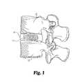
- FIG. 1is a side elevational view, in full section, of a roll of natural tissue material being used as a disc replacement device, according to one aspect of the present invention.
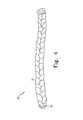
- FIG. 2shows a “roll” embodiment of the natural tissue implants of the present invention, according to one preferred embodiment.
- FIG. 3shows an alternative embodiment of the roll of natural tissue shown in FIG. 2 .
- FIG. 4shows a “braided” embodiment of the natural tissue implants of the present invention, according to one preferred embodiment.
- FIG. 5Ashows a perspective view of the braided implant of FIG. 4 , with a drawstring through the implant for causing the implant to bunch or fold.
- FIG. 5Bshows another perspective view of the braided implant of FIG. 5A .
- FIG. 6shows the braided implant of FIG. 5 with the braid being folded after pulling the drawstring.
- FIG. 7shows the braided implant of FIG. 5 being implanted into a disc nucleus, with the implant in its first, straightened configuration.
- FIG. 8shows the braided implant of FIG. 5 being implanted into a disc nucleus, with the implant beginning to fold.
- FIG. 9shows the braided implant of FIG. 5 being implanted into a disc nucleus, with the implant continuing to fold.
- FIG. 10shows the braided implant of FIG. 5 after it has been implanted into a disc nucleus, with the implant in its second, folded, or pleated, configuration.
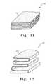
- FIG. 11shows a natural tissue implant made from a stack of separate sheets of tissue.
- FIG. 12shows a natural tissue implant made from a folded sheet of tissue.
- FIG. 13shows a natural tissue implant made from a multiplicity of sub-units, with each sub-unit comprising a roll of tissue.
- FIG. 14shows the natural tissue implant of FIG. 13 , with the sub-units being folded over to form a wider implant.
- FIG. 15shows the natural tissue implant of FIG. 13 , after the sub-units have been folded over to form a wider implant.
- FIG. 16shows an alternative embodiment of a natural tissue implant made from a multiplicity of sub-units, with each sub-unit comprising a roll of tissue.
- FIG. 17shows the natural tissue implant of FIG. 16 , with the sub-units being folded over to form a wider implant.
- FIG. 18shows a natural tissue implant made from a multiplicity of sub-units in an “accordion” embodiment.
- FIG. 19shows the natural tissue implant of FIG. 18 , with the sub-units being folded to form a narrower implant.
- FIG. 20shows the natural tissue implant of FIG. 18 , with the sub-units folded to form a wider implant.
- FIG. 21shows a stack of natural tissue implants connected together with a suture.
- FIG. 22shows a retaining clip for securing a stack of implants.
- FIG. 23shows an alternative retaining clip for securing a stack of implants.
- FIG. 24shows a “pillow” embodiment of the natural tissue implant of the present invention.
- FIG. 25shows a section view of the “pillow” embodiment of FIG. 24 .
- FIG. 26shows an alternative “pillow” embodiment of the natural tissue implant of the present invention.
- FIG. 27shows a “pouch” embodiment of the present invention, with the pouch in the disc nucleus space and no tissue pieces in the pouch.
- FIG. 28shows the “pouch” embodiment of FIG. 27 , with tissue pieces being inserted into the pouch.
- FIG. 29shows the “pouch” embodiment of FIG. 27 , with more tissue pieces being inserted into the pouch.
- FIG. 30shows the “pouch” embodiment of FIG. 27 , after the tissue pieces have been inserted into the pouch and the pouch has been closed.
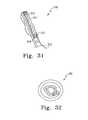
- FIG. 31shows and alternative embodiment of the present invention, with a braided tissue implant being formed into a knot.
- FIG. 32shows the implant of FIG. 31 , after the braided tissue implant has been formed into a knot.
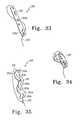
- FIG. 33shows and alternative embodiment of the present invention, with a braided tissue implant being formed into a knot.
- FIG. 34shows the implant of FIG. 33 , after the braided tissue implant has been formed into a knot.
- FIG. 35shows a further embodiment of the natural tissue implants of the present invention, showing the use of a drawstring to bunch or fold an unbraided implant.
- FIG. 36is a photo of a roll of natural material being inserted for use as a disc replacement device, according to one aspect of the present invention.
- FIG. 37is a histological section of a roll of natural material being used as a disc replacement device, according to one aspect of the present invention.
- one aspect of the present inventionprovides devices and methods for augmenting, repairing, or replacing all or part of an intervertebral disc using natural biological tissue.
- Natural, biological tissueis preferably used in the inventive devices and methods.
- the tissue used in the present inventionmay be any natural biological tissue that is implantable in a human patient.
- the tissuewill be selected from a tissue source appropriate to provide the strength and structural integrity necessary to function as described herein.
- the tissuemay be a flat tissue such as human, bovine, or porcine pericardium or small intestine submucosa (SIS).
- Ligamentssuch as anterior or posterior cruciate ligaments, fasciae such as fascia lata, tendons such as patella, hamstring, quadriceps and Achilles tendons, and other connective tissue may also be used.
- the tissuemay be autogenic, allogenic, or xenogenic with respect to the patient in which the tissue will be used. While human patients are particularly contemplated, animal patients may also be treated with the inventive devices and methods.
- the tissue of the present inventionis may be provided as one or more sheets, strips, plugs, fibers, or in any other form suitable for implantation.
- the tissuewhen used to encapsulate a core to provide a disc or nucleus replacement device, the tissue has sufficient size and strength to at least partially constrain the core material.
- the tissueWhen used without an additional core material (for example, as a nucleus or disc replacement device itself), the tissue has sufficient size and strength to provide the desired structure.
- the size and strength of the implantvaries, of course, according to whether it is used alone or in combination with other pieces, and with whether the tissue is used flat or is braided, folded, etc.
- the natural tissuemay vary in size and thickness depending on the configuration of the implant and how the material will be used.
- the sheetwhen a sheet of tissue is to be rolled, folded or layered into a solid plug, the sheet will preferably be about 5-50 mm wide and about 20-80 mm long.
- the sheets or stripswhen sheets or strips of tissue are to be used to encapsulate a core material, the sheets or strips will preferably be about 10-40 mm wide and about 30-60 mm long.
- strips that are about 30-200 mm long (more preferably about 50-100 mm long) by about 1-10 mm wide (more preferably about 2-5 mm wide)are often used. While thicker or thinner embodiments are occasionally preferred, in general the sheets will preferably be about 1-3 mm thick.
- the tissueis braided to form a braided implant.
- the braided implantmay have superior strength when compared with unbraided implants, yet it may also provide sufficient flexibility to allow it to be bunched or folded to fill a disc space.
- the braided implantis made from multiple (e.g., three or more) strips of flat tissue, while in other embodiments round strands are used.
- the braided implantsmay be braided to form extraordinarily strong, yet flexible, structures, such as when many thin/fine strands are braided together to form a natural tissue “rope.” The rope may then be used as one long, thin section, or it may be folded or coiled to form a more bulky structure.
- the braided structuresmay be used in a variety of medical applications, including to augment, repair or replace all or part of an intervertebral disc, or to augment, repair or replace all or part of nearly any other anatomical structure, including ligaments, tendons, etc.
- the natural tissue materialmay have elastic characteristics, or it may be inelastic.
- the tissuemay also be deformable or non-deformable.
- elasticis understood to mean that the material returns to its original shape, or nearly so, when stretched or compressed under implant conditions.
- the elastic nature of the tissueprovides the opportunity to better match the elastic modulus of surrounding host tissue, for example, thereby allowing the disc nucleus or disc replacement to flex more freely, to better contour to surrounding host tissues, and to reduce the potential for implant subsidence into the endplates.
- deformableis understood to mean that the material may decrease slightly in dimension in response to surrounding forces once implanted into the defect site, such that it accommodates the site of injury's absent or deformed tissue within the body to enhance or restore function.
- the natural tissue implants described hereinare made of soft, flexible tissue, although hard, inflexible tissue may be used in some alternative embodiments.
- the biological tissuemay have smooth surfaces to reduce the potential for wear. Moreover, if wear particles are generated from the tissue material, the body can degrade and metabolize those particles better than it could degrade and metabolize synthetic materials.
- natural, biological tissueprovides the potential for “scarring” and allows host tissues to grow into the tissue, thus reducing the likelihood of expulsion of the implant as has been reported with synthetic jacket designs.
- natural biologic materialmay be sutured before or after being positioned into place.
- the tissuemay comprise natural, biological tissue, or it may comprise a matrix derived from biological tissue.
- the biological tissuecan be either degradable or non-degradable in nature.
- the tissuemay be used to encapsulate an elastomeric or hydrogel nucleus or intervertebral disc replacement device, or it may be used as a nucleus or intervertebral disc replacement device itself, without an additional central core.
- the natural tissue materialmay be used to augment, repair, or replace all or part of an intervertebral disc, including a disc nucleus and/or a disc annulus, or it may be used to augment, repair, or replace all or part of some other orthopedic structure such as an anterior cruciate ligament, flexor tendon, rotator cuff, meniscus or other similar tissue within the body that may have elastic or deformable characteristics.
- an intervertebral discincluding a disc nucleus and/or a disc annulus
- some other orthopedic structuresuch as an anterior cruciate ligament, flexor tendon, rotator cuff, meniscus or other similar tissue within the body that may have elastic or deformable characteristics.
- the natural materialis to supplement or replace an intervertebral disc nucleus.
- the natural tissuemay be provided as a rolled, layered, braided, or folded “plug” of material (or series of such constructs) that are inserted into the disc annulus to supplement or replace the natural disc nuclear material.
- No synthetic core materialis required in this aspect of the invention, but such a core could be incorporated if desired.
- the natural tissue implantmay be used after a complete or partial discectomy with minimal, partial, or complete removal of the original disc nucleus.
- the natural materialmay be used to supplement or replace all, or substantially all, of the original disc—including the disc annulus. Additionally this embodiment may include a retaining mechanism for holding the replacement disc in place, and could incorporate a synthetic core material if desired.
- the natural biological tissuemay be used to encapsulate an elastomeric or hydrogel core to provide a nucleus replacement device.
- Hydrogel nucleus devicesmay require some form of encapsulation around the central hydrogel material to constrain them and make them compression resistant to loads across the disc.
- the natural materialmay be used as a patch or plug to close a hole in the disc annulus, or otherwise to repair a disc nucleus or annulus.
- the natural tissue implants of the present inventionmay be used in their hydrated form, or they may be fully or partially dehydrated prior to implantation.
- Dehydrated implantmay be smaller than hydrated implants, and thus are preferred for some applications since they can be implanted through a smaller incision.
- a natural tissue implantis implanted into a disc space in a dehydrated condition, and is rehydrated to form a substantially larger body.
- the rehydrated tissueincreases in size enough to increase the disc height, and preferably enough to distract the vertebrae.
- the natural tissue implantsare used in combination with synthetic materials.
- hydrogelsmay be used as indicated above, or other polymers, elastomers, plastics, fabrics, beads, fibers, etc., may additionally or alternatively be used.
- synthetic materialsthat may be used to form components of natural tissue implants include polyvinyl alcohol, polyacrylamide-polyacrylic acid, polyurethane, silicone, silicone polyurethane, polyethylene, propylene, polyester, polyterephthalate, polyaryletherketone, etc.
- growth factorssuch as TGF-Beta, BMP, etc. can be incorporated into the tissues to facilitate disc repair, regeneration or incorporation.
- FIG. 1shows one embodiment of an intervertebral disc nucleus pulposus replacement device.
- the natural tissue implantincludes at least one piece of natural tissue 11 positioned between adjacent vertebra 12 and 13 and retained within annulus fibrosis 14 . While the Figure illustrates the use of the implant to replace the entire disc nucleus, it is to be appreciated that the implant may be sized to replace or supplement all or only a portion of the actual nucleus pulposus, and to aid in maintaining a predetermined height of the disc space. Further, the illustrated implant has elastic characteristics to absorb forces transmitted through vertebrae 12 and 13 .
- natural tissue 11is configured as a roll of one or more sheets of tissue. Other suitable configurations may be utilized, however, as is discussed below.
- implant 20 in a state prior to implantationmay have a greater height “H” and a smaller width “W” than the implanted device shown in FIG. 1 due to the lack of compressive forces on the implant.
- implant 20may include one or more securement mechanisms 25 , such as sutures, staples and other fasteners as will be discussed below, for helping to maintain the given configuration of the implant.
- Securement mechanism 25may extend along all or only a portion of the length of implant 10 . Further, securement mechanism 25 may extend through all or only a portion of the thickness of implant 20 .
- FIG. 3shows an alternative embodiment of configurations of a natural tissue implant.
- implant 30comprises a long, thin roll of tissue that can be folded, coiled, etc. to fill a disc nucleus space.
- FIGS. 4-10show a “braided” embodiment of the natural tissue implant of the present invention.
- implant 50comprises long, braided strips 51 of natural tissue.
- a drawstring 52is provided to assist in folding the implant into a more compact configuration after implantation.
- Drawstring 52is secured to implant 50 near one end 54 , passes through the implant at a multiplicity of sites throughout the length of the implant, and exits the other end 55 of the implant to provide a portion 53 for pulling the drawstring and bunching the implant.
- drawstring 52passes from one side of the implant to the other each time it passes through the implant. In this manner, the drawstring can be used to “bunch” or “fold” the implant up into a multiplicity of folded portions.
- Implant 50has a first, straightened configuration as shown in FIG. 4 .
- implant 50has a length L that is at least five times its width W. More preferably, length L is at least ten times width W.
- FIGS. 5A and 5Bshow the braided tissue implant of FIG. 4 , with a drawstring passing through the implant.
- Drawstring 52is secured near one end 54 of the braided tissue implant, and passes through the implant at a multiplicity of sites from the first, secured end, to the second, free end 55 .
- drawstring 52preferably passes from one side of the braided tissue implant to the other side of the braided tissue implant when it passes through the implant at the multiplicity of sites.
- Drawstring 52has an effective length “E” defined by the length of the drawstring from the point where it first enters the braid near one end, to the point where it last exits the braid near its other end.
- Drawstring 52also includes an end portion 53 beyond its effective length, with end portion 53 being used to pull the drawstring and fold the braided implant. It is understood that for a specific length of drawstring, the relative portions of the drawstring that comprise the “effective length” and the “end portion” change as the drawstring is pulled to fold the implant.
- drawstring 52is pulled while holding free end 55 in a generally fixed location. This causes secured end 54 of the implant to be drawn toward free end 55 , thereby bunching or folding the implant.
- the number of foldsdepends on the number of sites in the implant through which drawstring 52 passes. For example, if drawstring 52 passes through implant 51 three times, three folds will be formed.
- the number of folds desired for a particular applicationvaries, with at least one fold being preferred for some embodiments, at least two folds being preferred for other embodiments, and at least three folds being preferred for yet other embodiments.
- an implanthaving improved structural properties and/or a preferred volume.
- desired structural properties and/or a desired final configurationmay be obtained. For example, wider folds may be obtained by passing the drawstring through the implant at sites that are farther apart, generally providing a wider, folded implant.
- a curved or arc-shaped implantmay be obtained by adjusting the location and size of the folds.
- FIG. 6shows implant 50 after the drawstring has been pulled and the implant has bunched or folded.
- the length L of the implantis reduced by the bunching/folding, while the width W is increased.
- implant 50has assumed its second, folded configuration in which length L is less than five times width W.
- length Lis no more than three times width W.
- the effective length of the drawstringhas been correspondingly reduced, since less of the drawstring now lies adjacent to or within the braided tissue.
- the length-to-width ratio of the implant in its folded configurationis no more than one-half the length-to-width ratio of the implant in its straightened configuration. Accordingly, if the implant in its straightened configuration has a length-to-width ratio of 10:1, the length-to-width ratio of the folded implant is preferably no more than 5:1, although it is more preferably no more than about 3:1 as indicated above.
- FIG. 7shows a braided implant being implanted into a disc nucleus space 78 defined by disc annulus 77 .
- Implant 71has a first end 74 , a second end 75 , and a length L that is at least five times its width W.
- a drawstring 72is secured near first end 74 , and passes through the implant and exits near second end 75 .
- drawstring 72passes into and out of implant 71 at a multiplicity of sites throughout the length of the implant, and includes a free end portion 73 .
- a cannula 76may be used to assist in inserting the implant through the disc annulus.
- FIG. 8shows the implant of FIG. 7 after more of the implant has been implanted, and after drawstring 72 has been pulled somewhat to begin bunching/folding the implant in the disc nucleus space.
- FIG. 9shows the implant after even more of the implant has been implanted, and after the bunching/folding has proceeded.
- FIG. 10shows the implant of FIG. 7 after the implant has been implanted all of the way into disc nucleus space 75 .
- Drawstring 72has been pulled sufficiently to bunch or fold the implant completely so as to provide a pleated configuration, so that the width of the implant is now nearly as great as the length.
- an implant having a length-to-width ratio of about 10:1 in its straightened configuration of FIG. 7has been folded to an implant of pleated configuration having a length-to-width ratio of about 2.5:1 in its straightened configuration of FIG. 10 .
- natural tissue implant 110may comprise a stack of separate sheets 111 .
- implant 120may comprise a strip 121 of tissue that has been folded over to form a multiplicity of layers in the implant.
- stitches or sutures or another securement mechanism connecting one or more of the individual “layers” of materialmay be used to enhance construct integrity.
- the implantmay be cut or otherwise fashioned to form an implant having a more natural shape, such as the shape of a natural disc nucleus.
- FIGS. 13-15show another embodiment of a natural tissue implant 130 .
- implant 130comprise a plurality of sub-units 131 , 132 , 133 , and 134 , joined together by any appropriate securement means, such as sutures 135 a - c .
- subunits 131 - 134are relatively small (e.g., 5-9 mm in diameter and 8-20 mm in length) so that they can be inserted through a relatively small aperture in the disc annulus following a discectomy procedure, when stacked as shown in FIG. 13 .
- sub-units 131 - 134may be folded as shown in FIGS. 14 and 15 to form, in a second state, a substantially wider and shorter implant.
- Such an implantcan, for example, better bear and distribute intervertebral forces.
- sub-units 131 - 134need not be cynlindrical, and can be another shape, such as an ovid shape, where the height of the implant is greater than the width (e.g. 5-8 mm width, 8-10 mm height, and 8-20 mm length). This may facilitate creation of a smaller incision in the annulus to allow implantation. Moreover, a lesser or greater number of sub-units may be joined.
- FIGS. 16-17show an embodiment similar to that shown in FIGS. 13-15 , but with implant 160 comprising a securement mechanism having sheets or strips of natural tissue 165 a - c being attached to all or a portion of the sides of the sub-units 161 - 164 to act as hinges and facilitate folding.
- FIGS. 18-20show a further embodiment of a disc nucleus replacement device, referred to hereinafter as an “accordion” embodiment.
- implant 180comprises a multiplicity of sub-units 181 - 188 , held together by securement means such as sutures 189 a - j .
- securement meanssuch as sutures 189 a - j .
- implant 180may assume a relatively long, narrow configuration in which the sub-units are folded in two columns of four subunits positioned end-to-end, or it may assume a relatively short, wide configuration in which the sub-units are folded in two columns of four sub-units positioned side-to-side.
- the long, narrow configurationmay be used when the implant is contained in a syringe 190 or other implantation instrument, and the short, wide configuration may be used when the implant has been implanted in a disc space.
- a multiplicity of sub-unitsmay be joined together with a long suture or cable 216 , as shown in FIG. 21 .
- Retaining clips 220 and/or 230may be used as shown in FIGS. 22 and 23 .
- natural tissueis used as a constraining jacket to encapsulate a synthetic elastomeric or hydrogel core.
- the natural tissueallows the core material to flex and deform under disc loading conditions, while still providing the necessary structural support.
- FIGS. 24 and 25show one embodiment of the natural tissue of the present invention being used to encapsulate an elastomeric core.
- Natural tissue 241constrains elastomeric core 245 to provide a disc nucleus or disc replacement device.
- Sutures 243may be used to hold two pieces of natural tissue together to form the implant.
- FIG. 26shows another embodiment of the natural tissue of the present invention being used to encapsulate an elastomeric core.
- Natural tissue 261constrains elastomeric core 262 to provide a disc nucleus or disc replacement device.
- Securement mechanismssuch as sutures 263 are used to close the constraining jacket.
- the disc nucleus or disc replacement devicemay be used to replace part or all of a damaged disc nucleus. Further, when stabilized to prevent expulsion from the disc space, the disc replacement device may also replace part or all of the disc annulus.
- the use of a natural material to form the deviceprovides the advantages identified above.
- the natural tissueis formed into a sack that is used to hold pieces of tissue.
- the sack and tissue contained thereinmay replace a natural disc nucleus.
- synthetic or natural tissueis fabricated into the shape of an empty pouch.
- This empty pouchcan be compressed down to a diameter small enough to be inserted through a small hole in the annulus of an intervertebral disc. Once inside the disc it can be filled by inserting pieces, strips, or a long strand of natural or synthetic tissue into the pouch. This inflation is done through a small aperture in the pouch. Once filled to capacity the aperture in the pouch may be sutured closed with a purse string type suture or a preinserted suture in a flap over the aperture.
- the pouch and contentsare manufactured out of material other than natural tissue, such as synthetic polymers (i.e. polyethylene, macron, etc.).
- the pouch embodimentavoids the insertion of large implants through a large hole in the annulus. Because a large hole is not required to insert the prosthesis, the implants are less prone to expulsion from the disc.
- the pouchcan also be inserted via a minimally invasive procedure.
- FIGS. 27-30show a disc annulus 271 with a natural tissue pouch 272 inserted therein.
- a loading cannula having an insertion tool 273may be used to insert pouch 272 into the disc space.
- pouch 272is preferably positioned over the loading cannula so that the cannula extends into the pouch.
- the insertion toolis used to push the pouch and cannula into position inside a disc annulus. The insertion tool is removed, leaving the cannula in place.
- the insertion tool 273 of the loading cannulais withdrawn, and pieces or strips of tissue 276 or synthetic material (not shown) are used to fill pouch 272 by passing them through cannula 274 .
- the cannulais withdrawn part way as the pouch is filled to facilitate loading of the entire pouch.
- the cannulais withdrawn as the pouch is filed to a desired density. After the pouch has been filled, it is secured at the previously open end by a securement means such as sutures 300 .
- a natural tissue implant having a hole at one endis used with a drawstring to form a piece of folded tissue.
- device 310may comprise a natural tissue implant 311 having a first end 315 , a second end 316 , and a first hole 313 .
- the implantmay be folded by pulling drawstring 312 through hole 313 to form a folded implant as shown in FIG. 32 .
- implant 310may be provided with multiple holes 313 , and may be used as a ligament replacement by placing fixation screws through the holes.
- FIGS. 33 and 34A further embodiment is shown in FIGS. 33 and 34 , with device 330 comprising a natural tissue implant 331 having a multiplicity of holes 333 a - b .
- a drawstring 332may be passed through holes 333 a and 333 b to fold the implant.
- one end of the implantmay be pulled through the to form a knotted implant, ad shown in FIG. 34 .
- FIG. 35shows an embodiment similar to the braided embodiment of FIGS. 6-10 , but with the implant comprising a strip or sheet of natural tissue that is not necessarily braided.
- Drawstring 352is secured to one end 351 b of tissue strip 351 , and is used to fold the implant by pulling the free end of the drawstring while holding the free end 351 a of implant 351 stable so that the remainder of the implant folds up.
- the drawstringmay be passed through a loop 357 at one end of the drawstring to facilitate tying the drawstring in a knot to hold the folded implant in its second, folded configuration.
- a body comprising natural tissuehas a first, straightened configuration that is more narrow, and a second, folded configuration that is wider (when compared to the first, straightened configuration). This facilitates implanting the body through a small incision or hole, and folding the implant to a wider configuration after the body has been implanted.
- the drawstring used to fold the natural tissue implants of the present inventionmay be made of natural tissue, or it may be made of another material.
- synthetic materialsmay be used to form the drawstring. Such synthetic materials may be resorbable, or they may be non-resorbable.
- the drawstringmay be made of wire or some other non-resorbable material.
- multiple drawstringsmay be used in a single device in some embodiments.
- a disc nucleus replacement device made of a roll of natural tissuewas implanted in a sheep.
- a photo of the natural tissue device being implanted in the animalis shown in FIG. 36 .
- a histological section showing the implant after six months in the animalis shown in FIG. 37 .
- the disc nucleus replacement devicefunctioned well for many months. The device remained in position within the nucleus space without expulsion, and maintained proper disc distraction and annulus tension without subsidence into the endplates. No evidence of wear particles, and no cellular inflammation were observed.
Landscapes
- Health & Medical Sciences (AREA)
- Life Sciences & Earth Sciences (AREA)
- Engineering & Computer Science (AREA)
- Biomedical Technology (AREA)
- Veterinary Medicine (AREA)
- Public Health (AREA)
- General Health & Medical Sciences (AREA)
- Animal Behavior & Ethology (AREA)
- Chemical & Material Sciences (AREA)
- Transplantation (AREA)
- Epidemiology (AREA)
- Oral & Maxillofacial Surgery (AREA)
- Vascular Medicine (AREA)
- Heart & Thoracic Surgery (AREA)
- Medicinal Chemistry (AREA)
- Orthopedic Medicine & Surgery (AREA)
- Dermatology (AREA)
- Chemical Kinetics & Catalysis (AREA)
- Botany (AREA)
- Molecular Biology (AREA)
- Surgery (AREA)
- Neurology (AREA)
- Cardiology (AREA)
- Urology & Nephrology (AREA)
- Zoology (AREA)
- Physical Education & Sports Medicine (AREA)
- Physics & Mathematics (AREA)
- Optics & Photonics (AREA)
- Prostheses (AREA)
- Materials For Medical Uses (AREA)
Abstract
Description
Claims (9)
Priority Applications (1)
| Application Number | Priority Date | Filing Date | Title |
|---|---|---|---|
| US10/666,900US7887593B2 (en) | 2002-09-18 | 2003-09-18 | Method of implanting natural tissue within the vertebral disc nucleus space using a drawstring |
Applications Claiming Priority (5)
| Application Number | Priority Date | Filing Date | Title |
|---|---|---|---|
| US41154702P | 2002-09-18 | 2002-09-18 | |
| US10/245,955US20040054414A1 (en) | 2002-09-18 | 2002-09-18 | Collagen-based materials and methods for augmenting intervertebral discs |
| US42661302P | 2002-11-15 | 2002-11-15 | |
| US10/645,006US7309359B2 (en) | 2003-08-21 | 2003-08-21 | Allogenic/xenogenic implants and methods for augmenting or repairing intervertebral discs |
| US10/666,900US7887593B2 (en) | 2002-09-18 | 2003-09-18 | Method of implanting natural tissue within the vertebral disc nucleus space using a drawstring |
Related Parent Applications (2)
| Application Number | Title | Priority Date | Filing Date |
|---|---|---|---|
| US10/245,955Continuation-In-PartUS20040054414A1 (en) | 2002-09-18 | 2002-09-18 | Collagen-based materials and methods for augmenting intervertebral discs |
| US10/645,006Continuation-In-PartUS7309359B2 (en) | 2002-09-18 | 2003-08-21 | Allogenic/xenogenic implants and methods for augmenting or repairing intervertebral discs |
Publications (2)
| Publication Number | Publication Date |
|---|---|
| US20040059418A1 US20040059418A1 (en) | 2004-03-25 |
| US7887593B2true US7887593B2 (en) | 2011-02-15 |
Family
ID=32030688
Family Applications (1)
| Application Number | Title | Priority Date | Filing Date |
|---|---|---|---|
| US10/666,900Expired - Fee RelatedUS7887593B2 (en) | 2002-09-18 | 2003-09-18 | Method of implanting natural tissue within the vertebral disc nucleus space using a drawstring |
Country Status (8)
| Country | Link |
|---|---|
| US (1) | US7887593B2 (en) |
| EP (1) | EP1549261A2 (en) |
| JP (1) | JP2005538810A (en) |
| KR (1) | KR101095771B1 (en) |
| CN (1) | CN1697634A (en) |
| AU (1) | AU2003276926B2 (en) |
| CA (1) | CA2499116A1 (en) |
| WO (1) | WO2004026190A2 (en) |
Cited By (13)
| Publication number | Priority date | Publication date | Assignee | Title |
|---|---|---|---|---|
| US20080306595A1 (en)* | 2004-03-26 | 2008-12-11 | Pearsalls Limited | Porous Implant For Spinal Disc Nucleus Replacement |
| US20090105732A1 (en)* | 2005-06-15 | 2009-04-23 | Matthew Yurek | Mechanical apparatus and method for delivering materials into the inter-vertebral body space for nucleus replacement |
| US20090125110A1 (en)* | 2002-11-05 | 2009-05-14 | Kuslich Stephen D | Semi-biological intervertebral disc replacement system |
| US20090247976A1 (en)* | 2008-03-12 | 2009-10-01 | Bernard Chaffringeon | Web for retention of internal bodily secretions |
| US20100114107A1 (en)* | 2000-08-30 | 2010-05-06 | Warsaw Orthopedic, Inc. | Intervertebral Disc Nucleus Implants and Methods |
| US20100168857A1 (en)* | 2008-05-30 | 2010-07-01 | Edwin Burton Hatch | Flexibly compliant ceramic prosthetic meniscus for the replacement of damaged cartilage in orthopedic surgical repair or reconstruction of hip, knee, ankle, shoulder, elbow. wrist and other anatomical joints |
| US20100217392A1 (en)* | 2009-02-23 | 2010-08-26 | Bartee Barry K | Reinforced ptfe medical barriers |
| US8282681B2 (en) | 2007-08-13 | 2012-10-09 | Nuvasive, Inc. | Bioresorbable spinal implant and related methods |
| US8377135B1 (en) | 2008-03-31 | 2013-02-19 | Nuvasive, Inc. | Textile-based surgical implant and related methods |
| US9198765B1 (en) | 2011-10-31 | 2015-12-01 | Nuvasive, Inc. | Expandable spinal fusion implants and related methods |
| US9445918B1 (en) | 2012-10-22 | 2016-09-20 | Nuvasive, Inc. | Expandable spinal fusion implants and related instruments and methods |
| US9700425B1 (en) | 2011-03-20 | 2017-07-11 | Nuvasive, Inc. | Vertebral body replacement and insertion methods |
| US12059335B2 (en) | 2014-06-24 | 2024-08-13 | Osteogenics Biomedical, Inc. | Device for guided bone and tissue regeneration |
Families Citing this family (114)
| Publication number | Priority date | Publication date | Assignee | Title |
|---|---|---|---|---|
| US6805695B2 (en) | 2000-04-04 | 2004-10-19 | Spinalabs, Llc | Devices and methods for annular repair of intervertebral discs |
| US6723335B1 (en) | 2000-04-07 | 2004-04-20 | Jeffrey William Moehlenbruck | Methods and compositions for treating intervertebral disc degeneration |
| US7204851B2 (en) | 2000-08-30 | 2007-04-17 | Sdgi Holdings, Inc. | Method and apparatus for delivering an intervertebral disc implant |
| US7503936B2 (en)* | 2000-08-30 | 2009-03-17 | Warsaw Orthopedic, Inc. | Methods for forming and retaining intervertebral disc implants |
| CA2549320A1 (en)* | 2000-08-30 | 2002-03-07 | Sdgi Holdings, Inc. | Intervertebral disc nucleus implants and methods |
| US8308797B2 (en) | 2002-01-04 | 2012-11-13 | Colibri Heart Valve, LLC | Percutaneously implantable replacement heart valve device and method of making same |
| US20040054414A1 (en) | 2002-09-18 | 2004-03-18 | Trieu Hai H. | Collagen-based materials and methods for augmenting intervertebral discs |
| US7744651B2 (en) | 2002-09-18 | 2010-06-29 | Warsaw Orthopedic, Inc | Compositions and methods for treating intervertebral discs with collagen-based materials |
| JP2006515765A (en) | 2002-11-15 | 2006-06-08 | エスディージーアイ・ホールディングス・インコーポレーテッド | Collagen-based materials and methods for treating synovial joints |
| AU2004208821B2 (en)* | 2003-01-31 | 2009-01-15 | Zimmer Orthobiologics Inc. | Hydrogel compositions comprising nucleus pulposus tissue |
| AU2004212942A1 (en) | 2003-02-14 | 2004-09-02 | Depuy Spine, Inc. | In-situ formed intervertebral fusion device |
| EP1610740A4 (en)* | 2003-04-04 | 2009-04-08 | Theken Disc Llc | Artificial disc prosthesis |
| US20050071012A1 (en)* | 2003-09-30 | 2005-03-31 | Hassan Serhan | Methods and devices to replace spinal disc nucleus pulposus |
| US7056337B2 (en) | 2003-10-21 | 2006-06-06 | Cook Incorporated | Natural tissue stent |
| AU2005245017A1 (en)* | 2004-05-21 | 2005-12-01 | Synthes Gmbh | Replacement of nucleus pulposus using a hydrogel |
| EP1614402A1 (en)* | 2004-07-09 | 2006-01-11 | Paul M. Tsou | Patch material for inervertebral disc annulus defect repair |
| US20060047296A1 (en)* | 2004-08-31 | 2006-03-02 | Sdg Holdings, Inc. | Annulus replacement system and technique |
| US20060058881A1 (en)* | 2004-09-16 | 2006-03-16 | Trieu Hai H | Intervertebral disc nucleus implants and methods |
| WO2006034436A2 (en) | 2004-09-21 | 2006-03-30 | Stout Medical Group, L.P. | Expandable support device and method of use |
| US20060089719A1 (en)* | 2004-10-21 | 2006-04-27 | Trieu Hai H | In situ formation of intervertebral disc implants |
| US20060095134A1 (en)* | 2004-10-28 | 2006-05-04 | Sdgi Holdings, Inc. | Materials, devices and methods for implantation of transformable implants |
| US8702718B2 (en) | 2005-04-29 | 2014-04-22 | Jmea Corporation | Implantation system for tissue repair |
| US7632313B2 (en) | 2005-04-29 | 2009-12-15 | Jmea Corporation | Disc repair system |
| US20060247781A1 (en)* | 2005-04-29 | 2006-11-02 | Sdgi Holdings, Inc. | Implant |
| US7608108B2 (en)* | 2005-04-29 | 2009-10-27 | Jmea Corporation | Tissue repair system |
| US20060253202A1 (en)* | 2005-05-05 | 2006-11-09 | Lipov Eugene G | Vertebral disc implant in fiber form |
| US8080061B2 (en)* | 2005-06-20 | 2011-12-20 | Synthes Usa, Llc | Apparatus and methods for treating bone |
| US8951285B2 (en)* | 2005-07-05 | 2015-02-10 | Mitralign, Inc. | Tissue anchor, anchoring system and methods of using the same |
| US20070010889A1 (en)* | 2005-07-06 | 2007-01-11 | Sdgi Holdings, Inc. | Foldable nucleus replacement device |
| EP1903949A2 (en) | 2005-07-14 | 2008-04-02 | Stout Medical Group, L.P. | Expandable support device and method of use |
| US7618457B2 (en) | 2005-08-10 | 2009-11-17 | Zimmer Spine, Inc. | Devices and methods for disc nucleus replacement |
| US8366773B2 (en) | 2005-08-16 | 2013-02-05 | Benvenue Medical, Inc. | Apparatus and method for treating bone |
| WO2008103781A2 (en)* | 2007-02-21 | 2008-08-28 | Benvenue Medical, Inc. | Devices for treating the spine |
| AU2006279558B2 (en) | 2005-08-16 | 2012-05-17 | Izi Medical Products, Llc | Spinal tissue distraction devices |
| FR2893248A1 (en)* | 2005-11-16 | 2007-05-18 | Vincent Pointillart | INTERVERTEBRAL DISC PROSTHESIS |
| WO2007076374A2 (en)* | 2005-12-19 | 2007-07-05 | Stout Medical Group, L.P. | Expandable support device and method of using the same |
| US20070150064A1 (en)* | 2005-12-22 | 2007-06-28 | Depuy Spine, Inc. | Methods and devices for intervertebral augmentation |
| US20070150063A1 (en)* | 2005-12-22 | 2007-06-28 | Depuy Spine, Inc. | Devices for intervertebral augmentation and methods of controlling their delivery |
| US20070150059A1 (en)* | 2005-12-22 | 2007-06-28 | Depuy Spine, Inc. | Methods and devices for intervertebral augmentation using injectable formulations and enclosures |
| US20070162132A1 (en) | 2005-12-23 | 2007-07-12 | Dominique Messerli | Flexible elongated chain implant and method of supporting body tissue with same |
| EP2023864B1 (en) | 2006-05-01 | 2019-07-10 | Stout Medical Group, L.P. | Expandable support device |
| US8821549B2 (en)* | 2006-06-13 | 2014-09-02 | Anova Corporation | Methods and apparatus for anulus repair |
| US8118779B2 (en) | 2006-06-30 | 2012-02-21 | Warsaw Orthopedic, Inc. | Collagen delivery device |
| US8399619B2 (en) | 2006-06-30 | 2013-03-19 | Warsaw Orthopedic, Inc. | Injectable collagen material |
| US8066750B2 (en) | 2006-10-06 | 2011-11-29 | Warsaw Orthopedic, Inc | Port structures for non-rigid bone plates |
| US8057458B2 (en)* | 2006-10-30 | 2011-11-15 | Warsaw Orthopedic, Inc. | Method for treating facet pain |
| WO2008070863A2 (en) | 2006-12-07 | 2008-06-12 | Interventional Spine, Inc. | Intervertebral implant |
| EP2124778B1 (en) | 2007-02-21 | 2019-09-25 | Benvenue Medical, Inc. | Devices for treating the spine |
| JP5328763B2 (en)* | 2007-04-10 | 2013-10-30 | アーティキュリンクス, インコーポレイテッド | Percutaneous delivery and retrieval system for a shape change orthopedic joint device |
| US20100286778A1 (en)* | 2007-04-18 | 2010-11-11 | Lukas Eisermann | Textile-Based Spinal Implant and Related Methods |
| US20080269897A1 (en)* | 2007-04-26 | 2008-10-30 | Abhijeet Joshi | Implantable device and methods for repairing articulating joints for using the same |
| US20080268056A1 (en)* | 2007-04-26 | 2008-10-30 | Abhijeet Joshi | Injectable copolymer hydrogel useful for repairing vertebral compression fractures |
| US8900307B2 (en) | 2007-06-26 | 2014-12-02 | DePuy Synthes Products, LLC | Highly lordosed fusion cage |
| EP2237748B1 (en) | 2008-01-17 | 2012-09-05 | Synthes GmbH | An expandable intervertebral implant |
| FR2929502B1 (en) | 2008-04-04 | 2011-04-08 | Clariance | NUCLEIC IMPLANT. |
| US8936641B2 (en) | 2008-04-05 | 2015-01-20 | DePuy Synthes Products, LLC | Expandable intervertebral implant |
| US20090297603A1 (en)* | 2008-05-29 | 2009-12-03 | Abhijeet Joshi | Interspinous dynamic stabilization system with anisotropic hydrogels |
| EP2339985A4 (en)* | 2008-09-12 | 2013-07-03 | Articulinx Inc | Tether-based orthopedic joint device delivery methods |
| WO2010056895A1 (en) | 2008-11-12 | 2010-05-20 | Stout Medical Group, L.P. | Fixation device and method |
| US20100211176A1 (en) | 2008-11-12 | 2010-08-19 | Stout Medical Group, L.P. | Fixation device and method |
| US8535327B2 (en) | 2009-03-17 | 2013-09-17 | Benvenue Medical, Inc. | Delivery apparatus for use with implantable medical devices |
| US9526620B2 (en) | 2009-03-30 | 2016-12-27 | DePuy Synthes Products, Inc. | Zero profile spinal fusion cage |
| JP2013504389A (en) | 2009-09-11 | 2013-02-07 | アーティキュリンクス, インコーポレイテッド | Disc-shaped orthopedic device |
| US8211126B2 (en) | 2009-09-22 | 2012-07-03 | Jmea Corporation | Tissue repair system |
| US9393129B2 (en) | 2009-12-10 | 2016-07-19 | DePuy Synthes Products, Inc. | Bellows-like expandable interbody fusion cage |
| EP2542184B1 (en) | 2010-03-01 | 2016-05-25 | Colibri Heart Valve LLC | Percutaneously deliverable heart valve and methods associated therewith |
| US8535380B2 (en) | 2010-05-13 | 2013-09-17 | Stout Medical Group, L.P. | Fixation device and method |
| US8979860B2 (en) | 2010-06-24 | 2015-03-17 | DePuy Synthes Products. LLC | Enhanced cage insertion device |
| US9907560B2 (en) | 2010-06-24 | 2018-03-06 | DePuy Synthes Products, Inc. | Flexible vertebral body shavers |
| JP5936610B2 (en) | 2010-06-28 | 2016-06-22 | コリブリ ハート バルブ エルエルシーColibri Heart Valve Llc | Device for intracavity delivery of an intravascular injection device |
| US8623091B2 (en) | 2010-06-29 | 2014-01-07 | DePuy Synthes Products, LLC | Distractible intervertebral implant |
| BR112013002765A2 (en) | 2010-07-15 | 2017-09-19 | Nlt Spine Ltd | deflectable implant, system and methods for implantation |
| EP2608747A4 (en) | 2010-08-24 | 2015-02-11 | Flexmedex Llc | Support device and method for use |
| US9402732B2 (en) | 2010-10-11 | 2016-08-02 | DePuy Synthes Products, Inc. | Expandable interspinous process spacer implant |
| US9149286B1 (en) | 2010-11-12 | 2015-10-06 | Flexmedex, LLC | Guidance tool and method for use |
| AU2011343755A1 (en) | 2010-12-14 | 2013-06-06 | Colibri Heart Valve Llc | Percutaneously deliverable heart valve including folded membrane cusps with integral leaflets |
| CN102293692A (en)* | 2011-06-18 | 2011-12-28 | 四川大学 | Bionic bone-repairing stent body with laminated structure and preparation method thereof |
| US8814873B2 (en) | 2011-06-24 | 2014-08-26 | Benvenue Medical, Inc. | Devices and methods for treating bone tissue |
| US9005289B1 (en) | 2011-07-01 | 2015-04-14 | Clemson University | Tissue engineered nucleus pulposus replacement |
| EP2747682A4 (en) | 2011-08-23 | 2015-01-21 | Flexmedex Llc | Tissue removal device and method |
| US20130282121A1 (en)* | 2012-03-22 | 2013-10-24 | Ann Prewett | Spinal facet augmentation implant and method |
| CN104582639A (en) | 2012-05-29 | 2015-04-29 | Nlt-脊椎有限公司 | Laterally deflectable implant |
| US20140186455A1 (en)* | 2012-12-31 | 2014-07-03 | Gino Bradica | Tissue repair system |
| US9717601B2 (en) | 2013-02-28 | 2017-08-01 | DePuy Synthes Products, Inc. | Expandable intervertebral implant, system, kit and method |
| US9522070B2 (en) | 2013-03-07 | 2016-12-20 | Interventional Spine, Inc. | Intervertebral implant |
| US10085783B2 (en) | 2013-03-14 | 2018-10-02 | Izi Medical Products, Llc | Devices and methods for treating bone tissue |
| US10149770B2 (en) | 2013-07-09 | 2018-12-11 | Seaspine, Inc. | Orthopedic implant with adjustable angle between tissue contact surfaces |
| US9820865B2 (en) | 2013-10-31 | 2017-11-21 | Nlt Spine Ltd. | Adjustable implant |
| US9737411B2 (en) | 2013-12-11 | 2017-08-22 | Nlt Spine Ltd. | Worm-gear actuated orthopedic implants and methods |
| US10492923B2 (en) | 2014-06-25 | 2019-12-03 | Seaspine, Inc. | Expanding implant with hinged arms |
| US11426290B2 (en) | 2015-03-06 | 2022-08-30 | DePuy Synthes Products, Inc. | Expandable intervertebral implant, system, kit and method |
| US11426489B2 (en)* | 2015-06-10 | 2022-08-30 | Globus Medical, Inc. | Biomaterial compositions, implants, and methods of making the same |
| US10561503B2 (en)* | 2015-07-21 | 2020-02-18 | Aleeva Medical Inc. | Method for relieving pain and regenerating intervertebral disc |
| WO2017044570A1 (en) | 2015-09-08 | 2017-03-16 | Clemson University | Decellularized biomaterial and method for formation |
| US10695463B2 (en) | 2015-09-08 | 2020-06-30 | Clemson University Research Foundation | Multi-layered biomimetic material and method of formation |
| EP3474784A2 (en) | 2016-06-28 | 2019-05-01 | Eit Emerging Implant Technologies GmbH | Expandable and angularly adjustable intervertebral cages with articulating joint |
| US11510788B2 (en) | 2016-06-28 | 2022-11-29 | Eit Emerging Implant Technologies Gmbh | Expandable, angularly adjustable intervertebral cages |
| IL248472A0 (en)* | 2016-10-13 | 2017-01-31 | Zion Karmon Ben | Devices for tissue regeneration |
| US10512545B2 (en)* | 2016-10-24 | 2019-12-24 | Corelink, Llc | Interbody spacer for spinal fusion |
| US10888433B2 (en) | 2016-12-14 | 2021-01-12 | DePuy Synthes Products, Inc. | Intervertebral implant inserter and related methods |
| US9974655B1 (en)* | 2016-12-19 | 2018-05-22 | Perumala Corporation | Disc and vertebral defect packing tape |
| US10398563B2 (en) | 2017-05-08 | 2019-09-03 | Medos International Sarl | Expandable cage |
| GB201708233D0 (en)* | 2017-05-23 | 2017-07-05 | Orthox Ltd | Implantable tissue repair devices and methods for manufacturing the same |
| US11344424B2 (en) | 2017-06-14 | 2022-05-31 | Medos International Sarl | Expandable intervertebral implant and related methods |
| US10940016B2 (en) | 2017-07-05 | 2021-03-09 | Medos International Sarl | Expandable intervertebral fusion cage |
| WO2019051476A1 (en) | 2017-09-11 | 2019-03-14 | Incubar, LLC | Conduit vascular implant sealing device for reducing endoleak |
| US11446156B2 (en) | 2018-10-25 | 2022-09-20 | Medos International Sarl | Expandable intervertebral implant, inserter instrument, and related methods |
| US11426286B2 (en) | 2020-03-06 | 2022-08-30 | Eit Emerging Implant Technologies Gmbh | Expandable intervertebral implant |
| US11850160B2 (en) | 2021-03-26 | 2023-12-26 | Medos International Sarl | Expandable lordotic intervertebral fusion cage |
| US11752009B2 (en) | 2021-04-06 | 2023-09-12 | Medos International Sarl | Expandable intervertebral fusion cage |
| WO2023062532A1 (en) | 2021-10-11 | 2023-04-20 | Tübi̇tak | Reference based positioning system |
| US12090064B2 (en) | 2022-03-01 | 2024-09-17 | Medos International Sarl | Stabilization members for expandable intervertebral implants, and related systems and methods |
| WO2024184357A1 (en)* | 2023-03-07 | 2024-09-12 | Moving Spine Ag | Intervertebral disc nucleus pulposus implant |
| CN117017578A (en)* | 2023-08-28 | 2023-11-10 | 上海市第六人民医院 | Cartilage repair implant |
Citations (85)
| Publication number | Priority date | Publication date | Assignee | Title |
|---|---|---|---|---|
| US3551560A (en) | 1967-10-02 | 1970-12-29 | Heinrich F Thiele | Process of reconstructing tendons,cartilage,nerve sheaths,and products |
| US3610243A (en)* | 1968-04-11 | 1971-10-05 | Jones Sr John L | Reticulated paper tampon |
| US3855638A (en) | 1970-06-04 | 1974-12-24 | Ontario Research Foundation | Surgical prosthetic device with porous metal coating |
| US3875595A (en) | 1974-04-15 | 1975-04-08 | Edward C Froning | Intervertebral disc prosthesis and instruments for locating same |
| US4060081A (en) | 1975-07-15 | 1977-11-29 | Massachusetts Institute Of Technology | Multilayer membrane useful as synthetic skin |
| US4085466A (en) | 1974-11-18 | 1978-04-25 | National Research Development Corporation | Prosthetic joint device |
| US4280954A (en) | 1975-07-15 | 1981-07-28 | Massachusetts Institute Of Technology | Crosslinked collagen-mucopolysaccharide composite materials |
| US4344193A (en) | 1980-11-28 | 1982-08-17 | Kenny Charles H | Meniscus prosthesis |
| US4349921A (en) | 1980-06-13 | 1982-09-21 | Kuntz J David | Intervertebral disc prosthesis |
| US4350629A (en) | 1981-07-29 | 1982-09-21 | Massachusetts Institute Of Technology | Procedures for preparing composite materials from collagen and glycosaminoglycan |
| US4378224A (en) | 1980-09-19 | 1983-03-29 | Nimni Marcel E | Coating for bioprosthetic device and method of making same |
| US4400833A (en) | 1981-06-10 | 1983-08-30 | Kurland Kenneth Z | Means and method of implanting bioprosthetics |
| US4418691A (en) | 1981-10-26 | 1983-12-06 | Massachusetts Institute Of Technology | Method of promoting the regeneration of tissue at a wound |
| US4448718A (en) | 1983-09-13 | 1984-05-15 | Massachusetts Institute Of Technology | Method for the preparation of collagen-glycosaminoglycan composite materials |
| US4458678A (en) | 1981-10-26 | 1984-07-10 | Massachusetts Institute Of Technology | Cell-seeding procedures involving fibrous lattices |
| US4505266A (en) | 1981-10-26 | 1985-03-19 | Massachusetts Institute Of Technology | Method of using a fibrous lattice |
| US4544516A (en) | 1982-07-28 | 1985-10-01 | Battelle Development Corporation | Collagen orientation |
| US4578079A (en) | 1982-08-04 | 1986-03-25 | La Jolla Cancer Research Foundation | Tetrapeptide |
| US4585458A (en)* | 1981-06-10 | 1986-04-29 | Kurland Kenneth Z | Means and method of implanting bioprosthetics |
| US4589881A (en) | 1982-08-04 | 1986-05-20 | La Jolla Cancer Research Foundation | Polypeptide |
| US4614794A (en) | 1983-10-04 | 1986-09-30 | Johnson & Johnson | Protein/polysaccharide complexes |
| US4627853A (en) | 1985-05-29 | 1986-12-09 | American Hospital Supply Corporation | Method of producing prostheses for replacement of articular cartilage and prostheses so produced |
| US4642120A (en) | 1983-03-23 | 1987-02-10 | Ramot University Authority For Applied Research And Industrial Development Ltd. | Repair of cartilage and bones |
| US4661111A (en) | 1982-08-04 | 1987-04-28 | La Jolla Cancer Research Foundation | Polypeptide |
| EP0277678A1 (en) | 1987-01-19 | 1988-08-10 | Stichting Science Park Groningen | A graft suitable for treatment by reconstructive surgery and having tissue-specific porosity, and a process for making such graft |
| US4772287A (en) | 1987-08-20 | 1988-09-20 | Cedar Surgical, Inc. | Prosthetic disc and method of implanting |
| US4787900A (en) | 1982-04-19 | 1988-11-29 | Massachusetts Institute Of Technology | Process for forming multilayer bioreplaceable blood vessel prosthesis |
| US4801299A (en) | 1983-06-10 | 1989-01-31 | University Patents, Inc. | Body implants of extracellular matrix and means and methods of making and using such implants |
| EP0305026A2 (en) | 1987-08-24 | 1989-03-01 | Alza Corporation | Transdermal drug delivery device with dual permeation enhancers |
| US4837285A (en) | 1984-03-27 | 1989-06-06 | Medimatrix | Collagen matrix beads for soft tissue repair |
| US4879135A (en) | 1984-07-23 | 1989-11-07 | University Of Medicine And Dentistry Of New Jersey | Drug bonded prosthesis and process for producing same |
| US4880492A (en) | 1986-09-16 | 1989-11-14 | Merck Patent Gesellschaft Mit Beschrankter Haftung | Organometallic compounds |
| WO1989010728A1 (en) | 1988-05-09 | 1989-11-16 | Massachusetts Institute Of Technology | Prosthesis for promotion of nerve regeneration |
| US4946792A (en) | 1989-08-18 | 1990-08-07 | Osteotech, Inc. | Process for debriding bone |
| US4976733A (en) | 1988-02-03 | 1990-12-11 | Biomedical Design, Inc. | Prevention of prosthesis calcification |
| FR2651994A1 (en)* | 1989-09-19 | 1991-03-22 | Laboureau Jacques Philippe | Prosthetic ligament comprising a network of synthetic or natural fibres impregnated with collagen |
| US5007934A (en) | 1987-07-20 | 1991-04-16 | Regen Corporation | Prosthetic meniscus |
| US5067962A (en) | 1989-04-18 | 1991-11-26 | Baxter International Inc. | Bioprosthetic ligament |
| US5108438A (en) | 1989-03-02 | 1992-04-28 | Regen Corporation | Prosthetic intervertebral disc |
| US5139069A (en)* | 1991-07-12 | 1992-08-18 | Amy Hong | Light proof pleated window shade |
| US5229497A (en) | 1990-10-03 | 1993-07-20 | Teepak, Inc. | Process for recovering collagen |
| US5258043A (en) | 1987-07-20 | 1993-11-02 | Regen Corporation | Method for making a prosthetic intervertebral disc |
| US5279539A (en)* | 1992-08-17 | 1994-01-18 | Ethicon, Inc. | Drawstring surgical pouch and method of use for preventing ovarian adhesions |
| US5507810A (en) | 1991-10-07 | 1996-04-16 | Osteotech, Inc. | Processing of fibrous connective tissue |
| US5713959A (en) | 1992-04-13 | 1998-02-03 | Scott P. Bartlett | Soft tissue implants |
| US5733337A (en) | 1995-04-07 | 1998-03-31 | Organogenesis, Inc. | Tissue repair fabric |
| US5922028A (en) | 1996-04-05 | 1999-07-13 | Depuy Orthopaedics, Inc. | Multi-layered SIS tissue graft construct for replacement of cartilaginous elements in situ |
| US5964807A (en) | 1996-08-08 | 1999-10-12 | Trustees Of The University Of Pennsylvania | Compositions and methods for intervertebral disc reformation |
| US5980504A (en) | 1996-08-13 | 1999-11-09 | Oratec Interventions, Inc. | Method for manipulating tissue of an intervertebral disc |
| US6007570A (en) | 1996-08-13 | 1999-12-28 | Oratec Interventions, Inc. | Apparatus with functional element for performing function upon intervertebral discs |
| US6025538A (en) | 1998-11-20 | 2000-02-15 | Musculoskeletal Transplant Foundation | Compound bone structure fabricated from allograft tissue |
| US6077989A (en) | 1996-05-28 | 2000-06-20 | Kandel; Rita | Resorbable implant biomaterial made of condensed calcium phosphate particles |
| US6080194A (en) | 1995-02-10 | 2000-06-27 | The Hospital For Joint Disease Orthopaedic Institute | Multi-stage collagen-based template or implant for use in the repair of cartilage lesions |
| US6099514A (en) | 1996-08-13 | 2000-08-08 | Oratec Interventions, Inc. | Method and apparatus for delivering or removing material from the interior of an intervertebral disc |
| US6123731A (en) | 1998-02-06 | 2000-09-26 | Osteotech, Inc. | Osteoimplant and method for its manufacture |
| US6140452A (en) | 1994-05-06 | 2000-10-31 | Advanced Bio Surfaces, Inc. | Biomaterial for in situ tissue repair |
| US6146422A (en) | 1999-01-25 | 2000-11-14 | Lawson; Kevin Jon | Prosthetic nucleus replacement for surgical reconstruction of intervertebral discs and treatment method |
| US6224630B1 (en) | 1998-05-29 | 2001-05-01 | Advanced Bio Surfaces, Inc. | Implantable tissue repair device |
| US6264695B1 (en) | 1999-09-30 | 2001-07-24 | Replication Medical, Inc. | Spinal nucleus implant |
| US6306177B1 (en) | 1994-05-06 | 2001-10-23 | Advanced Bio Surfaces, Inc. | Biomaterial system for in situ tissue repair |
| US6306169B1 (en) | 1997-03-07 | 2001-10-23 | Abonetics Ltd. | Tissue implant |
| US6322786B1 (en) | 1996-02-15 | 2001-11-27 | Kansas University Medical Center Research Institute, Inc. | Method of producing bone-inducing agent |
| US6340369B1 (en) | 1999-08-13 | 2002-01-22 | Bret A. Ferree | Treating degenerative disc disease with harvested disc cells and analogues of the extracellular matrix |
| US6344058B1 (en) | 1999-08-13 | 2002-02-05 | Bret A. Ferree | Treating degenerative disc disease through transplantation of allograft disc and vertebral endplates |
| US20020026244A1 (en) | 2000-08-30 | 2002-02-28 | Trieu Hai H. | Intervertebral disc nucleus implants and methods |
| US6352557B1 (en) | 1999-08-13 | 2002-03-05 | Bret A. Ferree | Treating degenerative disc disease through transplantion of extracellular nucleus pulposus matrix and autograft nucleus pulposus cells |
| US6352558B1 (en) | 1996-02-22 | 2002-03-05 | Ed. Geistlich Soehne Ag Fuer Chemische Industrie | Method for promoting regeneration of surface cartilage in a damage joint |
| WO2002017824A2 (en) | 2000-08-30 | 2002-03-07 | Sdgi Holdings, Inc. | Intervertebral disc nucleus implants and methods |
| US20020038150A1 (en) | 1998-02-27 | 2002-03-28 | Urry Dan W. | Injectable implants for tissue augmentation and restoration |
| WO2002034169A2 (en) | 2000-10-20 | 2002-05-02 | Sdgi Holdings, Inc. | Anchoring devices and implants for intervertebral disc augmentation |
| WO2002039889A2 (en) | 2000-11-14 | 2002-05-23 | S Gabbay | Implantable orthopedic support apparatus and method for making same |
| US6402784B1 (en) | 1997-07-10 | 2002-06-11 | Aberdeen Orthopaedic Developments Limited | Intervertebral disc nucleus prosthesis |
| US20020099258A1 (en)* | 2001-01-23 | 2002-07-25 | Staskin David R. | Sling delivery system and method of use |
| US6425919B1 (en) | 1999-08-18 | 2002-07-30 | Intrinsic Orthopedics, Inc. | Devices and methods of vertebral disc augmentation |
| US6428576B1 (en) | 1999-04-16 | 2002-08-06 | Endospine, Ltd. | System for repairing inter-vertebral discs |
| US20020107570A1 (en)* | 2000-12-08 | 2002-08-08 | Sybert Daryl R. | Biocompatible osteogenic band for repair of spinal disorders |
| US20020120270A1 (en) | 2001-02-28 | 2002-08-29 | Hai Trieu | Flexible systems for spinal stabilization and fixation |
| US20020151979A1 (en)* | 1999-08-18 | 2002-10-17 | Lambrecht Greg H. | Devices and method for nucleus pulposus augmentation and retention |
| US20020151981A1 (en) | 1999-10-14 | 2002-10-17 | Ferree Bret A. | Transplantation of engineered meniscus tissue to the intervertebral disc |
| WO2003066120A1 (en) | 2002-02-04 | 2003-08-14 | Ferree Bret A | Treating degenerative disc disease through transplantation of allograft disc |
| US6620196B1 (en)* | 2000-08-30 | 2003-09-16 | Sdgi Holdings, Inc. | Intervertebral disc nucleus implants and methods |
| WO2004026189A2 (en) | 2002-09-18 | 2004-04-01 | Sdgi Holdings, Inc. | Collagen-based materials and methods for augmenting intervertebral discs |
| US6733505B2 (en)* | 2000-04-26 | 2004-05-11 | Sdgi Holdings, Inc. | Apparatus and method for loading a prosthetic nucleus into a deployment cannula to replace the nucleus pulposus of an intervertebral disc |
| US6736838B1 (en)* | 2000-03-22 | 2004-05-18 | Zuli Holdings Ltd. | Method and apparatus for covering a stent |
| US6936070B1 (en)* | 2001-01-17 | 2005-08-30 | Nabil L. Muhanna | Intervertebral disc prosthesis and methods of implantation |
Family Cites Families (1)
| Publication number | Priority date | Publication date | Assignee | Title |
|---|---|---|---|---|
| US4945792A (en)* | 1988-01-21 | 1990-08-07 | Cameron Iron Works Usa, Inc. | Boring tool for machining the interior of narrow neck casing |
- 2003
- 2003-09-18AUAU2003276926Apatent/AU2003276926B2/ennot_activeCeased
- 2003-09-18CNCNA038249898Apatent/CN1697634A/enactivePending
- 2003-09-18CACA002499116Apatent/CA2499116A1/ennot_activeAbandoned
- 2003-09-18WOPCT/US2003/030136patent/WO2004026190A2/enactiveApplication Filing
- 2003-09-18JPJP2004538476Apatent/JP2005538810A/enactivePending
- 2003-09-18USUS10/666,900patent/US7887593B2/ennot_activeExpired - Fee Related
- 2003-09-18KRKR1020057004580Apatent/KR101095771B1/ennot_activeExpired - Fee Related
- 2003-09-18EPEP03797927Apatent/EP1549261A2/ennot_activeWithdrawn
Patent Citations (96)
| Publication number | Priority date | Publication date | Assignee | Title |
|---|---|---|---|---|
| US3551560A (en) | 1967-10-02 | 1970-12-29 | Heinrich F Thiele | Process of reconstructing tendons,cartilage,nerve sheaths,and products |
| US3610243A (en)* | 1968-04-11 | 1971-10-05 | Jones Sr John L | Reticulated paper tampon |
| US3855638A (en) | 1970-06-04 | 1974-12-24 | Ontario Research Foundation | Surgical prosthetic device with porous metal coating |
| US3875595A (en) | 1974-04-15 | 1975-04-08 | Edward C Froning | Intervertebral disc prosthesis and instruments for locating same |
| US4085466A (en) | 1974-11-18 | 1978-04-25 | National Research Development Corporation | Prosthetic joint device |
| US4060081A (en) | 1975-07-15 | 1977-11-29 | Massachusetts Institute Of Technology | Multilayer membrane useful as synthetic skin |
| US4280954A (en) | 1975-07-15 | 1981-07-28 | Massachusetts Institute Of Technology | Crosslinked collagen-mucopolysaccharide composite materials |
| US4349921A (en) | 1980-06-13 | 1982-09-21 | Kuntz J David | Intervertebral disc prosthesis |
| US4378224A (en) | 1980-09-19 | 1983-03-29 | Nimni Marcel E | Coating for bioprosthetic device and method of making same |
| US4344193A (en) | 1980-11-28 | 1982-08-17 | Kenny Charles H | Meniscus prosthesis |
| US4400833A (en) | 1981-06-10 | 1983-08-30 | Kurland Kenneth Z | Means and method of implanting bioprosthetics |
| US4585458A (en)* | 1981-06-10 | 1986-04-29 | Kurland Kenneth Z | Means and method of implanting bioprosthetics |
| US4350629A (en) | 1981-07-29 | 1982-09-21 | Massachusetts Institute Of Technology | Procedures for preparing composite materials from collagen and glycosaminoglycan |
| US4418691A (en) | 1981-10-26 | 1983-12-06 | Massachusetts Institute Of Technology | Method of promoting the regeneration of tissue at a wound |
| US4458678A (en) | 1981-10-26 | 1984-07-10 | Massachusetts Institute Of Technology | Cell-seeding procedures involving fibrous lattices |
| US4505266A (en) | 1981-10-26 | 1985-03-19 | Massachusetts Institute Of Technology | Method of using a fibrous lattice |
| US4787900A (en) | 1982-04-19 | 1988-11-29 | Massachusetts Institute Of Technology | Process for forming multilayer bioreplaceable blood vessel prosthesis |
| US4544516A (en) | 1982-07-28 | 1985-10-01 | Battelle Development Corporation | Collagen orientation |
| US4578079A (en) | 1982-08-04 | 1986-03-25 | La Jolla Cancer Research Foundation | Tetrapeptide |
| US4589881A (en) | 1982-08-04 | 1986-05-20 | La Jolla Cancer Research Foundation | Polypeptide |
| US4661111A (en) | 1982-08-04 | 1987-04-28 | La Jolla Cancer Research Foundation | Polypeptide |
| US4642120A (en) | 1983-03-23 | 1987-02-10 | Ramot University Authority For Applied Research And Industrial Development Ltd. | Repair of cartilage and bones |
| US4801299A (en) | 1983-06-10 | 1989-01-31 | University Patents, Inc. | Body implants of extracellular matrix and means and methods of making and using such implants |
| US4448718A (en) | 1983-09-13 | 1984-05-15 | Massachusetts Institute Of Technology | Method for the preparation of collagen-glycosaminoglycan composite materials |
| US4614794A (en) | 1983-10-04 | 1986-09-30 | Johnson & Johnson | Protein/polysaccharide complexes |
| US4837285A (en) | 1984-03-27 | 1989-06-06 | Medimatrix | Collagen matrix beads for soft tissue repair |
| US4879135A (en) | 1984-07-23 | 1989-11-07 | University Of Medicine And Dentistry Of New Jersey | Drug bonded prosthesis and process for producing same |
| US4627853A (en) | 1985-05-29 | 1986-12-09 | American Hospital Supply Corporation | Method of producing prostheses for replacement of articular cartilage and prostheses so produced |
| US4880492A (en) | 1986-09-16 | 1989-11-14 | Merck Patent Gesellschaft Mit Beschrankter Haftung | Organometallic compounds |
| EP0277678A1 (en) | 1987-01-19 | 1988-08-10 | Stichting Science Park Groningen | A graft suitable for treatment by reconstructive surgery and having tissue-specific porosity, and a process for making such graft |
| US5007934A (en) | 1987-07-20 | 1991-04-16 | Regen Corporation | Prosthetic meniscus |
| US5258043A (en) | 1987-07-20 | 1993-11-02 | Regen Corporation | Method for making a prosthetic intervertebral disc |
| US4904260A (en) | 1987-08-20 | 1990-02-27 | Cedar Surgical, Inc. | Prosthetic disc containing therapeutic material |
| US4772287A (en) | 1987-08-20 | 1988-09-20 | Cedar Surgical, Inc. | Prosthetic disc and method of implanting |
| EP0305026A2 (en) | 1987-08-24 | 1989-03-01 | Alza Corporation | Transdermal drug delivery device with dual permeation enhancers |
| US4976733A (en) | 1988-02-03 | 1990-12-11 | Biomedical Design, Inc. | Prevention of prosthesis calcification |
| WO1989010728A1 (en) | 1988-05-09 | 1989-11-16 | Massachusetts Institute Of Technology | Prosthesis for promotion of nerve regeneration |
| US5108438A (en) | 1989-03-02 | 1992-04-28 | Regen Corporation | Prosthetic intervertebral disc |
| US5067962A (en) | 1989-04-18 | 1991-11-26 | Baxter International Inc. | Bioprosthetic ligament |
| US4946792A (en) | 1989-08-18 | 1990-08-07 | Osteotech, Inc. | Process for debriding bone |
| FR2651994A1 (en)* | 1989-09-19 | 1991-03-22 | Laboureau Jacques Philippe | Prosthetic ligament comprising a network of synthetic or natural fibres impregnated with collagen |
| US5229497A (en) | 1990-10-03 | 1993-07-20 | Teepak, Inc. | Process for recovering collagen |
| US5139069A (en)* | 1991-07-12 | 1992-08-18 | Amy Hong | Light proof pleated window shade |
| US5507810A (en) | 1991-10-07 | 1996-04-16 | Osteotech, Inc. | Processing of fibrous connective tissue |
| US5607476A (en) | 1991-10-07 | 1997-03-04 | Osteotech, Inc. | Processing of fibrous connective tissue |
| US5713959A (en) | 1992-04-13 | 1998-02-03 | Scott P. Bartlett | Soft tissue implants |
| US5279539A (en)* | 1992-08-17 | 1994-01-18 | Ethicon, Inc. | Drawstring surgical pouch and method of use for preventing ovarian adhesions |
| US6306177B1 (en) | 1994-05-06 | 2001-10-23 | Advanced Bio Surfaces, Inc. | Biomaterial system for in situ tissue repair |
| US6140452A (en) | 1994-05-06 | 2000-10-31 | Advanced Bio Surfaces, Inc. | Biomaterial for in situ tissue repair |
| US6080194A (en) | 1995-02-10 | 2000-06-27 | The Hospital For Joint Disease Orthopaedic Institute | Multi-stage collagen-based template or implant for use in the repair of cartilage lesions |
| US5733337A (en) | 1995-04-07 | 1998-03-31 | Organogenesis, Inc. | Tissue repair fabric |
| US6322786B1 (en) | 1996-02-15 | 2001-11-27 | Kansas University Medical Center Research Institute, Inc. | Method of producing bone-inducing agent |
| US6352558B1 (en) | 1996-02-22 | 2002-03-05 | Ed. Geistlich Soehne Ag Fuer Chemische Industrie | Method for promoting regeneration of surface cartilage in a damage joint |
| US5922028A (en) | 1996-04-05 | 1999-07-13 | Depuy Orthopaedics, Inc. | Multi-layered SIS tissue graft construct for replacement of cartilaginous elements in situ |
| US6176880B1 (en) | 1996-04-05 | 2001-01-23 | Depuy Orthopaedics, Inc. | Tissue graft construct for replacement of cartilaginous structures |
| US6077989A (en) | 1996-05-28 | 2000-06-20 | Kandel; Rita | Resorbable implant biomaterial made of condensed calcium phosphate particles |
| US5964807A (en) | 1996-08-08 | 1999-10-12 | Trustees Of The University Of Pennsylvania | Compositions and methods for intervertebral disc reformation |
| US6240926B1 (en) | 1996-08-08 | 2001-06-05 | The Trustees Of The University Of Pennsylvania | Compositions and methods for intervertebral disc reformation |
| US6099514A (en) | 1996-08-13 | 2000-08-08 | Oratec Interventions, Inc. | Method and apparatus for delivering or removing material from the interior of an intervertebral disc |
| US6122549A (en) | 1996-08-13 | 2000-09-19 | Oratec Interventions, Inc. | Apparatus for treating intervertebral discs with resistive energy |
| US6126682A (en) | 1996-08-13 | 2000-10-03 | Oratec Interventions, Inc. | Method for treating annular fissures in intervertebral discs |
| US6007570A (en) | 1996-08-13 | 1999-12-28 | Oratec Interventions, Inc. | Apparatus with functional element for performing function upon intervertebral discs |
| US6095149A (en) | 1996-08-13 | 2000-08-01 | Oratec Interventions, Inc. | Method for treating intervertebral disc degeneration |
| US5980504A (en) | 1996-08-13 | 1999-11-09 | Oratec Interventions, Inc. | Method for manipulating tissue of an intervertebral disc |
| US6073051A (en) | 1996-08-13 | 2000-06-06 | Oratec Interventions, Inc. | Apparatus for treating intervertebal discs with electromagnetic energy |
| US6261311B1 (en) | 1996-08-13 | 2001-07-17 | Oratec Interventions, Inc. | Method and apparatus for treating intervertebral discs |
| US6290715B1 (en) | 1996-08-13 | 2001-09-18 | Oratec Interventions, Inc. | Method for delivering energy adjacent the inner wall of an intervertebral disc |
| US6306169B1 (en) | 1997-03-07 | 2001-10-23 | Abonetics Ltd. | Tissue implant |
| US6402784B1 (en) | 1997-07-10 | 2002-06-11 | Aberdeen Orthopaedic Developments Limited | Intervertebral disc nucleus prosthesis |
| US6123731A (en) | 1998-02-06 | 2000-09-26 | Osteotech, Inc. | Osteoimplant and method for its manufacture |
| US20020116069A1 (en) | 1998-02-27 | 2002-08-22 | Urry Dan W. | Injectable implants for tissue augmentation and restoration |
| US20020038150A1 (en) | 1998-02-27 | 2002-03-28 | Urry Dan W. | Injectable implants for tissue augmentation and restoration |
| US6224630B1 (en) | 1998-05-29 | 2001-05-01 | Advanced Bio Surfaces, Inc. | Implantable tissue repair device |
| US6025538A (en) | 1998-11-20 | 2000-02-15 | Musculoskeletal Transplant Foundation | Compound bone structure fabricated from allograft tissue |
| US6146422A (en) | 1999-01-25 | 2000-11-14 | Lawson; Kevin Jon | Prosthetic nucleus replacement for surgical reconstruction of intervertebral discs and treatment method |
| US6428576B1 (en) | 1999-04-16 | 2002-08-06 | Endospine, Ltd. | System for repairing inter-vertebral discs |
| US6344058B1 (en) | 1999-08-13 | 2002-02-05 | Bret A. Ferree | Treating degenerative disc disease through transplantation of allograft disc and vertebral endplates |
| US6352557B1 (en) | 1999-08-13 | 2002-03-05 | Bret A. Ferree | Treating degenerative disc disease through transplantion of extracellular nucleus pulposus matrix and autograft nucleus pulposus cells |
| US6340369B1 (en) | 1999-08-13 | 2002-01-22 | Bret A. Ferree | Treating degenerative disc disease with harvested disc cells and analogues of the extracellular matrix |
| US20020151979A1 (en)* | 1999-08-18 | 2002-10-17 | Lambrecht Greg H. | Devices and method for nucleus pulposus augmentation and retention |
| US6425919B1 (en) | 1999-08-18 | 2002-07-30 | Intrinsic Orthopedics, Inc. | Devices and methods of vertebral disc augmentation |
| US6264695B1 (en) | 1999-09-30 | 2001-07-24 | Replication Medical, Inc. | Spinal nucleus implant |
| US20020151981A1 (en) | 1999-10-14 | 2002-10-17 | Ferree Bret A. | Transplantation of engineered meniscus tissue to the intervertebral disc |
| US6736838B1 (en)* | 2000-03-22 | 2004-05-18 | Zuli Holdings Ltd. | Method and apparatus for covering a stent |
| US6733505B2 (en)* | 2000-04-26 | 2004-05-11 | Sdgi Holdings, Inc. | Apparatus and method for loading a prosthetic nucleus into a deployment cannula to replace the nucleus pulposus of an intervertebral disc |
| US6620196B1 (en)* | 2000-08-30 | 2003-09-16 | Sdgi Holdings, Inc. | Intervertebral disc nucleus implants and methods |
| US20020026244A1 (en) | 2000-08-30 | 2002-02-28 | Trieu Hai H. | Intervertebral disc nucleus implants and methods |
| WO2002017824A2 (en) | 2000-08-30 | 2002-03-07 | Sdgi Holdings, Inc. | Intervertebral disc nucleus implants and methods |
| WO2002034169A2 (en) | 2000-10-20 | 2002-05-02 | Sdgi Holdings, Inc. | Anchoring devices and implants for intervertebral disc augmentation |
| WO2002039889A2 (en) | 2000-11-14 | 2002-05-23 | S Gabbay | Implantable orthopedic support apparatus and method for making same |
| US20020107570A1 (en)* | 2000-12-08 | 2002-08-08 | Sybert Daryl R. | Biocompatible osteogenic band for repair of spinal disorders |
| US6936070B1 (en)* | 2001-01-17 | 2005-08-30 | Nabil L. Muhanna | Intervertebral disc prosthesis and methods of implantation |
| US20020099258A1 (en)* | 2001-01-23 | 2002-07-25 | Staskin David R. | Sling delivery system and method of use |
| US20020120270A1 (en) | 2001-02-28 | 2002-08-29 | Hai Trieu | Flexible systems for spinal stabilization and fixation |
| WO2003066120A1 (en) | 2002-02-04 | 2003-08-14 | Ferree Bret A | Treating degenerative disc disease through transplantation of allograft disc |
| WO2004026189A2 (en) | 2002-09-18 | 2004-04-01 | Sdgi Holdings, Inc. | Collagen-based materials and methods for augmenting intervertebral discs |
Non-Patent Citations (1)
| Title |
|---|
| FASCIAN-Printout from website: fascian.com. |
Cited By (25)
| Publication number | Priority date | Publication date | Assignee | Title |
|---|---|---|---|---|
| US20100114107A1 (en)* | 2000-08-30 | 2010-05-06 | Warsaw Orthopedic, Inc. | Intervertebral Disc Nucleus Implants and Methods |
| US20090125110A1 (en)* | 2002-11-05 | 2009-05-14 | Kuslich Stephen D | Semi-biological intervertebral disc replacement system |
| US8012211B2 (en)* | 2002-11-05 | 2011-09-06 | Spineology, Inc. | Semi-biological intervertebral disc replacement system |
| US20120116514A1 (en)* | 2002-11-05 | 2012-05-10 | Kuslich Stephen D | Semi-biological intervertebral disc replacement system |
| US20080306595A1 (en)* | 2004-03-26 | 2008-12-11 | Pearsalls Limited | Porous Implant For Spinal Disc Nucleus Replacement |
| US20090105732A1 (en)* | 2005-06-15 | 2009-04-23 | Matthew Yurek | Mechanical apparatus and method for delivering materials into the inter-vertebral body space for nucleus replacement |
| US7988735B2 (en)* | 2005-06-15 | 2011-08-02 | Matthew Yurek | Mechanical apparatus and method for delivering materials into the inter-vertebral body space for nucleus replacement |
| US8282681B2 (en) | 2007-08-13 | 2012-10-09 | Nuvasive, Inc. | Bioresorbable spinal implant and related methods |
| US20090247976A1 (en)* | 2008-03-12 | 2009-10-01 | Bernard Chaffringeon | Web for retention of internal bodily secretions |
| US8377135B1 (en) | 2008-03-31 | 2013-02-19 | Nuvasive, Inc. | Textile-based surgical implant and related methods |
| US20100168857A1 (en)* | 2008-05-30 | 2010-07-01 | Edwin Burton Hatch | Flexibly compliant ceramic prosthetic meniscus for the replacement of damaged cartilage in orthopedic surgical repair or reconstruction of hip, knee, ankle, shoulder, elbow. wrist and other anatomical joints |
| US8114156B2 (en)* | 2008-05-30 | 2012-02-14 | Edwin Burton Hatch | Flexibly compliant ceramic prosthetic meniscus for the replacement of damaged cartilage in orthopedic surgical repair or reconstruction of hip, knee, ankle, shoulder, elbow, wrist and other anatomical joints |
| US20100217392A1 (en)* | 2009-02-23 | 2010-08-26 | Bartee Barry K | Reinforced ptfe medical barriers |
| US8556990B2 (en)* | 2009-02-23 | 2013-10-15 | Barry K. Bartee | Reinforced PTFE medical barriers |
| US9700425B1 (en) | 2011-03-20 | 2017-07-11 | Nuvasive, Inc. | Vertebral body replacement and insertion methods |
| US10485672B2 (en) | 2011-03-20 | 2019-11-26 | Nuvasive, Inc. | Vertebral body replacement and insertion methods |
| US11389301B2 (en) | 2011-03-20 | 2022-07-19 | Nuvasive, Inc. | Vertebral body replacement and insertion methods |
| US12186198B2 (en) | 2011-03-20 | 2025-01-07 | Nuvasive, Inc. | Vertebral body replacement and insertion methods |
| US9198765B1 (en) | 2011-10-31 | 2015-12-01 | Nuvasive, Inc. | Expandable spinal fusion implants and related methods |
| US9655744B1 (en) | 2011-10-31 | 2017-05-23 | Nuvasive, Inc. | Expandable spinal fusion implants and related methods |
| US9445918B1 (en) | 2012-10-22 | 2016-09-20 | Nuvasive, Inc. | Expandable spinal fusion implants and related instruments and methods |
| US10350084B1 (en) | 2012-10-22 | 2019-07-16 | Nuvasive, Inc. | Expandable spinal fusion implant, related instruments and methods |
| US11399954B2 (en) | 2012-10-22 | 2022-08-02 | Nuvasive, Inc. | Expandable spinal fusion implant, related instruments and methods |
| US12048635B2 (en) | 2012-10-22 | 2024-07-30 | Nuvasive, Inc. | Expandable spinal fusion implant, related instruments and methods |
| US12059335B2 (en) | 2014-06-24 | 2024-08-13 | Osteogenics Biomedical, Inc. | Device for guided bone and tissue regeneration |
Also Published As
| Publication number | Publication date |
|---|---|
| WO2004026190A3 (en) | 2004-08-12 |
| US20040059418A1 (en) | 2004-03-25 |
| AU2003276926B2 (en) | 2009-02-05 |
| EP1549261A2 (en) | 2005-07-06 |
| KR101095771B1 (en) | 2011-12-21 |
| AU2003276926A1 (en) | 2004-04-08 |
| WO2004026190A2 (en) | 2004-04-01 |
| JP2005538810A (en) | 2005-12-22 |
| KR20050042825A (en) | 2005-05-10 |
| CA2499116A1 (en) | 2004-04-01 |
| CN1697634A (en) | 2005-11-16 |
Similar Documents
| Publication | Publication Date | Title |
|---|---|---|
| US7887593B2 (en) | Method of implanting natural tissue within the vertebral disc nucleus space using a drawstring | |
| US11992205B2 (en) | Devices and methods for stabilizing tissue and implants | |
| US11457958B2 (en) | Devices and methods for stabilizing tissue and implants | |
| US7066960B1 (en) | Intervertebral disk replacement | |
| US7547319B2 (en) | Mechanical apparatus and method for artificial disc replacement | |
| CN101132748B (en) | intervertebral disc repair | |
| US9226828B2 (en) | Devices and methods for stabilizing tissue and implants | |
| US7309359B2 (en) | Allogenic/xenogenic implants and methods for augmenting or repairing intervertebral discs | |
| US7601172B2 (en) | Mechanical apparatus and method for artificial disc replacement | |
| US20050216087A1 (en) | Disk repair structures for positioning disk repair material | |
| US20070067040A1 (en) | Methods and apparatus for reconstructing the anulus fibrosus | |
| US20090143862A1 (en) | System and method for blocking and/or retaining a prosthetic spinal implant | |
| US20050119754A1 (en) | Compositions and methods for treating intervertebral discs with collagen-based materials |
Legal Events
| Date | Code | Title | Description |
|---|---|---|---|
| AS | Assignment | Owner name:SDGI HOLDINGS, INC., DELAWARE Free format text:ASSIGNMENT OF ASSIGNORS INTEREST;ASSIGNORS:MCKAY, WILLIAM F.;TRIEU, HAI H.;REEL/FRAME:015845/0735 Effective date:20030916 | |
| AS | Assignment | Owner name:WARSAW ORTHOPEDIC, INC.,INDIANA Free format text:MERGER;ASSIGNOR:SDGI HOLDINGS, INC.;REEL/FRAME:018573/0086 Effective date:20061201 Owner name:WARSAW ORTHOPEDIC, INC., INDIANA Free format text:MERGER;ASSIGNOR:SDGI HOLDINGS, INC.;REEL/FRAME:018573/0086 Effective date:20061201 | |
| STCF | Information on status: patent grant | Free format text:PATENTED CASE | |
| CC | Certificate of correction | ||
| FPAY | Fee payment | Year of fee payment:4 | |
| AS | Assignment | Owner name:WARSAW ORTHOPEDIC, INC, INDIANA Free format text:CORRECTIVE ASSIGNMENT TO CORRECT T0 REMOVE APPLICATION NUMBER PREVIOUSLY RECORDED AT REEL: 018573 FRAME: 0086. ASSIGNOR(S) HEREBY CONFIRMS THE MERGER;ASSIGNOR:SDGI HOLDINGS, INC.;REEL/FRAME:033904/0891 Effective date:20061201 | |
| MAFP | Maintenance fee payment | Free format text:PAYMENT OF MAINTENANCE FEE, 8TH YEAR, LARGE ENTITY (ORIGINAL EVENT CODE: M1552) Year of fee payment:8 | |
| FEPP | Fee payment procedure | Free format text:MAINTENANCE FEE REMINDER MAILED (ORIGINAL EVENT CODE: REM.); ENTITY STATUS OF PATENT OWNER: LARGE ENTITY | |
| LAPS | Lapse for failure to pay maintenance fees | Free format text:PATENT EXPIRED FOR FAILURE TO PAY MAINTENANCE FEES (ORIGINAL EVENT CODE: EXP.); ENTITY STATUS OF PATENT OWNER: LARGE ENTITY | |
| STCH | Information on status: patent discontinuation | Free format text:PATENT EXPIRED DUE TO NONPAYMENT OF MAINTENANCE FEES UNDER 37 CFR 1.362 | |
| FP | Lapsed due to failure to pay maintenance fee | Effective date:20230215 |