US7883516B2 - Methods for removing kidney stones from the ureter - Google Patents
Methods for removing kidney stones from the ureterDownload PDFInfo
- Publication number
- US7883516B2 US7883516B2US11/777,515US77751507AUS7883516B2US 7883516 B2US7883516 B2US 7883516B2US 77751507 AUS77751507 AUS 77751507AUS 7883516 B2US7883516 B2US 7883516B2
- Authority
- US
- United States
- Prior art keywords
- length
- stone
- ureter
- kidney
- bladder
- Prior art date
- Legal status (The legal status is an assumption and is not a legal conclusion. Google has not performed a legal analysis and makes no representation as to the accuracy of the status listed.)
- Expired - Fee Related, expires
Links
Images


Classifications
- A—HUMAN NECESSITIES
- A61—MEDICAL OR VETERINARY SCIENCE; HYGIENE
- A61B—DIAGNOSIS; SURGERY; IDENTIFICATION
- A61B17/00—Surgical instruments, devices or methods
- A61B17/22—Implements for squeezing-off ulcers or the like on inner organs of the body; Implements for scraping-out cavities of body organs, e.g. bones; for invasive removal or destruction of calculus using mechanical vibrations; for removing obstructions in blood vessels, not otherwise provided for
- A61B17/22004—Implements for squeezing-off ulcers or the like on inner organs of the body; Implements for scraping-out cavities of body organs, e.g. bones; for invasive removal or destruction of calculus using mechanical vibrations; for removing obstructions in blood vessels, not otherwise provided for using mechanical vibrations, e.g. ultrasonic shock waves
- A61B17/22012—Implements for squeezing-off ulcers or the like on inner organs of the body; Implements for scraping-out cavities of body organs, e.g. bones; for invasive removal or destruction of calculus using mechanical vibrations; for removing obstructions in blood vessels, not otherwise provided for using mechanical vibrations, e.g. ultrasonic shock waves in direct contact with, or very close to, the obstruction or concrement
- A61B17/2202—Implements for squeezing-off ulcers or the like on inner organs of the body; Implements for scraping-out cavities of body organs, e.g. bones; for invasive removal or destruction of calculus using mechanical vibrations; for removing obstructions in blood vessels, not otherwise provided for using mechanical vibrations, e.g. ultrasonic shock waves in direct contact with, or very close to, the obstruction or concrement the ultrasound transducer being inside patient's body at the distal end of the catheter
- A—HUMAN NECESSITIES
- A61—MEDICAL OR VETERINARY SCIENCE; HYGIENE
- A61B—DIAGNOSIS; SURGERY; IDENTIFICATION
- A61B17/00—Surgical instruments, devices or methods
- A61B17/22—Implements for squeezing-off ulcers or the like on inner organs of the body; Implements for scraping-out cavities of body organs, e.g. bones; for invasive removal or destruction of calculus using mechanical vibrations; for removing obstructions in blood vessels, not otherwise provided for
- A61B17/22004—Implements for squeezing-off ulcers or the like on inner organs of the body; Implements for scraping-out cavities of body organs, e.g. bones; for invasive removal or destruction of calculus using mechanical vibrations; for removing obstructions in blood vessels, not otherwise provided for using mechanical vibrations, e.g. ultrasonic shock waves
- A61B17/22012—Implements for squeezing-off ulcers or the like on inner organs of the body; Implements for scraping-out cavities of body organs, e.g. bones; for invasive removal or destruction of calculus using mechanical vibrations; for removing obstructions in blood vessels, not otherwise provided for using mechanical vibrations, e.g. ultrasonic shock waves in direct contact with, or very close to, the obstruction or concrement
- A—HUMAN NECESSITIES
- A61—MEDICAL OR VETERINARY SCIENCE; HYGIENE
- A61B—DIAGNOSIS; SURGERY; IDENTIFICATION
- A61B17/00—Surgical instruments, devices or methods
- A61B17/22—Implements for squeezing-off ulcers or the like on inner organs of the body; Implements for scraping-out cavities of body organs, e.g. bones; for invasive removal or destruction of calculus using mechanical vibrations; for removing obstructions in blood vessels, not otherwise provided for
- A61B17/221—Gripping devices in the form of loops or baskets for gripping calculi or similar types of obstructions
- A—HUMAN NECESSITIES
- A61—MEDICAL OR VETERINARY SCIENCE; HYGIENE
- A61B—DIAGNOSIS; SURGERY; IDENTIFICATION
- A61B18/00—Surgical instruments, devices or methods for transferring non-mechanical forms of energy to or from the body
- A61B18/18—Surgical instruments, devices or methods for transferring non-mechanical forms of energy to or from the body by applying electromagnetic radiation, e.g. microwaves
- A61B18/20—Surgical instruments, devices or methods for transferring non-mechanical forms of energy to or from the body by applying electromagnetic radiation, e.g. microwaves using laser
- A61B18/22—Surgical instruments, devices or methods for transferring non-mechanical forms of energy to or from the body by applying electromagnetic radiation, e.g. microwaves using laser the beam being directed along or through a flexible conduit, e.g. an optical fibre; Couplings or hand-pieces therefor
- A61B18/24—Surgical instruments, devices or methods for transferring non-mechanical forms of energy to or from the body by applying electromagnetic radiation, e.g. microwaves using laser the beam being directed along or through a flexible conduit, e.g. an optical fibre; Couplings or hand-pieces therefor with a catheter
- A61B18/245—Surgical instruments, devices or methods for transferring non-mechanical forms of energy to or from the body by applying electromagnetic radiation, e.g. microwaves using laser the beam being directed along or through a flexible conduit, e.g. an optical fibre; Couplings or hand-pieces therefor with a catheter for removing obstructions in blood vessels or calculi
- A—HUMAN NECESSITIES
- A61—MEDICAL OR VETERINARY SCIENCE; HYGIENE
- A61B—DIAGNOSIS; SURGERY; IDENTIFICATION
- A61B17/00—Surgical instruments, devices or methods
- A61B17/22—Implements for squeezing-off ulcers or the like on inner organs of the body; Implements for scraping-out cavities of body organs, e.g. bones; for invasive removal or destruction of calculus using mechanical vibrations; for removing obstructions in blood vessels, not otherwise provided for
- A61B2017/22051—Implements for squeezing-off ulcers or the like on inner organs of the body; Implements for scraping-out cavities of body organs, e.g. bones; for invasive removal or destruction of calculus using mechanical vibrations; for removing obstructions in blood vessels, not otherwise provided for with an inflatable part, e.g. balloon, for positioning, blocking, or immobilisation
- A61B2017/22065—Functions of balloons
- A61B2017/22067—Blocking; Occlusion
- A—HUMAN NECESSITIES
- A61—MEDICAL OR VETERINARY SCIENCE; HYGIENE
- A61B—DIAGNOSIS; SURGERY; IDENTIFICATION
- A61B17/00—Surgical instruments, devices or methods
- A61B17/22—Implements for squeezing-off ulcers or the like on inner organs of the body; Implements for scraping-out cavities of body organs, e.g. bones; for invasive removal or destruction of calculus using mechanical vibrations; for removing obstructions in blood vessels, not otherwise provided for
- A61B2017/22051—Implements for squeezing-off ulcers or the like on inner organs of the body; Implements for scraping-out cavities of body organs, e.g. bones; for invasive removal or destruction of calculus using mechanical vibrations; for removing obstructions in blood vessels, not otherwise provided for with an inflatable part, e.g. balloon, for positioning, blocking, or immobilisation
- A61B2017/22065—Functions of balloons
- A61B2017/22069—Immobilising; Stabilising
- A—HUMAN NECESSITIES
- A61—MEDICAL OR VETERINARY SCIENCE; HYGIENE
- A61B—DIAGNOSIS; SURGERY; IDENTIFICATION
- A61B17/00—Surgical instruments, devices or methods
- A61B17/22—Implements for squeezing-off ulcers or the like on inner organs of the body; Implements for scraping-out cavities of body organs, e.g. bones; for invasive removal or destruction of calculus using mechanical vibrations; for removing obstructions in blood vessels, not otherwise provided for
- A61B17/221—Gripping devices in the form of loops or baskets for gripping calculi or similar types of obstructions
- A61B2017/2217—Gripping devices in the form of loops or baskets for gripping calculi or similar types of obstructions single wire changing shape to a gripping configuration
Definitions
- the present inventionrelates generally to medical apparatus and methods. More particularly, the present invention relates to methods for removing kidney stones from the ureter.
- kidney stonesIt is common for kidney stones to pass from the kidney through the ureter to the urinary bladder. While muscular peristalsis of the ureter will often pass the stones into the bladder without complication, in some instances large and/or irregularly shaped stones may become lodged within the ureter causing discomfort and potential damage to the ureter and upper collective system.
- extracorporeal shock wave lithotripsycan be used to break up the kidney stones but is often ineffective when the stones are present in the ureter.
- ESWLcan produce irregularly-shaped fragments which, while smaller than the original stone, may have sharp edges that will prevent spontaneous passage of the particles through the ureter.
- a stone or fragment, impacted in the ureterit is common practice to attempt capture, using a wire stone basket. The basket is introduced through a ureteroscope which itself is typically introduced retrograde through the urinary tract.
- ISWLfurther lithotripsy through the scope. It is often difficult to advance such stone baskets past the obstructing material. Attempts to pass wire baskets or other grasping apparatus past a stone lodged in the ureter also presents risk of damage to the ureter. Abrasion, stretching, or perforation of the ureter at the impaction site can cause local urine leakage or edema even if the stone or resulting debris is successfully captured; and removal of the basket with the stone may be quite difficult. In some instances, baskets containing captured stones or fragments cannot themselves be removed, and it is difficult if not impossible to release the captured stone material back into the lumen of the ureter. In those cases, the basket must often be retrieved surgically. Finally, if and/or when ISWL is performed, it would be useful to have some means of stabilizing stone fragments at the treatment site, rather than letting them escape up the ureter in a retrograde direction.
- the methods and apparatus of the present inventionshould be generally atraumatic in use, require significantly less skill than basket manipulation, optionally allow the release of captured material, should be simple and economical in construction and use, and should provide minimum risk and trauma to the patient. At least some of these objectives will be met by the inventions described hereinbelow.
- the present inventionprovides methods for removing kidney stones from a ureter.
- the methodscomprise occluding the ureter on the kidney or bladder side of the kidney stone, typically by compacting a length of material on that side of the stone to form an occluding structure.
- Energyis directed at the kidney stone to break the stone into fragments while the ureter remains occluded.
- the fragmentswill be irrigated, typically from an irrigation source directed towards the stone so that the irrigant flows through the fragments and then is diverted back by the occluding structure so that the stone fragments are dislodged and carried toward the bladder or kidney.
- the compacted length of material or other occluding structuremay be drawn through the ureter to transport any remaining fragments out of the ureter.
- the compacted length of materialWhile in some instances balloons, cages, or other structures which are generally impermeable to stone fragments and irrigant flow might be used as the occluding structure, it will be preferred to use the compacted length of material which has a number of advantages.
- the length of materialwill generally be relatively flexible or soft and will be atraumatic when it is compacted within the ureter.
- the compacted length of materialwill also conform to non-circular ureter geometries as well as to the irregular shape of the kidney stone prior to disruption. Additionally, the length of material can typically be drawn to a very narrow profile, thus facilitating introduction of the length of material past the kidney stone prior to compaction and enlargement.
- the ability to stretch and draw down the width of the materialis also advantageous if it is desired to withdraw the occluding structure from the ureter and/or to release the stone or stone fragments which may have been captured in the compacted material. Such release is very difficult with a wire basket or similar structure.
- the occluding structuretypically comprises a length of material which is initially positioned in the body lumen in a generally elongate or unfurled configuration. The length of material is subsequently pulled, furled, or drawn back on itself so that the material compresses or compacts into the desired occluding structure.
- the materialtypically comprises a polymer film, a woven fabric, a non-woven fabric, and composites and laminates thereof.
- Exemplary polymer materialsinclude polytetrafluoroethylene (PTFE), polyethylene (PE), perfluoroalkoxy (PFA), polyurethane (PU), perfluoromethylvinylether (MFA), and perfluoropropylvinylether (PPVE).
- exemplary materialsinclude films, fabrics woven of any supple material such as nylon, polyester, silk, etc., lamination of these materials, and the like.
- the materialswill generally be chosen so that they compress or compact into a relatively soft, non-traumatic mass of material.
- the compactionmay be by folding, twisting, spiraling, or otherwise collapsing so that the length of the material becomes shorter and the width becomes greater, where length is a dimension generally aligned with the axis of the body lumen and width is the dimension generally transverse to the axis when the material is in the body lumen.
- the length of material prior to compactionis in the range from 1 cm to 10 cm, usually from 2 cm to 6 cm, and most typically from 3 cm to 5 cm.
- the original lengthwill be foreshortened so that the resulting compacted mass has a width that approximates the internal diameter of the lumen in the range from 1 mm to 10 mm, usually from 2 mm to 6 mm, and preferably from 3 mm to 5 mm.
- the materialBy deploying the length of material in its elongated configuration, the material will have a very low profile which permits it to be advanced through narrow body lumens, and more particularly, past the kidney stones and other obstruction(s) which may be present.
- the compacted materialBy then compacting the length of material on a distal side of the kidney stone, the compacted material may then be drawn in a proximal direction to form an occlusion which is drawn proximally in order to contain or move debris during lithotripsy. As discussed above, the debris can be released at any time by simply elongating the length of material to return to its non-compacted state.
- the length of materialis advanced distally in the ureter and a distal location on the advanced length of material is drawn proximally to compact the material into a structure which at least partially occludes the ureter.
- the length of materialis advanced distally past the stone in the body lumen and thereafter drawn proximally against the stone.
- the length of materialmay be advanced in a variety of ways.
- the length of materialmay be advanced or otherwise introduced through a tubular guide.
- the tubular guideis first positioned through the body lumen and the length of material is advanced therethrough, typically using a separate advancement member.
- the tubular guide and the length of materialare introduced simultaneously. Note that the tubular guide may subsequently be drawn proximally in order to expose an unsupported portion of the material.
- the length of materialis advanced using an advancement member.
- the length of materialis attached at or near a distal end of the advancement member, such as a guidewire, and the advancement member and length of material are simultaneously introduced through the body lumen and optionally past an obstruction.
- the tubular guideis introduced through the body lumen where a length of material is originally carried within the interior of the tubular guide.
- the length of materialis everted over a distal end of the tube as the tube is introduced and acts as a barrier to protect the wall of the body lumen since the everted material will remain generally stationary relative to the wall.
- the length of materialis typically in the form of a sleeve which emerges from an interior lumen, passage, or receptacle of the tubular guide to cover an exterior of the tubular guide as the tubular guide is advanced and the sleeve everts.
- the tubular guide or other advancement memberwill also be used to draw back the advanced length of material proximally to compact the material into the occluding structure.
- the systems used to perform the methods of the present inventionmay consist only of the length of material and the tubular guide or other advancement member. More usually, however, the systems of the present invention will include at least a third component which comprises a tension member for drawing proximally on the length of material after it has been advanced by the tubular guide or other advancement member.
- the tension membermay have a wide variety of forms and may comprise suture, filament, a thread, a wire, a tube, or other elongate element that can be permanently or releasably attached to a distal location on the length of material.
- the tension memberwill be woven, threaded, or otherwise incorporated into the length of material to facilitate the compaction of the material as the tension member is pulled backward.
- the tension memberis a filament which is woven in and out of axially spaced-apart locations on the length of material to permit folding of the length of material as the tension member is drawn proximally.
- the tension membercould alternatively pass through loops or other attachment points on the length of material or could be woven in as part of the fabric of the length of material. Alternately, the tension member could pass through the lumen of a tubular sleeve of the material.
- the methods of the present inventionwill frequently comprise detaching the tension guide or other advancement member from the length of material prior to compacting the material.
- the tubular guidemay be partially withdrawn in a proximal direction leaving a distal portion of the length of material unsupported and ready for compaction.
- the length of materialwill comprise fold structures such as lines or other scored notched, or weakened regions or variations in thickness or geometry which impart a preferential folding pattern upon drawing the length of material in the proximal direction.
- Exemplary lengths of materialmay comprise strips, sleeves, ribbons, tubes, and the like, and preferred materials have been set forth above.
- a sleeve-like length of materialis introduced using a tubular guide.
- the sleeve materialis initially stowed within a central lumen or other passage or receptacle in the tubular guide.
- a first end of the sleeveis immobilized relative to an entry point into the body lumen being treated.
- the tubular guideis then advanced in a distal direction, and the length of material emerges from a distal end of the tube and everts so that the sleeve material covers the inner wall of the ureter.
- the length of materialacts as a protective barrier to reduce trauma to the wall of the ureter.
- tubular guidemay be withdrawn proximally from the sleeve until it is proximal to the obstruction.
- the sleevecan then be pulled back to provide the compacted material which is disposed adjacent the kidney stone. Pulling back the sleeve could be accomplished using the tubular guide, itself, but will more usually be accomplished using a separate tension member as described above.
- FIG. 1illustrates a ureter having a kidney stone lodged between the kidney and bladder.
- FIGS. 2A and 2Billustrate a first apparatus in accordance with the present invention which comprises a sleeve-like length of material, a tubular guide, and a tension member.
- FIGS. 3A-3Jillustrate use of the apparatus of FIGS. 2A and 2B for fragmenting and removing a kidney stone from a ureter.
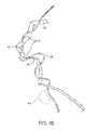
- FIG. 4Aillustrates a second apparatus useful for performing the methods constructed of the present invention comprising a length of material and a separate advancement member.
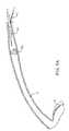
- FIG. 4Billustrates a third apparatus similar to the second apparatus of FIG. 4A , except that the advancement member is threaded through a portion of the ribbon-like length of material.
- FIGS. 5A-5Cillustrate use of the apparatus of FIG. 4A in accordance with the principles of the present invention.
- the present inventionmay be used for engaging, fragmenting, and retrieving a kidney stone KS or fragments from a ureter U between a kidney K and a bladder B.
- Access to the bladderwill be through the urethra UA using conventional access devices which will not be described herein.
- Access to the ureter Uwill be through the os O in a wall of the bladder leading into the lumen of the ureter.
- a first exemplary system 10 for performing the methods of the present inventioncomprises a sleeve-like length of material 12 and a tubular guide 14 , as shown in FIGS. 2A and 2B .
- the sleeve-like length of material 12has a trailing end 16 and an anchor end 18 .
- the length of the sleeve-like length of materialwill typically be in the range from 1 cm to 10 cm, usually from 2 cm to 6 cm, although much longer lengths may find use in different circumstances.
- the sleevewill usually have a continuous sidewall with no openings (other than at the trailing end 16 and anchor end 18 ), but could also have open regions, have a loose weave in the case of woven materials, or otherwise have openings or discontinuities in the sidewall without departing from the principles of the present invention.
- the sleeve-like length of material 12may be arranged so that it is initially within a central passage 20 of the tubular guide 14 .
- the material 12can be arranged so that the anchor and 18 of the sleeve-like length of material 12 will initially be on the exterior of the tubular guide 14 and generally held stationary as the tubular guide is advanced.
- the trailing end 16is everted over the distal end 22 of the guide member, generally as shown in FIG. 2B .
- the trailing end 16will usually include a tension member 24 which may be a suture, filament, thin wire, or other element which is attached at or near the terminus of the trailing end 16 and which preferably is woven and out of the material 12 over at least a portion of the length of material 12 .
- a tension member 24which may be a suture, filament, thin wire, or other element which is attached at or near the terminus of the trailing end 16 and which preferably is woven and out of the material 12 over at least a portion of the length of material 12 .
- woven or pleated structureswill be described in more detail hereinbelow. Pulling on the tension member 24 will collapse and compact the length of material 12 in order to provide the desired luminal occlusion.
- FIGS. 3A-3Juse of the system 10 for removing a kidney stone KS from a lumen L of a ureter U will be described.
- accessis gained to the os O of the bladder B ( FIG. 1 ) in a conventional manner.
- the tubular guide 14will then be passed through the os O and into the lumen L of the ureter with the anchor end 18 of the sleeve-like member 12 being held stationary relative to the os.
- Specific systems for doing thisare described in copending application Ser. No. 10/794,337, the full disclosure of which is incorporated herein by reference.
- a particular device for using a wire to advance and compact the length of materialis described in copending application Ser. No. 11/777,522, filed on the same day as the present application.
- the tubular guide 14is advanced so that the sleeve-like length of material 12 everts from the distal end 22 of the guide.
- the sleeve-like length of material 12will continue to be everted, but will have an exposed surface 13 which remains generally stationary relative to the inner wall of the ureter U and the exterior of the kidney stone KS.
- Such eversion of the sleeve-like length of material 12acts like a “tractor tread” in allowing the tubular guide 12 to bypass the kidney stone, as illustrated in FIG. 3C .
- the eversion of the length of material 12also reduces the risk of perforation or other trauma to the ureter.
- the tubular guide 14will continue to be advanced through the lumen L in the distal direction (toward the kidney K) until the trailing end 16 has been partly or fully exposed so that the region including the tension member 24 lies distal to the kidney stone KS.
- the tubular guide 14will be at least partly withdrawn in a proximal direction so that its distal end 22 is located proximal of the kidney stone KS, as shown in FIG. 3E .
- the portion of the sleeve-like member 12 which lies distal to the kidney stonewill radially collapse (since its internal support has been withdrawn) leaving a slack “shell” having the tension member 24 laced therethrough in place.
- the trailing end 16 of the sleeve-like member 12will be caused to axially collapse, generally in the manner of an accordion, as shown in FIG. 3F .
- the trailing end 16 of the sleeve-like member 12will be fully compacted against a distal surface of the kidney stone KS, as shown in FIG. 3G .
- an energy-delivery device 35will be introduced into the ureter U as generally described above.
- the energy-delivering deviceis typically delivered through a ureteroscope from below the stone (via the bladder), or a nephroscope from above (via the kidneys).
- Energy-delivering device 35may be configured to deliver laser energy, ultrasonic energy, hydraulic shock energy, or any other type of energy which can fragment the kidney stone KS.
- the energywill be delivered to fragment the kidney stone into a plurality of fragments KF, as generally shown in FIG. 3H .
- an irrigant fluidwill be introduced, typically through the same lumen in the ureteroscope or nephroscope which carries the energy delivery device 35 , as shown in FIG. 3I .
- the irrigation fluidis typically introduced at a rate in the range from about 0.25 ml/sec to 1 ml/sec, but this rate is not critical.
- the irrigant fluidwill flow toward the compacted trailing end 16 and will pass through the kidney stone fragments. Once the fluid engages the barrier formed by material 16 , the fluid flow direction will reverse, since the obstruction formed by the compacted material is generally impervious or at least resistant to fluid flow therethrough.
- the reversed flow of the irrigantwill carry at least some of the kidney stone fragments KF downstream toward the bladder or upstream toward the kidney, depending on the direction of introduction, while the compacted length of material 16 prevents migration or passage of fragments in the direction of irrigation.
- the ability to irrigate during fragmentation, without worry re. retropulsion of the stone or fragments,gives the physician a relatively clear view of the target stone/large fragments versus requiring him to work through an obscuring combination of blood and small stone fragments. Once the physician has reduced the stone/large fragments to an acceptable size, the compacted length of material 16 is drawn down toward the bladder in order to collect and remove any remaining kidney stone fragments KF, as shown in FIG. 3J .
- the stone fragmentsmay be withdrawn into the kidney and collected in a basket or by other conventional techniques. In some instances, it might be possible to redeploy the compacted length of material on the opposite side of the fragments and to use the compacted material to push the stone fragments out of the ureter.
- System 40comprises an advancement member 42 and a ribbon-like length of material 44 .
- the advancement membermay be a solid-core wire, a tube, or other small diameter or flat/thin member having sufficient column strength to permit its advancement through body lumen and preferably past an obstruction, such as a kidney stone in a ureter.
- the advancement membermay be in the form of a guidewire of the type commonly used in urological procedures.
- the ribbon-like length of material 44may be composed of any of the materials listed previously and may have a length in the ranges set forth above.
- the length of material 44will typically consist of only a single layer with a width in the range from 1 mm to 10 mm, usually from 2 mm to 6 mm, and a thickness of 1 mm or less.
- the ribbon-like length of material 44will comprise a flattened tube or other multiple-layer or laminated structure instead of a single layer as illustrated.
- the ribbon-like length of material 44may also have a plurality of axially spaced-apart fold structures 46 disposed over at least a distal length thereof.
- a distal end 48 of the length of material 44will be attached at or near a distal end of the advancement member 42 so that the advancement member can pull or otherwise carry the ribbon-like length of material through the target body lumen as it is advanced.
- the advancement member 42can be penetrated or “laced” through axially spaced-apart locations on the ribbon-like length of material 44 . As illustrated, the lacing occurs through consecutive sections defined by the fold structures 46 . In both cases, the advancement member 42 will be used to advance at least a portion of the ribbon 44 past a stone KS or other object to be retrieved or stabilized.
- the deployment system 40 of FIG. 4Ais introduced by advancing advancement member 42 through the os O ( FIG. 1 ) and into the lumen of the ureter U, as shown in FIG. 5A .
- the advancement member 42carries the ribbon-like length of material 44 distally within the lumen and past the kidney stone KS as shown in FIG. 5B .
- the advancement member 42may be drawn in the proximal direction, as shown in FIG. 5C , while the proximal portion of the ribbon-like length of material 44 is left in place. In this way, a region 50 of the ribbon-like length of material 44 which is distal to the kidney stone KS, as shown in FIG.
- the compacted structure 52may then be used as described previously, for moving and/or removing the kidney stone fragments into the bladder after lithotripsy.
Landscapes
- Health & Medical Sciences (AREA)
- Surgery (AREA)
- Life Sciences & Earth Sciences (AREA)
- Engineering & Computer Science (AREA)
- Biomedical Technology (AREA)
- Public Health (AREA)
- Vascular Medicine (AREA)
- Orthopedic Medicine & Surgery (AREA)
- Veterinary Medicine (AREA)
- Heart & Thoracic Surgery (AREA)
- Medical Informatics (AREA)
- Molecular Biology (AREA)
- Animal Behavior & Ethology (AREA)
- General Health & Medical Sciences (AREA)
- Nuclear Medicine, Radiotherapy & Molecular Imaging (AREA)
- Mechanical Engineering (AREA)
- Surgical Instruments (AREA)
- Prostheses (AREA)
Abstract
Description
Claims (20)
Priority Applications (2)
| Application Number | Priority Date | Filing Date | Title |
|---|---|---|---|
| US11/777,515US7883516B2 (en) | 2004-07-07 | 2007-07-13 | Methods for removing kidney stones from the ureter |
| US12/982,595US8753351B2 (en) | 2004-07-07 | 2010-12-30 | Methods for removing kidney stones from the ureter |
Applications Claiming Priority (2)
| Application Number | Priority Date | Filing Date | Title |
|---|---|---|---|
| US10/886,886US7462183B2 (en) | 2004-07-07 | 2004-07-07 | Methods for deploying conformed structures in body lumens |
| US11/777,515US7883516B2 (en) | 2004-07-07 | 2007-07-13 | Methods for removing kidney stones from the ureter |
Related Parent Applications (1)
| Application Number | Title | Priority Date | Filing Date |
|---|---|---|---|
| US10/886,886Continuation-In-PartUS7462183B2 (en) | 2004-07-07 | 2004-07-07 | Methods for deploying conformed structures in body lumens |
Related Child Applications (1)
| Application Number | Title | Priority Date | Filing Date |
|---|---|---|---|
| US12/982,595ContinuationUS8753351B2 (en) | 2004-07-07 | 2010-12-30 | Methods for removing kidney stones from the ureter |
Publications (2)
| Publication Number | Publication Date |
|---|---|
| US20080177277A1 US20080177277A1 (en) | 2008-07-24 |
| US7883516B2true US7883516B2 (en) | 2011-02-08 |
Family
ID=39642010
Family Applications (2)
| Application Number | Title | Priority Date | Filing Date |
|---|---|---|---|
| US11/777,515Expired - Fee RelatedUS7883516B2 (en) | 2004-07-07 | 2007-07-13 | Methods for removing kidney stones from the ureter |
| US12/982,595Expired - Fee RelatedUS8753351B2 (en) | 2004-07-07 | 2010-12-30 | Methods for removing kidney stones from the ureter |
Family Applications After (1)
| Application Number | Title | Priority Date | Filing Date |
|---|---|---|---|
| US12/982,595Expired - Fee RelatedUS8753351B2 (en) | 2004-07-07 | 2010-12-30 | Methods for removing kidney stones from the ureter |
Country Status (1)
| Country | Link |
|---|---|
| US (2) | US7883516B2 (en) |
Cited By (45)
| Publication number | Priority date | Publication date | Assignee | Title |
|---|---|---|---|---|
| US20100036410A1 (en)* | 2008-07-03 | 2010-02-11 | Hotspur Technologies, Inc. | Apparatus and methods for treating obstructions within body lumens |
| US20100036312A1 (en)* | 2008-06-08 | 2010-02-11 | Hotspur Technologies, Inc. | Apparatus and methods for removing obstructive material from body lumens |
| US20110098690A1 (en)* | 2004-07-07 | 2011-04-28 | Percutaneous Systems, Inc. | Methods for removing kidney stones from the ureter |
| US20130184658A1 (en)* | 2012-01-13 | 2013-07-18 | W. L. Gore & Associates, Inc. | Occlusion devices and methods of their manufacture and use |
| US8911450B2 (en) | 2004-07-07 | 2014-12-16 | Percutaneous Systems, Inc. | Methods and apparatus for deploying ureteral stents |
| US8926649B2 (en) | 2009-02-18 | 2015-01-06 | Hotspur Technologies, Inc. | Apparatus and methods for treating obstructions within body lumens |
| US8945160B2 (en) | 2008-07-03 | 2015-02-03 | Hotspur Technologies, Inc. | Apparatus and methods for treating obstructions within body lumens |
| US9072519B2 (en) | 2012-03-14 | 2015-07-07 | Gyrus Acmi, Inc. | Anti-retropulsion systems and methods |
| US9126013B2 (en) | 2012-04-27 | 2015-09-08 | Teleflex Medical Incorporated | Catheter with adjustable guidewire exit position |
| WO2016148747A1 (en) | 2015-03-19 | 2016-09-22 | Gyrus Acmi, Inc. D.B.A. Olympus Surgical Technologies America | Small fragment retrieval device |
| US9731099B2 (en) | 2009-02-18 | 2017-08-15 | Hotspur Technologies, Inc. | Apparatus and methods for treating obstructions within body lumens |
| CN108601599A (en)* | 2015-11-25 | 2018-09-28 | 尼尔拉维有限公司 | Clot retrieval device for removing an occluded clot from a blood vessel |
| US10363054B2 (en)* | 2014-11-26 | 2019-07-30 | Neuravi Limited | Clot retrieval device for removing occlusive clot from a blood vessel |
| EP3539485A1 (en) | 2018-03-15 | 2019-09-18 | Gyrus ACMI, Inc. d/b/a/ Olympus Surgical Technologies America | Small fragment retrieval device |
| US10617435B2 (en) | 2014-11-26 | 2020-04-14 | Neuravi Limited | Clot retrieval device for removing clot from a blood vessel |
| US10842498B2 (en) | 2018-09-13 | 2020-11-24 | Neuravi Limited | Systems and methods of restoring perfusion to a vessel |
| US10952760B2 (en) | 2011-03-09 | 2021-03-23 | Neuravi Limited | Clot retrieval device for removing a clot from a blood vessel |
| US11103264B2 (en) | 2013-03-14 | 2021-08-31 | Neuravi Limited | Devices and methods for removal of acute blockages from blood vessels |
| US11116530B2 (en) | 2018-02-02 | 2021-09-14 | Calyxo, Inc. | Devices and methods for minimally invasive kidney stone removal by combined aspiration and irrigation |
| US11147572B2 (en) | 2016-09-06 | 2021-10-19 | Neuravi Limited | Clot retrieval device for removing occlusive clot from a blood vessel |
| US11246612B2 (en) | 2010-10-22 | 2022-02-15 | Neuravi Limited | Clot engagement and removal system |
| US11253278B2 (en) | 2014-11-26 | 2022-02-22 | Neuravi Limited | Clot retrieval system for removing occlusive clot from a blood vessel |
| US11259824B2 (en) | 2011-03-09 | 2022-03-01 | Neuravi Limited | Clot retrieval device for removing occlusive clot from a blood vessel |
| US11395669B2 (en) | 2020-06-23 | 2022-07-26 | Neuravi Limited | Clot retrieval device with flexible collapsible frame |
| US11406416B2 (en) | 2018-10-02 | 2022-08-09 | Neuravi Limited | Joint assembly for vasculature obstruction capture device |
| US11439418B2 (en) | 2020-06-23 | 2022-09-13 | Neuravi Limited | Clot retrieval device for removing clot from a blood vessel |
| US11517340B2 (en) | 2019-12-03 | 2022-12-06 | Neuravi Limited | Stentriever devices for removing an occlusive clot from a vessel and methods thereof |
| US11529157B2 (en) | 2008-07-22 | 2022-12-20 | Neuravi Limited | Clot capture systems and associated methods |
| US11547427B2 (en) | 2013-03-14 | 2023-01-10 | Neuravi Limited | Clot retrieval devices |
| US11712231B2 (en) | 2019-10-29 | 2023-08-01 | Neuravi Limited | Proximal locking assembly design for dual stent mechanical thrombectomy device |
| US11717308B2 (en) | 2020-04-17 | 2023-08-08 | Neuravi Limited | Clot retrieval device for removing heterogeneous clots from a blood vessel |
| US11730501B2 (en) | 2020-04-17 | 2023-08-22 | Neuravi Limited | Floating clot retrieval device for removing clots from a blood vessel |
| US11737771B2 (en) | 2020-06-18 | 2023-08-29 | Neuravi Limited | Dual channel thrombectomy device |
| US11839392B2 (en) | 2013-03-14 | 2023-12-12 | Neuravi Limited | Clot retrieval device for removing clot from a blood vessel |
| US11864781B2 (en) | 2020-09-23 | 2024-01-09 | Neuravi Limited | Rotating frame thrombectomy device |
| US11871946B2 (en) | 2020-04-17 | 2024-01-16 | Neuravi Limited | Clot retrieval device for removing clot from a blood vessel |
| US11896249B2 (en) | 2020-07-02 | 2024-02-13 | Gyrus Acmi, Inc. | Lithotripsy system having a drill and lateral emitter |
| US11937836B2 (en) | 2020-06-22 | 2024-03-26 | Neuravi Limited | Clot retrieval system with expandable clot engaging framework |
| US11937837B2 (en) | 2020-12-29 | 2024-03-26 | Neuravi Limited | Fibrin rich / soft clot mechanical thrombectomy device |
| US11974764B2 (en) | 2021-06-04 | 2024-05-07 | Neuravi Limited | Self-orienting rotating stentriever pinching cells |
| US12029442B2 (en) | 2021-01-14 | 2024-07-09 | Neuravi Limited | Systems and methods for a dual elongated member clot retrieval apparatus |
| US12064130B2 (en) | 2021-03-18 | 2024-08-20 | Neuravi Limited | Vascular obstruction retrieval device having sliding cages pinch mechanism |
| US12076037B2 (en) | 2011-03-09 | 2024-09-03 | Neuravi Limited | Systems and methods to restore perfusion to a vessel |
| US12256989B2 (en) | 2022-09-29 | 2025-03-25 | Calyxo, Inc. | Tool guiding device for kidney stone treatment apparatus |
| US12329396B2 (en) | 2022-03-02 | 2025-06-17 | Calyxo, Inc. | Kidney stone treatment system |
Families Citing this family (12)
| Publication number | Priority date | Publication date | Assignee | Title |
|---|---|---|---|---|
| US7462183B2 (en)* | 2004-07-07 | 2008-12-09 | Percutaneous Systems, Inc. | Methods for deploying conformed structures in body lumens |
| WO2010065556A1 (en)* | 2008-12-01 | 2010-06-10 | Percutaneous Systems, Inc. | Methods and systems for capturing and removing urinary stones from body cavities |
| US20100274231A1 (en)* | 2009-04-24 | 2010-10-28 | Applied Medical Resources Corporation | Renal flushing catheter |
| US8858569B2 (en)* | 2012-02-16 | 2014-10-14 | Shaw P. Wan | Stone retrieval device |
| US10105159B2 (en)* | 2013-03-15 | 2018-10-23 | W.L. Gore Associates, Inc | Recanalization device |
| US9955986B2 (en) | 2015-10-30 | 2018-05-01 | Auris Surgical Robotics, Inc. | Basket apparatus |
| US10231793B2 (en) | 2015-10-30 | 2019-03-19 | Auris Health, Inc. | Object removal through a percutaneous suction tube |
| US9949749B2 (en) | 2015-10-30 | 2018-04-24 | Auris Surgical Robotics, Inc. | Object capture with a basket |
| RU2633594C1 (en)* | 2016-05-30 | 2017-10-13 | Нариман Казиханович Гаджиев | Method for urolithiasis treatment by percutaneous nephrolithotripsy |
| CN108042179B (en)* | 2017-11-27 | 2020-06-26 | 西安交通大学第一附属医院 | A stone flushing device for urology |
| US11896330B2 (en) | 2019-08-15 | 2024-02-13 | Auris Health, Inc. | Robotic medical system having multiple medical instruments |
| EP4084724A4 (en) | 2019-12-31 | 2023-12-27 | Auris Health, Inc. | ADVANCED BASKET DRIVE MODE |
Citations (44)
| Publication number | Priority date | Publication date | Assignee | Title |
|---|---|---|---|---|
| US4046149A (en) | 1975-01-31 | 1977-09-06 | Olympus Optical Co., Ltd. | Instrument for removing a foreign substance from the body cavity of human being |
| US4262677A (en) | 1979-03-26 | 1981-04-21 | Bader Robert F | Culture sampling device and method |
| US4295464A (en) | 1980-03-21 | 1981-10-20 | Shihata Alfred A | Ureteric stone extractor with two ballooned catheters |
| US4531933A (en) | 1982-12-07 | 1985-07-30 | C. R. Bard, Inc. | Helical ureteral stent |
| US4706671A (en) | 1985-05-02 | 1987-11-17 | Weinrib Harry P | Catheter with coiled tip |
| US4930496A (en)* | 1988-07-22 | 1990-06-05 | Vance Products, Inc. | Method and device for removing a stone from a ureter using extracorporeal shock wave lithotripsy |
| US5011488A (en)* | 1988-12-07 | 1991-04-30 | Robert Ginsburg | Thrombus extraction system |
| US5135534A (en)* | 1990-04-06 | 1992-08-04 | John Tulip | Laser lithotripsy |
| US5192286A (en)* | 1991-07-26 | 1993-03-09 | Regents Of The University Of California | Method and device for retrieving materials from body lumens |
| US5431676A (en) | 1993-03-05 | 1995-07-11 | Innerdyne Medical, Inc. | Trocar system having expandable port |
| US5454790A (en) | 1994-05-09 | 1995-10-03 | Innerdyne, Inc. | Method and apparatus for catheterization access |
| US5531717A (en) | 1993-12-12 | 1996-07-02 | Rtc, Inc. | Non-contaminating probe and methods of making and using same |
| US5599291A (en) | 1993-01-04 | 1997-02-04 | Menlo Care, Inc. | Softening expanding ureteral stent |
| US5676688A (en) | 1995-02-06 | 1997-10-14 | Rtc, Inc. | Variably inflatable medical device |
| US5681274A (en) | 1995-03-31 | 1997-10-28 | Boston Scientific Corporation | Variable length uretheral stent |
| EP0605427B1 (en) | 1991-06-07 | 1998-05-20 | Rtc Inc. | Non-contaminating probe |
| US5814058A (en) | 1993-03-05 | 1998-09-29 | Innerdyne, Inc. | Method and apparatus employing conformable sleeve for providing percutaneous access |
| US5836913A (en) | 1997-05-02 | 1998-11-17 | Innerdyne, Inc. | Device and method for accessing a body cavity |
| US5860972A (en)* | 1995-10-26 | 1999-01-19 | Xintec Corporation | Method of detection and destruction of urinary calculi and similar structures |
| US5989264A (en) | 1998-06-11 | 1999-11-23 | Ethicon Endo-Surgery, Inc. | Ultrasonic polyp snare |
| US6007488A (en) | 1997-05-12 | 1999-12-28 | Rtc, Inc. | Medical probe including an electrically conductive membrane suitable for medical uses |
| US6214037B1 (en) | 1999-03-18 | 2001-04-10 | Fossa Industries, Llc | Radially expanding stent |
| US6240968B1 (en) | 1996-08-14 | 2001-06-05 | Rtc, Inc. | Membranes suitable for medical use |
| US20010044595A1 (en) | 2000-05-02 | 2001-11-22 | Wilson-Cook Medical, Inc. | Introducer apparatus with eversible sleeve |
| DE10031661A1 (en) | 2000-06-29 | 2002-01-24 | Johannes Woitzik | Protective sleeve for biopsy needle, has thin tubular sheeting coating the canula inside and out |
| US20020183853A1 (en) | 1999-03-18 | 2002-12-05 | Fossa Industries, Llc | Radially expandable stents |
| US20030120281A1 (en) | 1998-04-23 | 2003-06-26 | Boston Scientific Corporation | Atraumatic medical retrieval device |
| US6623508B2 (en) | 1996-12-20 | 2003-09-23 | Gore Enterprise Holdings, Inc. | Self-expanding defect closure device and method of making and using |
| US6656146B1 (en) | 1995-11-07 | 2003-12-02 | Scimed Life Systems, Inc. | Medical device with tail(s) |
| US20030229332A1 (en)* | 2002-06-11 | 2003-12-11 | Scimed Life Systems, Inc. | Adjustable double balloon catheter with a through lumen for stone management |
| US6692484B1 (en)* | 1999-07-17 | 2004-02-17 | Wilson-Cook Medical Incorporated | Devices for extracting biliary or urinary stones |
| US20040092956A1 (en) | 2000-11-03 | 2004-05-13 | John Liddicoat | Catheter for removal of solids from surgical drains |
| US20040210239A1 (en)* | 1996-07-26 | 2004-10-21 | Nash John E. | System and method of use for treating occluded vessels and diseased tissue |
| US20040220587A1 (en) | 2002-01-16 | 2004-11-04 | Scimed Life Systems, Inc. | Treatment and removal of objects in anatomical lumens |
| US20050143678A1 (en)* | 2003-10-14 | 2005-06-30 | Pluromed, Inc. | Confinement of kidney-stone fragments during lithotripsy |
| US6929664B2 (en) | 2003-12-05 | 2005-08-16 | Fossa Medical, Inc. | Open lumen stents |
| US20050197627A1 (en) | 2004-03-05 | 2005-09-08 | Percutaneous Systems, Inc. | Method and system for deploying protective sleeve in intraluminal catherization and dilation |
| US20050228481A1 (en) | 2002-10-09 | 2005-10-13 | Fossa Medical, Inc | Eccentric lumen stents |
| US20060009784A1 (en) | 2004-07-07 | 2006-01-12 | Percutaneous Systems, Inc. | Methods and apparatus for deploying conformed structures in body lumens |
| US20060116693A1 (en) | 2004-12-01 | 2006-06-01 | Weisenburgh William B Ii | Apparatus and method for stone capture and removal |
| US20070016244A1 (en) | 2005-07-06 | 2007-01-18 | Percutaneous Systems, Inc. | Methods and apparatus for deploying ureteral stents |
| US7214229B2 (en) | 1999-03-18 | 2007-05-08 | Fossa Medical, Inc. | Radially expanding stents |
| US20070191768A1 (en) | 2006-02-13 | 2007-08-16 | Fossa Medical, Inc. | Methods and Apparatus for Temporarily Occluding Body Lumens |
| US7316663B2 (en) | 2000-05-26 | 2008-01-08 | Boston Scientific Scimed, Inc. | Ureteral stent |
Family Cites Families (23)
| Publication number | Priority date | Publication date | Assignee | Title |
|---|---|---|---|---|
| US2756752A (en) | 1953-12-23 | 1956-07-31 | Scherlis Irving | Surgical instrument |
| US4030406A (en) | 1967-04-18 | 1977-06-21 | Raoul Wander | Apparatus for sterilization |
| US4030503A (en) | 1975-11-05 | 1977-06-21 | Clark Iii William T | Embolectomy catheter |
| US4807626A (en) | 1985-02-14 | 1989-02-28 | Mcgirr Douglas B | Stone extractor and method |
| US4762130A (en) | 1987-01-15 | 1988-08-09 | Thomas J. Fogarty | Catheter with corkscrew-like balloon |
| US4813925A (en) | 1987-04-21 | 1989-03-21 | Medical Engineering Corporation | Spiral ureteral stent |
| GB8916158D0 (en) | 1989-07-14 | 1989-08-31 | Smiths Industries Plc | Catheters |
| US5879366A (en) | 1996-12-20 | 1999-03-09 | W.L. Gore & Associates, Inc. | Self-expanding defect closure device and method of making and using |
| US5972019A (en) | 1996-07-25 | 1999-10-26 | Target Therapeutics, Inc. | Mechanical clot treatment device |
| JP2001507598A (en) | 1996-12-31 | 2001-06-12 | クック ウロロジカル インク. | Ureteral stone occluder with blade filter |
| DE69839888D1 (en) | 1997-11-12 | 2008-09-25 | Genesis Technologies Llc | DEVICE FOR REMOVING OCCLUSIONS IN BIOLOGICAL PASSES |
| US6500185B1 (en) | 2000-09-29 | 2002-12-31 | Primus Medical, Inc. | Snare device |
| US6994093B2 (en) | 2001-02-28 | 2006-02-07 | Chase Medical, L.P. | Ventricular restoration shaping apparatus and method of use |
| JP4255286B2 (en) | 2001-05-17 | 2009-04-15 | ウィルソン−クック メディカル インコーポレイテッド | Intragastric device for treating obesity |
| US8506647B2 (en) | 2002-02-14 | 2013-08-13 | Boston Scientific Scimed, Inc. | System for maintaining body canal patency |
| US20030191492A1 (en) | 2002-04-05 | 2003-10-09 | Scimed Life Systems, Inc. | Radial coil expandable medical wire |
| US7682366B2 (en) | 2002-10-16 | 2010-03-23 | Olympus Corporation | Calculus manipulation apparatus |
| US7698205B2 (en) | 2003-06-05 | 2010-04-13 | Omx Technology Ab | Combination LEG price generation |
| US7316692B2 (en) | 2003-08-12 | 2008-01-08 | Boston Scientific Scimed, Inc. | Laser-cut clot puller |
| WO2005110244A1 (en) | 2004-05-07 | 2005-11-24 | Usgi Medical Inc. | Apparatus and methods for positioning and securing anchors |
| US7883516B2 (en) | 2004-07-07 | 2011-02-08 | Percutaneous Systems, Inc. | Methods for removing kidney stones from the ureter |
| US7070510B2 (en)* | 2004-08-17 | 2006-07-04 | Will Heddon | Bowling alley bumper system |
| US7879066B2 (en) | 2007-07-13 | 2011-02-01 | Percutaneous Sustems, Inc. | Apparatus for occluding body lumens |
- 2007
- 2007-07-13USUS11/777,515patent/US7883516B2/ennot_activeExpired - Fee Related
- 2010
- 2010-12-30USUS12/982,595patent/US8753351B2/ennot_activeExpired - Fee Related
Patent Citations (52)
| Publication number | Priority date | Publication date | Assignee | Title |
|---|---|---|---|---|
| US4046149A (en) | 1975-01-31 | 1977-09-06 | Olympus Optical Co., Ltd. | Instrument for removing a foreign substance from the body cavity of human being |
| US4262677A (en) | 1979-03-26 | 1981-04-21 | Bader Robert F | Culture sampling device and method |
| US4295464A (en) | 1980-03-21 | 1981-10-20 | Shihata Alfred A | Ureteric stone extractor with two ballooned catheters |
| US4531933A (en) | 1982-12-07 | 1985-07-30 | C. R. Bard, Inc. | Helical ureteral stent |
| US4706671A (en) | 1985-05-02 | 1987-11-17 | Weinrib Harry P | Catheter with coiled tip |
| US4930496A (en)* | 1988-07-22 | 1990-06-05 | Vance Products, Inc. | Method and device for removing a stone from a ureter using extracorporeal shock wave lithotripsy |
| US5011488A (en)* | 1988-12-07 | 1991-04-30 | Robert Ginsburg | Thrombus extraction system |
| US5135534A (en)* | 1990-04-06 | 1992-08-04 | John Tulip | Laser lithotripsy |
| US5711841A (en) | 1991-06-07 | 1998-01-27 | Rtc, Inc. | Methods of making and using non-contaminating probes |
| US5897535A (en) | 1991-06-07 | 1999-04-27 | Rtc, Inc. | Non-contaminating probe and methods of making and using same |
| EP0605427B1 (en) | 1991-06-07 | 1998-05-20 | Rtc Inc. | Non-contaminating probe |
| US5192286A (en)* | 1991-07-26 | 1993-03-09 | Regents Of The University Of California | Method and device for retrieving materials from body lumens |
| US5599291A (en) | 1993-01-04 | 1997-02-04 | Menlo Care, Inc. | Softening expanding ureteral stent |
| US6080174A (en) | 1993-03-05 | 2000-06-27 | Innerdyne, Inc. | Trocar system having expandable port |
| US6325812B1 (en) | 1993-03-05 | 2001-12-04 | Innerdyne, Inc. | Trocar system having expandable port |
| US5814058A (en) | 1993-03-05 | 1998-09-29 | Innerdyne, Inc. | Method and apparatus employing conformable sleeve for providing percutaneous access |
| US5431676A (en) | 1993-03-05 | 1995-07-11 | Innerdyne Medical, Inc. | Trocar system having expandable port |
| US6494893B2 (en) | 1993-03-05 | 2002-12-17 | Innderdyne, Inc. | Trocar system having expandable port |
| US5531717A (en) | 1993-12-12 | 1996-07-02 | Rtc, Inc. | Non-contaminating probe and methods of making and using same |
| US5454790A (en) | 1994-05-09 | 1995-10-03 | Innerdyne, Inc. | Method and apparatus for catheterization access |
| US5676688A (en) | 1995-02-06 | 1997-10-14 | Rtc, Inc. | Variably inflatable medical device |
| US5681274A (en) | 1995-03-31 | 1997-10-28 | Boston Scientific Corporation | Variable length uretheral stent |
| US5860972A (en)* | 1995-10-26 | 1999-01-19 | Xintec Corporation | Method of detection and destruction of urinary calculi and similar structures |
| US6656146B1 (en) | 1995-11-07 | 2003-12-02 | Scimed Life Systems, Inc. | Medical device with tail(s) |
| US6945950B2 (en) | 1995-11-07 | 2005-09-20 | Boston Scientific Scimed, Inc. | Ureteral stent with small bladder tail(s) |
| US20040210239A1 (en)* | 1996-07-26 | 2004-10-21 | Nash John E. | System and method of use for treating occluded vessels and diseased tissue |
| US6240968B1 (en) | 1996-08-14 | 2001-06-05 | Rtc, Inc. | Membranes suitable for medical use |
| US6623508B2 (en) | 1996-12-20 | 2003-09-23 | Gore Enterprise Holdings, Inc. | Self-expanding defect closure device and method of making and using |
| US5836913A (en) | 1997-05-02 | 1998-11-17 | Innerdyne, Inc. | Device and method for accessing a body cavity |
| US6007488A (en) | 1997-05-12 | 1999-12-28 | Rtc, Inc. | Medical probe including an electrically conductive membrane suitable for medical uses |
| US20030120281A1 (en) | 1998-04-23 | 2003-06-26 | Boston Scientific Corporation | Atraumatic medical retrieval device |
| US5989264A (en) | 1998-06-11 | 1999-11-23 | Ethicon Endo-Surgery, Inc. | Ultrasonic polyp snare |
| US20020183853A1 (en) | 1999-03-18 | 2002-12-05 | Fossa Industries, Llc | Radially expandable stents |
| US6214037B1 (en) | 1999-03-18 | 2001-04-10 | Fossa Industries, Llc | Radially expanding stent |
| US7214229B2 (en) | 1999-03-18 | 2007-05-08 | Fossa Medical, Inc. | Radially expanding stents |
| US6692484B1 (en)* | 1999-07-17 | 2004-02-17 | Wilson-Cook Medical Incorporated | Devices for extracting biliary or urinary stones |
| US20010044595A1 (en) | 2000-05-02 | 2001-11-22 | Wilson-Cook Medical, Inc. | Introducer apparatus with eversible sleeve |
| US7316663B2 (en) | 2000-05-26 | 2008-01-08 | Boston Scientific Scimed, Inc. | Ureteral stent |
| DE10031661A1 (en) | 2000-06-29 | 2002-01-24 | Johannes Woitzik | Protective sleeve for biopsy needle, has thin tubular sheeting coating the canula inside and out |
| US20040092956A1 (en) | 2000-11-03 | 2004-05-13 | John Liddicoat | Catheter for removal of solids from surgical drains |
| US20040220587A1 (en) | 2002-01-16 | 2004-11-04 | Scimed Life Systems, Inc. | Treatment and removal of objects in anatomical lumens |
| US20070088256A1 (en)* | 2002-06-11 | 2007-04-19 | Boston Scientific Scimed, Inc. | Adjustable double balloon catheter with a through lumen for stone management |
| US20030229332A1 (en)* | 2002-06-11 | 2003-12-11 | Scimed Life Systems, Inc. | Adjustable double balloon catheter with a through lumen for stone management |
| US20050228481A1 (en) | 2002-10-09 | 2005-10-13 | Fossa Medical, Inc | Eccentric lumen stents |
| US20050143678A1 (en)* | 2003-10-14 | 2005-06-30 | Pluromed, Inc. | Confinement of kidney-stone fragments during lithotripsy |
| US7217250B2 (en) | 2003-12-05 | 2007-05-15 | Fossa Medical, Inc. | Open lumen stents |
| US6929664B2 (en) | 2003-12-05 | 2005-08-16 | Fossa Medical, Inc. | Open lumen stents |
| US20050197627A1 (en) | 2004-03-05 | 2005-09-08 | Percutaneous Systems, Inc. | Method and system for deploying protective sleeve in intraluminal catherization and dilation |
| US20060009784A1 (en) | 2004-07-07 | 2006-01-12 | Percutaneous Systems, Inc. | Methods and apparatus for deploying conformed structures in body lumens |
| US20060116693A1 (en) | 2004-12-01 | 2006-06-01 | Weisenburgh William B Ii | Apparatus and method for stone capture and removal |
| US20070016244A1 (en) | 2005-07-06 | 2007-01-18 | Percutaneous Systems, Inc. | Methods and apparatus for deploying ureteral stents |
| US20070191768A1 (en) | 2006-02-13 | 2007-08-16 | Fossa Medical, Inc. | Methods and Apparatus for Temporarily Occluding Body Lumens |
Non-Patent Citations (5)
| Title |
|---|
| Bard Urological Division Catalog 1990, PA24, "Woven Blasucci Ureteral Catheters", 3 pages. |
| Garrido et al., "Utilización del catéter "stone sweeper" en la patolotgía litiásica del tracto urinario superior," [The use of Stone Sweeper catherer for stone disease of the upper urinary tract], Arch Esp Urol. Nov. 2006; 56(9):889-892. [English Abstract Only]. |
| International Search Report and Written Opinion of PCT Application No. PCT/US07/69182, dated Aug. 15, 2008, 6 pages total. |
| L'Esperance et al., "Ureteral Expanding Stent: A New Device for Urolithiasis," J Endourol. May 1, 2007; 21(5): 533-537. |
| Woitzik et al., "Polyethylene sheath device to reduce tumor cell seeding along the needle tract in percutaneous biopsy," (2003) Surg. Endosc. 17:311-314. |
Cited By (81)
| Publication number | Priority date | Publication date | Assignee | Title |
|---|---|---|---|---|
| US8753351B2 (en) | 2004-07-07 | 2014-06-17 | Percutaneous Systems, Inc. | Methods for removing kidney stones from the ureter |
| US8911450B2 (en) | 2004-07-07 | 2014-12-16 | Percutaneous Systems, Inc. | Methods and apparatus for deploying ureteral stents |
| US20110098690A1 (en)* | 2004-07-07 | 2011-04-28 | Percutaneous Systems, Inc. | Methods for removing kidney stones from the ureter |
| US9855067B2 (en) | 2008-06-08 | 2018-01-02 | Hotspur Technologies, Inc. | Removing obstructive material from body lumens |
| US20100036312A1 (en)* | 2008-06-08 | 2010-02-11 | Hotspur Technologies, Inc. | Apparatus and methods for removing obstructive material from body lumens |
| US10716586B2 (en) | 2008-06-08 | 2020-07-21 | Arrow International, Inc. | Apparatus and methods for removing obstructive material from body lumens |
| US8939991B2 (en) | 2008-06-08 | 2015-01-27 | Hotspur Technologies, Inc. | Apparatus and methods for removing obstructive material from body lumens |
| US8043313B2 (en) | 2008-07-03 | 2011-10-25 | Hotspur Technologies, Inc | Apparatus and methods for treating obstructions within body lumens |
| US8945160B2 (en) | 2008-07-03 | 2015-02-03 | Hotspur Technologies, Inc. | Apparatus and methods for treating obstructions within body lumens |
| US20100036410A1 (en)* | 2008-07-03 | 2010-02-11 | Hotspur Technologies, Inc. | Apparatus and methods for treating obstructions within body lumens |
| US10898695B2 (en) | 2008-07-03 | 2021-01-26 | Arrow International, Inc. | Apparatus and methods for treating obstructions within body lumens |
| US10624656B2 (en) | 2008-07-03 | 2020-04-21 | Arrow International, Inc. | Apparatus and methods for treating obstructions within body lumens |
| US9833599B2 (en) | 2008-07-03 | 2017-12-05 | Hotspur Technologies, Inc. | Apparatus and methods for treating obstructions within body lumens |
| US11529157B2 (en) | 2008-07-22 | 2022-12-20 | Neuravi Limited | Clot capture systems and associated methods |
| US9731099B2 (en) | 2009-02-18 | 2017-08-15 | Hotspur Technologies, Inc. | Apparatus and methods for treating obstructions within body lumens |
| US9757137B2 (en) | 2009-02-18 | 2017-09-12 | Hotspur Technologies, Inc. | Apparatus and methods for treating obstructions within body lumens |
| US9101382B2 (en) | 2009-02-18 | 2015-08-11 | Hotspur Technologies, Inc. | Apparatus and methods for treating obstructions within body lumens |
| US8926649B2 (en) | 2009-02-18 | 2015-01-06 | Hotspur Technologies, Inc. | Apparatus and methods for treating obstructions within body lumens |
| US11871949B2 (en) | 2010-10-22 | 2024-01-16 | Neuravi Limited | Clot engagement and removal system |
| US11246612B2 (en) | 2010-10-22 | 2022-02-15 | Neuravi Limited | Clot engagement and removal system |
| US12059164B2 (en) | 2011-03-09 | 2024-08-13 | Neuravi Limited | Clot retrieval device for removing occlusive clot from a blood vessel |
| US11998223B2 (en) | 2011-03-09 | 2024-06-04 | Neuravi Limited | Clot retrieval device for removing a clot from a blood vessel |
| US12076037B2 (en) | 2011-03-09 | 2024-09-03 | Neuravi Limited | Systems and methods to restore perfusion to a vessel |
| US11259824B2 (en) | 2011-03-09 | 2022-03-01 | Neuravi Limited | Clot retrieval device for removing occlusive clot from a blood vessel |
| US10952760B2 (en) | 2011-03-09 | 2021-03-23 | Neuravi Limited | Clot retrieval device for removing a clot from a blood vessel |
| US11529145B2 (en) | 2012-01-13 | 2022-12-20 | W. L. Gore & Associates, Inc. | Occlusion devices and methods of their manufacture and use |
| US10342548B2 (en)* | 2012-01-13 | 2019-07-09 | W. L. Gore & Associates, Inc. | Occlusion devices and methods of their manufacture and use |
| US20130184658A1 (en)* | 2012-01-13 | 2013-07-18 | W. L. Gore & Associates, Inc. | Occlusion devices and methods of their manufacture and use |
| US9737319B2 (en) | 2012-03-14 | 2017-08-22 | Gyrus Acmi, Inc. | Anti-retropulsion systems and methods |
| US9072519B2 (en) | 2012-03-14 | 2015-07-07 | Gyrus Acmi, Inc. | Anti-retropulsion systems and methods |
| US9126013B2 (en) | 2012-04-27 | 2015-09-08 | Teleflex Medical Incorporated | Catheter with adjustable guidewire exit position |
| US10105517B2 (en) | 2012-04-27 | 2018-10-23 | Teleflex Medical Incorporated | Catheter with adjustable guidewire exit position |
| US12402902B2 (en) | 2013-03-14 | 2025-09-02 | Neuravi Limited | Devices and methods for removal of acute blockages from blood vessels |
| US11871945B2 (en) | 2013-03-14 | 2024-01-16 | Neuravi Limited | Clot retrieval device for removing clot from a blood vessel |
| US11103264B2 (en) | 2013-03-14 | 2021-08-31 | Neuravi Limited | Devices and methods for removal of acute blockages from blood vessels |
| US11839392B2 (en) | 2013-03-14 | 2023-12-12 | Neuravi Limited | Clot retrieval device for removing clot from a blood vessel |
| US11547427B2 (en) | 2013-03-14 | 2023-01-10 | Neuravi Limited | Clot retrieval devices |
| US11937835B2 (en) | 2013-03-14 | 2024-03-26 | Neuravi Limited | Clot retrieval device for removing clot from a blood vessel |
| US11980379B2 (en) | 2014-11-26 | 2024-05-14 | Neuravi Limited | Clot retrieval system for removing occlusive clot from a blood vessel |
| US10363054B2 (en)* | 2014-11-26 | 2019-07-30 | Neuravi Limited | Clot retrieval device for removing occlusive clot from a blood vessel |
| US11857210B2 (en) | 2014-11-26 | 2024-01-02 | Neuravi Limited | Clot retrieval device for removing clot from a blood vessel |
| US11253278B2 (en) | 2014-11-26 | 2022-02-22 | Neuravi Limited | Clot retrieval system for removing occlusive clot from a blood vessel |
| US11712256B2 (en) | 2014-11-26 | 2023-08-01 | Neuravi Limited | Clot retrieval device for removing occlusive clot from a blood vessel |
| US10617435B2 (en) | 2014-11-26 | 2020-04-14 | Neuravi Limited | Clot retrieval device for removing clot from a blood vessel |
| US9931129B2 (en) | 2015-03-19 | 2018-04-03 | Gyrus Acmi, Inc. | Small fragment retrieval device |
| WO2016148747A1 (en) | 2015-03-19 | 2016-09-22 | Gyrus Acmi, Inc. D.B.A. Olympus Surgical Technologies America | Small fragment retrieval device |
| CN108601599A (en)* | 2015-11-25 | 2018-09-28 | 尼尔拉维有限公司 | Clot retrieval device for removing an occluded clot from a blood vessel |
| CN108601599B (en)* | 2015-11-25 | 2021-08-13 | 尼尔拉维有限公司 | Clot retrieval device for removing occlusive clots from blood vessels |
| US12133657B2 (en) | 2016-09-06 | 2024-11-05 | Neuravi Limited | Clot retrieval device for removing occlusive clot from a blood vessel |
| US11147572B2 (en) | 2016-09-06 | 2021-10-19 | Neuravi Limited | Clot retrieval device for removing occlusive clot from a blood vessel |
| US11116530B2 (en) | 2018-02-02 | 2021-09-14 | Calyxo, Inc. | Devices and methods for minimally invasive kidney stone removal by combined aspiration and irrigation |
| US12023059B2 (en) | 2018-02-02 | 2024-07-02 | Calyxo, Inc. | Devices and methods for minimally invasive kidney stone removal by combined aspiration and irrigation |
| US12318099B2 (en) | 2018-02-02 | 2025-06-03 | Calyxo, Inc. | Devices and methods for minimally invasive kidney stone removal by combined aspiration and irrigation |
| US11324526B2 (en) | 2018-02-02 | 2022-05-10 | Calyxo, Inc. | Devices and methods for minimally invasive kidney stone removal by combined aspiration and irrigation |
| EP3539485A1 (en) | 2018-03-15 | 2019-09-18 | Gyrus ACMI, Inc. d/b/a/ Olympus Surgical Technologies America | Small fragment retrieval device |
| US10842498B2 (en) | 2018-09-13 | 2020-11-24 | Neuravi Limited | Systems and methods of restoring perfusion to a vessel |
| US11963693B2 (en) | 2018-10-02 | 2024-04-23 | Neuravi Limited | Joint assembly for vasculature obstruction capture device |
| US11406416B2 (en) | 2018-10-02 | 2022-08-09 | Neuravi Limited | Joint assembly for vasculature obstruction capture device |
| US11712231B2 (en) | 2019-10-29 | 2023-08-01 | Neuravi Limited | Proximal locking assembly design for dual stent mechanical thrombectomy device |
| US12004731B2 (en) | 2019-10-29 | 2024-06-11 | Neuravi Limited | Proximal locking assembly design for dual stent mechanical thrombectomy device |
| US12023058B2 (en) | 2019-12-03 | 2024-07-02 | Neuravi Limited | Stentriever devices for removing an occlusive clot from a vessel and methods thereof |
| US11517340B2 (en) | 2019-12-03 | 2022-12-06 | Neuravi Limited | Stentriever devices for removing an occlusive clot from a vessel and methods thereof |
| US11871946B2 (en) | 2020-04-17 | 2024-01-16 | Neuravi Limited | Clot retrieval device for removing clot from a blood vessel |
| US12048446B2 (en) | 2020-04-17 | 2024-07-30 | Neuravi Limited | Clot retrieval device for removing heterogeneous clots from a blood vessel |
| US11717308B2 (en) | 2020-04-17 | 2023-08-08 | Neuravi Limited | Clot retrieval device for removing heterogeneous clots from a blood vessel |
| US11730501B2 (en) | 2020-04-17 | 2023-08-22 | Neuravi Limited | Floating clot retrieval device for removing clots from a blood vessel |
| US11737771B2 (en) | 2020-06-18 | 2023-08-29 | Neuravi Limited | Dual channel thrombectomy device |
| US12213691B2 (en) | 2020-06-18 | 2025-02-04 | Neuravi Limited | Dual channel thrombectomy device |
| US11937836B2 (en) | 2020-06-22 | 2024-03-26 | Neuravi Limited | Clot retrieval system with expandable clot engaging framework |
| US11439418B2 (en) | 2020-06-23 | 2022-09-13 | Neuravi Limited | Clot retrieval device for removing clot from a blood vessel |
| US11395669B2 (en) | 2020-06-23 | 2022-07-26 | Neuravi Limited | Clot retrieval device with flexible collapsible frame |
| US12419657B2 (en) | 2020-06-23 | 2025-09-23 | Neuravi Limited | Clot retrieval device for removing clot from a blood vessel |
| US11896249B2 (en) | 2020-07-02 | 2024-02-13 | Gyrus Acmi, Inc. | Lithotripsy system having a drill and lateral emitter |
| US11864781B2 (en) | 2020-09-23 | 2024-01-09 | Neuravi Limited | Rotating frame thrombectomy device |
| US11937837B2 (en) | 2020-12-29 | 2024-03-26 | Neuravi Limited | Fibrin rich / soft clot mechanical thrombectomy device |
| US12029442B2 (en) | 2021-01-14 | 2024-07-09 | Neuravi Limited | Systems and methods for a dual elongated member clot retrieval apparatus |
| US12064130B2 (en) | 2021-03-18 | 2024-08-20 | Neuravi Limited | Vascular obstruction retrieval device having sliding cages pinch mechanism |
| US11974764B2 (en) | 2021-06-04 | 2024-05-07 | Neuravi Limited | Self-orienting rotating stentriever pinching cells |
| US12329396B2 (en) | 2022-03-02 | 2025-06-17 | Calyxo, Inc. | Kidney stone treatment system |
| US12329399B2 (en) | 2022-03-02 | 2025-06-17 | Calyxo, Inc. | Kidney stone treatment system |
| US12256989B2 (en) | 2022-09-29 | 2025-03-25 | Calyxo, Inc. | Tool guiding device for kidney stone treatment apparatus |
Also Published As
| Publication number | Publication date |
|---|---|
| US20080177277A1 (en) | 2008-07-24 |
| US8753351B2 (en) | 2014-06-17 |
| US20110098690A1 (en) | 2011-04-28 |
Similar Documents
| Publication | Publication Date | Title |
|---|---|---|
| US7883516B2 (en) | Methods for removing kidney stones from the ureter | |
| US7462183B2 (en) | Methods for deploying conformed structures in body lumens | |
| US8911450B2 (en) | Methods and apparatus for deploying ureteral stents | |
| US20090287193A1 (en) | Systems and methods for stone removal | |
| US7879066B2 (en) | Apparatus for occluding body lumens | |
| US8986291B2 (en) | Methods and systems for capturing and removing urinary stones from body cavities | |
| US8475489B2 (en) | Apparatus for occluding body lumens | |
| US20080188866A1 (en) | Apparatus and methods for removing relatively large and small stones from a body passage | |
| CN103841905A (en) | Take out the system and how to use it | |
| WO2009140209A1 (en) | Systems and methods for stone removal |
Legal Events
| Date | Code | Title | Description |
|---|---|---|---|
| AS | Assignment | Owner name:PERCUTANEOUS SYSTEMS, INC., CALIFORNIA Free format text:ASSIGNMENT OF ASSIGNORS INTEREST;ASSIGNORS:HUANG, ALEXANDER L.;DESAI, RUPESH;REEL/FRAME:019821/0635;SIGNING DATES FROM 20070810 TO 20070821 Owner name:PERCUTANEOUS SYSTEMS, INC., CALIFORNIA Free format text:ASSIGNMENT OF ASSIGNORS INTEREST;ASSIGNORS:HUANG, ALEXANDER L.;DESAI, RUPESH;SIGNING DATES FROM 20070810 TO 20070821;REEL/FRAME:019821/0635 | |
| AS | Assignment | Owner name:VENTURE LENDING & LEASING IV, INC., AND VENTURE LE Free format text:SECURITY AGREEMENT;ASSIGNOR:PERCUTANEOUS SYSTEMS, INC.;REEL/FRAME:022703/0757 Effective date:20081030 | |
| AS | Assignment | Owner name:SILICON VALLEY BANK, CALIFORNIA Free format text:SECURITY AGREEMENT;ASSIGNOR:PERCUTANEOUS SYSTEMS, INC.;REEL/FRAME:023273/0986 Effective date:20081030 | |
| STCF | Information on status: patent grant | Free format text:PATENTED CASE | |
| AS | Assignment | Owner name:TAKAI HOSPITAL SUPPLY CO., LTD., JAPAN Free format text:ASSIGNMENT OF ASSIGNORS INTEREST;ASSIGNOR:PERCUTANEOUS SYSTEMS, INC.;REEL/FRAME:032316/0387 Effective date:20140226 | |
| REMI | Maintenance fee reminder mailed | ||
| FPAY | Fee payment | Year of fee payment:4 | |
| SULP | Surcharge for late payment | ||
| MAFP | Maintenance fee payment | Free format text:PAYMENT OF MAINTENANCE FEE, 8TH YR, SMALL ENTITY (ORIGINAL EVENT CODE: M2552) Year of fee payment:8 | |
| AS | Assignment | Owner name:TAKAI HOSPITAL SUPPLY CO., LTD., INDIANA Free format text:ASSIGNMENT OF ASSIGNORS INTEREST;ASSIGNOR:PERCUTANEOUS SYSTEMS INC.;REEL/FRAME:047028/0850 Effective date:20181002 | |
| AS | Assignment | Owner name:TAKAI HOSPITAL SUPPLY CO., LTD, JAPAN Free format text:ASSIGNMENT OF ASSIGNORS INTEREST;ASSIGNORS:PERCUTANEOUS SYSTEMS INC;PERCSYS SECURED CREDITORS LLC;REEL/FRAME:049832/0040 Effective date:20190720 | |
| FEPP | Fee payment procedure | Free format text:MAINTENANCE FEE REMINDER MAILED (ORIGINAL EVENT CODE: REM.); ENTITY STATUS OF PATENT OWNER: SMALL ENTITY | |
| LAPS | Lapse for failure to pay maintenance fees | Free format text:PATENT EXPIRED FOR FAILURE TO PAY MAINTENANCE FEES (ORIGINAL EVENT CODE: EXP.); ENTITY STATUS OF PATENT OWNER: SMALL ENTITY | |
| STCH | Information on status: patent discontinuation | Free format text:PATENT EXPIRED DUE TO NONPAYMENT OF MAINTENANCE FEES UNDER 37 CFR 1.362 | |
| FP | Lapsed due to failure to pay maintenance fee | Effective date:20230208 |