US7854756B2 - Medical devices - Google Patents
Medical devicesDownload PDFInfo
- Publication number
- US7854756B2 US7854756B2US10/762,816US76281604AUS7854756B2US 7854756 B2US7854756 B2US 7854756B2US 76281604 AUS76281604 AUS 76281604AUS 7854756 B2US7854756 B2US 7854756B2
- Authority
- US
- United States
- Prior art keywords
- ceramic fiber
- microns
- fibers
- ceramic
- stent
- Prior art date
- Legal status (The legal status is an assumption and is not a legal conclusion. Google has not performed a legal analysis and makes no representation as to the accuracy of the status listed.)
- Expired - Fee Related
Links
Images


Classifications
- A—HUMAN NECESSITIES
- A61—MEDICAL OR VETERINARY SCIENCE; HYGIENE
- A61F—FILTERS IMPLANTABLE INTO BLOOD VESSELS; PROSTHESES; DEVICES PROVIDING PATENCY TO, OR PREVENTING COLLAPSING OF, TUBULAR STRUCTURES OF THE BODY, e.g. STENTS; ORTHOPAEDIC, NURSING OR CONTRACEPTIVE DEVICES; FOMENTATION; TREATMENT OR PROTECTION OF EYES OR EARS; BANDAGES, DRESSINGS OR ABSORBENT PADS; FIRST-AID KITS
- A61F2/00—Filters implantable into blood vessels; Prostheses, i.e. artificial substitutes or replacements for parts of the body; Appliances for connecting them with the body; Devices providing patency to, or preventing collapsing of, tubular structures of the body, e.g. stents
- A61F2/82—Devices providing patency to, or preventing collapsing of, tubular structures of the body, e.g. stents
- A61F2/86—Stents in a form characterised by the wire-like elements; Stents in the form characterised by a net-like or mesh-like structure
- A61F2/90—Stents in a form characterised by the wire-like elements; Stents in the form characterised by a net-like or mesh-like structure characterised by a net-like or mesh-like structure
- A—HUMAN NECESSITIES
- A61—MEDICAL OR VETERINARY SCIENCE; HYGIENE
- A61L—METHODS OR APPARATUS FOR STERILISING MATERIALS OR OBJECTS IN GENERAL; DISINFECTION, STERILISATION OR DEODORISATION OF AIR; CHEMICAL ASPECTS OF BANDAGES, DRESSINGS, ABSORBENT PADS OR SURGICAL ARTICLES; MATERIALS FOR BANDAGES, DRESSINGS, ABSORBENT PADS OR SURGICAL ARTICLES
- A61L27/00—Materials for grafts or prostheses or for coating grafts or prostheses
- A61L27/02—Inorganic materials
- A61L27/10—Ceramics or glasses
- A—HUMAN NECESSITIES
- A61—MEDICAL OR VETERINARY SCIENCE; HYGIENE
- A61L—METHODS OR APPARATUS FOR STERILISING MATERIALS OR OBJECTS IN GENERAL; DISINFECTION, STERILISATION OR DEODORISATION OF AIR; CHEMICAL ASPECTS OF BANDAGES, DRESSINGS, ABSORBENT PADS OR SURGICAL ARTICLES; MATERIALS FOR BANDAGES, DRESSINGS, ABSORBENT PADS OR SURGICAL ARTICLES
- A61L27/00—Materials for grafts or prostheses or for coating grafts or prostheses
- A61L27/40—Composite materials, i.e. containing one material dispersed in a matrix of the same or different material
- A61L27/44—Composite materials, i.e. containing one material dispersed in a matrix of the same or different material having a macromolecular matrix
- A61L27/446—Composite materials, i.e. containing one material dispersed in a matrix of the same or different material having a macromolecular matrix with other specific inorganic fillers other than those covered by A61L27/443 or A61L27/46
- A—HUMAN NECESSITIES
- A61—MEDICAL OR VETERINARY SCIENCE; HYGIENE
- A61L—METHODS OR APPARATUS FOR STERILISING MATERIALS OR OBJECTS IN GENERAL; DISINFECTION, STERILISATION OR DEODORISATION OF AIR; CHEMICAL ASPECTS OF BANDAGES, DRESSINGS, ABSORBENT PADS OR SURGICAL ARTICLES; MATERIALS FOR BANDAGES, DRESSINGS, ABSORBENT PADS OR SURGICAL ARTICLES
- A61L29/00—Materials for catheters, medical tubing, cannulae, or endoscopes or for coating catheters
- A61L29/02—Inorganic materials
- A—HUMAN NECESSITIES
- A61—MEDICAL OR VETERINARY SCIENCE; HYGIENE
- A61L—METHODS OR APPARATUS FOR STERILISING MATERIALS OR OBJECTS IN GENERAL; DISINFECTION, STERILISATION OR DEODORISATION OF AIR; CHEMICAL ASPECTS OF BANDAGES, DRESSINGS, ABSORBENT PADS OR SURGICAL ARTICLES; MATERIALS FOR BANDAGES, DRESSINGS, ABSORBENT PADS OR SURGICAL ARTICLES
- A61L29/00—Materials for catheters, medical tubing, cannulae, or endoscopes or for coating catheters
- A61L29/12—Composite materials, i.e. containing one material dispersed in a matrix of the same or different material
- A61L29/126—Composite materials, i.e. containing one material dispersed in a matrix of the same or different material having a macromolecular matrix
- A—HUMAN NECESSITIES
- A61—MEDICAL OR VETERINARY SCIENCE; HYGIENE
- A61L—METHODS OR APPARATUS FOR STERILISING MATERIALS OR OBJECTS IN GENERAL; DISINFECTION, STERILISATION OR DEODORISATION OF AIR; CHEMICAL ASPECTS OF BANDAGES, DRESSINGS, ABSORBENT PADS OR SURGICAL ARTICLES; MATERIALS FOR BANDAGES, DRESSINGS, ABSORBENT PADS OR SURGICAL ARTICLES
- A61L29/00—Materials for catheters, medical tubing, cannulae, or endoscopes or for coating catheters
- A61L29/14—Materials characterised by their function or physical properties, e.g. lubricating compositions
- A61L29/18—Materials at least partially X-ray or laser opaque
- A—HUMAN NECESSITIES
- A61—MEDICAL OR VETERINARY SCIENCE; HYGIENE
- A61L—METHODS OR APPARATUS FOR STERILISING MATERIALS OR OBJECTS IN GENERAL; DISINFECTION, STERILISATION OR DEODORISATION OF AIR; CHEMICAL ASPECTS OF BANDAGES, DRESSINGS, ABSORBENT PADS OR SURGICAL ARTICLES; MATERIALS FOR BANDAGES, DRESSINGS, ABSORBENT PADS OR SURGICAL ARTICLES
- A61L31/00—Materials for other surgical articles, e.g. stents, stent-grafts, shunts, surgical drapes, guide wires, materials for adhesion prevention, occluding devices, surgical gloves, tissue fixation devices
- A61L31/02—Inorganic materials
- A61L31/026—Ceramic or ceramic-like structures, e.g. glasses
- A—HUMAN NECESSITIES
- A61—MEDICAL OR VETERINARY SCIENCE; HYGIENE
- A61L—METHODS OR APPARATUS FOR STERILISING MATERIALS OR OBJECTS IN GENERAL; DISINFECTION, STERILISATION OR DEODORISATION OF AIR; CHEMICAL ASPECTS OF BANDAGES, DRESSINGS, ABSORBENT PADS OR SURGICAL ARTICLES; MATERIALS FOR BANDAGES, DRESSINGS, ABSORBENT PADS OR SURGICAL ARTICLES
- A61L31/00—Materials for other surgical articles, e.g. stents, stent-grafts, shunts, surgical drapes, guide wires, materials for adhesion prevention, occluding devices, surgical gloves, tissue fixation devices
- A61L31/12—Composite materials, i.e. containing one material dispersed in a matrix of the same or different material
- A61L31/125—Composite materials, i.e. containing one material dispersed in a matrix of the same or different material having a macromolecular matrix
- A61L31/128—Composite materials, i.e. containing one material dispersed in a matrix of the same or different material having a macromolecular matrix containing other specific inorganic fillers not covered by A61L31/126 or A61L31/127
- A—HUMAN NECESSITIES
- A61—MEDICAL OR VETERINARY SCIENCE; HYGIENE
- A61L—METHODS OR APPARATUS FOR STERILISING MATERIALS OR OBJECTS IN GENERAL; DISINFECTION, STERILISATION OR DEODORISATION OF AIR; CHEMICAL ASPECTS OF BANDAGES, DRESSINGS, ABSORBENT PADS OR SURGICAL ARTICLES; MATERIALS FOR BANDAGES, DRESSINGS, ABSORBENT PADS OR SURGICAL ARTICLES
- A61L31/00—Materials for other surgical articles, e.g. stents, stent-grafts, shunts, surgical drapes, guide wires, materials for adhesion prevention, occluding devices, surgical gloves, tissue fixation devices
- A61L31/14—Materials characterised by their function or physical properties, e.g. injectable or lubricating compositions, shape-memory materials, surface modified materials
- A61L31/18—Materials at least partially X-ray or laser opaque
- A—HUMAN NECESSITIES
- A61—MEDICAL OR VETERINARY SCIENCE; HYGIENE
- A61F—FILTERS IMPLANTABLE INTO BLOOD VESSELS; PROSTHESES; DEVICES PROVIDING PATENCY TO, OR PREVENTING COLLAPSING OF, TUBULAR STRUCTURES OF THE BODY, e.g. STENTS; ORTHOPAEDIC, NURSING OR CONTRACEPTIVE DEVICES; FOMENTATION; TREATMENT OR PROTECTION OF EYES OR EARS; BANDAGES, DRESSINGS OR ABSORBENT PADS; FIRST-AID KITS
- A61F2210/00—Particular material properties of prostheses classified in groups A61F2/00 - A61F2/26 or A61F2/82 or A61F9/00 or A61F11/00 or subgroups thereof
- A61F2210/0076—Particular material properties of prostheses classified in groups A61F2/00 - A61F2/26 or A61F2/82 or A61F9/00 or A61F11/00 or subgroups thereof multilayered, e.g. laminated structures
- A—HUMAN NECESSITIES
- A61—MEDICAL OR VETERINARY SCIENCE; HYGIENE
- A61F—FILTERS IMPLANTABLE INTO BLOOD VESSELS; PROSTHESES; DEVICES PROVIDING PATENCY TO, OR PREVENTING COLLAPSING OF, TUBULAR STRUCTURES OF THE BODY, e.g. STENTS; ORTHOPAEDIC, NURSING OR CONTRACEPTIVE DEVICES; FOMENTATION; TREATMENT OR PROTECTION OF EYES OR EARS; BANDAGES, DRESSINGS OR ABSORBENT PADS; FIRST-AID KITS
- A61F2230/00—Geometry of prostheses classified in groups A61F2/00 - A61F2/26 or A61F2/82 or A61F9/00 or A61F11/00 or subgroups thereof
- A61F2230/0002—Two-dimensional shapes, e.g. cross-sections
- A61F2230/0004—Rounded shapes, e.g. with rounded corners
- A61F2230/0013—Horseshoe-shaped, e.g. crescent-shaped, C-shaped, U-shaped
- Y—GENERAL TAGGING OF NEW TECHNOLOGICAL DEVELOPMENTS; GENERAL TAGGING OF CROSS-SECTIONAL TECHNOLOGIES SPANNING OVER SEVERAL SECTIONS OF THE IPC; TECHNICAL SUBJECTS COVERED BY FORMER USPC CROSS-REFERENCE ART COLLECTIONS [XRACs] AND DIGESTS
- Y10—TECHNICAL SUBJECTS COVERED BY FORMER USPC
- Y10T—TECHNICAL SUBJECTS COVERED BY FORMER US CLASSIFICATION
- Y10T428/00—Stock material or miscellaneous articles
- Y10T428/13—Hollow or container type article [e.g., tube, vase, etc.]
- Y10T428/131—Glass, ceramic, or sintered, fused, fired, or calcined metal oxide or metal carbide containing [e.g., porcelain, brick, cement, etc.]
- Y10T428/1314—Contains fabric, fiber particle, or filament made of glass, ceramic, or sintered, fused, fired, or calcined metal oxide, or metal carbide or other inorganic compound [e.g., fiber glass, mineral fiber, sand, etc.]
- Y—GENERAL TAGGING OF NEW TECHNOLOGICAL DEVELOPMENTS; GENERAL TAGGING OF CROSS-SECTIONAL TECHNOLOGIES SPANNING OVER SEVERAL SECTIONS OF THE IPC; TECHNICAL SUBJECTS COVERED BY FORMER USPC CROSS-REFERENCE ART COLLECTIONS [XRACs] AND DIGESTS
- Y10—TECHNICAL SUBJECTS COVERED BY FORMER USPC
- Y10T—TECHNICAL SUBJECTS COVERED BY FORMER US CLASSIFICATION
- Y10T428/00—Stock material or miscellaneous articles
- Y10T428/13—Hollow or container type article [e.g., tube, vase, etc.]
- Y10T428/1352—Polymer or resin containing [i.e., natural or synthetic]
- Y10T428/1362—Textile, fabric, cloth, or pile containing [e.g., web, net, woven, knitted, mesh, nonwoven, matted, etc.]
- Y—GENERAL TAGGING OF NEW TECHNOLOGICAL DEVELOPMENTS; GENERAL TAGGING OF CROSS-SECTIONAL TECHNOLOGIES SPANNING OVER SEVERAL SECTIONS OF THE IPC; TECHNICAL SUBJECTS COVERED BY FORMER USPC CROSS-REFERENCE ART COLLECTIONS [XRACs] AND DIGESTS
- Y10—TECHNICAL SUBJECTS COVERED BY FORMER USPC
- Y10T—TECHNICAL SUBJECTS COVERED BY FORMER US CLASSIFICATION
- Y10T428/00—Stock material or miscellaneous articles
- Y10T428/13—Hollow or container type article [e.g., tube, vase, etc.]
- Y10T428/1352—Polymer or resin containing [i.e., natural or synthetic]
- Y10T428/139—Open-ended, self-supporting conduit, cylinder, or tube-type article
Definitions
- the inventionrelates to medical devices, such as, for example, stents, stent-grafts, and grafts, and methods of making the devices.
- the bodyincludes various passageways such as arteries, other blood vessels, and other body lumens. These passageways sometimes become occluded or weakened. For example, the passageways can be occluded by a tumor, restricted by plaque, or weakened by an aneurysm. When this occurs, the passageway can be reopened or reinforced, or even replaced, with a medical endoprosthesis.
- An endoprosthesisis typically a tubular member that is placed in a lumen in the body. Examples of endoprosthesis include stents and covered stents, sometimes called “stent-grafts”.
- Endoprosthesescan be delivered inside the body by a catheter that supports the endoprosthesis in a compacted or reduced-size form as the endoprosthesis is transported to a desired site. Upon reaching the site, the endoprosthesis is expanded, for example, so that it can contact the walls of the lumen.
- the expansion mechanismmay include forcing the endoprosthesis to expand radially.
- the expansion mechanismcan include the catheter carrying a balloon, which carries a balloon-expandable endoprosthesis.
- the ballooncan be inflated to deform and to fix the expanded endoprosthesis at a predetermined position in contact with the lumen wall.
- the ballooncan then be deflated, and the catheter withdrawn.
- the endoprosthesisis formed of an elastic material that can be reversibly compacted and expanded, e.g., elastically or through a material phase transition.
- the endoprosthesisis restrained in a compacted condition.
- the restraintis removed, for example, by retracting a restraining device such as an outer sheath, enabling the endoprosthesis to self-expand by its own internal elastic restoring force.
- endoprosthesesare sometimes made of relatively strong materials, such as stainless steel or Nitinol (a nickel-titanium alloy), formed into struts or wires.
- the inventionrelates to medical devices.
- the inventionfeatures a medical device having a structure that includes a first ceramic fiber. Each dimension of the fiber is equal to or greater than one micron.
- the inventionfeatures a medical device having a structure that includes a ceramic fiber and a non-ceramic fiber.
- the ceramic fiberis intertwined with the non-ceramic fiber.
- the inventionfeatures a method of making a medical device.
- the methodincludes co-knitting a ceramic fiber with a non-ceramic fiber.
- Each dimension of the ceramic fiberis equal to or greater than one micron.
- the inventionfeatures a medical device with a structure including a mixture.
- the mixtureincludes a polymer and ceramic fibers. Each dimension of the fibers is equal to or greater than one micron.
- the inventionfeatures a medical device with a structure that has a first layer including a polymer and a second layer including a ceramic fiber.
- the inventionfeatures a medical device that includes a tubular structure and a polymer element on the tubular structure.
- the polymer elementincludes a ceramic fiber. Each dimension of the ceramic fiber is equal to or greater than one micron.
- the inventionfeatures a medical device (e.g., a stent, a graft, a stent-graft, a medical balloon, a catheter) that includes a ceramic fiber, each dimension of the fiber being equal to or greater than one micron.
- a medical devicee.g., a stent, a graft, a stent-graft, a medical balloon, a catheter
- Embodimentsmay also include one or more of the following.
- the first ceramic fibercan include a first metalloid (e.g., silicon or boron) and a second metalloid.
- the first ceramic fibercan include silicon borocarbonitride.
- the first ceramic fibercan include a metalloid (e.g., silicon) and a non-metallic element.
- the first ceramic fibercan include silicon nitride and/or silicon carbide.
- the first ceramic fibercan include a metallic element (e.g., aluminum, calcium) and a nonmetallic element.
- the first ceramic fibercan include aluminum oxide and/or calcium oxide.
- the ceramic fiber(e.g., the first ceramic fiber) can be at least about five microns long.
- the ceramic fiber(e.g., the first ceramic fiber) can be from about five microns to about 25,000 microns long (e.g., from about ten microns to about 1,000 microns long, from about ten microns to about 100 microns long).
- the ceramic fiber(e.g., the first ceramic fiber) can be at least about five microns wide (e.g., from about five microns to about 500 microns wide, about ten microns wide).
- the ceramic fiber(e.g., the first ceramic fiber) can be from about one micron to about 50 microns wide.
- the ceramic fibercan have an aspect ratio of from about 5:1 to about 500:1 (e.g., from about 5:1 to about 200:1).
- the first ceramic fibercan extend continuously along an entire length of the device.
- the first ceramic fibercan extend helically about the device.
- the devicecan further include a second ceramic fiber that is different from the first ceramic fiber.
- the first and second ceramic fiberscan be co-knitted or co-woven.
- the devicecan be formed substantially of one or more ceramic fibers and/or can be reinforced by one or more ceramic fibers.
- the devicecan further include a non-ceramic fiber.
- the structurecan be a tubular member.
- the devicecan be in the form of a stent (e.g., a drainage stent), a graft, a stent-graft, a medical balloon, or a catheter.
- a stente.g., a drainage stent
- a grafte.g., a graft
- a stent-grafte.g., a medical balloon
- a cathetere.g., a catheter.
- the devicecan further include a polymer layer that is carried by the structure.
- the devicecan further include a therapeutic agent.
- the ceramic fibercan include a therapeutic agent.
- the first layercan include a therapeutic agent.
- Each dimension of the ceramic fibercan be equal to or greater than one micron.
- the non-ceramic fibercan include stainless steel and/or a nickel-titanium alloy.
- the ceramic fibercan be knitted, woven, twisted (e.g., intertwined), tied, or braided.
- the ceramic fibercan be knitted, woven, twisted (e.g., intertwined), tied, or braided with the non-ceramic fiber.
- the ceramic fiberscan be at least about five microns long.
- the ceramic fiberscan be from about five microns to about 25,000 microns long.
- Embodiments of the inventioncan include one or more of the following advantages. Ceramics are relatively inert, e.g., they resist oxidation or scaling. As a result, they are relatively biocompatible. Ceramics can have reduced occurrences of chemical, mineral, and/or bacterial deposits or encrustations. Low encrustation can be advantageous for certain medical devices, such as, for example, kidney drainage stents. Some ceramics are porous and can be used as drug carriers. Ceramics are durable (e.g., they can withstand frictional forces with relatively little wear), and are strong and flexible. Thus, ceramics are advantageous for use in certain medical devices, such as grafts. Ceramics also are advantageous for use in certain medical devices because ceramics are MRI-compatible.
- FIG. 1is a perspective view of an embodiment of an endoprosthesis.
- FIG. 2Ais an end view of an endoprosthesis; and FIG. 2B is a side view of the endoprosthesis of FIG. 2A .
- FIG. 2Cis an end view of an endoprosthesis; and FIG. 2D is a side view of the endoprosthesis of FIG. 2C .
- FIG. 2Eis an end view of an endoprosthesis; and FIG. 2F is a side view of the endoprosthesis of FIG. 2E .
- FIG. 3Ais a side view of an endoprosthesis.
- FIG. 3Bis a side view of an endoprosthesis.
- FIG. 3Cis a side view of an endoprosthesis.
- FIG. 4Ais a side view of an endoprosthesis.
- FIG. 4Bis a side view of an endoprosthesis.
- FIG. 5Ais a perspective view of an endoprosthesis; and FIG. 5B is a detailed view of the endoprosthesis of FIG. 5A .
- FIG. 6Ais a perspective view of an endoprosthesis; and FIG. 6B is a detailed view of the endoprosthesis of FIG. 6A .
- FIG. 7is a cross-sectional view of an endoprosthesis.
- an endoprosthesis 10(as shown, a stent) includes a ceramic fiber 12 formed into a deformable tubular member 14 .
- Endoprosthesis 10is capable of being deformed from a first, compacted shape to a second, expanded shape in which it can support a vessel in the body.
- fiber 12is knitted to form a series of interlocked loops 16 such that the fiber extends continuously along the length of tubular member.
- Fiber 12generally includes a ceramic material that is non-corrosive and experiences relatively low encrustation, e.g., microbial growth.
- the ceramic materialcan be resistant to wear resulting from frictional forces, e.g., when used in an abdominal aortic aneurysm (AAA) stent-graft that is exposed to high shearing forces.
- AAAabdominal aortic aneurysm
- ceramic fiber 12is relatively biocompatible, durable, and good for medical use.
- Ceramic fiber 12generally includes an inorganic material having non-metallic properties.
- ceramic fiber 12can include a metallic element and a non-metallic element, a metalloid and a non-metallic element, or two metalloids.
- metallic elementmeans those elements exhibiting chemical characteristics that are between metallic and non-metallic characteristics.
- Metalloidsinclude boron, silicon, germanium, arsenic, antimony, tellurium and polonium.
- Metallic elementsinclude those in Groups 3-12 of the period table.
- Non-metallic elementsinclude those elements, such as carbon, nitrogen, and oxygen, generally located on the periodic table to the right of the metalloids.
- Ceramic fiber 12can be commercially available.
- silicon borocarbonitridewhich is both an example of a ceramic having a metalloid (Si) and a non-metallic element (N), and an example of a ceramic having two metalloids (B and Si)
- Simetalloid
- B and Siceramic having two metalloids
- SIBORAMICsilicon borocarbonitride
- ceramics having a metallic element and a non-metallic elementinclude aluminum oxide fibers with added yttrium oxide and zirconium oxide, available from 3M (Minnesota) as NextelTM Fibers.
- fiber 12can have a width or diameter of from about five microns to about 1000 microns.
- Fiber 12can have a width or diameter that is equal to or greater than about five microns (e.g., greater than about 100 microns, greater than about 200 microns, greater than about 300 microns, greater than about 400 microns, greater than about 500 microns, greater than about 600 microns, greater than about 700 microns, greater than about 800 microns, greater than about 900 microns), and/or equal to or less than about 1000 microns (e.g., less than about 900 microns, less than about 800 microns, less than about 700 microns, less than about 600 microns, less than about 500 microns, less than about 400 microns, less than about 300 microns, less than about 200 microns, less than about 100 microns).
- five micronse.g., greater than about 100 microns, greater than about 200 microns, greater than about 300 microns, greater
- fiber 12has a width or diameter of from about five microns to about 20 microns (e.g., about ten microns), or a width or diameter of from about 100 microns to about 1000 microns (e.g., about 500 microns). In some embodiments (e.g., when fiber 12 is used to form a knitted, co-knitted or woven stent), fiber 12 has a width or diameter of from about ten microns to about 500 microns (e.g., about 50 microns).
- Fiber 12can have a cross-section that is circular or non-circular, such as oval, or regularly or irregularly polygonal having three, four, five, six, seven, or eight or more sides. In some cases, fiber 12 can be flat. Fiber 12 can be in the shape of a ribbon (e.g., a flat or wavy ribbon). Along its length, fiber 12 can be, for example, straight, wavy, coiled, or folded.
- the fibercan be formed into endoprosthesis 10 according to known processes.
- fiber 12can be knitted on a circular knitting machine as described, for example, in Heath, U.S. Pat. No. 5,725,570, and Andersen, U.S. Pat. No. 5,366,504.
- Ceramic fiber 12can be woven, as described in Mayer, U.S. Pat. No. 5,800,511, or crocheted.
- endoprosthesis 10can be formed by tying or knotting ceramic fiber 12 , as described in Sandock, U.S. Pat. No. 5,800,519.
- ceramic fiber 12can be used to form endoprosthesis 10 according to Wallsten, U.S. Pat. No. 4,655,771.
- ceramic fiber 12can be twisted, bent, or coiled to form endoprosthesis 10 .
- endoprosthesis 10can be of any desired shape and size (e.g., coronary stents, aortic stents, peripheral stents, gastrointestinal stents, urology stents, drainage stents, anastomosis stents, and neurology stents).
- endoprosthesis 10can have a diameter of, for example, from one millimeter to 46 millimeters.
- a coronary stentcan have an expanded diameter of from about two millimeters to about six millimeters.
- a peripheral stentcan have an expanded diameter of from about five millimeters to about 24 millimeters.
- a gastrointestinal and/or urology stentcan have an expanded diameter of from about six millimeters to about 30 millimeters.
- a neurology stentcan have an expanded diameter of from about one millimeter to about 12 millimeters.
- An abdominal aortic aneurysm (AAA) stent and a thoracic aortic aneurysm (TAA) stentcan have a diameter of from about 20 millimeters to about 46 millimeters.
- a drainage stente.g., suitable for use in a urethra or a ureter, can have a diameter of from about two millimeters to about ten millimeters.
- An anastomosis stentcan have a diameter of from about two millimeters to about 50 millimeters.
- endoprosthesis 10After endoprosthesis 10 is formed, it can be used, e.g., delivered and expanded, according to conventional methods.
- Methods of stent deliveryinclude, but are not limited to, balloon expansion, self expansion, or a combination of balloon expansion and self expansion. Delivery methods are described, for example, in Strecker, U.S. Pat. No. 4,922,905; Andersen et al., U.S. Pat. No. 5,366,504; Lukic et al., U.S. Pat. No. 5,709,703; and Lau et al., U.S. Pat. No. 6,488,694.
- Self-expansioncan be used to expand relatively non-elastic materials by, for example, the incorporation of elastic structures within the design of the device.
- an endoprosthesiscan be formed of an elastic fiber that is co-knit with a ceramic fiber.
- an endoprosthesiscan be formed of a mixture of elastic material and relatively non-elastic material.
- Suitable catheter systemsare described in, for example, Wang, U.S. Pat. No. 5,195,969, and Hamlin, U.S. Pat. No. 5,270,086.
- Suitable stents and stent deliveryare also exemplified by the Radius® or Symbiot® systems, available from Boston Scientific Scimed, Maple Grove, Minn.
- endoprosthesis 10can be modified into a stent-graft.
- the components of the stent-grafte.g., the stent or the graft
- the componentscan include the same materials or different materials.
- the stent alonecan include ceramic fibers
- the graft alonecan include ceramic fibers
- both the stent and the graftcan include ceramic fibers.
- the stentcan be metal, while the graft includes ceramic fibers.
- the stent-graftcan include and/or can be attached to a biocompatible, non-porous or semi-porous polymer matrix made of, for example, polytetrafluoroethylene (PTFE), expanded PTFE, polyethylene, urethane, or polypropylene.
- PTFEpolytetrafluoroethylene
- expanded PTFEpolyethylene
- urethanepolypropylene
- a stent-graft 100includes a stent 102 covered by a graft 104 .
- Advantages to such a structurecan include enhanced protection of the stent during delivery.
- a stent-graft 110includes a stent 112 that is lined on its interior surface 113 by a graft 114 .
- Graft 114can provide stent 112 with enhanced strength and/or can decrease the amount of turbulence in blood flow through stent 112 .
- a stent-graft 120includes a stent 122 that is both covered by a graft 124 and lined by a graft 126 .
- a stent-graft 130includes a stent 134 and a graft 132 that is longer than the stent.
- a stent-graft 140includes a stent 144 and a graft 142 that is shorter than the stent.
- the stent-graftcan include more than one stent.
- a stent-graft 150includes two stents, 152 and 154 , that are connected, and partially covered, by a graft 156 . In some embodiments, at least one of the stents is entirely covered by the graft.
- the stent-graftcan be bifurcated.
- the stent-graftcan be an abdominal aortic aneurysm (AAA) stent-graft.
- AAAabdominal aortic aneurysm
- an AAA stent-graft 200includes a stent 202 that is entirely covered by a graft 204 .
- stent 202can be a single bifurcated stent, and/or graft 204 can be a single bifurcated graft.
- stent 202 and/or graft 204can be formed of multiple modular components.
- an AAA stent-graft 210includes three stents 212 , 214 , and 216 , that are connected, and partially covered, by a graft 218 . In some embodiments, at least one of the stents is entirely covered by the graft.
- the stent-graftcan include one or more releasable therapeutic agents or pharmaceutically active compounds, such as described in Phan et al., U.S. Pat. No. 5,674,242; U.S. Ser. No. 10/232,265, filed on Aug. 30, 2002, and entitled “Drug Delivery Particle”; U.S. Published Application No. 2003/0003220 A1, published on Jan. 2, 2003; and U.S. Published Application No. 2003/0018380 A1, published on Jan. 23, 2003.
- all of the components of the stent-graftinclude a therapeutic agent or a pharmaceutically active compound.
- one componentincludes a therapeutic agent or a pharmaceutically active compound, while another component does not.
- therapeutic agents and pharmaceutically active compoundsinclude anti-thrombogenic agents, thrombogenic agents, antioxidants, anti-inflammatory agents, anesthetic agents, anti-coagulants, anti-restenosis agents, thrombosis agents, immunosuppressant agents, and antibiotics.
- ceramic fiber 12can include, or can be modified to include, the therapeutic agent, drug, or pharmaceutically active compound.
- certain ceramicsare relatively porous.
- the drugcan be loaded on the ceramics by dipping or soaking the ceramics in a solution containing the drug, and allowing the drug to diffuse through the pores.
- the drugcan be loaded on the ceramics by applying the drug under pressure to infuse it into any pores.
- ceramic fiber 12can be selectively surface modified to be relatively hydrophilic or hydrophobic. A drug can then be chemically bonded to the modified surface of the fiber.
- ceramic fiber 12can be roughened by, e.g., sand blasting, scraping or scoring the fiber.
- Ceramic fiber 12can be pitted (by, e.g., laser or molding processes) or made porous (by, e.g., a chemical process). Ceramic fiber 12 can include a tie layer that can adhere a drug to the ceramic fiber. The ceramic fiber can be coated with, e.g., a polymer coating that contains a therapeutic agent.
- the ceramic fibercan include a charged coating that renders the ceramic fiber capable of carrying a charged therapeutic agent.
- Charged coatingsare described, for example, in Palasis, U.S. Pat. No. 6,506,408 B1, which is hereby incorporated by reference.
- a ceramic fibercan be positively charged, while a therapeutic agent can be negatively charged.
- the negatively charged therapeutic agentcan be, e.g., a therapeutic agent that can associate with the positively charged moieties on the ceramic at below about a physiological pH (e.g., below about 7.4), and that can be substantially released therefrom at or above about a physiological pH.
- Negatively charged therapeutic agentsinclude nucleic acids such DNA, cDNA, RNA, antisense DNA or RNA, nucleotides, proteins, and oligopeptides.
- One or more negatively charged therapeutic agentscan be associated with the ceramic fiber. Moreover, it is also possible for the ceramic fiber to be associated with one or more other therapeutic agents that are not negatively charged. Thus, the ceramic fiber, in addition to negatively charged therapeutic agents, can include, for example, cationically charged, amphoteric or neutral therapeutic agents. The therapeutic agents do not have to be released from the ceramic fiber upon contact with fluid or tissue having a physiological pH. A ceramic fiber can thus include one or more negatively charged therapeutic agents and one or more other therapeutic agents that are not necessarily ionically bound to the ceramic fiber.
- an endoprosthesisincludes more than one fiber.
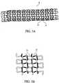
- a co-knitted stent 18is shown including a ceramic fiber 20 and a non-ceramic fiber 22 .
- Ceramic fiber 20can generally be as described above for fiber 12 .
- Non-ceramic fiber 22can be a material selected for its strength and/or elasticity. Examples of materials for non-ceramic fiber 22 include, stainless steel (e.g., 300 series stainless steel), Nitinol (which can provide endoprosthesis 18 with superelasticity and shape memory properties), and composite materials as described in Heath, U.S. Pat. No. 5,725,570, and Mayer, U.S. Pat. No. 5,800,511.
- non-ceramic fiber 22can be a bioabsorbable polymer or a non-bioabsorbable polymer.
- stent 18can be formed of two different ceramic fibers, e.g., a silicon borocarbonitride fiber and an aluminum oxide fiber. The two ceramic fibers can be the same. Either, both, or neither of the fibers can include a therapeutic agent or drug, e.g., by dipping, spray coating, or pad printing.
- Stent 18can be formed by knitting or weaving fibers into a tubular member.
- fibers 20 and 22(or two different ceramic fibers) can be continuous knitted in an alternating, sequential co-knit fashion, resulting in successive alternating rows of different material in a single knit structure.
- a co-knit tubular structurecan also be formed by, for example, using a parallel co-knitting process. Co-knitting processes are further described, for example, in Andersen, U.S. Pat. No. 5,366,504.
- fibers 20 and 22can be co-woven to form a stent 24 of open mesh or weave construction, having two sets of oppositely directed, parallel and spaced apart helically woven fibers. Fibers 20 and 22 are interwoven in an over and under braided configuration, forming intersection points 30 . Woven stents and methods of making them are described further in, for example, Mayer, U.S. Pat. No. 5,800,511.
- the stentcan be formed of coils of fibers 20 and 22 .
- fibers 20 and 22can be knotted, twisted, bent, or crocheted to form a stent.
- the tubular structures described above having one or more ceramic fiberscan be incorporated into a variety of medical devices.
- the ceramic tubular structurescan be attached (interiorly and/or exteriorly) to one or more graft materials, or be placed between multiple layers of graft material, to form a vascular graft.
- graft materialssuch as polytetrafluoroethylene (PTFE) and expanded PTFE, and methods of making a graft are described in Lentz et al., U.S. Pat. No. 6,428,571.
- the graftitself can be made of ceramic fibers.
- the graftcan include a therapeutic agent or pharmaceutically active compound.
- the ceramic tubular structurescan be attached (interiorly and/or exteriorly) to a polymer tube, or be placed between multiple layers of polymer tubes to form a composite tubing or a catheter.
- Suitable polymersare described, for example, in U.S. Published Application No. 2002/0165523 A1, published on Nov. 7, 2002.
- the composite tubing or cathetercan include multiple polymer layers, as described in U.S. Published Application No. 2002/0165523 A1.
- Methods of making a reinforced catheter, e.g., having a braided layerare described in, for example, Wilson et al., U.S. Pat. No. 6,425,898, which is hereby incorporated by reference.
- the ceramic tubular structurescan be used in, for example, chronic infusion catheters, AV catheters, neurointerventional catheters, drainage catheters, or inflation/dilatation catheters.
- ceramic fiberscan be used to reinforce endoscopes and/or tubing (e.g., in embodiments in which torqueability, kink resistance, flexibility, and/or crush resistance are desired).
- Tubing for medical applicationsis described, for example, in Wendlandt et al., U.S. Published Application No. 2003/0176849, which is hereby incorporated by reference.
- the composite tubingcan be drawn and blow molded to form a medical balloon, e.g., one having ceramic structure between two polymer layers.
- a medical balloone.g., one having ceramic structure between two polymer layers.
- Methods of forming a medical balloon, including suitable materials,are described, for example, in U.S. Ser. No. 10/263,225, filed on Oct. 2, 2002, and entitled “Medical Balloon”.
- the ceramic tubular structureswhich can be knitted, woven, braided, co-knitted, or co-woven, can reinforce the medical devices.
- a medical devicecan have two or more ceramic tubular structures.
- an endoprosthesis 38(as shown, a stent) includes a polymer matrix 40 containing chopped ceramic fibers 42 embedded in the matrix.
- chopped ceramic fibers 42 embedded in polymer matrix 40can enhance the strength and/or biocompatibility of the medical device.
- chopped fibers 42can include a therapeutic agent or pharmaceutically active compound. Fibers 42 can be formed by chopping ceramic fibers 12 into smaller pieces.
- ceramic fibers 42are shown in a stent in FIG. 7 , ceramic fibers 42 can be used in any of a number of different medical devices.
- ceramic fibers 42can be used to reinforce a suture.
- a suture that is reinforced with ceramic fibers 42can then be used to weave, knit, or otherwise form a graft or covering.
- ceramic fibers 42can be used to reinforce a stent-graft, polymer covering, or graft.
- a graft that is reinforced with ceramic fibers 42may be able to support itself without requiring the support of a stent.
- Ceramic fibers 42can be dispersed relatively evenly throughout the graft, or can be dispersed asymmetrically.
- the graftcan include a higher density of ceramic fibers at the ends of the graft.
- the ceramic fibersmay impart the ends of the graft with enhanced stiffness, such that the graft may be able to stay open without the support of a stent.
- the grafts or stent-graftscan include synthetic vascular grafts that can be used for replacement of blood vessels in part or in whole.
- the graftscan be tubular and can have, e.g., a woven, knit or velour construction.
- Preferred materials for the grafts and covering materials for the stent-graftsinclude polyethylene terephthalate and polytetrafluoroethylene.
- the vascular graftscan be reinforced with, for example, helices, and/or rings (e.g., to help provide uniform strength over the entire surface of the graft tubing).
- the materials of which such grafts can be constructedare biologically compatible materials including, but not limited to, thermoplastic materials such as polyester, polytetrafluoroethylene (PTFE), silicone and polyurethanes.
- the preferred materialsinclude polyester fibers and PTFE.
- the graftcan have a coating that includes a polymeric material and one or more therapeutic agents and/or pharmaceutically active compounds (e.g., paclitaxel, sirolimus, everolimus).
- the polymeric materialcan be a copolymer of polycaprolactone and polylactic acid. Grafts are described in Schmitt, U.S. Pat. No. 5,509,931; Schmitt, U.S. Pat. No. 5,527,353; and Popadiuk et al., U.S. Pat. No. 5,556,426, all of which are hereby incorporated by reference.
- Ceramic fibers 42can be used to reinforce a balloon.
- An advantage to a balloon reinforced by ceramic fibers 42is that the balloon can have a relatively low wall thickness (e.g., about 0.001 inch), while simultaneously being strong.
- Fibers 42are generally elongate structures having lengths greater than widths or diameters. In some cases, fibers 42 can have a length of from about five microns to about 25,000 microns (e.g., from about ten microns to about 1,000 microns, from about ten microns to about 100 microns).
- Fibers 42can have a length equal to or greater than about five microns (e.g., greater than about 5000 microns; greater than about 10,000 microns; greater than about 15,000 microns; greater than about 20,000 microns), and/or equal to or less than about 25,000 microns (e.g., less than about 20,000 microns; less than about 15,000 microns; less than about 10,000 microns; less than about 5,000 microns).
- the lengths of fibers 42may be uniform or relatively random.
- Fibers 42can have a width or diameter of from about one micron to about 50 microns (e.g., about ten microns). Fibers 42 can have a width or diameter that is equal to or greater than about one micron (e.g., greater than about five microns, greater than about ten microns, greater than about 15 microns, greater than about 20 microns, greater than about 25 microns, greater than about 30 microns, greater than about 35 microns, greater than about 40 microns, greater than about 45 microns), and/or equal to or less than about 50 microns (e.g., less than about 45 microns, less than about 40 microns, less than about 35 microns, less than about 30 microns, less than about 25 microns, less than about 20 microns, less than about 15 microns, less than about ten microns, less than about five microns).
- the width or diametercan be uniform or relatively random.
- fibers 42can have a width or diameter of less than about three microns (e.g., less than about two microns). In certain embodiments, fibers 42 can have a width or diameter of from about two nanometers to about four nanometers.
- fibers 42have length to width aspect ratios of from about 5:1 to about 500:1 (e.g., from about 5:1 to about 20:1), although higher aspect ratios are possible. Fibers 42 can have a length to width aspect ratio equal to or greater than about 5:1 (e.g., greater than about 50:1, greater than about 100:1, greater than about 150:1, greater than about 200:1, greater than about 250:1, greater than about 300:1, greater than about 350:1, greater than about 400:1, greater than about 450:1), and/or equal to or less than about 500:1 (e.g., less than about 450:1, less than about 400:1, less than about 350:1, less than about 300:1, less than about 250:1, less than about 200:1, less than about 150:1, less than about 100:1, less than about 50:1).
- a length to width aspect ratioequal to or greater than about 5:1 (e.g., greater than about 50:1, greater than about 100:1, greater than about 150:1, greater than about 200:1, greater than about 250
- the width used to determine the aspect ratiocan be the narrowest or broadest width.
- the lengthcan be the largest dimension of a fiber. Mixtures of fibers having two or more different aspect ratios and/or dimensions can be used.
- Smaller ceramic fibers 42may be used to reinforce thinner walls of an endoprosthesis, while larger ceramic fibers 42 may be used to reinforce thicker walls of an endoprosthesis.
- smaller fibersare used to reinforce, for example, sutures or balloons, which may require smaller fibers because of their smaller size.
- the fiberscan strengthen the device being reinforced such that the device can have even thinner walls.
- Larger fiberson the other hand, preferably are used to reinforce a catheter shaft or a stent covering.
- Ceramic fibers 42can be placed in different sections of an endoprosthesis (such as a catheter) to provide, e.g., variable stiffness.
- a cathetercan have a higher concentration of ceramic fibers 42 in its proximal end than in its distal end.
- the proximal endmay have enhanced stiffness, which can increase the pushability, torque, kink resistance, and/or control of the catheter.
- the distal endhas a lower concentration of ceramic fibers 42 , then it can serve as an atraumatic tip and provide flexibility when, for example, the catheter is negotiating a tortuous lumen.
- Ceramic fibers 42can be axially, radially, and/or circumferentially oriented in a medical device or component.
- the orientation of ceramic fibers 42 in a medical device or componentcan be selected to form a pattern or can be random.
- the orientation of ceramic fibers 42 in a medical device or componentcan be selected to enhance reinforcement of the device or component, and/or can be selected to enhance the flexibility, radial strength, and/or torqueability of the device or component.
- Ceramic fibers 42can have a variety of configurations or shapes. Fibers 42 can have a cross-section that is circular or non-circular, such as oval, or regularly or irregularly polygonal having three, four, five, six, seven, or eight or more sides. In some cases, fibers 42 can be flat. Fibers 42 can be in the shape of a ribbon (e.g., a flat or wavy ribbon). Along their length, fibers 42 can be, for example, straight, wavy, coiled, or folded. Fibers 42 can be textured, e.g. to combine more easily with the matrix in which they are suspended and/or to hold therapeutic agents for delivery. The surfaces of fibers 42 can be roughened. As an example, fibers 42 can be porous, etched, or grooved.
- Fibers 42can have a uniform or non-uniform thickness, e.g., the fibers can taper along their lengths. Mixtures of fibers having two or more different configurations or shapes can be used. In other embodiments, thin, flat shard-like fibers having irregular shapes can be used. Fibers 42 can be, e.g., whiskers, such as those described in Javier Llorca, “Fatigue of Particle- and Whisker-Reinforced Metal-Matrix Composites,” Progress in Materials Science 47 (2002), 283-353, which is hereby incorporated by reference.
- Polymer matrix 40can include one or more polymers that are biocompatible.
- the polymerpreferably has acceptable vascular compatibility, e.g., relatively low thrombogenecity and low cytotoxicity.
- the polymeris preferably stable in various bodily fluids. Suitable polymers are described in U.S. Ser. No. 10/229,548, filed on Aug. 28, 2002, and entitled “Medical Devices and Methods of Making the Same”.
- Polymer matrix 40 and ceramic fibers 42can be mixed together and formed into a medical device, e.g., by extruding the polymer matrix and the ceramic fibers 42 .
- a tube or a catheter from a composite including a polymerare described in Weber, U.S. Published Application No. 2003/0065355, which is hereby incorporated by reference.
- the tubecan be formed into a medical balloon, as described in U.S. Ser. No. 10/263,225, filed on Oct. 2, 2002, and entitled “Medical Balloon”.
- Methods of making a stent, including methods of preferentially orienting fibersare described in, for example, U.S. Ser. No. 10/229,548, filed on Aug.
- vascular graftMethods of making a vascular graft are described in, for example, Lentz et al., U.S. Pat. No. 6,428,571.
- the mixture of polymer matrix 40 and fibers 42can be used to make an embolic filter, e.g., a filter membrane, as described in, for example, Weber, U.S. Published Application No. 2003/0065355, incorporated above.
- the mixture of polymer matrix 40 and fibers 42can be injection molded to form certain medical devices.
- a mixture containing a relatively hard polymer, e.g., polycarbonate or acrylonitrile-butadiene-styrene (ABS)can be injection molded to form a port housing, as described, e.g., in U.S. Ser. No. 10/117,947, filed Apr. 8, 2002, and entitled “Medical Devices”.
- medical devicescan be reinforced with beads (with or without fibers 42 ).
- the beadscan be spherical, oval, or elliptical.
- the beadscan have widths that are equal to the diameters of the beads.
- the mixture of polymer matrix 40 and fibers 42can contain a radiopaque material, a material that is visible by magnetic resonance imaging (MRI), and/or an ultrasound contrast agent.
- radiopaque materialsinclude tantalum, tungsten, platinum, palladium, or gold.
- the radiopaque materialcan be placed inside an inclusion member.
- the radiopaque materiale.g., a band of radiopaque material, can be placed on a medical device at selected positions, such as, for example, on a catheter adjacent to a balloon.
- fibers 42can be rendered radiopaque (for example, by incorporating (e.g., coating) radiopaque metals or agents into the fibers).
- MRI visible materialsinclude non-ferrous metal-alloys containing paramagnetic elements (e.g., dysprosium or gadolinium) such as terbium-dysprosium, dysprosium, and gadolinium; non-ferrous metallic bands coated with an oxide or a carbide layer of dysprosium or gadolinium (e.g., Dy 2 O 3 or Gd 2 O 3 ); non-ferrous metals (e.g., copper, silver, platinum, or gold) coated with a layer of superparamagnetic material, such as nanocrystalline Fe 3 O 4 , CoFe 2 O 4 , MnFe 2 O 4 , or MgFe 2 O 4 ; and nanocrystalline particles of the transition metal oxides (e.g., oxides of Fe, Co, Ni).
- paramagnetic elementse.g., dysprosium or gadolinium
- non-ferrous metallic bands coated with an oxide or a carbide layer of dysprosium or gadolinium
- the ultrasound contrast agentcan be any material that enhances visibility during ultrasound imaging.
- An ultrasound contrast agentcan include a suspension having trapped bubbles of sufficient size to deflect sound waves.
- ceramic fibers 42can be rendered visible to ultrasound spectroscopy by, for example, surface roughening, bubble inclusions, and/or a coating.
- Any of the medical devices described abovecan include an MRI contrast agent, a radiopaque material, and/or an ultrasound contrast agent, in any combination or arrangement.
- Ceramic fibers 42can be used to form a composite. Composites are described in “Fatigue of Particle- and Whisker-Reinforced Metal-Matrix Composites,” incorporated above, and in references described therein. While ceramic fibers 42 are embedded in polymer matrix 40 in FIG. 4 , in some cases ceramic fibers 42 can be embedded in a metal (e.g., stainless steel, titanium, iridium), to form a ceramic reinforced metal matrix composite. The metal can then be used to make a medical device (e.g., a stent).
- a metale.g., stainless steel, titanium, iridium
- a ceramic fibercan be formed by depositing ceramic material onto a substrate (e.g., a metal wire filament, as described in, for example, Heath, U.S. Pat. No. 5,725,570, and Mayer, U.S. Pat. No. 5,800,511).
- the ceramiccan be deposited using techniques such as physical vapor deposition (PVD), chemical vapor deposition (CVD), or metalorganic chemical vapor deposition (MOCVD).
- Ceramic fibers 12 and 42 as described abovecan be used in many types of medical devices.
- medical devicesinclude catheters, guidewires, balloons, filters (e.g., vena cava filters), stents, stent-grafts, vascular grafts, intraluminal paving systems, and implants.
- filterse.g., vena cava filters
- stentse.g., vena cava filters
- stent-graftse.g., vena cava filters
- vascular graftsvascular grafts
- intraluminal paving systemse.g., intraluminal paving systems
- implantse.g., intraluminal paving systems
- Such devicescan be implanted or otherwise utilized in body lumina and organs such as the coronary vasculature, esophagus, trachea, colon, biliary tract, urinary tract, prostate, and brain.
- the filterscan include, for example, thrombus filters that can be placed at a selected location within the vascular system (e.g., the vena cava) and removed when no longer required.
- an implanted vascular filtercan have a polymeric material/drug outer coating thereon.
- the filtercan have a polymeric material/paclitaxel outer coating (e.g., a polylactic acid/polycaprolactone copolymer/paclitaxel coating).
- the polymeric coatingcan also have incorporated therein or thereon any other therapeutic agent that is used for reducing the formation of, or complications due to, clot formation or neointima formation.
- agentsinclude antithrombogenic agents and thrombolytic agents and other antiproliferative agents. Filters are described in International Application No. WO 96/17634 and International Application No. WO 96/12448, which are hereby incorporated by reference.
- a polymer matrix including chopped fiberscan be used as an intraluminal paving system.
- the polymer matrixcan be applied directly to an interior surface of vascular or non-vascular lumina.
- An intraluminal paving systemcan be formed, for example, by admixing a drug agent with a liquid polymer, in the absence of a solvent, to form a liquid polymer/drug agent mixture.
- the mixturecan then be applied directly to a luminal surface by any conventional method, such as by injecting the mixture against the luminal surface. Curing of the mixture can occur in-situ.
- a cross-linking or curing agentcan be added to the mixture prior to application thereof to the luminal surface.
- Curingcan also occur in-situ by exposing the polymer/drug agent mixture, after application to the luminal surface, to radiation such as ultraviolet radiation or laser light, heat, or by contact with metabolic fluids such as water at the site where the mixture has been applied to the luminal surface.
- the drug agent in the intraluminal paving systemis paclitaxel.
- the paclitaxelcan be incorporated in the polymeric material alone or in combination with another drug agent.
- the polymeric material incorporating the paclitaxel and, if desired, any additional therapeutic agent(s),can be either bioabsorbable or biostable. Any of the polymers described herein that may be formulated as a liquid may be used to form the polymer/drug agent mixture for use as an intraluminal paving system.
- ceramic fibers 12 and/or 42can be used in aneurysm filling devices (e.g., brain aneurysm filling devices).
- the ceramic fiberscan be radiopaque and/or MRI-compatible.
- the ceramic fiberscan be used to form the aneurysm filling devices (e.g., an aneurysm filling device can be formed by a continuous ceramic fiber) and/or as reinforcement in the aneurysm filling devices.
- the fiberscan be combined (e.g., woven, braided) with another type of material.
- An aneurysm filling device that includes one or more ceramic fiberscan also include one or more agents (e.g., thrombotic agents).
- Aneurysm filling devicesare described, for example, in Bashiri et al., U.S. Pat. No. 6,468,266; Abrams, U.S. Pat. No. 6,331,184; Wallace et al., U.S. Pat. No. 6,280,457; Palermo et al., U.S. Pat. No. 6,190,373; Bashiri et al., U.S. Pat. No. 6,165,178; and Chin et al., U.S. Pat. No. 5,928,260.
Landscapes
- Health & Medical Sciences (AREA)
- Chemical & Material Sciences (AREA)
- Veterinary Medicine (AREA)
- Life Sciences & Earth Sciences (AREA)
- Public Health (AREA)
- General Health & Medical Sciences (AREA)
- Animal Behavior & Ethology (AREA)
- Epidemiology (AREA)
- Engineering & Computer Science (AREA)
- Inorganic Chemistry (AREA)
- Vascular Medicine (AREA)
- Heart & Thoracic Surgery (AREA)
- Transplantation (AREA)
- Oral & Maxillofacial Surgery (AREA)
- Materials Engineering (AREA)
- Surgery (AREA)
- Composite Materials (AREA)
- Ceramic Engineering (AREA)
- Medicinal Chemistry (AREA)
- Dermatology (AREA)
- Physics & Mathematics (AREA)
- Optics & Photonics (AREA)
- Biomedical Technology (AREA)
- Cardiology (AREA)
- Materials For Medical Uses (AREA)
- Prostheses (AREA)
- Media Introduction/Drainage Providing Device (AREA)
- Nonwoven Fabrics (AREA)
Abstract
Description
Claims (19)
Priority Applications (4)
| Application Number | Priority Date | Filing Date | Title |
|---|---|---|---|
| US10/762,816US7854756B2 (en) | 2004-01-22 | 2004-01-22 | Medical devices |
| EP05705018AEP1706066A2 (en) | 2004-01-22 | 2005-01-06 | Medical devices comprising ceramic fibres |
| PCT/US2005/000200WO2005072651A2 (en) | 2004-01-22 | 2005-01-06 | Medical devices comprising ceramic fibres |
| US12/139,033US8048143B2 (en) | 2004-01-22 | 2008-06-13 | Medical devices |
Applications Claiming Priority (1)
| Application Number | Priority Date | Filing Date | Title |
|---|---|---|---|
| US10/762,816US7854756B2 (en) | 2004-01-22 | 2004-01-22 | Medical devices |
Related Child Applications (1)
| Application Number | Title | Priority Date | Filing Date |
|---|---|---|---|
| US12/139,033DivisionUS8048143B2 (en) | 2004-01-22 | 2008-06-13 | Medical devices |
Publications (2)
| Publication Number | Publication Date |
|---|---|
| US20050163954A1 US20050163954A1 (en) | 2005-07-28 |
| US7854756B2true US7854756B2 (en) | 2010-12-21 |
Family
ID=34794936
Family Applications (2)
| Application Number | Title | Priority Date | Filing Date |
|---|---|---|---|
| US10/762,816Expired - Fee RelatedUS7854756B2 (en) | 2004-01-22 | 2004-01-22 | Medical devices |
| US12/139,033Expired - Fee RelatedUS8048143B2 (en) | 2004-01-22 | 2008-06-13 | Medical devices |
Family Applications After (1)
| Application Number | Title | Priority Date | Filing Date |
|---|---|---|---|
| US12/139,033Expired - Fee RelatedUS8048143B2 (en) | 2004-01-22 | 2008-06-13 | Medical devices |
Country Status (3)
| Country | Link |
|---|---|
| US (2) | US7854756B2 (en) |
| EP (1) | EP1706066A2 (en) |
| WO (1) | WO2005072651A2 (en) |
Cited By (8)
| Publication number | Priority date | Publication date | Assignee | Title |
|---|---|---|---|---|
| US20080274311A1 (en)* | 2004-05-19 | 2008-11-06 | Jordi Relats Manent | Protective Tube |
| US20080312731A1 (en)* | 2004-01-22 | 2008-12-18 | Boston Scientific Scimed, Inc. | Medical devices |
| WO2018055630A1 (en)* | 2016-09-26 | 2018-03-29 | Galit Avior | Stent and stenting method |
| US10799375B2 (en)* | 2016-03-18 | 2020-10-13 | Hangzhou Weiqiang Medical Technology Co., Ltd. | Aortic bare stent and aortic dissection stent |
| US11266517B2 (en) | 2018-04-09 | 2022-03-08 | Boston Scientific Scimed, Inc. | Stent |
| US11559412B2 (en) | 2019-01-07 | 2023-01-24 | Boston Scientific Scimed, Inc. | Stent with anti-migration feature |
| US11918496B2 (en) | 2020-12-02 | 2024-03-05 | Boston Scientific Scimed, Inc. | Stent with improved deployment characteristics |
| US12329665B2 (en) | 2020-09-29 | 2025-06-17 | Boston Scientific Scimed, Inc. | Stent with anti-migration features |
Families Citing this family (86)
| Publication number | Priority date | Publication date | Assignee | Title |
|---|---|---|---|---|
| US7713297B2 (en) | 1998-04-11 | 2010-05-11 | Boston Scientific Scimed, Inc. | Drug-releasing stent with ceramic-containing layer |
| US7018401B1 (en) | 1999-02-01 | 2006-03-28 | Board Of Regents, The University Of Texas System | Woven intravascular devices and methods for making the same and apparatus for delivery of the same |
| WO2003002243A2 (en) | 2001-06-27 | 2003-01-09 | Remon Medical Technologies Ltd. | Method and device for electrochemical formation of therapeutic species in vivo |
| US7147661B2 (en) | 2001-12-20 | 2006-12-12 | Boston Scientific Santa Rosa Corp. | Radially expandable stent |
| US7722578B2 (en)* | 2004-09-08 | 2010-05-25 | Boston Scientific Scimed, Inc. | Medical devices |
| US8500797B2 (en)* | 2004-09-08 | 2013-08-06 | Boston Scientific Scimed, Inc. | Medical devices |
| US8048028B2 (en) | 2005-02-17 | 2011-11-01 | Boston Scientific Scimed, Inc. | Reinforced medical balloon |
| CA2609687C (en)* | 2005-05-24 | 2014-04-22 | Inspire M.D Ltd. | Stent apparatuses for treatment via body lumens and methods of use |
| US20070038290A1 (en)* | 2005-08-15 | 2007-02-15 | Bin Huang | Fiber reinforced composite stents |
| US8840660B2 (en) | 2006-01-05 | 2014-09-23 | Boston Scientific Scimed, Inc. | Bioerodible endoprostheses and methods of making the same |
| US8089029B2 (en) | 2006-02-01 | 2012-01-03 | Boston Scientific Scimed, Inc. | Bioabsorbable metal medical device and method of manufacture |
| US20070224235A1 (en) | 2006-03-24 | 2007-09-27 | Barron Tenney | Medical devices having nanoporous coatings for controlled therapeutic agent delivery |
| US8187620B2 (en) | 2006-03-27 | 2012-05-29 | Boston Scientific Scimed, Inc. | Medical devices comprising a porous metal oxide or metal material and a polymer coating for delivering therapeutic agents |
| US8048150B2 (en) | 2006-04-12 | 2011-11-01 | Boston Scientific Scimed, Inc. | Endoprosthesis having a fiber meshwork disposed thereon |
| US9101505B2 (en)* | 2006-04-27 | 2015-08-11 | Brs Holdings, Llc | Composite stent |
| US9155646B2 (en)* | 2006-04-27 | 2015-10-13 | Brs Holdings, Llc | Composite stent with bioremovable ceramic flakes |
| US20070258903A1 (en)* | 2006-05-02 | 2007-11-08 | Kleiner Lothar W | Methods, compositions and devices for treating lesioned sites using bioabsorbable carriers |
| US8815275B2 (en) | 2006-06-28 | 2014-08-26 | Boston Scientific Scimed, Inc. | Coatings for medical devices comprising a therapeutic agent and a metallic material |
| WO2008002778A2 (en) | 2006-06-29 | 2008-01-03 | Boston Scientific Limited | Medical devices with selective coating |
| GB0613670D0 (en) | 2006-07-10 | 2006-08-16 | Angiomed Ag | Tubular metal prosthesis and method of making it |
| EP2054537A2 (en) | 2006-08-02 | 2009-05-06 | Boston Scientific Scimed, Inc. | Endoprosthesis with three-dimensional disintegration control |
| US20080124373A1 (en)* | 2006-08-02 | 2008-05-29 | Inframat Corporation | Lumen - supporting devices and methods of making and using |
| US20080069854A1 (en)* | 2006-08-02 | 2008-03-20 | Inframat Corporation | Medical devices and methods of making and using |
| EP2068757B1 (en) | 2006-09-14 | 2011-05-11 | Boston Scientific Limited | Medical devices with drug-eluting coating |
| WO2008034007A2 (en)* | 2006-09-15 | 2008-03-20 | Boston Scientific Limited | Medical devices |
| ES2357661T3 (en) | 2006-09-15 | 2011-04-28 | Boston Scientific Scimed, Inc. | BIOEROSIONABLE ENDOPROOTHESIS WITH BIOESTABLE INORGANIC LAYERS. |
| JP2010503489A (en) | 2006-09-15 | 2010-02-04 | ボストン サイエンティフィック リミテッド | Biodegradable endoprosthesis and method for producing the same |
| EP2959925B1 (en) | 2006-09-15 | 2018-08-29 | Boston Scientific Limited | Medical devices and methods of making the same |
| WO2008034066A1 (en) | 2006-09-15 | 2008-03-20 | Boston Scientific Limited | Bioerodible endoprostheses and methods of making the same |
| WO2008036548A2 (en) | 2006-09-18 | 2008-03-27 | Boston Scientific Limited | Endoprostheses |
| MX2009004291A (en) | 2006-10-22 | 2009-09-07 | Idev Technologies Inc | Methods for securing strand ends and the resulting devices. |
| US7981150B2 (en) | 2006-11-09 | 2011-07-19 | Boston Scientific Scimed, Inc. | Endoprosthesis with coatings |
| EP3292837B1 (en) | 2006-11-22 | 2022-11-09 | Inspire M.D Ltd | Optimized stent jacket |
| ES2506144T3 (en) | 2006-12-28 | 2014-10-13 | Boston Scientific Limited | Bioerodible endoprosthesis and their manufacturing procedure |
| US8431149B2 (en) | 2007-03-01 | 2013-04-30 | Boston Scientific Scimed, Inc. | Coated medical devices for abluminal drug delivery |
| US8070797B2 (en) | 2007-03-01 | 2011-12-06 | Boston Scientific Scimed, Inc. | Medical device with a porous surface for delivery of a therapeutic agent |
| US8177834B2 (en)* | 2007-03-12 | 2012-05-15 | Cook Medical Technologies Llc | Woven fabric with shape memory element strands |
| US8067054B2 (en)* | 2007-04-05 | 2011-11-29 | Boston Scientific Scimed, Inc. | Stents with ceramic drug reservoir layer and methods of making and using the same |
| US7976915B2 (en) | 2007-05-23 | 2011-07-12 | Boston Scientific Scimed, Inc. | Endoprosthesis with select ceramic morphology |
| US9265636B2 (en)* | 2007-05-25 | 2016-02-23 | C. R. Bard, Inc. | Twisted stent |
| US7942926B2 (en) | 2007-07-11 | 2011-05-17 | Boston Scientific Scimed, Inc. | Endoprosthesis coating |
| US8002823B2 (en) | 2007-07-11 | 2011-08-23 | Boston Scientific Scimed, Inc. | Endoprosthesis coating |
| EP2187988B1 (en) | 2007-07-19 | 2013-08-21 | Boston Scientific Limited | Endoprosthesis having a non-fouling surface |
| US7931683B2 (en) | 2007-07-27 | 2011-04-26 | Boston Scientific Scimed, Inc. | Articles having ceramic coated surfaces |
| US8815273B2 (en) | 2007-07-27 | 2014-08-26 | Boston Scientific Scimed, Inc. | Drug eluting medical devices having porous layers |
| US20090030504A1 (en)* | 2007-07-27 | 2009-01-29 | Boston Scientific Scimed, Inc. | Medical devices comprising porous inorganic fibers for the release of therapeutic agents |
| WO2009018340A2 (en) | 2007-07-31 | 2009-02-05 | Boston Scientific Scimed, Inc. | Medical device coating by laser cladding |
| JP2010535541A (en) | 2007-08-03 | 2010-11-25 | ボストン サイエンティフィック リミテッド | Coating for medical devices with large surface area |
| US8052745B2 (en) | 2007-09-13 | 2011-11-08 | Boston Scientific Scimed, Inc. | Endoprosthesis |
| US8998978B2 (en)* | 2007-09-28 | 2015-04-07 | Abbott Cardiovascular Systems Inc. | Stent formed from bioerodible metal-bioceramic composite |
| WO2009047490A2 (en)* | 2007-10-08 | 2009-04-16 | Renishaw Plc | Catheter |
| US8029554B2 (en) | 2007-11-02 | 2011-10-04 | Boston Scientific Scimed, Inc. | Stent with embedded material |
| US8216632B2 (en) | 2007-11-02 | 2012-07-10 | Boston Scientific Scimed, Inc. | Endoprosthesis coating |
| US7938855B2 (en) | 2007-11-02 | 2011-05-10 | Boston Scientific Scimed, Inc. | Deformable underlayer for stent |
| US8834552B2 (en)* | 2007-12-27 | 2014-09-16 | Cook Medical Technologies Llc | Stent graft having floating yarns |
| US8920491B2 (en) | 2008-04-22 | 2014-12-30 | Boston Scientific Scimed, Inc. | Medical devices having a coating of inorganic material |
| US8932346B2 (en) | 2008-04-24 | 2015-01-13 | Boston Scientific Scimed, Inc. | Medical devices having inorganic particle layers |
| US7998192B2 (en) | 2008-05-09 | 2011-08-16 | Boston Scientific Scimed, Inc. | Endoprostheses |
| US8236046B2 (en) | 2008-06-10 | 2012-08-07 | Boston Scientific Scimed, Inc. | Bioerodible endoprosthesis |
| EP2303350A2 (en) | 2008-06-18 | 2011-04-06 | Boston Scientific Scimed, Inc. | Endoprosthesis coating |
| US9820746B2 (en)* | 2008-07-28 | 2017-11-21 | Incube Laboratories LLC | System and method for scaffolding anastomoses |
| US7985252B2 (en) | 2008-07-30 | 2011-07-26 | Boston Scientific Scimed, Inc. | Bioerodible endoprosthesis |
| US8709080B2 (en)* | 2008-09-19 | 2014-04-29 | E. Benson Hood Laboratories | Coated devices comprising a fiber mesh imbedded in the device walls |
| US8076529B2 (en) | 2008-09-26 | 2011-12-13 | Abbott Cardiovascular Systems, Inc. | Expandable member formed of a fibrous matrix for intraluminal drug delivery |
| US8226603B2 (en) | 2008-09-25 | 2012-07-24 | Abbott Cardiovascular Systems Inc. | Expandable member having a covering formed of a fibrous matrix for intraluminal drug delivery |
| US8049061B2 (en) | 2008-09-25 | 2011-11-01 | Abbott Cardiovascular Systems, Inc. | Expandable member formed of a fibrous matrix having hydrogel polymer for intraluminal drug delivery |
| US8382824B2 (en) | 2008-10-03 | 2013-02-26 | Boston Scientific Scimed, Inc. | Medical implant having NANO-crystal grains with barrier layers of metal nitrides or fluorides |
| US8231980B2 (en) | 2008-12-03 | 2012-07-31 | Boston Scientific Scimed, Inc. | Medical implants including iridium oxide |
| EP2403546A2 (en) | 2009-03-02 | 2012-01-11 | Boston Scientific Scimed, Inc. | Self-buffering medical implants |
| US8071156B2 (en) | 2009-03-04 | 2011-12-06 | Boston Scientific Scimed, Inc. | Endoprostheses |
| US8287937B2 (en) | 2009-04-24 | 2012-10-16 | Boston Scientific Scimed, Inc. | Endoprosthese |
| US8556957B2 (en)* | 2009-06-18 | 2013-10-15 | Medtronic Vascular, Inc. | Biodegradable medical device with hydroxyapatite filaments and biodegradable polymer fibers |
| US20110190886A1 (en)* | 2010-01-29 | 2011-08-04 | Wisconsin Alumni Research Foundation | Braided tertiary nanofibrous structure for ligament, tendon, and muscle tissue implant |
| WO2011119536A1 (en) | 2010-03-22 | 2011-09-29 | Abbott Cardiovascular Systems Inc. | Stent delivery system having a fibrous matrix covering with improved stent retention |
| US8668732B2 (en) | 2010-03-23 | 2014-03-11 | Boston Scientific Scimed, Inc. | Surface treated bioerodible metal endoprostheses |
| ES2418848B1 (en)* | 2012-02-10 | 2014-04-30 | Universidad De Valladolid | Supports for the regeneration, growth and transplantation of cells and tissues |
| US9333099B2 (en) | 2012-03-30 | 2016-05-10 | Abbott Cardiovascular Systems Inc. | Magnesium alloy implants with controlled degradation |
| US10736995B2 (en)* | 2012-05-02 | 2020-08-11 | Les Entreprises Nanostent Inc. | Bioresorbable medical devices and method of manufacturing the same |
| CN104936550B (en) | 2012-11-15 | 2017-09-22 | 恩菲纽姆血管技术有限公司 | Temporary vascular scaffolds and scoring devices |
| EP2967930B1 (en)* | 2013-03-13 | 2018-11-28 | Boston Scientific Scimed, Inc. | Anti-migration tissue anchoring system for a fully covered stent |
| WO2016054536A1 (en) | 2014-10-02 | 2016-04-07 | Boston Scientific Scimed, Inc. | Controlled ingrowth feature for antimigration |
| MX387162B (en)* | 2015-07-21 | 2025-03-18 | Avent Inc | ULTRASOUND CATHETER ASSEMBLY. |
| EP3391852A3 (en) | 2017-04-21 | 2018-11-14 | Cook Medical Technologies LLC | Reinforced graft prosthesis |
| US11684498B2 (en) | 2018-10-19 | 2023-06-27 | Inspire M.D Ltd. | Methods of using a self-adjusting stent assembly and kits including same |
| WO2020087288A1 (en)* | 2018-10-30 | 2020-05-07 | 高雄医学大学 | Biomedical material with synergistic effect, manufacturing method therefor and system comprising same |
| WO2021146021A1 (en) | 2020-01-13 | 2021-07-22 | Boston Scientific Scimed, Inc. | Anti-migration stent |
Citations (100)
| Publication number | Priority date | Publication date | Assignee | Title |
|---|---|---|---|---|
| US3914490A (en)* | 1973-08-01 | 1975-10-21 | Dana Corp | Ceramic fiber gasket |
| US4060412A (en)* | 1976-01-08 | 1977-11-29 | A Silag Inc. | Method for preparing a fiber reinforced metal matrix using microscopic fibers |
| US4446579A (en)* | 1978-12-04 | 1984-05-08 | Kyocera Corporation | Flexible ceramic bio-implant member |
| US4475892A (en)* | 1982-10-13 | 1984-10-09 | Jaff Investment Company | Microcellular ceramic material and process for manufacture thereof |
| US4604097A (en)* | 1985-02-19 | 1986-08-05 | University Of Dayton | Bioabsorbable glass fibers for use in the reinforcement of bioabsorbable polymers for bone fixation devices and artificial ligaments |
| US4655777A (en)* | 1983-12-19 | 1987-04-07 | Southern Research Institute | Method of producing biodegradable prosthesis and products therefrom |
| US4655771A (en) | 1982-04-30 | 1987-04-07 | Shepherd Patents S.A. | Prosthesis comprising an expansible or contractile tubular body |
| US4922905A (en) | 1985-11-30 | 1990-05-08 | Strecker Ernst P | Dilatation catheter |
| US5015253A (en) | 1989-06-15 | 1991-05-14 | Cordis Corporation | Non-woven endoprosthesis |
| US5195969A (en) | 1991-04-26 | 1993-03-23 | Boston Scientific Corporation | Co-extruded medical balloons and catheter using such balloons |
| US5221558A (en)* | 1990-01-12 | 1993-06-22 | Lanxide Technology Company, Lp | Method of making ceramic composite bodies |
| US5258445A (en)* | 1990-03-08 | 1993-11-02 | The B. F. Goodrich Company | Fiber-reinforced thermoplastic molding compositions using a copolyester |
| US5270086A (en) | 1989-09-25 | 1993-12-14 | Schneider (Usa) Inc. | Multilayer extrusion of angioplasty balloons |
| US5366504A (en) | 1992-05-20 | 1994-11-22 | Boston Scientific Corporation | Tubular medical prosthesis |
| US5380298A (en) | 1993-04-07 | 1995-01-10 | The United States Of America As Represented By The Secretary Of The Navy | Medical device with infection preventing feature |
| US5468544A (en)* | 1993-11-15 | 1995-11-21 | The Trustees Of The University Of Pennsylvania | Composite materials using bone bioactive glass and ceramic fibers |
| US5470345A (en) | 1994-06-16 | 1995-11-28 | Medtronic, Inc. | Implantable medical device with multi-layered ceramic enclosure |
| US5509931A (en) | 1990-08-28 | 1996-04-23 | Meadox Medicals, Inc. | Ravel-resistant self-supporting woven vascular graft |
| WO1996012448A1 (en) | 1994-10-25 | 1996-05-02 | Scimed Life Systems, Inc. | Removable thrombus filter |
| US5514112A (en) | 1992-10-02 | 1996-05-07 | Boston Scientific Corporation | Drainage catheter and method of use |
| WO1996017634A2 (en) | 1994-11-30 | 1996-06-13 | Boston Scientific Corporation | Blood clot filtering |
| US5527353A (en) | 1993-12-02 | 1996-06-18 | Meadox Medicals, Inc. | Implantable tubular prosthesis |
| US5530355A (en)* | 1993-05-13 | 1996-06-25 | Doty Scientific, Inc. | Solenoidal, octopolar, transverse gradient coils |
| US5556426A (en) | 1994-08-02 | 1996-09-17 | Meadox Medicals, Inc. | PTFE implantable tubular prostheses with external coil support |
| US5593719A (en) | 1994-03-29 | 1997-01-14 | Southwest Research Institute | Treatments to reduce frictional wear between components made of ultra-high molecular weight polyethylene and metal alloys |
| US5629077A (en) | 1994-06-27 | 1997-05-13 | Advanced Cardiovascular Systems, Inc. | Biodegradable mesh and film stent |
| US5674242A (en) | 1995-06-06 | 1997-10-07 | Quanam Medical Corporation | Endoprosthetic device with therapeutic compound |
| US5679470A (en) | 1993-01-19 | 1997-10-21 | Schneider (Usa) Inc. | Process for manufacturing clad composite stent |
| US5690670A (en) | 1989-12-21 | 1997-11-25 | Davidson; James A. | Stents of enhanced biocompatibility and hemocompatibility |
| US5709703A (en) | 1995-11-14 | 1998-01-20 | Schneider (Europe) A.G. | Stent delivery device and method for manufacturing same |
| US5709713A (en) | 1995-03-31 | 1998-01-20 | Cardiovascular Concepts, Inc. | Radially expansible vascular prosthesis having reversible and other locking structures |
| US5718973A (en) | 1993-08-18 | 1998-02-17 | W. L. Gore & Associates, Inc. | Tubular intraluminal graft |
| US5725573A (en) | 1994-03-29 | 1998-03-10 | Southwest Research Institute | Medical implants made of metal alloys bearing cohesive diamond like carbon coatings |
| US5725570A (en) | 1992-03-31 | 1998-03-10 | Boston Scientific Corporation | Tubular medical endoprostheses |
| US5733326A (en) | 1996-05-28 | 1998-03-31 | Cordis Corporation | Composite material endoprosthesis |
| US5780807A (en) | 1994-11-28 | 1998-07-14 | Advanced Cardiovascular Systems, Inc. | Method and apparatus for direct laser cutting of metal stents |
| US5797877A (en) | 1993-10-01 | 1998-08-25 | Boston Scientific Corporation | Medical device balloons containing thermoplastic elastomers |
| US5800519A (en) | 1994-04-29 | 1998-09-01 | Kopin Corporation | Tubular medical prosthesis for use in a body lumen |
| US5807954A (en)* | 1993-11-05 | 1998-09-15 | Lanxide Technology Company, Lp | Metal-nitrogen polymer compositions comprising organic electrophiles |
| DE19730296A1 (en) | 1997-07-15 | 1999-01-21 | Medic Medizintechnik Lothar Se | Implant for use in the body, e.g. heart flap |
| US5899935A (en) | 1997-08-04 | 1999-05-04 | Schneider (Usa) Inc. | Balloon expandable braided stent with restraint |
| EP0923913A2 (en) | 1997-12-19 | 1999-06-23 | Indigo Medical, Incorporated | Coiled multi-component polymer implant |
| WO1999032051A1 (en) | 1997-12-22 | 1999-07-01 | Impra, Inc. | Supported graft and methods of making same |
| US5928260A (en) | 1997-07-10 | 1999-07-27 | Scimed Life Systems, Inc. | Removable occlusion system for aneurysm neck |
| US5980972A (en) | 1996-12-20 | 1999-11-09 | Schneider (Usa) Inc | Method of applying drug-release coatings |
| US5984905A (en) | 1994-07-11 | 1999-11-16 | Southwest Research Institute | Non-irritating antimicrobial coating for medical implants and a process for preparing same |
| WO1999061081A1 (en) | 1998-05-27 | 1999-12-02 | Sepitec Foundation | Composite of polymer or ceramic materials and component made of such a composite |
| US6015432A (en) | 1998-02-25 | 2000-01-18 | Cordis Corporation | Wire reinforced vascular prosthesis |
| US6054532A (en)* | 1998-05-06 | 2000-04-25 | Bridgestone Corporation | Centipede polymers grafted with hydrogenated block copolymers and polypropylene and gels thereof |
| US6066100A (en) | 1991-05-23 | 2000-05-23 | Scimed Life Systems, Inc. | Intravascular device such as introducer sheath or balloon catheter or the like and methods for use thereof |
| WO2000038590A1 (en) | 1998-12-23 | 2000-07-06 | Stephen George Edward Barker | Endoluminal stent |
| US6087025A (en) | 1994-03-29 | 2000-07-11 | Southwest Research Institute | Application of diamond-like carbon coatings to cutting surfaces of metal cutting tools |
| US6099561A (en) | 1996-10-21 | 2000-08-08 | Inflow Dynamics, Inc. | Vascular and endoluminal stents with improved coatings |
| US6161399A (en) | 1997-10-24 | 2000-12-19 | Iowa-India Investments Company Limited | Process for manufacturing a wire reinforced monolayer fabric stent |
| US6162244A (en) | 1996-03-29 | 2000-12-19 | Willy Ruesch Ag | Layered stent |
| US6165178A (en) | 1997-08-29 | 2000-12-26 | Scimed Life Systems, Inc. | Fast detaching electrically isolated implant |
| US6165213A (en) | 1994-02-09 | 2000-12-26 | Boston Scientific Technology, Inc. | System and method for assembling an endoluminal prosthesis |
| US6190373B1 (en) | 1992-11-13 | 2001-02-20 | Scimed Life Systems, Inc. | Axially detachable embolic coil assembly |
| WO2001034062A2 (en) | 1999-10-25 | 2001-05-17 | Scimed Life Systems, Inc. | Dimensionally stable balloons |
| US6248571B1 (en) | 1995-08-31 | 2001-06-19 | Lonza Ag | Method of producing dihydroxypyrimidine derivatives |
| US6248129B1 (en) | 1990-09-14 | 2001-06-19 | Quanam Medical Corporation | Expandable polymeric stent with memory and delivery apparatus and method |
| US6250952B1 (en) | 1997-08-08 | 2001-06-26 | The Whitaker Corporation | Air bag connector |
| US6264689B1 (en) | 1998-03-31 | 2001-07-24 | Scimed Life Systems, Incorporated | Low profile medical stent |
| US6280457B1 (en) | 1999-06-04 | 2001-08-28 | Scimed Life Systems, Inc. | Polymer covered vaso-occlusive devices and methods of producing such devices |
| US6312458B1 (en) | 2000-01-19 | 2001-11-06 | Scimed Life Systems, Inc. | Tubular structure/stent/stent securement member |
| US6316018B1 (en) | 1997-04-30 | 2001-11-13 | Ni Ding | Drug-releasing coatings for medical devices |
| US6331184B1 (en) | 1999-12-10 | 2001-12-18 | Scimed Life Systems, Inc. | Detachable covering for an implantable medical device |
| US6335029B1 (en) | 1998-08-28 | 2002-01-01 | Scimed Life Systems, Inc. | Polymeric coatings for controlled delivery of active agents |
| US6340441B1 (en) | 1998-03-13 | 2002-01-22 | Scimed Life Systems, Inc. | Multi-layer guide wire and method of manufacture therefor |
| US20020016596A1 (en) | 1998-04-06 | 2002-02-07 | Kevin Cooper | Two phase thermally deformable biocompatible absorbable polymer matrix for use in medical devices |
| US20020082683A1 (en) | 1997-08-01 | 2002-06-27 | Stinson Jonathan S. | Radiopaque markers for implantable prostheses |
| US6425898B1 (en) | 1998-03-13 | 2002-07-30 | Cordis Corporation | Delivery apparatus for a self-expanding stent |
| US6428571B1 (en) | 1996-01-22 | 2002-08-06 | Scimed Life Systems, Inc. | Self-sealing PTFE vascular graft and manufacturing methods |
| US6451059B1 (en)* | 1999-11-12 | 2002-09-17 | Ethicon, Inc. | Viscous suspension spinning process for producing resorbable ceramic fibers and scaffolds |
| US6468304B1 (en) | 1997-07-16 | 2002-10-22 | Centre National De La Recherche Scientifique | Implantable device covered with polymer capable of releasing biologically active substances |
| US20020165523A1 (en) | 2000-03-02 | 2002-11-07 | Chin Albert C. C. | Multilayer medical device |
| US6488694B1 (en) | 1991-01-28 | 2002-12-03 | Advanced Cardiovascular Systems, Inc. | Stent delivery system |
| US20030003220A1 (en) | 2001-07-02 | 2003-01-02 | Sheng-Ping Zhong | Coating a medical appliance with a bubble jet printing head |
| US6506408B1 (en) | 2000-07-13 | 2003-01-14 | Scimed Life Systems, Inc. | Implantable or insertable therapeutic agent delivery device |
| US20030018380A1 (en) | 2000-07-07 | 2003-01-23 | Craig Charles H. | Platinum enhanced alloy and intravascular or implantable medical devices manufactured therefrom |
| US6520952B1 (en) | 2000-03-23 | 2003-02-18 | Neich Medical Co., Ltd. | Ceramic reinforced catheter |
| WO2003015840A2 (en) | 2001-08-17 | 2003-02-27 | Advanced Bio Prosthetic Surfaces, Ltd. | Embolic protection device |
| US20030065355A1 (en) | 2001-09-28 | 2003-04-03 | Jan Weber | Medical devices comprising nonomaterials and therapeutic methods utilizing the same |
| US6551352B2 (en)* | 2001-05-03 | 2003-04-22 | Bionx Implants, Inc. | Method for attaching axial filaments to a self expanding stent |
| US6558794B1 (en) | 2000-06-26 | 2003-05-06 | Technology Assessment & Transfer, Inc. | Ceramic particulate reinforced orthopedic implants |
| US6573331B1 (en)* | 1998-06-10 | 2003-06-03 | Institut Francais Du Petrole | Grafted block polymers containing at least a polyolefin or polydiene sequence comprising a succinimide cycle substituted on nitrogen |
| US6589286B1 (en)* | 2001-09-12 | 2003-07-08 | Jason Litner | Eustachian tube stent |
| WO2003055534A1 (en) | 2001-12-21 | 2003-07-10 | Psimedica Limited | Medical fibres & fabrics |
| US20030143350A1 (en) | 2002-01-14 | 2003-07-31 | Oscar Jimenez | Angioplasty super balloon fabrication with composite materials |
| US20030176849A1 (en) | 2000-03-08 | 2003-09-18 | Wendlandt Jeffrey Michael | Composite flexible tube for medical applications |
| US20030185895A1 (en) | 2002-03-29 | 2003-10-02 | Janel Lanphere | Drug delivery particle |
| US20030191356A1 (en) | 2002-04-08 | 2003-10-09 | Steve Moreci | Medical devices |
| WO2003082565A1 (en) | 2002-04-03 | 2003-10-09 | Tenax Fibers Gmbh | Composite material, method for the production and use thereof |
| US6692499B2 (en)* | 1997-07-02 | 2004-02-17 | Linvatec Biomaterials Oy | Surgical fastener for tissue treatment |
| US20040044397A1 (en) | 2002-08-28 | 2004-03-04 | Stinson Jonathan S. | Medical devices and methods of making the same |
| US6733533B1 (en)* | 2002-11-19 | 2004-05-11 | Zimmer Technology, Inc. | Artificial spinal disc |
| US6805706B2 (en)* | 2002-08-15 | 2004-10-19 | Gmp Cardiac Care, Inc. | Stent-graft with rails |
| US6807440B2 (en)* | 2001-11-09 | 2004-10-19 | Scimed Life Systems, Inc. | Ceramic reinforcement members for MRI devices |
| US20050149102A1 (en)* | 2003-12-22 | 2005-07-07 | Radisch Herbert R.Jr. | Medical device systems |
| US20050208094A1 (en)* | 2003-09-05 | 2005-09-22 | Armitage Bryan M | Bone cement compositions having fiber-reinforcement and/or increased flowability |
Family Cites Families (47)
| Publication number | Priority date | Publication date | Assignee | Title |
|---|---|---|---|---|
| US4001172A (en)* | 1972-04-03 | 1977-01-04 | Exxon Research And Engineering Company | Polymers with improved properties and process therefor |
| WO1988002741A1 (en)* | 1986-10-14 | 1988-04-21 | Nicolas Davidovits | Ceramic-ceramic composite material and production method |
| US4832895A (en)* | 1986-11-21 | 1989-05-23 | Hoechst Celanese Corporation | Boron-containing ceramics through the chemical conversian of borane-containing polymers |
| US4946919A (en)* | 1987-08-31 | 1990-08-07 | Hoechst Celanese Corp. | Boron ceramics from carboralated diacetylene polymers |
| USH812H (en)* | 1987-12-24 | 1990-08-07 | Shell Oil Company | Reinforced polymer |
| US5219959A (en)* | 1988-11-21 | 1993-06-15 | Tosoh Corporation | Production of poly(phenylene sulfide) of an improved reactivity |
| US5238742A (en)* | 1990-01-16 | 1993-08-24 | Dow Corning Corporation | Polymer derived ceramic fibers having improved thermal stability |
| US5169397A (en)* | 1990-02-08 | 1992-12-08 | Olympus Optical Co., Ltd. | Medical instrument |
| GB9122997D0 (en)* | 1991-10-30 | 1991-12-18 | Curran Dennis J G | Ceramic fibres |
| WO1995001138A1 (en)* | 1993-07-02 | 1995-01-12 | Materials Evolution And Development Usa, Inc. | Implantable system for cell growth control |
| US5869178A (en)* | 1994-08-25 | 1999-02-09 | The University Of North Carolina At Chapel Hill | Pultruded fiber-reinforced plastic and related apparatus and method |
| US5691062A (en)* | 1995-02-16 | 1997-11-25 | Clemson University | Molecularly bonded inherently conductive polymers on substrates and shaped articles thereof |
| FI105159B (en)* | 1996-10-25 | 2000-06-30 | Biocon Ltd | Surgical implant, agent or part thereof |
| US6287765B1 (en)* | 1998-05-20 | 2001-09-11 | Molecular Machines, Inc. | Methods for detecting and identifying single molecules |
| US6147135A (en)* | 1998-12-31 | 2000-11-14 | Ethicon, Inc. | Fabrication of biocompatible polymeric composites |
| US6254689B1 (en)* | 1999-03-09 | 2001-07-03 | Lucent Technologies Inc. | System and method for flash photolysis cleaning of a semiconductor processing chamber |
| US7244497B2 (en)* | 2001-09-21 | 2007-07-17 | Outlast Technologies, Inc. | Cellulosic fibers having enhanced reversible thermal properties and methods of forming thereof |
| EP1258229A1 (en)* | 2001-05-15 | 2002-11-20 | Vascular Technologies, Inc. | Flexible and elastic vascular stents and grafts |
| US20060204556A1 (en)* | 2001-12-07 | 2006-09-14 | Cytori Therapeutics, Inc. | Cell-loaded prostheses for regenerative intraluminal applications |
| US6881224B2 (en)* | 2001-12-28 | 2005-04-19 | St. Jude Medical, Inc. | Fatigue test for prosthetic stent |
| US7160929B1 (en)* | 2002-02-13 | 2007-01-09 | Wrigt Materials Research Co | Nanocomposite and molecular-composite polymer foams and method for their production |
| US6955716B2 (en)* | 2002-03-01 | 2005-10-18 | American Dental Association Foundation | Self-hardening calcium phosphate materials with high resistance to fracture, controlled strength histories and tailored macropore formation rates |
| US8337893B2 (en)* | 2002-07-10 | 2012-12-25 | Florida Research Foundation, Inc, University Of | Sol-gel derived bioactive glass polymer composite |
| AU2003263454A1 (en)* | 2002-09-19 | 2004-04-08 | Petrus Besselink | Vascular filter with improved strength and flexibility |
| US7625337B2 (en)* | 2003-01-17 | 2009-12-01 | Gore Enterprise Holdings, Inc. | Catheter assembly |
| US7875347B2 (en)* | 2003-12-29 | 2011-01-25 | General Electric Company | Composite coatings for groundwall insulation, method of manufacture thereof and articles derived therefrom |
| US7803457B2 (en)* | 2003-12-29 | 2010-09-28 | General Electric Company | Composite coatings for groundwall insulation, method of manufacture thereof and articles derived therefrom |
| US7854756B2 (en)* | 2004-01-22 | 2010-12-21 | Boston Scientific Scimed, Inc. | Medical devices |
| US8025960B2 (en)* | 2004-02-02 | 2011-09-27 | Nanosys, Inc. | Porous substrates, articles, systems and compositions comprising nanofibers and methods of their use and production |
| US20110039690A1 (en)* | 2004-02-02 | 2011-02-17 | Nanosys, Inc. | Porous substrates, articles, systems and compositions comprising nanofibers and methods of their use and production |
| CN101095277A (en)* | 2004-03-12 | 2007-12-26 | 斯里国际 | Mechanical meta-materials |
| US8043259B2 (en)* | 2004-05-24 | 2011-10-25 | Boston Scientific Scimed, Inc. | Medical device systems |
| US20060079624A1 (en)* | 2004-10-08 | 2006-04-13 | Hildeberto Nava | Crosslinkable polymer systems |
| US7250381B2 (en)* | 2004-11-12 | 2007-07-31 | Johns Manville | Fibrous nonwoven mats containing polyethermid fibers |
| US7491225B2 (en)* | 2005-02-16 | 2009-02-17 | Boston Scientific Scimed, Inc. | System and method for deploying a drug-eluting external body and tissue scaffold |
| US7381664B2 (en)* | 2005-03-17 | 2008-06-03 | Poly-Med Inc. | Surface electroconductive biostable polymeric articles |
| US8795348B2 (en)* | 2005-06-14 | 2014-08-05 | Boston Scientific Scimed, Inc. | Medical devices and related methods |
| DE602006003701D1 (en)* | 2005-08-26 | 2009-01-02 | Voith Patent Gmbh | With polymer blended polymer particles, method for producing the same and products made therefrom such. B. press felts |
| CA2647325C (en)* | 2005-12-12 | 2015-03-24 | Allaccem, Inc. | Methods and systems for preparing antimicrobial bridged polycyclic compounds |
| US8455088B2 (en)* | 2005-12-23 | 2013-06-04 | Boston Scientific Scimed, Inc. | Spun nanofiber, medical devices, and methods |
| US9155646B2 (en)* | 2006-04-27 | 2015-10-13 | Brs Holdings, Llc | Composite stent with bioremovable ceramic flakes |
| US9101505B2 (en)* | 2006-04-27 | 2015-08-11 | Brs Holdings, Llc | Composite stent |
| US20080051881A1 (en)* | 2006-08-24 | 2008-02-28 | Feng James Q | Medical devices comprising porous layers for the release of therapeutic agents |
| EP2219823A2 (en)* | 2007-10-16 | 2010-08-25 | HKPB Scientific Limited | Surface coating processes and uses of same |
| GB2463401B (en)* | 2008-11-12 | 2014-01-29 | Caris Life Sciences Luxembourg Holdings S A R L | Characterizing prostate disorders by analysis of microvesicles |
| WO2010088699A2 (en)* | 2009-02-02 | 2010-08-05 | Biomerix Corporation | Composite mesh devices and methods for soft tissue repair |
| US11794702B2 (en) | 2016-09-05 | 2023-10-24 | Koito Manufacturing Co., Ltd. | Cleaner, sensor with cleaner, and vehicle including cleaner or sensor with cleaner |
- 2004
- 2004-01-22USUS10/762,816patent/US7854756B2/ennot_activeExpired - Fee Related
- 2005
- 2005-01-06WOPCT/US2005/000200patent/WO2005072651A2/enactiveApplication Filing
- 2005-01-06EPEP05705018Apatent/EP1706066A2/ennot_activeWithdrawn
- 2008
- 2008-06-13USUS12/139,033patent/US8048143B2/ennot_activeExpired - Fee Related
Patent Citations (108)
| Publication number | Priority date | Publication date | Assignee | Title |
|---|---|---|---|---|
| US3914490A (en)* | 1973-08-01 | 1975-10-21 | Dana Corp | Ceramic fiber gasket |
| US4060412A (en)* | 1976-01-08 | 1977-11-29 | A Silag Inc. | Method for preparing a fiber reinforced metal matrix using microscopic fibers |
| US4446579A (en)* | 1978-12-04 | 1984-05-08 | Kyocera Corporation | Flexible ceramic bio-implant member |
| US4655771A (en) | 1982-04-30 | 1987-04-07 | Shepherd Patents S.A. | Prosthesis comprising an expansible or contractile tubular body |
| US4655771B1 (en) | 1982-04-30 | 1996-09-10 | Medinvent Ams Sa | Prosthesis comprising an expansible or contractile tubular body |
| US4475892A (en)* | 1982-10-13 | 1984-10-09 | Jaff Investment Company | Microcellular ceramic material and process for manufacture thereof |
| US4655777A (en)* | 1983-12-19 | 1987-04-07 | Southern Research Institute | Method of producing biodegradable prosthesis and products therefrom |
| US4604097B1 (en)* | 1985-02-19 | 1991-09-10 | Univ Dayton | |
| US4604097A (en)* | 1985-02-19 | 1986-08-05 | University Of Dayton | Bioabsorbable glass fibers for use in the reinforcement of bioabsorbable polymers for bone fixation devices and artificial ligaments |
| US4922905A (en) | 1985-11-30 | 1990-05-08 | Strecker Ernst P | Dilatation catheter |
| US5015253A (en) | 1989-06-15 | 1991-05-14 | Cordis Corporation | Non-woven endoprosthesis |
| US5270086A (en) | 1989-09-25 | 1993-12-14 | Schneider (Usa) Inc. | Multilayer extrusion of angioplasty balloons |
| US5690670A (en) | 1989-12-21 | 1997-11-25 | Davidson; James A. | Stents of enhanced biocompatibility and hemocompatibility |
| US5480676A (en)* | 1990-01-12 | 1996-01-02 | Lanxide Technology Company, Lp | Method of making ceramic composite bodies having a protective surface region thereon and bodies made thereby |
| US5221558A (en)* | 1990-01-12 | 1993-06-22 | Lanxide Technology Company, Lp | Method of making ceramic composite bodies |
| US5258445A (en)* | 1990-03-08 | 1993-11-02 | The B. F. Goodrich Company | Fiber-reinforced thermoplastic molding compositions using a copolyester |
| US5509931A (en) | 1990-08-28 | 1996-04-23 | Meadox Medicals, Inc. | Ravel-resistant self-supporting woven vascular graft |
| US6248129B1 (en) | 1990-09-14 | 2001-06-19 | Quanam Medical Corporation | Expandable polymeric stent with memory and delivery apparatus and method |
| US6488694B1 (en) | 1991-01-28 | 2002-12-03 | Advanced Cardiovascular Systems, Inc. | Stent delivery system |
| US5195969A (en) | 1991-04-26 | 1993-03-23 | Boston Scientific Corporation | Co-extruded medical balloons and catheter using such balloons |
| US6066100A (en) | 1991-05-23 | 2000-05-23 | Scimed Life Systems, Inc. | Intravascular device such as introducer sheath or balloon catheter or the like and methods for use thereof |
| US5725570A (en) | 1992-03-31 | 1998-03-10 | Boston Scientific Corporation | Tubular medical endoprostheses |
| US5366504A (en) | 1992-05-20 | 1994-11-22 | Boston Scientific Corporation | Tubular medical prosthesis |
| US5514112A (en) | 1992-10-02 | 1996-05-07 | Boston Scientific Corporation | Drainage catheter and method of use |
| US6190373B1 (en) | 1992-11-13 | 2001-02-20 | Scimed Life Systems, Inc. | Axially detachable embolic coil assembly |
| US5800511A (en) | 1993-01-19 | 1998-09-01 | Schneider (Usa) Inc | Clad composite stent |
| US5679470A (en) | 1993-01-19 | 1997-10-21 | Schneider (Usa) Inc. | Process for manufacturing clad composite stent |
| US5380298A (en) | 1993-04-07 | 1995-01-10 | The United States Of America As Represented By The Secretary Of The Navy | Medical device with infection preventing feature |
| US5530355A (en)* | 1993-05-13 | 1996-06-25 | Doty Scientific, Inc. | Solenoidal, octopolar, transverse gradient coils |
| US5718973A (en) | 1993-08-18 | 1998-02-17 | W. L. Gore & Associates, Inc. | Tubular intraluminal graft |
| US5797877A (en) | 1993-10-01 | 1998-08-25 | Boston Scientific Corporation | Medical device balloons containing thermoplastic elastomers |
| US5807954A (en)* | 1993-11-05 | 1998-09-15 | Lanxide Technology Company, Lp | Metal-nitrogen polymer compositions comprising organic electrophiles |
| US5645934A (en)* | 1993-11-15 | 1997-07-08 | Trustees Of The University Of Pennsylvania | Composite materials using bone bioactive glass and ceramic fibers |
| US5468544A (en)* | 1993-11-15 | 1995-11-21 | The Trustees Of The University Of Pennsylvania | Composite materials using bone bioactive glass and ceramic fibers |
| US5527353A (en) | 1993-12-02 | 1996-06-18 | Meadox Medicals, Inc. | Implantable tubular prosthesis |
| US6165213A (en) | 1994-02-09 | 2000-12-26 | Boston Scientific Technology, Inc. | System and method for assembling an endoluminal prosthesis |
| US5593719A (en) | 1994-03-29 | 1997-01-14 | Southwest Research Institute | Treatments to reduce frictional wear between components made of ultra-high molecular weight polyethylene and metal alloys |
| US5725573A (en) | 1994-03-29 | 1998-03-10 | Southwest Research Institute | Medical implants made of metal alloys bearing cohesive diamond like carbon coatings |
| US6087025A (en) | 1994-03-29 | 2000-07-11 | Southwest Research Institute | Application of diamond-like carbon coatings to cutting surfaces of metal cutting tools |
| US5800519A (en) | 1994-04-29 | 1998-09-01 | Kopin Corporation | Tubular medical prosthesis for use in a body lumen |
| US5470345A (en) | 1994-06-16 | 1995-11-28 | Medtronic, Inc. | Implantable medical device with multi-layered ceramic enclosure |
| US5629077A (en) | 1994-06-27 | 1997-05-13 | Advanced Cardiovascular Systems, Inc. | Biodegradable mesh and film stent |
| US5984905A (en) | 1994-07-11 | 1999-11-16 | Southwest Research Institute | Non-irritating antimicrobial coating for medical implants and a process for preparing same |
| US5556426A (en) | 1994-08-02 | 1996-09-17 | Meadox Medicals, Inc. | PTFE implantable tubular prostheses with external coil support |
| WO1996012448A1 (en) | 1994-10-25 | 1996-05-02 | Scimed Life Systems, Inc. | Removable thrombus filter |
| US5780807A (en) | 1994-11-28 | 1998-07-14 | Advanced Cardiovascular Systems, Inc. | Method and apparatus for direct laser cutting of metal stents |
| WO1996017634A2 (en) | 1994-11-30 | 1996-06-13 | Boston Scientific Corporation | Blood clot filtering |
| US5709713A (en) | 1995-03-31 | 1998-01-20 | Cardiovascular Concepts, Inc. | Radially expansible vascular prosthesis having reversible and other locking structures |
| US5674242A (en) | 1995-06-06 | 1997-10-07 | Quanam Medical Corporation | Endoprosthetic device with therapeutic compound |
| US6248571B1 (en) | 1995-08-31 | 2001-06-19 | Lonza Ag | Method of producing dihydroxypyrimidine derivatives |
| US5709703A (en) | 1995-11-14 | 1998-01-20 | Schneider (Europe) A.G. | Stent delivery device and method for manufacturing same |
| US6428571B1 (en) | 1996-01-22 | 2002-08-06 | Scimed Life Systems, Inc. | Self-sealing PTFE vascular graft and manufacturing methods |
| US6162244A (en) | 1996-03-29 | 2000-12-19 | Willy Ruesch Ag | Layered stent |
| US5733326A (en) | 1996-05-28 | 1998-03-31 | Cordis Corporation | Composite material endoprosthesis |
| US6099561A (en) | 1996-10-21 | 2000-08-08 | Inflow Dynamics, Inc. | Vascular and endoluminal stents with improved coatings |
| US5980972A (en) | 1996-12-20 | 1999-11-09 | Schneider (Usa) Inc | Method of applying drug-release coatings |
| US6316018B1 (en) | 1997-04-30 | 2001-11-13 | Ni Ding | Drug-releasing coatings for medical devices |
| US6692499B2 (en)* | 1997-07-02 | 2004-02-17 | Linvatec Biomaterials Oy | Surgical fastener for tissue treatment |
| US5928260A (en) | 1997-07-10 | 1999-07-27 | Scimed Life Systems, Inc. | Removable occlusion system for aneurysm neck |
| DE19730296A1 (en) | 1997-07-15 | 1999-01-21 | Medic Medizintechnik Lothar Se | Implant for use in the body, e.g. heart flap |
| US6468304B1 (en) | 1997-07-16 | 2002-10-22 | Centre National De La Recherche Scientifique | Implantable device covered with polymer capable of releasing biologically active substances |
| US20020082683A1 (en) | 1997-08-01 | 2002-06-27 | Stinson Jonathan S. | Radiopaque markers for implantable prostheses |
| US5899935A (en) | 1997-08-04 | 1999-05-04 | Schneider (Usa) Inc. | Balloon expandable braided stent with restraint |
| US6250952B1 (en) | 1997-08-08 | 2001-06-26 | The Whitaker Corporation | Air bag connector |
| US6165178A (en) | 1997-08-29 | 2000-12-26 | Scimed Life Systems, Inc. | Fast detaching electrically isolated implant |
| US6468266B1 (en) | 1997-08-29 | 2002-10-22 | Scimed Life Systems, Inc. | Fast detaching electrically isolated implant |
| US6161399A (en) | 1997-10-24 | 2000-12-19 | Iowa-India Investments Company Limited | Process for manufacturing a wire reinforced monolayer fabric stent |
| EP0923913A2 (en) | 1997-12-19 | 1999-06-23 | Indigo Medical, Incorporated | Coiled multi-component polymer implant |
| WO1999032051A1 (en) | 1997-12-22 | 1999-07-01 | Impra, Inc. | Supported graft and methods of making same |
| US6015432A (en) | 1998-02-25 | 2000-01-18 | Cordis Corporation | Wire reinforced vascular prosthesis |
| US6340441B1 (en) | 1998-03-13 | 2002-01-22 | Scimed Life Systems, Inc. | Multi-layer guide wire and method of manufacture therefor |
| US6425898B1 (en) | 1998-03-13 | 2002-07-30 | Cordis Corporation | Delivery apparatus for a self-expanding stent |
| US6264689B1 (en) | 1998-03-31 | 2001-07-24 | Scimed Life Systems, Incorporated | Low profile medical stent |
| US20020016596A1 (en) | 1998-04-06 | 2002-02-07 | Kevin Cooper | Two phase thermally deformable biocompatible absorbable polymer matrix for use in medical devices |
| US6054532A (en)* | 1998-05-06 | 2000-04-25 | Bridgestone Corporation | Centipede polymers grafted with hydrogenated block copolymers and polypropylene and gels thereof |
| WO1999061081A1 (en) | 1998-05-27 | 1999-12-02 | Sepitec Foundation | Composite of polymer or ceramic materials and component made of such a composite |
| US6573331B1 (en)* | 1998-06-10 | 2003-06-03 | Institut Francais Du Petrole | Grafted block polymers containing at least a polyolefin or polydiene sequence comprising a succinimide cycle substituted on nitrogen |
| US6335029B1 (en) | 1998-08-28 | 2002-01-01 | Scimed Life Systems, Inc. | Polymeric coatings for controlled delivery of active agents |
| WO2000038590A1 (en) | 1998-12-23 | 2000-07-06 | Stephen George Edward Barker | Endoluminal stent |
| US6280457B1 (en) | 1999-06-04 | 2001-08-28 | Scimed Life Systems, Inc. | Polymer covered vaso-occlusive devices and methods of producing such devices |
| WO2001034062A2 (en) | 1999-10-25 | 2001-05-17 | Scimed Life Systems, Inc. | Dimensionally stable balloons |
| US6451059B1 (en)* | 1999-11-12 | 2002-09-17 | Ethicon, Inc. | Viscous suspension spinning process for producing resorbable ceramic fibers and scaffolds |
| US6331184B1 (en) | 1999-12-10 | 2001-12-18 | Scimed Life Systems, Inc. | Detachable covering for an implantable medical device |
| US6312458B1 (en) | 2000-01-19 | 2001-11-06 | Scimed Life Systems, Inc. | Tubular structure/stent/stent securement member |
| US20020165523A1 (en) | 2000-03-02 | 2002-11-07 | Chin Albert C. C. | Multilayer medical device |
| US20030176849A1 (en) | 2000-03-08 | 2003-09-18 | Wendlandt Jeffrey Michael | Composite flexible tube for medical applications |
| US6520952B1 (en) | 2000-03-23 | 2003-02-18 | Neich Medical Co., Ltd. | Ceramic reinforced catheter |
| US6558794B1 (en) | 2000-06-26 | 2003-05-06 | Technology Assessment & Transfer, Inc. | Ceramic particulate reinforced orthopedic implants |
| US20030018380A1 (en) | 2000-07-07 | 2003-01-23 | Craig Charles H. | Platinum enhanced alloy and intravascular or implantable medical devices manufactured therefrom |
| US6506408B1 (en) | 2000-07-13 | 2003-01-14 | Scimed Life Systems, Inc. | Implantable or insertable therapeutic agent delivery device |
| US6551352B2 (en)* | 2001-05-03 | 2003-04-22 | Bionx Implants, Inc. | Method for attaching axial filaments to a self expanding stent |
| US20030003220A1 (en) | 2001-07-02 | 2003-01-02 | Sheng-Ping Zhong | Coating a medical appliance with a bubble jet printing head |
| WO2003015840A2 (en) | 2001-08-17 | 2003-02-27 | Advanced Bio Prosthetic Surfaces, Ltd. | Embolic protection device |
| US6589286B1 (en)* | 2001-09-12 | 2003-07-08 | Jason Litner | Eustachian tube stent |
| US20030065355A1 (en) | 2001-09-28 | 2003-04-03 | Jan Weber | Medical devices comprising nonomaterials and therapeutic methods utilizing the same |
| US20030093107A1 (en) | 2001-09-28 | 2003-05-15 | Edward Parsonage | Medical devices comprising nanocomposites |
| US6807440B2 (en)* | 2001-11-09 | 2004-10-19 | Scimed Life Systems, Inc. | Ceramic reinforcement members for MRI devices |
| WO2003055534A1 (en) | 2001-12-21 | 2003-07-10 | Psimedica Limited | Medical fibres & fabrics |
| US20050048859A1 (en)* | 2001-12-21 | 2005-03-03 | Laigh Canham | Medical fibres & fabrics |
| US20030143350A1 (en) | 2002-01-14 | 2003-07-31 | Oscar Jimenez | Angioplasty super balloon fabrication with composite materials |
| US20030185895A1 (en) | 2002-03-29 | 2003-10-02 | Janel Lanphere | Drug delivery particle |
| WO2003082565A1 (en) | 2002-04-03 | 2003-10-09 | Tenax Fibers Gmbh | Composite material, method for the production and use thereof |
| US20030191356A1 (en) | 2002-04-08 | 2003-10-09 | Steve Moreci | Medical devices |
| US6805706B2 (en)* | 2002-08-15 | 2004-10-19 | Gmp Cardiac Care, Inc. | Stent-graft with rails |
| US20040044397A1 (en) | 2002-08-28 | 2004-03-04 | Stinson Jonathan S. | Medical devices and methods of making the same |
| US6733533B1 (en)* | 2002-11-19 | 2004-05-11 | Zimmer Technology, Inc. | Artificial spinal disc |
| US20050208094A1 (en)* | 2003-09-05 | 2005-09-22 | Armitage Bryan M | Bone cement compositions having fiber-reinforcement and/or increased flowability |
| US20050149102A1 (en)* | 2003-12-22 | 2005-07-07 | Radisch Herbert R.Jr. | Medical device systems |
Non-Patent Citations (41)
| Title |
|---|
| "3M(TM) Nextel(TM) Braided Sleeving", p. 38. |
| "3M(TM) Nextel(TM) Ceramic Fiber Typical Properties", pp. 15-16. |
| "3M(TM) Nextel(TM) Fiber Terminology", pp. 4-5. |
| "3M(TM) Nextel(TM) Roving and Yarn 312", "3M(TM) Nextel(TM) Roving and Yarn 440", "3M(TM) Nextel(TM) Roving and Yarn 550", "3M(TM) Nextel(TM) Roving and Yarn 610", "3M(TM) Nextel(TM) Roving 650", "3M(TM) Nextel(TM) Roving 720", "3M(TM) Nextel(TM) Chopped Ceramic Fiber", "3M(TM) Nextel(TM) Sewing Thread", "3M(TM) Sewing Thread", pp. 18-23. |
| "3M(TM) Nextel(TM) Sewing Thread and 3M(TM) Sewing Thread", pp. 24-25. |
| "3M(TM) Nextel(TM) Woven and Braided Tape", p. 37. |
| "3M™ Nextel™ Braided Sleeving", p. 38. |
| "3M™ Nextel™ Ceramic Fiber Typical Properties", pp. 15-16. |
| "3M™ Nextel™ Fiber Terminology", pp. 4-5. |
| "3M™ Nextel™ Roving and Yarn 312", "3M™ Nextel™ Roving and Yarn 440", "3M™ Nextel™ Roving and Yarn 550", "3M™ Nextel™ Roving and Yarn 610", "3M™ Nextel™ Roving 650", "3M™ Nextel™ Roving 720", "3M™ Nextel™ Chopped Ceramic Fiber", "3M™ Nextel™ Sewing Thread", "3M™ Sewing Thread", pp. 18-23. |
| "3M™ Nextel™ Sewing Thread and 3M™ Sewing Thread", pp. 24-25. |
| "3M™ Nextel™ Woven and Braided Tape", p. 37. |
| "About Ceramics" The American Ceramics Society (2002) pp. 1-10. |
| "Development of High Strength/Ductility Mg Composites (New Concept" [online] [retrieved from the internet: www.postech.ac.kr/dept/mse/adl/research/jjkim/Mg-Composite.htm] (January 7, 2003) 3 pages. |
| "Fiber Reinforced Ceramic Composites" [online] [retrieved from the internet: http://www.knoveI.com/knovel2/Toc.jsp?BookID=364 (2003) 2 pages. |
| "Fiber Reinforced Composites and Structureal Materials Using Nanostructured Ceramic Fibers" [online] [retrieved from the internet: www.argonide.com/composites.html] (February 28, 2003) 3 pages. |
| "Fiber Selection Guide, 3M(TM) Nextel(TM) Industrial Fibers & 3M(TM) Nextel(TM) Composite Fibers", pp. 6-12. |
| "Fiber Selection Guide, 3M™ Nextel™ Industrial Fibers & 3M™ Nextel™ Composite Fibers", pp. 6-12. |
| "Health & Safety Bulletin, 3M(TM) Nextel(TM) 312, 440, 550, 610, 650 & 720", pp. 2-3. |
| "Health & Safety Bulletin, 3M™ Nextel™ 312, 440, 550, 610, 650 & 720", pp. 2-3. |
| "Nanoclays" [online] [retrieved from the internet: http://www.nanocor.com/nanoclays.asp] (2003) 1 page. |
| "Nanocomposite Technology" Chemistry Department of Marquette University (2001), 1 page. |
| "Nextel Ceramic Fibers" [online] [retrieved from the internet: http://www.3m.com/market/industrial/ceramics/materials/nextel.jhtml 1 page. (Apr. 23, 2003). |
| "Nextel Ceramic Textiles" Nextel Technical Notebook [online] [retrieved from the internet: http://www.3m.com/market/industrial/ceramics/misc/tech-notebook.jhtml (1995-2003) 2 pages. |
| "Products on the Basis of Ceramic Fibers" [online] [retrieved from the internet: http://www.izola.com/ua/eng/refer-m.htm] (2003) 3 pages. |
| "Siboarmic-Ceramic Fibres for High Temperature Matrix Composite Applications" [online] [retrieved from the internet: http://yet2.com/app/list/techpak?id=8746&sid=9084abc=0] (1999-2002) 1 page. |
| "Sizing", p. 17. |
| "Why Cloisite" [online] [retrieved from the internet: http://www.nanoclay.com/main2.html (2003) 1 page. |
| Frederick Tepper "Nano Size Alumina Fibers" [online] [retrieved from the internet] (2003). |
| G. Kaptay "Interfacial Criteria for Producing Ceramic Reinforced Metal-matrix Composites" Proc. Int. Conf. High Temperature Capillarity - Jun. 29-Jul. 2, 1997, pp. 388-394. |
| Harper, C.A., "Handbook of Plastics and Elastomers", 1975, 4 pages. |
| International Search Report and Written Opinion, PCT/US2005/000200, mailed Oct. 4, 2005. |
| Lorca, J.L., "Fatigue of particle-and whisker-reinforced metal-matrix composites", Progress in Materials Science 47 (2002) 283-353. |
| M. Kouzeli "Damage Micromechanisms in Infiltrated Ceramic Particle Reinforced Aluminum Composites" [online] [retrieved from the internet: [http://dmxwww.epfl.ch/lmm/damage-particle.html] (2003) 4 pages. |
| PCT/US2005/000200 Invitation to Pay Additional Fees (including Partial International Search Report) dated Jul. 15, 2005. |
| Susan A. Steeves "Nanometer-thick clay may yield groundbreaking technology" Purdue News (Apr. 15, 2003) 5 pages. |
| U.S. Appl. No. 10/263,225, filed Oct. 2, 2002, Bertolino et al. |
| U.S. Appl. No. 10/688,224, filed Oct. 15, 2003, Tan. |
| U.S. Appl. No. 10/849,742, filed May 20, 2004, Chen et al. |
| U.S. Appl. No. 60/327,629, filed Oct. 5, 2001, Horn et al. |
| U.S. Appl. No. 60/331,332, filed Sep. 28, 2001, Parsonage et al. |
Cited By (13)
| Publication number | Priority date | Publication date | Assignee | Title |
|---|---|---|---|---|
| US20080312731A1 (en)* | 2004-01-22 | 2008-12-18 | Boston Scientific Scimed, Inc. | Medical devices |
| US8048143B2 (en)* | 2004-01-22 | 2011-11-01 | Boston Scientific Scimed, Inc. | Medical devices |
| US20080274311A1 (en)* | 2004-05-19 | 2008-11-06 | Jordi Relats Manent | Protective Tube |
| US10799375B2 (en)* | 2016-03-18 | 2020-10-13 | Hangzhou Weiqiang Medical Technology Co., Ltd. | Aortic bare stent and aortic dissection stent |
| WO2018055630A1 (en)* | 2016-09-26 | 2018-03-29 | Galit Avior | Stent and stenting method |
| US11986407B2 (en) | 2018-04-09 | 2024-05-21 | Boston Scientific Scimed, Inc. | Stent |
| US11890212B2 (en) | 2018-04-09 | 2024-02-06 | Boston Scientific Scimed, Inc. | Stent |
| US11266517B2 (en) | 2018-04-09 | 2022-03-08 | Boston Scientific Scimed, Inc. | Stent |
| US12213899B2 (en) | 2018-04-09 | 2025-02-04 | Boston Scientific Scimed, Inc. | Stent |
| US12419765B2 (en) | 2018-04-09 | 2025-09-23 | Boston Scientific Scimed, Inc. | Stent |
| US11559412B2 (en) | 2019-01-07 | 2023-01-24 | Boston Scientific Scimed, Inc. | Stent with anti-migration feature |
| US12329665B2 (en) | 2020-09-29 | 2025-06-17 | Boston Scientific Scimed, Inc. | Stent with anti-migration features |
| US11918496B2 (en) | 2020-12-02 | 2024-03-05 | Boston Scientific Scimed, Inc. | Stent with improved deployment characteristics |
Also Published As
| Publication number | Publication date |
|---|---|
| EP1706066A2 (en) | 2006-10-04 |
| WO2005072651A2 (en) | 2005-08-11 |
| WO2005072651A3 (en) | 2005-12-01 |
| US20080312731A1 (en) | 2008-12-18 |
| US20050163954A1 (en) | 2005-07-28 |
| US8048143B2 (en) | 2011-11-01 |
Similar Documents
| Publication | Publication Date | Title |
|---|---|---|
| US7854756B2 (en) | Medical devices | |
| JP4741470B2 (en) | Medical instruments | |
| JP5632401B2 (en) | Temporary intraluminal stent and methods of making and using the same | |
| EP1545394B1 (en) | Medical devices | |
| JP5715138B2 (en) | Endoprosthesis comprising a filament repositioning member, ie a recovery member and a protective structure | |
| AU2008226869B2 (en) | Woven fabric with shape memory element strands | |
| EP2240215B1 (en) | Stent with anti-migration feature | |
| JP5139334B2 (en) | Hybrid amorphous metal alloy stent | |
| EP1711126B1 (en) | A drug delivery device | |
| US7582108B2 (en) | Tubular implant | |
| US20030153901A1 (en) | Drug delivery panel | |
| EP0804909A2 (en) | Three dimensional braided covered stent | |
| US20070060994A1 (en) | Blood flow diverters for the treatment of intracranial aneurysms | |
| EP2107895A1 (en) | Medical prosthesis and method of production | |
| JP2006515779A (en) | Medical device including two parts, one of which is less radiopaque than the other | |
| EP1874221A1 (en) | Endoprostheses | |
| EP1258229A1 (en) | Flexible and elastic vascular stents and grafts | |
| KR20220139729A (en) | Medical stent and medical tube | |
| HK1077188A (en) | Braided tubular implant |
Legal Events
| Date | Code | Title | Description |
|---|---|---|---|
| AS | Assignment | Owner name:SCIMED LIFE SYSTEMS, INC., MINNESOTA Free format text:ASSIGNMENT OF ASSIGNORS INTEREST;ASSIGNOR:SHAW, WILLIAM J.;REEL/FRAME:014925/0078 Effective date:20040116 | |
| AS | Assignment | Owner name:BOSTON SCIENTIFIC SCIMED, INC., MINNESOTA Free format text:CHANGE OF NAME;ASSIGNOR:SCIMED LIFE SYSTEMS, INC.;REEL/FRAME:018505/0868 Effective date:20050101 Owner name:BOSTON SCIENTIFIC SCIMED, INC.,MINNESOTA Free format text:CHANGE OF NAME;ASSIGNOR:SCIMED LIFE SYSTEMS, INC.;REEL/FRAME:018505/0868 Effective date:20050101 | |
| STCF | Information on status: patent grant | Free format text:PATENTED CASE | |
| CC | Certificate of correction | ||
| CC | Certificate of correction | ||
| FPAY | Fee payment | Year of fee payment:4 | |
| MAFP | Maintenance fee payment | Free format text:PAYMENT OF MAINTENANCE FEE, 8TH YEAR, LARGE ENTITY (ORIGINAL EVENT CODE: M1552) Year of fee payment:8 | |
| FEPP | Fee payment procedure | Free format text:MAINTENANCE FEE REMINDER MAILED (ORIGINAL EVENT CODE: REM.); ENTITY STATUS OF PATENT OWNER: LARGE ENTITY | |
| LAPS | Lapse for failure to pay maintenance fees | Free format text:PATENT EXPIRED FOR FAILURE TO PAY MAINTENANCE FEES (ORIGINAL EVENT CODE: EXP.); ENTITY STATUS OF PATENT OWNER: LARGE ENTITY | |
| STCH | Information on status: patent discontinuation | Free format text:PATENT EXPIRED DUE TO NONPAYMENT OF MAINTENANCE FEES UNDER 37 CFR 1.362 | |
| FP | Lapsed due to failure to pay maintenance fee | Effective date:20221221 |