US7838727B2 - Derivation of embryonic stem cells - Google Patents
Derivation of embryonic stem cellsDownload PDFInfo
- Publication number
- US7838727B2 US7838727B2US11/267,555US26755505AUS7838727B2US 7838727 B2US7838727 B2US 7838727B2US 26755505 AUS26755505 AUS 26755505AUS 7838727 B2US7838727 B2US 7838727B2
- Authority
- US
- United States
- Prior art keywords
- cells
- blastomere
- cell
- embryo
- culturing
- Prior art date
- Legal status (The legal status is an assumption and is not a legal conclusion. Google has not performed a legal analysis and makes no representation as to the accuracy of the status listed.)
- Active, expires
Links
Images


Classifications
- C—CHEMISTRY; METALLURGY
- C12—BIOCHEMISTRY; BEER; SPIRITS; WINE; VINEGAR; MICROBIOLOGY; ENZYMOLOGY; MUTATION OR GENETIC ENGINEERING
- C12N—MICROORGANISMS OR ENZYMES; COMPOSITIONS THEREOF; PROPAGATING, PRESERVING, OR MAINTAINING MICROORGANISMS; MUTATION OR GENETIC ENGINEERING; CULTURE MEDIA
- C12N5/00—Undifferentiated human, animal or plant cells, e.g. cell lines; Tissues; Cultivation or maintenance thereof; Culture media therefor
- C12N5/06—Animal cells or tissues; Human cells or tissues
- C12N5/0602—Vertebrate cells
- C12N5/0603—Embryonic cells ; Embryoid bodies
- C12N5/0606—Pluripotent embryonic cells, e.g. embryonic stem cells [ES]
- A—HUMAN NECESSITIES
- A61—MEDICAL OR VETERINARY SCIENCE; HYGIENE
- A61K—PREPARATIONS FOR MEDICAL, DENTAL OR TOILETRY PURPOSES
- A61K35/00—Medicinal preparations containing materials or reaction products thereof with undetermined constitution
- A61K35/12—Materials from mammals; Compositions comprising non-specified tissues or cells; Compositions comprising non-embryonic stem cells; Genetically modified cells
- A61K35/48—Reproductive organs
- A61K35/54—Ovaries; Ova; Ovules; Embryos; Foetal cells; Germ cells
- A61K35/545—Embryonic stem cells; Pluripotent stem cells; Induced pluripotent stem cells; Uncharacterised stem cells
- A—HUMAN NECESSITIES
- A61—MEDICAL OR VETERINARY SCIENCE; HYGIENE
- A61P—SPECIFIC THERAPEUTIC ACTIVITY OF CHEMICAL COMPOUNDS OR MEDICINAL PREPARATIONS
- A61P1/00—Drugs for disorders of the alimentary tract or the digestive system
- A61P1/16—Drugs for disorders of the alimentary tract or the digestive system for liver or gallbladder disorders, e.g. hepatoprotective agents, cholagogues, litholytics
- A—HUMAN NECESSITIES
- A61—MEDICAL OR VETERINARY SCIENCE; HYGIENE
- A61P—SPECIFIC THERAPEUTIC ACTIVITY OF CHEMICAL COMPOUNDS OR MEDICINAL PREPARATIONS
- A61P13/00—Drugs for disorders of the urinary system
- A61P13/12—Drugs for disorders of the urinary system of the kidneys
- A—HUMAN NECESSITIES
- A61—MEDICAL OR VETERINARY SCIENCE; HYGIENE
- A61P—SPECIFIC THERAPEUTIC ACTIVITY OF CHEMICAL COMPOUNDS OR MEDICINAL PREPARATIONS
- A61P17/00—Drugs for dermatological disorders
- A61P17/02—Drugs for dermatological disorders for treating wounds, ulcers, burns, scars, keloids, or the like
- A—HUMAN NECESSITIES
- A61—MEDICAL OR VETERINARY SCIENCE; HYGIENE
- A61P—SPECIFIC THERAPEUTIC ACTIVITY OF CHEMICAL COMPOUNDS OR MEDICINAL PREPARATIONS
- A61P19/00—Drugs for skeletal disorders
- A61P19/02—Drugs for skeletal disorders for joint disorders, e.g. arthritis, arthrosis
- A—HUMAN NECESSITIES
- A61—MEDICAL OR VETERINARY SCIENCE; HYGIENE
- A61P—SPECIFIC THERAPEUTIC ACTIVITY OF CHEMICAL COMPOUNDS OR MEDICINAL PREPARATIONS
- A61P19/00—Drugs for skeletal disorders
- A61P19/08—Drugs for skeletal disorders for bone diseases, e.g. rachitism, Paget's disease
- A61P19/10—Drugs for skeletal disorders for bone diseases, e.g. rachitism, Paget's disease for osteoporosis
- A—HUMAN NECESSITIES
- A61—MEDICAL OR VETERINARY SCIENCE; HYGIENE
- A61P—SPECIFIC THERAPEUTIC ACTIVITY OF CHEMICAL COMPOUNDS OR MEDICINAL PREPARATIONS
- A61P25/00—Drugs for disorders of the nervous system
- A—HUMAN NECESSITIES
- A61—MEDICAL OR VETERINARY SCIENCE; HYGIENE
- A61P—SPECIFIC THERAPEUTIC ACTIVITY OF CHEMICAL COMPOUNDS OR MEDICINAL PREPARATIONS
- A61P25/00—Drugs for disorders of the nervous system
- A61P25/04—Centrally acting analgesics, e.g. opioids
- A—HUMAN NECESSITIES
- A61—MEDICAL OR VETERINARY SCIENCE; HYGIENE
- A61P—SPECIFIC THERAPEUTIC ACTIVITY OF CHEMICAL COMPOUNDS OR MEDICINAL PREPARATIONS
- A61P25/00—Drugs for disorders of the nervous system
- A61P25/14—Drugs for disorders of the nervous system for treating abnormal movements, e.g. chorea, dyskinesia
- A61P25/16—Anti-Parkinson drugs
- A—HUMAN NECESSITIES
- A61—MEDICAL OR VETERINARY SCIENCE; HYGIENE
- A61P—SPECIFIC THERAPEUTIC ACTIVITY OF CHEMICAL COMPOUNDS OR MEDICINAL PREPARATIONS
- A61P25/00—Drugs for disorders of the nervous system
- A61P25/18—Antipsychotics, i.e. neuroleptics; Drugs for mania or schizophrenia
- A—HUMAN NECESSITIES
- A61—MEDICAL OR VETERINARY SCIENCE; HYGIENE
- A61P—SPECIFIC THERAPEUTIC ACTIVITY OF CHEMICAL COMPOUNDS OR MEDICINAL PREPARATIONS
- A61P25/00—Drugs for disorders of the nervous system
- A61P25/28—Drugs for disorders of the nervous system for treating neurodegenerative disorders of the central nervous system, e.g. nootropic agents, cognition enhancers, drugs for treating Alzheimer's disease or other forms of dementia
- A—HUMAN NECESSITIES
- A61—MEDICAL OR VETERINARY SCIENCE; HYGIENE
- A61P—SPECIFIC THERAPEUTIC ACTIVITY OF CHEMICAL COMPOUNDS OR MEDICINAL PREPARATIONS
- A61P27/00—Drugs for disorders of the senses
- A61P27/02—Ophthalmic agents
- A—HUMAN NECESSITIES
- A61—MEDICAL OR VETERINARY SCIENCE; HYGIENE
- A61P—SPECIFIC THERAPEUTIC ACTIVITY OF CHEMICAL COMPOUNDS OR MEDICINAL PREPARATIONS
- A61P3/00—Drugs for disorders of the metabolism
- A61P3/08—Drugs for disorders of the metabolism for glucose homeostasis
- A61P3/10—Drugs for disorders of the metabolism for glucose homeostasis for hyperglycaemia, e.g. antidiabetics
- A—HUMAN NECESSITIES
- A61—MEDICAL OR VETERINARY SCIENCE; HYGIENE
- A61P—SPECIFIC THERAPEUTIC ACTIVITY OF CHEMICAL COMPOUNDS OR MEDICINAL PREPARATIONS
- A61P43/00—Drugs for specific purposes, not provided for in groups A61P1/00-A61P41/00
- A—HUMAN NECESSITIES
- A61—MEDICAL OR VETERINARY SCIENCE; HYGIENE
- A61P—SPECIFIC THERAPEUTIC ACTIVITY OF CHEMICAL COMPOUNDS OR MEDICINAL PREPARATIONS
- A61P9/00—Drugs for disorders of the cardiovascular system
- A—HUMAN NECESSITIES
- A61—MEDICAL OR VETERINARY SCIENCE; HYGIENE
- A61P—SPECIFIC THERAPEUTIC ACTIVITY OF CHEMICAL COMPOUNDS OR MEDICINAL PREPARATIONS
- A61P9/00—Drugs for disorders of the cardiovascular system
- A61P9/10—Drugs for disorders of the cardiovascular system for treating ischaemic or atherosclerotic diseases, e.g. antianginal drugs, coronary vasodilators, drugs for myocardial infarction, retinopathy, cerebrovascula insufficiency, renal arteriosclerosis
- C—CHEMISTRY; METALLURGY
- C12—BIOCHEMISTRY; BEER; SPIRITS; WINE; VINEGAR; MICROBIOLOGY; ENZYMOLOGY; MUTATION OR GENETIC ENGINEERING
- C12N—MICROORGANISMS OR ENZYMES; COMPOSITIONS THEREOF; PROPAGATING, PRESERVING, OR MAINTAINING MICROORGANISMS; MUTATION OR GENETIC ENGINEERING; CULTURE MEDIA
- C12N15/00—Mutation or genetic engineering; DNA or RNA concerning genetic engineering, vectors, e.g. plasmids, or their isolation, preparation or purification; Use of hosts therefor
- C12N15/09—Recombinant DNA-technology
- C12N15/87—Introduction of foreign genetic material using processes not otherwise provided for, e.g. co-transformation
- C12N15/873—Techniques for producing new embryos, e.g. nuclear transfer, manipulation of totipotent cells or production of chimeric embryos
- C—CHEMISTRY; METALLURGY
- C12—BIOCHEMISTRY; BEER; SPIRITS; WINE; VINEGAR; MICROBIOLOGY; ENZYMOLOGY; MUTATION OR GENETIC ENGINEERING
- C12N—MICROORGANISMS OR ENZYMES; COMPOSITIONS THEREOF; PROPAGATING, PRESERVING, OR MAINTAINING MICROORGANISMS; MUTATION OR GENETIC ENGINEERING; CULTURE MEDIA
- C12N5/00—Undifferentiated human, animal or plant cells, e.g. cell lines; Tissues; Cultivation or maintenance thereof; Culture media therefor
- C12N5/06—Animal cells or tissues; Human cells or tissues
- C12N5/0602—Vertebrate cells
- C12N5/0603—Embryonic cells ; Embryoid bodies
- C12N5/0604—Whole embryos; Culture medium therefor
- C—CHEMISTRY; METALLURGY
- C12—BIOCHEMISTRY; BEER; SPIRITS; WINE; VINEGAR; MICROBIOLOGY; ENZYMOLOGY; MUTATION OR GENETIC ENGINEERING
- C12N—MICROORGANISMS OR ENZYMES; COMPOSITIONS THEREOF; PROPAGATING, PRESERVING, OR MAINTAINING MICROORGANISMS; MUTATION OR GENETIC ENGINEERING; CULTURE MEDIA
- C12N2501/00—Active agents used in cell culture processes, e.g. differentation
- C12N2501/10—Growth factors
- C12N2501/119—Other fibroblast growth factors, e.g. FGF-4, FGF-8, FGF-10
- C—CHEMISTRY; METALLURGY
- C12—BIOCHEMISTRY; BEER; SPIRITS; WINE; VINEGAR; MICROBIOLOGY; ENZYMOLOGY; MUTATION OR GENETIC ENGINEERING
- C12N—MICROORGANISMS OR ENZYMES; COMPOSITIONS THEREOF; PROPAGATING, PRESERVING, OR MAINTAINING MICROORGANISMS; MUTATION OR GENETIC ENGINEERING; CULTURE MEDIA
- C12N2501/00—Active agents used in cell culture processes, e.g. differentation
- C12N2501/60—Transcription factors
- C12N2501/603—Oct-3/4
- C—CHEMISTRY; METALLURGY
- C12—BIOCHEMISTRY; BEER; SPIRITS; WINE; VINEGAR; MICROBIOLOGY; ENZYMOLOGY; MUTATION OR GENETIC ENGINEERING
- C12N—MICROORGANISMS OR ENZYMES; COMPOSITIONS THEREOF; PROPAGATING, PRESERVING, OR MAINTAINING MICROORGANISMS; MUTATION OR GENETIC ENGINEERING; CULTURE MEDIA
- C12N2501/00—Active agents used in cell culture processes, e.g. differentation
- C12N2501/90—Polysaccharides
- C12N2501/91—Heparin
- C—CHEMISTRY; METALLURGY
- C12—BIOCHEMISTRY; BEER; SPIRITS; WINE; VINEGAR; MICROBIOLOGY; ENZYMOLOGY; MUTATION OR GENETIC ENGINEERING
- C12N—MICROORGANISMS OR ENZYMES; COMPOSITIONS THEREOF; PROPAGATING, PRESERVING, OR MAINTAINING MICROORGANISMS; MUTATION OR GENETIC ENGINEERING; CULTURE MEDIA
- C12N2502/00—Coculture with; Conditioned medium produced by
- C12N2502/02—Coculture with; Conditioned medium produced by embryonic cells
- C—CHEMISTRY; METALLURGY
- C12—BIOCHEMISTRY; BEER; SPIRITS; WINE; VINEGAR; MICROBIOLOGY; ENZYMOLOGY; MUTATION OR GENETIC ENGINEERING
- C12N—MICROORGANISMS OR ENZYMES; COMPOSITIONS THEREOF; PROPAGATING, PRESERVING, OR MAINTAINING MICROORGANISMS; MUTATION OR GENETIC ENGINEERING; CULTURE MEDIA
- C12N2506/00—Differentiation of animal cells from one lineage to another; Differentiation of pluripotent cells
- C12N2506/02—Differentiation of animal cells from one lineage to another; Differentiation of pluripotent cells from embryonic cells
Definitions
- This inventiongenerally relates to novel methods for deriving embryonic stem cells, those cells and cell lines, and the use of the cells for therapeutic and research purposes. It also relates to novel methods of establishing and storing an autologous stem cell line prior to implantation of an embryo, e.g. in conjunction with assisted reproductive technologies such as in vitro fertilization.
- embryonic stem cellshave only been grown from blastocyst-stage embryos.
- ES cell linesare conventionally isolated from the inner cell mass of blastocysts and in a few instances from cleavage stage embryos.
- Some people express the basic objection that embryonic stem (ES) cell researchis rooted in the fact that ES-cell derivation deprives preimplantation-stage embryos of any further potential to develop into a complete human being.
- the following inventionprovides novel and unexpected methods of deriving embryonic stem cell lines and other embryo-derived cells for use in research and in medicine.
- FIG. 1shows fibroblast-like cells originating from ES colonies that can be expanded for use as autologous feeders.
- FIG. 2depicts blastomere-derived mES cells stained for Lac-Z using a kit from Sigma (A).
- FIGS. 2B-2Dshow immunostaining for the same cells expressing both Oct-4 ( 2 B) and Lac-Z( 2 C).
- FIG. 2Drepresents counterstaining with DAPI. Bar, 100 um.
- FIG. 3shows differentiation of blastomere-derived mES cells in vivo and in vitro.
- FIG. 3Ashows a mouse embryo that was fixed in 2% glutaraldehyde, 4% paraformaldehyde overnight and stained for Lac-Z using a kit form Sigma.
- FIGS. 3B-3Dshow immunofluorescence staining for molecular markers of primitive endoderm ( ⁇ -feto protein, 3 B), ectoderm ( ⁇ III tubulin, 3 C) and mesoderm (muscle actin, 3 D). Bar, 100 um.
- FIG. 4illustrates stages of single blastomere growth in the presence ( 4 A- 4 F) or absence ( 4 G, 4 H) of mES cells.
- FIGS. 4Agreen fluorescence
- 4 BHaoffman modulation optics
- FIG. 4Bshows a protruding cluster of GFP-negative cells not visible in FIG. 4A .
- FIGS. 4Cgreen fluorescence
- 4 Dphase contrast
- FIGS. 4C and 4Dpoint to GFP-negative cells.
- FIG. 4Egreen fluorescence
- 4 Fphase contrast display growth of GFP + mES cells and cells arising from a single blastomere after mechanical dissociation of initial outgrowth.
- the arrows in FIGS. 4E and 4Fpoint to remaining GFP + mES cells.
- FIG. 4Grepresents cells derived from a single blastomere grown on MEF alone for four days without ES cells, stained with Tromal, which labels trophoblast cells.
- FIG. 4Hshows the same cells as Figure G, but are stained with DAPI to show the three nuclei. Scale bar, 100 um.
- FIG. 5shows PCR analysis of LacZ, GFP, and stem cell marker genes in embryonic stem and trophoblast stem (“TS”) cell lines.
- FIG. 5Aprovides PCR analysis using LacZ-specific primers demonstrating the presence of the LacZ gene in the ES and TS cell lines.
- FIG. 5Bshows PCR analysis for GFP-specific primers showing the absence of helper-ES cell (GFP positive) contamination.
- FIG. 5CRT-PCR analysis reveals robust expression of the Oct-4 gene ( 5 C) in the ES cell lines, but much lower levels in the TS cell lines. The TS cell lines showed a large PCR product in addition to the expected fragment.
- FIG. 5Drepresents analysis of nanog gene demonstrating moderate to high levels of expression in the ES cell lines, and moderate levels in the TS cell lines.
- FIG. 5Eshows similar levels of Rex-1 gene expression in ES and TS cells lines.
- FIG. 5Fshows high levels of trophoblast marker Cdx-2 gene expression in the TS cell lines, and low to negligible levels in the ES cell lines.
- FIG. 5Gshows ⁇ -Tubulin used as a control for the input of RNA samples. The abbreviations present in FIG.
- Mmolecular weight marker
- LacZgenomic DNA isolated from 129/Sv-ROSA26:LacZ mouse tails
- GFPgenomic DNA isolated from green fluorescent protein (GFP)-positive 129Sv/CD-1 mouse ES cells
- CD-1genomic DNA isolated from CD-1 mouse tails
- HH 2 O control.
- PLmouse placental RNA
- Mmolecular weight marker
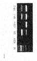
- FIG. 6illustrates a comparison of putative ES (left column) and TS (right column) cell lines derived from single blastomeres.
- FIGS. 6A and 6Bshow phase contrast photos of typical colonies.
- FIGS. 6C and 6Drepresent Lac-Z stained colonies, demonstrating their single blastomere origin.
- FIGS. 6E and 6Fshow alkaline phosphatase staining.
- FIGS. 6G and 6Hshow indirect immunofluorescence with antibodies to Oct-4.
- FIG. 6Idepicts putative ES cells stained with antibodies to SSEA-1.
- FIG. 6Jshows TROMA- 1 antibody staining of the putative TS cells (same field as FIG. 6H ). Scale bar, 200 um.
- FIG. 7shows LacZ stained placenta of 10.5 day chimera showing contribution of single blastomere-derived TS cells.
- the maternal portion of the placentahas been peeled away.
- the embryonic portion shownis photographed from the distal side of the disk and is approximately 4 mm in diameter.
- FIG. 8depicts differentiation of blastomere-derived mES cells in vitro and in vivo.
- FIGS. 8A-8Cshow immunofluorescence analysis of molecular markers of mesoderm (muscle actin, FIG. 8A ), primitive endoderm ( ⁇ -feto protein, FIG. 8B ), and ectoderm ( ⁇ III tubulin, FIG. 8C ).
- FIG. 8Ddepicts representative chromosome spreads of two single blastomere-derived mES cell line. G-banded karyotyping shows that lines Y 1 (top) and Y 7 (bottom) have XY and XX karyotypes, respectively.
- FIG. 8A-8Cshow immunofluorescence analysis of molecular markers of mesoderm (muscle actin, FIG. 8A ), primitive endoderm ( ⁇ -feto protein, FIG. 8B ), and ectoderm ( ⁇ III tubulin, FIG. 8C ).
- FIG. 8Ddepict
- FIG. 8Eshows hematoxylin and eosin stained section through a teratoma and shows examples of tissue from all three germ layers.
- Bnbone (mesoderm); nt, neural tissue (ectoderm); cre, ciliated respiratory epithelium (endoderm).
- the insert of FIG. 8Eis an enlarged region of ciliated respiratory epithelium.
- FIG. 8Fshows 11.5 day chimeric embryos produced from three of the putative ES cell lines, each of which shows the high degree of chimerism frequently observed.
- FIG. 8Gis a closeup view of lacZ-stained chimera from putative ES line J- 15 .
- FIG. 8Hdisplays chimeric pups generated by aggregating blastomere-derived (1 29/Sv) ES cells (lines J 15 and Y 1 ) with CD-1 mouse embryos. Scale bars: A-D—200 um, F—10 mm, G—2 mm.
- FIG. 9shows PCR analysis demonstrating the presence of the LacZ gene in purified sperm from chimeric mice produced from two different blastomere-derived ES cell lines.
- Mmolecular weight marker
- HH 2 O control
- ESDNA from mouse ES cells used to generate chimeric animals
- CD-1DNA from CD-1 mouse
- SP-1DNA from sperm of chimeric mouse No. 1
- SP-2DNA from sperm of chimeric mouse No. 2 .
- the present inventionprovides novel methods for deriving embryonic stem cells, those cells and cell lines, and uses of the embryonic stem cells and cell lines for therapeutic and research purposes. It also relates to a method of establishing and storing an autologous stem cell line from a blastomere retrieved prior to implantation of an embryo, e.g. in conjunction with assisted reproductive technologies such as in vitro fertilization (“IVF”).
- IVFin vitro fertilization
- This inventionprovides a method of producing an embryonic stem cell, comprising the step of culturing a blastomere obtained from an embryo, wherein the embryo remains viable.
- the blastomereis obtained from an embryo prior to compaction of the morula.
- the embryois obtained before formation of the blastocoel.
- the blastomeremay be obtained by partial or complete removal of the zona pellucida surrounding the embryo.
- the embryomay be implanted or cryopreserved.
- the blastomere obtained from the embryois cultured with any suitable cell to produce an ES cell.
- Cells suitable for culturing the blastomeresinclude, but are not limited to, embryonic stem cells, such as from already established lines, embryo carcinoma cells, murine embryonic fibroblasts, other embryo-like cells, cells of embryonic origin or cells derived from embryos, many of which are known in the art and available from the American Type Culture Collection, Manassas, VA 20110-2209, USA, and other sources.
- the blastomeremay also be cultured with factors that inhibit differentiation of the ES cell.
- the blastomereis cultured in the presence of heparin.
- Oct-4is introduced into the blastomere or alternatively, expression of endogenous Oct-4 is induced in the blastomere.
- the present inventionprovides a method of producing an ES cell comprising the steps of obtaining a blastomere from an embryo, wherein the embryo remains viable, aggregating the blastomere with ES cells, culturing the aggregated blastomere and ES cells until the blastomere exhibits properties of ES cells, and isolating the ES cells derived from the blastomere.
- the blastomere obtained from an embryois cultured with autologous feeder cells, wherein the feeder cells are produced by culturing a blastomere obtained from the same embryo under conditions to differentiate the blastomere into a somatic cell to produce the autologous feeder cells.
- a blastomere obtained from an embryoundergoes cell division and one progeny cell is used for genetic testing and another progeny cell is used to produce an ES cell.
- the method of producing an ES cell or ES cell linecomprises obtaining a blastomere through biopsy, removing the zona pellucida, separating the blastocyst into two segments, culturing one blastocyst segment in order to produce an ES cell or ES cell line and implanting or cryopreserving the remainder of the blastocyst.
- the methodcomprises the steps of obtaining a single blastomere prior to implantation and before formation of the blastocoel, culturing the blastomere, adding ES cells from already established lines, allowing the ES cells to clump around the blastomere until the blastomere exhibits ES cell growth and harvesting the resultant ES cell for therapeutic purposes.
- the methodcomprises the steps of obtaining a single blastomere before compaction of the morula, culturing the blastomere in standard culture conditions, adding mitotically inactivated ES cells from already established lines until ES cells begin to form, and harvesting or cryopreserving the resultant ES cells.
- the methodcomprises the steps of obtaining a single blastomere prior to implantation and before formation of the blastocoel, culturing the blastomere, adding ES cells from already established lines, and introducing recombinant Oct-4 into the blastomere or activating endogenous Oct-4.
- the ES cell produced from the blastomeremay be pluripotent or totipotent.
- Pluripotency or totipotency of the ES cellmay be determined by assaying for ES cell marker proteins.
- Such proteinsinclude Oct-4, SSEA-1, nanog, alkaline phosphatase and Res-1.
- the method of the inventionmay be performed on mammals, e.g., mice, rabbits, sheep, pigs, cows, primates and humans.
- the mammalis a non-human mammal.
- the mammalis a human.
- the present inventionalso provides methods of differentiating the ES cells produced by the methods of the invention.
- the ES cellsmay be differentiated into any cell type including those of mesodermal, endodermal and ectodermal origin.
- a differentiated cell typee.g., mesoderm, endoderm or ectoderm
- the inventionalso encompasses the ES cells produced by the methods of this invention, ES cell lines derived from these ES cells as well as differentiated cells derived from the ES cells or cell lines.
- the ES cells provided by this invention or cells derived from the ES cellsare useful for treating disorders amenable to cell therapy.
- Pharmaceutical compositionscomprising these cells together with a pharmaceutically acceptable medium or carrier are also provided.
- TStrophoblast stem cells
- blastomereobtained prior to compaction of the morula.
- the blastomereis obtained before formation of the blastocoel.
- the blastomeremay be obtained by partial or complete removal of the zona pellucida surrounding the embryo.
- the blastomere obtained from the embryois cultured with any suitable cell to produce a TS cell.
- Cells suitable for culturing the blastomeresinclude, but are not limited to, embryonic stem cells, such as from already established lines, embryo carcinoma cells, murine embryonic fibroblasts, other embryo-like cells, cells of embryonic origin or cells derived from embryos, many of which are known in the art and available from the American Type Culture Collection, Manassas, VA 20110-2209, USA, and other sources.
- the blastomeremay also be cultured with factors that induce differentiation of the ES cell. In one embodiment, the blastomere is cultured in the presence of FGF-4.
- the TS cell produced by the methods of the inventionmay express a TS cell marker, e.g., nanog, Rex-1, cdx-2.
- the TS cellmay also lack expression of Oct-4 or ⁇ -fetoprotein.
- the TS cellmay also be cultured to produce a TS cell line or differentiated.
- the present inventionis based in part on the discovery that stem cells can be generated from embryos without affecting viability of the embryo.
- these methodsutilize in vitro techniques currently used in pre-implantation genetic diagnosis (PGD).
- PGDpre-implantation genetic diagnosis
- ESpluripotent embryonic stem
- the blastomeremay be removed from an embryo at various developmental stages prior to implantation including but not limited to: before compaction of the morula, during compaction of the morula, before formation of the blastocoel or during the blastocyst stage.
- the inventionprovides methods for biopsy of a blastocyst which will produce embryonic stem cells, and the remainder of the blastocyst is implanted and results in a pregnancy and later in a live birth.
- the zona pellucidais removed from the blastocyst by any means known to those of ordinary skill in the art (in this instance using acidic tyrode solution pH 2.4), it is placed on culture ware with protein free media (CZB protein free media pH 7.4 is used, but other protein free media could be used) and it adheres, then the blastocyst is biopsied.
- the controversies associated with the derivation of embryonic stem cellsare circumvented by using a technique similar to that used in preimplantation genetic diagnosis (PGD) where a single blastomere is removed from the embryo, preferably before the compaction of the morula.
- PPDpreimplantation genetic diagnosis
- these methodscan be adapted for use in the present invention, for the removal of one or more cells from an embryo without affecting the continued development of the embryo.
- the biopsied blastomereis allowed to undergo cell division and one progeny cell is used for genetic testing and the remaining cells are used to generate stem cells.
- the biopsied embryomay be implanted at the blastocyst stage or frozen for implantation at a later time.
- the biopsyconsists of two stages. The first is to make a hole in, or in some instances fully remove, the zone pellucida that surrounds the embryo. Once the hole is made, the cells (preferably one or two) may then be removed from the embryo.
- the methodinvolves removing or generating an extraction hole in the zona pellucida, and can be carried out by one more techniques selected from the group consisting of physical manipulation, chemical treatment and enzymatic digestion. Exemplary techniques that can be used include:
- the procedureis performed on day 3 of embryo development, when the embryo is around 6-8 cell stage.
- the embryois placed in a drop of biopsy medium under mineral oil by holding it with a holding pipette.
- the zona pellucidais locally digested, by releasing acidified Tyrode's solution (Sigma, St. Louis, Mo. 63178) through an assistant hatching pipette.
- acidified Tyrode's solutionSigma, St. Louis, Mo. 63178
- the zona pellucida of the blastocystmay be at least partially digested by treatment with one or more enzymes or mixture of enzymes such as pronase.
- one or more enzymes or mixture of enzymessuch as pronase.
- Other types of proteases with the same or similar protease activity as pronasemay also be used.
- the isolated blastomeresmay be cultured by placing them on cultureware (e.g., in microwells) with media in standard culture conditions together with any suitable cells including but not limited to embryonic stem cells, such as from already established lines, embryo carcinoma cells, murine embryonic fibroblasts, other embryo-like cells, cells of embryonic origin or cells derived from embryos, many of which are known in the art and available from the American Type Culture Collection, Manassas, VA 20110-2209, USA, and other sources. These cells clump or aggregate around the blastomere. Other methods of aggregation including methods using microwell microbeads or the hanging drop method, or any other aggregation method known in the art may be used.
- embryonic stem cellssuch as from already established lines, embryo carcinoma cells, murine embryonic fibroblasts, other embryo-like cells, cells of embryonic origin or cells derived from embryos, many of which are known in the art and available from the American Type Culture Collection, Manassas, VA 20110-2209, USA, and other
- the cultured blastomeresexhibit ES cell growth perhaps as a result of cell-cell interactions between the blastomeres and the co-cultured embryonic cells or from interactions between the blastomeres and factors secreted by the embryonic cells.
- the blastomere(s)may be co-cultured with the remaining embryo.
- the blastomeresare co-cultured with the remaining embryo in a microdroplet culture system or other culture system known in the art, which permits cell-cell, cell-secreted factor and/or cell-matrix contact.
- the volume of the microdropmay be reduced, e.g., from 50 microliters to about 5 microliters to intensify the signal and promote cell-cell interactions.
- the blastomere culture conditionsmay include contacting the cells with factors that can inhibit or otherwise potentiate the differentiation of the cells, e.g., prevent the differentiation of the cells into non-ES cells, trophectoderm or other cell types.
- Such conditionscan include contacting the cultured cells with heparin or introducing Oct-4 into the cells (such as by including Oct-4 in the media) or activating endogenous Oct-4 in the cells.
- the present inventionalso provides a method of plating early pre-blastocyst embryos to make stem cells on autologous feeder cells.
- this methodcomprises (a) splitting a pre-blastocyst embryo, (b) plating one part into tissue culture under conditions to directly differentiate it into somatic cells to make feeder cells and (c) plating the other part of the pre-blastocyst embryo on the autologous feeder cells.
- the autologous feeder cells and ES cellsare produced from blastomeres removed from the pre-blastocyst embryo, thus, preserving the ability of the embryo to be implanted.
- Pluripotency of the ES cells produced by the methods of this inventioncan be determined by detecting expression of ES cell marker proteins.
- ES cell marker proteinsinclude but are not limited to octamer binding protein 4 (Oct-4), stage-specific embryonic antigen (SSEA)-1, nanog, alkaline phosphatase and Res-1.
- the putative ES cell linesmaintain pluripotency after more than 13, 20, 30, 40, 50, 60, 70, 80, 90 or 100 passages.
- the ES cellsmay also be assayed for maintenance of normal karyotype.
- This inventionalso provides methods of producing trophoblast stem (“TS” cells) by contacting blastomere outgrowths, which morphologically resemble trophoblast and/or extraembryonic endoderm, but which do not resemble ES cells, with FGF-4.
- FGF-4is added to the culture media of the outgrowths.
- TS cellscan be detected by assaying expression of proteins such as nanog, Rex-1, and Cdx-2, using procedures standard in the art.
- TS cell identificationcan also be evidenced by absence of the expression of proteins such as, but not limited to Oct-4 and ⁇ -feto protein.
- the present inventionprovides a method of treating a disorder amendable to cell therapy comprising administering to the affected subject a therapeutically effective amount of the ES cells of the invention.
- the ES cells of this inventionare suitable for any use that ES cells are useful.
- the methods of the inventionare used to remove a blastomere preceding implantation of an embryo after which the blastomere would be cultured as described above in order to derive and store embryonic stem cells for therapeutic uses using cell therapy should the child resulting from the embryo require, for example, disease therapy, tissue repair, transplantation, treatment of a cellular debilitation, or treatment of cellular dysfunctions in the future.
- cells derived from a blastomere, precompaction morula, compacting morula, or sectioned blastocystare directly differentiated in vitro or in vivo to generate differentiating or differentiated cells without generating an embryonic stem cell line.
- embryo-derived cellslike embryonic stem cells are useful in medical and biological research and in the treatment of disease by providing cells for use in cell therapy, e.g., allogeneic cell therapy.
- the embryonic stem cells and embryo-derived cells generated by the above-mentioned novel techniquesare utilized in research relating to cell biology, drug discovery, and in cell therapy, including but not limited to production of hematopoietic and hemangioblastic cells for the treatment of blood disorders, vascular disorders, heart disease, cancer, and wound healing, pancreatic beta cells useful in the treatment of diabetes, retinal cells such as neural cells and retinal pigment epithelial cells useful in the treatment of retinal disease such as retinitis pigmentosa and macular degeneration, neurons useful in treating Parkinson's disease, Alzheimer's disease, chronic pain, stroke, psychiatric disorders, and spinal cord injury, heart muscle cells useful in treating heart disease such as heart failure, skin cells useful in treating wounds for scarless wound repair, bums, promoting wound repair, and in treating skin aging, liver cells for the treatment of liver disease such as cirrhotic liver disease, kidney cells for the treatment of kidney disease such as renal failure, cartilage for the treatment of arthritis, lung cells for the treatment of lung disease
- Such cell therapy methodsmay involve use of the ES cells of this invention in combination with proliferation factors, lineage-commitment factors, or gene or proteins of interest.
- Treatment methodsmay include providing stem or appropriate precursor cells directly for transplantation where the tissue is regenerated in vivo or recreating the desired tissue in vitro and then providing the tissue to the affected subject.
- Single blastomereswere isolated from 8-cell stage 129/Sv-ROSA26:LacZ mouse embryos either by biopsy through a hole in the zona pellucida drilled using Piezo-pulse or by disaggregating of zona-denuded embryos in Ca++/Mg++ free PBS for 10 minutes.
- the biopsied (7-cell) embryoswere transferred to the oviducts of 1.5 days post coitum (d.p.c.) synchronized surrogate mothers, and each separated blastomere aggregated with a small clump (approximately 100 cells) of green fluorescent protein (GFP)-positive 129Sv/CD-1 mouse ES (mES) cells in a 300 um depression created by pressing an aggregation needle into the bottom of a plastic tissue culture plate. After incubation for 24-48h a growing “bud” of GFP-negative cells was observed on the sides of the majority (60%) of GFP-mES clusters (See FIG. 4 A, B).
- GFPgreen fluorescent protein
- mESmouse 129Sv/CD-1 mouse ES
- the cell clumpswere plated onto mitomicin C-treated mouse embryonic fibroblasts (MEF) and cultured in knockout DMEM (15% FCS, penicillin/streptomycin, Glutamax-I, ⁇ -mercaptoethanol, nonessential amino acids, LIF [2000U/ml], and MEK1 inhibitor [50 ⁇ M] (mES culture medium)).
- knockout DMEM10% FCS, penicillin/streptomycin, Glutamax-I, ⁇ -mercaptoethanol, nonessential amino acids, LIF [2000U/ml], and MEK1 inhibitor [50 ⁇ M] (mES culture medium)
- the majority of blastomeres54/91 formed rapidly growing clumps of cells within 4 days, which were separated from GFP-positive mES cells under a fluorescence microscope.
- the cellswere expanded by mechanical dissociation or trypsinization, while selecting for the colonies morphologically resembling ES cells and excluding any GFP-positive cells (
- FIG. 6 CSeveral lines of LacZ positive ES-like cells were produced (Table 1, FIG. 6 C) which maintained normal karyotype ( FIG. 8 D) and markers of pluripotency after over 50 passages.
- Indirect immunofluorescence staining for ES cell protein markerswas performed on cells growing on 4-well tissue culture plates. For example, see Lanza R, et al, Eds. Handbook of stem Cells. Vol 1 :Embryonic Stem Cells (Elsevier/Academic Press, San Diego, Calif., 2004); Evans, M.
- PCRPolymerase chain reaction
- ES-like cell culturesWhen the ES-like cell cultures were allowed to overgrow, they spontaneously differentiated into cells of all three germ layers, as evidenced by immunostaining with antibodies to muscle actin (mesoderm), ⁇ III tubulin (ectoderm), and ⁇ -feto protein (primitive endoderm)( FIG. 3 B-D and FIG. 8 A-C). Beating heart muscle, extraembryonic endoderm and multiple neuronal cell types were also routinely observed in differentiating cultures. To further demonstrate the pluripotency of the derived ES cells, ES cell lines were either injected into CD-1 mouse blastocysts or aggregated with 8-cell stage morulae as described previously (Hogan et al., supra) and transferred to recipient females.
- X-Gal (5-bromo—4-chloro—3-indolyl-beta-D-galactopyranoside) staining of the resulting chimeric fetusesshowed that the ES cell lines contributed to all organs ( FIG. 3 A), such as, heart, kidney, liver, lung, intestine, brain, blood, skin and genital ridge. Twenty-four of the fetuses (83%) were chimeric ( FIG. 8 F, G), and eight of nine (89%) pups ( FIG. 8H ) were chimeric; (the latter had the LacZ gene in their gametes (confirmed by PCR analysis; FIG. 9 ), and produced LacZ+ offspring when crossed with CD-1, confirming the contribution of the blastomere-derived ES cells to the germ line.).
- the ES cellswere injected into NOD-SCID mice and examined for their ability to differentiate into various cell types. Briefly, approximately 1 million ES cells were injected into the rear thigh of a NOD-SCID mouse. After about two months the mice were sacrificed and the teratomas excised, fixed in 4% paraformaldehyde, embedded in paraffin, and sectioned. The teratomas contained tissues from all three germ layers including bone and cartilage (mesoderm), neural rosettes (ectoderm), and ciliated respiratory epithelia (endoderm) among others ( FIG. 8E ).
- RT-PCR analysiswas performed as follows: Total RNA was isolated from ES and TS cells using an RNAeasy Mini Kit (Qiagen), and 1 ⁇ g RNA was subjected to first strand cDNA synthesis with an oligo (dT) primer, using AMV reverse transcriptase (Promega, Madison, Wis.). One tenth of the RT reaction was subjected to PCR amplification. PCR conditions for all genes were 95° C. for 9 min (1 cycle), 94° C. for 45 s, 62° C. for 1 min and 72° C. for 1.5 min with 2 mM Mg++ concentration, except for ⁇ -tubulin gene that was annealed at 64° C.
Landscapes
- Health & Medical Sciences (AREA)
- Life Sciences & Earth Sciences (AREA)
- Engineering & Computer Science (AREA)
- Bioinformatics & Cheminformatics (AREA)
- Chemical & Material Sciences (AREA)
- Organic Chemistry (AREA)
- Biomedical Technology (AREA)
- General Health & Medical Sciences (AREA)
- Genetics & Genomics (AREA)
- Public Health (AREA)
- Veterinary Medicine (AREA)
- Medicinal Chemistry (AREA)
- Animal Behavior & Ethology (AREA)
- Pharmacology & Pharmacy (AREA)
- Chemical Kinetics & Catalysis (AREA)
- Nuclear Medicine, Radiotherapy & Molecular Imaging (AREA)
- General Chemical & Material Sciences (AREA)
- Developmental Biology & Embryology (AREA)
- Zoology (AREA)
- Biotechnology (AREA)
- Wood Science & Technology (AREA)
- Reproductive Health (AREA)
- Gynecology & Obstetrics (AREA)
- General Engineering & Computer Science (AREA)
- Cell Biology (AREA)
- Neurosurgery (AREA)
- Neurology (AREA)
- Microbiology (AREA)
- Biochemistry (AREA)
- Physical Education & Sports Medicine (AREA)
- Rheumatology (AREA)
- Orthopedic Medicine & Surgery (AREA)
- Diabetes (AREA)
- Physics & Mathematics (AREA)
- Molecular Biology (AREA)
- Plant Pathology (AREA)
- Biophysics (AREA)
- Immunology (AREA)
- Urology & Nephrology (AREA)
- Cardiology (AREA)
Abstract
Description
- Partial zone dissection (PZD:): partial dissection of the zona pellucida, using a micro-pipette;
- Zona drilling: chemical opening of the zona pellucida zone through partial digestion with Tyrode acid;
- Zona drilling: enzymatic opening of the zona pellucida zone through partial digestion with pronase or other protease;
- zona pellucida thinning: thinning of the zona pellucida with Tyrode acid or laser;
- Point-like opening of the zona pellucida with laser;
- Point-like mechanical opening of the zona pellucida with Piezo micro-manipulator.
| No. non- | ||||||
| GFP | ||||||
| outgrowths | No. non- | Differentiation | ||||
| detected | GFP | ES lines | No |
| Exp. | No. | after plating | outgrowths | estab- | Markers | In vitro | muscle | chimeras/ |
| No. | blastomeres | on | passage | 1 | lished | Oct-4 | AP | Nanog | SSEA1 | Tb | AFP | fetuses | Karyotype | ||
| 1 | 22 | 14 | 6 | 0 | |||||||||||
| 2 | 24 | 13 | 8 | 0 | |||||||||||
| 3 | 24 | 13 | 6 | J15 | + | + | + | + | + | + | + | 7/9 | |||
| 4 | 21 | 16 | 5 | Y1 | + | + | + | + | + | + | + | 5/5 | 40XY | ||
| 5 | 15 | 8 | 5 | Y7 | + | + | + | + | + | + | + | 8/9 | 40XX | ||
| 6 | 19 | 11 | 6 | J5 | + | + | + | + | + | + | + | 4/6 | 60XXY | ||
| 6 | — | — | — | Y8 | + | + | + | + | + | + | + | — | 40XX | ||
Claims (25)
Priority Applications (9)
| Application Number | Priority Date | Filing Date | Title |
|---|---|---|---|
| US11/267,555US7838727B2 (en) | 2004-11-04 | 2005-11-04 | Derivation of embryonic stem cells |
| US11/800,366US7893315B2 (en) | 2004-11-04 | 2007-05-03 | Derivation of embryonic stem cells and embryo-derived cells |
| US12/905,839US8642328B2 (en) | 2004-11-04 | 2010-10-15 | Derivation of embryonic stem cells |
| US13/004,260US8742200B2 (en) | 2004-11-04 | 2011-01-11 | Derivation of embryonic stem cells and embryo-derived cells |
| US14/154,163US9550974B2 (en) | 2004-11-04 | 2014-01-13 | Derivation of embryonic stem cells |
| US14/266,114US9617512B2 (en) | 2004-11-04 | 2014-04-30 | Derivation of embryonic stem cells and embryo-derived cells |
| US15/390,319US20170204368A1 (en) | 2004-11-04 | 2016-12-23 | Derivation of embryonic stem cells |
| US15/448,571US10072243B2 (en) | 2004-11-04 | 2017-03-02 | Derivation of embryonic stem cells and embryo-derived cells |
| US16/113,839US20190062698A1 (en) | 2004-11-04 | 2018-08-27 | Derivation of embryonic stem cells and embryo-derived cells |
Applications Claiming Priority (6)
| Application Number | Priority Date | Filing Date | Title |
|---|---|---|---|
| US62482704P | 2004-11-04 | 2004-11-04 | |
| US66248905P | 2005-03-15 | 2005-03-15 | |
| US68715805P | 2005-06-03 | 2005-06-03 | |
| US72306605P | 2005-10-03 | 2005-10-03 | |
| US72677505P | 2005-10-14 | 2005-10-14 | |
| US11/267,555US7838727B2 (en) | 2004-11-04 | 2005-11-04 | Derivation of embryonic stem cells |
Related Child Applications (2)
| Application Number | Title | Priority Date | Filing Date |
|---|---|---|---|
| US11/800,366Continuation-In-PartUS7893315B2 (en) | 2004-11-04 | 2007-05-03 | Derivation of embryonic stem cells and embryo-derived cells |
| US12/905,839ContinuationUS8642328B2 (en) | 2004-11-04 | 2010-10-15 | Derivation of embryonic stem cells |
Publications (2)
| Publication Number | Publication Date |
|---|---|
| US20060206953A1 US20060206953A1 (en) | 2006-09-14 |
| US7838727B2true US7838727B2 (en) | 2010-11-23 |
Family
ID=36337005
Family Applications (4)
| Application Number | Title | Priority Date | Filing Date |
|---|---|---|---|
| US11/267,555Active2026-03-29US7838727B2 (en) | 2004-11-04 | 2005-11-04 | Derivation of embryonic stem cells |
| US12/905,839Active2026-06-21US8642328B2 (en) | 2004-11-04 | 2010-10-15 | Derivation of embryonic stem cells |
| US14/154,163ActiveUS9550974B2 (en) | 2004-11-04 | 2014-01-13 | Derivation of embryonic stem cells |
| US15/390,319AbandonedUS20170204368A1 (en) | 2004-11-04 | 2016-12-23 | Derivation of embryonic stem cells |
Family Applications After (3)
| Application Number | Title | Priority Date | Filing Date |
|---|---|---|---|
| US12/905,839Active2026-06-21US8642328B2 (en) | 2004-11-04 | 2010-10-15 | Derivation of embryonic stem cells |
| US14/154,163ActiveUS9550974B2 (en) | 2004-11-04 | 2014-01-13 | Derivation of embryonic stem cells |
| US15/390,319AbandonedUS20170204368A1 (en) | 2004-11-04 | 2016-12-23 | Derivation of embryonic stem cells |
Country Status (7)
| Country | Link |
|---|---|
| US (4) | US7838727B2 (en) |
| EP (3) | EP2960328A1 (en) |
| JP (2) | JP5728676B2 (en) |
| AU (1) | AU2005304978A1 (en) |
| CA (2) | CA2588088C (en) |
| HK (1) | HK1219295A1 (en) |
| WO (1) | WO2006052646A2 (en) |
Cited By (10)
| Publication number | Priority date | Publication date | Assignee | Title |
|---|---|---|---|---|
| US20100240132A1 (en)* | 2007-02-23 | 2010-09-23 | Robert Lanza | Highly efficient methods for reprogramming differentiated cells and for generating animals and embryonic stem cells from reprogrammed cells |
| US20110183415A1 (en)* | 2004-11-04 | 2011-07-28 | Young Gie Chung | Derivation of embryonic stem cells and embryo-derived cells |
| US8808687B2 (en) | 2010-07-12 | 2014-08-19 | Mark Humayun | Biocompatible substrate for facilitating interconnections between stem cells and target tissues and methods for implanting same |
| US8877489B2 (en) | 2011-12-05 | 2014-11-04 | California Institute Of Technology | Ultrathin parylene-C semipermeable membranes for biomedical applications |
| US9248013B2 (en) | 2011-12-05 | 2016-02-02 | California Institute Of Technology | 3-Dimensional parylene scaffold cage |
| US9550974B2 (en) | 2004-11-04 | 2017-01-24 | Astellas Institute For Regenerative Medicine | Derivation of embryonic stem cells |
| US10478206B2 (en) | 2011-04-29 | 2019-11-19 | University Of Southern California | Instruments and methods for the implantation of cell-seeded substrates |
| US11390885B2 (en) | 2014-09-15 | 2022-07-19 | Children's Medical Center Corporation | Methods and compositions to increase somatic cell nuclear transfer (SCNT) efficiency by removing histone H3-lysine trimethylation |
| US11422125B2 (en) | 2015-03-23 | 2022-08-23 | Astellas Institute For Regenerative Medicine | Assays for potency of human retinal pigment epithelium (RPE) cells and photoreceptor progenitors |
| EP4524568A2 (en) | 2010-07-23 | 2025-03-19 | Astellas Institute for Regenerative Medicine | Methods for detection of rare subpopulations of cells and highly purified compositions of cells |
Families Citing this family (25)
| Publication number | Priority date | Publication date | Assignee | Title |
|---|---|---|---|---|
| US7794704B2 (en) | 2004-01-23 | 2010-09-14 | Advanced Cell Technology, Inc. | Methods for producing enriched populations of human retinal pigment epithelium cells for treatment of retinal degeneration |
| WO2005070011A2 (en) | 2004-01-23 | 2005-08-04 | Advanced Cell Technology, Inc. | Improved modalities for the treatment of degenerative diseases of the retina |
| US7622108B2 (en) | 2004-04-23 | 2009-11-24 | Bioe, Inc. | Multi-lineage progenitor cells |
| GB0507755D0 (en)* | 2005-04-16 | 2005-05-25 | Univ Sheffield | Cytotrophoblast stem cells |
| EP1984487B1 (en) | 2005-08-03 | 2022-10-12 | Astellas Institute for Regenerative Medicine | Improved methods of reprogramming animal somatic cells |
| US7727763B2 (en)* | 2006-04-17 | 2010-06-01 | Bioe, Llc | Differentiation of multi-lineage progenitor cells to respiratory epithelial cells |
| KR101569204B1 (en)* | 2006-05-03 | 2015-11-17 | 오카타 세라퓨틱스, 인크. | Induction of embryonic stem cells and embryo-derived cells |
| US20070298496A1 (en)* | 2006-06-23 | 2007-12-27 | Hung-Chih Kuo | Method of deriving pluripotent stem cells from a single blastomere |
| CA2702386C (en) | 2007-10-12 | 2018-07-24 | Advanced Cell Technology, Inc. | Improved methods of producing rpe cells and compositions of rpe cells |
| EP2224940A1 (en)* | 2007-11-30 | 2010-09-08 | Cytomatrix PTY LTD | Methods of inducing pluripotency involving oct4 protein |
| EP2100954A1 (en)* | 2008-03-10 | 2009-09-16 | Assistance Publique - Hopitaux de Paris | Method for generating primate cardiac progenitor cells for clinical use from primate embryonic stem cells, and their applications |
| JP2009225675A (en)* | 2008-03-19 | 2009-10-08 | Tohoku Univ | Feeder cell derived from same kind of skin for preparing epithelial cell sheet |
| EP3075850B1 (en) | 2008-07-16 | 2019-02-06 | ID Pharma Co., Ltd. | Method for production of reprogrammed cell using chromosomally unintegrated virus vector |
| US8741649B2 (en) | 2009-09-04 | 2014-06-03 | The United States Of America, As Represented By The Secretary, Department Of Health And Human Services | Methods for enhancing genome stability and telomere elongation in embryonic stem cells |
| US10485829B2 (en) | 2009-11-17 | 2019-11-26 | Astellas Institute For Regenerative Medicine | Methods of producing human RPE cells and pharmaceutical preparations of human RPE cells |
| US20130149287A1 (en)* | 2010-07-30 | 2013-06-13 | Cambridge Enterprise Limited | Corticogenesis of Human Pluripotent Cells |
| WO2013010045A1 (en) | 2011-07-12 | 2013-01-17 | Biotime Inc. | Novel methods and formulations for orthopedic cell therapy |
| WO2013070734A1 (en) | 2011-11-07 | 2013-05-16 | Chaudhry Hina W | Methods of cardiac repair |
| TW202434266A (en) | 2011-11-14 | 2024-09-01 | 安斯泰來再生醫藥協會 | Pharmaceutical preparations of human rpe cells and uses thereof |
| KR101871084B1 (en) | 2013-12-11 | 2018-06-25 | 화이자 리미티드 | Method for producing retinal pigment epithelial cells |
| WO2016005985A2 (en) | 2014-07-09 | 2016-01-14 | Yissum Research Development Company Of The Hebrew University Of Jerusalem Ltd. | Method for reprogramming cells |
| MX2022005134A (en) | 2019-10-30 | 2022-05-30 | Astellas Inst For Regenerative Medicine | METHODS TO PRODUCE RETINAL PIGMENTARY EPITHELIUM CELLS. |
| WO2022235586A1 (en) | 2021-05-03 | 2022-11-10 | Astellas Institute For Regenerative Medicine | Methods of generating mature corneal endothelial cells |
| JP2024518409A (en) | 2021-05-07 | 2024-05-01 | アステラス インスティテュート フォー リジェネラティブ メディシン | Methods for producing mature hepatocytes |
| CA3230677A1 (en) | 2021-06-13 | 2022-12-22 | Yissum Research Development Company Of The Hebrew University Of Jerusalem Ltd. | Method for reprogramming human cells |
Citations (19)
| Publication number | Priority date | Publication date | Assignee | Title |
|---|---|---|---|---|
| WO1995017500A1 (en)* | 1993-12-23 | 1995-06-29 | Abs Global, Inc. | Embryonic stem cells as nuclear donors and nuclear transfer techniques to produce chimeric and transgenic animals |
| WO2001050848A2 (en) | 2000-01-07 | 2001-07-19 | Oregon Health And Science University | Clonal propagation of primate offspring by embryo splitting |
| US20020022268A1 (en) | 2000-01-11 | 2002-02-21 | Chunhui Xu | Conditioned media for propagating human pluripotent stem cells |
| WO2003018760A2 (en) | 2001-08-24 | 2003-03-06 | Advanced Cell Technology, Inc. | Screening assays for identifying differentiation-inducing agents and production of differentiated cells for cell therapy |
| US20030087859A1 (en) | 2001-02-21 | 2003-05-08 | Stefan Kochanek | Pigment epithelial cell of the eye, its production and use in therapy of an eye or CNS disease |
| US20040199935A1 (en) | 1999-06-30 | 2004-10-07 | Chapman Karen B. | Cytoplasmic transfer to de-differentiate recipient cells |
| US20040229350A1 (en)* | 2003-05-12 | 2004-11-18 | Nikolai Strelchenko | Morula derived embryonic stem cells |
| US20050118713A1 (en) | 2003-05-12 | 2005-06-02 | Nikolai Strelchenko | Morula derived embryonic stem cells |
| US20050138680A1 (en) | 2003-12-22 | 2005-06-23 | Animal Technology Institute Taiwan | Method for generating non-human mammalian chimeric embryo |
| WO2005070011A2 (en) | 2004-01-23 | 2005-08-04 | Advanced Cell Technology, Inc. | Improved modalities for the treatment of degenerative diseases of the retina |
| US20050265976A1 (en) | 1999-10-15 | 2005-12-01 | Advanced Cell Technology, Inc. | Method of differentiation of morula or inner cell mass cells and method of making lineage-defective embryonic stem cells |
| US20060014278A1 (en)* | 2002-12-10 | 2006-01-19 | Khillan Jaspal S | Method for programmed differentiation of stem cells |
| WO2006013519A1 (en) | 2004-07-26 | 2006-02-09 | Koninklijke Philips Electronics N.V. | Method of storing data in an information carrier, system for reading such an information carrier. |
| WO2006013573A2 (en) | 2004-08-06 | 2006-02-09 | Mendy Erad Ltd. | Early detection of harmful agents: method, system and kit |
| WO2006052646A2 (en) | 2004-11-04 | 2006-05-18 | Advanced Cell Technology, Inc. | Derivation of embryonic stem cells |
| WO2006080952A2 (en) | 2005-01-24 | 2006-08-03 | Advanced Cell Technology, Inc. | Improved modalities for the treatment of degenerative diseases of the retina |
| WO2007130664A2 (en) | 2006-05-03 | 2007-11-15 | Advanced Cell Technology, Inc. | Derivation of embryonic stem cells and embryo-derived cells |
| US20070298496A1 (en) | 2006-06-23 | 2007-12-27 | Hung-Chih Kuo | Method of deriving pluripotent stem cells from a single blastomere |
| US20080057041A1 (en)* | 2004-11-04 | 2008-03-06 | Chung Young G | Derivation of embryonic stem cells and embryo-derived cells |
Family Cites Families (15)
| Publication number | Priority date | Publication date | Assignee | Title |
|---|---|---|---|---|
| US6274705B1 (en)* | 1996-05-02 | 2001-08-14 | Aventis Pharmaceuticals Products Inc. | Antithrombotic azacycloalkylalkanoyl peptides and pseudopeptides |
| AU1439295A (en)* | 1993-12-17 | 1995-07-03 | Abs Global, Inc. | Ungulate preblastocyst derived embryonic stem cells and use thereof to produce cloned transgenic and chimeric ungulates |
| US5843780A (en) | 1995-01-20 | 1998-12-01 | Wisconsin Alumni Research Foundation | Primate embryonic stem cells |
| CA2394614A1 (en) | 1999-12-20 | 2001-06-28 | University Of Massachusetts, A Public Institution Of Higher Education Of The Commonwealth Of Massachusetts, As Represented By Its Amherst Campus | Embryonic or stem-like cells produced by cross species nuclear transplantation |
| US20030105082A1 (en)* | 2001-12-03 | 2003-06-05 | Murphy Greer Marechal | Methods for improving the therapeutic response of humans having major depression and carrying the gene for apolipoprotein E4 |
| US20030082598A1 (en) | 2001-08-22 | 2003-05-01 | Ecker David J. | Molecular interaction sites of 23S ribosomal RNA and methods of modulating the same |
| US7247477B2 (en)* | 2002-04-16 | 2007-07-24 | Technion Research & Development Foundation Ltd. | Methods for the in-vitro identification, isolation and differentiation of vasculogenic progenitor cells |
| US6699771B1 (en)* | 2002-08-06 | 2004-03-02 | Texas Instruments Incorporated | Process for optimizing junctions formed by solid phase epitaxy |
| WO2005080551A2 (en) | 2004-02-12 | 2005-09-01 | University Of Newcastle Upon Tyne | Stem cells |
| US7818929B2 (en)* | 2004-12-14 | 2010-10-26 | Anthony Hardwood Composites, Inc. | Laminated support mat |
| US8265357B2 (en)* | 2005-10-14 | 2012-09-11 | Unisense Fertilitech A/S | Determination of a change in a cell population |
| ATE454632T1 (en)* | 2006-04-20 | 2010-01-15 | Northern Sydney And Central Co | METHOD FOR ASSESSING THE VIABILITY OF EMBRYOS |
| JP2010518857A (en) | 2007-02-23 | 2010-06-03 | アドバンスド セル テクノロジー, インコーポレイテッド | A highly efficient method for the reprogramming of differentiated cells and the generation of animal and embryonic stem cells from the reprogrammed cells |
| KR20110106922A (en)* | 2009-01-13 | 2011-09-29 | 플루이다임 코포레이션 | Single Cell Nucleic Acid Analysis |
| CN103748216B (en)* | 2011-05-31 | 2018-01-02 | 尤尼森斯繁殖技术公司 | Embryo quality assessment based on blastomere cleavage and morphology |
- 2005
- 2005-11-04EPEP15164137.0Apatent/EP2960328A1/ennot_activeCeased
- 2005-11-04CACA2588088Apatent/CA2588088C/enactiveActive
- 2005-11-04JPJP2007540021Apatent/JP5728676B2/enactiveActive
- 2005-11-04USUS11/267,555patent/US7838727B2/enactiveActive
- 2005-11-04WOPCT/US2005/039776patent/WO2006052646A2/enactiveApplication Filing
- 2005-11-04EPEP11000551Apatent/EP2479256A1/ennot_activeWithdrawn
- 2005-11-04EPEP05819524Apatent/EP1812554A4/ennot_activeWithdrawn
- 2005-11-04AUAU2005304978Apatent/AU2005304978A1/ennot_activeAbandoned
- 2005-11-04CACA3015835Apatent/CA3015835A1/ennot_activeAbandoned
- 2010
- 2010-10-15USUS12/905,839patent/US8642328B2/enactiveActive
- 2012
- 2012-10-10JPJP2012225193Apatent/JP5841926B2/enactiveActive
- 2014
- 2014-01-13USUS14/154,163patent/US9550974B2/enactiveActive
- 2016
- 2016-06-22HKHK16107238.9Apatent/HK1219295A1/enunknown
- 2016-12-23USUS15/390,319patent/US20170204368A1/ennot_activeAbandoned
Patent Citations (21)
| Publication number | Priority date | Publication date | Assignee | Title |
|---|---|---|---|---|
| WO1995017500A1 (en)* | 1993-12-23 | 1995-06-29 | Abs Global, Inc. | Embryonic stem cells as nuclear donors and nuclear transfer techniques to produce chimeric and transgenic animals |
| US20040199935A1 (en) | 1999-06-30 | 2004-10-07 | Chapman Karen B. | Cytoplasmic transfer to de-differentiate recipient cells |
| US20050265976A1 (en) | 1999-10-15 | 2005-12-01 | Advanced Cell Technology, Inc. | Method of differentiation of morula or inner cell mass cells and method of making lineage-defective embryonic stem cells |
| WO2001050848A2 (en) | 2000-01-07 | 2001-07-19 | Oregon Health And Science University | Clonal propagation of primate offspring by embryo splitting |
| US20020035735A1 (en)* | 2000-01-07 | 2002-03-21 | Gerald Schatten | Clonal propagation of primate offspring by embryo splitting |
| US20020022268A1 (en) | 2000-01-11 | 2002-02-21 | Chunhui Xu | Conditioned media for propagating human pluripotent stem cells |
| US20030087859A1 (en) | 2001-02-21 | 2003-05-08 | Stefan Kochanek | Pigment epithelial cell of the eye, its production and use in therapy of an eye or CNS disease |
| WO2003018760A2 (en) | 2001-08-24 | 2003-03-06 | Advanced Cell Technology, Inc. | Screening assays for identifying differentiation-inducing agents and production of differentiated cells for cell therapy |
| US20060014278A1 (en)* | 2002-12-10 | 2006-01-19 | Khillan Jaspal S | Method for programmed differentiation of stem cells |
| US20040229350A1 (en)* | 2003-05-12 | 2004-11-18 | Nikolai Strelchenko | Morula derived embryonic stem cells |
| US20050118713A1 (en) | 2003-05-12 | 2005-06-02 | Nikolai Strelchenko | Morula derived embryonic stem cells |
| US20050138680A1 (en) | 2003-12-22 | 2005-06-23 | Animal Technology Institute Taiwan | Method for generating non-human mammalian chimeric embryo |
| WO2005070011A2 (en) | 2004-01-23 | 2005-08-04 | Advanced Cell Technology, Inc. | Improved modalities for the treatment of degenerative diseases of the retina |
| WO2006013519A1 (en) | 2004-07-26 | 2006-02-09 | Koninklijke Philips Electronics N.V. | Method of storing data in an information carrier, system for reading such an information carrier. |
| WO2006013573A2 (en) | 2004-08-06 | 2006-02-09 | Mendy Erad Ltd. | Early detection of harmful agents: method, system and kit |
| WO2006052646A2 (en) | 2004-11-04 | 2006-05-18 | Advanced Cell Technology, Inc. | Derivation of embryonic stem cells |
| US20060206953A1 (en) | 2004-11-04 | 2006-09-14 | Robert Lanza | Derivation of embryonic stem cells |
| US20080057041A1 (en)* | 2004-11-04 | 2008-03-06 | Chung Young G | Derivation of embryonic stem cells and embryo-derived cells |
| WO2006080952A2 (en) | 2005-01-24 | 2006-08-03 | Advanced Cell Technology, Inc. | Improved modalities for the treatment of degenerative diseases of the retina |
| WO2007130664A2 (en) | 2006-05-03 | 2007-11-15 | Advanced Cell Technology, Inc. | Derivation of embryonic stem cells and embryo-derived cells |
| US20070298496A1 (en) | 2006-06-23 | 2007-12-27 | Hung-Chih Kuo | Method of deriving pluripotent stem cells from a single blastomere |
Non-Patent Citations (40)
| Title |
|---|
| Andrews, From teratocarinomas to embryonic stem cells, Phil. Trans. R. Soc. Lond. B, 2002, vol. 357, pp. 405-417. |
| Becker, S., et al., "Embryonic Stem Cells From Single Blastomeres," Methods Enzymol., 418:108-116 (2006). |
| Bradley, "Production and analysis of chimaeric mice," in Teratocarcinomas and Embryonic Stem cells (1987) IRL Press Oxford, pp. 113-151. |
| Chesne, et al., "Nuclear transfer in cattle: birth of cloned calves and estimation of blastomere totipotency in morulae used as a source of nuclei," C R Acad Sci III. 1993;316(5):487-91. |
| Chung, Y., et al., "Embryonic and Extraembryonic Stem Cell Lines Derived From Single Mouse Blastomeres," Nature, 439(7073):216-219 (2006). |
| Cowan et al., "Derivation of embryonic stem-cell lines from human blastocysts," N. Engl. J. Med., 350:1353-1356 (2004). |
| Delhaise, F., et al., "Establishment of an Embryonic Stem Cell Line from 8-Cell Stage Mouse Embryos," European Journal of Morphology, 34(4):237-243 (1996). |
| Evans et al., "Establishment in culture of pluripotential cells from mouse embryos," Nature, 292:154-156 (1981). |
| Fong et al., Unsuccessful derivation of human embryonic stem cell lines from pairs of human blastomeres, Reprod Biomed Online. Aug. 2006;13(2):295-300. |
| Geber S et al., Blastomere development after embryo biopsy: a new model to predict embryo development and to select for transfer. Hum Reprod. Mar. 1999;14(3):782-6.* |
| Geber, Proliferation of blastomeres from biopsied cleavage stage human embryos in vitro: an alternative to blastocyst biopsy for preimplantation diagnosis, Human Reproduction, 1995, vol. 10(6), pp. 1492-1496. |
| Geber, S., et al., "Proliferation of Blastomeres from Biopsied Cleavage Stage Human Embryos in Vitro: an Alternative to Blastocyst Biopsy for Preimplantation Diagnosis," Human Reproduction, 10(6):1492-1496 (1995). |
| Harvey, Inducible control of gene expression: prospects for gene therapy, Current Opinion in Chemical Biology, 1998, vol. 2, pp. 512-518. |
| Klimanskaya et al., "Derivation of human embryonic stem cells from single blastomeres," Nature Protocols,2:8: 1963-1972 (2007). |
| Klimanskaya et al., "Human embryonic stem cell lines derived from single blastomeres" Nature, doi:10.1038/nature05142 (2006). |
| Klimanskaya, I., et al., "Humeri Embryonic Stem Cell Lines Derived From Single Blastomeres" Nature, 444:481-485 (2006). |
| Mitalipov, S. M., et al., "Development Of Rhesus Monkey Demi-Embryos Created By Blastomere Separation At The 2-Cell Stage," Theriogenology, 53:397 (2000). |
| Ogawa, A novel mechanism for regulating clonal propagation of mouse ES cells, Genes to Cell, 2004, vol. 9, pp. 417-477. |
| Ouhibi, Initial Culture Behaviour of Rat Blastocysts on Selected Feeder Cell Lines, Molecular Reproduction and Development, 1995, vol. 40, pp. 311-324. |
| Papioannou (In Joyner, A L 2nd ed. Gene. Targeting A practical Approach, Oxford University Press, pp. 107-146.* |
| Rani et al., A Simple and Convenient Method for Preparing Chimeric Animals from Embryonic stem (ES) Cells Transgenic Research 2003, 739-741.* |
| Robertson, "Embryo-derived stem cells," in Teratocarcinomas and Embryonic Stem cells (1987) IRL Press Oxford, pp. 71-112. |
| Schraermeyer et al., "Subretinally transplanted embryonic stem cells rescue photoreceptor cells from degeneration in the RCS rats," Cell Transplant. 2001;10(8):673-80. |
| Schuldiner, Selective Ablation of Human Embryonic Stem Cells Expressing a "Suicide" Gene, Stem Cell, 2003, vol. 21, pp. 257-265. |
| Senger. Pathways to Pregnancy and Parturition. Current Concepts, Inc.:Pullman, 1997. Chapter13, p. 221.* |
| Sills et al. "Identification and isolation of embryonic stem cells in reproductive endocrinology: theoretical protocols for conservation of human embryos derived from in vitro fertilization," Theoretical Biology and Meidcal Modelling. 2005,2:25, pp. 1-8. |
| Solter, Immunosurgery of mouse blastocyst, Proc. Nat. Acad. Sci., Dec. 1975, vol. 72(12), pp. 5099-5102. |
| Springer, Prodrug-activating systems in suicide gene therapy, The Journal of Clinical Investigation, May 2000, vol. 105(9), pp. 1161-1167. |
| Stem Cell: Scientific Progress and Future Research Directions. Department of Health and Human Services. Jun. 2001. . Chapter 1: The Stem Cell, pp. 1-4.* |
| Stem Cell: Scientific Progress and Future Research Directions. Department of Health and Human Services. Jun. 2001. </info/scireport/2001report>. Chapter 1: The Stem Cell, pp. 1-4.* |
| Supplementary European Search Report dated Aug. 6, 2008 for EP 05 81 9524. |
| Supplementary European Search Report received in EP 07 79 4602 dated Feb. 18, 2010. |
| Thomson et al., "Embryonic stem cell lines derived from human blastocysts," Science, 282:1145-1147 (1998). |
| Thomson, Isolation of a primate embryonic stem cell line, Proc. Natl. Acad. Sci., Aug. 1995, vol. 92, pp. 7844-7848. |
| Van de Velde H et al., Embryo implantation after biopsy of one or two cells from cleavage-stage embryos with a view to preimplantation genetic diagnosis. Prenat Diagn. Dec. 2000;20(13):1030-7.* |
| Van de Velde, The four blastomeres of a 4-cell stage human embryo are able to develop individually into blastocysts with inner cell mass and trophectoderm, Human Reproduction, 2008, vol. 23:8, pp. 1742-1747. |
| Wade, N. "Stem Cell Test Tried on Mice Saves Embryo." New York Times, Oct. 17, 2005, available on-line at http://www.nytimes.com/2005/10/17/health/17stem.html?pagewanted=print, pp. 1-2. |
| Wakayama, Efficient Establishment of Mouse Embryonic Stem Cell Lines from Single Blastomeres and Polar Bodies, Stem Cell, 2007, vol. 25, pp. 986-993. |
| Wang, et al., "Increases in phosphorylation of SAPK/JNK and p38MAPK correlate negatively with mouse embryo development after culture in different media," Fertil Steril. Apr. 2005;83 Suppl 1:1144-54. |
| Wilson et al., Biopsy of preimplantation mouse embryos: development of micromanipulated embryos and proliferation of single blastomeres in vitro.Biol Reprod. Jan. 1989;40(1):145-52.* |
Cited By (20)
| Publication number | Priority date | Publication date | Assignee | Title |
|---|---|---|---|---|
| US9617512B2 (en) | 2004-11-04 | 2017-04-11 | Astellas Institute For Regenerative Medicine | Derivation of embryonic stem cells and embryo-derived cells |
| US20110183415A1 (en)* | 2004-11-04 | 2011-07-28 | Young Gie Chung | Derivation of embryonic stem cells and embryo-derived cells |
| US8742200B2 (en) | 2004-11-04 | 2014-06-03 | Advanced Cell Technology, Inc. | Derivation of embryonic stem cells and embryo-derived cells |
| US10072243B2 (en) | 2004-11-04 | 2018-09-11 | Astellas Institute For Regenerative Medicine | Derivation of embryonic stem cells and embryo-derived cells |
| US9550974B2 (en) | 2004-11-04 | 2017-01-24 | Astellas Institute For Regenerative Medicine | Derivation of embryonic stem cells |
| US8796021B2 (en) | 2007-02-23 | 2014-08-05 | Advanced Cell Technology, Inc. | Blastomere culture to produce mammalian embryonic stem cells |
| US20100240132A1 (en)* | 2007-02-23 | 2010-09-23 | Robert Lanza | Highly efficient methods for reprogramming differentiated cells and for generating animals and embryonic stem cells from reprogrammed cells |
| US10584313B2 (en) | 2007-02-23 | 2020-03-10 | Astellas Institute For Regenerative Medicine | Method of producing a differentiated mammalian cell comprising culturing a single mammalian blastomere |
| US11154639B2 (en) | 2010-07-12 | 2021-10-26 | University Of Southern California | Biocompatible substrate for facilitating interconnections between stem cells and target tissues and methods for implanting same |
| US8808687B2 (en) | 2010-07-12 | 2014-08-19 | Mark Humayun | Biocompatible substrate for facilitating interconnections between stem cells and target tissues and methods for implanting same |
| US10188769B2 (en) | 2010-07-12 | 2019-01-29 | University Of Southern California | Biocompatible substrate for facilitating interconnections between stem cells and target tissues and methods for implanting same |
| EP4524568A2 (en) | 2010-07-23 | 2025-03-19 | Astellas Institute for Regenerative Medicine | Methods for detection of rare subpopulations of cells and highly purified compositions of cells |
| US10478206B2 (en) | 2011-04-29 | 2019-11-19 | University Of Southern California | Instruments and methods for the implantation of cell-seeded substrates |
| US8877489B2 (en) | 2011-12-05 | 2014-11-04 | California Institute Of Technology | Ultrathin parylene-C semipermeable membranes for biomedical applications |
| US9642940B2 (en) | 2011-12-05 | 2017-05-09 | California Institute Of Technology | 3-dimensional parylene scaffold cage |
| US11318225B2 (en) | 2011-12-05 | 2022-05-03 | California Institute Of Technology | Ultrathin parylene-C semipermeable membranes for biomedical applications |
| US9248013B2 (en) | 2011-12-05 | 2016-02-02 | California Institute Of Technology | 3-Dimensional parylene scaffold cage |
| US11390885B2 (en) | 2014-09-15 | 2022-07-19 | Children's Medical Center Corporation | Methods and compositions to increase somatic cell nuclear transfer (SCNT) efficiency by removing histone H3-lysine trimethylation |
| US11422125B2 (en) | 2015-03-23 | 2022-08-23 | Astellas Institute For Regenerative Medicine | Assays for potency of human retinal pigment epithelium (RPE) cells and photoreceptor progenitors |
| US11680941B2 (en) | 2015-03-23 | 2023-06-20 | Astellas Institute For Regenerative Medicine | Assays for potency of human retinal pigment epithelium (RPE) cells and photoreceptor progenitors |
Also Published As
| Publication number | Publication date |
|---|---|
| CA2588088C (en) | 2018-10-09 |
| EP2479256A1 (en) | 2012-07-25 |
| EP2960328A1 (en) | 2015-12-30 |
| US8642328B2 (en) | 2014-02-04 |
| EP1812554A4 (en) | 2008-10-08 |
| CA2588088A1 (en) | 2006-05-18 |
| US20060206953A1 (en) | 2006-09-14 |
| US20110150842A1 (en) | 2011-06-23 |
| HK1219295A1 (en) | 2017-03-31 |
| EP1812554A2 (en) | 2007-08-01 |
| US20170204368A1 (en) | 2017-07-20 |
| WO2006052646A3 (en) | 2006-09-28 |
| US20140377865A1 (en) | 2014-12-25 |
| CA3015835A1 (en) | 2006-05-18 |
| JP5841926B2 (en) | 2016-01-13 |
| JP2013046623A (en) | 2013-03-07 |
| AU2005304978A1 (en) | 2006-05-18 |
| JP5728676B2 (en) | 2015-06-03 |
| WO2006052646A2 (en) | 2006-05-18 |
| US9550974B2 (en) | 2017-01-24 |
| JP2008518616A (en) | 2008-06-05 |
Similar Documents
| Publication | Publication Date | Title |
|---|---|---|
| US9550974B2 (en) | Derivation of embryonic stem cells | |
| James et al. | Contribution of human embryonic stem cells to mouse blastocysts | |
| Baharvand et al. | Culture condition difference for establishment of new embryonic stem cell lines from the C57BL/6 and BALB/c mouse strains | |
| US20030113910A1 (en) | Pluripotent stem cells derived without the use of embryos or fetal tissue | |
| US8916380B2 (en) | Embryonic stem cell-like cells | |
| IL190869A (en) | Method of producing human stem cells by parthenogenic activation of human oocytes | |
| US20070298496A1 (en) | Method of deriving pluripotent stem cells from a single blastomere | |
| KR101680269B1 (en) | Embryonic stem cell line and method for preparing the same | |
| KR101213691B1 (en) | Preantral Follicle-Derived Embryonic Stem Cells | |
| AU2016204892A1 (en) | Derivation of Embryonic Stem Cells | |
| WO2006083133A2 (en) | Human embryonic stem cell created from an oocyte and a somatic cell derived from non-identical individuals and a method for preparing the same | |
| TWI392736B (en) | Method of deriving pluripotent stem cells from a single blastomere | |
| Choi et al. | Promoted expression of Igf-1, Dnmt3a and Oct-4 in the parthenogenetic murine blastocysts developed in an oil-free microtube culture system may support stem cell generation | |
| Takeuchi et al. | Harvesting of embryonic stem cells aimed at embryo conservation | |
| Goh | Using mouse embryonic fibroblast (MEF) as feeder cells for production of embryonic stem cell (ESC) line in the murine and caprine/Goh Siew Ying | |
| Ying | Using Mouse Embryonic Fibroblast (MEF) as Feeder Cells for Production of Embryonic Stem Cell (ESC) Line in the Murine and Caprine | |
| US20090305404A1 (en) | Methods and compositions relating to blastomere-derived human embryonic stem cells | |
| AU2013205483A1 (en) | Parthenogenic activation of human oocytes for the production of human embryonic stem cells |
Legal Events
| Date | Code | Title | Description |
|---|---|---|---|
| AS | Assignment | Owner name:ADVANCED CELL TECHNOLOGY, INC., MASSACHUSETTS Free format text:ASSIGNMENT OF ASSIGNORS INTEREST;ASSIGNORS:LANZA, ROBERT;CHUNG, YOUNG;REEL/FRAME:022811/0804 Effective date:20090319 | |
| STCF | Information on status: patent grant | Free format text:PATENTED CASE | |
| AS | Assignment | Owner name:CAMOFI MASTER LDC, NEW YORK Free format text:SECURITY AGREEMENT;ASSIGNORS:ADVANCED CELL TECHNOLOGY, INC.;MYTOGEN, INC.;REEL/FRAME:029698/0001 Effective date:20121231 Owner name:CAMHZN MASTER LDC, NEW YORK Free format text:SECURITY AGREEMENT;ASSIGNORS:ADVANCED CELL TECHNOLOGY, INC.;MYTOGEN, INC.;REEL/FRAME:029698/0001 Effective date:20121231 | |
| FPAY | Fee payment | Year of fee payment:4 | |
| AS | Assignment | Owner name:ADVANCED CELL TECHNOLOGY, INC., MASSACHUSETTS Free format text:RELEASE BY SECURED PARTY;ASSIGNORS:CAMOFI MASTER LDC;CAMHZN MASTER LDC;REEL/FRAME:034421/0863 Effective date:20141003 Owner name:MYTOGEN, INC., MASSACHUSETTS Free format text:RELEASE BY SECURED PARTY;ASSIGNORS:CAMOFI MASTER LDC;CAMHZN MASTER LDC;REEL/FRAME:034421/0863 Effective date:20141003 | |
| AS | Assignment | Owner name:OCATA THERAPEUTICS, INC., MASSACHUSETTS Free format text:CHANGE OF NAME;ASSIGNOR:ADVANCED CELL TECHNOLOGY, INC.;REEL/FRAME:037578/0683 Effective date:20141112 | |
| AS | Assignment | Owner name:ASTELLAS INSTITUTE FOR REGENERATIVE MEDICINE, MASS Free format text:CHANGE OF NAME;ASSIGNOR:OCATA THERAPEUTICS, INC.;REEL/FRAME:039493/0166 Effective date:20160502 | |
| FEPP | Fee payment procedure | Free format text:PAT HOLDER NO LONGER CLAIMS SMALL ENTITY STATUS, ENTITY STATUS SET TO UNDISCOUNTED (ORIGINAL EVENT CODE: STOL); ENTITY STATUS OF PATENT OWNER: LARGE ENTITY | |
| MAFP | Maintenance fee payment | Free format text:PAYMENT OF MAINTENANCE FEE, 8TH YEAR, LARGE ENTITY (ORIGINAL EVENT CODE: M1552) Year of fee payment:8 | |
| MAFP | Maintenance fee payment | Free format text:PAYMENT OF MAINTENANCE FEE, 12TH YEAR, LARGE ENTITY (ORIGINAL EVENT CODE: M1553); ENTITY STATUS OF PATENT OWNER: LARGE ENTITY Year of fee payment:12 |