US7833168B2 - Targeted biopsy delivery system - Google Patents
Targeted biopsy delivery systemDownload PDFInfo
- Publication number
- US7833168B2 US7833168B2US11/895,228US89522807AUS7833168B2US 7833168 B2US7833168 B2US 7833168B2US 89522807 AUS89522807 AUS 89522807AUS 7833168 B2US7833168 B2US 7833168B2
- Authority
- US
- United States
- Prior art keywords
- needle
- cannula
- biopsy
- biopsy needle
- needle guide
- Prior art date
- Legal status (The legal status is an assumption and is not a legal conclusion. Google has not performed a legal analysis and makes no representation as to the accuracy of the status listed.)
- Expired - Lifetime
Links
- 238000001574biopsyMethods0.000titleclaimsabstractdescription128
- 239000000523sampleSubstances0.000claimsdescription70
- 238000003384imaging methodMethods0.000claimsdescription15
- 239000000463materialSubstances0.000claimsdescription11
- 238000003780insertionMethods0.000claimsdescription10
- 230000037431insertionEffects0.000claimsdescription10
- HLXZNVUGXRDIFK-UHFFFAOYSA-Nnickel titaniumChemical compound[Ti].[Ti].[Ti].[Ti].[Ti].[Ti].[Ti].[Ti].[Ti].[Ti].[Ti].[Ni].[Ni].[Ni].[Ni].[Ni].[Ni].[Ni].[Ni].[Ni].[Ni].[Ni].[Ni].[Ni].[Ni]HLXZNVUGXRDIFK-UHFFFAOYSA-N0.000claimsdescription2
- 229910001000nickel titaniumInorganic materials0.000claimsdescription2
- 230000003068static effectEffects0.000claims2
- 210000001519tissueAnatomy0.000abstractdescription47
- 210000002307prostateAnatomy0.000abstractdescription38
- 230000008685targetingEffects0.000abstractdescription11
- 238000002604ultrasonographyMethods0.000description30
- 238000000034methodMethods0.000description28
- 230000008569processEffects0.000description13
- 210000000056organAnatomy0.000description12
- 210000000664rectumAnatomy0.000description10
- 238000002716delivery methodMethods0.000description6
- 239000002184metalSubstances0.000description6
- 239000003381stabilizerSubstances0.000description6
- 238000005452bendingMethods0.000description5
- 206010028980NeoplasmDiseases0.000description4
- 230000008901benefitEffects0.000description4
- 201000011510cancerDiseases0.000description4
- 238000010304firingMethods0.000description3
- 238000005070samplingMethods0.000description3
- 238000012285ultrasound imagingMethods0.000description3
- 208000012287ProlapseDiseases0.000description2
- 238000004458analytical methodMethods0.000description2
- 238000004590computer programMethods0.000description2
- 230000000694effectsEffects0.000description2
- 239000007787solidSubstances0.000description2
- 229910001220stainless steelInorganic materials0.000description2
- 206010060862Prostate cancerDiseases0.000description1
- 208000000236Prostatic NeoplasmsDiseases0.000description1
- 239000000427antigenSubstances0.000description1
- 102000036639antigensHuman genes0.000description1
- 108091007433antigensProteins0.000description1
- 230000008859changeEffects0.000description1
- 238000013461designMethods0.000description1
- 230000009977dual effectEffects0.000description1
- 238000005516engineering processMethods0.000description1
- 238000011156evaluationMethods0.000description1
- 239000007789gasSubstances0.000description1
- 230000012010growthEffects0.000description1
- 238000010438heat treatmentMethods0.000description1
- 239000011344liquid materialSubstances0.000description1
- 239000003550markerSubstances0.000description1
- 238000012544monitoring processMethods0.000description1
- 230000037361pathwayEffects0.000description1
- 230000035515penetrationEffects0.000description1
- 210000003899penisAnatomy0.000description1
- 238000003825pressingMethods0.000description1
- 230000037209prostate healthEffects0.000description1
- 208000023958prostate neoplasmDiseases0.000description1
- 230000005855radiationEffects0.000description1
- 238000012552reviewMethods0.000description1
- 239000010935stainless steelSubstances0.000description1
- 238000010561standard procedureMethods0.000description1
- 238000001356surgical procedureMethods0.000description1
- 238000012360testing methodMethods0.000description1
- 230000001225therapeutic effectEffects0.000description1
- 210000001635urinary tractAnatomy0.000description1
Images


Classifications
- A—HUMAN NECESSITIES
- A61—MEDICAL OR VETERINARY SCIENCE; HYGIENE
- A61B—DIAGNOSIS; SURGERY; IDENTIFICATION
- A61B10/00—Instruments for taking body samples for diagnostic purposes; Other methods or instruments for diagnosis, e.g. for vaccination diagnosis, sex determination or ovulation-period determination; Throat striking implements
- A61B10/02—Instruments for taking cell samples or for biopsy
- A61B10/0233—Pointed or sharp biopsy instruments
- A61B10/0266—Pointed or sharp biopsy instruments means for severing sample
- A61B10/0275—Pointed or sharp biopsy instruments means for severing sample with sample notch, e.g. on the side of inner stylet
- A—HUMAN NECESSITIES
- A61—MEDICAL OR VETERINARY SCIENCE; HYGIENE
- A61B—DIAGNOSIS; SURGERY; IDENTIFICATION
- A61B10/00—Instruments for taking body samples for diagnostic purposes; Other methods or instruments for diagnosis, e.g. for vaccination diagnosis, sex determination or ovulation-period determination; Throat striking implements
- A61B10/02—Instruments for taking cell samples or for biopsy
- A61B10/06—Biopsy forceps, e.g. with cup-shaped jaws
- A—HUMAN NECESSITIES
- A61—MEDICAL OR VETERINARY SCIENCE; HYGIENE
- A61B—DIAGNOSIS; SURGERY; IDENTIFICATION
- A61B17/00—Surgical instruments, devices or methods
- A61B17/32—Surgical cutting instruments
- A—HUMAN NECESSITIES
- A61—MEDICAL OR VETERINARY SCIENCE; HYGIENE
- A61B—DIAGNOSIS; SURGERY; IDENTIFICATION
- A61B17/00—Surgical instruments, devices or methods
- A61B17/34—Trocars; Puncturing needles
- A61B17/3403—Needle locating or guiding means
- A—HUMAN NECESSITIES
- A61—MEDICAL OR VETERINARY SCIENCE; HYGIENE
- A61B—DIAGNOSIS; SURGERY; IDENTIFICATION
- A61B10/00—Instruments for taking body samples for diagnostic purposes; Other methods or instruments for diagnosis, e.g. for vaccination diagnosis, sex determination or ovulation-period determination; Throat striking implements
- A61B10/02—Instruments for taking cell samples or for biopsy
- A61B10/0233—Pointed or sharp biopsy instruments
- A61B10/0241—Pointed or sharp biopsy instruments for prostate
- A—HUMAN NECESSITIES
- A61—MEDICAL OR VETERINARY SCIENCE; HYGIENE
- A61B—DIAGNOSIS; SURGERY; IDENTIFICATION
- A61B17/00—Surgical instruments, devices or methods
- A61B17/34—Trocars; Puncturing needles
- A61B17/3417—Details of tips or shafts, e.g. grooves, expandable, bendable; Multiple coaxial sliding cannulas, e.g. for dilating
- A61B17/3421—Cannulas
- A—HUMAN NECESSITIES
- A61—MEDICAL OR VETERINARY SCIENCE; HYGIENE
- A61B—DIAGNOSIS; SURGERY; IDENTIFICATION
- A61B10/00—Instruments for taking body samples for diagnostic purposes; Other methods or instruments for diagnosis, e.g. for vaccination diagnosis, sex determination or ovulation-period determination; Throat striking implements
- A61B10/02—Instruments for taking cell samples or for biopsy
- A61B2010/0225—Instruments for taking cell samples or for biopsy for taking multiple samples
- A—HUMAN NECESSITIES
- A61—MEDICAL OR VETERINARY SCIENCE; HYGIENE
- A61B—DIAGNOSIS; SURGERY; IDENTIFICATION
- A61B17/00—Surgical instruments, devices or methods
- A61B2017/00017—Electrical control of surgical instruments
- A61B2017/00199—Electrical control of surgical instruments with a console, e.g. a control panel with a display
- A—HUMAN NECESSITIES
- A61—MEDICAL OR VETERINARY SCIENCE; HYGIENE
- A61B—DIAGNOSIS; SURGERY; IDENTIFICATION
- A61B17/00—Surgical instruments, devices or methods
- A61B17/00234—Surgical instruments, devices or methods for minimally invasive surgery
- A61B2017/00238—Type of minimally invasive operation
- A61B2017/00274—Prostate operation, e.g. prostatectomy, turp, bhp treatment
- A—HUMAN NECESSITIES
- A61—MEDICAL OR VETERINARY SCIENCE; HYGIENE
- A61B—DIAGNOSIS; SURGERY; IDENTIFICATION
- A61B17/00—Surgical instruments, devices or methods
- A61B2017/00831—Material properties
- A61B2017/00862—Material properties elastic or resilient
- A—HUMAN NECESSITIES
- A61—MEDICAL OR VETERINARY SCIENCE; HYGIENE
- A61B—DIAGNOSIS; SURGERY; IDENTIFICATION
- A61B17/00—Surgical instruments, devices or methods
- A61B2017/00831—Material properties
- A61B2017/00867—Material properties shape memory effect
- A—HUMAN NECESSITIES
- A61—MEDICAL OR VETERINARY SCIENCE; HYGIENE
- A61B—DIAGNOSIS; SURGERY; IDENTIFICATION
- A61B17/00—Surgical instruments, devices or methods
- A61B17/34—Trocars; Puncturing needles
- A61B17/3403—Needle locating or guiding means
- A61B2017/3405—Needle locating or guiding means using mechanical guide means
- A61B2017/3411—Needle locating or guiding means using mechanical guide means with a plurality of holes, e.g. holes in matrix arrangement
- A—HUMAN NECESSITIES
- A61—MEDICAL OR VETERINARY SCIENCE; HYGIENE
- A61B—DIAGNOSIS; SURGERY; IDENTIFICATION
- A61B17/00—Surgical instruments, devices or methods
- A61B17/34—Trocars; Puncturing needles
- A61B17/3403—Needle locating or guiding means
- A61B2017/3413—Needle locating or guiding means guided by ultrasound
- A—HUMAN NECESSITIES
- A61—MEDICAL OR VETERINARY SCIENCE; HYGIENE
- A61B—DIAGNOSIS; SURGERY; IDENTIFICATION
- A61B18/00—Surgical instruments, devices or methods for transferring non-mechanical forms of energy to or from the body
- A61B2018/00315—Surgical instruments, devices or methods for transferring non-mechanical forms of energy to or from the body for treatment of particular body parts
- A61B2018/00547—Prostate
- A—HUMAN NECESSITIES
- A61—MEDICAL OR VETERINARY SCIENCE; HYGIENE
- A61B—DIAGNOSIS; SURGERY; IDENTIFICATION
- A61B34/00—Computer-aided surgery; Manipulators or robots specially adapted for use in surgery
- A61B34/10—Computer-aided planning, simulation or modelling of surgical operations
- A61B2034/107—Visualisation of planned trajectories or target regions
- A—HUMAN NECESSITIES
- A61—MEDICAL OR VETERINARY SCIENCE; HYGIENE
- A61B—DIAGNOSIS; SURGERY; IDENTIFICATION
- A61B90/00—Instruments, implements or accessories specially adapted for surgery or diagnosis and not covered by any of the groups A61B1/00 - A61B50/00, e.g. for luxation treatment or for protecting wound edges
- A61B90/36—Image-producing devices or illumination devices not otherwise provided for
- A61B90/37—Surgical systems with images on a monitor during operation
- A61B2090/378—Surgical systems with images on a monitor during operation using ultrasound
- A—HUMAN NECESSITIES
- A61—MEDICAL OR VETERINARY SCIENCE; HYGIENE
- A61B—DIAGNOSIS; SURGERY; IDENTIFICATION
- A61B34/00—Computer-aided surgery; Manipulators or robots specially adapted for use in surgery
- A61B34/10—Computer-aided planning, simulation or modelling of surgical operations
Definitions
- Prostate healthis a significant concern for men over the age of fifty. If prostate cancer is suspected from either a physical examination or because of a Prostate Specific Antigens test, a biopsy is performed to collect tissue samples from the prostate for evaluation by a pathologist. Prostate tumors are small growths scattered about the prostate. For this reason, a physician will take multiple tissue samples from different areas of the prostate, typically between 9 and 18 samples.
- TRUSTransrectal Ultrasound
- An end-fire ultrasound probeis used, which generates a pie-shaped image plane.
- Some end-fire probesare manufactured with a biopsy needle channel, which passes through the body of the probe at an angle, such that a biopsy needle set inserted through the biopsy needle channel exits the channel at a slight angle relative to the body of the probe.
- Most probesrequire a needle set guide tube to be affixed to the probe body, such that a needle set placed through the guide tube parallels the axis of the probe and the needle set can be extended beyond the end of the probe.
- the physicianinserts the ultrasound probe into the rectum, and moves the probe around until the specific area of the prostate to be sampled is identified. The physician then bends the probe upward, pointing the biopsy needle channel or biopsy needle set guide at the targeted area of the prostate. A needle set is inserted into and through the needle channel or guide, pushed through the rectum wall and into the prostate.
- Standard coring biopsy needles setsare made from substantially rigid, coaxially aligned, stainless steel wire and tubing. They are comprised of two basic components; an inner solid wire stylet with specimen notch and a hollow outer cutting cannula.
- Batten, et al(U.S. Pat. No. 5,398,690) discloses a slaved biopsy device, analysis apparatus, and process. In Batten, an ultrasound device is inserted into the male urinary tract through the penis, with the biopsy and treatment device inserted transrectally.
- Chin, et al(U.S. Pat. No. 6,179,249) discloses an ultrasound guided therapeutic and diagnostic device. Chin is a flexible ultrasound device used for laproscopic surgery. Lin (U.S. Pat. No. 6,261,234) disclosed a method and apparatus for ultrasound imaging with biplane instrument guidance.
- Lin's ultrasound deviceuses two transducers to create two image planes, and has a biopsy needle guide which directs a biopsy needle at the intersection of the imaging planes.
- Burney, et alU.S. Pat. No. 6,447,477) discloses surgical and pharmaceutical site access guide and methods. Burney shows a biopsy device in which a thick needle with side exit ports is inserted into the targeted tissue. Biopsy needles are then inserted into the thick needle, exiting out the side to take samples. Further, a number of systems have specified the use of flexible biopsy needle kits.
- a biopsy systemto allow a biopsy to be planned prior to the tissue sampling, to allow the biopsy needle to be precisely inserted into a targeted area and which is able to record the precise location from which the tissue sample is collected while the imaging device remains stationary. Users would also benefit from a flexible needle set which may be easily “fired” while in a curved position. Further, users would benefit from a means of precisely delivering a treatment to a targeted area of an organ or tissue mass.
- Another object of the inventionis to allow a biopsy plan to be formulated identifying the specific quadrants and areas of the prostate to be sampled.
- Another object of the inventionis to allow this biopsy plan to be saved as a reference point.
- Another object of the inventionis to allow a physician to adjust the biopsy needle guide to allow the physician to precisely insert the needle into the tissue at the planned location.
- Another object of the inventionis to allow a physician to monitor the needle set as it is inserted into the tissue, to verify that the needle is in the planned location.
- Another object of the inventionis to provide a biopsy needle guide which can be affixed to or associated with existing side-imaging transrectal ultrasound probes.
- a further object of the inventionis to allow the transrectal ultrasound probe to remain stationary while the biopsy samples are gathered from different areas of the prostate, thereby improving the accuracy of the procedure.
- a further object of the inventionis to allow the probe to remain stationary while the needle guide is moved longitudinally along the probe and is also rotated around the probe.
- a further object of the inventionis to provide a needle set guide which can redirect the needle set such that the needle set can be curved while still maintaining the freedom of movement to allow the firing and collecting of tissue samples.
- a further object of the inventionis to provide a biopsy needle set that may be redirected at an angle and further maintains its ability to be fired and so collect the tissue samples.
- An object of an alternative embodiment of the inventionis to allow a treatment plan to be formulated identifying the specific areas of tissue or an organ to be treated.
- a further object of an alternative embodiment of the inventionis to allow this treatment plan to be saved as a reference point.
- Another object of an alternative embodiment of the inventionis to allow a physician to precisely insert a needle or treatment delivery means into the tissue at the planned location.
- Another object of the inventionis to allow a physician to monitor the needle or treatment delivery method as it is inserted into the tissue, to verify that the needle or treatment delivery method is in the planned location.
- the present inventionrelates to a biopsy targeting system for use with ultrasound imaging devices, and particularly for use in sampling prostate tissue.
- the biopsy targeting systemconsists of a redirecting biopsy needle guide which works in conjunction with a side-view or end-fire transrectal ultrasound probe, a cooperating software program which can be loaded and operated on a computer controlled ultrasound system, and a bendable needle set.
- the transrectal ultrasound probeis placed in the cradle of a stabilizer.
- the redirecting needle guide positioning assemblyis also affixed to the cradle.
- the physicianadvances and adjusts the cradle to allow the transrectal probe to be inserted into the rectum of a patient.
- the physiciangenerates an ultrasound image while positioning the probe to insure that the patient's prostate is viewable within the viewing area of the probe. Once the probe is correctly positioned, the physician then locks the probe in place in the stabilizer.
- the physicianinitiates a full 3D scan of the prostate.
- the multiple image slicesare captured by the ultrasound system.
- the physicianlooks through these saved images, to identify possible problem areas of the prostate and further to decide which areas of the prostate to sample.
- physicianscollect 9 to 18 tissue samples from different areas of the prostate.
- the physicianis able to use the software program to project potential needle path lines onto the images of the prostate. These paths are shown as lines in views parallel to the needle path and as circles where the paths pierce the image plane. Each possible path is described by the positional settings of the redirecting needle guide.
- the physicianmoves a projected needle path line to intersect the planed area to be biopsied.
- the physiciancontinues to evaluate the prostate and target additional areas for sampling, again saving projected needle paths for each planned sample. Further, if the physician does not identify any possible problem areas, but wishes to take a standard biopsy, the physician can use a range of default setting on the computer program to project between 9 and 18 projected needle paths with a standard distribution throughout the prostate.
- the physicianinitiates the biopsy. All of the needle paths for a given longitudinal image are displayed on the ultrasound monitor. The display shows the coordinates of the planned needle paths which correlate to the positional setting of the redirecting needle guide. The physician then advances and/or rotates the redirecting needle guide to the correlating coordinates for the first planned needle path. The physician then inserts a flexible biopsy needle kit into the redirecting needle guide's needle insertion point. The needle set is advanced by hand through the needle set channel, including through the redirecting curve within the needle guide. This redirecting curve causes the needle to exit the needle guide, within the rectum of the patient, at an angle relative to the transrectal probe.
- the physicianpushes the needle guide through the tissue of the rectal wall and into the prostate, monitoring the progress of the needle on the ultrasound system and insuring that the actual path of the needle matches the planned needle path being projected on the image.
- the physicianuses a standard biopsy firing gun to “fire” the needle set, causing the stylet and cannula to quickly extend in sequence, cutting and capturing a slice of prostate tissue in the specimen notch of the needle set. Because the specimen notch is substantially longer than in standard biopsy needles and the cannula body is flexible, the needle set is very flexible and able to be fired even though bent.
- the specimen notchis extended to the curved portion of the needle set within the redirecting needle set guide, allowing the stylet to be quickly moved in reference to the cannula without binding.
- the physiciansaves the ultrasound image(s) on the computer program, creating a permanent record of the biopsy tissue location.
- the physicianthen removes the biopsy needle with captured tissue sample. Once removed, the cannula is retracted from the stylet, allowing the tissue sample to be placed into a tissue specimen dish.
- the physicianthen advances or moves the redirecting biopsy needle guide to the next planned needle path location, and repeats the procedure.
- FIG. 1is a perspective view of targetable biopsy system in conjunction with an ultrasound imaging system and stabilizer.
- FIG. 2is a perspective view of the redirecting needle set guide mounted on a side-imaging transrectal ultrasound probe.
- FIG. 3is a side view of the redirecting needle set guide mounted on a side-imaging transrectal ultrasound probe, showing the guide positioning assembly.
- FIG. 3Ais a front view of the redirecting needle set guide mounted on a side-imaging transrectal ultrasound probe in the manner shown in FIGS. 1 , 2 and 3 .
- FIG. 4is a planning software interface displayed on the monitor.
- FIG. 5is a schematic of the biopsy planning process.
- FIG. 6is a schematic of the biopsy procedure.
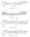
- FIG. 7is a side view of an embodiment of the targetable biopsy guide.
- FIG. 8is a side cutaway view of an embodiment of the targetable biopsy guide designed to be manufactured with an insertable metal tube.
- FIG. 9is a side cutaway view of an alternative embodiment of the targetable biopsy guide.
- FIG. 10is a side cutaway view of an alternative embodiment of the targetable biopsy guide with an enlarged bend channel.
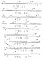
- FIG. 11shows a side view of a biopsy stylet with extended specimen notch.
- FIG. 12shows a side view of an alternative embodiment of the stylet with dual extended specimen notches.
- FIG. 13shows a side view of an alternative embodiment of the stylet with a tiered specimen notch.
- FIG. 14shows a side view of an alternate embodiment of the stylet with multiple notches to facilitate bending.
- FIG. 14Ais a side view of a flexible biopsy needle set in accordance with the present invention.
- FIG. 15shows a side view of an embodiment of the cannula in which the cannula tube has been ground down along its length to leave a flexible spine.
- FIG. 16shows a side view of an embodiment of the cannula in which the cannula tube has been spiral-cut along its length to facilitate bending of the cannula.
- FIG. 17shows a side view of an alternative embodiment of the cannula in which the tip of the cannula tube is uncut while the body of the cannula tube has been spiral-cut.
- FIG. 18shows a side view of an alternative embodiment of the cannula in which sections of the cannula tube alternate between cut and uncut.
- FIG. 19shows a side view of an embodiment of the cannula in which the cannula tube is encased in flexible tubing.
- FIG. 20shows a perspective view of an embodiment of the flexible needle set.
- FIG. 21is a side view of the traditional method of taking a prostate biopsy with a biopsy needle channel.
- FIG. 22is a side view of the bendable needle and biopsy targeting system mounted on a side-fired probe taking a biopsy.
- FIG. 23is a side view of the redirecting guide with a flexible needle set inserted and extending out of the guide such that the needle set is bent by the needle set channel bend.
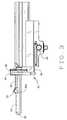
- the targeted biopsy systemis comprised of a redirecting guide 10 , positioning assembly 11 , targeting software system 12 (loaded on CPU 18 ) and flexible needle set 13 (best seen in FIG. 20 ).
- the positioning assembly 11is affixed to cradle 15 , which is a part of stepper and stabilizer 16 .
- ultrasound system 17which is comprised of ultrasound system CPU 18 , side view transrectal probe 19 and monitor 20 .
- Side view transrectal probeis comprised of probe tip 22 and probe imaging window 23 . As seen in FIGS.
- the redirecting guide 10consists of guide body 30 , needle set channel 31 , needle set insertion point 32 , and needle set exit point 33 , front body guide extensions 34 A and 34 B, imaging cutout 35 .
- needle set channel 31may be provided with enlarged bend channel 37 .
- the redirecting guide 10may be provided with insertable metal tube 38 .
- the redirecting guidemay contain one or more pathways may be used for insertion of the biopsy needle kit.
- the redirecting guidemay be comprised of a movable device such that the opening through which the needle kit exits may be moved relative to the opening into which the biopsy needle kit is placed.
- the redirecting guidemay straighten a previously curved biopsy needle kit such that the biopsy needle kit re-curve when leaving the redirecting guide.
- positioning assembly 11is comprised of rotational adjustment collar 40 , fixed collar 41 , longitudinal slides 42 and longitudinal position controller 43 .
- targeting software system 12is comprised of transverse image display 212 , Sagittal Image display 213 , longitudinal projected needle paths 51 a, b, c , etc and transverse projected needle paths 52 a, b, c , etc., in addition to various controls.
- flexible needle set 13consists of flexible stylet 60 and flexible cannula 61 .
- Stylet 60may be affixed to stylet hub 76 , with cannula 61 affixed to cannula hub 77 .
- cannula 61may be provided with depth markings 79 .
- the preferred flexible stylet 60consists of tip 62 , extended specimen notch 63 and stylet body 64 that is surrounded by the cannula body 66 .
- an alternative preferred flexible stylet 60consists of tip 62 and segmented specimen notches 72 a and 72 b .
- Alternative embodiments of flexible stylet 60as seen in FIGS. 13 and 14 , contain bending notches 70 and tiered specimen notch 71 .
- cannula 61consists of cutting tip 65 , cannula body 66 and cannula sheath 81 .
- the cannula sheathmay have beveled edges.
- a portion of the body of flexible cannula 61has been removed.
- cannula body 66may be provided with spiral cut 82 to facilitate bending.
- cannula body 66may be provided with non-spiral cut portion 83 at cutting tip 65 , to facilitate the straight entry of the cannula into the tissue.
- FIG. 16cannula body 66 may be provided with non-spiral cut portion 83 at cutting tip 65 , to facilitate the straight entry of the cannula into the tissue.
- cannula body 66may be provided with non-spiral cut portions 83 interspersed with spiral cuts 82 .
- flexible cannula 61consists of a cutting tip inserted into the flexible cannula body.
- both the stylet cannulacan be made from a range of flexible materials, including combinations of one or more materials, to facilitate the bendability. This may include traditional materials used in medical devices, such as stainless steel, as well as materials such an nitinol®.
- the cannula designmay mirror the stylet, such that portion or portions of the metal cannula tube are removed to create a metal component which has a metal cutting tip, a long spine consisting of only a portion of the cannula wall in the flexible part of the cannula and then the full tubular cannula.
- the machine cannulamay be partially or wholly incased in a cannula sheath, which may be plastic or some other material.
- FIG. 21shows a biopsy being performed using the standard method, using an end-fire ultrasound probe with a biopsy needle channel.
- the probeis inserted into the rectum, and then angled upward until the probe tip is pointed at the desired portion of the prostate.
- a needle setis then inserted through the biopsy needle channel guide into the prostate 2 .
- side view transrectal probe 19is mounted on the cradle 15 of a stabilizer 16 .
- Redirecting guide 10is also mounted on the cradle 15 , such that guide body 30 sits atop probe tip 22 .
- front body extensions 34 a and 34 bpartially wrap around probe tip 22 to help maintain the guide body 30 on the probe tip 22 .
- the cradle 15is moved forward, with the probe tip 22 inserted into patient's rectum 1 .
- Probe tip 22is generating ultrasound images, which are displayed on monitor 20 . The physician uses this image to insure that the entirety of prostate 2 is viewable by probe imaging window 23 . Once the probe tip 22 is correctly positioned, the physician locks in place cradle 15 .
- the biopsy planning processis illustrated in FIG. 5 .
- a representative display of the biopsy information to the useris shown in FIG. 4 .
- the processbegins with the planning software obtaining a set of volumetric data 101 .
- the volumetric dataconsists of two sets of sampled images. One set is of longitudinal images sampled at a regular angular spacing, and the other is a set of transverse images sampled at regular depth spacing. If only one of the two sets is available, one may be interpolated from the other.
- the physicianstarts the planning process by pressing button 203 to satisfy step 102 of FIG. 4 .
- the planning systemoverlays a series of lines 51 a, b, c , etc. and dots 52 a, b, c , etc.
- a recordis placed into needle path coordinates display window 204 showing the coordinates of the path.
- the usermay also remove a specific path from the plan by selecting button 207 .
- the physician or technicianmay then proceed with the biopsy procedure, to complete the series of precision located biopsy's to be taken through the usage of this instrument. For example, as can be noted in FIG. 6 , once a biopsy procedure has been completed, the physician then determines whether any more biopsies are needed, and where the biopsy locations may be determined. This can be seen at 301 . If no additional biopsies are required, this is the end of the procedure. If additional biopsies are considered as needed, the physician then adjusts the redirection of the guide 10 , and the longitudinal controller 40 , to mass the desired biopsy coordinates, as provided upon the scanner. This can be noted at 302 .
- the userinserts a needle set 13 into the channel 32 , to prepare for additional biopsies.
- the physicianthen inserts the needle into the patient, moving the needle in and out to adjust for depth, as determined by the scanner, as can be seen at 304 .
- the physiciancan determine if the needle tip is at the correct depth, at 305 . If it is not, then the physician may move the needle and adjust its depth further. If it is, the physician then fires the needle of the biopsy instrument, as at 306 .
- the physicianremoves the needle set 13 from the patient, having taken the biopsy as required.
- the tissue sampleis removed from the biopsy needle notch, for further analysis by the lab. This can be noted at 308 . When this is completed, this concludes the conduct of biopsies upon the patient.
- preplanned biopsy selection menu 209allows the user to select a pre-determined needle pattern, typically 9-12 needle paths, without having to select each needle path manually.
- the needle paths generatedcould need to be adjusted for the specific size of the organ.
- the size of the organcan be input by various means.
- the planning processallows the physician to modify the needle paths as needed and to approve that they are correct.
- Projected needle paths 51 a , 51 b , etc,include needle path location registry 50 , which indicate the horizontal and rotational position of the needle path in reference to the probe.
- the physicianrotates redirecting guide 10 using rotational adjustment collar 40 , and then advances the redirecting guide using longitudinal position controller 40 , both of which have position information which correlates to the needle path location registry 50 .
- the physicianinserts flexible needle set 13 into needle set insertion point 32 and into needle set channel 31 .
- the needle set 13is redirected at an angle away from the axis of probe tip 22 . Needle set 13 exits needle set exit point 33 .
- the physicianis able to see the needle set in the ultrasound image as it exits exit point 33 , allowing the physician to insure that the needle set 13 is in the path marked by projected needle path 51 a .
- the physicianmonitors the depth of the needle set 13 as it is pushed through the rectum wall and into the prostate 2 . Once the desired depth is reached, the physician stops inserting the needle set 13 .
- the needle set 13is “fired”. This causes flexible stylet 60 to rapidly advance a short distance, such that tissue from the prostate two prolapses into extended specimen notch 63 .
- FIG. 23provides a side cut-away view of the redirecting guide with a flexible needle set inserted and extending out of the guide such that the extended specimen notch is bent by the needle set channel bend.
- the inventionis used to plan and perform a targeted treatment of an organ or tissue mass.
- the processbegins with the planning software obtaining a set of volumetric data.
- the planning systemoverlays a series of needle path lines and needle path dots on the images in panes 212 and 213 , which represent the available needle paths with coordinates that match the coordinates on rotational adjustment collar 40 and longitudinal position controller 43 of positioning assembly 11 .
- the userselects specific needle paths, which are saved the treatment plan. Preplanned treatment selections allow the user to select a pre-determined needle pattern without having to select each needle path manually.
- the physicianrotates redirecting guide using rotational adjustment collar, and then advances the redirecting guide using longitudinal position controller, both of which have position information which correlates to the needle path location registry.
- the physicianthen inserts a flexible needle set or treatment delivery means into needle set insertion point 32 and into needle set channel 31 .
- the needle set or treatment delivery meansreaches the needle set channel bend, the needle set or treatment delivery method is redirected at an angle away from the axis of probe tip 22 . Needle set 13 exits needle set exit point 33 . Because of imaging cutout 35 , the physician is able to see the needle set or treatment delivery method in the ultrasound image as it exits exit point 33 .
- the physicianmonitors the depth of the needle set or treatment delivery method as it is pushed into the targeted organ or tissue mass. Once the desired depth is reached, the physician is able to undertake the preferred activity. This may include using the delivery means to inject a solid, gas or liquid material or other treatment apparatus into the targeted organ or tissue mass. Further, the physician may insert an organism into the targeted organ or tissue mass. The material may be deposited and left in the targeted organ or tissue mass. Further, material previously deposited may be removed. The use of the deposited material may be as a treatment, a marker, or other uses. Further, the delivery means may be used to apply energy to a targeted organ or tissue mass, including but not limited to heat, cold, light and radiation.
- the physicianthen removes the flexible needle set or treatment delivery method, and then resets the redirecting guide to the coordinates of the next saved projected needle path.
- the physicianhas the option of saving the image of the treatment needle in the targeted organ or tissue mass, to record the location of the treatment as delivered. The process is repeated until the physician has treated or marked all of the targeted areas of the organ or tissue mass.
Landscapes
- Health & Medical Sciences (AREA)
- Life Sciences & Earth Sciences (AREA)
- Surgery (AREA)
- General Health & Medical Sciences (AREA)
- Veterinary Medicine (AREA)
- Heart & Thoracic Surgery (AREA)
- Medical Informatics (AREA)
- Molecular Biology (AREA)
- Engineering & Computer Science (AREA)
- Animal Behavior & Ethology (AREA)
- Biomedical Technology (AREA)
- Public Health (AREA)
- Pathology (AREA)
- Nuclear Medicine, Radiotherapy & Molecular Imaging (AREA)
- Biodiversity & Conservation Biology (AREA)
- Ultra Sonic Daignosis Equipment (AREA)
- Radiation-Therapy Devices (AREA)
- Medicines Containing Antibodies Or Antigens For Use As Internal Diagnostic Agents (AREA)
- Nuclear Medicine (AREA)
- Investigating Or Analysing Biological Materials (AREA)
Abstract
Description
Rectum 1Prostate 2- redirecting
guide 10 - alternative redirecting guide10A
- positioning
assembly 11 - targeting software system12
- flexible needle set13
cradle 15stabilizer 16ultrasound system 17ultrasound system CPU 18- side view
transrectal probe 19 - monitor20
probe tip 22probe imaging window 23- guide
body 30 - needle set
channel 31 - needle set
insertion point 32 - needle set
exit point 33 - front
body guide extensions - imaging
cutout 35 - needle set
channel bend 36 enlarged bend channel 37- insertable metal tube38
rotational adjustment collar 40- fixed
collar 41 longitudinal slides 42longitudinal position controller 43- needle path location registry50
- needle path lines51
- needle path dots52
flexible stylet 60flexible cannula 61tip 62extended specimen notch 63stylet body 64- cutting
tip 65 cannula body 66- counter bore and taper67
- bending
notches 70 tiered specimen notch 71segmented specimen notch 72- removable needle set guide insert75
stylet hub 76cannula hub 77- strip78
depth markings 79cannula sheath 81- spiral cut82
non-spiral cut portion 83- beveled edge84
- Biopsy
attachment angle selector 201 and display - Biopsy
attachment depth selector 202 - and display
- needle path coordinates
display 204 - window
- Finished with
Biopsy Planning 206 - button
- Remove selected
biopsy location 207 - from plan button
- Add selected biopsy location to208
- plan button
- Select
pre-planned template 209 - Sagittal
image plane selector 210 - Transverse
image plane selector 211 Transverse image display 212Sagittal Image display 213
Claims (31)
Priority Applications (3)
| Application Number | Priority Date | Filing Date | Title |
|---|---|---|---|
| US11/895,228US7833168B2 (en) | 2003-08-13 | 2007-08-23 | Targeted biopsy delivery system |
| US12/214,163US20090054807A1 (en) | 2003-08-13 | 2008-06-17 | Targeted biopsy delivery system |
| US12/968,870US8317724B2 (en) | 2003-08-13 | 2010-12-15 | Targeted treatment delivery system |
Applications Claiming Priority (3)
| Application Number | Priority Date | Filing Date | Title |
|---|---|---|---|
| US49491003P | 2003-08-13 | 2003-08-13 | |
| US10/842,652US20050159676A1 (en) | 2003-08-13 | 2004-05-10 | Targeted biopsy delivery system |
| US11/895,228US7833168B2 (en) | 2003-08-13 | 2007-08-23 | Targeted biopsy delivery system |
Related Parent Applications (1)
| Application Number | Title | Priority Date | Filing Date |
|---|---|---|---|
| US10/842,652ContinuationUS20050159676A1 (en) | 2003-08-13 | 2004-05-10 | Targeted biopsy delivery system |
Related Child Applications (1)
| Application Number | Title | Priority Date | Filing Date |
|---|---|---|---|
| US12/214,163DivisionUS20090054807A1 (en) | 2003-08-13 | 2008-06-17 | Targeted biopsy delivery system |
Publications (2)
| Publication Number | Publication Date |
|---|---|
| US20070293787A1 US20070293787A1 (en) | 2007-12-20 |
| US7833168B2true US7833168B2 (en) | 2010-11-16 |
Family
ID=35394638
Family Applications (4)
| Application Number | Title | Priority Date | Filing Date |
|---|---|---|---|
| US10/842,652AbandonedUS20050159676A1 (en) | 2003-08-13 | 2004-05-10 | Targeted biopsy delivery system |
| US11/895,228Expired - LifetimeUS7833168B2 (en) | 2003-08-13 | 2007-08-23 | Targeted biopsy delivery system |
| US12/214,163AbandonedUS20090054807A1 (en) | 2003-08-13 | 2008-06-17 | Targeted biopsy delivery system |
| US12/968,870Expired - Fee RelatedUS8317724B2 (en) | 2003-08-13 | 2010-12-15 | Targeted treatment delivery system |
Family Applications Before (1)
| Application Number | Title | Priority Date | Filing Date |
|---|---|---|---|
| US10/842,652AbandonedUS20050159676A1 (en) | 2003-08-13 | 2004-05-10 | Targeted biopsy delivery system |
Family Applications After (2)
| Application Number | Title | Priority Date | Filing Date |
|---|---|---|---|
| US12/214,163AbandonedUS20090054807A1 (en) | 2003-08-13 | 2008-06-17 | Targeted biopsy delivery system |
| US12/968,870Expired - Fee RelatedUS8317724B2 (en) | 2003-08-13 | 2010-12-15 | Targeted treatment delivery system |
Country Status (11)
| Country | Link |
|---|---|
| US (4) | US20050159676A1 (en) |
| EP (1) | EP1765178B1 (en) |
| JP (1) | JP4837658B2 (en) |
| KR (1) | KR20070028402A (en) |
| CN (1) | CN101132736B (en) |
| AT (1) | ATE505136T1 (en) |
| AU (1) | AU2005244164B2 (en) |
| CA (1) | CA2566231C (en) |
| DE (1) | DE602005027462D1 (en) |
| ES (1) | ES2365476T3 (en) |
| WO (1) | WO2005110255A2 (en) |
Cited By (50)
| Publication number | Priority date | Publication date | Assignee | Title |
|---|---|---|---|---|
| US20100076305A1 (en)* | 2008-06-25 | 2010-03-25 | Deutsches Krebsforschungszentrum Stiftung Des Offentlichen Rechts | Method, system and computer program product for targeting of a target with an elongate instrument |
| US20110130680A1 (en)* | 2008-08-01 | 2011-06-02 | Ab Christer Dahlstrand | Device for Testing Needles |
| US8482606B2 (en) | 2006-08-02 | 2013-07-09 | Inneroptic Technology, Inc. | System and method of providing real-time dynamic imagery of a medical procedure site using multiple modalities |
| US8554307B2 (en) | 2010-04-12 | 2013-10-08 | Inneroptic Technology, Inc. | Image annotation in image-guided medical procedures |
| US8585598B2 (en) | 2009-02-17 | 2013-11-19 | Inneroptic Technology, Inc. | Systems, methods, apparatuses, and computer-readable media for image guided surgery |
| US8641621B2 (en) | 2009-02-17 | 2014-02-04 | Inneroptic Technology, Inc. | Systems, methods, apparatuses, and computer-readable media for image management in image-guided medical procedures |
| US8670816B2 (en) | 2012-01-30 | 2014-03-11 | Inneroptic Technology, Inc. | Multiple medical device guidance |
| US8781555B2 (en) | 2007-11-26 | 2014-07-15 | C. R. Bard, Inc. | System for placement of a catheter including a signal-generating stylet |
| US8784336B2 (en) | 2005-08-24 | 2014-07-22 | C. R. Bard, Inc. | Stylet apparatuses and methods of manufacture |
| US8831310B2 (en) | 2008-03-07 | 2014-09-09 | Inneroptic Technology, Inc. | Systems and methods for displaying guidance data based on updated deformable imaging data |
| US8849382B2 (en) | 2007-11-26 | 2014-09-30 | C. R. Bard, Inc. | Apparatus and display methods relating to intravascular placement of a catheter |
| US8858455B2 (en) | 2006-10-23 | 2014-10-14 | Bard Access Systems, Inc. | Method of locating the tip of a central venous catheter |
| US9125578B2 (en) | 2009-06-12 | 2015-09-08 | Bard Access Systems, Inc. | Apparatus and method for catheter navigation and tip location |
| US9265572B2 (en) | 2008-01-24 | 2016-02-23 | The University Of North Carolina At Chapel Hill | Methods, systems, and computer readable media for image guided ablation |
| US9265443B2 (en) | 2006-10-23 | 2016-02-23 | Bard Access Systems, Inc. | Method of locating the tip of a central venous catheter |
| US9339206B2 (en) | 2009-06-12 | 2016-05-17 | Bard Access Systems, Inc. | Adaptor for endovascular electrocardiography |
| US9415188B2 (en) | 2010-10-29 | 2016-08-16 | C. R. Bard, Inc. | Bioimpedance-assisted placement of a medical device |
| US9445734B2 (en) | 2009-06-12 | 2016-09-20 | Bard Access Systems, Inc. | Devices and methods for endovascular electrography |
| US9456766B2 (en) | 2007-11-26 | 2016-10-04 | C. R. Bard, Inc. | Apparatus for use with needle insertion guidance system |
| US9492097B2 (en) | 2007-11-26 | 2016-11-15 | C. R. Bard, Inc. | Needle length determination and calibration for insertion guidance system |
| US9521961B2 (en) | 2007-11-26 | 2016-12-20 | C. R. Bard, Inc. | Systems and methods for guiding a medical instrument |
| US9532724B2 (en) | 2009-06-12 | 2017-01-03 | Bard Access Systems, Inc. | Apparatus and method for catheter navigation using endovascular energy mapping |
| US9554716B2 (en) | 2007-11-26 | 2017-01-31 | C. R. Bard, Inc. | Insertion guidance system for needles and medical components |
| US9636031B2 (en) | 2007-11-26 | 2017-05-02 | C.R. Bard, Inc. | Stylets for use with apparatus for intravascular placement of a catheter |
| US9649048B2 (en) | 2007-11-26 | 2017-05-16 | C. R. Bard, Inc. | Systems and methods for breaching a sterile field for intravascular placement of a catheter |
| US9675319B1 (en) | 2016-02-17 | 2017-06-13 | Inneroptic Technology, Inc. | Loupe display |
| US9681823B2 (en) | 2007-11-26 | 2017-06-20 | C. R. Bard, Inc. | Integrated system for intravascular placement of a catheter |
| US9839372B2 (en) | 2014-02-06 | 2017-12-12 | C. R. Bard, Inc. | Systems and methods for guidance and placement of an intravascular device |
| US9901714B2 (en) | 2008-08-22 | 2018-02-27 | C. R. Bard, Inc. | Catheter assembly including ECG sensor and magnetic assemblies |
| US9901406B2 (en) | 2014-10-02 | 2018-02-27 | Inneroptic Technology, Inc. | Affected region display associated with a medical device |
| US9907513B2 (en) | 2008-10-07 | 2018-03-06 | Bard Access Systems, Inc. | Percutaneous magnetic gastrostomy |
| US9949700B2 (en) | 2015-07-22 | 2018-04-24 | Inneroptic Technology, Inc. | Medical device approaches |
| US10046139B2 (en) | 2010-08-20 | 2018-08-14 | C. R. Bard, Inc. | Reconfirmation of ECG-assisted catheter tip placement |
| US10188467B2 (en) | 2014-12-12 | 2019-01-29 | Inneroptic Technology, Inc. | Surgical guidance intersection display |
| US10278778B2 (en) | 2016-10-27 | 2019-05-07 | Inneroptic Technology, Inc. | Medical device navigation using a virtual 3D space |
| US10314559B2 (en) | 2013-03-14 | 2019-06-11 | Inneroptic Technology, Inc. | Medical device guidance |
| US10349890B2 (en) | 2015-06-26 | 2019-07-16 | C. R. Bard, Inc. | Connector interface for ECG-based catheter positioning system |
| US10449330B2 (en) | 2007-11-26 | 2019-10-22 | C. R. Bard, Inc. | Magnetic element-equipped needle assemblies |
| US10524691B2 (en) | 2007-11-26 | 2020-01-07 | C. R. Bard, Inc. | Needle assembly including an aligned magnetic element |
| US10751509B2 (en) | 2007-11-26 | 2020-08-25 | C. R. Bard, Inc. | Iconic representations for guidance of an indwelling medical device |
| US10973584B2 (en) | 2015-01-19 | 2021-04-13 | Bard Access Systems, Inc. | Device and method for vascular access |
| US10992079B2 (en) | 2018-10-16 | 2021-04-27 | Bard Access Systems, Inc. | Safety-equipped connection systems and methods thereof for establishing electrical connections |
| US11000207B2 (en) | 2016-01-29 | 2021-05-11 | C. R. Bard, Inc. | Multiple coil system for tracking a medical device |
| US11259879B2 (en) | 2017-08-01 | 2022-03-01 | Inneroptic Technology, Inc. | Selective transparency to assist medical device navigation |
| US11464578B2 (en) | 2009-02-17 | 2022-10-11 | Inneroptic Technology, Inc. | Systems, methods, apparatuses, and computer-readable media for image management in image-guided medical procedures |
| US11484365B2 (en) | 2018-01-23 | 2022-11-01 | Inneroptic Technology, Inc. | Medical image guidance |
| US11690806B2 (en) | 2018-05-24 | 2023-07-04 | Celanese Eva Performance Polymers Llc | Implantable device for sustained release of a macromolecular drug compound |
| US11690807B2 (en) | 2018-05-24 | 2023-07-04 | Celanese Eva Performance Polymers Llc | Implantable device for sustained release of a macromolecular drug compound |
| US12075989B2 (en) | 2020-11-23 | 2024-09-03 | Civco Medical Instruments Co., Inc. | Ultrasound transperineal puncture device guide with a pivotable needle holder |
| US12108225B2 (en) | 2018-05-24 | 2024-10-01 | Celanese Eva Performance Polymers Llc | Implantable device for sustained release of a macromolecular drug compound |
Families Citing this family (104)
| Publication number | Priority date | Publication date | Assignee | Title |
|---|---|---|---|---|
| US20050159676A1 (en)* | 2003-08-13 | 2005-07-21 | Taylor James D. | Targeted biopsy delivery system |
| US8070750B2 (en) | 2004-03-05 | 2011-12-06 | Depuy Mitek, Inc. | Tunnel notcher and guidewire delivery device |
| US8795195B2 (en)* | 2004-11-29 | 2014-08-05 | Senorx, Inc. | Graphical user interface for tissue biopsy system |
| US8788019B2 (en)* | 2005-02-28 | 2014-07-22 | Robarts Research Institute | System and method for performing a biopsy of a target volume and a computing device for planning the same |
| US7831293B2 (en)* | 2005-05-10 | 2010-11-09 | Advanced Clinical Solutions, Inc. | Method of defining a biological target for treatment |
| US8425418B2 (en)* | 2006-05-18 | 2013-04-23 | Eigen, Llc | Method of ultrasonic imaging and biopsy of the prostate |
| US8064664B2 (en)* | 2006-10-18 | 2011-11-22 | Eigen, Inc. | Alignment method for registering medical images |
| US7804989B2 (en)* | 2006-10-30 | 2010-09-28 | Eigen, Inc. | Object recognition system for medical imaging |
| US20080161687A1 (en)* | 2006-12-29 | 2008-07-03 | Suri Jasjit S | Repeat biopsy system |
| US12290277B2 (en) | 2007-01-02 | 2025-05-06 | Aquabeam, Llc | Tissue resection with pressure sensing |
| US9232959B2 (en) | 2007-01-02 | 2016-01-12 | Aquabeam, Llc | Multi fluid tissue resection methods and devices |
| US8175350B2 (en) | 2007-01-15 | 2012-05-08 | Eigen, Inc. | Method for tissue culture extraction |
| US7856130B2 (en)* | 2007-03-28 | 2010-12-21 | Eigen, Inc. | Object recognition system for medical imaging |
| US7832114B2 (en)* | 2007-04-04 | 2010-11-16 | Eigen, Llc | Tracker holder assembly |
| US20100152663A1 (en)* | 2007-04-05 | 2010-06-17 | Darr Allan J | Stylet for bilumenal flexible medical device |
| US8979803B2 (en)* | 2007-04-05 | 2015-03-17 | Allan J. Darr | Stylet for bilumenal flexible medical device |
| US20090048515A1 (en)* | 2007-08-14 | 2009-02-19 | Suri Jasjit S | Biopsy planning system |
| US8088072B2 (en)* | 2007-10-12 | 2012-01-03 | Gynesonics, Inc. | Methods and systems for controlled deployment of needles in tissue |
| US8571277B2 (en) | 2007-10-18 | 2013-10-29 | Eigen, Llc | Image interpolation for medical imaging |
| US7942829B2 (en)* | 2007-11-06 | 2011-05-17 | Eigen, Inc. | Biopsy planning and display apparatus |
| US20090227874A1 (en)* | 2007-11-09 | 2009-09-10 | Eigen, Inc. | Holder assembly for a medical imaging instrument |
| US8450290B2 (en)* | 2007-11-26 | 2013-05-28 | Enzon Pharmaceuticals, Inc. | Methods for treating androgen receptor dependent disorders including cancers |
| JP2009153831A (en)* | 2007-12-27 | 2009-07-16 | Ge Medical Systems Global Technology Co Llc | Structure for mounting puncture guide, ultrasonic probe, and ultrasonic diagnostic apparatus |
| US20090216250A1 (en)* | 2008-02-27 | 2009-08-27 | Ralph Zipper | Device and Method for Carrying Material Through Tissue |
| ES2769535T3 (en) | 2008-03-06 | 2020-06-26 | Aquabeam Llc | Tissue ablation and cauterization with optical energy carried in a fluid stream |
| US8864681B2 (en) | 2008-04-23 | 2014-10-21 | Devicor Medical Products, Inc. | Biopsy devices |
| US9782565B2 (en) | 2008-10-01 | 2017-10-10 | Covidien Lp | Endoscopic ultrasound-guided biliary access system |
| US9332973B2 (en) | 2008-10-01 | 2016-05-10 | Covidien Lp | Needle biopsy device with exchangeable needle and integrated needle protection |
| US9186128B2 (en) | 2008-10-01 | 2015-11-17 | Covidien Lp | Needle biopsy device |
| US11298113B2 (en) | 2008-10-01 | 2022-04-12 | Covidien Lp | Device for needle biopsy with integrated needle protection |
| US8968210B2 (en) | 2008-10-01 | 2015-03-03 | Covidien LLP | Device for needle biopsy with integrated needle protection |
| CN102271595A (en) | 2008-11-06 | 2011-12-07 | 恩克斯特拉公司 | Systems and methods for treatment of bph |
| JP2012508068A (en) | 2008-11-06 | 2012-04-05 | エヌエックスセラ インコーポレイテッド | System and method for treatment of prostate tissue |
| CN105434039B (en) | 2008-11-06 | 2019-01-15 | 恩克斯特拉公司 | System and method for treating prostata tissue |
| US8388611B2 (en)* | 2009-01-14 | 2013-03-05 | Nxthera, Inc. | Systems and methods for treatment of prostatic tissue |
| US20100179416A1 (en)* | 2009-01-14 | 2010-07-15 | Michael Hoey | Medical Systems and Methods |
| US9833277B2 (en) | 2009-04-27 | 2017-12-05 | Nxthera, Inc. | Systems and methods for prostate treatment |
| WO2011017665A2 (en)* | 2009-08-07 | 2011-02-10 | Thayer Intellectual Property, Inc. | Systems and methods for treatment of compressed nerves |
| US8753364B2 (en)* | 2009-08-07 | 2014-06-17 | Thayer Intellectual Property, Inc. | Systems and methods for treatment of compressed nerves |
| US8652157B2 (en) | 2009-08-07 | 2014-02-18 | Thayer Intellectual Property, Inc. | Systems and methods for treatment of compressed nerves |
| KR101109197B1 (en) | 2009-12-17 | 2012-01-30 | 국립암센터 | Needle feeder |
| EP2549963B1 (en) | 2010-03-25 | 2023-08-23 | Boston Scientific Scimed, Inc. | Systems for prostate treatment |
| US9044216B2 (en) | 2010-07-12 | 2015-06-02 | Best Medical International, Inc. | Biopsy needle assembly |
| US8758256B2 (en)* | 2010-07-12 | 2014-06-24 | Best Medical International, Inc. | Apparatus for brachytherapy that uses a scanning probe for treatment of malignant tissue |
| JP5482564B2 (en)* | 2010-08-18 | 2014-05-07 | ソニー株式会社 | Bioactive substance collection device |
| JP2012042310A (en)* | 2010-08-18 | 2012-03-01 | Sony Corp | Physiologically active substance collecting device and biological information acquisition method |
| CN103153201B (en) | 2010-10-05 | 2016-06-29 | 斯恩蒂斯有限公司 | Bone marrow harvesting device with flexible needle |
| US8585601B2 (en)* | 2010-10-18 | 2013-11-19 | CardioSonic Ltd. | Ultrasound transducer |
| US11612377B2 (en)* | 2010-12-16 | 2023-03-28 | Best Medical International, Inc. | Image guided surgical methodology and system employing patient movement detection and correction |
| US9414816B2 (en)* | 2011-06-23 | 2016-08-16 | Devicor Medical Products, Inc. | Introducer for biopsy device |
| CN102894994A (en)* | 2011-07-25 | 2013-01-30 | 滕学东 | Protective film for ultrasonic probe |
| CN103917200B (en) | 2011-09-13 | 2016-03-30 | 恩克斯特拉公司 | Systems and methods for prostate treatment |
| FR2983397B1 (en)* | 2011-12-06 | 2014-07-04 | Univ Paris Curie | DEVICE FOR ASSISTING THE POSITIONING OF A MEDICAL INSTRUMENT RELATIVELY TO AN INTERNAL ORGAN OF A PATIENT AND METHOD OF CONTROLLING SUCH A DEVICE |
| GB2500784B (en) | 2012-02-28 | 2015-07-22 | Spiration Inc | Lung Biopsy Needle |
| EP3351196A1 (en) | 2012-02-29 | 2018-07-25 | Procept Biorobotics Corporation | Automated image-guided tissue resection and treatment |
| EP2833815B1 (en) | 2012-04-03 | 2020-11-11 | Boston Scientific Scimed, Inc. | Induction coil vapor generator |
| EP2836133B1 (en)* | 2012-04-10 | 2018-08-29 | The Johns Hopkins University | Cohesive robot-ultrasound probe for prostate biopsy |
| US9131922B2 (en) | 2013-01-29 | 2015-09-15 | Eigen, Inc. | Calibration for 3D reconstruction of medical images from a sequence of 2D images |
| JP6403695B2 (en) | 2013-02-14 | 2018-10-10 | プロセプト バイオロボティクス コーポレイション | Aqua ablation aqua beam ophthalmic surgery method and apparatus |
| TWI592141B (en)* | 2013-03-07 | 2017-07-21 | 國立陽明大學 | Ultrasonic probe structure |
| BR112015022358A2 (en) | 2013-03-14 | 2017-07-18 | Nxthera Inc | method for treating abnormal prostate tissue, and, method for treating prostate cancer, and, prostate cancer therapy system |
| US20160038127A1 (en)* | 2013-03-15 | 2016-02-11 | R. Hashimshony Engineering Ltd. | Biopsy Probe and Use Thereof |
| US10206664B2 (en)* | 2013-11-13 | 2019-02-19 | Cook Medical Technologies Llc | Spiral cut biopsy cannula |
| US9968395B2 (en) | 2013-12-10 | 2018-05-15 | Nxthera, Inc. | Systems and methods for treating the prostate |
| US10194970B2 (en) | 2013-12-10 | 2019-02-05 | Nxthera, Inc. | Vapor ablation systems and methods |
| US9149260B2 (en) | 2014-02-28 | 2015-10-06 | 3DBiopsy LLC | Biopsy needle assembly |
| US10743909B2 (en)* | 2014-04-03 | 2020-08-18 | Corbin Clinical Resources, Llc | Transperineal prostate biopsy device, systems, and methods of use |
| US10064681B2 (en)* | 2014-04-03 | 2018-09-04 | Corbin Clinical Resources, Llc | Method, system, and device for planning and performing, guided and free-handed transperineal prostate biopsies |
| US12144519B2 (en) | 2014-04-03 | 2024-11-19 | Corbin Clinical Resources, Llc | Transperineal prostate biopsy and treatment methods |
| CN103919577A (en)* | 2014-04-13 | 2014-07-16 | 哈尔滨理工大学 | Cantilever prostate biopsy robot |
| JP6548730B2 (en)* | 2014-08-23 | 2019-07-24 | インテュイティブ サージカル オペレーションズ, インコーポレイテッド | System and method for display of pathological data in image guided procedures |
| US11033296B2 (en)* | 2014-08-23 | 2021-06-15 | Intuitive Surgical Operations, Inc. | Systems and methods for dynamic trajectory control |
| CN104224092B (en)* | 2014-09-19 | 2016-08-17 | 珠海普生医疗科技有限公司 | Elder generation of endoscope end construction |
| JP6689850B2 (en) | 2014-11-26 | 2020-04-28 | デビコー・メディカル・プロダクツ・インコーポレイテッドDevicor Medical Products, Inc. | Graphic user interface of biopsy device |
| EP3236859B1 (en)* | 2014-12-24 | 2021-03-31 | Koninklijke Philips N.V. | Needle trajectory prediction for target biopsy |
| US10342593B2 (en) | 2015-01-29 | 2019-07-09 | Nxthera, Inc. | Vapor ablation systems and methods |
| CN104688278A (en)* | 2015-03-11 | 2015-06-10 | 毕文志 | Bone tumor biopsy puncture needle |
| CA2982372A1 (en) | 2015-05-13 | 2016-11-17 | Nxthera, Inc. | Systems and methods for treating the bladder with condensable vapor |
| CN104939800B (en)* | 2015-06-16 | 2017-04-05 | 西南医科大学附属医院 | A kind of the intestines and stomach automatic detection surgery apparatus |
| US10849650B2 (en) | 2015-07-07 | 2020-12-01 | Eigen Health Services, Llc | Transperineal needle guidance |
| US10716544B2 (en) | 2015-10-08 | 2020-07-21 | Zmk Medical Technologies Inc. | System for 3D multi-parametric ultrasound imaging |
| CN105147335B (en)* | 2015-10-13 | 2017-11-28 | 拜耳斯特医疗机器人技术(天津)有限公司 | A kind of ultrasonic probe rotating device and its application method and application |
| SG11201809873UA (en)* | 2016-05-25 | 2018-12-28 | 3D Biopsy Inc | Biopsy needle design |
| CN106361396B (en)* | 2016-09-27 | 2018-11-23 | 熊力 | A kind of liver vessel folder closes device |
| KR20190075085A (en) | 2016-10-10 | 2019-06-28 | 쓰리디바이옵시 인코포레이티드 | Biopsy actuator assembly |
| US10751034B2 (en) | 2016-10-17 | 2020-08-25 | The Johna Hopkins University | Geometric biopsy plan optimization |
| CN115715689B (en) | 2016-11-11 | 2025-01-17 | 杰尼索尼克斯公司 | Tissue controlled treatment and dynamic interaction and comparison with tissue and/or treatment data |
| JP7129980B2 (en) | 2016-12-21 | 2022-09-02 | ボストン サイエンティフィック サイムド,インコーポレイテッド | Steam cautery system and method |
| WO2018129466A1 (en) | 2017-01-06 | 2018-07-12 | Nxthera, Inc. | Transperineal vapor ablation systems and methods |
| US11589845B2 (en)* | 2017-01-06 | 2023-02-28 | Sorek Medical Systems Ltd. | Core biopsy system for storage and preservation of multiple tissue samples |
| ES2972207T3 (en) | 2017-07-14 | 2024-06-11 | Amgen Inc | Needle insertion-retraction system with double torsion spring system |
| US20190282218A1 (en)* | 2018-03-14 | 2019-09-19 | SPIRATION, INC., d/b/a OLYMPUS RESPIRATORY AMERICA | Catheter Assembly With Offset Device For Tissue Sampling |
| US10912542B2 (en)* | 2018-03-14 | 2021-02-09 | Spiration, Inc. | Catheter assembly with offset device for tissue sampling |
| CN108888315B (en)* | 2018-04-17 | 2024-11-19 | 建湖县人民医院 | A universal digital transperineal prostate puncture stent |
| CN109011030B (en)* | 2018-08-08 | 2021-02-09 | 长沙理工大学 | Method and device for detecting and correcting position of needle of automatic injection instrument |
| PL4193932T3 (en)* | 2018-12-10 | 2024-11-12 | Devicor Medical Products, Inc. | Biopsy system with end deploy needle |
| KR102747176B1 (en)* | 2018-12-11 | 2024-12-27 | 삼성메디슨 주식회사 | Ultrasound imaging apparatus, method for controlling the same, and computer program product |
| WO2020231748A1 (en)* | 2019-05-10 | 2020-11-19 | Merit Medical Systems, Inc. | Drainage catheter exchange system and associated methods |
| US12245886B2 (en)* | 2020-03-16 | 2025-03-11 | Scopus Medical Systems, Ltd. | Guidance system for interventional devices with curved shape |
| GB2616235B (en)* | 2020-12-10 | 2025-03-12 | Nat Univ Hospital Singapore Pte Ltd | Balloon-anchored flexible needle and catheter for biopsy |
| US20220211263A1 (en)* | 2021-03-23 | 2022-07-07 | Axcess Instruments Inc. | Multi-piece access port imaging systems |
| CN115462837B (en)* | 2022-07-01 | 2025-08-29 | 海斯凯尔(山东)医学科技有限公司 | Image recording method, device and biopsy sampling equipment |
| CN116458972B (en)* | 2023-03-31 | 2023-12-01 | 拜斯特医疗科技(北京)有限公司 | Ultrasonic probe calibrating and positioning device for living body puncture |
| CN116509627B (en)* | 2023-04-23 | 2025-07-25 | 哈尔滨工业大学 | Inner ear injection sampling actuator of otology operation robot |
Citations (25)
| Publication number | Priority date | Publication date | Assignee | Title |
|---|---|---|---|---|
| US5149598A (en) | 1991-07-11 | 1992-09-22 | Acr Electronics, Inc. | Battery arrangement |
| US5398690A (en) | 1994-08-03 | 1995-03-21 | Batten; Bobby G. | Slaved biopsy device, analysis apparatus, and process |
| US5456258A (en) | 1993-12-20 | 1995-10-10 | Fuji Photo Optical Co., Ltd. | Catheter type ultrasound probe |
| US5660185A (en) | 1995-04-13 | 1997-08-26 | Neovision Corporation | Image-guided biopsy apparatus with enhanced imaging and methods |
| US5873828A (en) | 1994-02-18 | 1999-02-23 | Olympus Optical Co., Ltd. | Ultrasonic diagnosis and treatment system |
| US6102867A (en) | 1997-02-11 | 2000-08-15 | Tetrad Corporation | Sheath and methods of ultrasonic guidance of biopsy and catheter insertion |
| US6171249B1 (en) | 1997-10-14 | 2001-01-09 | Circon Corporation | Ultrasound guided therapeutic and diagnostic device |
| US6179249B1 (en) | 1996-12-26 | 2001-01-30 | Aerospatiale Societe Nationale Industrielle | Turbojet pod with laminar flow |
| US6238336B1 (en) | 1998-03-04 | 2001-05-29 | Asahi Kogaku Kogyo Kabushiki Kaisha | Ultrasonic endoscope including radial scanning and linear scanning ultrasonic transducers |
| US6261234B1 (en) | 1998-05-07 | 2001-07-17 | Diasonics Ultrasound, Inc. | Method and apparatus for ultrasound imaging with biplane instrument guidance |
| US6261243B1 (en) | 1998-10-13 | 2001-07-17 | Emx, Inc. | Biopsy marker assembly and method of use |
| US20020002349A1 (en) | 1996-10-11 | 2002-01-03 | Transvascular, Inc. | Systems and methods for delivering drugs to selected locations within the body |
| US6390973B1 (en) | 1998-06-25 | 2002-05-21 | Asahi Kogaku Kogyo Kabushiki Kaisha | Endoscope for ultrasonic examination and surgical treatment associated thereto |
| US6409666B1 (en) | 1999-04-15 | 2002-06-25 | Asahi Kogaku Kogyo Kabushiki Kaisha | Tip end of ultrasonic endoscope |
| US20020111634A1 (en) | 2000-08-30 | 2002-08-15 | Johns Hopkins University | Controllable motorized device for percutaneous needle placement in soft tissue target and methods and systems related thereto |
| US6447477B2 (en) | 1996-02-09 | 2002-09-10 | Emx, Inc. | Surgical and pharmaceutical site access guide and methods |
| US6485411B1 (en) | 2000-04-12 | 2002-11-26 | Circon Corporation | Endoscope shaft with superelastic alloy spiral frame and braid |
| US20030078502A1 (en) | 2001-10-23 | 2003-04-24 | Olympus Optical Co., Ltd. | Device for examining a subject capable of marking a boundary range for insertion/retraction of an insertion/retraction member that is inserted in and retracted from the subject |
| US20030120154A1 (en) | 2001-11-28 | 2003-06-26 | Frank Sauer | Method and apparatus for ultrasound guidance of needle biopsies |
| US20030135119A1 (en) | 2001-12-31 | 2003-07-17 | Medison Co., Ltd. | Method and apparatus for enabling a biopsy needle to be observed |
| US20040030250A1 (en) | 2000-06-22 | 2004-02-12 | Duncan Stewart | Injection system for gene delivery |
| US20040133111A1 (en) | 2003-01-03 | 2004-07-08 | Civco Medical Instruments Inc. | Shallow angle needle guide apparatus and method |
| US6884219B1 (en) | 2002-10-17 | 2005-04-26 | Rick L. Pruter | Method and disposable apparatus for guiding needles with an endocavity medical imaging device |
| US20050203413A1 (en) | 2003-08-07 | 2005-09-15 | Gabor Fichtinger | Transcavital needle insertion device |
| US7171255B2 (en) | 1995-07-26 | 2007-01-30 | Computerized Medical Systems, Inc. | Virtual reality 3D visualization for surgical procedures |
Family Cites Families (23)
| Publication number | Priority date | Publication date | Assignee | Title |
|---|---|---|---|---|
| US60493A (en)* | 1866-12-18 | elder | ||
| US5095910A (en)* | 1990-04-18 | 1992-03-17 | Advanced Technology Laboratories, Inc. | Ultrasonic imaging of biopsy needle |
| US5833627A (en)* | 1995-04-13 | 1998-11-10 | United States Surgical Corporation | Image-guided biopsy apparatus and methods of use |
| US5843050A (en)* | 1995-11-13 | 1998-12-01 | Micro Therapeutics, Inc. | Microcatheter |
| US5885258A (en)* | 1996-02-23 | 1999-03-23 | Memory Medical Systems, Inc. | Medical instrument with slotted memory metal tube |
| US6167296A (en)* | 1996-06-28 | 2000-12-26 | The Board Of Trustees Of The Leland Stanford Junior University | Method for volumetric image navigation |
| US6021342A (en)* | 1997-06-30 | 2000-02-01 | Neorad A/S | Apparatus for assisting percutaneous computed tomography-guided surgical activity |
| JPH11276422A (en)* | 1998-03-31 | 1999-10-12 | Fuji Photo Optical Co Ltd | Ultrasonic endoscope |
| FR2788956B1 (en)* | 1999-01-28 | 2001-03-30 | Seb Sa | CULINARY KIT WITH REMOVABLE GRIPPING HANDLE |
| JP3634655B2 (en)* | 1999-02-09 | 2005-03-30 | ペンタックス株式会社 | Endoscopic biopsy forceps |
| CN2383525Y (en)* | 1999-07-16 | 2000-06-21 | 常州市第一人民医院 | Detachable intrathoracic drain using biopsy needle for ingress |
| US6749560B1 (en)* | 1999-10-26 | 2004-06-15 | Circon Corporation | Endoscope shaft with slotted tube |
| US7201722B2 (en)* | 2000-04-18 | 2007-04-10 | Allegiance Corporation | Bone biopsy instrument having improved sample retention |
| US6569105B1 (en)* | 2000-09-14 | 2003-05-27 | Syntheon, Llc | Rotatable and deflectable biopsy forceps |
| US7033374B2 (en)* | 2000-09-26 | 2006-04-25 | Microvention, Inc. | Microcoil vaso-occlusive device with multi-axis secondary configuration |
| WO2002069808A2 (en)* | 2000-11-06 | 2002-09-12 | Suros Surgical Systems, Inc. | Biopsy apparatus |
| US6419641B1 (en)* | 2000-11-28 | 2002-07-16 | Promex, Llc | Flexible tip medical instrument |
| IL140494A0 (en)* | 2000-12-22 | 2002-02-10 | Pneumatic control system for a biopsy device | |
| ITSV20010008A1 (en)* | 2001-03-05 | 2002-09-05 | Esaote Spa | NEEDLE GUIDE DEVICE IN PARTICULAR FOR ECHOGRAPHIC PROBES AND COMBINATION OF ECHOGRAPHIC PROBE AND SAID NEEDLE GUIDE DEVICE |
| US6623448B2 (en)* | 2001-03-30 | 2003-09-23 | Advanced Cardiovascular Systems, Inc. | Steerable drug delivery device |
| CN2540160Y (en)* | 2002-04-03 | 2003-03-19 | 黎维忱 | Quick cutting biopsy needle |
| EP1545349A1 (en)* | 2002-07-12 | 2005-06-29 | Cook Urological Inc. | Flexible cannula shaft |
| US20050159676A1 (en) | 2003-08-13 | 2005-07-21 | Taylor James D. | Targeted biopsy delivery system |
- 2004
- 2004-05-10USUS10/842,652patent/US20050159676A1/ennot_activeAbandoned
- 2005
- 2005-05-04KRKR1020067025865Apatent/KR20070028402A/ennot_activeWithdrawn
- 2005-05-04EPEP05744025Apatent/EP1765178B1/ennot_activeExpired - Lifetime
- 2005-05-04DEDE602005027462Tpatent/DE602005027462D1/ennot_activeExpired - Lifetime
- 2005-05-04CNCN200580020326XApatent/CN101132736B/ennot_activeExpired - Fee Related
- 2005-05-04ESES05744025Tpatent/ES2365476T3/ennot_activeExpired - Lifetime
- 2005-05-04AUAU2005244164Apatent/AU2005244164B2/ennot_activeCeased
- 2005-05-04WOPCT/US2005/015431patent/WO2005110255A2/enactiveApplication Filing
- 2005-05-04ATAT05744025Tpatent/ATE505136T1/ennot_activeIP Right Cessation
- 2005-05-04CACA2566231Apatent/CA2566231C/ennot_activeExpired - Lifetime
- 2005-05-04JPJP2007513204Apatent/JP4837658B2/ennot_activeExpired - Lifetime
- 2007
- 2007-08-23USUS11/895,228patent/US7833168B2/ennot_activeExpired - Lifetime
- 2008
- 2008-06-17USUS12/214,163patent/US20090054807A1/ennot_activeAbandoned
- 2010
- 2010-12-15USUS12/968,870patent/US8317724B2/ennot_activeExpired - Fee Related
Patent Citations (27)
| Publication number | Priority date | Publication date | Assignee | Title |
|---|---|---|---|---|
| US5149598A (en) | 1991-07-11 | 1992-09-22 | Acr Electronics, Inc. | Battery arrangement |
| US5456258A (en) | 1993-12-20 | 1995-10-10 | Fuji Photo Optical Co., Ltd. | Catheter type ultrasound probe |
| US5873828A (en) | 1994-02-18 | 1999-02-23 | Olympus Optical Co., Ltd. | Ultrasonic diagnosis and treatment system |
| US5398690A (en) | 1994-08-03 | 1995-03-21 | Batten; Bobby G. | Slaved biopsy device, analysis apparatus, and process |
| US5660185A (en) | 1995-04-13 | 1997-08-26 | Neovision Corporation | Image-guided biopsy apparatus with enhanced imaging and methods |
| US7171255B2 (en) | 1995-07-26 | 2007-01-30 | Computerized Medical Systems, Inc. | Virtual reality 3D visualization for surgical procedures |
| US6447477B2 (en) | 1996-02-09 | 2002-09-10 | Emx, Inc. | Surgical and pharmaceutical site access guide and methods |
| US20020002349A1 (en) | 1996-10-11 | 2002-01-03 | Transvascular, Inc. | Systems and methods for delivering drugs to selected locations within the body |
| US6685648B2 (en) | 1996-10-11 | 2004-02-03 | Transvascular, Inc. | Systems and methods for delivering drugs to selected locations within the body |
| US6179249B1 (en) | 1996-12-26 | 2001-01-30 | Aerospatiale Societe Nationale Industrielle | Turbojet pod with laminar flow |
| US6102867A (en) | 1997-02-11 | 2000-08-15 | Tetrad Corporation | Sheath and methods of ultrasonic guidance of biopsy and catheter insertion |
| US6171249B1 (en) | 1997-10-14 | 2001-01-09 | Circon Corporation | Ultrasound guided therapeutic and diagnostic device |
| US6238336B1 (en) | 1998-03-04 | 2001-05-29 | Asahi Kogaku Kogyo Kabushiki Kaisha | Ultrasonic endoscope including radial scanning and linear scanning ultrasonic transducers |
| US6261234B1 (en) | 1998-05-07 | 2001-07-17 | Diasonics Ultrasound, Inc. | Method and apparatus for ultrasound imaging with biplane instrument guidance |
| US6390973B1 (en) | 1998-06-25 | 2002-05-21 | Asahi Kogaku Kogyo Kabushiki Kaisha | Endoscope for ultrasonic examination and surgical treatment associated thereto |
| US6261243B1 (en) | 1998-10-13 | 2001-07-17 | Emx, Inc. | Biopsy marker assembly and method of use |
| US6409666B1 (en) | 1999-04-15 | 2002-06-25 | Asahi Kogaku Kogyo Kabushiki Kaisha | Tip end of ultrasonic endoscope |
| US6485411B1 (en) | 2000-04-12 | 2002-11-26 | Circon Corporation | Endoscope shaft with superelastic alloy spiral frame and braid |
| US20040030250A1 (en) | 2000-06-22 | 2004-02-12 | Duncan Stewart | Injection system for gene delivery |
| US20020111634A1 (en) | 2000-08-30 | 2002-08-15 | Johns Hopkins University | Controllable motorized device for percutaneous needle placement in soft tissue target and methods and systems related thereto |
| US20030078502A1 (en) | 2001-10-23 | 2003-04-24 | Olympus Optical Co., Ltd. | Device for examining a subject capable of marking a boundary range for insertion/retraction of an insertion/retraction member that is inserted in and retracted from the subject |
| US20030120154A1 (en) | 2001-11-28 | 2003-06-26 | Frank Sauer | Method and apparatus for ultrasound guidance of needle biopsies |
| US6689067B2 (en) | 2001-11-28 | 2004-02-10 | Siemens Corporate Research, Inc. | Method and apparatus for ultrasound guidance of needle biopsies |
| US20030135119A1 (en) | 2001-12-31 | 2003-07-17 | Medison Co., Ltd. | Method and apparatus for enabling a biopsy needle to be observed |
| US6884219B1 (en) | 2002-10-17 | 2005-04-26 | Rick L. Pruter | Method and disposable apparatus for guiding needles with an endocavity medical imaging device |
| US20040133111A1 (en) | 2003-01-03 | 2004-07-08 | Civco Medical Instruments Inc. | Shallow angle needle guide apparatus and method |
| US20050203413A1 (en) | 2003-08-07 | 2005-09-15 | Gabor Fichtinger | Transcavital needle insertion device |
Non-Patent Citations (4)
| Title |
|---|
| "A Robotic System for Transrectal Needle Insertion into the Prostate with Integrated Ultrasound", Chad M. Schneider et al., Proceedings of the 2004 IEEE International conference on Robotics & Automation, Apr. 2004, pp. 365-370. |
| "Execution of Robot-Assisted Biopsies Within The Clinical Context," Rovetta et al, Journal of Image Guided Surgery, 1: 280-287, 1995. |
| "Transrectal Prostate Biopsy Inside Closed MRI Scanner with Remote Actuation, under Real-Time Image Guidance," Fichtinger et al, MICCAAI 2002, LNCS 2488 pp. 91-98, 2002. |
| U.S. Appl. No. 60/493,406, filed Aug. 7, 2003, Fichtinger et al. |
Cited By (103)
| Publication number | Priority date | Publication date | Assignee | Title |
|---|---|---|---|---|
| US10004875B2 (en) | 2005-08-24 | 2018-06-26 | C. R. Bard, Inc. | Stylet apparatuses and methods of manufacture |
| US11207496B2 (en) | 2005-08-24 | 2021-12-28 | C. R. Bard, Inc. | Stylet apparatuses and methods of manufacture |
| US8784336B2 (en) | 2005-08-24 | 2014-07-22 | C. R. Bard, Inc. | Stylet apparatuses and methods of manufacture |
| US10733700B2 (en) | 2006-08-02 | 2020-08-04 | Inneroptic Technology, Inc. | System and method of providing real-time dynamic imagery of a medical procedure site using multiple modalities |
| US8482606B2 (en) | 2006-08-02 | 2013-07-09 | Inneroptic Technology, Inc. | System and method of providing real-time dynamic imagery of a medical procedure site using multiple modalities |
| US10127629B2 (en) | 2006-08-02 | 2018-11-13 | Inneroptic Technology, Inc. | System and method of providing real-time dynamic imagery of a medical procedure site using multiple modalities |
| US9659345B2 (en) | 2006-08-02 | 2017-05-23 | Inneroptic Technology, Inc. | System and method of providing real-time dynamic imagery of a medical procedure site using multiple modalities |
| US11481868B2 (en) | 2006-08-02 | 2022-10-25 | Inneroptic Technology, Inc. | System and method of providing real-time dynamic imagery of a medical procedure she using multiple modalities |
| US8858455B2 (en) | 2006-10-23 | 2014-10-14 | Bard Access Systems, Inc. | Method of locating the tip of a central venous catheter |
| US9833169B2 (en) | 2006-10-23 | 2017-12-05 | Bard Access Systems, Inc. | Method of locating the tip of a central venous catheter |
| US9345422B2 (en) | 2006-10-23 | 2016-05-24 | Bard Acess Systems, Inc. | Method of locating the tip of a central venous catheter |
| US9265443B2 (en) | 2006-10-23 | 2016-02-23 | Bard Access Systems, Inc. | Method of locating the tip of a central venous catheter |
| US9526440B2 (en) | 2007-11-26 | 2016-12-27 | C.R. Bard, Inc. | System for placement of a catheter including a signal-generating stylet |
| US9681823B2 (en) | 2007-11-26 | 2017-06-20 | C. R. Bard, Inc. | Integrated system for intravascular placement of a catheter |
| US11707205B2 (en) | 2007-11-26 | 2023-07-25 | C. R. Bard, Inc. | Integrated system for intravascular placement of a catheter |
| US11529070B2 (en) | 2007-11-26 | 2022-12-20 | C. R. Bard, Inc. | System and methods for guiding a medical instrument |
| US10449330B2 (en) | 2007-11-26 | 2019-10-22 | C. R. Bard, Inc. | Magnetic element-equipped needle assemblies |
| US10342575B2 (en) | 2007-11-26 | 2019-07-09 | C. R. Bard, Inc. | Apparatus for use with needle insertion guidance system |
| US8781555B2 (en) | 2007-11-26 | 2014-07-15 | C. R. Bard, Inc. | System for placement of a catheter including a signal-generating stylet |
| US10524691B2 (en) | 2007-11-26 | 2020-01-07 | C. R. Bard, Inc. | Needle assembly including an aligned magnetic element |
| US10602958B2 (en) | 2007-11-26 | 2020-03-31 | C. R. Bard, Inc. | Systems and methods for guiding a medical instrument |
| US10238418B2 (en) | 2007-11-26 | 2019-03-26 | C. R. Bard, Inc. | Apparatus for use with needle insertion guidance system |
| US10231753B2 (en) | 2007-11-26 | 2019-03-19 | C. R. Bard, Inc. | Insertion guidance system for needles and medical components |
| US10751509B2 (en) | 2007-11-26 | 2020-08-25 | C. R. Bard, Inc. | Iconic representations for guidance of an indwelling medical device |
| US9456766B2 (en) | 2007-11-26 | 2016-10-04 | C. R. Bard, Inc. | Apparatus for use with needle insertion guidance system |
| US9492097B2 (en) | 2007-11-26 | 2016-11-15 | C. R. Bard, Inc. | Needle length determination and calibration for insertion guidance system |
| US9521961B2 (en) | 2007-11-26 | 2016-12-20 | C. R. Bard, Inc. | Systems and methods for guiding a medical instrument |
| US10165962B2 (en) | 2007-11-26 | 2019-01-01 | C. R. Bard, Inc. | Integrated systems for intravascular placement of a catheter |
| US10105121B2 (en) | 2007-11-26 | 2018-10-23 | C. R. Bard, Inc. | System for placement of a catheter including a signal-generating stylet |
| US9549685B2 (en) | 2007-11-26 | 2017-01-24 | C. R. Bard, Inc. | Apparatus and display methods relating to intravascular placement of a catheter |
| US9554716B2 (en) | 2007-11-26 | 2017-01-31 | C. R. Bard, Inc. | Insertion guidance system for needles and medical components |
| US9636031B2 (en) | 2007-11-26 | 2017-05-02 | C.R. Bard, Inc. | Stylets for use with apparatus for intravascular placement of a catheter |
| US9649048B2 (en) | 2007-11-26 | 2017-05-16 | C. R. Bard, Inc. | Systems and methods for breaching a sterile field for intravascular placement of a catheter |
| US11779240B2 (en) | 2007-11-26 | 2023-10-10 | C. R. Bard, Inc. | Systems and methods for breaching a sterile field for intravascular placement of a catheter |
| US11134915B2 (en) | 2007-11-26 | 2021-10-05 | C. R. Bard, Inc. | System for placement of a catheter including a signal-generating stylet |
| US8849382B2 (en) | 2007-11-26 | 2014-09-30 | C. R. Bard, Inc. | Apparatus and display methods relating to intravascular placement of a catheter |
| US10849695B2 (en) | 2007-11-26 | 2020-12-01 | C. R. Bard, Inc. | Systems and methods for breaching a sterile field for intravascular placement of a catheter |
| US11123099B2 (en) | 2007-11-26 | 2021-09-21 | C. R. Bard, Inc. | Apparatus for use with needle insertion guidance system |
| US9999371B2 (en) | 2007-11-26 | 2018-06-19 | C. R. Bard, Inc. | Integrated system for intravascular placement of a catheter |
| US10966630B2 (en) | 2007-11-26 | 2021-04-06 | C. R. Bard, Inc. | Integrated system for intravascular placement of a catheter |
| US9265572B2 (en) | 2008-01-24 | 2016-02-23 | The University Of North Carolina At Chapel Hill | Methods, systems, and computer readable media for image guided ablation |
| US8831310B2 (en) | 2008-03-07 | 2014-09-09 | Inneroptic Technology, Inc. | Systems and methods for displaying guidance data based on updated deformable imaging data |
| US20100076305A1 (en)* | 2008-06-25 | 2010-03-25 | Deutsches Krebsforschungszentrum Stiftung Des Offentlichen Rechts | Method, system and computer program product for targeting of a target with an elongate instrument |
| US8690795B2 (en)* | 2008-08-01 | 2014-04-08 | Ab Christer Dahlstrand | Device for testing needles |
| US20110130680A1 (en)* | 2008-08-01 | 2011-06-02 | Ab Christer Dahlstrand | Device for Testing Needles |
| US11027101B2 (en) | 2008-08-22 | 2021-06-08 | C. R. Bard, Inc. | Catheter assembly including ECG sensor and magnetic assemblies |
| US9901714B2 (en) | 2008-08-22 | 2018-02-27 | C. R. Bard, Inc. | Catheter assembly including ECG sensor and magnetic assemblies |
| US9907513B2 (en) | 2008-10-07 | 2018-03-06 | Bard Access Systems, Inc. | Percutaneous magnetic gastrostomy |
| US10398513B2 (en) | 2009-02-17 | 2019-09-03 | Inneroptic Technology, Inc. | Systems, methods, apparatuses, and computer-readable media for image management in image-guided medical procedures |
| US11464575B2 (en) | 2009-02-17 | 2022-10-11 | Inneroptic Technology, Inc. | Systems, methods, apparatuses, and computer-readable media for image guided surgery |
| US10136951B2 (en) | 2009-02-17 | 2018-11-27 | Inneroptic Technology, Inc. | Systems, methods, apparatuses, and computer-readable media for image guided surgery |
| US11464578B2 (en) | 2009-02-17 | 2022-10-11 | Inneroptic Technology, Inc. | Systems, methods, apparatuses, and computer-readable media for image management in image-guided medical procedures |
| US9398936B2 (en) | 2009-02-17 | 2016-07-26 | Inneroptic Technology, Inc. | Systems, methods, apparatuses, and computer-readable media for image guided surgery |
| US12419695B2 (en) | 2009-02-17 | 2025-09-23 | Inneroptic Technology, Inc. | Systems, methods, apparatuses, and computer-readable media for image management in image-guided medical procedures |
| US9364294B2 (en) | 2009-02-17 | 2016-06-14 | Inneroptic Technology, Inc. | Systems, methods, apparatuses, and computer-readable media for image management in image-guided medical procedures |
| US8690776B2 (en) | 2009-02-17 | 2014-04-08 | Inneroptic Technology, Inc. | Systems, methods, apparatuses, and computer-readable media for image guided surgery |
| US8641621B2 (en) | 2009-02-17 | 2014-02-04 | Inneroptic Technology, Inc. | Systems, methods, apparatuses, and computer-readable media for image management in image-guided medical procedures |
| US8585598B2 (en) | 2009-02-17 | 2013-11-19 | Inneroptic Technology, Inc. | Systems, methods, apparatuses, and computer-readable media for image guided surgery |
| US10231643B2 (en) | 2009-06-12 | 2019-03-19 | Bard Access Systems, Inc. | Apparatus and method for catheter navigation and tip location |
| US9532724B2 (en) | 2009-06-12 | 2017-01-03 | Bard Access Systems, Inc. | Apparatus and method for catheter navigation using endovascular energy mapping |
| US9125578B2 (en) | 2009-06-12 | 2015-09-08 | Bard Access Systems, Inc. | Apparatus and method for catheter navigation and tip location |
| US9339206B2 (en) | 2009-06-12 | 2016-05-17 | Bard Access Systems, Inc. | Adaptor for endovascular electrocardiography |
| US9445734B2 (en) | 2009-06-12 | 2016-09-20 | Bard Access Systems, Inc. | Devices and methods for endovascular electrography |
| US10271762B2 (en) | 2009-06-12 | 2019-04-30 | Bard Access Systems, Inc. | Apparatus and method for catheter navigation using endovascular energy mapping |
| US11419517B2 (en) | 2009-06-12 | 2022-08-23 | Bard Access Systems, Inc. | Apparatus and method for catheter navigation using endovascular energy mapping |
| US10912488B2 (en) | 2009-06-12 | 2021-02-09 | Bard Access Systems, Inc. | Apparatus and method for catheter navigation and tip location |
| US8554307B2 (en) | 2010-04-12 | 2013-10-08 | Inneroptic Technology, Inc. | Image annotation in image-guided medical procedures |
| US9107698B2 (en) | 2010-04-12 | 2015-08-18 | Inneroptic Technology, Inc. | Image annotation in image-guided medical procedures |
| US10046139B2 (en) | 2010-08-20 | 2018-08-14 | C. R. Bard, Inc. | Reconfirmation of ECG-assisted catheter tip placement |
| US9415188B2 (en) | 2010-10-29 | 2016-08-16 | C. R. Bard, Inc. | Bioimpedance-assisted placement of a medical device |
| US8670816B2 (en) | 2012-01-30 | 2014-03-11 | Inneroptic Technology, Inc. | Multiple medical device guidance |
| US10314559B2 (en) | 2013-03-14 | 2019-06-11 | Inneroptic Technology, Inc. | Medical device guidance |
| US9839372B2 (en) | 2014-02-06 | 2017-12-12 | C. R. Bard, Inc. | Systems and methods for guidance and placement of an intravascular device |
| US10863920B2 (en) | 2014-02-06 | 2020-12-15 | C. R. Bard, Inc. | Systems and methods for guidance and placement of an intravascular device |
| US12262960B2 (en) | 2014-10-02 | 2025-04-01 | Inneroptic Technology, Inc. | Affected region display associated with a medical device |
| US11684429B2 (en) | 2014-10-02 | 2023-06-27 | Inneroptic Technology, Inc. | Affected region display associated with a medical device |
| US9901406B2 (en) | 2014-10-02 | 2018-02-27 | Inneroptic Technology, Inc. | Affected region display associated with a medical device |
| US10820944B2 (en) | 2014-10-02 | 2020-11-03 | Inneroptic Technology, Inc. | Affected region display based on a variance parameter associated with a medical device |
| US10188467B2 (en) | 2014-12-12 | 2019-01-29 | Inneroptic Technology, Inc. | Surgical guidance intersection display |
| US10820946B2 (en) | 2014-12-12 | 2020-11-03 | Inneroptic Technology, Inc. | Surgical guidance intersection display |
| US11931117B2 (en) | 2014-12-12 | 2024-03-19 | Inneroptic Technology, Inc. | Surgical guidance intersection display |
| US11534245B2 (en) | 2014-12-12 | 2022-12-27 | Inneroptic Technology, Inc. | Surgical guidance intersection display |
| US10973584B2 (en) | 2015-01-19 | 2021-04-13 | Bard Access Systems, Inc. | Device and method for vascular access |
| US11026630B2 (en) | 2015-06-26 | 2021-06-08 | C. R. Bard, Inc. | Connector interface for ECG-based catheter positioning system |
| US10349890B2 (en) | 2015-06-26 | 2019-07-16 | C. R. Bard, Inc. | Connector interface for ECG-based catheter positioning system |
| US11103200B2 (en) | 2015-07-22 | 2021-08-31 | Inneroptic Technology, Inc. | Medical device approaches |
| US9949700B2 (en) | 2015-07-22 | 2018-04-24 | Inneroptic Technology, Inc. | Medical device approaches |
| US11000207B2 (en) | 2016-01-29 | 2021-05-11 | C. R. Bard, Inc. | Multiple coil system for tracking a medical device |
| US11179136B2 (en) | 2016-02-17 | 2021-11-23 | Inneroptic Technology, Inc. | Loupe display |
| US10433814B2 (en) | 2016-02-17 | 2019-10-08 | Inneroptic Technology, Inc. | Loupe display |
| US9675319B1 (en) | 2016-02-17 | 2017-06-13 | Inneroptic Technology, Inc. | Loupe display |
| US10278778B2 (en) | 2016-10-27 | 2019-05-07 | Inneroptic Technology, Inc. | Medical device navigation using a virtual 3D space |
| US11369439B2 (en) | 2016-10-27 | 2022-06-28 | Inneroptic Technology, Inc. | Medical device navigation using a virtual 3D space |
| US10772686B2 (en) | 2016-10-27 | 2020-09-15 | Inneroptic Technology, Inc. | Medical device navigation using a virtual 3D space |
| US11259879B2 (en) | 2017-08-01 | 2022-03-01 | Inneroptic Technology, Inc. | Selective transparency to assist medical device navigation |
| US11484365B2 (en) | 2018-01-23 | 2022-11-01 | Inneroptic Technology, Inc. | Medical image guidance |
| US11690806B2 (en) | 2018-05-24 | 2023-07-04 | Celanese Eva Performance Polymers Llc | Implantable device for sustained release of a macromolecular drug compound |
| US11690807B2 (en) | 2018-05-24 | 2023-07-04 | Celanese Eva Performance Polymers Llc | Implantable device for sustained release of a macromolecular drug compound |
| US11951215B2 (en) | 2018-05-24 | 2024-04-09 | Celanese Eva Performance Polymers Llc | Implantable device for sustained release of a macromolecular drug compound |
| US12108225B2 (en) | 2018-05-24 | 2024-10-01 | Celanese Eva Performance Polymers Llc | Implantable device for sustained release of a macromolecular drug compound |
| US10992079B2 (en) | 2018-10-16 | 2021-04-27 | Bard Access Systems, Inc. | Safety-equipped connection systems and methods thereof for establishing electrical connections |
| US11621518B2 (en) | 2018-10-16 | 2023-04-04 | Bard Access Systems, Inc. | Safety-equipped connection systems and methods thereof for establishing electrical connections |
| US12075989B2 (en) | 2020-11-23 | 2024-09-03 | Civco Medical Instruments Co., Inc. | Ultrasound transperineal puncture device guide with a pivotable needle holder |
Also Published As
| Publication number | Publication date |
|---|---|
| US20090054807A1 (en) | 2009-02-26 |
| US8317724B2 (en) | 2012-11-27 |
| JP4837658B2 (en) | 2011-12-14 |
| US20050159676A1 (en) | 2005-07-21 |
| EP1765178A2 (en) | 2007-03-28 |
| WO2005110255A3 (en) | 2007-02-15 |
| AU2005244164B2 (en) | 2011-02-03 |
| CA2566231C (en) | 2013-12-31 |
| DE602005027462D1 (en) | 2011-05-26 |
| AU2005244164A1 (en) | 2005-11-24 |
| WO2005110255A2 (en) | 2005-11-24 |
| CN101132736A (en) | 2008-02-27 |
| HK1117014A1 (en) | 2009-01-09 |
| ATE505136T1 (en) | 2011-04-15 |
| KR20070028402A (en) | 2007-03-12 |
| US20070293787A1 (en) | 2007-12-20 |
| EP1765178B1 (en) | 2011-04-13 |
| ES2365476T3 (en) | 2011-10-06 |
| CN101132736B (en) | 2011-06-15 |
| JP2007536063A (en) | 2007-12-13 |
| CA2566231A1 (en) | 2005-11-24 |
| EP1765178A4 (en) | 2009-04-08 |
| US20110144492A1 (en) | 2011-06-16 |
Similar Documents
| Publication | Publication Date | Title |
|---|---|---|
| US7833168B2 (en) | Targeted biopsy delivery system | |
| US9044216B2 (en) | Biopsy needle assembly | |
| US11612377B2 (en) | Image guided surgical methodology and system employing patient movement detection and correction | |
| EP0822785B1 (en) | Curved surgical instruments and method of mapping a curved path for stereotactic surgery | |
| JP3705816B2 (en) | Image-guided biopsy device and method with improved imaging | |
| US20110009748A1 (en) | Transperineal prostate biopsy system and methods | |
| JP2001515372A (en) | Adjustment system and method for automated biopsy device | |
| EP3277192B1 (en) | Devices for ultrasound imaging | |
| JP7442003B2 (en) | Puncture needle for endoscope | |
| HK1117014B (en) | Targeted biopsy delivery system | |
| WO2002062228A1 (en) | Biopsy apparatus and method | |
| CN210185620U (en) | Spinal column metastasis tumor puncture channel and device | |
| WO1996010953A1 (en) | Specification of amendment a percutaneous multicannula guide for biopsies and other percutaneous interventions |
Legal Events
| Date | Code | Title | Description |
|---|---|---|---|
| AS | Assignment | Owner name:ENVISIONEERING MEDICAL TECHNOLOGIES, LLC, MISSOURI Free format text:ASSIGNMENT OF ASSIGNORS INTEREST;ASSIGNORS:TAYLOR, JAMES D.;OLSON, BRUCE;LEWIS, STEPHEN;REEL/FRAME:023536/0371 Effective date:20091023 | |
| STCF | Information on status: patent grant | Free format text:PATENTED CASE | |
| AS | Assignment | Owner name:ENVISIONEERING, LLC, MISSOURI Free format text:AGREEMENT VOIDING PRIOR ASSIGNMENT;ASSIGNORS:TAYLOR, JAMES D.;OLSON, BRUCE;LEWIS, STEPHEN;REEL/FRAME:027017/0934 Effective date:20110915 | |
| AS | Assignment | Owner name:ENVISIONEERING, LLC, MISSOURI Free format text:CORRECTIVE ASSIGNMENT TO CORRECT THE INCORRECT SERIAL NUMBER ON THE AGREEMENT AND THE ASSIGNMENT FROM 12/968,870 TO THE CORRECT SERIAL NO. 11/895,228. PREVIOUSLY RECORDED ON REEL 027017 FRAME 0934. ASSIGNOR(S) HEREBY CONFIRMS THE AGREEMENT VOIDING PRIOR ASSIGNMENT;ASSIGNORS:TAYLOR, JAMES D.;OLSON, BRUCE A.;LEWIS, STEPHEN;REEL/FRAME:027033/0861 Effective date:20110915 | |
| AS | Assignment | Owner name:BEST MEDICAL INTERNATIONAL, INC, VIRGINIA Free format text:ASSIGNMENT OF ASSIGNORS INTEREST;ASSIGNOR:ENVISIONEERING, LLC;REEL/FRAME:029716/0193 Effective date:20121231 | |
| REMI | Maintenance fee reminder mailed | ||
| FPAY | Fee payment | Year of fee payment:4 | |
| SULP | Surcharge for late payment | ||
| AS | Assignment | Owner name:BEST MEDICAL INTERNATIONAL, INC., VIRGINIA Free format text:ASSIGNMENT OF ASSIGNORS INTEREST;ASSIGNOR:ENVISIONEERING, LLC;REEL/FRAME:039369/0947 Effective date:20121231 | |
| MAFP | Maintenance fee payment | Free format text:PAYMENT OF MAINTENANCE FEE, 8TH YR, SMALL ENTITY (ORIGINAL EVENT CODE: M2552) Year of fee payment:8 | |
| MAFP | Maintenance fee payment | Free format text:PAYMENT OF MAINTENANCE FEE, 12TH YR, SMALL ENTITY (ORIGINAL EVENT CODE: M2553); ENTITY STATUS OF PATENT OWNER: SMALL ENTITY Year of fee payment:12 |