US7828844B2 - Inserting lenses into corneal epithelial pockets to improve vision - Google Patents
Inserting lenses into corneal epithelial pockets to improve visionDownload PDFInfo
- Publication number
- US7828844B2 US7828844B2US10/661,400US66140003AUS7828844B2US 7828844 B2US7828844 B2US 7828844B2US 66140003 AUS66140003 AUS 66140003AUS 7828844 B2US7828844 B2US 7828844B2
- Authority
- US
- United States
- Prior art keywords
- epithelium
- lens
- bowman
- membrane
- corneal
- Prior art date
- Legal status (The legal status is an assumption and is not a legal conclusion. Google has not performed a legal analysis and makes no representation as to the accuracy of the status listed.)
- Expired - Fee Related, expires
Links
Images


Classifications
- A—HUMAN NECESSITIES
- A61—MEDICAL OR VETERINARY SCIENCE; HYGIENE
- A61F—FILTERS IMPLANTABLE INTO BLOOD VESSELS; PROSTHESES; DEVICES PROVIDING PATENCY TO, OR PREVENTING COLLAPSING OF, TUBULAR STRUCTURES OF THE BODY, e.g. STENTS; ORTHOPAEDIC, NURSING OR CONTRACEPTIVE DEVICES; FOMENTATION; TREATMENT OR PROTECTION OF EYES OR EARS; BANDAGES, DRESSINGS OR ABSORBENT PADS; FIRST-AID KITS
- A61F2/00—Filters implantable into blood vessels; Prostheses, i.e. artificial substitutes or replacements for parts of the body; Appliances for connecting them with the body; Devices providing patency to, or preventing collapsing of, tubular structures of the body, e.g. stents
- A61F2/02—Prostheses implantable into the body
- A61F2/14—Eye parts, e.g. lenses or corneal implants; Artificial eyes
- A61F2/145—Corneal inlays, onlays, or lenses for refractive correction
- A61F2/1453—Corneal lenses connected to distinct attachment means
- A—HUMAN NECESSITIES
- A61—MEDICAL OR VETERINARY SCIENCE; HYGIENE
- A61F—FILTERS IMPLANTABLE INTO BLOOD VESSELS; PROSTHESES; DEVICES PROVIDING PATENCY TO, OR PREVENTING COLLAPSING OF, TUBULAR STRUCTURES OF THE BODY, e.g. STENTS; ORTHOPAEDIC, NURSING OR CONTRACEPTIVE DEVICES; FOMENTATION; TREATMENT OR PROTECTION OF EYES OR EARS; BANDAGES, DRESSINGS OR ABSORBENT PADS; FIRST-AID KITS
- A61F9/00—Methods or devices for treatment of the eyes; Devices for putting in contact-lenses; Devices to correct squinting; Apparatus to guide the blind; Protective devices for the eyes, carried on the body or in the hand
- A61F9/0008—Introducing ophthalmic products into the ocular cavity or retaining products therein
- A61F9/0017—Introducing ophthalmic products into the ocular cavity or retaining products therein implantable in, or in contact with, the eye, e.g. ocular inserts
- A—HUMAN NECESSITIES
- A61—MEDICAL OR VETERINARY SCIENCE; HYGIENE
- A61F—FILTERS IMPLANTABLE INTO BLOOD VESSELS; PROSTHESES; DEVICES PROVIDING PATENCY TO, OR PREVENTING COLLAPSING OF, TUBULAR STRUCTURES OF THE BODY, e.g. STENTS; ORTHOPAEDIC, NURSING OR CONTRACEPTIVE DEVICES; FOMENTATION; TREATMENT OR PROTECTION OF EYES OR EARS; BANDAGES, DRESSINGS OR ABSORBENT PADS; FIRST-AID KITS
- A61F9/00—Methods or devices for treatment of the eyes; Devices for putting in contact-lenses; Devices to correct squinting; Apparatus to guide the blind; Protective devices for the eyes, carried on the body or in the hand
- A61F9/007—Methods or devices for eye surgery
- A61F9/013—Instruments for compensation of ocular refraction ; Instruments for use in cornea removal, for reshaping or performing incisions in the cornea
- A—HUMAN NECESSITIES
- A61—MEDICAL OR VETERINARY SCIENCE; HYGIENE
- A61L—METHODS OR APPARATUS FOR STERILISING MATERIALS OR OBJECTS IN GENERAL; DISINFECTION, STERILISATION OR DEODORISATION OF AIR; CHEMICAL ASPECTS OF BANDAGES, DRESSINGS, ABSORBENT PADS OR SURGICAL ARTICLES; MATERIALS FOR BANDAGES, DRESSINGS, ABSORBENT PADS OR SURGICAL ARTICLES
- A61L27/00—Materials for grafts or prostheses or for coating grafts or prostheses
- A61L27/36—Materials for grafts or prostheses or for coating grafts or prostheses containing ingredients of undetermined constitution or reaction products thereof, e.g. transplant tissue, natural bone, extracellular matrix
- A61L27/3604—Materials for grafts or prostheses or for coating grafts or prostheses containing ingredients of undetermined constitution or reaction products thereof, e.g. transplant tissue, natural bone, extracellular matrix characterised by the human or animal origin of the biological material, e.g. hair, fascia, fish scales, silk, shellac, pericardium, pleura, renal tissue, amniotic membrane, parenchymal tissue, fetal tissue, muscle tissue, fat tissue, enamel
- A—HUMAN NECESSITIES
- A61—MEDICAL OR VETERINARY SCIENCE; HYGIENE
- A61L—METHODS OR APPARATUS FOR STERILISING MATERIALS OR OBJECTS IN GENERAL; DISINFECTION, STERILISATION OR DEODORISATION OF AIR; CHEMICAL ASPECTS OF BANDAGES, DRESSINGS, ABSORBENT PADS OR SURGICAL ARTICLES; MATERIALS FOR BANDAGES, DRESSINGS, ABSORBENT PADS OR SURGICAL ARTICLES
- A61L27/00—Materials for grafts or prostheses or for coating grafts or prostheses
- A61L27/36—Materials for grafts or prostheses or for coating grafts or prostheses containing ingredients of undetermined constitution or reaction products thereof, e.g. transplant tissue, natural bone, extracellular matrix
- A61L27/3641—Materials for grafts or prostheses or for coating grafts or prostheses containing ingredients of undetermined constitution or reaction products thereof, e.g. transplant tissue, natural bone, extracellular matrix characterised by the site of application in the body
- A—HUMAN NECESSITIES
- A61—MEDICAL OR VETERINARY SCIENCE; HYGIENE
- A61L—METHODS OR APPARATUS FOR STERILISING MATERIALS OR OBJECTS IN GENERAL; DISINFECTION, STERILISATION OR DEODORISATION OF AIR; CHEMICAL ASPECTS OF BANDAGES, DRESSINGS, ABSORBENT PADS OR SURGICAL ARTICLES; MATERIALS FOR BANDAGES, DRESSINGS, ABSORBENT PADS OR SURGICAL ARTICLES
- A61L27/00—Materials for grafts or prostheses or for coating grafts or prostheses
- A61L27/36—Materials for grafts or prostheses or for coating grafts or prostheses containing ingredients of undetermined constitution or reaction products thereof, e.g. transplant tissue, natural bone, extracellular matrix
- A61L27/38—Materials for grafts or prostheses or for coating grafts or prostheses containing ingredients of undetermined constitution or reaction products thereof, e.g. transplant tissue, natural bone, extracellular matrix containing added animal cells
- A61L27/3804—Materials for grafts or prostheses or for coating grafts or prostheses containing ingredients of undetermined constitution or reaction products thereof, e.g. transplant tissue, natural bone, extracellular matrix containing added animal cells characterised by specific cells or progenitors thereof, e.g. fibroblasts, connective tissue cells, kidney cells
- A61L27/3813—Epithelial cells, e.g. keratinocytes, urothelial cells
- A—HUMAN NECESSITIES
- A61—MEDICAL OR VETERINARY SCIENCE; HYGIENE
- A61L—METHODS OR APPARATUS FOR STERILISING MATERIALS OR OBJECTS IN GENERAL; DISINFECTION, STERILISATION OR DEODORISATION OF AIR; CHEMICAL ASPECTS OF BANDAGES, DRESSINGS, ABSORBENT PADS OR SURGICAL ARTICLES; MATERIALS FOR BANDAGES, DRESSINGS, ABSORBENT PADS OR SURGICAL ARTICLES
- A61L27/00—Materials for grafts or prostheses or for coating grafts or prostheses
- A61L27/36—Materials for grafts or prostheses or for coating grafts or prostheses containing ingredients of undetermined constitution or reaction products thereof, e.g. transplant tissue, natural bone, extracellular matrix
- A61L27/38—Materials for grafts or prostheses or for coating grafts or prostheses containing ingredients of undetermined constitution or reaction products thereof, e.g. transplant tissue, natural bone, extracellular matrix containing added animal cells
- A61L27/3839—Materials for grafts or prostheses or for coating grafts or prostheses containing ingredients of undetermined constitution or reaction products thereof, e.g. transplant tissue, natural bone, extracellular matrix containing added animal cells characterised by the site of application in the body
- A—HUMAN NECESSITIES
- A61—MEDICAL OR VETERINARY SCIENCE; HYGIENE
- A61L—METHODS OR APPARATUS FOR STERILISING MATERIALS OR OBJECTS IN GENERAL; DISINFECTION, STERILISATION OR DEODORISATION OF AIR; CHEMICAL ASPECTS OF BANDAGES, DRESSINGS, ABSORBENT PADS OR SURGICAL ARTICLES; MATERIALS FOR BANDAGES, DRESSINGS, ABSORBENT PADS OR SURGICAL ARTICLES
- A61L27/00—Materials for grafts or prostheses or for coating grafts or prostheses
- A61L27/36—Materials for grafts or prostheses or for coating grafts or prostheses containing ingredients of undetermined constitution or reaction products thereof, e.g. transplant tissue, natural bone, extracellular matrix
- A61L27/38—Materials for grafts or prostheses or for coating grafts or prostheses containing ingredients of undetermined constitution or reaction products thereof, e.g. transplant tissue, natural bone, extracellular matrix containing added animal cells
- A61L27/3895—Materials for grafts or prostheses or for coating grafts or prostheses containing ingredients of undetermined constitution or reaction products thereof, e.g. transplant tissue, natural bone, extracellular matrix containing added animal cells using specific culture conditions, e.g. stimulating differentiation of stem cells, pulsatile flow conditions
- A—HUMAN NECESSITIES
- A61—MEDICAL OR VETERINARY SCIENCE; HYGIENE
- A61L—METHODS OR APPARATUS FOR STERILISING MATERIALS OR OBJECTS IN GENERAL; DISINFECTION, STERILISATION OR DEODORISATION OF AIR; CHEMICAL ASPECTS OF BANDAGES, DRESSINGS, ABSORBENT PADS OR SURGICAL ARTICLES; MATERIALS FOR BANDAGES, DRESSINGS, ABSORBENT PADS OR SURGICAL ARTICLES
- A61L2430/00—Materials or treatment for tissue regeneration
- A61L2430/16—Materials or treatment for tissue regeneration for reconstruction of eye parts, e.g. intraocular lens, cornea
- Y—GENERAL TAGGING OF NEW TECHNOLOGICAL DEVELOPMENTS; GENERAL TAGGING OF CROSS-SECTIONAL TECHNOLOGIES SPANNING OVER SEVERAL SECTIONS OF THE IPC; TECHNICAL SUBJECTS COVERED BY FORMER USPC CROSS-REFERENCE ART COLLECTIONS [XRACs] AND DIGESTS
- Y10—TECHNICAL SUBJECTS COVERED BY FORMER USPC
- Y10S—TECHNICAL SUBJECTS COVERED BY FORMER USPC CROSS-REFERENCE ART COLLECTIONS [XRACs] AND DIGESTS
- Y10S623/00—Prosthesis, i.e. artificial body members, parts thereof, or aids and accessories therefor
- Y10S623/902—Method of implanting
- Y10S623/905—Eye
- Y10S623/906—Corneal
Definitions
- the present inventionrelates to devices and methods of improving a patient's vision.
- the inventionrelates to improving vision of a patient by placing a corrective ocular device between an epithelium of the patient's eye and the stroma of the cornea of the patient's eye.
- the corrective ocular devicemay be a lens, including a corneal onlay.
- the corrective ocular devicemay have a preformed epithelial cell layer secured over the device when placed on an eye of a patient.
- the preformed epithelial cell layermay be synthesized in vitro or the preformed epithelial cell layer may include at least a portion of the patient's corneal epithelium.
- the cornea of the human eyeprovides between approximately 60 and 70 percent of the focusing power of the eye.
- lensesmay be placed in proximity of the cornea to augment the focusing capabilities of the eye.
- vision correction lensesinclude corneal inlays, which are implanted within the cornea, corneal onlays, which are placed over the cornea after the epithelium has been removed, and contact lenses, which are placed over the corneal epithelium.
- Corneal onlaysdiffer from contact lenses in that corneal onlays are covered by an epithelial cell layer compared to contact lenses that are placed over the corneal epithelium.
- corneal onlaysare placed on a deepithelialized cornea, it is necessary for the epithelium to be replaced over the onlay to prevent damage and infection to the eye.
- Epithelial cellsdevelop from the corneal limbus and migrate over the eye.
- many materials from which existing corneal onlays are manufactured fromdo not effectively promote epithelial cell growth and migration over the onlay.
- U.S. Pat. No. 5,171,318discloses the use of fibronectin disposed over the surface of an onlay to facilitate cell migration over the onlay and attachment to the onlay.
- U.S. Pat. No. 5,713,957discloses non-biodegradable non-hydrogel corneal onlays having large pores in the periphery of the onlay, which are intended to facilitate securement of the onlay to the eye by permitting cells to grow through the pores.
- U.S. Pat. No. 5,836,313discloses a composite hydrogel corneal onlay that comprises a layer of corneal tissue or collagen to improve cell migration over the corneal onlay.
- U.S. Pat. No. 5,994,133discloses corneal onlays fabricated from various polymers that permit epithelial cells to migrate over the onlay.
- U.S. Patent Publication No. US 2001/0047203 A1discloses corneal onlays with surface indentations that supports attachment and migration of the epithelial cells over the onlay.
- PCT Publication No. WO 02/06883discloses a corneal onlay derived from donor corneal tissue.
- WO 02/06883appears to disclose the use of an epithelial cell layer placed over the onlay; the epithelial cell layer may be obtained from donor tissue, such as fetal or embryonic tissue, or autologous tissue biopsies of corneal epithelial cells.
- the corneal onlays which require epithelial cells to migrate over the onlay surfacefail to provide satisfactory coverage of the onlay with the epithelium.
- the epithelial cellsmay not differentiate fully.
- the epithelial cellsmigrate, there may be a tendency for the epithelium to grow under the corneal onlay placed over the eye and cause the onlay to be dislodged or encapsulated.
- the recovery time for the epithelial cells to grow and migrate over the onlayis prohibitive and contributes to the undesirability of these approaches.
- WO 02/06883discloses the use of cultured epithelial cells to create a layer of epithelium that may be used to cover a corneal onlay, it does not disclose using cultured stem cells to create a layer of epithelium. Indeed, culturing stem cells to create a corneal epithelium has only recently been explored (e.g., see Han et al., “A fibrin-based bioengineered ocular surface with human corneal epithelial stem cells”, Cornea, 21(5): 505-510 (2002); and U.S. Patent Publication No. US 2002/0039788 A1). These references disclose culturing corneal epithelial stem cells to repair damaged ocular surfaces. Although complications did not appear to be too significant for correcting damaged ocular surfaces, it was noted that it may be problematic to use cultured stem cells with corrective lenses.
- the present inventionis directed to a corneal appliance or ocular device that is structured to improve a patient's vision, and methods of improving or correcting a patient's vision.
- the corneal appliancehas a lens or a lenticule, and a layer of epithelial cells disposed over the lens.
- the epithelial cellsmay be derived from autologous stem cells, or in other words, from stem cells obtained from the patient receiving the corneal appliance.
- the epithelial cellsmay include at least a portion of the patient's corneal epithelium that has been separated from Bowman's membrane and/or the stroma of the patient's cornea.
- a corneal appliancehas been invented that addresses the problems associated with current corneal onlays, and the use of epithelial cells in conjunction with onlays.
- methods of correcting a patient's visionhave been invented that include inserting a corrective ocular device beneath the corneal epithelium of the patient.
- a corneal appliance that is structured to be placed over a deepithelialized eyeincludes a lens and a layer of epithelial cells fixedly positioned over the lens.
- the epithelial cells of the appliancemay be derived from stem cells, which are grown in culture, or may be epithelial cells of the patient receiving the corneal appliance.
- the stem cells usedmay include corneal limbal stem cells, or may be exclusively corneal limbal stem cells.
- a corneal appliancemay be manufactured by a process comprising steps of culturing stem cells until at least a fraction of the stem cells have differentiated into corneal epithelial cells; and applying a plurality of cells obtained from the culture over an anterior surface of a lens to form a layer of epithelial cells that are fixedly secured over the lens before the lens is placed on an eye.
- a corneal appliancemay be obtained by a process of inserting a lens underneath an epithelium of an eye substantially without exposing or uncovering the underlying corneal surface and allowing the epithelium to be fixedly secured over the lens.
- the lens of the corneal appliancemay include collagen, including recombinant collagen.
- the lensmay be a synthetic stroma having a desired optical power, or the lens may be made from a hydrogel or non-hydrogel material suitable for vision correction lenses.
- the lensmay be structured to facilitate attachment of the cells to the lens, for example, by creating indentations in the lens.
- the appliancemay include a cellular attachment element disposed between the lens and the epithelial cells.
- the cells of the appliancemay be derived from cultured stem cells that are grown in vivo or ex vivo.
- the cellsmay be cultured in a culture dish, and then transferred to the lens.
- the cellsmay be transferred in a suspension, or as a layer of cells.
- the cellsmay be cultured on a surface of the lens.
- the cellsmay be cultured on a lens positioned in a lens mold adapted to provide conditions suitable for culturing cells.
- the cellsmay be cultured on the lens when the lens is placed over an eye.
- the cells that are applied to the lensmay be stem cells, a mixture of stem cells and differentiated epithelial cells, or differentiated epithelial cells without stem cells.
- the epithelial cells of the corneal appliancemay also be part of a layer of corneal epithelium of the patient receiving the appliance.
- a layer or flap of epithelium of the patientmay be created by separating the epithelium from the cornea of the patient. The layer may be completely removed from the cornea, or may be partially removed to create a flap that remains attached to the remaining epithelium of the patient.
- the layer or flap of epithelial cellsmay then be placed over the lens body of the corneal appliance.
- the layer of epithelial cellsis encouraged to attach to the lens body by providing a suspension of stem cells over the lens body.
- the epithelial cellsmay be a part of the epithelium that is separated from Bowman's membrane, but that is not part of an epithelial flap.
- the epithelial cellsmay be a portion of an epithelial pocket, such as, a portion of a preformed layer of epithelium that is located in proximity to where the layer of the epithelium begins to separate from the Bowman's membrane or stroma of the eye.
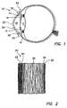
- FIG. 1is a diagram of a sectional view of a human eye.
- FIG. 2is a diagram of a magnified sectional view of the cornea of the human eye of FIG. 1 .
- FIG. 3Ais a diagram of a front plan view of a corneal appliance, as described herein.
- FIG. 3Bis a sectional view of the corneal appliance of FIG. 3A .
- FIG. 4Ais a diagram of a front plan view of a lens used in a corneal appliance, as described herein.
- FIG. 4Bis a sectional view of the lens of FIG. 4A .
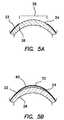
- FIG. 5Ais a diagram of a magnified sectional view of a deepithelialized cornea.
- FIG. 5Bis a diagram of the deepithelialized cornea of FIG. 5A with a corneal appliance placed over the cornea.
- FIG. 6Ais an illustration of a front plan view of an eye in which a preformed epithelial cell layer is formed as a flap.
- FIG. 6Bis a sectional view of the eye of FIG. 6A .
- FIG. 6Cis a sectional view similar to FIG. 6B in which a lens has been placed on the deepithelialized eye and the preformed layer of epithelium has been placed over the lens.
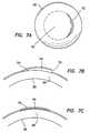
- FIG. 7Ais an illustration of a front plan view of an eye in which a preformed epithelial cell layer is formed as a pocket.
- FIG. 7Bis a sectional view of the eye of FIG. 7A .
- FIG. 7Cis a sectional view similar to FIG. 7B in which a lens has been placed in the pocket.
- FIG. 8Ais an illustration of a front plan view of an eye with a relatively large incision.
- FIG. 8Bis similar to FIG. 8A with a smaller incision.
- FIG. 8Cis similar to FIG. 8B with a smaller incision.
- FIG. 9Ais an illustration of a front plan view of an eye with a relatively small incision in the epithelium.
- FIG. 9Bis a view similar to FIG. 9A in which a fluid injector is inserted into the incision in the epithelium to deliver fluid therebeneath.
- FIG. 9Cis a sectional view of the eye of FIG. 9B after the fluid has been delivered beneath the epithelium.
- FIG. 9Dis a sectional view similar to FIG. 9C in which a lens has been inserted beneath a preformed epithelial cell layer.
- FIG. 10Ais a front plan view of an eye having an epithelial flap with an superiorly located hinge portion.
- FIG. 10Bis a front plan view of an eye having a central epithelial incision.
- FIG. 10Cis a front plan view of an eye having an offset epithelial incision.
- FIG. 10Dis a front plan view similar to FIG. 10C in which an offset incision is used to form two flaps with offset hinge portions.
- FIG. 10Eis a front plan view similar to FIG. 10B in which a central epithelial incision is used to form two flaps with offset hinge portions.
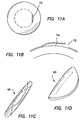
- FIG. 11Ais an illustration of a front plan view of an eye having an offset epithelial incision.
- FIG. 11Bis a sectional view of the eye of FIG. 11A .
- FIG. 11Cis an illustration of a perspective view of a folded lens configured to be inserted in an epithelial incision.
- FIG. 11Dis an illustration of a perspective view of a folded lens in which the lens is folded along its midline.
- FIG. 12Ais an illustration of a front plan view of a corneal onlay lens.
- FIG. 12Bis a sectional view of the lens of FIG. 12A .
- FIG. 12Cis a magnified sectional view of an edge of an onlay lens in which the edge is rounded.
- FIG. 12Dis a magnified sectional view of an edge of an onlay lens in which the edge includes a rounded anterior portion, and an apex on the posterior portion.
- FIG. 12Eis a magnified sectional view of an edge of an onlay lens in which the edge is similar to a knife edge.
- FIG. 13Ais an illustration of a front plan view of an onlay lens structured to correct an astigmatism.
- FIG. 13Bis a sectional view of an onlay lens similar to FIG. 13A in which the posterior surface of the lens includes a torus.
- FIG. 13Cis a sectional view of an onlay lens similar to FIG. 13A in which the anterior surface of the lens includes a torus.
- a typical human eye 10has a lens 12 and an iris 14 .
- Posterior chamber 16is located posterior to iris 14 and anterior chamber 18 is located anterior to iris 14 .
- Eye 10has a cornea 20 that consists of five layers, as discussed herein.
- One of the layers, corneal epithelium 22lines the anterior exterior surface of cornea 20 .
- Corneal epithelium 22is a stratified squamous epithelium that extends laterally to the limbus 32 . At limbus 32 , corneal epithelium 22 becomes thicker and less regular to define the conjunctiva 34 .
- FIG. 2illustrates a magnified view of the five layers of cornea 20 .
- cornea 20comprises corneal epithelium 22 , Bowman's membrane 24 , stroma 26 , Descemet's membrane 28 , and endothelium 30 .
- Corneal epithelium 22usually is about 5-6 cell layers thick (approximately 50 micrometers thick), and generally regenerates when the cornea is injured.
- Corneal epithelium 22provides a relatively smooth refractive surface and helps prevent infection of the eye.
- Bowman's membrane 24lies between epithelium 22 and the stroma 26 and is believed to protect the cornea from injury.
- Corneal stroma 26is a laminated structure of collagen which contains cells, such as fibroblasts and keratocytes, dispersed therein. Stroma 26 constitutes about 90% of the corneal thickness. Corneal endothelium 30 typically is a monolayer of low cuboidal or squamous cells that dehydrates the cornea by removing water from the cornea. An adult human cornea is typically about 500 ⁇ m (0.5 mm) thick and is typically devoid of blood vessels.
- Limbus 32shown in FIG. 1 , is a region of transitions where cornea becomes sclera, and conjunctiva. Limbus 32 contains stem cells, which are capable of differentiating into corneal epithelial cells, as described herein.
- a corneal appliance 60has been invented, as illustrated in FIG. 3A , that is structured to be placed over a deepithelialized eye and that generally comprises a lens 40 and a layer of epithelium 70 , or a layer of epithelial cells, located over the lens.
- Corneal appliance 60is structured to alter the focusing capabilities of a patient's eye, and preferably, the corneal appliance is structured to improve vision of a patient.
- Corneal appliance 60is intended to be placed over a deepithelialized cornea of an eye, and accordingly, corneal appliance 60 may be a corneal onlay.
- Corneal appliance 60includes a layer of epithelium 70 which reduces the healing time of a patient required after surgery, as compared to corneal onlays which depend on the regeneration and migration of epithelial cells over the corneal onlay after it is placed over the eye.
- the preformed layer of epithelium 70provides more uniform epithelial coverage over the cornea as compared to conventional corneal onlays.
- the epithelial cells located over the lensmay be obtained from the patient receiving the corneal appliance, and may be derived from stem cells of the patient, such as limbal stem cells, which may be cultured in vitro to define the layer of epithelium of the appliance.
- stem cells of the patientsuch as limbal stem cells, which may be cultured in vitro to define the layer of epithelium of the appliance.
- Autologous stem cellscontribute to reduced immunogenicity experienced by the patient receiving the appliance as compared to corneal onlays that utilize non-autologous sources of epithelial cells, such as from embryonic or fetal tissue.
- use of patient-specific stem cellsreduces the amount of biopsy tissue required for corneal onlays using mature or differentiated epithelial cells.
- the layer of epithelial cellsmay be formed by detaching a portion of a patient's epithelium to create an epithelial flap that can be resected and then placed back over a corneal onlay after the onlay has been placed over the eye. The incision around the flap may be mended over the onlay, as discussed herein, to maintain the onlay in a desired position over the eye.
- the preformed layer of epithelial cellsmay also be a portion of the patient's corneal epithelium that has been separated from the underlying Bowman's membrane or corneal stroma. The preformed layer may be separated from the underlying corneal structures with or without making an epithelial flap, depending on the particular embodiment of the invention.
- an incisionmay be made in the epithelium to provide access to the region between the epithelium and Bowman's membrane.
- the epitheliumcan be separated from Bowman's membrane by introducing a separator through the incision.
- the separatormay be a surgical device or may include a substance that can be injected through the incision.
- the separatoreffectively separates the epithelium from Bowman's membrane without significantly damaging Bowman's membrane.
- the separatormay also enable a relatively small cut to be made in Bowman's membrane, without substantially damaging Bowman's membrane, which may facilitate placement of the lens over the stroma and may promote more rapid and satisfactory healing of the eye.
- the corrective ocular devicesuch as a corneal onlay
- the epitheliumis not required to be realigned after insertion of the ocular device, and misalignment problems of the ocular device are reduced.
- the lens 40is maintained in a substantially fixed position on an eye relative to a lens, for example, a substantially identical lens, that is placed on an eye so that the epithelium is required to regenerate and migrate over the lens.
- the lens 40 used in corneal appliance 60may be fabricated from any suitable material that is optically clear to permit light to be transmitted to the retina of the eye when corneal appliance 60 is placed over the eye without compromising the ocular physiology of the eye.
- Lens 40has an anterior surface 42 , a posterior surface 44 , a peripheral edge 46 disposed at the juncture of anterior surface 42 and posterior surface 44 , as illustrated in FIGS. 4A and 4B .
- Anterior surface 42is typically convex and posterior surface 44 is typically concave, however, the posterior surface may also include one or more planar portions or surfaces, or may be substantially planar.
- Lens 40may also include an optic zone 48 and a peripheral zone 50 .
- optic zone 48is bounded by peripheral zone 50 , or in other words, optic zone is generally centrally located about an optical axis, such as a central optical axis, of the lens and peripheral zone 50 is disposed between an edge of optic zone 48 and peripheral edge 46 .
- Additional zones and lens configurationsmay be provided with the lens depending on the particular visual deficiency experienced by the patient.
- the lensesmay have junctionless zones, such as two or more zones that do not have a visually or optically detectable junction.
- the zones of the lensesmay be smooth and continuous, and the lenses may be optically optimized to correct not only refractive errors, but also other optic aberrations of the eye and/or the optical device independently or in combination with correcting refractive errors.
- lens 40may be structured to correct visual deficiencies including, and not limited to, myopia, hyperopia, astigmatism, and presbyopia.
- the lensmay correct or improve visual deficiencies by either optical means or physical means imposed on the stroma of the eye, or a combination thereof.
- the lens 40 of corneal appliance 60may be a monofocal lens or a multifocal lens, including, without limitation, a bifocal lens.
- the lens 40may be a toric lens, such as the lens illustrated in FIGS. 13A , 13 B, and 13 C.
- the lens 40may include a toric region 49 which may be effective when placed on an eye with an astigmatism to correct or reduce the effects of the astigmatism.
- the lens 40may include a toric region 49 a located on the posterior surface 44 of the lens 40 , as shown in FIG. 13B , or the lens 40 may include a toric region 49 B located on the anterior surface 42 , as shown in FIG. 13C .
- toric lensesmay be used without requiring a ballast to maintain proper orientation of the lens on the eye since the lens may be held in a relatively fixed position by the epithelium of the appliance. However, a ballast may be provided if desired.
- the lens 40may include a ballast, such as a prism, or it may include one or more thinned regions, such as one or more inferior and/or superior thin zones.
- the lensmay include one or more designs, such as concentric, aspheric (either with positive and/or negative spherical aberration), diffractive, and/or multi-zone refractive.
- the lensmay have an optical power ranging from about ⁇ 10.00 diopters to about +10.00 diopters, although other optical powers may be provided, and such other optical powers are within the scope of the present invention.
- a lens of the corneal appliancewill have a diameter between about 6 mm and about 12 mm.
- the diameter of the lenswill be between about 7 mm and about 10 mm.
- the optic zone of the lenstypically ranges from about 5 to about 11 mm, and preferably ranges from about 6 mm to about 8 mm, in diameter.
- the optic zonemay be provided on either the anterior or posterior surface of the lens.
- the posterior surface of the lens 40is specifically configured to substantially align with the anterior surface of a deepithelialized eye.
- the posterior surface of the lens 40may include one or more spherical or aspherical dimensions with a base curve that ranges from about 5.0 mm to about 12.0 mm in diameter, preferably from about 6.0 mm to about 9.0 mm, and more preferably about 7.0 mm to about 8.5 mm.
- the thickness of the lens 40 at or near the center of the lensi.e., the center thickness
- the center thicknessis typically greater than about 10 micrometers and is less than about 300 micrometers.
- the center thicknessis between about 30 micrometers and about 200 micrometers.
- the exact or specific thickness of the central regionmay be determined on a case-by-case basis by one of ordinary skill in the art since the maximum thickness is optical power and refractive index dependent.
- the thickness of the peripheral edge 46 of the lens 40is typically, but not always, less than the center thickness, as shown in FIGS. 12A , 12 B, 12 C, 12 D, and 12 E.
- the edge thicknessshould be thin enough to facilitate epithelial cell growth at the juncture of the lens and the Bowman's membrane or stroma of an eye, and may be thin enough to promote additional epithelial cell migration over the edge of the lens.
- the edge thickness of the lensis less than about 120 micrometers.
- the lens 40has an edge thickness less than about 60 micrometers, and preferably less than about 30 micrometers.
- the lens 40has an edge thickness of about 0 micrometers (for example, the thickness of a sharp knife edge).
- the lens edgemay be rounded on both the anterior and posterior surfaces, as shown at 46 A.
- the lens edgemay include a rounded anterior surface 42 and an apex on or near the posterior surface 44 , as shown at in FIG. 12D .
- the lens edgemay be shaped as a knife edge, such as at 46 B as shown in FIG. 12E .
- Lens 40may comprise synthetic or non-synthetic materials, and combinations thereof.
- synthetic materialsrefers to materials that are not obtained, for example, are not obtained directly, from animal subjects. Thus, synthetic materials specifically exclude donor corneal tissue.
- lens 40may be made from collagen, such as purified collagen.
- the collagenmay be collagen Type I, which is the type of collagen that defines the bulk of the corneal stroma, or lens 40 may be made from other types of collagen, including combinations of different types of collagen, such as types III, IV, V, and VII.
- the collagenmay be obtained from animals, including humans.
- collagen of the lens 40may be bovine collagen, porcine collagen, avian collagen, murine collagen, equine collagen, among others. Many different types of collagen useful in the lenses of the present invention are publicly available from companies, such as Becton Dickenson.
- the collagenmay be recombinantly synthesized, such as by using recombinant DNA technology.
- lens 40is not obtained from a donor patient, such as from corneal tissue of another individual person.
- Collagenmay be obtained using any conventional technique, as is practiced in the art.
- One source of publicly available recombinant collagenis FibroGen, South San Francisco, Calif.
- recombinant collagenmay be prepared and obtained using the methods disclosed in PCT Publication No. WO 93/07889 or WO 94/16570.
- the recombinant production techniques described in these PCT publicationsmay readily be adapted so as to produce many different types of collagens, human or non-human. Utilizing purified collagen simplifies procedures of making corneal onlays, as compared to corneal onlays that are obtained from donor tissue, such as disclosed in PCT Publication No.
- WO 02/06883For example, using purified collagen, including recombinantly synthesized collagen, steps of decellularization donor corneal tissue are avoided.
- the collagenmay be fully biodegradable or partially biodegradable, which may facilitate attachment of epithelial cells over the onlay by permitting native collagen created by the patient receiving the onlay to integrate and/or replace the collagen of the corneal appliance.
- the collagen used to manufacture lens 40may be populated with cells, such as corneal keratocytes, before being used in corneal appliance 60 . Cells may be added to the collagen by culturing a suspension of keratocytes and subsequently immersing the lens in a keratocyte medium, as disclosed in WO 02/06883.
- the cells that are used to populate the lensdo not generate an immune response, or generate a minimal immune response.
- the cellsmay be from an allogenic source, such as another person, an autologous source, such as the patient receiving the appliance, or may be from a xenogenic source.
- an allogenic sourcesuch as another person, an autologous source, such as the patient receiving the appliance
- cells obtained from xenogenic sourcesmay need to be modified to reduce the antigenicity or immunogenicity of the cells when administered to the patient to reduce the likelihood of developing an immune response.
- keratocytes from the patient's own stromamay populate the collagen lens, and the integration between the lens and the stroma may facilitate the fixation of the lens on the eye.
- lens 40may be manufactured by obtaining and culturing corneal keratocytes, as disclosed in PCT Publication No. WO 99/37752 and U.S. Pat. No. 5,827,641.
- the cultures of keratocyteswill be placed in a mold suitable for a vision correction lens, and will produce a collagen matrix similar to a normal stroma in vivo.
- the various moldswill thus produce a corneal appliance having a synthetic stroma with a desired optical power to correct a vision deficiency of the patient.
- Lens 40 of corneal appliance 60may be made from a polymeric hydrogel, as understood by persons of ordinary skill in the art.
- a polymeric hydrogelincludes a hydrogel-forming polymer, such as a water swellable polymer.
- the hydrogelitself includes such a polymer swollen with water.
- Polymeric hydrogels useful as corneal appliance lenses, for example, corneal onlaystypically have about 30% to about 80% by weight water, but may have about 20% to about 90% by weight water, or about 5% to about 95% by weight water, and have refractive indices between about 1.3 and about 1.5, for example about 1.4, which is similar to the refractive indices of water and a human cornea.
- hydrogel-forming polymer materials or components of the disclosed lensesinclude, without limitation, poly(2-hydroxyethyl methacrylate) PHEMA, poly(glycerol methacrylate) PGMA, polyelectrolyte materials, polyethylene oxide, polyvinyl alcohol, polydioxaline, poly(acrylic acid), poly(acrylamide), poly(N-vinyl pyrilidone) and the like and mixtures thereof. Many of such materials are publicly available.
- one or more monomers which do not themselves produce homopolymers which are not hydrogel-forming polymerssuch as methylmethacrylate (MMA), other methacrylates, acrylates and the like and mixtures thereof, can also be included in such hydrogel-forming polymer materials provided that the presence of units from such monomers does not interfere with the desired formation of a polymeric hydrogel.
- MMAmethylmethacrylate
- other methacrylates, acrylates and the like and mixtures thereofcan also be included in such hydrogel-forming polymer materials provided that the presence of units from such monomers does not interfere with the desired formation of a polymeric hydrogel.
- lens 40 of corneal appliance 60may be manufactured from a biocompatible, non-hydrogel material or component, such as disclosed in U.S. Pat. No. 5,713,957.
- non-hydrogel materialsinclude, and are not limited to, acrylics, polyolefins, fluoropolymers, silicones, styrenics, vinyls, polyesters, polyurethanes, polycarbonates, cellulosics, or proteins including collagen based materials.
- lens 40may comprise a cell growth substrate polymer, such as those disclosed in U.S. Pat. No. 5,994,133.
- corneal appliance 60comprises a lens 40 which includes a synthetic material, and more particularly, a non-donor corneal tissue material.
- the lensis made entirely from a synthetic material.
- the lensis made from a combination of collagen and a synthetic material, including, combinations of bovine collagen and a synthetic material, and combinations of recombinant collagen and synthetic materials.
- the lensmay include a poly(N-isopropylacrylamide) (polynipam) component. It has been found that a polynipam component may facilitate attachment of the lens to Bowman's membrane and/or epithelial cell layers to the lens at temperatures of about 37 degrees C.
- the polynipam componentfacilitates the in viva attachment of the epithelium to the lens at substantially normal body temperatures, and may be helpful in procedures in which the lens is to be removed from the eye, by cooling of the ocular tissue.
- the corneal appliance disclosed hereinmay provide vision correction to a subject in need thereof.
- the corneal appliance lensis designed to correct or reduce wavefront aberrations of a patient's eye.
- a wavefront aberrationis the three dimensional profile of the distance between a real light wave front of a central spot of light and a reference surface, e.g., an ideal spherical shape, as shown in FIG. 1 of U.S. Pat. No. 6,585,375, and as described in Mierdel et al., “Der Ophthalmologe”, No. 6, 1997.
- a wavefront aberrationmay be understood to be an optical path difference between an actual image wavefront and an ideal reference wavefront centered at an image point, at any point in the pupil of an eye. Methods of measuring wave-front aberration are well known to persons of ordinary skill in the art.
- an aberrometere.g., an instrument that measures the aberrations of an eye
- an aberrometermay be used to measure an aberrated image that leaves an eye, or may be used to measure the shape of a grid projected onto the retina.
- a relatively narrow input laser beammay be directed through the pupil and focused onto the retina of the patient's eye to generate a point-light source on the retina. The light is reflected from the retina back through the pupil, and the wavefront of the light passing from the eye is passed to a wavefront sensor.
- a wavefrontcan be defined as a surface that connects all field points of an electromagnetic wave that are equidistant from a light source.
- the light raysleave the eye and may pass through an array of lenses that detects the light rays' deviation.
- the wavefrontgets deviated or distorted by inhomogeneities in the refractive properties in the refractive media of the eye, such as the lens, the cornea, the aqueous humor, and the vitreous humor.
- the resulting imageis then typically recorded by a charge coupled device (CCD) camera, for example.
- CCDcharge coupled device
- the wavefrontis then typically reconstructed and the deviations are described mathematically in three dimensions.
- the wavefront deviationsmay be calculated, at least in part, by analyzing the direction of the light rays.
- parallel light beamsindicate a wavefront with little, if any, aberrations
- nonparallel light beamsindicate a wavefront with aberrations that do not give equidistant focal points.
- Zernike polynomialsare used to measure or analyze the ocular aberrations.
- Each Zernike polynomialdescribes a shape or a three-dimensional surface.
- Zernike polynomialsare an infinite set, but in ophthalmology, the Zernike polynomials are usually limited to the first fifteen polynomials.
- Second-order Zernike termsrepresent conventional aberrations, such as defocus and astigmatism. Aberrations above second-order aberrations are called higher-order aberrations. Higher-order aberrations typically cannot be corrected by conventional spherocylindrical lenses.
- higher-order aberrationsinclude, but are not limited to, coma, spherical aberrations, trefoil (wavefronts with threefold symmetry), and quadrefoil (wavefront shapes with fourfold symmetry). Many higher-order aberrations are not symmetrical, but some higher-order aberrations, such as spherical aberrations, may be symmetrical.
- the wavefront aberration of a patient's eyemay be measured and analyzed to facilitate appropriate lens construction.
- the lenses of the present inventioncan then be shaped, as discussed herein, taking into account any wavefront aberrations.
- a corneal applianceis obtained with a lens body configured to correct a wavefront aberration of a patient's eye.
- the wavefront aberration corrective surfacemay be provided on either the anterior surface, the posterior surface, or both the anterior and posterior surfaces.
- the present lensescorrect or reduce higher-order wavefront aberrations. In situations where the higher-order wavefront aberrations are asymmetrical, the lenses are configured to substantially maintain a desired orientation to correct the wavefront aberrations.
- Epithelial layer 70is fixed in position over lens 40 of corneal appliance 60 .
- Epithelial layer 70may comprise one or more layers of epithelial cells.
- the number of layers of epithelial cellsare preferably between 1 and 12, and more preferably are about 5-7 layers.
- the number of layers of epithelium 70closely matches the number of layers of corneal epithelium observed in vivo.
- the number of layers of epithelial cellsmay also change with time. For example, a single layer of epithelial cells may be positioned on lens 40 ex vivo, and the lens may be placed over an eye. After the procedure of placing the lens on the eye, the epithelial cells may continue to divide to form one or more additional layers of epithelial cells.
- an epithelial layer 70may comprise approximately 5-7 cell layers when it is placed over lens 40 .
- Epithelial layer 70is dimensioned to cover at least a fraction of anterior surface 42 of lens 40 .
- epithelial layer 70extends beyond peripheral edge 46 of lens 40 .
- a flap or fringe of epithelium 70extends from the edge of lens 40 , which may be useful to help secure corneal appliance 60 in an eye.
- epithelial layer 70does not extend to or beyond peripheral edge 46 , it is desirable to ensure that the epithelial cells either of the epithelial layer 70 or of the epithelium of the patient's eye continue to divide and migrate over the exposed portions of the lens. Suitable growth factors or other growth promoting strategies may be employed to achieve this result.
- epithelial layer 70may be derived from stem cells obtained from an autologous source.
- epithelial layeris derived from cultured stem cells obtained from the patient receiving the corneal appliance. This is in contrast to the corneal onlay disclosed in WO 02/06883, which utilizes epithelial cells from fetal or embryonic tissue, or epithelial cells obtained from the patient receiving the corneal onlay.
- epithelial cellsmay also be derived from any type of stem cell that can differentiate into corneal epithelial cells, including stem cells from fetal or embryonic tissue.
- the stem cells obtained from the patientare corneal epithelial limbal stem cells.
- the corneal epithelial limbal stem cellsmay be harvested, cultured, and prepared according to the methods disclosed in U.S. Patent Publication No. US 2002/0039788 A1, and by Han et al., “A fibrin-based bioengineered ocular surface with human corneal epithelial stem cells”, Cornea, 21(5): 505-510, 2002. Briefly, corneal epithelial stem cells may be cultured onto an extracellular matrix, which may comprise basement membrane components, such as laminin, fibronectin, elastin, integrins, and collagen.
- Cultured epithelial stem cellsare expanded on a feeder layer of replication defective, but metabolically active fibroblasts (such as 3T3 cells). After the epithelial colonies are established, the feeder cells are removed, and the epithelial cells are expanded by growth in a serum-free, low calcium medium, such as Keratocyte Growth Medium, KGM (Cascade Biologics, Oreg.). The cultured epithelial cells may then be trypsinized from their culture dish, suspended in Cornea Growth Medium, CGM (Cascade Biologics), and seeded on prepared fibrin gels.
- a serum-free, low calcium mediumsuch as Keratocyte Growth Medium, KGM (Cascade Biologics, Oreg.).
- KGMKeratocyte Growth Medium
- CGMCornea Growth Medium
- the fibrin gelsare made by mixing a fibrinogen solution (plasminogen-free fibrinogen, human, Calbiochem, San Diego, Calif.) in distilled water with calcium chloride, and aprotonin (Sigma) in a buffer, such as Tris Buffer, at a pH of about 7.0, such as 7.2. Cultured corneal fibroblasts and thrombin may be added to the solution, after which, the solution is dispensed into a holder to gel.
- a fibrinogen solutionplasmaogen-free fibrinogen, human, Calbiochem, San Diego, Calif.
- aprotoninSigma
- Tris BufferTris Buffer
- Epithelial layer 70is attached to anterior surface 42 of lens 40 so that epithelial layer 70 does not appreciably or noticeably move along the surface of the lens. Thus, when epithelial layer 70 and lens 40 are fixedly joined or coupled, they form corneal appliance 60 . Epithelial layer 70 may be attached to lens 40 either by chemical, biological, mechanical, or electrical methods.
- corneal appliance 60may also include a cellular attachment element disposed between epithelial layer 70 and anterior surface 42 of lens 40 .
- the cellular attachment elementfacilitates the stable positioning of epithelial layer 70 over lens 40 .
- cellular attachment elementsmay be desirable when utilizing lenses fabricated from collagen, most cellular attachment components may find increased use in the hydrogel or non-hydrogel lenses described hereinabove.
- Cellular attachment elementsmay include physical perturbations of the lens 40 , such as indentations provided in anterior surface 40 that facilitate cellular attachment and do not alter the optical properties of the lens. Indentations included pores that extend through the lens from the anterior surface to the posterior surface of the lens. The indentations may be provided over the entire lens or over a fraction of the lens.
- the indentationsmay also be provided in specific patterns and dimensions that facilitate cellular attachment of the epithelial layer to the lens.
- the indentationsmay be provided in a plurality of concentric rings emanating from the center of the lens and expanding radially outward.
- Cellular attachment elementmay also comprise a polymer that supports adhesion of the epithelial cells to the lens.
- the lensmay be made essentially from such polymers as disclosed in U.S. Pat. No. 5,994,133.
- these cell growth substrate polymersmay be chemically bonded or otherwise coated on the surface of a hydrogel or collagen based lens to facilitate cellular attachment to the lens.
- the cellular attachment elementmay also comprise a corneal enhancer molecule, such as a corneal enhancer molecule that specifically binds to a molecule present on the extracellular surface of an epithelial cell.
- suitable corneal enhancer moleculesinclude peptides, such as the tri-peptide, RGD, extracellular matrix proteins, corneal growth factors, and ligand-specific corneal enhancer species, such as laminin, fibronectin, substance P, fibronectin adhesion promoting peptide sequence, FAP, insulin-like growth factor-1 (IGF-1), k-laminin, talin, integrin, kalinin, fibroblast growth factor (FGF), and TGF- ⁇ , as disclosed in U.S. Patent Publication No. US 2002/0007217 A1.
- These corneal enhancer moleculesmay include a tether, which may enhance the ability of epithelial cells to attach and migrate over the lens 40 .
- lens 40 of corneal appliance 60may be made from collagen to mimic a native corneal stroma, a hydrogel, or a biocompatible non-hydrogel material.
- the lens of corneal appliance 60may be produced according to standard techniques known to those skilled in the art.
- a collagen matrixmay be formed and include stromal cells.
- Lens 40may be shaped in a conventionally dimensioned mold suitable for lenses, such as corneal onlays. For example, lens 40 may be ablated, molded, spin-casted and/or lathed, or combinations thereof.
- the molds used to manufacture corneal appliance 60may be structured to permit nutrient, liquid, and gas exchange with the cultured cells.
- a moldmay comprise one or more pores to permit nutrients and liquid and gas to flow to the cell culture.
- the moldsmay be made from any suitable, porous material, including, but not limited to, ceramics, mesh, such as stainless steel mesh, or membranes made from nylon, cellulose, or the like.
- the moldmay comprise a concave surface and a convex surface matingly shaped with respect to each other. The mold may be able to be placed in a well having culture medium to facilitate the culturing of the cells.
- the shape of the lensmay be determined by the mold designed for culturing (hereinafter referred to as the culturing mold), or may be shaped in a conventional mold. If shaped in a conventional mold, the lens may then be subsequently placed in a culture dish having a desired shape to preserve the shape of the lens, where the culture dish is structured to facilitate the culturing of the epithelial cells.
- Epithelial cell layer 70may be prepared essentially as described above.
- a fibrin matrixor other extracellular protein matrix, may be produced from serum and the corneal epithelial stem cells may be seeded in the matrix.
- the seeded matrixmay then be applied on the anterior surface of the lens.
- the cellsmay be applied by dispensing the matrix over the surface of the lens, or the cells may be applied as a relatively flexible layer of cells or a film of cells that sufficiently flexes to accommodate the curvature of the lens.
- the film of cellsmay comprise a film of corneal epithelial stem cells or a film of developed epithelial cells, which may be one or more layers thick, or a combination thereof.
- a layer of epithelial cellsmay be obtained by culturing immortalized human corneal epithelial cells, such as disclosed in U.S. Pat. No. 6,284,537. With such cell lines it is desirable to regulate cell growth once the corneal appliance is placed on the eye. Cell growth may be regulated using any conventional method known by persons of ordinary skill in the art.
- the epithelial cell layermay be a layer or flap of epithelial cells of the patient that has been separated from the patient's cornea, as described herein.
- the preformed layer of epithelial cellsmay be placed over the lens body after the lens body has been placed over the cornea.
- the lens bodymay or may not have received a surface treatment to help the layer of epithelial cells to attach to the lens body.
- a surface treatmentfor example, when lens bodies are used that are made from polymeric materials or composites that promote cellular attachment, it may not be necessary to include a surface treatment on the lens body.
- the corneal applianceincludes a suspension of epithelial stem cells provided on the anterior surface of the lens body.
- the suspensionmay be a fibrin-based suspension, as disclosed herein.
- the epithelial stem cells that are provided over the lens bodymay provide nutrients, such as growth promoting factors, that promote attachment of the layer of epithelial cells to the lens body.
- a suspension of stem cellsis provided over the lens body and the flap of epithelium is placed over the lens body, and the stem cells encourage attachment and growth of the epithelial cells of the flap over the lens body.
- the stem cellssurvive for a sufficient amount of time when placed on the lens body to promote the attachment of the epithelial cell layer to the lens body.
- corneal appliance 60may be manufactured by molding a synthetic material, such as recombinant collagen, in a lens mold having a desired structure to correct a visual deficiency.
- the collagen lensmay be populated with stromal keratocytes that have low antigenicity or immunogenicity.
- the collagen lensmay be modified on its surface to promote cellular attachment of the epithelial cells, and then a culture of epithelial stem cells may be placed on the collagen lens where they can grow and differentiate into an epithelial cell layer.
- Corneal appliance 60may be placed over an eye to provide the desired vision correction. Because corneal appliance 60 includes a layer of epithelium, as described hereinabove, it is desirable to remove at least a portion of the epithelium from the patient's eye receiving the appliance. The deepithelialized portion should at least have approximately the same dimensions as the corneal appliance. A deepithelialized cornea is illustrated in FIG. 5A .
- the epitheliummay be removed by any conventional method.
- an abrasive devicecan be used to remove the epithelium
- a small rotating brushmay be used
- sterile cocainemay be applied to the epithelium
- an alcohol washsuch as an ethanol wash
- a source of electromagnetic energy on the epitheliumsuch as with the LASEK and LASIK procedures, which are well known.
- a portion of the epitheliummay be removed using a separator that can separate the epithelium from Bowman's membrane to form a pre-formed layer of epithelial cells.
- a separatoris a sub-epithelial separator developed by Dr.
- the separatormay include a suction device, or ring, that can deliver suction to the epithelium to cause the epithelium to be lifted from the cornea.
- a cutting devicesuch as a blade, including a microkeratome, which may or may not be a part of the separator can then be used to cut the portion of the epithelium that is being lifted from the cornea to create a flap, or to completely remove that portion of the epithelium that is being manipulated.
- the separatorcan include a temperature controller that causes temperature changes in a portion of the device that contacts the epithelium.
- the separatormay be cooled to cause the epithelium to attach to a cooled region of the separator so that it may be lifted from the cornea, and then may be warmed, passively or actively to allow the epithelial tissue that has been cut to be released from the separator.
- the temperature controlenables the handling of the epithelial cells of the epithelium without undue damage and cellular injury to the epithelial cells during the procedure. It appears that the cooling not only provides a convenient way of attaching the epithelium to the separator, but that the cooling provides protection to the cells that are being manipulated during the manipulation procedure.
- an electromagnetic energy sourcesuch as a laser
- a fluidsuch as water or saline
- the electromagnetic energymay be desirable to remove one or more small portions of Bowman's membrane, as indicated herein to facilitate more rapid healing of the ocular tissue. However, in certain situations, the Bowman's membrane is left entirely intact.
- corneal appliance 60may be placed on the deepithelialized cornea.
- the lens of the applianceis made from collagen, the lens may make a natural bond with the Bowman's membrane that holds the lens in place on the eye.
- additional adhesive mechanismsmay be used to facilitate securing the appliance on the eye.
- gluepreferably a biodegradable glue, may be applied to the overlying fringe of epithelium 70 , dissolvable sutures may be used to secure the fringe of epithelium to the eye, or pressure applied by a bandage can be used to hold the appliance in place until the epithelium has bonded with the rest of the eye.
- a fibrin-based stem cell matrixmay be applied as an adhesive to help maintain the placement of the epithelium and to promote healing and development of the epithelium.
- the epithelium of appliance 60blends together with any remaining corneal epithelium that remains on the eye, as shown in FIG. 5B .
- corneal appliance 60has a layer of epithelium that is more reliably or consistently attached to the lens body than an epithelium that is attached to a lens body obtained from donor tissue, such as disclosed in PCT Publication No. WO 02/06883.
- Corneal appliance 60may provide a substantial improvement in the field of corrective vision technology.
- the applianceis a device that provides long-term vision correction that can be reversed, as opposed to procedures that permanently alter the shape of a patient's cornea, such as LASEK and LASIK procedures.
- the corneal appliancemay be easily removed from the patient if complications develop or the patient's vision changes.
- corneal appliance 60provides for long-term, but reversible, vision correction.
- a procedure for improving a patient's visionmay begin by a patient with a vision defect visiting a physician.
- the physicianharvests a sample of corneal epithelial stem cells from the patient and sends the sample of cells to a lab for culturing.
- the cellsare seeded and cultured in a fibrin matrix, as described above, and are applied to the anterior surface of a lens.
- the lensmay be treated or modified on its anterior surface to promote cellular attachment of the epithelial cells.
- the surface treatmentmay include physical perturbations, such as roughening of the lens surface, or may include providing the lens with one or more cellular attachment elements, as discussed hereinabove.
- the cultured cellsAfter approximately 10-20 days, the cultured cells have developed into a layer of epithelial cells that substantially covers the entire surface of the lens.
- the corneal appliancemay then be delivered to the physician's office.
- the patientreturns to the physician's office for the procedure, which includes removing the epithelium from the patient's cornea and applying the corneal appliance to the deepithelialized cornea.
- the epitheliumis only removed to the Bowman's membrane, and is removed so that the diameter of the deepithelialized portion of the cornea corresponds to the diameter of the epithelial layer of the corneal appliance.
- another method of improving a patient's visionincludes creating a slit, incision, or opening in the patient's corneal epithelium that is large enough to permit a lens, as described above to be inserted into through the slit underneath the epithelium, as shown in FIGS. 7A , 7 B, and 7 C.
- the epitheliummay be separated from the Bowman's membrane using standard blunt dissection techniques or other conventional methodology to form preformed epithelial cell layer 70 .
- the corneal epitheliummay be separated from the cornea using a separator, as discussed above.
- the epitheliummay be separated to form a flap of tissue ( FIGS.
- an adhesivesuch as a corneal epithelial layer derived from stem cells, or a stem cell suspension, as disclosed hereinabove, may be applied to the slit region of the patient's epithelium to promote the healing of the incision.
- a method of correcting or improving visionincludes a step of inserting a vision correcting ocular device, for example, a corrective lens or lens body, beneath the epithelium of a patient's cornea substantially without uncovering or exposing an anterior surface of the cornea located under the epithelium, such as shown in FIGS. 7A , 7 B, and 7 C.
- the anterior surface of the corneamay be Bowman's membrane, or it may include one or more portions of the corneal stroma.
- This methodis in contrast to techniques that produce a flap of epithelial tissue to expose or uncover an anterior surface of the cornea, as discussed herein, and as shown in FIGS. 6A , 6 B, and 6 C.
- the ocular deviceBy inserting an ocular device beneath an epithelium but on or above the stroma or Bowman's membrane, the ocular device is effectively substantially fixedly positioned with respect to the eye, for example, by the epithelium, to provide the desired vision correction.
- this methodprovides for relatively enhanced healing or reduced times and reduced side effects relative to methods that produce a flap of epithelial tissue to insert an ocular device.
- the lensmay be inserted by inserting the ocular device through an incision formed in the epithelium.
- An incisionmay be formed at any desired region around the epithelium, but in preferred embodiments, the incision or incisions is formed either in the temporal portion of the epithelium (e.g., the portion of the epithelium that is located away from the nose of a patient), or in the medial portion of the epithelium.
- the incisionis preferably formed to provide an opening in the epithelium, for example, of suitable size, to accommodate a corrective ocular device to be inserted therethrough without creating an epithelial flap.
- the preformed epithelial layer diameter 70 Dmay also vary, as shown in FIGS. 8A , 8 B, and 8 C.
- a relatively large incision 72 as shown in FIG. 8Amay provide a relatively small preformed epithelial diameter 70 D.
- the incision sizemay be varied to accommodate various insertion techniques, such as whether a lens is deformed prior to insertion.
- a large incisionmay be formed when a lens is inserted in a substantially undeformed state, or a small incision may be formed when a lens is inserted in a deformed state.
- the deformed ocular devicecan assume its native or original configuration (e.g., the configuration of the ocular device before being deformed).
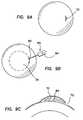
- an incision 72may be made in the epithelium of an eye, as shown in FIG. 11A and FIG. 11B .
- the lens 40may then be “rolled”, as shown in FIG. 11C , or “folded”, as shown in FIG. 11D so that the lens can be inserted in the incision 72 .
- the lens 40 shown in FIG. 11Dis folded along its midline so that two substantially equal-sized portions overlap.
- the deformed lensesmay then be inserted into the incision 72 as indicated herein.
- the incisioncan be made by cutting or slicing the epithelium using a sharp instrument, such as a microkeratome and the like, including the microkeratome disclosed hereinabove.
- a sharp instrumentsuch as a microkeratome and the like, including the microkeratome disclosed hereinabove.
- the incisioncan be made by using blunt dissection to separate epithelial cells to create an opening in the epithelium without cutting or slicing the epithelium. Blunt dissection provides an advantage of reduced injury to the epithelial cells and/or epithelial tissue.
- a blunt shaped instrumentthat has a thickness that reduces the potential for tearing the epithelium as it is being separated from Bowman's membrane, and for damaging Bowman's membrane of the corneal stroma.
- One suitable blunt dissectorincludes a plate, a wire, or a knife with a dull edge.
- a spatulais also a suitable blunt dissection apparatus. The blunt dissector is inserted under the epithelium and is gently urged across the underlying corneal surface to “tease” the epithelium from Bowman's membrane. The separation appears to follow a path of least resistance to provide a substantially complete separation of the epithelium from Bowman's membrane substantially without damaging either the epithelium or the underlying cornea. Separation proceeds across the surface of the cornea to obtain a void sized to accommodate a corrective ocular device.
- only one incisionis made in the epithelium, but in additional embodiments, two or more incisions can be made in the epithelium to permit insertion of the ocular device.
- the incisionsmay be parallel to each other or may be orthogonal to each other.
- two incisionsmay be made that intersect to form four flaps of epithelial tissue.
- the ocular devicemay be a vision correcting lens, such as a corneal onlay.
- the ocular devicemay comprise a synthetic material, including a synthetic polymeric material, as discussed above.
- the ocular devicemay be a contact lens that is structured to be placed between the epithelium and Bowman's membrane of the cornea.
- a portion of the epitheliummay be lifted or spaced apart from the cornea.
- An incisionmay be made in the epithelium after the portion of epithelium has been lifted or spaced apart.
- An incisionis preferably made in the raised or lifted portion; however, in certain embodiments, an incision may be made in a region of the epithelium that is located at a site spaced apart from, but in proximity to, a site at which the epithelium begins to be spaced apart from Bowman's membrane.
- the ocular devicemay then be inserted through the incision.
- the ocular devicemay be inserted by using forceps, or other similar device.
- the ocular devicemay be inserted by using an inserter that is configured to deform at least a portion of the ocular device so that the device can fit through the incision, for example, through a smaller incision that would be necessary if the ocular device was not deformed.
- the ocular devicemay be folded or rolled or curled so that its cross-sectional area is reduced while it is being inserted beneath the epithelium, as discussed herein.
- a corneal onlay insertion devicemay be a syringe like device which includes a body with a distal end dimensioned to pass the lens under the corneal epithelium of an eye.
- the corneal onlay insertion devicemay be similar, or at least somewhat similar, to well known and publicly available intraocular lens inserters.
- the epitheliummay be raised using any suitable technique that permits the epithelium to be separated from Bowman's membrane preferably without substantially damaging Bowman's membrane or the corneal stroma.
- a portion of the epitheliumis raised using a vacuum.
- the vacuummay be provided with a microkeratome, such as with the separator disclosed in U.S. Patent Publication Nos. 2003/0018347 and 2003/0018348, or it may be provided as a separate instrument.
- the epitheliummay be lifted by delivering a fluid beneath a portion of the epithelium, as shown in FIGS. 9A , 9 B, 9 C, and 9 D.
- a small incision 72may be made in the epithelium of an eye, as shown in FIG. 9A .
- a syringe device 80 having a distal end 82 and a fluid 84 located in the body of the syringe devicemay be placed in proximity to the eye so that the distal end 82 can pass the fluid 84 beneath the epithelium of the eye, as shown in FIG. 9B .
- the fluid 84causes the preformed layer of epithelium 70 to be separated from the stroma of an eye, as shown in FIG. 9C .
- a lens 40may then be placed under epithelium 70 , and as the fluid 84 decreases in volume, the epithelium 70 is placed over the lens 40 to form corneal appliance 60 , as shown in FIG. 9D .
- the delivery of fluidcauses the epithelium to swell to create a bulge of epithelial tissue that is spaced apart from Bowman's membrane, as indicated above.
- One suitable fluidmay include sodium chloride, for example, an aqueous sodium chloride solution.
- Another fluidmay include a gel.
- the gelmay be a gel that includes at least one water soluble or water swellable polymeric material, for example, at least one cellulosic component, such as hydroxymethylcellulose and the like, and/or one or more other water soluble or water swellable polymeric materials.
- the fluidcomprises a gel sold as GENTEAL gel by CibaVision, Duluth, Ga.
- an effective amount of a preserving agentmay be applied to the epithelium to reduce cellular injury and death, and to preserve the epithelium in a viable state.
- the preserving agentmay act as a moisturizer to maintain the epithelium in a moisturized state.
- the epithelium preserving agentmaybe include a gel, and in certain embodiments, the epithelium preserving agent comprises a component selected from the group consisting of water soluble polymeric materials, water swellable polymeric materials, and mixtures thereof.
- the epithelium preserving agentincludes at least one cellulosic component.
- the epithelium preserving agentincludes hydroxymethylcellulose.
- One suitable epithelium preserving agentis the GENTEAL gel identified above.
- a method for correcting or improving visionincludes raising a portion of an epithelium of a cornea of an eye away from Bowman's membrane, cutting a portion of the epithelium to create an elongate incision in the epithelium substantially without damaging the Bowman's membrane, and inserting a corrective ocular device through the incision so that the ocular device is located between the epithelium and Bowman's membrane.
- the epitheliummay be raised using a vacuum, a liquid, or any other suitable device.
- Liquids used to raise the epitheliummay include sodium chloride and/or other tonicity agents.
- the liquidsare hypertonic aqueous liquids.
- the liquidis an aqueous solution containing about 5% (w/v) of sodium chloride.
- One or more incisionsmay be made in the epithelium using a cutting procedure or blunt dissection procedures, as discussed above.
- the epitheliumis cut without forming an epithelial flap.
- the ocular deviceis inserted beneath the epithelium substantially without uncovering or exposing an anterior surface of Bowman's membrane.
- the methodmay be practiced by applying one or more epithelial preserving agents to the epithelium.
- the stroma of the corneais preferably maintained in a substantially intact or undamaged state.
- a method for correcting or improving visionincludes applying a liquid to the epithelium of a cornea of an eye to loosen the epithelium substantially without killing or otherwise devitalizing epithelial cells, treating the epithelium to provide and/or maintain the epithelium in a moisturized state, raising a portion of the loosened epithelium from a surface of the cornea located below the epithelium, separating the raised portion of the epithelium from the surface of the cornea, forming one or more elongate incisions in the raised portion of the epithelium, and inserting a corrective ocular device beneath the epithelium through the one or more elongate incisions.
- the methodmay also include a step of delivering a substance beneath the raised portion of the epithelium to maintain a spaced apart relationship between the epithelium and the surface of the cornea, prior to forming an incision in the epithelium.
- Suitable liquids for loosening the epithelium without devitalizing or killing epithelial cellsinclude sodium chloride and/or other tonicity agents, for example, in aqueous solutions.
- the liquidis a hypertonic aqueous liquid.
- the methods disclosed hereinmay also be practiced by scoring a portion of the epithelium to create an epithelial defect prior to applying the liquid.
- the treating step of the foregoing methodmay include applying a gel to the epithelium, such as a gel that contains a water soluble polymeric material, a water swellable polymeric material, or combinations or mixtures thereof.
- a gelincludes at least one cellulosic component, such as hydroxymethylcellulose, and the like and mixtures thereof.
- the epitheliummay be raised or lifted using a vacuum, or other appropriate device, and the epithelium may be separated using a blunt dissection device, such as a spatula or wire.
- the gel-containing composition identified abovemay also be delivered beneath the raised epithelium to maintain the epithelium in a spaced apart relationship from Bowman's membrane.
- Incisionsare formed in practicing this method using a microkeratome to cut or slice one or more portions of the epithelium.
- incisionsare made in the epithelium to create or form one or more epithelial flaps which are hinged portions of epithelial tissue that can be folded or rolled back, or positioned to expose an underlying surface of the cornea.
- a single incisionis made in the epithelium to create a flap 70 of epithelium that includes a hinged portion 76 located at the periphery of the eye, as shown in FIG. 10A , where the hinged portion is located in a superior region of an eye. As shown in FIG.
- a medial incision 72may be formed, and two flaps 70 a and 70 b ( FIG. 10E ) may be obtained with hinge portions 76 offset from a medial position of the eye.
- an incision 72may be formed away from the medial portion of the eye, such as at a temporal region of the eye. This offset incision may then be used to form two flaps 70 a and 70 b as shown in FIG. 10D with hinge portions 76 offset from a medial region of the eye.
- the incisionis formed offset from the pupil of the eye to reduce potential injury to the cornea above the pupil.
- a plurality of incisionsare made in the epithelium to form a plurality of flaps that can each be folded back to expose an underlying surface of the cornea.
- a substantially vertical incisioncan be made along the midline of the eye, and a substantially horizontal incision can be made to intersect the vertical incision to create four flaps of epithelial tissue.
- an ocular deviceis inserted on the exposed underlying corneal surface, and the flaps of tissue are replaced over the ocular device.
- the ocular deviceis preferably a vision correcting lens, and in certain embodiments, the ocular device is a contact lens that is structured to be placed under the epithelium of a cornea of an eye. In additional embodiments, the ocular device is a corneal onlay.
- a method of correcting or improving vision in accordance with the invention disclosed hereinmay be performed by scoring the epithelium to make a small, linear, 1- to 2-mm epithelial defect, similar to a small scratch in the epithelium.
- a tonicity componentsuch as 5% sodium chloride, is applied for 10 seconds over the entire cornea.
- the tonicity componentis effective to stiffen and loosen the epithelial cells without killing them.
- the tonicity componentmay then be rinsed away.
- the epitheliumis kept moist using a moisturizer or epithelial preservative.
- suitable moisturizers or epithelial preservativesinclude water swellable polymers and/or water soluble polymers, as discussed above.
- a suitable moisturizeris GENTEAL gel (hydroxymethylcellulose 0.3%; CIBA Vision, Duluth, Ga.).
- a microkeratome suction ringmay then be placed onto the limbus, and centered over the cornea. While the pressure on the eye is raised, a spatula or other blunt dissection device (e.g., as sold by Mastel Precision Surgical Instruments, Rapid City, S.D.) is used to slip through the small linear epithelial defect, and mechanically strip off epithelial cells, for example an epithelial cell layer, using a “spatulating” or blunt dissection technique.
- the suction ringis typically applied for less than 30 seconds, and not more than twice for a given procedure.
- the epitheliumis then filled with a substance to raise the epithelium into a gumdrop-like shape, away from Bowman's layer.
- One suitable substanceis GENTEAL gel.
- a version of the butterfly LASEK techniquemay be performed, for example by making an incision down the middle of the epithelial “gumdrop,” and pushing the two halves aside. If one cut is not sufficient to expose Bowman's layer and to accommodate the corrective ocular device, one or more additional incisions can be made in the epithelial sheet to form multiple quadrants (e.g., four) of epithelial tissue.
- the flaps or quadrants of epithelial tissuemay then be laid back over the limbus, out of the way of the ocular device to be inserted.
- the gelBefore inserting the ocular device, the gel may be rinsed away with a moist cellulose sponge, being careful not to damage the epithelial sheet.
- the epithelial layersmay then be folded back into place over the corrective ocular device.
- the epitheliummay then be covered and/or may receive one or more healing agents, which may include antimicrobial components to promote healing of the epithelium.
- the step of treating the epithelium to provide and/or maintain the epithelium in a moisturized statemay be omitted, and the method may include a step of delivering a substance beneath the raised portion of the epithelium to maintain a spaced apart relationship between the epithelium and the surface of the cornea.
- the foregoing methodsmay also include a step of applying a healing agent to the epithelium to promote a more rapid and effective healing of the epithelium after insertion of the lens.
- the healing agentincludes an antimicrobial, for example, selected from such materials which are conventional and/or well known for use in ophthalmic applications, to reduce potential contamination and infection.
- the healing agentsmay be any suitable ophthalmic composition which promotes cellular growth, such as epithelial cell growth, and/or reduces cellular death.
- a reversible vision correction procedureincludes a step of inserting a corrective ocular device beneath an epithelium of a cornea of an eye, preferably, substantially without damaging Bowman's membrane of the cornea, and a step of removing the corrective ocular device from the eye.
- a corrective ocular devicebeneath an epithelium of a cornea of an eye, preferably, substantially without damaging Bowman's membrane of the cornea
- a step of removing the corrective ocular device from the eyee.g., a patient finds that the corrective ocular device is or becomes insufficient to provide the desired vision correction, or is otherwise unsatisfactory in performance or comfort, the ocular device can be removed, and the patient's vision can be returned to its previous state.
- a patientcan experience an improvement in vision similar to that provided by current LASIK and LASEK procedures, but with the advantage of being able to restore the patient's vision if the patient or physician is not completely satisfied with the vision correction.
- the methodmay also include another step of inserting another corrective ocular device after the first ocular device is removed. For example, if the correction provided by the first ocular device is not sufficient to adequately improve the patient's vision, a second ocular device with different vision correcting properties may be inserted to obtain the desired vision correction.
- the corrective ocular deviceis preferably a vision correcting lens, however, other suitable devices that may augment the focusing capabilities of the eye may be utilized.
- the ocular devicemay be inserted under the epithelium by forming one or more epithelial flaps, or by forming an incision without forming an epithelial flap, as disclosed above.
- a moisturizer or epithelial preserving agentis administered to provide and/or maintain the epithelium in a moisturized state.
- the epithelial preserving agentmay be a gel-like composition including a water soluble polymeric material, a water swellable polymeric material, and/or mixtures thereof, as disclosed above.
- the incisions in the epitheliumcan be made by cutting the epithelium by using a microkeratome or similar instruments, or by separating the epithelial tissue without devitalizing the epithelial tissue, such as by using a blunt dissector, as disclosed above.
Landscapes
- Health & Medical Sciences (AREA)
- Life Sciences & Earth Sciences (AREA)
- Engineering & Computer Science (AREA)
- Biomedical Technology (AREA)
- Chemical & Material Sciences (AREA)
- Veterinary Medicine (AREA)
- Public Health (AREA)
- General Health & Medical Sciences (AREA)
- Animal Behavior & Ethology (AREA)
- Oral & Maxillofacial Surgery (AREA)
- Transplantation (AREA)
- Vascular Medicine (AREA)
- Epidemiology (AREA)
- Dermatology (AREA)
- Chemical Kinetics & Catalysis (AREA)
- Botany (AREA)
- Medicinal Chemistry (AREA)
- Ophthalmology & Optometry (AREA)
- Heart & Thoracic Surgery (AREA)
- Zoology (AREA)
- Cell Biology (AREA)
- Cardiology (AREA)
- Urology & Nephrology (AREA)
- Surgery (AREA)
- Developmental Biology & Embryology (AREA)
- Nuclear Medicine, Radiotherapy & Molecular Imaging (AREA)
- Molecular Biology (AREA)
- Prostheses (AREA)
- Micro-Organisms Or Cultivation Processes Thereof (AREA)
- Materials For Medical Uses (AREA)
Abstract
Description
Claims (109)
Priority Applications (2)
| Application Number | Priority Date | Filing Date | Title |
|---|---|---|---|
| US10/661,400US7828844B2 (en) | 2002-09-13 | 2003-09-12 | Inserting lenses into corneal epithelial pockets to improve vision |
| US11/427,293US20060241751A1 (en) | 2002-09-13 | 2006-06-28 | Corneal onlays and methods of producing same |
Applications Claiming Priority (4)
| Application Number | Priority Date | Filing Date | Title |
|---|---|---|---|
| US41083702P | 2002-09-13 | 2002-09-13 | |
| US46400403P | 2003-04-18 | 2003-04-18 | |
| US46459003P | 2003-04-21 | 2003-04-21 | |
| US10/661,400US7828844B2 (en) | 2002-09-13 | 2003-09-12 | Inserting lenses into corneal epithelial pockets to improve vision |
Related Child Applications (1)
| Application Number | Title | Priority Date | Filing Date |
|---|---|---|---|
| US11/427,293DivisionUS20060241751A1 (en) | 2002-09-13 | 2006-06-28 | Corneal onlays and methods of producing same |
Publications (2)
| Publication Number | Publication Date |
|---|---|
| US20050080484A1 US20050080484A1 (en) | 2005-04-14 |
| US7828844B2true US7828844B2 (en) | 2010-11-09 |
Family
ID=31999232
Family Applications (2)
| Application Number | Title | Priority Date | Filing Date |
|---|---|---|---|
| US10/661,400Expired - Fee RelatedUS7828844B2 (en) | 2002-09-13 | 2003-09-12 | Inserting lenses into corneal epithelial pockets to improve vision |
| US11/427,293AbandonedUS20060241751A1 (en) | 2002-09-13 | 2006-06-28 | Corneal onlays and methods of producing same |
Family Applications After (1)
| Application Number | Title | Priority Date | Filing Date |
|---|---|---|---|
| US11/427,293AbandonedUS20060241751A1 (en) | 2002-09-13 | 2006-06-28 | Corneal onlays and methods of producing same |
Country Status (10)
| Country | Link |
|---|---|
| US (2) | US7828844B2 (en) |
| EP (1) | EP1549255A4 (en) |
| JP (1) | JP2006508709A (en) |
| KR (1) | KR20050042819A (en) |
| AU (1) | AU2003270593A1 (en) |
| BR (1) | BR0314266A (en) |
| CA (1) | CA2498717A1 (en) |
| MX (1) | MXPA05002669A (en) |
| TW (1) | TW200420275A (en) |
| WO (1) | WO2004024035A1 (en) |
Cited By (24)
| Publication number | Priority date | Publication date | Assignee | Title |
|---|---|---|---|---|
| US20070208422A1 (en)* | 2006-01-26 | 2007-09-06 | Wake Forest University Health Sciences | Medical tools for facilitating deep lamellar endothelial keratoplasty and related methods |
| US7976577B2 (en) | 2005-04-14 | 2011-07-12 | Acufocus, Inc. | Corneal optic formed of degradation resistant polymer |
| US8079706B2 (en) | 2003-06-17 | 2011-12-20 | Acufocus, Inc. | Method and apparatus for aligning a mask with the visual axis of an eye |
| USD656526S1 (en) | 2009-11-10 | 2012-03-27 | Acufocus, Inc. | Ocular mask |
| US8343215B2 (en) | 1999-03-01 | 2013-01-01 | Acufocus, Inc. | System and method for increasing the depth of focus of the human eye |
| US8460374B2 (en) | 2003-05-28 | 2013-06-11 | Acufocus, Inc. | Mask configured to maintain nutrient transport without producing visible diffraction patterns |
| US9005281B2 (en) | 2009-08-13 | 2015-04-14 | Acufocus, Inc. | Masked intraocular implants and lenses |
| US9204962B2 (en) | 2013-03-13 | 2015-12-08 | Acufocus, Inc. | In situ adjustable optical mask |
| US9308082B2 (en) | 2012-08-07 | 2016-04-12 | RegenEye, L.L.C. | Ocular collar stent for treating narrowing of the irideocorneal angle |
| US9427922B2 (en) | 2013-03-14 | 2016-08-30 | Acufocus, Inc. | Process for manufacturing an intraocular lens with an embedded mask |
| US9545303B2 (en) | 2011-12-02 | 2017-01-17 | Acufocus, Inc. | Ocular mask having selective spectral transmission |
| US20170265991A1 (en)* | 2014-12-29 | 2017-09-21 | EyeYon Medical Ltd. | Keratoprosthesis |
| US9974645B2 (en) | 2012-08-07 | 2018-05-22 | RegenEye, L.L.C. | Method of reducing the occurrence of macular and neuroretinal degenerations by alleviating age related retinal stresses as a contributing factor in a mammalian eye |
| US10092393B2 (en) | 2013-03-14 | 2018-10-09 | Allotex, Inc. | Corneal implant systems and methods |
| US10265161B2 (en) | 2012-08-07 | 2019-04-23 | Regeneye L. L. C. | Ocular collar stent for treating narrowing of the irideocorneal angle |
| US20190269826A1 (en)* | 2014-05-12 | 2019-09-05 | Gholam A. Peyman | Method Of Corneal Transplantation Or Corneal Inlay Implantation With Cross-Linking |
| US10441548B2 (en) | 2015-11-12 | 2019-10-15 | Graybug Vision, Inc. | Aggregating microparticles for medical therapy |
| US10449090B2 (en) | 2015-07-31 | 2019-10-22 | Allotex, Inc. | Corneal implant systems and methods |
| US10881503B2 (en) | 2014-05-12 | 2021-01-05 | Gholam A. Peyman | Method of corneal transplantation or corneal inlay implantation with cross-linking |
| US11160870B2 (en) | 2017-05-10 | 2021-11-02 | Graybug Vision, Inc. | Extended release microparticles and suspensions thereof for medical therapy |
| US11338059B2 (en) | 2014-05-12 | 2022-05-24 | Gholam A. Peyman | Method of corneal and scleral inlay crosslinking and preservation |
| US11565023B2 (en) | 2014-05-12 | 2023-01-31 | Gholam A. Peyman | Method of corneal transplantation or corneal inlay implantation with cross-linking |
| US11666777B2 (en) | 2014-05-12 | 2023-06-06 | Gholam A. Peyman | Photodynamic therapy technique for preventing damage to the fovea of the eye or another body portion of a patient |
| US12396889B2 (en) | 2014-05-12 | 2025-08-26 | Gholam A. Peyman | Lamellar corneal autologous or homologous graft in refractive surgery |
Families Citing this family (81)
| Publication number | Priority date | Publication date | Assignee | Title |
|---|---|---|---|---|
| US8668735B2 (en) | 2000-09-12 | 2014-03-11 | Revision Optics, Inc. | Corneal implant storage and delivery devices |
| DE10152152B4 (en)* | 2001-10-25 | 2004-02-26 | Carl Zeiss Meditec Ag | cutter |
| EP1549255A4 (en)* | 2002-09-13 | 2007-12-19 | Coopervision Inc | Devices and methods for improving vision |
| WO2007135173A1 (en)* | 2006-05-23 | 2007-11-29 | Albert Daxer | Corneal implant and method for correction of impaired vision in the human eye |
| US20110098790A1 (en)* | 2009-10-26 | 2011-04-28 | Albert Daxer | Methods for treating corneal disease |
| US20070010008A1 (en)* | 2005-06-29 | 2007-01-11 | Tissuetech, Inc. | Ex vivo expansion of primary animal cells |
| MXPA03011987A (en) | 2003-12-19 | 2005-06-23 | Osio Sancho Alberto | Method for treating presbyopia by inducing changes in the corneal power and physiology. |
| US10835371B2 (en) | 2004-04-30 | 2020-11-17 | Rvo 2.0, Inc. | Small diameter corneal inlay methods |
| US20050246016A1 (en)* | 2004-04-30 | 2005-11-03 | Intralens Vision, Inc. | Implantable lenses with modified edge regions |
| US20080262610A1 (en)* | 2007-04-20 | 2008-10-23 | Alan Lang | Biomechanical design of intracorneal inlays |
| US8057541B2 (en) | 2006-02-24 | 2011-11-15 | Revision Optics, Inc. | Method of using small diameter intracorneal inlays to treat visual impairment |
| JP2007537829A (en) | 2004-05-20 | 2007-12-27 | クーパーヴィジョン インコーポレイテッド | Corneal onlay and wavefront aberration correction for visual enhancement |
| JP2008509748A (en)* | 2004-08-13 | 2008-04-03 | オタワ ヘルス リサーチ インスティテュート | Visual enhancement ophthalmic device and related methods and compositions |
| US20060116762A1 (en)* | 2004-11-30 | 2006-06-01 | Xin Hong | Aspheric lenticule for keratophakia |
| US9999497B2 (en)* | 2005-01-31 | 2018-06-19 | Yichieh Shiuey | Corneal implants and methods and systems for placement |
| US20060173539A1 (en)* | 2005-01-31 | 2006-08-03 | Yichieh Shiuey | Corneal implants and methods and systems for placement |
| US7261412B2 (en) | 2005-06-30 | 2007-08-28 | Visx, Incorporated | Presbyopia correction through negative high-order spherical aberration |
| US20070182920A1 (en)* | 2006-02-08 | 2007-08-09 | Coopervision, Inc. | Corneal Onlays and Related Methods |
| US10555805B2 (en) | 2006-02-24 | 2020-02-11 | Rvo 2.0, Inc. | Anterior corneal shapes and methods of providing the shapes |
| MX2008011680A (en) | 2006-03-16 | 2008-12-10 | Daxer Albert | Structural member. |
| US7883520B2 (en)* | 2006-04-10 | 2011-02-08 | Forsight Labs, Llc | Corneal epithelial pocket formation systems, components and methods |
| US20070280994A1 (en)* | 2006-06-01 | 2007-12-06 | Cunanan Crystal M | Ocular Tissue Separation Areas With Barrier Regions For Inlays Or Other Refractive Procedures |
| EP2052698A4 (en) | 2006-08-08 | 2010-01-06 | Fundacion Inasmet | IMPLANTABLE OPTICAL SYSTEM, ITS DEVELOPMENT METHOD AND APPLICATIONS |
| GB0620537D0 (en)* | 2006-10-17 | 2006-11-22 | Univ Southampton | Copolymers suitable for use in corneal bandages |
| CN101636197A (en)* | 2006-10-30 | 2010-01-27 | 伊齐耶·舒埃 | Method and system for fixing a corneal prosthesis |
| US8080593B2 (en)* | 2006-11-29 | 2011-12-20 | University Of Southern California | Reversible thermoresponsive adhesives for implants |
| US9271828B2 (en) | 2007-03-28 | 2016-03-01 | Revision Optics, Inc. | Corneal implant retaining devices and methods of use |
| US9549848B2 (en) | 2007-03-28 | 2017-01-24 | Revision Optics, Inc. | Corneal implant inserters and methods of use |
| US8162953B2 (en) | 2007-03-28 | 2012-04-24 | Revision Optics, Inc. | Insertion system for corneal implants |
| US20080281419A1 (en)* | 2007-05-10 | 2008-11-13 | Matheny Robert G | Breast implants and compositions of extracellular matrix |
| US8956408B2 (en) | 2007-07-23 | 2015-02-17 | Powervision, Inc. | Lens delivery system |
| US8668734B2 (en) | 2010-07-09 | 2014-03-11 | Powervision, Inc. | Intraocular lens delivery devices and methods of use |
| US8968396B2 (en) | 2007-07-23 | 2015-03-03 | Powervision, Inc. | Intraocular lens delivery systems and methods of use |
| WO2009124021A2 (en)* | 2008-03-31 | 2009-10-08 | Lenticular Research Group, Llc | Processes and apparatus for preventing, delaying or ameliorating one or more symptoms of presbyopia |
| EP2265216B1 (en)* | 2008-04-02 | 2016-10-12 | Junzhong Liang | Methods and devices for refractive corrections of presbyopia |
| WO2009146151A2 (en)* | 2008-04-04 | 2009-12-03 | Forsight Labs, Llc | Corneal onlay devices and methods |
| US9539143B2 (en) | 2008-04-04 | 2017-01-10 | Revision Optics, Inc. | Methods of correcting vision |
| JP2011516180A (en)* | 2008-04-04 | 2011-05-26 | レヴィジオン・オプティックス・インコーポレーテッド | Corneal inlay design and method for correcting vision |
| KR101683042B1 (en)* | 2008-04-04 | 2016-12-06 | 포사이트 비젼4, 인크. | Therapeutic device for pain management and vision |
| US9125735B2 (en)* | 2008-04-04 | 2015-09-08 | Forsight Labs, Llc | Method of correcting vision using corneal onlays |
| US20100087920A1 (en)* | 2008-10-07 | 2010-04-08 | Forsight Labs, Llc | Corneal Onlay Lenses and Related Methods for Improving Vision of Presbyopic Patients |
| WO2010099310A1 (en)* | 2009-02-26 | 2010-09-02 | Wake Forest University Health Sciences | Endothelial scaffolds |
| EP2405827B1 (en) | 2009-03-10 | 2020-05-13 | The Johns Hopkins University | Biological tissue connection and repair devices |
| US10004593B2 (en) | 2009-08-13 | 2018-06-26 | Acufocus, Inc. | Intraocular lens with elastic mask |
| WO2011020074A1 (en) | 2009-08-13 | 2011-02-17 | Acufocus, Inc. | Corneal inlay with nutrient transport structures |
| US8591025B1 (en) | 2012-09-11 | 2013-11-26 | Nexisvision, Inc. | Eye covering and refractive correction methods for LASIK and other applications |
| US9498385B2 (en) | 2009-10-23 | 2016-11-22 | Nexisvision, Inc. | Conformable therapeutic shield for vision and pain |
| ES2649890T3 (en) | 2009-10-23 | 2018-01-16 | Nexisvision, Inc. | Corneal enervation for the treatment of eye pain |
| EP2506803A1 (en)* | 2009-12-04 | 2012-10-10 | AcuFocus, Inc. | Corneal implant for refractive correction |
| JP5671241B2 (en)* | 2010-02-21 | 2015-02-18 | 株式会社シード | Mold for annular medical device and annular medical device molded using this mold |
| US8469948B2 (en) | 2010-08-23 | 2013-06-25 | Revision Optics, Inc. | Methods and devices for forming corneal channels |
| CN108309509A (en) | 2010-09-30 | 2018-07-24 | 伊齐耶·舒埃 | Artificial cornea |
| US9622911B2 (en) | 2010-09-30 | 2017-04-18 | Cxl Ophthalmics, Llc | Ophthalmic treatment device, system, and method of use |
| AU2011323743B2 (en) | 2010-10-25 | 2016-01-28 | Nexisvision, Inc. | Methods and apparatus to identify eye coverings for vision |
| EP2688515B1 (en) | 2011-03-24 | 2021-05-19 | Alcon Inc. | Intraocular lens loading systems and methods of use |
| US12044905B2 (en) | 2011-04-28 | 2024-07-23 | Journey1 Inc | Contact lenses for refractive correction |
| US8678584B2 (en) | 2012-04-20 | 2014-03-25 | Nexisvision, Inc. | Contact lenses for refractive correction |
| AU2012249773A1 (en) | 2011-04-28 | 2013-11-07 | Nexisvision, Inc. | Eye covering and refractive correction methods and apparatus having improved tear flow, comfort, and/or applicability |
| WO2013059813A1 (en) | 2011-10-21 | 2013-04-25 | Revision Optics, Inc. | Corneal implant storage and delivery devices |
| EP2830554A1 (en) | 2012-03-29 | 2015-02-04 | CXL Ophthalmics, LLC | Ocular cross-linking system and method for sealing corneal wounds |
| EP2830637B1 (en) | 2012-03-29 | 2024-08-21 | Epion Therapeutics, Inc. | Compositions and methods for treating or preventing diseases associated with oxidative stress |
| EP4420725A3 (en) | 2012-03-29 | 2025-04-16 | Epion Therapeutics, Inc. | Ocular treatment solutions, delivery devices and delivery augmentation methods |
| US9465233B2 (en) | 2012-04-20 | 2016-10-11 | Nexisvision, Inc. | Bimodular contact lenses |
| WO2013184239A1 (en) | 2012-04-20 | 2013-12-12 | Nexisvision, Inc. | Contact lenses for refractive correction |
| ES2994608T3 (en) | 2012-08-10 | 2025-01-27 | Osio Corp D/B/A Yolia Health | Contact lens use in the treatment of an ophthalmologic condition |
| EP3785668A1 (en) | 2013-03-15 | 2021-03-03 | Alcon Inc. | Intraocular lens storage and loading devices and methods of use |
| EP3014345A2 (en) | 2013-06-26 | 2016-05-04 | Nexisvision, Inc. | Contact lenses for refractive correction |
| US9341864B2 (en) | 2013-11-15 | 2016-05-17 | Nexisvision, Inc. | Contact lenses having a reinforcing scaffold |
| WO2015116559A1 (en) | 2014-01-29 | 2015-08-06 | Nexisvision, Inc. | Multifocal bimodulus contact lenses |
| US9943403B2 (en) | 2014-11-19 | 2018-04-17 | Acufocus, Inc. | Fracturable mask for treating presbyopia |
| AU2015385773A1 (en) | 2015-03-12 | 2017-10-05 | Revision Optics, Inc. | Methods of correcting vision |
| US20180250167A1 (en)* | 2015-09-05 | 2018-09-06 | Indywidualna Specjalistyczna Praktyka Lekarska Dr Med. Bartlomiej Kaluzny | Tool for corneal and scleral incisions |
| ES2972581T3 (en) | 2015-10-05 | 2024-06-13 | Acufocus Inc | Intraocular lens molding methods |
| KR102407311B1 (en) | 2015-11-24 | 2022-06-10 | 아큐포커스, 인크. | Toroidal eyelet intraocular lens with extended depth of focus |
| CA3067212A1 (en) | 2017-06-16 | 2018-12-20 | AesculaTech, Inc. | Thermoresponsive polymers and uses thereof |
| US10966863B2 (en)* | 2018-01-08 | 2021-04-06 | EyeYon Medical Ltd. | Treatment to improve adhesive properties of corneal implant |
| EP3790508A4 (en) | 2018-05-09 | 2022-02-09 | AcuFocus, Inc. | INTRAOCULAR IMPLANT WITH REMOVABLE OPTICS |
| EP3886675B1 (en)* | 2018-11-30 | 2025-10-15 | AMO Groningen B.V. | Systems and methods for evaluating and reducing negative dysphotopsia |
| US20230000617A1 (en)* | 2019-10-06 | 2023-01-05 | Precise Bio 3D Ltd | Bioengineered corneal grafts |
| EP3842079A1 (en)* | 2019-12-24 | 2021-06-30 | Universiteit Antwerpen | Ophthalmological device for the treatment of lscd and substrate for use in same |
| MY208025A (en)* | 2020-12-23 | 2025-04-08 | Univ Kebangsaan Malaysia | A corneal-healing device and method for producing the same |
Citations (188)
| Publication number | Priority date | Publication date | Assignee | Title |
|---|---|---|---|---|
| US1569707A (en)* | 1922-03-29 | 1926-01-12 | Gussie M Bunnell | Dumping truck |
| US1999286A (en)* | 1933-07-12 | 1935-04-30 | Bolt Minor T De | Brake |
| US2134744A (en)* | 1936-12-21 | 1938-11-01 | Raybestos Manhattan Inc | Friction material and method of producing same |
| US2227827A (en)* | 1938-09-21 | 1941-01-07 | Union Switch & Signal Co | Manufacture of devices presenting electrical asymmetric conductivity |
| US2286718A (en)* | 1935-12-17 | 1942-06-16 | Curtis David | Anesthetic compound and solvent for the same |
| US3361200A (en)* | 1965-03-01 | 1968-01-02 | Lawrence S. Chambers | Equipment and method for servicing submarine oil wells |
| US4078564A (en)* | 1976-02-24 | 1978-03-14 | Novo Enzyme Corporation | Intralenticular cataract surgery |
| US4126904A (en)* | 1977-03-31 | 1978-11-28 | Shepard Dennis D | Artificial lens and method of locating on the cornea |
| GB1569707A (en) | 1976-07-15 | 1980-06-18 | Ici Ltd | Atomisation of liquids |
| US4223984A (en)* | 1979-04-04 | 1980-09-23 | Opticol Corporation | Collagen soft contact lens |
| US4268131A (en)* | 1979-04-11 | 1981-05-19 | Opticol Corporation | Fiber collagen contact lens |
| US4346482A (en)* | 1981-01-22 | 1982-08-31 | Tennant Jerald L | Living contact lens |
| US4452925A (en)* | 1981-02-09 | 1984-06-05 | National Patent Development Corporation | Biologically stabilized compositions comprising collagen as the minor component with ethylenically unsaturated compounds used as contact lenses |
| US4452235A (en)* | 1982-01-04 | 1984-06-05 | Reynolds Alvin E | Method for corneal curvature adjustment |
| US4452776A (en)* | 1979-08-20 | 1984-06-05 | Eye Research Institute Of Retina Foundation | Hydrogel implant article and method |
| US4563779A (en)* | 1984-01-27 | 1986-01-14 | Kelman Charles D | Corneal implant and method of making the same |
| US4581030A (en)* | 1982-09-30 | 1986-04-08 | Massachusetts General Hospital | Collagen replacement prothesis for the cornea |
| US4600533A (en)* | 1984-12-24 | 1986-07-15 | Collagen Corporation | Collagen membranes for medical use |
| US4619657A (en)* | 1984-09-07 | 1986-10-28 | Keates Richard H | Flexible intraocular lens holder |
| US4619257A (en)* | 1984-11-30 | 1986-10-28 | Board Of Regents, The University Of Texas System | Apparatus and method for cryopreparing corneal tissue for surgical procedures |
| US4621912A (en)* | 1985-02-14 | 1986-11-11 | Meyer Donald R | Foraminated optical contact lens |
| US4624669A (en)* | 1984-09-26 | 1986-11-25 | Surgidev Corporation | Corneal inlay with holes |
| US4636210A (en)* | 1985-12-09 | 1987-01-13 | Hoffer Kenneth J | Multi-part intraocular lens and method of implanting it in an eye |
| US4655774A (en)* | 1986-01-03 | 1987-04-07 | Choyce D Peter | Intra-corneal implant for correction of aniridia |
| US4676790A (en)* | 1985-09-25 | 1987-06-30 | Kern Seymour P | Method of manufacture and implantation of corneal inlays |
| US4693715A (en)* | 1986-06-19 | 1987-09-15 | Abel Robert Jr | Artificial cornea |
| US4715858A (en)* | 1986-07-25 | 1987-12-29 | Lindstrom Richard L | Epicorneal lens |
| WO1988002622A1 (en) | 1986-10-16 | 1988-04-21 | Cbs Lens | Collagen-hydrogel lens for promoting epithelial cell growth |
| US4780409A (en) | 1985-05-02 | 1988-10-25 | Genetic Systems Corporation | Thermally induced phase separation immunoassay |
| US4784485A (en)* | 1984-11-29 | 1988-11-15 | Unisearch Limited | Contact lens zonometer |
| US4799931A (en)* | 1986-05-14 | 1989-01-24 | Lindstrom Richard L | Intracorneal lens |
| US4810082A (en)* | 1987-07-01 | 1989-03-07 | Abel Robert Jr | Corneal onlay lens |
| US4819617A (en)* | 1986-09-04 | 1989-04-11 | University Of Florida | Viscoelastic material for ophthalmic surgery |
| US4834748A (en)* | 1987-09-29 | 1989-05-30 | Allergan, Inc. | Method and apparatus for removing corneal tissue |
| US4851003A (en)* | 1988-01-05 | 1989-07-25 | Lindstrom Richard L | Corneal implant lens with fixation holes |
| US4923467A (en)* | 1988-03-02 | 1990-05-08 | Thompson Keith P | Apparatus and process for application and adjustable reprofiling of synthetic lenticules for vision correction |
| US4952212A (en)* | 1985-11-13 | 1990-08-28 | Imperial Chemical Industries Plc | Ocular treatment |
| EP0387975A1 (en) | 1989-03-16 | 1990-09-19 | W.R. Grace & Co.-Conn. | Cell culture substrate cell sheet, cell cluster and preparations thereof |
| US4959353A (en)* | 1984-10-19 | 1990-09-25 | University Of Louisville Foundation | Promotion of corneal stroma wound healing with human epidermal growth factor prepared from recombinant DNA |
| US4973493A (en)* | 1982-09-29 | 1990-11-27 | Bio-Metric Systems, Inc. | Method of improving the biocompatibility of solid surfaces |
| US4978713A (en)* | 1987-12-16 | 1990-12-18 | Ciba-Geigy Corporation | Polyvinyl alcohol derivatives containing pendant vinylic monomer reaction product units bound through ether groups and hydrogel contact lenses made therefrom |
| US4979959A (en)* | 1986-10-17 | 1990-12-25 | Bio-Metric Systems, Inc. | Biocompatible coating for solid surfaces |
| US4981841A (en)* | 1986-04-04 | 1991-01-01 | Allergan, Inc. | Methods and materials for use in corneal wound healing |
| US4983181A (en)* | 1986-10-16 | 1991-01-08 | Cbs Lens, | Collagen hydrogel for promoting epithelial cell growth and artificial lens using the same |
| US4994081A (en)* | 1986-10-16 | 1991-02-19 | Cbs Lens | Method for locating on a cornea an artificial lens fabricated from a collagen-hydrogel for promoting epithelial cell growth |
| US5019097A (en)* | 1989-11-22 | 1991-05-28 | Allergan, Inc. | Corneal onlay lenses and methods for attaching same |
| US5044743A (en) | 1988-12-20 | 1991-09-03 | Allergan, Inc. | Corrective lens system |
| US5104408A (en)* | 1988-03-02 | 1992-04-14 | Thompson Keith P | Apparatus and process for application and adjustable reprofiling of synthetic lenticules for vision correction |
| US5108428A (en)* | 1988-03-02 | 1992-04-28 | Minnesota Mining And Manufacturing Company | Corneal implants and manufacture and use thereof |
| US5112350A (en)* | 1986-10-16 | 1992-05-12 | Cbs Lens, A California General Partnership | Method for locating on a cornea an artificial lens fabricated from a collagen-hydrogel for promoting epithelial cell growth and regeneration of the stroma |
| US5114627A (en)* | 1986-10-16 | 1992-05-19 | Cbs Lens | Method for producing a collagen hydrogel |
| WO1992014420A1 (en) | 1991-02-15 | 1992-09-03 | Cbs Lens | Collagen hydrogel methods |
| US5151310A (en)* | 1990-05-28 | 1992-09-29 | Pioneer Electronic Corporation | Information recording medium |
| US5156622A (en)* | 1988-03-02 | 1992-10-20 | Thompson Keith P | Apparatus and process for application and adjustable reprofiling of synthetic lenticules for vision correction |
| US5163956A (en)* | 1990-11-06 | 1992-11-17 | General Electric Company | Methods and lenticules for modifying the cornea |
| US5171318A (en)* | 1987-11-09 | 1992-12-15 | Chiron Ophthalmics, Inc. | Treated corneal prosthetic device |
| US5192316A (en)* | 1988-02-16 | 1993-03-09 | Allergan, Inc. | Ocular device |
| US5196026A (en)* | 1991-09-16 | 1993-03-23 | Chiron Ophthalmics, Inc. | Method of implanting corneal inlay lenses smaller than the optic zone |
| US5196027A (en)* | 1990-05-02 | 1993-03-23 | Thompson Keith P | Apparatus and process for application and adjustable reprofiling of synthetic lenticules for vision correction |
| US5201764A (en)* | 1990-02-28 | 1993-04-13 | Autogenesis Technologies, Inc. | Biologically compatible collagenous reaction product and articles useful as medical implants produced therefrom |
| WO1993007889A1 (en) | 1991-10-23 | 1993-04-29 | Thomas Jefferson University | Synthesis of human procollagens and collagens in recombinant dna systems |
| US5244799A (en)* | 1987-05-20 | 1993-09-14 | Anderson David M | Preparation of a polymeric hydrogel containing micropores and macropores for use as a cell culture substrate |
| US5263992A (en)* | 1986-10-17 | 1993-11-23 | Bio-Metric Systems, Inc. | Biocompatible device with covalently bonded biocompatible agent |
| US5288436A (en)* | 1990-11-06 | 1994-02-22 | Colloptics, Inc. | Methods of fabricating a collagen lenticule precursor for modifying the cornea |
| US5292514A (en)* | 1992-06-24 | 1994-03-08 | Minnesota Mining And Manufacturing Company | Azlactone-functional substrates, corneal prostheses, and manufacture and use thereof |
| US5300118A (en)* | 1992-09-21 | 1994-04-05 | Keravision | Adjustable devices for corneal curvature adjustment |
| US5330911A (en) | 1989-09-28 | 1994-07-19 | Board Of Regents, The University Of Texas System | Surfaces having desirable cell adhesive effects |
| WO1994016570A1 (en) | 1993-01-28 | 1994-08-04 | Collagen Corporation | Production of human recombinant collagen in the milk of transgenic animals |
| WO1994017851A1 (en) | 1993-02-08 | 1994-08-18 | Massachusetts Institute Of Technology | Bilayer composite hydrogels for corneal prostheses |
| US5395385A (en)* | 1989-12-14 | 1995-03-07 | Corneal Contouring, Inc. | Apparatus for surgically re-profiling the cornea |
| US5401508A (en)* | 1992-01-15 | 1995-03-28 | Allergan, Inc. | Hydrogel compositions and structures made from same |
| CA2134744A1 (en) | 1993-11-03 | 1995-05-04 | Woonza M. Rhee | Clear, chemically modified collagen-synthetic polymer conjugates for ophthalmic applications |
| WO1995013764A1 (en) | 1993-11-19 | 1995-05-26 | Ciba-Geigy Ag | Corneal onlays |
| US5433745A (en) | 1993-10-13 | 1995-07-18 | Allergan, Inc. | Corneal implants and methods for producing same |
| US5443473A (en)* | 1991-03-15 | 1995-08-22 | Anika Research, Inc. | Method for treating myopia |
| US5470831A (en) | 1990-12-21 | 1995-11-28 | Curative Technologies, Inc. | Angiogenic peptides |
| US5475052A (en)* | 1988-11-21 | 1995-12-12 | Collagen Corporation | Collagen-synthetic polymer matrices prepared using a multiple step reaction |
| US5496339A (en)* | 1994-05-17 | 1996-03-05 | Koepnick; Russell G. | Universal automated keratectomy apparatus and method |
| US5547468A (en)* | 1990-07-12 | 1996-08-20 | University Of Miami | Instruments for use in performing gel injection adjustable keratoplasty |
| US5552452A (en)* | 1993-03-15 | 1996-09-03 | Arch Development Corp. | Organic tissue glue for closure of wounds |
| US5587175A (en)* | 1991-10-30 | 1996-12-24 | Mdv Technologies, Inc. | Medical uses of in situ formed gels |
| US5614587A (en)* | 1988-11-21 | 1997-03-25 | Collagen Corporation | Collagen-based bioadhesive compositions |
| GB2305608A (en) | 1995-09-29 | 1997-04-16 | Duckworth & Kent Ltd | Surgical Knives |
| US5632773A (en)* | 1994-05-03 | 1997-05-27 | Allergan, Inc. | Biostable corneal implants |
| WO1998003267A1 (en) | 1996-07-23 | 1998-01-29 | Electrosols Ltd. | A dispensing device and method for forming material |
| US5722971A (en) | 1995-10-20 | 1998-03-03 | Peyman; Gholam A. | Intrastromal corneal modification |
| US5744545A (en)* | 1988-11-21 | 1998-04-28 | Collagen Corporation | Biocompatible adhesive compositions |
| WO1998031316A1 (en) | 1997-01-17 | 1998-07-23 | Celadon Science, Llc | Methods for promoting healing of corneal resurfacing wounds |
| US5800541A (en)* | 1988-11-21 | 1998-09-01 | Collagen Corporation | Collagen-synthetic polymer matrices prepared using a multiple step reaction |
| US5827641A (en) | 1992-11-13 | 1998-10-27 | Parenteau; Nancy L. | In vitro cornea equivalent model |
| US5832313A (en)* | 1990-08-11 | 1998-11-03 | Minolta Co., Ltd. | Camera |
| CA2286718A1 (en) | 1997-04-25 | 1998-11-05 | Gholam A. Peyman | A universal implant blank for modifying corneal curvature and methods of modifying corneal curvature therewith |
| US5836313A (en) | 1993-02-08 | 1998-11-17 | Massachusetts Institute Of Technology | Methods for making composite hydrogels for corneal prostheses |
| CA2227827A1 (en) | 1998-01-23 | 1999-07-23 | Unknown | In vitro artificial cornea and sclera |
| US5964748A (en) | 1995-10-20 | 1999-10-12 | Peyman; Gholam A. | Intrastromal corneal modification |
| US5984914A (en)* | 1998-07-03 | 1999-11-16 | Cumming; J. Stuart | Apparatus and method for corneal keratotomy |
| US5994133A (en)* | 1995-04-04 | 1999-11-30 | Novartis Ag | Cell growth substrate polymer |
| US6015609A (en)* | 1996-04-04 | 2000-01-18 | Navartis Ag | Process for manufacture of a porous polymer from a mixture |
| WO2000007525A1 (en) | 1998-08-05 | 2000-02-17 | Keravision, Inc. | Corneal implant with migration preventer |
| US6030634A (en) | 1996-12-20 | 2000-02-29 | The Chinese University Of Hong Kong | Polymer gel composition and uses therefor |
| US6055990A (en)* | 1997-04-21 | 2000-05-02 | Thompson; Keith P. | Polymerizing gel intrakeratophakia-PGI |
| US6060530A (en)* | 1996-04-04 | 2000-05-09 | Novartis Ag | Process for manufacture of a porous polymer by use of a porogen |
| US6063073A (en) | 1997-04-25 | 2000-05-16 | Peyman; Gholam A. | Universal implant blank for modifying corneal curvature and methods of modifying corneal curvature therewith |
| US6071293A (en)* | 1997-04-25 | 2000-06-06 | Krumeich; Joerg H. | Automatic microkeratome |
| WO2000035524A2 (en) | 1998-12-17 | 2000-06-22 | Electrosols Ltd. | Inhaler__________________________________________________ |
| US6086204A (en)* | 1999-09-20 | 2000-07-11 | Magnante; Peter C. | Methods and devices to design and fabricate surfaces on contact lenses and on corneal tissue that correct the eye's optical aberrations |
| US6090995A (en)* | 1989-09-15 | 2000-07-18 | Surmodics, Inc. | Surface modifying composition and method |
| US6099541A (en)* | 1997-05-30 | 2000-08-08 | Summit Technology, Inc. | Surgical microtomes |
| US6103528A (en) | 1998-04-17 | 2000-08-15 | Battelle Memorial Institute | Reversible gelling culture media for in-vitro cell culture in three-dimensional matrices |
| WO2000067694A1 (en) | 1999-05-06 | 2000-11-16 | Electrosols Ltd. | A method and apparatus for manufacturing dissolvable tablets |
| US6165488A (en)* | 1996-10-07 | 2000-12-26 | Societe Anonyme De Developpement Des Utilisations Du Collagene S.A.D.U.C. | Adhesive composition with macromolecular polyaldehyde base and method for cross-linking collagen |
| US6186148B1 (en)* | 1998-02-04 | 2001-02-13 | Kiyoshi Okada | Prevention of posterior capsular opacification |
| US6197019B1 (en) | 1994-04-25 | 2001-03-06 | Gholam A. Peyman | Universal implant blank for modifying corneal curvature and methods of modifying corneal curvature therewith |
| US6203538B1 (en) | 1995-11-03 | 2001-03-20 | Gholam A. Peyman | Intrastromal corneal modification |
| US6221067B1 (en) | 1995-10-20 | 2001-04-24 | Gholam A. Peyman | Corneal modification via implantation |
| DE19947711A1 (en) | 1999-10-04 | 2001-05-03 | Norbert Schrage | Implant especially for the treatment of glaucoma comprises removable peduncular body for insertion in the eye |
| US6241766B1 (en)* | 1998-10-29 | 2001-06-05 | Allergan Sales, Inc. | Intraocular lenses made from polymeric compositions |
| US6271278B1 (en) | 1997-05-13 | 2001-08-07 | Purdue Research Foundation | Hydrogel composites and superporous hydrogel composites having fast swelling, high mechanical strength, and superabsorbent properties |
| US6280470B1 (en) | 1995-10-20 | 2001-08-28 | Gholam A. Peyman | Intrastromal corneal modification |
| US20010018612A1 (en)* | 1997-08-07 | 2001-08-30 | Carson Daniel R. | Intracorneal lens |
| US6284537B1 (en) | 1996-12-24 | 2001-09-04 | Nestec S.A. | Immortalized human corneal epithelial cell line |
| US20010027314A1 (en) | 1995-10-20 | 2001-10-04 | Peyman Gholam A. | Intrastromal corneal modification via laser |
| US20010034516A1 (en) | 1997-04-25 | 2001-10-25 | Peyman Gholam A. | Universal implant blank for modifying corneal curvature and methods of modifying corneal curvature therewith |
| US6323278B2 (en) | 1995-10-05 | 2001-11-27 | Cohesion Technologies, Inc. | Method of making crosslinked polymer matrices in tissue treatment applications |
| US20010047203A1 (en)* | 1998-08-12 | 2001-11-29 | Dalton Beatrice Ann | Corneal onlay |
| US6335006B1 (en)* | 1999-03-22 | 2002-01-01 | Boston Innovative Optics, Inc. | Methods of using agents that act on the epithelial sheet of a human eye |
| US20020007217A1 (en) | 2000-03-31 | 2002-01-17 | Jacob Jean T. | Surface modifications for enhanced epithelialization |
| WO2002006883A2 (en) | 2000-07-18 | 2002-01-24 | Edward Perez | Pre-fabricated corneal tissue lens and method of corneal overlay to correct vision (ii) |
| US20020022013A1 (en)* | 2000-08-14 | 2002-02-21 | Jorg Leukel | Biomedical moldings |
| US6361560B1 (en) | 1998-12-23 | 2002-03-26 | Anamed, Inc. | Corneal implant and method of manufacture |
| US20020039788A1 (en) | 2000-02-29 | 2002-04-04 | Isseroff Roslyn R. | Corneal epithelial graft composites |
| US20020052596A1 (en) | 2000-04-21 | 2002-05-02 | Ioannis Pallikaris | Device for the shaping of a substance on the surface of a cornea |
| US20020052615A1 (en)* | 2000-06-02 | 2002-05-02 | Rod Ross | Cutting blade assembly for a microkeratome |
| US6384105B1 (en) | 1999-04-16 | 2002-05-07 | William Marsh Rice University | Poly(Propylene Fumarate) cross linked with Poly(Ethylene Glycol) |
| US20020055753A1 (en) | 1997-12-18 | 2002-05-09 | Thomas A. Silvestrini | Corneal implant methods and pliable implant therefor |
| US6388047B1 (en) | 1999-04-12 | 2002-05-14 | Cornell Research Foundation, Inc. | Hydrogel-forming system with hydrophobic and hydrophilic components |
| US20020071097A1 (en)* | 2000-12-08 | 2002-06-13 | Ross Denwood F. | Ocular aberration correction taking into account fluctuations due to biophysical rhythms |
| US6454802B1 (en) | 2000-08-21 | 2002-09-24 | Bausch & Lomb Incorporated | Intraocular lens implant for the prevention of secondary cataracts |
| US20020138069A1 (en) | 2001-03-23 | 2002-09-26 | Peyman Gholam A. | Adjustable ablatable inlay |
| WO2002092142A2 (en) | 2001-05-14 | 2002-11-21 | Electrosols Limited | Polymeric corneal bandages/lenses for delivery of a pharmaceutical |
| US20030018347A1 (en) | 2001-07-23 | 2003-01-23 | Ioannis Pallikaris | Device for separating the epithelium layer from the surface of the cornea of an eye |
| US20030018123A1 (en)* | 2001-07-04 | 2003-01-23 | Ooo " Nauchno-Experimentalnoe Proizvodstvo Mikrokhirurgiya Glaza " | Biocompatible optically transparent polymeric material |
| US6511949B1 (en)* | 1996-02-07 | 2003-01-28 | Rohto Pharmaceutical Co., Ltd. | Ophthalmic composition with regulated viscosity |
| US6551307B2 (en) | 2001-03-23 | 2003-04-22 | Gholam A. Peyman | Vision correction using intrastromal pocket and flap |
| US20030093083A1 (en) | 2001-11-09 | 2003-05-15 | Peyman Gholam A. | Method and apparatus for alignment of intracorneal inlay |
| US6566406B1 (en) | 1998-12-04 | 2003-05-20 | Incept, Llc | Biocompatible crosslinked polymers |
| US6579918B1 (en)* | 1998-05-26 | 2003-06-17 | Novartis Ag | Composite ophthalmic lens |
| US6585375B2 (en) | 2000-02-16 | 2003-07-01 | Wavelight Laser Technologies Ag | Method for producing an artificial ocular lense |
| US6607522B1 (en)* | 2000-03-16 | 2003-08-19 | General Hospital Corporation | Methods for tissue welding using laser-activated protein solders |
| US20030220653A1 (en)* | 2000-07-18 | 2003-11-27 | Edward Perez | Methods for producing epithelial flaps on the cornea and for placement of ocular devices and lenses beneath an epithelial flap or membrane, epithelial delaminating devices, and structures of epithelium and ocular devices and lenses |
| US20040015234A1 (en) | 2002-07-19 | 2004-01-22 | Peyman Gholam A. | Gradual correction of corneal refractive error using multiple inlays |
| WO2004014969A1 (en) | 2002-08-09 | 2004-02-19 | Ottawa Health Research Institute | Bio-synthetic matrix and uses thereof |
| WO2004015090A2 (en) | 2002-08-09 | 2004-02-19 | Ottawa Health Research Institute | Innervated artificial tissues and uses thereof |
| US6702807B2 (en)* | 2001-09-10 | 2004-03-09 | Minu, L.L.C. | Ablatable intracorneal inlay with predetermined refractive properties |
| US20040046287A1 (en)* | 2002-09-06 | 2004-03-11 | Andino Rafael Victor | Method for making opthalmic devices |
| WO2004024035A1 (en) | 2002-09-13 | 2004-03-25 | Ocular Sciences, Inc. | Devices and methods for improving vision |
| US6717651B2 (en)* | 2000-04-12 | 2004-04-06 | Nikon Corporation | Exposure apparatus, method for manufacturing thereof and method for manufacturing microdevice |
| WO2004028356A1 (en) | 2002-09-25 | 2004-04-08 | Bausch & Lomb Incorporated | Wavefront-generated custom ophthalmic surfaces |
| US20040075807A1 (en)* | 2000-11-10 | 2004-04-22 | Ocular Sciences, Inc. | Junctionless ophthalmic lenses and methods for making same |
| WO2004052254A1 (en) | 2002-12-10 | 2004-06-24 | Sightrate B.V. | Disposable separator for separating the epithelium layer from the cornea of an eye |
| US20040125459A1 (en)* | 1999-12-15 | 2004-07-01 | Nikon Corporation | Optical integrator, illumination optical apparatus, exposure apparatus, and observation apparatus |
| US20040142038A1 (en)* | 2002-10-18 | 2004-07-22 | Echols Joel S. | Three layer artificial tear formulation |
| US20040183998A1 (en)* | 2003-03-21 | 2004-09-23 | Luce David A. | Method and apparatus for measuring biomechanical characteristics of corneal tissue |
| US20040243160A1 (en)* | 2003-05-27 | 2004-12-02 | Yichieh Shiuey, M.D. | System for cutting the cornea of an eye |
| WO2005030102A1 (en) | 2003-09-22 | 2005-04-07 | Tissue Engineering Refraction, Inc. | Corneal retention device or corneal stabilizing tool |
| WO2005042043A2 (en) | 2003-10-28 | 2005-05-12 | Medtronic, Inc. | Methods of preparing crosslinked materials and bioprosthetic devices |
| US6897064B2 (en) | 2001-07-13 | 2005-05-24 | Mebiol Inc. | Cell or tissue-culturing carrier, and culturing method |
| WO2005049071A2 (en) | 2003-11-14 | 2005-06-02 | Tissue Engineering Refraction, Inc. | Epithelium treatment methods and devices for treating the epithelium |
| US6918904B1 (en)* | 2001-11-07 | 2005-07-19 | Minu, L.L.C. | Method of reshaping the cornea by controlled thermal delivery |
| US20050196427A1 (en)* | 2004-01-23 | 2005-09-08 | California Institute Of Technology | Engineered proteins, and methods of making and using |
| US20050251185A1 (en)* | 2004-05-07 | 2005-11-10 | Gebauer Detlev P | Microkeratome and surgical blade for this |
| US20050259221A1 (en)* | 2004-05-20 | 2005-11-24 | Coopervision, Inc | Corneal onlays and wavefront aberration correction to enhance vision |
| WO2006007408A1 (en) | 2004-06-16 | 2006-01-19 | Tissue Engineering Refraction, Inc. | Epithelial delaminating device |
| WO2006015490A1 (en) | 2004-08-13 | 2006-02-16 | Ottawa Health Research Institute | Ophthalmic devices and related methods and compositions |
| US7008447B2 (en)* | 2001-05-11 | 2006-03-07 | Koziol Jeffrey E | Method for producing a multifocal corneal surface using intracorneal microscopic lenses |
| US20060052796A1 (en)* | 2004-09-08 | 2006-03-09 | Edward Perez | Combined epithelial delaminator and inserter |
| US20060064112A1 (en)* | 2004-09-08 | 2006-03-23 | Edward Perez | Ocular device applicator |
| US20060071356A1 (en)* | 2004-10-04 | 2006-04-06 | Kevin Beebe | Method for separating excess material from a lens mold |
| US20060190004A1 (en)* | 2001-10-25 | 2006-08-24 | Manfred Dick | Cutting device |
| WO2006116732A2 (en) | 2005-04-28 | 2006-11-02 | Tissue Engineering Refraction Inc. | Epithelial pocket expanding tool and combination epithelial delaminating device and corneal reformer |
| WO2006116601A2 (en) | 2005-04-27 | 2006-11-02 | Tissue Engineering Refraction Inc. | Epithelial delaminating device (v) and blades useful in that device |
| US20070016292A1 (en)* | 2003-11-14 | 2007-01-18 | Edward Perez | Epithelium treatment methods and devices for treating the epithelium |
| US7166118B2 (en)* | 2002-11-27 | 2007-01-23 | Bausch & Lomb Incorporated | Microkeratome blade assembly |
| US20070026046A1 (en) | 2005-05-27 | 2007-02-01 | University Of Ottawa | Neoglycopolymer-cross-linked biopolymer matrix |
| WO2007028258A2 (en) | 2005-09-09 | 2007-03-15 | Ottawa Health Research Institute | Interpenetrating networks, and related methods and compositions |
| US7207998B2 (en)* | 1998-08-12 | 2007-04-24 | Biovision Ag | Intracorneal lens placement method and apparatus |
| US20070182920A1 (en)* | 2006-02-08 | 2007-08-09 | Coopervision, Inc. | Corneal Onlays and Related Methods |
| US20070239184A1 (en)* | 2006-04-10 | 2007-10-11 | Markus Gaeckle | Corneal epithelial pocket formation systems, components and methods |
Family Cites Families (8)
| Publication number | Priority date | Publication date | Assignee | Title |
|---|---|---|---|---|
| US3454966A (en)* | 1965-02-11 | 1969-07-15 | Hyman Rosen | Prosthetic corneal fabrication with heating and cooling means to facilitate attachment to corneal tissue |
| US4298004A (en)* | 1979-02-27 | 1981-11-03 | Schachar Ronald A | Surgical method for altering the curvature of the cornea of rabbits |
| US5008102A (en)* | 1986-02-26 | 1991-04-16 | York Kenneth K | Biocompatible intraocular light-screening compositions and methods of intraocular light screening |
| EP1071212B1 (en)* | 1998-02-12 | 2010-01-06 | Omron Corporation | Solid-state relay |
| AU2002340719B2 (en)* | 2001-05-04 | 2007-06-28 | Lockheed Martin Corporation | System and method for narrowband pre-detection signal processing for passive coherent location applications |
| US6623522B2 (en)* | 2001-11-07 | 2003-09-23 | Alok Nigam | Myopic corneal ring with central accommodating portion |
| US20030187504A1 (en)* | 2002-04-01 | 2003-10-02 | Weinschenk Joseph I. | Adjustable intraocular lens |
| US7857849B2 (en)* | 2004-10-05 | 2010-12-28 | The Board Of Trustees Of The Leland Stanford Junior Iniversity | Artificial corneal implant |
- 2003
- 2003-09-02EPEP03752298Apatent/EP1549255A4/ennot_activeWithdrawn
- 2003-09-02BRBR0314266-3Apatent/BR0314266A/ennot_activeIP Right Cessation
- 2003-09-02JPJP2004536187Apatent/JP2006508709A/enactivePending
- 2003-09-02MXMXPA05002669Apatent/MXPA05002669A/enunknown
- 2003-09-02WOPCT/US2003/028657patent/WO2004024035A1/enactiveApplication Filing
- 2003-09-02KRKR1020057004372Apatent/KR20050042819A/ennot_activeCeased
- 2003-09-02CACA002498717Apatent/CA2498717A1/ennot_activeAbandoned
- 2003-09-02AUAU2003270593Apatent/AU2003270593A1/ennot_activeAbandoned
- 2003-09-12TWTW092125287Apatent/TW200420275A/enunknown
- 2003-09-12USUS10/661,400patent/US7828844B2/ennot_activeExpired - Fee Related
- 2006
- 2006-06-28USUS11/427,293patent/US20060241751A1/ennot_activeAbandoned
Patent Citations (234)
| Publication number | Priority date | Publication date | Assignee | Title |
|---|---|---|---|---|
| US1569707A (en)* | 1922-03-29 | 1926-01-12 | Gussie M Bunnell | Dumping truck |
| US1999286A (en)* | 1933-07-12 | 1935-04-30 | Bolt Minor T De | Brake |
| US2286718A (en)* | 1935-12-17 | 1942-06-16 | Curtis David | Anesthetic compound and solvent for the same |
| US2134744A (en)* | 1936-12-21 | 1938-11-01 | Raybestos Manhattan Inc | Friction material and method of producing same |
| US2227827A (en)* | 1938-09-21 | 1941-01-07 | Union Switch & Signal Co | Manufacture of devices presenting electrical asymmetric conductivity |
| US3361200A (en)* | 1965-03-01 | 1968-01-02 | Lawrence S. Chambers | Equipment and method for servicing submarine oil wells |
| US4078564B1 (en)* | 1976-02-24 | 1986-09-16 | ||
| US4078564A (en)* | 1976-02-24 | 1978-03-14 | Novo Enzyme Corporation | Intralenticular cataract surgery |
| GB1569707A (en) | 1976-07-15 | 1980-06-18 | Ici Ltd | Atomisation of liquids |
| US4126904A (en)* | 1977-03-31 | 1978-11-28 | Shepard Dennis D | Artificial lens and method of locating on the cornea |
| US4223984A (en)* | 1979-04-04 | 1980-09-23 | Opticol Corporation | Collagen soft contact lens |
| US4268131A (en)* | 1979-04-11 | 1981-05-19 | Opticol Corporation | Fiber collagen contact lens |
| US4452776A (en)* | 1979-08-20 | 1984-06-05 | Eye Research Institute Of Retina Foundation | Hydrogel implant article and method |
| US4346482A (en)* | 1981-01-22 | 1982-08-31 | Tennant Jerald L | Living contact lens |
| US4452925A (en)* | 1981-02-09 | 1984-06-05 | National Patent Development Corporation | Biologically stabilized compositions comprising collagen as the minor component with ethylenically unsaturated compounds used as contact lenses |
| US4452235A (en)* | 1982-01-04 | 1984-06-05 | Reynolds Alvin E | Method for corneal curvature adjustment |
| US4973493A (en)* | 1982-09-29 | 1990-11-27 | Bio-Metric Systems, Inc. | Method of improving the biocompatibility of solid surfaces |
| US4581030A (en)* | 1982-09-30 | 1986-04-08 | Massachusetts General Hospital | Collagen replacement prothesis for the cornea |
| US4563779A (en)* | 1984-01-27 | 1986-01-14 | Kelman Charles D | Corneal implant and method of making the same |
| US4619657A (en)* | 1984-09-07 | 1986-10-28 | Keates Richard H | Flexible intraocular lens holder |
| US4624669A (en)* | 1984-09-26 | 1986-11-25 | Surgidev Corporation | Corneal inlay with holes |
| US4959353A (en)* | 1984-10-19 | 1990-09-25 | University Of Louisville Foundation | Promotion of corneal stroma wound healing with human epidermal growth factor prepared from recombinant DNA |
| US4784485A (en)* | 1984-11-29 | 1988-11-15 | Unisearch Limited | Contact lens zonometer |
| US4619257A (en)* | 1984-11-30 | 1986-10-28 | Board Of Regents, The University Of Texas System | Apparatus and method for cryopreparing corneal tissue for surgical procedures |
| US4689399A (en)* | 1984-12-24 | 1987-08-25 | Collagen Corporation | Collagen membranes for medical use |
| US4600533A (en)* | 1984-12-24 | 1986-07-15 | Collagen Corporation | Collagen membranes for medical use |
| US4725671A (en)* | 1984-12-24 | 1988-02-16 | Collagen Corporation | Collagen membranes for medical use |
| US4655980A (en)* | 1984-12-24 | 1987-04-07 | Collagen Corporation | Process of making collagen membranes for medical use |
| US4621912A (en)* | 1985-02-14 | 1986-11-11 | Meyer Donald R | Foraminated optical contact lens |
| US4780409A (en) | 1985-05-02 | 1988-10-25 | Genetic Systems Corporation | Thermally induced phase separation immunoassay |
| US4676790A (en)* | 1985-09-25 | 1987-06-30 | Kern Seymour P | Method of manufacture and implantation of corneal inlays |
| US4952212A (en)* | 1985-11-13 | 1990-08-28 | Imperial Chemical Industries Plc | Ocular treatment |
| US4636210A (en)* | 1985-12-09 | 1987-01-13 | Hoffer Kenneth J | Multi-part intraocular lens and method of implanting it in an eye |
| US4655774A (en)* | 1986-01-03 | 1987-04-07 | Choyce D Peter | Intra-corneal implant for correction of aniridia |
| US4981841A (en)* | 1986-04-04 | 1991-01-01 | Allergan, Inc. | Methods and materials for use in corneal wound healing |
| US4799931A (en)* | 1986-05-14 | 1989-01-24 | Lindstrom Richard L | Intracorneal lens |
| US4693715A (en)* | 1986-06-19 | 1987-09-15 | Abel Robert Jr | Artificial cornea |
| US4715858A (en)* | 1986-07-25 | 1987-12-29 | Lindstrom Richard L | Epicorneal lens |
| US4819617A (en)* | 1986-09-04 | 1989-04-11 | University Of Florida | Viscoelastic material for ophthalmic surgery |
| WO1988002622A1 (en) | 1986-10-16 | 1988-04-21 | Cbs Lens | Collagen-hydrogel lens for promoting epithelial cell growth |
| US5114627A (en)* | 1986-10-16 | 1992-05-19 | Cbs Lens | Method for producing a collagen hydrogel |
| US5716633A (en)* | 1986-10-16 | 1998-02-10 | Cbs Lens, A California General Partnership | Collagen-hydrogel for promoting epithelial cell growth and regeneration of the stroma and artificial lens using the same |
| US4994081A (en)* | 1986-10-16 | 1991-02-19 | Cbs Lens | Method for locating on a cornea an artificial lens fabricated from a collagen-hydrogel for promoting epithelial cell growth |
| US4983181A (en)* | 1986-10-16 | 1991-01-08 | Cbs Lens, | Collagen hydrogel for promoting epithelial cell growth and artificial lens using the same |
| US5112350A (en)* | 1986-10-16 | 1992-05-12 | Cbs Lens, A California General Partnership | Method for locating on a cornea an artificial lens fabricated from a collagen-hydrogel for promoting epithelial cell growth and regeneration of the stroma |
| US5213720A (en)* | 1986-10-16 | 1993-05-25 | Cbs Lens, A California General Partnership | Method of fabricating a collagen-hydrogel |
| US5522888A (en)* | 1986-10-16 | 1996-06-04 | Cbs Lens, A California General Partnership | Collagen-hydrogel for promoting epithelial cell growth and regeneration of the stroma |
| US4979959A (en)* | 1986-10-17 | 1990-12-25 | Bio-Metric Systems, Inc. | Biocompatible coating for solid surfaces |
| US5263992A (en)* | 1986-10-17 | 1993-11-23 | Bio-Metric Systems, Inc. | Biocompatible device with covalently bonded biocompatible agent |
| US5244799A (en)* | 1987-05-20 | 1993-09-14 | Anderson David M | Preparation of a polymeric hydrogel containing micropores and macropores for use as a cell culture substrate |
| US4810082A (en)* | 1987-07-01 | 1989-03-07 | Abel Robert Jr | Corneal onlay lens |
| US4834748A (en)* | 1987-09-29 | 1989-05-30 | Allergan, Inc. | Method and apparatus for removing corneal tissue |
| US5171318A (en)* | 1987-11-09 | 1992-12-15 | Chiron Ophthalmics, Inc. | Treated corneal prosthetic device |
| US4978713A (en)* | 1987-12-16 | 1990-12-18 | Ciba-Geigy Corporation | Polyvinyl alcohol derivatives containing pendant vinylic monomer reaction product units bound through ether groups and hydrogel contact lenses made therefrom |
| US4851003A (en)* | 1988-01-05 | 1989-07-25 | Lindstrom Richard L | Corneal implant lens with fixation holes |
| US5192316A (en)* | 1988-02-16 | 1993-03-09 | Allergan, Inc. | Ocular device |
| US5156622A (en)* | 1988-03-02 | 1992-10-20 | Thompson Keith P | Apparatus and process for application and adjustable reprofiling of synthetic lenticules for vision correction |
| US5489300A (en)* | 1988-03-02 | 1996-02-06 | Minnesota Mining And Manufacturing Co. | Surgical method for implanting a corneal implant |
| US4923467A (en)* | 1988-03-02 | 1990-05-08 | Thompson Keith P | Apparatus and process for application and adjustable reprofiling of synthetic lenticules for vision correction |
| US5108428A (en)* | 1988-03-02 | 1992-04-28 | Minnesota Mining And Manufacturing Company | Corneal implants and manufacture and use thereof |
| US5104408A (en)* | 1988-03-02 | 1992-04-14 | Thompson Keith P | Apparatus and process for application and adjustable reprofiling of synthetic lenticules for vision correction |
| US5800541A (en)* | 1988-11-21 | 1998-09-01 | Collagen Corporation | Collagen-synthetic polymer matrices prepared using a multiple step reaction |
| US5565519A (en)* | 1988-11-21 | 1996-10-15 | Collagen Corporation | Clear, chemically modified collagen-synthetic polymer conjugates for ophthalmic applications |
| US5475052A (en)* | 1988-11-21 | 1995-12-12 | Collagen Corporation | Collagen-synthetic polymer matrices prepared using a multiple step reaction |
| US5614587A (en)* | 1988-11-21 | 1997-03-25 | Collagen Corporation | Collagen-based bioadhesive compositions |
| US5744545A (en)* | 1988-11-21 | 1998-04-28 | Collagen Corporation | Biocompatible adhesive compositions |
| US5044743A (en) | 1988-12-20 | 1991-09-03 | Allergan, Inc. | Corrective lens system |
| EP0387975A1 (en) | 1989-03-16 | 1990-09-19 | W.R. Grace & Co.-Conn. | Cell culture substrate cell sheet, cell cluster and preparations thereof |
| US6090995A (en)* | 1989-09-15 | 2000-07-18 | Surmodics, Inc. | Surface modifying composition and method |
| US5330911A (en) | 1989-09-28 | 1994-07-19 | Board Of Regents, The University Of Texas System | Surfaces having desirable cell adhesive effects |
| US5019097A (en)* | 1989-11-22 | 1991-05-28 | Allergan, Inc. | Corneal onlay lenses and methods for attaching same |
| US5395385A (en)* | 1989-12-14 | 1995-03-07 | Corneal Contouring, Inc. | Apparatus for surgically re-profiling the cornea |
| US5201764A (en)* | 1990-02-28 | 1993-04-13 | Autogenesis Technologies, Inc. | Biologically compatible collagenous reaction product and articles useful as medical implants produced therefrom |
| US5196027A (en)* | 1990-05-02 | 1993-03-23 | Thompson Keith P | Apparatus and process for application and adjustable reprofiling of synthetic lenticules for vision correction |
| US5151310A (en)* | 1990-05-28 | 1992-09-29 | Pioneer Electronic Corporation | Information recording medium |
| US5547468A (en)* | 1990-07-12 | 1996-08-20 | University Of Miami | Instruments for use in performing gel injection adjustable keratoplasty |
| US5832313A (en)* | 1990-08-11 | 1998-11-03 | Minolta Co., Ltd. | Camera |
| US5288436A (en)* | 1990-11-06 | 1994-02-22 | Colloptics, Inc. | Methods of fabricating a collagen lenticule precursor for modifying the cornea |
| US5163956A (en)* | 1990-11-06 | 1992-11-17 | General Electric Company | Methods and lenticules for modifying the cornea |
| US5470831A (en) | 1990-12-21 | 1995-11-28 | Curative Technologies, Inc. | Angiogenic peptides |
| WO1992014420A1 (en) | 1991-02-15 | 1992-09-03 | Cbs Lens | Collagen hydrogel methods |
| US5443473A (en)* | 1991-03-15 | 1995-08-22 | Anika Research, Inc. | Method for treating myopia |
| US5196026A (en)* | 1991-09-16 | 1993-03-23 | Chiron Ophthalmics, Inc. | Method of implanting corneal inlay lenses smaller than the optic zone |
| WO1993007889A1 (en) | 1991-10-23 | 1993-04-29 | Thomas Jefferson University | Synthesis of human procollagens and collagens in recombinant dna systems |
| US5587175A (en)* | 1991-10-30 | 1996-12-24 | Mdv Technologies, Inc. | Medical uses of in situ formed gels |
| US5401508A (en)* | 1992-01-15 | 1995-03-28 | Allergan, Inc. | Hydrogel compositions and structures made from same |
| US5292514A (en)* | 1992-06-24 | 1994-03-08 | Minnesota Mining And Manufacturing Company | Azlactone-functional substrates, corneal prostheses, and manufacture and use thereof |
| US5300118A (en)* | 1992-09-21 | 1994-04-05 | Keravision | Adjustable devices for corneal curvature adjustment |
| US5827641A (en) | 1992-11-13 | 1998-10-27 | Parenteau; Nancy L. | In vitro cornea equivalent model |
| WO1994016570A1 (en) | 1993-01-28 | 1994-08-04 | Collagen Corporation | Production of human recombinant collagen in the milk of transgenic animals |
| US5836313A (en) | 1993-02-08 | 1998-11-17 | Massachusetts Institute Of Technology | Methods for making composite hydrogels for corneal prostheses |
| WO1994017851A1 (en) | 1993-02-08 | 1994-08-18 | Massachusetts Institute Of Technology | Bilayer composite hydrogels for corneal prostheses |
| US5552452A (en)* | 1993-03-15 | 1996-09-03 | Arch Development Corp. | Organic tissue glue for closure of wounds |
| US5433745A (en) | 1993-10-13 | 1995-07-18 | Allergan, Inc. | Corneal implants and methods for producing same |
| CA2134744A1 (en) | 1993-11-03 | 1995-05-04 | Woonza M. Rhee | Clear, chemically modified collagen-synthetic polymer conjugates for ophthalmic applications |
| US5713957A (en) | 1993-11-19 | 1998-02-03 | Ciba Vision Corporation | Corneal onlays |
| WO1995013764A1 (en) | 1993-11-19 | 1995-05-26 | Ciba-Geigy Ag | Corneal onlays |
| US6197019B1 (en) | 1994-04-25 | 2001-03-06 | Gholam A. Peyman | Universal implant blank for modifying corneal curvature and methods of modifying corneal curvature therewith |
| US5632773A (en)* | 1994-05-03 | 1997-05-27 | Allergan, Inc. | Biostable corneal implants |
| US5690657A (en)* | 1994-05-17 | 1997-11-25 | Koepnick; Russell G. | Universal automated keratectomy apparatus and method |
| US5496339A (en)* | 1994-05-17 | 1996-03-05 | Koepnick; Russell G. | Universal automated keratectomy apparatus and method |
| US5994133A (en)* | 1995-04-04 | 1999-11-30 | Novartis Ag | Cell growth substrate polymer |
| GB2305608A (en) | 1995-09-29 | 1997-04-16 | Duckworth & Kent Ltd | Surgical Knives |
| US6323278B2 (en) | 1995-10-05 | 2001-11-27 | Cohesion Technologies, Inc. | Method of making crosslinked polymer matrices in tissue treatment applications |
| US5722971A (en) | 1995-10-20 | 1998-03-03 | Peyman; Gholam A. | Intrastromal corneal modification |
| US6221067B1 (en) | 1995-10-20 | 2001-04-24 | Gholam A. Peyman | Corneal modification via implantation |
| US5964748A (en) | 1995-10-20 | 1999-10-12 | Peyman; Gholam A. | Intrastromal corneal modification |
| US6280470B1 (en) | 1995-10-20 | 2001-08-28 | Gholam A. Peyman | Intrastromal corneal modification |
| US20010027314A1 (en) | 1995-10-20 | 2001-10-04 | Peyman Gholam A. | Intrastromal corneal modification via laser |
| US6217571B1 (en) | 1995-10-20 | 2001-04-17 | Gholam A. Peyman | Intrastromal corneal modification |
| US6203538B1 (en) | 1995-11-03 | 2001-03-20 | Gholam A. Peyman | Intrastromal corneal modification |
| US6511949B1 (en)* | 1996-02-07 | 2003-01-28 | Rohto Pharmaceutical Co., Ltd. | Ophthalmic composition with regulated viscosity |
| US6060530A (en)* | 1996-04-04 | 2000-05-09 | Novartis Ag | Process for manufacture of a porous polymer by use of a porogen |
| US6015609A (en)* | 1996-04-04 | 2000-01-18 | Navartis Ag | Process for manufacture of a porous polymer from a mixture |
| WO1998003267A1 (en) | 1996-07-23 | 1998-01-29 | Electrosols Ltd. | A dispensing device and method for forming material |
| US6165488A (en)* | 1996-10-07 | 2000-12-26 | Societe Anonyme De Developpement Des Utilisations Du Collagene S.A.D.U.C. | Adhesive composition with macromolecular polyaldehyde base and method for cross-linking collagen |
| US6030634A (en) | 1996-12-20 | 2000-02-29 | The Chinese University Of Hong Kong | Polymer gel composition and uses therefor |
| US6284537B1 (en) | 1996-12-24 | 2001-09-04 | Nestec S.A. | Immortalized human corneal epithelial cell line |
| WO1998031316A1 (en) | 1997-01-17 | 1998-07-23 | Celadon Science, Llc | Methods for promoting healing of corneal resurfacing wounds |
| US6055990A (en)* | 1997-04-21 | 2000-05-02 | Thompson; Keith P. | Polymerizing gel intrakeratophakia-PGI |
| US6071293A (en)* | 1997-04-25 | 2000-06-06 | Krumeich; Joerg H. | Automatic microkeratome |
| CA2286718A1 (en) | 1997-04-25 | 1998-11-05 | Gholam A. Peyman | A universal implant blank for modifying corneal curvature and methods of modifying corneal curvature therewith |
| US6063073A (en) | 1997-04-25 | 2000-05-16 | Peyman; Gholam A. | Universal implant blank for modifying corneal curvature and methods of modifying corneal curvature therewith |
| US5919185A (en) | 1997-04-25 | 1999-07-06 | Peyman; Gholam A. | Universal implant blank for modifying corneal curvature and methods of modifying corneal curvature therewith |
| US20010034516A1 (en) | 1997-04-25 | 2001-10-25 | Peyman Gholam A. | Universal implant blank for modifying corneal curvature and methods of modifying corneal curvature therewith |
| US6271278B1 (en) | 1997-05-13 | 2001-08-07 | Purdue Research Foundation | Hydrogel composites and superporous hydrogel composites having fast swelling, high mechanical strength, and superabsorbent properties |
| US6099541A (en)* | 1997-05-30 | 2000-08-08 | Summit Technology, Inc. | Surgical microtomes |
| US20010018612A1 (en)* | 1997-08-07 | 2001-08-30 | Carson Daniel R. | Intracorneal lens |
| US20020055753A1 (en) | 1997-12-18 | 2002-05-09 | Thomas A. Silvestrini | Corneal implant methods and pliable implant therefor |
| US6645715B1 (en)* | 1998-01-23 | 2003-11-11 | University Of Ottawa | Artificial cornea |
| CA2227827A1 (en) | 1998-01-23 | 1999-07-23 | Unknown | In vitro artificial cornea and sclera |
| WO1999037752A1 (en) | 1998-01-23 | 1999-07-29 | University Of Ottawa | Artificial cornea |
| US6186148B1 (en)* | 1998-02-04 | 2001-02-13 | Kiyoshi Okada | Prevention of posterior capsular opacification |
| US6103528A (en) | 1998-04-17 | 2000-08-15 | Battelle Memorial Institute | Reversible gelling culture media for in-vitro cell culture in three-dimensional matrices |
| US6579918B1 (en)* | 1998-05-26 | 2003-06-17 | Novartis Ag | Composite ophthalmic lens |
| US5984914A (en)* | 1998-07-03 | 1999-11-16 | Cumming; J. Stuart | Apparatus and method for corneal keratotomy |
| WO2000007525A1 (en) | 1998-08-05 | 2000-02-17 | Keravision, Inc. | Corneal implant with migration preventer |
| US20010047203A1 (en)* | 1998-08-12 | 2001-11-29 | Dalton Beatrice Ann | Corneal onlay |
| US6454800B2 (en)* | 1998-08-12 | 2002-09-24 | Novartis Ag | Corneal onlay |
| US7207998B2 (en)* | 1998-08-12 | 2007-04-24 | Biovision Ag | Intracorneal lens placement method and apparatus |
| US6241766B1 (en)* | 1998-10-29 | 2001-06-05 | Allergan Sales, Inc. | Intraocular lenses made from polymeric compositions |
| US6566406B1 (en) | 1998-12-04 | 2003-05-20 | Incept, Llc | Biocompatible crosslinked polymers |
| WO2000035524A2 (en) | 1998-12-17 | 2000-06-22 | Electrosols Ltd. | Inhaler__________________________________________________ |
| US6361560B1 (en) | 1998-12-23 | 2002-03-26 | Anamed, Inc. | Corneal implant and method of manufacture |
| US20020065555A1 (en) | 1998-12-23 | 2002-05-30 | Alok Nigam | Corneal implant and method of manufacture |
| US6335006B1 (en)* | 1999-03-22 | 2002-01-01 | Boston Innovative Optics, Inc. | Methods of using agents that act on the epithelial sheet of a human eye |
| US6388047B1 (en) | 1999-04-12 | 2002-05-14 | Cornell Research Foundation, Inc. | Hydrogel-forming system with hydrophobic and hydrophilic components |
| US6384105B1 (en) | 1999-04-16 | 2002-05-07 | William Marsh Rice University | Poly(Propylene Fumarate) cross linked with Poly(Ethylene Glycol) |
| WO2000067694A1 (en) | 1999-05-06 | 2000-11-16 | Electrosols Ltd. | A method and apparatus for manufacturing dissolvable tablets |
| US6086204A (en)* | 1999-09-20 | 2000-07-11 | Magnante; Peter C. | Methods and devices to design and fabricate surfaces on contact lenses and on corneal tissue that correct the eye's optical aberrations |
| DE19947711A1 (en) | 1999-10-04 | 2001-05-03 | Norbert Schrage | Implant especially for the treatment of glaucoma comprises removable peduncular body for insertion in the eye |
| US20040125459A1 (en)* | 1999-12-15 | 2004-07-01 | Nikon Corporation | Optical integrator, illumination optical apparatus, exposure apparatus, and observation apparatus |
| US6585375B2 (en) | 2000-02-16 | 2003-07-01 | Wavelight Laser Technologies Ag | Method for producing an artificial ocular lense |
| US20020039788A1 (en) | 2000-02-29 | 2002-04-04 | Isseroff Roslyn R. | Corneal epithelial graft composites |
| US6607522B1 (en)* | 2000-03-16 | 2003-08-19 | General Hospital Corporation | Methods for tissue welding using laser-activated protein solders |
| US7077839B2 (en)* | 2000-03-16 | 2006-07-18 | General Hospital Corporation | Methods for tissue welding using laser-activated protein solders |
| US20020007217A1 (en) | 2000-03-31 | 2002-01-17 | Jacob Jean T. | Surface modifications for enhanced epithelialization |
| US6689165B2 (en)* | 2000-03-31 | 2004-02-10 | Board Of Supervisors Of Louisana State University And Agricultural And Mechanical College | Surface modifications for enhanced epithelialization |
| US6717651B2 (en)* | 2000-04-12 | 2004-04-06 | Nikon Corporation | Exposure apparatus, method for manufacturing thereof and method for manufacturing microdevice |
| US20020052596A1 (en) | 2000-04-21 | 2002-05-02 | Ioannis Pallikaris | Device for the shaping of a substance on the surface of a cornea |
| US20020052615A1 (en)* | 2000-06-02 | 2002-05-02 | Rod Ross | Cutting blade assembly for a microkeratome |
| US6544286B1 (en) | 2000-07-18 | 2003-04-08 | Tissue Engineering Refraction, Inc. | Pre-fabricated corneal tissue lens method of corneal overlay to correct vision |
| WO2002006883A2 (en) | 2000-07-18 | 2002-01-24 | Edward Perez | Pre-fabricated corneal tissue lens and method of corneal overlay to correct vision (ii) |
| US20050070942A1 (en)* | 2000-07-18 | 2005-03-31 | Edward Perez | Device for lifting an epitheleal layer and placing a corrective lens beneath it |
| US20030105521A1 (en)* | 2000-07-18 | 2003-06-05 | Edward Perez | Pre-fabricated corneal tissue lens and method of corneal overlay to correct vision |
| US6880558B2 (en)* | 2000-07-18 | 2005-04-19 | Tissue Engineering Refractions, Inc. | Method of lifting an epithelial layer and placing a corrective lens beneath it |
| US20030220653A1 (en)* | 2000-07-18 | 2003-11-27 | Edward Perez | Methods for producing epithelial flaps on the cornea and for placement of ocular devices and lenses beneath an epithelial flap or membrane, epithelial delaminating devices, and structures of epithelium and ocular devices and lenses |
| US20050124982A1 (en)* | 2000-07-18 | 2005-06-09 | Edward Perez | Method of producing an epithelial flap (II) |
| US20020022013A1 (en)* | 2000-08-14 | 2002-02-21 | Jorg Leukel | Biomedical moldings |
| US6454802B1 (en) | 2000-08-21 | 2002-09-24 | Bausch & Lomb Incorporated | Intraocular lens implant for the prevention of secondary cataracts |
| US20040075807A1 (en)* | 2000-11-10 | 2004-04-22 | Ocular Sciences, Inc. | Junctionless ophthalmic lenses and methods for making same |
| US6547391B2 (en)* | 2000-12-08 | 2003-04-15 | Johnson & Johnson Vision Care, Inc. | Ocular aberration correction taking into account fluctuations due to biophysical rhythms |
| US20020071097A1 (en)* | 2000-12-08 | 2002-06-13 | Ross Denwood F. | Ocular aberration correction taking into account fluctuations due to biophysical rhythms |
| US20020138069A1 (en) | 2001-03-23 | 2002-09-26 | Peyman Gholam A. | Adjustable ablatable inlay |
| US6551307B2 (en) | 2001-03-23 | 2003-04-22 | Gholam A. Peyman | Vision correction using intrastromal pocket and flap |
| US7008447B2 (en)* | 2001-05-11 | 2006-03-07 | Koziol Jeffrey E | Method for producing a multifocal corneal surface using intracorneal microscopic lenses |
| WO2002092142A3 (en) | 2001-05-14 | 2003-01-03 | Electrosols Ltd | Polymeric corneal bandages/lenses for delivery of a pharmaceutical |
| US20040170666A1 (en)* | 2001-05-14 | 2004-09-02 | Richard Keates | Eye coverings |
| WO2002092142A2 (en) | 2001-05-14 | 2002-11-21 | Electrosols Limited | Polymeric corneal bandages/lenses for delivery of a pharmaceutical |
| US20030018123A1 (en)* | 2001-07-04 | 2003-01-23 | Ooo " Nauchno-Experimentalnoe Proizvodstvo Mikrokhirurgiya Glaza " | Biocompatible optically transparent polymeric material |
| US6897064B2 (en) | 2001-07-13 | 2005-05-24 | Mebiol Inc. | Cell or tissue-culturing carrier, and culturing method |
| US20030018347A1 (en) | 2001-07-23 | 2003-01-23 | Ioannis Pallikaris | Device for separating the epithelium layer from the surface of the cornea of an eye |
| US7004953B2 (en)* | 2001-07-23 | 2006-02-28 | Fos Holding S.A. | Device for separating the epithelium layer from the surface of the cornea of an eye |
| US7156859B2 (en)* | 2001-07-23 | 2007-01-02 | Fos Holding S.A. | Device for separating the epithelium layer from the surface of the cornea of an eye |
| US20030018348A1 (en) | 2001-07-23 | 2003-01-23 | Ioannis Pallikaris | Device for separating the epithelium layer from the surface of the cornea of an eye |
| US6702807B2 (en)* | 2001-09-10 | 2004-03-09 | Minu, L.L.C. | Ablatable intracorneal inlay with predetermined refractive properties |
| US20060190004A1 (en)* | 2001-10-25 | 2006-08-24 | Manfred Dick | Cutting device |
| US6918904B1 (en)* | 2001-11-07 | 2005-07-19 | Minu, L.L.C. | Method of reshaping the cornea by controlled thermal delivery |
| US20030093083A1 (en) | 2001-11-09 | 2003-05-15 | Peyman Gholam A. | Method and apparatus for alignment of intracorneal inlay |
| US20040015234A1 (en) | 2002-07-19 | 2004-01-22 | Peyman Gholam A. | Gradual correction of corneal refractive error using multiple inlays |
| US6855163B2 (en)* | 2002-07-19 | 2005-02-15 | Minu, Llc | Gradual correction of corneal refractive error using multiple inlays |
| EP1530600B1 (en) | 2002-08-09 | 2007-03-14 | Ottawa Health Research Institute | Bio-synthetic matrix and uses thereof |
| US20060134050A1 (en)* | 2002-08-09 | 2006-06-22 | May Griffith | Bio-synthetic matrix and uses thereof |
| US20060034807A1 (en)* | 2002-08-09 | 2006-02-16 | Ottawa Health Research Institute | Innervated artificial tissues and uses thereof |
| US20060246113A1 (en)* | 2002-08-09 | 2006-11-02 | Ottawa Health Research Institute | Bio-synthetic matrix and uses thereof |
| EP1741457A1 (en) | 2002-08-09 | 2007-01-10 | Ottawa Health Research Institute | Ocular implant |
| WO2004014969A1 (en) | 2002-08-09 | 2004-02-19 | Ottawa Health Research Institute | Bio-synthetic matrix and uses thereof |
| WO2004015090A2 (en) | 2002-08-09 | 2004-02-19 | Ottawa Health Research Institute | Innervated artificial tissues and uses thereof |
| US20040046287A1 (en)* | 2002-09-06 | 2004-03-11 | Andino Rafael Victor | Method for making opthalmic devices |
| US20050080484A1 (en)* | 2002-09-13 | 2005-04-14 | Ocular Sciences, Inc. | Devices and methods for improving vision |
| WO2004024035A1 (en) | 2002-09-13 | 2004-03-25 | Ocular Sciences, Inc. | Devices and methods for improving vision |
| US20060241751A1 (en)* | 2002-09-13 | 2006-10-26 | Marmo J C | Corneal onlays and methods of producing same |
| WO2004028356A1 (en) | 2002-09-25 | 2004-04-08 | Bausch & Lomb Incorporated | Wavefront-generated custom ophthalmic surfaces |
| US20040142038A1 (en)* | 2002-10-18 | 2004-07-22 | Echols Joel S. | Three layer artificial tear formulation |
| US7166118B2 (en)* | 2002-11-27 | 2007-01-23 | Bausch & Lomb Incorporated | Microkeratome blade assembly |
| WO2004052254A1 (en) | 2002-12-10 | 2004-06-24 | Sightrate B.V. | Disposable separator for separating the epithelium layer from the cornea of an eye |
| US20040183998A1 (en)* | 2003-03-21 | 2004-09-23 | Luce David A. | Method and apparatus for measuring biomechanical characteristics of corneal tissue |
| US20040243160A1 (en)* | 2003-05-27 | 2004-12-02 | Yichieh Shiuey, M.D. | System for cutting the cornea of an eye |
| US20060247660A1 (en)* | 2003-09-22 | 2006-11-02 | Edward Perez | Corneal retention or stabilizing tool |
| WO2005030102A1 (en) | 2003-09-22 | 2005-04-07 | Tissue Engineering Refraction, Inc. | Corneal retention device or corneal stabilizing tool |
| WO2005042043A2 (en) | 2003-10-28 | 2005-05-12 | Medtronic, Inc. | Methods of preparing crosslinked materials and bioprosthetic devices |
| US7053051B2 (en)* | 2003-10-28 | 2006-05-30 | Medtronic, Inc. | Methods of preparing crosslinked materials and bioprosthetic devices |
| US20070016292A1 (en)* | 2003-11-14 | 2007-01-18 | Edward Perez | Epithelium treatment methods and devices for treating the epithelium |
| WO2005049071A2 (en) | 2003-11-14 | 2005-06-02 | Tissue Engineering Refraction, Inc. | Epithelium treatment methods and devices for treating the epithelium |
| US20050196427A1 (en)* | 2004-01-23 | 2005-09-08 | California Institute Of Technology | Engineered proteins, and methods of making and using |
| US20050251185A1 (en)* | 2004-05-07 | 2005-11-10 | Gebauer Detlev P | Microkeratome and surgical blade for this |
| WO2005116729A2 (en) | 2004-05-20 | 2005-12-08 | Coopervision, Inc., | Corneal onlays and wavefront aberration correction to enhance vision |
| US20080024723A1 (en) | 2004-05-20 | 2008-01-31 | Coopervision, Inc. | Corneal onlays and wavefront aberration correction to enhance vision |
| US20050259221A1 (en)* | 2004-05-20 | 2005-11-24 | Coopervision, Inc | Corneal onlays and wavefront aberration correction to enhance vision |
| US20070265649A1 (en) | 2004-06-16 | 2007-11-15 | Edward Perez | Epithelial delaminating device |
| WO2006007408A1 (en) | 2004-06-16 | 2006-01-19 | Tissue Engineering Refraction, Inc. | Epithelial delaminating device |
| US20080269119A1 (en)* | 2004-08-13 | 2008-10-30 | May Griffith | Ophthalmic Device and Related Methods and Compositions |
| WO2006020859A2 (en) | 2004-08-13 | 2006-02-23 | Ottawa Health Research Institute | Vision enhancing ophthalmic devices and related methods and compositions |
| WO2006015490A1 (en) | 2004-08-13 | 2006-02-16 | Ottawa Health Research Institute | Ophthalmic devices and related methods and compositions |
| US20060134170A1 (en)* | 2004-08-13 | 2006-06-22 | May Griffith | Vision enhancing ophthalmic devices and related methods and compositions |
| US20060052796A1 (en)* | 2004-09-08 | 2006-03-09 | Edward Perez | Combined epithelial delaminator and inserter |
| US20060064112A1 (en)* | 2004-09-08 | 2006-03-23 | Edward Perez | Ocular device applicator |
| US20060071356A1 (en)* | 2004-10-04 | 2006-04-06 | Kevin Beebe | Method for separating excess material from a lens mold |
| WO2006116601A2 (en) | 2005-04-27 | 2006-11-02 | Tissue Engineering Refraction Inc. | Epithelial delaminating device (v) and blades useful in that device |
| WO2006116732A2 (en) | 2005-04-28 | 2006-11-02 | Tissue Engineering Refraction Inc. | Epithelial pocket expanding tool and combination epithelial delaminating device and corneal reformer |
| US20070026046A1 (en) | 2005-05-27 | 2007-02-01 | University Of Ottawa | Neoglycopolymer-cross-linked biopolymer matrix |
| WO2007028258A2 (en) | 2005-09-09 | 2007-03-15 | Ottawa Health Research Institute | Interpenetrating networks, and related methods and compositions |
| US20070182920A1 (en)* | 2006-02-08 | 2007-08-09 | Coopervision, Inc. | Corneal Onlays and Related Methods |
| US20070239184A1 (en)* | 2006-04-10 | 2007-10-11 | Markus Gaeckle | Corneal epithelial pocket formation systems, components and methods |
Non-Patent Citations (89)
Cited By (46)
| Publication number | Priority date | Publication date | Assignee | Title |
|---|---|---|---|---|
| US8752958B2 (en) | 1999-03-01 | 2014-06-17 | Boston Innovative Optics, Inc. | System and method for increasing the depth of focus of the human eye |
| US8343215B2 (en) | 1999-03-01 | 2013-01-01 | Acufocus, Inc. | System and method for increasing the depth of focus of the human eye |
| US9138142B2 (en) | 2003-05-28 | 2015-09-22 | Acufocus, Inc. | Masked intraocular devices |
| US8460374B2 (en) | 2003-05-28 | 2013-06-11 | Acufocus, Inc. | Mask configured to maintain nutrient transport without producing visible diffraction patterns |
| US8858624B2 (en) | 2003-05-28 | 2014-10-14 | Acufocus, Inc. | Method for increasing the depth of focus of a patient |
| US8079706B2 (en) | 2003-06-17 | 2011-12-20 | Acufocus, Inc. | Method and apparatus for aligning a mask with the visual axis of an eye |
| US8864824B2 (en) | 2003-06-17 | 2014-10-21 | Acufocus, Inc. | Method and apparatus for aligning a mask with the visual axis of an eye |
| US7976577B2 (en) | 2005-04-14 | 2011-07-12 | Acufocus, Inc. | Corneal optic formed of degradation resistant polymer |
| US8287592B2 (en) | 2005-04-14 | 2012-10-16 | Acufocus, Inc. | Ophthalmic devices having a degradation resistant polymer |
| US9433495B2 (en) | 2006-01-26 | 2016-09-06 | Wake Forest University Health Sciences | Medical tools for small incision eye surgery |
| US10959835B2 (en) | 2006-01-26 | 2021-03-30 | Wake Forest University Health Sciences | Medical kits and methods for small incision eye surgery |
| US8470029B2 (en)* | 2006-01-26 | 2013-06-25 | Wake Forest University Health Sciences | Medical tools for facilitating deep lamellar endothelial keratoplasty and related methods |
| US20070208422A1 (en)* | 2006-01-26 | 2007-09-06 | Wake Forest University Health Sciences | Medical tools for facilitating deep lamellar endothelial keratoplasty and related methods |
| US8673002B2 (en) | 2006-01-26 | 2014-03-18 | Wake Forest University Health Science | Medical tools for facilitating keratoplasty and related methods |
| US10258461B2 (en) | 2006-01-26 | 2019-04-16 | Wake Forest University Health Sciences | Medical kits and methods for small incision eye surgery |
| US11806228B2 (en) | 2006-01-26 | 2023-11-07 | Wake Forest University Health Sciences | Methods for small incision eye surgery |
| US9492272B2 (en) | 2009-08-13 | 2016-11-15 | Acufocus, Inc. | Masked intraocular implants and lenses |
| US9005281B2 (en) | 2009-08-13 | 2015-04-14 | Acufocus, Inc. | Masked intraocular implants and lenses |
| USD656526S1 (en) | 2009-11-10 | 2012-03-27 | Acufocus, Inc. | Ocular mask |
| USD681086S1 (en) | 2009-11-10 | 2013-04-30 | Acufocus, Inc. | Ocular mask |
| US9545303B2 (en) | 2011-12-02 | 2017-01-17 | Acufocus, Inc. | Ocular mask having selective spectral transmission |
| US9308082B2 (en) | 2012-08-07 | 2016-04-12 | RegenEye, L.L.C. | Ocular collar stent for treating narrowing of the irideocorneal angle |
| US10265161B2 (en) | 2012-08-07 | 2019-04-23 | Regeneye L. L. C. | Ocular collar stent for treating narrowing of the irideocorneal angle |
| US9974645B2 (en) | 2012-08-07 | 2018-05-22 | RegenEye, L.L.C. | Method of reducing the occurrence of macular and neuroretinal degenerations by alleviating age related retinal stresses as a contributing factor in a mammalian eye |
| US10939995B2 (en) | 2013-03-13 | 2021-03-09 | Acufocus, Inc. | In situ adjustable optical mask |
| US9603704B2 (en) | 2013-03-13 | 2017-03-28 | Acufocus, Inc. | In situ adjustable optical mask |
| US9204962B2 (en) | 2013-03-13 | 2015-12-08 | Acufocus, Inc. | In situ adjustable optical mask |
| US10350058B2 (en) | 2013-03-13 | 2019-07-16 | Acufocus, Inc. | In situ adjustable optical mask |
| US11771552B2 (en) | 2013-03-13 | 2023-10-03 | Acufocus, Inc. | In situ adjustable optical mask |
| US10092393B2 (en) | 2013-03-14 | 2018-10-09 | Allotex, Inc. | Corneal implant systems and methods |
| US9427922B2 (en) | 2013-03-14 | 2016-08-30 | Acufocus, Inc. | Process for manufacturing an intraocular lens with an embedded mask |
| US11565023B2 (en) | 2014-05-12 | 2023-01-31 | Gholam A. Peyman | Method of corneal transplantation or corneal inlay implantation with cross-linking |
| US11666777B2 (en) | 2014-05-12 | 2023-06-06 | Gholam A. Peyman | Photodynamic therapy technique for preventing damage to the fovea of the eye or another body portion of a patient |
| US10583221B2 (en)* | 2014-05-12 | 2020-03-10 | Gholam A. Peyman | Method of corneal transplantation or corneal inlay implantation with cross-linking |
| US12396889B2 (en) | 2014-05-12 | 2025-08-26 | Gholam A. Peyman | Lamellar corneal autologous or homologous graft in refractive surgery |
| US20190269826A1 (en)* | 2014-05-12 | 2019-09-05 | Gholam A. Peyman | Method Of Corneal Transplantation Or Corneal Inlay Implantation With Cross-Linking |
| US10881503B2 (en) | 2014-05-12 | 2021-01-05 | Gholam A. Peyman | Method of corneal transplantation or corneal inlay implantation with cross-linking |
| US11338059B2 (en) | 2014-05-12 | 2022-05-24 | Gholam A. Peyman | Method of corneal and scleral inlay crosslinking and preservation |
| US20170265991A1 (en)* | 2014-12-29 | 2017-09-21 | EyeYon Medical Ltd. | Keratoprosthesis |
| US10449090B2 (en) | 2015-07-31 | 2019-10-22 | Allotex, Inc. | Corneal implant systems and methods |
| US12070422B2 (en) | 2015-07-31 | 2024-08-27 | Allotex, Inc. | Corneal implant systems and methods |
| US10952900B2 (en) | 2015-07-31 | 2021-03-23 | Allotex, Inc. | Corneal implant systems and methods |
| US11331276B2 (en) | 2015-11-12 | 2022-05-17 | Graybug Vision, Inc. | Aggregating microparticles for medical therapy |
| US11564890B2 (en) | 2015-11-12 | 2023-01-31 | Graybug Vision, Inc. | Aggregating microparticles for medical therapy |
| US10441548B2 (en) | 2015-11-12 | 2019-10-15 | Graybug Vision, Inc. | Aggregating microparticles for medical therapy |
| US11160870B2 (en) | 2017-05-10 | 2021-11-02 | Graybug Vision, Inc. | Extended release microparticles and suspensions thereof for medical therapy |
Also Published As
| Publication number | Publication date |
|---|---|
| AU2003270593A1 (en) | 2004-04-30 |
| WO2004024035A1 (en) | 2004-03-25 |
| TW200420275A (en) | 2004-10-16 |
| US20060241751A1 (en) | 2006-10-26 |
| EP1549255A4 (en) | 2007-12-19 |
| CA2498717A1 (en) | 2004-03-25 |
| BR0314266A (en) | 2005-07-26 |
| EP1549255A1 (en) | 2005-07-06 |
| MXPA05002669A (en) | 2005-08-19 |
| JP2006508709A (en) | 2006-03-16 |
| US20050080484A1 (en) | 2005-04-14 |
| KR20050042819A (en) | 2005-05-10 |
Similar Documents
| Publication | Publication Date | Title |
|---|---|---|
| US7828844B2 (en) | Inserting lenses into corneal epithelial pockets to improve vision | |
| US7585075B2 (en) | Corneal onlays and wavefront aberration correction to enhance vision | |
| US6544286B1 (en) | Pre-fabricated corneal tissue lens method of corneal overlay to correct vision | |
| AU666501B2 (en) | Corneal inlay lenses smaller than the optic zone | |
| US5139518A (en) | Methods employed in replacement of the corneal endothelium | |
| US20030220653A1 (en) | Methods for producing epithelial flaps on the cornea and for placement of ocular devices and lenses beneath an epithelial flap or membrane, epithelial delaminating devices, and structures of epithelium and ocular devices and lenses | |
| US20070182920A1 (en) | Corneal Onlays and Related Methods | |
| US20060020267A1 (en) | Intrastromal devices and methods for improving vision | |
| AU2001278947A1 (en) | Pre-fabricated corneal tissue lens and method of corneal overlay to correct vision (ii) | |
| US9937033B1 (en) | Corneal lenslet implantation with a cross-linked cornea | |
| Lane et al. | Polysulfone corneal lenses | |
| US10709546B2 (en) | Intracorneal lens implantation with a cross-linked cornea | |
| CN1694658A (en) | Devices and methods for improving vision | |
| Xie et al. | Artificial cornea: towards a synthetic onlay for correction of refractive error | |
| HK1085113A (en) | Devices and methods for improving vision | |
| Hoffmann et al. | Mechanical methods in refractive corneal surgery | |
| HK1102754A (en) | Corneal onlays and wavefront aberration correction to enhance vision |
Legal Events
| Date | Code | Title | Description |
|---|---|---|---|
| AS | Assignment | Owner name:KEYBANK NATIONAL ASSOCIATION, OHIO Free format text:SECURITY INTEREST;ASSIGNOR:TCC ACQUISITION CORP.;REEL/FRAME:016016/0090 Effective date:20050106 | |
| AS | Assignment | Owner name:TCC ACQUISITION CORP., CALIFORNIA Free format text:RELEASE BY SECURED PARTY;ASSIGNOR:KEYBANK NATIONAL ASSOCIATION;REEL/FRAME:017057/0830 Effective date:20051212 | |
| AS | Assignment | Owner name:COOPERVISION, INC., NEW YORK Free format text:ASSIGNMENT OF ASSIGNORS INTEREST;ASSIGNORS:MARMO, J. CHRISTOPHER;BACK, ARTHUR;REEL/FRAME:018178/0899 Effective date:20060825 | |
| AS | Assignment | Owner name:COOPERVISION, INC., NEW YORK Free format text:ASSIGNMENT OF ASSIGNORS INTEREST;ASSIGNOR:HOLLRIGEL, GREG S.;REEL/FRAME:018577/0275 Effective date:20061117 | |
| AS | Assignment | Owner name:COOPERVISION, INC., NEW YORK Free format text:ASSIGNMENT OF ASSIGNORS INTEREST;ASSIGNORS:OCULAR SCIENCES, INC.;TCC ACQUISITION CORP.;REEL/FRAME:019333/0407 Effective date:20070502 | |
| AS | Assignment | Owner name:FORSIGHT LABS, LLC, CALIFORNIA Free format text:ASSIGNMENT OF ASSIGNORS INTEREST;ASSIGNOR:COOPERVISION, INC.;REEL/FRAME:021612/0022 Effective date:20080915 | |
| REMI | Maintenance fee reminder mailed | ||
| LAPS | Lapse for failure to pay maintenance fees | ||
| STCH | Information on status: patent discontinuation | Free format text:PATENT EXPIRED DUE TO NONPAYMENT OF MAINTENANCE FEES UNDER 37 CFR 1.362 | |
| FP | Lapsed due to failure to pay maintenance fee | Effective date:20141109 |