US7813778B2 - Implantable tissue ischemia sensor - Google Patents
Implantable tissue ischemia sensorDownload PDFInfo
- Publication number
- US7813778B2 US7813778B2US11/193,071US19307105AUS7813778B2US 7813778 B2US7813778 B2US 7813778B2US 19307105 AUS19307105 AUS 19307105AUS 7813778 B2US7813778 B2US 7813778B2
- Authority
- US
- United States
- Prior art keywords
- tissue
- ischemia
- risk
- implantable device
- light
- Prior art date
- Legal status (The legal status is an assumption and is not a legal conclusion. Google has not performed a legal analysis and makes no representation as to the accuracy of the status listed.)
- Expired - Fee Related, expires
Links
- 208000028867ischemiaDiseases0.000titleclaimsabstractdescription100
- 238000002513implantationMethods0.000claimsabstractdescription11
- 102000001554HemoglobinsHuman genes0.000claimsdescription11
- 108010054147HemoglobinsProteins0.000claimsdescription11
- 230000006870functionEffects0.000claimsdescription11
- 230000001939inductive effectEffects0.000claimsdescription10
- 210000000056organAnatomy0.000claimsdescription9
- 238000004611spectroscopical analysisMethods0.000claimsdescription9
- 102000036675MyoglobinHuman genes0.000claimsdescription8
- 108010062374MyoglobinProteins0.000claimsdescription8
- 210000001072colonAnatomy0.000claimsdescription8
- 239000013307optical fiberSubstances0.000claimsdescription7
- 102000018832CytochromesHuman genes0.000claimsdescription6
- 108010052832CytochromesProteins0.000claimsdescription6
- 230000000287tissue oxygenationEffects0.000claimsdescription6
- 230000003834intracellular effectEffects0.000claimsdescription5
- 230000006378damageEffects0.000claimsdescription4
- 230000004217heart functionEffects0.000claimsdescription4
- 208000032064Chronic Limb-Threatening IschemiaDiseases0.000claimsdescription3
- 206010034576Peripheral ischaemiaDiseases0.000claimsdescription3
- 208000025865UlcerDiseases0.000claimsdescription3
- 208000029028brain injuryDiseases0.000claimsdescription3
- 230000034994deathEffects0.000claimsdescription3
- 230000035876healingEffects0.000claimsdescription3
- 238000011542limb amputationMethods0.000claimsdescription3
- 230000019432tissue deathEffects0.000claimsdescription3
- 231100000397ulcerToxicity0.000claimsdescription3
- 208000019553vascular diseaseDiseases0.000claimsdescription3
- 230000001413cellular effectEffects0.000claims2
- 230000002438mitochondrial effectEffects0.000claims2
- 239000002775capsuleSubstances0.000claims1
- 210000005095gastrointestinal systemAnatomy0.000claims1
- 238000001514detection methodMethods0.000abstractdescription11
- 230000007774longtermEffects0.000abstractdescription6
- 210000001519tissueAnatomy0.000description66
- 239000008280bloodSubstances0.000description23
- 210000004369bloodAnatomy0.000description23
- 238000006213oxygenation reactionMethods0.000description17
- 230000003287optical effectEffects0.000description14
- QVGXLLKOCUKJST-UHFFFAOYSA-Natomic oxygenChemical compound[O]QVGXLLKOCUKJST-UHFFFAOYSA-N0.000description13
- 230000008901benefitEffects0.000description13
- 229910052760oxygenInorganic materials0.000description13
- 239000001301oxygenSubstances0.000description13
- 210000002216heartAnatomy0.000description10
- 239000000835fiberSubstances0.000description9
- 230000017531blood circulationEffects0.000description8
- 230000000694effectsEffects0.000description8
- 230000008878couplingEffects0.000description7
- 238000010168coupling processMethods0.000description7
- 238000005859coupling reactionMethods0.000description7
- 238000012544monitoring processMethods0.000description6
- 238000012545processingMethods0.000description6
- 230000002829reductive effectEffects0.000description6
- 241001465754MetazoaSpecies0.000description5
- 238000002835absorbanceMethods0.000description5
- 239000000975dyeSubstances0.000description5
- 230000000302ischemic effectEffects0.000description5
- 238000005259measurementMethods0.000description5
- 238000000034methodMethods0.000description5
- 230000005540biological transmissionEffects0.000description4
- 210000004165myocardiumAnatomy0.000description4
- 230000010412perfusionEffects0.000description4
- 230000004044responseEffects0.000description4
- 239000000523sampleSubstances0.000description4
- 238000012360testing methodMethods0.000description4
- 210000000779thoracic wallAnatomy0.000description4
- OAICVXFJPJFONN-UHFFFAOYSA-NPhosphorusChemical compound[P]OAICVXFJPJFONN-UHFFFAOYSA-N0.000description3
- 208000037265diseases, disorders, signs and symptomsDiseases0.000description3
- 210000001035gastrointestinal tractAnatomy0.000description3
- 238000001727in vivoMethods0.000description3
- 210000004072lungAnatomy0.000description3
- 239000003550markerSubstances0.000description3
- 239000000463materialSubstances0.000description3
- 210000003205muscleAnatomy0.000description3
- 208000010125myocardial infarctionDiseases0.000description3
- 238000001356surgical procedureMethods0.000description3
- 239000013598vectorSubstances0.000description3
- 238000001069Raman spectroscopyMethods0.000description2
- 206010040047SepsisDiseases0.000description2
- 208000007536ThrombosisDiseases0.000description2
- 230000001154acute effectEffects0.000description2
- 238000002266amputationMethods0.000description2
- 238000004458analytical methodMethods0.000description2
- 238000000149argon plasma sinteringMethods0.000description2
- 230000000747cardiac effectEffects0.000description2
- 210000004027cellAnatomy0.000description2
- 230000001684chronic effectEffects0.000description2
- 230000004087circulationEffects0.000description2
- 210000004351coronary vesselAnatomy0.000description2
- 238000013461designMethods0.000description2
- 201000010099diseaseDiseases0.000description2
- 210000003754fetusAnatomy0.000description2
- 230000006872improvementEffects0.000description2
- 230000003993interactionEffects0.000description2
- 210000000936intestineAnatomy0.000description2
- JVTAAEKCZFNVCJ-UHFFFAOYSA-Nlactic acidChemical compoundCC(O)C(O)=OJVTAAEKCZFNVCJ-UHFFFAOYSA-N0.000description2
- 210000004185liverAnatomy0.000description2
- 238000000968medical method and processMethods0.000description2
- 230000002503metabolic effectEffects0.000description2
- 210000003470mitochondriaAnatomy0.000description2
- 210000000663muscle cellAnatomy0.000description2
- 208000031225myocardial ischemiaDiseases0.000description2
- 229930027945nicotinamide-adenine dinucleotideNatural products0.000description2
- 239000004033plasticSubstances0.000description2
- 229920003023plasticPolymers0.000description2
- 230000000284resting effectEffects0.000description2
- 238000001228spectrumMethods0.000description2
- 230000001225therapeutic effectEffects0.000description2
- 206010056340Diabetic ulcerDiseases0.000description1
- 239000004593EpoxySubstances0.000description1
- 206010059028Gastrointestinal ischaemiaDiseases0.000description1
- 206010019280Heart failuresDiseases0.000description1
- 206010061218InflammationDiseases0.000description1
- 208000035901Ischaemic ulcerDiseases0.000description1
- 108010044467IsoenzymesProteins0.000description1
- 241000124008MammaliaSpecies0.000description1
- ACFIXJIJDZMPPO-NNYOXOHSSA-NNADPHChemical compoundC1=CCC(C(=O)N)=CN1[C@H]1[C@H](O)[C@H](O)[C@@H](COP(O)(=O)OP(O)(=O)OC[C@@H]2[C@H]([C@@H](OP(O)(O)=O)[C@@H](O2)N2C3=NC=NC(N)=C3N=C2)O)O1ACFIXJIJDZMPPO-NNYOXOHSSA-N0.000description1
- 206010053159Organ failureDiseases0.000description1
- 239000004698PolyethyleneSubstances0.000description1
- 206010037660PyrexiaDiseases0.000description1
- 208000001647Renal InsufficiencyDiseases0.000description1
- 208000006011StrokeDiseases0.000description1
- 208000027418Wounds and injuryDiseases0.000description1
- 210000001015abdomenAnatomy0.000description1
- 230000001133accelerationEffects0.000description1
- 238000013459approachMethods0.000description1
- 210000001367arteryAnatomy0.000description1
- 230000001580bacterial effectEffects0.000description1
- 230000015572biosynthetic processEffects0.000description1
- 230000036772blood pressureEffects0.000description1
- 238000009534blood testMethods0.000description1
- 210000004556brainAnatomy0.000description1
- 230000019522cellular metabolic processEffects0.000description1
- 239000011248coating agentSubstances0.000description1
- 238000000576coating methodMethods0.000description1
- 230000001447compensatory effectEffects0.000description1
- 230000008602contractionEffects0.000description1
- 239000002872contrast mediaSubstances0.000description1
- 238000001816coolingMethods0.000description1
- 210000005257cortical tissueAnatomy0.000description1
- 230000003247decreasing effectEffects0.000description1
- 230000001934delayEffects0.000description1
- 230000003111delayed effectEffects0.000description1
- 206010012601diabetes mellitusDiseases0.000description1
- 238000010586diagramMethods0.000description1
- 230000004064dysfunctionEffects0.000description1
- 230000005670electromagnetic radiationEffects0.000description1
- 238000005538encapsulationMethods0.000description1
- 238000005516engineering processMethods0.000description1
- 210000003238esophagusAnatomy0.000description1
- 238000000605extractionMethods0.000description1
- 239000007850fluorescent dyeSubstances0.000description1
- 239000011521glassSubstances0.000description1
- 208000019622heart diseaseDiseases0.000description1
- 230000006266hibernationEffects0.000description1
- 238000003384imaging methodMethods0.000description1
- 210000000987immune systemAnatomy0.000description1
- 239000007943implantSubstances0.000description1
- 208000015181infectious diseaseDiseases0.000description1
- 230000004054inflammatory processEffects0.000description1
- 208000014674injuryDiseases0.000description1
- 210000003734kidneyAnatomy0.000description1
- 201000006370kidney failureDiseases0.000description1
- 238000009533lab testMethods0.000description1
- 235000014655lactic acidNutrition0.000description1
- 239000004310lactic acidSubstances0.000description1
- 231100000518lethalToxicity0.000description1
- 230000001665lethal effectEffects0.000description1
- 230000007246mechanismEffects0.000description1
- 230000037323metabolic rateEffects0.000description1
- 210000001700mitochondrial membraneAnatomy0.000description1
- 210000000107myocyteAnatomy0.000description1
- BOPGDPNILDQYTO-NNYOXOHSSA-Nnicotinamide-adenine dinucleotideChemical compoundC1=CCC(C(=O)N)=CN1[C@H]1[C@H](O)[C@H](O)[C@@H](COP(O)(=O)OP(O)(=O)OC[C@@H]2[C@H]([C@@H](O)[C@@H](O2)N2C3=NC=NC(N)=C3N=C2)O)O1BOPGDPNILDQYTO-NNYOXOHSSA-N0.000description1
- 230000036961partial effectEffects0.000description1
- 230000035479physiological effects, processes and functionsEffects0.000description1
- 239000006187pillSubstances0.000description1
- 230000010287polarizationEffects0.000description1
- -1polyethylenePolymers0.000description1
- 229920000573polyethylenePolymers0.000description1
- 102000004169proteins and genesHuman genes0.000description1
- 108090000623proteins and genesProteins0.000description1
- 239000010453quartzSubstances0.000description1
- 210000000664rectumAnatomy0.000description1
- 230000001105regulatory effectEffects0.000description1
- 230000008439repair processEffects0.000description1
- 230000002441reversible effectEffects0.000description1
- 230000000630rising effectEffects0.000description1
- 210000002966serumAnatomy0.000description1
- 238000007493shaping processMethods0.000description1
- VYPSYNLAJGMNEJ-UHFFFAOYSA-Nsilicon dioxideInorganic materialsO=[Si]=OVYPSYNLAJGMNEJ-UHFFFAOYSA-N0.000description1
- 210000002784stomachAnatomy0.000description1
- 239000000126substanceSubstances0.000description1
- 229910000811surgical stainless steelInorganic materials0.000description1
- 230000008337systemic blood flowEffects0.000description1
- 231100000331toxicToxicity0.000description1
- 230000002588toxic effectEffects0.000description1
- 210000003462veinAnatomy0.000description1
- 230000003612virological effectEffects0.000description1
- 230000003245working effectEffects0.000description1
Images


Classifications
- A—HUMAN NECESSITIES
- A61—MEDICAL OR VETERINARY SCIENCE; HYGIENE
- A61B—DIAGNOSIS; SURGERY; IDENTIFICATION
- A61B5/00—Measuring for diagnostic purposes; Identification of persons
- A61B5/0002—Remote monitoring of patients using telemetry, e.g. transmission of vital signals via a communication network
- A61B5/0031—Implanted circuitry
- A—HUMAN NECESSITIES
- A61—MEDICAL OR VETERINARY SCIENCE; HYGIENE
- A61B—DIAGNOSIS; SURGERY; IDENTIFICATION
- A61B5/00—Measuring for diagnostic purposes; Identification of persons
- A61B5/02—Detecting, measuring or recording for evaluating the cardiovascular system, e.g. pulse, heart rate, blood pressure or blood flow
- A61B5/026—Measuring blood flow
- A61B5/0261—Measuring blood flow using optical means, e.g. infrared light
- A—HUMAN NECESSITIES
- A61—MEDICAL OR VETERINARY SCIENCE; HYGIENE
- A61B—DIAGNOSIS; SURGERY; IDENTIFICATION
- A61B5/00—Measuring for diagnostic purposes; Identification of persons
- A61B5/145—Measuring characteristics of blood in vivo, e.g. gas concentration or pH-value ; Measuring characteristics of body fluids or tissues, e.g. interstitial fluid or cerebral tissue
- A61B5/14542—Measuring characteristics of blood in vivo, e.g. gas concentration or pH-value ; Measuring characteristics of body fluids or tissues, e.g. interstitial fluid or cerebral tissue for measuring blood gases
- A—HUMAN NECESSITIES
- A61—MEDICAL OR VETERINARY SCIENCE; HYGIENE
- A61B—DIAGNOSIS; SURGERY; IDENTIFICATION
- A61B5/00—Measuring for diagnostic purposes; Identification of persons
- A61B5/145—Measuring characteristics of blood in vivo, e.g. gas concentration or pH-value ; Measuring characteristics of body fluids or tissues, e.g. interstitial fluid or cerebral tissue
- A61B5/1455—Measuring characteristics of blood in vivo, e.g. gas concentration or pH-value ; Measuring characteristics of body fluids or tissues, e.g. interstitial fluid or cerebral tissue using optical sensors, e.g. spectral photometrical oximeters
- A61B5/1459—Measuring characteristics of blood in vivo, e.g. gas concentration or pH-value ; Measuring characteristics of body fluids or tissues, e.g. interstitial fluid or cerebral tissue using optical sensors, e.g. spectral photometrical oximeters invasive, e.g. introduced into the body by a catheter
- A—HUMAN NECESSITIES
- A61—MEDICAL OR VETERINARY SCIENCE; HYGIENE
- A61B—DIAGNOSIS; SURGERY; IDENTIFICATION
- A61B5/00—Measuring for diagnostic purposes; Identification of persons
- A61B5/41—Detecting, measuring or recording for evaluating the immune or lymphatic systems
- A61B5/412—Detecting or monitoring sepsis
Definitions
- the present inventionrelates to implantable devices and methods for providing localized measurements of tissue ischemia, and more particularly relates to the embedding of a visible light source, a sensor, a power source, and a transmitter into a long-term implantable shell for the purpose of performing real-time spectroscopic analysis of in vivo tissue perfusion sensitive to local tissue ischemia.
- Ischemiaan insufficient delivery of oxygen to meet a tissue's metabolic needs—is unreliable. Ischemia is especially difficult to detect when the ischemia is due to a localized interruption of blood flow—such as during a heart attack or stroke.
- Existing laboratory tests for ischemiasuch as serum enzyme-leakage tests (e.g., for tests for cardiac isoenzymes after a heart attack) or EKG electrical tests, are insensitive indicators of such local tissue ischemia, especially during the early stages.
- ischemiais a result of low oxygenation in a local tissue, which is reflected in the local capillary oxygenation, not in the oxygenation of the arterial or venous blood when measured in the large central arteries and veins.
- Noninvasive imaging of ischemialacks the immediacy that allows for early intervention or real-time feedback to other devices such as pacemakers.
- Non-implantable ischemia sensorsare known.
- U.S. Pat. No. 6,532,381teaches the detection of ischemia using externally measured electrical (EKG) monitoring and microprocessor control.
- EKGexternally measured electrical
- such devices monitoring multiple external sites using wire leads placed upon the chest wallare not designed for implantability, which requires that issues of size, power consumption, biocompatibility, and robustness over time be optimized alongside sensing performance, a non-trivial task.
- Implantable sensorsare also well known. However, implantable sensors designed to detect ischemia are rare in the art, and none of these detect tissue ischemia directly.
- U.S. Pat. No. 5,135,004, US Appn 2004/0122478, and WO 00/64534predict the presence of ischemia based upon the electrical (EKG), blood pressure, local pH, and/or physical (acceleration during contraction) characteristics of the heart, while U.S. Pat. No. 6,527,729 discloses an implantable acoustic sensor that responds to heart failure by changes in the sound of the heartbeat. Further, U.S. Pat. No.
- organs other than the heartare frequent sites of ischemia (such as in the kidney, liver, or gut), and the prior art is not directed to these other organs at all. Therefore, none of the above devices detect local tissue ischemia directly, nor can they be applied generally to any organ without regard to site.
- All of the above devicesare limited by being either non-implantable, by being at best an indirect measures of local tissue ischemia, or by being restricted to use in just one organ such as the heart due to the indirect measures of ischemia (such as sound or movement) which they employ.
- None of the prior devices or methodsallow for a direct detection of local tissue ischemia in a broad array of target sites using a long-term or short-term implantable system sensitive to local ischemia,
- the inventorshave discovered that the site at which tissue ischemia occurs is always local, and that local tissue physiology in nearly every case will attempt to compensate for this local ischemia, producing a direct depression then partial compensation on the capillary hemoglobin saturation. This local effect is often not measurable using standard blood monitoring, and capitalizing on this local capillary effect allows for the design a highly localized, fully-implantable ischemia detector.
- a salient feature of the present inventionis that the detection and treatment of ischemia is aided by use of an implantable ischemia sensor.
- an object of the present inventionis to provide a fully-implantable ischemia detector.
- the inventionprovides a direct, quantitative measure or index of local tissue ischemia.
- the inventionprovides a short-term implantation, such as optical fibers within the heart muscle after bypass surgery, or an implant in the lung tissue for short-term monitoring after a transplant, or even a swallowable device for detecting ischemia in the gut as it passes through the enteric system.
- a short-term implantationsuch as optical fibers within the heart muscle after bypass surgery, or an implant in the lung tissue for short-term monitoring after a transplant, or even a swallowable device for detecting ischemia in the gut as it passes through the enteric system.
- the improved ischemia detection system as describedhas many advantages one or more of which are descipled below. While a number of advantages are set forth for illustrative purposes only, these advantages are not intended to limit the scope of the claims in any way.
- One advantageis that a physician or surgeon can obtain real-time feedback regarding local tissue ischemia in high-risk patients, and to respond accordingly, while any injury remains reversible.
- Another advantageis that this system may be safely deployed within a living body.
- the systemcan be actively coupled to a therapeutic device, such as a pacemaker, to provide feedback to the pacing function, or passively coupled to a therapeutic device, such as applied to a stent to monitor stent performance over time.
- a therapeutic devicesuch as a pacemaker
- the systemmay be constructed to detect ischemia using light, which allows for simple, safe, and non-electrical transmission of the measuring photons as required.
- Sources of local tissue signalsinclude but are not limited to capillary hemoglobin (not in the arterial or venous circulation but locally in the capillaries in the tissue), myoglobin (which is extravascular and within muscle cells in the tissue itself) and cytochrome (which is intracellular within the mitochondria of the cells of the tissue itself).
- Another advantageis that use of broadband light can allow for determination of tissue ischemia using spectroscopy, and in particular differential spectroscopy, which allows for compensation of light scattering by tissues.
- ischemia sensingmay be used to enable detection of many types of disease, such as tissue rejection, tissue infection, vessel leakage, vessel occlusion, and the like, many of which produce ischemia as an aspect of the disease.
- an implantable device or system with broadband light sourcefor generating light, and for delivering this light to a sample for the purpose of enabling spectroscopic ischemia detection.
- the systemuses a phosphor-coated white LED to produce continuous, broadband light from 400 nm to 700 nm, which is transmitted directly to a target site. Scattered light returning from the target is detected by a wavelength-sensitive detector, and a signal related to ischemia is generated using this wavelength-sensitive information via spectroscopic analysis. Finally, this signal is sent out from the device using radiofrequency (RF) transmission.
- RFradiofrequency
- FIG. 1is a schematic diagram of an implantable tissue ischemia detector incorporating a white LED and constructed in accordance with embodiments of the invention.
- FIG. 2is a schematic of an external coil, for powering the implantable device and for receiving a signal related to the presence or degree of tissue ischemia, attached to an external monitor system.
- FIGS. 3A to 3Eshows five exemplary schematics of the optical sensor unit.
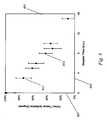
- FIG. 4shows data from the colon of a live subject during periods of low systemic blood flow, which led to local ischemia, as collected and analyzed in real time by a medical monitor constructed in accordance with embodiments of the invention.
- ImplantableIntended for implantation internally in a living body, such as into or between internal tissues. Implantable devices typically must be biocompatible (i.e., have a biocompatible exterior), or else the host subject's immune system will attack the implanted object or the device will have a toxic effect upon the host.
- Implantabledoes not require any fixed duration.
- Implantable as used hereincan mean short-term implantation, such as removable fibers inserted in the heart or lung, or a swallowable device such as an gastrointestinal ischemia monitor.
- Implantable as used hereinmay also be a long-term implantation, such as a pacemaker feedback system which monitors the heart or muscle, or a liver-based MEMS device that monitors for rejection.
- Fully-ImplantableComplete implantation into a living body, without a physical connection to the external body.
- Fully-implantable devicesmay contain an embedded power supply, receive power from another implanted device (such as a pacemaker), or receive power from an external source such as via transcutaneous inductive coupling.
- Fully-implantable devicesmay still communicate with receivers external to the body via non-physical means, such as electromagnetic waves from RFID chips.
- An implantable systemmay be embedded long-term, such as buried deep within a body to monitor for organ rejection or cardiac ischemia.
- An implantable systemmay also be used short-term only, such as a swallowable pill that monitors for ischemic ulcers or polyps in the esophagus, stomach, intestines, and colon, and is passed via the rectum when the scanning is complete in a few hours to a few days.
- TissueMaterial from a living animal, plant, viral, or bacterial subject, with an emphasis on mammals, especially humans.
- PerfusionThe flow of blood to a tissue or region, which differs from tissue ischemia in that low flow does not guarantee ischemia.
- Blood OxygenationThe saturation of the hemoglobin in arterial and venous blood, which differs from tissue ischemia. Arterial blood can be 100% oxygenated, yet a blood clot in the coronary artery will produce severe ischemia despite the 100% arterial saturation. Similarly, a local occlusion may produce lethal local ischemia, while the average venous oxygenation is not lowered detectably due to the small contribution of that local tissue to the overall venous blood oxygenation.
- IschemiaA local condition of tissue in which the delivery of oxygen to the tissue is locally inadequate to meet its metabolic needs. Such conditions vary from tissue to tissue.
- the brainhas a high metabolic rate and is easily made ischemic, even during simple tasks such as deep thought and insight, unless there is a local and rapid increase in the baseline blood flow and oxygen delivery to the metabolizing cortical tissues.
- the growing fetusis in a relative hibernation state, with very low oxygen needs in most tissues, and is more difficult to make ischemic.
- Early and mild ischemiais often evidenced by increases in the amount of oxygen extracted from the blood being delivered to the capillary bed, resulting in decreased tissue oxygenation.
- Ischemiais therefore distinguished from perfusion (i.e., blood flow) in that low blood flow does not guarantee ischemia (such as during tissue cooling or in the fetus), nor does high flow rule out ischemia (such as during sepsis, fever, or intense work). Ischemia is a co-existing condition in many different types of illnesses, including sepsis, tissue rejection, heart attack, stroke, organ failure, diabetic disease, and other conditions.
- TargetA material to be detected, imaged, or studied.
- one target siteis the intestine.
- Target SignalA sensed signal specific to the target. This signal may be enhanced through use of a contrast agent. This signal may be produced by scattering, absorbance, phosphorescence, fluorescence, Raman effects, or other known spectroscopy techniques.
- Visible LightElectromagnetic radiation from blue to yellow, namely with wavelengths between 400 nm and 625 microns, but especially those green to orange wavelengths between 475 and 600 nm where the absorbance by capillary hemoglobin (not in the arterial or venous circulation but locally in the capillaries in the tissue), myoglobin (which is extravascular and within muscle cells in the tissue itself) and cytochrome (which is intracellular within the mitochondria of the tissue itself) is the strongest.
- Broadband LightLight produced over a wide range of wavelengths sufficient to perform solution of multiple simultaneous spectroscopic equations. For tissue, a width of at least 40 mn is likely to be needed, while in the preferred embodiment a broadband white LED produces light from 400 nm to beyond 700 rim.
- LEDA light emitting diode.
- White LEDA broadband, visible wavelength LED, often comprised of a blue LED and a blue-absorbing broad-emitting phosphor that emits over a wide range of visible wavelengths. Other phosphors can be substituted. As used in the examples herein, any broadband LED could be used, even if not emitting over a full (white) spectrum. For example, a green LED emitting over a FWHM range of 100 nm would be considered to be broadband.
- a source of illuminating photonsmay be composed of a simple light bulb, a laser, a flash lamp, an LED, a white LED, or another light source or combination of sources, or it may be a complex form including but not limited to, a light emitter such as a bulb or light emitting diode, one or more filter elements, a transmission element such as an integrated optical fiber, a guidance element such as a reflective prism or internal lens, and other elements intended to enhance the optical coupling of the light from the source to the tissue or sample under study.
- the lightmay be generated using electrical input (such as with an LED), optical input (such as a fluorescent dye in a fiber responding to light), or any other source of energy, internal or external to the source.
- the light sourcemay be continuously on, pulsed, or even analyzed as time-, frequency-, or spatially-resolved.
- the light emittermay comprise a single or multiple light emitting elements, such as a combination of different light emitting diodes to produce a spectrum of light.
- Light Detector or Light SensorA detector that generates a measurable signal in response to the light incident on the detector.
- Optical CouplingThe arrangement of two elements such that light exiting the first element interacts, at least in part, with the second element. This may be free-space (unaided) transmission through air or space, or may require use of intervening optical elements such as lenses, filters, fused fiber expanders, collimators, concentrators, collectors, optical fibers, prisms, mirrors, or mirrored surfaces and the like.
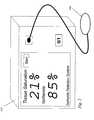
- FIG. 1shows device 101 implanted into the chest wall of patient 98 .
- patient 98is shown for illustrative purposes, and is not considered a part of the invention.
- a cut-away schematic of device 101 showing the interior of implantable device 101is shown at the top of FIG. 1 .
- Device 101is surrounded by biocompatible exterior 102 .
- exterior 102is constructed from approved Class VI materials as recognized by the U.S. FDA or other medical device regulatory agencies, such as polyethylene or surgical steel. Portions of the sensor, power supply, light source, or transmitter may protrude as needed from this shell within the spirit of this invention, provided that the protruding parts themselves are biocompatible.
- light source 103is illustrated in its component parts.
- broad spectrum white lightis emitted by a high conversion-efficiency white LED source 105 (in this case, The LED Light, model T1-3/4-20W-a, Fallon, Nev.).
- diode source 105is embedded into a plastic beam-shaping mount using optical clear epoxy 111 to allow light generated in LED 105 to be collimated, thus remaining at a near-constant diameter after passing through optical window 115 to leave device 101 .
- Lightthen is able to pass forward as shown by light path vectors 119 , with at least a portion of this light optically coupled to target region 125 .
- target region 125may be in some instances a living tissue, the tissue itself is not considered to be a claimed part of this invention.
- Collection window 141in this embodiment is a glass, plastic, or quartz window, but can alternatively be merely an aperture, or even be a lens, as required.
- Lightthen strikes sensor 155 , where it is sensed and detected.
- Sensor 155may comprise a number of discrete detectors configured to be wavelength-sensitive, or may be a continuous CCD spectrometer, with entry of light by wavelength controlled by gratings, filters, or wavelength-specific optical fibers. In any event, sensor 155 transmits an ischemia signal related to the detected light backscattered from target 125 , producing an electrical signal sent via wires 161 and 163 a sending unit 167 , such as a transmitter chip. The signal transmitted by the sending unit 167 is received by the receiver 183 where it can be further processed to provide a display.
- light source 103also has two electrical connections 175 and 176 , connecting light source 103 to power source 179 .
- power source 179is an inductive power supply, capable of receiving an inductive field from externally powered coil and RFID receiver 183 ( FIG. 2 ) placed outside of the body, in order to produce power for device 101 as required.
- external powered coil 183is shown for the purposes of example and illustration, but is not considered a required part of this invention.
- source 179could merely be a long-lived implantable battery, in which case an external powered coil may not be required at all.
- Device 101is implanted in a patient, for example in the chest wall of a patient undergoing coronary artery repair for heart disease.
- the devicemay measure the muscle directly, or it can be placed at a distance.
- vectors 119are fiber optics extended from device 101 and into close proximity to the target heart muscle, sufficient for optical coupling. Then the patient is allowed to heal after surgery, and the implantable device is left inside the patient's body, without a direct physical connection to the outside world.
- device 101is normally powered down and in a resting (off) state. At some point, it is desired to test the target heart muscle for the presence of ischemia.
- external inductive coil 183is connected to external monitor is brought into close proximity to the chest wall over the site of implantation of device 101 . Referring back to FIG. 1 , through inductive coupling external coil 183 induces a current in inductive power source 179 located within device 101 , producing sufficient power for device 101 to power up and turn on.
- Light source 103begins to illuminate the target 125 , in this case heart muscle.
- Sensor 155which is an embedded spectrophotometer in some embodiments, receives backscattered light, resolves the incoming light by wavelength, a marker of ischemia.
- the result of this determinationis sent to sending unit 167 , which in the exemplary embodiment is an RF transmitter that sends the sensed signals to external RFID receiver 184 .
- the signal received by receiver 184may be processed for the oxygenation of the hemoglobin in the terminal capillary beds, a marker of ischemia, by external monitor 313 , as shown in the data collected and plotted under the Example section, below.
- An example of a system for indicating oxygenationis described in U.S. Pat. No. 5,987,346, incorporated herein by reference.
- external coil 183is moved away from device 101 , and device 101 powers down and returns to a resting state.
- power source 179may be charged during proximity to external coil, or have an internal battery source, allowing device 101 to operate when external coil 179 is not present.
- Sending unit 167may then transmit without being directly queried, such as in response to a dangerous level of ischemia.
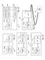
- the sensor 155is merely single photodiode 411 and processing electronics 413 .
- Photodiode 411is made wavelength sensitive through the design of LED 105 as a cluster of LEDs of different wavelengths, each emitting at a different time or modulation frequency to allow decoding of the illuminating wavelength by photodiode 411 and processing unit electronics 413 .
- sensor 155may comprise a set of different photodiodes 421 A through 421 N, FIG.
- sensor 155may be single photodiode 431 with electronically variable filter 433 , FIG. 3C , allowing the wavelength transmitted to be selected and processed by processing unit electronics 435 .
- sensor 155may be CCD chip 441 with filter window 443 , FIG. 3D , that varies over its length, allowing only certain wavelengths to reach each portion of CCD 441 , allowing decoding of the illuminating wavelength by processing unit electronics 447 .
- sensor 155comprises CCD chip 451 with optical fibers 453 attached to CCD 451 in a linear array. Fibers 453 are manufactured such that each fiber has a different interference coating on end 454 , allowing each fiber to transmit a different narrow wavelength range, allowing decoding of the illuminating wavelength by processing unit electronics 457 . Fibers 453 are biocompatible and can extend outside of device case 102 , allowing device 101 to be placed remotely the target to be monitored, and for the free end of fibers 453 to be placed in proximity to target 125 .
- an optical sensorsimilar in basis of operation to device 101 , is implanted into abdomen of a patient undergoing colon surgery.
- the animalreceives heart-lung bypass, such that the blood flow and oxygen content of the blood is exactly controlled by a bypass specialist rather than by the animal's own heart and lungs, affording the ability to create and resolve ischemia at will.
- An aortic Doppler probeis placed, which measures the delivery of blood to tissue. In this case, when the rate of the pump is lowered to zero flow, ischemia must exist in the tissues being monitored.
- Analysis of the tissue ischemiais performed by broadband, visible light, differential spectroscopy.
- the first differential (for example) of the wavelength vs. intensity curve sent from the sending unitis processed to remove many of the effects caused by light scattering by the local tissue, and the resultant signal is analyzed using a least-squares minimization of the fitting error to known components of the tissue (such as myoglobin, capillary hemoglobin, or cytochromes).
- the signal that is measuredis a function of the presence, absence, or risk, or degree of ischemia. This can have clinical implications and applications in many different medical areas, such as impending risk of tissue death (as seen in the colon study above), impending risk of organ rejection (as inflammation results in increased total blood content, while potentially reducing oxygenation) cardiac function (as improved cardiac function is associated with a body-wide improvement in tissue ischemia as well as a likely improvement in myocardial ischemia), treatment efficacy for arterial or venous vascular disease (as the real-time effects of such interventions on tissue oxygenation adequacy can be used as a treatment signal to guide chemical and physical interventions), risk of renal damage (as kidney failure is often the result of acute or chronic reduced oxygen delivery), risk of brain injury (as stroke is often the result of acute and chronic reduced oxygen delivery), risk of colon death (as the colon does not have a large capacity to increase blood and oxygen delivery in times of stress over baseline), risk of limb amputation (as limbs with good capillary
- the creation of graded ischemiais detected by the present invention.
- graph 601the flow detected by the Doppler probe is plotted on horizontal axis 603 versus the presence of ischemia as detected by the present invention using optical spectroscopy plotted on vertical axis 607 . Data are plotted as means with standard error bars 613 .
- graph 601when the blood flow to the gut is reduced to zero, the detection of the presence of ischemia rises to 100%, shown at data point 617 .
- Ischemiais diagnosed by low local tissue oxygenation, not blood oxygenation or flow.
- arterial bloodmay be well oxygenated, but the delivery of this arterial blood to the tissue is insufficient (such as with a blood clot); in this case the tissue is indeed ischemia while the arterial blood oxygenation is normal.
- Blood flowalso differs from a direct measure of ischemia. For example, in a cooled patient on heart-lung bypass, blood flow may be very, very low; however, the cooled tissues, whose oxygen need has been reduced by the low temperature, are not ischemic.
- ischemic heart“hibernates” in order to reduce its own oxygen need, and may not be ischemic at reduced flow.
- flowwas controlled sufficiently to allow for a low or zero flow to be consistent with ischemia, but such conclusions cannot be always made so clearly in the living non-experimental subject.
- the signal detected from the tissuewas a hemoglobin absorbance signal derived from the capillary bed. While absorbance is ideal for hemoglobin analysis, as described in the preferred embodiment, other interactions may be preferable for other measurements.
- the interaction with the illuminating light that provides the contrastcan include absorbance, polarization, optical rotation, scattering, fluorescence, Raman effects, phosphorescence, or fluorescence decay, and measures of a contrast effect may reasonably include one or more of these effects.
- Other tissue componentscould be measured, including NADH, NADPH, cytochromes in their oxidized and reduced forms, or even ischemia or oxygen sensitive dyes.
- myoglobinis another protein whose saturation is related to the presence or absence of ischemia.
- a combination of hemoglobin in the capillaries as well as myoglobin in the heart, or just myoglobin in the heart myocytescan serve as a marker of ischemia.
- an injectable dye, sensitive to local ischemiacan be used to generate an optical signal directly related to the presence of ischemia, such as by changing color in response to mitochondrial membrane charge or in response to intracellular pH.
- Such use of dyes to label cells in vivo with optical dyeshas been demonstrated in vivo by several groups, and the coupling of an ischemia sensitive dye to use of the present invention to detect ischemia (and conditions which are a function of ischemia) would fall within the spirit of the present invention.
- a devicecomprising a phosphor-coated white LED and integrated collimating optics conFIG.d to produce continuous, broadband light from 400 nm to 700 nm in a collimated beam, which is then directly transmitted to a target site.
- Light backscattered by the target siteis collected by a sensor, allowing for a direct measure of ischemia to be determined, and subsequently transmitted by a sending unit.
- Poweris provided by an internal power source, which may in turn be itself powered by an external inductive coil that is brought in proximity to the implanted device in order to provide energy as needed.
- the entire implantable deviceis encapsulated by a biocompatible shell to add long-term safety while implanted. Used alone, or in combination with an estimate of arterial oxygenation, venous oxygenation, or even of blood flow, this device allows for an index of ischemia to be determined without additional invasiveness beyond the initial implantation.
- the present devicemay be interrogated using inductive technology and RF coupling. Implantable devices incorporating the ischemia system, and medical methods of use, are described. This device has immediate application to several important problems, both medical and industrial, and thus constitutes an important advance in the art.
Landscapes
- Health & Medical Sciences (AREA)
- Life Sciences & Earth Sciences (AREA)
- Physics & Mathematics (AREA)
- Engineering & Computer Science (AREA)
- Animal Behavior & Ethology (AREA)
- Veterinary Medicine (AREA)
- Pathology (AREA)
- Biomedical Technology (AREA)
- Heart & Thoracic Surgery (AREA)
- Medical Informatics (AREA)
- Molecular Biology (AREA)
- Surgery (AREA)
- Biophysics (AREA)
- General Health & Medical Sciences (AREA)
- Public Health (AREA)
- Optics & Photonics (AREA)
- Computer Networks & Wireless Communication (AREA)
- Spectroscopy & Molecular Physics (AREA)
- Immunology (AREA)
- Vascular Medicine (AREA)
- Hematology (AREA)
- Cardiology (AREA)
- Physiology (AREA)
- Measurement Of The Respiration, Hearing Ability, Form, And Blood Characteristics Of Living Organisms (AREA)
- Electrotherapy Devices (AREA)
Abstract
Description
Claims (18)
Priority Applications (6)
| Application Number | Priority Date | Filing Date | Title |
|---|---|---|---|
| US11/193,071US7813778B2 (en) | 2005-07-29 | 2005-07-29 | Implantable tissue ischemia sensor |
| PCT/US2006/029615WO2007016437A2 (en) | 2005-07-29 | 2006-07-27 | Implantable tissue ischemia sensor |
| JP2008524234AJP2009502360A (en) | 2005-07-29 | 2006-07-27 | Implantable tissue ischemia sensor |
| EP06788915AEP1912559A4 (en) | 2005-07-29 | 2006-07-27 | Implantable tissue ischemia sensor |
| US12/858,396US20100312081A1 (en) | 2005-07-29 | 2010-08-17 | Implantable Tissue Ischemia Sensor |
| US13/591,629US20120316411A1 (en) | 2005-07-29 | 2012-08-22 | Remote oximetry monitoring system and method |
Applications Claiming Priority (1)
| Application Number | Priority Date | Filing Date | Title |
|---|---|---|---|
| US11/193,071US7813778B2 (en) | 2005-07-29 | 2005-07-29 | Implantable tissue ischemia sensor |
Related Child Applications (1)
| Application Number | Title | Priority Date | Filing Date |
|---|---|---|---|
| US12/858,396ContinuationUS20100312081A1 (en) | 2005-07-29 | 2010-08-17 | Implantable Tissue Ischemia Sensor |
Publications (2)
| Publication Number | Publication Date |
|---|---|
| US20070027371A1 US20070027371A1 (en) | 2007-02-01 |
| US7813778B2true US7813778B2 (en) | 2010-10-12 |
Family
ID=37695273
Family Applications (3)
| Application Number | Title | Priority Date | Filing Date |
|---|---|---|---|
| US11/193,071Expired - Fee RelatedUS7813778B2 (en) | 2005-07-29 | 2005-07-29 | Implantable tissue ischemia sensor |
| US12/858,396AbandonedUS20100312081A1 (en) | 2005-07-29 | 2010-08-17 | Implantable Tissue Ischemia Sensor |
| US13/591,629AbandonedUS20120316411A1 (en) | 2005-07-29 | 2012-08-22 | Remote oximetry monitoring system and method |
Family Applications After (2)
| Application Number | Title | Priority Date | Filing Date |
|---|---|---|---|
| US12/858,396AbandonedUS20100312081A1 (en) | 2005-07-29 | 2010-08-17 | Implantable Tissue Ischemia Sensor |
| US13/591,629AbandonedUS20120316411A1 (en) | 2005-07-29 | 2012-08-22 | Remote oximetry monitoring system and method |
Country Status (4)
| Country | Link |
|---|---|
| US (3) | US7813778B2 (en) |
| EP (1) | EP1912559A4 (en) |
| JP (1) | JP2009502360A (en) |
| WO (1) | WO2007016437A2 (en) |
Cited By (21)
| Publication number | Priority date | Publication date | Assignee | Title |
|---|---|---|---|---|
| US20070015981A1 (en)* | 2003-08-29 | 2007-01-18 | Benaron David A | Device and methods for the detection of locally-weighted tissue ischemia |
| US20080009689A1 (en)* | 2002-04-09 | 2008-01-10 | Benaron David A | Difference-weighted somatic spectroscopy |
| US20080188727A1 (en)* | 2002-04-09 | 2008-08-07 | Benaron David A | Broadband solid-state spectroscopy illuminator and method |
| US20090076348A1 (en)* | 2007-09-14 | 2009-03-19 | Corventis, Inc. | Injectable Device for Physiological Monitoring |
| US20100022856A1 (en)* | 2008-07-28 | 2010-01-28 | Medtronic, Inc. | Implantable optical hemodynamic sensor including light transmission member |
| US20100312081A1 (en)* | 2005-07-29 | 2010-12-09 | Spectros Corporation | Implantable Tissue Ischemia Sensor |
| US20100317940A1 (en)* | 2009-06-10 | 2010-12-16 | Kuhn Jonathan L | Absolute calibrated tissue oxygen saturation and total hemoglobin volume fraction |
| US8285356B2 (en) | 2007-09-14 | 2012-10-09 | Corventis, Inc. | Adherent device with multiple physiological sensors |
| US20130030255A1 (en)* | 2011-07-26 | 2013-01-31 | Embry Ii William Ben | Biocompatible implant device |
| US8374688B2 (en) | 2007-09-14 | 2013-02-12 | Corventis, Inc. | System and methods for wireless body fluid monitoring |
| US8412317B2 (en) | 2008-04-18 | 2013-04-02 | Corventis, Inc. | Method and apparatus to measure bioelectric impedance of patient tissue |
| US8460189B2 (en) | 2007-09-14 | 2013-06-11 | Corventis, Inc. | Adherent cardiac monitor with advanced sensing capabilities |
| US20130289372A1 (en)* | 2011-10-21 | 2013-10-31 | Incube Labs, Llc | Implantable oximetric measurement apparatus and method of use |
| US8718752B2 (en) | 2008-03-12 | 2014-05-06 | Corventis, Inc. | Heart failure decompensation prediction based on cardiac rhythm |
| US8897868B2 (en) | 2007-09-14 | 2014-11-25 | Medtronic, Inc. | Medical device automatic start-up upon contact to patient tissue |
| US20150006196A1 (en)* | 2007-04-18 | 2015-01-01 | Weinmann Geraete Fuer Medizin Gmbh & Co. Kg | Method and device for updating medical apparatus |
| US9411936B2 (en) | 2007-09-14 | 2016-08-09 | Medtronic Monitoring, Inc. | Dynamic pairing of patients to data collection gateways |
| US9496733B2 (en) | 2013-09-13 | 2016-11-15 | Boston Scientific Neuromodulation Corporation | Optical communications between an implantable medical device and external charger |
| US10018559B2 (en) | 2015-04-17 | 2018-07-10 | Koninklijke Philips N.V. | Tissue inspection system with optical fibers and continuous calibration |
| US12123654B2 (en) | 2010-05-04 | 2024-10-22 | Fractal Heatsink Technologies LLC | System and method for maintaining efficiency of a fractal heat sink |
| US12251201B2 (en) | 2019-08-16 | 2025-03-18 | Poltorak Technologies Llc | Device and method for medical diagnostics |
Families Citing this family (29)
| Publication number | Priority date | Publication date | Assignee | Title |
|---|---|---|---|---|
| US10010277B2 (en)* | 2006-06-22 | 2018-07-03 | The General Hospital Corporation | Cancer detection by optical measurement of compression-induced transients |
| US20080018424A1 (en)* | 2006-07-10 | 2008-01-24 | 3M Innovative Properties Company | Inductive sensor |
| US7948380B2 (en)* | 2006-09-06 | 2011-05-24 | 3M Innovative Properties Company | Spatially distributed remote sensor |
| US20090246797A1 (en)* | 2008-03-28 | 2009-10-01 | Nellcor Puritan Bennett Llc | Medical device for the assessment of internal organ tissue and technique for using the same |
| US8473036B2 (en)* | 2008-04-02 | 2013-06-25 | The Trustees Of The University Of Pennsylvania | In vivo measurement of mitochondrial function |
| EP2346389B1 (en) | 2008-07-28 | 2013-05-22 | Medtronic, Inc. | Implantable optical hemodynamic sensor including light transmission member |
| US20100022861A1 (en)* | 2008-07-28 | 2010-01-28 | Medtronic, Inc. | Implantable optical hemodynamic sensor including an extension member |
| US9545215B2 (en)* | 2008-07-31 | 2017-01-17 | Medtronic, Inc. | Apparatus and method for detecting cardiac events |
| US9737213B1 (en)* | 2009-03-24 | 2017-08-22 | Vioptix, Inc. | Using an oximeter probe to detect intestinal ischemia |
| US9339221B1 (en)* | 2009-03-24 | 2016-05-17 | Vioptix, Inc. | Diagnosing intestinal ischemia based on oxygen saturation measurements |
| WO2011058604A1 (en)* | 2009-11-12 | 2011-05-19 | トヨタ自動車株式会社 | Fuel cell |
| US20120130203A1 (en)* | 2010-11-24 | 2012-05-24 | Fujitsu Limited | Inductively-Powered Ring-Based Sensor |
| US8641610B2 (en) | 2010-12-20 | 2014-02-04 | Covidien Lp | Access assembly with translating lumens |
| US8602983B2 (en) | 2010-12-20 | 2013-12-10 | Covidien Lp | Access assembly having undercut structure |
| US8696557B2 (en) | 2010-12-21 | 2014-04-15 | Covidien Lp | Access assembly including inflatable seal member |
| US9649113B2 (en) | 2011-04-27 | 2017-05-16 | Covidien Lp | Device for monitoring physiological parameters in vivo |
| WO2012158748A1 (en)* | 2011-05-17 | 2012-11-22 | Landy Aaron Toth | Devices, systems, and methods for assessing implants, organs, transplants, tissues, synthetic constructs, vascular grafts, and the like |
| JP2014534012A (en)* | 2011-10-20 | 2014-12-18 | ザ ジェネラル ホスピタル コーポレイション | Implantable imaging configuration and method of using the same |
| EP2586369B1 (en)* | 2011-10-27 | 2017-07-26 | St. Jude Medical AB | Ischemia detection |
| CN104395721A (en) | 2012-03-16 | 2015-03-04 | 维塔尔传感器控股有限公司 | Permittivity shielding |
| US9403016B2 (en) | 2012-03-27 | 2016-08-02 | The University Of Vermont And State Agricultural College | Cardiac pacemaker and uses thereof |
| US9936951B2 (en)* | 2013-03-12 | 2018-04-10 | Covidien Lp | Interchangeable tip reload |
| US10420583B2 (en) | 2013-05-22 | 2019-09-24 | Covidien Lp | Methods and apparatus for controlling surgical instruments using a port assembly |
| US10265013B2 (en) | 2013-09-06 | 2019-04-23 | Somnology, Inc. | System and method for sleep disorder diagnosis and treatment |
| US10265014B2 (en) | 2013-09-06 | 2019-04-23 | Somnology, Inc. | System and method for sleep disorder diagnosis and treatment |
| US10835184B2 (en)* | 2015-04-22 | 2020-11-17 | Arizona Board Of Regents On Behalf Of Arizona State University | Device for neuroprosthetics with autonomous tunable actuators |
| US11324430B2 (en)* | 2018-01-15 | 2022-05-10 | The Johns Hopkins University | Sensor-based ischemia detection |
| US11864906B2 (en) | 2019-06-20 | 2024-01-09 | International Business Machines Corporation | LIDAR implantable biosensor for imaging biological tissue |
| US11883028B2 (en) | 2021-09-08 | 2024-01-30 | Covidien Lp | Systems and methods for post-operative anastomotic leak detection |
Citations (71)
| Publication number | Priority date | Publication date | Assignee | Title |
|---|---|---|---|---|
| USRE29304E (en) | 1963-10-21 | 1977-07-12 | Raydne Limited | Plasma light source for spectroscopic investigation |
| US4164374A (en) | 1977-09-26 | 1979-08-14 | Ford Motor Company | Spectrophotometer utilizing a solid state source of radiant energy having a controllable frequency spectra characteristic |
| US4427889A (en) | 1979-08-23 | 1984-01-24 | Carl Zeiss Stiftung | Method and apparatus for molecular spectroscopy, particularly for the determination of products of metabolism |
| US4513751A (en) | 1979-03-07 | 1985-04-30 | Sumitomo Electric Industries, Ltd. | Method for measuring oxygen metabolism in internal organ or tissue |
| US4660974A (en) | 1984-04-14 | 1987-04-28 | Carl-Zeiss-Stiftung | Arrangement for determining the spectral characteristic of the refractive index of a fluid |
| US4697593A (en) | 1984-06-26 | 1987-10-06 | Evans John M | Method and apparatus for measuring blood oxygen levels |
| US5040533A (en)* | 1989-12-29 | 1991-08-20 | Medical Engineering And Development Institute Incorporated | Implantable cardiovascular treatment device container for sensing a physiological parameter |
| US5135004A (en) | 1991-03-12 | 1992-08-04 | Incontrol, Inc. | Implantable myocardial ischemia monitor and related method |
| US5190040A (en) | 1986-12-26 | 1993-03-02 | Nihon Kohden Corporation | Apparatus for measuring the change in the concentration of a pigment in blood |
| US5199428A (en) | 1991-03-22 | 1993-04-06 | Medtronic, Inc. | Implantable electrical nerve stimulator/pacemaker with ischemia for decreasing cardiac workload |
| US5259052A (en) | 1984-06-08 | 1993-11-02 | Amp Incorporated | High precision optical fiber connectors |
| US5280788A (en) | 1991-02-26 | 1994-01-25 | Massachusetts Institute Of Technology | Devices and methods for optical diagnosis of tissue |
| US5318022A (en) | 1991-03-01 | 1994-06-07 | John Taboada | Method and apparatus for determining hemoglobin oxygenation such as in ocular and other vascular beds |
| US5329922A (en) | 1992-10-19 | 1994-07-19 | Atlee Iii John L | Oximetric esophageal probe |
| US5355425A (en) | 1992-09-04 | 1994-10-11 | Braiman Mark S | Light coupling device for optical fibers |
| US5357954A (en) | 1993-01-04 | 1994-10-25 | Respiratory Support Products, Inc. | Optical blood oxygen saturation probe for insertion into the esophagus |
| US5417207A (en) | 1993-12-06 | 1995-05-23 | Sensor Devices, Inc. | Apparatus for the invasive use of oximeter probes |
| US5520190A (en)* | 1994-10-31 | 1996-05-28 | Ventritex, Inc. | Cardiac blood flow sensor and method |
| US5645059A (en) | 1993-12-17 | 1997-07-08 | Nellcor Incorporated | Medical sensor with modulated encoding scheme |
| US5672875A (en) | 1992-07-15 | 1997-09-30 | Optix Lp | Methods of minimizing scattering and improving tissue sampling in non-invasive testing and imaging |
| US5696861A (en) | 1996-08-13 | 1997-12-09 | Schimmeyer; Werner K. | Method and apparatus for simultaneously connecting data/signal communication lines and power lines to a data/RF receiver/transmitter |
| US5733313A (en)* | 1996-08-01 | 1998-03-31 | Exonix Corporation | RF coupled, implantable medical device with rechargeable back-up power source |
| US5743261A (en) | 1993-12-06 | 1998-04-28 | Sensor Devices, Inc. | Methods and apparatus for the invasive use of oximeter probes |
| US5769791A (en) | 1992-09-14 | 1998-06-23 | Sextant Medical Corporation | Tissue interrogating device and methods |
| US5830132A (en) | 1993-08-24 | 1998-11-03 | Robinson; Mark R. | Robust accurate non-invasive analyte monitor |
| US5830137A (en) | 1996-11-18 | 1998-11-03 | University Of South Florida | Green light pulse oximeter |
| US5833603A (en)* | 1996-03-13 | 1998-11-10 | Lipomatrix, Inc. | Implantable biosensing transponder |
| US5901261A (en) | 1997-06-19 | 1999-05-04 | Visionex, Inc. | Fiber optic interface for optical probes with enhanced photonic efficiency, light manipulation, and stray light rejection |
| US5902235A (en) | 1989-03-29 | 1999-05-11 | Somanetics Corporation | Optical cerebral oximeter |
| US5931779A (en) | 1996-06-06 | 1999-08-03 | Wisconsin Alumni Research Foundation | Real-time in-vivo measurement of myoglobin oxygen saturation |
| US5941822A (en) | 1997-03-17 | 1999-08-24 | Polartechnics Limited | Apparatus for tissue type recognition within a body canal |
| US5974210A (en) | 1997-01-15 | 1999-10-26 | Perkin-Elmer Ltd. | Probe for spectroscopic analysis |
| US5987346A (en) | 1993-02-26 | 1999-11-16 | Benaron; David A. | Device and method for classification of tissue |
| WO2000001295A1 (en) | 1998-07-07 | 2000-01-13 | Lightouch Medical, Inc. | Tissue modulation process for quantitative noninvasive in vivo spectroscopic analysis of tissues |
| US6043893A (en) | 1998-10-09 | 2000-03-28 | Universities Space Research Association | Manually portable reflectance spectrometer |
| US6119031A (en) | 1996-11-21 | 2000-09-12 | Boston Scientific Corporation | Miniature spectrometer |
| US6122536A (en) | 1995-07-06 | 2000-09-19 | Animas Corporation | Implantable sensor and system for measurement and control of blood constituent levels |
| US6134460A (en)* | 1988-11-02 | 2000-10-17 | Non-Invasive Technology, Inc. | Spectrophotometers with catheters for measuring internal tissue |
| WO2000064534A1 (en) | 1999-04-26 | 2000-11-02 | Cardiac Pacemakers, Inc. | Method and apparatus for detecting changes in electrocardiogram signals |
| US6167297A (en) | 1999-05-05 | 2000-12-26 | Benaron; David A. | Detecting, localizing, and targeting internal sites in vivo using optical contrast agents |
| US6226082B1 (en) | 1998-06-25 | 2001-05-01 | Amira Medical | Method and apparatus for the quantitative analysis of a liquid sample with surface enhanced spectroscopy |
| US6251068B1 (en) | 1998-05-18 | 2001-06-26 | Fuji Photo Optical Co., Ltd. | Endoscopic observation system |
| US6252254B1 (en) | 1998-02-06 | 2001-06-26 | General Electric Company | Light emitting device with phosphor composition |
| EP1111333A1 (en) | 1999-06-29 | 2001-06-27 | Omron Corporation | Light source device, spectroscope comprising the light source device, and film thickness sensor |
| US6256524B1 (en) | 1998-09-09 | 2001-07-03 | The United States Of America As Represented By The Secretary Of The Army | Pulse oximeter sensor combined with a combination oropharyngeal airway and bite block |
| US6277078B1 (en) | 1999-11-19 | 2001-08-21 | Remon Medical Technologies, Ltd. | System and method for monitoring a parameter associated with the performance of a heart |
| US6353226B1 (en) | 1998-11-23 | 2002-03-05 | Abbott Laboratories | Non-invasive sensor capable of determining optical parameters in a sample having multiple layers |
| US6381018B1 (en) | 1998-07-28 | 2002-04-30 | The Regents Of The University Of California | Method for measuring changes in light absorption of highly scattering media |
| US20020082488A1 (en) | 1998-06-03 | 2002-06-27 | Ammar Al-Ali | Stereo pulse oximeter |
| WO2003003914A1 (en) | 2001-07-02 | 2003-01-16 | Masimo Corporation | Low power pulse oximeter |
| US6527729B1 (en) | 1999-11-10 | 2003-03-04 | Pacesetter, Inc. | Method for monitoring patient using acoustic sensor |
| US6532381B2 (en) | 2001-01-11 | 2003-03-11 | Ge Medical Systems Information Technologies, Inc. | Patient monitor for determining a probability that a patient has acute cardiac ischemia |
| US6533466B1 (en) | 2000-09-07 | 2003-03-18 | International Business Machines Corporation | Hybrid connector assembly for electrical conductors and fiber optic data conductors |
| US6550979B1 (en) | 2001-10-19 | 2003-04-22 | Corning Cable Systems Llc | Floating connector subassembly and connector including same |
| US6588938B1 (en) | 2000-10-18 | 2003-07-08 | Fitel Usa Corp. | Optical/electrical plug connector |
| US6599025B1 (en) | 1998-03-11 | 2003-07-29 | Ccs Technology, Inc. | Hybrid data plug |
| US6615065B1 (en) | 1998-10-13 | 2003-09-02 | Somanetics Corporation | Multi-channel non-invasive tissue oximeter |
| US6612857B2 (en) | 2001-07-05 | 2003-09-02 | Bernard R. Tolmie | Electrical connector system and method having optical and/or cooling capability |
| US6662033B2 (en) | 1994-04-01 | 2003-12-09 | Nellcor Incorporated | Pulse oximeter and sensor optimized for low saturation |
| US6711426B2 (en) | 2002-04-09 | 2004-03-23 | Spectros Corporation | Spectroscopy illuminator with improved delivery efficiency for high optical density and reduced thermal load |
| EP0926981B1 (en) | 1996-09-13 | 2004-03-31 | Non-Invasive Technology, Inc. | Non-invasive imaging of biological tissue |
| US20040122478A1 (en) | 2002-12-20 | 2004-06-24 | Stadler Robert W. | Method and apparatus for gauging severity of myocardial ischemic episodes |
| US20040218873A1 (en) | 2003-04-24 | 2004-11-04 | Zenya Nagashima | Electro-optical composite connector, electro-optical composite cable, and network devices using the same |
| US20040220460A1 (en) | 2003-04-30 | 2004-11-04 | Medtronic, Inc. | Normalization method for a chronically implanted optical sensor |
| US6842635B1 (en) | 1998-08-13 | 2005-01-11 | Edwards Lifesciences Llc | Optical device |
| US20050010113A1 (en) | 2001-07-16 | 2005-01-13 | Art, Advanced Research Technologies, Inc. | Choice of wavelengths for multiwavelength optical imaging |
| US6859658B1 (en) | 1998-11-18 | 2005-02-22 | Lea Medizintechnik Gmbh | Device for non-invasively detecting the oxygen metabolism in tissues |
| US6921920B2 (en) | 2001-08-31 | 2005-07-26 | Smith & Nephew, Inc. | Solid-state light source |
| US20060105319A1 (en) | 2002-07-26 | 2006-05-18 | Obi Aps | Method for converting venous blood values to arterial blood values, system for utilising said method and devices for such system |
| US20070015981A1 (en)* | 2003-08-29 | 2007-01-18 | Benaron David A | Device and methods for the detection of locally-weighted tissue ischemia |
| US20070016080A1 (en) | 2005-04-28 | 2007-01-18 | Research Foundation Of The City University Of New York | Imaging systems and methods to improve backscattering imaging using circular polarization memory |
Family Cites Families (42)
| Publication number | Priority date | Publication date | Assignee | Title |
|---|---|---|---|---|
| US3872455A (en)* | 1971-11-17 | 1975-03-18 | Monitron Ind | Physiological measurement display system |
| JPS6043134B2 (en)* | 1977-08-25 | 1985-09-26 | 信紘 佐藤 | Device for measuring reflection characteristics of biological organs and tissues |
| CA1327838C (en)* | 1988-06-13 | 1994-03-15 | Fred Zacouto | Implantable device to prevent blood clotting disorders |
| FR2637807B1 (en)* | 1988-10-14 | 1997-10-31 | Zacouto Fred | DEVICE FOR PROTECTION AGAINST BLOOD-RELATED CONDITIONS, IN PARTICULAR THROMBOSIS, EMBOLIES, HEMORRHAGIA, HEMOPATHIES AND PRESENCE OF ABNORMAL ELEMENTS IN THE BLOOD |
| US5873821A (en)* | 1992-05-18 | 1999-02-23 | Non-Invasive Technology, Inc. | Lateralization spectrophotometer |
| US5040538A (en)* | 1989-09-05 | 1991-08-20 | Siemens-Pacesetter, Inc. | Pulsed light blood oxygen content sensor system and method of using same |
| US6785568B2 (en)* | 1992-05-18 | 2004-08-31 | Non-Invasive Technology Inc. | Transcranial examination of the brain |
| US5777350A (en)* | 1994-12-02 | 1998-07-07 | Nichia Chemical Industries, Ltd. | Nitride semiconductor light-emitting device |
| EP0762109A3 (en)* | 1995-08-30 | 1997-09-17 | Kyoto Daiichi Kagaku Kk | Method of and apparatus for measuring lactic acid in organism |
| JP2000503154A (en)* | 1996-01-11 | 2000-03-14 | エムアールジェイ インコーポレイテッド | System for controlling access and distribution of digital ownership |
| US6790178B1 (en)* | 1999-09-24 | 2004-09-14 | Healthetech, Inc. | Physiological monitor and associated computation, display and communication unit |
| US20010044588A1 (en)* | 1996-02-22 | 2001-11-22 | Mault James R. | Monitoring system |
| US5879294A (en)* | 1996-06-28 | 1999-03-09 | Hutchinson Technology Inc. | Tissue chromophore measurement system |
| US6120460A (en)* | 1996-09-04 | 2000-09-19 | Abreu; Marcio Marc | Method and apparatus for signal acquisition, processing and transmission for evaluation of bodily functions |
| US5782756A (en)* | 1996-09-19 | 1998-07-21 | Nellcor Puritan Bennett Incorporated | Method and apparatus for in vivo blood constituent analysis |
| US6324418B1 (en)* | 1997-09-29 | 2001-11-27 | Boston Scientific Corporation | Portable tissue spectroscopy apparatus and method |
| US6415166B1 (en)* | 1997-09-26 | 2002-07-02 | Datex-Ohmeda, Inc. | Photoplethysmographic device with remote facsimile |
| US6432364B1 (en)* | 1998-07-06 | 2002-08-13 | Suzuki Motor Corporation | SPR sensor cell and immunoassay apparatus using the same |
| CA2339506C (en)* | 1998-08-07 | 2011-05-31 | Infinite Biomedical Technologies, Incorporated | Implantable myocardial ischemia detection, indication and action technology |
| US6127783A (en)* | 1998-12-18 | 2000-10-03 | Philips Electronics North America Corp. | LED luminaire with electronically adjusted color balance |
| US6438399B1 (en)* | 1999-02-16 | 2002-08-20 | The Children's Hospital Of Philadelphia | Multi-wavelength frequency domain near-infrared cerebral oximeter |
| US6216021B1 (en)* | 1999-06-04 | 2001-04-10 | The Board Of Trustees Of The University Of Illinois | Method for measuring absolute saturation of time-varying and other hemoglobin compartments |
| US6515273B2 (en)* | 1999-08-26 | 2003-02-04 | Masimo Corporation | System for indicating the expiration of the useful operating life of a pulse oximetry sensor |
| US6596235B2 (en)* | 1999-09-30 | 2003-07-22 | Therox, Inc. | Method for blood oxygenation |
| WO2001073407A2 (en)* | 2000-03-29 | 2001-10-04 | The Dow Chemical Company | Method for the determination of an acid or a base in a non-aqueous liquid |
| DE10016349B4 (en)* | 2000-04-03 | 2007-09-27 | Sensopart Industriesensorik Gmbh | Method and arrangement for detecting and / or detecting an object |
| US20010056226A1 (en)* | 2000-04-18 | 2001-12-27 | Richard Zodnik | Integrated telemedicine computer system |
| US8055329B2 (en)* | 2001-01-22 | 2011-11-08 | Spectrum Dynamics Llc | Ingestible device for radioimaging of the gastrointestinal tract |
| US6678398B2 (en)* | 2000-09-18 | 2004-01-13 | Sti Medical Systems, Inc. | Dual mode real-time screening and rapid full-area, selective-spectral, remote imaging and analysis device and process |
| US6798517B2 (en)* | 2000-09-28 | 2004-09-28 | Color-Spec Technologies, Inc. | Handheld, portable color measuring device with display |
| WO2002069825A2 (en)* | 2001-03-02 | 2002-09-12 | Palomar Medical Technologies, Inc. | Apparatus and method for photocosmetic and photodermatological treatment |
| US20030036031A1 (en)* | 2001-08-20 | 2003-02-20 | Lieb Joseph Alexander | Light-emitting handpiece for curing photopolymerizable resins |
| CA2458656A1 (en)* | 2001-08-30 | 2003-03-13 | Medtronic, Inc. | System and method for detecting myocardial ischemia |
| US20030073889A1 (en)* | 2001-10-11 | 2003-04-17 | Keilbach Kevin A. | Monitoring led wavelength shift in photoplethysmography |
| US6832113B2 (en)* | 2001-11-16 | 2004-12-14 | Cardiac Pacemakers, Inc. | Non-invasive method and apparatus for cardiac pacemaker pacing parameter optimization and monitoring of cardiac dysfunction |
| US20030111533A1 (en)* | 2001-12-19 | 2003-06-19 | Koninklijke Philips Electronics N.V. | RGB led based white light control system with quasi-uniform color metric |
| WO2004066825A2 (en)* | 2003-01-31 | 2004-08-12 | The Board Of Trustees Of The Leland Stanford Junior University | Detection of apex motion for monitoring cardiac dysfunction |
| US7494459B2 (en)* | 2003-06-26 | 2009-02-24 | Biophan Technologies, Inc. | Sensor-equipped and algorithm-controlled direct mechanical ventricular assist device |
| US20060167334A1 (en)* | 2003-06-26 | 2006-07-27 | Anstadt Mark P | Method and apparatus for direct mechanical ventricular actuation with favorable conditioning and minimal heart stress |
| US7435229B2 (en)* | 2004-02-25 | 2008-10-14 | Wolf Erich W | System for transcutaneous monitoring of intracranial pressure (ICP) using near infrared (NIR) telemetry |
| US7813778B2 (en)* | 2005-07-29 | 2010-10-12 | Spectros Corporation | Implantable tissue ischemia sensor |
| TW200719865A (en)* | 2005-11-30 | 2007-06-01 | Kuo-Yuan Chang | Simple and easy measuring device of physiological signal having Universal Serial Bus |
- 2005
- 2005-07-29USUS11/193,071patent/US7813778B2/ennot_activeExpired - Fee Related
- 2006
- 2006-07-27JPJP2008524234Apatent/JP2009502360A/enactivePending
- 2006-07-27WOPCT/US2006/029615patent/WO2007016437A2/enactiveApplication Filing
- 2006-07-27EPEP06788915Apatent/EP1912559A4/ennot_activeWithdrawn
- 2010
- 2010-08-17USUS12/858,396patent/US20100312081A1/ennot_activeAbandoned
- 2012
- 2012-08-22USUS13/591,629patent/US20120316411A1/ennot_activeAbandoned
Patent Citations (76)
| Publication number | Priority date | Publication date | Assignee | Title |
|---|---|---|---|---|
| USRE29304E (en) | 1963-10-21 | 1977-07-12 | Raydne Limited | Plasma light source for spectroscopic investigation |
| US4164374A (en) | 1977-09-26 | 1979-08-14 | Ford Motor Company | Spectrophotometer utilizing a solid state source of radiant energy having a controllable frequency spectra characteristic |
| US4513751A (en) | 1979-03-07 | 1985-04-30 | Sumitomo Electric Industries, Ltd. | Method for measuring oxygen metabolism in internal organ or tissue |
| US4427889A (en) | 1979-08-23 | 1984-01-24 | Carl Zeiss Stiftung | Method and apparatus for molecular spectroscopy, particularly for the determination of products of metabolism |
| US4660974A (en) | 1984-04-14 | 1987-04-28 | Carl-Zeiss-Stiftung | Arrangement for determining the spectral characteristic of the refractive index of a fluid |
| US5259052A (en) | 1984-06-08 | 1993-11-02 | Amp Incorporated | High precision optical fiber connectors |
| US4697593A (en) | 1984-06-26 | 1987-10-06 | Evans John M | Method and apparatus for measuring blood oxygen levels |
| US5190040A (en) | 1986-12-26 | 1993-03-02 | Nihon Kohden Corporation | Apparatus for measuring the change in the concentration of a pigment in blood |
| US6134460A (en)* | 1988-11-02 | 2000-10-17 | Non-Invasive Technology, Inc. | Spectrophotometers with catheters for measuring internal tissue |
| US5902235A (en) | 1989-03-29 | 1999-05-11 | Somanetics Corporation | Optical cerebral oximeter |
| US5040533A (en)* | 1989-12-29 | 1991-08-20 | Medical Engineering And Development Institute Incorporated | Implantable cardiovascular treatment device container for sensing a physiological parameter |
| US5280788A (en) | 1991-02-26 | 1994-01-25 | Massachusetts Institute Of Technology | Devices and methods for optical diagnosis of tissue |
| US5318022A (en) | 1991-03-01 | 1994-06-07 | John Taboada | Method and apparatus for determining hemoglobin oxygenation such as in ocular and other vascular beds |
| US5135004A (en) | 1991-03-12 | 1992-08-04 | Incontrol, Inc. | Implantable myocardial ischemia monitor and related method |
| US5199428A (en) | 1991-03-22 | 1993-04-06 | Medtronic, Inc. | Implantable electrical nerve stimulator/pacemaker with ischemia for decreasing cardiac workload |
| US5672875A (en) | 1992-07-15 | 1997-09-30 | Optix Lp | Methods of minimizing scattering and improving tissue sampling in non-invasive testing and imaging |
| US5355425A (en) | 1992-09-04 | 1994-10-11 | Braiman Mark S | Light coupling device for optical fibers |
| US5769791A (en) | 1992-09-14 | 1998-06-23 | Sextant Medical Corporation | Tissue interrogating device and methods |
| US5329922A (en) | 1992-10-19 | 1994-07-19 | Atlee Iii John L | Oximetric esophageal probe |
| US5357954A (en) | 1993-01-04 | 1994-10-25 | Respiratory Support Products, Inc. | Optical blood oxygen saturation probe for insertion into the esophagus |
| US5987346A (en) | 1993-02-26 | 1999-11-16 | Benaron; David A. | Device and method for classification of tissue |
| US5830132A (en) | 1993-08-24 | 1998-11-03 | Robinson; Mark R. | Robust accurate non-invasive analyte monitor |
| US6278889B1 (en) | 1993-08-24 | 2001-08-21 | Mark R. Robinson | Robust accurate non-invasive analyte monitor |
| US5417207A (en) | 1993-12-06 | 1995-05-23 | Sensor Devices, Inc. | Apparatus for the invasive use of oximeter probes |
| US5743261A (en) | 1993-12-06 | 1998-04-28 | Sensor Devices, Inc. | Methods and apparatus for the invasive use of oximeter probes |
| US5645059A (en) | 1993-12-17 | 1997-07-08 | Nellcor Incorporated | Medical sensor with modulated encoding scheme |
| US6662033B2 (en) | 1994-04-01 | 2003-12-09 | Nellcor Incorporated | Pulse oximeter and sensor optimized for low saturation |
| US5520190A (en)* | 1994-10-31 | 1996-05-28 | Ventritex, Inc. | Cardiac blood flow sensor and method |
| US6122536A (en) | 1995-07-06 | 2000-09-19 | Animas Corporation | Implantable sensor and system for measurement and control of blood constituent levels |
| US5833603A (en)* | 1996-03-13 | 1998-11-10 | Lipomatrix, Inc. | Implantable biosensing transponder |
| US5931779A (en) | 1996-06-06 | 1999-08-03 | Wisconsin Alumni Research Foundation | Real-time in-vivo measurement of myoglobin oxygen saturation |
| US5733313A (en)* | 1996-08-01 | 1998-03-31 | Exonix Corporation | RF coupled, implantable medical device with rechargeable back-up power source |
| US5696861A (en) | 1996-08-13 | 1997-12-09 | Schimmeyer; Werner K. | Method and apparatus for simultaneously connecting data/signal communication lines and power lines to a data/RF receiver/transmitter |
| EP0926981B1 (en) | 1996-09-13 | 2004-03-31 | Non-Invasive Technology, Inc. | Non-invasive imaging of biological tissue |
| US5830137A (en) | 1996-11-18 | 1998-11-03 | University Of South Florida | Green light pulse oximeter |
| US6119031A (en) | 1996-11-21 | 2000-09-12 | Boston Scientific Corporation | Miniature spectrometer |
| US5974210A (en) | 1997-01-15 | 1999-10-26 | Perkin-Elmer Ltd. | Probe for spectroscopic analysis |
| US5941822A (en) | 1997-03-17 | 1999-08-24 | Polartechnics Limited | Apparatus for tissue type recognition within a body canal |
| US5901261A (en) | 1997-06-19 | 1999-05-04 | Visionex, Inc. | Fiber optic interface for optical probes with enhanced photonic efficiency, light manipulation, and stray light rejection |
| US6252254B1 (en) | 1998-02-06 | 2001-06-26 | General Electric Company | Light emitting device with phosphor composition |
| US6599025B1 (en) | 1998-03-11 | 2003-07-29 | Ccs Technology, Inc. | Hybrid data plug |
| US6251068B1 (en) | 1998-05-18 | 2001-06-26 | Fuji Photo Optical Co., Ltd. | Endoscopic observation system |
| US20020082488A1 (en) | 1998-06-03 | 2002-06-27 | Ammar Al-Ali | Stereo pulse oximeter |
| US6226082B1 (en) | 1998-06-25 | 2001-05-01 | Amira Medical | Method and apparatus for the quantitative analysis of a liquid sample with surface enhanced spectroscopy |
| EP1094746B1 (en) | 1998-07-07 | 2002-10-02 | Lightouch Medical, Inc. | Tissue modulation process for quantitative noninvasive in vivo spectroscopic analysis of tissues |
| WO2000001295A1 (en) | 1998-07-07 | 2000-01-13 | Lightouch Medical, Inc. | Tissue modulation process for quantitative noninvasive in vivo spectroscopic analysis of tissues |
| US6381018B1 (en) | 1998-07-28 | 2002-04-30 | The Regents Of The University Of California | Method for measuring changes in light absorption of highly scattering media |
| US6842635B1 (en) | 1998-08-13 | 2005-01-11 | Edwards Lifesciences Llc | Optical device |
| US6256524B1 (en) | 1998-09-09 | 2001-07-03 | The United States Of America As Represented By The Secretary Of The Army | Pulse oximeter sensor combined with a combination oropharyngeal airway and bite block |
| US6043893A (en) | 1998-10-09 | 2000-03-28 | Universities Space Research Association | Manually portable reflectance spectrometer |
| US6615065B1 (en) | 1998-10-13 | 2003-09-02 | Somanetics Corporation | Multi-channel non-invasive tissue oximeter |
| US6859658B1 (en) | 1998-11-18 | 2005-02-22 | Lea Medizintechnik Gmbh | Device for non-invasively detecting the oxygen metabolism in tissues |
| US6353226B1 (en) | 1998-11-23 | 2002-03-05 | Abbott Laboratories | Non-invasive sensor capable of determining optical parameters in a sample having multiple layers |
| WO2000064534A1 (en) | 1999-04-26 | 2000-11-02 | Cardiac Pacemakers, Inc. | Method and apparatus for detecting changes in electrocardiogram signals |
| US6167297A (en) | 1999-05-05 | 2000-12-26 | Benaron; David A. | Detecting, localizing, and targeting internal sites in vivo using optical contrast agents |
| EP1111333A1 (en) | 1999-06-29 | 2001-06-27 | Omron Corporation | Light source device, spectroscope comprising the light source device, and film thickness sensor |
| US6527729B1 (en) | 1999-11-10 | 2003-03-04 | Pacesetter, Inc. | Method for monitoring patient using acoustic sensor |
| US6277078B1 (en) | 1999-11-19 | 2001-08-21 | Remon Medical Technologies, Ltd. | System and method for monitoring a parameter associated with the performance of a heart |
| US6533466B1 (en) | 2000-09-07 | 2003-03-18 | International Business Machines Corporation | Hybrid connector assembly for electrical conductors and fiber optic data conductors |
| US6588938B1 (en) | 2000-10-18 | 2003-07-08 | Fitel Usa Corp. | Optical/electrical plug connector |
| US6532381B2 (en) | 2001-01-11 | 2003-03-11 | Ge Medical Systems Information Technologies, Inc. | Patient monitor for determining a probability that a patient has acute cardiac ischemia |
| WO2003003914A1 (en) | 2001-07-02 | 2003-01-16 | Masimo Corporation | Low power pulse oximeter |
| US6612857B2 (en) | 2001-07-05 | 2003-09-02 | Bernard R. Tolmie | Electrical connector system and method having optical and/or cooling capability |
| US20050010113A1 (en) | 2001-07-16 | 2005-01-13 | Art, Advanced Research Technologies, Inc. | Choice of wavelengths for multiwavelength optical imaging |
| US6921920B2 (en) | 2001-08-31 | 2005-07-26 | Smith & Nephew, Inc. | Solid-state light source |
| US6550979B1 (en) | 2001-10-19 | 2003-04-22 | Corning Cable Systems Llc | Floating connector subassembly and connector including same |
| US6711426B2 (en) | 2002-04-09 | 2004-03-23 | Spectros Corporation | Spectroscopy illuminator with improved delivery efficiency for high optical density and reduced thermal load |
| US7062306B2 (en) | 2002-04-09 | 2006-06-13 | Spectros Corporation | Spectroscopy illuminator with improved delivery efficiency for high optical density and reduced thermal load |
| US20060105319A1 (en) | 2002-07-26 | 2006-05-18 | Obi Aps | Method for converting venous blood values to arterial blood values, system for utilising said method and devices for such system |
| US20040122478A1 (en) | 2002-12-20 | 2004-06-24 | Stadler Robert W. | Method and apparatus for gauging severity of myocardial ischemic episodes |
| WO2004058344A2 (en) | 2002-12-20 | 2004-07-15 | Medtronic, Inc. | Method and apparatus for gauging severity of myocardial ischemic episodes |
| US20040218873A1 (en) | 2003-04-24 | 2004-11-04 | Zenya Nagashima | Electro-optical composite connector, electro-optical composite cable, and network devices using the same |
| US6944488B2 (en) | 2003-04-30 | 2005-09-13 | Medtronic, Inc. | Normalization method for a chronically implanted optical sensor |
| US20040220460A1 (en) | 2003-04-30 | 2004-11-04 | Medtronic, Inc. | Normalization method for a chronically implanted optical sensor |
| US20070015981A1 (en)* | 2003-08-29 | 2007-01-18 | Benaron David A | Device and methods for the detection of locally-weighted tissue ischemia |
| US20070016080A1 (en) | 2005-04-28 | 2007-01-18 | Research Foundation Of The City University Of New York | Imaging systems and methods to improve backscattering imaging using circular polarization memory |
Non-Patent Citations (6)
| Title |
|---|
| Benaron, D., "Continuous, Noninvasive, and Localized Microvascular Tissue Oximetry Using Visible Light Spectroscopy," Anesthesiology, vol. 100, No. 6, Jun. 2004, pp. 1469-1475. |
| International Search Report and Written Opinion for International Application PCT/US2006/029615, mailed Apr. 4, 2007. |
| International Search Report and Written Opinion for PCT/2003/10674 dated Nov. 25, 2003. |
| International Search Report and Written Opinion for PCT/2007/013779 dated Oct. 14, 2008. |
| International Search Report and Written Opinion for PCT/2008/057096 dated Jul. 31, 2008. |
| Supplemental European Search Report for EP 03718250.8 dated Jan. 30, 2008. |
Cited By (37)
| Publication number | Priority date | Publication date | Assignee | Title |
|---|---|---|---|---|
| US20080009689A1 (en)* | 2002-04-09 | 2008-01-10 | Benaron David A | Difference-weighted somatic spectroscopy |
| US20080188727A1 (en)* | 2002-04-09 | 2008-08-07 | Benaron David A | Broadband solid-state spectroscopy illuminator and method |
| US20080287758A1 (en)* | 2002-04-09 | 2008-11-20 | Benaron David A | Illuminator probe with memory device and method |
| US20090048502A1 (en)* | 2002-04-09 | 2009-02-19 | Benaron David A | Broadband illuminator for oximetry, hemoglobin, met-hemoglobin, carboxy-hemoglobin, and other blood component monitoring |
| US20090187086A1 (en)* | 2002-04-09 | 2009-07-23 | Benaron David A | Integrated White LED Illuminator and Color Sensor Detector System and Method |
| US20100198030A1 (en)* | 2002-04-09 | 2010-08-05 | Spectros Corporation | Solid-State General Illumination With Broadband White LED And Integrated Heat Sink |
| US20070015981A1 (en)* | 2003-08-29 | 2007-01-18 | Benaron David A | Device and methods for the detection of locally-weighted tissue ischemia |
| US20100312081A1 (en)* | 2005-07-29 | 2010-12-09 | Spectros Corporation | Implantable Tissue Ischemia Sensor |
| US10939818B2 (en)* | 2007-04-18 | 2021-03-09 | Loewenstein Medical Technology S.A. | Method and device for updating medical apparatus |
| US20150006196A1 (en)* | 2007-04-18 | 2015-01-01 | Weinmann Geraete Fuer Medizin Gmbh & Co. Kg | Method and device for updating medical apparatus |
| US8684925B2 (en)* | 2007-09-14 | 2014-04-01 | Corventis, Inc. | Injectable device for physiological monitoring |
| US9770182B2 (en) | 2007-09-14 | 2017-09-26 | Medtronic Monitoring, Inc. | Adherent device with multiple physiological sensors |
| US20090076348A1 (en)* | 2007-09-14 | 2009-03-19 | Corventis, Inc. | Injectable Device for Physiological Monitoring |
| US8374688B2 (en) | 2007-09-14 | 2013-02-12 | Corventis, Inc. | System and methods for wireless body fluid monitoring |
| US10599814B2 (en) | 2007-09-14 | 2020-03-24 | Medtronic Monitoring, Inc. | Dynamic pairing of patients to data collection gateways |
| US10405809B2 (en) | 2007-09-14 | 2019-09-10 | Medtronic Monitoring, Inc | Injectable device for physiological monitoring |
| US8460189B2 (en) | 2007-09-14 | 2013-06-11 | Corventis, Inc. | Adherent cardiac monitor with advanced sensing capabilities |
| US8285356B2 (en) | 2007-09-14 | 2012-10-09 | Corventis, Inc. | Adherent device with multiple physiological sensors |
| US9579020B2 (en) | 2007-09-14 | 2017-02-28 | Medtronic Monitoring, Inc. | Adherent cardiac monitor with advanced sensing capabilities |
| US9538960B2 (en) | 2007-09-14 | 2017-01-10 | Medtronic Monitoring, Inc. | Injectable physiological monitoring system |
| US8790257B2 (en) | 2007-09-14 | 2014-07-29 | Corventis, Inc. | Multi-sensor patient monitor to detect impending cardiac decompensation |
| US8897868B2 (en) | 2007-09-14 | 2014-11-25 | Medtronic, Inc. | Medical device automatic start-up upon contact to patient tissue |
| US9411936B2 (en) | 2007-09-14 | 2016-08-09 | Medtronic Monitoring, Inc. | Dynamic pairing of patients to data collection gateways |
| US9186089B2 (en) | 2007-09-14 | 2015-11-17 | Medtronic Monitoring, Inc. | Injectable physiological monitoring system |
| US8718752B2 (en) | 2008-03-12 | 2014-05-06 | Corventis, Inc. | Heart failure decompensation prediction based on cardiac rhythm |
| US9668667B2 (en) | 2008-04-18 | 2017-06-06 | Medtronic Monitoring, Inc. | Method and apparatus to measure bioelectric impedance of patient tissue |
| US8412317B2 (en) | 2008-04-18 | 2013-04-02 | Corventis, Inc. | Method and apparatus to measure bioelectric impedance of patient tissue |
| US20100022856A1 (en)* | 2008-07-28 | 2010-01-28 | Medtronic, Inc. | Implantable optical hemodynamic sensor including light transmission member |
| US20100317940A1 (en)* | 2009-06-10 | 2010-12-16 | Kuhn Jonathan L | Absolute calibrated tissue oxygen saturation and total hemoglobin volume fraction |
| US8463346B2 (en)* | 2009-06-10 | 2013-06-11 | Medtronic, Inc. | Absolute calibrated tissue oxygen saturation and total hemoglobin volume fraction |
| US12123654B2 (en) | 2010-05-04 | 2024-10-22 | Fractal Heatsink Technologies LLC | System and method for maintaining efficiency of a fractal heat sink |
| US20130030255A1 (en)* | 2011-07-26 | 2013-01-31 | Embry Ii William Ben | Biocompatible implant device |
| US20130289372A1 (en)* | 2011-10-21 | 2013-10-31 | Incube Labs, Llc | Implantable oximetric measurement apparatus and method of use |
| US9949677B2 (en)* | 2011-10-21 | 2018-04-24 | Incube Labs, Llc | Implantable oximetric measurement apparatus and method of use |
| US9496733B2 (en) | 2013-09-13 | 2016-11-15 | Boston Scientific Neuromodulation Corporation | Optical communications between an implantable medical device and external charger |
| US10018559B2 (en) | 2015-04-17 | 2018-07-10 | Koninklijke Philips N.V. | Tissue inspection system with optical fibers and continuous calibration |
| US12251201B2 (en) | 2019-08-16 | 2025-03-18 | Poltorak Technologies Llc | Device and method for medical diagnostics |
Also Published As
| Publication number | Publication date |
|---|---|
| JP2009502360A (en) | 2009-01-29 |
| US20120316411A1 (en) | 2012-12-13 |
| WO2007016437A3 (en) | 2007-05-31 |
| WO2007016437A2 (en) | 2007-02-08 |
| EP1912559A4 (en) | 2012-02-08 |
| EP1912559A2 (en) | 2008-04-23 |
| US20070027371A1 (en) | 2007-02-01 |
| US20100312081A1 (en) | 2010-12-09 |
Similar Documents
| Publication | Publication Date | Title |
|---|---|---|
| US7813778B2 (en) | Implantable tissue ischemia sensor | |
| US20070015981A1 (en) | Device and methods for the detection of locally-weighted tissue ischemia | |
| US11759131B2 (en) | Marker monitoring via a medical device | |
| US7252659B2 (en) | Implanted surgical drain with sensing and transmitting elements for monitoring internal tissue condition | |
| US9044179B2 (en) | Oxygen sensor for internal monitoring of tissue oxygen in vivo | |
| US7534207B2 (en) | Implantable device with sensors for differential monitoring of internal condition | |
| US20080188727A1 (en) | Broadband solid-state spectroscopy illuminator and method | |
| US20090187086A1 (en) | Integrated White LED Illuminator and Color Sensor Detector System and Method | |
| JP2009502360A5 (en) | ||
| US20080051838A1 (en) | Integrated cardiac rhythm management system with heart valve | |
| US8781547B2 (en) | Method and apparatus for calibrating an absolute oxygen saturation sensor | |
| JP2000506410A (en) | Implantable biosensing transponder | |
| US20100312128A1 (en) | Systems and methods for monitoring blood partitioning and organ function | |
| Theodor et al. | Implantable pulse oximetry on subcutaneous tissue | |
| CN103249361A (en) | Coefficent determination for blood oxygen saturation and total hemoglobin concentration indices | |
| WO2007008057A1 (en) | Tissue vitality monitoring system and method and surgical technique | |
| Baldini et al. | Optical-fiber medical sensors | |
| Ruh et al. | Determination of vessel wall dynamics by optical microsensors | |
| EP4445946A1 (en) | System and method for identifying an infection indication in a patient | |
| WO1999065382A1 (en) | Apparatus for spectrophotometry and method of obtaining spectrophotometrical information |
Legal Events
| Date | Code | Title | Description |
|---|---|---|---|
| AS | Assignment | Owner name:SPECTROS CORPORATION, CALIFORNIA Free format text:ASSIGNMENT OF ASSIGNORS INTEREST;ASSIGNORS:BENARON, DAVID A.;PARACHIKOV, ILIAN H.;FIERRO, MICHAEL R.;REEL/FRAME:017552/0989;SIGNING DATES FROM 20051021 TO 20051215 Owner name:SPECTROS CORPORATION, CALIFORNIA Free format text:ASSIGNMENT OF ASSIGNORS INTEREST;ASSIGNORS:BENARON, DAVID A.;PARACHIKOV, ILIAN H.;FIERRO, MICHAEL R.;SIGNING DATES FROM 20051021 TO 20051215;REEL/FRAME:017552/0989 | |
| REMI | Maintenance fee reminder mailed | ||
| FPAY | Fee payment | Year of fee payment:4 | |
| SULP | Surcharge for late payment | ||
| AS | Assignment | Owner name:ALIPHCOM, CALIFORNIA Free format text:ASSIGNMENT OF ASSIGNORS INTEREST;ASSIGNOR:SPECTROS CORPORATION;REEL/FRAME:036630/0699 Effective date:20150917 | |
| AS | Assignment | Owner name:BLACKROCK ADVISORS, LLC, NEW JERSEY Free format text:SECURITY INTEREST;ASSIGNOR:ALIPHCOM;REEL/FRAME:037196/0229 Effective date:20150917 | |
| FEPP | Fee payment procedure | Free format text:MAINTENANCE FEE REMINDER MAILED (ORIGINAL EVENT CODE: REM.) | |
| LAPS | Lapse for failure to pay maintenance fees | Free format text:PATENT EXPIRED FOR FAILURE TO PAY MAINTENANCE FEES (ORIGINAL EVENT CODE: EXP.); ENTITY STATUS OF PATENT OWNER: SMALL ENTITY | |
| STCH | Information on status: patent discontinuation | Free format text:PATENT EXPIRED DUE TO NONPAYMENT OF MAINTENANCE FEES UNDER 37 CFR 1.362 | |
| FP | Lapsed due to failure to pay maintenance fee | Effective date:20181012 | |
| AS | Assignment | Owner name:JB IP ACQUISITION LLC, NEW YORK Free format text:ASSIGNMENT OF ASSIGNORS INTEREST;ASSIGNORS:ALIPHCOM, LLC;BODYMEDIA, INC.;REEL/FRAME:049805/0582 Effective date:20180205 | |
| AS | Assignment | Owner name:J FITNESS LLC, NEW YORK Free format text:UCC FINANCING STATEMENT;ASSIGNOR:JAWBONE HEALTH HUB, INC.;REEL/FRAME:049825/0659 Effective date:20180205 Owner name:J FITNESS LLC, NEW YORK Free format text:SECURITY INTEREST;ASSIGNOR:JB IP ACQUISITION, LLC;REEL/FRAME:049825/0907 Effective date:20180205 Owner name:J FITNESS LLC, NEW YORK Free format text:UCC FINANCING STATEMENT;ASSIGNOR:JB IP ACQUISITION, LLC;REEL/FRAME:049825/0718 Effective date:20180205 | |
| AS | Assignment | Owner name:ALIPHCOM LLC, NEW YORK Free format text:RELEASE BY SECURED PARTY;ASSIGNOR:BLACKROCK ADVISORS, LLC;REEL/FRAME:050005/0095 Effective date:20190529 | |
| AS | Assignment | Owner name:J FITNESS LLC, NEW YORK Free format text:RELEASE BY SECURED PARTY;ASSIGNORS:JAWBONE HEALTH HUB, INC.;JB IP ACQUISITION, LLC;REEL/FRAME:050067/0286 Effective date:20190808 |