US7813609B2 - Imaging catheter with integrated reference reflector - Google Patents
Imaging catheter with integrated reference reflectorDownload PDFInfo
- Publication number
- US7813609B2 US7813609B2US11/983,526US98352607AUS7813609B2US 7813609 B2US7813609 B2US 7813609B2US 98352607 AUS98352607 AUS 98352607AUS 7813609 B2US7813609 B2US 7813609B2
- Authority
- US
- United States
- Prior art keywords
- imaging
- optical
- probe
- scattering
- light
- Prior art date
- Legal status (The legal status is an assumption and is not a legal conclusion. Google has not performed a legal analysis and makes no representation as to the accuracy of the status listed.)
- Active
Links
Images


Classifications
- G—PHYSICS
- G02—OPTICS
- G02B—OPTICAL ELEMENTS, SYSTEMS OR APPARATUS
- G02B23/00—Telescopes, e.g. binoculars; Periscopes; Instruments for viewing the inside of hollow bodies; Viewfinders; Optical aiming or sighting devices
- G02B23/24—Instruments or systems for viewing the inside of hollow bodies, e.g. fibrescopes
- G02B23/2407—Optical details
- G02B23/2423—Optical details of the distal end
- A—HUMAN NECESSITIES
- A61—MEDICAL OR VETERINARY SCIENCE; HYGIENE
- A61B—DIAGNOSIS; SURGERY; IDENTIFICATION
- A61B5/00—Measuring for diagnostic purposes; Identification of persons
- A61B5/0059—Measuring for diagnostic purposes; Identification of persons using light, e.g. diagnosis by transillumination, diascopy, fluorescence
- A61B5/0062—Arrangements for scanning
- A61B5/0066—Optical coherence imaging
- A—HUMAN NECESSITIES
- A61—MEDICAL OR VETERINARY SCIENCE; HYGIENE
- A61B—DIAGNOSIS; SURGERY; IDENTIFICATION
- A61B5/00—Measuring for diagnostic purposes; Identification of persons
- A61B5/68—Arrangements of detecting, measuring or recording means, e.g. sensors, in relation to patient
- A61B5/6846—Arrangements of detecting, measuring or recording means, e.g. sensors, in relation to patient specially adapted to be brought in contact with an internal body part, i.e. invasive
- A61B5/6847—Arrangements of detecting, measuring or recording means, e.g. sensors, in relation to patient specially adapted to be brought in contact with an internal body part, i.e. invasive mounted on an invasive device
- A61B5/6852—Catheters
- G—PHYSICS
- G02—OPTICS
- G02B—OPTICAL ELEMENTS, SYSTEMS OR APPARATUS
- G02B5/00—Optical elements other than lenses
- G02B5/08—Mirrors
Definitions
- This inventionrelates to optical imaging, and more specifically to the design of fiber optic probes for optical coherence tomography (OCT) and related imaging techniques.
- OCToptical coherence tomography
- Optical interferenceis a phenomena used widely throughout the sciences.
- short-coherenceor ‘low-coherence’
- interferometric imaginglight from a known and controlled optical path (the ‘reference path’) is caused to interfere with light returned from an unknown path such that information about this unknown path (the ‘sample path’) may be determined by an analysis of the resulting interferogram.
- the interferogramcontains the depth location information of structures within the sample being analyzed. Scanning short-coherence light over a sample volume to produce tomographic images is known as Optical Coherence Tomography, or OCT.
- OCTOptical Coherence Tomography
- practical laser-based light sources with coherence lengths of 20 ⁇ m or lesshave become available, promoting the use of OCT in several fields including ophthalmology, general microscopy, cardiology and oncology.
- OCToptical coherence tomography
- the optical ‘zero-point’is critical. This defines where, in the image space, the so-called reference plane exists.
- reference planesare in the x-y plane, and the depth occurs along the z-axis.
- the most useful reference planeis the outer surface of the catheter tip itself, and all distances are measured outward from this location.
- the x-y-z spaceis best represented in polar coordinates (angle and radial distance). Hence the z-axis becomes the radial distance from the center.
- setting a match pointmeans that the optical length from the chosen reference plane in the sample is equal to the primary optical length in the reference arm.
- the high speed changing of the reference arm length in scanningrepresents only a small variation on the primary length. Because OCT penetrates tissue only a few millimeters at most, the scan is practically limited to typically 1-5 mm, whereas the actual lengths of the sample and reference arms may be several meters.
- the instrument itselfwill be located outside the nominal ‘sterile field’ surrounding the patient, the catheter itself will be inside this field, and an umbilical will be used to join the two.
- the total optical length of the sample armcan easily approach 5 meters, which will also be the primary length of the reference arm. Since scanning will be perhaps 5 mm, this represents 0.1% of the total length. Measurement accuracy is required to be 0.1 mm or better in this application. Since it is not cost-effective to control the lengths of each optical fiber within the catheter and umbilical to sub-millimeter dimensions, most design approaches use an adjustable reference path within the optical imaging equipment to adjust to each catheter as it is used.
- a medical applicationmay use many disposable catheters per day; all interfaced to the same imaging equipment.
- the primary path length adjustmentcan work quite effectively, it usually requires an initial adjustment by a skilled operator who understands the optical reflection pattern or ‘signature’ of the catheters that will be recorded by OCT to determine how to adjust the reference path to coincide with the outer surface of the catheter tip.
- the adjustment of the image zero-point, or reference plane locationis performed by adjusting the primary path-length of the reference arm.
- This adjustmentis often termed ‘z-offset’ of the reference arm and is controlled via a motor, called simply the z-offset motor.
- the instrument z-offsetis zero when the sample arm length (catheter) is manufactured exactly as designed; is negative when the catheter is too short; and positive when the catheter is too long.
- optical catheterstypically have a lens and reflector structure placed at their distal tip to focus and direct the light for scanning purposes.
- the lighttypically propagates through one or more transparent sheaths that comprise the catheter outer structure.
- Each of the interfacescan and will cause a reflection that will be detected by OCT.
- OCToptical reference point
- the use of software to detect the proper zero-offset (‘z-offset’)is extremely problematic and unreliable.
- the optical fibercan stretch significantly on these scales when the catheter is advanced or retracted.
- the known yield strength of standard optical fibers used in OCTand the catheter length, it is easy to show that 10 mm of stretch can occur before the fiber breaks.
- Typical forces encountered in real situationswill only cause a 1 mm stretch or less, but many medical measurements require accuracy of 1 ⁇ 4 millimeter or better.
- the inventionrelates to fiber optic imaging probe having an elongated section and a proximal and a distal end, the probe comprising a thin controlled optical scattering material applied to the distal end.
- the inventionin another aspect, relates to an optical element.
- the optical elementincludes a membrane having a first surface and a second surface.
- the membraneincludes a polymer and at least one back-scattering element for controlled optical back-scattering disposed therein. Further, the membrane allows transmission of substantially undistorted imaging light.
- the optical elementcan further include a plurality of back-scattering elements wherein the at least one back-scattering element and each of the plurality of back-scattering elements is a particle having a particle dimension, the plurality of back-scattering elements disposed within the polymer.
- the membraneis shaped to form a curved surface suitable for engulfing, surrounding, enveloping or otherwise covering an optical fiber endface or micro-lens.
- the particle dimensionin some preferred embodiments, is less than about 1.5 ⁇ m.
- the particlescan include titanium, zinc, aluminum, and/or other materials suitable for scattering light.
- the plurality of scattering elementscan have a concentration of about 0.1% doping concentration by volume.
- the optical elementcan further include an elongate member, wherein the membrane is shaped to form a sheath within which the elongate member is disposed to form a portion of a probe tip.
- the inventionin another aspect, relates to an imaging probe.
- the probeincludes an elongate section having a first end and a second end; the second end forming a probe tip capable of intra-lumen imaging, the probe tip comprising a scattering material, the elongate section adapted to transmit light reflected by the scattering material to the first end of the elongate section.
- the elongate sectionis an optical fiber.
- the elongate sectioncan be a sheath.
- the probecan further include an optical fiber disposed within the sheath.
- the scattering materialcan include a plurality of light scattering particles dispersed in a matrix.
- the scattering particlescan include titanium and/or other materials known to scatter light.
- the matrixcan include polyethylene terepthalate and/or other polymers.
- the inventionin another aspect, relates to a lens assembly.
- the lens assemblyincludes a micro-lens; a beam director in optical communication with the micro-lens; and a substantially transparent film.
- the substantially transparent filmis capable of bi-directionally transmitting light, and generating a controlled amount of backscatter.
- the filmsurrounds a portion of the beam director.
- the controlled amount of backscatteris in an amount of light at least sufficient to generate a reference point in an imaging system for calibration of at least one imaging system parameter.
- the substantially transparent filmcan also include a plurality of scattering particles.
- the micro-lenscan be in optical communication with an optical fiber.
- the substantially transparent filmcan be shaped to form an imaging probe tip. Also, the probe tip can be used for optical coherence tomography imaging.
- the inventionin still another aspect, relates to a method of calibrating an optical coherence tomography system.
- the methodincludes generating scan data in response to light reflected from a sample, the reflected light passing through a bi-directional substantially transparent optical element; generating reference data in response to scattered light reflected from a scattering element disposed within the bi-directional substantially transparent optical element; and calibrating the optical coherence tomography system to determine the relative position of subsequent scans in response to the reference data.
- scan dataincludes a set of angles and a set of radial distances.
- the reference datacan include a set of angles and a set of radial distances.
- the step of calibratingcan include searching for a ring pattern within the reference data.
- the inventionin yet another aspect, relates to a method of fabricating an optical element.
- the methodincludes selecting a membrane material suitable for intra-lumen use in an animal; selecting a dopant suitable for dispersion in the membrane material, the dopant adapted to scatter light in response to an optical source; determining a dopant volume concentration such that a radial scan of a doped membrane generates a defined pattern; doping the membrane with the selected dopant to substantially obtain the determined dopant volume concentration; and shaping the membrane for intra-lumen use in an animal.
- the membraneincludes polyethylene terepthalate.
- the dopant volume concentrationcan include about 0.1% doping concentration by volume.
- the selected dopantscan include an oxide.
- the defined patterncan be selected from the group consisting of a ring and a spiral.
- FIG. 1is a block diagram of an OCT system suitable for use with the optical probe of the invention
- FIG. 2is a schematic diagram of an optical catheter system, suitable for use with OCT imaging in coronary arteries;
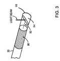
- FIG. 3is a schematic diagram of the optical fiber tip, with micro lens and protective cover
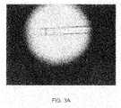
- FIG. 3 ais a photomicrograph of an embodiment of the probe shown schematically in FIG. 3 .
- FIGS. 4 a and 4 bdepict an image taken with a doped plastic lens cover and an undoped plastic (such as PET) cover, respectively;
- FIG. 4 cdepicts an OCT image in which an over-concentration of the dopant TiO 2 is used and the resulting clumping leads to optical shadowing;
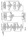
- FIG. 5is a flow chart of an embodiment of an algorithm used to detect the PET ring
- FIG. 6is a flow chart of an embodiment of an algorithm used to set the location of the PET ring
- FIG. 7depicts an image of a coronary artery made using the components in FIGS. 1 and 2 in which the zero-point offset is set correctly;
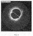
- FIG. 8depicts an image of a coronary artery made using the components in FIGS. 1 and 2 in which the zero-point offset is set incorrectly (the zero-point is too short and has been moved within the optical fiber causing the image to expand away from the center);
- FIG. 9depicts an image of a coronary artery made using the components in FIGS. 1 and 2 in which the zero-point offset is set incorrectly (the zero-point is too long and has moved to a point outside the fiber, causing the image to contract towards the center);
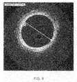
- FIG. 10depicts a magnified OCT image of a catheter center showing characteristic ring reflections according to an embodiment of the invention
- a generalized OCT interferometer 10is shown which is suitable for use with the catheter imaging system of the invention.
- a light source 14such as a diode laser, produces short-coherence length light that passes by way of an optical fiber 18 into an optical fiber coupler 22 .
- Light entering the coupler 22is the split along two optical fiber paths 26 and 30 .
- One path 26terminates at a movable reflector 34 , while the other enters a probe 38 and is emitted toward an object of interest 42 .
- Light reflected by the movable reflector 34passes back along optical fiber 26 to the coupler 22 .
- light reflected by the object of interest 42passes back along optical fiber 30 to the coupler 22 and combines with the light reflected by the movable reflector 34 to form an interference pattern.
- This combined lightpasses through optical fiber 46 and is detected by a detector 50 .
- the output signal from the detector 50is processed by electronics 54 and an image formed on display 58 .
- FIG. 2An example of an imaging catheter suitable for use as the probe in FIG. 1 is shown in FIG. 2 .
- the imaging probe 38is the tip of a coronary vessel imaging catheter.
- a connector 62attaches to the optical coupler 22 of the system of FIG. 1 .
- the optical fiber 30enters a y-body connector 66 attached to a balloon catheter 70 .
- This catheter 70includes a flushing port 74 and a balloon inflation port 78 as well as a flush exit 82 .
- FIG. 3depicts an embodiment of the image wire tip of the probe 38 .
- the optical fiber 30terminates in a micro-lens assembly 86 which focuses the light at a distance from the micro-lens assembly 86 .
- Light emitted from the micro-lens assembly 86is reflected by a beam deflector 90 so to as to pass at substantially right angles to the optical axis of the fiber 30 .
- the entire fiber assemblyis covered by a protective transparent sheath 94 sealed at one end 98 as discussed below.
- a particularly advantageous fiber lens designuses total internal reflection from a silica-air interface to provide the needed radial scan inside a lumen, such as an artery, by simply rotating the fiber. Since the total internal reflection depends on the refractive index mismatch between the silica and air, direct immersion in a fluid will eliminate this reflection and the light will propagate forward instead, destroying the ability to take useful radial scan.
- the air-silica interfacecan be preserved by using a clear protective cover 94 adhered directly to the fiber lens assembly.
- a covercan be made preferably from a heat-shrinkable material such as polyester (Polyethylene Terepthalate, or PET). PET is widely used in industry and medical devices and has good biomedical compatibility properties.
- Such a PET coverhas inherent low back-reflection, so in its usual format it is unsuitable for the purpose of providing a fixed reference reflection.
- dopantscan be added to the raw PET material (before the tube shape is formed) increasing the native back-reflection.
- TiO 2titanium dioxide
- TiO 2is used in many paint formulations due to its excellent light scattering properties. Further it is inert and can be made in bulk. The particle size can be made much smaller than the optical wavelengths of interest (nominally 1.3 ⁇ m), making the scattering ‘Rayleigh’ in nature. Thus the outgoing and returning light wavefronts are not appreciably disturbed, thereby minimizing any potential image degradation at sufficiently low concentrations of dopant.
- a key step in the creation of the materialis uniformly mixing TiO 2 particles in the raw PET such that, when drawing the PET into tubing, the correct concentration is realized with high uniformity.
- OCT imaginghas tremendous sensitivity and large dynamic range (typically 100 dB of sensitivity and >60 dB of dynamic range can be achieved in practical instruments) care must be used to calculate then achieve the optimal doping level of TiO 2 in the material.
- Basic scattering theorycan be used to arrive at a doping concentration in the material.
- the minimum noise in the instrumentis about ⁇ 100 dB. That is, about 1 ten-billionth of the optical output power applied to the object of interest and a typical image has approximately 40 dB of useful dynamic range.
- the image processing electronics and softwareare optimized for this range, so the probe reflector element should be optimized to be near the maximum detectable peak of the image intensity, which is about ⁇ 60 dB ( ⁇ 100+40). This means that the probe reflector should be the brightest object in the image.
- the probe reflector elementcan include, but is not limited to, a membrane, a film, a cap, a cover, or other material.
- the reflector elementis flexible or inflexible.
- the reflector elementcan be shaped in various geometries, such that portions of the reflector are curved, planar, or substantially planar.
- L Ris the return light fraction
- ⁇ bis the scattering cross-section (calculated from standard MIE theory)
- V iis the volume of the particle
- l cis the interaction length (from Radar theory), in this case the coherence length of the OCT light
- ⁇is acceptance angle (solid angle) of the micro-lens.
- the total light returned from the probe reference reflector element materialshould be equal to the single particle light fraction times the volume fraction (doping concentration). Because this should be equal to about ⁇ 60 dB (from above), a reduction of ⁇ 30 dB (or 0.001) is required. Therefore, the volume fraction should be about 0.001, or about 0.1% doping concentration by volume. This should result in a strong, but not overpowering reference reflection by the TiO 2 particles.
- the doped PET materialproduces a consistent, bright ring in the image as shown in FIG. 4 a , as compared to an undoped cover FIG. 4 b .
- the zero point offset positionis unstable, either by purposely modifying the reference path length or through stretching or compressing of the fibers during normal use, the ring is more of a spiral shape. If the concentration of TiO 2 particles is too high, the particles cast shadows due to clumping as shown in FIG. 4 c .
- the probe reflector elementis a membrane that is capable of transmitting substantially undistorted imaging light.
- substantially undistorted imaging lightmeans light that is suitable for generating an image of a sample or a sample element.
- the captured data in ‘raw’ formatis a series of radial scans, each occurring at evenly spaced angles, very much like the spokes in bicycle wheel.
- the raw datais stored simply in a conventional array memory format, where columns represent angles, and each row is a particular radial distance.
- the image of a perfect circle stored in memorywould occur along the same row for each column, i.e. a straight line with zero (flat) slope.
- a spiral patternis stored as a straight line with a slope, positive if the spiral is expanding, negative if the spiral is contracting.
- the signal from the PET materialproduces a line in the image that may have a flat, positive or negative slope depending on whether the optical path length is constant, increasing or decreasing.
- the magnitude of the slopeis then proportional to the rate of change of the fiber path length in either direction due to stretching or shrinking. Because the zero point offset position is now detectable, a software algorithm can be used to isolate the PET ring by taking advantage of its bright reflection, known thickness and expected straight line representation in memory.
- the basic steps of the algorithmare shown in FIG. 5 .
- the OCT imageis obtained (Step 1 ) and first analyzed on a statistical basis. This analysis calculates the number of pixels for each given intensity value. The histogram is then used to generate a “Global Threshold” value to separate the foreground tissue from background noise (Step 2 ). Because the image intensity will eventually fall to the background noise level, the intensity at large radial distances can be used to estimate the overall ‘noise-floor’ of the system. This value is then be used to produce a binary image (Step 3 ). Intensity values above the threshold are set to one; those below the threshold are set to zero. By analyzing the binary image and not the input OCT image, the dependence on the absolute level of the doped PET reflection is minimized.
- Step 4a one dimensional spatial filter that is designed to have peak response for a signal with thickness similar to the known PET layer thickness and adjacent black space. As shown in the figure, the influence of the tissue is greatly minimized by the spatial filter, while the PET ring is preserved.
- the next step in the processis to average all of the scan lines in each quadrant of the filtered binary image together to produce one representative scan line per quadrant.
- This processserves to emphasize image content that is concentric or nearly concentric.
- the averageis performed on a quadrant basis, as opposed to the full 360 degrees, so that the PET signal from a moving reference path (which would be spiral shaped) is not lost in the summation process.
- the resulting four average linesare each smoothed with a simple boxcar filter, and the brightest three peaks on each are located.
- Step 6the peak from each quadrant's average line is selected that together produces the best ring.
- a recursive algorithmis used to analyze each potential group by first computing the sum of the four points and then the mean square error (MSE) of a line fitted to the points using a least squares fit algorithm. The resulting MSE is combined with the sum of the four points to form a score. This score serves to emphasize potential rings that are bright (larger sum) and flat (smaller MSE). The group with the largest score is chosen as the winner and its sum is compared to a cutoff to determine if the result is valid.
- MSEmean square error
- an initial coarse calibrationis performed by rotating the fiber and adjusting the reference path control motor as shown in FIG. 6 .
- the z-offset motor in the reference armis initially swept at high speed (Step 10 ) between its limits while searching for the PET ring. Once the ring is found, the motor speed is slowed (Step 12 ) and the PET image is moved close to its desired location (zero-point, here termed the “Loose Range”).
- the motoris stepped (Step 14 ) until the PET image is in its final allowed range (the “Tight Range”).
- the Z-Offsetmay drift slightly, resulting in the PET moving outside of the tight range.
- the motoris reactivated to step the PET back into the tight range.
- the tight range allowanceis a balance set by the desired measurement accuracy and the minimization of constant z-offset motor movements
- the PET ringas defined by the least squares fitted line of the winning group, is displayed at a fixed location (radius) on the screen based on the known physical location of the PET in the micro-lens assembly.
- the z-offset of each image frameis adjusted in or out so that the PET ring ends up at the desired location.
- FIG. 7is an OCT image of a coronary artery in which the z-offset is set correctly. The vessel diameter is thus correctly measured as 2.55 mm.
- FIG. 8is an OCT image of a coronary artery in which the z-offset is set incorrectly such that the z-offset is positioned within the lens assembly. The vessel diameter is thus incorrectly measured as 2.97 mm.
- FIG. 9is an OCT image of a coronary artery in which the z-offset is set incorrectly such that the z-offset is positioned outside the protective PET cover.
- the vessel diameteris thus incorrectly measured as 2.00 mm.
- the present inventionprovides a method for determining the equalization of the reference and sample paths in an OCT interferometer, to thereby provide an accurate measure of the objects of interest.
- FIG. 10is another OCT image generated in accordance with aspects of the invention. Specifically, it is a magnified OCT image of a catheter center showing characteristic ring reflections arising from the micro-lens PET layer (innermost) 100 and the image wire plastic sheath (middle) 102 .
- the outer ring 104corresponds to the inside wall edge of the plastic tubing into which the image wire was inserted to generate the image depicted in FIG. 10 . However, due to thickness of the tubing, the outer wall edge is not seen in the image.
- the PET ringis generated using standard, un-doped PET. As shown, the image wire is pressed against the side of the tubing thereby causing the third outer ring 104 to be non-concentric.
Landscapes
- Health & Medical Sciences (AREA)
- Life Sciences & Earth Sciences (AREA)
- Physics & Mathematics (AREA)
- Surgery (AREA)
- General Health & Medical Sciences (AREA)
- Engineering & Computer Science (AREA)
- Biomedical Technology (AREA)
- Heart & Thoracic Surgery (AREA)
- Medical Informatics (AREA)
- Molecular Biology (AREA)
- Biophysics (AREA)
- Animal Behavior & Ethology (AREA)
- Pathology (AREA)
- Public Health (AREA)
- Veterinary Medicine (AREA)
- Astronomy & Astrophysics (AREA)
- General Physics & Mathematics (AREA)
- Optics & Photonics (AREA)
- Nuclear Medicine, Radiotherapy & Molecular Imaging (AREA)
- Radiology & Medical Imaging (AREA)
- Investigating Or Analysing Materials By Optical Means (AREA)
Abstract
Description
Claims (19)
Priority Applications (16)
| Application Number | Priority Date | Filing Date | Title |
|---|---|---|---|
| US11/983,526US7813609B2 (en) | 2007-11-12 | 2007-11-12 | Imaging catheter with integrated reference reflector |
| CN201210269037.5ACN102783937B (en) | 2007-11-12 | 2008-11-12 | Optical coherence tomography (OCT) and related imaging method |
| PCT/US2008/012701WO2009064410A2 (en) | 2007-11-12 | 2008-11-12 | Imaging catheter with integrated reference reflector |
| EP16153232.0AEP3045108B1 (en) | 2007-11-12 | 2008-11-12 | Imaging catheter with integrated reference reflector |
| EP08849600AEP2211697A2 (en) | 2007-11-12 | 2008-11-12 | Imaging catheter with integrated reference reflector |
| JP2010533124AJP5093787B2 (en) | 2007-11-12 | 2008-11-12 | Imaging catheter with integrated reference reflector |
| CN2008801155924ACN101854850B (en) | 2007-11-12 | 2008-11-12 | Imaging catheter with integrated reference reflector |
| ES16153232TES2748041T3 (en) | 2007-11-12 | 2008-11-12 | Imaging catheter with integrated reference reflector |
| US12/765,501US8582934B2 (en) | 2007-11-12 | 2010-04-22 | Miniature optical elements for fiber-optic beam shaping |
| US12/886,265US8116605B2 (en) | 2007-11-12 | 2010-09-20 | Imaging catheter with integrated reference reflector |
| US13/298,836US8503844B2 (en) | 2007-11-12 | 2011-11-17 | Imaging catheter with integrated reference reflector |
| JP2012146805AJP5622796B2 (en) | 2007-11-12 | 2012-06-29 | Imaging catheter with integrated reference reflector |
| US13/617,800US9007696B2 (en) | 2007-11-12 | 2012-09-14 | Imaging catheter with integrated reference reflector |
| US14/052,411US9091524B2 (en) | 2007-11-12 | 2013-10-11 | Miniature optical elements for fiber-optic beam shaping |
| US14/744,861US9404731B2 (en) | 2007-11-12 | 2015-06-19 | Miniature optical elements for fiber-optic beam shaping |
| US15/198,581US9864140B2 (en) | 2007-11-12 | 2016-06-30 | Miniature optical elements for fiber-optic beam shaping |
Applications Claiming Priority (1)
| Application Number | Priority Date | Filing Date | Title |
|---|---|---|---|
| US11/983,526US7813609B2 (en) | 2007-11-12 | 2007-11-12 | Imaging catheter with integrated reference reflector |
Related Parent Applications (2)
| Application Number | Title | Priority Date | Filing Date |
|---|---|---|---|
| PCT/US2008/001270Continuation-In-PartWO2008094633A1 (en) | 2007-01-31 | 2008-01-30 | Methods and systems for an electronic device and a stand |
| PCT/US2008/012701Continuation-In-PartWO2009064410A2 (en) | 2007-11-12 | 2008-11-12 | Imaging catheter with integrated reference reflector |
Related Child Applications (2)
| Application Number | Title | Priority Date | Filing Date |
|---|---|---|---|
| US12/765,501Continuation-In-PartUS8582934B2 (en) | 2007-11-12 | 2010-04-22 | Miniature optical elements for fiber-optic beam shaping |
| US12/886,265DivisionUS8116605B2 (en) | 2007-11-12 | 2010-09-20 | Imaging catheter with integrated reference reflector |
Publications (2)
| Publication Number | Publication Date |
|---|---|
| US20090122320A1 US20090122320A1 (en) | 2009-05-14 |
| US7813609B2true US7813609B2 (en) | 2010-10-12 |
Family
ID=40340624
Family Applications (4)
| Application Number | Title | Priority Date | Filing Date |
|---|---|---|---|
| US11/983,526ActiveUS7813609B2 (en) | 2007-11-12 | 2007-11-12 | Imaging catheter with integrated reference reflector |
| US12/886,265ActiveUS8116605B2 (en) | 2007-11-12 | 2010-09-20 | Imaging catheter with integrated reference reflector |
| US13/298,836ActiveUS8503844B2 (en) | 2007-11-12 | 2011-11-17 | Imaging catheter with integrated reference reflector |
| US13/617,800Active2028-05-14US9007696B2 (en) | 2007-11-12 | 2012-09-14 | Imaging catheter with integrated reference reflector |
Family Applications After (3)
| Application Number | Title | Priority Date | Filing Date |
|---|---|---|---|
| US12/886,265ActiveUS8116605B2 (en) | 2007-11-12 | 2010-09-20 | Imaging catheter with integrated reference reflector |
| US13/298,836ActiveUS8503844B2 (en) | 2007-11-12 | 2011-11-17 | Imaging catheter with integrated reference reflector |
| US13/617,800Active2028-05-14US9007696B2 (en) | 2007-11-12 | 2012-09-14 | Imaging catheter with integrated reference reflector |
Country Status (6)
| Country | Link |
|---|---|
| US (4) | US7813609B2 (en) |
| EP (2) | EP3045108B1 (en) |
| JP (2) | JP5093787B2 (en) |
| CN (2) | CN102783937B (en) |
| ES (1) | ES2748041T3 (en) |
| WO (1) | WO2009064410A2 (en) |
Cited By (152)
| Publication number | Priority date | Publication date | Assignee | Title |
|---|---|---|---|---|
| US20110071405A1 (en)* | 2009-09-23 | 2011-03-24 | Lightlab Imaging, Inc. | Apparatus, Systems, and Methods of in-vivo Blood Clearing in a Lumen |
| US20110071404A1 (en)* | 2009-09-23 | 2011-03-24 | Lightlab Imaging, Inc. | Lumen Morphology and Vascular Resistance Measurements Data Collection Systems, Apparatus and Methods |
| US20110151980A1 (en)* | 2009-12-22 | 2011-06-23 | Lightlab Imaging, Inc. | Torque limiter for an oct catheter |
| US20110228280A1 (en)* | 2010-03-17 | 2011-09-22 | Lightlab Imaging, Inc. | Intensity Noise Reduction Methods and Apparatus for Interferometric Sensing and Imaging Systems |
| US8358461B2 (en) | 2008-09-03 | 2013-01-22 | Lightlab Imaging Inc. | Wavelength-tunable light source |
| US8361097B2 (en) | 2008-04-23 | 2013-01-29 | Avinger, Inc. | Catheter system and method for boring through blocked vascular passages |
| US8449468B2 (en) | 2006-11-08 | 2013-05-28 | Lightlab Imaging, Inc. | Opto-acoustic imaging devices and methods |
| US8526472B2 (en) | 2009-09-03 | 2013-09-03 | Axsun Technologies, Inc. | ASE swept source with self-tracking filter for OCT medical imaging |
| US8548571B2 (en) | 2009-12-08 | 2013-10-01 | Avinger, Inc. | Devices and methods for predicting and preventing restenosis |
| US8582109B1 (en) | 2011-08-01 | 2013-11-12 | Lightlab Imaging, Inc. | Swept mode-hopping laser system, methods, and devices for frequency-domain optical coherence tomography |
| US8582619B2 (en) | 2011-03-15 | 2013-11-12 | Lightlab Imaging, Inc. | Methods, systems, and devices for timing control in electromagnetic radiation sources |
| US8644913B2 (en) | 2011-03-28 | 2014-02-04 | Avinger, Inc. | Occlusion-crossing devices, imaging, and atherectomy devices |
| US8670129B2 (en) | 2009-09-03 | 2014-03-11 | Axsun Technologies, Inc. | Filtered ASE swept source for OCT medical imaging |
| US8696695B2 (en) | 2009-04-28 | 2014-04-15 | Avinger, Inc. | Guidewire positioning catheter |
| US8831321B1 (en) | 2011-11-07 | 2014-09-09 | Lightlab Imaging, Inc. | Side branch detection methods, systems and devices |
| WO2014192866A1 (en) | 2013-05-29 | 2014-12-04 | 住友電気工業株式会社 | Catheter for optical coherence tomograph, and catheter production method |
| US8926590B2 (en) | 2009-12-22 | 2015-01-06 | Lightlab Imaging, Inc. | Torque limiter for an OCT catheter |
| US8953911B1 (en) | 2011-10-28 | 2015-02-10 | Lightlab Imaging, Inc. | Spectroscopic imaging probes, devices, and methods |
| US9007696B2 (en) | 2007-11-12 | 2015-04-14 | Lightlab Imaging, Inc. | Imaging catheter with integrated reference reflector |
| US9069396B2 (en) | 2013-03-12 | 2015-06-30 | Lightlab Imaging, Inc. | Controller and user interface device, systems, and methods |
| US9091524B2 (en) | 2007-11-12 | 2015-07-28 | Lightlab Imaging, Inc. | Miniature optical elements for fiber-optic beam shaping |
| US9125562B2 (en) | 2009-07-01 | 2015-09-08 | Avinger, Inc. | Catheter-based off-axis optical coherence tomography imaging system |
| WO2015156945A1 (en) | 2014-04-11 | 2015-10-15 | Jeremy Stigall | Imaging and treatment device |
| US9164240B2 (en) | 2011-03-31 | 2015-10-20 | Lightlab Imaging, Inc. | Optical buffering methods, apparatus, and systems for increasing the repetition rate of tunable light sources |
| US9173591B2 (en) | 2013-03-08 | 2015-11-03 | Lightlab Imaging, Inc. | Stent visualization and malapposition detection systems, devices, and methods |
| US9286673B2 (en) | 2012-10-05 | 2016-03-15 | Volcano Corporation | Systems for correcting distortions in a medical image and methods of use thereof |
| US9292918B2 (en) | 2012-10-05 | 2016-03-22 | Volcano Corporation | Methods and systems for transforming luminal images |
| US9301687B2 (en) | 2013-03-13 | 2016-04-05 | Volcano Corporation | System and method for OCT depth calibration |
| US9307926B2 (en) | 2012-10-05 | 2016-04-12 | Volcano Corporation | Automatic stent detection |
| US9324141B2 (en) | 2012-10-05 | 2016-04-26 | Volcano Corporation | Removal of A-scan streaking artifact |
| US9345406B2 (en) | 2011-11-11 | 2016-05-24 | Avinger, Inc. | Occlusion-crossing devices, atherectomy devices, and imaging |
| US9345398B2 (en) | 2012-05-14 | 2016-05-24 | Avinger, Inc. | Atherectomy catheter drive assemblies |
| US9345510B2 (en) | 2010-07-01 | 2016-05-24 | Avinger, Inc. | Atherectomy catheters with longitudinally displaceable drive shafts |
| US9351698B2 (en) | 2013-03-12 | 2016-05-31 | Lightlab Imaging, Inc. | Vascular data processing and image registration systems, methods, and apparatuses |
| US9360630B2 (en) | 2011-08-31 | 2016-06-07 | Volcano Corporation | Optical-electrical rotary joint and methods of use |
| US9367965B2 (en) | 2012-10-05 | 2016-06-14 | Volcano Corporation | Systems and methods for generating images of tissue |
| US9383263B2 (en) | 2012-12-21 | 2016-07-05 | Volcano Corporation | Systems and methods for narrowing a wavelength emission of light |
| US9453966B2 (en) | 2014-05-16 | 2016-09-27 | Sumitomo Electric Industries, Ltd. | Optical probe |
| US9478940B2 (en) | 2012-10-05 | 2016-10-25 | Volcano Corporation | Systems and methods for amplifying light |
| US9486143B2 (en) | 2012-12-21 | 2016-11-08 | Volcano Corporation | Intravascular forward imaging device |
| US9498247B2 (en) | 2014-02-06 | 2016-11-22 | Avinger, Inc. | Atherectomy catheters and occlusion crossing devices |
| US9498600B2 (en) | 2009-07-01 | 2016-11-22 | Avinger, Inc. | Atherectomy catheter with laterally-displaceable tip |
| US9557156B2 (en) | 2012-05-14 | 2017-01-31 | Avinger, Inc. | Optical coherence tomography with graded index fiber for biological imaging |
| US9592075B2 (en) | 2014-02-06 | 2017-03-14 | Avinger, Inc. | Atherectomy catheters devices having multi-channel bushings |
| US9596993B2 (en) | 2007-07-12 | 2017-03-21 | Volcano Corporation | Automatic calibration systems and methods of use |
| US9610064B2 (en) | 2011-05-31 | 2017-04-04 | Desmond Adler | Multimodal imaging system, apparatus, and methods |
| US9612105B2 (en) | 2012-12-21 | 2017-04-04 | Volcano Corporation | Polarization sensitive optical coherence tomography system |
| US9622706B2 (en) | 2007-07-12 | 2017-04-18 | Volcano Corporation | Catheter for in vivo imaging |
| US9677869B2 (en) | 2012-12-05 | 2017-06-13 | Perimeter Medical Imaging, Inc. | System and method for generating a wide-field OCT image of a portion of a sample |
| US9702762B2 (en) | 2013-03-15 | 2017-07-11 | Lightlab Imaging, Inc. | Calibration and image processing devices, methods, and systems |
| US9709379B2 (en) | 2012-12-20 | 2017-07-18 | Volcano Corporation | Optical coherence tomography system that is reconfigurable between different imaging modes |
| US9730613B2 (en) | 2012-12-20 | 2017-08-15 | Volcano Corporation | Locating intravascular images |
| US9770172B2 (en) | 2013-03-07 | 2017-09-26 | Volcano Corporation | Multimodal segmentation in intravascular images |
| US9788790B2 (en) | 2009-05-28 | 2017-10-17 | Avinger, Inc. | Optical coherence tomography for biological imaging |
| US9833221B2 (en) | 2013-03-15 | 2017-12-05 | Lightlab Imaging, Inc. | Apparatus and method of image registration |
| US9858668B2 (en) | 2012-10-05 | 2018-01-02 | Volcano Corporation | Guidewire artifact removal in images |
| US9854979B2 (en) | 2013-03-15 | 2018-01-02 | Avinger, Inc. | Chronic total occlusion crossing devices with imaging |
| US9867530B2 (en) | 2006-08-14 | 2018-01-16 | Volcano Corporation | Telescopic side port catheter device with imaging system and method for accessing side branch occlusions |
| US9940723B2 (en) | 2014-12-12 | 2018-04-10 | Lightlab Imaging, Inc. | Systems and methods to detect and display endovascular features |
| US9949754B2 (en) | 2011-03-28 | 2018-04-24 | Avinger, Inc. | Occlusion-crossing devices |
| US9996921B2 (en) | 2015-05-17 | 2018-06-12 | LIGHTLAB IMAGING, lNC. | Detection of metal stent struts |
| US10028725B2 (en) | 2013-03-11 | 2018-07-24 | Lightlab Imaging, Inc. | Friction torque limiter for an imaging catheter |
| US10058284B2 (en) | 2012-12-21 | 2018-08-28 | Volcano Corporation | Simultaneous imaging, monitoring, and therapy |
| US10070827B2 (en) | 2012-10-05 | 2018-09-11 | Volcano Corporation | Automatic image playback |
| US10089755B2 (en) | 2015-07-25 | 2018-10-02 | Lightlab Imaging, Inc. | Guidewire detection systems, methods, and apparatuses |
| US10109058B2 (en) | 2015-05-17 | 2018-10-23 | Lightlab Imaging, Inc. | Intravascular imaging system interfaces and stent detection methods |
| US10130259B2 (en) | 2014-02-05 | 2018-11-20 | British Columbia Cancer Agency Branch | Systems for optical imaging of biological tissues |
| US10130386B2 (en) | 2013-07-08 | 2018-11-20 | Avinger, Inc. | Identification of elastic lamina to guide interventional therapy |
| US10140712B2 (en) | 2015-05-17 | 2018-11-27 | Lightlab Imaging, Inc. | Detection of stent struts relative to side branches |
| US10166003B2 (en) | 2012-12-21 | 2019-01-01 | Volcano Corporation | Ultrasound imaging with variable line density |
| US10172582B2 (en) | 2015-11-18 | 2019-01-08 | Lightlab Imaging, Inc. | X-ray image feature detection and registration systems and methods |
| US10191220B2 (en) | 2012-12-21 | 2019-01-29 | Volcano Corporation | Power-efficient optical circuit |
| US10222956B2 (en) | 2015-05-17 | 2019-03-05 | Lightlab Imaging, Inc. | Intravascular imaging user interface systems and methods |
| US10219887B2 (en) | 2013-03-14 | 2019-03-05 | Volcano Corporation | Filters with echogenic characteristics |
| US10219780B2 (en) | 2007-07-12 | 2019-03-05 | Volcano Corporation | OCT-IVUS catheter for concurrent luminal imaging |
| US10226597B2 (en) | 2013-03-07 | 2019-03-12 | Volcano Corporation | Guidewire with centering mechanism |
| US10234676B1 (en) | 2018-01-24 | 2019-03-19 | Canon U.S.A., Inc. | Optical probes with reflecting components for astigmatism correction |
| US10238367B2 (en) | 2012-12-13 | 2019-03-26 | Volcano Corporation | Devices, systems, and methods for targeted cannulation |
| US10292677B2 (en) | 2013-03-14 | 2019-05-21 | Volcano Corporation | Endoluminal filter having enhanced echogenic properties |
| US10307070B2 (en) | 2014-04-04 | 2019-06-04 | St. Jude Medical Coordination Center Bvba | Intravascular pressure and flow data diagnostic systems, devices, and methods |
| US10332228B2 (en) | 2012-12-21 | 2019-06-25 | Volcano Corporation | System and method for graphical processing of medical data |
| US10335173B2 (en) | 2012-09-06 | 2019-07-02 | Avinger, Inc. | Re-entry stylet for catheter |
| US10338795B2 (en) | 2015-07-25 | 2019-07-02 | Lightlab Imaging, Inc. | Intravascular data visualization and interface systems and methods |
| US10357277B2 (en) | 2014-07-08 | 2019-07-23 | Avinger, Inc. | High speed chronic total occlusion crossing devices |
| US10363062B2 (en) | 2011-10-17 | 2019-07-30 | Avinger, Inc. | Atherectomy catheters and non-contact actuation mechanism for catheters |
| US10413317B2 (en) | 2012-12-21 | 2019-09-17 | Volcano Corporation | System and method for catheter steering and operation |
| US10420530B2 (en) | 2012-12-21 | 2019-09-24 | Volcano Corporation | System and method for multipath processing of image signals |
| US10426590B2 (en) | 2013-03-14 | 2019-10-01 | Volcano Corporation | Filters with echogenic characteristics |
| US10453190B2 (en) | 2015-11-23 | 2019-10-22 | Lightlab Imaging, Inc. | Detection of and validation of shadows in intravascular images |
| US10453196B2 (en) | 2015-11-18 | 2019-10-22 | Lightlab Imaging, Inc. | Detection of stent struts relative to side branches |
| US10499813B2 (en) | 2014-09-12 | 2019-12-10 | Lightlab Imaging, Inc. | Methods, systems and apparatus for temporal calibration of an intravascular imaging system |
| WO2020002177A1 (en) | 2018-06-28 | 2020-01-02 | Koninklijke Philips N.V. | Internal ultrasound assisted local therapeutic delivery |
| WO2020002179A1 (en) | 2018-06-28 | 2020-01-02 | Koninklijke Philips N.V. | External targeted delivery of active therapeutic agents |
| US10548478B2 (en) | 2010-07-01 | 2020-02-04 | Avinger, Inc. | Balloon atherectomy catheters with imaging |
| US10561303B2 (en) | 2018-01-24 | 2020-02-18 | Canon U.S.A., Inc. | Optical probes with correction components for astigmatism correction |
| US10568520B2 (en) | 2015-07-13 | 2020-02-25 | Avinger, Inc. | Micro-molded anamorphic reflector lens for image guided therapeutic/diagnostic catheters |
| US10568586B2 (en) | 2012-10-05 | 2020-02-25 | Volcano Corporation | Systems for indicating parameters in an imaging data set and methods of use |
| US10577573B2 (en) | 2017-07-18 | 2020-03-03 | Perimeter Medical Imaging, Inc. | Sample container for stabilizing and aligning excised biological tissue samples for ex vivo analysis |
| US10593037B2 (en) | 2016-04-14 | 2020-03-17 | Lightlab Imaging, Inc. | Method, apparatus, and system to identify branches of a blood vessel |
| US10595820B2 (en) | 2012-12-20 | 2020-03-24 | Philips Image Guided Therapy Corporation | Smooth transition catheters |
| US10606064B2 (en) | 2018-01-24 | 2020-03-31 | Canon U.S.A., Inc. | Optical probes with astigmatism correction |
| US10631754B2 (en) | 2016-05-16 | 2020-04-28 | Lightlab Imaging, Inc. | Intravascular absorbable stent detection and diagnostic methods and systems |
| US10631718B2 (en) | 2015-08-31 | 2020-04-28 | Gentuity, Llc | Imaging system includes imaging probe and delivery devices |
| US10638939B2 (en) | 2013-03-12 | 2020-05-05 | Philips Image Guided Therapy Corporation | Systems and methods for diagnosing coronary microvascular disease |
| US10648918B2 (en) | 2011-08-03 | 2020-05-12 | Lightlab Imaging, Inc. | Systems, methods and apparatus for determining a fractional flow reserve (FFR) based on the minimum lumen area (MLA) and the constant |
| US10646198B2 (en) | 2015-05-17 | 2020-05-12 | Lightlab Imaging, Inc. | Intravascular imaging and guide catheter detection methods and systems |
| US10724082B2 (en) | 2012-10-22 | 2020-07-28 | Bio-Rad Laboratories, Inc. | Methods for analyzing DNA |
| US10758207B2 (en) | 2013-03-13 | 2020-09-01 | Philips Image Guided Therapy Corporation | Systems and methods for producing an image from a rotational intravascular ultrasound device |
| US10792012B2 (en) | 2012-11-19 | 2020-10-06 | Lightlab Imaging, Inc. | Interface devices, systems and methods for multimodal probes |
| US10791923B2 (en) | 2018-09-24 | 2020-10-06 | Canon U.S.A., Inc. | Ball lens for optical probe and methods therefor |
| US10806329B2 (en) | 2018-01-24 | 2020-10-20 | Canon U.S.A., Inc. | Optical probes with optical-correction components |
| US10816789B2 (en) | 2018-01-24 | 2020-10-27 | Canon U.S.A., Inc. | Optical probes that include optical-correction components for astigmatism correction |
| US10905394B2 (en) | 2015-04-20 | 2021-02-02 | Philips Image Guided Therapy Corporation | Dual lumen diagnostic catheter |
| US10932670B2 (en) | 2013-03-15 | 2021-03-02 | Avinger, Inc. | Optical pressure sensor assembly |
| US10939826B2 (en) | 2012-12-20 | 2021-03-09 | Philips Image Guided Therapy Corporation | Aspirating and removing biological material |
| US10942022B2 (en) | 2012-12-20 | 2021-03-09 | Philips Image Guided Therapy Corporation | Manual calibration of imaging system |
| US10993694B2 (en) | 2012-12-21 | 2021-05-04 | Philips Image Guided Therapy Corporation | Rotational ultrasound imaging catheter with extended catheter body telescope |
| US11026591B2 (en) | 2013-03-13 | 2021-06-08 | Philips Image Guided Therapy Corporation | Intravascular pressure sensor calibration |
| US11040140B2 (en) | 2010-12-31 | 2021-06-22 | Philips Image Guided Therapy Corporation | Deep vein thrombosis therapeutic methods |
| US11096717B2 (en) | 2013-03-15 | 2021-08-24 | Avinger, Inc. | Tissue collection device for catheter |
| US11141063B2 (en) | 2010-12-23 | 2021-10-12 | Philips Image Guided Therapy Corporation | Integrated system architectures and methods of use |
| US11154313B2 (en) | 2013-03-12 | 2021-10-26 | The Volcano Corporation | Vibrating guidewire torquer and methods of use |
| US11166668B2 (en) | 2014-07-24 | 2021-11-09 | Lightlab Imaging, Inc. | Pre and post stent planning along with vessel visualization and diagnostic systems, devices, and methods for automatically identifying stent expansion profile |
| US11224459B2 (en) | 2016-06-30 | 2022-01-18 | Avinger, Inc. | Atherectomy catheter with shapeable distal tip |
| US11272845B2 (en) | 2012-10-05 | 2022-03-15 | Philips Image Guided Therapy Corporation | System and method for instant and automatic border detection |
| US11278206B2 (en) | 2015-04-16 | 2022-03-22 | Gentuity, Llc | Micro-optic probes for neurology |
| US11278248B2 (en) | 2016-01-25 | 2022-03-22 | Avinger, Inc. | OCT imaging catheter with lag correction |
| US11284916B2 (en) | 2012-09-06 | 2022-03-29 | Avinger, Inc. | Atherectomy catheters and occlusion crossing devices |
| US11311200B1 (en) | 2014-08-27 | 2022-04-26 | Lightlab Imaging, Inc. | Systems and methods to measure physiological flow in coronary arteries |
| US11344327B2 (en) | 2016-06-03 | 2022-05-31 | Avinger, Inc. | Catheter device with detachable distal end |
| US11344373B2 (en) | 2018-05-29 | 2022-05-31 | Lightlab Imaging, Inc. | Stent expansion display, systems, and methods |
| US11350832B2 (en) | 2014-08-27 | 2022-06-07 | St. Jude Medical Coordination Center Bvba | Cardiac cycle-based diagnostic systems and methods |
| US11382653B2 (en) | 2010-07-01 | 2022-07-12 | Avinger, Inc. | Atherectomy catheter |
| US11399863B2 (en) | 2016-04-01 | 2022-08-02 | Avinger, Inc. | Atherectomy catheter with serrated cutter |
| US11406412B2 (en) | 2012-05-14 | 2022-08-09 | Avinger, Inc. | Atherectomy catheters with imaging |
| US11406498B2 (en) | 2012-12-20 | 2022-08-09 | Philips Image Guided Therapy Corporation | Implant delivery system and implants |
| US11684242B2 (en) | 2017-11-28 | 2023-06-27 | Gentuity, Llc | Imaging system |
| US11793400B2 (en) | 2019-10-18 | 2023-10-24 | Avinger, Inc. | Occlusion-crossing devices |
| US11883107B2 (en) | 2016-09-28 | 2024-01-30 | Lightlab Imaging, Inc. | Stent planning systems and methods using vessel representation obtained via intravascular probe by determining stent effectiveness score and fractional flow reserve |
| US11923067B2 (en) | 2012-12-12 | 2024-03-05 | Lightlab Imaging, Inc. | Method and apparatus for automated determination of stent landing zones based on a maximum diameter of a segmented blood vessel data obtained by intravascular device |
| US12036074B2 (en) | 2017-10-02 | 2024-07-16 | Lightlab Imaging, Inc. | Intravascular data collection probes and related assemblies |
| US12082912B2 (en) | 2009-09-23 | 2024-09-10 | Lightlab Imaging, Inc. | Lumen morphology and vascular resistance measurements data collection systems apparatus and methods |
| US12121206B2 (en) | 2022-02-24 | 2024-10-22 | Canon U.S.A., Inc. | Accurate z-offset calibration for OCT system |
| US12167867B2 (en) | 2018-04-19 | 2024-12-17 | Avinger, Inc. | Occlusion-crossing devices |
| US12193789B2 (en) | 2011-05-27 | 2025-01-14 | Lightlab Imaging, Inc. | Optical coherence tomography and pressure based systems and methods |
| US12201477B2 (en) | 2012-10-05 | 2025-01-21 | Philips Image Guided Therapy Corporation | Methods and systems for establishing parameters for three-dimensional imaging |
| US12239412B2 (en) | 2019-05-21 | 2025-03-04 | Spryte Medical, Inc. | Systems and methods for OCT-guided treatment of a patient |
| US12262872B2 (en) | 2018-09-17 | 2025-04-01 | Gentuity, Llc | Imaging system with optical pathway |
| US12343198B2 (en) | 2013-03-14 | 2025-07-01 | Philips Image Guided Therapy Corporation | Delivery catheter having imaging capabilities |
| USD1083949S1 (en) | 2021-10-15 | 2025-07-15 | Lightlab Imaging, Inc. | Display screen or portion thereof with graphical user interface |
| US12364385B2 (en) | 2019-04-30 | 2025-07-22 | Gentuity, Llc | Imaging probe with fluid pressurization element |
| US12440114B2 (en) | 2023-12-18 | 2025-10-14 | St. Jude Medical Coordination Center Bvba | Cardiac cycle-based diagnostic systems and methods |
Families Citing this family (25)
| Publication number | Priority date | Publication date | Assignee | Title |
|---|---|---|---|---|
| JP5069585B2 (en)* | 2008-02-25 | 2012-11-07 | 富士フイルム株式会社 | Optical tomographic imaging system using an optical probe |
| WO2011084863A2 (en) | 2010-01-07 | 2011-07-14 | Cheetah Omni, Llc | Fiber lasers and mid-infrared light sources in methods and systems for selective biological tissue processing and spectroscopy |
| JP5485760B2 (en)* | 2010-03-26 | 2014-05-07 | テルモ株式会社 | Optical coherence tomographic image forming apparatus and control method thereof |
| JP6153721B2 (en)* | 2012-11-15 | 2017-06-28 | 医療法人社団清友会 | Guide jig |
| CN103239210A (en)* | 2013-05-07 | 2013-08-14 | 深圳市中科微光医疗器械技术有限公司 | Ultra-miniature optical coherence tomography probe for eliminating interference rings |
| JP2015104582A (en)* | 2013-11-29 | 2015-06-08 | 株式会社ニデック | Optical tomographic imaging device and optical tomographic imaging program |
| US10123835B2 (en)* | 2014-04-02 | 2018-11-13 | Covidien Lp | Electrosurgical devices including transverse electrode configurations and methods relating to the same |
| KR101609365B1 (en)* | 2014-05-27 | 2016-04-21 | 주식회사 고영테크놀러지 | Removable optical coherence tomography apparatus |
| WO2016040528A1 (en) | 2014-09-10 | 2016-03-17 | Medical Instrument Development Laboratories, Inc. | Application of highly scattering materials to surgical illumination |
| CN104825118B (en)* | 2015-05-08 | 2017-04-26 | 南京微创医学科技股份有限公司 | Balloon catheter applied to OCT (optical coherence tomography) endoscopic scanning imaging, use method and OCT imaging system |
| KR101638016B1 (en)* | 2015-05-28 | 2016-07-08 | 광주과학기술원 | Endoscope |
| KR101766328B1 (en)* | 2015-05-28 | 2017-08-08 | 광주과학기술원 | Microscope |
| CN107121159A (en)* | 2016-12-23 | 2017-09-01 | 哈尔滨医科大学附属第二医院 | A kind of automatic light path calibration method recognized based on based endoscopic imaging and system |
| CN106691380A (en)* | 2016-12-23 | 2017-05-24 | 哈尔滨医科大学附属第二医院 | OCT probe used for optical path automatic calibration |
| CN107518877A (en)* | 2017-08-25 | 2017-12-29 | 广州永士达医疗科技有限责任公司 | A kind of calibrating installation and method of OCT conduits |
| US20190200854A1 (en)* | 2018-01-03 | 2019-07-04 | Leica Microsystems Inc. | Model Eye Design for Calibrating Imaging Systems and Related Methods, Systems and Devices |
| CN111819417B (en)* | 2018-03-01 | 2022-03-22 | 爱尔康公司 | Common-path waveguide for stabilizing optical coherence tomography imaging |
| JP2021182948A (en) | 2018-06-06 | 2021-12-02 | オリンパス株式会社 | Adhesion structure for endoscope |
| CN110487818B (en)* | 2019-08-27 | 2021-12-10 | 南京品兴科技有限公司 | Detection device, detection system and detection method |
| CN110749553A (en)* | 2019-10-09 | 2020-02-04 | 南京沃福曼医疗科技有限公司 | Method for improving demodulation effect of catheter polarization sensitive optical coherence tomography |
| CN113349737B (en)* | 2021-06-30 | 2023-05-26 | 深圳英美达医疗技术有限公司 | Calibration method of OCT (optical coherence tomography) image of intravascular dual-mode imaging system |
| US11980443B2 (en) | 2021-07-16 | 2024-05-14 | Canon U.S.A., Inc. | Devices, systems, and methods for image synchronization in intracoronary imaging |
| CN114511615A (en)* | 2021-12-31 | 2022-05-17 | 深圳市中科微光医疗器械技术有限公司 | Method and device for calibrating image |
| CN117948883B (en)* | 2024-01-31 | 2024-08-23 | 江苏宇特光电科技股份有限公司 | Fusion power detection method for optical fiber end face |
| CN117958768B (en)* | 2024-03-05 | 2024-12-20 | 深圳大学 | Optical fiber OCT probe integrating distributed temperature sensing function |
Citations (39)
| Publication number | Priority date | Publication date | Assignee | Title |
|---|---|---|---|---|
| US4548473A (en) | 1983-05-12 | 1985-10-22 | Honeywell Inc. | Optical filter |
| JPS63127201A (en) | 1986-11-17 | 1988-05-31 | Matsushita Electric Ind Co Ltd | Color filter |
| US5321501A (en) | 1991-04-29 | 1994-06-14 | Massachusetts Institute Of Technology | Method and apparatus for optical imaging with means for controlling the longitudinal range of the sample |
| US5465147A (en) | 1991-04-29 | 1995-11-07 | Massachusetts Institute Of Technology | Method and apparatus for acquiring images using a ccd detector array and no transverse scanner |
| US5518810A (en) | 1993-06-30 | 1996-05-21 | Mitsubishi Materials Corporation | Infrared ray cutoff material and infrared cutoff powder use for same |
| US5632767A (en) | 1994-09-09 | 1997-05-27 | Rare Earth Medical, Inc. | Loop diffusers for diffusion of optical radiation |
| US5643253A (en) | 1995-06-06 | 1997-07-01 | Rare Earth Medical, Inc. | Phototherapy apparatus with integral stopper device |
| US5748598A (en) | 1995-12-22 | 1998-05-05 | Massachusetts Institute Of Technology | Apparatus and methods for reading multilayer storage media using short coherence length sources |
| US5784352A (en) | 1995-07-21 | 1998-07-21 | Massachusetts Institute Of Technology | Apparatus and method for accessing data on multilayered optical media |
| US5908415A (en) | 1994-09-09 | 1999-06-01 | Rare Earth Medical, Inc. | Phototherapy methods and apparatus |
| US5947959A (en) | 1994-09-09 | 1999-09-07 | Rare Earth Medical, Inc. | Phototherapeutic apparatus with diffusive tip assembly |
| US5956355A (en) | 1991-04-29 | 1999-09-21 | Massachusetts Institute Of Technology | Method and apparatus for performing optical measurements using a rapidly frequency-tuned laser |
| US6111645A (en) | 1991-04-29 | 2000-08-29 | Massachusetts Institute Of Technology | Grating based phase control optical delay line |
| US6134003A (en) | 1991-04-29 | 2000-10-17 | Massachusetts Institute Of Technology | Method and apparatus for performing optical measurements using a fiber optic imaging guidewire, catheter or endoscope |
| US6191862B1 (en) | 1999-01-20 | 2001-02-20 | Lightlab Imaging, Llc | Methods and apparatus for high speed longitudinal scanning in imaging systems |
| US6270492B1 (en) | 1994-09-09 | 2001-08-07 | Cardiofocus, Inc. | Phototherapeutic apparatus with diffusive tip assembly |
| US6348960B1 (en) | 1998-11-06 | 2002-02-19 | Kimotot Co., Ltd. | Front scattering film |
| US6445939B1 (en) | 1999-08-09 | 2002-09-03 | Lightlab Imaging, Llc | Ultra-small optical probes, imaging optics, and methods for using same |
| US6485413B1 (en) | 1991-04-29 | 2002-11-26 | The General Hospital Corporation | Methods and apparatus for forward-directed optical scanning instruments |
| US6501551B1 (en) | 1991-04-29 | 2002-12-31 | Massachusetts Institute Of Technology | Fiber optic imaging endoscope interferometer with at least one faraday rotator |
| US6552796B2 (en) | 2001-04-06 | 2003-04-22 | Lightlab Imaging, Llc | Apparatus and method for selective data collection and signal to noise ratio enhancement using optical coherence tomography |
| US6564087B1 (en) | 1991-04-29 | 2003-05-13 | Massachusetts Institute Of Technology | Fiber optic needle probes for optical coherence tomography imaging |
| US20030091814A1 (en) | 1991-12-21 | 2003-05-15 | Volker Benz | Infrared-reflecting bodies |
| US6570659B2 (en) | 2001-03-16 | 2003-05-27 | Lightlab Imaging, Llc | Broadband light source system and method and light source combiner |
| US20030118302A1 (en)* | 2001-02-16 | 2003-06-26 | James Benjamin F. | Optical fiber including a diffuser portion and continuous sleeve for the transmission of light |
| US20030216720A1 (en) | 2002-05-14 | 2003-11-20 | Cardiofocus, Inc. | Safety shut-off device for laser surgical instruments employing blackbody emitters |
| US20040059397A1 (en) | 1999-07-14 | 2004-03-25 | Sinofsky Edward L. | Intralumenal contact sensor |
| US20040215166A1 (en) | 2003-04-25 | 2004-10-28 | Michael Atlas | Flush catheter with flow directing sheath |
| US6879851B2 (en) | 2001-06-07 | 2005-04-12 | Lightlab Imaging, Llc | Fiber optic endoscopic gastrointestinal probe |
| US6891984B2 (en) | 2002-07-25 | 2005-05-10 | Lightlab Imaging, Llc | Scanning miniature optical probes with optical distortion correction and rotational control |
| US20050128479A1 (en) | 2003-08-14 | 2005-06-16 | Cytonome, Inc. | Optical detector for a particle sorting system |
| US6974557B1 (en)* | 2001-12-18 | 2005-12-13 | Advanced Cardiovasculer Systems, Inc. | Methods for forming an optical window for an intracorporeal device and for joining parts |
| US20060095065A1 (en) | 2004-09-24 | 2006-05-04 | Tetsuaki Tanimura | Fluid occluding devices and methods |
| US20060241503A1 (en) | 2005-02-10 | 2006-10-26 | Lightlab Imaging, Inc. | Optical coherence tomography apparatus and methods |
| US20070015969A1 (en)* | 2005-06-06 | 2007-01-18 | Board Of Regents, The University Of Texas System | OCT using spectrally resolved bandwidth |
| US20080015560A1 (en)* | 2004-11-16 | 2008-01-17 | Biotex, Inc. | Light diffusing tip |
| US20080161696A1 (en) | 2006-11-08 | 2008-07-03 | Lightlab Imaging, Inc. | Opto-acoustic imaging devices and methods |
| US20080165366A1 (en) | 2007-01-10 | 2008-07-10 | Lightlab Imaging, Inc. | Methods and apparatus for swept-source optical coherence tomography |
| US7408648B2 (en) | 2005-05-06 | 2008-08-05 | Siemens Aktiengesellschaft | Method for tomographically displaying a cavity by optical coherence tomography (OCT) and an OCT device for carrying out the method |
Family Cites Families (36)
| Publication number | Priority date | Publication date | Assignee | Title |
|---|---|---|---|---|
| US5509093A (en) | 1993-10-13 | 1996-04-16 | Micron Optics, Inc. | Temperature compensated fiber fabry-perot filters |
| US5548598A (en)* | 1994-03-28 | 1996-08-20 | Motorola | In a data communications systems a method of forward error correction |
| US6069681A (en)* | 1994-11-08 | 2000-05-30 | Fuji Photo Film Co., Ltd. | Device for image shooting both sides of documents |
| US5619368A (en) | 1995-05-16 | 1997-04-08 | Massachusetts Inst. Of Technology | Optical frequency shifter |
| US20020161351A1 (en) | 1998-09-01 | 2002-10-31 | Samson Wilfred J. | Method and apparatus for treating acute myocardial infarction with selective hypothermic perfusion |
| US7180600B2 (en)* | 1998-09-21 | 2007-02-20 | Olympus Corporation | Optical imaging apparatus |
| US6891894B1 (en) | 1999-11-18 | 2005-05-10 | Lg Electronics Inc. | Method for decoding and displaying digital broadcasting signals |
| US7231243B2 (en) | 2000-10-30 | 2007-06-12 | The General Hospital Corporation | Optical methods for tissue analysis |
| US6760112B2 (en)* | 2001-02-17 | 2004-07-06 | Lucent Technologies Inc. | Grin-fiber lens based optical endoscopes |
| US6768756B2 (en) | 2001-03-12 | 2004-07-27 | Axsun Technologies, Inc. | MEMS membrane with integral mirror/lens |
| US6706004B2 (en) | 2001-05-31 | 2004-03-16 | Infraredx, Inc. | Balloon catheter |
| US20100076320A1 (en) | 2003-04-25 | 2010-03-25 | Lightlab Imaging, Llc | Flush catheter with flow directing sheath |
| US20060241493A1 (en)* | 2003-04-28 | 2006-10-26 | Feldman Marc D | Catheter imaging probe and method |
| US8315282B2 (en) | 2005-01-20 | 2012-11-20 | Massachusetts Institute Of Technology | Fourier domain mode locking: method and apparatus for control and improved performance |
| EP2264841B1 (en) | 2005-01-20 | 2016-01-20 | Massachusetts Institute of Technology (MIT) | Mode locking methods and apparatus |
| US7415049B2 (en) | 2005-03-28 | 2008-08-19 | Axsun Technologies, Inc. | Laser with tilted multi spatial mode resonator tuning element |
| AU2006304783A1 (en)* | 2005-10-20 | 2007-04-26 | Board Of Regents, The University Of Texas System | Rotating optical catheter tip for optical coherence tomography |
| JP5044126B2 (en) | 2006-02-23 | 2012-10-10 | オリンパス株式会社 | Endoscope observation apparatus and operation method of endoscope for image formation |
| US9596993B2 (en) | 2007-07-12 | 2017-03-21 | Volcano Corporation | Automatic calibration systems and methods of use |
| US8395781B2 (en) | 2007-07-12 | 2013-03-12 | Volcano Corporation | Automatic calibration systems and methods of use |
| US8582934B2 (en) | 2007-11-12 | 2013-11-12 | Lightlab Imaging, Inc. | Miniature optical elements for fiber-optic beam shaping |
| US7813609B2 (en) | 2007-11-12 | 2010-10-12 | Lightlab Imaging, Inc. | Imaging catheter with integrated reference reflector |
| US20110190586A1 (en) | 2008-03-28 | 2011-08-04 | Volcano Corporation | Methods and systems for intravascular imaging and flushing |
| CN102046071B (en) | 2008-06-02 | 2013-11-06 | 光学实验室成像公司 | Quantitative methods for obtaining tissue characteristics from optical coherence tomography images |
| DE102008045634A1 (en) | 2008-09-03 | 2010-03-04 | Ludwig-Maximilians-Universität München | Wavelength tunable light source |
| AU2009305771B2 (en) | 2008-10-14 | 2013-08-15 | Lightlab Imaging, Inc. | Methods for stent strut detection and related measurement and display using optical coherence tomography |
| EP2742858B1 (en) | 2009-09-23 | 2024-06-05 | Light-Lab Imaging Inc. | Lumen morphology and vascular resistance measurements data collection systems, apparatus and methods |
| CA2765410C (en) | 2009-09-23 | 2015-05-05 | Lightlab Imaging, Inc. | Apparatus, systems, and methods of in-vivo blood clearing in a lumen |
| US8926590B2 (en) | 2009-12-22 | 2015-01-06 | Lightlab Imaging, Inc. | Torque limiter for an OCT catheter |
| US8206377B2 (en) | 2009-12-22 | 2012-06-26 | Lightlab Imaging, Inc. | Torque limiter for an OCT catheter |
| US8478384B2 (en) | 2010-01-19 | 2013-07-02 | Lightlab Imaging, Inc. | Intravascular optical coherence tomography system with pressure monitoring interface and accessories |
| ES2645392T3 (en) | 2010-03-17 | 2017-12-05 | Lightlab Imaging, Inc. | Methods and apparatus for intensity noise reduction for interferometric detection and imaging systems |
| US8582619B2 (en) | 2011-03-15 | 2013-11-12 | Lightlab Imaging, Inc. | Methods, systems, and devices for timing control in electromagnetic radiation sources |
| US9164240B2 (en) | 2011-03-31 | 2015-10-20 | Lightlab Imaging, Inc. | Optical buffering methods, apparatus, and systems for increasing the repetition rate of tunable light sources |
| CN103959043B (en) | 2011-05-31 | 2016-11-02 | 光学实验室成像公司 | Multimodal imaging systems, devices and methods |
| US20130051728A1 (en) | 2011-08-31 | 2013-02-28 | Lightlab Imaging, Inc. | Optical Imaging Probes and Related Methods |
- 2007
- 2007-11-12USUS11/983,526patent/US7813609B2/enactiveActive
- 2008
- 2008-11-12CNCN201210269037.5Apatent/CN102783937B/enactiveActive
- 2008-11-12EPEP16153232.0Apatent/EP3045108B1/enactiveActive
- 2008-11-12EPEP08849600Apatent/EP2211697A2/ennot_activeCeased
- 2008-11-12ESES16153232Tpatent/ES2748041T3/enactiveActive
- 2008-11-12CNCN2008801155924Apatent/CN101854850B/enactiveActive
- 2008-11-12JPJP2010533124Apatent/JP5093787B2/enactiveActive
- 2008-11-12WOPCT/US2008/012701patent/WO2009064410A2/enactiveApplication Filing
- 2010
- 2010-09-20USUS12/886,265patent/US8116605B2/enactiveActive
- 2011
- 2011-11-17USUS13/298,836patent/US8503844B2/enactiveActive
- 2012
- 2012-06-29JPJP2012146805Apatent/JP5622796B2/enactiveActive
- 2012-09-14USUS13/617,800patent/US9007696B2/enactiveActive
Patent Citations (49)
| Publication number | Priority date | Publication date | Assignee | Title |
|---|---|---|---|---|
| US4548473A (en) | 1983-05-12 | 1985-10-22 | Honeywell Inc. | Optical filter |
| JPS63127201A (en) | 1986-11-17 | 1988-05-31 | Matsushita Electric Ind Co Ltd | Color filter |
| US5459570A (en) | 1991-04-29 | 1995-10-17 | Massachusetts Institute Of Technology | Method and apparatus for performing optical measurements |
| US6160826A (en) | 1991-04-29 | 2000-12-12 | Massachusetts Institute Of Technology | Method and apparatus for performing optical frequency domain reflectometry |
| US5465147A (en) | 1991-04-29 | 1995-11-07 | Massachusetts Institute Of Technology | Method and apparatus for acquiring images using a ccd detector array and no transverse scanner |
| US6421164B2 (en) | 1991-04-29 | 2002-07-16 | Massachusetts Institute Of Technology | Interferometeric imaging with a grating based phase control optical delay line |
| US6485413B1 (en) | 1991-04-29 | 2002-11-26 | The General Hospital Corporation | Methods and apparatus for forward-directed optical scanning instruments |
| US6564087B1 (en) | 1991-04-29 | 2003-05-13 | Massachusetts Institute Of Technology | Fiber optic needle probes for optical coherence tomography imaging |
| US5321501A (en) | 1991-04-29 | 1994-06-14 | Massachusetts Institute Of Technology | Method and apparatus for optical imaging with means for controlling the longitudinal range of the sample |
| US6282011B1 (en) | 1991-04-29 | 2001-08-28 | Massachusetts Institute Of Technology | Grating based phase control optical delay line |
| US6501551B1 (en) | 1991-04-29 | 2002-12-31 | Massachusetts Institute Of Technology | Fiber optic imaging endoscope interferometer with at least one faraday rotator |
| US5956355A (en) | 1991-04-29 | 1999-09-21 | Massachusetts Institute Of Technology | Method and apparatus for performing optical measurements using a rapidly frequency-tuned laser |
| US6111645A (en) | 1991-04-29 | 2000-08-29 | Massachusetts Institute Of Technology | Grating based phase control optical delay line |
| US6134003A (en) | 1991-04-29 | 2000-10-17 | Massachusetts Institute Of Technology | Method and apparatus for performing optical measurements using a fiber optic imaging guidewire, catheter or endoscope |
| US20030091814A1 (en) | 1991-12-21 | 2003-05-15 | Volker Benz | Infrared-reflecting bodies |
| US5518810A (en) | 1993-06-30 | 1996-05-21 | Mitsubishi Materials Corporation | Infrared ray cutoff material and infrared cutoff powder use for same |
| US5947959A (en) | 1994-09-09 | 1999-09-07 | Rare Earth Medical, Inc. | Phototherapeutic apparatus with diffusive tip assembly |
| US6270492B1 (en) | 1994-09-09 | 2001-08-07 | Cardiofocus, Inc. | Phototherapeutic apparatus with diffusive tip assembly |
| US5908415A (en) | 1994-09-09 | 1999-06-01 | Rare Earth Medical, Inc. | Phototherapy methods and apparatus |
| US5632767A (en) | 1994-09-09 | 1997-05-27 | Rare Earth Medical, Inc. | Loop diffusers for diffusion of optical radiation |
| US5643253A (en) | 1995-06-06 | 1997-07-01 | Rare Earth Medical, Inc. | Phototherapy apparatus with integral stopper device |
| US5784352A (en) | 1995-07-21 | 1998-07-21 | Massachusetts Institute Of Technology | Apparatus and method for accessing data on multilayered optical media |
| US5748598A (en) | 1995-12-22 | 1998-05-05 | Massachusetts Institute Of Technology | Apparatus and methods for reading multilayer storage media using short coherence length sources |
| US6348960B1 (en) | 1998-11-06 | 2002-02-19 | Kimotot Co., Ltd. | Front scattering film |
| US6191862B1 (en) | 1999-01-20 | 2001-02-20 | Lightlab Imaging, Llc | Methods and apparatus for high speed longitudinal scanning in imaging systems |
| US20040059397A1 (en) | 1999-07-14 | 2004-03-25 | Sinofsky Edward L. | Intralumenal contact sensor |
| US6445939B1 (en) | 1999-08-09 | 2002-09-03 | Lightlab Imaging, Llc | Ultra-small optical probes, imaging optics, and methods for using same |
| US20030118302A1 (en)* | 2001-02-16 | 2003-06-26 | James Benjamin F. | Optical fiber including a diffuser portion and continuous sleeve for the transmission of light |
| US6570659B2 (en) | 2001-03-16 | 2003-05-27 | Lightlab Imaging, Llc | Broadband light source system and method and light source combiner |
| US6552796B2 (en) | 2001-04-06 | 2003-04-22 | Lightlab Imaging, Llc | Apparatus and method for selective data collection and signal to noise ratio enhancement using optical coherence tomography |
| US6879851B2 (en) | 2001-06-07 | 2005-04-12 | Lightlab Imaging, Llc | Fiber optic endoscopic gastrointestinal probe |
| US6974557B1 (en)* | 2001-12-18 | 2005-12-13 | Advanced Cardiovasculer Systems, Inc. | Methods for forming an optical window for an intracorporeal device and for joining parts |
| US20030216720A1 (en) | 2002-05-14 | 2003-11-20 | Cardiofocus, Inc. | Safety shut-off device for laser surgical instruments employing blackbody emitters |
| US6891984B2 (en) | 2002-07-25 | 2005-05-10 | Lightlab Imaging, Llc | Scanning miniature optical probes with optical distortion correction and rotational control |
| US20050201662A1 (en) | 2002-07-25 | 2005-09-15 | Petersen Christopher L. | Scanning miniature optical probes with optical distortion correction and rotational control |
| US7241286B2 (en) | 2003-04-25 | 2007-07-10 | Lightlab Imaging, Llc | Flush catheter with flow directing sheath |
| US20040215166A1 (en) | 2003-04-25 | 2004-10-28 | Michael Atlas | Flush catheter with flow directing sheath |
| US20070260198A1 (en) | 2003-04-25 | 2007-11-08 | Lightlab Imaging, Llc | Flush catheter with flow directing sheath |
| US20050128479A1 (en) | 2003-08-14 | 2005-06-16 | Cytonome, Inc. | Optical detector for a particle sorting system |
| US20060274313A1 (en) | 2003-08-14 | 2006-12-07 | Cytonome, Inc. | Optical detector for a particle sorting system |
| US20080030865A1 (en) | 2003-08-14 | 2008-02-07 | Cytonome, Inc. | Optical detector for a particle sorting system |
| US20080180666A1 (en) | 2003-08-14 | 2008-07-31 | Cytonome, Inc. | Optical detector for a particle sorting system |
| US20060095065A1 (en) | 2004-09-24 | 2006-05-04 | Tetsuaki Tanimura | Fluid occluding devices and methods |
| US20080015560A1 (en)* | 2004-11-16 | 2008-01-17 | Biotex, Inc. | Light diffusing tip |
| US20060241503A1 (en) | 2005-02-10 | 2006-10-26 | Lightlab Imaging, Inc. | Optical coherence tomography apparatus and methods |
| US7408648B2 (en) | 2005-05-06 | 2008-08-05 | Siemens Aktiengesellschaft | Method for tomographically displaying a cavity by optical coherence tomography (OCT) and an OCT device for carrying out the method |
| US20070015969A1 (en)* | 2005-06-06 | 2007-01-18 | Board Of Regents, The University Of Texas System | OCT using spectrally resolved bandwidth |
| US20080161696A1 (en) | 2006-11-08 | 2008-07-03 | Lightlab Imaging, Inc. | Opto-acoustic imaging devices and methods |
| US20080165366A1 (en) | 2007-01-10 | 2008-07-10 | Lightlab Imaging, Inc. | Methods and apparatus for swept-source optical coherence tomography |
Non-Patent Citations (4)
| Title |
|---|
| Annex to Form PCT/ISA206 Communication Relating to the Results of the Partial International Search for International application No. PCT/US2008/012701, mailed on Feb. 27, 2009. |
| International Search Report, International application No. PCT/US2008/012701, mailed on May 14, 2009. |
| PCT International Preliminary Report on Patentability for International Application No. PCT/US2008/12701, issued May 18, 2010. |
| Written Opinion of the International Searching Authority, International application No. PCT/US2008/012701, mailed on May 14, 2009. |
Cited By (263)
| Publication number | Priority date | Publication date | Assignee | Title |
|---|---|---|---|---|
| US9867530B2 (en) | 2006-08-14 | 2018-01-16 | Volcano Corporation | Telescopic side port catheter device with imaging system and method for accessing side branch occlusions |
| US8753281B2 (en) | 2006-11-08 | 2014-06-17 | Lightlab Imaging Inc. | Opto-acoustic imaging devices and methods |
| US8449468B2 (en) | 2006-11-08 | 2013-05-28 | Lightlab Imaging, Inc. | Opto-acoustic imaging devices and methods |
| US10219780B2 (en) | 2007-07-12 | 2019-03-05 | Volcano Corporation | OCT-IVUS catheter for concurrent luminal imaging |
| US9622706B2 (en) | 2007-07-12 | 2017-04-18 | Volcano Corporation | Catheter for in vivo imaging |
| US9596993B2 (en) | 2007-07-12 | 2017-03-21 | Volcano Corporation | Automatic calibration systems and methods of use |
| US11350906B2 (en) | 2007-07-12 | 2022-06-07 | Philips Image Guided Therapy Corporation | OCT-IVUS catheter for concurrent luminal imaging |
| US9404731B2 (en) | 2007-11-12 | 2016-08-02 | Lightlab Imaging, Inc. | Miniature optical elements for fiber-optic beam shaping |
| US9091524B2 (en) | 2007-11-12 | 2015-07-28 | Lightlab Imaging, Inc. | Miniature optical elements for fiber-optic beam shaping |
| US9007696B2 (en) | 2007-11-12 | 2015-04-14 | Lightlab Imaging, Inc. | Imaging catheter with integrated reference reflector |
| US9864140B2 (en) | 2007-11-12 | 2018-01-09 | Lightlab Imaging, Inc. | Miniature optical elements for fiber-optic beam shaping |
| US9572492B2 (en) | 2008-04-23 | 2017-02-21 | Avinger, Inc. | Occlusion-crossing devices, imaging, and atherectomy devices |
| US8361097B2 (en) | 2008-04-23 | 2013-01-29 | Avinger, Inc. | Catheter system and method for boring through blocked vascular passages |
| US9918734B2 (en) | 2008-04-23 | 2018-03-20 | Avinger, Inc. | Catheter system and method for boring through blocked vascular passages |
| US10869685B2 (en) | 2008-04-23 | 2020-12-22 | Avinger, Inc. | Catheter system and method for boring through blocked vascular passages |
| US8810901B2 (en) | 2008-09-03 | 2014-08-19 | Lightlab Imaging, Inc. | Wavelength-tunable light source |
| US8358461B2 (en) | 2008-09-03 | 2013-01-22 | Lightlab Imaging Inc. | Wavelength-tunable light source |
| US11076773B2 (en) | 2009-04-28 | 2021-08-03 | Avinger, Inc. | Guidewire positioning catheter |
| US8696695B2 (en) | 2009-04-28 | 2014-04-15 | Avinger, Inc. | Guidewire positioning catheter |
| US11998311B2 (en) | 2009-04-28 | 2024-06-04 | Avinger, Inc. | Guidewire positioning catheter |
| US9642646B2 (en) | 2009-04-28 | 2017-05-09 | Avinger, Inc. | Guidewire positioning catheter |
| US9788790B2 (en) | 2009-05-28 | 2017-10-17 | Avinger, Inc. | Optical coherence tomography for biological imaging |
| US12178613B2 (en) | 2009-05-28 | 2024-12-31 | Avinger, Inc. | Optical coherence tomography for biological imaging |
| US10342491B2 (en) | 2009-05-28 | 2019-07-09 | Avinger, Inc. | Optical coherence tomography for biological imaging |
| US11839493B2 (en) | 2009-05-28 | 2023-12-12 | Avinger, Inc. | Optical coherence tomography for biological imaging |
| US11284839B2 (en) | 2009-05-28 | 2022-03-29 | Avinger, Inc. | Optical coherence tomography for biological imaging |
| US10052125B2 (en) | 2009-07-01 | 2018-08-21 | Avinger, Inc. | Atherectomy catheter with laterally-displaceable tip |
| US9125562B2 (en) | 2009-07-01 | 2015-09-08 | Avinger, Inc. | Catheter-based off-axis optical coherence tomography imaging system |
| US12089868B2 (en) | 2009-07-01 | 2024-09-17 | Avinger, Inc. | Methods of using atherectomy catheter with deflectable distal tip |
| US12053260B2 (en) | 2009-07-01 | 2024-08-06 | Avinger, Inc. | Catheter-based off-axis optical coherence tomography imaging system |
| US11717314B2 (en) | 2009-07-01 | 2023-08-08 | Avinger, Inc. | Atherectomy catheter with laterally-displaceable tip |
| US9498600B2 (en) | 2009-07-01 | 2016-11-22 | Avinger, Inc. | Atherectomy catheter with laterally-displaceable tip |
| US10729326B2 (en) | 2009-07-01 | 2020-08-04 | Avinger, Inc. | Catheter-based off-axis optical coherence tomography imaging system |
| US9041936B2 (en) | 2009-09-03 | 2015-05-26 | Axsun Technologies, Inc. | ASE swept source with self-tracking filter for OCT medical imaging |
| US9696471B2 (en) | 2009-09-03 | 2017-07-04 | Axsun Technologies Llc | Filtered ASE swept source for OCT medical imaging |
| US8670129B2 (en) | 2009-09-03 | 2014-03-11 | Axsun Technologies, Inc. | Filtered ASE swept source for OCT medical imaging |
| US8526472B2 (en) | 2009-09-03 | 2013-09-03 | Axsun Technologies, Inc. | ASE swept source with self-tracking filter for OCT medical imaging |
| US12138017B2 (en) | 2009-09-23 | 2024-11-12 | Lightlab Imaging, Inc. | Lumen morphology and vascular resistance measurements data collection systems apparatus and methods |
| US12285240B2 (en) | 2009-09-23 | 2025-04-29 | Lightlab Imaging, Inc. | Lumen morphology and vascular resistance measurements data collection systems apparatus and methods |
| US20110071404A1 (en)* | 2009-09-23 | 2011-03-24 | Lightlab Imaging, Inc. | Lumen Morphology and Vascular Resistance Measurements Data Collection Systems, Apparatus and Methods |
| US9138147B2 (en) | 2009-09-23 | 2015-09-22 | Lightlab Imaging, Inc. | Lumen morphology image reconstruction based on the scan line data of OCT |
| US12121325B2 (en) | 2009-09-23 | 2024-10-22 | Lightlab Imaging, Inc. | Lumen morphology and vascular resistance measurements data collection systems apparatus and methods |
| US12402796B2 (en) | 2009-09-23 | 2025-09-02 | Lightlab Imaging, Inc. | Lumen morphology and vascular resistance measurements data collection systems apparatus and methods |
| US9572495B2 (en) | 2009-09-23 | 2017-02-21 | Lightlab Imaging, Inc. | Optical coherence tomography lumen morphology and vascular resistance measurements methods for blood vessel evaluations |
| US8412312B2 (en) | 2009-09-23 | 2013-04-02 | Lightlab Imaging, Inc. | Apparatus, systems, and methods of in-vivo blood clearing in a lumen |
| US20110071405A1 (en)* | 2009-09-23 | 2011-03-24 | Lightlab Imaging, Inc. | Apparatus, Systems, and Methods of in-vivo Blood Clearing in a Lumen |
| US9526424B2 (en) | 2009-09-23 | 2016-12-27 | Lightlab Imaging, Inc. | Apparatus, systems, and methods of in-vivo blood clearing in a lumen |
| US12426789B2 (en) | 2009-09-23 | 2025-09-30 | Lightlab Imaging, Inc. | Blood vessel lumen morphology and minimum lumen area measurements data collection by intravascular imaging systems for stenosis or stent planning |
| US12274532B2 (en) | 2009-09-23 | 2025-04-15 | Lightlab Imaging, Inc. | Lumen morphology and vascular resistance measurements data collection systems apparatus and methods |
| US12082912B2 (en) | 2009-09-23 | 2024-09-10 | Lightlab Imaging, Inc. | Lumen morphology and vascular resistance measurements data collection systems apparatus and methods |
| US8548571B2 (en) | 2009-12-08 | 2013-10-01 | Avinger, Inc. | Devices and methods for predicting and preventing restenosis |
| US8206377B2 (en) | 2009-12-22 | 2012-06-26 | Lightlab Imaging, Inc. | Torque limiter for an OCT catheter |
| US8926590B2 (en) | 2009-12-22 | 2015-01-06 | Lightlab Imaging, Inc. | Torque limiter for an OCT catheter |
| US20110151980A1 (en)* | 2009-12-22 | 2011-06-23 | Lightlab Imaging, Inc. | Torque limiter for an oct catheter |
| US10006753B2 (en) | 2010-03-17 | 2018-06-26 | Lightlab Imaging, Inc. | Intensity noise reduction methods and apparatus for interferometric sensing and imaging systems |
| US20110228280A1 (en)* | 2010-03-17 | 2011-09-22 | Lightlab Imaging, Inc. | Intensity Noise Reduction Methods and Apparatus for Interferometric Sensing and Imaging Systems |
| US8948613B2 (en) | 2010-03-17 | 2015-02-03 | Lightlab Imaging, Inc. | Intensity noise reduction methods and apparatus for interferometric sensing and imaging systems |
| US11382653B2 (en) | 2010-07-01 | 2022-07-12 | Avinger, Inc. | Atherectomy catheter |
| US10349974B2 (en) | 2010-07-01 | 2019-07-16 | Avinger, Inc. | Atherectomy catheters with longitudinally displaceable drive shafts |
| US9345510B2 (en) | 2010-07-01 | 2016-05-24 | Avinger, Inc. | Atherectomy catheters with longitudinally displaceable drive shafts |
| US10548478B2 (en) | 2010-07-01 | 2020-02-04 | Avinger, Inc. | Balloon atherectomy catheters with imaging |
| US11141063B2 (en) | 2010-12-23 | 2021-10-12 | Philips Image Guided Therapy Corporation | Integrated system architectures and methods of use |
| US11040140B2 (en) | 2010-12-31 | 2021-06-22 | Philips Image Guided Therapy Corporation | Deep vein thrombosis therapeutic methods |
| US8582619B2 (en) | 2011-03-15 | 2013-11-12 | Lightlab Imaging, Inc. | Methods, systems, and devices for timing control in electromagnetic radiation sources |
| US8948228B2 (en) | 2011-03-15 | 2015-02-03 | Lightlab Imaging, Inc. | Methods, systems, and devices for timing control in electromagnetic radiation sources |
| US11903677B2 (en) | 2011-03-28 | 2024-02-20 | Avinger, Inc. | Occlusion-crossing devices, imaging, and atherectomy devices |
| US8644913B2 (en) | 2011-03-28 | 2014-02-04 | Avinger, Inc. | Occlusion-crossing devices, imaging, and atherectomy devices |
| US12137931B2 (en) | 2011-03-28 | 2024-11-12 | Avinger, Inc. | Occlusion-crossing devices |
| US11134849B2 (en) | 2011-03-28 | 2021-10-05 | Avinger, Inc. | Occlusion-crossing devices, imaging, and atherectomy devices |
| US9949754B2 (en) | 2011-03-28 | 2018-04-24 | Avinger, Inc. | Occlusion-crossing devices |
| US10952763B2 (en) | 2011-03-28 | 2021-03-23 | Avinger, Inc. | Occlusion-crossing devices |
| US12257029B2 (en) | 2011-03-28 | 2025-03-25 | Avinger, Inc. | Occlusion-crossing devices, imaging, and atherectomy devices |
| US9983356B2 (en) | 2011-03-31 | 2018-05-29 | Lightlab Imaging, Inc. | Optical buffer-based apparatus for increasing light source repetition rate |
| US9164240B2 (en) | 2011-03-31 | 2015-10-20 | Lightlab Imaging, Inc. | Optical buffering methods, apparatus, and systems for increasing the repetition rate of tunable light sources |
| US12193789B2 (en) | 2011-05-27 | 2025-01-14 | Lightlab Imaging, Inc. | Optical coherence tomography and pressure based systems and methods |
| US9610064B2 (en) | 2011-05-31 | 2017-04-04 | Desmond Adler | Multimodal imaging system, apparatus, and methods |
| US11241154B2 (en) | 2011-05-31 | 2022-02-08 | Lightlab Imaging, Inc. | Multimodal imaging system, apparatus, and methods |
| US9488464B1 (en) | 2011-08-01 | 2016-11-08 | Lightlab Imaging, Inc. | Swept mode-hopping laser system, methods, and devices for frequency-domain optical coherence tomography |
| US8582109B1 (en) | 2011-08-01 | 2013-11-12 | Lightlab Imaging, Inc. | Swept mode-hopping laser system, methods, and devices for frequency-domain optical coherence tomography |
| US10648918B2 (en) | 2011-08-03 | 2020-05-12 | Lightlab Imaging, Inc. | Systems, methods and apparatus for determining a fractional flow reserve (FFR) based on the minimum lumen area (MLA) and the constant |
| US9360630B2 (en) | 2011-08-31 | 2016-06-07 | Volcano Corporation | Optical-electrical rotary joint and methods of use |
| US10363062B2 (en) | 2011-10-17 | 2019-07-30 | Avinger, Inc. | Atherectomy catheters and non-contact actuation mechanism for catheters |
| US9435956B1 (en) | 2011-10-28 | 2016-09-06 | Lightlab Imaging, Inc. | Spectroscopic imaging probes, devices, and methods |
| US8953911B1 (en) | 2011-10-28 | 2015-02-10 | Lightlab Imaging, Inc. | Spectroscopic imaging probes, devices, and methods |
| US8831321B1 (en) | 2011-11-07 | 2014-09-09 | Lightlab Imaging, Inc. | Side branch detection methods, systems and devices |
| US11135019B2 (en) | 2011-11-11 | 2021-10-05 | Avinger, Inc. | Occlusion-crossing devices, atherectomy devices, and imaging |
| US12257003B2 (en) | 2011-11-11 | 2025-03-25 | Avinger, Inc. | Occlusion-crossing devices, atherectomy devices, and imaging |
| US9345406B2 (en) | 2011-11-11 | 2016-05-24 | Avinger, Inc. | Occlusion-crossing devices, atherectomy devices, and imaging |
| US9557156B2 (en) | 2012-05-14 | 2017-01-31 | Avinger, Inc. | Optical coherence tomography with graded index fiber for biological imaging |
| US11206975B2 (en) | 2012-05-14 | 2021-12-28 | Avinger, Inc. | Atherectomy catheter drive assemblies |
| US10244934B2 (en) | 2012-05-14 | 2019-04-02 | Avinger, Inc. | Atherectomy catheter drive assemblies |
| US9345398B2 (en) | 2012-05-14 | 2016-05-24 | Avinger, Inc. | Atherectomy catheter drive assemblies |
| US11406412B2 (en) | 2012-05-14 | 2022-08-09 | Avinger, Inc. | Atherectomy catheters with imaging |
| US11647905B2 (en) | 2012-05-14 | 2023-05-16 | Avinger, Inc. | Optical coherence tomography with graded index fiber for biological imaging |
| US10952615B2 (en) | 2012-05-14 | 2021-03-23 | Avinger, Inc. | Optical coherence tomography with graded index fiber for biological imaging |
| US12171407B2 (en) | 2012-05-14 | 2024-12-24 | Avinger, Inc. | Atherectomy catheter drive assemblies |
| US10335173B2 (en) | 2012-09-06 | 2019-07-02 | Avinger, Inc. | Re-entry stylet for catheter |
| US11284916B2 (en) | 2012-09-06 | 2022-03-29 | Avinger, Inc. | Atherectomy catheters and occlusion crossing devices |
| US11272845B2 (en) | 2012-10-05 | 2022-03-15 | Philips Image Guided Therapy Corporation | System and method for instant and automatic border detection |
| US9324141B2 (en) | 2012-10-05 | 2016-04-26 | Volcano Corporation | Removal of A-scan streaking artifact |
| US10568586B2 (en) | 2012-10-05 | 2020-02-25 | Volcano Corporation | Systems for indicating parameters in an imaging data set and methods of use |
| US11510632B2 (en) | 2012-10-05 | 2022-11-29 | Philips Image Guided Therapy Corporation | Systems for indicating parameters in an imaging data set and methods of use |
| US9858668B2 (en) | 2012-10-05 | 2018-01-02 | Volcano Corporation | Guidewire artifact removal in images |
| US9307926B2 (en) | 2012-10-05 | 2016-04-12 | Volcano Corporation | Automatic stent detection |
| US9286673B2 (en) | 2012-10-05 | 2016-03-15 | Volcano Corporation | Systems for correcting distortions in a medical image and methods of use thereof |
| US10070827B2 (en) | 2012-10-05 | 2018-09-11 | Volcano Corporation | Automatic image playback |
| US9478940B2 (en) | 2012-10-05 | 2016-10-25 | Volcano Corporation | Systems and methods for amplifying light |
| US11864870B2 (en) | 2012-10-05 | 2024-01-09 | Philips Image Guided Therapy Corporation | System and method for instant and automatic border detection |
| US11890117B2 (en) | 2012-10-05 | 2024-02-06 | Philips Image Guided Therapy Corporation | Systems for indicating parameters in an imaging data set and methods of use |
| US9292918B2 (en) | 2012-10-05 | 2016-03-22 | Volcano Corporation | Methods and systems for transforming luminal images |
| US12201477B2 (en) | 2012-10-05 | 2025-01-21 | Philips Image Guided Therapy Corporation | Methods and systems for establishing parameters for three-dimensional imaging |
| US9367965B2 (en) | 2012-10-05 | 2016-06-14 | Volcano Corporation | Systems and methods for generating images of tissue |
| US10724082B2 (en) | 2012-10-22 | 2020-07-28 | Bio-Rad Laboratories, Inc. | Methods for analyzing DNA |
| US12127882B2 (en) | 2012-11-19 | 2024-10-29 | Lightlab Imaging, Inc. | Multimodal imaging systems probes and methods |
| US12127881B2 (en) | 2012-11-19 | 2024-10-29 | Lightlab Imaging, Inc. | Interface devices, systems and methods for multimodal probes |
| US10792012B2 (en) | 2012-11-19 | 2020-10-06 | Lightlab Imaging, Inc. | Interface devices, systems and methods for multimodal probes |
| US11701089B2 (en) | 2012-11-19 | 2023-07-18 | Lightlab Imaging, Inc. | Multimodal imaging systems, probes and methods |
| US10359271B2 (en) | 2012-12-05 | 2019-07-23 | Perimeter Medical Imaging, Inc. | System and method for tissue differentiation in imaging |
| US9677869B2 (en) | 2012-12-05 | 2017-06-13 | Perimeter Medical Imaging, Inc. | System and method for generating a wide-field OCT image of a portion of a sample |
| US11923067B2 (en) | 2012-12-12 | 2024-03-05 | Lightlab Imaging, Inc. | Method and apparatus for automated determination of stent landing zones based on a maximum diameter of a segmented blood vessel data obtained by intravascular device |
| US10238367B2 (en) | 2012-12-13 | 2019-03-26 | Volcano Corporation | Devices, systems, and methods for targeted cannulation |
| US11406498B2 (en) | 2012-12-20 | 2022-08-09 | Philips Image Guided Therapy Corporation | Implant delivery system and implants |
| US11892289B2 (en) | 2012-12-20 | 2024-02-06 | Philips Image Guided Therapy Corporation | Manual calibration of imaging system |
| US10942022B2 (en) | 2012-12-20 | 2021-03-09 | Philips Image Guided Therapy Corporation | Manual calibration of imaging system |
| US9709379B2 (en) | 2012-12-20 | 2017-07-18 | Volcano Corporation | Optical coherence tomography system that is reconfigurable between different imaging modes |
| US9730613B2 (en) | 2012-12-20 | 2017-08-15 | Volcano Corporation | Locating intravascular images |
| US10939826B2 (en) | 2012-12-20 | 2021-03-09 | Philips Image Guided Therapy Corporation | Aspirating and removing biological material |
| US11141131B2 (en) | 2012-12-20 | 2021-10-12 | Philips Image Guided Therapy Corporation | Smooth transition catheters |
| US10595820B2 (en) | 2012-12-20 | 2020-03-24 | Philips Image Guided Therapy Corporation | Smooth transition catheters |
| US10420530B2 (en) | 2012-12-21 | 2019-09-24 | Volcano Corporation | System and method for multipath processing of image signals |
| US9486143B2 (en) | 2012-12-21 | 2016-11-08 | Volcano Corporation | Intravascular forward imaging device |
| US11786213B2 (en) | 2012-12-21 | 2023-10-17 | Philips Image Guided Therapy Corporation | System and method for multipath processing of image signals |
| US10993694B2 (en) | 2012-12-21 | 2021-05-04 | Philips Image Guided Therapy Corporation | Rotational ultrasound imaging catheter with extended catheter body telescope |
| US10166003B2 (en) | 2012-12-21 | 2019-01-01 | Volcano Corporation | Ultrasound imaging with variable line density |
| US10058284B2 (en) | 2012-12-21 | 2018-08-28 | Volcano Corporation | Simultaneous imaging, monitoring, and therapy |
| US10413317B2 (en) | 2012-12-21 | 2019-09-17 | Volcano Corporation | System and method for catheter steering and operation |
| US9383263B2 (en) | 2012-12-21 | 2016-07-05 | Volcano Corporation | Systems and methods for narrowing a wavelength emission of light |
| US9612105B2 (en) | 2012-12-21 | 2017-04-04 | Volcano Corporation | Polarization sensitive optical coherence tomography system |
| US10332228B2 (en) | 2012-12-21 | 2019-06-25 | Volcano Corporation | System and method for graphical processing of medical data |
| US11253225B2 (en) | 2012-12-21 | 2022-02-22 | Philips Image Guided Therapy Corporation | System and method for multipath processing of image signals |
| US10191220B2 (en) | 2012-12-21 | 2019-01-29 | Volcano Corporation | Power-efficient optical circuit |
| US9770172B2 (en) | 2013-03-07 | 2017-09-26 | Volcano Corporation | Multimodal segmentation in intravascular images |
| US10226597B2 (en) | 2013-03-07 | 2019-03-12 | Volcano Corporation | Guidewire with centering mechanism |
| US9173591B2 (en) | 2013-03-08 | 2015-11-03 | Lightlab Imaging, Inc. | Stent visualization and malapposition detection systems, devices, and methods |
| US10028725B2 (en) | 2013-03-11 | 2018-07-24 | Lightlab Imaging, Inc. | Friction torque limiter for an imaging catheter |
| US10331099B2 (en) | 2013-03-12 | 2019-06-25 | Lightlab Imaging, Inc. | Controller and user interface device, systems, and methods |
| US11154313B2 (en) | 2013-03-12 | 2021-10-26 | The Volcano Corporation | Vibrating guidewire torquer and methods of use |
| US9907527B2 (en) | 2013-03-12 | 2018-03-06 | Lightlab Imaging, Inc. | Vascular data processing and image registration systems, methods, and apparatuses |
| US12350018B2 (en) | 2013-03-12 | 2025-07-08 | Philips Image Guided Therapy Corporation | Systems and methods for diagnosing coronary microvascular disease |
| US10687777B2 (en) | 2013-03-12 | 2020-06-23 | Lightlab Imaging, Inc. | Vascular data processing and image registration systems, methods, and apparatuses |
| US10638939B2 (en) | 2013-03-12 | 2020-05-05 | Philips Image Guided Therapy Corporation | Systems and methods for diagnosing coronary microvascular disease |
| US9351698B2 (en) | 2013-03-12 | 2016-05-31 | Lightlab Imaging, Inc. | Vascular data processing and image registration systems, methods, and apparatuses |
| US9989945B2 (en) | 2013-03-12 | 2018-06-05 | Lightlab Imaging, Inc. | Controller and user interface device, systems, and methods |
| US9069396B2 (en) | 2013-03-12 | 2015-06-30 | Lightlab Imaging, Inc. | Controller and user interface device, systems, and methods |
| US10758207B2 (en) | 2013-03-13 | 2020-09-01 | Philips Image Guided Therapy Corporation | Systems and methods for producing an image from a rotational intravascular ultrasound device |
| US9301687B2 (en) | 2013-03-13 | 2016-04-05 | Volcano Corporation | System and method for OCT depth calibration |
| US11026591B2 (en) | 2013-03-13 | 2021-06-08 | Philips Image Guided Therapy Corporation | Intravascular pressure sensor calibration |
| US10426590B2 (en) | 2013-03-14 | 2019-10-01 | Volcano Corporation | Filters with echogenic characteristics |
| US10219887B2 (en) | 2013-03-14 | 2019-03-05 | Volcano Corporation | Filters with echogenic characteristics |
| US10292677B2 (en) | 2013-03-14 | 2019-05-21 | Volcano Corporation | Endoluminal filter having enhanced echogenic properties |
| US12343198B2 (en) | 2013-03-14 | 2025-07-01 | Philips Image Guided Therapy Corporation | Delivery catheter having imaging capabilities |
| US9854979B2 (en) | 2013-03-15 | 2018-01-02 | Avinger, Inc. | Chronic total occlusion crossing devices with imaging |
| US11096717B2 (en) | 2013-03-15 | 2021-08-24 | Avinger, Inc. | Tissue collection device for catheter |
| US10551251B2 (en) | 2013-03-15 | 2020-02-04 | Lightlab Imaging, Inc. | Calibration and image processing devices, methods, and systems |
| US11723538B2 (en) | 2013-03-15 | 2023-08-15 | Avinger, Inc. | Optical pressure sensor assembly |
| US9833221B2 (en) | 2013-03-15 | 2017-12-05 | Lightlab Imaging, Inc. | Apparatus and method of image registration |
| US11980386B2 (en) | 2013-03-15 | 2024-05-14 | Avinger, Inc. | Tissue collection device for catheter |
| US11435233B2 (en) | 2013-03-15 | 2022-09-06 | Lightlab Imaging, Inc. | Calibration and image processing devices, methods, and systems |
| US9702762B2 (en) | 2013-03-15 | 2017-07-11 | Lightlab Imaging, Inc. | Calibration and image processing devices, methods, and systems |
| US10932670B2 (en) | 2013-03-15 | 2021-03-02 | Avinger, Inc. | Optical pressure sensor assembly |
| US11890076B2 (en) | 2013-03-15 | 2024-02-06 | Avinger, Inc. | Chronic total occlusion crossing devices with imaging |
| US10722121B2 (en) | 2013-03-15 | 2020-07-28 | Avinger, Inc. | Chronic total occlusion crossing devices with imaging |
| WO2014192866A1 (en) | 2013-05-29 | 2014-12-04 | 住友電気工業株式会社 | Catheter for optical coherence tomograph, and catheter production method |
| US11944342B2 (en) | 2013-07-08 | 2024-04-02 | Avinger, Inc. | Identification of elastic lamina to guide interventional therapy |
| US10130386B2 (en) | 2013-07-08 | 2018-11-20 | Avinger, Inc. | Identification of elastic lamina to guide interventional therapy |
| US10806484B2 (en) | 2013-07-08 | 2020-10-20 | Avinger, Inc. | Identification of elastic lamina to guide interventional therapy |
| US10130259B2 (en) | 2014-02-05 | 2018-11-20 | British Columbia Cancer Agency Branch | Systems for optical imaging of biological tissues |
| US9592075B2 (en) | 2014-02-06 | 2017-03-14 | Avinger, Inc. | Atherectomy catheters devices having multi-channel bushings |
| US10470795B2 (en) | 2014-02-06 | 2019-11-12 | Avinger, Inc. | Atherectomy catheters and occlusion crossing devices |
| US9498247B2 (en) | 2014-02-06 | 2016-11-22 | Avinger, Inc. | Atherectomy catheters and occlusion crossing devices |
| US10568655B2 (en) | 2014-02-06 | 2020-02-25 | Avinger, Inc. | Atherectomy catheters devices having multi-channel bushings |
| US10307070B2 (en) | 2014-04-04 | 2019-06-04 | St. Jude Medical Coordination Center Bvba | Intravascular pressure and flow data diagnostic systems, devices, and methods |
| US11559218B2 (en) | 2014-04-04 | 2023-01-24 | St. Jude Medical Coordination Center Bvba | Intravascular pressure and flow data diagnostic systems, devices, and methods |
| WO2015156945A1 (en) | 2014-04-11 | 2015-10-15 | Jeremy Stigall | Imaging and treatment device |
| US9453966B2 (en) | 2014-05-16 | 2016-09-27 | Sumitomo Electric Industries, Ltd. | Optical probe |
| US11931061B2 (en) | 2014-07-08 | 2024-03-19 | Avinger, Inc. | High speed chronic total occlusion crossing devices |
| US10357277B2 (en) | 2014-07-08 | 2019-07-23 | Avinger, Inc. | High speed chronic total occlusion crossing devices |
| US11147583B2 (en) | 2014-07-08 | 2021-10-19 | Avinger, Inc. | High speed chronic total occlusion crossing devices |
| US11166668B2 (en) | 2014-07-24 | 2021-11-09 | Lightlab Imaging, Inc. | Pre and post stent planning along with vessel visualization and diagnostic systems, devices, and methods for automatically identifying stent expansion profile |
| US11883138B2 (en) | 2014-08-27 | 2024-01-30 | St. Jude Medical Coordination Center Bvba | Cardiac cycle-based diagnostic systems and methods |
| US11350832B2 (en) | 2014-08-27 | 2022-06-07 | St. Jude Medical Coordination Center Bvba | Cardiac cycle-based diagnostic systems and methods |
| US11311200B1 (en) | 2014-08-27 | 2022-04-26 | Lightlab Imaging, Inc. | Systems and methods to measure physiological flow in coronary arteries |
| US10499813B2 (en) | 2014-09-12 | 2019-12-10 | Lightlab Imaging, Inc. | Methods, systems and apparatus for temporal calibration of an intravascular imaging system |
| US9940723B2 (en) | 2014-12-12 | 2018-04-10 | Lightlab Imaging, Inc. | Systems and methods to detect and display endovascular features |
| US10878572B2 (en) | 2014-12-12 | 2020-12-29 | Lightlab Imaging, Inc. | Systems and methods to detect and display endovascular features |
| US11461902B2 (en) | 2014-12-12 | 2022-10-04 | Lightlab Imaging, Inc. | Systems and methods to detect and display endovascular features |
| US11278206B2 (en) | 2015-04-16 | 2022-03-22 | Gentuity, Llc | Micro-optic probes for neurology |
| US10905394B2 (en) | 2015-04-20 | 2021-02-02 | Philips Image Guided Therapy Corporation | Dual lumen diagnostic catheter |
| US10713786B2 (en) | 2015-05-17 | 2020-07-14 | Lightlab Imaging, Inc. | Detection of metal stent struts |
| US10140712B2 (en) | 2015-05-17 | 2018-11-27 | Lightlab Imaging, Inc. | Detection of stent struts relative to side branches |
| US11532087B2 (en) | 2015-05-17 | 2022-12-20 | Lightlab Imaging, Inc. | Stent detection methods and imaging system interfaces |
| US10109058B2 (en) | 2015-05-17 | 2018-10-23 | Lightlab Imaging, Inc. | Intravascular imaging system interfaces and stent detection methods |
| US10646198B2 (en) | 2015-05-17 | 2020-05-12 | Lightlab Imaging, Inc. | Intravascular imaging and guide catheter detection methods and systems |
| US11367186B2 (en) | 2015-05-17 | 2022-06-21 | Lightlab Imaging, Inc. | Detection of metal stent struts |
| US10902599B2 (en) | 2015-05-17 | 2021-01-26 | Lightlab Imaging, Inc. | Stent detection methods and imaging system interfaces |
| US9996921B2 (en) | 2015-05-17 | 2018-06-12 | LIGHTLAB IMAGING, lNC. | Detection of metal stent struts |
| US10222956B2 (en) | 2015-05-17 | 2019-03-05 | Lightlab Imaging, Inc. | Intravascular imaging user interface systems and methods |
| US11974830B2 (en) | 2015-07-13 | 2024-05-07 | Avinger, Inc. | Micro-molded anamorphic reflector lens for image guided therapeutic/diagnostic catheters |
| US11033190B2 (en) | 2015-07-13 | 2021-06-15 | Avinger, Inc. | Micro-molded anamorphic reflector lens for image guided therapeutic/diagnostic catheters |
| US11627881B2 (en) | 2015-07-13 | 2023-04-18 | Avinger, Inc. | Micro-molded anamorphic reflector lens for image guided therapeutic/diagnostic catheters |
| US10568520B2 (en) | 2015-07-13 | 2020-02-25 | Avinger, Inc. | Micro-molded anamorphic reflector lens for image guided therapeutic/diagnostic catheters |
| US11768593B2 (en) | 2015-07-25 | 2023-09-26 | Lightlab Imaging, Inc. | Intravascular data visualization and interface systems and methods |
| US10529093B2 (en) | 2015-07-25 | 2020-01-07 | Lightlab Imaging, Inc. | Guidewire detection systems, methods, and apparatuses |
| US10089755B2 (en) | 2015-07-25 | 2018-10-02 | Lightlab Imaging, Inc. | Guidewire detection systems, methods, and apparatuses |
| US11287961B2 (en) | 2015-07-25 | 2022-03-29 | Lightlab Imaging, Inc. | Intravascular data visualization and interface systems and methods |
| US10338795B2 (en) | 2015-07-25 | 2019-07-02 | Lightlab Imaging, Inc. | Intravascular data visualization and interface systems and methods |
| US12232705B2 (en) | 2015-08-31 | 2025-02-25 | Spryte Medical, Inc. | Imaging system includes imaging probe and delivery devices |
| US10631718B2 (en) | 2015-08-31 | 2020-04-28 | Gentuity, Llc | Imaging system includes imaging probe and delivery devices |
| US11583172B2 (en) | 2015-08-31 | 2023-02-21 | Gentuity, Llc | Imaging system includes imaging probe and delivery devices |
| US11064873B2 (en) | 2015-08-31 | 2021-07-20 | Gentuity, Llc | Imaging system includes imaging probe and delivery devices |
| US11937786B2 (en) | 2015-08-31 | 2024-03-26 | Gentuity, Llc | Imaging system includes imaging probe and delivery devices |
| US11020078B2 (en) | 2015-11-18 | 2021-06-01 | Lightlab Imaging, Inc. | X-ray image feature detection and registration systems and methods |
| US12161500B2 (en) | 2015-11-18 | 2024-12-10 | Lightlab Imaging, Inc. | X-ray image feature detection and registration systems and methods |
| US10172582B2 (en) | 2015-11-18 | 2019-01-08 | Lightlab Imaging, Inc. | X-ray image feature detection and registration systems and methods |
| US11633167B2 (en) | 2015-11-18 | 2023-04-25 | Lightlab Imaging, Inc. | X-ray image feature detection and registration systems and methods |
| US10453196B2 (en) | 2015-11-18 | 2019-10-22 | Lightlab Imaging, Inc. | Detection of stent struts relative to side branches |
| US10327726B2 (en) | 2015-11-18 | 2019-06-25 | Lightlab Imaging, Inc. | X-ray image feature detection and registration systems and methods |
| US10342502B2 (en) | 2015-11-18 | 2019-07-09 | Lightlab Imaging, Inc. | X-ray image feature detection and registration systems and methods |
| US11850089B2 (en) | 2015-11-19 | 2023-12-26 | Lightlab Imaging, Inc. | Intravascular imaging and guide catheter detection methods and systems |
| US10453190B2 (en) | 2015-11-23 | 2019-10-22 | Lightlab Imaging, Inc. | Detection of and validation of shadows in intravascular images |
| US11278248B2 (en) | 2016-01-25 | 2022-03-22 | Avinger, Inc. | OCT imaging catheter with lag correction |
| US11957376B2 (en) | 2016-04-01 | 2024-04-16 | Avinger, Inc. | Atherectomy catheter with serrated cutter |
| US11399863B2 (en) | 2016-04-01 | 2022-08-02 | Avinger, Inc. | Atherectomy catheter with serrated cutter |
| US10593037B2 (en) | 2016-04-14 | 2020-03-17 | Lightlab Imaging, Inc. | Method, apparatus, and system to identify branches of a blood vessel |
| US11475560B2 (en) | 2016-04-14 | 2022-10-18 | Lightlab Imaging, Inc. | Method, apparatus, and system to identify branches of a blood vessel |
| US10631754B2 (en) | 2016-05-16 | 2020-04-28 | Lightlab Imaging, Inc. | Intravascular absorbable stent detection and diagnostic methods and systems |
| US12279789B2 (en) | 2016-06-03 | 2025-04-22 | Avinger, Inc. | Catheter device with detachable distal end |
| US11344327B2 (en) | 2016-06-03 | 2022-05-31 | Avinger, Inc. | Catheter device with detachable distal end |
| US12161360B2 (en) | 2016-06-30 | 2024-12-10 | Avinger, Inc. | Atherectomy catheter with shaped distal tip |
| US11224459B2 (en) | 2016-06-30 | 2022-01-18 | Avinger, Inc. | Atherectomy catheter with shapeable distal tip |
| US11883107B2 (en) | 2016-09-28 | 2024-01-30 | Lightlab Imaging, Inc. | Stent planning systems and methods using vessel representation obtained via intravascular probe by determining stent effectiveness score and fractional flow reserve |
| US10894939B2 (en) | 2017-07-18 | 2021-01-19 | Perimeter Medical Imaging, Inc. | Sample container for stabilizing and aligning excised biological tissue samples for ex vivo analysis |
| US10577573B2 (en) | 2017-07-18 | 2020-03-03 | Perimeter Medical Imaging, Inc. | Sample container for stabilizing and aligning excised biological tissue samples for ex vivo analysis |
| US12263047B2 (en) | 2017-10-02 | 2025-04-01 | Lightlab Imaging, Inc. | Intravascular data collection probes and related assemblies |
| US12036074B2 (en) | 2017-10-02 | 2024-07-16 | Lightlab Imaging, Inc. | Intravascular data collection probes and related assemblies |
| US11684242B2 (en) | 2017-11-28 | 2023-06-27 | Gentuity, Llc | Imaging system |
| US10606064B2 (en) | 2018-01-24 | 2020-03-31 | Canon U.S.A., Inc. | Optical probes with astigmatism correction |
| US10816789B2 (en) | 2018-01-24 | 2020-10-27 | Canon U.S.A., Inc. | Optical probes that include optical-correction components for astigmatism correction |
| US10806329B2 (en) | 2018-01-24 | 2020-10-20 | Canon U.S.A., Inc. | Optical probes with optical-correction components |
| US10234676B1 (en) | 2018-01-24 | 2019-03-19 | Canon U.S.A., Inc. | Optical probes with reflecting components for astigmatism correction |
| US10561303B2 (en) | 2018-01-24 | 2020-02-18 | Canon U.S.A., Inc. | Optical probes with correction components for astigmatism correction |
| US12167867B2 (en) | 2018-04-19 | 2024-12-17 | Avinger, Inc. | Occlusion-crossing devices |
| US11344373B2 (en) | 2018-05-29 | 2022-05-31 | Lightlab Imaging, Inc. | Stent expansion display, systems, and methods |
| WO2020002179A1 (en) | 2018-06-28 | 2020-01-02 | Koninklijke Philips N.V. | External targeted delivery of active therapeutic agents |
| WO2020002177A1 (en) | 2018-06-28 | 2020-01-02 | Koninklijke Philips N.V. | Internal ultrasound assisted local therapeutic delivery |
| US12262872B2 (en) | 2018-09-17 | 2025-04-01 | Gentuity, Llc | Imaging system with optical pathway |
| US10791923B2 (en) | 2018-09-24 | 2020-10-06 | Canon U.S.A., Inc. | Ball lens for optical probe and methods therefor |
| US12364385B2 (en) | 2019-04-30 | 2025-07-22 | Gentuity, Llc | Imaging probe with fluid pressurization element |
| US12239412B2 (en) | 2019-05-21 | 2025-03-04 | Spryte Medical, Inc. | Systems and methods for OCT-guided treatment of a patient |
| US11793400B2 (en) | 2019-10-18 | 2023-10-24 | Avinger, Inc. | Occlusion-crossing devices |
| USD1083949S1 (en) | 2021-10-15 | 2025-07-15 | Lightlab Imaging, Inc. | Display screen or portion thereof with graphical user interface |
| US12121206B2 (en) | 2022-02-24 | 2024-10-22 | Canon U.S.A., Inc. | Accurate z-offset calibration for OCT system |
| US12440114B2 (en) | 2023-12-18 | 2025-10-14 | St. Jude Medical Coordination Center Bvba | Cardiac cycle-based diagnostic systems and methods |
Also Published As
| Publication number | Publication date |
|---|---|
| CN101854850A (en) | 2010-10-06 |
| CN101854850B (en) | 2012-09-05 |
| JP5622796B2 (en) | 2014-11-12 |
| US20090122320A1 (en) | 2009-05-14 |
| US20120057157A1 (en) | 2012-03-08 |
| US9007696B2 (en) | 2015-04-14 |
| JP5093787B2 (en) | 2012-12-12 |
| WO2009064410A2 (en) | 2009-05-22 |
| ES2748041T3 (en) | 2020-03-12 |
| EP3045108B1 (en) | 2019-09-25 |
| US20130010303A1 (en) | 2013-01-10 |
| EP2211697A2 (en) | 2010-08-04 |
| US8116605B2 (en) | 2012-02-14 |
| JP2012185182A (en) | 2012-09-27 |
| WO2009064410A3 (en) | 2009-07-09 |
| JP2011503575A (en) | 2011-01-27 |
| EP3045108A1 (en) | 2016-07-20 |
| CN102783937A (en) | 2012-11-21 |
| US8503844B2 (en) | 2013-08-06 |
| CN102783937B (en) | 2014-07-16 |
| US20110007315A1 (en) | 2011-01-13 |
Similar Documents
| Publication | Publication Date | Title |
|---|---|---|
| US7813609B2 (en) | Imaging catheter with integrated reference reflector | |
| US9404731B2 (en) | Miniature optical elements for fiber-optic beam shaping | |
| CN105283901B (en) | Intravascular image data processing system and method for detecting calibration components in blood vessels | |
| JP6968775B2 (en) | Calibration of OCT-NIRAF multi-modality probe | |
| van Leeuwen et al. | Measurement of the axial point spread function in scattering media using single-mode fiber-based optical coherence tomography | |
| US20190133529A1 (en) | Optical Pressure Measurement | |
| KR20170008172A (en) | Image processing apparatus, image processing method, and optical coherence tomography apparatus | |
| JP2015070939A (en) | Measurement system | |
| EP3569136A1 (en) | Probe with a grating arranged inside a drive cable and a method for alignment of optics in an endoscopic probe | |
| CN110680281A (en) | Optical nondestructive detection system for scar blood vessel imaging | |
| US20240125588A1 (en) | NIRAF Calibration Packaging Phantom | |
| US20240122481A1 (en) | NIRAF Calibration Sheath Phantom | |
| Zysk et al. | Needle-probe system for the measurement of tissue refractive index | |
| Dobre | Design considerations for ease of access and maneuverability of OCT imaging platforms in the oral cavity | |
| WO2011105962A1 (en) | Optical coherence imaging device, and method for forming an optical coherence imaging device |
Legal Events
| Date | Code | Title | Description |
|---|---|---|---|
| AS | Assignment | Owner name:LIGHTLAB IMAGING, INC., MASSACHUSETTS Free format text:ASSIGNMENT OF ASSIGNORS INTEREST;ASSIGNORS:PETERSEN, CHRISTOPHER;MCCARTIN, STEPHEN;SCHMITT, JOSEPH M.;AND OTHERS;REEL/FRAME:022306/0069;SIGNING DATES FROM 20071005 TO 20090218 Owner name:LIGHTLAB IMAGING, INC., MASSACHUSETTS Free format text:ASSIGNMENT OF ASSIGNORS INTEREST;ASSIGNORS:PETERSEN, CHRISTOPHER;MCCARTIN, STEPHEN;SCHMITT, JOSEPH M.;AND OTHERS;SIGNING DATES FROM 20071005 TO 20090218;REEL/FRAME:022306/0069 | |
| STCF | Information on status: patent grant | Free format text:PATENTED CASE | |
| FPAY | Fee payment | Year of fee payment:4 | |
| MAFP | Maintenance fee payment | Free format text:PAYMENT OF MAINTENANCE FEE, 8TH YEAR, LARGE ENTITY (ORIGINAL EVENT CODE: M1552) Year of fee payment:8 | |
| MAFP | Maintenance fee payment | Free format text:PAYMENT OF MAINTENANCE FEE, 12TH YEAR, LARGE ENTITY (ORIGINAL EVENT CODE: M1553); ENTITY STATUS OF PATENT OWNER: LARGE ENTITY Year of fee payment:12 |