US7794946B1 - Microarray and uses therefor - Google Patents
Microarray and uses thereforDownload PDFInfo
- Publication number
- US7794946B1 US7794946B1US09/245,615US24561599AUS7794946B1US 7794946 B1US7794946 B1US 7794946B1US 24561599 AUS24561599 AUS 24561599AUS 7794946 B1US7794946 B1US 7794946B1
- Authority
- US
- United States
- Prior art keywords
- antibodies
- kit according
- antibody
- protein
- solid surface
- Prior art date
- Legal status (The legal status is an assumption and is not a legal conclusion. Google has not performed a legal analysis and makes no representation as to the accuracy of the status listed.)
- Expired - Fee Related
Links
Images


Classifications
- C—CHEMISTRY; METALLURGY
- C12—BIOCHEMISTRY; BEER; SPIRITS; WINE; VINEGAR; MICROBIOLOGY; ENZYMOLOGY; MUTATION OR GENETIC ENGINEERING
- C12Q—MEASURING OR TESTING PROCESSES INVOLVING ENZYMES, NUCLEIC ACIDS OR MICROORGANISMS; COMPOSITIONS OR TEST PAPERS THEREFOR; PROCESSES OF PREPARING SUCH COMPOSITIONS; CONDITION-RESPONSIVE CONTROL IN MICROBIOLOGICAL OR ENZYMOLOGICAL PROCESSES
- C12Q1/00—Measuring or testing processes involving enzymes, nucleic acids or microorganisms; Compositions therefor; Processes of preparing such compositions
- C12Q1/68—Measuring or testing processes involving enzymes, nucleic acids or microorganisms; Compositions therefor; Processes of preparing such compositions involving nucleic acids
- C12Q1/6813—Hybridisation assays
- C12Q1/6834—Enzymatic or biochemical coupling of nucleic acids to a solid phase
- C12Q1/6837—Enzymatic or biochemical coupling of nucleic acids to a solid phase using probe arrays or probe chips
- G—PHYSICS
- G01—MEASURING; TESTING
- G01N—INVESTIGATING OR ANALYSING MATERIALS BY DETERMINING THEIR CHEMICAL OR PHYSICAL PROPERTIES
- G01N33/00—Investigating or analysing materials by specific methods not covered by groups G01N1/00 - G01N31/00
- G01N33/48—Biological material, e.g. blood, urine; Haemocytometers
- G01N33/50—Chemical analysis of biological material, e.g. blood, urine; Testing involving biospecific ligand binding methods; Immunological testing
- G01N33/53—Immunoassay; Biospecific binding assay; Materials therefor
- G01N33/543—Immunoassay; Biospecific binding assay; Materials therefor with an insoluble carrier for immobilising immunochemicals
- G01N33/544—Immunoassay; Biospecific binding assay; Materials therefor with an insoluble carrier for immobilising immunochemicals the carrier being organic
- G01N33/548—Carbohydrates, e.g. dextran
- G—PHYSICS
- G01—MEASURING; TESTING
- G01N—INVESTIGATING OR ANALYSING MATERIALS BY DETERMINING THEIR CHEMICAL OR PHYSICAL PROPERTIES
- G01N33/00—Investigating or analysing materials by specific methods not covered by groups G01N1/00 - G01N31/00
- G01N33/48—Biological material, e.g. blood, urine; Haemocytometers
- G01N33/50—Chemical analysis of biological material, e.g. blood, urine; Testing involving biospecific ligand binding methods; Immunological testing
- G01N33/53—Immunoassay; Biospecific binding assay; Materials therefor
- G01N33/543—Immunoassay; Biospecific binding assay; Materials therefor with an insoluble carrier for immobilising immunochemicals
- G01N33/54393—Improving reaction conditions or stability, e.g. by coating or irradiation of surface, by reduction of non-specific binding, by promotion of specific binding
- G—PHYSICS
- G01—MEASURING; TESTING
- G01N—INVESTIGATING OR ANALYSING MATERIALS BY DETERMINING THEIR CHEMICAL OR PHYSICAL PROPERTIES
- G01N33/00—Investigating or analysing materials by specific methods not covered by groups G01N1/00 - G01N31/00
- G01N33/48—Biological material, e.g. blood, urine; Haemocytometers
- G01N33/50—Chemical analysis of biological material, e.g. blood, urine; Testing involving biospecific ligand binding methods; Immunological testing
- G01N33/68—Chemical analysis of biological material, e.g. blood, urine; Testing involving biospecific ligand binding methods; Immunological testing involving proteins, peptides or amino acids
- G01N33/6803—General methods of protein analysis not limited to specific proteins or families of proteins
- G01N33/6845—Methods of identifying protein-protein interactions in protein mixtures
- G—PHYSICS
- G01—MEASURING; TESTING
- G01N—INVESTIGATING OR ANALYSING MATERIALS BY DETERMINING THEIR CHEMICAL OR PHYSICAL PROPERTIES
- G01N33/00—Investigating or analysing materials by specific methods not covered by groups G01N1/00 - G01N31/00
- G01N33/48—Biological material, e.g. blood, urine; Haemocytometers
- G01N33/50—Chemical analysis of biological material, e.g. blood, urine; Testing involving biospecific ligand binding methods; Immunological testing
- G01N33/68—Chemical analysis of biological material, e.g. blood, urine; Testing involving biospecific ligand binding methods; Immunological testing involving proteins, peptides or amino acids
- G01N33/6854—Immunoglobulins
- B—PERFORMING OPERATIONS; TRANSPORTING
- B01—PHYSICAL OR CHEMICAL PROCESSES OR APPARATUS IN GENERAL
- B01J—CHEMICAL OR PHYSICAL PROCESSES, e.g. CATALYSIS OR COLLOID CHEMISTRY; THEIR RELEVANT APPARATUS
- B01J2219/00—Chemical, physical or physico-chemical processes in general; Their relevant apparatus
- B01J2219/00274—Sequential or parallel reactions; Apparatus and devices for combinatorial chemistry or for making arrays; Chemical library technology
- B01J2219/00583—Features relative to the processes being carried out
- B01J2219/00603—Making arrays on substantially continuous surfaces
- B01J2219/00605—Making arrays on substantially continuous surfaces the compounds being directly bound or immobilised to solid supports
- B01J2219/00608—DNA chips
- B—PERFORMING OPERATIONS; TRANSPORTING
- B01—PHYSICAL OR CHEMICAL PROCESSES OR APPARATUS IN GENERAL
- B01J—CHEMICAL OR PHYSICAL PROCESSES, e.g. CATALYSIS OR COLLOID CHEMISTRY; THEIR RELEVANT APPARATUS
- B01J2219/00—Chemical, physical or physico-chemical processes in general; Their relevant apparatus
- B01J2219/00274—Sequential or parallel reactions; Apparatus and devices for combinatorial chemistry or for making arrays; Chemical library technology
- B01J2219/00583—Features relative to the processes being carried out
- B01J2219/00603—Making arrays on substantially continuous surfaces
- B01J2219/00605—Making arrays on substantially continuous surfaces the compounds being directly bound or immobilised to solid supports
- B01J2219/0061—The surface being organic
- B—PERFORMING OPERATIONS; TRANSPORTING
- B01—PHYSICAL OR CHEMICAL PROCESSES OR APPARATUS IN GENERAL
- B01J—CHEMICAL OR PHYSICAL PROCESSES, e.g. CATALYSIS OR COLLOID CHEMISTRY; THEIR RELEVANT APPARATUS
- B01J2219/00—Chemical, physical or physico-chemical processes in general; Their relevant apparatus
- B01J2219/00274—Sequential or parallel reactions; Apparatus and devices for combinatorial chemistry or for making arrays; Chemical library technology
- B01J2219/00583—Features relative to the processes being carried out
- B01J2219/00603—Making arrays on substantially continuous surfaces
- B01J2219/00605—Making arrays on substantially continuous surfaces the compounds being directly bound or immobilised to solid supports
- B01J2219/00612—Making arrays on substantially continuous surfaces the compounds being directly bound or immobilised to solid supports the surface being inorganic
- B—PERFORMING OPERATIONS; TRANSPORTING
- B01—PHYSICAL OR CHEMICAL PROCESSES OR APPARATUS IN GENERAL
- B01J—CHEMICAL OR PHYSICAL PROCESSES, e.g. CATALYSIS OR COLLOID CHEMISTRY; THEIR RELEVANT APPARATUS
- B01J2219/00—Chemical, physical or physico-chemical processes in general; Their relevant apparatus
- B01J2219/00274—Sequential or parallel reactions; Apparatus and devices for combinatorial chemistry or for making arrays; Chemical library technology
- B01J2219/00583—Features relative to the processes being carried out
- B01J2219/00603—Making arrays on substantially continuous surfaces
- B01J2219/00605—Making arrays on substantially continuous surfaces the compounds being directly bound or immobilised to solid supports
- B01J2219/00623—Immobilisation or binding
- B01J2219/00626—Covalent
- B—PERFORMING OPERATIONS; TRANSPORTING
- B01—PHYSICAL OR CHEMICAL PROCESSES OR APPARATUS IN GENERAL
- B01J—CHEMICAL OR PHYSICAL PROCESSES, e.g. CATALYSIS OR COLLOID CHEMISTRY; THEIR RELEVANT APPARATUS
- B01J2219/00—Chemical, physical or physico-chemical processes in general; Their relevant apparatus
- B01J2219/00274—Sequential or parallel reactions; Apparatus and devices for combinatorial chemistry or for making arrays; Chemical library technology
- B01J2219/00583—Features relative to the processes being carried out
- B01J2219/00603—Making arrays on substantially continuous surfaces
- B01J2219/00605—Making arrays on substantially continuous surfaces the compounds being directly bound or immobilised to solid supports
- B01J2219/00632—Introduction of reactive groups to the surface
- B01J2219/00637—Introduction of reactive groups to the surface by coating it with another layer
- B—PERFORMING OPERATIONS; TRANSPORTING
- B01—PHYSICAL OR CHEMICAL PROCESSES OR APPARATUS IN GENERAL
- B01J—CHEMICAL OR PHYSICAL PROCESSES, e.g. CATALYSIS OR COLLOID CHEMISTRY; THEIR RELEVANT APPARATUS
- B01J2219/00—Chemical, physical or physico-chemical processes in general; Their relevant apparatus
- B01J2219/00274—Sequential or parallel reactions; Apparatus and devices for combinatorial chemistry or for making arrays; Chemical library technology
- B01J2219/00583—Features relative to the processes being carried out
- B01J2219/00603—Making arrays on substantially continuous surfaces
- B01J2219/00639—Making arrays on substantially continuous surfaces the compounds being trapped in or bound to a porous medium
- B01J2219/00641—Making arrays on substantially continuous surfaces the compounds being trapped in or bound to a porous medium the porous medium being continuous, e.g. porous oxide substrates
- B—PERFORMING OPERATIONS; TRANSPORTING
- B01—PHYSICAL OR CHEMICAL PROCESSES OR APPARATUS IN GENERAL
- B01J—CHEMICAL OR PHYSICAL PROCESSES, e.g. CATALYSIS OR COLLOID CHEMISTRY; THEIR RELEVANT APPARATUS
- B01J2219/00—Chemical, physical or physico-chemical processes in general; Their relevant apparatus
- B01J2219/00274—Sequential or parallel reactions; Apparatus and devices for combinatorial chemistry or for making arrays; Chemical library technology
- B01J2219/00583—Features relative to the processes being carried out
- B01J2219/00603—Making arrays on substantially continuous surfaces
- B01J2219/00659—Two-dimensional arrays
Definitions
- the invention disclosed hereinrelates to new methods of using microarray technologies.
- the methodsare useful for identifying and characterizing specific antibodies as well as the characterization of different tissues or cells by protein or nucleic acid analysis.
- Antibodies that bind to specific antigenshave been produced by a variety of methods including immunization of animals, fusion of mammalian spleen cells to immortalized cells to produce hybridomas, random peptide generation using phage or bacterial display and constrained peptide libraries. Regardless of how the desired antibody is generated, the methods currently available to identify one with a particular binding specificity are generally laborious and incapable of the simultaneous testing of large numbers of unknowns.
- One methodinvolves binding the antigen to a porous membrane, such as nitrocellulose, contacting the membrane with a source of test antibodies, then determining whether or not any of the test antibodies has bound to the antigen. This method only allows the testing of one source of test antibodies per piece of porous membrane, making the method both inconvenient and wasteful of materials.
- Antibody/antigen reactionscan also be evaluated in plastic plates, such as 96-well microtiter plates, using methods similar to those described above. This method is likewise limited in the number of samples that can be tested in any one assay, thus requiring many assays to fully evaluate a large number of antibody unknowns.
- ChangU.S. Pat. No. 4,591,570, issued May 27, 1986 describes an array of a limited number of characterized antibodies to known antigens on a glass surface that can be used to bind to specific antigens on the surface of whole cells.
- the invention disclosed hereincomprises methods of using microarrays to simplify analysis and characterization of genes and their function.
- the methodsare used to identify and characterize antibodies having binding affinity for a specific target antigen.
- This methodcomprises contacting an array of uncharacterized antibodies bound to a solid surface with at least one target antigen and identifying the antibodies to which the target antigen binds.
- the methodcan be performed under a variety of conditions to identify antibodies with a range of binding affinities.
- a second aspect of the inventioncomprises a method of determining gene expression at the protein level comprising contacting an array of characterized or uncharacterized antibodies on a solid surface with one or more proteins and identifying the antibodies to which said protein(s) binds. This method can be further used to compare the protein expression in two different populations of cells, such as normal cells and cancer cells or resting cells and stimulated cells.
- a related embodimentcan be used as a tool in the diagnosis of various disorders.
- a further aspect of the inventioncomprises a method of determining gene expression at the protein level comprising contacting a microarray of nucleic acid samples derived from a variety of different sources with one or more nucleic acid probes then identifying the sample or samples to which the probe binds.
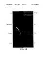
- FIGS. 1A , 1 B, and 1 Cshow microarrays of antibodies bound to positively charged nylon, reacted with antigen and detected by non-fluorescent means.
- FIG. 2shows a microarray produced using a robotic arraying apparatus. Antigen binding is detected by non-fluorescent means.
- FIG. 3shows the ability of the antibody microarrays to evaluate relative binding affinities to a specific antigen.
- FIG. 4shows a microarray of polyclonal antibodies in comparison to a microarray of monoclonal antibodies.
- FIGS. 5A , 5 B and 5 Cshow a microarray of antibodies reacted with a cell lysate under conditions that vary the amount of background binding.
- the present inventiondiscloses methods of using microarrays to simplify analysis and characterization of genes and their function.
- the methodsare used for identifying and characterizing antibodies having binding specificity to a particular antigen or set of antigens.
- This methodutilizes microarray technology to create ordered matrices of large numbers of uncharacterized antibodies which can then be contacted with antigen under a variety of conditions.
- the methodis rapid and simple to perform and is applicable to the simultaneous screening of very large numbers of antibodies.
- uncharacterized antibodiesare bound to a solid surface in an array format consisting of discrete spots whose spatial location can be easily identified. Each location represents an antibody from a known source, such as a particular hybridoma growing in a well in a 96-well microtiter plate. The space between the antibody spots is treated to minimize non-specific binding to the solid support.
- the arrayed antibodiesare then contacted with an antigen, or a set of antigens, for which specific antibodies are sought.
- the antigen solutionis left in contact with the array for an amount of time sufficient to allow antigen:antibody complexes to form (generally 10 minutes to 2 hours), then the unbound antigen is washed away under suitable conditions. Bound antigen is detected at a particular antibody spot using one of a variety of detection methods, thus identifying the source of an antibody specific for the particular antigen.
- antibodyis used herein in the broadest sense and specifically includes intact monoclonal antibodies, polyclonal antibodies, multispecific antibodies (e.g. bispecific antibodies) formed from at least two intact antibodies, and antibody fragments, including single chain antibodies, so long as they exhibit the desired binding properties as described herein.
- a host animal of any of a number of speciessuch as rabbits, goats, sheep, horse, cow, mice, rats, etc. is immunized by injection with an antigenic preparation which may be derived from cells or microorganisms, or may be recombinantly or synthetically produced.
- adjuvantswell known in the art may be used to enhance the production of antibodies by the immunized host, for example, Freund's adjuvant (complete and incomplete), mineral gels such as aluminum hydroxide, surface active substances such as lysolecithin, pluronic polyols, polyanions, peptides, oil emulsions, keyhole limpet hemocyanins, dinitrophenol, liposomes, potentially useful human adjuvants such as BCG (Bacille Calmette-Guerin) and Propionibacterium acanes , and the like.
- BCGBacille Calmette-Guerin
- the term “monoclonal antibody” as used hereinrefers to an antibody obtained from a population of substantially homogeneous antibodies, i.e., the individual antibodies comprising the population are identical except for possible naturally occurring mutations that may be present in minor amounts. Monoclonal antibodies are highly specific, being directed against a single antigenic site. Furthermore, in contrast to conventional (polyclonal) antibody preparations which typically include different antibodies directed against different determinants (epitopes), each monoclonal antibody is directed against a single determinant on the antigen.
- Preferred antibodiesare mAbs, which may be of any immunoglobulin class including IgG, IgM, IgE, IgA, and any subclass or isotype thereof.
- monoclonal antibodiesare advantageous in that they are synthesized by hybridoma culture, uncontaminated by other immunoglobulins.
- the modifier “monoclonal”indicates the character of the antibody as being obtained from a substantially homogeneous population of antibodies, and is not to be construed as requiring production of the antibody by any particular method.
- the monoclonal antibodies to be used in accordance with the present inventionmay be made by the hybridoma method first described by Kohler et al., Nature, 256:495 (1975), or may be made by recombinant DNA methods (see, e.g., U.S. Pat. No. 4,816,567, incorporated by reference herein).
- the “monoclonal antibodies”may also be isolated from phage antibody libraries using the techniques described in Clackson et al., Nature, 352:624-628 (1991) and Marks et al., J. Mol. Biol., 222:581-597 (1991), for example.
- the monoclonal antibodies contemplated for use hereinspecifically include “chimeric” antibodies (immunoglobulins) in which a portion of the heavy and/or light chain is identical with or homologous to corresponding sequences in antibodies derived from a particular species or belonging to a particular antibody class or subclass, while the remainder of the chain(s) is identical with or homologous to corresponding sequences in antibodies derived from another species or belonging to another antibody class or subclass, as well as fragments of such antibodies, so long as they exhibit the desired biological activity (U.S. Pat. No. 4,816,567; Morrison et al., Proc. Natl. Acad. Sci. USA, 81:6851-6855 (1984)).
- chimeric antibodiesimmunoglobulins in which a portion of the heavy and/or light chain is identical with or homologous to corresponding sequences in antibodies derived from a particular species or belonging to a particular antibody class or subclass, while the remainder of the chain(s) is identical with or homologous
- “Humanized” forms of non-human (e.g., murine) antibodiesare chimeric immunoglobulins, immunoglobulin chains or fragments thereof (such as Fv, Fab, Fab′, F(ab′) 2 or other antigen-binding subsequences of antibodies) which contain minimal sequence derived from non-human immunoglobulin.
- humanized antibodiesare human immunoglobulins (recipient antibody) in which residues from a complementarity-determining region (CDR) of the recipient are replaced by residues from a CDR of a non-human species (donor antibody) such as mouse, rat or rabbit having the desired specificity, affinity, and capacity.
- humanized antibodiesmay comprise residues which are found neither in the recipient antibody nor in the imported CDR or framework sequences. These modifications are made to further refine and maximize antibody performance.
- the humanized antibodywill comprise substantially all of at least one, and typically two, variable domains, in which all or substantially all of the CDR regions correspond to those of a non-human immunoglobulin and all or substantially all of the FR regions are those of a human immunoglobulin sequence.
- the humanized antibodyoptimally also will comprise at least a portion of an immunoglobulin constant region (Fc), typically that of a human immunoglobulin.
- the humanized antibodyincludes a PRIMATIZEDTM antibody wherein the antigen-binding region of the antibody is derived from an antibody produced by immunizing macaque monkeys with the antigen of interest.
- Antibody fragmentscomprise a portion of an intact antibody, preferably the antigen binding or variable region of the intact antibody.
- antibody fragmentsinclude Fab, Fab′, F(ab′) 2 , and Fv fragments; diabodies; linear antibodies (Zapata et al. Protein Eng. 8(10):1057-1062 (1995)); single-chain antibody molecules; and multispecific antibodies formed from antibody fragments.
- Single-chain antibodiesare antibody fragments comprising the V H and V L domains of an antibody, wherein these domains are present in a single polypeptide chain.
- the Fv polypeptidefurther comprises a polypeptide linker between the V H and V L domains which enables the sFv to form the desired structure for antigen binding.
- diabodiesrefers to small antibody fragments with two antigen-binding sites, which fragments comprise a heavy-chain variable domain (V H ) connected to a light-chain variable domain (V L ) in the same polypeptide chain (V H -V L ).
- V Hheavy-chain variable domain
- V Llight-chain variable domain
- the domainsare forced to pair with the complementary domains of another chain and create two antigen-binding sites.
- Diabodiesare described more fully in, for example, EP 404,097; WO 93/11161; and Hollinger et al., Proc. Natl. Acad. Sci. USA, 90:6444-6448 (1993).
- the antibodies employed in the inventioncan be isolated prior to creating a microarray.
- An “isolated” moleculewhether an antibody, antigen or nucleic acid, is one which has been identified and separated and/or recovered from a component of its natural environment. Contaminant components of its natural environment are materials which would interfere with particular uses for the molecule, and may include enzymes, hormones, and other proteinaceous or nonproteinaceous solutes.
- a proteinwill be purified (1) to greater than 95% by weight of protein as determined by the Lowry method, and most preferably more than 99% by weight, (2) to a degree sufficient to obtain at least 15 residues of N-terminal or internal amino acid sequence by use of a spinning cup sequenator, or (3) to homogeneity by SDS-PAGE under reducing or nonreducing conditions using Coomassie blue or, preferably, silver stain.
- Isolated proteinincludes the protein in situ within recombinant cells since at least one component of the protein's natural environment will not be present. Ordinarily, however, isolated protein will be prepared by at least one purification step. Unpurified antibodies, such as those found in serum, can also be employed in the present invention.
- isolatedin reference to nucleic acid is meant a polymer of 14, 17, 21 or more contiguous nucleotides, including DNA or RNA that is isolated from a natural source or that is synthesized.
- the isolated nucleic acid of the present inventionis unique in the sense that it is not found in a pure or separated state in nature.
- Use of the term “isolated”indicates that a naturally occurring sequence has been removed from its normal cellular (i.e., chromosomal) environment. Thus, the sequence may be in a cell-free solution or placed in a different cellular environment.
- sequenceis the only nucleotide sequence present, but that it is essentially free (about 90-95% pure at least) of non-nucleotide material naturally associated with it and thus is meant to be distinguished from isolated chromosomes.
- One particularly useful method of isolating antibodiesis affinity purification.
- Resins suitable for antibody purificationare well known in the art, for example, protein A sepharose.
- a recombinant antibodycan be engineered to contain an affinity purification tag to facilitate its purification.
- Resins suitable for antibody purificationare well known in the art, for example, protein A sepharose.
- Affinity purification tagsare generally peptide sequences that can interact with a binding partner immobilized on a solid support. Synthetic DNA sequences encoding multiple consecutive single amino acids, such as histidine, when fused to the expressed protein, may be used for one-step purification of the recombinant protein by high affinity binding to a resin column, such as nickel sepharose. An endopeptidase recognition sequence can be engineered between the polyamino acid tag and the protein of interest to allow subsequent removal of the leader peptide by digestion with enterokinase, and other proteases.
- Sequences encoding peptides such as the chitin binding domain (which binds to chitin), biotin (which binds to avidin and strepavidin), and the likecan also be used for facilitating purification of the protein of interest.
- the affinity purification tagcan be separated from the protein of interest by methods well known in the art, including the use of inteins (protein self-splicing elements, Chong, et al, Gene 192:271-281, 1997).
- the process of isolationcan be used to simultaneously normalize yield and isolate the antibody.
- the samplescan be contacted with an amount of resin whose maximum binding capacity is 10 mgs. Thus any antibody greater than this amount will pass through the resin unbound. The maximum bound amount can then be eluted from the resin.
- microarraysMethods for creating microarrays are known in the art including printing on a solid surface using pins (passive pins, quill pins, and the like) or spotting with individual drops of solution. Passive pins draw up enough sample to dispense a single spot. Quill pins draw up enough liquid to dispense multiple spots. Bubble printers use a loop to capture a small volume which is dispensed by pushing a rod through the loop. Microdispensing uses a syringe mechanism to deliver multiple spots of a fixed volume.
- solid supportscan be arrayed using pizoelectric (ink jet) technology, which actively transfers samples to a solid support.
- Shalon and BrownWO 95/35505, published Dec. 28, 1995 which is incorporated herein by reference in its entirety.
- the method and apparatus described in Shalon and Browncan create an array of up to six hundred spots per square centimeter on a glass slide using a volume of 0.01 to 100 nl per spot. Suitable concentrations of antibody range from about 1 ng/ ⁇ l to about 1 ⁇ g/ ⁇ l.
- each spotcan contain one or more than one distinct antibody.
- microarrayscan be created on a variety of solid surfaces such as plastics (eg. polycarbonate), complex carbohydrates (eg. agarose and sepharose), acrylic resins (eg. polyacrylamide and latex beads), and nitrocellulose.
- plasticseg. polycarbonate
- complex carbohydrateseg. agarose and sepharose
- acrylic resinseg. polyacrylamide and latex beads
- nitrocellulosee.g. polyacrylamide and latex beads
- Preferred solid support materialsinclude glass slides, silicon wafers, and positively charged nylon. Specific examples of suitable solid supports are described in the Examples below.
- BSAbovine serum albumin
- the arrays used to identify antigen-specific antibodiesare contacted with a solution containing one or more known antigens in order to identify antibodies in the array with binding specificity for the antigen.
- the antigensare often proteins, although they may also be organic chemical compounds, carbohydrates, nucleic acids, and the like. They may be isolated or semi-isolated, recombinant or naturally occurring.
- the amount of antigen usedcan vary from about 1-100 ng/ ⁇ l.
- the antigenis left in contact with the array for an amount of time sufficient for antibody:antigen complexes to form, should one of the antibodies in the array be specific for the antigen.
- the amount of time sufficient for this purposewill range from 5 minutes to 24 hours, and will generally be from 0.5 to 2 hours.
- EST fragmentsare relatively short cDNA sequences that have been randomly generated and sequenced, generally as part of an ongoing effort to map an entire genome (Adams, et al, Science 252(5013):1651-1656, 1991). Large numbers of these sequences are available in public databases. The identity of the proteins encoded by the vast majority of these sequences is unknown. The following discussion, although directed to the expression of EST-encoded peptides, is equally applicable to any expressed product of a nucleic acid sequence, including full-length proteins.
- Prokaryotic hostsare, generally, very efficient and convenient for the production of recombinant proteins and are, therefore, one type of preferred expression system for EST fragments. Prokaryotes most frequently are represented by various strains of E. coli . However, other microbial strains may also be used, including other bacterial strains.
- plasmid vectorsthat contain replication sites and control sequences derived from a species compatible with the host may be used.
- suitable plasmid vectorsmay include pBR322, pUC118, pUC119, and the like;
- suitable phage or bacteriophage vectorsmay include ⁇ gt10, ⁇ gt11, and the like;
- suitable virus vectorsmay include pMAM-neo, PKRC and the like.
- the selected vector of the present inventionhas the capacity to replicate in the selected host cell.
- prokaryotic hostsinclude bacteria such as E. coli and those from genera such as Bacillus, Streptomyces, Pseudomonas, Salmonella, Serratia , and the like. However, under such conditions, the polypeptide will not be glycosylated.
- the prokaryotic host selected for use hereinmust be compatible with the replicon and control sequences in the expression plasmid.
- a functional prokaryotic promotersuch as the T7 promoter or RSC promoter.
- Such promotersmay be either constitutive or, more preferably, regulatable (i.e., inducible or derepressible).
- constitutive promotersinclude the int promoter of bacteriophage ⁇ , the bla promoter of the ⁇ -lactamase gene sequence of pBR322, the CAT promoter of the chloramphenicol acetyl transferase gene sequence of pPR325, and the like.
- inducible prokaryotic promotersexamples include the major right and left promoters of bacteriophage (P L and P R ), the trp, reca, lacZ, LacI, and gal promoters of E. coli , the ⁇ -amylase (Ulmanen Ett at., J. Bacteriol. 162:176-182, 1985) and the sigma-28-specific promoters of B.
- subtilis(Gilman et al., Gene sequence 32:11-20(1984)), the promoters of the bacteriophages of Bacillus (Gryczan, In: The Molecular Biology of the Bacilli, Academic Press, Inc., NY (1982)), Streptomyces promoters (Ward et at., Mol. Gen. Genet. 203:468-478, 1986), and the like.
- Exemplary prokaryotic promotersare reviewed by Glick (J. Ind. Microtiot. 1:277-282, 1987); Cenatiempo (Biochimie 68:505-516, 1986); and Gottesman (Ann. Rev. Genet. 18:415-442, 1984).
- ribosome binding siteupstream of the gene sequence-encoding sequence.
- Such ribosome binding sitesare disclosed, for example, by Gold et at. (Ann. Rev. Microbiol. 35:365-404, 1981).
- the selection of control sequences, expression vectors, transformation methods, and the like,are dependent on the type of host cell used to express the gene.
- Host cellswhich may be used in the expression systems of the present invention are not strictly limited, provided that they are suitable for use in the expression of the peptide of interest. Suitable hosts may often include eukaryotic cells. Preferred eukaryotic hosts include, for example, yeast, fungi, insect cells, and mammalian cells either in vivo, or in tissue culture.
- Mammalian cells which may be useful as hostsinclude HeLa cells, cells of fibroblast origin such as VERO, 3T3 or CHOKl, HEK 293 cells or cells of lymphoid origin (such as 32D cells) and their derivatives.
- Preferred mammalian host cellsinclude SP2/0 and JS58L, as well as neuroblastoma cell lines such as IMR 332 and PC12 which may provide better capacities for correct post-translational processing.
- plant cellsare also available as hosts, and control sequences compatible with plant cells are available, such as the cauliflower mosaic virus 35S and 19S, nopaline synthase promoter and polyadenylation signal sequences, and the like.
- Another preferred hostis an insect cell, for example the Drosophila larvae. Using insect cells as hosts, the Drosophila alcohol dehydrogenase promoter can be used. Rubin, Science 240:1453-1459, 1988).
- baculovirus vectorscan be engineered to express large amounts of peptide encoded by an EST fragment in insects cells (Jasny, Science 238:1653, 1987); Miller et al., In: Genetic Engineering (1986), Setlow, J. K., et al., eds., Plenum, Vol. 8, pp. 277-297).
- yeast gene sequence expression systemscan be utilized which incorporate promoter and termination elements from the actively expressed gene sequences coding for glycolytic enzymes which are produced in large quantities when yeast are grown in media rich in glucose.
- Known glycolytic gene sequencescan also provide very efficient transcriptional control signals.
- Yeastprovides substantial advantages in that it can also carry out posttranslational peptide modifications.
- Yeastrecognizes leader sequences on cloned mammalian gene sequence products and secretes peptides bearing leader sequences (i.e., pre-peptides).
- leader sequencesi.e., pre-peptides
- transcriptional and translational regulatory sequencesmay be employed, depending upon the nature of the host.
- the transcriptional and translational regulatory signalsmay be derived from viral sources, such as adenovirus, bovine papilloma virus, cytomegalovirus, simian virus, or the like, where the regulatory signals are associated with a particular gene sequence which has a high level of expression.
- promoters from mammalian expression productssuch as actin, collagen, myosin, and the like, may be employed.
- Transcriptional initiation regulatory signalsmay be selected which allow for repression or activation, so that expression of the gene sequences can be modulated.
- regulatory signalswhich are temperature-sensitive so that by varying the temperature, expression can be repressed or initiated, or are subject to chemical (such as metabolite) regulation.
- eukaryotic regulatory regionsSuch regions will, in general, include a promoter region sufficient to direct the initiation of RNA synthesis.
- Preferred eukaryotic promotersinclude, for example, the promoter of the mouse metallothionein I gene sequence (Hamer et al., J. Mol. Appl. Gen. 1:273-288, 1982); the TK promoter of Herpes virus (McKnight, Cell 31:355-365, 1982); the SV40 early promoter (Benoist et al., Nature (London) 290:304-310, 1981); the yeast gal4 gene sequence promoter (Johnston et al., Proc. Natl. Acad. Sci.
- An EST fragment and an operably linked promotermay be introduced into a recipient prokaryotic or eukaryotic cell either as a nonreplicating DNA (or RNA) molecule, which may either be a linear molecule or, more preferably, a closed covalent circular molecule (a plasmid). Since such molecules are incapable of autonomous replication, the expression of the gene may occur through the transient expression of the introduced sequence. Alternatively, permanent or stable expression may occur through the integration of the introduced DNA sequence into the host chromosome.
- RNAnonreplicating DNA
- a vectormay be employed which is capable of integrating the desired gene sequences into the host cell chromosome.
- Cells which have stably integrated the introduced DNA into their chromosomescan be selected by also introducing one or more markers which allow for selection of host cells which contain the expression vector.
- the markermay provide for prototrophy to an auxotrophic host, biocide resistance, e.g., antibiotics, or heavy metals, such as copper, or the like.
- the selectable marker gene sequencecan either be directly linked to the DNA gene sequences to be expressed, or introduced into the same cell by cotransfection.
- Common selectable marker gene sequencesinclude those for resistance to antibiotics such as ampicillin, tetracycline, kanamycin, bleomycin, streptomycin, hygromycin, neomycin, ZeocinTM, and the like.
- Selectable auxotrophic gene sequencesinclude, for example, hisD, which allows growth in histidine free media in the presence of histidinol.
- Additional elementsmay also be needed for optimal synthesis of single chain binding protein mRNA. These elements may include splice signals, as well as transcription promoters, enhancers, and termination signals. cDNA expression vectors incorporating such elements include those described by Okayama, Mol. Cell. Bio. 3:280, 1983.
- the recombinant antigenmay be produced as a fusion protein.
- a fusion proteinWhen two protein-coding sequences not normally associated with each other in nature are in the same reading frame the resulting expressed protein is called a “fusion protein” as two distinct proteins have been “fused” together. Fusion proteins have a wide variety of uses. For example, two functional enzymes can be fused to produce a single protein with multiple enzymatic activities or short peptide sequences, such as epitope tags or affinity purification tags (see above), can be fused to a larger protein and serve as aids in purification or as means of identifying the expressed protein by serving as epitopes detectable by specific antibodies.
- Epitope tagsare short peptide sequences that are recognized by epitope-specific antibodies.
- a fusion protein comprising a recombinant protein and an epitope tagcan be simply and easily purified using an antibody bound to a chromatography resin.
- the presence of the epitope tagfurthermore allows the recombinant protein to be detected in subsequent assays, such as Western blots, without having to produce an antibody specific for the recombinant protein itself.
- Examples of commonly used epitope tagsinclude V5, glutathione-S-transferase (GST), hemagglutinin (HA), the peptide Phe-His-His-Thr-Thr (SEQ ID NO:1), chitin binding domain, and the like.
- a fusion proteinmay be a means by which the recombinant antigen protein can be easily detected.
- the fusion componentcan itself be a detectable moiety, such as fluorescent protein (fluorescent green protein, fluorescent yellow protein, and the like), or alternatively can be one member of a specific binding pair (such as biotin and streptavidin, for example) which can be detected by reacting with the other member conjugated to a detectable substance.
- the introduced nucleic acid moleculecan be incorporated into a plasmid or viral vector capable of autonomous replication in the recipient host. Any of a wide variety of vectors may be employed for this purpose. Factors of importance in selecting a particular plasmid or viral vector include: the ease with which recipient cells that contain the vector may be recognized and selected from those recipient cells which do not contain the vector; the number of copies of the vector which are desired in a particular host; and whether it is desirable to be able to “shuttle” the vector between host cells of different species.
- Suitable prokaryotic vectorsinclude plasmids such as those capable of replication in E. coli (for example, pBR322, ColEl, pSC101, PACYC 184, itVX, pRSET, pBAD (Invitrogen, Carlsbad, Calif.), and the like).
- E. colifor example, pBR322, ColEl, pSC101, PACYC 184, itVX, pRSET, pBAD (Invitrogen, Carlsbad, Calif.), and the like.
- Such plasmidsare disclosed by Sambrook (cf. “Molecular Cloning: A Laboratory Manual”, second edition, edited by Sambrook, Fritsch, & Maniatis, Cold Spring Harbor Laboratory, (1989)).
- Bacillus plasmidsinclude pCl94, pC221, pTl27, and the like, and are disclosed by Gryczan (In: The Molecular Biology of the Bacilli, Academic Press, NY (1982), pp.
- Suitable Streptomyces plasmidsinclude plJlOl (Kendall et al., J. Bacteriol. 169:4177-4183, 1987), and streptomyces bacteriophages such as ⁇ C31 (Chater et al., In: Sixth International Symposium on Actinomycetales Biology, Akademiai Kaido, Budapest, Hungary (1986), pp. 45-54). Pseudomonas plasmids are reviewed by John et al. (Rev. Infect. Dis. 8:693-704, 1986), and Izaki (Jpn. J. Bacteriol. 33:729-742, 1978).
- Suitable eukaryotic plasmidsinclude, for example, BPV, vaccinia, SV40, 2-micron circle, pCDN3.1 (Invitrogen), and the like, or their derivatives.
- Such plasmidsare well known in the art (Botstein et al., Miami Wntr. Symp. 19:265-274, 1982); Broach, In: “The Molecular Biology of the Yeast Saccharomyces : Life Cycle and Inheritance”, Cold Spring Harbor Laboratory, Cold Spring Harbor, N.Y., p. 445-470 (1981); Broach, Cell 28:203-204, 1982); B (Dilon et at., J. Clin. Hematol. Oncol. 10:39-48, 1980); Maniatis, In: Cell Biology: A Comprehensive Treatise, Vol. 3, Gene Sequence Expression, Academic Press, NY, pp. 563-608 (1980).
- the antibody:antigen complexescan be detected using one of several techniques known in the art. Suitable washing conditions are known to those skilled in the art (see, for example, Ausubel, et al, Short Protocols in Molecular Biology, 3rd ed. 1995). Exemplary washing conditions are shown in the examples below.
- expression vectorscan be used that form chimeric fusion peptides as described above.
- the epitope tagged antigencan be detected using an antibody specific for the tag sequence. This antibody may be itself detectably labeled or can be detected with a third detectably-labeled antibody.
- the antigencan be complexed with biotin and detected using detectably-labeled avidin or streptavidin.
- the antigenitself can also be detectably labeled, such as with a fluorescent dye compound.
- detectably labeledis intended to encompass antigen directly coupled to a detectable substance, such as a fluorescent dye, and antigen coupled to a member of binding pair, such as biotin/streptavidin, or an epitope tag that can specifically interact with a molecule that can be detected, such as by producing a colored substrate or fluorescence.
- Substances suitable for detectably labeling proteinsinclude fluorescent dyes such as fluorescein isothiocyanate (FITC), fluorescein, rhodamine, tetramethyl-rhodamine-5-(and 6)-isothiocyanate (TRITC), Texas red, cyanine dyes (Cy3 and Cy5, for example), and the like; and enzymes that react with colorometric substrates such as horseradish peroxidase.
- fluorescent dyesis generally preferred in the practice of the invention as they can be detected at very low amounts.
- each antigencan be labeled with a distinct fluorescent compound for simultaneous detection. Labeled spots on the array are detected using a fluorimeter, the presence of a signal indicating an antigen bound to a specific antibody.
- Antigen-containing reaction solutionscan contain varying degrees of salt or be conducted at varying pH levels.
- the binding reactioncan be carried out at varying temperatures.
- Each set of conditionswill identify antibodies with different affinity for the antigen.
- antibodies that bind at pH 2may have utility under highly acidic conditions such as those that exist in the stomach.
- antibodies that bind at temperatures near boilingmay be useful in studying thermophilic organisms.
- pH conditionswill range from 2-10 (most preferably around pH 8), temperatures from 0° C.-100° C. and salt conditions from 1 ⁇ M to 5 M (in the case of NaCl).
- Affinity constantsare a measure of the interaction between a particular ligand and its cognate receptor.
- the “binding affinity” or the measure of the strength of association between a particular antibody:antigen interactionis generally measured by affinity constants for the equilibrium concentrations of associated and dissociated configurations of the antibody and its antigen.
- Antibody fragmentswill generally have affinities in the range of about 10 ⁇ 6 M to 10 ⁇ 7 M.
- microarrays of uncharacterized antibodiesare used to compare the protein expression profiles of cells. For example, comparisons can be made between a population of cells from one tissue, such as arterial endothelial cells, and a second tissue, such as venous endothelial cells or from cells derived from a particular tissue but from different species. Comparisons can be made between normal cells and cells from the same tissue type that originate from an individual with a pathogenic disorder. For example, comparisons can be made between normal cells and cancer cells. Comparisons can additionally be made between cells in a resting state and cells in an activated state, for example, resting T-cells and activated T-cells.
- the disclosed arraysare useful for evaluating the expression of proteins by pathogens, such as, for example, bacteria, parasites, viruses, and the like.
- a solution(such as a lysate) made from the pathogen which represents all proteins expressed by the pathogen can be used to contact an antibody array to identify antibodies recognizing pathogen-expressed proteins. These antibodies have utility as diagnostic agents as well as potential therapeutics.
- Cellular lysatescan be used as “antigens” as described above and reacted with two identical microarrays. Antibodies reactive in one array but not the other would indicate the presence of a differentially expressed protein. This antibody is then useful for the subsequent isolation and identification of those proteins that are different in two populations of cells. In the case of normal and cancer cells, for example, one may be able to identify proteins expressed in the cancer cell that contribute to its malignant state.
- microarrayscan be composed of previously characterized antibodies. These microarrays have a variety of uses, one of which is cell profiling.
- an arraycan be composed of antibodies that recognize a set of antigens known to be present in activated T-cells but not in resting T-cells. A population of T-cells can then be lysed and the lysate contacted with the array to determine if the population has the profile of activated or resting T-cells.
- Microarrays and the methods disclosed hereincan be used in methods of diagnosing particular disorders. For example, a collection of antibodies specific for a range of antigens associated with one or more disorders can be arrayed and contacted with a bodily fluid containing antigens whose presence, or absence, would indicate a particular disorder.
- the advantage of using a microarray over a conventional immunoassayis the ability to include a population of antibodies diagnostic for a variety of disorders on a single surface, significantly reducing time, costs and materials needed to effect a diagnosis.
- a single microarray assaycould be used to make a specific diagnosis, thus allowing the patient to be properly treated.
- Patients suffering from stroke or brain infarctsrelease several proteins into cerebrospinal fluid, examples of which are neuron specific enolyse (NSE) from neuronal cells and S-100 from glial cells and astrocytes.
- NSEneuron specific enolyse
- Such proteinsare not released in conditions that may have similar symptoms, such as drug reactions, making proper diagnosis more difficult.
- a diagnostic arraycould readily detect these and other proteins in the CSF, leading to a rapid clinical diagnosis and treatment.
- microarraysare employed to characterize protein expression patterns using nucleic acid samples. Briefly, nucleic acid molecules from a whole cell or tissue are applied to a solid support using a microarray format. The arrayed nucleic acid samples are then contacted with a nucleic acid probe specific for a gene encoding a known protein. The probe solution is left in contact with the array for an amount of time sufficient to allow sample:probe complexes to form, then the unbound probe is washed away under suitable conditions (see, for example, Ausubel, et al, Short Protocols in Molecular Biology, 3rd ed. 1995 and the examples below). Bound probe is detected at one or more nucleic acid sample spots using one of a variety of detection methods.
- the microarraycan be constructed from nucleic acid samples isolated from a single tissue type but from a large number of species, with each spot representing a particular species. Thus in a single assay format one can determine the evolutionary development of the protein represented by the probe.
- the microarraycan be constructed of multiple tissue types from a single species, or from different developmental stages of a single species (or multiple species) thus simply and efficiently determining tissue expression of the protein represented by the probe.
- a microarraycan be constructed with arrayed samples representing all the developmental stages of Drosophila , a well known organism the study of which has led to a greater understanding of mammalian physiology and development.
- the nucleic acid samplecan be messenger ribonucleic acid (mRNA) or can be complementary deoxyribonucleic acid (cDNA), including EST fragments.
- Methods for extracting and isolating nucleic acids from cellsare well known in the art (for example phenol extraction/ethanol precipitation, ammonium acetate precipitation, cesium chloride gradients, and the like), as are methods for generating cDNA (see, for example, “Molecular Cloning: A Laboratory Manual,” second edition, edited by Sambrook, Fritsch, & Maniatis, Cold Spring Harbor Laboratory, 1989; and Ausubel, et al, Short Protocols in Molecular Biology, 3rd ed. 1995, both of which are incorporated by reference herein).
- Microarrays of these nucleic acidsare created using the methods described above. Techniques for coupling nucleic acids to solid supports used to construct microarrays are well known in the art, including the poly-L-lysine and phenylboronic acid methods described in the Examples below.
- the nucleic acid probes used in the invention methodscan be designed based on the sequence of a gene encoding a known protein or can be an EST fragment, as described above.
- One skilled in the artcan readily design such probes based on the known sequence using methods of computer alignment and sequence analysis known in the art (e.g. “Molecular Cloning: A Laboratory Manual”, second edition, edited by Sambrook, Fritsch, & Maniatis, Cold Spring Harbor Laboratory, 1989; Ausubel, et al, Short Protocols in Molecular Biology, 3rd ed. 1995).
- the probecan comprise any number of nucleotides but will preferably be not fewer than 10 nucleotides and preferably not more than about 300 nucleotides in length.
- the probes of the inventioncan be labeled by standard labeling techniques such as with a radiolabel, enzyme label, fluorescent label, biotin-avidin label, chemiluminescent label, and the like. After hybridization, the probes may be detected using known methods. Preferred labels are fluorescent labels, as described above.
- the nucleic acid probes of the present inventioninclude RNA as well as DNA probes and nucleic acids modified in the sugar, phosphate or even the base portion as long as the probe still retains the ability to specifically hybridize under conditions as disclosed herein. Such probes are generated using techniques known in the art.
- hybridizerefers to a method of interacting a nucleic acid sequence with a DNA or RNA molecule in solution or on a solid support, such as cellulose or nitrocellulose. If a nucleic acid sequence binds to the DNA or RNA molecule with sufficiently high affinity, it is said to “hybridize” to the DNA or RNA molecule.
- the strength of the interaction between the probing sequence and its targetcan be assessed by varying the stringency of the hybridization conditions. Various low to high stringency hybridization conditions may be used depending upon the specificity and selectivity desired. Stringency is controlled by varying salt or denaturant concentrations. Examples of hybridization conditions are shown in the Examples below.
- microarrayscan be composed of randomly generated polynucleotides (DNA or RNA) and contacted with proteins to identify unique binding pairs.
- Polynucleotidesare now known to bind to proteins and may have potential as diagnostics and therapeutics (see, for example, Allen, et al, Virology 209(2):327-336, 1995; Binkley, et al, Nucleic Acids Res. 23(16):3198-3205, 1995). Polynucleotides can be evaluated in very large numbers using the methods disclosed herein thus increasing the likelihood of identifying a useful binder.
- Glass slidesare prepared as follows: NaOH (50 g) is dissolved in 150 ml of double distilled water (ddH 2 O), then 200 ml of 95% EtOH is added while stirring. If the solution becomes cloudy, ddH 2 O is added until it becomes clear. Approximately 30 ⁇ lass slides (Gold Seal, Cat. No. 3010) are soaked in the NaOH/EtOH solution for 2 hours, shaking. The slides are then rinsed three times with ddH 2 O. The slides are next soaked in a poly-L-lysine solution (70 ml poly-L-lysine (Sigma Cat. No. 8920) to 280 ddH 2 O) for 1 hour. Excess liquid is removed by spinning the slides in a rack on a microtiter plate carrier at 500 rpm. The slides are dried at 40° C. for 5 minutes, then stored in a closed box for at least 2 weeks prior to use.
- ddH 2 Odouble distilled water
- EtOH95% EtOH
- a cDNA microarrayis prepared as follows: Total mRNA is isolated from tissue (for example, nerve cells) of a variety of species representative of different classes of organisms such as Drosophila , nematode, salmon, clam, chicken, mouse, dog, goat, spider monkey, chimpanzee, human, and the like, by the FastTrac method (Stratagene, La Jolla, Calif.) or other common methods. mRNA is also obtained from a variety of unicellular organisms such as E. coli , yeast, B. subtilis , mycoplasma and the like.
- Eurokaryotic mRNAis enriched from total RNA using oligo(dT) cellulose (Ausubel, et al, Short Protocols in Molecular Biology, 3rd ed. 1995, pgs 4-11-4-12). Equivalent amounts (for example, 1 ⁇ g) of mRNA from each source are placed in a separate well of one or more 96 well microtiter plates and precipitated with cold EtOH. The precipitate is rinsed with 70% EtOH and allowed to dry.
- the dried mRNAis resuspended in 3 ⁇ SSC (sodium chloride/sodium citrate—20 ⁇ solution is 3 M NaCl (175 g/L0 0.3 M trisodium citrate 2H 2 O (88 g/L adjusted to pH 7.0 with 1 M HCl) then spotted onto a previously prepared glass slide using an array device (for example, Shalon and Brown (WO 95/35505, published Dec. 28, 1995)).
- the prepared arraycan be kept for a long period of time before probing, however, if the slides are to be kept for long periods of time, stability is increased by converting each mRNA sample into cDNA using techniques known in the art, such as PCR.
- the arrayis rehydrated by suspending the slide over a dish of ddH 2 O (50° C.) for approximately one minute.
- the slideis quickly (approximately 3 seconds) dried by placing it on a surface heated to 100° C. (mRNA side up).
- the mRNAis crosslinked to the poly-L-lysine coating of the slide using ultraviolet radiation using a StratalinkerTM UV device according to the manufacturer's instructions (Stratagene) set at 60 milliJoules.
- the slidesare next soaked in a solution of 5 grams of succinic anhydride (Aldrich Cat. No. 23, 969-0) dissolved in 315 ml of N-methyl-pyrrilidinone (Aldrich Cat. No. 32, 863-4) plus 35 mls of 0.2 M sodium borate (brought to pH 8.0 with NaOH) for 15 minutes with shaking.
- the slideis then transferred to a 95° C. water bath for 2 minutes followed by 95% EtOH for 1 minute. Excess liquid is removed from the slides by spinning a rack of slides on a microtiter plate carrier at 500 rpm.
- a probe sequence of a known proteinis labeled using standard protocols, for example by using a CyDye Nick Translation kit (Amersham).
- the labeled probe(approximately 1 ⁇ g/ml) is resuspended in 4 ⁇ SSC (10 ⁇ l) to which is added 0.2 ⁇ l 10% sodium dodecyl sulfate (SDS).
- SDSsodium dodecyl sulfate
- the probeis boiled for 2 minutes, then cooled for 10 seconds and transferred to the array by pipette.
- the arrayis covered by a 22 mm ⁇ 22 mm cover slip, and the slide is placed in a humid hybridization chamber and submerged into a hot water bath 75° C.).
- the slideis left in the bath for 10-24 hours, then the cover slip is removed and the slide rinsed in 0.2 ⁇ SSC with 0.1% SDS several times. Excess wash buffer is removed by centrifugation on a microtiter plate carrier as described above. The slide is scanned using a spectrofluorometer, such as the ScanArray 3000 (General Scanning Inc., Watertown, Mass.). For probes labeled with Cy5, for example, fluorescence is measured at 670 nm.
- Localization of spots on the array to which the probe hybridizesindicates that the species represented by the spot expresses a protein similar or identical to the probe protein.
- Glass slides(Fisher Catalog No. 12-544-4) are soaked in an acid bath (1 hour in 0.1 M HCl), then washed with water and dried at room temperature. The slides should not be aggressively dried, such as in an oven. The slides are next soaked in a silane solution overnight at room temperature (5% APTES (3-aminopropyl-triethoxysilane, Aldrich 28, 177-8), 0.3% DIEA (Sigma) v/v in EtOH). The slides may be sonicated for 10-15 minutes right after being placed in the APTES solution.
- APTES3-aminopropyl-triethoxysilane
- the slidesare rinsed with isopropyl alcohol, then sonicated in isopropyl alcohol for several minutes. Sonication should remove any white silane residue on the slides. If the residue remains, the slides should be discarded. After sonication, the slides are left to cure/dry for at least 24 hours before use.
- the cured slidesare next soaked in a linker solution overnight at room temperature.
- the linker solutionis made by dissolving 115 mg of 9Y SA(OCH 2 CN)—X—COOH (Prolix, Bothell, Wash.) in 1 ml dimethylformamide (DMF) plus 60 ⁇ l DIEA, then adding 60 mg TSTU (Sigma) and leaving for 15 minutes at room temperature. This stock is diluted in 270 ml of isopropyl alcohol plus 270 ⁇ l DIEA before using.
- the slidesare removed from the linker solution and soaked in 1 M NH 2 OH, 1 mM EDTA, 0.1 M NaHCO 2 (pH 10) for 4 hours at room temperature. This solution is removed, the slides are extensively washed with water then let air dry at room temperature. The slides can be stored at room temperature away from light before using to make arrays.
- Aldehyde, nitrocellulose, polystyrene and Surmodics slideswere purchased from various outside vendors (aldehyde Slides—Cel Associates, Inc., Houston, Tex.; nitrocellulose Slides—Molecular Probes, Inc., Eugene, Oreg.; polystyrene Slides—Nunc, Inc., Naperville, Ill.; Surmodics Slides—Surmodics, Inc., Eden Prairie, Minn.). Surmodics slides have an undisclosed polymer on the glass surface which forms a covalent linkage with proteins under the appropriate conditions (described by the manufacturer).
- the nitrocellulose, aldehyde, and polystyrene slideswere immediately blocked for 1 hour with PEST and 3% milk, washed 3 times with PBST, and hybridized with 50 ⁇ l of GAMG-CY3 for 30 minutes.
- Surmodics slideswere incubated overnight in a moist salt chamber as recommended by the manufacturer. The following day, the Surmodics slides were processed as described above. Following hybridization all of the various slides were washed 3 times in PBST, dried and scanned using a Scan Array 3000 fluorescent scanner.
- Positively charged nylon filters(Zeta Probe Membranes, Bio Rad Laboratories, Hercules, Calif.) were hand arrayed using 1 ⁇ l of anti-His, anti-V5, anti-thioredoxin (anti-Thio), anti-FOS, anti-PLC-gamma and anti-CREB antibodies (Invitrogen, Carlsbad, Calif.; all antibodies were approximately 1 mg/ml). Filters were blocked for 1 hour with PBST and 3% milk, washed three times with PBST, and incubated with 1 ⁇ g/ml biotinylated D1 protein for three hours at room temperature.
- D1is a creatine kinase fusion protein isolated from a human fetal heart cDNA library and cloned into the pBAD-Thio-His-TOPO vector (Invitrogen, Carlsbad, Calif.) to create a Thioredoxin-V5-His-creatine kinase fusion protein.
- D1was biotinylated using the EZ-LinkTM Sulfo-NHS-LC Biotinylation Kit (Pierce, Rockford, Ill.) used according to the manufacturer's instructions).
- filterswere treated with streptavidin/alkaline phosphatase conjugate or streptavidin/horseradish peroxidase conjugate (Boehringer Mannheim, GmbH Germany) for 1 hour at room temperature.
- the filterswere washed 5 times with PBST, dried, and developed by immersion in ECL chemiluminescent substrate (ECL—Amersham, Arlington Heights, Ill.) or the chromogenic substrate BCIP/NBT (Sigma Chemicals, St. Louis, Mo.). Filters developed with ECL were exposed to Kodak chemiluminescent film for 1 to 10 seconds.
- ECLECL chemiluminescent substrate
- BCIP/NBTSigma Chemicals, St. Louis, Mo.
- FIGS. 1A , 1 B, and 1 CThe results are shown in FIGS. 1A , 1 B, and 1 C. In all cases, only the antibodies specific for epitopes on the fusion protein antigen were detectable, and only in the arrayed spots, showing that the system has both good signal to noise ratio and specificity.
- Anti-His, anti-V5, anti-FOS, anti-PLC-gamma, 25C1DG, and anti-VEGF (vascular endothelial growth factor) antibodieswere arrayed on a nitrocellulose slide and reacted with biotinylated D1 protein as previously described. Binding was detected with streptavidin—Cy3 as described above. The anti-V5 antibodies spots showed red, the anti-His spots showed green, while the negative controls were undetecable (see FIG. 3 ). When viewed in a black and white drawing, relative increase in binding affinity is visualized by an increase of white in a given area. The color of the spots generally indicates a higher amount of fluorescently labeled antigen present, and thus indicates relative binding affinity between antibody and antigen. Colors, in decending order from highest to lowest affinity, are white, red, yellow, green, and blue. Using this technique, multiple antibodies can be tested for their affinity to a single antigen.
- polyclonal antibodieswere arrayed by hand on a nitrocellulose slide, three polyclonal antibodies (anti-E12 (unpurified rabbit polyclonal sera to a His-V5-thioredoxin-thymidine kinase fusion protein), anti-lexA (lexA repressor protein), and anti-GFP(Green fluorescent protein)) and three monoclonal antibodies (anti-V5, anti-His and anti-GalU (a mammalian transcription factor).
- the slidewas blocked with PBST and 3% milk for 1 hour at room temperature, and incubated with the E12-biotin conjugate, prepared according to the protocol used for D1 protein.
- bindingwas detected with both the antigen specific polyclonal antibody (anti-E12) and the antigen specific monoclonal antibodies (anti-His, anti-V5) and not with any of the negative control antibodies.
- CHO cells expressing high levels of beta-galactosidasewere grown to confluency in a T-175 flask. (Hams media with Pen/Strep, and L-glutamine plus 10% FCS, at 37° C. with 5% CO 2 ) Cells were harvested using Trypsin/EDTA. NP40 extracts were prepared by pelleting the cells (10 7 cells), washing once in PBS and resuspending in 5% NP40. Cell debris was removed by centriguation. Soluble protein was biotinylated using a Pierce biotinylation kit according to the manufacturer's instructions.
- Nitrocellulose slides(see above) containing arrayed monoclonal antibodies (anti-beta-gal, anti-His, anti-Thio, anti-V5, anti-FOS, anti-PLC-gamma, anti-VEGF and 25C10G (an anti-CREB antibody) were blocked, washed, hybridized and developed with streptavidin-CY3 as described in Example VI supra.
- monoclonal antibodiesanti-beta-gal, anti-His, anti-Thio, anti-V5, anti-FOS, anti-PLC-gamma, anti-VEGF and 25C10G (an anti-CREB antibody
Landscapes
- Health & Medical Sciences (AREA)
- Life Sciences & Earth Sciences (AREA)
- Engineering & Computer Science (AREA)
- Immunology (AREA)
- Chemical & Material Sciences (AREA)
- Molecular Biology (AREA)
- Urology & Nephrology (AREA)
- Hematology (AREA)
- Biomedical Technology (AREA)
- Physics & Mathematics (AREA)
- Analytical Chemistry (AREA)
- Biotechnology (AREA)
- Biochemistry (AREA)
- Microbiology (AREA)
- General Health & Medical Sciences (AREA)
- Food Science & Technology (AREA)
- Pathology (AREA)
- General Physics & Mathematics (AREA)
- Medicinal Chemistry (AREA)
- Cell Biology (AREA)
- Proteomics, Peptides & Aminoacids (AREA)
- Organic Chemistry (AREA)
- Biophysics (AREA)
- Wood Science & Technology (AREA)
- Bioinformatics & Cheminformatics (AREA)
- Zoology (AREA)
- Genetics & Genomics (AREA)
- General Engineering & Computer Science (AREA)
- Bioinformatics & Computational Biology (AREA)
- Chemical Kinetics & Catalysis (AREA)
- Peptides Or Proteins (AREA)
- Preparation Of Compounds By Using Micro-Organisms (AREA)
- Measuring Or Testing Involving Enzymes Or Micro-Organisms (AREA)
- Micro-Organisms Or Cultivation Processes Thereof (AREA)
Abstract
Description
| TABLE 1 | ||||
| Detection | ||||
| Antibody | Conc. | Antigen | Conc. | Level |
| PLC-gamma | 1 μg/μl | GAMG-CY3 | 10 μg/ml | +++ |
| 100 ng/μl | 10 μg/ml | +++ | ||
| 10 ng/μl | 10 μg/ml | + | ||
| 1 ng/μl | 10 μg/ml | − | ||
| mouse IgG | 1 μg/μl | GAMG-CY3 | 10 μg/ml | +++ |
| 100 ng/μl | 10 μg/ml | +++ | ||
| 10 ng/μl | 10 μg/ml | + | ||
| 1 ng/μl | 10 μg/ml | − | ||
| PLC-gamma | 1 μvg/μl | GAMG-CY3 | 1 μg/ml | + |
| 100 ng/μl | 1 μg/ml | + | ||
| 10 ng/μl | 1 μg/ml | − | ||
| 1 ng/μl | 1 μg/ml | − | ||
| mouse IgG | 1 μg/μl | GAMG-CY3 | 1 μg/ml | + |
| 100 ng/μl | 1 μg/ml | + | ||
| 10 ng/μl | 1 μg/ml | − | ||
| 1 ng/μl | 1 μg/ml | − | ||
| PLC-gamma | 1 μg/μl | GAMG-CY5 | 10 μg/ml | +++ |
| 100 ng/μl | 10 μg/ml | +++ | ||
| 10 ng/μl | 10 μg/ml | + | ||
| 1 ng/μl | 10 μg/ml | − | ||
| mouse IgG | 1 μg/μl | GAMG-CY5 | 10 μg/ml | +++ |
| 100 ng/μl | 10 μg/ml | +++ | ||
| 10 ng/μl | 10 μg/ml | + | ||
| 1 ng/μl | 10 μg/ml | − | ||
| PLC-gamma | 1 μg/μl | GAMG-CY5 | 1 μg/ml | + |
| 100 ng/μl | 1 μg/ml | + | ||
| 10 ng/μl | 1 μg/ml | − | ||
| 1 ng/μl | 1 μg/ml | − | ||
| mouse IgG | 1 μg/μl | GAMGCY5 | 1 μg/ml | + |
| 100 ng/μl | 1 μg/ml | + | ||
| 10 ng/μl | 1 μg/ml | − | ||
| 1 ng/μl | 1 μg/ml | − | ||
| +++ strong signal, | ||||
| ++ moderate signal | ||||
| + weak signal, | ||||
| − no signal | ||||
Claims (23)
Priority Applications (5)
| Application Number | Priority Date | Filing Date | Title |
|---|---|---|---|
| US09/245,615US7794946B1 (en) | 1998-02-04 | 1999-02-04 | Microarray and uses therefor |
| US10/035,368US20020164656A1 (en) | 1998-02-04 | 2001-10-26 | Microarrays and uses therefor |
| US12/490,069US8012703B2 (en) | 1998-02-04 | 2009-06-23 | Microarrays and uses therefor |
| US13/173,637US8637264B2 (en) | 1998-02-04 | 2011-06-30 | Microarrays and uses therefor |
| US13/648,169US20130123127A1 (en) | 1998-02-04 | 2012-10-09 | Microarrays and uses thereof |
Applications Claiming Priority (2)
| Application Number | Priority Date | Filing Date | Title |
|---|---|---|---|
| US7360598P | 1998-02-04 | 1998-02-04 | |
| US09/245,615US7794946B1 (en) | 1998-02-04 | 1999-02-04 | Microarray and uses therefor |
Related Child Applications (2)
| Application Number | Title | Priority Date | Filing Date |
|---|---|---|---|
| US10/035,368DivisionUS20020164656A1 (en) | 1998-02-04 | 2001-10-26 | Microarrays and uses therefor |
| US3536101ADivision | 2001-12-28 | 2001-12-28 |
Publications (1)
| Publication Number | Publication Date |
|---|---|
| US7794946B1true US7794946B1 (en) | 2010-09-14 |
Family
ID=22114700
Family Applications (5)
| Application Number | Title | Priority Date | Filing Date |
|---|---|---|---|
| US09/245,615Expired - Fee RelatedUS7794946B1 (en) | 1998-02-04 | 1999-02-04 | Microarray and uses therefor |
| US10/035,368AbandonedUS20020164656A1 (en) | 1998-02-04 | 2001-10-26 | Microarrays and uses therefor |
| US12/490,069Expired - Fee RelatedUS8012703B2 (en) | 1998-02-04 | 2009-06-23 | Microarrays and uses therefor |
| US13/173,637Expired - Fee RelatedUS8637264B2 (en) | 1998-02-04 | 2011-06-30 | Microarrays and uses therefor |
| US13/648,169AbandonedUS20130123127A1 (en) | 1998-02-04 | 2012-10-09 | Microarrays and uses thereof |
Family Applications After (4)
| Application Number | Title | Priority Date | Filing Date |
|---|---|---|---|
| US10/035,368AbandonedUS20020164656A1 (en) | 1998-02-04 | 2001-10-26 | Microarrays and uses therefor |
| US12/490,069Expired - Fee RelatedUS8012703B2 (en) | 1998-02-04 | 2009-06-23 | Microarrays and uses therefor |
| US13/173,637Expired - Fee RelatedUS8637264B2 (en) | 1998-02-04 | 2011-06-30 | Microarrays and uses therefor |
| US13/648,169AbandonedUS20130123127A1 (en) | 1998-02-04 | 2012-10-09 | Microarrays and uses thereof |
Country Status (8)
| Country | Link |
|---|---|
| US (5) | US7794946B1 (en) |
| EP (3) | EP1060395B1 (en) |
| JP (1) | JP2002502977A (en) |
| AT (1) | ATE393912T1 (en) |
| AU (1) | AU2583899A (en) |
| CA (1) | CA2318175A1 (en) |
| DE (1) | DE69938623T2 (en) |
| WO (1) | WO1999040434A1 (en) |
Cited By (2)
| Publication number | Priority date | Publication date | Assignee | Title |
|---|---|---|---|---|
| US20030228709A1 (en)* | 2002-03-25 | 2003-12-11 | Kozlowski Roland Zbignieiw | Arrays |
| US8012703B2 (en) | 1998-02-04 | 2011-09-06 | Life Technologies Corporation | Microarrays and uses therefor |
Families Citing this family (68)
| Publication number | Priority date | Publication date | Assignee | Title |
|---|---|---|---|---|
| CA2319828A1 (en)* | 1998-01-29 | 1999-08-05 | Miller, Samuel | High density arrays for proteome analysis and methods and compositions therefor |
| US6780582B1 (en) | 1998-07-14 | 2004-08-24 | Zyomyx, Inc. | Arrays of protein-capture agents and methods of use thereof |
| US6576478B1 (en) | 1998-07-14 | 2003-06-10 | Zyomyx, Inc. | Microdevices for high-throughput screening of biomolecules |
| US6682942B1 (en) | 1998-07-14 | 2004-01-27 | Zyomyx, Inc. | Microdevices for screening biomolecules |
| US6897073B2 (en) | 1998-07-14 | 2005-05-24 | Zyomyx, Inc. | Non-specific binding resistant protein arrays and methods for making the same |
| US6406921B1 (en) | 1998-07-14 | 2002-06-18 | Zyomyx, Incorporated | Protein arrays for high-throughput screening |
| WO2000029444A1 (en) | 1998-11-16 | 2000-05-25 | Genway Biotech, Inc. | Generation of antibodies using polynucleotide vaccination in avian species |
| WO2000039580A1 (en) | 1998-12-23 | 2000-07-06 | University Of Sydney | An assay to detect a binding partner |
| EP1211514A4 (en)* | 1999-08-31 | 2005-03-09 | Mitsubishi Chem Corp | METHOD OF ANALYZING A MUTUAL INTERACTION BETWEEN A PROTEIN AND A MOLECULE |
| CA2385614A1 (en)* | 1999-10-13 | 2001-04-19 | Timothy Johann | Multiplex cytokine analysis |
| JP2003516526A (en) | 1999-11-28 | 2003-05-13 | ラ ホヤ ファーマシューティカル カンパニー | Methods for treating lupus based on antibody affinity and screening methods and compositions for use thereof |
| GB9928789D0 (en)* | 1999-12-03 | 2000-02-02 | Medical Res Council | Naive screening method |
| JP2003519495A (en)* | 2000-01-11 | 2003-06-24 | マキシジェン, インコーポレイテッド | Integrated systems and methods for diversity generation and screening |
| US9335328B2 (en)* | 2000-02-03 | 2016-05-10 | Instant Medical Diagnostics, Llc | Method and apparatus for signal transduction pathway profiling |
| CA2400789A1 (en) | 2000-02-22 | 2001-08-30 | Genospectra, Inc. | Microarray fabrication techniques and apparatus |
| CN1416365A (en) | 2000-02-22 | 2003-05-07 | 基因谱公司 | Microarray fabrication tech and appts. |
| CA2408291C (en) | 2000-05-04 | 2014-07-15 | Yale University | High density protein arrays for screening of protein activity |
| DE10023130B4 (en)* | 2000-05-11 | 2007-10-18 | Fraunhofer-Gesellschaft zur Förderung der angewandten Forschung e.V. | Hypha specific factors from Candida albicans |
| WO2002008262A2 (en)* | 2000-07-26 | 2002-01-31 | Stanford University | Bstp-5 proteins and related reagents and methods of use thereof |
| US20020048823A1 (en)* | 2000-08-11 | 2002-04-25 | Qianjin Hu | Methods and universal monoclonal antibody array |
| AUPR005600A0 (en)* | 2000-09-12 | 2000-10-05 | University Of Sydney, The | Diagnostic assay |
| WO2002025288A2 (en) | 2000-09-22 | 2002-03-28 | Clontech Laboratories Inc. | Highly sensitive proteomic analysis methods and kits and systems for practicing the same |
| US20020137104A1 (en)* | 2000-11-27 | 2002-09-26 | Antonio Cosma | High throughput determination of antigen expression |
| EP1270740A1 (en)* | 2001-06-29 | 2003-01-02 | SIRS-Lab GmbH | Biochip and its use for determining inflammation |
| US7297553B2 (en) | 2002-05-28 | 2007-11-20 | Nanosphere, Inc. | Method for attachment of silylated molecules to glass surfaces |
| AU2002322458A1 (en) | 2001-07-13 | 2003-01-29 | Nanosphere, Inc. | Method for immobilizing molecules onto surfaces |
| KR20030011982A (en)* | 2001-07-25 | 2003-02-12 | 주식회사 인투젠 | Target gene for diagnosis of gastric cancer and development of anticancer drugs identified by cDNA microarray analysis and solid support for microarray analysis arrayed the same |
| WO2004035742A2 (en)* | 2002-10-15 | 2004-04-29 | Abmetrix, Inc. | Sets of digital antibodies directed against short epitopes, and methods using same |
| AU2003284166A1 (en)* | 2002-10-23 | 2004-05-13 | University Of Hawaii | Methods for diagnosing and treating pre-term labor |
| EP1583845A4 (en)* | 2003-01-02 | 2006-06-28 | Bioforce Nanosciences Inc | Method and apparatus for molecular analysis in small sample volumes |
| EP1611255A2 (en)* | 2003-04-02 | 2006-01-04 | SIRS-Lab GmbH | Method for recognising acute generalised inflammatory conditions (sirs), sepsis, sepsis-like conditions and systemic infections |
| DE10315031B4 (en)* | 2003-04-02 | 2014-02-13 | Analytik Jena Ag | Method for detecting sepsis and / or sepsis-like conditions |
| US20040248323A1 (en) | 2003-06-09 | 2004-12-09 | Protometrix, Inc. | Methods for conducting assays for enzyme activity on protein microarrays |
| DE102004015605B4 (en)* | 2004-03-30 | 2012-04-26 | Sirs-Lab Gmbh | Method for predicting the individual disease course in sepsis |
| DE102004049897B4 (en) | 2004-10-13 | 2007-11-22 | Sirs-Lab Gmbh | Method for distinguishing between non-infectious and infectious causes of multiple organ failure |
| US20060292559A1 (en)* | 2005-06-23 | 2006-12-28 | Beckman Coulter, Inc. | Cell-based microarrays, and methods for their preparation and use |
| EP1917347B1 (en) | 2005-08-22 | 2015-10-07 | Cornell Research Foundation, Inc. | Compositions and methods for analyzing protein interactions |
| EP2403624A4 (en) | 2009-03-02 | 2012-08-22 | Dignity Health | DIAGNOSTIC INSTRUMENTS AND METHOD FOR THEIR USE |
| AU2010226726B8 (en) | 2009-03-16 | 2014-08-14 | Pangu Biopharma Limited | Compositions and methods comprising histidyl-tRNA synthetase splice variants having non-canonical biological activities |
| WO2010120509A2 (en) | 2009-03-31 | 2010-10-21 | Atyr Pharma, Inc. | Compositions and methods comprising aspartyl-trna synthetases having non-canonical biological activities |
| US8980253B2 (en) | 2010-04-26 | 2015-03-17 | Atyr Pharma, Inc. | Innovative discovery of therapeutic, diagnostic, and antibody compositions related to protein fragments of cysteinyl-tRNA synthetase |
| EP2563381B1 (en) | 2010-04-27 | 2017-08-09 | aTyr Pharma, Inc. | Innovative discovery of therapeutic, diagnostic, and antibody compositions related to protein fragments of isoleucyl trna synthetases |
| CA2797271C (en) | 2010-04-28 | 2021-05-25 | Atyr Pharma, Inc. | Innovative discovery of therapeutic, diagnostic, and antibody compositions related to protein fragments of alanyl trna synthetases |
| CA2800557A1 (en) | 2010-04-29 | 2011-11-03 | Medical Prognosis Institute A/S | Methods and devices for predicting treatment efficacy |
| JP5991963B2 (en) | 2010-04-29 | 2016-09-14 | エータイアー ファーマ, インコーポレイテッド | Innovative discovery of therapeutic, diagnostic and antibody compositions related to protein fragments of valyl tRNA synthetase |
| JP6008838B2 (en) | 2010-04-29 | 2016-10-19 | エータイアー ファーマ, インコーポレイテッド | Innovative discovery of therapeutic, diagnostic and antibody compositions related to protein fragments of asparaginyl tRNA synthetase |
| US9068177B2 (en) | 2010-04-29 | 2015-06-30 | Atyr Pharma, Inc | Innovative discovery of therapeutic, diagnostic, and antibody compositions related to protein fragments of glutaminyl-tRNA synthetases |
| EP2566496B1 (en) | 2010-05-03 | 2018-02-28 | aTyr Pharma, Inc. | Innovative discovery of therapeutic, diagnostic, and antibody compositions related to protein fragments of methionyl-trna synthetases |
| EP2566515B1 (en) | 2010-05-03 | 2017-08-02 | aTyr Pharma, Inc. | Innovative discovery of therapeutic, diagnostic, and antibody compositions related to protein fragments of arginyl-trna synthetases |
| EP2566495B1 (en) | 2010-05-03 | 2017-03-01 | aTyr Pharma, Inc. | Innovative discovery of therapeutic, diagnostic, and antibody compositions related to protein fragments of phenylalanyl-alpha-trna synthetases |
| CN102985103A (en) | 2010-05-04 | 2013-03-20 | Atyr医药公司 | Innovative discovery of therapeutic, diagnostic, and antibody compositions related to protein fragments of p38 multi-tRNA synthetase complex |
| WO2011143482A2 (en) | 2010-05-14 | 2011-11-17 | Atyr Pharma, Inc. | Innovative discovery of therapeutic, diagnostic, and antibody compositions related to protein fragments of phenylalanyl-beta-trna synthetases |
| WO2011146410A2 (en) | 2010-05-17 | 2011-11-24 | Atyr Pharma, Inc. | Innovative discovery of therapeutic, diagnostic, and antibody compositions related to protein fragments of leucyl-trna synthetases |
| CA2800281C (en) | 2010-06-01 | 2021-01-12 | Atyr Pharma, Inc. | Innovative discovery of therapeutic, diagnostic, and antibody compositions related to protein fragments of lysyl-trna synthetases |
| CA2804416C (en) | 2010-07-12 | 2020-04-28 | Atyr Pharma, Inc. | Innovative discovery of therapeutic, diagnostic, and antibody compositions related to protein fragments of glycyl-trna synthetases |
| US9188593B2 (en) | 2010-07-16 | 2015-11-17 | The University Of British Columbia | Methods for assaying cellular binding interactions |
| AU2011293294B2 (en) | 2010-08-25 | 2016-03-24 | Pangu Biopharma Limited | Innovative discovery of therapeutic, diagnostic, and antibody compositions related to protein fragments of Tyrosyl-tRNA synthetases |
| WO2012163541A1 (en) | 2011-06-01 | 2012-12-06 | Medical Prognosis Institute A/S | Methods and devices for prognosis of cancer relapse |
| ES1076494Y (en)* | 2012-02-15 | 2012-06-12 | Duart Andoni Monforte | DEVICE FOR DRINKING ELABORATION |
| WO2013123432A2 (en) | 2012-02-16 | 2013-08-22 | Atyr Pharma, Inc. | Histidyl-trna synthetases for treating autoimmune and inflammatory diseases |
| DE102012020496A1 (en) | 2012-10-18 | 2014-04-24 | Charité - Universitätsmedizin Berlin | Invitro diagnosis and/or early detection of neurofibromatosis type 1 and/or neurofibromatosis type 1-associated tumor from sample of a patient for treating the patient with the tumor, comprises detecting protein or its fragment in sample |
| WO2014085434A1 (en) | 2012-11-27 | 2014-06-05 | Pontificia Universidad Catolica De Chile | Compositions and methods for diagnosing thyroid tumors |
| WO2014195032A1 (en) | 2013-06-07 | 2014-12-11 | Medical Prognosis Institute A/S | Methods and devices for predicting treatment efficacy of fulvestrant in cancer patients |
| EP3169815B1 (en) | 2014-07-15 | 2020-12-23 | Ontario Institute For Cancer Research | Methods and devices for predicting anthracycline treatment efficacy |
| CN108398564B (en)* | 2018-03-07 | 2020-08-07 | 深圳市伯劳特生物制品有限公司 | anti-TORCH-IgG type antibody spectrum chip, preparation method thereof and TORCH detection kit |
| CN108398554B (en)* | 2018-03-07 | 2020-08-07 | 深圳市伯劳特生物制品有限公司 | anti-TORCH-IgM antibody spectrum chip, preparation method thereof and TORCH detection kit |
| CN108398550B (en)* | 2018-03-07 | 2022-07-26 | 深圳市伯劳特生物制品有限公司 | Composition, chip, preparation method of chip and detection device comprising chip |
| US20250034623A1 (en) | 2021-10-25 | 2025-01-30 | Singular Genomics Systems, Inc. | Manipulating and detecting biological samples |
Citations (183)
| Publication number | Priority date | Publication date | Assignee | Title |
|---|---|---|---|---|
| US4071409A (en) | 1976-05-20 | 1978-01-31 | Corning Glass Works | Immobilization of proteins on inorganic support materials |
| US4281061A (en)* | 1979-07-27 | 1981-07-28 | Syva Company | Double antibody for enhanced sensitivity in immunoassay |
| US4444879A (en)* | 1981-01-29 | 1984-04-24 | Science Research Center, Inc. | Immunoassay with article having support film and immunological counterpart of analyte |
| US4483929A (en) | 1982-05-03 | 1984-11-20 | Liposome Technology Incorporated | Liposomes with glycolipid-linked antibodies |
| US4514508A (en)* | 1982-07-06 | 1985-04-30 | Biond Inc. | Assaying for a multiplicity of antigens or antibodies with a detection compound |
| GB2099578B (en) | 1981-04-29 | 1985-07-24 | Ciba Geigy Ag | Supported reagents and kits for immunological analysis |
| US4562157A (en) | 1983-05-25 | 1985-12-31 | National Research Development Corporation | Diagnostic device incorporating a biochemical ligand |
| US4591570A (en) | 1983-02-02 | 1986-05-27 | Centocor, Inc. | Matrix of antibody-coated spots for determination of antigens |
| US4690715A (en) | 1982-06-18 | 1987-09-01 | American Telephone And Telegraph Company, At&T Bell Laboratories | Modification of the properties of metals |
| US4722896A (en) | 1981-01-26 | 1988-02-02 | The Beth Israel Hospital Association | Method for affinity purification of hybridoma antibodies |
| US4728591A (en) | 1986-03-07 | 1988-03-01 | Trustees Of Boston University | Self-assembled nanometer lithographic masks and templates and method for parallel fabrication of nanometer scale multi-device structures |
| US4802951A (en) | 1986-03-07 | 1989-02-07 | Trustees Of Boston University | Method for parallel fabrication of nanometer scale multi-device structures |
| US4816567A (en) | 1983-04-08 | 1989-03-28 | Genentech, Inc. | Recombinant immunoglobin preparations |
| US4865998A (en) | 1983-03-11 | 1989-09-12 | Sloan-Kettering Institute For Cancer Research | Monoclonal antibodies to human lung cancers and method |
| US4894146A (en) | 1986-01-27 | 1990-01-16 | University Of Utah | Thin channel split flow process and apparatus for particle fractionation |
| US4908112A (en) | 1988-06-16 | 1990-03-13 | E. I. Du Pont De Nemours & Co. | Silicon semiconductor wafer for analyzing micronic biological samples |
| US4973493A (en) | 1982-09-29 | 1990-11-27 | Bio-Metric Systems, Inc. | Method of improving the biocompatibility of solid surfaces |
| US4987032A (en) | 1987-06-26 | 1991-01-22 | Fuji Photo Film Co., Ltd. | Functional organic thin film and method of manufacture thereof |
| US4994373A (en) | 1983-01-27 | 1991-02-19 | Enzo Biochem, Inc. | Method and structures employing chemically-labelled polynucleotide probes |
| US5075077A (en) | 1988-08-02 | 1991-12-24 | Abbott Laboratories | Test card for performing assays |
| US5079600A (en) | 1987-03-06 | 1992-01-07 | Schnur Joel M | High resolution patterning on solid substrates |
| US5096807A (en) | 1985-03-06 | 1992-03-17 | Murex Corporation | Imaging immunoassay detection system with background compensation and its use |
| US5143854A (en) | 1989-06-07 | 1992-09-01 | Affymax Technologies N.V. | Large scale photolithographic solid phase synthesis of polypeptides and receptor binding screening thereof |
| US5154808A (en) | 1987-06-26 | 1992-10-13 | Fuji Photo Film Co., Ltd. | Functional organic thin film and process for producing the same |
| US5160597A (en) | 1989-04-21 | 1992-11-03 | Eniricerche S.P.A. | Sensor with antigen chemically bonded to a semiconductor device |
| US5242828A (en) | 1988-11-10 | 1993-09-07 | Pharmacia Biosensor Ab | Sensing surfaces capable of selective biomolecular interactions, to be used in biosensor systems |
| US5252743A (en) | 1989-11-13 | 1993-10-12 | Affymax Technologies N.V. | Spatially-addressable immobilization of anti-ligands on surfaces |
| US5270167A (en) | 1987-07-09 | 1993-12-14 | Dicor Technologies, Inc. | Methods of identification employing antibody profiles |
| US5281540A (en) | 1988-08-02 | 1994-01-25 | Abbott Laboratories | Test array for performing assays |
| US5283173A (en) | 1990-01-24 | 1994-02-01 | The Research Foundation Of State University Of New York | System to detect protein-protein interactions |
| US5283283A (en) | 1991-08-02 | 1994-02-01 | The Goodyear Tire & Rubber Company | High modulus blend of poly(α-amino acid) in rubbery elastomer |
| US5294369A (en) | 1990-12-05 | 1994-03-15 | Akzo N.V. | Ligand gold bonding |
| US5296114A (en) | 1991-12-06 | 1994-03-22 | Ciba-Geigy Corporation | Electrophoretic separating device and electrophoretic separating method |
| US5304487A (en) | 1992-05-01 | 1994-04-19 | Trustees Of The University Of Pennsylvania | Fluid handling in mesoscale analytical devices |
| US5342692A (en) | 1986-11-20 | 1994-08-30 | Biocircuits Corporation | Lipid-protein compositions and articles and methods for their preparation |
| US5348886A (en) | 1992-09-04 | 1994-09-20 | Monsanto Company | Method of producing recombinant eukaryotic viruses in bacteria |
| US5376252A (en) | 1990-05-10 | 1994-12-27 | Pharmacia Biosensor Ab | Microfluidic structure and process for its manufacture |
| US5384263A (en) | 1987-10-13 | 1995-01-24 | Terrapin Technologies, Inc. | Method to produce immunodiagnostic reagents |
| US5384261A (en) | 1991-11-22 | 1995-01-24 | Affymax Technologies N.V. | Very large scale immobilized polymer synthesis using mechanically directed flow paths |
| US5405766A (en) | 1992-03-26 | 1995-04-11 | Her Majesty The Queen In Right Of Canada, As Represented By The Minister Of National Defence | Immobilization of biologically active protein on a support with a 7-18 carbon spacer and a bifunctional phospholipid |
| US5412087A (en) | 1992-04-24 | 1995-05-02 | Affymax Technologies N.V. | Spatially-addressable immobilization of oligonucleotides and other biological polymers on surfaces |
| US5424186A (en) | 1989-06-07 | 1995-06-13 | Affymax Technologies N.V. | Very large scale immobilized polymer synthesis |
| US5429807A (en) | 1993-10-28 | 1995-07-04 | Beckman Instruments, Inc. | Method and apparatus for creating biopolymer arrays on a solid support surface |
| US5429708A (en) | 1993-12-22 | 1995-07-04 | The Board Of Trustees Of The Leland Stanford Junior University | Molecular layers covalently bonded to silicon surfaces |
| US5432099A (en) | 1987-08-06 | 1995-07-11 | Multilyte Limited | Determination of ambient concentation of several analytes |
| US5436134A (en) | 1993-04-13 | 1995-07-25 | Molecular Probes, Inc. | Cyclic-substituted unsymmetrical cyanine dyes |
| EP0664452A2 (en) | 1994-01-19 | 1995-07-26 | Roche Diagnostics GmbH | Biotin-silane compounds and binding matrix containing these compounds |
| US5441876A (en) | 1993-07-30 | 1995-08-15 | The United States Of America As Represented By The Secretary Of The Navy | Process for the preparation of headgroup-modified phospholipids using phosphatidylhydroxyalkanols as intermediates |
| US5466589A (en) | 1990-11-19 | 1995-11-14 | Biotechnology Research & Development Corporation | Coated substrates employing oriented layers of mutant heme proteins and methods of making and using same |
| US5472881A (en) | 1992-11-12 | 1995-12-05 | University Of Utah Research Foundation | Thiol labeling of DNA for attachment to gold surfaces |
| WO1995035505A1 (en)* | 1994-06-17 | 1995-12-28 | The Board Of Trustees Of The Leland Stanford Junior University | Method and apparatus for fabricating microarrays of biological samples |
| US5489678A (en) | 1989-06-07 | 1996-02-06 | Affymax Technologies N.V. | Photolabile nucleoside and peptide protecting groups |
| US5491097A (en) | 1989-06-15 | 1996-02-13 | Biocircuits Corporation | Analyte detection with multilayered bioelectronic conductivity sensors |
| US5498545A (en)* | 1994-07-21 | 1996-03-12 | Vestal; Marvin L. | Mass spectrometer system and method for matrix-assisted laser desorption measurements |
| US5506121A (en) | 1992-11-03 | 1996-04-09 | Institut Fur Bioanalytik Gemeinnutzige Gesellschaft MBH | Fusion peptides with binding activity for streptavidin |
| US5512492A (en) | 1993-05-18 | 1996-04-30 | University Of Utah Research Foundation | Waveguide immunosensor with coating chemistry providing enhanced sensitivity |
| US5512131A (en) | 1993-10-04 | 1996-04-30 | President And Fellows Of Harvard College | Formation of microstamped patterns on surfaces and derivative articles |
| US5514501A (en) | 1994-06-07 | 1996-05-07 | The United States Of America As Represented By The Secretary Of Commerce | Process for UV-photopatterning of thiolate monolayers self-assembled on gold, silver and other substrates |
| US5516635A (en) | 1991-10-15 | 1996-05-14 | Ekins; Roger P. | Binding assay employing labelled reagent |
| US5520787A (en) | 1994-02-09 | 1996-05-28 | Abbott Laboratories | Diagnostic flow cell device |
| US5532142A (en) | 1993-02-12 | 1996-07-02 | Board Of Regents, The University Of Texas System | Method of isolation and purification of fusion polypeptides |
| US5532128A (en) | 1991-11-19 | 1996-07-02 | Houston Advanced Research Center | Multi-site detection apparatus |
| US5538897A (en) | 1994-03-14 | 1996-07-23 | University Of Washington | Use of mass spectrometry fragmentation patterns of peptides to identify amino acid sequences in databases |
| US5541070A (en) | 1987-10-13 | 1996-07-30 | Kauvar; Lawrence M. | Method to identify and characterize candidate drugs |
| US5545531A (en) | 1995-06-07 | 1996-08-13 | Affymax Technologies N.V. | Methods for making a device for concurrently processing multiple biological chip assays |
| EP0404097B1 (en) | 1989-06-22 | 1996-09-04 | BEHRINGWERKE Aktiengesellschaft | Bispecific and oligospecific, mono- and oligovalent receptors, production and applications thereof |
| US5571410A (en) | 1994-10-19 | 1996-11-05 | Hewlett Packard Company | Fully integrated miniaturized planar liquid sample handling and analysis device |
| US5580733A (en)* | 1991-01-31 | 1996-12-03 | Wayne State University | Vaporization and sequencing of nucleic acids |
| US5585069A (en) | 1994-11-10 | 1996-12-17 | David Sarnoff Research Center, Inc. | Partitioned microelectronic and fluidic device array for clinical diagnostics and chemical synthesis |
| US5594111A (en) | 1994-01-28 | 1997-01-14 | Prolinx, Inc. | Phenylboronic acid complexes for bioconjugate preparation |
| US5603351A (en) | 1995-06-07 | 1997-02-18 | David Sarnoff Research Center, Inc. | Method and system for inhibiting cross-contamination in fluids of combinatorial chemistry device |
| US5605662A (en)* | 1993-11-01 | 1997-02-25 | Nanogen, Inc. | Active programmable electronic devices for molecular biological analysis and diagnostics |
| US5620850A (en) | 1994-09-26 | 1997-04-15 | President And Fellows Of Harvard College | Molecular recognition at surfaces derivatized with self-assembled monolayers |
| US5622826A (en) | 1994-12-22 | 1997-04-22 | Houston Advanced Research Center | Method for immobilization of molecules on platinum solid support surfaces |
| US5623055A (en) | 1994-01-28 | 1997-04-22 | Prolinx, Inc. | Phenylboronic acid complexes derived from aminosalicylic acid for bioconjugate preparation |
| US5624711A (en) | 1995-04-27 | 1997-04-29 | Affymax Technologies, N.V. | Derivatization of solid supports and methods for oligomer synthesis |
| US5627369A (en)* | 1995-05-19 | 1997-05-06 | Perseptive Biosystems, Inc. | Time-of-flight mass spectrometry analysis of biomolecules |
| US5629213A (en) | 1995-03-03 | 1997-05-13 | Kornguth; Steven E. | Analytical biosensor |
| US5637469A (en) | 1992-05-01 | 1997-06-10 | Trustees Of The University Of Pennsylvania | Methods and apparatus for the detection of an analyte utilizing mesoscale flow systems |
| US5643948A (en) | 1986-06-11 | 1997-07-01 | Procyon Pharmaceuticals, Inc. | Protein kinase C modulators. K. |
| US5648470A (en) | 1994-01-28 | 1997-07-15 | Prolinx, Inc. | Phenylboronic acid complexing reagents derived from aminosalicyclic acid |
| US5653939A (en) | 1991-11-19 | 1997-08-05 | Massachusetts Institute Of Technology | Optical and electrical methods and apparatus for molecule detection |
| US5665582A (en) | 1990-10-29 | 1997-09-09 | Dekalb Genetics Corp. | Isolation of biological materials |
| US5674712A (en) | 1993-03-10 | 1997-10-07 | Eniricerche S.P.A. | Recombinant vector and use thereof for exocellular preparation of antibodies in single molecule form from Bacillus subtilis |
| US5677196A (en) | 1993-05-18 | 1997-10-14 | University Of Utah Research Foundation | Apparatus and methods for multi-analyte homogeneous fluoro-immunoassays |
| US5677195A (en) | 1991-11-22 | 1997-10-14 | Affymax Technologies N.V. | Combinatorial strategies for polymer synthesis |
| US5688642A (en) | 1994-12-01 | 1997-11-18 | The United States Of America As Represented By The Secretary Of The Navy | Selective attachment of nucleic acid molecules to patterned self-assembled surfaces |
| US5700642A (en) | 1995-05-22 | 1997-12-23 | Sri International | Oligonucleotide sizing using immobilized cleavable primers |
| US5719060A (en) | 1993-05-28 | 1998-02-17 | Baylor College Of Medicine | Method and apparatus for desorption and ionization of analytes |
| US5726026A (en) | 1992-05-01 | 1998-03-10 | Trustees Of The University Of Pennsylvania | Mesoscale sample preparation device and systems for determination and processing of analytes |
| US5731152A (en) | 1996-05-13 | 1998-03-24 | Motorola, Inc. | Methods and systems for biological reagent placement |
| US5741700A (en) | 1993-08-11 | 1998-04-21 | University Of Chicago | Method of immobilizing water-soluble bioorganic compounds on a capillary-porous carrier |
| US5756355A (en) | 1992-04-22 | 1998-05-26 | Ecole Polytechnique Federale De Lausanne | Lipid membrane sensors |
| US5763170A (en) | 1991-04-16 | 1998-06-09 | Amersham International Plc | Method for forming an array of biological particles |
| US5763263A (en) | 1995-11-27 | 1998-06-09 | Dehlinger; Peter J. | Method and apparatus for producing position addressable combinatorial libraries |
| US5766908A (en) | 1995-03-08 | 1998-06-16 | Akzo Nobel Nv | High-flux semipermeable membrane containing immobilized affinity ligands |
| US5776706A (en) | 1990-10-31 | 1998-07-07 | Coulter Corporation | Polymeric particles having a biodegradable gelatin or aminodextran coating and processes for making same |
| US5776748A (en) | 1993-10-04 | 1998-07-07 | President And Fellows Of Harvard College | Method of formation of microstamped patterns on plates for adhesion of cells and other biological materials, devices and uses therefor |
| US5776674A (en) | 1995-06-05 | 1998-07-07 | Seq, Ltd | Chemical biochemical and biological processing in thin films |
| US5807755A (en) | 1987-08-06 | 1998-09-15 | Multilyte Limited | Determination of ambient concentrations of several analytes |
| US5814565A (en) | 1995-02-23 | 1998-09-29 | University Of Utah Research Foundation | Integrated optic waveguide immunosensor |
| US5821063A (en)* | 1995-05-19 | 1998-10-13 | Perseptive Biosystems, Inc. | Methods for sequencing polymers using mass spectrometry |
| US5827658A (en) | 1996-08-09 | 1998-10-27 | The United States Of America As Reprsented By The Department Of Health And Human Services | Isolation of amplified genes via cDNA subtractive hybridization |
| US5834319A (en) | 1993-12-24 | 1998-11-10 | Ekins; Roger P. | Back-titration assay using two different markers |
| US5837551A (en) | 1993-12-24 | 1998-11-17 | Ekins; Roger P. | Binding assay |
| US5837832A (en) | 1993-06-25 | 1998-11-17 | Affymetrix, Inc. | Arrays of nucleic acid probes on biological chips |
| US5840300A (en) | 1995-09-11 | 1998-11-24 | Trustees Of The University Of Pennsylvania | Methods and compositions comprising single chain recombinant antibodies |
| US5843767A (en) | 1993-10-28 | 1998-12-01 | Houston Advanced Research Center | Microfabricated, flowthrough porous apparatus for discrete detection of binding reactions |
| US5851840A (en) | 1994-01-19 | 1998-12-22 | Boehringer Mannheim Gmbh | Biotinsilane compounds and a binding matrix containing these compounds |
| US5854018A (en) | 1981-02-25 | 1998-12-29 | Washington Research Foundation | Expression of polypeptides in yeast |
| US5858188A (en) | 1990-02-28 | 1999-01-12 | Aclara Biosciences, Inc. | Acrylic microchannels and their use in electrophoretic applications |
| US5861242A (en) | 1993-06-25 | 1999-01-19 | Affymetrix, Inc. | Array of nucleic acid probes on biological chips for diagnosis of HIV and methods of using the same |
| US5866363A (en) | 1985-08-28 | 1999-02-02 | Pieczenik; George | Method and means for sorting and identifying biological information |
| US5885793A (en) | 1991-12-02 | 1999-03-23 | Medical Research Council | Production of anti-self antibodies from antibody segment repertoires and displayed on phage |
| US5905024A (en) | 1996-12-17 | 1999-05-18 | University Of Chicago | Method for performing site-specific affinity fractionation for use in DNA sequencing |
| US5922617A (en) | 1997-11-12 | 1999-07-13 | Functional Genetics, Inc. | Rapid screening assay methods and devices |
| US5922594A (en) | 1994-09-26 | 1999-07-13 | Biacore Ab | Method of producing bilayer lipid membranes |
| US5925552A (en) | 1996-04-25 | 1999-07-20 | Medtronic, Inc. | Method for attachment of biomolecules to medical devices surfaces |
| WO1999039210A1 (en) | 1998-01-29 | 1999-08-05 | Miller, Samuel | High density arrays for proteome analysis and methods and compositions therefor |
| US5942443A (en) | 1996-06-28 | 1999-08-24 | Caliper Technologies Corporation | High throughput screening assay systems in microscale fluidic devices |
| US5945334A (en) | 1994-06-08 | 1999-08-31 | Affymetrix, Inc. | Apparatus for packaging a chip |
| US5981734A (en) | 1997-07-17 | 1999-11-09 | University Of Chicago | Methods for immobilizing nucleic acids on a gel substrate |
| US6001607A (en) | 1998-04-13 | 1999-12-14 | Incyte Pharmaceuticals, Inc. | Human growth-associated methyltransfeases |
| US6051380A (en) | 1993-11-01 | 2000-04-18 | Nanogen, Inc. | Methods and procedures for molecular biological analysis and diagnostics |
| US6064754A (en) | 1996-11-29 | 2000-05-16 | Oxford Glycosciences (Uk) Ltd. | Computer-assisted methods and apparatus for identification and characterization of biomolecules in a biological sample |
| US6066448A (en) | 1995-03-10 | 2000-05-23 | Meso Sclae Technologies, Llc. | Multi-array, multi-specific electrochemiluminescence testing |
| US6083763A (en) | 1996-12-31 | 2000-07-04 | Genometrix Inc. | Multiplexed molecular analysis apparatus and method |
| US6087103A (en) | 1998-03-04 | 2000-07-11 | Lifespan Biosciences, Inc. | Tagged ligand arrays for identifying target-ligand interactions |
| US6087102A (en) | 1998-01-07 | 2000-07-11 | Clontech Laboratories, Inc. | Polymeric arrays and methods for their use in binding assays |
| US6100099A (en) | 1994-09-06 | 2000-08-08 | Abbott Laboratories | Test strip having a diagonal array of capture spots |
| US6103479A (en) | 1996-05-30 | 2000-08-15 | Cellomics, Inc. | Miniaturized cell array methods and apparatus for cell-based screening |
| US6107059A (en) | 1992-04-29 | 2000-08-22 | Affymax Technologies N.V. | Peptide library and screening method |
| US6121048A (en) | 1994-10-18 | 2000-09-19 | Zaffaroni; Alejandro C. | Method of conducting a plurality of reactions |
| US6140045A (en) | 1995-03-10 | 2000-10-31 | Meso Scale Technologies | Multi-array, multi-specific electrochemiluminescence testing |
| EP0818467B1 (en) | 1996-07-12 | 2000-11-02 | Nec Corporation | Aligned peptide array and a rational and rapid method for the detection of a binding or interaction site of a protein by using the same |
| US6190908B1 (en) | 1998-08-12 | 2001-02-20 | The Scripps Research Institute | Modulation of polypeptide display on modified filamentous phage |
| US6197506B1 (en) | 1989-06-07 | 2001-03-06 | Affymetrix, Inc. | Method of detecting nucleic acids |
| US6197599B1 (en) | 1998-07-30 | 2001-03-06 | Guorong Chin | Method to detect proteins |
| US6225047B1 (en) | 1997-06-20 | 2001-05-01 | Ciphergen Biosystems, Inc. | Use of retentate chromatography to generate difference maps |
| US6232066B1 (en) | 1997-12-19 | 2001-05-15 | Neogen, Inc. | High throughput assay system |
| US6303344B1 (en) | 1996-12-18 | 2001-10-16 | Maxygen, Inc. | Methods and compositions for polypeptide engineering |
| US6309820B1 (en) | 1995-04-07 | 2001-10-30 | University Of North Carolina At Chapel Hill | Polypeptides having a functional domain of interest and methods of identifying and using same |
| US6316186B1 (en) | 1994-03-11 | 2001-11-13 | Multilyte Limited | Binding assay using binding agents with tail groups |
| US6329209B1 (en) | 1998-07-14 | 2001-12-11 | Zyomyx, Incorporated | Arrays of protein-capture agents and methods of use thereof |
| US20020007051A1 (en) | 1999-12-10 | 2002-01-17 | David Cheo | Use of multiple recombination sites with unique specificity in recombinational cloning |
| US6346413B1 (en) | 1989-06-07 | 2002-02-12 | Affymetrix, Inc. | Polymer arrays |
| US6350369B1 (en) | 1998-04-14 | 2002-02-26 | California Institute Of Technology | Method and system for determining analyte activity |
| US6391625B1 (en) | 1999-06-28 | 2002-05-21 | Lg Electronics Inc. | Biochip and method for patterning and measuring biomaterial of the same |
| US6399365B2 (en) | 1994-06-08 | 2002-06-04 | Affymetrix, Inc. | Bioarray chip reaction apparatus and its manufacture |
| US6403320B1 (en) | 1989-06-07 | 2002-06-11 | Affymetrix, Inc. | Support bound probes and methods of analysis using the same |
| US6406840B1 (en) | 1999-12-17 | 2002-06-18 | Biomosaic Systems, Inc. | Cell arrays and the uses thereof |
| US6440662B1 (en) | 1995-12-01 | 2002-08-27 | Innogenetics N.V. | Impedimetric detection system and method of production thereof |
| US20020119579A1 (en) | 1998-07-14 | 2002-08-29 | Peter Wagner | Arrays devices and methods of use thereof |
| US20020132272A1 (en) | 1998-07-14 | 2002-09-19 | Peter Wagner | Non-specific binding resistant protein arrays and methods for making the same |
| US6454924B2 (en) | 2000-02-23 | 2002-09-24 | Zyomyx, Inc. | Microfluidic devices and methods |
| US6476215B1 (en) | 1997-08-01 | 2002-11-05 | Canon Kabushiki Kaisha | Ink jet method of spotting a probe and manufacturing a probe array |
| US20020164656A1 (en) | 1998-02-04 | 2002-11-07 | Hoeffler James P. | Microarrays and uses therefor |
| US20030017149A1 (en) | 1996-10-10 | 2003-01-23 | Hoeffler James P. | Single chain monoclonal antibody fusion reagents that regulate transcription in vivo |
| US6531283B1 (en) | 2000-06-20 | 2003-03-11 | Molecular Staging, Inc. | Protein expression profiling |
| US6544739B1 (en) | 1990-12-06 | 2003-04-08 | Affymetrix, Inc. | Method for marking samples |
| US20030073811A1 (en) | 2000-08-17 | 2003-04-17 | Sense Proteomic Limited | Method |
| US6565045B1 (en) | 1999-04-16 | 2003-05-20 | Onera | Aircraft aerodynamic surface with trailing edge deflector |
| US6576478B1 (en) | 1998-07-14 | 2003-06-10 | Zyomyx, Inc. | Microdevices for high-throughput screening of biomolecules |
| US6635311B1 (en) | 1999-01-07 | 2003-10-21 | Northwestern University | Methods utilizing scanning probe microscope tips and products therefor or products thereby |
| US20030207467A1 (en) | 2000-05-04 | 2003-11-06 | Michael Snyder | Protein chips for high throughput screening of protein activity |
| US6682942B1 (en) | 1998-07-14 | 2004-01-27 | Zyomyx, Inc. | Microdevices for screening biomolecules |
| US20040024151A1 (en) | 2000-08-14 | 2004-02-05 | Stefan Becker | Functionalized perylene tetracarboxylic acid diimides |
| US6692751B1 (en) | 1988-05-06 | 2004-02-17 | New York Blood Center | Methods and systems for producing recombinant viral antigens |
| US6699665B1 (en) | 2000-11-08 | 2004-03-02 | Surface Logix, Inc. | Multiple array system for integrating bioarrays |
| US6780582B1 (en) | 1998-07-14 | 2004-08-24 | Zyomyx, Inc. | Arrays of protein-capture agents and methods of use thereof |
| US6790940B1 (en) | 1995-03-01 | 2004-09-14 | Deutsches Krebsforschungszentrum Stiftung Des Offentlichen Rechts | Antibodies active against a fusion polypeptide comprising a histidine portion |
| US20040248323A1 (en) | 2003-06-09 | 2004-12-09 | Protometrix, Inc. | Methods for conducting assays for enzyme activity on protein microarrays |
| US20050026215A1 (en) | 2003-07-17 | 2005-02-03 | Predki Paul F. | Method for the prediction of an epitope |
| US20050095646A1 (en) | 2001-11-19 | 2005-05-05 | Sherman Michael I. | Method of using a non-antibody protein to detect and measure an analyte |
| US6899137B2 (en) | 1999-06-28 | 2005-05-31 | California Institute Of Technology | Microfabricated elastomeric valve and pump systems |
| US6919211B1 (en) | 1989-06-07 | 2005-07-19 | Affymetrix, Inc. | Polypeptide arrays |
| US20050182242A1 (en) | 2001-05-11 | 2005-08-18 | Michael Snyder | Global analysis of protein activities using proteome chips |
| US6943034B1 (en) | 1991-11-22 | 2005-09-13 | Affymetrix, Inc. | Combinatorial strategies for polymer synthesis |
| US20050233473A1 (en) | 2002-08-16 | 2005-10-20 | Zyomyx, Inc. | Methods and reagents for surface functionalization |
| US6960457B1 (en) | 1997-09-04 | 2005-11-01 | Stanford University | Reversible immobilization of arginine-tagged moieties on a silicate surface |
| US20050244854A1 (en) | 1998-04-30 | 2005-11-03 | Max-Planck-Gesellschaft Zur Forderung Der Wissenschaften E.V. | Novel method for the identification of clones conferring a desired biological property from an expression library |
| US7132251B1 (en) | 2000-05-04 | 2006-11-07 | Procognia Ltd | Method and composition for analyzing a carbohydrate polymer |
| US7354721B2 (en) | 2000-09-22 | 2008-04-08 | Clontech Laboratories, Inc. | Highly sensitive proteomic analysis methods, and kits and systems for practicing the same |
Family Cites Families (16)
| Publication number | Priority date | Publication date | Assignee | Title |
|---|---|---|---|---|
| JPS62502495A (en)* | 1985-04-10 | 1987-09-24 | イムニコン・コ−ポレ−ション | Direct homogeneity test |
| JPH056800Y2 (en)* | 1987-04-27 | 1993-02-22 | ||
| CA1339069C (en)* | 1987-11-09 | 1997-07-29 | Henry Lee Niman | Polypeptide-induced monoclonal receptors to protein ligand |
| FI903489A7 (en) | 1988-11-11 | 1990-07-10 | Medical Res Council | Ligands containing one moiety, receptors containing these ligands, methods for their preparation and uses of the ligands and receptors |
| US6040138A (en)* | 1995-09-15 | 2000-03-21 | Affymetrix, Inc. | Expression monitoring by hybridization to high density oligonucleotide arrays |
| DE3939973A1 (en) | 1989-12-02 | 1991-06-06 | Basf Ag | TWO-DIMENSIONAL CRYSTALLIZED MACROMOLECULAR LAYERS |
| GB9015198D0 (en) | 1990-07-10 | 1990-08-29 | Brien Caroline J O | Binding substance |
| WO1992020791A1 (en) | 1990-07-10 | 1992-11-26 | Cambridge Antibody Technology Limited | Methods for producing members of specific binding pairs |
| ATE207080T1 (en) | 1991-11-25 | 2001-11-15 | Enzon Inc | MULTIVALENT ANTIGEN-BINDING PROTEINS |
| WO1993019172A1 (en) | 1992-03-24 | 1993-09-30 | Cambridge Antibody Technology Limited | Methods for producing members of specific binding pairs |
| KR19990008052A (en)* | 1995-04-25 | 1999-01-25 | 마이클 피 노바 | Remotely programmable matrix with storage device and uses thereof |
| DE19543232A1 (en) | 1995-11-07 | 1997-05-15 | Knoell Hans Forschung Ev | Production of matrix-bound miniaturised combinatorial polymer and oligomer library |
| DE19548152A1 (en) | 1995-12-22 | 1997-06-26 | Boehringer Mannheim Gmbh | Process for covering a surface with a film of an oligoethylene glycol derivative |
| US5837196A (en)* | 1996-01-26 | 1998-11-17 | The Regents Of The University Of California | High density array fabrication and readout method for a fiber optic biosensor |
| US7107329B1 (en) | 1999-05-21 | 2006-09-12 | Lucent Technologies Inc. | In networks of interconnected router nodes for routing data traffic, a method of and system for imperceptibly upgrading router node software and the like without traffic interruption |
| GB0300150D0 (en) | 2003-01-04 | 2003-02-05 | Banks J & Co Ltd | Lockable releasable safety catch |
- 1999
- 1999-02-04DEDE69938623Tpatent/DE69938623T2/ennot_activeExpired - Lifetime
- 1999-02-04ATAT99905748Tpatent/ATE393912T1/ennot_activeIP Right Cessation
- 1999-02-04WOPCT/US1999/002442patent/WO1999040434A1/enactiveApplication Filing
- 1999-02-04EPEP99905748Apatent/EP1060395B1/ennot_activeExpired - Lifetime
- 1999-02-04AUAU25838/99Apatent/AU2583899A/ennot_activeAbandoned
- 1999-02-04USUS09/245,615patent/US7794946B1/ennot_activeExpired - Fee Related
- 1999-02-04EPEP09180044Apatent/EP2189791A3/ennot_activeWithdrawn
- 1999-02-04JPJP2000530797Apatent/JP2002502977A/enactivePending
- 1999-02-04CACA002318175Apatent/CA2318175A1/ennot_activeAbandoned
- 1999-02-04EPEP08155312Apatent/EP1965213A3/ennot_activeWithdrawn
- 2001
- 2001-10-26USUS10/035,368patent/US20020164656A1/ennot_activeAbandoned
- 2009
- 2009-06-23USUS12/490,069patent/US8012703B2/ennot_activeExpired - Fee Related
- 2011
- 2011-06-30USUS13/173,637patent/US8637264B2/ennot_activeExpired - Fee Related
- 2012
- 2012-10-09USUS13/648,169patent/US20130123127A1/ennot_activeAbandoned
Patent Citations (224)
| Publication number | Priority date | Publication date | Assignee | Title |
|---|---|---|---|---|
| US4071409A (en) | 1976-05-20 | 1978-01-31 | Corning Glass Works | Immobilization of proteins on inorganic support materials |
| US4281061A (en)* | 1979-07-27 | 1981-07-28 | Syva Company | Double antibody for enhanced sensitivity in immunoassay |
| US4722896A (en) | 1981-01-26 | 1988-02-02 | The Beth Israel Hospital Association | Method for affinity purification of hybridoma antibodies |
| US4444879A (en)* | 1981-01-29 | 1984-04-24 | Science Research Center, Inc. | Immunoassay with article having support film and immunological counterpart of analyte |
| US5854018A (en) | 1981-02-25 | 1998-12-29 | Washington Research Foundation | Expression of polypeptides in yeast |
| GB2099578B (en) | 1981-04-29 | 1985-07-24 | Ciba Geigy Ag | Supported reagents and kits for immunological analysis |
| US4483929A (en) | 1982-05-03 | 1984-11-20 | Liposome Technology Incorporated | Liposomes with glycolipid-linked antibodies |
| US4690715A (en) | 1982-06-18 | 1987-09-01 | American Telephone And Telegraph Company, At&T Bell Laboratories | Modification of the properties of metals |
| US4514508A (en)* | 1982-07-06 | 1985-04-30 | Biond Inc. | Assaying for a multiplicity of antigens or antibodies with a detection compound |
| US4973493A (en) | 1982-09-29 | 1990-11-27 | Bio-Metric Systems, Inc. | Method of improving the biocompatibility of solid surfaces |
| US4994373A (en) | 1983-01-27 | 1991-02-19 | Enzo Biochem, Inc. | Method and structures employing chemically-labelled polynucleotide probes |
| US4591570A (en) | 1983-02-02 | 1986-05-27 | Centocor, Inc. | Matrix of antibody-coated spots for determination of antigens |
| US4865998A (en) | 1983-03-11 | 1989-09-12 | Sloan-Kettering Institute For Cancer Research | Monoclonal antibodies to human lung cancers and method |
| US4816567A (en) | 1983-04-08 | 1989-03-28 | Genentech, Inc. | Recombinant immunoglobin preparations |
| US4562157A (en) | 1983-05-25 | 1985-12-31 | National Research Development Corporation | Diagnostic device incorporating a biochemical ligand |
| US5096807A (en) | 1985-03-06 | 1992-03-17 | Murex Corporation | Imaging immunoassay detection system with background compensation and its use |
| US5866363A (en) | 1985-08-28 | 1999-02-02 | Pieczenik; George | Method and means for sorting and identifying biological information |
| US4894146A (en) | 1986-01-27 | 1990-01-16 | University Of Utah | Thin channel split flow process and apparatus for particle fractionation |
| US4728591A (en) | 1986-03-07 | 1988-03-01 | Trustees Of Boston University | Self-assembled nanometer lithographic masks and templates and method for parallel fabrication of nanometer scale multi-device structures |
| US4802951A (en) | 1986-03-07 | 1989-02-07 | Trustees Of Boston University | Method for parallel fabrication of nanometer scale multi-device structures |
| US5643948A (en) | 1986-06-11 | 1997-07-01 | Procyon Pharmaceuticals, Inc. | Protein kinase C modulators. K. |
| US5342692A (en) | 1986-11-20 | 1994-08-30 | Biocircuits Corporation | Lipid-protein compositions and articles and methods for their preparation |
| US5079600A (en) | 1987-03-06 | 1992-01-07 | Schnur Joel M | High resolution patterning on solid substrates |
| US4987032A (en) | 1987-06-26 | 1991-01-22 | Fuji Photo Film Co., Ltd. | Functional organic thin film and method of manufacture thereof |
| US5154808A (en) | 1987-06-26 | 1992-10-13 | Fuji Photo Film Co., Ltd. | Functional organic thin film and process for producing the same |
| US5270167A (en) | 1987-07-09 | 1993-12-14 | Dicor Technologies, Inc. | Methods of identification employing antibody profiles |
| US5807755A (en) | 1987-08-06 | 1998-09-15 | Multilyte Limited | Determination of ambient concentrations of several analytes |
| US5432099A (en) | 1987-08-06 | 1995-07-11 | Multilyte Limited | Determination of ambient concentation of several analytes |
| US5384263A (en) | 1987-10-13 | 1995-01-24 | Terrapin Technologies, Inc. | Method to produce immunodiagnostic reagents |
| US5541070A (en) | 1987-10-13 | 1996-07-30 | Kauvar; Lawrence M. | Method to identify and characterize candidate drugs |
| US6692751B1 (en) | 1988-05-06 | 2004-02-17 | New York Blood Center | Methods and systems for producing recombinant viral antigens |
| US4908112A (en) | 1988-06-16 | 1990-03-13 | E. I. Du Pont De Nemours & Co. | Silicon semiconductor wafer for analyzing micronic biological samples |
| US5281540A (en) | 1988-08-02 | 1994-01-25 | Abbott Laboratories | Test array for performing assays |
| US5075077A (en) | 1988-08-02 | 1991-12-24 | Abbott Laboratories | Test card for performing assays |
| US5242828A (en) | 1988-11-10 | 1993-09-07 | Pharmacia Biosensor Ab | Sensing surfaces capable of selective biomolecular interactions, to be used in biosensor systems |
| US5160597A (en) | 1989-04-21 | 1992-11-03 | Eniricerche S.P.A. | Sensor with antigen chemically bonded to a semiconductor device |
| US5489678A (en) | 1989-06-07 | 1996-02-06 | Affymax Technologies N.V. | Photolabile nucleoside and peptide protecting groups |
| US6197506B1 (en) | 1989-06-07 | 2001-03-06 | Affymetrix, Inc. | Method of detecting nucleic acids |
| US5143854A (en) | 1989-06-07 | 1992-09-01 | Affymax Technologies N.V. | Large scale photolithographic solid phase synthesis of polypeptides and receptor binding screening thereof |
| US5405783A (en) | 1989-06-07 | 1995-04-11 | Affymax Technologies N.V. | Large scale photolithographic solid phase synthesis of an array of polymers |
| US6416952B1 (en) | 1989-06-07 | 2002-07-09 | Affymetrix, Inc. | Photolithographic and other means for manufacturing arrays |
| US6610482B1 (en) | 1989-06-07 | 2003-08-26 | Affymetrix, Inc. | Support bound probes and methods of analysis using the same |
| US5424186A (en) | 1989-06-07 | 1995-06-13 | Affymax Technologies N.V. | Very large scale immobilized polymer synthesis |
| US6403320B1 (en) | 1989-06-07 | 2002-06-11 | Affymetrix, Inc. | Support bound probes and methods of analysis using the same |
| US5744305A (en) | 1989-06-07 | 1998-04-28 | Affymetrix, Inc. | Arrays of materials attached to a substrate |
| EP0619321B1 (en) | 1989-06-07 | 1999-01-07 | Affymax Technologies N.V. | Method and apparatus for investigating polynucleotide or amino acid sequences |
| US6346413B1 (en) | 1989-06-07 | 2002-02-12 | Affymetrix, Inc. | Polymer arrays |
| US6600031B1 (en) | 1989-06-07 | 2003-07-29 | Affymetrix, Inc. | Methods of making nucleic acid or oligonucleotide arrays |
| US5510270A (en) | 1989-06-07 | 1996-04-23 | Affymax Technologies N.V. | Synthesis and screening of immobilized oligonucleotide arrays |
| US5445934A (en) | 1989-06-07 | 1995-08-29 | Affymax Technologies N.V. | Array of oligonucleotides on a solid substrate |
| US6124102A (en) | 1989-06-07 | 2000-09-26 | Affymetrix, Inc. | Methods for determining receptor-ligand binding using probe arrays |
| US6919211B1 (en) | 1989-06-07 | 2005-07-19 | Affymetrix, Inc. | Polypeptide arrays |
| US5491097A (en) | 1989-06-15 | 1996-02-13 | Biocircuits Corporation | Analyte detection with multilayered bioelectronic conductivity sensors |
| EP0404097B1 (en) | 1989-06-22 | 1996-09-04 | BEHRINGWERKE Aktiengesellschaft | Bispecific and oligospecific, mono- and oligovalent receptors, production and applications thereof |
| US5252743A (en) | 1989-11-13 | 1993-10-12 | Affymax Technologies N.V. | Spatially-addressable immobilization of anti-ligands on surfaces |
| US5283173A (en) | 1990-01-24 | 1994-02-01 | The Research Foundation Of State University Of New York | System to detect protein-protein interactions |
| US5858188A (en) | 1990-02-28 | 1999-01-12 | Aclara Biosciences, Inc. | Acrylic microchannels and their use in electrophoretic applications |
| US5376252A (en) | 1990-05-10 | 1994-12-27 | Pharmacia Biosensor Ab | Microfluidic structure and process for its manufacture |
| US5665582A (en) | 1990-10-29 | 1997-09-09 | Dekalb Genetics Corp. | Isolation of biological materials |
| US5776706A (en) | 1990-10-31 | 1998-07-07 | Coulter Corporation | Polymeric particles having a biodegradable gelatin or aminodextran coating and processes for making same |
| US5466589A (en) | 1990-11-19 | 1995-11-14 | Biotechnology Research & Development Corporation | Coated substrates employing oriented layers of mutant heme proteins and methods of making and using same |
| US5294369A (en) | 1990-12-05 | 1994-03-15 | Akzo N.V. | Ligand gold bonding |
| US6544739B1 (en) | 1990-12-06 | 2003-04-08 | Affymetrix, Inc. | Method for marking samples |
| US5580733A (en)* | 1991-01-31 | 1996-12-03 | Wayne State University | Vaporization and sequencing of nucleic acids |
| US5763170A (en) | 1991-04-16 | 1998-06-09 | Amersham International Plc | Method for forming an array of biological particles |
| US5283283A (en) | 1991-08-02 | 1994-02-01 | The Goodyear Tire & Rubber Company | High modulus blend of poly(α-amino acid) in rubbery elastomer |
| US5516635A (en) | 1991-10-15 | 1996-05-14 | Ekins; Roger P. | Binding assay employing labelled reagent |
| US5532128A (en) | 1991-11-19 | 1996-07-02 | Houston Advanced Research Center | Multi-site detection apparatus |
| US5653939A (en) | 1991-11-19 | 1997-08-05 | Massachusetts Institute Of Technology | Optical and electrical methods and apparatus for molecule detection |
| EP1086742B1 (en) | 1991-11-22 | 2004-03-24 | Affymetrix, Inc. | Combinatorial strategies for polymer synthesis |
| US5677195A (en) | 1991-11-22 | 1997-10-14 | Affymax Technologies N.V. | Combinatorial strategies for polymer synthesis |
| US5384261A (en) | 1991-11-22 | 1995-01-24 | Affymax Technologies N.V. | Very large scale immobilized polymer synthesis using mechanically directed flow paths |
| EP0972564B1 (en) | 1991-11-22 | 2003-05-28 | Affymetrix, Inc. (a Delaware Corporation) | Method of forming arrays of polymers |
| US6040193A (en) | 1991-11-22 | 2000-03-21 | Affymetrix, Inc. | Combinatorial strategies for polymer synthesis |
| US6943034B1 (en) | 1991-11-22 | 2005-09-13 | Affymetrix, Inc. | Combinatorial strategies for polymer synthesis |
| US5885793A (en) | 1991-12-02 | 1999-03-23 | Medical Research Council | Production of anti-self antibodies from antibody segment repertoires and displayed on phage |
| US5296114A (en) | 1991-12-06 | 1994-03-22 | Ciba-Geigy Corporation | Electrophoretic separating device and electrophoretic separating method |
| US5405766A (en) | 1992-03-26 | 1995-04-11 | Her Majesty The Queen In Right Of Canada, As Represented By The Minister Of National Defence | Immobilization of biologically active protein on a support with a 7-18 carbon spacer and a bifunctional phospholipid |
| US5756355A (en) | 1992-04-22 | 1998-05-26 | Ecole Polytechnique Federale De Lausanne | Lipid membrane sensors |
| US5412087A (en) | 1992-04-24 | 1995-05-02 | Affymax Technologies N.V. | Spatially-addressable immobilization of oligonucleotides and other biological polymers on surfaces |
| US6107059A (en) | 1992-04-29 | 2000-08-22 | Affymax Technologies N.V. | Peptide library and screening method |
| US5928880A (en) | 1992-05-01 | 1999-07-27 | Trustees Of The University Of Pennsylvania | Mesoscale sample preparation device and systems for determination and processing of analytes |
| US5635358A (en) | 1992-05-01 | 1997-06-03 | Trustees Of The University Of Pennsylvania | Fluid handling methods for use in mesoscale analytical devices |
| US5637469A (en) | 1992-05-01 | 1997-06-10 | Trustees Of The University Of Pennsylvania | Methods and apparatus for the detection of an analyte utilizing mesoscale flow systems |
| US5304487A (en) | 1992-05-01 | 1994-04-19 | Trustees Of The University Of Pennsylvania | Fluid handling in mesoscale analytical devices |
| US5866345A (en) | 1992-05-01 | 1999-02-02 | The Trustees Of The University Of Pennsylvania | Apparatus for the detection of an analyte utilizing mesoscale flow systems |
| US5726026A (en) | 1992-05-01 | 1998-03-10 | Trustees Of The University Of Pennsylvania | Mesoscale sample preparation device and systems for determination and processing of analytes |
| US5348886A (en) | 1992-09-04 | 1994-09-20 | Monsanto Company | Method of producing recombinant eukaryotic viruses in bacteria |
| US5506121A (en) | 1992-11-03 | 1996-04-09 | Institut Fur Bioanalytik Gemeinnutzige Gesellschaft MBH | Fusion peptides with binding activity for streptavidin |
| US5472881A (en) | 1992-11-12 | 1995-12-05 | University Of Utah Research Foundation | Thiol labeling of DNA for attachment to gold surfaces |
| US5532142A (en) | 1993-02-12 | 1996-07-02 | Board Of Regents, The University Of Texas System | Method of isolation and purification of fusion polypeptides |
| US5674712A (en) | 1993-03-10 | 1997-10-07 | Eniricerche S.P.A. | Recombinant vector and use thereof for exocellular preparation of antibodies in single molecule form from Bacillus subtilis |
| US5436134A (en) | 1993-04-13 | 1995-07-25 | Molecular Probes, Inc. | Cyclic-substituted unsymmetrical cyanine dyes |
| US5677196A (en) | 1993-05-18 | 1997-10-14 | University Of Utah Research Foundation | Apparatus and methods for multi-analyte homogeneous fluoro-immunoassays |
| US5512492A (en) | 1993-05-18 | 1996-04-30 | University Of Utah Research Foundation | Waveguide immunosensor with coating chemistry providing enhanced sensitivity |
| US5719060A (en) | 1993-05-28 | 1998-02-17 | Baylor College Of Medicine | Method and apparatus for desorption and ionization of analytes |
| US5837832A (en) | 1993-06-25 | 1998-11-17 | Affymetrix, Inc. | Arrays of nucleic acid probes on biological chips |
| US5861242A (en) | 1993-06-25 | 1999-01-19 | Affymetrix, Inc. | Array of nucleic acid probes on biological chips for diagnosis of HIV and methods of using the same |
| US5441876A (en) | 1993-07-30 | 1995-08-15 | The United States Of America As Represented By The Secretary Of The Navy | Process for the preparation of headgroup-modified phospholipids using phosphatidylhydroxyalkanols as intermediates |
| US5741700A (en) | 1993-08-11 | 1998-04-21 | University Of Chicago | Method of immobilizing water-soluble bioorganic compounds on a capillary-porous carrier |
| US5776748A (en) | 1993-10-04 | 1998-07-07 | President And Fellows Of Harvard College | Method of formation of microstamped patterns on plates for adhesion of cells and other biological materials, devices and uses therefor |
| US5512131A (en) | 1993-10-04 | 1996-04-30 | President And Fellows Of Harvard College | Formation of microstamped patterns on surfaces and derivative articles |
| US5429807A (en) | 1993-10-28 | 1995-07-04 | Beckman Instruments, Inc. | Method and apparatus for creating biopolymer arrays on a solid support surface |
| US5843767A (en) | 1993-10-28 | 1998-12-01 | Houston Advanced Research Center | Microfabricated, flowthrough porous apparatus for discrete detection of binding reactions |
| US5605662A (en)* | 1993-11-01 | 1997-02-25 | Nanogen, Inc. | Active programmable electronic devices for molecular biological analysis and diagnostics |
| US6051380A (en) | 1993-11-01 | 2000-04-18 | Nanogen, Inc. | Methods and procedures for molecular biological analysis and diagnostics |
| US5429708A (en) | 1993-12-22 | 1995-07-04 | The Board Of Trustees Of The Leland Stanford Junior University | Molecular layers covalently bonded to silicon surfaces |
| US5834319A (en) | 1993-12-24 | 1998-11-10 | Ekins; Roger P. | Back-titration assay using two different markers |
| US5837551A (en) | 1993-12-24 | 1998-11-17 | Ekins; Roger P. | Binding assay |
| US5851840A (en) | 1994-01-19 | 1998-12-22 | Boehringer Mannheim Gmbh | Biotinsilane compounds and a binding matrix containing these compounds |
| EP0664452A2 (en) | 1994-01-19 | 1995-07-26 | Roche Diagnostics GmbH | Biotin-silane compounds and binding matrix containing these compounds |
| US5594111A (en) | 1994-01-28 | 1997-01-14 | Prolinx, Inc. | Phenylboronic acid complexes for bioconjugate preparation |
| US5648470A (en) | 1994-01-28 | 1997-07-15 | Prolinx, Inc. | Phenylboronic acid complexing reagents derived from aminosalicyclic acid |
| US5623055A (en) | 1994-01-28 | 1997-04-22 | Prolinx, Inc. | Phenylboronic acid complexes derived from aminosalicylic acid for bioconjugate preparation |
| US5520787A (en) | 1994-02-09 | 1996-05-28 | Abbott Laboratories | Diagnostic flow cell device |
| US6316186B1 (en) | 1994-03-11 | 2001-11-13 | Multilyte Limited | Binding assay using binding agents with tail groups |
| US5538897A (en) | 1994-03-14 | 1996-07-23 | University Of Washington | Use of mass spectrometry fragmentation patterns of peptides to identify amino acid sequences in databases |
| US5514501A (en) | 1994-06-07 | 1996-05-07 | The United States Of America As Represented By The Secretary Of Commerce | Process for UV-photopatterning of thiolate monolayers self-assembled on gold, silver and other substrates |
| US5945334A (en) | 1994-06-08 | 1999-08-31 | Affymetrix, Inc. | Apparatus for packaging a chip |
| US6399365B2 (en) | 1994-06-08 | 2002-06-04 | Affymetrix, Inc. | Bioarray chip reaction apparatus and its manufacture |
| WO1995035505A1 (en)* | 1994-06-17 | 1995-12-28 | The Board Of Trustees Of The Leland Stanford Junior University | Method and apparatus for fabricating microarrays of biological samples |
| US6110426A (en) | 1994-06-17 | 2000-08-29 | The Board Of Trustees Of The Leland Stanford Junior University | Methods for fabricating microarrays of biological samples |
| US5807522A (en) | 1994-06-17 | 1998-09-15 | The Board Of Trustees Of The Leland Stanford Junior University | Methods for fabricating microarrays of biological samples |
| US5498545A (en)* | 1994-07-21 | 1996-03-12 | Vestal; Marvin L. | Mass spectrometer system and method for matrix-assisted laser desorption measurements |
| US6100099A (en) | 1994-09-06 | 2000-08-08 | Abbott Laboratories | Test strip having a diagonal array of capture spots |
| US5620850A (en) | 1994-09-26 | 1997-04-15 | President And Fellows Of Harvard College | Molecular recognition at surfaces derivatized with self-assembled monolayers |
| US5922594A (en) | 1994-09-26 | 1999-07-13 | Biacore Ab | Method of producing bilayer lipid membranes |
| US6121048A (en) | 1994-10-18 | 2000-09-19 | Zaffaroni; Alejandro C. | Method of conducting a plurality of reactions |
| US5571410A (en) | 1994-10-19 | 1996-11-05 | Hewlett Packard Company | Fully integrated miniaturized planar liquid sample handling and analysis device |
| US5585069A (en) | 1994-11-10 | 1996-12-17 | David Sarnoff Research Center, Inc. | Partitioned microelectronic and fluidic device array for clinical diagnostics and chemical synthesis |
| US5593838A (en) | 1994-11-10 | 1997-01-14 | David Sarnoff Research Center, Inc. | Partitioned microelectronic device array |
| US5681484A (en) | 1994-11-10 | 1997-10-28 | David Sarnoff Research Center, Inc. | Etching to form cross-over, non-intersecting channel networks for use in partitioned microelectronic and fluidic device arrays for clinical diagnostics and chemical synthesis |
| US5858804A (en) | 1994-11-10 | 1999-01-12 | Sarnoff Corporation | Immunological assay conducted in a microlaboratory array |
| US5688642A (en) | 1994-12-01 | 1997-11-18 | The United States Of America As Represented By The Secretary Of The Navy | Selective attachment of nucleic acid molecules to patterned self-assembled surfaces |
| US5622826A (en) | 1994-12-22 | 1997-04-22 | Houston Advanced Research Center | Method for immobilization of molecules on platinum solid support surfaces |
| US5814565A (en) | 1995-02-23 | 1998-09-29 | University Of Utah Research Foundation | Integrated optic waveguide immunosensor |
| US6790940B1 (en) | 1995-03-01 | 2004-09-14 | Deutsches Krebsforschungszentrum Stiftung Des Offentlichen Rechts | Antibodies active against a fusion polypeptide comprising a histidine portion |
| US5629213A (en) | 1995-03-03 | 1997-05-13 | Kornguth; Steven E. | Analytical biosensor |
| US5766908A (en) | 1995-03-08 | 1998-06-16 | Akzo Nobel Nv | High-flux semipermeable membrane containing immobilized affinity ligands |
| US6090545A (en) | 1995-03-10 | 2000-07-18 | Meso Scale Technologies, Llc. | Multi-array, multi-specific electrochemiluminescence testing |
| US6140045A (en) | 1995-03-10 | 2000-10-31 | Meso Scale Technologies | Multi-array, multi-specific electrochemiluminescence testing |
| US6066448A (en) | 1995-03-10 | 2000-05-23 | Meso Sclae Technologies, Llc. | Multi-array, multi-specific electrochemiluminescence testing |
| US6309820B1 (en) | 1995-04-07 | 2001-10-30 | University Of North Carolina At Chapel Hill | Polypeptides having a functional domain of interest and methods of identifying and using same |
| US5624711A (en) | 1995-04-27 | 1997-04-29 | Affymax Technologies, N.V. | Derivatization of solid supports and methods for oligomer synthesis |
| US5919523A (en) | 1995-04-27 | 1999-07-06 | Affymetrix, Inc. | Derivatization of solid supports and methods for oligomer synthesis |
| US5627369A (en)* | 1995-05-19 | 1997-05-06 | Perseptive Biosystems, Inc. | Time-of-flight mass spectrometry analysis of biomolecules |
| US5821063A (en)* | 1995-05-19 | 1998-10-13 | Perseptive Biosystems, Inc. | Methods for sequencing polymers using mass spectrometry |
| US5700642A (en) | 1995-05-22 | 1997-12-23 | Sri International | Oligonucleotide sizing using immobilized cleavable primers |
| US5776674A (en) | 1995-06-05 | 1998-07-07 | Seq, Ltd | Chemical biochemical and biological processing in thin films |
| US5874219A (en) | 1995-06-07 | 1999-02-23 | Affymetrix, Inc. | Methods for concurrently processing multiple biological chip assays |
| US5545531A (en) | 1995-06-07 | 1996-08-13 | Affymax Technologies N.V. | Methods for making a device for concurrently processing multiple biological chip assays |
| US5603351A (en) | 1995-06-07 | 1997-02-18 | David Sarnoff Research Center, Inc. | Method and system for inhibiting cross-contamination in fluids of combinatorial chemistry device |
| US5840300A (en) | 1995-09-11 | 1998-11-24 | Trustees Of The University Of Pennsylvania | Methods and compositions comprising single chain recombinant antibodies |
| US5763263A (en) | 1995-11-27 | 1998-06-09 | Dehlinger; Peter J. | Method and apparatus for producing position addressable combinatorial libraries |
| US6440662B1 (en) | 1995-12-01 | 2002-08-27 | Innogenetics N.V. | Impedimetric detection system and method of production thereof |
| US5925552A (en) | 1996-04-25 | 1999-07-20 | Medtronic, Inc. | Method for attachment of biomolecules to medical devices surfaces |
| US5731152A (en) | 1996-05-13 | 1998-03-24 | Motorola, Inc. | Methods and systems for biological reagent placement |
| US6103479A (en) | 1996-05-30 | 2000-08-15 | Cellomics, Inc. | Miniaturized cell array methods and apparatus for cell-based screening |
| US5942443A (en) | 1996-06-28 | 1999-08-24 | Caliper Technologies Corporation | High throughput screening assay systems in microscale fluidic devices |
| EP0818467B1 (en) | 1996-07-12 | 2000-11-02 | Nec Corporation | Aligned peptide array and a rational and rapid method for the detection of a binding or interaction site of a protein by using the same |
| US5827658A (en) | 1996-08-09 | 1998-10-27 | The United States Of America As Reprsented By The Department Of Health And Human Services | Isolation of amplified genes via cDNA subtractive hybridization |
| US20030017149A1 (en) | 1996-10-10 | 2003-01-23 | Hoeffler James P. | Single chain monoclonal antibody fusion reagents that regulate transcription in vivo |
| US6064754A (en) | 1996-11-29 | 2000-05-16 | Oxford Glycosciences (Uk) Ltd. | Computer-assisted methods and apparatus for identification and characterization of biomolecules in a biological sample |
| US5905024A (en) | 1996-12-17 | 1999-05-18 | University Of Chicago | Method for performing site-specific affinity fractionation for use in DNA sequencing |
| US6303344B1 (en) | 1996-12-18 | 2001-10-16 | Maxygen, Inc. | Methods and compositions for polypeptide engineering |
| US6083763A (en) | 1996-12-31 | 2000-07-04 | Genometrix Inc. | Multiplexed molecular analysis apparatus and method |
| US6225047B1 (en) | 1997-06-20 | 2001-05-01 | Ciphergen Biosystems, Inc. | Use of retentate chromatography to generate difference maps |
| US5981734A (en) | 1997-07-17 | 1999-11-09 | University Of Chicago | Methods for immobilizing nucleic acids on a gel substrate |
| US6476215B1 (en) | 1997-08-01 | 2002-11-05 | Canon Kabushiki Kaisha | Ink jet method of spotting a probe and manufacturing a probe array |
| US6960457B1 (en) | 1997-09-04 | 2005-11-01 | Stanford University | Reversible immobilization of arginine-tagged moieties on a silicate surface |
| US5922617A (en) | 1997-11-12 | 1999-07-13 | Functional Genetics, Inc. | Rapid screening assay methods and devices |
| US6232066B1 (en) | 1997-12-19 | 2001-05-15 | Neogen, Inc. | High throughput assay system |
| US6087102A (en) | 1998-01-07 | 2000-07-11 | Clontech Laboratories, Inc. | Polymeric arrays and methods for their use in binding assays |
| WO1999039210A1 (en) | 1998-01-29 | 1999-08-05 | Miller, Samuel | High density arrays for proteome analysis and methods and compositions therefor |
| US20020164656A1 (en) | 1998-02-04 | 2002-11-07 | Hoeffler James P. | Microarrays and uses therefor |
| US6087103A (en) | 1998-03-04 | 2000-07-11 | Lifespan Biosciences, Inc. | Tagged ligand arrays for identifying target-ligand interactions |
| US6001607A (en) | 1998-04-13 | 1999-12-14 | Incyte Pharmaceuticals, Inc. | Human growth-associated methyltransfeases |
| US6350369B1 (en) | 1998-04-14 | 2002-02-26 | California Institute Of Technology | Method and system for determining analyte activity |
| US20050244854A1 (en) | 1998-04-30 | 2005-11-03 | Max-Planck-Gesellschaft Zur Forderung Der Wissenschaften E.V. | Novel method for the identification of clones conferring a desired biological property from an expression library |
| US6365418B1 (en) | 1998-07-14 | 2002-04-02 | Zyomyx, Incorporated | Arrays of protein-capture agents and methods of use thereof |
| US6475808B1 (en) | 1998-07-14 | 2002-11-05 | Zyomyx, Incorporated | Arrays of proteins and methods of use thereof |
| US6475809B1 (en) | 1998-07-14 | 2002-11-05 | Zyomyx, Incorporated | Protein arrays for high-throughput screening |
| US20030003599A1 (en) | 1998-07-14 | 2003-01-02 | Peter Wagner | Arrays of protein-capture agents and methods of use thereof |
| US20060035387A1 (en) | 1998-07-14 | 2006-02-16 | Zyomyx, Inc. | Non-specific binding resistant protein arrays and methods for making the same |
| US20050008674A1 (en) | 1998-07-14 | 2005-01-13 | Peter Wagner | Protein arrays for high-throughput screening |
| US20020132272A1 (en) | 1998-07-14 | 2002-09-19 | Peter Wagner | Non-specific binding resistant protein arrays and methods for making the same |
| US6780582B1 (en) | 1998-07-14 | 2004-08-24 | Zyomyx, Inc. | Arrays of protein-capture agents and methods of use thereof |
| US20050014292A1 (en) | 1998-07-14 | 2005-01-20 | Peter Wagner | Protein arrays for high-throughput screening |
| US20020119579A1 (en) | 1998-07-14 | 2002-08-29 | Peter Wagner | Arrays devices and methods of use thereof |
| US6576478B1 (en) | 1998-07-14 | 2003-06-10 | Zyomyx, Inc. | Microdevices for high-throughput screening of biomolecules |
| US6582969B1 (en) | 1998-07-14 | 2003-06-24 | Zyomyx, Inc. | Microdevices for high-throughput screening of biomolecules |
| US6596545B1 (en) | 1998-07-14 | 2003-07-22 | Zyomyx, Inc. | Microdevices for screening biomolecules |
| US20020110933A1 (en) | 1998-07-14 | 2002-08-15 | Peter Wagner | Arrays of proteins and methods of use thereof |
| US20020106702A1 (en) | 1998-07-14 | 2002-08-08 | Peter Wagner | Protein arrays for high-throughput screening |
| US6630358B1 (en) | 1998-07-14 | 2003-10-07 | Zyomyx, Incorporated | Arrays of proteins and methods of use thereof |
| US6406921B1 (en) | 1998-07-14 | 2002-06-18 | Zyomyx, Incorporated | Protein arrays for high-throughput screening |
| US6329209B1 (en) | 1998-07-14 | 2001-12-11 | Zyomyx, Incorporated | Arrays of protein-capture agents and methods of use thereof |
| US6682942B1 (en) | 1998-07-14 | 2004-01-27 | Zyomyx, Inc. | Microdevices for screening biomolecules |
| US20040241751A1 (en) | 1998-07-14 | 2004-12-02 | Peter Wagner | Arrays of protein-capture agents and methods of use thereof |
| US6897073B2 (en) | 1998-07-14 | 2005-05-24 | Zyomyx, Inc. | Non-specific binding resistant protein arrays and methods for making the same |
| US20050100947A1 (en) | 1998-07-14 | 2005-05-12 | Zyomyx, Inc. | Array devices and methods of use thereof |
| US6197599B1 (en) | 1998-07-30 | 2001-03-06 | Guorong Chin | Method to detect proteins |
| US6190908B1 (en) | 1998-08-12 | 2001-02-20 | The Scripps Research Institute | Modulation of polypeptide display on modified filamentous phage |
| US6635311B1 (en) | 1999-01-07 | 2003-10-21 | Northwestern University | Methods utilizing scanning probe microscope tips and products therefor or products thereby |
| US6565045B1 (en) | 1999-04-16 | 2003-05-20 | Onera | Aircraft aerodynamic surface with trailing edge deflector |
| US6899137B2 (en) | 1999-06-28 | 2005-05-31 | California Institute Of Technology | Microfabricated elastomeric valve and pump systems |
| US6391625B1 (en) | 1999-06-28 | 2002-05-21 | Lg Electronics Inc. | Biochip and method for patterning and measuring biomaterial of the same |
| US20020007051A1 (en) | 1999-12-10 | 2002-01-17 | David Cheo | Use of multiple recombination sites with unique specificity in recombinational cloning |
| US6406840B1 (en) | 1999-12-17 | 2002-06-18 | Biomosaic Systems, Inc. | Cell arrays and the uses thereof |
| US6720157B2 (en) | 2000-02-23 | 2004-04-13 | Zyomyx, Inc. | Chips having elevated sample surfaces |
| US6454924B2 (en) | 2000-02-23 | 2002-09-24 | Zyomyx, Inc. | Microfluidic devices and methods |
| US7132251B1 (en) | 2000-05-04 | 2006-11-07 | Procognia Ltd | Method and composition for analyzing a carbohydrate polymer |
| US20030207467A1 (en) | 2000-05-04 | 2003-11-06 | Michael Snyder | Protein chips for high throughput screening of protein activity |
| US6531283B1 (en) | 2000-06-20 | 2003-03-11 | Molecular Staging, Inc. | Protein expression profiling |
| US20040024151A1 (en) | 2000-08-14 | 2004-02-05 | Stefan Becker | Functionalized perylene tetracarboxylic acid diimides |
| US20030073811A1 (en) | 2000-08-17 | 2003-04-17 | Sense Proteomic Limited | Method |
| US7354721B2 (en) | 2000-09-22 | 2008-04-08 | Clontech Laboratories, Inc. | Highly sensitive proteomic analysis methods, and kits and systems for practicing the same |
| US6699665B1 (en) | 2000-11-08 | 2004-03-02 | Surface Logix, Inc. | Multiple array system for integrating bioarrays |
| US20050182242A1 (en) | 2001-05-11 | 2005-08-18 | Michael Snyder | Global analysis of protein activities using proteome chips |
| US20050095646A1 (en) | 2001-11-19 | 2005-05-05 | Sherman Michael I. | Method of using a non-antibody protein to detect and measure an analyte |
| US20050233473A1 (en) | 2002-08-16 | 2005-10-20 | Zyomyx, Inc. | Methods and reagents for surface functionalization |
| US20040248323A1 (en) | 2003-06-09 | 2004-12-09 | Protometrix, Inc. | Methods for conducting assays for enzyme activity on protein microarrays |
| US20050118665A1 (en) | 2003-06-09 | 2005-06-02 | Zhou Fang X. | Methods for conducting assays for enzyme activity on protein microarrays |
| US20050026215A1 (en) | 2003-07-17 | 2005-02-03 | Predki Paul F. | Method for the prediction of an epitope |
Non-Patent Citations (209)
Cited By (6)
| Publication number | Priority date | Publication date | Assignee | Title |
|---|---|---|---|---|
| US8012703B2 (en) | 1998-02-04 | 2011-09-06 | Life Technologies Corporation | Microarrays and uses therefor |
| US20120004131A1 (en)* | 1998-02-04 | 2012-01-05 | Life Technologies Corporation | Microarrays and uses therefor |
| US20130123127A1 (en)* | 1998-02-04 | 2013-05-16 | Life Technologies Corporation | Microarrays and uses thereof |
| US8637264B2 (en)* | 1998-02-04 | 2014-01-28 | Life Technologies Corporation | Microarrays and uses therefor |
| US20030228709A1 (en)* | 2002-03-25 | 2003-12-11 | Kozlowski Roland Zbignieiw | Arrays |
| US20110172123A1 (en)* | 2002-03-25 | 2011-07-14 | Roland Zbignieiw Kozlowski | Arrays |
Also Published As
| Publication number | Publication date |
|---|---|
| EP1060395A1 (en) | 2000-12-20 |
| US8637264B2 (en) | 2014-01-28 |
| DE69938623D1 (en) | 2008-06-12 |
| EP2189791A3 (en) | 2011-03-09 |
| WO1999040434A1 (en) | 1999-08-12 |
| EP1965213A2 (en) | 2008-09-03 |
| EP1060395B1 (en) | 2008-04-30 |
| EP2189791A2 (en) | 2010-05-26 |
| EP1060395A4 (en) | 2005-02-09 |
| US20100022407A1 (en) | 2010-01-28 |
| CA2318175A1 (en) | 1999-08-12 |
| US20120004131A1 (en) | 2012-01-05 |
| DE69938623T2 (en) | 2009-05-28 |
| AU2583899A (en) | 1999-08-23 |
| JP2002502977A (en) | 2002-01-29 |
| US20130123127A1 (en) | 2013-05-16 |
| EP1965213A3 (en) | 2009-07-15 |
| US20020164656A1 (en) | 2002-11-07 |
| US8012703B2 (en) | 2011-09-06 |
| ATE393912T1 (en) | 2008-05-15 |
Similar Documents
| Publication | Publication Date | Title |
|---|---|---|
| US7794946B1 (en) | Microarray and uses therefor | |
| Lueking et al. | A nonredundant human protein chip for antibody screening and serum profiling | |
| US8969009B2 (en) | Identification of discriminant proteins through antibody profiling, methods and apparatus for identifying an individual | |
| US7429466B2 (en) | Methods and arrays for detecting biological molecules | |
| WO2001014425A1 (en) | Multipurpose diagnostic systems using protein chips | |
| US20060003381A1 (en) | Methods for assembling protein microarrays | |
| US20020015958A1 (en) | Colloid compositions for solid phase biomolecular analytical, preparative and identification systems | |
| US9410965B2 (en) | Identification of discriminant proteins through antibody profiling, methods and apparatus for identifying an individual | |
| WO2011153464A2 (en) | In situ oriented immobilization of proteins on a support | |
| US7723044B2 (en) | Methods and apparatus for detection of viral infection | |
| JP2003527605A (en) | Methods for making and using biological material microarrays | |
| Sompuram et al. | A water-stable protected isocyanate glass array substrate | |
| AU2003252937A1 (en) | Microarrays and Uses Therefor | |
| AU2007200935A1 (en) | Microarrays and uses therefor | |
| AU2005308967A1 (en) | Protein-biochip for validating binding agents | |
| US20010053520A1 (en) | Methods of making and using microarrays of biological materials | |
| US20030082560A1 (en) | Method of making interactive protein arrays | |
| JP2000146971A (en) | Carrier for immobilization of biologically active substances | |
| Souplet et al. | In situ ligation between peptides and silica nanoparticles for making peptide microarrays on polycarbonate | |
| JP2021189080A (en) | Method for immobilizing compound on substrate and method for detecting immobilized compound | |
| EP1432817A1 (en) | Methods and arrays for detecting biological molecules | |
| WO2008147345A2 (en) | System and method for antigen structure-independent detection of antigens captured on antibody arrays |
Legal Events
| Date | Code | Title | Description |
|---|---|---|---|
| AS | Assignment | Owner name:INVITROGEN CORPORATION, CALIFORNIA Free format text:ASSIGNMENT OF ASSIGNORS INTEREST;ASSIGNORS:HOEFFLER, JAMES P.;FERANDEZ, JOSEPH M.;NASOFF, MARC S.;REEL/FRAME:009952/0871 Effective date:19990316 | |
| AS | Assignment | Owner name:BANK OF AMERICA, N.A., AS COLLATERAL AGENT, WASHIN Free format text:SECURITY AGREEMENT;ASSIGNOR:LIFE TECHNOLOGIES CORPORATION;REEL/FRAME:021975/0467 Effective date:20081121 | |
| AS | Assignment | Owner name:LIFE TECHNOLOGIES CORPORATION, MINNESOTA Free format text:CHANGE OF NAME;ASSIGNORS:LT NAME CORPORATION;INVITROGEN CORPORATION;REEL/FRAME:024647/0538 Effective date:20081121 | |
| STCF | Information on status: patent grant | Free format text:PATENTED CASE | |
| AS | Assignment | Owner name:LIFE TECHNOLOGIES CORPORATION, CALIFORNIA Free format text:LIEN RELEASE;ASSIGNOR:BANK OF AMERICA, N.A.;REEL/FRAME:030182/0461 Effective date:20100528 | |
| FPAY | Fee payment | Year of fee payment:4 | |
| MAFP | Maintenance fee payment | Free format text:PAYMENT OF MAINTENANCE FEE, 8TH YEAR, LARGE ENTITY (ORIGINAL EVENT CODE: M1552) Year of fee payment:8 | |
| FEPP | Fee payment procedure | Free format text:MAINTENANCE FEE REMINDER MAILED (ORIGINAL EVENT CODE: REM.); ENTITY STATUS OF PATENT OWNER: LARGE ENTITY | |
| LAPS | Lapse for failure to pay maintenance fees | Free format text:PATENT EXPIRED FOR FAILURE TO PAY MAINTENANCE FEES (ORIGINAL EVENT CODE: EXP.); ENTITY STATUS OF PATENT OWNER: LARGE ENTITY | |
| STCH | Information on status: patent discontinuation | Free format text:PATENT EXPIRED DUE TO NONPAYMENT OF MAINTENANCE FEES UNDER 37 CFR 1.362 | |
| FP | Lapsed due to failure to pay maintenance fee | Effective date:20220914 |