US7780730B2 - Nasal implant introduced through a non-surgical injection technique - Google Patents
Nasal implant introduced through a non-surgical injection techniqueDownload PDFInfo
- Publication number
- US7780730B2 US7780730B2US11/898,768US89876807AUS7780730B2US 7780730 B2US7780730 B2US 7780730B2US 89876807 AUS89876807 AUS 89876807AUS 7780730 B2US7780730 B2US 7780730B2
- Authority
- US
- United States
- Prior art keywords
- implant
- nasal
- nose
- poly
- needle
- Prior art date
- Legal status (The legal status is an assumption and is not a legal conclusion. Google has not performed a legal analysis and makes no representation as to the accuracy of the status listed.)
- Active, expires
Links
- 239000007943implantSubstances0.000titleclaimsabstractdescription193
- 238000000034methodMethods0.000titleabstractdescription37
- 238000002347injectionMethods0.000titleabstractdescription22
- 239000007924injectionSubstances0.000titleabstractdescription22
- 239000002537cosmeticSubstances0.000claimsabstractdescription13
- -1polyethylenePolymers0.000claimsdescription81
- 239000000463materialSubstances0.000claimsdescription59
- 238000002513implantationMethods0.000claimsdescription24
- 229920000642polymerPolymers0.000claimsdescription16
- 229910045601alloyInorganic materials0.000claimsdescription10
- 239000000956alloySubstances0.000claimsdescription10
- 238000003780insertionMethods0.000claimsdescription9
- 230000037431insertionEffects0.000claimsdescription9
- RTAQQCXQSZGOHL-UHFFFAOYSA-NTitaniumChemical compound[Ti]RTAQQCXQSZGOHL-UHFFFAOYSA-N0.000claimsdescription7
- 229910052751metalInorganic materials0.000claimsdescription6
- 239000002184metalSubstances0.000claimsdescription6
- 229910001000nickel titaniumInorganic materials0.000claimsdescription6
- 229910052719titaniumInorganic materials0.000claimsdescription6
- 239000010936titaniumSubstances0.000claimsdescription6
- 238000002595magnetic resonance imagingMethods0.000claimsdescription5
- PXHVJJICTQNCMI-UHFFFAOYSA-NNickelChemical compound[Ni]PXHVJJICTQNCMI-UHFFFAOYSA-N0.000claimsdescription4
- 239000004698PolyethyleneSubstances0.000claimsdescription4
- HLXZNVUGXRDIFK-UHFFFAOYSA-Nnickel titaniumChemical compound[Ti].[Ti].[Ti].[Ti].[Ti].[Ti].[Ti].[Ti].[Ti].[Ti].[Ti].[Ni].[Ni].[Ni].[Ni].[Ni].[Ni].[Ni].[Ni].[Ni].[Ni].[Ni].[Ni].[Ni].[Ni]HLXZNVUGXRDIFK-UHFFFAOYSA-N0.000claimsdescription4
- 229920000573polyethylenePolymers0.000claimsdescription4
- 239000004743PolypropyleneSubstances0.000claimsdescription3
- 239000004793PolystyreneSubstances0.000claimsdescription3
- 150000002739metalsChemical class0.000claimsdescription3
- 229920001155polypropylenePolymers0.000claimsdescription3
- 229920002223polystyrenePolymers0.000claimsdescription3
- 229910001080W alloyInorganic materials0.000claimsdescription2
- HZEWFHLRYVTOIW-UHFFFAOYSA-N[Ti].[Ni]Chemical compound[Ti].[Ni]HZEWFHLRYVTOIW-UHFFFAOYSA-N0.000claimsdescription2
- 230000007246mechanismEffects0.000claimsdescription2
- 229910001092metal group alloyInorganic materials0.000claimsdescription2
- 229910052759nickelInorganic materials0.000claimsdescription2
- ZONODCCBXBRQEZ-UHFFFAOYSA-Nplatinum tungstenChemical compound[W].[Pt]ZONODCCBXBRQEZ-UHFFFAOYSA-N0.000claimsdescription2
- 239000011148porous materialSubstances0.000claimsdescription2
- 238000005452bendingMethods0.000claims1
- JVTAAEKCZFNVCJ-UHFFFAOYSA-Nlactic acidChemical compoundCC(O)C(O)=OJVTAAEKCZFNVCJ-UHFFFAOYSA-N0.000claims1
- 229960000448lactic acidDrugs0.000claims1
- 229920001432poly(L-lactide)Polymers0.000claims1
- 229950008885polyglycolic acidDrugs0.000claims1
- 239000004633polyglycolic acidSubstances0.000claims1
- PAPBSGBWRJIAAV-UHFFFAOYSA-Nε-CaprolactoneChemical compoundO=C1CCCCCO1PAPBSGBWRJIAAV-UHFFFAOYSA-N0.000claims1
- 230000007547defectEffects0.000abstractdescription6
- 230000008859changeEffects0.000abstractdescription4
- 230000004075alterationEffects0.000abstractdescription2
- 230000001815facial effectEffects0.000abstractdescription2
- 210000001331noseAnatomy0.000description73
- 210000000845cartilageAnatomy0.000description65
- 210000001519tissueAnatomy0.000description19
- 210000002050maxillaAnatomy0.000description13
- 210000002184nasal cartilageAnatomy0.000description13
- 229920001577copolymerPolymers0.000description9
- 238000001356surgical procedureMethods0.000description9
- 210000001142backAnatomy0.000description8
- 239000000203mixtureSubstances0.000description8
- 238000005516engineering processMethods0.000description7
- VLKZOEOYAKHREP-UHFFFAOYSA-Nn-HexaneChemical compoundCCCCCCVLKZOEOYAKHREP-UHFFFAOYSA-N0.000description7
- 230000015556catabolic processEffects0.000description6
- 239000002131composite materialSubstances0.000description6
- 238000006731degradation reactionMethods0.000description6
- 229910052588hydroxylapatiteInorganic materials0.000description6
- XYJRXVWERLGGKC-UHFFFAOYSA-Dpentacalcium;hydroxide;triphosphateChemical compound[OH-].[Ca+2].[Ca+2].[Ca+2].[Ca+2].[Ca+2].[O-]P([O-])([O-])=O.[O-]P([O-])([O-])=O.[O-]P([O-])([O-])=OXYJRXVWERLGGKC-UHFFFAOYSA-D0.000description6
- NOQGZXFMHARMLW-UHFFFAOYSA-NDaminozideChemical compoundCN(C)NC(=O)CCC(O)=ONOQGZXFMHARMLW-UHFFFAOYSA-N0.000description5
- 229920002732PolyanhydridePolymers0.000description5
- 210000001699lower legAnatomy0.000description5
- 229920000747poly(lactic acid)Polymers0.000description5
- 229920000728polyesterPolymers0.000description5
- 229920002635polyurethanePolymers0.000description5
- 239000004814polyurethaneSubstances0.000description5
- 201000002859sleep apneaDiseases0.000description5
- 210000004872soft tissueAnatomy0.000description5
- 229920001059synthetic polymerPolymers0.000description5
- 206010028748Nasal obstructionDiseases0.000description4
- 206010041235SnoringDiseases0.000description4
- MCMNRKCIXSYSNV-UHFFFAOYSA-NZirconium dioxideChemical compoundO=[Zr]=OMCMNRKCIXSYSNV-UHFFFAOYSA-N0.000description4
- 238000013459approachMethods0.000description4
- 239000003589local anesthetic agentSubstances0.000description4
- 230000004048modificationEffects0.000description4
- 238000012986modificationMethods0.000description4
- 210000000537nasal boneAnatomy0.000description4
- 210000002850nasal mucosaAnatomy0.000description4
- 229920001223polyethylene glycolPolymers0.000description4
- 239000005020polyethylene terephthalateSubstances0.000description4
- 238000002435rhinoplastyMethods0.000description4
- 239000004952PolyamideSubstances0.000description3
- 229920000954PolyglycolidePolymers0.000description3
- 210000003484anatomyAnatomy0.000description3
- 230000008901benefitEffects0.000description3
- 210000000988bone and boneAnatomy0.000description3
- 239000001506calcium phosphateSubstances0.000description3
- 229920002678cellulosePolymers0.000description3
- 239000003795chemical substances by applicationSubstances0.000description3
- 230000018109developmental processEffects0.000description3
- 208000037265diseases, disorders, signs and symptomsDiseases0.000description3
- 230000006870functionEffects0.000description3
- 239000011159matrix materialSubstances0.000description3
- 230000000704physical effectEffects0.000description3
- 229920002647polyamidePolymers0.000description3
- 229920001610polycaprolactonePolymers0.000description3
- 229920000139polyethylene terephthalatePolymers0.000description3
- 229920001296polysiloxanePolymers0.000description3
- 229910001220stainless steelInorganic materials0.000description3
- 239000010935stainless steelSubstances0.000description3
- 230000000699topical effectEffects0.000description3
- QORWJWZARLRLPR-UHFFFAOYSA-Htricalcium bis(phosphate)Chemical compound[Ca+2].[Ca+2].[Ca+2].[O-]P([O-])([O-])=O.[O-]P([O-])([O-])=OQORWJWZARLRLPR-UHFFFAOYSA-H0.000description3
- 230000002792vascularEffects0.000description3
- 208000000884Airway ObstructionDiseases0.000description2
- AEMRFAOFKBGASW-UHFFFAOYSA-NGlycolic acidChemical compoundOCC(O)=OAEMRFAOFKBGASW-UHFFFAOYSA-N0.000description2
- NNJVILVZKWQKPM-UHFFFAOYSA-NLidocaineChemical compoundCCN(CC)CC(=O)NC1=C(C)C=CC=C1CNNJVILVZKWQKPM-UHFFFAOYSA-N0.000description2
- 229920000106Liquid crystal polymerPolymers0.000description2
- 208000002847Surgical WoundDiseases0.000description2
- 239000002253acidSubstances0.000description2
- PNEYBMLMFCGWSK-UHFFFAOYSA-Naluminium oxideInorganic materials[O-2].[O-2].[O-2].[Al+3].[Al+3]PNEYBMLMFCGWSK-UHFFFAOYSA-N0.000description2
- 230000003444anaesthetic effectEffects0.000description2
- 239000000560biocompatible materialSubstances0.000description2
- 229920002988biodegradable polymerPolymers0.000description2
- 239000004621biodegradable polymerSubstances0.000description2
- 229910000389calcium phosphateInorganic materials0.000description2
- 235000011010calcium phosphatesNutrition0.000description2
- 239000001913celluloseSubstances0.000description2
- 235000010980celluloseNutrition0.000description2
- 229920002301cellulose acetatePolymers0.000description2
- 229910017052cobaltInorganic materials0.000description2
- 239000010941cobaltSubstances0.000description2
- GUTLYIVDDKVIGB-UHFFFAOYSA-Ncobalt atomChemical compound[Co]GUTLYIVDDKVIGB-UHFFFAOYSA-N0.000description2
- 239000006071creamSubstances0.000description2
- 238000011161developmentMethods0.000description2
- 238000002059diagnostic imagingMethods0.000description2
- 201000010099diseaseDiseases0.000description2
- 239000003814drugSubstances0.000description2
- 238000012377drug deliveryMethods0.000description2
- 230000000694effectsEffects0.000description2
- 238000001839endoscopyMethods0.000description2
- 150000002148estersChemical class0.000description2
- 239000002241glass-ceramicSubstances0.000description2
- 238000010438heat treatmentMethods0.000description2
- 208000014674injuryDiseases0.000description2
- 229960004194lidocaineDrugs0.000description2
- 210000004877mucosaAnatomy0.000description2
- 210000003928nasal cavityAnatomy0.000description2
- 210000000492nasalseptumAnatomy0.000description2
- BASFCYQUMIYNBI-UHFFFAOYSA-NplatinumChemical compound[Pt]BASFCYQUMIYNBI-UHFFFAOYSA-N0.000description2
- 229920000111poly(butyric acid)Polymers0.000description2
- 229920001306poly(lactide-co-caprolactone)Polymers0.000description2
- 229920000070poly-3-hydroxybutyratePolymers0.000description2
- 229920000515polycarbonatePolymers0.000description2
- 239000004417polycarbonateSubstances0.000description2
- 229920001299polypropylene fumaratePolymers0.000description2
- 230000008569processEffects0.000description2
- 235000018102proteinsNutrition0.000description2
- 102000004169proteins and genesHuman genes0.000description2
- 108090000623proteins and genesProteins0.000description2
- 230000008439repair processEffects0.000description2
- 238000011160researchMethods0.000description2
- 230000029058respiratory gaseous exchangeEffects0.000description2
- 238000012552reviewMethods0.000description2
- 239000007787solidSubstances0.000description2
- 239000000243solutionSubstances0.000description2
- 238000005728strengtheningMethods0.000description2
- 239000000126substanceSubstances0.000description2
- 238000012360testing methodMethods0.000description2
- BFKJFAAPBSQJPD-UHFFFAOYSA-NtetrafluoroetheneChemical groupFC(F)=C(F)FBFKJFAAPBSQJPD-UHFFFAOYSA-N0.000description2
- 230000008733traumaEffects0.000description2
- 235000019731tricalcium phosphateNutrition0.000description2
- 238000012800visualizationMethods0.000description2
- XLYOFNOQVPJJNP-UHFFFAOYSA-NwaterSubstancesOXLYOFNOQVPJJNP-UHFFFAOYSA-N0.000description2
- 230000003313weakening effectEffects0.000description2
- LNAZSHAWQACDHT-XIYTZBAFSA-N(2r,3r,4s,5r,6s)-4,5-dimethoxy-2-(methoxymethyl)-3-[(2s,3r,4s,5r,6r)-3,4,5-trimethoxy-6-(methoxymethyl)oxan-2-yl]oxy-6-[(2r,3r,4s,5r,6r)-4,5,6-trimethoxy-2-(methoxymethyl)oxan-3-yl]oxyoxaneChemical compoundCO[C@@H]1[C@@H](OC)[C@H](OC)[C@@H](COC)O[C@H]1O[C@H]1[C@H](OC)[C@@H](OC)[C@H](O[C@H]2[C@@H]([C@@H](OC)[C@H](OC)O[C@@H]2COC)OC)O[C@@H]1COCLNAZSHAWQACDHT-XIYTZBAFSA-N0.000description1
- 229920003178(lactide-co-glycolide) polymerPolymers0.000description1
- RPZANUYHRMRTTE-UHFFFAOYSA-N2,3,4-trimethoxy-6-(methoxymethyl)-5-[3,4,5-trimethoxy-6-(methoxymethyl)oxan-2-yl]oxyoxane;1-[[3,4,5-tris(2-hydroxybutoxy)-6-[4,5,6-tris(2-hydroxybutoxy)-2-(2-hydroxybutoxymethyl)oxan-3-yl]oxyoxan-2-yl]methoxy]butan-2-olChemical compoundCOC1C(OC)C(OC)C(COC)OC1OC1C(OC)C(OC)C(OC)OC1COC.CCC(O)COC1C(OCC(O)CC)C(OCC(O)CC)C(COCC(O)CC)OC1OC1C(OCC(O)CC)C(OCC(O)CC)C(OCC(O)CC)OC1COCC(O)CCRPZANUYHRMRTTE-UHFFFAOYSA-N0.000description1
- FHVDTGUDJYJELY-UHFFFAOYSA-N6-{[2-carboxy-4,5-dihydroxy-6-(phosphanyloxy)oxan-3-yl]oxy}-4,5-dihydroxy-3-phosphanyloxane-2-carboxylic acidChemical compoundO1C(C(O)=O)C(P)C(O)C(O)C1OC1C(C(O)=O)OC(OP)C(O)C1OFHVDTGUDJYJELY-UHFFFAOYSA-N0.000description1
- 240000005020Acaciella glaucaSpecies0.000description1
- 102000009027AlbuminsHuman genes0.000description1
- 108010088751AlbuminsProteins0.000description1
- 229920002799BoPETPolymers0.000description1
- 229920002134Carboxymethyl cellulosePolymers0.000description1
- 229920000623Cellulose acetate phthalatePolymers0.000description1
- DQEFEBPAPFSJLV-UHFFFAOYSA-NCellulose propionateChemical compoundCCC(=O)OCC1OC(OC(=O)CC)C(OC(=O)CC)C(OC(=O)CC)C1OC1C(OC(=O)CC)C(OC(=O)CC)C(OC(=O)CC)C(COC(=O)CC)O1DQEFEBPAPFSJLV-UHFFFAOYSA-N0.000description1
- 229920002284Cellulose triacetatePolymers0.000description1
- 229920002101ChitinPolymers0.000description1
- 229910000684Cobalt-chromeInorganic materials0.000description1
- 102000008186CollagenHuman genes0.000description1
- 108010035532CollagenProteins0.000description1
- 206010010356Congenital anomalyDiseases0.000description1
- 229920000742CottonPolymers0.000description1
- 229920004934Dacron®Polymers0.000description1
- 229920002307DextranPolymers0.000description1
- 241000896693DisaSpecies0.000description1
- 102000016942ElastinHuman genes0.000description1
- 108010014258ElastinProteins0.000description1
- 239000001856Ethyl celluloseSubstances0.000description1
- ZZSNKZQZMQGXPY-UHFFFAOYSA-NEthyl celluloseChemical compoundCCOCC1OC(OC)C(OCC)C(OCC)C1OC1C(O)C(O)C(OC)C(CO)O1ZZSNKZQZMQGXPY-UHFFFAOYSA-N0.000description1
- 229920000544Gore-TexPolymers0.000description1
- 229920002153Hydroxypropyl cellulosePolymers0.000description1
- 229920000271Kevlar®Polymers0.000description1
- OUYCCCASQSFEME-QMMMGPOBSA-NL-tyrosineChemical compoundOC(=O)[C@@H](N)CC1=CC=C(O)C=C1OUYCCCASQSFEME-QMMMGPOBSA-N0.000description1
- 239000004977Liquid-crystal polymers (LCPs)Substances0.000description1
- 239000004472LysineSubstances0.000description1
- 239000002616MRI contrast agentSubstances0.000description1
- 239000005041Mylar™Substances0.000description1
- 241001193704OrbusSpecies0.000description1
- 229920003171Poly (ethylene oxide)Polymers0.000description1
- 229920001305Poly(isodecyl(meth)acrylate)Polymers0.000description1
- 229920002845Poly(methacrylic acid)Polymers0.000description1
- 229920002319Poly(methyl acrylate)Polymers0.000description1
- 229920001283Polyalkylene terephthalatePolymers0.000description1
- 239000002202Polyethylene glycolSubstances0.000description1
- 229920002873PolyethyleniminePolymers0.000description1
- 239000004642PolyimideSubstances0.000description1
- 229920001710PolyorthoesterPolymers0.000description1
- 102000016611ProteoglycansHuman genes0.000description1
- 108010067787ProteoglycansProteins0.000description1
- 229920002472StarchPolymers0.000description1
- QAOWNCQODCNURD-UHFFFAOYSA-LSulfateChemical compound[O-]S([O-])(=O)=OQAOWNCQODCNURD-UHFFFAOYSA-L0.000description1
- 239000004809TeflonSubstances0.000description1
- 229920006362Teflon®Polymers0.000description1
- 239000004974Thermotropic liquid crystalSubstances0.000description1
- 229920002494ZeinPolymers0.000description1
- NNLVGZFZQQXQNW-ADJNRHBOSA-N[(2r,3r,4s,5r,6s)-4,5-diacetyloxy-3-[(2s,3r,4s,5r,6r)-3,4,5-triacetyloxy-6-(acetyloxymethyl)oxan-2-yl]oxy-6-[(2r,3r,4s,5r,6s)-4,5,6-triacetyloxy-2-(acetyloxymethyl)oxan-3-yl]oxyoxan-2-yl]methyl acetateChemical compoundO([C@@H]1O[C@@H]([C@H]([C@H](OC(C)=O)[C@H]1OC(C)=O)O[C@H]1[C@@H]([C@@H](OC(C)=O)[C@H](OC(C)=O)[C@@H](COC(C)=O)O1)OC(C)=O)COC(=O)C)[C@@H]1[C@@H](COC(C)=O)O[C@@H](OC(C)=O)[C@H](OC(C)=O)[C@H]1OC(C)=ONNLVGZFZQQXQNW-ADJNRHBOSA-N0.000description1
- WAIPAZQMEIHHTJ-UHFFFAOYSA-N[Cr].[Co]Chemical compound[Cr].[Co]WAIPAZQMEIHHTJ-UHFFFAOYSA-N0.000description1
- IYKJEILNJZQJPU-UHFFFAOYSA-Nacetic acid;butanedioic acidChemical compoundCC(O)=O.OC(=O)CCC(O)=OIYKJEILNJZQJPU-UHFFFAOYSA-N0.000description1
- 150000007513acidsChemical class0.000description1
- NIXOWILDQLNWCW-UHFFFAOYSA-Nacrylic acid groupChemical groupC(C=C)(=O)ONIXOWILDQLNWCW-UHFFFAOYSA-N0.000description1
- 230000006978adaptationEffects0.000description1
- 238000007792additionMethods0.000description1
- 230000032683agingEffects0.000description1
- 229940072056alginateDrugs0.000description1
- 235000010443alginic acidNutrition0.000description1
- 229920000615alginic acidPolymers0.000description1
- 229920013820alkyl cellulosePolymers0.000description1
- 125000000217alkyl groupChemical group0.000description1
- 125000002947alkylene groupChemical group0.000description1
- 229920003235aromatic polyamidePolymers0.000description1
- 229910000063azeneInorganic materials0.000description1
- 230000002146bilateral effectEffects0.000description1
- 239000005313bioactive glassSubstances0.000description1
- 239000004623biodegradable polyanhydrideSubstances0.000description1
- 229920000229biodegradable polyesterPolymers0.000description1
- 239000004622biodegradable polyesterSubstances0.000description1
- 239000005312bioglassSubstances0.000description1
- 239000012620biological materialSubstances0.000description1
- 230000002051biphasic effectEffects0.000description1
- 229920001400block copolymerPolymers0.000description1
- 210000004204blood vesselAnatomy0.000description1
- 239000001768carboxy methyl celluloseSubstances0.000description1
- 235000010948carboxy methyl celluloseNutrition0.000description1
- 239000008112carboxymethyl-celluloseSubstances0.000description1
- 239000005018caseinSubstances0.000description1
- BECPQYXYKAMYBN-UHFFFAOYSA-Ncasein, tech.Chemical compoundNCCCCC(C(O)=O)N=C(O)C(CC(O)=O)N=C(O)C(CCC(O)=N)N=C(O)C(CC(C)C)N=C(O)C(CCC(O)=O)N=C(O)C(CC(O)=O)N=C(O)C(CCC(O)=O)N=C(O)C(C(C)O)N=C(O)C(CCC(O)=N)N=C(O)C(CCC(O)=N)N=C(O)C(CCC(O)=N)N=C(O)C(CCC(O)=O)N=C(O)C(CCC(O)=O)N=C(O)C(COP(O)(O)=O)N=C(O)C(CCC(O)=N)N=C(O)C(N)CC1=CC=CC=C1BECPQYXYKAMYBN-UHFFFAOYSA-N0.000description1
- 235000021240caseinsNutrition0.000description1
- 229920006217cellulose acetate butyratePolymers0.000description1
- 229940081734cellulose acetate phthalateDrugs0.000description1
- 229920003086cellulose etherPolymers0.000description1
- 229920006218cellulose propionatePolymers0.000description1
- 239000000919ceramicSubstances0.000description1
- 229910010293ceramic materialInorganic materials0.000description1
- 125000003636chemical groupChemical group0.000description1
- 239000000788chromium alloySubstances0.000description1
- 239000011248coating agentSubstances0.000description1
- 238000000576coating methodMethods0.000description1
- 239000010952cobalt-chromeSubstances0.000description1
- 229920001436collagenPolymers0.000description1
- 150000001875compoundsChemical class0.000description1
- 238000010276constructionMethods0.000description1
- 229910052593corundumInorganic materials0.000description1
- 238000002316cosmetic surgeryMethods0.000description1
- 238000011461current therapyMethods0.000description1
- 239000007857degradation productSubstances0.000description1
- 230000002939deleterious effectEffects0.000description1
- 238000013461designMethods0.000description1
- KPUWHANPEXNPJT-UHFFFAOYSA-NdisiloxaneChemical class[SiH3]O[SiH3]KPUWHANPEXNPJT-UHFFFAOYSA-N0.000description1
- 208000035475disorderDiseases0.000description1
- 210000004728ear cartilageAnatomy0.000description1
- 229920002549elastinPolymers0.000description1
- 229910000701elgiloys (Co-Cr-Ni Alloy)Inorganic materials0.000description1
- 229920006351engineering plasticPolymers0.000description1
- 230000007071enzymatic hydrolysisEffects0.000description1
- 238000006047enzymatic hydrolysis reactionMethods0.000description1
- 230000003628erosive effectEffects0.000description1
- 229920001249ethyl cellulosePolymers0.000description1
- 235000019325ethyl celluloseNutrition0.000description1
- 239000005038ethylene vinyl acetateSubstances0.000description1
- 230000001497fibrovascularEffects0.000description1
- 210000003811fingerAnatomy0.000description1
- 238000002594fluoroscopyMethods0.000description1
- 238000002695general anesthesiaMethods0.000description1
- SYUXAJSOZXEFPP-UHFFFAOYSA-NglutinNatural productsCOc1c(O)cc2OC(=CC(=O)c2c1O)c3ccccc3OC4OC(CO)C(O)C(O)C4OSYUXAJSOZXEFPP-UHFFFAOYSA-N0.000description1
- 150000004676glycansChemical class0.000description1
- PCHJSUWPFVWCPO-UHFFFAOYSA-NgoldChemical compound[Au]PCHJSUWPFVWCPO-UHFFFAOYSA-N0.000description1
- 229910052737goldInorganic materials0.000description1
- 239000010931goldSubstances0.000description1
- 238000003306harvestingMethods0.000description1
- HCDGVLDPFQMKDK-UHFFFAOYSA-NhexafluoropropyleneChemical groupFC(F)=C(F)C(F)(F)FHCDGVLDPFQMKDK-UHFFFAOYSA-N0.000description1
- 230000007062hydrolysisEffects0.000description1
- 238000006460hydrolysis reactionMethods0.000description1
- 230000002209hydrophobic effectEffects0.000description1
- 229920013821hydroxy alkyl cellulosePolymers0.000description1
- 230000033444hydroxylationEffects0.000description1
- 238000005805hydroxylation reactionMethods0.000description1
- 239000001863hydroxypropyl celluloseSubstances0.000description1
- 235000010977hydroxypropyl celluloseNutrition0.000description1
- 229920003088hydroxypropyl methyl cellulosePolymers0.000description1
- 239000001866hydroxypropyl methyl celluloseSubstances0.000description1
- 235000010979hydroxypropyl methyl celluloseNutrition0.000description1
- UFVKGYZPFZQRLF-UHFFFAOYSA-Nhydroxypropyl methyl celluloseChemical compoundOC1C(O)C(OC)OC(CO)C1OC1C(O)C(O)C(OC2C(C(O)C(OC3C(C(O)C(O)C(CO)O3)O)C(CO)O2)O)C(CO)O1UFVKGYZPFZQRLF-UHFFFAOYSA-N0.000description1
- 229920003132hydroxypropyl methylcellulose phthalatePolymers0.000description1
- 229940031704hydroxypropyl methylcellulose phthalateDrugs0.000description1
- 238000001727in vivoMethods0.000description1
- 238000011065in-situ storageMethods0.000description1
- 229910010272inorganic materialInorganic materials0.000description1
- 239000011147inorganic materialSubstances0.000description1
- 230000003434inspiratory effectEffects0.000description1
- 230000001788irregularEffects0.000description1
- 239000004761kevlarSubstances0.000description1
- 239000004310lactic acidSubstances0.000description1
- 230000005923long-lasting effectEffects0.000description1
- 230000007257malfunctionEffects0.000description1
- 238000007726management methodMethods0.000description1
- 230000037353metabolic pathwayEffects0.000description1
- 150000002736metal compoundsChemical class0.000description1
- 125000005395methacrylic acid groupChemical group0.000description1
- 229920000609methyl cellulosePolymers0.000description1
- 239000001923methylcelluloseSubstances0.000description1
- 235000010981methylcelluloseNutrition0.000description1
- 239000000178monomerSubstances0.000description1
- 238000000465mouldingMethods0.000description1
- 229920005615natural polymerPolymers0.000description1
- 210000005036nerveAnatomy0.000description1
- 229920001220nitrocellulosPolymers0.000description1
- 231100001081no carcinogenicityToxicity0.000description1
- 231100000956nontoxicityToxicity0.000description1
- 239000011368organic materialSubstances0.000description1
- 230000002138osteoinductive effectEffects0.000description1
- 230000003647oxidationEffects0.000description1
- 238000007254oxidation reactionMethods0.000description1
- 210000003254palateAnatomy0.000description1
- 230000000737periodic effectEffects0.000description1
- 239000006069physical mixtureSubstances0.000description1
- 229910052697platinumInorganic materials0.000description1
- 229920000233poly(alkylene oxides)Polymers0.000description1
- 229920000083poly(allylamine)Polymers0.000description1
- 229920001490poly(butyl methacrylate) polymerPolymers0.000description1
- 229920000117poly(dioxanone)Polymers0.000description1
- 229920001483poly(ethyl methacrylate) polymerPolymers0.000description1
- 229920000212poly(isobutyl acrylate)Polymers0.000description1
- 229920000205poly(isobutyl methacrylate)Polymers0.000description1
- 229920000196poly(lauryl methacrylate)Polymers0.000description1
- 229920003229poly(methyl methacrylate)Polymers0.000description1
- 229920000184poly(octadecyl acrylate)Polymers0.000description1
- 229920002401polyacrylamidePolymers0.000description1
- 229920001281polyalkylenePolymers0.000description1
- 229920001515polyalkylene glycolPolymers0.000description1
- 229920002530polyetherether ketonePolymers0.000description1
- 229920000129polyhexylmethacrylatePolymers0.000description1
- 229920001721polyimidePolymers0.000description1
- 229920000197polyisopropyl acrylatePolymers0.000description1
- 229920001444polymaleic acidPolymers0.000description1
- 239000004926polymethyl methacrylateSubstances0.000description1
- 229920000182polyphenyl methacrylatePolymers0.000description1
- 229920001282polysaccharidePolymers0.000description1
- 239000005017polysaccharideSubstances0.000description1
- 229920002689polyvinyl acetatePolymers0.000description1
- 239000011118polyvinyl acetateSubstances0.000description1
- 229920002451polyvinyl alcoholPolymers0.000description1
- 229920000915polyvinyl chloridePolymers0.000description1
- 239000004800polyvinyl chlorideSubstances0.000description1
- 229920001290polyvinyl esterPolymers0.000description1
- 229920001289polyvinyl etherPolymers0.000description1
- 229920001291polyvinyl halidePolymers0.000description1
- 229920000036polyvinylpyrrolidonePolymers0.000description1
- 239000001267polyvinylpyrrolidoneSubstances0.000description1
- 235000013855polyvinylpyrrolidoneNutrition0.000description1
- 238000011084recoveryMethods0.000description1
- 235000003499redwoodNutrition0.000description1
- 230000004044responseEffects0.000description1
- 230000000717retained effectEffects0.000description1
- 239000012056semi-solid materialSubstances0.000description1
- 239000012781shape memory materialSubstances0.000description1
- 238000007493shaping processMethods0.000description1
- 210000002027skeletal muscleAnatomy0.000description1
- 239000011343solid materialSubstances0.000description1
- 229910001256stainless steel alloyInorganic materials0.000description1
- 239000008107starchSubstances0.000description1
- 235000019698starchNutrition0.000description1
- 230000035882stressEffects0.000description1
- 230000007847structural defectEffects0.000description1
- 238000006467substitution reactionMethods0.000description1
- 229910021653sulphate ionInorganic materials0.000description1
- 239000013589supplementSubstances0.000description1
- 239000000725suspensionSubstances0.000description1
- 229910052715tantalumInorganic materials0.000description1
- GUVRBAGPIYLISA-UHFFFAOYSA-Ntantalum atomChemical compound[Ta]GUVRBAGPIYLISA-UHFFFAOYSA-N0.000description1
- RCINICONZNJXQF-MZXODVADSA-NtaxolChemical compoundO([C@@H]1[C@@]2(C[C@@H](C(C)=C(C2(C)C)[C@H](C([C@]2(C)[C@@H](O)C[C@H]3OC[C@]3([C@H]21)OC(C)=O)=O)OC(=O)C)OC(=O)[C@H](O)[C@@H](NC(=O)C=1C=CC=CC=1)C=1C=CC=CC=1)O)C(=O)C1=CC=CC=C1RCINICONZNJXQF-MZXODVADSA-N0.000description1
- 229940124597therapeutic agentDrugs0.000description1
- 238000002560therapeutic procedureMethods0.000description1
- 210000003813thumbAnatomy0.000description1
- 229940078499tricalcium phosphateDrugs0.000description1
- 229910000391tricalcium phosphateInorganic materials0.000description1
- OUYCCCASQSFEME-UHFFFAOYSA-NtyrosineNatural productsOC(=O)C(N)CC1=CC=C(O)C=C1OUYCCCASQSFEME-UHFFFAOYSA-N0.000description1
- 238000002604ultrasonographyMethods0.000description1
- 229920002554vinyl polymerPolymers0.000description1
- 230000000007visual effectEffects0.000description1
- 239000010456wollastoniteSubstances0.000description1
- 229910052882wollastoniteInorganic materials0.000description1
- 229910001845yogo sapphireInorganic materials0.000description1
- 239000005019zeinSubstances0.000description1
- 229940093612zeinDrugs0.000description1
Images


Classifications
- A—HUMAN NECESSITIES
- A61—MEDICAL OR VETERINARY SCIENCE; HYGIENE
- A61F—FILTERS IMPLANTABLE INTO BLOOD VESSELS; PROSTHESES; DEVICES PROVIDING PATENCY TO, OR PREVENTING COLLAPSING OF, TUBULAR STRUCTURES OF THE BODY, e.g. STENTS; ORTHOPAEDIC, NURSING OR CONTRACEPTIVE DEVICES; FOMENTATION; TREATMENT OR PROTECTION OF EYES OR EARS; BANDAGES, DRESSINGS OR ABSORBENT PADS; FIRST-AID KITS
- A61F2/00—Filters implantable into blood vessels; Prostheses, i.e. artificial substitutes or replacements for parts of the body; Appliances for connecting them with the body; Devices providing patency to, or preventing collapsing of, tubular structures of the body, e.g. stents
- A61F2/02—Prostheses implantable into the body
- A61F2/18—Internal ear or nose parts, e.g. ear-drums
- A61F2/186—Nose parts
- A—HUMAN NECESSITIES
- A61—MEDICAL OR VETERINARY SCIENCE; HYGIENE
- A61F—FILTERS IMPLANTABLE INTO BLOOD VESSELS; PROSTHESES; DEVICES PROVIDING PATENCY TO, OR PREVENTING COLLAPSING OF, TUBULAR STRUCTURES OF THE BODY, e.g. STENTS; ORTHOPAEDIC, NURSING OR CONTRACEPTIVE DEVICES; FOMENTATION; TREATMENT OR PROTECTION OF EYES OR EARS; BANDAGES, DRESSINGS OR ABSORBENT PADS; FIRST-AID KITS
- A61F5/00—Orthopaedic methods or devices for non-surgical treatment of bones or joints; Nursing devices ; Anti-rape devices
- A61F5/01—Orthopaedic devices, e.g. long-term immobilising or pressure directing devices for treating broken or deformed bones such as splints, casts or braces
- A61F5/08—Devices for correcting deformities of the nose ; Devices for enlarging the nostril, e.g. for breathing improvement
Definitions
- This inventionhas been created without the sponsorship or funding of any federally sponsored research or development program.
- the inventionrelates to methods, implants, and devices for non-surgically supporting the nasal valve, and achieving cosmetic changes to the shape of the nose (e.g., rhinoplasty).
- the deviceis introduced through an injectable method into the nasal tissue, and by specially designed suture.
- the internal nasal valveis the narrowest point in the nasal airway and is the point that often limits inspiration flow. A large percentage of inspiratory resistance is attributable to internal nasal valve function or malfunction. Collapse of one or both internal nasal valves is a common cause of nasal airway obstruction. Narrowness of the nasal valve may lead to difficulty in respiration and snoring as well as other breathing related disorders such as sleep apnea. Internal nasal valve collapse can be a consequence of previous surgery, trauma, aging, or primary weakness of the upper or lower lateral cartilage and is often symptomatic and debilitating.
- a pocketis formed overlying the cartilages of the nose with the pocket sized to receive the batten graft. Placement of the batten graft is shown in FIG. 4 (page 577) of Millman, et al., “Alar Batten Grafting for Management of the Collapsed Nasal Valve”, Laryngoscope, Vol. 112, pp. 574-579 (2002). Other nasal valve surgeries are described in Kalan, et al., “Treatment of External Nasal Valve (Alar Rim) Collapse with an Alar Strut”, Journal of Laryngology and Otology , Vol.
- External (non-implanted) nasal dilatorswhich are placed temporarily, and are removed by the patient are also available.
- Such external devicesare possibly placed on the outside surface of the nose such as the “Breathe Right strips, U.S. Pat. No. 5,533,440, or U.S. Pat. No. 7,114,495 by Lockwood.
- Other devicesmay be placed in the nasal cavity (but not implanted in the nose), such as U.S. Pat. No. 7,055,523 given to Brown, and U.S. Pat. No. 6,978,781 given to Jordan.
- U.S. Pat. No. 7,055,523given to Brown
- U.S. Pat. No. 6,978,781given to Jordan.
- Such devicescan be uncomfortable, unsightly, and require the patient to remove and replace the device on a periodic basis.
- the implant of this inventionis inserted by means of an injection technique, and does not require surgical incisions. It is inserted percutaneously or transmucosally, usually under local anesthetic only.
- the implantmay have different shapes and/or physical properties than previous implants described. This allows for it to be inserted by means of a non surgical technique, and the position may be adjusted initially after placement.
- One aspect of the inventionwould permit the implant to be adjusted after implantation
- the current inventionprovides a device and means whereby the device(s) is embedded within the tissue of the nose. It is designed to be permanent or long lasting. It is not visible externally, and does not require the replacement or the adjustment by the patient and/or the physician.
- Objects of the present inventioninclude providing a method and system for treating internal nasal valve collapse. According to the present invention, this is achieved by non-surgical or minimally invasive treatment.
- Treatment of the internal nasal valveincludes injecting a working implant into the tissue of the patient, affecting the internal nasal valve of the patient.
- the injection of the implant of this invention into the tissue surrounding internal nasal valve, according to the invention,causes an alteration or a change in the internal nasal valve angle.
- the increase in internal nasal valve angleis affected by the working device which causes an increase in the structural strength of the tissue surrounding the nasal valve, thus preventing the tissue from collapsing during inspiration.
- the working deviceis injected into more lateral structures of the nose which causes adjustment of the position of the lateral aspect of the lateral nasal cartilage whereby affecting the external nasal valve.
- the working deviceis injected into more lateral structures of the nose, strengthening the lateral nasal cartilage, supporting the external nasal valve, and preventing collapse during inspiration.
- the working deviceis injected into more lateral structures of the nose, strengthening the attachments of lateral nasal cartilage to the bone adjacent to the piriform aperture, and supporting the external nasal valve, and preventing collapse during inspiration.
- the treatment methodincludes inserting an implant adjacent to lower lateral cartilage, the nasal dorsum, the paramedian tissue of the nasal dorsum, or the collumella to change the external shape of the nose.
- the implantis cylindrical in shape, though other shapes have also been described and fall within the scope of the present invention.
- the sizeis selected such that the implant can fit in the core of a needle (similar to a hypodermic needle). It is introduced into the nasal tissue by inserting the needle into the desired location. The implant is then maintained in that position by application of gentle pressure on the implant by an advancement shaft as the needle is withdrawn.
- the implanthas variable physical properties, depending on the particular application.
- Implantsmay have a rigid or flexible shape or configuration.
- the insertmay be moldable such that the shape is changed and maintained just before or after implantation, or later modified as desired by the patient or as needed to obtain the results desired.
- implantscan have shape-memory, with a tendency to return to its preset shape when deflected.
- the inventionin another embodiment, relates to an injection device for introducing the implant, which comprises of an introduction needle containing the implant.
- the implantmay have sutures attached at either or both ends, and a separate guiding needle attached to at least one end of one of the sutures. This needle may be passed along the desired path of the implant. The attached sutures can then be used to guide the implantation of the implant, and to adjust position in the tissue immediately after implantation. These sutures may then be trimmed as needed.
- the treatment methodmay be used to treat, nasal snoring, sleep apnea, and/or internal nasal valve collapse.
- a system for non-surgically treating the nasal valves of a patient or changing the shape of the noseincludes an introducing needle, an implant present within the needle, an advancement shaft, and a handle portion.
- the systemincludes a pre-loaded syringe with a working device.
- the working deviceis comprising a solid or a semi-solid material, or a hollow or a non-hollow cylinder of material.
- the working deviceis comprising a woven mesh of material, biodegradable material, or a combination thereof.
- the nasal implant proposedis an implant introduced into the nose through an injection technique, either transmucosally from inside the nose, or transcutaneously, from outside the nose.
- the implant of the present inventionis intended for insertion into the nose tissue and serves to augment or modify the structure of the nose and the nasal or flow passages.
- the implantprovides support of nasal valves in the nose and may serve to fill defects and/or supplement or modify the contour of the nose in the manner desired for the purpose.
- the implantis introduced into a desired location within the nose or nasal passageway using an injection method.
- the implantis first incorporated into an injection device.
- the injection devicemay include a stop mechanism which serves to indicate when the implant is fully implanted.
- the implantmay be of any appropriate shape, including a cylindrical, an oval, or a rectangular and may include one or more tapered ends
- the implant of the present inventionmay be malleable, which would permit the shape of the implant to be adjusted before or after implantation. While not required, it may be preferred to use an implant made of a material which has shape memory properties. This property would permit the shape memory properties to be activated, or adjusted after implantation with the application of an external condition, such as temperature, magnetic field, or light.
- the implantmay have spring like properties.
- the implantmay be manufactured from a solid material, a composite of materials and may be a single material or may be a composite of one or more materials.
- the implantmay be in the form of a rod or rod-like structure or may have a woven or braided structure.
- the implantmay be woven or braided with several materials.
- the implantmay be manufactured with biodegradable materials, including those with shape memory.
- the implantmay be introduced or injected through a transmucosal or transcutaneous route.
- the implantmay be implanted within soft tissue of the nose in a location appropriate to provide the desired effect or result.
- the implantWhen implanted into the soft tissue of the nose, the implant may serve to support the soft tissue relative to the underlying bone structure. Further, the implant may be used to augment the lower lateral cartilages of the nose.
- the implantmay be placed in the nose superior or inferior to the nasal cartilage. Further it may be placed in a manner which will serve to augment the dorsum of the nose, or the collumella and may additionally be placed in a paramedian location in the nose.
- the implantalong with the inserting device and may be altered to be used in other areas of the body such as, but not limited to, naso-labial folds, lips, and marrienette lines.
- One embodiment of the inventionis to provide a non-surgical approach for treating and eliminating these cosmetic conditions whether related to nasal valve problems or any other cosmetic related conditions.
- the implantmay be introduced into the desired location using a pull through technique, a guiding needle, or a combination of such techniques.
- the implantmay be provided separately or with an injection device Where desired the implant may incorporate a special pull through suture.
- the implantmay additionally include a trailing suture. Where a pull through needle or trailing suture is used, both may be made or manufactured using materials which are dissolvable after the implant process is completed.
- FIG. 1is a perspective view of the front, top and left side of a patient's nose showing skeletal components and cartilages. Labeled are the Nose (N), Nasal bone (NB), Left upper cartilage (LUC), Right upper lateral cartilage (RUC), septal cartilage (SC), Right lower lateral cartilage (RLC), and Left lower lateral cartilage (LLC), and Left accessory nasal cartilage (LANC).
- NNose
- NBNasal bone
- LOCLeft upper cartilage
- RECRight upper lateral cartilage
- SCseptal cartilage
- RLCRight lower lateral cartilage
- LLCLeft lower lateral cartilage
- LLCLeft accessory nasal cartilage
- FIG. 2is a bottom plan view of the components of FIG. 1 ;
- the Medial Crus (MC) and Lateral Crus (LC)are labeled, which together comprise the lateral cartilage.
- the Right Nose (RN) and Left nose (LN)are also labeled.
- FIG. 3is a right side elevation view of the components of FIG. 1 ; In this illustration, in addition to the labels above, the Right accessory nasal cartilage (RANC) is shown.
- RNCRight accessory nasal cartilage
- FIG. 4is an oblique lateral view of the nasal structures.
- the nasal dorsumhas both a bony and a cartilaginous component to give the nose the lateral profile.
- the upper lateral cartilage (ULC), and lower lateral cartilages (LLC)are labeled, and give the nose its structure.
- the Maxillary bone (MB)is the bone of the maxilla adjacent to the nose.
- the nasal shape and valveis also affected by the strength of the attachment of the lower lateral cartilage to the maxillary bone.
- FIG. 5is a bottom plan view of the nose showing the relation of the lower lateral cartilage (LLC) to the Medial cartilage (MC) and both their relationship to the nasal airway (NA).
- LLClower lateral cartilage
- MCMedial cartilage
- NAnasal airway
- RNARight nasal airway
- LNAleft nasal airway
- FIG. 6is a cross sectional view of the nose.
- the Lateral cartilage (LC)refers to either the upper or lower lateral cartilage, depending on the area of collapse.
- the Nasal valve angle (NVA)is the angle created by the lateral cartilage and the septum (S).
- the lateral cartilage in this caseis thinner, and weaker, and there is corresponding narrowing of the nasal airway (NA) due to collapse and narrowing of the nasal valve.
- FIG. 7is a cross sectional view of the nose after implantation of an implant (I).
- the Lateral cartilage (LC)is supported by the implant, (I).
- the Nasal valve angle (NVA)is thus supported from collapsing during inspiration, widening the nasal airway (NA).
- the implantis implanted on the outer surface of the lateral cartilage and septum (S).
- FIG. 8is an oblique lateral view of the nasal structures after implantation of an two implants acting as a batten grafts.
- the lower lateral cartilages (LLC) and Maxillary bones (MB)are labeled.
- the implants ( 11 ) and ( 12 )give support to the lateral edge of the lower lateral cartilage, and strengthen its attachments to the maxillary bone.
- the implant 11is situated medial to the lower lateral cartilage, but overlying the maxillary bone, while implant 12 is placed lateral to the lower lateral cartilage, and also lateral to the maxillary bone.
- FIG. 9is an oblique lateral view of the nasal structures after implantation of the proposed implants into other areas of the nose to achieve structural and cosmetic changes in the nose, not necessarily related to the nasal valve.
- Some of the implant locations proposed here and included for illustration purposesare: dorsal implants (DI), used to modify the dorsal profile of the nose; Inferior Lower lateral cartilage Implant (ILLCI), which strengthens the lower lateral cartilage shape and form, thus also affecting the shape of the nose, and of the nasal tip; and Superior Lower Lateral Cartilage implant (SLLCI) also used to modify the shape of the lower lateral cartilage, and the nasal form; and a Collumella Implant (CI) used to give support to the collumella.
- DIdorsal implants
- ILLCIInferior Lower lateral cartilage Implant
- SLLCISuperior Lower Lateral Cartilage implant
- CICollumella Implant
- FIG. 10is an oblique lateral view of the nasal structures after implantation of the proposed implants into further areas of the nose to achieve structural and cosmetic changes in the nose. Shown here is an implant that is can be used as a Total Dorsal Profile Implant (TDPI), Paramedian Implants (PI), and Tip Implants (TI).
- TDPITotal Dorsal Profile Implant
- PIParamedian Implants
- TITip Implants
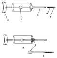
- FIG. 11The proposed modification to the nasal implant, where the implant, (c), has attached to it a guiding needle, (a), a “pull through” suture, (b), and a trailing suture, (d).
- FIG. 12The introducing device composed of the implant (d) placed in an introducing needle (e). There is the advancement shaft (c) which is used to advance the implant (d), into the desired location. The stop (b) prevents the thumb control (a) and advancement shaft (c) from advancing after the implant is expressed from the introducing needle, and placed in the desired location in the tissue.
- FIGS. 12 A and Bshow an alternate arrangement in which the injector device (A) and the introduction needle and implant (B) are packaged separately, but can be attached through a locking attachment (f).
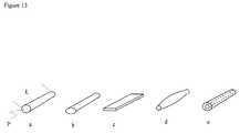
- FIG. 13Representative shapes of the nasal implants.
- the implantsin general is elongated in shape having a length L and side profile P as shown in [ FIG. 13 a ].
- the side profilemay be oval in shape ( FIG. 13 b ), rectangular or oblong ( FIG. 13 c ), or have tapered at the ends ( FIG. 13 d ).
- itmay be a hollow cylinder, or made of two or more materials ( FIG. 13 e ).
- FIG. 14The standard introduction method of the implant, in this case implanted into the lateral nose, to support the external nasal valve.
- the introduction needle containing the implantis first introduced through the nasal mucosa, and deep to the lower lateral cartilage.
- the introduction needleis then advanced into the desired location for the implant ( FIG. 14-2 ).
- the introduction needleis then withdrawn, as the implant is maintained in position by advancing the advancement shaft ( FIG. 14-3 ).
- the implantis now present in the desired location ( FIG. 14-4 ).
- the implants shapemay then be adjusted after implantation to assume the desired shape ( FIG. 14-5 ).
- FIGS. 15( a )-( b ).The introduction method using the additional introduction modification shown in FIG. 11 .
- the guiding needleis first introduced and passed to along the path through which the implant will be placed.
- the “pull through” sutureis then advanced.
- the implantis then advanced, usually with the guide of an introduction needle system as shown in FIG. 15( b )- 4 .
- the introduction needleis removed while the advancement shaft is advanced to maintain the implant in the desired location ( FIG. 15( b )- 5 ).
- the pull through and trailing suturesmay then used to finely adjust the position of the implant after implantation ( FIG. 15-6) .
- the suturesare then cut off at the skin or mucosal surface FIG. 15( b )- 7 ).
- the structure of the lower two thirds of the noseis determined primarily by the nasal septal cartilage, and paired upper and lower lateral cartilages, covered by a soft tissue envelope [ FIGS. 1-4 ]. Structural weakness of these cartilages, or their attachments may result in deleterious functional and cosmetic changes to the nose.
- the nasal valvesmay collapse contributing to a dynamic nasal obstruction. Internal and external nasal valves exist.
- the internal nasal valvesare formed by the angle of the septum and the lower edge of the upper lateral cartilage [ FIG. 5 ].
- air flowcreates a negative pressure on the cartilage, resulting in a collapse of the nasal walls, as seen in FIG. 6 .
- there may also be collapse of the nasal valve at restresulting in a constricted airway even at rest which is further exacerbated with inspiration.
- the external valveis formed by the shape of the lower lateral cartilage, and the strength of the attachments of these lower lateral cartilages to the lateral anterior maxillary bone. Weakening of this lateral cartilage also leads to airway obstruction which is exacerbated with inspiration.
- Nasal patencyis critical to the airway, and nasal obstruction can contribute to snoring, sleep apnea, and disrupted sleep.
- the patency of a good nasal airwayis also critical for the growing number of people using continuous positive airway pressure (CPAP) for sleep apnea.
- CPAPcontinuous positive airway pressure
- Collapse and weakening of the nasal cartilagecan also lead to external deformities and cosmetic changes to the nose. Loss of support and volume of the lower lateral cartilages, mid-nasal portion or the dorsum can lead to undesirable cosmetic changes. Relative tissue defects on the dorsum of the nose may lead to an irregular nasal profile. These cosmetic differences have traditionally been addressed by surgical rhinoplasty.
- Non-surgical devicesinclude external splints placed on the nasal surface which splay the lateral nasal walls outwardly, thus widening the nasal airway. External strips are such an external device (U.S. Pat. Nos. 5,533,440, 6,238,411, 6,982,359, 7,114,495).
- Other devices previously proposedinclude cone shaped applicators placed into the nostril, or dilators having a variety of proposed shapes (U.S. Pat. Nos. 7,055,523, 6,978,781).
- Surgical therapies for repair of the valve collapseinclude insertion of spreader grafts (for internal valve collapse), batton grafts (for external valve collapse), or suspension sutures. These are placed through surgical incision techniques, or external rhinoplasty approach.
- the graftsare most commonly harvested from septal or auricular cartilage.
- Implantse.g., stents
- implantsmade from alloplastic materials inserted surgically through an external approach have also been described (U.S. Pat. Nos. 6,106,541, 6,454,803, 6,322,590, 2,173,848). These prevent the morbidity or limitations of homografts, but still require an incision surgical technique for placement.
- Implants introduced through injection techniquehave been introduced into the palate and used for the treatment of snoring and sleep apnea (U.S. Pat. No. 7,077,144).
- FIGS. 1-3show in perspective, bottom plan and right side elevation, respectively, components of the nose with skeletal muscle, soft tissue (such as external skin or nasal mucosa) and blood vessels removed.
- the nose Nincludes nasal bone NB at an upper end of the nose.
- the bottom of the nose Nincludes the lower cartilage also referred to as the major alar cartilage.
- the lower cartilages RLC, LLCinclude an external component referred to as the lateral crus LC and an internal component referred to as the medial crus MC.
- the medial crus and septal nasal cartilagecreate a nasal septum NS divide the nose N into a left nostril LN and a right nostril RN.
- Upper cartilagesreside between the lower cartilages and the nasal bones NB.
- the upper cartilagesinclude both a right upper cartilage RUC and a left upper cartilage LUC separated by a septal cartilage SC extending down the bridge of the nose N.
- the opposing edges of the lower cartilage LLC, RLC and upper cartilage LUC, RUCmay move relative to one another. Disposed between the opposing edges is an accessory nasal cartilage (left and right) LANC, RANC.
- a weakened nasal valvemay collapse inwardly as illustrated in FIG. 6 .
- FIG. 6it will be appreciated that the inward deflection is exaggerated for ease of illustration. It will be noted with reference FIG. 5 that the narrow angle between the LLC and the MC illustrated in FIG. 6 , and the relative weakness of the LLC contribute to the inward deflection and collapse of the airway.
- the implant to be used in the present inventionshould be adapted for deployment in a nose.
- the implantshould be adapted for introduction into the nose of a patient, to be reliably positioned or installed within said nose and/or to be retained in said nose.
- the adaptationmay be such that the form (or shape) of the implant is adapted or preformed to the anatomy of the nose for which it is intended.
- the location in the nose and the required effecte.g. bulking only or stiffening
- the implantis self-holding when imbedded in the tissue.
- Implantsthat can be used in the present invention include metallic implants, polymeric implants, biodegradable implants and covered or coated implants. They may be composed of a variety of metal compounds and/or polymeric materials, fabricated in innumerable designs, composed of degradable and/or nondegradable components, fully or partially covered with graft materials (such as the so called “covered stents”) or “sleeves”, and can be bare metal or drug-eluting.
- the implantsmay be comprised of a metal or metal alloy such as stainless steel, spring tempered stainless steel, stainless steel alloys, gold, platinum, super elastic alloys, cobalt-chromium alloys and other cobalt-containing alloys (including ELGILOY (Combined Metals of Chicago, Grove Village, Ill.), PHYNOX (Alloy Wire International, United Kingdom) and CONICHROME (Carpenter Technology Corporation, Wyomissing, Pa.)), titanium-containing alloys, platinum-tungsten alloys, nickel-containing alloys, nickel-titanium alloys (including nitinol), malleable metals (including tantalum); a composite material or a clad composite material and/or other functionally equivalent materials; and/or a polymeric (non-biodegradable or biodegradable) material.
- a metal or metal alloysuch as stainless steel, spring tempered stainless steel, stainless steel alloys, gold, platinum, super elastic alloys, cobalt-chromium alloys and other cobalt-containing alloy
- polymers that may be included in the implant constructioninclude polyethylene, polypropylene, polyurethanes, polyesters, such as polyethylene terephthalate (e.g., DACRON or MYLAR (E. I. DuPont De Nemours and Company, Wilmington, Del.)), polyamides, polyaramids (e.g., KEVLAR from E.I. DuPont De Nemours and Company), polyfluorocarbons such as poly(tetrafluoroethylene with and without copolymerized hexafluoropropylene) (available, e.g., under the trade name TEFLON (E. I.
- Stentsalso may be made with engineering plastics, such as thermotropic liquid crystal polymers (LCP), such as those formed from p,p′-dihydroxy-polynuclear-aromatics or dicarboxy-polynuclear-aromatics.
- LCPthermotropic liquid crystal polymers
- implantse.g., stents
- Removable drug-eluting stentsare described, e.g., in Lambert, T. (1993) J. Am. Coll. Cardiol.: 21: 483A.
- the stentmay be adapted to release the desired agent at only the distal ends, or along the entire body of the stent.
- stentsthat are specifically designed for drug delivery can be used.
- Examples of these specialized drug delivery stents as well as traditional stentsinclude those from Conor Medsystems (Palo Alto, Calif.) (e.g., U.S. Pat. Nos. 6,527,799; 6,293,967; 6,290,673; 6,241,762; U.S. Patent Application Publication Nos. 2003/0199970 and 2003/0167085; and PCT Publication No. WO 03/015664).
- Boston Scientific Corporatione.g., the drug-eluting TAXUS EXPRESS 2 Paclitaxel-Eluting Coronary Stent System
- over the wire stent stentstentssuch as the Express 2 Coronary Stent System and NIR Elite OTW Stent System
- rapid exchange stentssuch as the EXPRESS 2 Coronary Stent
- the implantsare inserted in a similar fashion regardless of the site or the disease being treated. Briefly, a preinsertion examination is conducted by direct visualization, possible endoscopy, and rarely diagnostic imaging. The areas of structural defects, volume defects, of dynamic collapse of the nose are noted.
- the implant size and materialis selected to suit the particular application, where more than one implant material and size may be available.
- Topical local anestheticmay be applied by a combination of topical anesthetic cream applied to the skin (e.g. 4% lidocaine cream available commercially) and/or topical anesthetic solution (e.g 4% lidocaine solution) applied on a cotton pledget in the nasal cavity.
- Topical anestheticmay be infiltrated directly in the area where the implant will be placed, or also injected to perform regional blocks, such as an infraorbital nerve block.
- the implantis then introduced through an injection technique as illustrated in this patent (for example, see FIG. 14 ).
- the implantis introduced through the injection method into the desired location in the nasal tissues.
- the introducing needleis gradually withdrawn, while the implant is maintained in its desired position by means of the advancement shaft of the introduction device.
- the puncture site performed by the introduction needleis small, and does not require repair.
- the implant shapemay be adjusted manually.
- a special conditionis applied to allow for adjustment of the shape of the implant.
- heatmay be applied by external application of a heating pad to the nose. This is transmitted through the tissue to the implant which raises its temperature.
- the shape of the implantis then adjusted to the desired shape, and the external heat source is removed. The implant then maintains this new shape as it is cools.
- a post insertion examinationis performed to visually confirm that the desired structural and shape change to the nose has been achieved. Rarely, diagnostic imaging or endoscopy may also be used at this stage.
- Implantsare typically maneuvered into place directed by visual and tactile control.
- the implante.g., stent
- the radio-opaque or MRI visible materialmay be in the form of one or more markers (e.g., bands of material that are disposed on either end of the implant).
- the material of the implantmay be solid, (e.g. titanium, nitinol, or Gore-tex), braided or woven from a single material (such as titanium, or Polyethylene Terephthalate, or a combination of materials).
- the woven materialsmay have pores which allow ingrowth of tissue after implantation. It may be manufactured from biodegradable materials (e.g poly-L lactic, Poly-D lactic, and poly-L glycolic acid) which will gradually absorb after implantation. It may be malleable, allowing adjustment of the shape before, or after implantation.
- Synthetic polymersprovide for very suitable organic implant (e.g., stent) materials. Advantages of such polymers include the ability to tailor mechanical properties and degradation kinetics to suit various applications. Synthetic polymers are also attractive because they can be fabricated into various shapes. Numerous synthetic polymers can be used to prepare synthetic polymer-comprising stents useful in aspects of the invention. They may be obtained from sources such as Sigma Chemical Co., St. Louis, Mo., Polysciences, Warrenton, Pa., Aldrich, Milwaukee, Wis., Fluka, Ronkonkoma, N.Y., and BioRad, Richmond, Calif.
- Representative synthetic polymersinclude alkyl cellulose, cellulose esters, cellulose ethers, hydroxyalkyl celluloses, nitrocelluloses, polyalkylene glycols, polyalkylene oxides, polyalkylene terephthalates, polyalkylenes, polyamides, polyanhydrides, polycarbonates, polyesters, polyglycolides, polymers of acrylic and methacrylic esters, polyacrylamides, polyorthoesters, polyphe azenes, polysiloxanes, polyurethanes, polyvinyl ohols, polyvinyl esters, polyvinyl ethers, polyvinyl halides, polyvinylpyrrolidone, poly(ether ether ketone)s, silicone-based polymers and blends and copolymers of the above.
- the stentmay comprise both oligomers and polymers of the above.
- polymersinclude poly(methyl methacrylate), poly(ethyl methacrylate), poly(butyl methacrylate), poly(isobutyl methacrylate), poly(hexyl methacrylate), poly(isodecyl methacrylate), poly(lauryl methacrylate), poly(phenyl methacrylate), poly(methyl acrylate), poly(isopropyl acrylate), poly(isobutyl acrylate), poly(octadecyl acrylate), polyethylene, polypropylene, poly(ethylene glycol), poly(ethylene oxide), poly(ethylene terephthalate), poly(vinyl alcohols), poly(vinyl acetate), poly(vinyl chloride), polystyrene, polyurethane, poly(lactic acid), poly(butyric acid), poly(valeric acid), poly[lactide-co-glycolide], poly(fumaric acid), poly(maleic acid), copolymers of polyurethane, poly(
- the polymers used in implantsmay be non-biodegradable.
- non-biodegradable polymersinclude ethylene vinyl acetate (EVA), poly(meth)acrylic acid, polyamides, silicone-based polymers and copolymers and mixtures thereof.
- Polymers used in implantsmay also be biodegradable.
- the rate of degradation of the biodegradable stentis determined by factors such as configurational structure, copolymer ratio, crystallinity, molecular weight, morphology, stresses, amount of residual monomer, porosity and site of implantation.
- the skilled personwill be able to choose the combination of factors and characteristics such that the rate of degradation is optimized.
- biodegradable polymersinclude synthetic polymers such as polyesters, polyanhydrides, poly(ortho)esters, polyurethanes, siloxane-based polyurethanes, poly(butyric acid), tyrosine-based polycarbonates, and natural polymers and polymers derived therefrom such as albumin, alginate, casein, chitin, ch[embedded image not shown]osan, collagen, dextran, elastin, proteoglycans, gelati[embedded image not shown] and other hydrophilic proteins, glutin, zein and other prolamines and hydrophobic proteins, starch and other polysaccharides including cellulose and derivatives thereof (e.g.
- polyesters, polyanhydrides, polystyrenes and blends thereofare polyesters, polyanhydrides, polystyrenes and blends thereof.
- the polyesters and polyanhydridesare advantageous due to their ease of degradation by hydrolysis of ester linkage, degradation products being resorbed through the metabolic pathways of the body in some cases and because of their potential to tailor the structure to alter degradation rates.
- the mechanical properties of the biodegradable materialare preferably selected such that early degradation and concomitant loss of mechanical strength required for it's functioning as a structure supporting implant is prevented.
- Biodegradable polyestersare for instance poly(glycolic acid) (PGA), poly(lactic acid) (PLA), poly(glycolic-co-lactic acid) (PGLA), poly(dioxanone), poly(caprolactone) (PCL), poly(3-hydroxybutyrate) (PHB), poly(3-hydroxyvalerate) (PHV), poly(lactide-co-caprolactone) (PLCL), poly(valerolactone) (PVL), poly(tartronic acid), poly( ⁇ -malonic acid), poly(propylene fumarate) (PPF) (preferably photo cross-linkable), poly(ethylene glycol)/poly(lactic acid) (PELA) block copolymer, poly(L-lactic acid-e-caprolactone) copolymer, and poly(lactide)-poly(ethylene glycol) copolymers.
- PGApoly(glycolic acid)
- PLApoly(lactic acid)
- PGLApoly(glycolic-co-lactic
- Biodegradable polyanhydridesare for instance poly[1,6-bis(carboxyphenoxy)hexane], poly(fumaric-co-sebacic)acid or P(FA:SA), and such polyanhydrides may be used in the form of copolymers with polyimides or poly(anhydrides-co-imides) such as poly-[trimellitylimidoglycine-co-bis(carboxyphenoxy)hexane], poly[pyromellitylimidoalanine-co-1,6-bis(carboph-enoxy)-hexane], poly[sebacic acid-co-1,6-bis(p-carboxyphenoxy)hexane] or P(SA:CPH) and poly[sebacic acid-co-1,3-bis(p-carboxyphenoxy)propane] or P(SA:CPP).
- polyimides or poly(anhydrides-co-imides)such as poly-[trimellitylimidoglycine-co
- biocompatible materialsthat are accepted by the tissue surface.
- the broad term biocompatibleincludes also nontoxicity, noncarcinogenity, chemical inertness, and stability of the material in the living body.
- Exemplary biocompatible materialsare titanium, alumina, zirconia, stainless steel, cobalt and alloys thereof and ceramic materials derived therefrom such as ZrO2 and/or Al2O3.
- inorganic implantse.g., stents
- CaPcalcium phosphate matrices
- HAhydroxyapatite
- BCPbiphasic calcium phosphate
- CaP, sintered hydroxyapatite and bioactive glasses or ceramics, such as 45S5 Bioglass® (US Biomaterials Corp, USA), and apatite- and wollastonite-containing glass-ceramic (glass-ceramic A-W)may also be used.
- Very suitable matrix materialsare the combined materials such as osteoinductive hydroxyapatite/(HA/TCP) matrices, preferably BCP.
- the implant (A)is elongated in shape having a length L and side profile P [ FIG. 13 a ].
- the side profilemay be oval in shape, rectangular, or tapered at the ends [ FIGS. 13( b, c , and d )].
- itmay be a hollow cylinder ( FIG. 13 e ), or made of a composite of materials, or a braid of wires.
- the injection routemay include transcutaneous route, i.e. through the nasal skin, or transmucossally, through the internal mucosa of the nose. The intention of this device is that it be introduced under local anesthetic.
- the nasal implantmay also incorporate an introduction needle and suture, and/or a trailing suture ( FIG. 11 ).
- the guiding needleis introduced through the skin, or mucosa, and tracked along the desired path to the location chosen for the implant and then to a location where it exits the body, such as through the skin.
- This guiding needleis introduced and guided by the physicians fingers, or using standard medical instruments.
- This method for introduction of the implantwhich includes a “pull through” and trailing sutures is illustrated in FIGS. 15( a )-( b ). After introduction of the “pull through” suture shown in FIGS. 15 ( a )-( b ), the guiding needle may be cut off.
- the implantis then introduced with the injection device in FIG. 12 , with the guidance of the “pull through” suture, as illustrated in FIGS. 15( a -( b ).
- itmay be introduced without an injection device, but simply by guiding it to the desired location by gently pulling on the “pull through” suture.
- the trailing suturecan also be used to make adjustments to the position of the implant in situ. When the desired position is accomplished, the “pull through” suture, and the trailing sutures are cut.
- the suturecan be made of absorbable material.
- the suturemay have a diameter similar to the diameter of the implant, or be smaller or larger.
- the injector deviceallows the introduction of the implant into the body through an injection technique. Shown in FIG. 12 , it incorporates the nasal implant (FIG. 14 - d ), an introducing needle (FIG. 14 - e ).
- the implantsare introduced through an injection device either through the transcutaneous route or through the nasal mucosa.
- the implantis placed through a straight introducer device, or a specially curved introducer device, or may be malleable to allow for special shaping of the needle prior to injection.
- These implantsmay be placed adjacent to the upper lateral cartilage, below the nasal surface, as illustrated in FIG. 7 . This will apply lateral force to the medial portion of the lateral nasal cartilage, stenting the internal nasal valves open. This is an alternative to the spreader grafts currently placed surgically.
- the implantsmay be placed adjacent to the lateral edge of the lower lateral cartilages.
- the implantsmay extend to the bony process of the anterior maxillary bone as illustrated in FIG. 8 . This will secure the lateral cartilage more securely to the maxillary bone, preventing lateral nasal collapse.
- These implantsmay be placed to secure the external valve in place of alar batten grafts that are now employed and are applied surgically. They may be inserted overlying or underlying the lateral surface of the lower lateral cartilage.
- the implantsmay have a straight or curved shape. Alternately, they may have a malleable property, and can have the shape adjusted after implantation. They may also have shape memory properties (such as composed of Nitinol) which allows for their shape to assume a predetermined shape after implantation. Use of inserts made of materials which have shape memory properties permit the implant to assume a preset shape after insertion. Alternately, certain conditions may be applied, such as application of heat, cold, light, or a magnetic field, that will allow the material to assume a desired fixed or modified shape after implantation. The necessary condition will depend on the intrinsic properties of the shape memory material chosen to produce the implant. The fixed shape of the implant may also be adjusted before or after insertion.
- the implantmay be composed of biodegradable materials, with or without shape memory.
- the implantsmay be introduced transcutaneously or transmucosally to improve the structural strength of the nasal cartilages, or to fill defects in the nasal contours.
- Examples of proposed areas where the implants can be placedinclude locations adjacent to the lower lateral cartilage as lateral alar implants, in the mid nasal region, and the nasal dorsum, or the collumella, as illustrated in FIG. 9 , and FIG. 10 .
- the firstis a 1.4 cm long, 0.8 mm thick titanium rod that is incorporated in a 16 gauge injection needle. This is designed for use into the lateral nasal wall which supports the external valve.
- the implantis injected in a fashion similar to the technique illustrated in FIG. 14 .
- the currently preferred method of introductionis transmucosaly (from inside the vestibule of the nose).
- the implantis placed between the lower lateral cartilage and the nasal mucosa, and extends over the maxillary bone. When placed, it appears similar to the implant I 1 shown in FIG. 8 .
- One implantis placed for each side of the nose, if bilateral valve collapse is present. After implantation, the shape of the implant can be adjusted by molding the shape of the titanium implant.
- a 1.8 cm long and 1 mm thick rod manufactured from a 85:15 poly (L-lactide-co-glycolide) polymerhas been produced. It is introduced using a 14 gauge needle. This polymer is bio-absorbable. It is inserted transcutaneously over the medial portion of the nose, similar to the one shown in FIG. 7 . After implantation, the material has some structural strength, and by providing an upward force on the medial portion of the upper lateral cartilage, it supports the internal nasal valve, preventing its collapse.
- the shape of the implantcan be adjusted after implantation by temporarily placing a heating pad at 64 degrees Celsius on the surface of the nose. This temperature is tolerated by the human nose for a brief period of time. The temperature is transmitted to the implant, and at such a temperature the polymer softens, and the implant shape can manually adjusted. The implant retains the new shape as it cools.
- the third example usedis a tapered rod with an oval shaped cross section similar to the implant shown in FIG. 13 b .
- the implant dimensionsmay be trimmed prior to implantation depending on the particular size desired for the particular patient to be implanted.
- This implantis manufactured from a porous polyethylene. It is inert, non-absorbable, and has a porous structural surface which allows for fibrovascular tissue ingrowth. It is introduced through a 14 gauge needle, into the dorsum of the nose, filling in volume defects in the nose. This is similar to the DI implant shown FIG. 9 .
Landscapes
- Health & Medical Sciences (AREA)
- Otolaryngology (AREA)
- Public Health (AREA)
- Life Sciences & Earth Sciences (AREA)
- Veterinary Medicine (AREA)
- Engineering & Computer Science (AREA)
- Biomedical Technology (AREA)
- Heart & Thoracic Surgery (AREA)
- Vascular Medicine (AREA)
- Pulmonology (AREA)
- Animal Behavior & Ethology (AREA)
- General Health & Medical Sciences (AREA)
- Cardiology (AREA)
- Oral & Maxillofacial Surgery (AREA)
- Transplantation (AREA)
- Nursing (AREA)
- Orthopedic Medicine & Surgery (AREA)
- Prostheses (AREA)
Abstract
Description
Claims (12)
Priority Applications (6)
| Application Number | Priority Date | Filing Date | Title |
|---|---|---|---|
| US11/898,768US7780730B2 (en) | 2006-09-25 | 2007-09-14 | Nasal implant introduced through a non-surgical injection technique |
| US12/507,697US8133276B2 (en) | 2006-09-25 | 2009-07-22 | Nasal implant introduced through a non-surgical injection technique |
| US12/833,504US20100280611A1 (en) | 2006-09-25 | 2010-07-09 | Nasal implant introduced through an injection technique |
| US13/348,105US8784488B2 (en) | 2006-09-25 | 2012-01-11 | Nasal implant introduced through a non-surgical injection technique |
| US14/331,805US10603163B2 (en) | 2006-09-25 | 2014-07-15 | Nasal implant introduced through a non-surgical injection technique |
| US16/793,925US12133794B2 (en) | 2006-09-25 | 2020-02-18 | Nasal implant introduced through a non-surgical injection technique |
Applications Claiming Priority (2)
| Application Number | Priority Date | Filing Date | Title |
|---|---|---|---|
| US84673606P | 2006-09-25 | 2006-09-25 | |
| US11/898,768US7780730B2 (en) | 2006-09-25 | 2007-09-14 | Nasal implant introduced through a non-surgical injection technique |
Related Child Applications (1)
| Application Number | Title | Priority Date | Filing Date |
|---|---|---|---|
| US12/507,697DivisionUS8133276B2 (en) | 2006-09-25 | 2009-07-22 | Nasal implant introduced through a non-surgical injection technique |
Publications (2)
| Publication Number | Publication Date |
|---|---|
| US20080077240A1 US20080077240A1 (en) | 2008-03-27 |
| US7780730B2true US7780730B2 (en) | 2010-08-24 |
Family
ID=39226073
Family Applications (6)
| Application Number | Title | Priority Date | Filing Date |
|---|---|---|---|
| US11/898,768Active2027-10-20US7780730B2 (en) | 2006-09-25 | 2007-09-14 | Nasal implant introduced through a non-surgical injection technique |
| US12/507,697ActiveUS8133276B2 (en) | 2006-09-25 | 2009-07-22 | Nasal implant introduced through a non-surgical injection technique |
| US12/833,504AbandonedUS20100280611A1 (en) | 2006-09-25 | 2010-07-09 | Nasal implant introduced through an injection technique |
| US13/348,105Active2027-09-19US8784488B2 (en) | 2006-09-25 | 2012-01-11 | Nasal implant introduced through a non-surgical injection technique |
| US14/331,805Active2029-04-27US10603163B2 (en) | 2006-09-25 | 2014-07-15 | Nasal implant introduced through a non-surgical injection technique |
| US16/793,925Active2030-06-09US12133794B2 (en) | 2006-09-25 | 2020-02-18 | Nasal implant introduced through a non-surgical injection technique |
Family Applications After (5)
| Application Number | Title | Priority Date | Filing Date |
|---|---|---|---|
| US12/507,697ActiveUS8133276B2 (en) | 2006-09-25 | 2009-07-22 | Nasal implant introduced through a non-surgical injection technique |
| US12/833,504AbandonedUS20100280611A1 (en) | 2006-09-25 | 2010-07-09 | Nasal implant introduced through an injection technique |
| US13/348,105Active2027-09-19US8784488B2 (en) | 2006-09-25 | 2012-01-11 | Nasal implant introduced through a non-surgical injection technique |
| US14/331,805Active2029-04-27US10603163B2 (en) | 2006-09-25 | 2014-07-15 | Nasal implant introduced through a non-surgical injection technique |
| US16/793,925Active2030-06-09US12133794B2 (en) | 2006-09-25 | 2020-02-18 | Nasal implant introduced through a non-surgical injection technique |
Country Status (1)
| Country | Link |
|---|---|
| US (6) | US7780730B2 (en) |
Cited By (36)
| Publication number | Priority date | Publication date | Assignee | Title |
|---|---|---|---|---|
| US20080125805A1 (en)* | 1999-12-09 | 2008-05-29 | Mische Hans A | Method and devices for the treatment of nasal sinus disorders |
| WO2014134303A1 (en) | 2013-02-27 | 2014-09-04 | Spirox, Inc. | Nasal implants and systems and methods of use |
| US8936594B2 (en) | 2011-06-14 | 2015-01-20 | Aerin Medical Inc. | Methods and devices to treat nasal airways |
| US8986301B2 (en) | 2012-06-13 | 2015-03-24 | Aerin Medical Inc. | Methods and devices to treat nasal airways |
| WO2015107543A3 (en)* | 2013-12-13 | 2015-11-19 | Sujai Jayprakash Joshi | A nasal splint and a method of fixing the nasal splint in a nose |
| WO2016033196A1 (en) | 2014-08-26 | 2016-03-03 | Spirox, Inc. | Nasal implants and systems and method of use |
| US9415194B2 (en) | 2011-06-14 | 2016-08-16 | Aerin Medical Inc. | Post nasal drip treatment |
| WO2017053824A1 (en) | 2015-09-25 | 2017-03-30 | Spirox, Inc. | Nasal implants and systems and method of use |
| WO2017192394A1 (en) | 2016-05-02 | 2017-11-09 | Entellus Medical, Inc. | Nasal valve implants and methods of implanting the same |
| WO2018125868A1 (en) | 2016-12-30 | 2018-07-05 | Spirox, Inc. | Nasal implants and methods of use |
| US10159538B2 (en) | 2014-07-25 | 2018-12-25 | Arrinex, Inc. | Apparatus and method for treating rhinitis |
| US10307200B2 (en) | 2013-09-30 | 2019-06-04 | Arrinex, Inc. | Apparatus and methods for treating rhinitis |
| US10456185B2 (en) | 2011-06-14 | 2019-10-29 | Aerin Medical, Inc. | Methods and devices to treat nasal airways |
| US10603059B2 (en) | 2013-09-13 | 2020-03-31 | Aerin Medical Inc. | Hyaline cartilage shaping |
| US10603163B2 (en) | 2006-09-25 | 2020-03-31 | Spirox, Inc. | Nasal implant introduced through a non-surgical injection technique |
| USD880694S1 (en) | 2017-05-01 | 2020-04-07 | Aerin Medical, Inc. | Nasal airway medical instrument |
| USD881904S1 (en) | 2018-10-31 | 2020-04-21 | Aerin Medical Inc. | Display screen with animated graphical user interface |
| KR20200057132A (en)* | 2018-11-15 | 2020-05-26 | 진오메카디칼 주식회사 | Inserting apparatus for medical treatment implant |
| US10722282B2 (en) | 2011-06-14 | 2020-07-28 | Aerin Medical, Inc. | Methods and devices to treat nasal airways |
| US10786383B2 (en) | 2008-11-19 | 2020-09-29 | Spirox, Inc. | Apparatus and methods for correcting nasal valve collapse |
| USD902412S1 (en) | 2018-10-31 | 2020-11-17 | Aerin Medical, Inc. | Electrosurgery console |
| US10864035B2 (en) | 2016-03-04 | 2020-12-15 | Aerin Medical, Inc. | Eustachian tube modification |
| EP3824832A1 (en) | 2019-11-08 | 2021-05-26 | K2M, Inc. | Anterior tether tensioner |
| US11033318B2 (en) | 2011-06-14 | 2021-06-15 | Aerin Medical, Inc. | Methods and devices to treat nasal airways |
| US11096738B2 (en) | 2017-05-05 | 2021-08-24 | Aerin Medical, Inc. | Treatment of spinal tissue |
| US11116566B2 (en) | 2016-12-22 | 2021-09-14 | Aerin Medical, Inc. | Soft palate treatment |
| US11241271B2 (en) | 2011-06-14 | 2022-02-08 | Aerin Medical Inc. | Methods of treating nasal airways |
| US11278356B2 (en) | 2017-04-28 | 2022-03-22 | Arrinex, Inc. | Systems and methods for locating blood vessels in the treatment of rhinitis |
| US11304746B2 (en) | 2011-06-14 | 2022-04-19 | Aerin Medical Inc. | Method of treating airway tissue to reduce mucus secretion |
| US11464542B2 (en) | 2017-03-28 | 2022-10-11 | Spirox, Inc. | Nasal delivery tools, systems, and methods of use |
| WO2022261474A1 (en)* | 2021-06-10 | 2022-12-15 | Spirair, Inc. | Devices and methods for manipulating nasal tissues |
| US11554027B2 (en) | 2018-09-10 | 2023-01-17 | Siesta Medical, Inc. | Systems and methods for nasal support |
| US11806071B2 (en) | 2016-12-22 | 2023-11-07 | Aerin Medical Inc. | Soft palate treatment |
| US12083034B2 (en) | 2022-12-09 | 2024-09-10 | Spirair, Inc. | Devices, systems, and methods for manipulating nasal tissues |
| US12357817B2 (en) | 2021-04-06 | 2025-07-15 | Aerin Medical Inc. | Nasal neuromodulation devices and methods |
| US12369963B1 (en) | 2011-06-14 | 2025-07-29 | Aerin Medical Inc. | Methods and devices to treat nasal airways |
Families Citing this family (25)
| Publication number | Priority date | Publication date | Assignee | Title |
|---|---|---|---|---|
| US8303640B2 (en)* | 2007-07-30 | 2012-11-06 | Audubon Technologies, Llc | Device for maintaining patent paranasal sinus ostia |
| US8267962B2 (en)* | 2007-12-03 | 2012-09-18 | Stupak Howard D | Device for repositioning cartilage and method of use |
| CH704793B1 (en)* | 2010-01-26 | 2013-10-31 | Velimir-Josef Novak | Nasenwand implant and Operationsset for correcting cartilage weakness of the human nose. |
| JP5061325B2 (en)* | 2010-03-05 | 2012-10-31 | セブン ドリーマーズ ラボラトリーズ,インコーポレイテッド | Pharyngeal dilation device |
| US8480737B2 (en)* | 2010-09-27 | 2013-07-09 | Ethicon, Inc. | Columellar strut for nasal tip support |
| CN102451050B (en)* | 2010-10-29 | 2014-03-19 | 上海交通大学医学院附属第九人民医院 | Artificial bionic nose stent |
| US9510940B2 (en) | 2011-02-17 | 2016-12-06 | Ethicon, Inc. | Bioabsorbable multilayer nasal valve spreader graft |
| US8764799B2 (en)* | 2011-05-31 | 2014-07-01 | Douglas C. Harrington | Device and method for nasal surgery |
| US20130317540A1 (en) | 2012-05-22 | 2013-11-28 | Krasimira Hristov | Universal bioabsorbable nasal implant kit |
| CA2911226C (en) | 2013-05-23 | 2024-02-20 | S.T.S. Medical Ltd. | Shape change structure |
| US9480828B2 (en)* | 2013-11-14 | 2016-11-01 | Gyrus Acmi, Inc. | Treatment method for the inferior turbinate |
| WO2016084087A2 (en)* | 2014-11-26 | 2016-06-02 | S.T.S. Medical Ltd. | Shape change structure for treatment of nasal conditions including sinusitis |
| KR200479230Y1 (en)* | 2015-07-27 | 2016-01-07 | 박종식 | Implant for rhinoseptoplasty |
| KR101868613B1 (en)* | 2016-09-27 | 2018-06-19 | 안태환 | Nasal implant for nasal surgery |
| BE1025061B1 (en)* | 2017-03-17 | 2018-10-17 | Cerhum Société Anonyme | Nasal implant |
| JP7173986B2 (en) | 2017-04-13 | 2022-11-17 | スピロックス,インク. | Nasal implants, delivery tools, systems, and methods of use |
| TWI637736B (en)* | 2017-05-18 | 2018-10-11 | 尤詡鑫 | Nasal implant surgery instrument |
| CN109820622A (en)* | 2019-03-22 | 2019-05-31 | 上海菲思挺医疗器械有限公司 | Nasal septum extending bracket |
| RU191163U1 (en)* | 2019-04-09 | 2019-07-25 | федеральное государственное бюджетное образовательное учреждение высшего образования "Башкирский государственный медицинский университет" Министерства здравоохранения Российской Федерации | SILICONE INNER TIRE WITH INTEGRATED AIR DUCT, PRESSURE DISTRIBUTOR PREFERREDLY IN THE FRONT NOSE PARTITION DEPARTMENTS AND PERIPHERAL LOCKERS |
| RU2744154C1 (en)* | 2020-05-20 | 2021-03-03 | Татьяна Юльевна Владимирова | Method for nosal dorsum augmentation |
| WO2021252806A1 (en) | 2020-06-10 | 2021-12-16 | The Board Of Trustees Of The Leland Stanford Junior University | Systems and methods for altering the shape of nasal tissues |
| CN112826631B (en)* | 2021-02-07 | 2025-01-10 | 东华大学 | An anti-infection nasal vestibule coated stent and its preparation method and application |
| US20220370197A1 (en)* | 2021-05-20 | 2022-11-24 | Acclarent, Inc. | Balloon catheter for prolapsed nasal valve procedure |
| US20230077040A1 (en)* | 2021-08-25 | 2023-03-09 | Arthur Yajing YU | Nonsurgical methods for nasal modification including nasal tip elevation and/or elongation |
| US20240041591A1 (en)* | 2021-11-09 | 2024-02-08 | Hae Seok OH | Lifting suture for rhinoplasty and manufacturing method thereof |
Citations (7)
| Publication number | Priority date | Publication date | Assignee | Title |
|---|---|---|---|---|
| US20020019670A1 (en)* | 1997-02-28 | 2002-02-14 | Jerald M. Crawley | Implantable tissue augmentation device |
| US6431174B1 (en)* | 2000-08-10 | 2002-08-13 | Pi Medical, Inc. | Method and apparatus to treat conditions of the naso-pharyngeal area |
| US20060276817A1 (en)* | 2005-06-03 | 2006-12-07 | Restore Medical, Inc. | Nasal valve treatment method & apparatus |
| US20070219575A1 (en) | 2006-03-20 | 2007-09-20 | Mejia Marta L | Orthonostric device and method of forming the same |
| WO2007134005A1 (en) | 2006-05-15 | 2007-11-22 | Mayo Foundation For Medical Education And Research | Devices and methods to treat nasal passages |
| US7322993B2 (en) | 2002-02-04 | 2008-01-29 | Restore Medical Inc. | Stiffening pharyngeal wall treatment |
| US20080027480A1 (en) | 2006-06-29 | 2008-01-31 | Aspire Medical, Inc. | Methods and devices for rhinoplasty and treating internal valve stenosis |
Family Cites Families (124)
| Publication number | Priority date | Publication date | Assignee | Title |
|---|---|---|---|---|
| US2173848A (en) | 1937-12-27 | 1939-09-26 | Ingersoll Milling Machine Co | Cutter and blade therefor |
| US3395709A (en) | 1966-01-03 | 1968-08-06 | Frank F. Rubin | Method and means for cartilage reshaping |
| US3716058A (en) | 1970-07-17 | 1973-02-13 | Atlanta Res Inst | Barbed suture |
| US4461281A (en) | 1977-06-15 | 1984-07-24 | Carson Robert W | Arthroscopic surgical apparatus and method |
| US4265246A (en)* | 1979-05-30 | 1981-05-05 | Barry Cosmetic Suture Company, Inc. | Sterile cosmetic suture and scalp implant for attaching hair pieces |
| CA1221596A (en) | 1984-03-09 | 1987-05-12 | David Evans | Surgical needle |
| US4938234A (en) | 1988-07-15 | 1990-07-03 | Capriotti Robert J | Method of surgically implanting a contour nasal implant |
| US5131382A (en) | 1989-03-27 | 1992-07-21 | Meyer William F | Endoscopic percutaneous discectomy device |
| US5030232A (en)* | 1989-04-06 | 1991-07-09 | Pham Van H | Nasal implant device with improved contour |
| US5545208A (en) | 1990-02-28 | 1996-08-13 | Medtronic, Inc. | Intralumenal drug eluting prosthesis |
| AU8074591A (en) | 1990-06-15 | 1992-01-07 | Cortrak Medical, Inc. | Drug delivery apparatus and method |
| US5163952A (en) | 1990-09-14 | 1992-11-17 | Michael Froix | Expandable polymeric stent with memory and delivery apparatus and method |
| US5356431A (en) | 1990-11-16 | 1994-10-18 | Pierce Frank C | Connective tissue stabilizer and method of use |
| US5893840A (en) | 1991-01-04 | 1999-04-13 | Medtronic, Inc. | Releasable microcapsules on balloon catheters |
| US5344453A (en) | 1991-05-30 | 1994-09-06 | Boston Medical Products, Inc. | Thyroplasty implant |
| US5411550A (en) | 1991-09-16 | 1995-05-02 | Atrium Medical Corporation | Implantable prosthetic device for the delivery of a bioactive material |
| WO1993006792A1 (en) | 1991-10-04 | 1993-04-15 | Scimed Life Systems, Inc. | Biodegradable drug delivery vascular stent |
| US5464450A (en) | 1991-10-04 | 1995-11-07 | Scimed Lifesystems Inc. | Biodegradable drug delivery vascular stent |
| US5500013A (en) | 1991-10-04 | 1996-03-19 | Scimed Life Systems, Inc. | Biodegradable drug delivery vascular stent |
| US5531744A (en) | 1991-11-01 | 1996-07-02 | Medical Scientific, Inc. | Alternative current pathways for bipolar surgical cutting tool |
| US5261916A (en) | 1991-12-12 | 1993-11-16 | Target Therapeutics | Detachable pusher-vasoocclusive coil assembly with interlocking ball and keyway coupling |
| US5683448A (en) | 1992-02-21 | 1997-11-04 | Boston Scientific Technology, Inc. | Intraluminal stent and graft |
| US5599352A (en) | 1992-03-19 | 1997-02-04 | Medtronic, Inc. | Method of making a drug eluting stent |
| US5342348A (en) | 1992-12-04 | 1994-08-30 | Kaplan Aaron V | Method and device for treating and enlarging body lumens |
| US5419760A (en) | 1993-01-08 | 1995-05-30 | Pdt Systems, Inc. | Medicament dispensing stent for prevention of restenosis of a blood vessel |
| US5464650A (en) | 1993-04-26 | 1995-11-07 | Medtronic, Inc. | Intravascular stent and method |
| ES2122282T3 (en) | 1993-04-30 | 1998-12-16 | United States Surgical Corp | SURGICAL INSTRUMENT THAT HAS AN ARTICULATED JAW STRUCTURE. |
| US6241747B1 (en) | 1993-05-03 | 2001-06-05 | Quill Medical, Inc. | Barbed Bodily tissue connector |
| US5533440A (en) | 1993-07-07 | 1996-07-09 | Winmint Manufacturing Pty Limited | Rotisserie |
| US6165210A (en) | 1994-04-01 | 2000-12-26 | Gore Enterprise Holdings, Inc. | Self-expandable helical intravascular stent and stent-graft |
| US5891108A (en) | 1994-09-12 | 1999-04-06 | Cordis Corporation | Drug delivery stent |
| US5649977A (en) | 1994-09-22 | 1997-07-22 | Advanced Cardiovascular Systems, Inc. | Metal reinforced polymer stent |
| US5730744A (en) | 1994-09-27 | 1998-03-24 | Justin; Daniel F. | Soft tissue screw, delivery device, and method |
| US5637113A (en) | 1994-12-13 | 1997-06-10 | Advanced Cardiovascular Systems, Inc. | Polymer film for wrapping a stent structure |
| US5782795A (en) | 1995-06-30 | 1998-07-21 | Xomed Surgical Products, Inc. | Surgical suction cutting instrument with internal irrigation |
| US5735811A (en) | 1995-11-30 | 1998-04-07 | Pharmasonics, Inc. | Apparatus and methods for ultrasonically enhanced fluid delivery |
| US6010054A (en) | 1996-02-20 | 2000-01-04 | Imagyn Medical Technologies | Linear stapling instrument with improved staple cartridge |
| NZ535694A (en) | 1996-05-10 | 2006-10-27 | Wallace J | A dressing mechanism with elastic end sections with openings and laminated central section |
| US5951586A (en) | 1996-05-15 | 1999-09-14 | Medtronic, Inc. | Intraluminal stent |
| US5785647A (en) | 1996-07-31 | 1998-07-28 | United States Surgical Corporation | Surgical instruments useful for spinal surgery |
| US6099561A (en) | 1996-10-21 | 2000-08-08 | Inflow Dynamics, Inc. | Vascular and endoluminal stents with improved coatings |
| ZA9710342B (en) | 1996-11-25 | 1998-06-10 | Alza Corp | Directional drug delivery stent and method of use. |
| US5980551A (en) | 1997-02-07 | 1999-11-09 | Endovasc Ltd., Inc. | Composition and method for making a biodegradable drug delivery stent |
| US5843172A (en) | 1997-04-15 | 1998-12-01 | Advanced Cardiovascular Systems, Inc. | Porous medicated stent |
| US6106541A (en) | 1997-05-16 | 2000-08-22 | Hurbis; Charles G. | Surgically implantable nasal dilator |
| US5980564A (en) | 1997-08-01 | 1999-11-09 | Schneider (Usa) Inc. | Bioabsorbable implantable endoprosthesis with reservoir |
| US5972027A (en) | 1997-09-30 | 1999-10-26 | Scimed Life Systems, Inc | Porous stent drug delivery system |
| US6238411B1 (en) | 1997-12-16 | 2001-05-29 | Robert H. Thorner | Internal nasal dilator |
| US6241762B1 (en) | 1998-03-30 | 2001-06-05 | Conor Medsystems, Inc. | Expandable medical device with ductile hinges |
| US7208010B2 (en) | 2000-10-16 | 2007-04-24 | Conor Medsystems, Inc. | Expandable medical device for delivery of beneficial agent |
| US5980566A (en) | 1998-04-11 | 1999-11-09 | Alt; Eckhard | Vascular and endoluminal stents with iridium oxide coating |
| US5897523A (en) | 1998-04-13 | 1999-04-27 | Ethicon Endo-Surgery, Inc. | Articulating ultrasonic surgical instrument |
| US6293967B1 (en) | 1998-10-29 | 2001-09-25 | Conor Medsystems, Inc. | Expandable medical device with ductile hinges |
| US6063101A (en) | 1998-11-20 | 2000-05-16 | Precision Vascular Systems, Inc. | Stent apparatus and method |
| US7226467B2 (en) | 1999-04-09 | 2007-06-05 | Evalve, Inc. | Fixation device delivery catheter, systems and methods of use |
| US6268405B1 (en) | 1999-05-04 | 2001-07-31 | Porex Surgical, Inc. | Hydrogels and methods of making and using same |
| US6290673B1 (en) | 1999-05-20 | 2001-09-18 | Conor Medsystems, Inc. | Expandable medical device delivery system and method |
| WO2001001957A1 (en) | 1999-05-27 | 2001-01-11 | Biocompatibles Limited | Local drug delivery |
| CA2686633A1 (en) | 1999-06-14 | 2000-12-21 | Vivus, Inc. | Combination therapy for effecting weight loss and treating obesity |
| US6502574B2 (en) | 1999-09-17 | 2003-01-07 | Pi Medical, Inc. | Lateral stiffening snoring treatment |
| US6450169B1 (en) | 1999-09-17 | 2002-09-17 | Pi Medical, Inc. | Braided implant for snoring treatment |
| US6390096B1 (en) | 1999-09-17 | 2002-05-21 | Pi Medical, Inc. | Needle with pre-loaded implant for snoring treatment |
| HK1047878A1 (en) | 1999-09-17 | 2003-03-14 | Restore Medical, Inc. | Implants and methods for snoring treatment |
| US6250307B1 (en) | 1999-09-17 | 2001-06-26 | Pi Medical, Inc. | Snoring treatment |
| US6601584B2 (en) | 1999-09-17 | 2003-08-05 | Pi Medical, Inc. | Contracting snoring treatment implant |
| US6415796B1 (en) | 1999-09-17 | 2002-07-09 | Pi Medical, Inc. | Placement tool for snoring treatment |
| US6516806B2 (en) | 1999-09-17 | 2003-02-11 | Pi Medical, Inc. | Compliant snoring treatment implant |
| US6322590B1 (en) | 1999-11-09 | 2001-11-27 | Michael J. Sillers | Internal nasal implant |
| KR100707773B1 (en)* | 2000-03-14 | 2007-04-17 | 나카가와 아키오 | Nasal Tracheal Tubes and Nasal Tracheal Tube Apparatus Used for Endoscopic Plastic Surgery |
| US7011622B2 (en) | 2000-03-15 | 2006-03-14 | Ams Research Corporation | Parylene coated components for artificial sphincters |
| US6454803B1 (en) | 2000-05-23 | 2002-09-24 | Romo, Iii Thomas | External nasal valve batten implant device and method |
| US6676698B2 (en) | 2000-06-26 | 2004-01-13 | Rex Medicol, L.P. | Vascular device with valve for approximating vessel wall |
| US6656195B2 (en) | 2000-09-22 | 2003-12-02 | Medtronic Xomed, Inc. | Flexible inner tubular members and rotary tissue cutting instruments having flexible inner tubular members |
| EP1498084B1 (en) | 2000-10-16 | 2014-06-18 | Innovational Holdings, LLC | Expandable medical device for delivery of beneficial agent |
| US7427292B2 (en) | 2001-05-16 | 2008-09-23 | Michael Sachs | Maximal nasal internal support system |
| US8403954B2 (en) | 2001-05-22 | 2013-03-26 | Sanostec Corp. | Nasal congestion, obstruction relief, and drug delivery |
| US6911017B2 (en)* | 2001-09-19 | 2005-06-28 | Advanced Cardiovascular Systems, Inc. | MRI visible catheter balloon |
| US7396232B2 (en) | 2001-12-05 | 2008-07-08 | Ophir Fromovich | Periosteal distraction |
| US8409250B2 (en) | 2002-01-23 | 2013-04-02 | Arthrex, Inc. | Meniscal repair system and method |
| US20030149447A1 (en) | 2002-02-01 | 2003-08-07 | Morency Steven David | Barbed surgical suture |
| US7441559B2 (en) | 2002-09-06 | 2008-10-28 | Koninklijke Philips Electronics N.V. | Devices, systems, and methods to fixate tissue within the regions of body, such as the pharyngeal conduit |
| PL223153B1 (en) | 2002-09-18 | 2016-10-31 | Allergan Inc | Methods and apparatus for delivery of ocular implants |
| AU2003282886B2 (en) | 2002-09-30 | 2009-07-23 | Board Of Regents, The University Of Texas System | Stent delivery system and method of use |
| US7381222B2 (en) | 2002-12-30 | 2008-06-03 | Quiescence Medical, Inc. | Stent for maintaining patency of a body region |
| US7992566B2 (en) | 2002-12-30 | 2011-08-09 | Quiescence Medical, Inc. | Apparatus and methods for treating sleep apnea |
| DE20307058U1 (en) | 2003-05-07 | 2003-08-14 | Heinz Kurz GmbH Medizintechnik, 72144 Dußlingen | Implant to spread the nostrils |
| USD536792S1 (en) | 2003-09-19 | 2007-02-13 | Restore Medical, Inc. | Implant delivery tool |
| US20050154412A1 (en) | 2003-09-19 | 2005-07-14 | Restore Medical, Inc. | Airway implant and delivery tool |
| US6899105B2 (en) | 2003-09-19 | 2005-05-31 | Restore Medical, Inc. | Airway implant cartridge and kit |
| US7237554B2 (en) | 2003-10-31 | 2007-07-03 | Restore Medical, Inc. | Airway implant |
| US7213599B2 (en) | 2003-10-31 | 2007-05-08 | Restore Medical, Inc. | Airway implant |
| US7666203B2 (en)* | 2003-11-06 | 2010-02-23 | Nmt Medical, Inc. | Transseptal puncture apparatus |
| WO2005046516A2 (en) | 2003-11-10 | 2005-05-26 | Angiotech International Ag | Medical implants and anti-scarring agents |
| AU2004293075A1 (en)* | 2003-11-20 | 2005-06-09 | Angiotech International Ag | Soft tissue implants and anti-scarring agents |
| US7803150B2 (en) | 2004-04-21 | 2010-09-28 | Acclarent, Inc. | Devices, systems and methods useable for treating sinusitis |
| US7419497B2 (en) | 2004-04-21 | 2008-09-02 | Acclarent, Inc. | Methods for treating ethmoid disease |
| US8764729B2 (en) | 2004-04-21 | 2014-07-01 | Acclarent, Inc. | Frontal sinus spacer |
| US7114495B2 (en) | 2004-05-05 | 2006-10-03 | Silver Eagle Labs Inc. | Nasal strip with variable spring rate |
| WO2005115543A1 (en) | 2004-05-20 | 2005-12-08 | Wisconsin Alumni Research Foundation | Directionally emitting radioactive sources for brachytherapy |
| JP2008508013A (en) | 2004-07-27 | 2008-03-21 | バイオメット ユーケー リミテッド | Bone jig |
| US7641688B2 (en)* | 2004-09-16 | 2010-01-05 | Evera Medical, Inc. | Tissue augmentation device |
| US7882842B2 (en) | 2004-09-21 | 2011-02-08 | Pavad Medical, Inc. | Airway implant sensors and methods of making and using the same |
| JP4981690B2 (en) | 2005-02-08 | 2012-07-25 | コーニンクレッカ フィリップス エレクトロニクス エヌ ヴィ | System and method for percutaneous lingual plastic surgery |
| US8220466B2 (en) | 2005-02-08 | 2012-07-17 | Koninklijke Philips Electronics N.V. | System and method for percutaneous palate remodeling |
| US7055523B1 (en) | 2005-02-24 | 2006-06-06 | Brown Thomas W | Internal nasal dilator and delivery mechanism |
| US7322356B2 (en) | 2005-02-24 | 2008-01-29 | Restore Medical, Inc. | Combination sleep apnea treatment |
| US6978781B1 (en) | 2005-03-11 | 2005-12-27 | John Jordan | Nasal dilator |
| JP5247428B2 (en) | 2005-04-04 | 2013-07-24 | インターセクト エント, インコーポレイテッド | Apparatus and method for treating sinus symptoms |
| US7337781B2 (en) | 2005-04-15 | 2008-03-04 | Restore Medical, Inc. | Implant for tongue |
| GB0517499D0 (en) | 2005-08-26 | 2005-10-05 | West Hertfordshire Hospitals N | Surgical scaffold |
| US20070250118A1 (en) | 2006-04-20 | 2007-10-25 | Masini Michael A | Compression tissue repair apparatus and methods |
| EP2023826B1 (en) | 2006-05-12 | 2017-06-21 | ArthroCare Corporation | Middle turbinate medializer |
| DE102006023058B3 (en) | 2006-05-17 | 2007-10-04 | Heinz Kurz Gmbh Medizintechnik | Roof shaped surgical or orthopedic implant for splaying of nostrils, has two side sections whose free ends run around angle larger than that of remaining parts of side sections against rear section downwards |
| US20070277831A1 (en) | 2006-06-01 | 2007-12-06 | Reba Luhrs | Nasal assembly and method of positioning nasogastric tubing |
| US20080097335A1 (en) | 2006-08-04 | 2008-04-24 | Allergan, Inc. | Ocular implant delivery assemblies |
| US20080066794A1 (en) | 2006-09-20 | 2008-03-20 | Zachary Durfee | Automatic hunting blind |
| US7780730B2 (en) | 2006-09-25 | 2010-08-24 | Iyad Saidi | Nasal implant introduced through a non-surgical injection technique |
| US20080078412A1 (en) | 2006-10-03 | 2008-04-03 | Restore Medical, Inc. | Tongue implant |
| US20080167628A1 (en) | 2007-01-05 | 2008-07-10 | Boston Scientific Scimed, Inc. | Stent delivery system |
| US8747436B2 (en) | 2007-06-13 | 2014-06-10 | Ethicon, Inc. | Bi-directional barbed suture |
| US9149379B2 (en) | 2007-07-16 | 2015-10-06 | Cook Medical Technologies Llc | Delivery device |
| WO2009036290A1 (en) | 2007-09-14 | 2009-03-19 | Entrigue Surgical, Inc. | Implant system |
| US9597220B2 (en) | 2008-11-19 | 2017-03-21 | Spirox, Inc. | Apparatus and methods for correcting nasal valve collapse |
| ES2671997T3 (en) | 2013-02-27 | 2018-06-12 | Spirox, Inc. | Nasal implants and systems |
- 2007
- 2007-09-14USUS11/898,768patent/US7780730B2/enactiveActive
- 2009
- 2009-07-22USUS12/507,697patent/US8133276B2/enactiveActive
- 2010
- 2010-07-09USUS12/833,504patent/US20100280611A1/ennot_activeAbandoned
- 2012
- 2012-01-11USUS13/348,105patent/US8784488B2/enactiveActive
- 2014
- 2014-07-15USUS14/331,805patent/US10603163B2/enactiveActive
- 2020
- 2020-02-18USUS16/793,925patent/US12133794B2/enactiveActive
Patent Citations (7)
| Publication number | Priority date | Publication date | Assignee | Title |
|---|---|---|---|---|
| US20020019670A1 (en)* | 1997-02-28 | 2002-02-14 | Jerald M. Crawley | Implantable tissue augmentation device |
| US6431174B1 (en)* | 2000-08-10 | 2002-08-13 | Pi Medical, Inc. | Method and apparatus to treat conditions of the naso-pharyngeal area |
| US7322993B2 (en) | 2002-02-04 | 2008-01-29 | Restore Medical Inc. | Stiffening pharyngeal wall treatment |
| US20060276817A1 (en)* | 2005-06-03 | 2006-12-07 | Restore Medical, Inc. | Nasal valve treatment method & apparatus |
| US20070219575A1 (en) | 2006-03-20 | 2007-09-20 | Mejia Marta L | Orthonostric device and method of forming the same |
| WO2007134005A1 (en) | 2006-05-15 | 2007-11-22 | Mayo Foundation For Medical Education And Research | Devices and methods to treat nasal passages |
| US20080027480A1 (en) | 2006-06-29 | 2008-01-31 | Aspire Medical, Inc. | Methods and devices for rhinoplasty and treating internal valve stenosis |
Cited By (109)
| Publication number | Priority date | Publication date | Assignee | Title |
|---|---|---|---|---|
| US9351772B2 (en) | 1999-12-09 | 2016-05-31 | Hans A. Mische | Method and devices for the treatment of nasal sinus disorders |
| US20080125805A1 (en)* | 1999-12-09 | 2008-05-29 | Mische Hans A | Method and devices for the treatment of nasal sinus disorders |
| US12133794B2 (en) | 2006-09-25 | 2024-11-05 | Stryker Corporation | Nasal implant introduced through a non-surgical injection technique |
| US10603163B2 (en) | 2006-09-25 | 2020-03-31 | Spirox, Inc. | Nasal implant introduced through a non-surgical injection technique |
| US11806265B2 (en) | 2008-11-19 | 2023-11-07 | Spirox, Inc. | Apparatus and methods for correcting nasal valve collapse |
| US10786383B2 (en) | 2008-11-19 | 2020-09-29 | Spirox, Inc. | Apparatus and methods for correcting nasal valve collapse |
| US10779873B2 (en) | 2011-06-14 | 2020-09-22 | Aerin Medical, Inc. | Methods of treating nasal airways |
| US10335221B2 (en) | 2011-06-14 | 2019-07-02 | Aerin Medical, Inc. | Methods and devices to treat nasal airways |
| US9237924B2 (en) | 2011-06-14 | 2016-01-19 | Aerin Medical, Inc. | Methods and devices to treat nasal airways |
| US11457971B2 (en) | 2011-06-14 | 2022-10-04 | Aerin Medical Inc. | Methods and devices to treat nasal airways |
| US9179967B2 (en) | 2011-06-14 | 2015-11-10 | Aerin Medical, Inc. | Devices to treat nasal airways |
| US9415194B2 (en) | 2011-06-14 | 2016-08-16 | Aerin Medical Inc. | Post nasal drip treatment |
| US9433463B2 (en) | 2011-06-14 | 2016-09-06 | Aerin Medical, Inc. | Devices to treat nasal airways |
| US9452010B2 (en) | 2011-06-14 | 2016-09-27 | Aerin Medical, Inc. | Methods and devices to treat airways |
| US12053227B2 (en) | 2011-06-14 | 2024-08-06 | Aerin Medical Inc. | Methods of treating nasal airways |
| US9486278B2 (en) | 2011-06-14 | 2016-11-08 | Aerin Medical Inc. | Methods and devices to treat nasal airways |
| US9526571B2 (en) | 2011-06-14 | 2016-12-27 | Aerin Medical, Inc. | Methods and devices to treat nasal airways |
| US12369966B2 (en) | 2011-06-14 | 2025-07-29 | Aerin Medical Inc. | Methods and devices to treat nasal airways |
| US9687296B2 (en) | 2011-06-14 | 2017-06-27 | Aerin Medical Inc. | Devices to treat nasal airways |
| US9788886B2 (en) | 2011-06-14 | 2017-10-17 | Aerin Medical Inc. | Methods and devices to treat nasal airways |
| US9801752B2 (en) | 2011-06-14 | 2017-10-31 | Aerin Medical, Inc. | Post nasal drip treatment |
| US12369963B1 (en) | 2011-06-14 | 2025-07-29 | Aerin Medical Inc. | Methods and devices to treat nasal airways |
| US9888957B2 (en) | 2011-06-14 | 2018-02-13 | Aerin Medical Inc. | Pressure sensitive tissue treatment device |
| US9913682B2 (en) | 2011-06-14 | 2018-03-13 | Aerin Medical, Inc. | Methods and devices to treat nasal airways |
| US9943361B2 (en) | 2011-06-14 | 2018-04-17 | Aerin Medical Inc. | Treating upper airway nerve tissue |
| US12369973B1 (en) | 2011-06-14 | 2025-07-29 | Aerin Medical Inc. | Methods for reducing mucus production |
| US10028780B2 (en) | 2011-06-14 | 2018-07-24 | Aerin Medical, Inc. | Methods and devices to treat nasal airways |
| US10932853B2 (en) | 2011-06-14 | 2021-03-02 | Aerin Medical, Inc. | Methods of treating nasal airways |
| US11304746B2 (en) | 2011-06-14 | 2022-04-19 | Aerin Medical Inc. | Method of treating airway tissue to reduce mucus secretion |
| US10265115B2 (en) | 2011-06-14 | 2019-04-23 | Aerin Medical, Inc. | Methods and devices to treat nasal airways |
| US11759222B2 (en) | 2011-06-14 | 2023-09-19 | Aerin Medical Inc. | Methods and devices to treat nasal airways |
| US11033318B2 (en) | 2011-06-14 | 2021-06-15 | Aerin Medical, Inc. | Methods and devices to treat nasal airways |
| US10376300B2 (en) | 2011-06-14 | 2019-08-13 | Aerin Medical, Inc. | Methods and devices to treat nasal airways |
| US10398489B2 (en) | 2011-06-14 | 2019-09-03 | Aerin Medical, Inc. | Methods of treating nasal airways with radiofrequency energy |
| US11766286B2 (en) | 2011-06-14 | 2023-09-26 | Aerin Medical Inc. | Methods and devices to treat nasal airways |
| US11801084B2 (en) | 2011-06-14 | 2023-10-31 | Aerin Medical Inc. | Methods and devices to treat nasal airways |
| US10456185B2 (en) | 2011-06-14 | 2019-10-29 | Aerin Medical, Inc. | Methods and devices to treat nasal airways |
| US11241271B2 (en) | 2011-06-14 | 2022-02-08 | Aerin Medical Inc. | Methods of treating nasal airways |
| US12357378B2 (en) | 2011-06-14 | 2025-07-15 | Aerin Medical Inc. | Treating upper airway nerve tissue |
| US10456186B1 (en) | 2011-06-14 | 2019-10-29 | Aerin Medical, Inc. | Methods and devices to treat nasal airways |
| US10485603B2 (en) | 2011-06-14 | 2019-11-26 | Aerin Medical, Inc. | Methods and devices to treat nasal airways |
| US8936594B2 (en) | 2011-06-14 | 2015-01-20 | Aerin Medical Inc. | Methods and devices to treat nasal airways |
| US9072597B2 (en) | 2011-06-14 | 2015-07-07 | Aerin Medical Inc. | Methods and devices to treat nasal airways |
| US11510722B2 (en) | 2011-06-14 | 2022-11-29 | Aerin Medical Inc. | Pressure sensitive tissue treatment device |
| US9179964B2 (en) | 2011-06-14 | 2015-11-10 | Aerin Medical, Inc. | Methods and devices to treat nasal airways |
| US12364532B2 (en) | 2011-06-14 | 2025-07-22 | Aerin Medical Inc. | Methods and devices to treat nasal airways |
| US10470814B2 (en) | 2011-06-14 | 2019-11-12 | Aerin Medical, Inc. | Pressure sensitive tissue treatment device |
| US10631925B2 (en) | 2011-06-14 | 2020-04-28 | Aerin Medical, Inc. | Treating upper airway nerve tissue |
| US11832876B2 (en) | 2011-06-14 | 2023-12-05 | Aerin Medical Inc. | Treating upper airway nerve tissue |
| US10722282B2 (en) | 2011-06-14 | 2020-07-28 | Aerin Medical, Inc. | Methods and devices to treat nasal airways |
| US8986301B2 (en) | 2012-06-13 | 2015-03-24 | Aerin Medical Inc. | Methods and devices to treat nasal airways |
| US10588740B2 (en) | 2013-02-27 | 2020-03-17 | Spirox, Inc . | Nasal implants and systems and methods of use |
| WO2014134303A1 (en) | 2013-02-27 | 2014-09-04 | Spirox, Inc. | Nasal implants and systems and methods of use |
| US11890186B2 (en) | 2013-02-27 | 2024-02-06 | Spirox, Inc. | Nasal implants and systems and methods of use |
| EP3400906A1 (en) | 2013-02-27 | 2018-11-14 | Spirox, Inc. | Nasal implants |
| US9480594B2 (en) | 2013-02-27 | 2016-11-01 | Spirox, Inc. | Nasal implants and systems and methods of use |
| US10603059B2 (en) | 2013-09-13 | 2020-03-31 | Aerin Medical Inc. | Hyaline cartilage shaping |
| US12256981B2 (en) | 2013-09-30 | 2025-03-25 | Arrinex, Inc. | Apparatus and methods for treating rhinitis |
| US10512498B2 (en) | 2013-09-30 | 2019-12-24 | Arrinex, Inc. | Apparatus and methods for treating rhinitis |
| US10448985B2 (en) | 2013-09-30 | 2019-10-22 | Arrinex, Inc. | Apparatus and methods for treating rhinitis |
| US10307200B2 (en) | 2013-09-30 | 2019-06-04 | Arrinex, Inc. | Apparatus and methods for treating rhinitis |
| WO2015107543A3 (en)* | 2013-12-13 | 2015-11-19 | Sujai Jayprakash Joshi | A nasal splint and a method of fixing the nasal splint in a nose |
| US12193883B2 (en) | 2014-07-25 | 2025-01-14 | Arrinex, Inc. | Apparatus and method for treating rhinitis |
| US10470837B2 (en) | 2014-07-25 | 2019-11-12 | Arrinex, Inc. | Apparatus and method for treating rhinitis |
| US10159538B2 (en) | 2014-07-25 | 2018-12-25 | Arrinex, Inc. | Apparatus and method for treating rhinitis |
| US10980631B2 (en) | 2014-08-26 | 2021-04-20 | Spirox, Inc. | Nasal implants and systems and method of use |
| US12213877B2 (en) | 2014-08-26 | 2025-02-04 | Stryker Corporation | Nasal implants and systems and method of use |
| EP4534135A2 (en) | 2014-08-26 | 2025-04-09 | Stryker Corporation | Nasal implant |
| US10398545B2 (en) | 2014-08-26 | 2019-09-03 | Spirox, Inc. | Nasal implants and systems and method of use |
| WO2016033196A1 (en) | 2014-08-26 | 2016-03-03 | Spirox, Inc. | Nasal implants and systems and method of use |
| EP4268864A2 (en) | 2015-09-25 | 2023-11-01 | Spirox, Inc. | Nasal implants |
| US11737865B2 (en) | 2015-09-25 | 2023-08-29 | Spirox, Inc. | Nasal implants and systems and method of use |
| WO2017053824A1 (en) | 2015-09-25 | 2017-03-30 | Spirox, Inc. | Nasal implants and systems and method of use |
| US10993800B2 (en) | 2015-09-25 | 2021-05-04 | Spirox, Inc. | Nasal implants and systems and method of use |
| US10864035B2 (en) | 2016-03-04 | 2020-12-15 | Aerin Medical, Inc. | Eustachian tube modification |
| US11969200B2 (en) | 2016-03-04 | 2024-04-30 | Aerin Medical Inc. | Eustachian tube modification |
| JP7493648B2 (en) | 2016-05-02 | 2024-05-31 | エンテラス メディカル インコーポレイテッド | Nasal valve implant and method for implanting same |
| EP4299100A2 (en) | 2016-05-02 | 2024-01-03 | Entellus Medical, Inc. | Nasal valve implants |
| WO2017192394A1 (en) | 2016-05-02 | 2017-11-09 | Entellus Medical, Inc. | Nasal valve implants and methods of implanting the same |
| US10987133B2 (en) | 2016-05-02 | 2021-04-27 | Entellus Medical, Inc. | Nasal valve implants and methods of implanting the same |
| JP2023100862A (en)* | 2016-05-02 | 2023-07-19 | エンテラス メディカル インコーポレイテッド | Nasal valve implant and method of implanting the same |
| US11931071B2 (en) | 2016-05-02 | 2024-03-19 | Entellus Medical, Inc. | Nasal valve implants and methods of implanting the same |
| US12336750B2 (en) | 2016-12-22 | 2025-06-24 | Aerin Medical Inc. | Soft palate treatment |
| US11116566B2 (en) | 2016-12-22 | 2021-09-14 | Aerin Medical, Inc. | Soft palate treatment |
| US11806071B2 (en) | 2016-12-22 | 2023-11-07 | Aerin Medical Inc. | Soft palate treatment |
| US11576770B2 (en)* | 2016-12-30 | 2023-02-14 | Spirox, Inc. | Nasal implants and methods of use |
| WO2018125868A1 (en) | 2016-12-30 | 2018-07-05 | Spirox, Inc. | Nasal implants and methods of use |
| US12171654B2 (en) | 2016-12-30 | 2024-12-24 | Stryker Corporation | Nasal implants and methods of use |
| EP4467101A2 (en) | 2016-12-30 | 2024-11-27 | Stryker Corporation | Nasal implants |
| US11464542B2 (en) | 2017-03-28 | 2022-10-11 | Spirox, Inc. | Nasal delivery tools, systems, and methods of use |
| US11877771B2 (en) | 2017-03-28 | 2024-01-23 | Spirox, Inc. | Nasal delivery tools, systems, and methods of use |
| US12144521B2 (en) | 2017-03-28 | 2024-11-19 | Stryker Corporation | Nasal delivery tools, systems, and methods of use |
| US11278356B2 (en) | 2017-04-28 | 2022-03-22 | Arrinex, Inc. | Systems and methods for locating blood vessels in the treatment of rhinitis |
| USD946149S1 (en) | 2017-05-01 | 2022-03-15 | Aerin Medical Inc. | Nasal airway medical instrument |
| USD946150S1 (en) | 2017-05-01 | 2022-03-15 | Aerin Medical Inc. | Nasal airway medical instrument |
| USD880694S1 (en) | 2017-05-01 | 2020-04-07 | Aerin Medical, Inc. | Nasal airway medical instrument |
| US11096738B2 (en) | 2017-05-05 | 2021-08-24 | Aerin Medical, Inc. | Treatment of spinal tissue |
| US11554027B2 (en) | 2018-09-10 | 2023-01-17 | Siesta Medical, Inc. | Systems and methods for nasal support |
| USD902412S1 (en) | 2018-10-31 | 2020-11-17 | Aerin Medical, Inc. | Electrosurgery console |
| USD881904S1 (en) | 2018-10-31 | 2020-04-21 | Aerin Medical Inc. | Display screen with animated graphical user interface |
| KR102201822B1 (en) | 2018-11-15 | 2021-01-13 | 진오메카디칼 주식회사 | Inserting apparatus for medical treatment implant |
| KR20200057132A (en)* | 2018-11-15 | 2020-05-26 | 진오메카디칼 주식회사 | Inserting apparatus for medical treatment implant |
| US11298164B2 (en) | 2019-11-08 | 2022-04-12 | K2M, Inc. | Anterior tether tensioner |
| EP3824832A1 (en) | 2019-11-08 | 2021-05-26 | K2M, Inc. | Anterior tether tensioner |
| US12127771B2 (en) | 2019-11-08 | 2024-10-29 | K2M, Inc. | Anterior tether tensioner |
| US12357817B2 (en) | 2021-04-06 | 2025-07-15 | Aerin Medical Inc. | Nasal neuromodulation devices and methods |
| WO2022261474A1 (en)* | 2021-06-10 | 2022-12-15 | Spirair, Inc. | Devices and methods for manipulating nasal tissues |
| US12357493B2 (en) | 2022-12-09 | 2025-07-15 | Spirair, Inc. | Devices, systems, and methods for manipulating nasal tissues |
| US12083034B2 (en) | 2022-12-09 | 2024-09-10 | Spirair, Inc. | Devices, systems, and methods for manipulating nasal tissues |
Also Published As
| Publication number | Publication date |
|---|---|
| US20150012090A1 (en) | 2015-01-08 |
| US20080077240A1 (en) | 2008-03-27 |
| US20200253726A1 (en) | 2020-08-13 |
| US8133276B2 (en) | 2012-03-13 |
| US10603163B2 (en) | 2020-03-31 |
| US20120109298A1 (en) | 2012-05-03 |
| US8784488B2 (en) | 2014-07-22 |
| US12133794B2 (en) | 2024-11-05 |
| US20100280611A1 (en) | 2010-11-04 |
| US20090292358A1 (en) | 2009-11-26 |
Similar Documents
| Publication | Publication Date | Title |
|---|---|---|
| US12133794B2 (en) | Nasal implant introduced through a non-surgical injection technique | |
| JP5834337B2 (en) | Partially erosive system for the treatment of obstructive sleep apnea | |
| US7100613B2 (en) | Braided implant for snoring treatment | |
| US7237553B2 (en) | Needle with pre-loaded implant for snoring treatment | |
| CA2381904C (en) | Implants and methods for snoring treatment | |
| JP6057889B2 (en) | Sleep apnea treatment system and method | |
| US6453905B1 (en) | Multi-component snoring treatment | |
| US20120136440A1 (en) | Method And Apparatus For Treating Airway Obstruction | |
| US9895252B2 (en) | Septal implant | |
| US20010050084A1 (en) | Compliant snoring treatment implant | |
| US20010054428A1 (en) | Snoring treatment implant and method | |
| KR20110042094A (en) | Methods and apparatus for forming secondary airways for treating obstructive sleep apnea | |
| KR20040011479A (en) | Braided palatal implant for snoring treatment | |
| WO2013177238A1 (en) | Universal bioabsorable nasal implant kit | |
| Steitz et al. | New Techniques in Nasal Valve Repair |
Legal Events
| Date | Code | Title | Description |
|---|---|---|---|
| STCF | Information on status: patent grant | Free format text:PATENTED CASE | |
| AS | Assignment | Owner name:VENTURE LENDING & LEASING VI, INC., CALIFORNIA Free format text:SECURITY AGREEMENT;ASSIGNOR:NASOFORM, INC.;REEL/FRAME:027784/0067 Effective date:20120222 | |
| AS | Assignment | Owner name:NASOFORM, INC., CALIFORNIA Free format text:CONFIRMATORY ASSIGNMENT;ASSIGNOR:SAIDI, IYAD;REEL/FRAME:031039/0958 Effective date:20130226 | |
| FPAY | Fee payment | Year of fee payment:4 | |
| AS | Assignment | Owner name:SPIROX, INC., CALIFORNIA Free format text:CHANGE OF NAME;ASSIGNOR:NASOFORM, INC.;REEL/FRAME:033650/0127 Effective date:20130516 | |
| MAFP | Maintenance fee payment | Free format text:PAYMENT OF MAINTENANCE FEE, 8TH YR, SMALL ENTITY (ORIGINAL EVENT CODE: M2552) Year of fee payment:8 | |
| FEPP | Fee payment procedure | Free format text:ENTITY STATUS SET TO UNDISCOUNTED (ORIGINAL EVENT CODE: BIG.); ENTITY STATUS OF PATENT OWNER: LARGE ENTITY | |
| MAFP | Maintenance fee payment | Free format text:PAYMENT OF MAINTENANCE FEE, 12TH YEAR, LARGE ENTITY (ORIGINAL EVENT CODE: M1553); ENTITY STATUS OF PATENT OWNER: LARGE ENTITY Year of fee payment:12 | |
| AS | Assignment | Owner name:STRYKER CORPORATION, MICHIGAN Free format text:ASSIGNMENT OF ASSIGNORS INTEREST;ASSIGNOR:SPIROX, INC.;REEL/FRAME:068178/0172 Effective date:20240617 | |
| AS | Assignment | Owner name:STRYKER CORPORATION, MICHIGAN Free format text:CHANGE OF ADDRESS;ASSIGNOR:STRYKER CORPORATION;REEL/FRAME:069737/0184 Effective date:20241217 |