US7767222B2 - Apparatus and method for preventing adhesions between an implant and surrounding tissues - Google Patents
Apparatus and method for preventing adhesions between an implant and surrounding tissuesDownload PDFInfo
- Publication number
- US7767222B2 US7767222B2US10/632,014US63201403AUS7767222B2US 7767222 B2US7767222 B2US 7767222B2US 63201403 AUS63201403 AUS 63201403AUS 7767222 B2US7767222 B2US 7767222B2
- Authority
- US
- United States
- Prior art keywords
- implant
- resorbable
- bone
- membrane
- thin membrane
- Prior art date
- Legal status (The legal status is an assumption and is not a legal conclusion. Google has not performed a legal analysis and makes no representation as to the accuracy of the status listed.)
- Active, expires
Links
- 239000007943implantSubstances0.000titleclaimsabstractdescription95
- 238000000034methodMethods0.000titleclaimsdescription57
- 239000012528membraneSubstances0.000claimsabstractdescription113
- 239000000463materialSubstances0.000claimsabstractdescription51
- 210000000056organAnatomy0.000claimsabstractdescription15
- 210000000481breastAnatomy0.000claimsabstractdescription9
- 239000012620biological materialSubstances0.000claimsabstractdescription8
- 210000004379membraneAnatomy0.000claimsdescription108
- 229920000642polymerPolymers0.000claimsdescription31
- 210000001519tissueAnatomy0.000claimsdescription26
- 210000000988bone and boneAnatomy0.000claimsdescription17
- 238000000576coating methodMethods0.000claimsdescription16
- 230000004888barrier functionEffects0.000claimsdescription14
- 229920001577copolymerPolymers0.000claimsdescription12
- 239000011248coating agentSubstances0.000claimsdescription10
- 210000003205muscleAnatomy0.000claimsdescription10
- ZWEHNKRNPOVVGH-UHFFFAOYSA-N2-ButanoneChemical compoundCCC(C)=OZWEHNKRNPOVVGH-UHFFFAOYSA-N0.000claimsdescription9
- 241001269524DuraSpecies0.000claimsdescription9
- 210000001367arteryAnatomy0.000claimsdescription9
- 210000000845cartilageAnatomy0.000claimsdescription9
- 210000002615epidermisAnatomy0.000claimsdescription9
- 210000003195fasciaAnatomy0.000claimsdescription9
- JJTUDXZGHPGLLC-UHFFFAOYSA-NlactideChemical compoundCC1OC(=O)C(C)OC1=OJJTUDXZGHPGLLC-UHFFFAOYSA-N0.000claimsdescription9
- 210000003041ligamentAnatomy0.000claimsdescription9
- 210000005036nerveAnatomy0.000claimsdescription9
- 210000003516pericardiumAnatomy0.000claimsdescription9
- 210000004303peritoneumAnatomy0.000claimsdescription9
- 210000002435tendonAnatomy0.000claimsdescription9
- 210000003462veinAnatomy0.000claimsdescription9
- 239000002904solventSubstances0.000claimsdescription8
- JJTUDXZGHPGLLC-IMJSIDKUSA-N4511-42-6Chemical compoundC[C@@H]1OC(=O)[C@H](C)OC1=OJJTUDXZGHPGLLC-IMJSIDKUSA-N0.000claimsdescription6
- CSCPPACGZOOCGX-UHFFFAOYSA-NAcetoneChemical compoundCC(C)=OCSCPPACGZOOCGX-UHFFFAOYSA-N0.000claimsdescription6
- WEVYAHXRMPXWCK-UHFFFAOYSA-NAcetonitrileChemical compoundCC#NWEVYAHXRMPXWCK-UHFFFAOYSA-N0.000claimsdescription6
- XEKOWRVHYACXOJ-UHFFFAOYSA-NEthyl acetateChemical compoundCCOC(C)=OXEKOWRVHYACXOJ-UHFFFAOYSA-N0.000claimsdescription6
- WYURNTSHIVDZCO-UHFFFAOYSA-NTetrahydrofuranChemical compoundC1CCOC1WYURNTSHIVDZCO-UHFFFAOYSA-N0.000claimsdescription6
- 239000000853adhesiveSubstances0.000claimsdescription6
- 230000001070adhesive effectEffects0.000claimsdescription6
- 210000000936intestineAnatomy0.000claimsdescription6
- 210000001672ovaryAnatomy0.000claimsdescription6
- 210000004872soft tissueAnatomy0.000claimsdescription6
- 239000012530fluidSubstances0.000claimsdescription5
- -1methyl pyroleChemical compound0.000claimsdescription5
- 238000005507sprayingMethods0.000claimsdescription5
- 238000001035dryingMethods0.000claimsdescription4
- 238000001356surgical procedureMethods0.000claimsdescription4
- 210000004369bloodAnatomy0.000claimsdescription3
- 239000008280bloodSubstances0.000claimsdescription3
- 239000002639bone cementSubstances0.000claimsdescription3
- 230000008468bone growthEffects0.000claimsdescription3
- 210000001185bone marrowAnatomy0.000claimsdescription3
- 230000010478bone regenerationEffects0.000claimsdescription3
- 210000004027cellAnatomy0.000claimsdescription3
- 239000004053dental implantSubstances0.000claimsdescription3
- 210000004207dermisAnatomy0.000claimsdescription3
- 210000002257embryonic structureAnatomy0.000claimsdescription3
- 210000003743erythrocyteAnatomy0.000claimsdescription3
- 210000000744eyelidAnatomy0.000claimsdescription3
- 239000003292glueSubstances0.000claimsdescription3
- 239000003324growth hormone secretagogueSubstances0.000claimsdescription3
- 210000003709heart valveAnatomy0.000claimsdescription3
- 210000000265leukocyteAnatomy0.000claimsdescription3
- 239000000203mixtureSubstances0.000claimsdescription3
- 230000000926neurological effectEffects0.000claimsdescription3
- 229920001296polysiloxanePolymers0.000claimsdescription3
- 239000002356single layerSubstances0.000claimsdescription3
- 210000003491skinAnatomy0.000claimsdescription3
- YLQBMQCUIZJEEH-UHFFFAOYSA-NtetrahydrofuranNatural productsC=1C=COC=1YLQBMQCUIZJEEH-UHFFFAOYSA-N0.000claimsdescription3
- FAPWRFPIFSIZLT-UHFFFAOYSA-MSodium chlorideChemical compound[Na+].[Cl-]FAPWRFPIFSIZLT-UHFFFAOYSA-M0.000claimsdescription2
- 125000004122cyclic groupChemical group0.000claimsdescription2
- 238000002513implantationMethods0.000claimsdescription2
- 239000011780sodium chlorideSubstances0.000claimsdescription2
- 239000002861polymer materialSubstances0.000claims2
- 238000007605air dryingMethods0.000claims1
- 239000010410layerSubstances0.000claims1
- 208000031737Tissue AdhesionsDiseases0.000abstractdescription5
- 239000007921spraySubstances0.000abstractdescription3
- 239000000243solutionSubstances0.000description5
- PAPBSGBWRJIAAV-UHFFFAOYSA-Nε-CaprolactoneChemical compoundO=C1CCCCCO1PAPBSGBWRJIAAV-UHFFFAOYSA-N0.000description5
- 230000008901benefitEffects0.000description4
- 230000009477glass transitionEffects0.000description4
- 229920006209poly(L-lactide-co-D,L-lactide)Polymers0.000description4
- 229920000747poly(lactic acid)Polymers0.000description4
- RKDVKSZUMVYZHH-UHFFFAOYSA-N1,4-dioxane-2,5-dioneChemical compoundO=C1COC(=O)CO1RKDVKSZUMVYZHH-UHFFFAOYSA-N0.000description3
- 210000001508eyeAnatomy0.000description3
- 238000004519manufacturing processMethods0.000description3
- 238000012986modificationMethods0.000description3
- 230000004048modificationEffects0.000description3
- 230000008569processEffects0.000description3
- 239000000126substanceSubstances0.000description3
- YEJRWHAVMIAJKC-UHFFFAOYSA-N4-ButyrolactoneChemical compoundO=C1CCCO1YEJRWHAVMIAJKC-UHFFFAOYSA-N0.000description2
- OZJPLYNZGCXSJM-UHFFFAOYSA-N5-valerolactoneChemical compoundO=C1CCCCO1OZJPLYNZGCXSJM-UHFFFAOYSA-N0.000description2
- HEDRZPFGACZZDS-UHFFFAOYSA-NChloroformChemical compoundClC(Cl)ClHEDRZPFGACZZDS-UHFFFAOYSA-N0.000description2
- 206010070245Foreign bodyDiseases0.000description2
- 208000005422Foreign-Body reactionDiseases0.000description2
- 206010061218InflammationDiseases0.000description2
- 210000003484anatomyAnatomy0.000description2
- 238000006243chemical reactionMethods0.000description2
- 230000035876healingEffects0.000description2
- 230000004054inflammatory processEffects0.000description2
- 239000000178monomerSubstances0.000description2
- YFHICDDUDORKJB-UHFFFAOYSA-Ntrimethylene carbonateChemical compoundO=C1OCCCO1YFHICDDUDORKJB-UHFFFAOYSA-N0.000description2
- ULKFLOVGORAZDI-UHFFFAOYSA-N3,3-dimethyloxetan-2-oneChemical compoundCC1(C)COC1=OULKFLOVGORAZDI-UHFFFAOYSA-N0.000description1
- 102000008186CollagenHuman genes0.000description1
- 108010035532CollagenProteins0.000description1
- 108010080379Fibrin Tissue AdhesiveProteins0.000description1
- 241001465754MetazoaSpecies0.000description1
- 239000004830Super GlueSubstances0.000description1
- 239000000443aerosolSubstances0.000description1
- 230000005875antibody responseEffects0.000description1
- 230000003416augmentationEffects0.000description1
- 230000015572biosynthetic processEffects0.000description1
- 229920001400block copolymerPolymers0.000description1
- 229930188620butyrolactoneNatural products0.000description1
- 125000003178carboxy groupChemical group[H]OC(*)=O0.000description1
- 239000004568cementSubstances0.000description1
- 229920001436collagenPolymers0.000description1
- 238000000748compression mouldingMethods0.000description1
- 238000001816coolingMethods0.000description1
- 238000009792diffusion processMethods0.000description1
- 201000010099diseaseDiseases0.000description1
- 208000037265diseases, disorders, signs and symptomsDiseases0.000description1
- 230000000694effectsEffects0.000description1
- FGBJXOREULPLGL-UHFFFAOYSA-Nethyl cyanoacrylateChemical compoundCCOC(=O)C(=C)C#NFGBJXOREULPLGL-UHFFFAOYSA-N0.000description1
- 238000001125extrusionMethods0.000description1
- 239000004744fabricSubstances0.000description1
- 239000000835fiberSubstances0.000description1
- 239000010408filmSubstances0.000description1
- 239000006260foamSubstances0.000description1
- 238000009472formulationMethods0.000description1
- 238000009963fullingMethods0.000description1
- 239000000499gelSubstances0.000description1
- 150000004676glycansChemical class0.000description1
- 210000002216heartAnatomy0.000description1
- 238000010438heat treatmentMethods0.000description1
- 229920001519homopolymerPolymers0.000description1
- 208000027866inflammatory diseaseDiseases0.000description1
- 238000001746injection mouldingMethods0.000description1
- 210000003734kidneyAnatomy0.000description1
- 239000007788liquidSubstances0.000description1
- 210000004185liverAnatomy0.000description1
- 230000005012migrationEffects0.000description1
- 238000013508migrationMethods0.000description1
- 239000003595mistSubstances0.000description1
- 238000000465mouldingMethods0.000description1
- 239000003960organic solventSubstances0.000description1
- 229920000515polycarbonatePolymers0.000description1
- 239000004417polycarbonateSubstances0.000description1
- 229920000570polyetherPolymers0.000description1
- 229920001282polysaccharidePolymers0.000description1
- 239000005017polysaccharideSubstances0.000description1
- 239000003380propellantSubstances0.000description1
- 230000001681protective effectEffects0.000description1
- 239000011241protective layerSubstances0.000description1
- 230000004044responseEffects0.000description1
- 231100000241scarToxicity0.000description1
- 230000037390scarringEffects0.000description1
- 230000008467tissue growthEffects0.000description1
Images

Classifications
- A—HUMAN NECESSITIES
- A61—MEDICAL OR VETERINARY SCIENCE; HYGIENE
- A61L—METHODS OR APPARATUS FOR STERILISING MATERIALS OR OBJECTS IN GENERAL; DISINFECTION, STERILISATION OR DEODORISATION OF AIR; CHEMICAL ASPECTS OF BANDAGES, DRESSINGS, ABSORBENT PADS OR SURGICAL ARTICLES; MATERIALS FOR BANDAGES, DRESSINGS, ABSORBENT PADS OR SURGICAL ARTICLES
- A61L27/00—Materials for grafts or prostheses or for coating grafts or prostheses
- A61L27/50—Materials characterised by their function or physical properties, e.g. injectable or lubricating compositions, shape-memory materials, surface modified materials
- A61L27/58—Materials at least partially resorbable by the body
- A—HUMAN NECESSITIES
- A61—MEDICAL OR VETERINARY SCIENCE; HYGIENE
- A61B—DIAGNOSIS; SURGERY; IDENTIFICATION
- A61B17/00—Surgical instruments, devices or methods
- A61B17/064—Surgical staples, i.e. penetrating the tissue
- A—HUMAN NECESSITIES
- A61—MEDICAL OR VETERINARY SCIENCE; HYGIENE
- A61C—DENTISTRY; APPARATUS OR METHODS FOR ORAL OR DENTAL HYGIENE
- A61C8/00—Means to be fixed to the jaw-bone for consolidating natural teeth or for fixing dental prostheses thereon; Dental implants; Implanting tools
- A61C8/0012—Means to be fixed to the jaw-bone for consolidating natural teeth or for fixing dental prostheses thereon; Dental implants; Implanting tools characterised by the material or composition, e.g. ceramics, surface layer, metal alloy
- A61C8/0016—Means to be fixed to the jaw-bone for consolidating natural teeth or for fixing dental prostheses thereon; Dental implants; Implanting tools characterised by the material or composition, e.g. ceramics, surface layer, metal alloy polymeric material
- A—HUMAN NECESSITIES
- A61—MEDICAL OR VETERINARY SCIENCE; HYGIENE
- A61L—METHODS OR APPARATUS FOR STERILISING MATERIALS OR OBJECTS IN GENERAL; DISINFECTION, STERILISATION OR DEODORISATION OF AIR; CHEMICAL ASPECTS OF BANDAGES, DRESSINGS, ABSORBENT PADS OR SURGICAL ARTICLES; MATERIALS FOR BANDAGES, DRESSINGS, ABSORBENT PADS OR SURGICAL ARTICLES
- A61L27/00—Materials for grafts or prostheses or for coating grafts or prostheses
- A61L27/28—Materials for coating prostheses
- A61L27/34—Macromolecular materials
- A—HUMAN NECESSITIES
- A61—MEDICAL OR VETERINARY SCIENCE; HYGIENE
- A61L—METHODS OR APPARATUS FOR STERILISING MATERIALS OR OBJECTS IN GENERAL; DISINFECTION, STERILISATION OR DEODORISATION OF AIR; CHEMICAL ASPECTS OF BANDAGES, DRESSINGS, ABSORBENT PADS OR SURGICAL ARTICLES; MATERIALS FOR BANDAGES, DRESSINGS, ABSORBENT PADS OR SURGICAL ARTICLES
- A61L31/00—Materials for other surgical articles, e.g. stents, stent-grafts, shunts, surgical drapes, guide wires, materials for adhesion prevention, occluding devices, surgical gloves, tissue fixation devices
- A61L31/08—Materials for coatings
- A61L31/10—Macromolecular materials
- A—HUMAN NECESSITIES
- A61—MEDICAL OR VETERINARY SCIENCE; HYGIENE
- A61N—ELECTROTHERAPY; MAGNETOTHERAPY; RADIATION THERAPY; ULTRASOUND THERAPY
- A61N1/00—Electrotherapy; Circuits therefor
- A61N1/18—Applying electric currents by contact electrodes
- A61N1/32—Applying electric currents by contact electrodes alternating or intermittent currents
- A61N1/36—Applying electric currents by contact electrodes alternating or intermittent currents for stimulation
- A61N1/372—Arrangements in connection with the implantation of stimulators
- A61N1/375—Constructional arrangements, e.g. casings
- A61N1/37512—Pacemakers
- A—HUMAN NECESSITIES
- A61—MEDICAL OR VETERINARY SCIENCE; HYGIENE
- A61B—DIAGNOSIS; SURGERY; IDENTIFICATION
- A61B17/00—Surgical instruments, devices or methods
- A61B17/04—Surgical instruments, devices or methods for suturing wounds; Holders or packages for needles or suture materials
- A61B17/06—Needles ; Sutures; Needle-suture combinations; Holders or packages for needles or suture materials
- A61B17/06166—Sutures
- A—HUMAN NECESSITIES
- A61—MEDICAL OR VETERINARY SCIENCE; HYGIENE
- A61B—DIAGNOSIS; SURGERY; IDENTIFICATION
- A61B17/00—Surgical instruments, devices or methods
- A61B17/064—Surgical staples, i.e. penetrating the tissue
- A61B17/0642—Surgical staples, i.e. penetrating the tissue for bones, e.g. for osteosynthesis or connecting tendon to bone
- A—HUMAN NECESSITIES
- A61—MEDICAL OR VETERINARY SCIENCE; HYGIENE
- A61B—DIAGNOSIS; SURGERY; IDENTIFICATION
- A61B17/00—Surgical instruments, devices or methods
- A61B17/56—Surgical instruments or methods for treatment of bones or joints; Devices specially adapted therefor
- A61B17/58—Surgical instruments or methods for treatment of bones or joints; Devices specially adapted therefor for osteosynthesis, e.g. bone plates, screws or setting implements
- A61B17/68—Internal fixation devices, including fasteners and spinal fixators, even if a part thereof projects from the skin
- A61B17/70—Spinal positioners or stabilisers, e.g. stabilisers comprising fluid filler in an implant
- A—HUMAN NECESSITIES
- A61—MEDICAL OR VETERINARY SCIENCE; HYGIENE
- A61B—DIAGNOSIS; SURGERY; IDENTIFICATION
- A61B17/00—Surgical instruments, devices or methods
- A61B17/56—Surgical instruments or methods for treatment of bones or joints; Devices specially adapted therefor
- A61B17/58—Surgical instruments or methods for treatment of bones or joints; Devices specially adapted therefor for osteosynthesis, e.g. bone plates, screws or setting implements
- A61B17/68—Internal fixation devices, including fasteners and spinal fixators, even if a part thereof projects from the skin
- A61B17/80—Cortical plates, i.e. bone plates; Instruments for holding or positioning cortical plates, or for compressing bones attached to cortical plates
- A—HUMAN NECESSITIES
- A61—MEDICAL OR VETERINARY SCIENCE; HYGIENE
- A61B—DIAGNOSIS; SURGERY; IDENTIFICATION
- A61B17/00—Surgical instruments, devices or methods
- A61B17/56—Surgical instruments or methods for treatment of bones or joints; Devices specially adapted therefor
- A61B17/58—Surgical instruments or methods for treatment of bones or joints; Devices specially adapted therefor for osteosynthesis, e.g. bone plates, screws or setting implements
- A61B17/68—Internal fixation devices, including fasteners and spinal fixators, even if a part thereof projects from the skin
- A61B17/84—Fasteners therefor or fasteners being internal fixation devices
- A61B17/86—Pins or screws or threaded wires; nuts therefor
- A—HUMAN NECESSITIES
- A61—MEDICAL OR VETERINARY SCIENCE; HYGIENE
- A61B—DIAGNOSIS; SURGERY; IDENTIFICATION
- A61B17/00—Surgical instruments, devices or methods
- A61B2017/00004—(bio)absorbable, (bio)resorbable or resorptive
- A—HUMAN NECESSITIES
- A61—MEDICAL OR VETERINARY SCIENCE; HYGIENE
- A61B—DIAGNOSIS; SURGERY; IDENTIFICATION
- A61B90/00—Instruments, implements or accessories specially adapted for surgery or diagnosis and not covered by any of the groups A61B1/00 - A61B50/00, e.g. for luxation treatment or for protecting wound edges
- A61B90/08—Accessories or related features not otherwise provided for
- A61B2090/0815—Implantable devices for insertion in between organs or other soft tissues
- A61B2090/0816—Implantable devices for insertion in between organs or other soft tissues for preventing adhesion
- A—HUMAN NECESSITIES
- A61—MEDICAL OR VETERINARY SCIENCE; HYGIENE
- A61F—FILTERS IMPLANTABLE INTO BLOOD VESSELS; PROSTHESES; DEVICES PROVIDING PATENCY TO, OR PREVENTING COLLAPSING OF, TUBULAR STRUCTURES OF THE BODY, e.g. STENTS; ORTHOPAEDIC, NURSING OR CONTRACEPTIVE DEVICES; FOMENTATION; TREATMENT OR PROTECTION OF EYES OR EARS; BANDAGES, DRESSINGS OR ABSORBENT PADS; FIRST-AID KITS
- A61F2/00—Filters implantable into blood vessels; Prostheses, i.e. artificial substitutes or replacements for parts of the body; Appliances for connecting them with the body; Devices providing patency to, or preventing collapsing of, tubular structures of the body, e.g. stents
- A61F2/02—Prostheses implantable into the body
- A61F2/12—Mammary prostheses
- A—HUMAN NECESSITIES
- A61—MEDICAL OR VETERINARY SCIENCE; HYGIENE
- A61F—FILTERS IMPLANTABLE INTO BLOOD VESSELS; PROSTHESES; DEVICES PROVIDING PATENCY TO, OR PREVENTING COLLAPSING OF, TUBULAR STRUCTURES OF THE BODY, e.g. STENTS; ORTHOPAEDIC, NURSING OR CONTRACEPTIVE DEVICES; FOMENTATION; TREATMENT OR PROTECTION OF EYES OR EARS; BANDAGES, DRESSINGS OR ABSORBENT PADS; FIRST-AID KITS
- A61F2/00—Filters implantable into blood vessels; Prostheses, i.e. artificial substitutes or replacements for parts of the body; Appliances for connecting them with the body; Devices providing patency to, or preventing collapsing of, tubular structures of the body, e.g. stents
- A61F2/02—Prostheses implantable into the body
- A61F2/28—Bones
- A61F2/2846—Support means for bone substitute or for bone graft implants, e.g. membranes or plates for covering bone defects
- A—HUMAN NECESSITIES
- A61—MEDICAL OR VETERINARY SCIENCE; HYGIENE
- A61F—FILTERS IMPLANTABLE INTO BLOOD VESSELS; PROSTHESES; DEVICES PROVIDING PATENCY TO, OR PREVENTING COLLAPSING OF, TUBULAR STRUCTURES OF THE BODY, e.g. STENTS; ORTHOPAEDIC, NURSING OR CONTRACEPTIVE DEVICES; FOMENTATION; TREATMENT OR PROTECTION OF EYES OR EARS; BANDAGES, DRESSINGS OR ABSORBENT PADS; FIRST-AID KITS
- A61F2/00—Filters implantable into blood vessels; Prostheses, i.e. artificial substitutes or replacements for parts of the body; Appliances for connecting them with the body; Devices providing patency to, or preventing collapsing of, tubular structures of the body, e.g. stents
- A61F2/0077—Special surfaces of prostheses, e.g. for improving ingrowth
- A61F2002/009—Special surfaces of prostheses, e.g. for improving ingrowth for hindering or preventing attachment of biological tissue
- A—HUMAN NECESSITIES
- A61—MEDICAL OR VETERINARY SCIENCE; HYGIENE
- A61F—FILTERS IMPLANTABLE INTO BLOOD VESSELS; PROSTHESES; DEVICES PROVIDING PATENCY TO, OR PREVENTING COLLAPSING OF, TUBULAR STRUCTURES OF THE BODY, e.g. STENTS; ORTHOPAEDIC, NURSING OR CONTRACEPTIVE DEVICES; FOMENTATION; TREATMENT OR PROTECTION OF EYES OR EARS; BANDAGES, DRESSINGS OR ABSORBENT PADS; FIRST-AID KITS
- A61F2/00—Filters implantable into blood vessels; Prostheses, i.e. artificial substitutes or replacements for parts of the body; Appliances for connecting them with the body; Devices providing patency to, or preventing collapsing of, tubular structures of the body, e.g. stents
- A61F2/02—Prostheses implantable into the body
- A61F2/28—Bones
- A61F2002/2817—Bone stimulation by chemical reactions or by osteogenic or biological products for enhancing ossification, e.g. by bone morphogenetic or morphogenic proteins [BMP] or by transforming growth factors [TGF]
- A—HUMAN NECESSITIES
- A61—MEDICAL OR VETERINARY SCIENCE; HYGIENE
- A61F—FILTERS IMPLANTABLE INTO BLOOD VESSELS; PROSTHESES; DEVICES PROVIDING PATENCY TO, OR PREVENTING COLLAPSING OF, TUBULAR STRUCTURES OF THE BODY, e.g. STENTS; ORTHOPAEDIC, NURSING OR CONTRACEPTIVE DEVICES; FOMENTATION; TREATMENT OR PROTECTION OF EYES OR EARS; BANDAGES, DRESSINGS OR ABSORBENT PADS; FIRST-AID KITS
- A61F2/00—Filters implantable into blood vessels; Prostheses, i.e. artificial substitutes or replacements for parts of the body; Appliances for connecting them with the body; Devices providing patency to, or preventing collapsing of, tubular structures of the body, e.g. stents
- A61F2/02—Prostheses implantable into the body
- A61F2/28—Bones
- A61F2002/2835—Bone graft implants for filling a bony defect or an endoprosthesis cavity, e.g. by synthetic material or biological material
- A—HUMAN NECESSITIES
- A61—MEDICAL OR VETERINARY SCIENCE; HYGIENE
- A61F—FILTERS IMPLANTABLE INTO BLOOD VESSELS; PROSTHESES; DEVICES PROVIDING PATENCY TO, OR PREVENTING COLLAPSING OF, TUBULAR STRUCTURES OF THE BODY, e.g. STENTS; ORTHOPAEDIC, NURSING OR CONTRACEPTIVE DEVICES; FOMENTATION; TREATMENT OR PROTECTION OF EYES OR EARS; BANDAGES, DRESSINGS OR ABSORBENT PADS; FIRST-AID KITS
- A61F2/00—Filters implantable into blood vessels; Prostheses, i.e. artificial substitutes or replacements for parts of the body; Appliances for connecting them with the body; Devices providing patency to, or preventing collapsing of, tubular structures of the body, e.g. stents
- A61F2/02—Prostheses implantable into the body
- A61F2/30—Joints
- A61F2002/30001—Additional features of subject-matter classified in A61F2/28, A61F2/30 and subgroups thereof
- A61F2002/30003—Material related properties of the prosthesis or of a coating on the prosthesis
- A61F2002/3006—Properties of materials and coating materials
- A61F2002/30062—(bio)absorbable, biodegradable, bioerodable, (bio)resorbable, resorptive
- A61F2002/30064—Coating or prosthesis-covering structure made of biodegradable material
- A—HUMAN NECESSITIES
- A61—MEDICAL OR VETERINARY SCIENCE; HYGIENE
- A61F—FILTERS IMPLANTABLE INTO BLOOD VESSELS; PROSTHESES; DEVICES PROVIDING PATENCY TO, OR PREVENTING COLLAPSING OF, TUBULAR STRUCTURES OF THE BODY, e.g. STENTS; ORTHOPAEDIC, NURSING OR CONTRACEPTIVE DEVICES; FOMENTATION; TREATMENT OR PROTECTION OF EYES OR EARS; BANDAGES, DRESSINGS OR ABSORBENT PADS; FIRST-AID KITS
- A61F2/00—Filters implantable into blood vessels; Prostheses, i.e. artificial substitutes or replacements for parts of the body; Appliances for connecting them with the body; Devices providing patency to, or preventing collapsing of, tubular structures of the body, e.g. stents
- A61F2/02—Prostheses implantable into the body
- A61F2/30—Joints
- A61F2/30767—Special external or bone-contacting surface, e.g. coating for improving bone ingrowth
- A61F2002/30932—Special external or bone-contacting surface, e.g. coating for improving bone ingrowth for retarding or preventing ingrowth of bone tissue
- A—HUMAN NECESSITIES
- A61—MEDICAL OR VETERINARY SCIENCE; HYGIENE
- A61F—FILTERS IMPLANTABLE INTO BLOOD VESSELS; PROSTHESES; DEVICES PROVIDING PATENCY TO, OR PREVENTING COLLAPSING OF, TUBULAR STRUCTURES OF THE BODY, e.g. STENTS; ORTHOPAEDIC, NURSING OR CONTRACEPTIVE DEVICES; FOMENTATION; TREATMENT OR PROTECTION OF EYES OR EARS; BANDAGES, DRESSINGS OR ABSORBENT PADS; FIRST-AID KITS
- A61F2/00—Filters implantable into blood vessels; Prostheses, i.e. artificial substitutes or replacements for parts of the body; Appliances for connecting them with the body; Devices providing patency to, or preventing collapsing of, tubular structures of the body, e.g. stents
- A61F2/02—Prostheses implantable into the body
- A61F2/30—Joints
- A61F2/46—Special tools for implanting artificial joints
- A61F2002/4631—Special tools for implanting artificial joints the prosthesis being specially adapted for being cemented
- A—HUMAN NECESSITIES
- A61—MEDICAL OR VETERINARY SCIENCE; HYGIENE
- A61F—FILTERS IMPLANTABLE INTO BLOOD VESSELS; PROSTHESES; DEVICES PROVIDING PATENCY TO, OR PREVENTING COLLAPSING OF, TUBULAR STRUCTURES OF THE BODY, e.g. STENTS; ORTHOPAEDIC, NURSING OR CONTRACEPTIVE DEVICES; FOMENTATION; TREATMENT OR PROTECTION OF EYES OR EARS; BANDAGES, DRESSINGS OR ABSORBENT PADS; FIRST-AID KITS
- A61F2310/00—Prostheses classified in A61F2/28 or A61F2/30 - A61F2/44 being constructed from or coated with a particular material
- A61F2310/00005—The prosthesis being constructed from a particular material
- A61F2310/00353—Bone cement, e.g. polymethylmethacrylate or PMMA
- A—HUMAN NECESSITIES
- A61—MEDICAL OR VETERINARY SCIENCE; HYGIENE
- A61L—METHODS OR APPARATUS FOR STERILISING MATERIALS OR OBJECTS IN GENERAL; DISINFECTION, STERILISATION OR DEODORISATION OF AIR; CHEMICAL ASPECTS OF BANDAGES, DRESSINGS, ABSORBENT PADS OR SURGICAL ARTICLES; MATERIALS FOR BANDAGES, DRESSINGS, ABSORBENT PADS OR SURGICAL ARTICLES
- A61L2430/00—Materials or treatment for tissue regeneration
- A61L2430/02—Materials or treatment for tissue regeneration for reconstruction of bones; weight-bearing implants
Definitions
- This inventionrelates generally to medical devices and, more particularly, to methods and apparatus for reducing post-surgical adhesions between living tissues and an implant introduced into a surgical site of a patient;
- a major clinical problem relating to surgical procedures or inflammatory diseasescan be unwanted tissue growth, or adhesion, which can occur during the initial phases of the healing process after surgery or disease.
- Another problemcan be foreign body reactions in response to medical devices or implants introduced into a surgical site.
- Still another problemcan be leakage, migration or diffusion of substances, for instance fluids, from an implant into surrounding tissues.
- barrier materialsin the form of gels, coatings, fabrics, foams, films, and the like, that are placed between a healing post-surgical site and adjacent surrounding tissue.
- barrier materialscan be found in U.S. Pat. No. 5,412,068 to Tang et al., U.S. Pat. No. 5,795,584 to Totakura, U.S. Pat. Nos. 6,034,140 and 6,133,325 to Schwartz et al., and U.S. Pat. No. 6,136,333 to Cohn et al., all of which are expressly incorporated herein by reference.
- the patent to Tang et al.discloses films and other bioresorbable medical devices formed from polycarbonate fibers.
- the patent to Totakuradiscloses surgical adhesion barriers comprising copolymers and/or block copolymers derived from trimethylene carbonate.
- the patents to Schwartz et al.disclose anti-adhesion membranes made of carboxyl-containing polysaccharides and polyethers.
- the patent to Cohn et al.discloses polymeric anti-adhesion compositions comprising poly(ester)/poly(oxyalkylene) ABA triblocks or AB diblocks.
- biocompatible polymeric coatingsto medical devices, such as, for instance, stents.
- An exemplary method for coating a stentis disclosed in U.S. Pat. No. 6,153,252 to Hossainy et al., also expressly incorporated herein by reference.
- barrier materialsmay be resorbed into the body too quickly, yielding undesirable drops in local pH levels, which may cause or exacerbate such problems as local inflammation, discomfort and/or foreign antibody responses.
- Other materialsmay take too long to resorb, may be insufficiently malleable, or may require complex chemical formulations and/or reactions which can increase the cost of manufacturing.
- anti-adhesion membrane materialscan be suitable for placement onto implants introduced into a surgical site of a patient, in order to prevent undesired reactions between the implant and surrounding tissues.
- the implantmay be either a biological implant such as a transplanted organ, or a non-biological implant such as a medical device implant.
- bone graft substitutesbone cement, tissue glues and adhesives, bone fixation members (plates, mesh, screws and rods), prostheses, tissue augmentation devices (such as breast implants, penile implants and collagen), pacemakers, defibrillators, eye spheres, sutures, tacks, staples, cochlear implants, pumps, artificial organs, non-resorbable sheets and membranes, bone growth stimulators, neurological stimulators, dental implants, guided tissue and guided bone regeneration membranes, eye lid weights and tympanostomy tubes.
- the type of membrane material in any particular applicationis determined depending on the application and the characteristics of the surgical site to which the membrane is being applied.
- the resorbable micro-membranes, or filmsthat are disclosed in U.S. patent application Ser. No. 09/805,411 filed Mar. 12, 2001, and expressly incorporated herein by reference.
- the aforementioned applicationdiscloses scar tissue-reduction barrier membranes that are constructed entirely of resorbable polymers, and are engineered to be absorbed into the body relatively slowly over time in order, for example, to reduce potential negative side effects.
- the membrane materialis selected from the group consisting of lactide polymers (e.g., copolymers) of two or more lactides. It has now been found that these polylactide membranes have additional use as protective barriers for use on foreign bodies such as implants.
- an anti-adhesion membraneis applied onto an implant before the body of the implant is introduced into a surgical site of a patient.
- the implantmay comprise either biological material, such as a transplanted organ, or non-biological material such as a medical device implant.
- the membranemay be applied to the implant in a variety of ways.
- a membrane according to the present inventionis shrink-wrapped around an implant, such as a pace-maker.
- an implantsuch as a breast implant, is spray-coated with the membrane material disclosed herein.
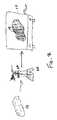
- FIG. 1is a schematic illustration showing a method of coating a pace-maker according to one embodiment of the invention.
- FIG. 2is a schematic illustration showing a method of coating a breast implant according to another embodiment of the invention.
- the present inventionis directed to a method of reducing post-surgical adhesions between an implant and surrounding tissues at a surgical site, comprising a step of applying and/or forming an anti-adhesion membrane on and around the implant.
- the apparatuswhich comprises the implant and the anti-adhesion membrane, may then be placed in a patient at a surgical site.
- Membranes of the present inventionmay be constructed from various biodegradable materials, such as resorbable polymers.
- the membrane material applied to or formed on the implantscomprises lactide polymers, such as copolymers of two or more lactide monomers.
- the membrane materialis preferably selected from the group consisting of lactide polymers (e.g., copolymers) of two or more monomers.
- non-limiting polymers which may be used to form membranes of the present inventioninclude polymers (e.g., copolymers) of lactide (L, D, DL, or combinations thereof), glycolide, trimethylene carbonate, caprolactone and/or physical and chemical combinations thereof.
- the membranescomprise a polylactide, which can be a copolymer of L-lactide and D,L-lactide.
- the copolymercan comprise about 60-80% of L-lactide and about 20-40% of D,L-lactide, and in a preferred embodiment the copolymer comprises poly (L-lactide-co-D,L-lactide) 70:30 Resomer LR708 manufactured and supplied from Boehringer Ingelheim KG of Germany.
- Membranes constructed from this materialhave been found to retard or prevent tissue adhesions, reduce scarring and/or inflammation, and to be resorbable within 24 months or less of implantation into the mammalian body.
- the membranesare formed by polymers (e.g., homo and/or copolymers) derived from one or more cyclic esters, such as lactide (i.e., L, D, DL, or combinations thereof), epsilon-caprolactone and glycolide.
- the membranesin one embodiment can comprise about 1 to 99% epsilon-caprolactone, or in another embodiment can comprise 20 to 40% epsilon-caprolactone.
- a membranecomprises 65:35 poly (L-lactide-co-epsilon-caprolactone).
- the anti-adhesion membrane of the present inventionmay be applied to a wide variety of foreign bodies including, but not limited to, grafting material, transplanted organs and medical devices, including medical devices which may be surrounded by living tissue including, but not limited to, fascia, soft tissues, muscle, organs, fat, adipose, membranes, pericardium, plura, periostium, peritoneum, dura, bowels, intestines, ovaries, veins, arteries, epidermis, tendons, ligaments, nerves, bone and cartilage.
- the grafting materialmay comprise autograft material, xenograft material, allograft material, and combinations thereof.
- Other implantscomprise non-biological materials, such as medical devices.
- suitable non-biological implantsinclude, but are not limited to, bone graft substitutes, bone cement, tissue glues and adhesives, bone fixation members, defibrillators, eye spheres, sutures, staples, cochlear implants, pumps, artificial organs, non-resorbable membranes, bone growth stimulators, neurological stimulators, dental implants, guided tissue and guided bone regeneration membranes, eyelid weights, and tympanostomy tubes.
- Other medical devicesmay include prosthetics, such as a fluid filled prosthesis.
- a fluid filled prosthesisis a breast implant, such as a saline or silicone breast implant.
- medical devicesmay include electronic instruments, such as a pacemaker.
- the membranemay be formed or applied on or over the implant or device using any of a number of techniques including, but not limited to, wrapping, interweaving, blanketing, draping, taping, adjacent placement, juxtaposed positioning, and sandwiching.
- the anti-adhesion materialis pre-formed as a membrane before application onto the implant.
- the materialis applied as a coating which dries to form a membrane or barrier around the implant.
- the coated membranemay be effective as a protective layer around the implant, and/or may be effective as a resorbable barrier, as disclosed herein.
- FIG. 1A method of applying a pre-formed membrane is shown in FIG. 1 .
- FIG. 2A method of forming a membrane by coating and drying is illustrated in FIG. 2 .
- the material used to form the membranes in both figurescan comprise, for example, poly (L-lactide-co-D,L-lactide).
- the membrane or barriermay include one or more portions that comprise a resorbable polymer or polymers, and one or more portions that comprise nonresorbable polymers.
- FIG. 1shows a method of applying a pre-formed membrane 10 to a medical device 11 such as a pacemaker 12 .
- the pre-formed membrane 10is preferably a non-porous film formed of a single layer of polylactide material adapted to maintain a smooth-faced barrier between an implant and surrounding tissues. More preferably, the material in its final form has viscosity property, such as an inherent viscosity, in the range of about 0.20 to about 10.00 g/dL at 25° C. in chloroform. For many applications, the viscosity property of the disclosed membranes is optimally in the range of about 1.00 to about 3.00 g/dL.
- the viscosity propertyis greater than 1 g/dL, and preferably greater than 2 g/dL, and more preferably about 3 g/dL. It is also highly preferable that the thickness of the membrane be less than about 300 microns, and more preferably, in the range of about 10 to about 100 microns. In one preferred embodiment, the thickness is in the range of about 10 to about 50 microns. This thickness should be uniform in the axial and transaxial directions, except at the outermost edges of the membrane 10 , where the thickness can be 2 to 4 times thicker than the rest of the membrane. The thicker edges can provide the membrane with added attachment strength and reduce the risk of damage in for example attachment applications.
- the membrane 10be pre-formed using the extrusion and stretching techniques disclosed in U.S. Provisional Patent Application Ser. No. 60/399,792, and U.S. Provisional Patent Application Ser. No. 60/408,393.
- other standard film-forming processesmay be used in modified embodiments.
- compression moldingmay be used, or any appropriate technique described in, for instance, the Encyclopedia of Polymer Science and Engineering, Vol. 12, pp. 204-210 (1988), the contents of which are incorporated herein by reference.
- certain techniquessuch as injection molding, may not be suitable, and may not provide sufficient performance.
- Multilayer membranesmay provide improved benefits and advantages especially when more than one resorbable material is used, such as a first resorbable material that degrades at a first rate, and a second resorbable material that degrades at a second rate.
- the membrane 10is placed on or under the pacemaker 12 , and wrapped around the pacemaker 12 to substantially encase the pacemaker. As illustrated in FIG. 1 , the pacemaker is completely encased. For other implants, including pacemakers, the membrane 10 may be wrapped around the majority of the implant so that only a minor portion of the implant is in direct contact with a biological environment of a human or animal patient in which the implant is placed. As shown in FIG. 1 , an air blower 14 or other heating device is then used to increase the temperature of the membrane 10 , which may be supported on a suitable frame or holder (not shown), to a value above glass transition temperature. In the case of the preferred polylactide material, the glass transition temperature is about 55° C. to about 60° C.
- the membrane 10shrinks in a predictable manner, depending on the process used to manufacture the membrane. For instance, a membrane which has been monoaxially extruded according to the process disclosed in the aforementioned co-pending U.S. patent application, may shrink by a factor of about 3 along one axis (in a preferred embodiment, the longitudinal axis), and by a factor of about 10-15% on a transverse axis. A membrane which has been biaxially extruded may, in one embodiment, shrink approximately equally along both axes. After cooling back below glass transition temperature, the membrane 10 hardens or is set in its new, wrapped configuration 16 . The wrapped configuration may include the membrane 10 wrapped tightly around the implant.
- substantially all of the exposed surfaces of the implantare covered by the membrane.
- the membraneis in direct contact with the surfaces of the implant.
- the membranemay be wrapped around the implant so that the membrane is not in contact with the implant surfaces, but substantially surrounds the implant.
- the membranemay be configured as a bag that can be wrapped around the implant with a gas or liquid filled space between the membrane and the implant. It is not believed that adhesives or other fixation structures or methods are needed to maintain the membrane 10 in its wrapped configuration. However, it is within the scope of the invention to apply molding to the wrapped implant, such as pacemaker 12 , as it is heated and cooled, or to apply cement or other adhesives to the corners of the membrane as an added step.
- a pre-formed membranemay be secured to bone using resorbable bone screws or tacks. In other cases, tucking or folding a membrane into anatomical crevices may be sufficient to fix its position.
- An adhesivesuch as a fibrin sealant or a resorbable cyanoacrylate adhesive may further be utilized to secure the pre-formed membranes, alone or in combination with any of the other means of attachment discussed above.
- a pre-formed membranecan be heat bonded, such as with a bipolar electro-cautery device, ultrasonically welded, or similarly sealed directly to the surrounding or adjacent structure.
- a coating solution 20is created by dissolving a bioresorbable polymer such as the poly(L-lactide-co-D,L-lactide) material described above in a suitable solvent.
- the solventcan be selected from the group comprising ethyl acetate, acetonitrile, acetone, methyl ethyl ketone (MEK), tetrahydrofuran (THF), methyl pyrole, and any combination of two or more of the above.
- the solution 20has a concentration of about 0.1 to about 5.0% by weight of the bioresorbable polymer.
Landscapes
- Health & Medical Sciences (AREA)
- Life Sciences & Earth Sciences (AREA)
- Public Health (AREA)
- Veterinary Medicine (AREA)
- Animal Behavior & Ethology (AREA)
- General Health & Medical Sciences (AREA)
- Engineering & Computer Science (AREA)
- Epidemiology (AREA)
- Oral & Maxillofacial Surgery (AREA)
- Surgery (AREA)
- Biomedical Technology (AREA)
- Nuclear Medicine, Radiotherapy & Molecular Imaging (AREA)
- Heart & Thoracic Surgery (AREA)
- Orthopedic Medicine & Surgery (AREA)
- Dentistry (AREA)
- Ceramic Engineering (AREA)
- Radiology & Medical Imaging (AREA)
- Dermatology (AREA)
- Chemical & Material Sciences (AREA)
- Molecular Biology (AREA)
- Medicinal Chemistry (AREA)
- Transplantation (AREA)
- Medical Informatics (AREA)
- Vascular Medicine (AREA)
- Biophysics (AREA)
- Rheumatology (AREA)
- Prostheses (AREA)
- Materials For Medical Uses (AREA)
- Surgical Instruments (AREA)
- Electrotherapy Devices (AREA)
Abstract
Description
Claims (32)
Priority Applications (4)
| Application Number | Priority Date | Filing Date | Title |
|---|---|---|---|
| US10/632,014US7767222B2 (en) | 2002-07-31 | 2003-07-31 | Apparatus and method for preventing adhesions between an implant and surrounding tissues |
| US11/652,724US8048444B2 (en) | 2002-07-31 | 2007-01-11 | Apparatus and method for preventing adhesions between an implant and surrounding tissues |
| US11/653,064US7744915B2 (en) | 2002-07-31 | 2007-01-12 | Apparatus and method for preventing adhesions between an implant and surrounding tissues |
| US12/511,898US20090288753A1 (en) | 2002-07-31 | 2009-07-29 | Apparatus and method for preventing adhesions between an implant and surrounding tissues |
Applications Claiming Priority (3)
| Application Number | Priority Date | Filing Date | Title |
|---|---|---|---|
| US39981302P | 2002-07-31 | 2002-07-31 | |
| US40913702P | 2002-09-09 | 2002-09-09 | |
| US10/632,014US7767222B2 (en) | 2002-07-31 | 2003-07-31 | Apparatus and method for preventing adhesions between an implant and surrounding tissues |
Related Child Applications (2)
| Application Number | Title | Priority Date | Filing Date |
|---|---|---|---|
| US11/652,724Continuation-In-PartUS8048444B2 (en) | 2002-07-31 | 2007-01-11 | Apparatus and method for preventing adhesions between an implant and surrounding tissues |
| US11/653,064DivisionUS7744915B2 (en) | 2002-07-31 | 2007-01-12 | Apparatus and method for preventing adhesions between an implant and surrounding tissues |
Publications (2)
| Publication Number | Publication Date |
|---|---|
| US20040115241A1 US20040115241A1 (en) | 2004-06-17 |
| US7767222B2true US7767222B2 (en) | 2010-08-03 |
Family
ID=31191312
Family Applications (2)
| Application Number | Title | Priority Date | Filing Date |
|---|---|---|---|
| US10/632,014Active2026-06-19US7767222B2 (en) | 2002-07-31 | 2003-07-31 | Apparatus and method for preventing adhesions between an implant and surrounding tissues |
| US11/653,064Expired - LifetimeUS7744915B2 (en) | 2002-07-31 | 2007-01-12 | Apparatus and method for preventing adhesions between an implant and surrounding tissues |
Family Applications After (1)
| Application Number | Title | Priority Date | Filing Date |
|---|---|---|---|
| US11/653,064Expired - LifetimeUS7744915B2 (en) | 2002-07-31 | 2007-01-12 | Apparatus and method for preventing adhesions between an implant and surrounding tissues |
Country Status (9)
| Country | Link |
|---|---|
| US (2) | US7767222B2 (en) |
| EP (1) | EP1545389B1 (en) |
| JP (1) | JP2005537909A (en) |
| KR (1) | KR101065155B1 (en) |
| AU (2) | AU2003259684C1 (en) |
| CA (1) | CA2494230C (en) |
| DK (1) | DK1545389T3 (en) |
| MX (1) | MXPA05001149A (en) |
| WO (1) | WO2004010854A2 (en) |
Cited By (3)
| Publication number | Priority date | Publication date | Assignee | Title |
|---|---|---|---|---|
| US20070297987A1 (en)* | 2006-06-26 | 2007-12-27 | Shawn Stad | Anti-Adhesion Sheet |
| US20110125263A1 (en)* | 2007-08-24 | 2011-05-26 | Brown University | Method for producing nanostructures on a surface of a medical implant |
| US9999500B2 (en) | 2011-03-10 | 2018-06-19 | University Of Florida Research Foundation, Inc. | Anti thrombogenic heart valve and medical implements |
Families Citing this family (50)
| Publication number | Priority date | Publication date | Assignee | Title |
|---|---|---|---|---|
| US20060083767A1 (en)* | 2003-02-27 | 2006-04-20 | Kai Deusch | Surgical prosthesis having biodegradable and nonbiodegradable regions |
| WO2005094915A1 (en)* | 2004-03-31 | 2005-10-13 | Nipro Corporation | Antiadhesive kit, process for producing the same and method of adhesion prevention |
| ES2432556T3 (en) | 2004-08-04 | 2013-12-04 | Evonik Corporation | Methods for manufacturing supply devices and their devices |
| US8414907B2 (en) | 2005-04-28 | 2013-04-09 | Warsaw Orthopedic, Inc. | Coatings on medical implants to guide soft tissue healing |
| US9119901B2 (en) | 2005-04-28 | 2015-09-01 | Warsaw Orthopedic, Inc. | Surface treatments for promoting selective tissue attachment to medical impants |
| US8805547B2 (en)* | 2005-06-30 | 2014-08-12 | Domestic Legacy Limited Partnership | Extra-cochlear implanted hearing aid device |
| CN103251449B (en) | 2005-10-13 | 2016-03-02 | 斯恩蒂斯有限公司 | Drug-impregnated encasement |
| US20070123923A1 (en)* | 2005-11-30 | 2007-05-31 | Lindstrom Curtis C | Implantable medical device minimizing rotation and dislocation |
| US20070173934A1 (en)* | 2006-01-20 | 2007-07-26 | Sdgi Holdings, Inc. | Devices to protect features on an implant and methods of use |
| US7650194B2 (en)* | 2006-03-22 | 2010-01-19 | Fritsch Michael H | Intracochlear nanotechnology and perfusion hearing aid device |
| US7833284B2 (en) | 2006-06-28 | 2010-11-16 | The Cleveland Clinic Foundation | Anti-adhesion membrane |
| WO2008058232A2 (en)* | 2006-11-08 | 2008-05-15 | Massachusetts Eye & Ear Infirmary | Spatially-focused actuation in a neural prosthesis |
| AU2008232682B2 (en)* | 2007-03-29 | 2013-03-21 | Medtronic, Inc. | Biodegradable, polymer coverings for breast implants |
| US8313527B2 (en)* | 2007-11-05 | 2012-11-20 | Allergan, Inc. | Soft prosthesis shell texturing method |
| ES2724704T3 (en)* | 2007-11-14 | 2019-09-13 | G Patrick Maxwell | Interconnected Medical Implant Assembly |
| US8124601B2 (en)* | 2007-11-21 | 2012-02-28 | Bristol-Myers Squibb Company | Compounds for the treatment of Hepatitis C |
| JP5502751B2 (en) | 2007-12-20 | 2014-05-28 | エボニック コーポレイション | Process for preparing microparticles with low residual solvent concentration |
| US8690900B2 (en) | 2008-07-21 | 2014-04-08 | The Cleveland Clinic Foundation | Apparatus and method for connecting two elongate body tissues |
| US9050184B2 (en) | 2008-08-13 | 2015-06-09 | Allergan, Inc. | Dual plane breast implant |
| US8506627B2 (en) | 2008-08-13 | 2013-08-13 | Allergan, Inc. | Soft filled prosthesis shell with discrete fixation surfaces |
| EP2381974B1 (en) | 2008-12-29 | 2014-12-17 | Synthes GmbH | A method of forming and the resulting membrane composition for surgical site preservation |
| WO2010088699A2 (en)* | 2009-02-02 | 2010-08-05 | Biomerix Corporation | Composite mesh devices and methods for soft tissue repair |
| CA2941286C (en)* | 2009-05-13 | 2018-10-16 | Allergan, Inc. | Implants and methods for manufacturing same |
| US20110093069A1 (en) | 2009-10-16 | 2011-04-21 | Allergan, Inc. | Implants and methdos for manufacturing same |
| WO2011094155A2 (en) | 2010-01-28 | 2011-08-04 | Allergan, Inc. | Open celled foams, implants including them and processes for making same |
| US8877822B2 (en) | 2010-09-28 | 2014-11-04 | Allergan, Inc. | Porogen compositions, methods of making and uses |
| US9044897B2 (en) | 2010-09-28 | 2015-06-02 | Allergan, Inc. | Porous materials, methods of making and uses |
| US9072821B2 (en) | 2010-02-05 | 2015-07-07 | Allergan, Inc. | Biocompatible structures and compositions |
| US20110196488A1 (en)* | 2010-02-03 | 2011-08-11 | Allergan, Inc. | Degradation resistant implantable materials and methods |
| US8889751B2 (en) | 2010-09-28 | 2014-11-18 | Allergan, Inc. | Porous materials, methods of making and uses |
| US9138308B2 (en) | 2010-02-03 | 2015-09-22 | Apollo Endosurgery, Inc. | Mucosal tissue adhesion via textured surface |
| US9138309B2 (en) | 2010-02-05 | 2015-09-22 | Allergan, Inc. | Porous materials, methods of making and uses |
| US9205577B2 (en) | 2010-02-05 | 2015-12-08 | Allergan, Inc. | Porogen compositions, methods of making and uses |
| US8909348B2 (en) | 2010-03-30 | 2014-12-09 | Domestic Legacy Limited Partnership | Cochlear implant stabilization and positioning device |
| JP2011212209A (en)* | 2010-03-31 | 2011-10-27 | Japan Medical Materials Corp | Support for guided bone regeneration |
| CA2797691A1 (en) | 2010-04-27 | 2011-11-03 | Alexei Goraltchouk | Foam-like materials and methods for producing same |
| AU2011252017B2 (en) | 2010-05-11 | 2015-07-16 | Allergan, Inc. | Porogen compositions, methods of making and uses |
| US11202853B2 (en) | 2010-05-11 | 2021-12-21 | Allergan, Inc. | Porogen compositions, methods of making and uses |
| EP2982343B1 (en)* | 2010-07-09 | 2017-01-04 | Synthes GmbH | Self-detaching layer for easy implant removal |
| US8679279B2 (en) | 2010-11-16 | 2014-03-25 | Allergan, Inc. | Methods for creating foam-like texture |
| US8546458B2 (en) | 2010-12-07 | 2013-10-01 | Allergan, Inc. | Process for texturing materials |
| US8801782B2 (en) | 2011-12-15 | 2014-08-12 | Allergan, Inc. | Surgical methods for breast reconstruction or augmentation |
| TWI590843B (en) | 2011-12-28 | 2017-07-11 | 信迪思有限公司 | Films and methods of manufacture |
| CA2895083A1 (en) | 2012-12-13 | 2014-06-19 | Allergan, Inc. | Device and method for making a variable surface breast implant |
| US20160144067A1 (en) | 2013-06-21 | 2016-05-26 | DePuy Synthes Products, Inc. | Films and methods of manufacture |
| US20170065394A1 (en) | 2014-03-05 | 2017-03-09 | Medizinische Hochschule Hannover | Medical implant, medical device and method for making a medical implant |
| US9539086B2 (en) | 2014-05-16 | 2017-01-10 | Allergan, Inc. | Soft filled prosthesis shell with variable texture |
| US10092392B2 (en) | 2014-05-16 | 2018-10-09 | Allergan, Inc. | Textured breast implant and methods of making same |
| EP3501559B1 (en)* | 2017-12-22 | 2021-11-17 | BIOTRONIK SE & Co. KG | System with an intracardiac implant and a cover for the implant |
| KR102388509B1 (en) | 2021-06-14 | 2022-04-20 | (주)씨앤엘디 | Film type anti-adhesion composition with excellent mucosal adhesion and swelling properties |
Citations (67)
| Publication number | Priority date | Publication date | Assignee | Title |
|---|---|---|---|---|
| US3636956A (en)* | 1970-05-13 | 1972-01-25 | Ethicon Inc | Polylactide sutures |
| US3874986A (en) | 1974-05-20 | 1975-04-01 | Gen Electric | Laminated porous/non-porous membranes |
| US4464320A (en) | 1980-01-14 | 1984-08-07 | Whitney & Company, Inc. | Reaction injection molding system for expanded synthetic articles |
| US4603695A (en) | 1983-07-05 | 1986-08-05 | Japan Medical Supply Co., Ltd. | Use of molded polymeric material for preventing adhesion of vital tissues |
| EP0224460A2 (en) | 1985-11-22 | 1987-06-03 | Swedish Graft Technique AB | Implant lens and method and apparatus for its production |
| US4769038A (en) | 1986-03-18 | 1988-09-06 | C. R. Bard, Inc. | Prostheses and techniques and repair of inguinal and femoral hernias |
| US4955907A (en)* | 1987-12-22 | 1990-09-11 | Ledergerber Walter J | Implantable prosthetic device |
| WO1990013302A1 (en) | 1989-04-28 | 1990-11-15 | Brigham And Women's Hospital | Novel materials and methods for guided tissue regeneration |
| US5030220A (en) | 1990-03-29 | 1991-07-09 | Advanced Spine Fixation Systems Incorporated | Spine fixation system |
| US5047054A (en)* | 1990-10-17 | 1991-09-10 | Smith & Nephew Richards, Inc. | Triazine resin coated prosthetic implants |
| US5227412A (en) | 1987-12-28 | 1993-07-13 | Biomaterials Universe, Inc. | Biodegradable and resorbable surgical material and process for preparation of the same |
| WO1993017635A1 (en) | 1992-03-04 | 1993-09-16 | C.R. Bard, Inc. | Composite prosthesis and method for limiting the incidence of postoperative adhesions |
| WO1993020859A1 (en) | 1992-04-20 | 1993-10-28 | Board Of Regents Of The University Of Washington | Sustained release compositions for delivery of growth factors |
| US5270300A (en) | 1991-09-06 | 1993-12-14 | Robert Francis Shaw | Methods and compositions for the treatment and repair of defects or lesions in cartilage or bone |
| US5380329A (en) | 1992-07-28 | 1995-01-10 | Dental Marketing Specialists, Inc. | Bone augmentation method and apparatus |
| US5412068A (en) | 1987-12-17 | 1995-05-02 | United States Surgical Corporation | Medical devices fabricated from homopolymers and copolymers having recurring carbonate units |
| JPH07116241A (en) | 1993-08-31 | 1995-05-09 | Kyocera Corp | Absorbable biomaterial and method for producing the same |
| US5437672A (en) | 1992-11-12 | 1995-08-01 | Alleyne; Neville | Spinal cord protection device |
| US5508036A (en)* | 1992-04-24 | 1996-04-16 | Osteotech, Inc. | Devices for preventing tissue adhesion |
| US5525646A (en) | 1991-03-04 | 1996-06-11 | Lundgren; Dan | Bioresorbable material and an article of manufacture made of such material for medical use |
| US5609629A (en) | 1995-06-07 | 1997-03-11 | Med Institute, Inc. | Coated implantable medical device |
| US5626861A (en) | 1994-04-01 | 1997-05-06 | Massachusetts Institute Of Technology | Polymeric-hydroxyapatite bone composite |
| US5679723A (en) | 1994-11-30 | 1997-10-21 | Ethicon, Inc. | Hard tissue bone cements and substitutes |
| US5686090A (en) | 1993-01-28 | 1997-11-11 | Ethicon, Inc. | Multi-layered implant |
| US5759190A (en) | 1996-08-30 | 1998-06-02 | Vts Holdings Limited | Method and kit for autologous transplantation |
| US5776195A (en) | 1995-12-28 | 1998-07-07 | Derycke; Raymond Rene | Procedure and device for facilitating osseous growth |
| US5795584A (en) | 1993-01-27 | 1998-08-18 | United States Surgical Corporation | Post-surgical anti-adhesion device |
| US5797946A (en) | 1995-07-13 | 1998-08-25 | Origin Medsystems, Inc. | Method for arterial harvest and anastomosis for coronary bypass grafting |
| US5906997A (en) | 1997-06-17 | 1999-05-25 | Fzio Med, Inc. | Bioresorbable compositions of carboxypolysaccharide polyether intermacromolecular complexes and methods for their use in reducing surgical adhesions |
| US5932539A (en) | 1996-10-15 | 1999-08-03 | The Board Of Trustees Of The University Of Illinois | Biodegradable polymer matrix for tissue repair |
| WO1999051163A1 (en) | 1998-04-03 | 1999-10-14 | Bionx Implants Oy | Hernia mesh |
| US6005162A (en) | 1988-04-20 | 1999-12-21 | Norian Corporation | Methods of repairing bone |
| WO2000015270A1 (en) | 1998-09-10 | 2000-03-23 | Schering Aktiengesellschaft | Coated medical devices and implants |
| WO2000015273A1 (en) | 1998-09-11 | 2000-03-23 | Gerhard Schmidmaier | Biologically active implants |
| JP2000503555A (en) | 1995-04-13 | 2000-03-28 | ヘキスト・アクチエンゲゼルシヤフト | Coating for biological material that can be introduced into the bloodstream or tissue of the human body |
| JP2000189509A (en) | 1998-12-25 | 2000-07-11 | Shimadzu Corp | Bioabsorbable medical film |
| US6113640A (en) | 1997-06-11 | 2000-09-05 | Bionx Implants Oy | Reconstructive bioabsorbable joint prosthesis |
| JP2000265333A (en) | 1999-03-15 | 2000-09-26 | Takasago Internatl Corp | Biodegradable composite fiber and method for producing the same |
| US6132668A (en) | 1985-09-26 | 2000-10-17 | Foster-Miller, Inc. | Biaxially oriented ordered polymer films |
| US6136333A (en) | 1996-07-11 | 2000-10-24 | Life Medical Sciences, Inc. | Methods and compositions for reducing or eliminating post-surgical adhesion formation |
| WO2000062707A2 (en) | 1999-04-15 | 2000-10-26 | University College London | Use of osseo-promotive membranes in orthopaedics |
| US6153252A (en) | 1998-06-30 | 2000-11-28 | Ethicon, Inc. | Process for coating stents |
| US6211217B1 (en) | 1999-03-16 | 2001-04-03 | Novartis Ag | Method for reducing pericardial fibrosis and adhesion formation |
| US6244868B1 (en) | 1997-12-10 | 2001-06-12 | Douglas Alan Schappert | Integrated guided-tissue-regeneration barrier for root-form dental implants |
| US20010004693A1 (en) | 1998-04-03 | 2001-06-21 | W. Burkhead | Anatomical fixation implant |
| US6280473B1 (en) | 1996-08-19 | 2001-08-28 | Macropore, Inc. | Resorbable, macro-porous, non-collapsing and flexible membrane barrier for skeletal repair and regeneration |
| WO2001067987A1 (en) | 2000-03-10 | 2001-09-20 | Macropore, Inc. | Resorbable micro-membrane for attenuation of scar tissue |
| US6331312B1 (en) | 1995-05-19 | 2001-12-18 | Etex Corporation | Bioresorbable ceramic composites |
| US6333029B1 (en) | 1999-06-30 | 2001-12-25 | Ethicon, Inc. | Porous tissue scaffoldings for the repair of regeneration of tissue |
| US20010056303A1 (en) | 2000-06-20 | 2001-12-27 | Caneiro Juan Manuel Bellon | Thoracic/abdominal wall prosthesis that stimulates and modulates connective tissue ingrowth, integrates within host tissue and allows mesothelial deposition, avoiding adhesions and erosion of the viscera |
| US6391059B1 (en) | 1998-04-07 | 2002-05-21 | Macropore, Inc. | Membrane with tissue-guiding surface corrugations |
| US6451373B1 (en)* | 2000-08-04 | 2002-09-17 | Advanced Cardiovascular Systems, Inc. | Method of forming a therapeutic coating onto a surface of an implantable prosthesis |
| US20020173213A1 (en) | 2001-05-16 | 2002-11-21 | Benjamin Chu | Biodegradable and/or bioabsorbable fibrous articles and methods for using the articles for medical applications |
| US6530956B1 (en) | 1998-09-10 | 2003-03-11 | Kevin A. Mansmann | Resorbable scaffolds to promote cartilage regeneration |
| US20030059463A1 (en)* | 1999-12-07 | 2003-03-27 | Mika Lahtinen | Medical device |
| JP2003103429A (en) | 2001-09-28 | 2003-04-08 | Mitsubishi Materials Corp | Cutting tool |
| US6596267B1 (en) | 1997-08-27 | 2003-07-22 | California Institute Of Technology | Methods and compositions to prevent formation of adhesions in biological tissues |
| US20030185874A1 (en) | 2002-02-28 | 2003-10-02 | Calhoun Christopher J. | Methods for governing bone growth |
| EP1384450A1 (en) | 2002-07-21 | 2004-01-28 | Aesculap Ag | Flat implant for use in surgery |
| US20040018175A1 (en) | 2000-08-18 | 2004-01-29 | Dimitrijevich Slobodan Dan | Pericardial anti-adhesion patch |
| US20040030304A1 (en) | 2000-05-09 | 2004-02-12 | Kenneth Hunt | Abdominal wound dressing |
| US6719795B1 (en) | 2001-04-25 | 2004-04-13 | Macropore Biosurgery, Inc. | Resorbable posterior spinal fusion system |
| US20050074495A1 (en) | 1997-06-17 | 2005-04-07 | Fziomed, Inc. | Compositions of polyacids and methods for their use in reducing adhesions |
| US20050175665A1 (en) | 2003-11-20 | 2005-08-11 | Angiotech International Ag | Polymer compositions and methods for their use |
| JP2007504227A (en) | 2003-09-05 | 2007-03-01 | ポセイドン オーシャン サイエンシズ | Menthol propylene glycol-carbonate and the like as pest repellent |
| JP2008033718A (en) | 2006-07-31 | 2008-02-14 | Sanyo Electric Co Ltd | Imaging device and regional enlargement display method |
| JP2008300481A (en) | 2007-05-30 | 2008-12-11 | Micronics Japan Co Ltd | Semiconductor inspection equipment |
Family Cites Families (4)
| Publication number | Priority date | Publication date | Assignee | Title |
|---|---|---|---|---|
| US4298998A (en)* | 1980-12-08 | 1981-11-10 | Naficy Sadeque S | Breast prosthesis with biologically absorbable outer container |
| US5464650A (en)* | 1993-04-26 | 1995-11-07 | Medtronic, Inc. | Intravascular stent and method |
| US5562715A (en)* | 1994-12-01 | 1996-10-08 | Czura; John J. | Cardiac pulse generator |
| US7592017B2 (en)* | 2000-03-10 | 2009-09-22 | Mast Biosurgery Ag | Resorbable thin membranes |
- 2003
- 2003-07-31USUS10/632,014patent/US7767222B2/enactiveActive
- 2003-07-31DKDK03772191.7Tpatent/DK1545389T3/enactive
- 2003-07-31EPEP03772191.7Apatent/EP1545389B1/ennot_activeExpired - Lifetime
- 2003-07-31WOPCT/US2003/024824patent/WO2004010854A2/enactiveSearch and Examination
- 2003-07-31JPJP2005505647Apatent/JP2005537909A/enactivePending
- 2003-07-31KRKR1020057001734Apatent/KR101065155B1/ennot_activeExpired - Lifetime
- 2003-07-31AUAU2003259684Apatent/AU2003259684C1/ennot_activeExpired
- 2003-07-31MXMXPA05001149Apatent/MXPA05001149A/enactiveIP Right Grant
- 2003-07-31CACA2494230Apatent/CA2494230C/ennot_activeExpired - Fee Related
- 2007
- 2007-01-12USUS11/653,064patent/US7744915B2/ennot_activeExpired - Lifetime
- 2008
- 2008-11-20AUAU2008246260Apatent/AU2008246260B2/ennot_activeCeased
Patent Citations (74)
| Publication number | Priority date | Publication date | Assignee | Title |
|---|---|---|---|---|
| US3636956A (en)* | 1970-05-13 | 1972-01-25 | Ethicon Inc | Polylactide sutures |
| US3874986A (en) | 1974-05-20 | 1975-04-01 | Gen Electric | Laminated porous/non-porous membranes |
| US4464320A (en) | 1980-01-14 | 1984-08-07 | Whitney & Company, Inc. | Reaction injection molding system for expanded synthetic articles |
| US4603695A (en) | 1983-07-05 | 1986-08-05 | Japan Medical Supply Co., Ltd. | Use of molded polymeric material for preventing adhesion of vital tissues |
| US6132668A (en) | 1985-09-26 | 2000-10-17 | Foster-Miller, Inc. | Biaxially oriented ordered polymer films |
| EP0224460A2 (en) | 1985-11-22 | 1987-06-03 | Swedish Graft Technique AB | Implant lens and method and apparatus for its production |
| US4769038A (en) | 1986-03-18 | 1988-09-06 | C. R. Bard, Inc. | Prostheses and techniques and repair of inguinal and femoral hernias |
| US5412068A (en) | 1987-12-17 | 1995-05-02 | United States Surgical Corporation | Medical devices fabricated from homopolymers and copolymers having recurring carbonate units |
| US4955907A (en)* | 1987-12-22 | 1990-09-11 | Ledergerber Walter J | Implantable prosthetic device |
| US5227412A (en) | 1987-12-28 | 1993-07-13 | Biomaterials Universe, Inc. | Biodegradable and resorbable surgical material and process for preparation of the same |
| US6005162A (en) | 1988-04-20 | 1999-12-21 | Norian Corporation | Methods of repairing bone |
| WO1990013302A1 (en) | 1989-04-28 | 1990-11-15 | Brigham And Women's Hospital | Novel materials and methods for guided tissue regeneration |
| US5030220A (en) | 1990-03-29 | 1991-07-09 | Advanced Spine Fixation Systems Incorporated | Spine fixation system |
| US5047054A (en)* | 1990-10-17 | 1991-09-10 | Smith & Nephew Richards, Inc. | Triazine resin coated prosthetic implants |
| US5525646A (en) | 1991-03-04 | 1996-06-11 | Lundgren; Dan | Bioresorbable material and an article of manufacture made of such material for medical use |
| US5270300A (en) | 1991-09-06 | 1993-12-14 | Robert Francis Shaw | Methods and compositions for the treatment and repair of defects or lesions in cartilage or bone |
| WO1993017635A1 (en) | 1992-03-04 | 1993-09-16 | C.R. Bard, Inc. | Composite prosthesis and method for limiting the incidence of postoperative adhesions |
| WO1993020859A1 (en) | 1992-04-20 | 1993-10-28 | Board Of Regents Of The University Of Washington | Sustained release compositions for delivery of growth factors |
| US5508036A (en)* | 1992-04-24 | 1996-04-16 | Osteotech, Inc. | Devices for preventing tissue adhesion |
| US5380329A (en) | 1992-07-28 | 1995-01-10 | Dental Marketing Specialists, Inc. | Bone augmentation method and apparatus |
| US5437672A (en) | 1992-11-12 | 1995-08-01 | Alleyne; Neville | Spinal cord protection device |
| US5795584A (en) | 1993-01-27 | 1998-08-18 | United States Surgical Corporation | Post-surgical anti-adhesion device |
| US5686090A (en) | 1993-01-28 | 1997-11-11 | Ethicon, Inc. | Multi-layered implant |
| JPH07116241A (en) | 1993-08-31 | 1995-05-09 | Kyocera Corp | Absorbable biomaterial and method for producing the same |
| US5626861A (en) | 1994-04-01 | 1997-05-06 | Massachusetts Institute Of Technology | Polymeric-hydroxyapatite bone composite |
| US5679723A (en) | 1994-11-30 | 1997-10-21 | Ethicon, Inc. | Hard tissue bone cements and substitutes |
| JP2000503555A (en) | 1995-04-13 | 2000-03-28 | ヘキスト・アクチエンゲゼルシヤフト | Coating for biological material that can be introduced into the bloodstream or tissue of the human body |
| US6331312B1 (en) | 1995-05-19 | 2001-12-18 | Etex Corporation | Bioresorbable ceramic composites |
| US5609629A (en) | 1995-06-07 | 1997-03-11 | Med Institute, Inc. | Coated implantable medical device |
| US5797946A (en) | 1995-07-13 | 1998-08-25 | Origin Medsystems, Inc. | Method for arterial harvest and anastomosis for coronary bypass grafting |
| US5776195A (en) | 1995-12-28 | 1998-07-07 | Derycke; Raymond Rene | Procedure and device for facilitating osseous growth |
| US6136333A (en) | 1996-07-11 | 2000-10-24 | Life Medical Sciences, Inc. | Methods and compositions for reducing or eliminating post-surgical adhesion formation |
| US6280473B1 (en) | 1996-08-19 | 2001-08-28 | Macropore, Inc. | Resorbable, macro-porous, non-collapsing and flexible membrane barrier for skeletal repair and regeneration |
| US5759190A (en) | 1996-08-30 | 1998-06-02 | Vts Holdings Limited | Method and kit for autologous transplantation |
| US5932539A (en) | 1996-10-15 | 1999-08-03 | The Board Of Trustees Of The University Of Illinois | Biodegradable polymer matrix for tissue repair |
| US6113640A (en) | 1997-06-11 | 2000-09-05 | Bionx Implants Oy | Reconstructive bioabsorbable joint prosthesis |
| US20050074495A1 (en) | 1997-06-17 | 2005-04-07 | Fziomed, Inc. | Compositions of polyacids and methods for their use in reducing adhesions |
| US5906997A (en) | 1997-06-17 | 1999-05-25 | Fzio Med, Inc. | Bioresorbable compositions of carboxypolysaccharide polyether intermacromolecular complexes and methods for their use in reducing surgical adhesions |
| US6133325A (en) | 1997-06-17 | 2000-10-17 | Fziomed, Inc. | Bioresorbable compositions of carboxypolysaccharide polyether intermacromolecular complexes and methods for their use in reducing surgical adhesions |
| US6034140A (en) | 1997-06-17 | 2000-03-07 | Fziomed, Inc. | Bioresorbable compositions of carboxypolysaccharide polyether intermacromolecular complexes and methods for their use in reducing surgical adhesions |
| US6596267B1 (en) | 1997-08-27 | 2003-07-22 | California Institute Of Technology | Methods and compositions to prevent formation of adhesions in biological tissues |
| US6244868B1 (en) | 1997-12-10 | 2001-06-12 | Douglas Alan Schappert | Integrated guided-tissue-regeneration barrier for root-form dental implants |
| US20010004693A1 (en) | 1998-04-03 | 2001-06-21 | W. Burkhead | Anatomical fixation implant |
| WO1999051163A1 (en) | 1998-04-03 | 1999-10-14 | Bionx Implants Oy | Hernia mesh |
| US6391059B1 (en) | 1998-04-07 | 2002-05-21 | Macropore, Inc. | Membrane with tissue-guiding surface corrugations |
| US6153252A (en) | 1998-06-30 | 2000-11-28 | Ethicon, Inc. | Process for coating stents |
| US6530956B1 (en) | 1998-09-10 | 2003-03-11 | Kevin A. Mansmann | Resorbable scaffolds to promote cartilage regeneration |
| WO2000015270A1 (en) | 1998-09-10 | 2000-03-23 | Schering Aktiengesellschaft | Coated medical devices and implants |
| WO2000015273A1 (en) | 1998-09-11 | 2000-03-23 | Gerhard Schmidmaier | Biologically active implants |
| JP2000189509A (en) | 1998-12-25 | 2000-07-11 | Shimadzu Corp | Bioabsorbable medical film |
| JP2000265333A (en) | 1999-03-15 | 2000-09-26 | Takasago Internatl Corp | Biodegradable composite fiber and method for producing the same |
| US6211217B1 (en) | 1999-03-16 | 2001-04-03 | Novartis Ag | Method for reducing pericardial fibrosis and adhesion formation |
| WO2000062707A2 (en) | 1999-04-15 | 2000-10-26 | University College London | Use of osseo-promotive membranes in orthopaedics |
| US6333029B1 (en) | 1999-06-30 | 2001-12-25 | Ethicon, Inc. | Porous tissue scaffoldings for the repair of regeneration of tissue |
| US20030059463A1 (en)* | 1999-12-07 | 2003-03-27 | Mika Lahtinen | Medical device |
| US20020001609A1 (en)* | 2000-03-10 | 2002-01-03 | Macropore, Inc. | Resorbable barrier micro-membranes for attenuation of scar tissue during healing |
| WO2001067987A1 (en) | 2000-03-10 | 2001-09-20 | Macropore, Inc. | Resorbable micro-membrane for attenuation of scar tissue |
| US6531146B2 (en) | 2000-03-10 | 2003-03-11 | Macropore, Inc. | Resorbable barrier micro-membranes for attenuation of scar tissue during healing |
| US6673362B2 (en) | 2000-03-10 | 2004-01-06 | Macropore Biosurgery, Inc. | Resorbable barrier micro-membranes for attenuation of scar tissue during healing |
| US20040030304A1 (en) | 2000-05-09 | 2004-02-12 | Kenneth Hunt | Abdominal wound dressing |
| US20010056303A1 (en) | 2000-06-20 | 2001-12-27 | Caneiro Juan Manuel Bellon | Thoracic/abdominal wall prosthesis that stimulates and modulates connective tissue ingrowth, integrates within host tissue and allows mesothelial deposition, avoiding adhesions and erosion of the viscera |
| US6451373B1 (en)* | 2000-08-04 | 2002-09-17 | Advanced Cardiovascular Systems, Inc. | Method of forming a therapeutic coating onto a surface of an implantable prosthesis |
| US20040018175A1 (en) | 2000-08-18 | 2004-01-29 | Dimitrijevich Slobodan Dan | Pericardial anti-adhesion patch |
| US6719795B1 (en) | 2001-04-25 | 2004-04-13 | Macropore Biosurgery, Inc. | Resorbable posterior spinal fusion system |
| US7074239B1 (en) | 2001-04-25 | 2006-07-11 | Cytori Therapeutics, Inc. | Resorbable posterior spinal fusion system |
| US20020173213A1 (en) | 2001-05-16 | 2002-11-21 | Benjamin Chu | Biodegradable and/or bioabsorbable fibrous articles and methods for using the articles for medical applications |
| JP2003103429A (en) | 2001-09-28 | 2003-04-08 | Mitsubishi Materials Corp | Cutting tool |
| US20030185874A1 (en) | 2002-02-28 | 2003-10-02 | Calhoun Christopher J. | Methods for governing bone growth |
| US7537782B2 (en) | 2002-02-28 | 2009-05-26 | Kensey Nash Corporation | Methods for governing bone growth |
| EP1384450A1 (en) | 2002-07-21 | 2004-01-28 | Aesculap Ag | Flat implant for use in surgery |
| JP2007504227A (en) | 2003-09-05 | 2007-03-01 | ポセイドン オーシャン サイエンシズ | Menthol propylene glycol-carbonate and the like as pest repellent |
| US20050175665A1 (en) | 2003-11-20 | 2005-08-11 | Angiotech International Ag | Polymer compositions and methods for their use |
| JP2008033718A (en) | 2006-07-31 | 2008-02-14 | Sanyo Electric Co Ltd | Imaging device and regional enlargement display method |
| JP2008300481A (en) | 2007-05-30 | 2008-12-11 | Micronics Japan Co Ltd | Semiconductor inspection equipment |
Non-Patent Citations (20)
| Title |
|---|
| Arm et al., "Sustained Release Compositions for delivery of Groth Factors", Oct. 28, 1993, International Application Published Under the PCT, WO 93/20859. (Previously submitted).* |
| Arm et al., Sustained Release Compositions for Delivery of Growth Factors, Oct. 28, 1993, International Application Published Under the PCT, WO 93/20859.* |
| Casey K. Lee and Harold Alexander, Prevention of Postlaminectomy Scar Formation, 305-312, Published Apr. 1984. |
| Casey K. Lee et al. "Prevention of Postlaminectomy Scar Formation" Spine, vol. 9, No. 3, 1984, p. 305-312. |
| Dieter Bendix "Chemical synthesis of polylactide and its copolymers for medical applications" Polymer Degradation and Stability 59 (1998) p. 129-135. |
| Dieter Bendix, Chemical Synthesis of Polylactide and its Copolymers for Medical Applications, 129-135, Published 1988. |
| Gates, Kimberly "Controlled Drug Delivery Using Bioerodible Polymeric Systems for the Treatment of Periodontitis" Graduate Department of Pharmaceutical Sciences, University of Toronto (1999), printed pp. 1-173, especially p. 56. |
| International Search Report and Written Opinion from application No. PCT/US09/49728, mailed Aug. 19, 2009. |
| International Search Report and Written Opinion, PCT/IB2008/003797, mailed Jan. 12, 2010. |
| International Search Report, Jan. 11, 2005, PCT/US03/23919. |
| International Search Report, Jan. 11, 2005, PCT/US03/24824. |
| International Search Report, Jan. 11, 2005. |
| International Search Report, Jun. 6, 2001, PCT/US01/07989. |
| International Search Report, Mar. 2, 2006, PCT/US05/28834. |
| International Serch Report, Mar. 2, 2006, PCT/US05/28834. |
| Maglio G et al. "Compatibilized poly (Epsilon-Caprolactone)/Poly(L-Lactide) Blends for Biomidical Uses" Macromol, Rapid Commun 20 No. 4, p. 236-238 (1999). |
| Massie et al., "Antifbrotics gels versus a barrier sheet in the prevention of epidural fibrosis postlaminectomy.", 2001, Presented at 16th Annual Meeting of North American Spine Society. (See IDS Submission).* |
| Supplementary European Search Report from application No. EP 03772191, mailed Aug. 31, 2009. |
| Supplementary European Search Report from application No. EP 05786506, mailed Sep. 10, 2009. |
| Welch et al., "Use of polylactide resorbable film as an adhesion barrier", Nov. 2002, Journal of Neurosurgery: Spine, vol. 97, pp. 413-422.* |
Cited By (4)
| Publication number | Priority date | Publication date | Assignee | Title |
|---|---|---|---|---|
| US20070297987A1 (en)* | 2006-06-26 | 2007-12-27 | Shawn Stad | Anti-Adhesion Sheet |
| US8709094B2 (en)* | 2006-06-26 | 2014-04-29 | DePuy Synthes Products, LLC | Anti-adhesion sheet |
| US20110125263A1 (en)* | 2007-08-24 | 2011-05-26 | Brown University | Method for producing nanostructures on a surface of a medical implant |
| US9999500B2 (en) | 2011-03-10 | 2018-06-19 | University Of Florida Research Foundation, Inc. | Anti thrombogenic heart valve and medical implements |
Also Published As
| Publication number | Publication date |
|---|---|
| EP1545389B1 (en) | 2020-04-22 |
| DK1545389T3 (en) | 2020-07-20 |
| KR101065155B1 (en) | 2011-09-16 |
| MXPA05001149A (en) | 2005-11-23 |
| CA2494230A1 (en) | 2004-02-05 |
| HK1085642A1 (en) | 2006-09-01 |
| JP2005537909A (en) | 2005-12-15 |
| WO2004010854A9 (en) | 2004-04-15 |
| EP1545389A4 (en) | 2009-09-30 |
| AU2003259684C1 (en) | 2009-03-26 |
| WO2004010854A3 (en) | 2005-02-17 |
| US7744915B2 (en) | 2010-06-29 |
| WO2004010854A2 (en) | 2004-02-05 |
| US20040115241A1 (en) | 2004-06-17 |
| AU2003259684A1 (en) | 2004-02-16 |
| EP1545389A2 (en) | 2005-06-29 |
| AU2008246260B2 (en) | 2010-02-25 |
| CA2494230C (en) | 2012-03-06 |
| AU2008246260A1 (en) | 2008-12-11 |
| KR20050026547A (en) | 2005-03-15 |
| AU2003259684B2 (en) | 2008-09-25 |
| US20070116735A1 (en) | 2007-05-24 |
Similar Documents
| Publication | Publication Date | Title |
|---|---|---|
| US7767222B2 (en) | Apparatus and method for preventing adhesions between an implant and surrounding tissues | |
| US8048444B2 (en) | Apparatus and method for preventing adhesions between an implant and surrounding tissues | |
| US5061281A (en) | Bioresorbable polymers and implantation devices thereof | |
| EP0226061B1 (en) | High molecular weight bioresorbable polymers and implantation devices thereof | |
| JP2005537909A5 (en) | ||
| CN107756781B (en) | Hernia patch and preparation method thereof | |
| EP1867348A3 (en) | Medical devices and applications of polyhydroxyalkanoate polymers | |
| AU2011202203A1 (en) | Surgical prosthesis having biodegradable and nonbiodegradable regions | |
| US20100266662A1 (en) | Methods of promoting enhanced healing of tissues after cardiac surgery | |
| JP2010540003A (en) | Resorbable barrier micromembrane for reducing healing scar tissue | |
| US20060095139A1 (en) | Composite prosthetic implant | |
| KR20100022237A (en) | Surgical mesh and method for fabricating the same | |
| CN100515364C (en) | A device used to prevent adhesion between the implant and surrounding tissue | |
| HK1085642B (en) | Apparatus for preventing adhesions between an implant and surrounding tissues | |
| KR101367978B1 (en) | Block-polymer membranes for attenuation of scar tissue | |
| RU2316290C2 (en) | Gauze endoprosthesis usable in plastic surgery | |
| US20100266663A1 (en) | Tissue-treating implantable compositions | |
| US20110274739A1 (en) | Methods for governing tissue growth | |
| RU2707551C1 (en) | Method of making biodegradable lacoprothesis | |
| Rajput et al. | The Role of Advanced Polymers in Surgery & Medical Devices | |
| Valente et al. | Biodegradable Polymer (D, L-Lactide-ε-Caprolactone) in Aortic Vascular Prosthesis: Morphological Evaluation in an Animal Model | |
| Berman | The use of Gore-Tex® materials in the practice of cosmetic surgery |
Legal Events
| Date | Code | Title | Description |
|---|---|---|---|
| AS | Assignment | Owner name:MARCOPORE BIOSURGERY, INC., CALIFORNIA Free format text:ASSIGNMENT OF ASSIGNORS INTEREST;ASSIGNORS:CALHOUN, CHRISTOPHER J.;HOLMES, RALPH E.;CIRNWALL, G. BRYAN;REEL/FRAME:015960/0992 Effective date:20040212 | |
| AS | Assignment | Owner name:MACROPORE BIOSURGERY, INC., CALIFORNIA Free format text:ASSIGNMENT OF ASSIGNORS INTEREST;ASSIGNORS:CALHOUN, CHRISTOPHER J.;HOLMES, RALPH E.;CORNWALL, G. BRYAN;SIGNING DATES FROM 20040204 TO 20040212;REEL/FRAME:018598/0891 Owner name:MACROPORE BIOSURGERY, INC., CALIFORNIA Free format text:ASSIGNMENT OF ASSIGNORS INTEREST;ASSIGNORS:CALHOUN, CHRISTOPHER J.;HOLMES, RALPH E.;CORNWALL, G. BRYAN;REEL/FRAME:018598/0891;SIGNING DATES FROM 20040204 TO 20040212 | |
| AS | Assignment | Owner name:MAST BIOSURGERY AG,SWITZERLAND Free format text:ASSIGNMENT OF ASSIGNORS INTEREST;ASSIGNOR:MACROPORE BIOSURGERY, INC.;REEL/FRAME:019171/0597 Effective date:20040507 Owner name:MAST BIOSURGERY AG, SWITZERLAND Free format text:ASSIGNMENT OF ASSIGNORS INTEREST;ASSIGNOR:MACROPORE BIOSURGERY, INC.;REEL/FRAME:019171/0597 Effective date:20040507 | |
| STCF | Information on status: patent grant | Free format text:PATENTED CASE | |
| FPAY | Fee payment | Year of fee payment:4 | |
| MAFP | Maintenance fee payment | Free format text:PAYMENT OF MAINTENANCE FEE, 8TH YR, SMALL ENTITY (ORIGINAL EVENT CODE: M2552) Year of fee payment:8 | |
| MAFP | Maintenance fee payment | Free format text:PAYMENT OF MAINTENANCE FEE, 12TH YR, SMALL ENTITY (ORIGINAL EVENT CODE: M2553); ENTITY STATUS OF PATENT OWNER: SMALL ENTITY Year of fee payment:12 |