US7760924B2 - System and method for generating a 2D image from a tomosynthesis data set - Google Patents
System and method for generating a 2D image from a tomosynthesis data setDownload PDFInfo
- Publication number
- US7760924B2 US7760924B2US12/276,006US27600608AUS7760924B2US 7760924 B2US7760924 B2US 7760924B2US 27600608 AUS27600608 AUS 27600608AUS 7760924 B2US7760924 B2US 7760924B2
- Authority
- US
- United States
- Prior art keywords
- mammogram
- images
- image
- tomosynthesis
- ray
- Prior art date
- Legal status (The legal status is an assumption and is not a legal conclusion. Google has not performed a legal analysis and makes no representation as to the accuracy of the status listed.)
- Expired - Lifetime
Links
Images


Classifications
- G—PHYSICS
- G06—COMPUTING OR CALCULATING; COUNTING
- G06T—IMAGE DATA PROCESSING OR GENERATION, IN GENERAL
- G06T11/00—2D [Two Dimensional] image generation
- G06T11/003—Reconstruction from projections, e.g. tomography
- G06T11/006—Inverse problem, transformation from projection-space into object-space, e.g. transform methods, back-projection, algebraic methods
- A—HUMAN NECESSITIES
- A61—MEDICAL OR VETERINARY SCIENCE; HYGIENE
- A61B—DIAGNOSIS; SURGERY; IDENTIFICATION
- A61B6/00—Apparatus or devices for radiation diagnosis; Apparatus or devices for radiation diagnosis combined with radiation therapy equipment
- A61B6/02—Arrangements for diagnosis sequentially in different planes; Stereoscopic radiation diagnosis
- A61B6/025—Tomosynthesis
- A—HUMAN NECESSITIES
- A61—MEDICAL OR VETERINARY SCIENCE; HYGIENE
- A61B—DIAGNOSIS; SURGERY; IDENTIFICATION
- A61B6/00—Apparatus or devices for radiation diagnosis; Apparatus or devices for radiation diagnosis combined with radiation therapy equipment
- A61B6/50—Apparatus or devices for radiation diagnosis; Apparatus or devices for radiation diagnosis combined with radiation therapy equipment specially adapted for specific body parts; specially adapted for specific clinical applications
- A61B6/502—Apparatus or devices for radiation diagnosis; Apparatus or devices for radiation diagnosis combined with radiation therapy equipment specially adapted for specific body parts; specially adapted for specific clinical applications for diagnosis of breast, i.e. mammography
- G—PHYSICS
- G06—COMPUTING OR CALCULATING; COUNTING
- G06T—IMAGE DATA PROCESSING OR GENERATION, IN GENERAL
- G06T15/00—3D [Three Dimensional] image rendering
- G06T15/08—Volume rendering
- G—PHYSICS
- G06—COMPUTING OR CALCULATING; COUNTING
- G06T—IMAGE DATA PROCESSING OR GENERATION, IN GENERAL
- G06T7/00—Image analysis
- G06T7/0002—Inspection of images, e.g. flaw detection
- G06T7/0012—Biomedical image inspection
- G—PHYSICS
- G06—COMPUTING OR CALCULATING; COUNTING
- G06T—IMAGE DATA PROCESSING OR GENERATION, IN GENERAL
- G06T2207/00—Indexing scheme for image analysis or image enhancement
- G06T2207/10—Image acquisition modality
- G06T2207/10072—Tomographic images
- G06T2207/10081—Computed x-ray tomography [CT]
- G—PHYSICS
- G06—COMPUTING OR CALCULATING; COUNTING
- G06T—IMAGE DATA PROCESSING OR GENERATION, IN GENERAL
- G06T2207/00—Indexing scheme for image analysis or image enhancement
- G06T2207/10—Image acquisition modality
- G06T2207/10116—X-ray image
- G—PHYSICS
- G06—COMPUTING OR CALCULATING; COUNTING
- G06T—IMAGE DATA PROCESSING OR GENERATION, IN GENERAL
- G06T2207/00—Indexing scheme for image analysis or image enhancement
- G06T2207/20—Special algorithmic details
- G06T2207/20212—Image combination
- G06T2207/20221—Image fusion; Image merging
- G—PHYSICS
- G06—COMPUTING OR CALCULATING; COUNTING
- G06T—IMAGE DATA PROCESSING OR GENERATION, IN GENERAL
- G06T2207/00—Indexing scheme for image analysis or image enhancement
- G06T2207/30—Subject of image; Context of image processing
- G06T2207/30004—Biomedical image processing
- G06T2207/30068—Mammography; Breast
- G—PHYSICS
- G06—COMPUTING OR CALCULATING; COUNTING
- G06T—IMAGE DATA PROCESSING OR GENERATION, IN GENERAL
- G06T2210/00—Indexing scheme for image generation or computer graphics
- G06T2210/41—Medical
- G—PHYSICS
- G06—COMPUTING OR CALCULATING; COUNTING
- G06T—IMAGE DATA PROCESSING OR GENERATION, IN GENERAL
- G06T2211/00—Image generation
- G06T2211/40—Computed tomography
- G06T2211/421—Filtered back projection [FBP]
- G—PHYSICS
- G06—COMPUTING OR CALCULATING; COUNTING
- G06T—IMAGE DATA PROCESSING OR GENERATION, IN GENERAL
- G06T2211/00—Image generation
- G06T2211/40—Computed tomography
- G06T2211/424—Iterative
Definitions
- This patent specificationpertains to x-ray mammography and tomosynthesis, and more specifically to techniques and equipment for acquiring and/or synthesizing, processing, storing and displaying mammograms, tomosynthesis projection images, synthesized two-dimensional (2D) images and tomosynthesis reconstructed images, and to medical image softcopy reading systems, to hanging protocols and to other medical image display features.
- Mammographyhas long been used to screen for breast cancer and other abnormalities and for diagnostics.
- mammogramswere formed on X-ray film, but more recently flat panel digital imagers have been introduced that acquire a mammogram in digital form and thereby facilitate analysis and storage and provide other benefits as well.
- X-ray tomosynthesis of the breasthas been proposed recently, as discussed in the earlier-filed applications identified above, and clinical testing has been carried out.
- Hologic, Inc.has demonstrated at trade shows in this country a fused, multimode mammography/tomosynthesis system that takes either or both types of images, either while the breast remains immobilized or in different compressions of the breast.
- Tomosynthesis as used in the systems and methods disclosed in this patent specificationtypically involves acquiring a plurality of tomosynthesis projection images Tp at respective angles relative to the breast, and reconstructing there from a plurality of tomosynthesis reconstructed images Tr representative of breast slices that have selective thicknesses.
- a synthesized 2D imageis generated using at least one of the tomosynthesis projection images TP and/or the tomosynthesis reconstructed images Tr.
- the synthesized 2 D imageis referred to herein as a synthesized mammogram (Ms) or other synthesized 2D tomosynthesis image (T2d).
- the synthesized 2D image of the present inventionis advantageously displayed together with tomosynthesis image data (Tr and/or Tp images) at a review workstation.
- tomosynthesis image dataTro and/or Tp images
- a medical professionalmay utilize existing expertise gained from past review of mammogram data to more efficiently assess and view the 3D tomosynthesis data, without independent acquisition of a mammogram.
- the synthesized 2D image Msmay be displayed together with an Mp image previously obtained for the patient, to enable comparison of like images using known methods before using the Tr data.
- the method of synthesizing 2D images from Tp and/or Tr datamay be used to compare mammograms obtained by a mammography-only machine against existing tomosynthesis data for a patient, thereby increasing the utility of tomosynthesis data by facilitating transport between systems of differing capabilities.
- systemsincluding mammo only systems, tomo only systems and combo systems, which may benefit from the ability to synthesize a 2D image from tomosynthesis data, either for comparison with mammography data, or increasing the efficiency of diagnostic workflow.
- T imagesProper display techniques make the presentation of Ms, Mp, Tp and/or Tr images (collectively referred to here as T images) more effective and efficient for review by health professionals.
- T imagesTomosynthesis projection images Tp are acquired, (with or without conventional 2D mammograms Mp) improved display methods facilitate the display of both T and Mp and/or Ms images.
- Effective display approachesalso are desirable when tomosynthesis images Tp and/or Tr that are acquired at one time need to be compared to mammograms Mp and/or to tomosynthesis images Tp and/or Tr acquired at a different time.
- the present inventionenables generation of a synthesized mammogram image Ms.
- Effective displaysalso are desirable when only Tr and/or Tp images are being displayed.
- An Ms imagemay be provided in any number of ways using one or more Tp images and/or one or more Tr images. A variety of techniques for generating Ms images will be described in more detail below.
- the Ms imagemay be dynamically generated prior to display, or alternatively may be pre-generated and stored.
- Ms imagesmay be dynamically synthesized prior to display of Tr/Tp images, may be generated upon acquisition of the Tp images and stored with Tp images, or may be generated following reconstruction of the Tr images, using a combination of Tp and Tr images.
- the displaymay be adapted to provide concurrent, toggled, overlaid or cine display of any combination of one or more of the Ms, Mp, Tp and Tr images.
- Concurrent displaymay be in the form of a side by side view, or alternatively may be in the form of a thumbnail scout view of one image provided within another image.
- CADComputer Aided Detection
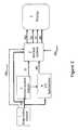
- FIG. 1is a block diagram illustrating flow of data through a system which includes a combination mammography/tomosynthesis acquisition station or a tomosynthesis only acquisition station and where reconstruction of tomosynthesis slice images Tr and synthesis of the Ms images occurs after storage of acquired tomosynthesis projection images Tp.
- FIG. 2is a block diagram illustrating flow of data through a system which includes a combination mammography/tomosynthesis acquisition station or a tomosynthesis only acquisition station and where the reconstruction of images Tr occurs before storage of the image data.
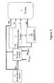
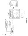
- FIG. 3is a block diagram illustrating flow of data through a system which includes a mammography-only acquisition system, and where reconstruction of tomosynthesis slice images Tr and/or synthesis of the Ms images occurs after storage of acquired tomosynthesis projection images Tp, FIG. 3 illustrates an example where four units acquiring Tp images feed a single unit that reconstructs Tr images.
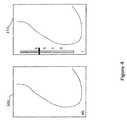
- FIG. 4illustrates a concurrent display of an Ms image and a Tr in substantially same area on a screen, with an example of a non-numeric indication of a thickness and position in the breast of a breast slice represented by a Tr image.
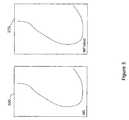
- FIG. 5illustrates a concurrent display of Ms and Mp images, at separate areas on a screen or as combined images.
- FIG. 6Aillustrates a display of a Tr image, with an integrated window including a scout view of a 2D synthesized image, for use in guiding a medical professional's evaluation of the Tr data.
- FIG. 6Billustrates a display of a Tr image with an integrated window that includes both an Ms image and a legacy Mp image, shown as scout views, for use in guiding a medical professional's evaluation and workflow using the Tr image data.
- FIG. 7illustrates a display of Ms/Mp/Tr/Tp images with CAD marks and a non-numeric indication of Tr images in which CAD marks exist.
- FIG. 8is a block diagram illustrating exemplary components of an x-ray acquisition and display system that incorporates the 2D synthesis method of the present invention.
- FIG. 9is a flow diagram provided to illustrate exemplary steps that may be performed in a 2D image synthesis process which uses tomosynthesis data.
- Mprefers to a conventional mammogram, which is a two-dimensional projection image of a breast and encompasses both a digital image as acquired by a flat panel detector or another imaging device and the image after conventional processing to prepare it for display to a health professional or for storage, e.g. in the PACS system of a hospital or another institution.
- Mp currentrefers to a mammogram that is acquired by an acquisition system for patient diagnosis
- Mp legacyrefers to a mammogram image of a patient that had been taken on a previous review of the patient, for example one that is to be used for comparison against an Mp current to identify changes in a patient's breast structure.
- Tprefers to an image that is similarly two-dimensional but is taken at a respective tomosynthesis angle between the breast and the origin of the imaging X-rays (typically the focal spot of an X-ray tube), and also encompasses the image as acquired as well as the image after being processed for display or for some other use.
- Trrefers to an image that is reconstructed from images Tp, for example in the manner described in said earlier-filed patent applications, and represents a slice of the breast as it would appear in a projection X-ray image of that slice at any desired angle, not only at an angle used for Tp or Mp images.
- Msrefers to synthesized 2D projection images which simulate mammography images, such as a craniocaudal (CC)_ or mediolateral oblique (MLO) images, and are constructed using tomosynthesis projection images Tp, tomosynthesis reconstructed images Tr or a combination thereof.
- Ms imagesmay be provided for display to a health professional or for storage in the PACS system of a hospital or another institution.
- Tp, Tr, Ms and Mpalso encompasses information, in whatever form, that is sufficient to describe such an image for display, further processing, or storage.
- the images Mp, Ms. Tp and Trtypically are in digital form before being displayed, and are defined by information identifying properties of each pixel in a two-dimensional array of pixels.
- the pixel valuestypically relate to respective measured or estimated or computed responses to X-rays of corresponding volumes in the breast (voxels or columns of tissue).
- the geometry of the tomosynthesis images (Tr and Tp) and mammography images (Ms, Mp)are matched to a common coordinate system as described in U.S.
- FIG. 1illustrates flow of data in one example of an image generation and display system disclosed in this patent specification.
- An image data acquisition system 1acquires tomosynthesis image data for Tp images of patients' breasts, and can take the form of and use the acquisition methods of any of the systems disclosed in said earlier-filed patent applications. If the system is a combo system, Mp images may also be generated (as indicated by dashed line and label Mp current in FIG. 1 ). Some dedicated tomosynthesis systems or combo systems may be adapted to accept and store legacy mammogram images (indicated via dashed line and legend Mp legacy in FIG. 1 ) in a Picture Archiving and Communication System (PACS) storage device 2 , although it is not a requirement that any Mp images be acquired or pre-stored.
- PPSPicture Archiving and Communication System
- the data describing projection images Tpare sent to storage device 2 , which is preferably a DICOM-compliant PACS.
- the data Tp imagesare sent, from either acquisition system 1 or from storage device 2 , to a computer system 3 configured as a reconstruction engine that can perform tomosynthesis reconstruction into images Tr representing breast slices of selected thickness and at selected orientations, as disclosed in said earlier-filed patent applications.
- the computer systemmay be further configured with 2D synthesis functionality 4 , which may operate substantially in parallel with reconstruction engine 3 to generate a synthesized 2D image (interchangeably referenced as T2d or Ms).
- T2dsynthesized 2D image
- the reconstructed slice images Trare then sent to a display system 5 so that they can be viewed. If the reconstruction engine 3 is connected to display 5 via a fast link, then large datasets can be transmitted quickly.
- Other images, such as the Ms, Mp and/or Tp imagesmay also be forwarded to the display unit for concurrent or toggled viewing.
- an improvement in reconstruction algorithmsimproves image quality so as to allow detection of a cancerous lesion in an image where it was not visible using a previous version of the reconstruction algorithm and the then existing standard of care. While it could be useful to see older images processed with the newer algorithms, it may also be important to allow the re-display of images as they were viewed during an original detection/diagnosis.
- One way to accomplish this in accordance with the disclosure in this patent specificationis to put a version number or some other information in the data for Tp images, which identifies the software and/or hardware versions of the Tp image data acquisition and/or Tr image reconstruction system at the time of acquisition, or to otherwise associate such information with the Tp images. During reconstruction at a later time, the reconstruction engine reads this version number or other similar information and reconstructs using the appropriate algorithm.
- system upgradescan maintain a library of older algorithms and/or hardware so as to be able to reconstruct using the proper technique.
- a patientmay have compiled a history of mammogram images associated with past screenings. Such a patient may be examined at a facility with updated equipment, for example one that includes a dedicated tomosynthesis system.
- a facility with updated equipmentfor example one that includes a dedicated tomosynthesis system.
- To compare historical information against existing diagnostic imagesit may be desirable to store such legacy Mp images, generate Ms images from a tomosynthesis acquisition of the dedicated tomo system and compare like-formatted representations.
- FIG. 2an alternative design of an image acquisition and display system of the present invention is illustrated in FIG. 2 .
- the reconstruction unit 3 and 2D synthesizer 4are directly coupled to the acquisition station 1 , and it is the reconstruction images Tr and synthesized images Ms that are sent to storage system 2 for subsequent display on display devices 5 , which may also store legacy Mp images.
- Tr and Ms images reconstructed or synthesized from Tp image data taken after the upgradewill automatically reflect this new algorithm
- Tr and Ms images reconstructed or synthesized from Tp image data taken prior to the upgradewill have been reconstructed with the older version and properly stored as such.
- the images stored on a PACSwill be the same as they were viewed by the radiologist or other health professional during the detection/diagnosis or other earlier review.
- Another advantage of the system of FIG. 2is the reduced system reconstruction burden compared to the system in FIG. 1 , where the reconstruction engine is just prior to the display. If there are multiple acquisition systems, for example four systems that are all pushing images to the display, then the reconstruction engine will need to reconstruct images at 4 times the rate of a reconstruction engine in a system having only one acquisition system, for the same total patient throughput.
- FIG. 3illustrates another image acquisition and display system which may benefit from the 2D image synthesizing process of the present invention.
- the image acquisition device 11is a mammography only device.
- One advantage of digital imagingis the portability of data; it is conceivable that patients that switch between different imaging locations may be exposed to imaging equipment with different capabilities. For example, a patient may undergo an exam at an imaging center that includes a tomosynthesis only system, and subsequently undergo an exam at a location that includes a mammography only acquisition system, (or visa versa).
- a 2D mammogramfrom existing Tr and/or Tp data.
- storage device 2is adapted to store both legacy Mp (if any) as well as legacy Tp images.
- the systemmay include reconstruction unit 3 .
- Other systems which have only the capability of viewing mammogramsmay not include this unit, and thus the unit and tomosynthesis data are all shown in dashed lines in FIG. 3 .
- Tp and/or Tr dataare retrieved from the storage device and forwarded to the 2D synthesizer 4 .
- the resulting Ms imageis displayed together with the current Mp on display 5 .
- the 2D synthesizing softwaremay be provided as a downloadable application that facilitates viewing of tomosynthesis data on existing mammography systems.
- One way to reduce the size of an original dataset for a Tp imageis to bin the projection Tp data to as large a pixel size as practical without reducing clinical efficacy of the final Ms, Tp or Tr images.
- Methods that can be used to reduce the Tp image sizeare described in U.S. patent application Ser. No. 11/271,050, (referred to herein as the '050 application) filed Nov. 10, 2005 by the assignee of the present invention, and incorporated by reference herein.
- the imagescan be formatted consistent with DICOM standards.
- each raw or displayed projection image set, synthesized image or reconstructed slice image set for a single viewis stored as a single Secondary Capture image instance according to DICOM.
- the image pixel datacan be encoded in a selected compressed format (CODEC) that includes all projection or slice images.
- the imaging and display system of the present inventionincludes a 2D synthesizer for generating 2D images simulating mammograms taken at both a CC and MLO orientation using a combination of one or more Tp and/or Tr images.
- a display of the systempreferably should be able to display Ms, Mp and Tr (and/or Tp) images concurrently (either in separate windows on the display or overlaid) or sequentially or in toggled mode, wherein the Ms, Mp, Tp and Tr images may be those currently acquired, or those that were acquired in previous studies.
- the displaycan simultaneously or sequentially or in toggled mode display mammograms (Ms, Mp) and tomosynthesis images Tr (and/or Tp) from the current and previous studies.
- Tr slicescan be reconstructed all to the same size, which can be the same as the size of an Mp or Ms image of the breast, or they can be initially reconstructed to sizes determined by the fan shape of the x-ray beam used in the acquisition and later converted to that same size by appropriate interpolate]on/extrapolation.
- Images of different types and from different sourcescan be displayed in desirable size and resolution.
- an imagecan be displayed in (1) Fit To View Port mode, in which the size of the displayed image size is maximized such that the entire imaged breast tissue is visible, (2) True Size mode, in which a display pixel on the screen corresponds to a pixel of the image, or (3) Right Size mode, in which the size of a displayed image is adjusted so that it matches that of another image that is concurrently displayed or with which the displayed image is or can be toggled.
- Selected hanging protocolsare provided that are unique to the different types of images with which the disclosed system deals.
- the hanging protocols for 2D imagese.g. Ms or Mp images
- 3D imagese.g. Tr images
- the hanging protocols for 2D imagesare linked so that when one type of image is displayed for a given breast the other type is displayed as well.
- Ms/Mp image of a breastis displayed
- a tile of the Tr images and/or of the Tp imagesis automatically displayed at the same time, with a desired hanging protocol that may involve scrolling or cine mode presentation, or may require user input so select a particular subset of the Tr and/or Tp images or a particular individual Tr/Tp image.
- a combined hanging protocol setcan be provided for 2D and 3D images that are concurrently displayed or toggled such that only one type is displayed at one time.
- the combined hanging protocolcan include provisions for linked display of CAD information associated with one or both of the 2D and 3D images involved in the hanging protocol.
- the hanging protocols for 2D imagesare made different from those for 3D images.
- FIG. 4An icon is used to identify an image type.
- the symbol MS on the left image 300indicates that it is a synthesized mammogram.
- the symbol T on the right image 310indicates that it is a tomosynthesis slice image Tr.
- a symbol Tp(not shown) can be used to indicate that the displayed image is a tomosynthesis projection image Tp
- a symbol 2Dmay be used to indicate that it is a 2D image
- the symbol 3D(also not shown) can be used to indicate that an image on the display is a 3D image.
- the imagescan be displayed without an identification of the type of image.
- a Tr image and an Mp or Ms imagecan be displayed at the same time or toggled without displaying an indication of the type of the image that is visible. This may be desirable in cases such as when a user has a familiar hanging protocol and does not need an express identification of the type of image.
- the system described as a non-limiting example in this patent specificationis capable of receiving and displaying selectively the tomosynthesis projection images Tp, the tomosynthesis reconstruction images Tr, the synthesized mammogram image Ms and/or the mammogram images Mp, or a single type, or any sub combination of types. It can receive images stored uncompressed, losslessly compressed, and lossyly compressed. It can also include algorithms to decompress images sent in compressed format.
- the systemhas software to perform reconstruction of tomosynthesis image data for images Tp into images Tr and software for synthesizing mammogram images Ms. Further, it can include software to generate 3D display images from the tomosynthesis reconstructed images Tr using standard known methods such as MIP (Maximum Intensity Projection), summing, and/or weighted summing algorithms.
- FIG. 5illustrates an exemplary display of an Mp image together with a 2D synthesized image; each image may be labeled to indicate whether the image is from a current acquisition, or based on legacy data.
- the Mp imagemay be a stored legacy mammogram
- the 2D Ms imagemay be generated from a current tomosynthesis acquisition and may be provided as an initial view to guide the medical professional's perusal of the tomosynthesis data.
- the Mp imagemay be based on a current acquisition, for example via a combo mammo/tomo system or by a mammography-only system, and the Ms image may be generated from previously stored tomosynthesis data, such as described in FIG. 3 , thereby allowing for comparison of like images to more easily identify regions of interest.
- FIG. 6Aillustrates the display of the synthesized 2D image as a scout view thumbnail image which may be provided as an overlay in a display of Tp and/or Tr images. Such a scout view may be used to guide the workflow of the medical professional during review of the tomosynthesis images.
- FIG. 61Billustrates two scout views, showing, for example, a legacy 2D image together with a current 2D image. The ability to compare the two images may further assist the medical workflow.
- FIG. 6Billustrates a legacy Mp image together with an Ms image
- any two 2D images associated with differently timed acquisitionscould be used, and the present invention is not limited that the particular images shown in the figures.
- FIG. 7illustrates a display of two synthesized 2D images, of different views (CC and MLO).
- a Computer Assisted Detection (CAD) processhas been applied to either the synthesized 2D views or alternatively to the reconstructed tomosynthesis data, providing resulting CAD marks 350 .
- the CAD marks resulting from processing a mammogrammay be projected onto the 3D tomosynthesis image and visa versa.
- the present inventionfurther envisions that the CAD marks may be similarly translated across images when using 2D synthesized images.
- a menu driven interfacemay be automatically populated with the types of images that are available for display, including both currently acquired images and a selection of available legacy images.
- Softwareallows the selection of one or more image planes, for use in image processing, or to change window/level or to change slice height, etc.
- the menu driven interfacemay be further populated with display arrangements, including overlaid, cine, inset views, etc.
- Alternative methods, such as drag and drop techniquescan be used to position images on the screen. These sets of images can be on one monitor, or on multiple monitors or other displays.
- the displaycan map the images so the pixel spacings are identical.
- This pixel spacing adjustmentcan also be used for Ms, Mp and Tr/Tp images.
- the Ms, Mp and Tr/Tp imagesare displayed at the same pixel size.
- Zoomingcan be done on any of the images on the display.
- the zoomed areawill zoom both the Ms/Mp and the Tr slice images as they are toggled.
- Window/levelcan be independently, or jointly, applied to any combination of images on the display.
- the window/levelcan be applied to just the single displayed Tr slice image, or all the Tr slice images. If there is a magnified region of an image, window/level can be selectively applied just to the magnified region or to the entire image.
- FIG. 8illustrates an overall mammography/tomography system in which the preferred but non-limiting examples discussed above can be implemented.
- the Figureillustrates in block diagram form an x-ray data acquisition unit 100 that includes an x-ray source 110 imaging a breast 112 .
- An x-ray imager 116such as a flat panel x-ray imager commercially available from the assignee of this patent specification generates projection image data that can be a mammogram Mp or a tomosynthesis projection image Tp.
- X-ray source 110is mounted for movement so that images Tp can be taken at different angles.
- X-ray imager 116can be stationary or it can also move, preferably in synchronism with movement of x-ray source 110 .
- Elements 110 and 116communicate with x-ray data acquisition control 118 that controls operations in a manner known from said earlier-filed patent specifications.
- X-ray image data from imager 116is delivered to processing unit 120 .
- Processing unit 120comprises reconstruction software 122 , which may be stored in a computer readable medium of unit 12 .
- the reconstruction softwareprocesses x-ray image data as known from said earlier-filed patent application into Tp and Tr image data, which may be stored in storage device 130 and displayed at image display unit 150 as disclosed in the various embodiments described above.
- Processing unit 120further includes 2D synthesis software which uses one or more of the Tp and/or Tr images to synthesize a 2D image.
- any Tp image taken during the tomosynthesis scanmay be used as 2D image.
- An alternate methodwill now be described although it should be understood that the present invention is not limited to any particular method of synthesizing a 2D image, but rather encompasses any synthesizing technique which can be used to generate a 2D image from a tomosynthesis data set.
- a tomo data setconsists of Tp 0 raw projections, Tp processed projections, and Tr reconstructed slices.
- the Tp processed projectionshave been processed as described in the '650 application to perform at least one of coordinate geometry matching and data set size reduction.
- a single ‘synthesized’ 2D image T2dis built from the 30 tomo data set alone.
- the 2D synthesized imagemay provide a quick overview of the breast anatomy to facilitate diagnosis and help the radiologist focus on specific regions when analyzing the 3D slices.
- the image T2dmay replace the Mp image that would normally be present in a combo mode procedure, or may be viewed against legacy Mp images, or displayed in a variety of other combinations.
- FIG. 9is a flow diagram that illustrates exemplary steps that may be performed in a 2D synthesis process.
- FIG. 9assumes that methods described in the '650 application have been used to generate a set of slices Tr as in put to the process, wherein the set of slices are represented in a Cone Beam or Cartesian coordinate system.
- the Tr data setis apportioned into a slabbed set of slices Tslab. That is a number of images Tr are effectively combined, using maximum intensity projection (MIP) or averaging to generate a set of Tslab slices. Equation I below illustrates how the set Tslab is formed using MIP, while Equation II below illustrates how the set Tslab may be formed using averaging.
- MIPmaximum intensity projection
- Tr[j, z]where j is the pixel index of the image and z is the slice number.
- T slab[ j,z ]MAX( Tr[j,z ⁇ N slab/2], Tr[j,z ⁇ N slab/2+1, . . . , Tr[j,z+N slab/2])
- T slab[ j,z ]AVE( Tr[j,z ⁇ N slab/2], Tr[j,z ⁇ N slab/2+1, . . . , Tr[j,z+N slab/2]) Equation II
- the set of slicesis re-projected to produce an initial image T2d0.
- Re-projection methodsare well known in the field of image processing. A source point and image plane is chosen, on opposite sides of the image volume. Pixels are obtained by projecting the source point through the slice set to an image plane point. The pixel value is summed at each slice location by interpolating values in the original slices. Note, in the case of cone beam coordinate system reconstruction as described in the '650 application, the re-projection is just a sum of pixel values, with no interpolation involved, and is represented by Equation III below:
- zminmay be chosen to exclude slices near the breast boundary, or skin. This may reduce artifacts.
- Nzmax ⁇ zmin+1.
- Step 930performs an optional step of filtering the re-projected image to produce T2d.
- the filtering that is performedshould be generally in the direction of the source motion in the original tomosynthesis image acquisition. Although not required, filtering may help reduce additional blur produced in the re-projection due to artifacts in the slices Tr. It is further noted that the filtering step 930 may be performed prior to the re-projection of step 920, but at a computational cost.
- the advantages of using the cone beam geometry reconstructed slices as inputare as follows. Interpolation at step 920 because geometric correlation was already performed in the reconstruction. Thus the final image T2d will be registered geometrically with the original set of slices Tr, as described in the '650 application. The registration would facilitate diagnosis as well as the display of CAD results on T2d, where the CAD results are derived from the 3D images Tr. The 3D CAD results may also be re-projected (or summed) as in step 2 and overlaid on T2d.
- the synthesized imagesmay be generated and displayed in conjunction with combination mammography/tomography acquisition stations and tomosynthesis only acquisition stations.
- the imagesmay even be generated and displayed in combination with mammography only acquisition stations when legacy tomosynthesis data is available.
Landscapes
- Engineering & Computer Science (AREA)
- Health & Medical Sciences (AREA)
- Life Sciences & Earth Sciences (AREA)
- Physics & Mathematics (AREA)
- Medical Informatics (AREA)
- Theoretical Computer Science (AREA)
- General Physics & Mathematics (AREA)
- Radiology & Medical Imaging (AREA)
- General Health & Medical Sciences (AREA)
- Nuclear Medicine, Radiotherapy & Molecular Imaging (AREA)
- Surgery (AREA)
- Pathology (AREA)
- Optics & Photonics (AREA)
- Biomedical Technology (AREA)
- Heart & Thoracic Surgery (AREA)
- Molecular Biology (AREA)
- High Energy & Nuclear Physics (AREA)
- Animal Behavior & Ethology (AREA)
- Biophysics (AREA)
- Public Health (AREA)
- Veterinary Medicine (AREA)
- Computer Graphics (AREA)
- Oral & Maxillofacial Surgery (AREA)
- Dentistry (AREA)
- Algebra (AREA)
- Mathematical Analysis (AREA)
- Mathematical Optimization (AREA)
- Mathematical Physics (AREA)
- Pure & Applied Mathematics (AREA)
- Quality & Reliability (AREA)
- Computer Vision & Pattern Recognition (AREA)
- Apparatus For Radiation Diagnosis (AREA)
- Image Processing (AREA)
- Ultra Sonic Daignosis Equipment (AREA)
Abstract
Description
Tslab[j,z]=MAX(Tr[j,z−Nslab/2],Tr[j,z−Nslab/2+1, . . . ,Tr[j,z+Nslab/2])
Tslab[j,z]=AVE(Tr[j,z−Nslab/2],Tr[j,z−Nslab/2+1, . . . ,Tr[j,z+Nslab/2]) Equation II
Claims (18)
Priority Applications (19)
| Application Number | Priority Date | Filing Date | Title |
|---|---|---|---|
| US12/276,006US7760924B2 (en) | 2002-11-27 | 2008-11-21 | System and method for generating a 2D image from a tomosynthesis data set |
| US12/471,981US8571289B2 (en) | 2002-11-27 | 2009-05-26 | System and method for generating a 2D image from a tomosynthesis data set |
| CN200980101409.XACN102763137B (en) | 2008-11-21 | 2009-11-20 | Systems and methods for generating 2D images from tomographic datasets |
| PCT/US2009/065288WO2010059920A2 (en) | 2008-11-21 | 2009-11-20 | System and method for generating a 2d image from a tomosynthesis data set |
| DE202009019204.0UDE202009019204U1 (en) | 2008-11-21 | 2009-11-20 | X-ray detection system for generating a synthesized 2D mammogram from images reconstructed by tomosynthesis |
| EP18207785.9AEP3471064A1 (en) | 2008-11-21 | 2009-11-20 | System and method for generating a synthesized mammogram from a subset of tomosynthesis projection images |
| CN201610209698.7ACN105832360B (en) | 2008-11-21 | 2009-11-20 | System and method for generating 2D image by chromatographic data collection |
| JP2011537644AJP5501370B2 (en) | 2008-11-21 | 2009-11-20 | System and method for generating 2D images from tomosynthesis data sets |
| EP09796173AEP2215600A2 (en) | 2008-11-21 | 2009-11-20 | System and method for generating a 2d image from a tomosynthesis data set |
| CA2702782ACA2702782C (en) | 2008-11-21 | 2009-11-20 | System and method for generating a 2d image from a tomosynthesis data set |
| US14/360,389US10008184B2 (en) | 2005-11-10 | 2012-11-26 | System and method for generating a 2D image using mammography and/or tomosynthesis image data |
| US14/044,959US8897535B2 (en) | 2002-11-27 | 2013-10-03 | System and method for generating a 2D image from a tomosynthesis data set |
| JP2014047021AJP5837116B2 (en) | 2008-11-21 | 2014-03-11 | System and method for generating 2D images from tomosynthesis data sets |
| US14/549,604US9456797B2 (en) | 2002-11-27 | 2014-11-21 | System and method for generating a 2D image from a tomosynthesis data set |
| JP2015216402AJP6208731B2 (en) | 2008-11-21 | 2015-11-04 | System and method for generating 2D images from tomosynthesis data sets |
| US15/088,844US9808215B2 (en) | 2002-11-27 | 2016-04-01 | System and method for generating a 2D image from a tomosynthesis data set |
| JP2017171832AJP6530456B2 (en) | 2008-11-21 | 2017-09-07 | System and method for generating 2D images from tomosynthesis data sets |
| US15/802,225US10010302B2 (en) | 2002-11-27 | 2017-11-02 | System and method for generating a 2D image from a tomosynthesis data set |
| US16/013,782US10413263B2 (en) | 2002-11-27 | 2018-06-20 | System and method for generating a 2D image from a tomosynthesis data set |
Applications Claiming Priority (8)
| Application Number | Priority Date | Filing Date | Title |
|---|---|---|---|
| US10/305,480US7123684B2 (en) | 2002-11-27 | 2002-11-27 | Full field mammography with tissue exposure control, tomosynthesis, and dynamic field of view processing |
| US10/723,486US7831296B2 (en) | 2002-11-27 | 2003-11-26 | X-ray mammography with tomosynthesis |
| US62851604P | 2004-11-15 | 2004-11-15 | |
| US63129604P | 2004-11-26 | 2004-11-26 | |
| US11/271,050US7577282B2 (en) | 2002-11-27 | 2005-11-10 | Image handling and display in X-ray mammography and tomosynthesis |
| US60406906A | 2006-11-24 | 2006-11-24 | |
| US11/827,909US7616801B2 (en) | 2002-11-27 | 2007-07-13 | Image handling and display in x-ray mammography and tomosynthesis |
| US12/276,006US7760924B2 (en) | 2002-11-27 | 2008-11-21 | System and method for generating a 2D image from a tomosynthesis data set |
Related Parent Applications (1)
| Application Number | Title | Priority Date | Filing Date |
|---|---|---|---|
| US11/827,909Continuation-In-PartUS7616801B2 (en) | 2002-11-27 | 2007-07-13 | Image handling and display in x-ray mammography and tomosynthesis |
Related Child Applications (1)
| Application Number | Title | Priority Date | Filing Date |
|---|---|---|---|
| US12/471,981Continuation-In-PartUS8571289B2 (en) | 2002-11-27 | 2009-05-26 | System and method for generating a 2D image from a tomosynthesis data set |
Publications (2)
| Publication Number | Publication Date |
|---|---|
| US20090123052A1 US20090123052A1 (en) | 2009-05-14 |
| US7760924B2true US7760924B2 (en) | 2010-07-20 |
Family
ID=42198818
Family Applications (1)
| Application Number | Title | Priority Date | Filing Date |
|---|---|---|---|
| US12/276,006Expired - LifetimeUS7760924B2 (en) | 2002-11-27 | 2008-11-21 | System and method for generating a 2D image from a tomosynthesis data set |
Country Status (7)
| Country | Link |
|---|---|
| US (1) | US7760924B2 (en) |
| EP (2) | EP2215600A2 (en) |
| JP (4) | JP5501370B2 (en) |
| CN (2) | CN102763137B (en) |
| CA (1) | CA2702782C (en) |
| DE (1) | DE202009019204U1 (en) |
| WO (1) | WO2010059920A2 (en) |
Cited By (72)
| Publication number | Priority date | Publication date | Assignee | Title |
|---|---|---|---|---|
| US20080123916A1 (en)* | 2006-05-22 | 2008-05-29 | Upmc | System and Method for Improved Viewing and Navigation of Digital Images |
| US20080155451A1 (en)* | 2006-12-21 | 2008-06-26 | Sectra Ab | Dynamic slabbing to render views of medical image data |
| US20080152086A1 (en)* | 2006-12-21 | 2008-06-26 | Sectra Ab | Synchronized viewing of tomosynthesis and/or mammograms |
| US20080155468A1 (en)* | 2006-12-21 | 2008-06-26 | Sectra Ab | Cad-based navigation of views of medical image data stacks or volumes |
| US20090268865A1 (en)* | 2003-11-26 | 2009-10-29 | Baorui Ren | X-ray imaging with X-ray markers that provide adjunct information but preserve image quality |
| US20110135185A1 (en)* | 2002-11-27 | 2011-06-09 | Hologic, Inc. | Image handling and display in x-ray mammography and tomosynthesis |
| DE102011076929A1 (en) | 2011-06-03 | 2012-12-06 | Siemens Ag | Method and apparatus for displaying volume data for a study of density properties |
| DE102011076930A1 (en) | 2011-06-03 | 2012-12-06 | Siemens Aktiengesellschaft | Method and device for adapting the representation of volume data of an object |
| US8705690B2 (en) | 2011-01-25 | 2014-04-22 | Siemens Aktiengesellschaft | Imaging method with improved display of a tissue region, imaging device, and computer program product |
| DE102013211547B3 (en)* | 2013-06-19 | 2014-05-22 | Siemens Aktiengesellschaft | Method for producing tomo-synthetic X-ray image of breast of patient, involves producing difference image from synthetic projection image and projection image by subtraction, and using difference image for reconstructing X-ray image |
| WO2014207080A1 (en)* | 2013-06-28 | 2014-12-31 | Koninklijke Philips N.V. | Methods for generation of edge-preserving synthetic mammograms from tomosynthesis data |
| WO2015054518A1 (en)* | 2013-10-09 | 2015-04-16 | Hologic, Inc | X-ray breast tomosynthesis enhancing spatial resolution including in the thickness direction of a flattened breast |
| US9020579B2 (en) | 2011-03-08 | 2015-04-28 | Hologic, Inc. | System and method for dual energy and/or contrast enhanced breast imaging for screening, diagnosis and biopsy |
| WO2015063188A1 (en)* | 2013-10-30 | 2015-05-07 | Koninklijke Philips N.V. | Method and device for displaying medical images |
| US9047498B2 (en) | 2011-11-29 | 2015-06-02 | Siemens Aktiengesellschaft | Method for reconstructing a reconstruction data set containing two-dimensional virtual X-ray images |
| US9066706B2 (en) | 2004-11-26 | 2015-06-30 | Hologic, Inc. | Integrated multi-mode mammography/tomosynthesis x-ray system and method |
| US20160035102A1 (en)* | 2014-07-31 | 2016-02-04 | Siemens Aktiengesellschaft | Method and system for computing digital tomosynthesis images |
| WO2016078958A1 (en) | 2014-11-20 | 2016-05-26 | Koninklijke Philips N.V. | Method for generation of synthetic mammograms from tomosynthesis data |
| US9460508B2 (en) | 2002-11-27 | 2016-10-04 | Hologic, Inc. | Image handling and display in X-ray mammography and tomosynthesis |
| US9456797B2 (en) | 2002-11-27 | 2016-10-04 | Hologic, Inc. | System and method for generating a 2D image from a tomosynthesis data set |
| US9498175B2 (en) | 2002-11-27 | 2016-11-22 | Hologic, Inc. | System and method for low dose tomosynthesis |
| DE102015217141A1 (en) | 2015-09-08 | 2017-03-09 | Siemens Healthcare Gmbh | Generating contrast-enhanced image data of breast tissue to be examined |
| US9805507B2 (en) | 2012-02-13 | 2017-10-31 | Hologic, Inc | System and method for navigating a tomosynthesis stack using synthesized image data |
| US9808214B2 (en) | 2010-10-05 | 2017-11-07 | Hologic, Inc. | Upright X-ray breast imaging with a CT mode, multiple tomosynthesis modes, and a mammography mode |
| US9940738B2 (en) | 2013-01-10 | 2018-04-10 | Hologic, Inc. | System and method for reducing data transmission volume in tomosynthesis |
| US10008184B2 (en) | 2005-11-10 | 2018-06-26 | Hologic, Inc. | System and method for generating a 2D image using mammography and/or tomosynthesis image data |
| US20180256126A1 (en)* | 2014-09-30 | 2018-09-13 | Fujifilm Corporation | Image displaying device, image processing device, radiographic imaging system, sectional image displaying method, and non-transitory computer readable medium |
| WO2018183550A1 (en) | 2017-03-30 | 2018-10-04 | Hologic, Inc. | System and method for targeted object enhancement to generate synthetic breast tissue images |
| WO2018183548A1 (en) | 2017-03-30 | 2018-10-04 | Hologic, Inc. | System and method for hierarchical multi-level feature image synthesis and representation |
| WO2018183549A1 (en) | 2017-03-30 | 2018-10-04 | Hologic, Inc. | System and method for synthesizing low-dimensional image data from high-dimensional image data using an object grid enhancement |
| US10111631B2 (en) | 2014-02-28 | 2018-10-30 | Hologic, Inc. | System and method for generating and displaying tomosynthesis image slabs |
| US10332280B2 (en) | 2013-10-24 | 2019-06-25 | Canon Kabushiki Kaisha | Information processing apparatus, information processing method, and control apparatus |
| US10346977B2 (en) | 2017-01-23 | 2019-07-09 | Electronics & Telecommunications Research Institute | Method and device for generating 2D medical image based on plate interpolation |
| US10535167B2 (en) | 2014-12-31 | 2020-01-14 | General Electric Company | Method and system for tomosynthesis projection image enhancement and review |
| US10573276B2 (en) | 2011-11-27 | 2020-02-25 | Hologic, Inc. | System and method for generating a 2D image using mammography and/or tomosynthesis image data |
| WO2020068767A1 (en) | 2018-09-28 | 2020-04-02 | Hologic, Inc. | System and method for synthetic breast tissue image generation by high density element suppression |
| US10638994B2 (en) | 2002-11-27 | 2020-05-05 | Hologic, Inc. | X-ray mammography with tomosynthesis |
| EP3646798A1 (en) | 2013-10-24 | 2020-05-06 | Hologic, Inc. | System and method for navigating x-ray guided breast biopsy |
| US10702233B2 (en) | 2017-09-28 | 2020-07-07 | Siemens Healthcare Gmbh | Determining a two-dimensional mammography dataset |
| US10830712B2 (en)* | 2017-03-27 | 2020-11-10 | KUB Technologies, Inc. | System and method for cabinet x-ray systems with camera |
| US10881359B2 (en) | 2017-08-22 | 2021-01-05 | Hologic, Inc. | Computed tomography system for imaging multiple anatomical targets |
| US10959694B2 (en) | 2002-11-27 | 2021-03-30 | Hologic, Inc. | Full field mammography with tissue exposure control, tomosynthesis, and dynamic field of view processing |
| US10987081B2 (en) | 2019-06-21 | 2021-04-27 | Fujifilm Corporation | Radiation imaging system and transmission device |
| US11076820B2 (en) | 2016-04-22 | 2021-08-03 | Hologic, Inc. | Tomosynthesis with shifting focal spot x-ray system using an addressable array |
| US11090017B2 (en) | 2018-09-13 | 2021-08-17 | Hologic, Inc. | Generating synthesized projection images for 3D breast tomosynthesis or multi-mode x-ray breast imaging |
| US20210256742A1 (en)* | 2015-07-28 | 2021-08-19 | PME IP Pty Ltd | Apparatus and method for visualizing digital breast tomosynthesis and other volumetric images |
| US11403483B2 (en) | 2017-06-20 | 2022-08-02 | Hologic, Inc. | Dynamic self-learning medical image method and system |
| US11419569B2 (en) | 2017-08-16 | 2022-08-23 | Hologic, Inc. | Image quality compliance tool |
| US20220270306A1 (en)* | 2021-02-19 | 2022-08-25 | Fujifilm Corporation | Information processing device, information processing method, program, and radiography system |
| US11449999B2 (en) | 2019-09-30 | 2022-09-20 | Fujifilm Corporation | Display control device, method for operating display control device, and program for operating display control device |
| US11452486B2 (en) | 2006-02-15 | 2022-09-27 | Hologic, Inc. | Breast biopsy and needle localization using tomosynthesis systems |
| US11471118B2 (en) | 2020-03-27 | 2022-10-18 | Hologic, Inc. | System and method for tracking x-ray tube focal spot position |
| US11481038B2 (en) | 2020-03-27 | 2022-10-25 | Hologic, Inc. | Gesture recognition in controlling medical hardware or software |
| US11510306B2 (en) | 2019-12-05 | 2022-11-22 | Hologic, Inc. | Systems and methods for improved x-ray tube life |
| US11589944B2 (en) | 2013-03-15 | 2023-02-28 | Hologic, Inc. | Tomosynthesis-guided biopsy apparatus and method |
| US11694792B2 (en) | 2019-09-27 | 2023-07-04 | Hologic, Inc. | AI system for predicting reading time and reading complexity for reviewing 2D/3D breast images |
| US11701199B2 (en) | 2009-10-08 | 2023-07-18 | Hologic, Inc. | Needle breast biopsy system and method of use |
| US11775156B2 (en) | 2010-11-26 | 2023-10-03 | Hologic, Inc. | User interface for medical image review workstation |
| US11783476B2 (en) | 2019-10-25 | 2023-10-10 | DeepHealth, Inc. | System and method for analyzing three-dimensional image data |
| US11786191B2 (en) | 2021-05-17 | 2023-10-17 | Hologic, Inc. | Contrast-enhanced tomosynthesis with a copper filter |
| US11883206B2 (en) | 2019-07-29 | 2024-01-30 | Hologic, Inc. | Personalized breast imaging system |
| US12029499B2 (en) | 2018-05-04 | 2024-07-09 | Hologic, Inc. | Biopsy needle visualization |
| US12121304B2 (en) | 2018-05-04 | 2024-10-22 | Hologic, Inc. | Introducer and localization wire visualization |
| US12170140B2 (en) | 2018-11-25 | 2024-12-17 | Hologic, Inc. | Customizable multimodality image hanging protocols |
| US12186119B2 (en) | 2021-10-05 | 2025-01-07 | Hologic, Inc. | Interactive model interface for image selection in medical imaging systems |
| US12191027B2 (en) | 2019-03-29 | 2025-01-07 | Hologic, Inc. | Snip-triggered digital image report generation |
| US12211608B2 (en) | 2013-03-15 | 2025-01-28 | Hologic, Inc. | System and method for navigating a tomosynthesis stack including automatic focusing |
| US12236597B2 (en) | 2021-11-29 | 2025-02-25 | Hologic, Inc. | Systems and methods for correlating objects of interest |
| US12236582B2 (en) | 2018-09-24 | 2025-02-25 | Hologic, Inc. | Breast mapping and abnormality localization |
| US12254586B2 (en) | 2021-10-25 | 2025-03-18 | Hologic, Inc. | Auto-focus tool for multimodality image review |
| US12367574B2 (en) | 2019-12-23 | 2025-07-22 | DeepHealth, Inc. | Systems and methods for analyzing two-dimensional and three-dimensional image data |
| US12414217B2 (en) | 2022-02-07 | 2025-09-09 | Hologic, Inc. | Systems and methods for adaptively controlling filament current in an X-ray tube |
Families Citing this family (71)
| Publication number | Priority date | Publication date | Assignee | Title |
|---|---|---|---|---|
| US7787672B2 (en) | 2004-11-04 | 2010-08-31 | Dr Systems, Inc. | Systems and methods for matching, naming, and displaying medical images |
| US7660488B2 (en) | 2004-11-04 | 2010-02-09 | Dr Systems, Inc. | Systems and methods for viewing medical images |
| US7920152B2 (en) | 2004-11-04 | 2011-04-05 | Dr Systems, Inc. | Systems and methods for viewing medical 3D imaging volumes |
| US7970625B2 (en) | 2004-11-04 | 2011-06-28 | Dr Systems, Inc. | Systems and methods for retrieval of medical data |
| US7885440B2 (en) | 2004-11-04 | 2011-02-08 | Dr Systems, Inc. | Systems and methods for interleaving series of medical images |
| US7953614B1 (en) | 2006-11-22 | 2011-05-31 | Dr Systems, Inc. | Smart placement rules |
| US8380533B2 (en) | 2008-11-19 | 2013-02-19 | DR Systems Inc. | System and method of providing dynamic and customizable medical examination forms |
| US8712120B1 (en) | 2009-09-28 | 2014-04-29 | Dr Systems, Inc. | Rules-based approach to transferring and/or viewing medical images |
| US9087400B2 (en) | 2009-12-17 | 2015-07-21 | Koninklijke Philips N.V. | Reconstructing an object of interest |
| FR2954556B1 (en)* | 2009-12-22 | 2017-07-28 | Gen Electric | METHOD OF PROCESSING TOMOSYNTHESIS ACQUISITIONS TO OBTAIN REPRESENTATION OF THE CONTENT OF AN ORGAN |
| FR2967520B1 (en)* | 2010-11-16 | 2012-12-21 | Gen Electric | METHOD FOR PROCESSING RADIOLOGICAL IMAGES OF A PATIENT |
| JP2012245329A (en)* | 2011-05-31 | 2012-12-13 | Fujifilm Corp | Image processing apparatus, radiographic image radiographing system, image processing program, and image processing method |
| US9075899B1 (en) | 2011-08-11 | 2015-07-07 | D.R. Systems, Inc. | Automated display settings for categories of items |
| DE102012215997B4 (en) | 2012-09-10 | 2022-10-06 | Siemens Healthcare Gmbh | Contrast-enhanced recording of objects |
| JP5893540B2 (en)* | 2012-09-28 | 2016-03-23 | 富士フイルム株式会社 | Image display system, radiation image capturing system, image display control program, and image display control method |
| US9495604B1 (en) | 2013-01-09 | 2016-11-15 | D.R. Systems, Inc. | Intelligent management of computerized advanced processing |
| WO2014109197A1 (en) | 2013-01-11 | 2014-07-17 | 富士フイルム株式会社 | Image display device, image display method, medical image diagnosatic device, medical image diagnosatic method, medical image diagnosatic system, data preparation device, data preparation method, program, and recording medium |
| DK3005298T3 (en)* | 2013-06-06 | 2019-07-22 | Volpara Health Tech Limited | PROCEDURE FOR RECONSTRUCTION OF A PROJECT IMAGE PROJECT |
| JP6027687B2 (en) | 2013-09-30 | 2016-11-16 | 富士フイルム株式会社 | Breast thickness measuring apparatus and breast thickness measuring method |
| KR102096410B1 (en)* | 2014-05-02 | 2020-04-03 | 삼성전자주식회사 | Medical image apparatus and control method for the same |
| JP6127032B2 (en)* | 2014-09-26 | 2017-05-10 | 富士フイルム株式会社 | Radiation image capturing system, image processing apparatus, image processing method, and image processing program |
| KR101838626B1 (en) | 2014-11-18 | 2018-03-14 | 한국과학기술원 | Mammogram synthesizing device and method using digital breast tomosynthetic images |
| GB2533801B (en) | 2014-12-31 | 2018-09-12 | Gen Electric | Method and system for tomosynthesis projection images enhancement |
| JP2016131573A (en)* | 2015-01-15 | 2016-07-25 | キヤノン株式会社 | Control device of tomosynthesis imaging, radiographic device, control system, control method, and program |
| US20170046483A1 (en) | 2015-04-30 | 2017-02-16 | D.R. Systems, Inc. | Database systems and interactive user interfaces for dynamic interaction with, and comparison of, digital medical image data |
| JP6502509B2 (en) | 2015-09-10 | 2019-04-17 | 富士フイルム株式会社 | Image processing apparatus, radiographic imaging system, image processing method, and image processing program |
| JP6556005B2 (en) | 2015-09-29 | 2019-08-07 | 富士フイルム株式会社 | Tomographic image generating apparatus, method and program |
| JP2017143943A (en) | 2016-02-16 | 2017-08-24 | 富士フイルム株式会社 | Radiation image processing apparatus, method and program |
| JP6821943B2 (en)* | 2016-04-22 | 2021-01-27 | コニカミノルタ株式会社 | Image generation system |
| US10973479B2 (en) | 2016-05-16 | 2021-04-13 | Canon Medical Systems Corporation | X-ray diagnosis apparatus, X-ray diagnosis apparatus controlling method, and X-ray diagnosis system |
| JP6853004B2 (en)* | 2016-09-16 | 2021-03-31 | キヤノンメディカルシステムズ株式会社 | Medical image processing equipment and mammography equipment |
| CN111343922B (en)* | 2017-12-14 | 2024-09-24 | 株式会社岛津制作所 | Radiation tomographic image processing apparatus and radiation tomographic imaging apparatus |
| CN108852392A (en)* | 2018-05-21 | 2018-11-23 | 苏州达影医疗设备有限公司 | Cone-shaped beam image reconstruction method, image system and camera chain |
| WO2020066109A1 (en)* | 2018-09-27 | 2020-04-02 | 富士フイルム株式会社 | Tomographic image generation device, method, and program |
| JP7017492B2 (en) | 2018-09-27 | 2022-02-08 | 富士フイルム株式会社 | Tomographic image generator, method and program |
| EP3657442B1 (en)* | 2018-11-23 | 2022-10-26 | Siemens Healthcare GmbH | Synthetic mammogramm with embossed image impression |
| JP7387270B2 (en) | 2019-03-06 | 2023-11-28 | キヤノンメディカルシステムズ株式会社 | Medical image processing device, learning method, X-ray diagnostic device, medical image processing method, and program |
| JP7242436B2 (en) | 2019-06-13 | 2023-03-20 | 富士フイルム株式会社 | Image interpretation support device, image interpretation support method, and image interpretation support program |
| JP7236948B2 (en) | 2019-07-16 | 2023-03-10 | 富士フイルム株式会社 | Image processing system, image processing method, and image processing program |
| JP7113790B2 (en)* | 2019-07-29 | 2022-08-05 | 富士フイルム株式会社 | Image processing device, method and program |
| JP7209599B2 (en) | 2019-07-29 | 2023-01-20 | 富士フイルム株式会社 | Image processing device, method and program |
| JP2021029285A (en)* | 2019-08-15 | 2021-03-01 | 富士フイルム株式会社 | Image display devices, methods and programs, image management devices, methods and programs |
| JP7326070B2 (en) | 2019-08-27 | 2023-08-15 | 富士フイルム株式会社 | Image display device, method and program, image management device, method and program |
| JP7208874B2 (en) | 2019-08-27 | 2023-01-19 | 富士フイルム株式会社 | Imaging control device, method and program |
| JP7203705B2 (en)* | 2019-09-17 | 2023-01-13 | 富士フイルム株式会社 | Image processing device, method and program, and image display device, method and program |
| JP7542059B2 (en)* | 2019-09-27 | 2024-08-29 | ホロジック, インコーポレイテッド | Motion detection of internal breast tissue in tomosynthesis |
| EP4101385B1 (en) | 2020-02-04 | 2024-10-16 | FUJIFILM Corporation | Image setting device, method, and program |
| EP4101386A4 (en) | 2020-02-04 | 2023-07-12 | FUJIFILM Corporation | IMAGE ADJUSTMENT DEVICE, METHOD AND PROGRAM |
| WO2021157180A1 (en) | 2020-02-04 | 2021-08-12 | 富士フイルム株式会社 | Image setting device, method, and program |
| EP4119055B1 (en) | 2020-03-13 | 2024-10-30 | FUJIFILM Corporation | Image generation device and program, learning device and program, and image processing device and program |
| CN115297778B (en) | 2020-03-18 | 2025-08-08 | 富士胶片株式会社 | Image processing device, method, and recording medium |
| JP7446410B2 (en) | 2020-03-18 | 2024-03-08 | 富士フイルム株式会社 | Image processing device, method and program |
| US11308594B2 (en)* | 2020-05-15 | 2022-04-19 | GE Precision Healthcare LLC | Tomosynthesis dataset generation using pre-exposure acquisition |
| JP7612692B2 (en) | 2020-07-22 | 2025-01-14 | 富士フイルム株式会社 | Image processing device, method and program |
| JP7504739B2 (en)* | 2020-09-25 | 2024-06-24 | キヤノンメディカルシステムズ株式会社 | Medical image display device and medical image display method |
| JP7451748B2 (en) | 2020-09-29 | 2024-03-18 | 富士フイルム株式会社 | Image processing device, image processing device operating method, image processing device operating program |
| WO2022070570A1 (en) | 2020-09-30 | 2022-04-07 | 富士フイルム株式会社 | Image-processing device, image-processing method, and image-processing program |
| WO2022097524A1 (en) | 2020-11-09 | 2022-05-12 | 富士フイルム株式会社 | Image processing device, method and program, and image display device, method and program |
| JP7584326B2 (en) | 2021-02-26 | 2024-11-15 | 富士フイルム株式会社 | Radiation image capturing system, image processing device, image processing method, and image processing program |
| JP7531426B2 (en) | 2021-02-26 | 2024-08-09 | 富士フイルム株式会社 | IMAGE PROCESSING DEVICE, IMAGE CAPTURE CONTROL DEVICE, RADIOLOGICAL IMAGE CAPTURE SYSTEM, IMAGE PROCESSING METHOD, AND IMAGE PROCESSING PROGRAM |
| JP7674126B2 (en) | 2021-03-24 | 2025-05-09 | 富士フイルム株式会社 | IMAGE PROCESSING APPARATUS, RADIOLOGICAL IMAGE CAPTURE SYSTEM, IMAGE PROCESSING METHOD, AND IMAGE PROCESSING PROGRAM |
| JP7607485B2 (en) | 2021-03-24 | 2024-12-27 | 富士フイルム株式会社 | IMAGE PROCESSING APPARATUS, RADIOLOGICAL IMAGE CAPTURE SYSTEM, IMAGE PROCESSING METHOD, AND IMAGE PROCESSING PROGRAM |
| JP7542477B2 (en) | 2021-03-29 | 2024-08-30 | 富士フイルム株式会社 | IMAGE PROCESSING APPARATUS, IMAGE PROCESSING METHOD, AND IMAGE PROCESSING PROGRAM |
| JP7607489B2 (en) | 2021-03-29 | 2024-12-27 | 富士フイルム株式会社 | IMAGE PROCESSING APPARATUS, IMAGE PROCESSING METHOD, AND IMAGE PROCESSING PROGRAM |
| JP2022162969A (en)* | 2021-04-13 | 2022-10-25 | コニカミノルタ株式会社 | MEDICAL IMAGE PROCESSING APPARATUS AND MEDICAL IMAGE PROCESSING PROGRAM |
| JP7710950B2 (en) | 2021-09-30 | 2025-07-22 | 富士フイルム株式会社 | IMAGE PROCESSING APPARATUS, IMAGE PROCESSING METHOD, AND IMAGE PROCESSING PROGRAM |
| WO2023171073A1 (en)* | 2022-03-08 | 2023-09-14 | 富士フイルム株式会社 | Image processing device, method, and program |
| US20240282431A1 (en)* | 2023-02-17 | 2024-08-22 | GE Precision Healthcare LLC | System and method for automated longitudinal review |
| JP2025040874A (en) | 2023-09-12 | 2025-03-25 | 富士フイルム株式会社 | IMAGE PROCESSING APPARATUS, RADIOLOGICAL IMAGE CAPTURE SYSTEM AND PROGRAM |
| JP2025051943A (en) | 2023-09-25 | 2025-04-07 | 富士フイルム株式会社 | Image processing apparatus, radiation image capturing system and program |
| JP2025138515A (en) | 2024-03-11 | 2025-09-25 | 富士フイルム株式会社 | Image processing device, radiation image capturing system, and program |
Citations (6)
| Publication number | Priority date | Publication date | Assignee | Title |
|---|---|---|---|---|
| US20030007598A1 (en)* | 2000-11-24 | 2003-01-09 | U-Systems, Inc. | Breast cancer screening with adjunctive ultrasound mammography |
| US20030095624A1 (en)* | 2001-11-21 | 2003-05-22 | Eberhard Jeffrey Wayne | Dose management system for mammographic tomosynthesis |
| US20030194050A1 (en)* | 2002-04-15 | 2003-10-16 | General Electric Company | Multi modality X-ray and nuclear medicine mammography imaging system and method |
| US20040052328A1 (en)* | 2002-09-13 | 2004-03-18 | Sabol John M. | Computer assisted analysis of tomographic mammography data |
| US20040094167A1 (en)* | 2000-03-17 | 2004-05-20 | Brady John Michael | Three-dimensional reconstructions of a breast from two x-ray mammographics |
| US7142633B2 (en)* | 2004-03-31 | 2006-11-28 | General Electric Company | Enhanced X-ray imaging system and method |
Family Cites Families (23)
| Publication number | Priority date | Publication date | Assignee | Title |
|---|---|---|---|---|
| JP3617698B2 (en)* | 1995-07-17 | 2005-02-09 | 東芝医用システムエンジニアリング株式会社 | Diagnosis support device |
| FR2829918A1 (en)* | 2001-09-25 | 2003-03-28 | Ge Med Sys Global Tech Co Llc | Imaging method in mammography apparatus, involves moving compression paddle in compression position, in projection to different directions perpendicular to X-ray propagation direction |
| US7218766B2 (en)* | 2002-04-15 | 2007-05-15 | General Electric Company | Computer aided detection (CAD) for 3D digital mammography |
| US6707878B2 (en)* | 2002-04-15 | 2004-03-16 | General Electric Company | Generalized filtered back-projection reconstruction in digital tomosynthesis |
| JP4341210B2 (en)* | 2002-07-04 | 2009-10-07 | コニカミノルタホールディングス株式会社 | Medical image processing apparatus, medical image processing method, program, and recording medium |
| US7577282B2 (en)* | 2002-11-27 | 2009-08-18 | Hologic, Inc. | Image handling and display in X-ray mammography and tomosynthesis |
| DE10345073A1 (en)* | 2003-09-26 | 2005-05-04 | Siemens Ag | Multislice tomographic imaging unit control procedure uses display of volume rendered overview for selection of relevant area for calculation of final image |
| US20050089205A1 (en)* | 2003-10-23 | 2005-04-28 | Ajay Kapur | Systems and methods for viewing an abnormality in different kinds of images |
| US8768026B2 (en)* | 2003-11-26 | 2014-07-01 | Hologic, Inc. | X-ray imaging with x-ray markers that provide adjunct information but preserve image quality |
| US20050226365A1 (en)* | 2004-03-30 | 2005-10-13 | Kabushiki Kaisha Toshiba | Radius-in-image dependent detector row filtering for windmill artifact reduction |
| US7702142B2 (en)* | 2004-11-15 | 2010-04-20 | Hologic, Inc. | Matching geometry generation and display of mammograms and tomosynthesis images |
| JP3891442B2 (en)* | 2005-05-25 | 2007-03-14 | 株式会社日立メディコ | 3D image processing method |
| US7245694B2 (en)* | 2005-08-15 | 2007-07-17 | Hologic, Inc. | X-ray mammography/tomosynthesis of patient's breast |
| US7764820B2 (en)* | 2005-08-24 | 2010-07-27 | The General Hospital Corporation | Multi-threshold peripheral equalization method and apparatus for digital mammography and breast tomosynthesis |
| US20070052700A1 (en)* | 2005-09-07 | 2007-03-08 | Wheeler Frederick W | System and method for 3D CAD using projection images |
| FR2897461A1 (en)* | 2006-02-16 | 2007-08-17 | Gen Electric | X-RAY DEVICE AND IMAGE PROCESSING METHOD |
| DE102006016601A1 (en)* | 2006-04-06 | 2007-10-18 | Siemens Ag | Method and device for providing tomographic image data of an object |
| FR2902218A1 (en)* | 2006-06-07 | 2007-12-14 | Gen Electric | METHOD FOR PROCESSING TOMOSYNTHESIS IMAGES FOR DETECTION OF RADIOLOGICAL SIGNS |
| JP2008068032A (en)* | 2006-09-15 | 2008-03-27 | Toshiba Corp | Image display device |
| US7991105B2 (en)* | 2006-10-17 | 2011-08-02 | Koninklijke Philips Electronics N.V. | Visualization of 3D images in combination with 2D projection images |
| US20080108895A1 (en)* | 2006-11-06 | 2008-05-08 | General Electric Company | Method and system for defining at least one acquisition and processing parameter in a tomosynthesis system |
| US8044972B2 (en)* | 2006-12-21 | 2011-10-25 | Sectra Mamea Ab | Synchronized viewing of tomosynthesis and/or mammograms |
| JP2008253401A (en)* | 2007-04-02 | 2008-10-23 | Toshiba Corp | Data management system |
- 2008
- 2008-11-21USUS12/276,006patent/US7760924B2/ennot_activeExpired - Lifetime
- 2009
- 2009-11-20EPEP09796173Apatent/EP2215600A2/ennot_activeCeased
- 2009-11-20JPJP2011537644Apatent/JP5501370B2/enactiveActive
- 2009-11-20DEDE202009019204.0Upatent/DE202009019204U1/ennot_activeExpired - Lifetime
- 2009-11-20EPEP18207785.9Apatent/EP3471064A1/ennot_activeCeased
- 2009-11-20CNCN200980101409.XApatent/CN102763137B/enactiveActive
- 2009-11-20CNCN201610209698.7Apatent/CN105832360B/enactiveActive
- 2009-11-20CACA2702782Apatent/CA2702782C/enactiveActive
- 2009-11-20WOPCT/US2009/065288patent/WO2010059920A2/enactiveApplication Filing
- 2014
- 2014-03-11JPJP2014047021Apatent/JP5837116B2/enactiveActive
- 2015
- 2015-11-04JPJP2015216402Apatent/JP6208731B2/enactiveActive
- 2017
- 2017-09-07JPJP2017171832Apatent/JP6530456B2/enactiveActive
Patent Citations (6)
| Publication number | Priority date | Publication date | Assignee | Title |
|---|---|---|---|---|
| US20040094167A1 (en)* | 2000-03-17 | 2004-05-20 | Brady John Michael | Three-dimensional reconstructions of a breast from two x-ray mammographics |
| US20030007598A1 (en)* | 2000-11-24 | 2003-01-09 | U-Systems, Inc. | Breast cancer screening with adjunctive ultrasound mammography |
| US20030095624A1 (en)* | 2001-11-21 | 2003-05-22 | Eberhard Jeffrey Wayne | Dose management system for mammographic tomosynthesis |
| US20030194050A1 (en)* | 2002-04-15 | 2003-10-16 | General Electric Company | Multi modality X-ray and nuclear medicine mammography imaging system and method |
| US20040052328A1 (en)* | 2002-09-13 | 2004-03-18 | Sabol John M. | Computer assisted analysis of tomographic mammography data |
| US7142633B2 (en)* | 2004-03-31 | 2006-11-28 | General Electric Company | Enhanced X-ray imaging system and method |
Cited By (161)
| Publication number | Priority date | Publication date | Assignee | Title |
|---|---|---|---|---|
| US9042612B2 (en) | 2002-11-27 | 2015-05-26 | Hologic, Inc. | Image handling and display in X-ray mammography and tomosynthesis |
| US10010302B2 (en) | 2002-11-27 | 2018-07-03 | Hologic, Inc. | System and method for generating a 2D image from a tomosynthesis data set |
| US9456797B2 (en) | 2002-11-27 | 2016-10-04 | Hologic, Inc. | System and method for generating a 2D image from a tomosynthesis data set |
| US11372534B2 (en) | 2002-11-27 | 2022-06-28 | Hologic, Inc. | Image handling and display in x-ray mammography and tomosynthesis |
| US9460508B2 (en) | 2002-11-27 | 2016-10-04 | Hologic, Inc. | Image handling and display in X-ray mammography and tomosynthesis |
| US20110135185A1 (en)* | 2002-11-27 | 2011-06-09 | Hologic, Inc. | Image handling and display in x-ray mammography and tomosynthesis |
| US9808215B2 (en) | 2002-11-27 | 2017-11-07 | Hologic, Inc. | System and method for generating a 2D image from a tomosynthesis data set |
| US10959694B2 (en) | 2002-11-27 | 2021-03-30 | Hologic, Inc. | Full field mammography with tissue exposure control, tomosynthesis, and dynamic field of view processing |
| US9851888B2 (en) | 2002-11-27 | 2017-12-26 | Hologic, Inc. | Image handling and display in X-ray mammography and tomosynthesis |
| US10296199B2 (en) | 2002-11-27 | 2019-05-21 | Hologic, Inc. | Image handling and display in X-Ray mammography and tomosynthesis |
| US8285020B2 (en)* | 2002-11-27 | 2012-10-09 | Hologic, Inc. | Image handling and display in x-ray mammography and tomosynthesis |
| US10719223B2 (en) | 2002-11-27 | 2020-07-21 | Hologic, Inc. | Image handling and display in X-ray mammography and tomosynthesis |
| US10638994B2 (en) | 2002-11-27 | 2020-05-05 | Hologic, Inc. | X-ray mammography with tomosynthesis |
| US10452252B2 (en) | 2002-11-27 | 2019-10-22 | Hologic, Inc. | Image handling and display in X-ray mammography and tomosynthesis |
| US9498175B2 (en) | 2002-11-27 | 2016-11-22 | Hologic, Inc. | System and method for low dose tomosynthesis |
| US10108329B2 (en) | 2002-11-27 | 2018-10-23 | Hologic, Inc. | Image handling and display in x-ray mammography and tomosynthesis |
| US10413263B2 (en) | 2002-11-27 | 2019-09-17 | Hologic, Inc. | System and method for generating a 2D image from a tomosynthesis data set |
| US10952692B2 (en) | 2003-11-26 | 2021-03-23 | Hologic, Inc. | X-ray imaging with x-ray markers that provide adjunct information but preserve image quality |
| US10398398B2 (en) | 2003-11-26 | 2019-09-03 | Hologic, Inc. | X-ray imaging with x-ray markers that provide adjunct information but preserve image quality |
| US10413255B2 (en) | 2003-11-26 | 2019-09-17 | Hologic, Inc. | System and method for low dose tomosynthesis |
| US8768026B2 (en) | 2003-11-26 | 2014-07-01 | Hologic, Inc. | X-ray imaging with x-ray markers that provide adjunct information but preserve image quality |
| US11096644B2 (en) | 2003-11-26 | 2021-08-24 | Hologic, Inc. | X-ray mammography with tomosynthesis |
| US20090268865A1 (en)* | 2003-11-26 | 2009-10-29 | Baorui Ren | X-ray imaging with X-ray markers that provide adjunct information but preserve image quality |
| US11464472B2 (en) | 2003-11-26 | 2022-10-11 | Hologic, Inc. | X-ray imaging with x-ray markers that provide adjunct information but preserve image quality |
| US10194875B2 (en) | 2004-11-26 | 2019-02-05 | Hologic, Inc. | Integrated multi-mode mammography/tomosynthesis X-ray system and method |
| US11617548B2 (en) | 2004-11-26 | 2023-04-04 | Hologic, Inc. | Integrated multi-mode mammography/tomosynthesis x-ray system and method |
| US10905385B2 (en) | 2004-11-26 | 2021-02-02 | Hologic, Inc. | Integrated multi-mode mammography/tomosynthesis x-ray system and method |
| US9066706B2 (en) | 2004-11-26 | 2015-06-30 | Hologic, Inc. | Integrated multi-mode mammography/tomosynthesis x-ray system and method |
| US9549709B2 (en) | 2004-11-26 | 2017-01-24 | Hologic, Inc. | Integrated multi-mode mammography/tomosynthesis X-ray system and method |
| US10008184B2 (en) | 2005-11-10 | 2018-06-26 | Hologic, Inc. | System and method for generating a 2D image using mammography and/or tomosynthesis image data |
| US12193853B2 (en) | 2006-02-15 | 2025-01-14 | Hologic, Inc. | Breast biopsy and needle localization using tomosynthesis systems |
| US11918389B2 (en) | 2006-02-15 | 2024-03-05 | Hologic, Inc. | Breast biopsy and needle localization using tomosynthesis systems |
| US11452486B2 (en) | 2006-02-15 | 2022-09-27 | Hologic, Inc. | Breast biopsy and needle localization using tomosynthesis systems |
| US8417006B2 (en)* | 2006-05-22 | 2013-04-09 | Upmc | System and method for improved viewing and navigation of digital images |
| US8249315B2 (en)* | 2006-05-22 | 2012-08-21 | Upmc | System and method for improved viewing and navigation of digital images |
| US20080123916A1 (en)* | 2006-05-22 | 2008-05-29 | Upmc | System and Method for Improved Viewing and Navigation of Digital Images |
| US20080155468A1 (en)* | 2006-12-21 | 2008-06-26 | Sectra Ab | Cad-based navigation of views of medical image data stacks or volumes |
| US20080155451A1 (en)* | 2006-12-21 | 2008-06-26 | Sectra Ab | Dynamic slabbing to render views of medical image data |
| US8051386B2 (en)* | 2006-12-21 | 2011-11-01 | Sectra Ab | CAD-based navigation of views of medical image data stacks or volumes |
| US20080152086A1 (en)* | 2006-12-21 | 2008-06-26 | Sectra Ab | Synchronized viewing of tomosynthesis and/or mammograms |
| US7992100B2 (en) | 2006-12-21 | 2011-08-02 | Sectra Ab | Dynamic slabbing to render views of medical image data |
| US8044972B2 (en) | 2006-12-21 | 2011-10-25 | Sectra Mamea Ab | Synchronized viewing of tomosynthesis and/or mammograms |
| US12193886B2 (en) | 2009-10-08 | 2025-01-14 | Hologic, Inc. | Needle breast biopsy system and method of use |
| US11701199B2 (en) | 2009-10-08 | 2023-07-18 | Hologic, Inc. | Needle breast biopsy system and method of use |
| US10792003B2 (en) | 2010-10-05 | 2020-10-06 | Hologic, Inc. | X-ray breast tomosynthesis enhancing spatial resolution including in the thickness direction of a flattened breast |
| US9668711B2 (en)* | 2010-10-05 | 2017-06-06 | Hologic, Inc | X-ray breast tomosynthesis enhancing spatial resolution including in the thickness direction of a flattened breast |
| US10098601B2 (en) | 2010-10-05 | 2018-10-16 | Hologic, Inc. | X-ray breast tomosynthesis enhancing spatial resolution including in the thickness direction of a flattened breast |
| US11478206B2 (en) | 2010-10-05 | 2022-10-25 | Hologic, Inc. | X-ray breast tomosynthesis enhancing spatial resolution including in the thickness direction of a flattened breast |
| US11191502B2 (en) | 2010-10-05 | 2021-12-07 | Hologic, Inc. | Upright x-ray breast imaging with a CT mode, multiple tomosynthesis modes, and a mammography mode |
| US12144668B2 (en) | 2010-10-05 | 2024-11-19 | Hologic, Inc. | Upright x-ray breast imaging with a CT mode, multiple tomosynthesis modes, and a mammography mode |
| US9808214B2 (en) | 2010-10-05 | 2017-11-07 | Hologic, Inc. | Upright X-ray breast imaging with a CT mode, multiple tomosynthesis modes, and a mammography mode |
| US11775156B2 (en) | 2010-11-26 | 2023-10-03 | Hologic, Inc. | User interface for medical image review workstation |
| US8705690B2 (en) | 2011-01-25 | 2014-04-22 | Siemens Aktiengesellschaft | Imaging method with improved display of a tissue region, imaging device, and computer program product |
| US12239471B2 (en) | 2011-03-08 | 2025-03-04 | Hologic, Inc. | System and method for dual energy and/or contrast enhanced breast imaging for screening, diagnosis and biopsy |
| US9020579B2 (en) | 2011-03-08 | 2015-04-28 | Hologic, Inc. | System and method for dual energy and/or contrast enhanced breast imaging for screening, diagnosis and biopsy |
| US10357211B2 (en) | 2011-03-08 | 2019-07-23 | Hologic, Inc. | Method for dual energy and/or contrast enhanced breast imaging for screening, diagnosis and biopsy |
| US11406332B2 (en) | 2011-03-08 | 2022-08-09 | Hologic, Inc. | System and method for dual energy and/or contrast enhanced breast imaging for screening, diagnosis and biopsy |
| US9113796B2 (en) | 2011-06-03 | 2015-08-25 | Siemens Aktiengesellschaft | Method and device for adjusting the visualization of volume data of an object |
| DE102011076930A1 (en) | 2011-06-03 | 2012-12-06 | Siemens Aktiengesellschaft | Method and device for adapting the representation of volume data of an object |
| DE102011076929A1 (en) | 2011-06-03 | 2012-12-06 | Siemens Ag | Method and apparatus for displaying volume data for a study of density properties |
| US10573276B2 (en) | 2011-11-27 | 2020-02-25 | Hologic, Inc. | System and method for generating a 2D image using mammography and/or tomosynthesis image data |
| US12183309B2 (en) | 2011-11-27 | 2024-12-31 | Hologic, Inc. | System and method for generating a 2D image using mammography and/or tomosynthesis image data |
| US10978026B2 (en) | 2011-11-27 | 2021-04-13 | Hologic, Inc. | System and method for generating a 2D image using mammography and/or tomosynthesis image data |
| US11837197B2 (en) | 2011-11-27 | 2023-12-05 | Hologic, Inc. | System and method for generating a 2D image using mammography and/or tomosynthesis image data |
| US11508340B2 (en) | 2011-11-27 | 2022-11-22 | Hologic, Inc. | System and method for generating a 2D image using mammography and/or tomosynthesis image data |
| US9047498B2 (en) | 2011-11-29 | 2015-06-02 | Siemens Aktiengesellschaft | Method for reconstructing a reconstruction data set containing two-dimensional virtual X-ray images |
| US12307604B2 (en) | 2012-02-13 | 2025-05-20 | Hologic, Inc. | System and method for navigating a tomosynthesis stack using synthesized image data |
| US10977863B2 (en) | 2012-02-13 | 2021-04-13 | Hologic, Inc. | System and method for navigating a tomosynthesis stack using synthesized image data |
| US11663780B2 (en) | 2012-02-13 | 2023-05-30 | Hologic Inc. | System and method for navigating a tomosynthesis stack using synthesized image data |
| US10410417B2 (en) | 2012-02-13 | 2019-09-10 | Hologic, Inc. | System and method for navigating a tomosynthesis stack using synthesized image data |
| EP3315072A1 (en) | 2012-02-13 | 2018-05-02 | Hologic Inc. | System and method for navigating a tomosynthesis stack using synthesized image data |
| US9805507B2 (en) | 2012-02-13 | 2017-10-31 | Hologic, Inc | System and method for navigating a tomosynthesis stack using synthesized image data |
| US9940738B2 (en) | 2013-01-10 | 2018-04-10 | Hologic, Inc. | System and method for reducing data transmission volume in tomosynthesis |
| US11589944B2 (en) | 2013-03-15 | 2023-02-28 | Hologic, Inc. | Tomosynthesis-guided biopsy apparatus and method |
| US12211608B2 (en) | 2013-03-15 | 2025-01-28 | Hologic, Inc. | System and method for navigating a tomosynthesis stack including automatic focusing |
| US12324707B2 (en) | 2013-03-15 | 2025-06-10 | Hologic, Inc. | Tomosynthesis-guided biopsy in prone |
| US12064291B2 (en) | 2013-03-15 | 2024-08-20 | Hologic, Inc. | Tomosynthesis-guided biopsy in prone |
| DE102013211547B3 (en)* | 2013-06-19 | 2014-05-22 | Siemens Aktiengesellschaft | Method for producing tomo-synthetic X-ray image of breast of patient, involves producing difference image from synthetic projection image and projection image by subtraction, and using difference image for reconstructing X-ray image |
| WO2014207080A1 (en)* | 2013-06-28 | 2014-12-31 | Koninklijke Philips N.V. | Methods for generation of edge-preserving synthetic mammograms from tomosynthesis data |
| US9836872B2 (en) | 2013-06-28 | 2017-12-05 | Koninklijke Philips N.V. | Methods for generation of edge=preserving synthetic mammograms from tomosynthesis data |
| CN105637562B (en)* | 2013-10-09 | 2019-04-09 | 霍罗吉克公司 | Enhancing includes the X-ray breast tomography of the spatial resolution on the thickness direction of the breast of flattening |
| WO2015054518A1 (en)* | 2013-10-09 | 2015-04-16 | Hologic, Inc | X-ray breast tomosynthesis enhancing spatial resolution including in the thickness direction of a flattened breast |
| US10621758B2 (en) | 2013-10-24 | 2020-04-14 | Canon Kabushiki Kaisha | Information processing apparatus, information processing method, and control apparatus |
| US11364005B2 (en) | 2013-10-24 | 2022-06-21 | Hologic, Inc. | System and method for navigating x-ray guided breast biopsy |
| EP4278977A2 (en) | 2013-10-24 | 2023-11-22 | Hologic, Inc. | System and method for navigating x-ray guided breast biopsy |
| EP3646798A1 (en) | 2013-10-24 | 2020-05-06 | Hologic, Inc. | System and method for navigating x-ray guided breast biopsy |
| US10332280B2 (en) | 2013-10-24 | 2019-06-25 | Canon Kabushiki Kaisha | Information processing apparatus, information processing method, and control apparatus |
| US12029602B2 (en) | 2013-10-24 | 2024-07-09 | Hologic, Inc. | System and method for navigating x-ray guided breast biopsy |
| WO2015063188A1 (en)* | 2013-10-30 | 2015-05-07 | Koninklijke Philips N.V. | Method and device for displaying medical images |
| RU2686953C2 (en)* | 2013-10-30 | 2019-05-06 | Конинклейке Филипс Н.В. | Method and apparatus for displaying medical images |
| US10448911B2 (en) | 2013-10-30 | 2019-10-22 | Koninklijke Philips N.V. | Method and device for displaying medical images |
| US11419565B2 (en) | 2014-02-28 | 2022-08-23 | IIologic, Inc. | System and method for generating and displaying tomosynthesis image slabs |
| US11801025B2 (en) | 2014-02-28 | 2023-10-31 | Hologic, Inc. | System and method for generating and displaying tomosynthesis image slabs |
| US10575807B2 (en) | 2014-02-28 | 2020-03-03 | Hologic, Inc. | System and method for generating and displaying tomosynthesis image slabs |
| US10111631B2 (en) | 2014-02-28 | 2018-10-30 | Hologic, Inc. | System and method for generating and displaying tomosynthesis image slabs |
| EP3868301A1 (en) | 2014-02-28 | 2021-08-25 | Hologic, Inc. | System and method for generating and displaying tomosynthesis image slabs |
| EP3417786A1 (en) | 2014-02-28 | 2018-12-26 | Hologic, Inc. | System and method for generating and displaying tomosynthesis image slabs |
| US20160035102A1 (en)* | 2014-07-31 | 2016-02-04 | Siemens Aktiengesellschaft | Method and system for computing digital tomosynthesis images |
| US10140715B2 (en)* | 2014-07-31 | 2018-11-27 | Siemens Aktiengesellschaft | Method and system for computing digital tomosynthesis images |
| US10786218B2 (en)* | 2014-09-30 | 2020-09-29 | Fujifilm Corporation | Image displaying device, image processing device, radiographic imaging system, sectional image displaying method, and non-transitory computer readable medium |
| US20180256126A1 (en)* | 2014-09-30 | 2018-09-13 | Fujifilm Corporation | Image displaying device, image processing device, radiographic imaging system, sectional image displaying method, and non-transitory computer readable medium |
| US10255697B2 (en) | 2014-11-20 | 2019-04-09 | Koninklijke Philips N.V. | Method for generation of synthetic mammograms from tomosynthesis data |
| WO2016078958A1 (en) | 2014-11-20 | 2016-05-26 | Koninklijke Philips N.V. | Method for generation of synthetic mammograms from tomosynthesis data |
| US10535167B2 (en) | 2014-12-31 | 2020-01-14 | General Electric Company | Method and system for tomosynthesis projection image enhancement and review |
| US11620773B2 (en)* | 2015-07-28 | 2023-04-04 | PME IP Pty Ltd | Apparatus and method for visualizing digital breast tomosynthesis and other volumetric images |
| US20210256742A1 (en)* | 2015-07-28 | 2021-08-19 | PME IP Pty Ltd | Apparatus and method for visualizing digital breast tomosynthesis and other volumetric images |
| US20230215060A1 (en)* | 2015-07-28 | 2023-07-06 | PME IP Pty Ltd | Apparatus and method for visualizing digital breast tomosynthesis and other volumetric images |
| US12340444B2 (en)* | 2015-07-28 | 2025-06-24 | PME IP Pty Ltd | Apparatus and method for visualizing digital breast tomosynthesis and other volumetric images |
| US10820873B2 (en) | 2015-09-08 | 2020-11-03 | Siemens Healthcare Gmbh | Method, system, and computer program product for generating contrast-enhanced image data of breast tissue |
| DE102015217141A1 (en) | 2015-09-08 | 2017-03-09 | Siemens Healthcare Gmbh | Generating contrast-enhanced image data of breast tissue to be examined |
| US11076820B2 (en) | 2016-04-22 | 2021-08-03 | Hologic, Inc. | Tomosynthesis with shifting focal spot x-ray system using an addressable array |
| US10346977B2 (en) | 2017-01-23 | 2019-07-09 | Electronics & Telecommunications Research Institute | Method and device for generating 2D medical image based on plate interpolation |
| US10830712B2 (en)* | 2017-03-27 | 2020-11-10 | KUB Technologies, Inc. | System and method for cabinet x-ray systems with camera |
| US11399790B2 (en)* | 2017-03-30 | 2022-08-02 | Hologic, Inc. | System and method for hierarchical multi-level feature image synthesis and representation |
| US20230082494A1 (en)* | 2017-03-30 | 2023-03-16 | Hologic, Inc. | System and method for targeted object enhancement to generate synthetic breast tissue images |
| WO2018183550A1 (en) | 2017-03-30 | 2018-10-04 | Hologic, Inc. | System and method for targeted object enhancement to generate synthetic breast tissue images |
| WO2018183548A1 (en) | 2017-03-30 | 2018-10-04 | Hologic, Inc. | System and method for hierarchical multi-level feature image synthesis and representation |
| WO2018183549A1 (en) | 2017-03-30 | 2018-10-04 | Hologic, Inc. | System and method for synthesizing low-dimensional image data from high-dimensional image data using an object grid enhancement |
| US12211124B2 (en) | 2017-03-30 | 2025-01-28 | Hologic, Inc. | System and method for synthesizing low-dimensional image data from high-dimensional image data using an object grid enhancement |
| US20210100518A1 (en)* | 2017-03-30 | 2021-04-08 | Hologic, Inc. | System and method for targeted object enhancement to generate synthetic breast tissue images |
| US12070349B2 (en)* | 2017-03-30 | 2024-08-27 | Hologic, Inc. | System and method for targeted object enhancement to generate synthetic breast tissue images |
| US11983799B2 (en) | 2017-03-30 | 2024-05-14 | Hologic, Inc. | System and method for synthesizing low-dimensional image data from high-dimensional image data using an object grid enhancement |
| US11455754B2 (en) | 2017-03-30 | 2022-09-27 | Hologic, Inc. | System and method for synthesizing low-dimensional image data from high-dimensional image data using an object grid enhancement |
| US11445993B2 (en)* | 2017-03-30 | 2022-09-20 | Hologic, Inc. | System and method for targeted object enhancement to generate synthetic breast tissue images |
| US11957497B2 (en)* | 2017-03-30 | 2024-04-16 | Hologic, Inc | System and method for hierarchical multi-level feature image synthesis and representation |
| US11850021B2 (en) | 2017-06-20 | 2023-12-26 | Hologic, Inc. | Dynamic self-learning medical image method and system |
| US11403483B2 (en) | 2017-06-20 | 2022-08-02 | Hologic, Inc. | Dynamic self-learning medical image method and system |
| US11419569B2 (en) | 2017-08-16 | 2022-08-23 | Hologic, Inc. | Image quality compliance tool |
| US11672500B2 (en) | 2017-08-16 | 2023-06-13 | Hologic, Inc. | Image quality compliance tool |
| US12053319B2 (en) | 2017-08-16 | 2024-08-06 | Hologic, Inc. | Image quality compliance tool |
| US12011310B2 (en) | 2017-08-16 | 2024-06-18 | Hologic, Inc. | Image quality compliance tool |
| US10881359B2 (en) | 2017-08-22 | 2021-01-05 | Hologic, Inc. | Computed tomography system for imaging multiple anatomical targets |
| US10702233B2 (en) | 2017-09-28 | 2020-07-07 | Siemens Healthcare Gmbh | Determining a two-dimensional mammography dataset |
| US12121304B2 (en) | 2018-05-04 | 2024-10-22 | Hologic, Inc. | Introducer and localization wire visualization |
| US12029499B2 (en) | 2018-05-04 | 2024-07-09 | Hologic, Inc. | Biopsy needle visualization |
| US12433692B2 (en) | 2018-05-04 | 2025-10-07 | Hologic, Inc. | Biopsy needle visualization |
| US11090017B2 (en) | 2018-09-13 | 2021-08-17 | Hologic, Inc. | Generating synthesized projection images for 3D breast tomosynthesis or multi-mode x-ray breast imaging |
| US12236582B2 (en) | 2018-09-24 | 2025-02-25 | Hologic, Inc. | Breast mapping and abnormality localization |
| US12207963B2 (en) | 2018-09-28 | 2025-01-28 | Hologic, Inc. | Image generation by high density element suppression |
| US11857358B2 (en) | 2018-09-28 | 2024-01-02 | Hologic, Inc. | System and method for synthetic breast tissue image generation by high density element suppression |
| WO2020068767A1 (en) | 2018-09-28 | 2020-04-02 | Hologic, Inc. | System and method for synthetic breast tissue image generation by high density element suppression |
| US12170140B2 (en) | 2018-11-25 | 2024-12-17 | Hologic, Inc. | Customizable multimodality image hanging protocols |
| US12191027B2 (en) | 2019-03-29 | 2025-01-07 | Hologic, Inc. | Snip-triggered digital image report generation |
| US10987081B2 (en) | 2019-06-21 | 2021-04-27 | Fujifilm Corporation | Radiation imaging system and transmission device |
| US11883206B2 (en) | 2019-07-29 | 2024-01-30 | Hologic, Inc. | Personalized breast imaging system |
| US12226233B2 (en) | 2019-07-29 | 2025-02-18 | Hologic, Inc. | Personalized breast imaging system |
| US11694792B2 (en) | 2019-09-27 | 2023-07-04 | Hologic, Inc. | AI system for predicting reading time and reading complexity for reviewing 2D/3D breast images |
| US12119107B2 (en) | 2019-09-27 | 2024-10-15 | Hologic, Inc. | AI system for predicting reading time and reading complexity for reviewing 2D/3D breast images |
| US11449999B2 (en) | 2019-09-30 | 2022-09-20 | Fujifilm Corporation | Display control device, method for operating display control device, and program for operating display control device |
| US11783476B2 (en) | 2019-10-25 | 2023-10-10 | DeepHealth, Inc. | System and method for analyzing three-dimensional image data |
| US11510306B2 (en) | 2019-12-05 | 2022-11-22 | Hologic, Inc. | Systems and methods for improved x-ray tube life |
| US12367574B2 (en) | 2019-12-23 | 2025-07-22 | DeepHealth, Inc. | Systems and methods for analyzing two-dimensional and three-dimensional image data |
| US11481038B2 (en) | 2020-03-27 | 2022-10-25 | Hologic, Inc. | Gesture recognition in controlling medical hardware or software |
| US11471118B2 (en) | 2020-03-27 | 2022-10-18 | Hologic, Inc. | System and method for tracking x-ray tube focal spot position |
| US12354189B2 (en)* | 2021-02-19 | 2025-07-08 | Fujifilm Corporation | Information processing device, information processing method, program, and radiography system |
| US20220270306A1 (en)* | 2021-02-19 | 2022-08-25 | Fujifilm Corporation | Information processing device, information processing method, program, and radiography system |
| US11786191B2 (en) | 2021-05-17 | 2023-10-17 | Hologic, Inc. | Contrast-enhanced tomosynthesis with a copper filter |
| US12186119B2 (en) | 2021-10-05 | 2025-01-07 | Hologic, Inc. | Interactive model interface for image selection in medical imaging systems |
| US12254586B2 (en) | 2021-10-25 | 2025-03-18 | Hologic, Inc. | Auto-focus tool for multimodality image review |
| US12236597B2 (en) | 2021-11-29 | 2025-02-25 | Hologic, Inc. | Systems and methods for correlating objects of interest |
| US12414217B2 (en) | 2022-02-07 | 2025-09-09 | Hologic, Inc. | Systems and methods for adaptively controlling filament current in an X-ray tube |
Also Published As
| Publication number | Publication date |
|---|---|
| EP2215600A2 (en) | 2010-08-11 |
| CA2702782C (en) | 2020-06-30 |
| JP5837116B2 (en) | 2015-12-24 |
| CN105832360A (en) | 2016-08-10 |
| JP2018000984A (en) | 2018-01-11 |
| JP2014128716A (en) | 2014-07-10 |
| DE202009019204U1 (en) | 2019-04-24 |
| CA2702782A1 (en) | 2010-05-21 |
| CN102763137A (en) | 2012-10-31 |
| US20090123052A1 (en) | 2009-05-14 |
| JP6208731B2 (en) | 2017-10-04 |
| CN105832360B (en) | 2019-03-15 |
| WO2010059920A2 (en) | 2010-05-27 |
| EP3471064A1 (en) | 2019-04-17 |
| JP5501370B2 (en) | 2014-05-21 |
| CN102763137B (en) | 2016-04-06 |
| JP2016032674A (en) | 2016-03-10 |
| JP2012512669A (en) | 2012-06-07 |
| WO2010059920A3 (en) | 2014-04-03 |
| JP6530456B2 (en) | 2019-06-12 |
Similar Documents
| Publication | Publication Date | Title |
|---|---|---|
| US10413263B2 (en) | System and method for generating a 2D image from a tomosynthesis data set | |
| US7760924B2 (en) | System and method for generating a 2D image from a tomosynthesis data set | |
| US11372534B2 (en) | Image handling and display in x-ray mammography and tomosynthesis | |
| US10452252B2 (en) | Image handling and display in X-ray mammography and tomosynthesis |
Legal Events
| Date | Code | Title | Description |
|---|---|---|---|
| AS | Assignment | Owner name:HOLOGIC INC., MASSACHUSETTS Free format text:ASSIGNMENT OF ASSIGNORS INTEREST;ASSIGNORS:RUTH, CHRISTOPHER;SMITH, ANDREW;STEIN, JAY;REEL/FRAME:022126/0621 Effective date:20081205 | |
| STCF | Information on status: patent grant | Free format text:PATENTED CASE | |
| AS | Assignment | Owner name:THIRD WAVE TECHNOLOGIES, INC., WISCONSIN Free format text:TERMINATION OF PATENT SECURITY AGREEMENTS AND RELEASE OF SECURITY INTERESTS;ASSIGNOR:GOLDMAN SACHS CREDIT PARTNERS, L.P., AS COLLATERAL AGENT;REEL/FRAME:024892/0001 Effective date:20100819 Owner name:CYTYC SURGICAL PRODUCTS LIMITED PARTNERSHIP, MASSA Free format text:TERMINATION OF PATENT SECURITY AGREEMENTS AND RELEASE OF SECURITY INTERESTS;ASSIGNOR:GOLDMAN SACHS CREDIT PARTNERS, L.P., AS COLLATERAL AGENT;REEL/FRAME:024892/0001 Effective date:20100819 Owner name:CYTYC CORPORATION, MASSACHUSETTS Free format text:TERMINATION OF PATENT SECURITY AGREEMENTS AND RELEASE OF SECURITY INTERESTS;ASSIGNOR:GOLDMAN SACHS CREDIT PARTNERS, L.P., AS COLLATERAL AGENT;REEL/FRAME:024892/0001 Effective date:20100819 Owner name:CYTYC SURGICAL PRODUCTS II LIMITED PARTNERSHIP, MA Free format text:TERMINATION OF PATENT SECURITY AGREEMENTS AND RELEASE OF SECURITY INTERESTS;ASSIGNOR:GOLDMAN SACHS CREDIT PARTNERS, L.P., AS COLLATERAL AGENT;REEL/FRAME:024892/0001 Effective date:20100819 Owner name:DIRECT RADIOGRAPHY CORP., DELAWARE Free format text:TERMINATION OF PATENT SECURITY AGREEMENTS AND RELEASE OF SECURITY INTERESTS;ASSIGNOR:GOLDMAN SACHS CREDIT PARTNERS, L.P., AS COLLATERAL AGENT;REEL/FRAME:024892/0001 Effective date:20100819 Owner name:SUROS SURGICAL SYSTEMS, INC., INDIANA Free format text:TERMINATION OF PATENT SECURITY AGREEMENTS AND RELEASE OF SECURITY INTERESTS;ASSIGNOR:GOLDMAN SACHS CREDIT PARTNERS, L.P., AS COLLATERAL AGENT;REEL/FRAME:024892/0001 Effective date:20100819 Owner name:HOLOGIC, INC., MASSACHUSETTS Free format text:TERMINATION OF PATENT SECURITY AGREEMENTS AND RELEASE OF SECURITY INTERESTS;ASSIGNOR:GOLDMAN SACHS CREDIT PARTNERS, L.P., AS COLLATERAL AGENT;REEL/FRAME:024892/0001 Effective date:20100819 Owner name:CYTYC SURGICAL PRODUCTS III, INC., MASSACHUSETTS Free format text:TERMINATION OF PATENT SECURITY AGREEMENTS AND RELEASE OF SECURITY INTERESTS;ASSIGNOR:GOLDMAN SACHS CREDIT PARTNERS, L.P., AS COLLATERAL AGENT;REEL/FRAME:024892/0001 Effective date:20100819 Owner name:CYTYC PRENATAL PRODUCTS CORP., MASSACHUSETTS Free format text:TERMINATION OF PATENT SECURITY AGREEMENTS AND RELEASE OF SECURITY INTERESTS;ASSIGNOR:GOLDMAN SACHS CREDIT PARTNERS, L.P., AS COLLATERAL AGENT;REEL/FRAME:024892/0001 Effective date:20100819 Owner name:BIOLUCENT, LLC, CALIFORNIA Free format text:TERMINATION OF PATENT SECURITY AGREEMENTS AND RELEASE OF SECURITY INTERESTS;ASSIGNOR:GOLDMAN SACHS CREDIT PARTNERS, L.P., AS COLLATERAL AGENT;REEL/FRAME:024892/0001 Effective date:20100819 Owner name:R2 TECHNOLOGY, INC., CALIFORNIA Free format text:TERMINATION OF PATENT SECURITY AGREEMENTS AND RELEASE OF SECURITY INTERESTS;ASSIGNOR:GOLDMAN SACHS CREDIT PARTNERS, L.P., AS COLLATERAL AGENT;REEL/FRAME:024892/0001 Effective date:20100819 | |
| AS | Assignment | Owner name:GOLDMAN SACHS BANK USA, NEW YORK Free format text:SECURITY AGREEMENT;ASSIGNORS:HOLOGIC, INC.;BIOLUCENT, LLC;CYTYC CORPORATION;AND OTHERS;REEL/FRAME:028810/0745 Effective date:20120801 | |
| FPAY | Fee payment | Year of fee payment:4 | |
| AS | Assignment | Owner name:CYTYC SURGICAL PRODUCTS, LIMITED PARTNERSHIP, MASSACHUSETTS Free format text:SECURITY INTEREST RELEASE REEL/FRAME 028810/0745;ASSIGNOR:GOLDMAN SACHS BANK USA, AS COLLATERAL AGENT;REEL/FRAME:035820/0239 Effective date:20150529 Owner name:HOLOGIC, INC., MASSACHUSETTS Free format text:SECURITY INTEREST RELEASE REEL/FRAME 028810/0745;ASSIGNOR:GOLDMAN SACHS BANK USA, AS COLLATERAL AGENT;REEL/FRAME:035820/0239 Effective date:20150529 Owner name:CYTYC CORPORATION, MASSACHUSETTS Free format text:SECURITY INTEREST RELEASE REEL/FRAME 028810/0745;ASSIGNOR:GOLDMAN SACHS BANK USA, AS COLLATERAL AGENT;REEL/FRAME:035820/0239 Effective date:20150529 Owner name:SUROS SURGICAL SYSTEMS, INC., MASSACHUSETTS Free format text:SECURITY INTEREST RELEASE REEL/FRAME 028810/0745;ASSIGNOR:GOLDMAN SACHS BANK USA, AS COLLATERAL AGENT;REEL/FRAME:035820/0239 Effective date:20150529 Owner name:GEN-PROBE INCORPORATED, MASSACHUSETTS Free format text:SECURITY INTEREST RELEASE REEL/FRAME 028810/0745;ASSIGNOR:GOLDMAN SACHS BANK USA, AS COLLATERAL AGENT;REEL/FRAME:035820/0239 Effective date:20150529 Owner name:BIOLUCENT, LLC, MASSACHUSETTS Free format text:SECURITY INTEREST RELEASE REEL/FRAME 028810/0745;ASSIGNOR:GOLDMAN SACHS BANK USA, AS COLLATERAL AGENT;REEL/FRAME:035820/0239 Effective date:20150529 Owner name:THIRD WAVE TECHNOLOGIES, INC., MASSACHUSETTS Free format text:SECURITY INTEREST RELEASE REEL/FRAME 028810/0745;ASSIGNOR:GOLDMAN SACHS BANK USA, AS COLLATERAL AGENT;REEL/FRAME:035820/0239 Effective date:20150529 Owner name:CYTYC SURGICAL PRODUCTS, LIMITED PARTNERSHIP, MASS Free format text:SECURITY INTEREST RELEASE REEL/FRAME 028810/0745;ASSIGNOR:GOLDMAN SACHS BANK USA, AS COLLATERAL AGENT;REEL/FRAME:035820/0239 Effective date:20150529 | |
| AS | Assignment | Owner name:BANK OF AMERICA, N.A., AS COLLATERAL AGENT, NORTH CAROLINA Free format text:SECURITY AGREEMENT;ASSIGNORS:HOLOGIC, INC.;BIOLUCENT, LLC;CYTYC CORPORATION;AND OTHERS;REEL/FRAME:036307/0199 Effective date:20150529 Owner name:BANK OF AMERICA, N.A., AS COLLATERAL AGENT, NORTH Free format text:SECURITY AGREEMENT;ASSIGNORS:HOLOGIC, INC.;BIOLUCENT, LLC;CYTYC CORPORATION;AND OTHERS;REEL/FRAME:036307/0199 Effective date:20150529 | |
| AS | Assignment | Owner name:CYTYC SURGICAL PRODUCTS, LIMITED PARTNERSHIP, MASSACHUSETTS Free format text:CORRECTIVE ASSIGNMENT TO CORRECT THE INCORRECT PATENT NO. 8081301 PREVIOUSLY RECORDED AT REEL: 035820 FRAME: 0239. ASSIGNOR(S) HEREBY CONFIRMS THE SECURITY INTEREST RELEASE;ASSIGNOR:GOLDMAN SACHS BANK USA, AS COLLATERAL AGENT;REEL/FRAME:044727/0529 Effective date:20150529 Owner name:GOLDMAN SACHS BANK USA, NEW YORK Free format text:CORRECTIVE ASSIGNMENT TO CORRECT THE INCORRECT PATENT NO. 8081301 PREVIOUSLY RECORDED AT REEL: 028810 FRAME: 0745. ASSIGNOR(S) HEREBY CONFIRMS THE SECURITY AGREEMENT;ASSIGNORS:HOLOGIC, INC.;BIOLUCENT, LLC;CYTYC CORPORATION;AND OTHERS;REEL/FRAME:044432/0565 Effective date:20120801 Owner name:BIOLUCENT, LLC, MASSACHUSETTS Free format text:CORRECTIVE ASSIGNMENT TO CORRECT THE INCORRECT PATENT NO. 8081301 PREVIOUSLY RECORDED AT REEL: 035820 FRAME: 0239. ASSIGNOR(S) HEREBY CONFIRMS THE SECURITY INTEREST RELEASE;ASSIGNOR:GOLDMAN SACHS BANK USA, AS COLLATERAL AGENT;REEL/FRAME:044727/0529 Effective date:20150529 Owner name:CYTYC SURGICAL PRODUCTS, LIMITED PARTNERSHIP, MASS Free format text:CORRECTIVE ASSIGNMENT TO CORRECT THE INCORRECT PATENT NO. 8081301 PREVIOUSLY RECORDED AT REEL: 035820 FRAME: 0239. ASSIGNOR(S) HEREBY CONFIRMS THE SECURITY INTEREST RELEASE;ASSIGNOR:GOLDMAN SACHS BANK USA, AS COLLATERAL AGENT;REEL/FRAME:044727/0529 Effective date:20150529 Owner name:GEN-PROBE INCORPORATED, MASSACHUSETTS Free format text:CORRECTIVE ASSIGNMENT TO CORRECT THE INCORRECT PATENT NO. 8081301 PREVIOUSLY RECORDED AT REEL: 035820 FRAME: 0239. ASSIGNOR(S) HEREBY CONFIRMS THE SECURITY INTEREST RELEASE;ASSIGNOR:GOLDMAN SACHS BANK USA, AS COLLATERAL AGENT;REEL/FRAME:044727/0529 Effective date:20150529 Owner name:CYTYC CORPORATION, MASSACHUSETTS Free format text:CORRECTIVE ASSIGNMENT TO CORRECT THE INCORRECT PATENT NO. 8081301 PREVIOUSLY RECORDED AT REEL: 035820 FRAME: 0239. ASSIGNOR(S) HEREBY CONFIRMS THE SECURITY INTEREST RELEASE;ASSIGNOR:GOLDMAN SACHS BANK USA, AS COLLATERAL AGENT;REEL/FRAME:044727/0529 Effective date:20150529 Owner name:SUROS SURGICAL SYSTEMS, INC., MASSACHUSETTS Free format text:CORRECTIVE ASSIGNMENT TO CORRECT THE INCORRECT PATENT NO. 8081301 PREVIOUSLY RECORDED AT REEL: 035820 FRAME: 0239. ASSIGNOR(S) HEREBY CONFIRMS THE SECURITY INTEREST RELEASE;ASSIGNOR:GOLDMAN SACHS BANK USA, AS COLLATERAL AGENT;REEL/FRAME:044727/0529 Effective date:20150529 Owner name:THIRD WAVE TECHNOLOGIES, INC., MASSACHUSETTS Free format text:CORRECTIVE ASSIGNMENT TO CORRECT THE INCORRECT PATENT NO. 8081301 PREVIOUSLY RECORDED AT REEL: 035820 FRAME: 0239. ASSIGNOR(S) HEREBY CONFIRMS THE SECURITY INTEREST RELEASE;ASSIGNOR:GOLDMAN SACHS BANK USA, AS COLLATERAL AGENT;REEL/FRAME:044727/0529 Effective date:20150529 Owner name:HOLOGIC, INC., MASSACHUSETTS Free format text:CORRECTIVE ASSIGNMENT TO CORRECT THE INCORRECT PATENT NO. 8081301 PREVIOUSLY RECORDED AT REEL: 035820 FRAME: 0239. ASSIGNOR(S) HEREBY CONFIRMS THE SECURITY INTEREST RELEASE;ASSIGNOR:GOLDMAN SACHS BANK USA, AS COLLATERAL AGENT;REEL/FRAME:044727/0529 Effective date:20150529 | |
| MAFP | Maintenance fee payment | Free format text:PAYMENT OF MAINTENANCE FEE, 8TH YEAR, LARGE ENTITY (ORIGINAL EVENT CODE: M1552) Year of fee payment:8 | |
| MAFP | Maintenance fee payment | Free format text:PAYMENT OF MAINTENANCE FEE, 12TH YEAR, LARGE ENTITY (ORIGINAL EVENT CODE: M1553); ENTITY STATUS OF PATENT OWNER: LARGE ENTITY Year of fee payment:12 |