US7758639B2 - Mitral valve device using conditioned shape memory alloy - Google Patents
Mitral valve device using conditioned shape memory alloyDownload PDFInfo
- Publication number
- US7758639B2 US7758639B2US11/655,710US65571007AUS7758639B2US 7758639 B2US7758639 B2US 7758639B2US 65571007 AUS65571007 AUS 65571007AUS 7758639 B2US7758639 B2US 7758639B2
- Authority
- US
- United States
- Prior art keywords
- mitral valve
- spring
- catheter
- sma
- valve annulus
- Prior art date
- Legal status (The legal status is an assumption and is not a legal conclusion. Google has not performed a legal analysis and makes no representation as to the accuracy of the status listed.)
- Expired - Lifetime, expires
Links
Images


Classifications
- A—HUMAN NECESSITIES
- A61—MEDICAL OR VETERINARY SCIENCE; HYGIENE
- A61F—FILTERS IMPLANTABLE INTO BLOOD VESSELS; PROSTHESES; DEVICES PROVIDING PATENCY TO, OR PREVENTING COLLAPSING OF, TUBULAR STRUCTURES OF THE BODY, e.g. STENTS; ORTHOPAEDIC, NURSING OR CONTRACEPTIVE DEVICES; FOMENTATION; TREATMENT OR PROTECTION OF EYES OR EARS; BANDAGES, DRESSINGS OR ABSORBENT PADS; FIRST-AID KITS
- A61F2/00—Filters implantable into blood vessels; Prostheses, i.e. artificial substitutes or replacements for parts of the body; Appliances for connecting them with the body; Devices providing patency to, or preventing collapsing of, tubular structures of the body, e.g. stents
- A61F2/02—Prostheses implantable into the body
- A61F2/24—Heart valves ; Vascular valves, e.g. venous valves; Heart implants, e.g. passive devices for improving the function of the native valve or the heart muscle; Transmyocardial revascularisation [TMR] devices; Valves implantable in the body
- A61F2/2442—Annuloplasty rings or inserts for correcting the valve shape; Implants for improving the function of a native heart valve
- A61F2/2451—Inserts in the coronary sinus for correcting the valve shape
- A—HUMAN NECESSITIES
- A61—MEDICAL OR VETERINARY SCIENCE; HYGIENE
- A61F—FILTERS IMPLANTABLE INTO BLOOD VESSELS; PROSTHESES; DEVICES PROVIDING PATENCY TO, OR PREVENTING COLLAPSING OF, TUBULAR STRUCTURES OF THE BODY, e.g. STENTS; ORTHOPAEDIC, NURSING OR CONTRACEPTIVE DEVICES; FOMENTATION; TREATMENT OR PROTECTION OF EYES OR EARS; BANDAGES, DRESSINGS OR ABSORBENT PADS; FIRST-AID KITS
- A61F2210/00—Particular material properties of prostheses classified in groups A61F2/00 - A61F2/26 or A61F2/82 or A61F9/00 or A61F11/00 or subgroups thereof
- A61F2210/0014—Particular material properties of prostheses classified in groups A61F2/00 - A61F2/26 or A61F2/82 or A61F9/00 or A61F11/00 or subgroups thereof using shape memory or superelastic materials, e.g. nitinol
- A61F2210/0023—Particular material properties of prostheses classified in groups A61F2/00 - A61F2/26 or A61F2/82 or A61F9/00 or A61F11/00 or subgroups thereof using shape memory or superelastic materials, e.g. nitinol operated at different temperatures whilst inside or touching the human body, heated or cooled by external energy source or cold supply
Definitions
- the present inventiongenerally pertains apparatus and a method for treating mitral insufficiency and, more specifically, for treating dilation of the mitral valve annulus of the human heart and concomitant blood leakage, by using force applied with a device formed at least in part of a shape memory alloy that is conditioned to a martensite state before being inserted into a venous system of a patient and advanced into the patient's coronary sinus.
- Mitral insufficiencyis the inability of the mitral valve to close completely and can occur for several reasons, such as ischemic disease, degenerative disease of the mitral apparatus, rheumatic fever, endocarditis, congenital heart disease, and cardiomyopathy. Because the mitral valve does not close completely, “mitral regurgitation” occurs. Blood thus leaks back through the mitral valve, and the heart becomes less efficient. Over time, the reduced pumping efficiency can cause the heart to become enlarged.
- the four major structural components of the mitral valveare the annulus, the two leaflets, the chorda, and the papillary muscles. Any one or all of these components, in different combinations, may be injured or suffer from a congenital defect and cause the insufficiency.
- Annular dilatationis a major component in the pathology of mitral insufficiency, regardless of its cause. Moreover, many patients experience mitral insufficiency primarily, or only, due to posterior annular dilatation. Annular dilation can occur when the annulus of the anterior leaflet does not dilate because it is anchored to the fibrous skeleton of the base of the heart.
- mitral valve replacement or repairboth types of treatment require open heart surgery.
- Replacementcan be performed using either mechanical or biological valves.
- Mitral valve repairis theoretically possible if a substantially normal anterior leaflet is present.
- the four basic techniques for repairinclude: (a) the use of an annuloplasty ring; (b) quadrangular segmental resection of a diseased posterior leaflet; (c) shortening of elongated chorda; and (d) transposition of posterior leaflet chorda to the anterior leaflet.
- Annuloplasty ringsare employed to achieve a durable reduction of the annular dilatation.
- annuloplasty ringsare sutured along the posterior mitral leaflet adjacent to the mitral annulus in the left atrium.
- the installation procedure employeddepends upon the specific annuloplasty ring being installed. For example, a Duran ring encircles the valve completely, whereas others types of rings are open towards the anterior leaflet.
- the ringcan either be rigid, as in a Carpentier ring, or flexible, but non-elastic, like the Duran ring or a Cosgrove-Edwards ring.
- a structural element made of such materialscan be deformed from an original, heat-stable configuration to a second, heat-unstable configuration.
- the elementIn the heat-unstable configuration, the element is said to have shape memory because, upon the application of heat alone, the element can be caused to revert, or to attempt to revert, from its deformed configuration to its original, heat-stable configuration.
- the metal element“remembers” its programmed shape. Programming is accomplished by thermally or mechanically stressing the element, while bending it into a desired shape.
- the shape memory capabilityoccurs when the alloys undergo a reversible transformation from an austenitic state to a martensite state, with a change in temperature.
- This transformationis sometimes referred to as a thermo-elastic martensite transformation.
- An element made from such alloysfor example a hollow sleeve, is easily loaded and deformed from its original configuration to a new configuration if it has been cooled below the temperature at which the alloy is transformed from the austenitic state to the martensite state.
- the temperature at which this transformation from austenite to martensite beginsis usually referred to as M s (martensite start), and the temperature at which the transformation is complete is M f (martensite final).
- a sthe deformed object will begin to recover to its programmed shape. Assuming that the element is unconstrained, it will assume its programmed shape when it has been fully transformed to an austenitic state (where A f is the temperature at which the recovery is complete).
- SMAsshape memory alloys
- SIMstress-induced martensite
- the behavior of an SMA when the deforming stress is releaseddiffers. If the temperature is below A s , the thermally induced martensite is stable; but if the temperature is above A s , the martensite is unstable, so that the SMA transforms back to austenite and returns (or attempts to return) to its original shape.
- the term “unstable martensite” or (UM)describes a martensite state of an SMA alloy that is at or above the alloy's A s temperature. Under certain circumstances, this effect is actually seen in almost all alloys that exhibit a thermo-elastic martensitic transformation, along with the shape memory effect. However, the extent of the temperature range over which UM is observed and the stress and strain ranges for the effect vary greatly with the alloy.
- shape memory alloysi.e., their ability to return to a programmed shape after experiencing a relatively substantial deformation, in mitral valve therapy, without requiring the delicacy of alloying control and/or the temperature control of placement or removal needed by thermally activated SMA devices.
- Nickel titanium SMA compositionscan be tuned with appropriate heat treatments to adjust the A f temperature of the material.
- Compositionscomprising nickel, in about 50 to 60% Ni atomic percent (hereinafter referred to as at. %), using Ti for the remainder of the composition, can have characteristic A f temperatures ranging from 0-100° C. By heat-treating these alloys at or near approximately 500° C., it is possible to precipitate nickel in or out of the Ni—Ti matrix so as to adjust the A f to a specific and desired temperature.
- the A f temperature of a SMAcan be readily determined.
- a cooled SMA samplecomprising stable thermally induced martensite at a temperature well below its A f
- a distinct temperaturecan be identified at which the sample has recovered-fully to its programmed shape. It is at this A f temperature that the entire sample has transformed back to an austenite state.
- the A f temperature of local regions of a componentcan be adjusted individually and determined in a similar manner, also.
- the alloyBy adjusting the SMA's characteristic A f below body temperature, the alloy will exhibit super-elastic or pseudo-elastic properties at body temperature, allowing it to experience as much as 8% strain and still fully recover.
- the SMAis initially austenitic and, under no load, it is not strained.
- the strain developed in the SMAcauses it to undergo a phase transformation to UM.
- the SMA that is UMUpon unloading, the SMA that is UM will recover to its programmed shape and revert back to an austenitic phase.
- SMA alloysare internally stressed and deliver resistance forces of different magnitudes at the same strain state.
- the loading curvedescribes loading (stress) versus strain required to deform an element from its programmed shape while the unloading curve is descriptive of the load (stress) versus strain curve exhibited while the element is recovering to its programmed shape after being loaded, and thus recovering to a zero strain state.
- the unloading curvecan be much lower in magnitude than the loading curve.
- This bimodal (BM) elastic effecti.e., the hysteresis between the two curves) can only be accomplished at a constant temperature if the material is conditioned to a state of UM (along the loading curve).
- a device made from an SMA alloycan be manipulated from one performance level to another simply by varying the load applied to the device, thereby changing its level of stored strain energy.
- the bimodal (BM) effect between the curvesenables a medical device to be assisted, using force, to a different equilibrium condition as the device bears on soft tissue.
- a medical device made from this family of SMAscan be deployed from a delivery system (allowing it to partially recover towards a programmed shape along its unloading curve) to achieve a balanced, low force condition in a patient's body. Using hydraulic, pneumatic, electrical, heat energy, or mechanical force, the device can be assisted to further displace tissue, by adjusting the load along the unloading curve, to approach a zero strain state.
- UMcan be stress induced in SMAs by imparting sufficient stress to transform an SMA element from an austenite to a UM state. This type of UM is referred to as strain induced.
- SMA elementscan be cooled to form stable, thermally induced martensite. The SMA element can then be easily deformed to a new shape, constrained in the new shape, and then warmed to a temperature above the A f temperature of the SMA to create a UM state.
- conditioning techniquesThere are also combinations of these conditioning techniques that will accomplish the same UM condition. These conditioning methods inevitably create a condition of stored strain energy sufficient to enable self-actuation and adjustment of medical devices remotely placed in a patient's body.
- SMA element with an A f temperature adjusted above body temperaturewill remain in a state of stable martensite in the human body if unconstrained. At body temperature, an SMA element in this condition will not recover to an original programmed shape upon loading and unload. If sufficiently loaded, its shape will be altered and it will remain in the new shape. In this bending process, SMA comprising primarily nickel and titanium, as described above, work hardens at a high rate, which increases the alloy's effective stiffness and strength. A device that is self-actuating must avoid these problems if it is to be practical for use in modifying the annulus of a mitral valve to correct mitral valve leakage. Accordingly, such a device should be formed using an SMA that is super-elastic at body temperatures, so that when unloaded, the device will recover to its programmed shape when unloaded within the body of a patient.
- the present inventiontakes advantage of the coronary sinus being adjacent to the mitral annulus, and the properties of SMAs that are conditioned to a state of UM.
- mitral valve repaircan be carried out using catheter-guided techniques to deploy a device within the coronary sinus, so that the device self actuates when released from a constraint.
- a device for treatment of mitral insufficiencyis sized so as to be capable of insertion into the coronary sinus and is formed at least in part of an SMA having two states.
- the deviceIn a first state, the device has a shape adaptable to fit the shape of the coronary sinus, but when allowed to transform to the second state, the device assumes a second shape that enables a force to be applied to modify the mitral valve annulus in a way that reduces mitral valve regurgitation.
- the transformation from the first shape to the second shapeis facilitated at least partially by utilizing the release of strain energy as the device or a portion thereof is unconstrained and allowed to change from a state of UM to a lower strain condition.
- the devicemay change to an austenite state upon being unloaded.
- UMis generally induced at strain levels above about 1.0%, and more typically, at strain levels above about 1.5%.
- coronary sinusis meant to refer to not only the coronary sinus itself, but in addition, to encompass the venous system associated with the coronary sinus, including the great cardiac vein, the coronary sinus, the junction between the cardiac vein and the coronary sinus, and the right atrium of the human heart.
- the present inventionis intended to be delivered into the coronary sinus, because the coronary sinus is advantageously located adjacent to the mitral valve of the human heart and in a location to which the device can be maneuvered through peripheral vasculature, using common or custom catheter-based instruments, without the need for an open chest operation.
- a method of altering the shape of the mitral valve annulusincludes the steps of inserting a device at least partially comprising an SMA constrained in a state of UM, into the coronary sinus, and releasing the constraint to allow the device to recover towards a previously programmed shape and lower strain state.
- a forceis applied to the device while it is positioned in the coronary sinus so as to adjust the intrinsic stiffness and shape of the device and thereby alter the shape of the coronary sinus to modify the shape of the mitral valve annulus.
- the deviceis formed from an SMA that has been treated so that it is super-elastic within the body of a patient.
- the super-elastic propertiesare employed by the device in its change of configuration between constrained and relaxed states.
- An appropriate treatmentcan involve a combination of cold working (for example by swaging, drawing or, in particular, by mandrel expansion) and heat treatment at a temperature that is less than the recrystallization temperature of the alloy while the device is constrained in the configuration resulting from the cold work.
- a plurality of the cold work and heat treatment stepscan be used.
- the devicecan then be deformed towards the configuration of its first shape in its constrained first state, the deformation being recoverable and substantially elastic. In this way, deformations of up to 8% strain can be imparted and recovered substantially elastically.
- Alloys from which the device can be madeinclude Ni—Ti based alloys, especially Ni—Ti binary alloys, such as those in which the nickel content is at least about 50% at. %, and preferably, at least about 50.5 at. %.
- the nickel contentwill usefully be less than about 54 at. %, and preferably, less than about 52 at. %.
- the devicemay be produced from other Ni—Ti based alloys, including alloys with ternary and quaternary additions. Examples of other elements that can be incorporated as additions to the alloy include Fe, Co, Cr, Al, Cu, and V. Other elements can be present in amounts up to about 10 at. %, and preferably, up to about 5 at. %.
- a devicefor treatment of mitral insufficiency.
- the devicehas an elongate body with dimensions selected so that the device can readily be inserted into the coronary sinus and at least in part is formed of a material having two states, including a first state in which the device has a shape that is adaptable to fit the shape of the coronary sinus, and a second state in which the device is transformed from the said first state to assume a shape having either a reduced radius of curvature or an increased radius of curvature.
- the radius of curvature of the coronary sinusis thus modified by the device, as well as the radius of the circumference of the mitral valve annulus, when the elongate body is positioned in or through the coronary sinus.
- the distal and proximal ends of the device, and points in between,apply localized forces on the mitral annulus at one or more discrete locations.
- the transformation from the first to the second stateis facilitated through the use of SMA constrained in a UM state, utilizing the release of strain energy as the device or portion thereof is unconstrained and allowed to transform from UM to a lower strain condition (and in at least one embodiment, to transform to stable austenite) upon unloading.
- the constraining elementcan be a typical catheter, e.g., of the type commonly used to deliver devices such as arterial stents.
- An optimal method to use the above-described deviceincludes deploying the device and then assisting it into a more optimal shape using a drawstring (tether) or other element, to vary the length of the device along one side and thereby cause a curvature enhancement of the device. This mechanical assistance will alter the stiffness and enhance performance of the device by utilizing the BM effect of UM, as described above.
- the deviceincludes one or more stents that are formed of SMA alloy having UM properties for self-deployment and adjustment.
- the devicemay further include wires and/or spring elements for shortening the distance between the stent sections using UM properties.
- the stent sectionsmay be actuated by force provided by balloon devices or they may be self-actuated by the transformation from UM, as described above.
- Stent radial stiffness and wire tension performanceis adjustable using mechanical or balloon devices to enable a new stiffness condition using the BM effect of UM.
- the devicescould optionally include dedicated anchor structures that apply a low expansive force against the wall of the coronary sinus.
- the present inventionis directed to an assembly that includes a tubular delivery device in which the mitral annulus shaping device is disposed and constrained, prior to insertion into a patient's body through a catheter.
- the constraintimparts a strain in excess of 1.5% on a region of the mitral valve annulus reshaping device so that it is UM at normal body temperature.
- Still another aspect of the present inventionis directed to a construction in which a mitral annulus shaping device can be constrained in a strained configuration for delivery into the coronary sinus within a hollow member, such as a catheter.
- a suitable cathetermight be formed, for example, from a polymeric material that constrains the mitral annulus shaping device while disposed in the catheter, and which facilitates discharge of the mitral valve annulus reshaping device from the catheter.
- the mitral valve annulus reshaping devicecan be discharged from the delivery device either by advancing the mitral annulus shaping device forward with respect to the delivery device, or by withdrawing the delivery device from the site at which the mitral annulus shaping device is being deployed.
- the configuration of the delivery deviceis selected so that it can properly contain the mitral valve annulus reshaping device and withstand the elastic forces exerted by the device prior to discharge from the delivery device.
- the delivery devicehas a minimum wall thickness necessary to satisfy these criteria.
- a constraint provided according to the present inventionhas the advantage of being thin-walled and flexible in bending, while also having sufficient radial stiffness to be able to withstand the forces exerted by the mitral annulus shaping device as it attempts to recover, even when these forces are applied over a long period of time, at temperatures above body temperature.
- the mitral valve annulus reshaping assemblyincludes means for facilitating release of the mitral valve annulus reshaping device from within the delivery device.
- one of the contacting surfaces of the shaping device and the delivery devicecan be coated with a material that reduces friction effects between those surfaces.
- FIG. 1illustrates the stress-strain behavior of an alloy, which exhibits stress versus strain behavior due to induced martensite when the A f temperature is at or above the temperature of a human body;
- FIG. 2illustrates the stress-strain behavior of an alloy, which exhibits constant stress versus strain behavior due to induced martensite when the A f temperature is below body temperature;
- FIG. 3illustrates the stress-strain behavior of an SMA, wherein the A f temperature of the material is below body temperature, and wherein the SMA has been transformed to a state of martensite for insertion into a delivery system, constrained as UM while delivered, then partially relaxed to a lower strain state in the coronary sinus, assisted with an external force to a further relaxed lower strain state, and made stiffer due to modulation of the SMA bias stiffness as the material is loaded with force from tissue recoil, upon removal of the external assisting force;
- FIG. 4is a superior view of a human heart with the atria removed to expose a plurality of valves and show the relationship between the mitral valve and the coronary sinus;
- FIG. 5is a pre-curved inner dilator and a straight guide catheter for use in introducing a mitral valve annulus reshaping device into the coronary sinus;
- FIG. 6is a delivery assembly for a mitral valve reshaping device constrained in a delivery cartridge adapted to couple to the catheter of FIG. 5 , to enable the reshaping device to be advanced with an included pusher into and through the catheter to a delivery site in a human coronary sinus;
- FIG. 7shows the human heart of FIG. 4 , with a mitral valve annulus reshaping device constrained within a catheter used to introduce the device into the coronary sinus;
- FIG. 8shows the human heart of FIG. 4 , with a mitral valve annulus reshaping device that has been deployed outside the constraint of the catheter and allowed to transform to a second state in the human coronary sinus, so that the device now has a reduced radius of curvature, producing a force that acts on the annulus of the mitral valve to modify its shape;
- FIG. 9shows the human heart of FIG. 4 and the mitral valve annulus reshaping device of FIG. 8 , after tissue rebound has increased the strain in the SMA of the device so that it has an increased radius of curvature;
- FIG. 10shows the human heart of FIG. 4 and a side elevation view of an assembly including a pusher, a mitral valve annulus reshaping device, and a tether for modifying the radius of curvature of the device and varying the strain in the device;
- FIG. 11is an enlarged side elevational view of the mitral valve annulus reshaping device and tether line shown in FIG. 10
- FIG. 12shows the human heart of FIG. 4 , with a mitral valve annulus reshaping device that has been allowed to transform to a second state, in the human coronary sinus, where the second state increases the radius of curvature of the mitral valve annulus;
- FIG. 13shows a mitral valve annulus reshaping device comprising dedicated anchor elements that are self-actuated from a state of unstable martensite to a second state with an SMA spring connector and bias stiffness adjusting tether in a human coronary sinus;
- FIG. 14is the mitral valve annulus reshaping device of FIG. 13 , wherein the SMA spring connector has been adjusted to increase the spring element stiffness and thus straighten the coronary sinus to reshape the mitral valve annulus;
- FIG. 15illustrates the coronary sinus and mitral valve and shows a mitral valve annulus reshaping device comprising stent elements that are self-deploying from a state of unstable martensite to a second state with an SMA spring connector and bias stiffness adjusting tether in a human coronary sinus;
- FIG. 16is a schematic illustration showing a programmed shape of another mitral valve annulus reshaping device having a wire or arched leaf spring with a radius of curvature, R;
- FIG. 17is a schematic diagram illustrating the mitral valve annulus reshaping device of FIG. 16 disposed within the coronary sinus and partially straightened to have a larger radius of curvature, R′;
- FIG. 18is a schematic diagram illustrating the mitral valve annulus reshaping device of FIGS. 16 and 17 , after the device has been tuned with a tether to have provide a different force against the mitral valve and to have an even larger radius of curvature, R′′.
- FIGS. 1-3illustrate stress-strain curves for martensite-austenitic conversion of an SMA.
- the SMAis warmed to human body temperature (herein considered to be about 37° C.), which is between M s and M d for the SMA, so that the SMA is initially austenitic.
- M sis equal to M f
- a sis equal to A f .
- FIG. 1shows the case when the A s temperature is adjusted higher than 37° C., so that any martensite formed by an applied stress is stable; while FIGS. 2 and 3 show cases where the A f temperature is adjusted below 37° C., so that austenite, at zero stress, is the only stable state, and any martensite that is formed, is unstable.
- the deformationis not recoverable until the SMA is heated above A s , resulting in a reversion of the SMA to austenite.
- the original (programmed) shapewill be essentially completely recovered; but if constrained, the SMA will recover only to the extent permitted by the constraint.
- the stress produced in the samplewill be constant, regardless of the strain, provided that the strain lies within the “plateau” region of the stress-strain curve, i.e., along line AB.
- the stresswill be ⁇ M , and a known, constant force (calculable from ⁇ M ) can be applied over a relatively wide strain range.
- ⁇ Mmay be comparatively high, e.g., more than 50 ksi
- ⁇ Ais usually substantially lower, e.g., less than 10 ksi, thereby creating a constant-force spring with an effective working strain range of about 5% ( ⁇ B ⁇ A ).
- the shape change available in the SMA, using UMis thus self-actuated, rather than thermally actuated and controlled, permitting greater control over a device incorporating the SMA.
- the key difference between the material properties of the SMAs shown in FIGS. 1 and 2is the relationship between the SMA A f temperature and the normal body temperature of a patient. As the A f temperature is adjusted downwardly, the hysteresis region bound by the path O, A, B, C, D, E, O is raised, thus increasing internal stress at a given strain condition.
- UMcan be produced in an SMA having an A f temperature set below body temperature by freezing the alloy to a temperature well below its A f temperature (so that it behaves like the material shown in FIG. 1 ); loading the alloy to point C (and possibly then unloading the alloy to point D), as in FIG. 1 ; constraining the SMA in either the deformed shape while at point C or D, and then elevating the temperature of the SMA to human body temperature (which is above A f ), while constraining the alloy (e.g., in a catheter or a delivery device) while at points C or D, as shown in FIG. 2 .
- the SMAcomprises UM, which will impart force on the constraining catheter, thus generating a self-actuating driving force directed to achieving a lower strain state more nearly at point E or eventually, at point O, in FIG. 2 .
- the SMAis conditioned to a state of UM through a thermal-mechanical process.
- the martensiteis thermally induced by cooling the SMA and then imparting stress and warming, and the deformed alloy is thus constrained in a state of UM.
- FIG. 3illustrates a stress versus strain path performed by a mitral valve reshaping device as it is loaded into a constraining catheter and/or delivery device, deployed, and then adjusted to enhance its stiffness after being deployed within the coronary sinus.
- the SMAcan be conditioned to a state of UM at a point between point A and point C in FIG. 3 . It is in this condition that the device would be positioned in the coronary sinus for use in reshaping the mitral valve annulus.
- the reshaping deviceUpon deployment in the coronary sinus, the reshaping device is released from its constraining delivery device, reducing stress to a point E (as shown in FIG. 3 ).
- Point Ehas been arbitrarily chosen as a point of reduced strain at which the device has applied force to the coronary sinus tissue and has come to a state of balanced force equilibrium with that tissue.
- This exampleillustrates how a device made from SMA material and conditioned to a state of UM, can reliably perform work on tissue in the remote location of the human coronary sinus as a self-actuated single component.
- the material stiffness of the SMA comprising the devicecan be adjusted and enhanced. For example, by assisting the SMA of the device to a lower strain state, such as a point F, the device can be stiffened using stored energy arising from the elasticity of the deformed tissue. As the assisting load is removed, the elastic recoil of the tissue will push the reshaping device towards a higher strain state, i.e., towards or beyond point G.
- the slope between F and Grepresents the new modulus of rigidity of this mitral valve annulus reshaping device.
- a mitral valve annulus reshaping devicecan be advantageously adjusted to a required stiffness to reshape the mitral valve annulus as needed to reduce mitral valve regurgitation.
- the ability to reversibly change between the austenitic and martensite states after the device has been deployed in the body of the patientprovides greater flexibility and control of a mitral valve annulus modification using the present invention.
- SMAsinometrial senor
- At least a portion of the SMAmay be in an austenitic state when introduced into the body of a patient and then changed, at least partially, to a martensite state.
- the SMA comprising the devicewill be constrained and introduced into the patient's body as UM.
- the UM state of the devicecan be achieved using any of the approaches discussed above.
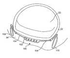
- FIG. 4illustrates a human heart 20 in which the atria has been removed to expose a mitral valve 22 , an aortic valve 24 , and a tricuspid valve 26 . Also partially shown are a circumflex artery 36 and a coronary artery 38 .
- Mitral valve 22which is located between the aorta and left ventricle, includes an anterior cusp 28 and a posterior cusp 30 . Surrounding the anterior cusp and posterior cusp is an annulus 32 that maintains the spacing of the cusp when a mitral valve closes during a left ventricular contraction.
- Coronary sinus 34extends adjacent to annulus 32 , along the atrial ventricular groove between the left atrium and left ventricle of the heart.
- coronary sinus 34is generally coplanar with annulus 32 , it is ideally disposed to facilitate modification of the shape of the annulus to correct a leakage or blood regurgitation problem with the mitral valve.
- the present inventiontakes advantage of the disposition of the coronary sinus relative to the annulus of the mitral valve by enabling insertion of a device into the patient's body, for modifying the shape of the annulus from within the coronary sinus.
- mitral valve annulus reshaping devicecan be constrained within a catheter in preparation for insertion within the coronary sinus, and the catheter can then be guided into the coronary sinus, using a guide wire or other appropriate means. Once thus in place, the reshaping device can either be pushed from the catheter, or the catheter can be pulled back, leaving the device in a desired position within the coronary sinus.
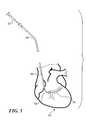
- FIG. 5illustrates apparatus that facilitates a preferred approach for inserting a catheter 40 into coronary sinus 34 .
- This apparatusincludes a pre-curved inner dilator 54 , which can be manually shaped into a curve to match the anatomical characteristics of the patient, and which has a proximal end 52 that extends proximally of catheter 40 .
- the pre-shaped curve in inner dilator 54enables advancing the dilator around a relatively sharp bend, as is necessary to enter the coronary sinus.
- the inner dilatoris used to advance the catheter through the patient's venous system and into the coronary sinus, pushing, rotating, and manipulating the dilator as required.
- the catheterWhen positioned as required for deployment of the reshaping device, the catheter will typically extend from an incision in the patient's jugular vein (not shown) and down through the vena cava. From the superior vena cava, the catheter will extend into the right atrium of the heart, and continue along the wall of the right atrium and into the coronary sinus. Once the inner dilator and catheter 40 have been advanced so that the distal end of the catheter is disposed where desired within the coronary sinus 34 , the pre-curved inner dilator is withdrawn to enable insertion of the mitral valve annulus reshaping device.
- FIG. 6illustrates an assembly that is preferably used for introducing a mitral valve annulus reshaping device into the coronary sinus through catheter 40 .
- the assemblyincludes a cartridge 42 within which the mitral valve annulus reshaping device is constrained in a UM state.
- cartridge 42is coupled to the proximal end of catheter 40 to facilitate deployment of the mitral valve annulus reshaping device into the coronary sinus through the catheter.
- a pusher cable 46extends from a handle 48 .
- handle 48includes a snap lock 45 that engages a control knob 44 , locking handle 48 onto control knob 44 , while still enabling the control knob to be rotated in engagement with threads (not shown) that are formed on the exterior surface of cartridge 42 .
- pusher cable 46advances the mitral valve annulus reshaping device from inside cartridge 42 into catheter 40 and toward the distal end of the catheter.
- control knob 44controls the advancement and deployment of a mitral valve annulus reshaping device within the coronary sinus.
- a release knob 50is employed for uncoupling pusher cable 46 from the device after it has been fully deployed within the coronary sinus and adjusted as desired by the medical personnel using the assembly.
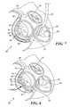
- FIG. 7illustrates the disposition of catheter 40 within the coronary sinus while a mitral valve annulus reshaping device 60 remains inside the catheter 40 , ready to be deployed within coronary sinus 34 .
- the mitral valve annulus reshaping devicehas a distal end 62 that is generally aligned with the distal end of catheter 40 , while a proximal end 64 of the device is well inside catheter 40 .
- FIG. 7thus shows the disposition of the mitral valve annulus reshaping device prior to rotating control knob 44 to force the device from inside catheter 40 so that it is released and unconstrained within the coronary sinus.
- the shape memory alloy comprising the reshaping deviceWhile the mitral valve annulus reshaping device remains constrained within catheter 40 , the shape memory alloy comprising the reshaping device at least partially remains as UM.
- the shape memory alloy comprising the devicehas a characteristic temperature, A f , that is below or equal the normal body temperature of the patient. Accordingly, the SMA is in a super-elastic state, and the device can readily be delivered into the coronary sinus through catheter 40 while constrained in the UM state. Because the SMA of the device is super-elastic, the device is readily deformed to a size that fits within the catheter and can be advanced into the coronary sinus.
- the interior surface of the distal portion of catheter 40 or the exterior surface of the mitral valve annulus reshaping devicecan be coated with a friction reducing material, such as a lubricating material or the catheter provided with a low friction lining material to facilitate deployment of the device from the distal end of the catheter.
- a friction reducing materialsuch as a lubricating material or the catheter provided with a low friction lining material to facilitate deployment of the device from the distal end of the catheter.
- the SMA comprising the mitral valve annulus reshaping device 60at least partially converts from UM to its austenitic state once released from the constraint of the catheter as is illustrated in FIG. 8 .
- reshaping device 60is fully outside of the catheter and deployed within the coronary sinus.
- the mitral valve annulus reshaping deviceis enabled to change to a second, relatively lower strain state having a reduced radius of curvature, so that a distal end of the device exerts a force against the annulus of the mitral valve. This release from the constraint imposed by the catheter enables at least a partial recovery to the programmed shape of the device.
- the mitral valve annulus reshaping deviceis constrained so that its programmed shape curves with a reduced radius substantially in the plane of the mitral valve annulus, bringing the distal end of the device into contact with and exerting force upon the annulus as shown in FIG. 8 .
- a removable tether(as shown in FIGS. 10 and 11 ) can, be employed to further reduce the radius of curvature of the reshaping device to reduce its internal strain condition.
- the SMA of the devicecan be modified to have less stiffness.
- the reduced radius of curvature of the annulus in FIG. 8is in contrast to the increased radius of curvature of the annulus as shown in FIG. 9 .
- the programmed shape of mitral valve annulus reshaping device 60has been modified using a straightening rod (not shown) that is inserted through the catheter, before the catheter is removed from the venous system of the patient.
- the straightening rodcan be temporarily advanced into the coronary sinus, to act upon the mitral valve annulus reshaping device so as to increase the radius of curvature of the reshaping device and thereby reduce internally stored strain.
- the straining rodis used to adjust the mitral valve annulus reshaping device stiffness and reduce the normal force applied by the reshaping device against the tissue of the coronary sinus adjacent to the mitral valve annulus.
- physiological parameterssuch as blood pressure, fluoroscopic images, ultrasound flow patterns through the heart, and an electrocardiogram of the patient
- medical personnelcan determine the effect of reshaping the annulus of the mitral valve and modify the extent of the reshaping as necessary to achieve a desired improvement in the functioning of the mitral valve.
- a physicianwill desire to provide an optimal correction of a defect in the mitral valve, and the present invention provides the means to vary the degree to which the annulus is reshaped and thereby control the changes to the mitral valve operation as desired.
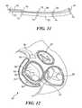
- FIGS. 10 and 11illustrate a mitral valve annulus reshaping device 70 that can readily be modified once it has been disposed within the coronary sinus of a patient.
- device 70is pushed from catheter 40 (not shown in this Figure) using a pusher 72 , which can remain in place after catheter 40 has been partially withdrawn.
- a tether 80Coupled to device 70 is a tether 80 , which extends through a lumen 82 in pusher 72 and through a plurality of bores 78 formed within guides 74 that are disposed at spaced apart locations along the longitudinal access of device 70 .
- An end terminal 76is disposed at the distal end of mitral valve annulus reshaping device 70 , and tether 80 also extends through bores 78 within end terminal 76 , and loops back through bores 78 in each of guides 74 , extending out through lumen 82 in pusher 72 to the proximal end of the pusher, which is disposed outside the body of the patient (not shown).
- reshaping device 70when unconstrained by catheter 40 , reshaping device 70 at least partially converts from UM to its austenitic state in which it attempts to assume its programmed curved shape.
- the austenitic statecan be achieved, at least partially, while mitral valve annulus reshaping device 70 is within the body of the patient.
- tether 80can be pulled while holding pusher catheter 82 against the proximal end of device 70 , to assist the device in applying force against the adjacent tissue of the coronary sinus after the reshaping device has been deployed within the coronary sinus.
- pusher catheter 82Using the tether end pusher catheter 72 in this manner, it is possible to reduce the reshaping device radius of curvature and thereby reduce its internally stored strain condition, until the tether is released, which then increases the loading and strain on the device.
- the tension applied by tether 80is usable to adjust the stiffness of the mitral valve annulus reshaping device and thereby vary the force that it applies against tissue adjacent to the mitral valve annulus within the coronary sinus.
- the second state and programmed shape of the SMA comprising a mitral valve reshaping device 90 and its placement in the coronary sinuscan be chosen to cause an increase in the radius of curvature of the annulus when the constraint of the catheter is removed, in contrast to the decrease in radius of curvature of the annulus caused by the embodiment of FIGS. 8 and 9 .
- an increase in the radius of curvature of the annulusmay be preferred to a decrease in the radius of curvature to correct problems with leakage through the mitral valve.
- FIGS. 13 and 14illustrate a mitral valve annulus reshaping device 100 that includes helical coil spring 102 disposed between straight wire sections 104 and 106 .
- the helical coil springis preferably formed of SMA.
- a distal anchor 108is also formed from SMA and is initially deployed and permitted to at least partially change from UM to its austenitic state as the device is initially pushed (and/or pulled) from catheter 40 (not shown in this view).
- catheter 40is initially positioned within coronary sinus 34 at a location such that as mitral valve annulus reshaping device 100 is forced from the distal end of the catheter and distal anchor 108 is allowed freedom from the constraint of the catheter, the distal anchor 108 will change from its UM state toward its austenitic state in which it has a loop shape with a relatively larger radial extent than when constrained inside the catheter.
- the released loopexpands radially outward and engages the interior surface of the coronary sinus, over a distributed area.
- the catheteris withdrawn further, beyond helical coil spring 102 , finally enabling a proximal anchor 110 to be freed from the constraint of the catheter.
- the proximal anchoralso at least partially changes from the UM state toward the austenitic state, enabling the expanding loop shape of the proximal anchor 110 to anchor device 100 at its desired disposition within the coronary sinus.
- a tether line 112Prior to deploying and releasing the constraint on proximal anchor 110 , the user will apply tension to a tether line 112 .
- the tether lineforms a double loop around and through the distal and proximal ends of helical coil spring 102 .
- the tensile load resulting from the application of a tensile load on the tether linewill cause straight wire sections 104 and 106 and helical coil spring 102 to form UM.
- the proximally applied tension in the tether linepulls the distal anchor, straight wire section 104 , and tissue distal to the helical coil spring in the proximal direction.
- the helical coil springwill thus be assisted to relax to its tightly wound programmed shape, which is therefore at a lower strain state.
- the tension applied to tether line 112determines the force applied against the adjacent tissue to modify the shape annulus 32 , before the final disposition of proximal anchor 110 is determined.
- FIG. 14spring stiffness has been increased in the helical coil 102 causing the mitral valve annulus reshaping device to straighten the adjacent tissue and annulus 32 .
- FIG. 14also illustrates the reshaping device after tether line 112 has been removed, which is accomplished by releasing one end of the tether line and pulling the tether line from the loops around the coils of the helical coil spring.
- mitral valve annulus reshaping device 120is illustrated.
- This devicealso includes a helical coil 122 , which is disposed between straight sections 124 and 126 a tether 132 for adjusting the stiffness of the spring and the relative force applied by the device against the adjacent tissue and mitral valve annulus.
- mitral valve annulus reshaping device 120includes a distal stent 128 and a proximal stent 130 that are also preferably formed of SMA. Distal stent 128 is allowed to expand as it converts from UM toward its austenitic state once the constraint of the catheter is removed.
- distal end of catheter 40be disposed within coronary sinus at about the location where distal stent 128 is to be disposed as it is allowed to expand to its programmed shape.
- the usercan modify the tension distal of helical coil 122 and thereby achieve a desired modification of annulus 32 .
- the usercan modify the strain and stress by varying the tension in tether line 132 to change the force applied to the adjacent tissue by device 120 , using the tether line to change the stress applied to the helical coils of the SMA comprising the device.
- mitral valve annulus reshaping device 120is very similar to the embodiment shown in FIGS. 13 and 14 .
- one or both of the distal and proximal stents of this embodimentmight be made of a non-SMA metal, or of an SMA metal whose A f is above normal body temperature, and expanded radially into contact with the interior surface of the coronary sinus using a conventional catheter inflatable balloon coupled to an external source of a pressurized fluid.
- FIGS. 16-18illustrate another embodiment of the mitral valve annulus reshaping device that includes an SMA metal wire or arched leaf spring 140 with a programmed shape having a relatively small radius of curvature, R.
- arched leaf spring 140At each end of arched leaf spring 140 are disposed loops 142 and 144 , also preferably formed of super-elastic SMA so that they elastically expand radially outward when released from a catheter or other restraint that is used to insert the mitral valve annulus reshaping device intravenously into coronary sinus 34 of a patient.
- the SMA metalcomprising the device it will change from its UM state toward its austenitic state.
- loop 142when loop 142 is released from the catheter, the expansion of loop 142 , as the SMA material comprising it returns to its programmed shape, will bring the loop into contact with the inner surface of coronary sinus 34 , so as to anchor the distal end of the mitral valve annulus reshaping device at a desired location and orientation within the coronary sinus.
- a tether 146passes through loop 144 , and both ends of the tether extend outside the patient's body through the venous system within the catheter (not shown), enabling medical personnel to apply tension to the tether after the distal end of arched leaf spring 140 has been deployed within the coronary sinus and anchored by loop 142 .
- tether 146it is possible to applying loading tension to arched leaf spring 140 , thereby adjusting the stiffness of the arched leaf spring and the relative force applied by the device against the adjacent tissue and mitral valve annulus, prior to releasing loop 144 from the constraint of the catheter.
- the applied tensiontends to straighten arched leaf spring 140 , so that it has a greater radius of curvature, R′, as shown in FIG.
- Tether 146can be employed to make further adjustments to the device by loading and unloading the tension applied, reversibly tuning the device from UM to austenite and back. As shown in FIG. 18 , the arched loop has been tuned in this manner to have an even greater radius of curvature, R′′, relative to its initial programmed curved radius of curvature, R, which is shown in FIG. 16 .
- Each of the embodiments disclosed aboveillustrates how SMA and its super-elasticity can be applied in modifying the shape of the annulus and thereby correcting defects in a mitral valve within the body of a patient.
- the characteristics of the SMA comprising each of these embodiments, as illustrated in FIG. 3is employed to good effect, since it permits the user to modify the stiffness of the SMA comprising the device and the force applied to the adjacent tissue by the SMA even after the mitral valve annulus reshaping device has been deployed in the coronary sinus.
- the SMAcan be reversibly changed between the martensite and austenitic states while within the body of the patient, as necessary to achieve a desired modification of the annulus and corresponding improvement in the functioning of the mitral valve.
Landscapes
- Health & Medical Sciences (AREA)
- Cardiology (AREA)
- Life Sciences & Earth Sciences (AREA)
- Animal Behavior & Ethology (AREA)
- Engineering & Computer Science (AREA)
- Biomedical Technology (AREA)
- Heart & Thoracic Surgery (AREA)
- Vascular Medicine (AREA)
- Oral & Maxillofacial Surgery (AREA)
- Transplantation (AREA)
- General Health & Medical Sciences (AREA)
- Public Health (AREA)
- Veterinary Medicine (AREA)
- Prostheses (AREA)
- Surgical Instruments (AREA)
- Media Introduction/Drainage Providing Device (AREA)
Abstract
Description
Claims (14)
Priority Applications (3)
| Application Number | Priority Date | Filing Date | Title |
|---|---|---|---|
| US11/655,710US7758639B2 (en) | 2003-02-03 | 2007-01-18 | Mitral valve device using conditioned shape memory alloy |
| US12/838,189US20100280602A1 (en) | 2003-02-03 | 2010-07-16 | Mitral Valve Device Using Conditioned Shape Memory Alloy |
| US13/359,307US20120123532A1 (en) | 2003-02-03 | 2012-01-26 | Devices and Methods for Reducing Mitral Valve Regurgitation |
Applications Claiming Priority (2)
| Application Number | Priority Date | Filing Date | Title |
|---|---|---|---|
| US10/359,016US7314485B2 (en) | 2003-02-03 | 2003-02-03 | Mitral valve device using conditioned shape memory alloy |
| US11/655,710US7758639B2 (en) | 2003-02-03 | 2007-01-18 | Mitral valve device using conditioned shape memory alloy |
Related Parent Applications (1)
| Application Number | Title | Priority Date | Filing Date |
|---|---|---|---|
| US10/359,016ContinuationUS7314485B2 (en) | 2003-02-03 | 2003-02-03 | Mitral valve device using conditioned shape memory alloy |
Related Child Applications (1)
| Application Number | Title | Priority Date | Filing Date |
|---|---|---|---|
| US12/838,189ContinuationUS20100280602A1 (en) | 2003-02-03 | 2010-07-16 | Mitral Valve Device Using Conditioned Shape Memory Alloy |
Publications (2)
| Publication Number | Publication Date |
|---|---|
| US20070135912A1 US20070135912A1 (en) | 2007-06-14 |
| US7758639B2true US7758639B2 (en) | 2010-07-20 |
Family
ID=32771316
Family Applications (4)
| Application Number | Title | Priority Date | Filing Date |
|---|---|---|---|
| US10/359,016Expired - LifetimeUS7314485B2 (en) | 2003-02-03 | 2003-02-03 | Mitral valve device using conditioned shape memory alloy |
| US11/655,710Expired - LifetimeUS7758639B2 (en) | 2003-02-03 | 2007-01-18 | Mitral valve device using conditioned shape memory alloy |
| US12/838,189AbandonedUS20100280602A1 (en) | 2003-02-03 | 2010-07-16 | Mitral Valve Device Using Conditioned Shape Memory Alloy |
| US13/359,307AbandonedUS20120123532A1 (en) | 2003-02-03 | 2012-01-26 | Devices and Methods for Reducing Mitral Valve Regurgitation |
Family Applications Before (1)
| Application Number | Title | Priority Date | Filing Date |
|---|---|---|---|
| US10/359,016Expired - LifetimeUS7314485B2 (en) | 2003-02-03 | 2003-02-03 | Mitral valve device using conditioned shape memory alloy |
Family Applications After (2)
| Application Number | Title | Priority Date | Filing Date |
|---|---|---|---|
| US12/838,189AbandonedUS20100280602A1 (en) | 2003-02-03 | 2010-07-16 | Mitral Valve Device Using Conditioned Shape Memory Alloy |
| US13/359,307AbandonedUS20120123532A1 (en) | 2003-02-03 | 2012-01-26 | Devices and Methods for Reducing Mitral Valve Regurgitation |
Country Status (1)
| Country | Link |
|---|---|
| US (4) | US7314485B2 (en) |
Cited By (22)
| Publication number | Priority date | Publication date | Assignee | Title |
|---|---|---|---|---|
| US20110035000A1 (en)* | 2002-01-30 | 2011-02-10 | Cardiac Dimensions, Inc. | Tissue Shaping Device |
| US20120123532A1 (en)* | 2003-02-03 | 2012-05-17 | Cardiac Dimensions, Inc. | Devices and Methods for Reducing Mitral Valve Regurgitation |
| US8250960B2 (en) | 2008-08-11 | 2012-08-28 | Cardiac Dimensions, Inc. | Catheter cutting tool |
| US9956077B2 (en) | 2003-12-19 | 2018-05-01 | Cardiac Dimensions Pty. Ltd. | Mitral valve annuloplasty device with twisted anchor |
| US10016272B2 (en) | 2014-09-12 | 2018-07-10 | Mitral Valve Technologies Sarl | Mitral repair and replacement devices and methods |
| US10052199B2 (en) | 2014-02-21 | 2018-08-21 | Mitral Valve Technologies Sarl | Devices, systems and methods for delivering a prosthetic mitral valve and anchoring device |
| US10226330B2 (en) | 2013-08-14 | 2019-03-12 | Mitral Valve Technologies Sarl | Replacement heart valve apparatus and methods |
| US10226339B2 (en) | 2012-01-31 | 2019-03-12 | Mitral Valve Technologies Sarl | Mitral valve docking devices, systems and methods |
| US10390953B2 (en) | 2017-03-08 | 2019-08-27 | Cardiac Dimensions Pty. Ltd. | Methods and devices for reducing paravalvular leakage |
| US10456258B2 (en) | 2002-05-08 | 2019-10-29 | Cardiac Dimensions Pty. Ltd. | Tissue shaping device |
| US10588742B2 (en) | 2013-08-14 | 2020-03-17 | Mitral Valve Technologies Sarl | Coiled anchor for supporting prosthetic heart valve, prosthetic heart valve, and deployment device |
| US10758241B1 (en) | 2019-03-25 | 2020-09-01 | Laminar, Inc. | Devices, systems, and methods for treating the left atrial appendage |
| US10856985B2 (en) | 2017-11-21 | 2020-12-08 | Abbott Cardiovascular Systems Inc. | System and method for annuloplasty |
| US11026791B2 (en) | 2018-03-20 | 2021-06-08 | Medtronic Vascular, Inc. | Flexible canopy valve repair systems and methods of use |
| US11033257B2 (en) | 2005-01-20 | 2021-06-15 | Cardiac Dimensions Pty. Ltd. | Tissue shaping device |
| US11285005B2 (en) | 2006-07-17 | 2022-03-29 | Cardiac Dimensions Pty. Ltd. | Mitral valve annuloplasty device with twisted anchor |
| US11285003B2 (en) | 2018-03-20 | 2022-03-29 | Medtronic Vascular, Inc. | Prolapse prevention device and methods of use thereof |
| US11311380B2 (en) | 2003-05-02 | 2022-04-26 | Cardiac Dimensions Pty. Ltd. | Device and method for modifying the shape of a body organ |
| US11596771B2 (en) | 2020-12-14 | 2023-03-07 | Cardiac Dimensions Pty. Ltd. | Modular pre-loaded medical implants and delivery systems |
| US11666444B2 (en) | 2017-08-03 | 2023-06-06 | The Regents Of The University Of California | Atrial cage for placement, securing and anchoring of atrioventricular valves |
| US11890187B2 (en) | 2010-03-05 | 2024-02-06 | Edwards Lifesciences Corporation | Retaining mechanisms for prosthetic valves |
| US12303116B2 (en) | 2020-03-24 | 2025-05-20 | Laminar, Inc. | Devices, systems, and methods for occluding cavities within the body |
Families Citing this family (198)
| Publication number | Priority date | Publication date | Assignee | Title |
|---|---|---|---|---|
| US7192442B2 (en)* | 1999-06-30 | 2007-03-20 | Edwards Lifesciences Ag | Method and device for treatment of mitral insufficiency |
| US6440164B1 (en) | 1999-10-21 | 2002-08-27 | Scimed Life Systems, Inc. | Implantable prosthetic valve |
| US7296577B2 (en) | 2000-01-31 | 2007-11-20 | Edwards Lifescience Ag | Transluminal mitral annuloplasty with active anchoring |
| US6602286B1 (en) | 2000-10-26 | 2003-08-05 | Ernst Peter Strecker | Implantable valve system |
| US7510576B2 (en) | 2001-01-30 | 2009-03-31 | Edwards Lifesciences Ag | Transluminal mitral annuloplasty |
| US6800090B2 (en)* | 2001-05-14 | 2004-10-05 | Cardiac Dimensions, Inc. | Mitral valve therapy device, system and method |
| US7635387B2 (en) | 2001-11-01 | 2009-12-22 | Cardiac Dimensions, Inc. | Adjustable height focal tissue deflector |
| US7311729B2 (en)* | 2002-01-30 | 2007-12-25 | Cardiac Dimensions, Inc. | Device and method for modifying the shape of a body organ |
| US6949122B2 (en) | 2001-11-01 | 2005-09-27 | Cardiac Dimensions, Inc. | Focused compression mitral valve device and method |
| US6793673B2 (en) | 2002-12-26 | 2004-09-21 | Cardiac Dimensions, Inc. | System and method to effect mitral valve annulus of a heart |
| US7179282B2 (en)* | 2001-12-05 | 2007-02-20 | Cardiac Dimensions, Inc. | Device and method for modifying the shape of a body organ |
| US7004958B2 (en)* | 2002-03-06 | 2006-02-28 | Cardiac Dimensions, Inc. | Transvenous staples, assembly and method for mitral valve repair |
| US6797001B2 (en)* | 2002-03-11 | 2004-09-28 | Cardiac Dimensions, Inc. | Device, assembly and method for mitral valve repair |
| US7007698B2 (en) | 2002-04-03 | 2006-03-07 | Boston Scientific Corporation | Body lumen closure |
| US6752828B2 (en) | 2002-04-03 | 2004-06-22 | Scimed Life Systems, Inc. | Artificial valve |
| CA2877641C (en) | 2002-05-08 | 2017-01-17 | Cardiac Dimensions Pty. Ltd. | Device and method for modifying the shape of a body organ |
| EP1531762B1 (en)* | 2002-08-29 | 2010-04-14 | St. Jude Medical, Cardiology Division, Inc. | Implantable devices for controlling the internal circumference of an anatomic orifice or lumen |
| US8758372B2 (en) | 2002-08-29 | 2014-06-24 | St. Jude Medical, Cardiology Division, Inc. | Implantable devices for controlling the size and shape of an anatomical structure or lumen |
| US7087064B1 (en) | 2002-10-15 | 2006-08-08 | Advanced Cardiovascular Systems, Inc. | Apparatuses and methods for heart valve repair |
| AU2003285943B2 (en) | 2002-10-24 | 2008-08-21 | Boston Scientific Limited | Venous valve apparatus and method |
| US7485143B2 (en) | 2002-11-15 | 2009-02-03 | Abbott Cardiovascular Systems Inc. | Apparatuses and methods for heart valve repair |
| US9149602B2 (en) | 2005-04-22 | 2015-10-06 | Advanced Cardiovascular Systems, Inc. | Dual needle delivery system |
| US7331972B1 (en) | 2002-11-15 | 2008-02-19 | Abbott Cardiovascular Systems Inc. | Heart valve chord cutter |
| US7404824B1 (en) | 2002-11-15 | 2008-07-29 | Advanced Cardiovascular Systems, Inc. | Valve aptation assist device |
| US7335213B1 (en) | 2002-11-15 | 2008-02-26 | Abbott Cardiovascular Systems Inc. | Apparatus and methods for heart valve repair |
| US7981152B1 (en) | 2004-12-10 | 2011-07-19 | Advanced Cardiovascular Systems, Inc. | Vascular delivery system for accessing and delivering devices into coronary sinus and other vascular sites |
| US8187324B2 (en) | 2002-11-15 | 2012-05-29 | Advanced Cardiovascular Systems, Inc. | Telescoping apparatus for delivering and adjusting a medical device in a vessel |
| US7837729B2 (en) | 2002-12-05 | 2010-11-23 | Cardiac Dimensions, Inc. | Percutaneous mitral valve annuloplasty delivery system |
| US7316708B2 (en) | 2002-12-05 | 2008-01-08 | Cardiac Dimensions, Inc. | Medical device delivery system |
| US6945957B2 (en) | 2002-12-30 | 2005-09-20 | Scimed Life Systems, Inc. | Valve treatment catheter and methods |
| US20040133240A1 (en)* | 2003-01-07 | 2004-07-08 | Cardiac Dimensions, Inc. | Electrotherapy system, device, and method for treatment of cardiac valve dysfunction |
| US20040158321A1 (en)* | 2003-02-12 | 2004-08-12 | Cardiac Dimensions, Inc. | Method of implanting a mitral valve therapy device |
| US20040254600A1 (en)* | 2003-02-26 | 2004-12-16 | David Zarbatany | Methods and devices for endovascular mitral valve correction from the left coronary sinus |
| JP4691017B2 (en)* | 2003-03-18 | 2011-06-01 | セント ジュード メディカル インコーポレイテッド | Body tissue remodeling method and apparatus |
| US7887582B2 (en) | 2003-06-05 | 2011-02-15 | Cardiac Dimensions, Inc. | Device and method for modifying the shape of a body organ |
| US7351259B2 (en)* | 2003-06-05 | 2008-04-01 | Cardiac Dimensions, Inc. | Device, system and method to affect the mitral valve annulus of a heart |
| EP1646332B1 (en) | 2003-07-18 | 2015-06-17 | Edwards Lifesciences AG | Remotely activated mitral annuloplasty system |
| US7998112B2 (en) | 2003-09-30 | 2011-08-16 | Abbott Cardiovascular Systems Inc. | Deflectable catheter assembly and method of making same |
| US7004176B2 (en)* | 2003-10-17 | 2006-02-28 | Edwards Lifesciences Ag | Heart valve leaflet locator |
| JP4464972B2 (en)* | 2003-12-17 | 2010-05-19 | クック・インコーポレイテッド | Interconnected leg extensions for endoluminal prostheses |
| US7837728B2 (en) | 2003-12-19 | 2010-11-23 | Cardiac Dimensions, Inc. | Reduced length tissue shaping device |
| US7794496B2 (en) | 2003-12-19 | 2010-09-14 | Cardiac Dimensions, Inc. | Tissue shaping device with integral connector and crimp |
| US20060271174A1 (en)* | 2003-12-19 | 2006-11-30 | Gregory Nieminen | Mitral Valve Annuloplasty Device with Wide Anchor |
| US7854761B2 (en) | 2003-12-19 | 2010-12-21 | Boston Scientific Scimed, Inc. | Methods for venous valve replacement with a catheter |
| US8128681B2 (en) | 2003-12-19 | 2012-03-06 | Boston Scientific Scimed, Inc. | Venous valve apparatus, system, and method |
| US7674284B2 (en) | 2004-03-31 | 2010-03-09 | Cook Incorporated | Endoluminal graft |
| US7993397B2 (en) | 2004-04-05 | 2011-08-09 | Edwards Lifesciences Ag | Remotely adjustable coronary sinus implant |
| US7377941B2 (en)* | 2004-06-29 | 2008-05-27 | Micardia Corporation | Adjustable cardiac valve implant with selective dimensional adjustment |
| US20080183285A1 (en)* | 2004-06-29 | 2008-07-31 | Micardia Corporation | Adjustable cardiac valve implant with selective dimensional adjustment |
| US20080015688A1 (en)* | 2004-06-29 | 2008-01-17 | Micardia Corporation | Adjustable multi-segment cardiac valve implant with selective dimensional adjustment |
| US20080288060A1 (en)* | 2004-07-06 | 2008-11-20 | Baker Medical Research Institute | Treating Valvular Insufficiency |
| US20060015178A1 (en)* | 2004-07-15 | 2006-01-19 | Shahram Moaddeb | Implants and methods for reshaping heart valves |
| US7566343B2 (en) | 2004-09-02 | 2009-07-28 | Boston Scientific Scimed, Inc. | Cardiac valve, system, and method |
| US7211110B2 (en) | 2004-12-09 | 2007-05-01 | Edwards Lifesciences Corporation | Diagnostic kit to assist with heart valve annulus adjustment |
| KR100614654B1 (en)* | 2005-01-04 | 2006-08-22 | 삼성전자주식회사 | Wireless transmitters provide effective power compensation for output changes with temperature and process |
| DE102005003632A1 (en) | 2005-01-20 | 2006-08-17 | Fraunhofer-Gesellschaft zur Förderung der angewandten Forschung e.V. | Catheter for the transvascular implantation of heart valve prostheses |
| US7854755B2 (en) | 2005-02-01 | 2010-12-21 | Boston Scientific Scimed, Inc. | Vascular catheter, system, and method |
| US20060173490A1 (en) | 2005-02-01 | 2006-08-03 | Boston Scientific Scimed, Inc. | Filter system and method |
| US7878966B2 (en) | 2005-02-04 | 2011-02-01 | Boston Scientific Scimed, Inc. | Ventricular assist and support device |
| US7670368B2 (en) | 2005-02-07 | 2010-03-02 | Boston Scientific Scimed, Inc. | Venous valve apparatus, system, and method |
| US7780722B2 (en) | 2005-02-07 | 2010-08-24 | Boston Scientific Scimed, Inc. | Venous valve apparatus, system, and method |
| US7867274B2 (en) | 2005-02-23 | 2011-01-11 | Boston Scientific Scimed, Inc. | Valve apparatus, system and method |
| US8608797B2 (en) | 2005-03-17 | 2013-12-17 | Valtech Cardio Ltd. | Mitral valve treatment techniques |
| US8864823B2 (en) | 2005-03-25 | 2014-10-21 | StJude Medical, Cardiology Division, Inc. | Methods and apparatus for controlling the internal circumference of an anatomic orifice or lumen |
| EP2767260B1 (en)* | 2005-03-25 | 2019-07-03 | St. Jude Medical, Cardiology Division, Inc. | Apparatus for controlling the internal circumference of an anatomic orifice or lumen |
| US7722666B2 (en) | 2005-04-15 | 2010-05-25 | Boston Scientific Scimed, Inc. | Valve apparatus, system and method |
| US20060238019A1 (en)* | 2005-04-21 | 2006-10-26 | Mark Yu | Brakable wheel hub device |
| US7357815B2 (en)* | 2005-04-21 | 2008-04-15 | Micardia Corporation | Dynamically adjustable implants and methods for reshaping tissue |
| US8333777B2 (en) | 2005-04-22 | 2012-12-18 | Benvenue Medical, Inc. | Catheter-based tissue remodeling devices and methods |
| CA2611545A1 (en)* | 2005-06-07 | 2006-12-14 | The International Heart Institute Of Montana Foundation | A system, including method and apparatus for percutaneous endovascular treatment of functional mitral valve insufficiency |
| US8012198B2 (en) | 2005-06-10 | 2011-09-06 | Boston Scientific Scimed, Inc. | Venous valve, system, and method |
| US8951285B2 (en) | 2005-07-05 | 2015-02-10 | Mitralign, Inc. | Tissue anchor, anchoring system and methods of using the same |
| US9492277B2 (en) | 2005-08-30 | 2016-11-15 | Mayo Foundation For Medical Education And Research | Soft body tissue remodeling methods and apparatus |
| US7569071B2 (en) | 2005-09-21 | 2009-08-04 | Boston Scientific Scimed, Inc. | Venous valve, system, and method with sinus pocket |
| US20070173926A1 (en)* | 2005-12-09 | 2007-07-26 | Bobo Donald E Jr | Anchoring system for medical implant |
| US20070213813A1 (en) | 2005-12-22 | 2007-09-13 | Symetis Sa | Stent-valves for valve replacement and associated methods and systems for surgery |
| US7799038B2 (en) | 2006-01-20 | 2010-09-21 | Boston Scientific Scimed, Inc. | Translumenal apparatus, system, and method |
| US7637946B2 (en)* | 2006-02-09 | 2009-12-29 | Edwards Lifesciences Corporation | Coiled implant for mitral valve repair |
| US7749249B2 (en) | 2006-02-21 | 2010-07-06 | Kardium Inc. | Method and device for closing holes in tissue |
| US7503932B2 (en) | 2006-04-11 | 2009-03-17 | Cardiac Dimensions, Inc. | Mitral valve annuloplasty device with vena cava anchor |
| US8454683B2 (en)* | 2006-04-12 | 2013-06-04 | Medtronic Vascular, Inc. | Annuloplasty device having a helical anchor and methods for its use |
| US20070244555A1 (en)* | 2006-04-12 | 2007-10-18 | Medtronic Vascular, Inc. | Annuloplasty Device Having a Helical Anchor and Methods for its Use |
| WO2007136532A2 (en)* | 2006-05-03 | 2007-11-29 | St. Jude Medical, Inc. | Soft body tissue remodeling methods and apparatus |
| US8449605B2 (en) | 2006-06-28 | 2013-05-28 | Kardium Inc. | Method for anchoring a mitral valve |
| US7837610B2 (en) | 2006-08-02 | 2010-11-23 | Kardium Inc. | System for improving diastolic dysfunction |
| US8430926B2 (en)* | 2006-08-11 | 2013-04-30 | Japd Consulting Inc. | Annuloplasty with enhanced anchoring to the annulus based on tissue healing |
| US20080065205A1 (en)* | 2006-09-11 | 2008-03-13 | Duy Nguyen | Retrievable implant and method for treatment of mitral regurgitation |
| US9883943B2 (en) | 2006-12-05 | 2018-02-06 | Valtech Cardio, Ltd. | Implantation of repair devices in the heart |
| AU2007330338A1 (en) | 2006-12-05 | 2008-06-12 | Valtech Cardio, Ltd. | Segmented ring placement |
| US11259924B2 (en) | 2006-12-05 | 2022-03-01 | Valtech Cardio Ltd. | Implantation of repair devices in the heart |
| CA2674485A1 (en) | 2007-01-03 | 2008-07-17 | Mitralsolutions, Inc. | Implantable devices for controlling the size and shape of an anatomical structure or lumen |
| WO2008091493A1 (en) | 2007-01-08 | 2008-07-31 | California Institute Of Technology | In-situ formation of a valve |
| WO2008097999A2 (en) | 2007-02-05 | 2008-08-14 | Mitralsolutions, Inc. | Minimally invasive system for delivering and securing an annular implant |
| US7967853B2 (en) | 2007-02-05 | 2011-06-28 | Boston Scientific Scimed, Inc. | Percutaneous valve, system and method |
| US11660190B2 (en) | 2007-03-13 | 2023-05-30 | Edwards Lifesciences Corporation | Tissue anchors, systems and methods, and devices |
| US7896915B2 (en) | 2007-04-13 | 2011-03-01 | Jenavalve Technology, Inc. | Medical device for treating a heart valve insufficiency |
| US8828079B2 (en) | 2007-07-26 | 2014-09-09 | Boston Scientific Scimed, Inc. | Circulatory valve, system and method |
| US7892276B2 (en) | 2007-12-21 | 2011-02-22 | Boston Scientific Scimed, Inc. | Valve with delayed leaflet deployment |
| BR112012021347A2 (en) | 2008-02-26 | 2019-09-24 | Jenavalve Tecnology Inc | stent for positioning and anchoring a valve prosthesis at an implantation site in a patient's heart |
| US9044318B2 (en) | 2008-02-26 | 2015-06-02 | Jenavalve Technology Gmbh | Stent for the positioning and anchoring of a valvular prosthesis |
| US8382829B1 (en) | 2008-03-10 | 2013-02-26 | Mitralign, Inc. | Method to reduce mitral regurgitation by cinching the commissure of the mitral valve |
| US7972370B2 (en)* | 2008-04-24 | 2011-07-05 | Medtronic Vascular, Inc. | Stent graft system and method of use |
| US20090287304A1 (en) | 2008-05-13 | 2009-11-19 | Kardium Inc. | Medical Device for Constricting Tissue or a Bodily Orifice, for example a mitral valve |
| EP2296744B1 (en) | 2008-06-16 | 2019-07-31 | Valtech Cardio, Ltd. | Annuloplasty devices |
| US8715342B2 (en) | 2009-05-07 | 2014-05-06 | Valtech Cardio, Ltd. | Annuloplasty ring with intra-ring anchoring |
| US8241351B2 (en) | 2008-12-22 | 2012-08-14 | Valtech Cardio, Ltd. | Adjustable partial annuloplasty ring and mechanism therefor |
| US8911494B2 (en) | 2009-05-04 | 2014-12-16 | Valtech Cardio, Ltd. | Deployment techniques for annuloplasty ring |
| US9011530B2 (en) | 2008-12-22 | 2015-04-21 | Valtech Cardio, Ltd. | Partially-adjustable annuloplasty structure |
| US10517719B2 (en) | 2008-12-22 | 2019-12-31 | Valtech Cardio, Ltd. | Implantation of repair devices in the heart |
| WO2010073246A2 (en) | 2008-12-22 | 2010-07-01 | Valtech Cardio, Ltd. | Adjustable annuloplasty devices and adjustment mechanisms therefor |
| JP2012515625A (en)* | 2009-01-22 | 2012-07-12 | セント・ジュード・メディカル,カーディオロジー・ディヴィジョン,インコーポレイテッド | Magnetic docking system and method for long term adjustment of implantable devices |
| JP2012515624A (en) | 2009-01-22 | 2012-07-12 | セント・ジュード・メディカル,カーディオロジー・ディヴィジョン,インコーポレイテッド | Postoperative adjustment tool, minimally invasive mounting device, and adjustable tricuspid valve ring |
| US8353956B2 (en) | 2009-02-17 | 2013-01-15 | Valtech Cardio, Ltd. | Actively-engageable movement-restriction mechanism for use with an annuloplasty structure |
| US9968452B2 (en) | 2009-05-04 | 2018-05-15 | Valtech Cardio, Ltd. | Annuloplasty ring delivery cathethers |
| WO2011041571A2 (en) | 2009-10-01 | 2011-04-07 | Kardium Inc. | Medical device, kit and method for constricting tissue or a bodily orifice, for example, a mitral valve |
| US10098737B2 (en) | 2009-10-29 | 2018-10-16 | Valtech Cardio, Ltd. | Tissue anchor for annuloplasty device |
| US9011520B2 (en) | 2009-10-29 | 2015-04-21 | Valtech Cardio, Ltd. | Tissue anchor for annuloplasty device |
| US9180007B2 (en) | 2009-10-29 | 2015-11-10 | Valtech Cardio, Ltd. | Apparatus and method for guide-wire based advancement of an adjustable implant |
| US8734467B2 (en) | 2009-12-02 | 2014-05-27 | Valtech Cardio, Ltd. | Delivery tool for implantation of spool assembly coupled to a helical anchor |
| US8870950B2 (en) | 2009-12-08 | 2014-10-28 | Mitral Tech Ltd. | Rotation-based anchoring of an implant |
| US8475525B2 (en)* | 2010-01-22 | 2013-07-02 | 4Tech Inc. | Tricuspid valve repair using tension |
| US9107749B2 (en) | 2010-02-03 | 2015-08-18 | Edwards Lifesciences Corporation | Methods for treating a heart |
| US10856978B2 (en) | 2010-05-20 | 2020-12-08 | Jenavalve Technology, Inc. | Catheter system |
| WO2011147849A1 (en) | 2010-05-25 | 2011-12-01 | Jenavalve Technology Inc. | Prosthetic heart valve and transcatheter delivered endoprosthesis comprising a prosthetic heart valve and a stent |
| US11653910B2 (en) | 2010-07-21 | 2023-05-23 | Cardiovalve Ltd. | Helical anchor implantation |
| US8940002B2 (en) | 2010-09-30 | 2015-01-27 | Kardium Inc. | Tissue anchor system |
| US9072511B2 (en) | 2011-03-25 | 2015-07-07 | Kardium Inc. | Medical kit for constricting tissue or a bodily orifice, for example, a mitral valve |
| EP3345573B1 (en) | 2011-06-23 | 2020-01-29 | Valtech Cardio, Ltd. | Closure element for use with annuloplasty structure |
| US10792152B2 (en) | 2011-06-23 | 2020-10-06 | Valtech Cardio, Ltd. | Closed band for percutaneous annuloplasty |
| EP2734157B1 (en)* | 2011-07-21 | 2018-09-05 | 4Tech Inc. | Apparatus for tricuspid valve repair using tension |
| US9668859B2 (en) | 2011-08-05 | 2017-06-06 | California Institute Of Technology | Percutaneous heart valve delivery systems |
| US8858623B2 (en) | 2011-11-04 | 2014-10-14 | Valtech Cardio, Ltd. | Implant having multiple rotational assemblies |
| EP3656434B1 (en) | 2011-11-08 | 2021-10-20 | Valtech Cardio, Ltd. | Controlled steering functionality for implant-delivery tool |
| EP2881083B1 (en) | 2011-12-12 | 2017-03-22 | David Alon | Heart valve repair device |
| US10398555B2 (en) | 2011-12-12 | 2019-09-03 | Cardiac Implants Llc | Magnetically coupled cinching of a loop installed in a valve annulus |
| CA2900930A1 (en) | 2012-02-13 | 2013-08-22 | Mitraspan, Inc. | Method and apparatus for repairing a mitral valve |
| US10076414B2 (en) | 2012-02-13 | 2018-09-18 | Mitraspan, Inc. | Method and apparatus for repairing a mitral valve |
| US9445899B2 (en) | 2012-08-22 | 2016-09-20 | Joseph M. Arcidi | Method and apparatus for mitral valve annuloplasty |
| US9216018B2 (en) | 2012-09-29 | 2015-12-22 | Mitralign, Inc. | Plication lock delivery system and method of use thereof |
| EP2911593B1 (en) | 2012-10-23 | 2020-03-25 | Valtech Cardio, Ltd. | Percutaneous tissue anchor techniques |
| WO2014064694A2 (en) | 2012-10-23 | 2014-05-01 | Valtech Cardio, Ltd. | Controlled steering functionality for implant-delivery tool |
| WO2014087402A1 (en) | 2012-12-06 | 2014-06-12 | Valtech Cardio, Ltd. | Techniques for guide-wire based advancement of a tool |
| US20150351906A1 (en) | 2013-01-24 | 2015-12-10 | Mitraltech Ltd. | Ventricularly-anchored prosthetic valves |
| EP2961351B1 (en) | 2013-02-26 | 2018-11-28 | Mitralign, Inc. | Devices for percutaneous tricuspid valve repair |
| US9687346B2 (en) | 2013-03-14 | 2017-06-27 | Edwards Lifesciences Corporation | Multi-stranded heat set annuloplasty rings |
| US10449333B2 (en) | 2013-03-14 | 2019-10-22 | Valtech Cardio, Ltd. | Guidewire feeder |
| CN105283214B (en) | 2013-03-15 | 2018-10-16 | 北京泰德制药股份有限公司 | Translate conduit, system and its application method |
| WO2014144247A1 (en) | 2013-03-15 | 2014-09-18 | Arash Kheradvar | Handle mechanism and functionality for repositioning and retrieval of transcatheter heart valves |
| EP2805695A1 (en)* | 2013-05-21 | 2014-11-26 | Medtentia International Ltd Oy | Medical system for annuloplasty |
| JP6440694B2 (en) | 2013-06-06 | 2018-12-19 | デイヴィッド・アロン | Heart valve repair and replacement |
| CN105491978A (en) | 2013-08-30 | 2016-04-13 | 耶拿阀门科技股份有限公司 | Radially collapsible frame for a prosthetic valve and method for manufacturing such a frame |
| US10070857B2 (en) | 2013-08-31 | 2018-09-11 | Mitralign, Inc. | Devices and methods for locating and implanting tissue anchors at mitral valve commissure |
| WO2015059699A2 (en) | 2013-10-23 | 2015-04-30 | Valtech Cardio, Ltd. | Anchor magazine |
| US9610162B2 (en) | 2013-12-26 | 2017-04-04 | Valtech Cardio, Ltd. | Implantation of flexible implant |
| EP3174502B1 (en) | 2014-07-30 | 2022-04-06 | Cardiovalve Ltd | Apparatus for implantation of an articulatable prosthetic valve |
| EP3922213A1 (en) | 2014-10-14 | 2021-12-15 | Valtech Cardio, Ltd. | Leaflet-restraining techniques |
| CN110141399B (en) | 2015-02-05 | 2021-07-27 | 卡迪尔维尔福股份有限公司 | Prosthetic valve with axial sliding frame |
| US20160256269A1 (en) | 2015-03-05 | 2016-09-08 | Mitralign, Inc. | Devices for treating paravalvular leakage and methods use thereof |
| EP3270825B1 (en) | 2015-03-20 | 2020-04-22 | JenaValve Technology, Inc. | Heart valve prosthesis delivery system |
| CN107847320B (en) | 2015-04-30 | 2020-03-17 | 瓦尔泰克卡迪欧有限公司 | Valvuloplasty techniques |
| US10709555B2 (en) | 2015-05-01 | 2020-07-14 | Jenavalve Technology, Inc. | Device and method with reduced pacemaker rate in heart valve replacement |
| CA3006662C (en)* | 2015-12-10 | 2023-12-19 | Mvrx, Inc. | Devices, systems, and methods for reshaping a heart valve annulus |
| US10751182B2 (en) | 2015-12-30 | 2020-08-25 | Edwards Lifesciences Corporation | System and method for reshaping right heart |
| US10828160B2 (en) | 2015-12-30 | 2020-11-10 | Edwards Lifesciences Corporation | System and method for reducing tricuspid regurgitation |
| US10531866B2 (en) | 2016-02-16 | 2020-01-14 | Cardiovalve Ltd. | Techniques for providing a replacement valve and transseptal communication |
| WO2017195125A1 (en) | 2016-05-13 | 2017-11-16 | Jenavalve Technology, Inc. | Heart valve prosthesis delivery system and method for delivery of heart valve prosthesis with introducer sheath and loading system |
| US20200146854A1 (en) | 2016-05-16 | 2020-05-14 | Elixir Medical Corporation | Methods and devices for heart valve repair |
| US10702274B2 (en) | 2016-05-26 | 2020-07-07 | Edwards Lifesciences Corporation | Method and system for closing left atrial appendage |
| GB201611910D0 (en) | 2016-07-08 | 2016-08-24 | Valtech Cardio Ltd | Adjustable annuloplasty device with alternating peaks and troughs |
| US20190231525A1 (en) | 2016-08-01 | 2019-08-01 | Mitraltech Ltd. | Minimally-invasive delivery systems |
| CA3031187A1 (en) | 2016-08-10 | 2018-02-15 | Cardiovalve Ltd. | Prosthetic valve with concentric frames |
| CR20190069A (en) | 2016-08-26 | 2019-05-14 | Edwards Lifesciences Corp | VALVES AND COUPLING SYSTEMS OF CORAZON VALVES |
| US10722359B2 (en) | 2016-08-26 | 2020-07-28 | Edwards Lifesciences Corporation | Heart valve docking devices and systems |
| US12329641B2 (en) | 2016-08-26 | 2025-06-17 | Edwards Lifesciences Corporation | Heart valve docking devices and systems |
| WO2018138658A1 (en) | 2017-01-27 | 2018-08-02 | Jenavalve Technology, Inc. | Heart valve mimicry |
| EP3592292B1 (en) | 2017-03-09 | 2024-11-06 | Medtronic Inc. | Stented prosthesis delivery devices having steering capabilities |
| US11045627B2 (en) | 2017-04-18 | 2021-06-29 | Edwards Lifesciences Corporation | Catheter system with linear actuation control mechanism |
| US12064347B2 (en) | 2017-08-03 | 2024-08-20 | Cardiovalve Ltd. | Prosthetic heart valve |
| US11793633B2 (en) | 2017-08-03 | 2023-10-24 | Cardiovalve Ltd. | Prosthetic heart valve |
| USD890333S1 (en) | 2017-08-21 | 2020-07-14 | Edwards Lifesciences Corporation | Heart valve docking coil |
| US10835221B2 (en) | 2017-11-02 | 2020-11-17 | Valtech Cardio, Ltd. | Implant-cinching devices and systems |
| US11135062B2 (en) | 2017-11-20 | 2021-10-05 | Valtech Cardio Ltd. | Cinching of dilated heart muscle |
| CN116531147A (en) | 2018-01-24 | 2023-08-04 | 爱德华兹生命科学创新(以色列)有限公司 | Contraction of annuloplasty structures |
| EP4248904A3 (en) | 2018-01-26 | 2023-11-29 | Edwards Lifesciences Innovation (Israel) Ltd. | Techniques for facilitating heart valve tethering and chord replacement |
| EP3820406B1 (en) | 2018-07-12 | 2023-12-20 | Edwards Lifesciences Innovation (Israel) Ltd. | Annuloplasty systems and locking tools therefor |
| SG11202112651QA (en) | 2019-05-29 | 2021-12-30 | Valtech Cardio Ltd | Tissue anchor handling systems and methods |
| WO2021007815A1 (en)* | 2019-07-17 | 2021-01-21 | 诺尔医疗(深圳)有限公司 | Method for manufacturing bending-resistant intracranial electrode, intracranial depth electrode, and electroencephalograph |
| US12364606B2 (en) | 2019-07-23 | 2025-07-22 | Edwards Lifesciences Innovation (Israel) Ltd. | Fluoroscopic visualization of heart valve anatomy |
| JP2022546160A (en) | 2019-08-30 | 2022-11-04 | エドワーズ ライフサイエンシーズ イノベーション (イスラエル) リミテッド | Anchor channel tip |
| EP4034042A1 (en) | 2019-09-25 | 2022-08-03 | Cardiac Implants LLC | Cardiac valve annulus reduction system |
| JP7583036B2 (en)* | 2019-10-03 | 2024-11-13 | タウ メディカル インコーポレイテッド | Devices, systems and methods for mitigating mitral valve regurgitation |
| EP4193934A1 (en) | 2019-10-29 | 2023-06-14 | Edwards Lifesciences Innovation (Israel) Ltd. | Annuloplasty and tissue anchor technologies |
| US12023247B2 (en) | 2020-05-20 | 2024-07-02 | Edwards Lifesciences Corporation | Reducing the diameter of a cardiac valve annulus with independent control over each of the anchors that are launched into the annulus |
| CA3182316A1 (en) | 2020-06-19 | 2021-12-23 | Edwards Lifesciences Innovation (Israel) Ltd. | Self-stopping tissue anchors |
| US12357459B2 (en) | 2020-12-03 | 2025-07-15 | Cardiovalve Ltd. | Transluminal delivery system |
| CN112914723B (en)* | 2021-02-02 | 2023-07-21 | 广东工业大学 | Nickel-titanium alloy thrombus removal device for complex blood vessels and preparation method |
| CN115737201A (en)* | 2021-09-03 | 2023-03-07 | 瀚芯医疗科技(深圳)有限公司 | Mitral annulus contracting device |
| WO2024102411A1 (en) | 2022-11-09 | 2024-05-16 | Jenavalve Technology, Inc. | Catheter system for sequential deployment of an expandable implant |
Citations (198)
| Publication number | Priority date | Publication date | Assignee | Title |
|---|---|---|---|---|
| US3974526A (en) | 1973-07-06 | 1976-08-17 | Dardik Irving I | Vascular prostheses and process for producing the same |
| US3995623A (en) | 1974-12-23 | 1976-12-07 | American Hospital Supply Corporation | Multipurpose flow-directed catheter |
| US4055861A (en) | 1975-04-11 | 1977-11-01 | Rhone-Poulenc Industries | Support for a natural human heart valve |
| US4164046A (en) | 1977-05-16 | 1979-08-14 | Cooley Denton | Valve prosthesis |
| US4485816A (en) | 1981-06-25 | 1984-12-04 | Alchemia | Shape-memory surgical staple apparatus and method for use in surgical suturing |
| US4550870A (en) | 1983-10-13 | 1985-11-05 | Alchemia Ltd. Partnership | Stapling device |
| US4588395A (en) | 1978-03-10 | 1986-05-13 | Lemelson Jerome H | Catheter and method |
| US4830023A (en) | 1987-11-27 | 1989-05-16 | Medi-Tech, Incorporated | Medical guidewire |
| US5061277A (en) | 1986-08-06 | 1991-10-29 | Baxter International Inc. | Flexible cardiac valvular support prosthesis |
| US5099838A (en) | 1988-12-15 | 1992-03-31 | Medtronic, Inc. | Endocardial defibrillation electrode system |
| US5104404A (en) | 1989-10-02 | 1992-04-14 | Medtronic, Inc. | Articulated stent |
| US5250071A (en) | 1992-09-22 | 1993-10-05 | Target Therapeutics, Inc. | Detachable embolic coil assembly using interlocking clasps and method of use |
| US5261916A (en) | 1991-12-12 | 1993-11-16 | Target Therapeutics | Detachable pusher-vasoocclusive coil assembly with interlocking ball and keyway coupling |
| US5265601A (en) | 1992-05-01 | 1993-11-30 | Medtronic, Inc. | Dual chamber cardiac pacing from a single electrode |
| US5350420A (en) | 1989-07-31 | 1994-09-27 | Baxter International Inc. | Flexible annuloplasty ring and holder |
| US5433727A (en) | 1994-08-16 | 1995-07-18 | Sideris; Eleftherios B. | Centering buttoned device for the occlusion of large defects for occluding |
| US5441515A (en) | 1993-04-23 | 1995-08-15 | Advanced Cardiovascular Systems, Inc. | Ratcheting stent |
| US5449373A (en) | 1994-03-17 | 1995-09-12 | Medinol Ltd. | Articulated stent |
| US5454365A (en) | 1990-11-05 | 1995-10-03 | Bonutti; Peter M. | Mechanically expandable arthroscopic retractors |
| US5458615A (en) | 1993-07-06 | 1995-10-17 | Advanced Cardiovascular Systems, Inc. | Stent delivery system |
| US5474557A (en) | 1993-09-21 | 1995-12-12 | Mai; Christian | Multibranch osteosynthesis clip with dynamic compression and self-retention |
| US5507295A (en) | 1992-07-01 | 1996-04-16 | British Technology Group Limited | Medical devices |
| US5507802A (en) | 1993-06-02 | 1996-04-16 | Cardiac Pathways Corporation | Method of mapping and/or ablation using a catheter having a tip with fixation means |
| US5514161A (en) | 1994-04-05 | 1996-05-07 | Ela Medical S.A. | Methods and apparatus for controlling atrial stimulation in a double atrial triple chamber cardiac pacemaker |
| US5554177A (en) | 1995-03-27 | 1996-09-10 | Medtronic, Inc. | Method and apparatus to optimize pacing based on intensity of acoustic signal |
| US5562698A (en) | 1994-03-09 | 1996-10-08 | Cook Incorporated | Intravascular treatment system |
| US5575818A (en) | 1995-02-14 | 1996-11-19 | Corvita Corporation | Endovascular stent with locking ring |
| US5584867A (en) | 1994-04-05 | 1996-12-17 | Ela Medical S.A. | Method and apparatus for controlling a double atrial triple chamber cardiac pacemaker having a fallback mode |
| US5601600A (en) | 1995-09-08 | 1997-02-11 | Conceptus, Inc. | Endoluminal coil delivery system having a mechanical release mechanism |
| US5662703A (en) | 1995-04-14 | 1997-09-02 | Schneider (Usa) Inc. | Rolling membrane stent delivery device |
| US5676671A (en) | 1995-04-12 | 1997-10-14 | Inoue; Kanji | Device for introducing an appliance to be implanted into a catheter |
| US5733325A (en) | 1993-11-04 | 1998-03-31 | C. R. Bard, Inc. | Non-migrating vascular prosthesis and minimally invasive placement system |
| US5741297A (en) | 1996-08-28 | 1998-04-21 | Simon; Morris | Daisy occluder and method for septal defect repair |
| US5752969A (en) | 1993-06-17 | 1998-05-19 | Sofamor S.N.C. | Instrument for the surgical treatment of an intervertebral disc by the anterior route |
| JP2754067B2 (en) | 1989-01-17 | 1998-05-20 | 日本ゼオン株式会社 | Medical body wall hole plugging jig |
| US5800519A (en) | 1994-04-29 | 1998-09-01 | Kopin Corporation | Tubular medical prosthesis for use in a body lumen |
| US5824071A (en) | 1996-09-16 | 1998-10-20 | Circulation, Inc. | Apparatus for treatment of ischemic heart disease by providing transvenous myocardial perfusion |
| US5836882A (en) | 1997-03-17 | 1998-11-17 | Frazin; Leon J. | Method and apparatus of localizing an insertion end of a probe within a biotic structure |
| WO1998056435A1 (en) | 1997-06-13 | 1998-12-17 | Micro Therapeutics, Inc. | Contoured syringe and novel luer hub and methods for embolizing blood vessels |
| US5871501A (en) | 1994-01-18 | 1999-02-16 | Datascope Investment Corp. | Guide wire with releasable barb anchor |
| EP0903110A1 (en) | 1997-09-12 | 1999-03-24 | B. Braun Medical Inc. | Introducer for an expandable vascular occlusion device |
| US5895391A (en) | 1996-09-27 | 1999-04-20 | Target Therapeutics, Inc. | Ball lock joint and introducer for vaso-occlusive member |
| US5899882A (en) | 1994-10-27 | 1999-05-04 | Novoste Corporation | Catheter apparatus for radiation treatment of a desired area in the vascular system of a patient |
| US5908404A (en) | 1996-05-13 | 1999-06-01 | Elliott; James B. | Methods for inserting an implant |
| US5928258A (en) | 1997-09-26 | 1999-07-27 | Corvita Corporation | Method and apparatus for loading a stent or stent-graft into a delivery sheath |
| US5954761A (en) | 1997-03-25 | 1999-09-21 | Intermedics Inc. | Implantable endocardial lead assembly having a stent |
| US5961545A (en) | 1997-01-17 | 1999-10-05 | Meadox Medicals, Inc. | EPTFE graft-stent composite device |
| US5978705A (en) | 1997-03-14 | 1999-11-02 | Uab Research Foundation | Method and apparatus for treating cardiac arrhythmia using auxiliary pulse |
| US6007519A (en) | 1997-07-30 | 1999-12-28 | Rosselli; Matteo | Central access cannulation device |
| EP0968688A1 (en) | 1998-07-03 | 2000-01-05 | Cordis Europa N.V. | Improved vascular filter for controlled release |
| US6015402A (en) | 1997-03-07 | 2000-01-18 | Sahota; Harvinder | Wire perfusion catheter |
| US6022371A (en) | 1996-10-22 | 2000-02-08 | Scimed Life Systems, Inc. | Locking stent |
| US6027517A (en) | 1994-02-24 | 2000-02-22 | Radiance Medical Systems, Inc. | Fixed focal balloon for interactive angioplasty and stent implantation catheter with focalized balloon |
| US6053900A (en) | 1996-02-16 | 2000-04-25 | Brown; Joe E. | Apparatus and method for delivering diagnostic and therapeutic agents intravascularly |
| US6056775A (en) | 1996-05-31 | 2000-05-02 | Ave Galway Limited | Bifurcated endovascular stents and method and apparatus for their placement |
| US6077295A (en) | 1996-07-15 | 2000-06-20 | Advanced Cardiovascular Systems, Inc. | Self-expanding stent delivery system |
| US6080182A (en) | 1996-12-20 | 2000-06-27 | Gore Enterprise Holdings, Inc. | Self-expanding defect closure device and method of making and using |
| US6086611A (en) | 1997-09-25 | 2000-07-11 | Ave Connaught | Bifurcated stent |
| US6096064A (en) | 1997-09-19 | 2000-08-01 | Intermedics Inc. | Four chamber pacer for dilated cardiomyopthy |
| WO2000044313A1 (en) | 1999-01-27 | 2000-08-03 | Viacor Incorporated | Cardiac valve procedure methods and devices |
| US6099552A (en) | 1997-11-12 | 2000-08-08 | Boston Scientific Corporation | Gastrointestinal copression clips |
| US6129755A (en) | 1998-01-09 | 2000-10-10 | Nitinol Development Corporation | Intravascular stent having an improved strut configuration |
| WO2000060995A2 (en) | 1999-04-09 | 2000-10-19 | Evalve, Inc. | Methods and apparatus for cardiac valve repair |
| JP2000308652A (en) | 1999-03-09 | 2000-11-07 | Jostra Medizintechnik Ag | Loop forming artificial organ |
| EP1050274A1 (en) | 1998-11-20 | 2000-11-08 | Medical Industries Corporation | Hemostatic agent inserting device |
| WO2000074603A1 (en) | 1999-06-08 | 2000-12-14 | S & A Rings, Llc | Annuloplasty rings for heart valve replacement and repair |
| WO2001000111A1 (en) | 1999-06-29 | 2001-01-04 | Jan Otto Solem | Device and method for treatment of mitral insufficiency |
| US6171320B1 (en) | 1996-12-25 | 2001-01-09 | Niti Alloys Technologies Ltd. | Surgical clip |
| US6183512B1 (en) | 1999-04-16 | 2001-02-06 | Edwards Lifesciences Corporation | Flexible annuloplasty system |
| US6190406B1 (en) | 1998-01-09 | 2001-02-20 | Nitinal Development Corporation | Intravascular stent having tapered struts |
| US6200336B1 (en) | 1998-06-02 | 2001-03-13 | Cook Incorporated | Multiple-sided intraluminal medical device |
| WO2001019292A1 (en) | 1999-09-17 | 2001-03-22 | Cardiac Concepts, Inc. | Mitral valve annuloplasty ring and method |
| EP1095634A2 (en) | 1999-10-27 | 2001-05-02 | Cordis Corporation | Rapid exchange self-expanding stent delivery catheter system |
| US6228098B1 (en) | 1998-07-10 | 2001-05-08 | General Surgical Innovations, Inc. | Apparatus and method for surgical fastening |
| US6241757B1 (en) | 1997-02-04 | 2001-06-05 | Solco Surgical Instrument Co., Ltd. | Stent for expanding body's lumen |
| US6254628B1 (en) | 1996-12-09 | 2001-07-03 | Micro Therapeutics, Inc. | Intracranial stent |
| WO2001050985A1 (en) | 2000-01-14 | 2001-07-19 | Viacor Incorporated | Tissue annuloplasty band and apparatus and method for fashioning, sizing and implanting the same |
| US6267783B1 (en) | 1998-11-09 | 2001-07-31 | Cordis Corporation | Stent which is easily recaptured and repositioned within the body |
| WO2001054618A1 (en) | 2000-01-31 | 2001-08-02 | Mitralife | Percutaneous mitral annuloplasty and cardiac reinforcement |
| US6275730B1 (en) | 1997-03-14 | 2001-08-14 | Uab Research Foundation | Method and apparatus for treating cardiac arrythmia |
| US20010018611A1 (en) | 1999-06-30 | 2001-08-30 | Solem Jan Otto | Method and device for treatment of mitral insufficiency |
| US6312446B1 (en) | 1996-03-22 | 2001-11-06 | Scimed Life Systems, Inc. | Apparatus and method for closing a septal defect |
| US20010041899A1 (en) | 1998-03-27 | 2001-11-15 | James B. Hunt | Minimally-invasive medical retrieval device |
| WO2001087180A2 (en) | 2000-05-17 | 2001-11-22 | Xtent Medical Inc | Selectively expandable and releasable stent |
| US6334864B1 (en) | 2000-05-17 | 2002-01-01 | Aga Medical Corp. | Alignment member for delivering a non-symmetric device with a predefined orientation |
| WO2002000099A2 (en) | 2000-06-23 | 2002-01-03 | Viacor Incorporated | Automated annular plication for mitral valve repair |
| WO2002001999A2 (en) | 2000-06-30 | 2002-01-10 | Viacor, Incorporated | Method and apparatus for performing a procedure on a cardiac valve |
| WO2002005888A1 (en) | 2000-06-30 | 2002-01-24 | Viacor Incorporated | Intravascular filter with debris entrapment mechanism |
| US6342067B1 (en) | 1998-01-09 | 2002-01-29 | Nitinol Development Corporation | Intravascular stent having curved bridges for connecting adjacent hoops |
| US6345198B1 (en) | 1998-01-23 | 2002-02-05 | Pacesetter, Inc. | Implantable stimulation system for providing dual bipolar sensing using an electrode positioned in proximity to the tricuspid valve and programmable polarity |
| US6352553B1 (en) | 1995-12-14 | 2002-03-05 | Gore Enterprise Holdings, Inc. | Stent-graft deployment apparatus and method |
| US6352561B1 (en) | 1996-12-23 | 2002-03-05 | W. L. Gore & Associates | Implant deployment apparatus |
| WO2002019951A1 (en) | 2000-09-07 | 2002-03-14 | Viacor, Inc. | Fixation band for affixing a prosthetic heart valve to tissue |
| US6358195B1 (en) | 2000-03-09 | 2002-03-19 | Neoseed Technology Llc | Method and apparatus for loading radioactive seeds into brachytherapy needles |
| WO2002034118A2 (en) | 2000-10-27 | 2002-05-02 | Viacor, Inc. | Intracardiovascular access (icvatm) system |
| US6395017B1 (en) | 1996-11-15 | 2002-05-28 | C. R. Bard, Inc. | Endoprosthesis delivery catheter with sequential stage control |
| US20020065554A1 (en) | 2000-10-25 | 2002-05-30 | Streeter Richard B. | Mitral shield |
| WO2002047539A2 (en) | 2000-12-15 | 2002-06-20 | Viacor, Inc. | Apparatus and method for replacing aortic valve |
| US20020087173A1 (en) | 2000-12-28 | 2002-07-04 | Alferness Clifton A. | Mitral valve constricting device, system and method |
| US6419696B1 (en) | 2000-07-06 | 2002-07-16 | Paul A. Spence | Annuloplasty devices and related heart valve repair methods |
| US20020095167A1 (en) | 2000-10-23 | 2002-07-18 | Liddicoat John R. | Automated annular plication for mitral valve repair |
| WO2002060352A1 (en) | 2001-01-30 | 2002-08-08 | Ev3 Santa Rosa, Inc. | Medical system and method for remodeling an extravascular tissue structure |
| WO2002062408A2 (en) | 2001-02-05 | 2002-08-15 | Viacor, Inc. | Method and apparatus for improving mitral valve function |
| WO2002062263A2 (en) | 2001-02-05 | 2002-08-15 | Viacor, Inc. | Apparatus and method for reducing mitral regurgitation |
| US6442427B1 (en) | 2000-04-27 | 2002-08-27 | Medtronic, Inc. | Method and system for stimulating a mammalian heart |
| WO2002076284A2 (en) | 2001-03-23 | 2002-10-03 | Viacor, Inc. | Method and apparatus for reducing mitral regurgitation |
| WO2002078576A2 (en) | 2001-03-29 | 2002-10-10 | Viacor, Inc. | Method and apparatus for improving mitral valve function |
| US6464720B2 (en) | 1997-09-24 | 2002-10-15 | Cook Incorporated | Radially expandable stent |
| US20020151961A1 (en) | 2000-01-31 | 2002-10-17 | Lashinski Randall T. | Medical system and method for remodeling an extravascular tissue structure |
| US20020156526A1 (en) | 2001-04-24 | 2002-10-24 | Hlavka Edwin J. | Method and apparatus for catheter-based annuloplasty |
| US20020161377A1 (en) | 2001-04-27 | 2002-10-31 | Dmitry Rabkin | Apparatus for delivering, repositioning and/or retrieving self-expanding stents |
| EP0893133B1 (en) | 1997-07-24 | 2002-11-06 | Medex | Medical liquid injection device, syringe for use with the device and method of engaging the syringe |
| US20020183838A1 (en) | 2001-03-29 | 2002-12-05 | Liddicoat John R. | Method and apparatus for improving mitral valve function |
| US20020183837A1 (en) | 2001-03-05 | 2002-12-05 | Streeter Richard B. | Apparatus and method for reducing mitral regurgitation |
| US20020188170A1 (en) | 2001-04-27 | 2002-12-12 | Santamore William P. | Prevention of myocardial infarction induced ventricular expansion and remodeling |
| US6503271B2 (en) | 1998-01-09 | 2003-01-07 | Cordis Corporation | Intravascular device with improved radiopacity |
| US20030018358A1 (en) | 1999-06-25 | 2003-01-23 | Vahid Saadat | Apparatus and methods for treating tissue |
| US20030040771A1 (en) | 1999-02-01 | 2003-02-27 | Hideki Hyodoh | Methods for creating woven devices |
| US20030078654A1 (en) | 2001-08-14 | 2003-04-24 | Taylor Daniel C. | Method and apparatus for improving mitral valve function |
| US20030078465A1 (en)* | 2001-10-16 | 2003-04-24 | Suresh Pai | Systems for heart treatment |
| US20030083613A1 (en) | 1999-05-11 | 2003-05-01 | Schaer Alan K. | Catheter positioning system |
| US20030088305A1 (en) | 2001-10-26 | 2003-05-08 | Cook Incorporated | Prostheses for curved lumens |
| WO2003037171A2 (en) | 2001-11-01 | 2003-05-08 | Cardiac Dimensions, Inc. | Focused compression mitral valve device and method |
| US6562067B2 (en) | 2001-06-08 | 2003-05-13 | Cordis Corporation | Stent with interlocking elements |
| US6569198B1 (en) | 2000-03-31 | 2003-05-27 | Richard A. Wilson | Mitral or tricuspid valve annuloplasty prosthetic device |
| WO2003049647A1 (en) | 2001-12-11 | 2003-06-19 | Bentley Surgical Gmbh | Implant for treatment of the insufficiency of a cardiac valve |
| WO2003049648A2 (en) | 2001-12-05 | 2003-06-19 | Cardiac Dimensions, Inc. | Anchor and pull mitral valve device and method |
| US6589208B2 (en) | 2000-06-20 | 2003-07-08 | Applied Medical Resources Corporation | Self-deploying catheter assembly |
| US20030130730A1 (en) | 2001-10-26 | 2003-07-10 | Cohn William E. | Method and apparatus for reducing mitral regurgitation |
| WO2003055417A1 (en) | 2001-12-28 | 2003-07-10 | Edwards Lifesciences Ag | Delayed memory device |
| US20030135267A1 (en) | 2002-01-11 | 2003-07-17 | Solem Jan Otto | Delayed memory device |
| US6602288B1 (en) | 2000-10-05 | 2003-08-05 | Edwards Lifesciences Corporation | Minimally-invasive annuloplasty repair segment delivery template, system and method of use |
| WO2003063735A2 (en) | 2002-01-30 | 2003-08-07 | Cardiac Dimensions, Inc. | Fixed length anchor and pull mitral valve device and method |
| US20030171776A1 (en) | 2002-03-06 | 2003-09-11 | Cardiac Dimensions, Inc. | Transvenous staples, assembly and method for mitral valve repair |
| US6623521B2 (en) | 1998-02-17 | 2003-09-23 | Md3, Inc. | Expandable stent with sliding and locking radial elements |
| US6629994B2 (en) | 2001-06-11 | 2003-10-07 | Advanced Cardiovascular Systems, Inc. | Intravascular stent |
| US6643546B2 (en) | 2001-02-13 | 2003-11-04 | Quetzal Biomedical, Inc. | Multi-electrode apparatus and method for treatment of congestive heart failure |
| US6648881B2 (en) | 1999-04-19 | 2003-11-18 | Cardiac Pacemakers, Inc. | Method for reducing arterial restenosis in the presence of an intravascular stent |
| US20030236569A1 (en) | 2002-01-30 | 2003-12-25 | Cardiac Dimensions, Inc. | Device and method for modifying the shape of a body organ |
| US6676702B2 (en) | 2001-05-14 | 2004-01-13 | Cardiac Dimensions, Inc. | Mitral valve therapy assembly and method |
| US20040010305A1 (en) | 2001-12-05 | 2004-01-15 | Cardiac Dimensions, Inc. | Device and method for modifying the shape of a body organ |
| US6689164B1 (en) | 1999-10-12 | 2004-02-10 | Jacques Seguin | Annuloplasty device for use in minimally invasive procedure |
| US20040039443A1 (en) | 1999-06-30 | 2004-02-26 | Solem Jan Otto | Method and device for treatment of mitral insufficiency |
| US6709425B2 (en) | 1998-09-30 | 2004-03-23 | C. R. Bard, Inc. | Vascular inducing implants |
| US6716158B2 (en) | 2001-09-07 | 2004-04-06 | Mardil, Inc. | Method and apparatus for external stabilization of the heart |
| US6721598B1 (en) | 2001-08-31 | 2004-04-13 | Pacesetter, Inc. | Coronary sinus cardiac lead for stimulating and sensing in the right and left heart and system |
| US20040073302A1 (en) | 2002-02-05 | 2004-04-15 | Jonathan Rourke | Method and apparatus for improving mitral valve function |
| US6723038B1 (en) | 2000-10-06 | 2004-04-20 | Myocor, Inc. | Methods and devices for improving mitral valve function |
| US6733521B2 (en) | 2001-04-11 | 2004-05-11 | Trivascular, Inc. | Delivery system and method for endovascular graft |
| US20040098116A1 (en) | 2002-11-15 | 2004-05-20 | Callas Peter L. | Valve annulus constriction apparatus and method |
| US6743219B1 (en) | 2000-08-02 | 2004-06-01 | Cordis Corporation | Delivery apparatus for a self-expanding stent |
| WO2004045463A2 (en) | 2002-11-15 | 2004-06-03 | Advanced Cardiovascular Systems, Inc. | Apparatuses and methods for hart valve repair |
| US20040111095A1 (en) | 2002-12-05 | 2004-06-10 | Cardiac Dimensions, Inc. | Medical device delivery system |
| US20040127982A1 (en) | 2002-10-01 | 2004-07-01 | Ample Medical, Inc. | Devices, systems, and methods for reshaping a heart valve annulus |
| US20040133220A1 (en) | 2000-01-31 | 2004-07-08 | Randall Lashinski | Adjustable transluminal annuloplasty system |
| US20040133240A1 (en) | 2003-01-07 | 2004-07-08 | Cardiac Dimensions, Inc. | Electrotherapy system, device, and method for treatment of cardiac valve dysfunction |
| US6764510B2 (en) | 2002-01-09 | 2004-07-20 | Myocor, Inc. | Devices and methods for heart valve treatment |
| US20040148020A1 (en) | 2002-11-12 | 2004-07-29 | Vidlund Robert M. | Devices and methods for heart valve treatment |
| US20040148021A1 (en) | 2002-08-29 | 2004-07-29 | Cartledge Richard G. | Implantable devices for controlling the internal circumference of an anatomic orifice or lumen |
| US20040148019A1 (en) | 2002-11-12 | 2004-07-29 | Vidlund Robert M. | Devices and methods for heart valve treatment |
| US20040153147A1 (en) | 2003-02-03 | 2004-08-05 | Cardiac Dimensions, Inc. | Mitral valve device using conditioned shape memory alloy |
| US20040158321A1 (en) | 2003-02-12 | 2004-08-12 | Cardiac Dimensions, Inc. | Method of implanting a mitral valve therapy device |
| US6776784B2 (en) | 2001-09-06 | 2004-08-17 | Core Medical, Inc. | Clip apparatus for closing septal defects and methods of use |
| US6793673B2 (en) | 2002-12-26 | 2004-09-21 | Cardiac Dimensions, Inc. | System and method to effect mitral valve annulus of a heart |
| US6797001B2 (en) | 2002-03-11 | 2004-09-28 | Cardiac Dimensions, Inc. | Device, assembly and method for mitral valve repair |
| US6800090B2 (en) | 2001-05-14 | 2004-10-05 | Cardiac Dimensions, Inc. | Mitral valve therapy device, system and method |
| US6805128B1 (en) | 1996-10-22 | 2004-10-19 | Epicor Medical, Inc. | Apparatus and method for ablating tissue |
| US6810882B2 (en) | 2001-01-30 | 2004-11-02 | Ev3 Santa Rosa, Inc. | Transluminal mitral annuloplasty |
| US20040220654A1 (en) | 2003-05-02 | 2004-11-04 | Cardiac Dimensions, Inc. | Device and method for modifying the shape of a body organ |
| US20040220657A1 (en) | 2003-05-02 | 2004-11-04 | Cardiac Dimensions, Inc., A Washington Corporation | Tissue shaping device with conformable anchors |
| US6821297B2 (en) | 2000-02-02 | 2004-11-23 | Robert V. Snyders | Artificial heart valve, implantation instrument and method therefor |
| US6824562B2 (en) | 2002-05-08 | 2004-11-30 | Cardiac Dimensions, Inc. | Body lumen device anchor, device and assembly |
| US6827690B2 (en) | 1999-11-16 | 2004-12-07 | Cardiac Intelligence Corporation | System and method for providing diagnosis and monitoring of myocardial ischemia for use in automated patient care |
| US20040260342A1 (en) | 1999-07-28 | 2004-12-23 | Cardica, Inc. | Apparatus for performing anastomosis |
| US20050004667A1 (en) | 2003-06-05 | 2005-01-06 | Cardiac Dimensions, Inc. A Delaware Corporation | Device, system and method to affect the mitral valve annulus of a heart |
| US20050010240A1 (en) | 2003-06-05 | 2005-01-13 | Cardiac Dimensions Inc., A Washington Corporation | Device and method for modifying the shape of a body organ |
| US20050021121A1 (en) | 2001-11-01 | 2005-01-27 | Cardiac Dimensions, Inc., A Delaware Corporation | Adjustable height focal tissue deflector |
| US20050060030A1 (en) | 2000-01-31 | 2005-03-17 | Lashinski Randall T. | Remotely activated mitral annuloplasty system and methods |
| US6881220B2 (en) | 1998-09-30 | 2005-04-19 | Bard Peripheral Vascular, Inc. | Method of recapturing a stent |
| US20050096666A1 (en) | 2002-12-05 | 2005-05-05 | Gordon Lucas S. | Percutaneous mitral valve annuloplasty delivery system |
| US20050096740A1 (en) | 2001-01-30 | 2005-05-05 | Edwards Lifesciences Ag | Transluminal mitral annuloplasty |
| US20050107810A1 (en) | 2002-06-13 | 2005-05-19 | Guided Delivery Systems, Inc. | Devices and methods for heart valve repair |
| US6899734B2 (en) | 2001-03-23 | 2005-05-31 | Howmedica Osteonics Corp. | Modular implant for fusing adjacent bone structure |
| US20050137449A1 (en) | 2003-12-19 | 2005-06-23 | Cardiac Dimensions, Inc. | Tissue shaping device with self-expanding anchors |
| US20050137450A1 (en) | 2003-12-19 | 2005-06-23 | Cardiac Dimensions, Inc., A Washington Corporation | Tapered connector for tissue shaping device |
| US20050137685A1 (en)* | 2003-12-19 | 2005-06-23 | Cardiac Dimensions, Inc., A Washington Corporation | Reduced length tissue shaping device |
| US20050137451A1 (en)* | 2003-12-19 | 2005-06-23 | Cardiac Dimensions, Inc. A Washington Corporation | Tissue shaping device with integral connector and crimp |
| US20050209690A1 (en) | 2002-01-30 | 2005-09-22 | Mathis Mark L | Body lumen shaping device with cardiac leads |
| US6960229B2 (en) | 2002-01-30 | 2005-11-01 | Cardiac Dimensions, Inc. | Device and method for modifying the shape of a body organ |
| US20050272969A1 (en) | 2001-12-05 | 2005-12-08 | Alferness Clifton A | Device and method for modifying the shape of a body organ |
| US20060041305A1 (en) | 1996-06-20 | 2006-02-23 | Karl-Lutz Lauterjung | Prosthetic repair of body passages |
| US20060161169A1 (en) | 2003-05-02 | 2006-07-20 | Cardiac Dimensions, Inc., A Delaware Corporation | Device and method for modifying the shape of a body organ |
| US20060167544A1 (en) | 2005-01-20 | 2006-07-27 | Cardiac Dimensions, Inc. | Tissue Shaping Device |
| US20060271174A1 (en) | 2003-12-19 | 2006-11-30 | Gregory Nieminen | Mitral Valve Annuloplasty Device with Wide Anchor |
| US20060276891A1 (en) | 2003-12-19 | 2006-12-07 | Gregory Nieminen | Mitral Valve Annuloplasty Device with Twisted Anchor |
| US7152605B2 (en) | 1998-11-06 | 2006-12-26 | Ev3 Endovascular, Inc. | Adjustable left atrial appendage implant deployment system |
| US20090228100A1 (en)* | 2003-12-16 | 2009-09-10 | Edwards Lifesciences Corporation | Methods and Devices for Heart Valve Repair |
| US20100030330A1 (en)* | 2008-08-01 | 2010-02-04 | Edwards Lifesciences Corporation | Device and method for mitral valve repair |
Family Cites Families (10)
| Publication number | Priority date | Publication date | Assignee | Title |
|---|---|---|---|---|
| US580519A (en)* | 1897-04-13 | Circuit-controlling apparatus | ||
| US3786806A (en)* | 1972-11-22 | 1974-01-22 | A Johnson | Thermoconstrictive surgical appliance |
| US5147370A (en)* | 1991-06-12 | 1992-09-15 | Mcnamara Thomas O | Nitinol stent for hollow body conduits |
| US5554181A (en)* | 1994-05-04 | 1996-09-10 | Regents Of The University Of Minnesota | Stent |
| US5617854A (en)* | 1994-06-22 | 1997-04-08 | Munsif; Anand | Shaped catheter device and method |
| US6045497A (en)* | 1997-01-02 | 2000-04-04 | Myocor, Inc. | Heart wall tension reduction apparatus and method |
| US6556873B1 (en)* | 1999-11-29 | 2003-04-29 | Medtronic, Inc. | Medical electrical lead having variable bending stiffness |
| US6562066B1 (en)* | 2001-03-02 | 2003-05-13 | Eric C. Martin | Stent for arterialization of the coronary sinus and retrograde perfusion of the myocardium |
| US6805710B2 (en)* | 2001-11-13 | 2004-10-19 | Edwards Lifesciences Corporation | Mitral valve annuloplasty ring for molding left ventricle geometry |
| US8006594B2 (en)* | 2008-08-11 | 2011-08-30 | Cardiac Dimensions, Inc. | Catheter cutting tool |
- 2003
- 2003-02-03USUS10/359,016patent/US7314485B2/ennot_activeExpired - Lifetime
- 2007
- 2007-01-18USUS11/655,710patent/US7758639B2/ennot_activeExpired - Lifetime
- 2010
- 2010-07-16USUS12/838,189patent/US20100280602A1/ennot_activeAbandoned
- 2012
- 2012-01-26USUS13/359,307patent/US20120123532A1/ennot_activeAbandoned
Patent Citations (274)
| Publication number | Priority date | Publication date | Assignee | Title |
|---|---|---|---|---|
| US3974526A (en) | 1973-07-06 | 1976-08-17 | Dardik Irving I | Vascular prostheses and process for producing the same |
| US3995623A (en) | 1974-12-23 | 1976-12-07 | American Hospital Supply Corporation | Multipurpose flow-directed catheter |
| US4055861A (en) | 1975-04-11 | 1977-11-01 | Rhone-Poulenc Industries | Support for a natural human heart valve |
| US4164046A (en) | 1977-05-16 | 1979-08-14 | Cooley Denton | Valve prosthesis |
| US4588395A (en) | 1978-03-10 | 1986-05-13 | Lemelson Jerome H | Catheter and method |
| US4485816A (en) | 1981-06-25 | 1984-12-04 | Alchemia | Shape-memory surgical staple apparatus and method for use in surgical suturing |
| US4550870A (en) | 1983-10-13 | 1985-11-05 | Alchemia Ltd. Partnership | Stapling device |
| US5061277B1 (en) | 1986-08-06 | 2000-02-29 | Baxter Travenol Lab | Flexible cardiac valvular support prosthesis |
| US5061277A (en) | 1986-08-06 | 1991-10-29 | Baxter International Inc. | Flexible cardiac valvular support prosthesis |
| US4830023A (en) | 1987-11-27 | 1989-05-16 | Medi-Tech, Incorporated | Medical guidewire |
| US5099838A (en) | 1988-12-15 | 1992-03-31 | Medtronic, Inc. | Endocardial defibrillation electrode system |
| JP2754067B2 (en) | 1989-01-17 | 1998-05-20 | 日本ゼオン株式会社 | Medical body wall hole plugging jig |
| US5350420A (en) | 1989-07-31 | 1994-09-27 | Baxter International Inc. | Flexible annuloplasty ring and holder |
| US5104404A (en) | 1989-10-02 | 1992-04-14 | Medtronic, Inc. | Articulated stent |
| US5454365A (en) | 1990-11-05 | 1995-10-03 | Bonutti; Peter M. | Mechanically expandable arthroscopic retractors |
| US5261916A (en) | 1991-12-12 | 1993-11-16 | Target Therapeutics | Detachable pusher-vasoocclusive coil assembly with interlocking ball and keyway coupling |
| US5265601A (en) | 1992-05-01 | 1993-11-30 | Medtronic, Inc. | Dual chamber cardiac pacing from a single electrode |
| US5507295A (en) | 1992-07-01 | 1996-04-16 | British Technology Group Limited | Medical devices |
| US5250071A (en) | 1992-09-22 | 1993-10-05 | Target Therapeutics, Inc. | Detachable embolic coil assembly using interlocking clasps and method of use |
| US5441515A (en) | 1993-04-23 | 1995-08-15 | Advanced Cardiovascular Systems, Inc. | Ratcheting stent |
| US5507802A (en) | 1993-06-02 | 1996-04-16 | Cardiac Pathways Corporation | Method of mapping and/or ablation using a catheter having a tip with fixation means |
| US5752969A (en) | 1993-06-17 | 1998-05-19 | Sofamor S.N.C. | Instrument for the surgical treatment of an intervertebral disc by the anterior route |
| US5458615A (en) | 1993-07-06 | 1995-10-17 | Advanced Cardiovascular Systems, Inc. | Stent delivery system |
| US5474557A (en) | 1993-09-21 | 1995-12-12 | Mai; Christian | Multibranch osteosynthesis clip with dynamic compression and self-retention |
| US6077297A (en) | 1993-11-04 | 2000-06-20 | C. R. Bard, Inc. | Non-migrating vascular prosthesis and minimally invasive placement system therefor |
| US5891193A (en) | 1993-11-04 | 1999-04-06 | C.R. Bard, Inc. | Non-migrating vascular prosthesis and minimally invasive placement system therefor |
| US5935161A (en) | 1993-11-04 | 1999-08-10 | C. R. Bard, Inc. | Non-migrating vascular prosthesis and minimally invasive placement system therefor |
| US5733325A (en) | 1993-11-04 | 1998-03-31 | C. R. Bard, Inc. | Non-migrating vascular prosthesis and minimally invasive placement system |
| US5871501A (en) | 1994-01-18 | 1999-02-16 | Datascope Investment Corp. | Guide wire with releasable barb anchor |
| US6027517A (en) | 1994-02-24 | 2000-02-22 | Radiance Medical Systems, Inc. | Fixed focal balloon for interactive angioplasty and stent implantation catheter with focalized balloon |
| US5562698A (en) | 1994-03-09 | 1996-10-08 | Cook Incorporated | Intravascular treatment system |
| US5449373A (en) | 1994-03-17 | 1995-09-12 | Medinol Ltd. | Articulated stent |
| US5584867A (en) | 1994-04-05 | 1996-12-17 | Ela Medical S.A. | Method and apparatus for controlling a double atrial triple chamber cardiac pacemaker having a fallback mode |
| US5514161A (en) | 1994-04-05 | 1996-05-07 | Ela Medical S.A. | Methods and apparatus for controlling atrial stimulation in a double atrial triple chamber cardiac pacemaker |
| US5800519A (en) | 1994-04-29 | 1998-09-01 | Kopin Corporation | Tubular medical prosthesis for use in a body lumen |
| US5433727A (en) | 1994-08-16 | 1995-07-18 | Sideris; Eleftherios B. | Centering buttoned device for the occlusion of large defects for occluding |
| US5899882A (en) | 1994-10-27 | 1999-05-04 | Novoste Corporation | Catheter apparatus for radiation treatment of a desired area in the vascular system of a patient |
| US5575818A (en) | 1995-02-14 | 1996-11-19 | Corvita Corporation | Endovascular stent with locking ring |
| US5554177A (en) | 1995-03-27 | 1996-09-10 | Medtronic, Inc. | Method and apparatus to optimize pacing based on intensity of acoustic signal |
| US5676671A (en) | 1995-04-12 | 1997-10-14 | Inoue; Kanji | Device for introducing an appliance to be implanted into a catheter |
| US5662703A (en) | 1995-04-14 | 1997-09-02 | Schneider (Usa) Inc. | Rolling membrane stent delivery device |
| US5601600A (en) | 1995-09-08 | 1997-02-11 | Conceptus, Inc. | Endoluminal coil delivery system having a mechanical release mechanism |
| US6352553B1 (en) | 1995-12-14 | 2002-03-05 | Gore Enterprise Holdings, Inc. | Stent-graft deployment apparatus and method |
| US6053900A (en) | 1996-02-16 | 2000-04-25 | Brown; Joe E. | Apparatus and method for delivering diagnostic and therapeutic agents intravascularly |
| US6312446B1 (en) | 1996-03-22 | 2001-11-06 | Scimed Life Systems, Inc. | Apparatus and method for closing a septal defect |
| US5908404A (en) | 1996-05-13 | 1999-06-01 | Elliott; James B. | Methods for inserting an implant |
| US6056775A (en) | 1996-05-31 | 2000-05-02 | Ave Galway Limited | Bifurcated endovascular stents and method and apparatus for their placement |
| US20060041305A1 (en) | 1996-06-20 | 2006-02-23 | Karl-Lutz Lauterjung | Prosthetic repair of body passages |
| US6077295A (en) | 1996-07-15 | 2000-06-20 | Advanced Cardiovascular Systems, Inc. | Self-expanding stent delivery system |
| US5741297A (en) | 1996-08-28 | 1998-04-21 | Simon; Morris | Daisy occluder and method for septal defect repair |
| US5824071A (en) | 1996-09-16 | 1998-10-20 | Circulation, Inc. | Apparatus for treatment of ischemic heart disease by providing transvenous myocardial perfusion |
| JP2001503291A (en) | 1996-09-16 | 2001-03-13 | サーキュレイション,インコーポレイテッド | Device for treating ischemic heart disease by providing transvenous myocardial perfusion |
| US5895391A (en) | 1996-09-27 | 1999-04-20 | Target Therapeutics, Inc. | Ball lock joint and introducer for vaso-occlusive member |
| US6805128B1 (en) | 1996-10-22 | 2004-10-19 | Epicor Medical, Inc. | Apparatus and method for ablating tissue |
| US6022371A (en) | 1996-10-22 | 2000-02-08 | Scimed Life Systems, Inc. | Locking stent |
| US6395017B1 (en) | 1996-11-15 | 2002-05-28 | C. R. Bard, Inc. | Endoprosthesis delivery catheter with sequential stage control |
| US6254628B1 (en) | 1996-12-09 | 2001-07-03 | Micro Therapeutics, Inc. | Intracranial stent |
| US6080182A (en) | 1996-12-20 | 2000-06-27 | Gore Enterprise Holdings, Inc. | Self-expanding defect closure device and method of making and using |
| US6352561B1 (en) | 1996-12-23 | 2002-03-05 | W. L. Gore & Associates | Implant deployment apparatus |
| US6171320B1 (en) | 1996-12-25 | 2001-01-09 | Niti Alloys Technologies Ltd. | Surgical clip |
| US5961545A (en) | 1997-01-17 | 1999-10-05 | Meadox Medicals, Inc. | EPTFE graft-stent composite device |
| US6241757B1 (en) | 1997-02-04 | 2001-06-05 | Solco Surgical Instrument Co., Ltd. | Stent for expanding body's lumen |
| US6015402A (en) | 1997-03-07 | 2000-01-18 | Sahota; Harvinder | Wire perfusion catheter |
| US6275730B1 (en) | 1997-03-14 | 2001-08-14 | Uab Research Foundation | Method and apparatus for treating cardiac arrythmia |
| US5978705A (en) | 1997-03-14 | 1999-11-02 | Uab Research Foundation | Method and apparatus for treating cardiac arrhythmia using auxiliary pulse |
| US5836882A (en) | 1997-03-17 | 1998-11-17 | Frazin; Leon J. | Method and apparatus of localizing an insertion end of a probe within a biotic structure |
| US5954761A (en) | 1997-03-25 | 1999-09-21 | Intermedics Inc. | Implantable endocardial lead assembly having a stent |
| WO1998056435A1 (en) | 1997-06-13 | 1998-12-17 | Micro Therapeutics, Inc. | Contoured syringe and novel luer hub and methods for embolizing blood vessels |
| EP0893133B1 (en) | 1997-07-24 | 2002-11-06 | Medex | Medical liquid injection device, syringe for use with the device and method of engaging the syringe |
| US6007519A (en) | 1997-07-30 | 1999-12-28 | Rosselli; Matteo | Central access cannulation device |
| EP0903110A1 (en) | 1997-09-12 | 1999-03-24 | B. Braun Medical Inc. | Introducer for an expandable vascular occlusion device |
| US5984944A (en) | 1997-09-12 | 1999-11-16 | B. Braun Medical, Inc. | Introducer for an expandable vascular occlusion device |
| US6096064A (en) | 1997-09-19 | 2000-08-01 | Intermedics Inc. | Four chamber pacer for dilated cardiomyopthy |
| US6464720B2 (en) | 1997-09-24 | 2002-10-15 | Cook Incorporated | Radially expandable stent |
| US6086611A (en) | 1997-09-25 | 2000-07-11 | Ave Connaught | Bifurcated stent |
| US5928258A (en) | 1997-09-26 | 1999-07-27 | Corvita Corporation | Method and apparatus for loading a stent or stent-graft into a delivery sheath |
| US6099552A (en) | 1997-11-12 | 2000-08-08 | Boston Scientific Corporation | Gastrointestinal copression clips |
| US6190406B1 (en) | 1998-01-09 | 2001-02-20 | Nitinal Development Corporation | Intravascular stent having tapered struts |
| US6503271B2 (en) | 1998-01-09 | 2003-01-07 | Cordis Corporation | Intravascular device with improved radiopacity |
| US6129755A (en) | 1998-01-09 | 2000-10-10 | Nitinol Development Corporation | Intravascular stent having an improved strut configuration |
| US6935404B2 (en) | 1998-01-09 | 2005-08-30 | Thomas Duerig | Intravascular device with improved radiopacity |
| US6342067B1 (en) | 1998-01-09 | 2002-01-29 | Nitinol Development Corporation | Intravascular stent having curved bridges for connecting adjacent hoops |
| US6345198B1 (en) | 1998-01-23 | 2002-02-05 | Pacesetter, Inc. | Implantable stimulation system for providing dual bipolar sensing using an electrode positioned in proximity to the tricuspid valve and programmable polarity |
| US6623521B2 (en) | 1998-02-17 | 2003-09-23 | Md3, Inc. | Expandable stent with sliding and locking radial elements |
| US20010041899A1 (en) | 1998-03-27 | 2001-11-15 | James B. Hunt | Minimally-invasive medical retrieval device |
| US6200336B1 (en) | 1998-06-02 | 2001-03-13 | Cook Incorporated | Multiple-sided intraluminal medical device |
| US6099549A (en) | 1998-07-03 | 2000-08-08 | Cordis Corporation | Vascular filter for controlled release |
| EP0968688A1 (en) | 1998-07-03 | 2000-01-05 | Cordis Europa N.V. | Improved vascular filter for controlled release |
| US6228098B1 (en) | 1998-07-10 | 2001-05-08 | General Surgical Innovations, Inc. | Apparatus and method for surgical fastening |
| US6652538B2 (en) | 1998-07-10 | 2003-11-25 | General Surgical Innovations, Inc. | Apparatus and method for surgical fastening |
| US6709425B2 (en) | 1998-09-30 | 2004-03-23 | C. R. Bard, Inc. | Vascular inducing implants |
| US6881220B2 (en) | 1998-09-30 | 2005-04-19 | Bard Peripheral Vascular, Inc. | Method of recapturing a stent |
| US7152605B2 (en) | 1998-11-06 | 2006-12-26 | Ev3 Endovascular, Inc. | Adjustable left atrial appendage implant deployment system |
| US6267783B1 (en) | 1998-11-09 | 2001-07-31 | Cordis Corporation | Stent which is easily recaptured and repositioned within the body |
| EP1050274A1 (en) | 1998-11-20 | 2000-11-08 | Medical Industries Corporation | Hemostatic agent inserting device |
| WO2000044313A1 (en) | 1999-01-27 | 2000-08-03 | Viacor Incorporated | Cardiac valve procedure methods and devices |
| US20030040771A1 (en) | 1999-02-01 | 2003-02-27 | Hideki Hyodoh | Methods for creating woven devices |
| JP2000308652A (en) | 1999-03-09 | 2000-11-07 | Jostra Medizintechnik Ag | Loop forming artificial organ |
| WO2000060995A2 (en) | 1999-04-09 | 2000-10-19 | Evalve, Inc. | Methods and apparatus for cardiac valve repair |
| US6629534B1 (en) | 1999-04-09 | 2003-10-07 | Evalve, Inc. | Methods and apparatus for cardiac valve repair |
| US6183512B1 (en) | 1999-04-16 | 2001-02-06 | Edwards Lifesciences Corporation | Flexible annuloplasty system |
| US6648881B2 (en) | 1999-04-19 | 2003-11-18 | Cardiac Pacemakers, Inc. | Method for reducing arterial restenosis in the presence of an intravascular stent |
| US20030083613A1 (en) | 1999-05-11 | 2003-05-01 | Schaer Alan K. | Catheter positioning system |
| US6602289B1 (en) | 1999-06-08 | 2003-08-05 | S&A Rings, Llc | Annuloplasty rings of particular use in surgery for the mitral valve |
| WO2000074603A1 (en) | 1999-06-08 | 2000-12-14 | S & A Rings, Llc | Annuloplasty rings for heart valve replacement and repair |
| US20030018358A1 (en) | 1999-06-25 | 2003-01-23 | Vahid Saadat | Apparatus and methods for treating tissue |
| US6626899B2 (en) | 1999-06-25 | 2003-09-30 | Nidus Medical, Llc | Apparatus and methods for treating tissue |
| JP2003503101A (en) | 1999-06-29 | 2003-01-28 | ソレム、ヤン、オットー | Apparatus and method for treating mitral insufficiency |
| US6210432B1 (en) | 1999-06-29 | 2001-04-03 | Jan Otto Solem | Device and method for treatment of mitral insufficiency |
| WO2001000111A1 (en) | 1999-06-29 | 2001-01-04 | Jan Otto Solem | Device and method for treatment of mitral insufficiency |
| US6997951B2 (en)* | 1999-06-30 | 2006-02-14 | Edwards Lifesciences Ag | Method and device for treatment of mitral insufficiency |
| US20040102840A1 (en) | 1999-06-30 | 2004-05-27 | Solem Jan Otto | Method and device for treatment of mitral insufficiency |
| US20040039443A1 (en) | 1999-06-30 | 2004-02-26 | Solem Jan Otto | Method and device for treatment of mitral insufficiency |
| US20030069636A1 (en) | 1999-06-30 | 2003-04-10 | Solem Jan Otto | Method for treatment of mitral insufficiency |
| US20010018611A1 (en) | 1999-06-30 | 2001-08-30 | Solem Jan Otto | Method and device for treatment of mitral insufficiency |
| US20040260342A1 (en) | 1999-07-28 | 2004-12-23 | Cardica, Inc. | Apparatus for performing anastomosis |
| WO2001019292A1 (en) | 1999-09-17 | 2001-03-22 | Cardiac Concepts, Inc. | Mitral valve annuloplasty ring and method |
| US6689164B1 (en) | 1999-10-12 | 2004-02-10 | Jacques Seguin | Annuloplasty device for use in minimally invasive procedure |
| EP1095634A2 (en) | 1999-10-27 | 2001-05-02 | Cordis Corporation | Rapid exchange self-expanding stent delivery catheter system |
| US6827690B2 (en) | 1999-11-16 | 2004-12-07 | Cardiac Intelligence Corporation | System and method for providing diagnosis and monitoring of myocardial ischemia for use in automated patient care |
| WO2001050985A1 (en) | 2000-01-14 | 2001-07-19 | Viacor Incorporated | Tissue annuloplasty band and apparatus and method for fashioning, sizing and implanting the same |
| US20010049558A1 (en) | 2000-01-14 | 2001-12-06 | Liddicoat John R. | Tissue annuloplasty band and apparatus and method for fashioning, sizing and implanting the same |
| US6402781B1 (en) | 2000-01-31 | 2002-06-11 | Mitralife | Percutaneous mitral annuloplasty and cardiac reinforcement |
| US20040138744A1 (en) | 2000-01-31 | 2004-07-15 | Randall Lashinski | Transluminal mitral annuloplasty with active anchoring |
| US20010044568A1 (en) | 2000-01-31 | 2001-11-22 | Langberg Jonathan J. | Endoluminal ventricular retention |
| US20040176840A1 (en) | 2000-01-31 | 2004-09-09 | Langberg Jonathan J. | Percutaneous mitral annuloplasty with hemodynamic monitoring |
| US20020016628A1 (en) | 2000-01-31 | 2002-02-07 | Langberg Jonathan J. | Percutaneous mitral annuloplasty with hemodynamic monitoring |
| US20020151961A1 (en) | 2000-01-31 | 2002-10-17 | Lashinski Randall T. | Medical system and method for remodeling an extravascular tissue structure |
| US20040133220A1 (en) | 2000-01-31 | 2004-07-08 | Randall Lashinski | Adjustable transluminal annuloplasty system |
| JP2003521310A (en) | 2000-01-31 | 2003-07-15 | イーヴィー3 サンタ ローザ インコーポレイテッド | Percutaneous mitral valve annuloplasty and cardiac augmentation |
| US6537314B2 (en) | 2000-01-31 | 2003-03-25 | Ev3 Santa Rosa, Inc. | Percutaneous mitral annuloplasty and cardiac reinforcement |
| US20050060030A1 (en) | 2000-01-31 | 2005-03-17 | Lashinski Randall T. | Remotely activated mitral annuloplasty system and methods |
| WO2001054618A1 (en) | 2000-01-31 | 2001-08-02 | Mitralife | Percutaneous mitral annuloplasty and cardiac reinforcement |
| US6821297B2 (en) | 2000-02-02 | 2004-11-23 | Robert V. Snyders | Artificial heart valve, implantation instrument and method therefor |
| US6358195B1 (en) | 2000-03-09 | 2002-03-19 | Neoseed Technology Llc | Method and apparatus for loading radioactive seeds into brachytherapy needles |
| US6569198B1 (en) | 2000-03-31 | 2003-05-27 | Richard A. Wilson | Mitral or tricuspid valve annuloplasty prosthetic device |
| US6442427B1 (en) | 2000-04-27 | 2002-08-27 | Medtronic, Inc. | Method and system for stimulating a mammalian heart |
| US6334864B1 (en) | 2000-05-17 | 2002-01-01 | Aga Medical Corp. | Alignment member for delivering a non-symmetric device with a predefined orientation |
| WO2001087180A2 (en) | 2000-05-17 | 2001-11-22 | Xtent Medical Inc | Selectively expandable and releasable stent |
| US7175653B2 (en) | 2000-05-17 | 2007-02-13 | Xtent Medical Inc. | Selectively expandable and releasable stent |
| US6589208B2 (en) | 2000-06-20 | 2003-07-08 | Applied Medical Resources Corporation | Self-deploying catheter assembly |
| WO2002000099A2 (en) | 2000-06-23 | 2002-01-03 | Viacor Incorporated | Automated annular plication for mitral valve repair |
| US20020042621A1 (en) | 2000-06-23 | 2002-04-11 | Liddicoat John R. | Automated annular plication for mitral valve repair |
| WO2002001999A2 (en) | 2000-06-30 | 2002-01-10 | Viacor, Incorporated | Method and apparatus for performing a procedure on a cardiac valve |
| US20020042651A1 (en) | 2000-06-30 | 2002-04-11 | Liddicoat John R. | Method and apparatus for performing a procedure on a cardiac valve |
| WO2002005888A1 (en) | 2000-06-30 | 2002-01-24 | Viacor Incorporated | Intravascular filter with debris entrapment mechanism |
| US20020049468A1 (en) | 2000-06-30 | 2002-04-25 | Streeter Richard B. | Intravascular filter with debris entrapment mechanism |
| US6419696B1 (en) | 2000-07-06 | 2002-07-16 | Paul A. Spence | Annuloplasty devices and related heart valve repair methods |
| US6773446B1 (en) | 2000-08-02 | 2004-08-10 | Cordis Corporation | Delivery apparatus for a self-expanding stent |
| US6743219B1 (en) | 2000-08-02 | 2004-06-01 | Cordis Corporation | Delivery apparatus for a self-expanding stent |
| WO2002019951A1 (en) | 2000-09-07 | 2002-03-14 | Viacor, Inc. | Fixation band for affixing a prosthetic heart valve to tissue |
| US20020055774A1 (en) | 2000-09-07 | 2002-05-09 | Liddicoat John R. | Fixation band for affixing a prosthetic heart valve to tissue |
| US6602288B1 (en) | 2000-10-05 | 2003-08-05 | Edwards Lifesciences Corporation | Minimally-invasive annuloplasty repair segment delivery template, system and method of use |
| US6723038B1 (en) | 2000-10-06 | 2004-04-20 | Myocor, Inc. | Methods and devices for improving mitral valve function |
| US20020095167A1 (en) | 2000-10-23 | 2002-07-18 | Liddicoat John R. | Automated annular plication for mitral valve repair |
| US20020065554A1 (en) | 2000-10-25 | 2002-05-30 | Streeter Richard B. | Mitral shield |
| WO2002034118A2 (en) | 2000-10-27 | 2002-05-02 | Viacor, Inc. | Intracardiovascular access (icvatm) system |
| US20020138044A1 (en) | 2000-10-27 | 2002-09-26 | Streeter Richard B. | Intracardiovascular access (ICVATM) system |
| WO2002047539A2 (en) | 2000-12-15 | 2002-06-20 | Viacor, Inc. | Apparatus and method for replacing aortic valve |
| US20020087173A1 (en) | 2000-12-28 | 2002-07-04 | Alferness Clifton A. | Mitral valve constricting device, system and method |
| WO2002053206A2 (en) | 2000-12-28 | 2002-07-11 | Cardiac Dimensions, Inc. | Mitral valve constricting device, system and method |
| WO2002060352A1 (en) | 2001-01-30 | 2002-08-08 | Ev3 Santa Rosa, Inc. | Medical system and method for remodeling an extravascular tissue structure |
| US20050096740A1 (en) | 2001-01-30 | 2005-05-05 | Edwards Lifesciences Ag | Transluminal mitral annuloplasty |
| US6810882B2 (en) | 2001-01-30 | 2004-11-02 | Ev3 Santa Rosa, Inc. | Transluminal mitral annuloplasty |
| US6656221B2 (en) | 2001-02-05 | 2003-12-02 | Viacor, Inc. | Method and apparatus for improving mitral valve function |
| US6790231B2 (en) | 2001-02-05 | 2004-09-14 | Viacor, Inc. | Apparatus and method for reducing mitral regurgitation |
| WO2002062270A1 (en) | 2001-02-05 | 2002-08-15 | Edwards Lifesciences Ag | Device for treatment of mitral insufficiency |
| WO2002062408A2 (en) | 2001-02-05 | 2002-08-15 | Viacor, Inc. | Method and apparatus for improving mitral valve function |
| WO2002062263A2 (en) | 2001-02-05 | 2002-08-15 | Viacor, Inc. | Apparatus and method for reducing mitral regurgitation |
| US6643546B2 (en) | 2001-02-13 | 2003-11-04 | Quetzal Biomedical, Inc. | Multi-electrode apparatus and method for treatment of congestive heart failure |
| WO2002096275A2 (en) | 2001-03-05 | 2002-12-05 | Viacor, Incorporated | Apparatus and method for reducing mitral regurgitation |
| US20020183837A1 (en) | 2001-03-05 | 2002-12-05 | Streeter Richard B. | Apparatus and method for reducing mitral regurgitation |
| US20020183841A1 (en) | 2001-03-23 | 2002-12-05 | Cohn William E. | Method and apparatus for reducing mitral regurgitation |
| WO2002076284A2 (en) | 2001-03-23 | 2002-10-03 | Viacor, Inc. | Method and apparatus for reducing mitral regurgitation |
| US6899734B2 (en) | 2001-03-23 | 2005-05-31 | Howmedica Osteonics Corp. | Modular implant for fusing adjacent bone structure |
| US20020183838A1 (en) | 2001-03-29 | 2002-12-05 | Liddicoat John R. | Method and apparatus for improving mitral valve function |
| WO2002078576A2 (en) | 2001-03-29 | 2002-10-10 | Viacor, Inc. | Method and apparatus for improving mitral valve function |
| US6733521B2 (en) | 2001-04-11 | 2004-05-11 | Trivascular, Inc. | Delivery system and method for endovascular graft |
| US6718985B2 (en) | 2001-04-24 | 2004-04-13 | Edwin J. Hlavka | Method and apparatus for catheter-based annuloplasty using local plications |
| US20020156526A1 (en) | 2001-04-24 | 2002-10-24 | Hlavka Edwin J. | Method and apparatus for catheter-based annuloplasty |
| US20020161377A1 (en) | 2001-04-27 | 2002-10-31 | Dmitry Rabkin | Apparatus for delivering, repositioning and/or retrieving self-expanding stents |
| US20020188170A1 (en) | 2001-04-27 | 2002-12-12 | Santamore William P. | Prevention of myocardial infarction induced ventricular expansion and remodeling |
| US6966926B2 (en) | 2001-05-14 | 2005-11-22 | Cardiac Dimensions, Inc. | Mitral valve therapy assembly and method |
| US6800090B2 (en) | 2001-05-14 | 2004-10-05 | Cardiac Dimensions, Inc. | Mitral valve therapy device, system and method |
| US20050027353A1 (en) | 2001-05-14 | 2005-02-03 | Alferness Clifton A. | Mitral valve therapy device, system and method |
| US20050261704A1 (en) | 2001-05-14 | 2005-11-24 | Mathis Mark L | Mitral valve therapy assembly and method |
| US20050027351A1 (en) | 2001-05-14 | 2005-02-03 | Cardiac Dimensions, Inc. A Washington Corporation | Mitral valve regurgitation treatment device and method |
| US20050033419A1 (en) | 2001-05-14 | 2005-02-10 | Alferness Clifton A. | Mitral valve therapy device, system and method |
| US20050038507A1 (en) | 2001-05-14 | 2005-02-17 | Alferness Clifton A. | Mitral valve therapy device, system and method |
| US6676702B2 (en) | 2001-05-14 | 2004-01-13 | Cardiac Dimensions, Inc. | Mitral valve therapy assembly and method |
| US6562067B2 (en) | 2001-06-08 | 2003-05-13 | Cordis Corporation | Stent with interlocking elements |
| US6599314B2 (en) | 2001-06-08 | 2003-07-29 | Cordis Corporation | Apparatus and method for stenting a vessel using balloon-actuated stent with interlocking elements |
| US6629994B2 (en) | 2001-06-11 | 2003-10-07 | Advanced Cardiovascular Systems, Inc. | Intravascular stent |
| WO2003015611A2 (en) | 2001-08-14 | 2003-02-27 | Viacor, Inc. | Method and apparatus for improving mitral valve function |
| US20030078654A1 (en) | 2001-08-14 | 2003-04-24 | Taylor Daniel C. | Method and apparatus for improving mitral valve function |
| US6721598B1 (en) | 2001-08-31 | 2004-04-13 | Pacesetter, Inc. | Coronary sinus cardiac lead for stimulating and sensing in the right and left heart and system |
| US6776784B2 (en) | 2001-09-06 | 2004-08-17 | Core Medical, Inc. | Clip apparatus for closing septal defects and methods of use |
| US6716158B2 (en) | 2001-09-07 | 2004-04-06 | Mardil, Inc. | Method and apparatus for external stabilization of the heart |
| US20050197694A1 (en) | 2001-10-16 | 2005-09-08 | Extensia Medical, Inc. | Systems for heart treatment |
| US20030078465A1 (en)* | 2001-10-16 | 2003-04-24 | Suresh Pai | Systems for heart treatment |
| US20050197692A1 (en) | 2001-10-16 | 2005-09-08 | Extensia Medical, Inc. | Systems for heart treatment |
| US20050197693A1 (en) | 2001-10-16 | 2005-09-08 | Extensia Medical, Inc. | Systems for heart treatment |
| US20030088305A1 (en) | 2001-10-26 | 2003-05-08 | Cook Incorporated | Prostheses for curved lumens |
| US20030130730A1 (en) | 2001-10-26 | 2003-07-10 | Cohn William E. | Method and apparatus for reducing mitral regurgitation |
| WO2003037171A2 (en) | 2001-11-01 | 2003-05-08 | Cardiac Dimensions, Inc. | Focused compression mitral valve device and method |
| US20040249452A1 (en) | 2001-11-01 | 2004-12-09 | Adams John M. | Focused compression mitral valve device and method |
| US20050021121A1 (en) | 2001-11-01 | 2005-01-27 | Cardiac Dimensions, Inc., A Delaware Corporation | Adjustable height focal tissue deflector |
| US6949122B2 (en) | 2001-11-01 | 2005-09-27 | Cardiac Dimensions, Inc. | Focused compression mitral valve device and method |
| US20050149182A1 (en) | 2001-12-05 | 2005-07-07 | Alferness Clifton A. | Anchor and pull mitral valve device and method |
| US20040193260A1 (en) | 2001-12-05 | 2004-09-30 | Alferness Clifton A. | Anchor and pull mitral valve device and method |
| US20060142854A1 (en) | 2001-12-05 | 2006-06-29 | Cardiac Dimensions, Inc. | Device and method for modifying the shape of a body organ |
| US20070055293A1 (en) | 2001-12-05 | 2007-03-08 | Alferness Clifton A | Device and method for modifying the shape of a body organ |
| US20050272969A1 (en) | 2001-12-05 | 2005-12-08 | Alferness Clifton A | Device and method for modifying the shape of a body organ |
| US6908478B2 (en) | 2001-12-05 | 2005-06-21 | Cardiac Dimensions, Inc. | Anchor and pull mitral valve device and method |
| US20040010305A1 (en) | 2001-12-05 | 2004-01-15 | Cardiac Dimensions, Inc. | Device and method for modifying the shape of a body organ |
| WO2003049648A2 (en) | 2001-12-05 | 2003-06-19 | Cardiac Dimensions, Inc. | Anchor and pull mitral valve device and method |
| WO2003049647A1 (en) | 2001-12-11 | 2003-06-19 | Bentley Surgical Gmbh | Implant for treatment of the insufficiency of a cardiac valve |
| US20050080483A1 (en)* | 2001-12-28 | 2005-04-14 | Solem Jan Otto | Delayed memory device |
| WO2003055417A1 (en) | 2001-12-28 | 2003-07-10 | Edwards Lifesciences Ag | Delayed memory device |
| US6764510B2 (en) | 2002-01-09 | 2004-07-20 | Myocor, Inc. | Devices and methods for heart valve treatment |
| US20030135267A1 (en) | 2002-01-11 | 2003-07-17 | Solem Jan Otto | Delayed memory device |
| WO2003059198A2 (en) | 2002-01-14 | 2003-07-24 | Viacor, Inc. | Method and apparatus for reducing mitral regurgitation |
| US20040019377A1 (en) | 2002-01-14 | 2004-01-29 | Taylor Daniel C. | Method and apparatus for reducing mitral regurgitation |
| US6960229B2 (en) | 2002-01-30 | 2005-11-01 | Cardiac Dimensions, Inc. | Device and method for modifying the shape of a body organ |
| WO2003063735A2 (en) | 2002-01-30 | 2003-08-07 | Cardiac Dimensions, Inc. | Fixed length anchor and pull mitral valve device and method |
| US20070066879A1 (en) | 2002-01-30 | 2007-03-22 | Mathis Mark L | Body lumen shaping device with cardiac leads |
| US20030236569A1 (en) | 2002-01-30 | 2003-12-25 | Cardiac Dimensions, Inc. | Device and method for modifying the shape of a body organ |
| US20050216077A1 (en) | 2002-01-30 | 2005-09-29 | Mathis Mark L | Fixed length anchor and pull mitral valve device and method |
| US20050209690A1 (en) | 2002-01-30 | 2005-09-22 | Mathis Mark L | Body lumen shaping device with cardiac leads |
| US6976995B2 (en) | 2002-01-30 | 2005-12-20 | Cardiac Dimensions, Inc. | Fixed length anchor and pull mitral valve device and method |
| US20040073302A1 (en) | 2002-02-05 | 2004-04-15 | Jonathan Rourke | Method and apparatus for improving mitral valve function |
| US20060030882A1 (en) | 2002-03-06 | 2006-02-09 | Adams John M | Transvenous staples, assembly and method for mitral valve repair |
| US20030171776A1 (en) | 2002-03-06 | 2003-09-11 | Cardiac Dimensions, Inc. | Transvenous staples, assembly and method for mitral valve repair |
| US6797001B2 (en) | 2002-03-11 | 2004-09-28 | Cardiac Dimensions, Inc. | Device, assembly and method for mitral valve repair |
| US20050065598A1 (en) | 2002-03-11 | 2005-03-24 | Mathis Mark L. | Device, assembly and method for mitral valve repair |
| US20050149179A1 (en) | 2002-05-08 | 2005-07-07 | Mathis Mark L. | Body lumen device anchor, device and assembly |
| US20060173536A1 (en) | 2002-05-08 | 2006-08-03 | Mathis Mark L | Body lumen device anchor, device and assembly |
| US20050187619A1 (en) | 2002-05-08 | 2005-08-25 | Mathis Mark L. | Body lumen device anchor, device and assembly |
| US20050149180A1 (en) | 2002-05-08 | 2005-07-07 | Mathis Mark L. | Body lumen device anchor, device and assembly |
| US6824562B2 (en) | 2002-05-08 | 2004-11-30 | Cardiac Dimensions, Inc. | Body lumen device anchor, device and assembly |
| US20050107810A1 (en) | 2002-06-13 | 2005-05-19 | Guided Delivery Systems, Inc. | Devices and methods for heart valve repair |
| US20040102839A1 (en) | 2002-06-26 | 2004-05-27 | Cohn William E. | Method and apparatus for improving mitral valve function |
| US20040148021A1 (en) | 2002-08-29 | 2004-07-29 | Cartledge Richard G. | Implantable devices for controlling the internal circumference of an anatomic orifice or lumen |
| US20040127982A1 (en) | 2002-10-01 | 2004-07-01 | Ample Medical, Inc. | Devices, systems, and methods for reshaping a heart valve annulus |
| US20040148019A1 (en) | 2002-11-12 | 2004-07-29 | Vidlund Robert M. | Devices and methods for heart valve treatment |
| US20040148020A1 (en) | 2002-11-12 | 2004-07-29 | Vidlund Robert M. | Devices and methods for heart valve treatment |
| US20040133273A1 (en) | 2002-11-15 | 2004-07-08 | Cox Daniel L. | Apparatuses and methods for heart valve repair |
| WO2004045463A2 (en) | 2002-11-15 | 2004-06-03 | Advanced Cardiovascular Systems, Inc. | Apparatuses and methods for hart valve repair |
| US20040098116A1 (en) | 2002-11-15 | 2004-05-20 | Callas Peter L. | Valve annulus constriction apparatus and method |
| US20040111095A1 (en) | 2002-12-05 | 2004-06-10 | Cardiac Dimensions, Inc. | Medical device delivery system |
| US20050096666A1 (en) | 2002-12-05 | 2005-05-05 | Gordon Lucas S. | Percutaneous mitral valve annuloplasty delivery system |
| US20050119673A1 (en) | 2002-12-05 | 2005-06-02 | Gordon Lucas S. | Percutaneous mitral valve annuloplasty device delivery method |
| US6964683B2 (en) | 2002-12-26 | 2005-11-15 | Cardiac Dimensions, Inc. | System and method to effect the mitral valve annulus of a heart |
| US6793673B2 (en) | 2002-12-26 | 2004-09-21 | Cardiac Dimensions, Inc. | System and method to effect mitral valve annulus of a heart |
| US20060020335A1 (en) | 2002-12-26 | 2006-01-26 | Leonard Kowalsky | System and method to effect the mitral valve annulus of a heart |
| US20040133240A1 (en) | 2003-01-07 | 2004-07-08 | Cardiac Dimensions, Inc. | Electrotherapy system, device, and method for treatment of cardiac valve dysfunction |
| US20040153147A1 (en) | 2003-02-03 | 2004-08-05 | Cardiac Dimensions, Inc. | Mitral valve device using conditioned shape memory alloy |
| US20040158321A1 (en) | 2003-02-12 | 2004-08-12 | Cardiac Dimensions, Inc. | Method of implanting a mitral valve therapy device |
| US20040220657A1 (en) | 2003-05-02 | 2004-11-04 | Cardiac Dimensions, Inc., A Washington Corporation | Tissue shaping device with conformable anchors |
| US20060161169A1 (en) | 2003-05-02 | 2006-07-20 | Cardiac Dimensions, Inc., A Delaware Corporation | Device and method for modifying the shape of a body organ |
| US20040220654A1 (en) | 2003-05-02 | 2004-11-04 | Cardiac Dimensions, Inc. | Device and method for modifying the shape of a body organ |
| US20050010240A1 (en) | 2003-06-05 | 2005-01-13 | Cardiac Dimensions Inc., A Washington Corporation | Device and method for modifying the shape of a body organ |
| US20050004667A1 (en) | 2003-06-05 | 2005-01-06 | Cardiac Dimensions, Inc. A Delaware Corporation | Device, system and method to affect the mitral valve annulus of a heart |
| US20060116758A1 (en) | 2003-06-05 | 2006-06-01 | Gary Swinford | Device, System and Method to Affect the Mitral Valve Annulus of a Heart |
| US20090228100A1 (en)* | 2003-12-16 | 2009-09-10 | Edwards Lifesciences Corporation | Methods and Devices for Heart Valve Repair |
| US20050137451A1 (en)* | 2003-12-19 | 2005-06-23 | Cardiac Dimensions, Inc. A Washington Corporation | Tissue shaping device with integral connector and crimp |
| US20060191121A1 (en) | 2003-12-19 | 2006-08-31 | Lucas Gordon | Tissue Shaping Device with Integral Connector and Crimp |
| US20060271174A1 (en) | 2003-12-19 | 2006-11-30 | Gregory Nieminen | Mitral Valve Annuloplasty Device with Wide Anchor |
| US20060276891A1 (en) | 2003-12-19 | 2006-12-07 | Gregory Nieminen | Mitral Valve Annuloplasty Device with Twisted Anchor |
| US20050137449A1 (en) | 2003-12-19 | 2005-06-23 | Cardiac Dimensions, Inc. | Tissue shaping device with self-expanding anchors |
| US20050137450A1 (en) | 2003-12-19 | 2005-06-23 | Cardiac Dimensions, Inc., A Washington Corporation | Tapered connector for tissue shaping device |
| US20050137685A1 (en)* | 2003-12-19 | 2005-06-23 | Cardiac Dimensions, Inc., A Washington Corporation | Reduced length tissue shaping device |
| US20060167544A1 (en) | 2005-01-20 | 2006-07-27 | Cardiac Dimensions, Inc. | Tissue Shaping Device |
| US20100030330A1 (en)* | 2008-08-01 | 2010-02-04 | Edwards Lifesciences Corporation | Device and method for mitral valve repair |
Non-Patent Citations (15)
| Title |
|---|
| El-Maasarany et al.; The coronary sinus conduit function: Anatomical study (relationship to adjacent structures); http://europace.oxfordjournals.org/cge/content/full/7/5/475. |
| Gordon et al.; U.S. Appl. No. 11/971,174 entitled "Medical device delivery system," filed Jan. 8, 2008. |
| Gray, H. Anatomy of the Human Body. The Systemic Veins. Philadelphia: Lea & Febiger, 1918; Bartleby.com. 2000. Available at www.bartleby.com/107/. Accessed Jun. 7, 2006. |
| Hayner et al.; U.S. Appl. No. 12/189,527 entitled "Catheter cutting tool," filed Aug. 11, 2008. |
| Heartsite.com. Echocardiogram, 1999; p. 1-4. A.S.M. Systems Inc. Available at: http://www.heartsite.com/html/echocardiogram.html. Accessed Jul. 1, 2005. |
| Mathis et al., U.S. Appl. No. 11/782,490 entitled "Device and method for modifying the shape of a body organ," filed Jul. 24, 2007. |
| Mathis et al., U.S. Appl. No. 11/782,508, entitled "Device and method for modifying the shape of a body organ," filed Jul. 24, 2007. |
| Mathis et al., U.S. Appl. No. 11/782,527 entitled "Device and method for modifying the shape of a body organ," filed Jul. 24, 2007. |
| Mathis et al.; U.S. Appl. No. 12/016,054 entitled "Fixed anchor and pull mitral valve device and method," filed Jan. 17, 2008. |
| Mathis et al; U.S. Appl. No. 11/963,417 entitled "Device and method for modifying the shape of a body organ," filed Dec. 21, 2007. |
| Mathis, et al; U.S. Appl. No. 11/279,352, entitled "Mitral Valve Annuloplasty Device with Vena Cava Anchor," filed Apr. 11, 2006 (SLG # 10057-739.201). |
| Nieminen et al; U.S. Appl. No. 12/060,781 entitled "Tissue shaping device," filed Apr. 1, 2008. |
| Pai, Suresh; U.S. Appl. No. 60/329,694 entitled "Percutaneous cardiac support structures and deployment means," filed Oct. 16, 2001. |
| Papageorgiou, P., et al. Coronary Sinus Pacing Prevents Induction of Atrial Fibrillation. Circulation. 1997; 96(6): 1893-1898. |
| Yamanouchi, et al.; Activation Mapping from the coronary sinus may be limited by anatomic variations; vol. 21 pp. 2522-2526; Nov. 1998. |
Cited By (63)
| Publication number | Priority date | Publication date | Assignee | Title |
|---|---|---|---|---|
| US10206778B2 (en) | 2002-01-30 | 2019-02-19 | Cardiac Dimensions Pty. Ltd. | Tissue shaping device |
| US9827098B2 (en) | 2002-01-30 | 2017-11-28 | Cardiac Dimensions Pty. Ltd. | Fixed anchor and pull mitral valve device and method |
| US10327900B2 (en) | 2002-01-30 | 2019-06-25 | Cardiac Dimensions Pty. Ltd. | Tissue shaping device |
| US8974525B2 (en) | 2002-01-30 | 2015-03-10 | Cardiac Dimensions Pty. Ltd. | Tissue shaping device |
| US9320600B2 (en) | 2002-01-30 | 2016-04-26 | Cardiac Dimensions Pty. Ltd. | Tissue shaping device |
| US9597186B2 (en) | 2002-01-30 | 2017-03-21 | Cardiac Dimensions Pty. Ltd. | Tissue shaping device |
| US9827100B2 (en) | 2002-01-30 | 2017-11-28 | Cardiac Dimensions Pty. Ltd. | Tissue shaping device |
| US20110035000A1 (en)* | 2002-01-30 | 2011-02-10 | Cardiac Dimensions, Inc. | Tissue Shaping Device |
| US9827099B2 (en) | 2002-01-30 | 2017-11-28 | Cardiac Dimensions Pty. Ltd. | Tissue shaping device |
| US9956076B2 (en) | 2002-01-30 | 2018-05-01 | Cardiac Dimensions Pty. Ltd. | Tissue shaping device |
| US10052205B2 (en) | 2002-01-30 | 2018-08-21 | Cardiac Dimensions Pty. Ltd. | Fixed anchor and pull mitral valve device and method |
| US10456257B2 (en) | 2002-05-08 | 2019-10-29 | Cardiac Dimensions Pty. Ltd. | Tissue shaping device |
| US10456258B2 (en) | 2002-05-08 | 2019-10-29 | Cardiac Dimensions Pty. Ltd. | Tissue shaping device |
| US20120123532A1 (en)* | 2003-02-03 | 2012-05-17 | Cardiac Dimensions, Inc. | Devices and Methods for Reducing Mitral Valve Regurgitation |
| US11311380B2 (en) | 2003-05-02 | 2022-04-26 | Cardiac Dimensions Pty. Ltd. | Device and method for modifying the shape of a body organ |
| US11452603B2 (en) | 2003-05-02 | 2022-09-27 | Cardiac Dimensions Pty. Ltd. | Device and method for modifying the shape of a body organ |
| US11318016B2 (en) | 2003-12-19 | 2022-05-03 | Cardiac Dimensions Pty. Ltd. | Mitral valve annuloplasty device with twisted anchor |
| US11109971B2 (en) | 2003-12-19 | 2021-09-07 | Cardiac Dimensions Pty. Ltd. | Mitral valve annuloplasty device with twisted anchor |
| US10166102B2 (en) | 2003-12-19 | 2019-01-01 | Cardiac Dimensions Pty. Ltd. | Mitral valve annuloplasty device with twisted anchor |
| US9956077B2 (en) | 2003-12-19 | 2018-05-01 | Cardiac Dimensions Pty. Ltd. | Mitral valve annuloplasty device with twisted anchor |
| US10449048B2 (en) | 2003-12-19 | 2019-10-22 | Cardiac Dimensions Pty. Ltd. | Mitral valve annuloplasty device with twisted anchor |
| US11033257B2 (en) | 2005-01-20 | 2021-06-15 | Cardiac Dimensions Pty. Ltd. | Tissue shaping device |
| US12016538B2 (en) | 2005-01-20 | 2024-06-25 | Cardiac Dimensions Pty. Ltd. | Tissue shaping device |
| US11285005B2 (en) | 2006-07-17 | 2022-03-29 | Cardiac Dimensions Pty. Ltd. | Mitral valve annuloplasty device with twisted anchor |
| US12193936B2 (en) | 2006-07-17 | 2025-01-14 | Cardiac Dimensions Pty. Ltd. | Mitral valve annuloplasty device with twisted anchor |
| US8250960B2 (en) | 2008-08-11 | 2012-08-28 | Cardiac Dimensions, Inc. | Catheter cutting tool |
| US12245933B2 (en) | 2010-03-05 | 2025-03-11 | Edwards Lifesciences Corporation | Methods for treating a deficient native mitral valve |
| US11918461B2 (en) | 2010-03-05 | 2024-03-05 | Edwards Lifesciences Corporation | Methods for treating a deficient native mitral valve |
| US11890187B2 (en) | 2010-03-05 | 2024-02-06 | Edwards Lifesciences Corporation | Retaining mechanisms for prosthetic valves |
| US11925553B2 (en) | 2012-01-31 | 2024-03-12 | Mitral Valve Technologies Sarl | Valve docking devices, systems and methods |
| US10226339B2 (en) | 2012-01-31 | 2019-03-12 | Mitral Valve Technologies Sarl | Mitral valve docking devices, systems and methods |
| US12295840B2 (en) | 2012-01-31 | 2025-05-13 | Mitral Valve Technologies Sarl | Valve docking devices, systems and methods |
| US11166812B2 (en) | 2012-01-31 | 2021-11-09 | Mitral Valve Technologies Sari | Valve docking devices, systems and methods |
| US11376124B2 (en) | 2012-01-31 | 2022-07-05 | Mitral Valve Technologies Sarl | Valve docking devices, systems and methods |
| US11523899B2 (en) | 2013-08-14 | 2022-12-13 | Mitral Valve Technologies Sarl | Coiled anchor for supporting prosthetic heart valve, prosthetic heart valve, and deployment device |
| US11229515B2 (en) | 2013-08-14 | 2022-01-25 | Mitral Valve Technologies Sarl | Replacement heart valve systems and methods |
| US10588742B2 (en) | 2013-08-14 | 2020-03-17 | Mitral Valve Technologies Sarl | Coiled anchor for supporting prosthetic heart valve, prosthetic heart valve, and deployment device |
| US11304797B2 (en) | 2013-08-14 | 2022-04-19 | Mitral Valve Technologies Sarl | Replacement heart valve methods |
| US10226330B2 (en) | 2013-08-14 | 2019-03-12 | Mitral Valve Technologies Sarl | Replacement heart valve apparatus and methods |
| US11234811B2 (en) | 2013-08-14 | 2022-02-01 | Mitral Valve Technologies Sarl | Replacement heart valve systems and methods |
| US12011348B2 (en) | 2013-08-14 | 2024-06-18 | Mitral Valve Technologies Sarl | Coiled anchor for supporting prosthetic heart valve, prosthetic heart valve, and deployment device |
| US11974914B2 (en) | 2014-02-21 | 2024-05-07 | Mitral Valve Technologies Sarl | Devices, systems and methods for delivering a prosthetic mitral valve and anchoring device |
| US10052199B2 (en) | 2014-02-21 | 2018-08-21 | Mitral Valve Technologies Sarl | Devices, systems and methods for delivering a prosthetic mitral valve and anchoring device |
| US10898320B2 (en) | 2014-02-21 | 2021-01-26 | Mitral Valve Technologies Sarl | Devices, systems and methods for delivering a prosthetic mitral valve and anchoring device |
| US10653519B2 (en) | 2014-09-12 | 2020-05-19 | Mitral Valve Technologies Sarl | Mitral repair and replacement devices and methods |
| US11406493B2 (en) | 2014-09-12 | 2022-08-09 | Mitral Valve Technologies Sarl | Mitral repair and replacement devices and methods |
| US10016272B2 (en) | 2014-09-12 | 2018-07-10 | Mitral Valve Technologies Sarl | Mitral repair and replacement devices and methods |
| US11951000B2 (en) | 2014-09-12 | 2024-04-09 | Mitral Valve Technologies Sarl | Mitral repair and replacement devices and methods |
| US11399939B2 (en) | 2017-03-08 | 2022-08-02 | Cardiac Dimensions Pty. Ltd. | Methods and devices for reducing paravalvular leakage |
| US10390953B2 (en) | 2017-03-08 | 2019-08-27 | Cardiac Dimensions Pty. Ltd. | Methods and devices for reducing paravalvular leakage |
| US11666444B2 (en) | 2017-08-03 | 2023-06-06 | The Regents Of The University Of California | Atrial cage for placement, securing and anchoring of atrioventricular valves |
| US10856985B2 (en) | 2017-11-21 | 2020-12-08 | Abbott Cardiovascular Systems Inc. | System and method for annuloplasty |
| US11026791B2 (en) | 2018-03-20 | 2021-06-08 | Medtronic Vascular, Inc. | Flexible canopy valve repair systems and methods of use |
| US11931261B2 (en) | 2018-03-20 | 2024-03-19 | Medtronic Vascular, Inc. | Prolapse prevention device and methods of use thereof |
| US11701228B2 (en) | 2018-03-20 | 2023-07-18 | Medtronic Vascular, Inc. | Flexible canopy valve repair systems and methods of use |
| US11285003B2 (en) | 2018-03-20 | 2022-03-29 | Medtronic Vascular, Inc. | Prolapse prevention device and methods of use thereof |
| US12343257B2 (en) | 2018-03-20 | 2025-07-01 | Medtronic Vascular, Inc. | Prolapse prevention device and methods of use thereof |
| US11399843B2 (en) | 2019-03-25 | 2022-08-02 | Laminar, Inc. | Devices, systems, and methods for treating the left atrial appendage |
| US10758241B1 (en) | 2019-03-25 | 2020-09-01 | Laminar, Inc. | Devices, systems, and methods for treating the left atrial appendage |
| US11219462B2 (en) | 2019-03-25 | 2022-01-11 | Laminar, Inc. | Devices, systems, and methods for treating the left atrial appendage |
| US11123080B2 (en) | 2019-03-25 | 2021-09-21 | Laminar, Inc. | Devices, systems, and methods for treating the left atrial appendage |
| US12303116B2 (en) | 2020-03-24 | 2025-05-20 | Laminar, Inc. | Devices, systems, and methods for occluding cavities within the body |
| US11596771B2 (en) | 2020-12-14 | 2023-03-07 | Cardiac Dimensions Pty. Ltd. | Modular pre-loaded medical implants and delivery systems |
Also Published As
| Publication number | Publication date |
|---|---|
| US20070135912A1 (en) | 2007-06-14 |
| US7314485B2 (en) | 2008-01-01 |
| US20040153147A1 (en) | 2004-08-05 |
| US20120123532A1 (en) | 2012-05-17 |
| US20100280602A1 (en) | 2010-11-04 |
Similar Documents
| Publication | Publication Date | Title |
|---|---|---|
| US7758639B2 (en) | Mitral valve device using conditioned shape memory alloy | |
| US11497605B2 (en) | Mitral valve treatment techniques | |
| US20220117733A1 (en) | Device And Method For Improving The Function Of A Heart Valve | |
| US8187324B2 (en) | Telescoping apparatus for delivering and adjusting a medical device in a vessel | |
| US8784479B2 (en) | Method of replacing native heart valve tissue | |
| US7364588B2 (en) | Device, assembly and method for mitral valve repair | |
| US7837728B2 (en) | Reduced length tissue shaping device | |
| WO2003059198A2 (en) | Method and apparatus for reducing mitral regurgitation | |
| WO2005046530A1 (en) | Coronary sinus approach for repair of mitral valve reguritation | |
| JP2004528059A (en) | Mitral valve compression device, system and method | |
| CA2546523C (en) | Device for modifying the shape of a body organ |
Legal Events
| Date | Code | Title | Description |
|---|---|---|---|
| AS | Assignment | Owner name:CARDIAC DIMENSIONS, INC., WASHINGTON Free format text:ASSIGNMENT OF ASSIGNORS INTEREST;ASSIGNOR:MATHIS, MARK L.;REEL/FRAME:019095/0260 Effective date:20030226 | |
| STCF | Information on status: patent grant | Free format text:PATENTED CASE | |
| FPAY | Fee payment | Year of fee payment:4 | |
| AS | Assignment | Owner name:CARDIAC DIMENSIONS PTY. LTD., WASHINGTON Free format text:ASSIGNMENT OF ASSIGNORS INTEREST;ASSIGNOR:CARDIAC DIMENSIONS, INC.;REEL/FRAME:032759/0069 Effective date:20140411 | |
| MAFP | Maintenance fee payment | Free format text:PAYMENT OF MAINTENANCE FEE, 8TH YR, SMALL ENTITY (ORIGINAL EVENT CODE: M2552) Year of fee payment:8 | |
| AS | Assignment | Owner name:OXFORD FINANCE LLC, AS COLLATERAL AGENT, VIRGINIA Free format text:SECURITY INTEREST;ASSIGNOR:CARDIAC DIMENSIONS PTY LTD;REEL/FRAME:045958/0663 Effective date:20180413 | |
| MAFP | Maintenance fee payment | Free format text:PAYMENT OF MAINTENANCE FEE, 12TH YR, SMALL ENTITY (ORIGINAL EVENT CODE: M2553); ENTITY STATUS OF PATENT OWNER: SMALL ENTITY Year of fee payment:12 | |
| AS | Assignment | Owner name:OXFORD FINANCE LLC, VIRGINIA Free format text:FIRST AMENDMENT TO INTELLECTUAL PROPERTY SECURITY AGREEMENT;ASSIGNOR:CARDIAC DIMENSIONS PTY LTD;REEL/FRAME:063167/0447 Effective date:20230223 | |
| AS | Assignment | Owner name:CLARET EUROPEAN SPECIALTY LENDING COMPANY III, S.A R.L, LUXEMBOURG Free format text:SECURITY INTEREST;ASSIGNOR:CARDIAC DIMENSIONS PTY LTD;REEL/FRAME:064181/0642 Effective date:20230629 | |
| AS | Assignment | Owner name:CARDIAC DIMENSIONS PTY LTD, AUSTRALIA Free format text:TERMINATION AND RELEASE OF INTELLECTUAL PROPERTY SECURITY AGREEMENT;ASSIGNOR:OXFORD FINANCE LLC;REEL/FRAME:064224/0199 Effective date:20230630 | |
| AS | Assignment | Owner name:CARDIAC DIMENSIONS PTY LTD, AUSTRALIA Free format text:RELEASE BY SECURED PARTY;ASSIGNOR:CLARET EUROPEAN SPECIALTY LENDING COMPANY III, S.A R.L.;REEL/FRAME:071936/0011 Effective date:20250714 |