US7758636B2 - Expandable medical device with openings for delivery of multiple beneficial agents - Google Patents
Expandable medical device with openings for delivery of multiple beneficial agentsDownload PDFInfo
- Publication number
- US7758636B2 US7758636B2US11/079,967US7996705AUS7758636B2US 7758636 B2US7758636 B2US 7758636B2US 7996705 AUS7996705 AUS 7996705AUS 7758636 B2US7758636 B2US 7758636B2
- Authority
- US
- United States
- Prior art keywords
- different
- drug
- openings
- agents
- stent
- Prior art date
- Legal status (The legal status is an assumption and is not a legal conclusion. Google has not performed a legal analysis and makes no representation as to the accuracy of the status listed.)
- Expired - Fee Related, expires
Links
Images


Classifications
- A—HUMAN NECESSITIES
- A61—MEDICAL OR VETERINARY SCIENCE; HYGIENE
- A61F—FILTERS IMPLANTABLE INTO BLOOD VESSELS; PROSTHESES; DEVICES PROVIDING PATENCY TO, OR PREVENTING COLLAPSING OF, TUBULAR STRUCTURES OF THE BODY, e.g. STENTS; ORTHOPAEDIC, NURSING OR CONTRACEPTIVE DEVICES; FOMENTATION; TREATMENT OR PROTECTION OF EYES OR EARS; BANDAGES, DRESSINGS OR ABSORBENT PADS; FIRST-AID KITS
- A61F2/00—Filters implantable into blood vessels; Prostheses, i.e. artificial substitutes or replacements for parts of the body; Appliances for connecting them with the body; Devices providing patency to, or preventing collapsing of, tubular structures of the body, e.g. stents
- A61F2/82—Devices providing patency to, or preventing collapsing of, tubular structures of the body, e.g. stents
- A61F2/856—Single tubular stent with a side portal passage
- A—HUMAN NECESSITIES
- A61—MEDICAL OR VETERINARY SCIENCE; HYGIENE
- A61F—FILTERS IMPLANTABLE INTO BLOOD VESSELS; PROSTHESES; DEVICES PROVIDING PATENCY TO, OR PREVENTING COLLAPSING OF, TUBULAR STRUCTURES OF THE BODY, e.g. STENTS; ORTHOPAEDIC, NURSING OR CONTRACEPTIVE DEVICES; FOMENTATION; TREATMENT OR PROTECTION OF EYES OR EARS; BANDAGES, DRESSINGS OR ABSORBENT PADS; FIRST-AID KITS
- A61F2/00—Filters implantable into blood vessels; Prostheses, i.e. artificial substitutes or replacements for parts of the body; Appliances for connecting them with the body; Devices providing patency to, or preventing collapsing of, tubular structures of the body, e.g. stents
- A61F2/82—Devices providing patency to, or preventing collapsing of, tubular structures of the body, e.g. stents
- A61F2/86—Stents in a form characterised by the wire-like elements; Stents in the form characterised by a net-like or mesh-like structure
- A61F2/90—Stents in a form characterised by the wire-like elements; Stents in the form characterised by a net-like or mesh-like structure characterised by a net-like or mesh-like structure
- A61F2/91—Stents in a form characterised by the wire-like elements; Stents in the form characterised by a net-like or mesh-like structure characterised by a net-like or mesh-like structure made from perforated sheets or tubes, e.g. perforated by laser cuts or etched holes
- A61F2/915—Stents in a form characterised by the wire-like elements; Stents in the form characterised by a net-like or mesh-like structure characterised by a net-like or mesh-like structure made from perforated sheets or tubes, e.g. perforated by laser cuts or etched holes with bands having a meander structure, adjacent bands being connected to each other
- A—HUMAN NECESSITIES
- A61—MEDICAL OR VETERINARY SCIENCE; HYGIENE
- A61L—METHODS OR APPARATUS FOR STERILISING MATERIALS OR OBJECTS IN GENERAL; DISINFECTION, STERILISATION OR DEODORISATION OF AIR; CHEMICAL ASPECTS OF BANDAGES, DRESSINGS, ABSORBENT PADS OR SURGICAL ARTICLES; MATERIALS FOR BANDAGES, DRESSINGS, ABSORBENT PADS OR SURGICAL ARTICLES
- A61L31/00—Materials for other surgical articles, e.g. stents, stent-grafts, shunts, surgical drapes, guide wires, materials for adhesion prevention, occluding devices, surgical gloves, tissue fixation devices
- A61L31/08—Materials for coatings
- A61L31/10—Macromolecular materials
- A—HUMAN NECESSITIES
- A61—MEDICAL OR VETERINARY SCIENCE; HYGIENE
- A61L—METHODS OR APPARATUS FOR STERILISING MATERIALS OR OBJECTS IN GENERAL; DISINFECTION, STERILISATION OR DEODORISATION OF AIR; CHEMICAL ASPECTS OF BANDAGES, DRESSINGS, ABSORBENT PADS OR SURGICAL ARTICLES; MATERIALS FOR BANDAGES, DRESSINGS, ABSORBENT PADS OR SURGICAL ARTICLES
- A61L31/00—Materials for other surgical articles, e.g. stents, stent-grafts, shunts, surgical drapes, guide wires, materials for adhesion prevention, occluding devices, surgical gloves, tissue fixation devices
- A61L31/14—Materials characterised by their function or physical properties, e.g. injectable or lubricating compositions, shape-memory materials, surface modified materials
- A61L31/146—Porous materials, e.g. foams or sponges
- A—HUMAN NECESSITIES
- A61—MEDICAL OR VETERINARY SCIENCE; HYGIENE
- A61L—METHODS OR APPARATUS FOR STERILISING MATERIALS OR OBJECTS IN GENERAL; DISINFECTION, STERILISATION OR DEODORISATION OF AIR; CHEMICAL ASPECTS OF BANDAGES, DRESSINGS, ABSORBENT PADS OR SURGICAL ARTICLES; MATERIALS FOR BANDAGES, DRESSINGS, ABSORBENT PADS OR SURGICAL ARTICLES
- A61L31/00—Materials for other surgical articles, e.g. stents, stent-grafts, shunts, surgical drapes, guide wires, materials for adhesion prevention, occluding devices, surgical gloves, tissue fixation devices
- A61L31/14—Materials characterised by their function or physical properties, e.g. injectable or lubricating compositions, shape-memory materials, surface modified materials
- A61L31/16—Biologically active materials, e.g. therapeutic substances
- A—HUMAN NECESSITIES
- A61—MEDICAL OR VETERINARY SCIENCE; HYGIENE
- A61P—SPECIFIC THERAPEUTIC ACTIVITY OF CHEMICAL COMPOUNDS OR MEDICINAL PREPARATIONS
- A61P29/00—Non-central analgesic, antipyretic or antiinflammatory agents, e.g. antirheumatic agents; Non-steroidal antiinflammatory drugs [NSAID]
- A—HUMAN NECESSITIES
- A61—MEDICAL OR VETERINARY SCIENCE; HYGIENE
- A61P—SPECIFIC THERAPEUTIC ACTIVITY OF CHEMICAL COMPOUNDS OR MEDICINAL PREPARATIONS
- A61P43/00—Drugs for specific purposes, not provided for in groups A61P1/00-A61P41/00
- A—HUMAN NECESSITIES
- A61—MEDICAL OR VETERINARY SCIENCE; HYGIENE
- A61P—SPECIFIC THERAPEUTIC ACTIVITY OF CHEMICAL COMPOUNDS OR MEDICINAL PREPARATIONS
- A61P9/00—Drugs for disorders of the cardiovascular system
- A—HUMAN NECESSITIES
- A61—MEDICAL OR VETERINARY SCIENCE; HYGIENE
- A61P—SPECIFIC THERAPEUTIC ACTIVITY OF CHEMICAL COMPOUNDS OR MEDICINAL PREPARATIONS
- A61P9/00—Drugs for disorders of the cardiovascular system
- A61P9/10—Drugs for disorders of the cardiovascular system for treating ischaemic or atherosclerotic diseases, e.g. antianginal drugs, coronary vasodilators, drugs for myocardial infarction, retinopathy, cerebrovascula insufficiency, renal arteriosclerosis
- A—HUMAN NECESSITIES
- A61—MEDICAL OR VETERINARY SCIENCE; HYGIENE
- A61F—FILTERS IMPLANTABLE INTO BLOOD VESSELS; PROSTHESES; DEVICES PROVIDING PATENCY TO, OR PREVENTING COLLAPSING OF, TUBULAR STRUCTURES OF THE BODY, e.g. STENTS; ORTHOPAEDIC, NURSING OR CONTRACEPTIVE DEVICES; FOMENTATION; TREATMENT OR PROTECTION OF EYES OR EARS; BANDAGES, DRESSINGS OR ABSORBENT PADS; FIRST-AID KITS
- A61F2/00—Filters implantable into blood vessels; Prostheses, i.e. artificial substitutes or replacements for parts of the body; Appliances for connecting them with the body; Devices providing patency to, or preventing collapsing of, tubular structures of the body, e.g. stents
- A61F2/82—Devices providing patency to, or preventing collapsing of, tubular structures of the body, e.g. stents
- A61F2/86—Stents in a form characterised by the wire-like elements; Stents in the form characterised by a net-like or mesh-like structure
- A61F2/90—Stents in a form characterised by the wire-like elements; Stents in the form characterised by a net-like or mesh-like structure characterised by a net-like or mesh-like structure
- A61F2/91—Stents in a form characterised by the wire-like elements; Stents in the form characterised by a net-like or mesh-like structure characterised by a net-like or mesh-like structure made from perforated sheets or tubes, e.g. perforated by laser cuts or etched holes
- A61F2/915—Stents in a form characterised by the wire-like elements; Stents in the form characterised by a net-like or mesh-like structure characterised by a net-like or mesh-like structure made from perforated sheets or tubes, e.g. perforated by laser cuts or etched holes with bands having a meander structure, adjacent bands being connected to each other
- A61F2002/91508—Stents in a form characterised by the wire-like elements; Stents in the form characterised by a net-like or mesh-like structure characterised by a net-like or mesh-like structure made from perforated sheets or tubes, e.g. perforated by laser cuts or etched holes with bands having a meander structure, adjacent bands being connected to each other the meander having a difference in amplitude along the band
- A—HUMAN NECESSITIES
- A61—MEDICAL OR VETERINARY SCIENCE; HYGIENE
- A61F—FILTERS IMPLANTABLE INTO BLOOD VESSELS; PROSTHESES; DEVICES PROVIDING PATENCY TO, OR PREVENTING COLLAPSING OF, TUBULAR STRUCTURES OF THE BODY, e.g. STENTS; ORTHOPAEDIC, NURSING OR CONTRACEPTIVE DEVICES; FOMENTATION; TREATMENT OR PROTECTION OF EYES OR EARS; BANDAGES, DRESSINGS OR ABSORBENT PADS; FIRST-AID KITS
- A61F2/00—Filters implantable into blood vessels; Prostheses, i.e. artificial substitutes or replacements for parts of the body; Appliances for connecting them with the body; Devices providing patency to, or preventing collapsing of, tubular structures of the body, e.g. stents
- A61F2/82—Devices providing patency to, or preventing collapsing of, tubular structures of the body, e.g. stents
- A61F2/86—Stents in a form characterised by the wire-like elements; Stents in the form characterised by a net-like or mesh-like structure
- A61F2/90—Stents in a form characterised by the wire-like elements; Stents in the form characterised by a net-like or mesh-like structure characterised by a net-like or mesh-like structure
- A61F2/91—Stents in a form characterised by the wire-like elements; Stents in the form characterised by a net-like or mesh-like structure characterised by a net-like or mesh-like structure made from perforated sheets or tubes, e.g. perforated by laser cuts or etched holes
- A61F2/915—Stents in a form characterised by the wire-like elements; Stents in the form characterised by a net-like or mesh-like structure characterised by a net-like or mesh-like structure made from perforated sheets or tubes, e.g. perforated by laser cuts or etched holes with bands having a meander structure, adjacent bands being connected to each other
- A61F2002/91516—Stents in a form characterised by the wire-like elements; Stents in the form characterised by a net-like or mesh-like structure characterised by a net-like or mesh-like structure made from perforated sheets or tubes, e.g. perforated by laser cuts or etched holes with bands having a meander structure, adjacent bands being connected to each other the meander having a change in frequency along the band
- A—HUMAN NECESSITIES
- A61—MEDICAL OR VETERINARY SCIENCE; HYGIENE
- A61F—FILTERS IMPLANTABLE INTO BLOOD VESSELS; PROSTHESES; DEVICES PROVIDING PATENCY TO, OR PREVENTING COLLAPSING OF, TUBULAR STRUCTURES OF THE BODY, e.g. STENTS; ORTHOPAEDIC, NURSING OR CONTRACEPTIVE DEVICES; FOMENTATION; TREATMENT OR PROTECTION OF EYES OR EARS; BANDAGES, DRESSINGS OR ABSORBENT PADS; FIRST-AID KITS
- A61F2/00—Filters implantable into blood vessels; Prostheses, i.e. artificial substitutes or replacements for parts of the body; Appliances for connecting them with the body; Devices providing patency to, or preventing collapsing of, tubular structures of the body, e.g. stents
- A61F2/82—Devices providing patency to, or preventing collapsing of, tubular structures of the body, e.g. stents
- A61F2/86—Stents in a form characterised by the wire-like elements; Stents in the form characterised by a net-like or mesh-like structure
- A61F2/90—Stents in a form characterised by the wire-like elements; Stents in the form characterised by a net-like or mesh-like structure characterised by a net-like or mesh-like structure
- A61F2/91—Stents in a form characterised by the wire-like elements; Stents in the form characterised by a net-like or mesh-like structure characterised by a net-like or mesh-like structure made from perforated sheets or tubes, e.g. perforated by laser cuts or etched holes
- A61F2/915—Stents in a form characterised by the wire-like elements; Stents in the form characterised by a net-like or mesh-like structure characterised by a net-like or mesh-like structure made from perforated sheets or tubes, e.g. perforated by laser cuts or etched holes with bands having a meander structure, adjacent bands being connected to each other
- A61F2002/91525—Stents in a form characterised by the wire-like elements; Stents in the form characterised by a net-like or mesh-like structure characterised by a net-like or mesh-like structure made from perforated sheets or tubes, e.g. perforated by laser cuts or etched holes with bands having a meander structure, adjacent bands being connected to each other within the whole structure different bands showing different meander characteristics, e.g. frequency or amplitude
- A—HUMAN NECESSITIES
- A61—MEDICAL OR VETERINARY SCIENCE; HYGIENE
- A61F—FILTERS IMPLANTABLE INTO BLOOD VESSELS; PROSTHESES; DEVICES PROVIDING PATENCY TO, OR PREVENTING COLLAPSING OF, TUBULAR STRUCTURES OF THE BODY, e.g. STENTS; ORTHOPAEDIC, NURSING OR CONTRACEPTIVE DEVICES; FOMENTATION; TREATMENT OR PROTECTION OF EYES OR EARS; BANDAGES, DRESSINGS OR ABSORBENT PADS; FIRST-AID KITS
- A61F2/00—Filters implantable into blood vessels; Prostheses, i.e. artificial substitutes or replacements for parts of the body; Appliances for connecting them with the body; Devices providing patency to, or preventing collapsing of, tubular structures of the body, e.g. stents
- A61F2/82—Devices providing patency to, or preventing collapsing of, tubular structures of the body, e.g. stents
- A61F2/86—Stents in a form characterised by the wire-like elements; Stents in the form characterised by a net-like or mesh-like structure
- A61F2/90—Stents in a form characterised by the wire-like elements; Stents in the form characterised by a net-like or mesh-like structure characterised by a net-like or mesh-like structure
- A61F2/91—Stents in a form characterised by the wire-like elements; Stents in the form characterised by a net-like or mesh-like structure characterised by a net-like or mesh-like structure made from perforated sheets or tubes, e.g. perforated by laser cuts or etched holes
- A61F2/915—Stents in a form characterised by the wire-like elements; Stents in the form characterised by a net-like or mesh-like structure characterised by a net-like or mesh-like structure made from perforated sheets or tubes, e.g. perforated by laser cuts or etched holes with bands having a meander structure, adjacent bands being connected to each other
- A61F2002/91533—Stents in a form characterised by the wire-like elements; Stents in the form characterised by a net-like or mesh-like structure characterised by a net-like or mesh-like structure made from perforated sheets or tubes, e.g. perforated by laser cuts or etched holes with bands having a meander structure, adjacent bands being connected to each other characterised by the phase between adjacent bands
- A61F2002/91541—Adjacent bands are arranged out of phase
- A—HUMAN NECESSITIES
- A61—MEDICAL OR VETERINARY SCIENCE; HYGIENE
- A61F—FILTERS IMPLANTABLE INTO BLOOD VESSELS; PROSTHESES; DEVICES PROVIDING PATENCY TO, OR PREVENTING COLLAPSING OF, TUBULAR STRUCTURES OF THE BODY, e.g. STENTS; ORTHOPAEDIC, NURSING OR CONTRACEPTIVE DEVICES; FOMENTATION; TREATMENT OR PROTECTION OF EYES OR EARS; BANDAGES, DRESSINGS OR ABSORBENT PADS; FIRST-AID KITS
- A61F2/00—Filters implantable into blood vessels; Prostheses, i.e. artificial substitutes or replacements for parts of the body; Appliances for connecting them with the body; Devices providing patency to, or preventing collapsing of, tubular structures of the body, e.g. stents
- A61F2/82—Devices providing patency to, or preventing collapsing of, tubular structures of the body, e.g. stents
- A61F2/86—Stents in a form characterised by the wire-like elements; Stents in the form characterised by a net-like or mesh-like structure
- A61F2/90—Stents in a form characterised by the wire-like elements; Stents in the form characterised by a net-like or mesh-like structure characterised by a net-like or mesh-like structure
- A61F2/91—Stents in a form characterised by the wire-like elements; Stents in the form characterised by a net-like or mesh-like structure characterised by a net-like or mesh-like structure made from perforated sheets or tubes, e.g. perforated by laser cuts or etched holes
- A61F2/915—Stents in a form characterised by the wire-like elements; Stents in the form characterised by a net-like or mesh-like structure characterised by a net-like or mesh-like structure made from perforated sheets or tubes, e.g. perforated by laser cuts or etched holes with bands having a meander structure, adjacent bands being connected to each other
- A61F2002/9155—Adjacent bands being connected to each other
- A61F2002/91558—Adjacent bands being connected to each other connected peak to peak
- A—HUMAN NECESSITIES
- A61—MEDICAL OR VETERINARY SCIENCE; HYGIENE
- A61F—FILTERS IMPLANTABLE INTO BLOOD VESSELS; PROSTHESES; DEVICES PROVIDING PATENCY TO, OR PREVENTING COLLAPSING OF, TUBULAR STRUCTURES OF THE BODY, e.g. STENTS; ORTHOPAEDIC, NURSING OR CONTRACEPTIVE DEVICES; FOMENTATION; TREATMENT OR PROTECTION OF EYES OR EARS; BANDAGES, DRESSINGS OR ABSORBENT PADS; FIRST-AID KITS
- A61F2250/00—Special features of prostheses classified in groups A61F2/00 - A61F2/26 or A61F2/82 or A61F9/00 or A61F11/00 or subgroups thereof
- A61F2250/0014—Special features of prostheses classified in groups A61F2/00 - A61F2/26 or A61F2/82 or A61F9/00 or A61F11/00 or subgroups thereof having different values of a given property or geometrical feature, e.g. mechanical property or material property, at different locations within the same prosthesis
- A61F2250/0035—Special features of prostheses classified in groups A61F2/00 - A61F2/26 or A61F2/82 or A61F9/00 or A61F11/00 or subgroups thereof having different values of a given property or geometrical feature, e.g. mechanical property or material property, at different locations within the same prosthesis differing in release or diffusion time
- A—HUMAN NECESSITIES
- A61—MEDICAL OR VETERINARY SCIENCE; HYGIENE
- A61F—FILTERS IMPLANTABLE INTO BLOOD VESSELS; PROSTHESES; DEVICES PROVIDING PATENCY TO, OR PREVENTING COLLAPSING OF, TUBULAR STRUCTURES OF THE BODY, e.g. STENTS; ORTHOPAEDIC, NURSING OR CONTRACEPTIVE DEVICES; FOMENTATION; TREATMENT OR PROTECTION OF EYES OR EARS; BANDAGES, DRESSINGS OR ABSORBENT PADS; FIRST-AID KITS
- A61F2250/00—Special features of prostheses classified in groups A61F2/00 - A61F2/26 or A61F2/82 or A61F9/00 or A61F11/00 or subgroups thereof
- A61F2250/0058—Additional features; Implant or prostheses properties not otherwise provided for
- A61F2250/0067—Means for introducing or releasing pharmaceutical products into the body
- A61F2250/0068—Means for introducing or releasing pharmaceutical products into the body the pharmaceutical product being in a reservoir
- A—HUMAN NECESSITIES
- A61—MEDICAL OR VETERINARY SCIENCE; HYGIENE
- A61L—METHODS OR APPARATUS FOR STERILISING MATERIALS OR OBJECTS IN GENERAL; DISINFECTION, STERILISATION OR DEODORISATION OF AIR; CHEMICAL ASPECTS OF BANDAGES, DRESSINGS, ABSORBENT PADS OR SURGICAL ARTICLES; MATERIALS FOR BANDAGES, DRESSINGS, ABSORBENT PADS OR SURGICAL ARTICLES
- A61L2300/00—Biologically active materials used in bandages, wound dressings, absorbent pads or medical devices
- A61L2300/40—Biologically active materials used in bandages, wound dressings, absorbent pads or medical devices characterised by a specific therapeutic activity or mode of action
- A61L2300/416—Anti-neoplastic or anti-proliferative or anti-restenosis or anti-angiogenic agents, e.g. paclitaxel, sirolimus
- A—HUMAN NECESSITIES
- A61—MEDICAL OR VETERINARY SCIENCE; HYGIENE
- A61L—METHODS OR APPARATUS FOR STERILISING MATERIALS OR OBJECTS IN GENERAL; DISINFECTION, STERILISATION OR DEODORISATION OF AIR; CHEMICAL ASPECTS OF BANDAGES, DRESSINGS, ABSORBENT PADS OR SURGICAL ARTICLES; MATERIALS FOR BANDAGES, DRESSINGS, ABSORBENT PADS OR SURGICAL ARTICLES
- A61L2300/00—Biologically active materials used in bandages, wound dressings, absorbent pads or medical devices
- A61L2300/40—Biologically active materials used in bandages, wound dressings, absorbent pads or medical devices characterised by a specific therapeutic activity or mode of action
- A61L2300/45—Mixtures of two or more drugs, e.g. synergistic mixtures
- A—HUMAN NECESSITIES
- A61—MEDICAL OR VETERINARY SCIENCE; HYGIENE
- A61L—METHODS OR APPARATUS FOR STERILISING MATERIALS OR OBJECTS IN GENERAL; DISINFECTION, STERILISATION OR DEODORISATION OF AIR; CHEMICAL ASPECTS OF BANDAGES, DRESSINGS, ABSORBENT PADS OR SURGICAL ARTICLES; MATERIALS FOR BANDAGES, DRESSINGS, ABSORBENT PADS OR SURGICAL ARTICLES
- A61L2300/00—Biologically active materials used in bandages, wound dressings, absorbent pads or medical devices
- A61L2300/60—Biologically active materials used in bandages, wound dressings, absorbent pads or medical devices characterised by a special physical form
- A61L2300/602—Type of release, e.g. controlled, sustained, slow
Definitions
- the present inventionrelates to tissue-supporting medical devices, and more particularly to expandable, non-removable devices that are implanted within a bodily lumen of a living animal or human to support the organ and maintain patency, and that have openings for delivery of a plurality of beneficial agents to the intervention site.
- Known stent designsinclude monofilament wire coil stents (U.S. Pat. No. 4,969,458); welded metal cages (U.S. Pat. Nos. 4,733,665 and 4,776,337); and, most prominently, thin-walled metal cylinders with axial slots formed around the circumference (U.S. Pat. Nos. 4,733,665; 4,739,762; and 4,776,337).
- Known construction materials for use in stentsinclude polymers, organic fabrics and biocompatible metals, such as, stainless steel, gold, silver, tantalum, titanium, and shape memory alloys, such as Nitinol.
- U.S. Pat. No. 6,241,762which is incorporated herein by reference in its entirety discloses a non-prismatic stent design which remedies several performance deficiencies of previous stents.
- preferred embodiments of this patentprovide a stent with large, non-deforming strut and link elements, which can contain holes without compromising the mechanical properties of the strut or link elements, or the device as a whole. Further, these holes may serve as large, protected reservoirs for delivering various beneficial agents to the device implantation site without the need for a surface coating on the stent.
- restenosisis a major complication that can arise following vascular interventions such as angioplasty and the implantation of stents.
- vascular interventionssuch as angioplasty and the implantation of stents.
- restenosisis a wound healing process that reduces the vessel lumen diameter by extracellular matrix deposition and vascular smooth muscle cell proliferation and which may ultimately result in renarrowing or even reocclusion of the lumen.
- the overall restenosis rate for bare metal stentsis still reported in the range of 25% to 50% within six to twelve months after an angioplasty procedure. To treat this condition, additional revascularization procedures are frequently required, thereby increasing trauma and risk to the patient.
- U.S. Pat. No. 5,716,981discloses a stent that is surface-coated with a composition comprising a polymer carrier and paclitaxel (a well-known compound that is commonly used in the treatment of cancerous tumors).
- paclitaxela well-known compound that is commonly used in the treatment of cancerous tumors.
- the patentoffers detailed descriptions of methods for coating stent surfaces, such as spraying and dipping, as well as the desired character of the coating itself: it should “coat the stent smoothly and evenly” and “provide a uniform, predictable, prolonged release of the anti-angiogenic factor.” Surface coatings, however, can provide little actual control over the release kinetics of beneficial agents.
- coatingsare necessarily very thin, typically 5 to 8 microns deep.
- the surface area of the stentby comparison is very large, so that the entire volume of the beneficial agent has a very short diffusion path to discharge into the surrounding tissue.
- the resulting cumulative drug release profileis characterized by a large initial burst, followed by a rapid approach to an asymptote, rather than the desired “uniform, prolonged release,” or linear release.
- Increasing the thickness of the surface coatinghas the beneficial effects of improving drug release kinetics including the ability to better control drug release and to allow increased drug loading.
- the increased coating thicknessresults in increased overall thickness of the stent wall. This is undesirable for a number of reasons, including increased trauma to the vessel lumen during implantation, reduced flow cross-section of the lumen after implantation, and increased vulnerability of the coating to mechanical failure or damage during expansion and implantation.
- Coating thicknessis one of several factors that affect the release kinetics of the beneficial agent, and limitations on thickness thereby limit the range of release rates, durations, and the like that can be achieved.
- lumen tissue portions immediately adjacent to the strutsacquire much higher concentrations of drug than more remote tissue portions, such as those located in the middle of the “diamond” shaped strut cells.
- this concentration gradient of drug within the lumen wallremains higher over time for hydrophobic beneficial agents, such as paclitaxel or Rapamycin, which have proven to be the most effective anti-proliferatives to date.
- beneficial agentssuch as paclitaxel or Rapamycin
- Another significant problemis that expansion of the stent may stress an overlying polymeric coating causing the coating to peel, crack, or rupture which may effect drug release kinetics or have other untoward effects. These effects have been observed in first generation drug coated stents when these stents are expanded to larger diameters, preventing their use thus far in larger diameter arteries. Further, expansion of such a coated stent in an atherosclerotic blood vessel will place circumferential shear forces on the polymeric coating, which may cause the coating to separate from the underlying stent surface. Such separation may again have untoward effects including embolization of coating fragments causing vascular obstruction.
- a stentcapable of delivering a relatively large volume of a beneficial agent to a traumatized site in a vessel lumen while avoiding the numerous problems associated with surface coatings containing beneficial agents, without increasing the effective wall thickness of the stent, and without adversely impacting the mechanical expansion properties of the stent.
- tissue supporting devicewith different beneficial agents provided in different holes to achieve a desired spatial distribution of two or more beneficial agents.
- tissue supporting devicewith different beneficial agents provided in different holes to achieve a desired different release kinetic for two different beneficial agents from the same device.
- an expandable medical device for delivery of a beneficial agentcomprises a substantially cylindrical device which is expandable from a cylinder having a first diameter to a cylinder having a second diameter; a first plurality of openings formed in the substantially cylindrical device containing a first beneficial agent for delivery to tissue, wherein the first beneficial agent is arranged to be delivered according to a first release curve to target a first biological process of restenosis; and a second plurality of openings formed in the substantially cylindrical device containing a second beneficial agent for delivery to tissue.
- the second beneficial agentis arranged to be delivered according to a second release curve different from the first release curve to target a second biological process of restenosis.
- a method of reducing restenosis in a body passagewaycomprises the steps of positioning a tissue supporting device in a body passageway to support the tissue, the tissue supporting device containing a first and a second beneficial agent in openings in the device and delivering the first and second beneficial agents to tissue adjacent the tissue supporting device to treat restenosis by two different mechanisms of action.
- an expandable medical device for delivery of a beneficial agentcomprises a substantially cylindrical device which is expandable from a cylinder having a first diameter to a cylinder having a second diameter; a first plurality of openings formed in the substantially cylindrical device containing a first beneficial agent for delivery to tissue, wherein the first beneficial agent is arranged to be delivered according to a first release curve to target a restenosis; and a second plurality of openings formed in the substantially cylindrical device containing a second beneficial agent for delivery to tissue.
- the second beneficial agentis arranged to be delivered according to a second release curve different from the first release curve to target a disease other than restenosis.
- FIG. 1is an isometric view of an expandable medical device with a beneficial agent at the ends.
- FIG. 2is an isometric view of an expandable medical device with a beneficial agent at a central portion and no beneficial agent at the ends.
- FIG. 3is an isometric view of an expandable medical device with different beneficial agents in different holes.
- FIG. 4is an isometric view of an expandable medical device with different beneficial agents in alternating holes.
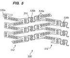
- FIG. 5is an enlarged side view of a portion of an expandable medical device with beneficial agent openings in the bridging elements.
- FIG. 6is an enlarged side view of a portion of an expandable medical device with a bifurcation opening.
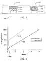
- FIG. 7is a cross sectional view of an expandable medical device having a combination of a first agent, such as an anti-inflammatory agent, in a first plurality of holes and a second agent, such as an anti-proliferative agent, in a second plurality of holes.
- a first agentsuch as an anti-inflammatory agent
- a second agentsuch as an anti-proliferative agent
- FIG. 8is a graph of the release rates of one example of an anti-inflammatory and an anti-proliferative delivered by the expandable medical device of FIG. 7 .
- FIG. 1illustrates an expandable medical device having a plurality of holes containing a beneficial agent for delivery to tissue by the expandable medical device.
- the expandable medical device 10 shown in FIG. 1is cut from a tube of material to form a cylindrical expandable device.
- the expandable medical device 10includes a plurality of cylindrical sections 12 interconnected by a plurality of bridging elements 14 .
- the bridging elements 14allow the tissue supporting device to bend axially when passing through the torturous path of vasculature to a deployment site and allow the device to bend axially when necessary to match the curvature of a lumen to be supported.
- Each of the cylindrical tubes 12is formed by a network of elongated struts 18 which are interconnected by ductile hinges 20 and circumferential struts 22 .
- the ductile hinges 20deform while the struts 18 are not deformed. Further details of one example of the expandable medical device are described in U.S. Pat. No. 6,241,762 which is incorporated herein by reference in its entirety.
- the elongated struts 18 and circumferential struts 22include openings 30 , some of which contain a beneficial agent for delivery to the lumen in which the expandable medical device is implanted.
- other portions of the device 10such as the bridging elements 14 , may include openings, as discussed below with respect to FIG. 5 .
- the openings 30are provided in non-deforming portions of the device 10 , such as the struts 18 , so that the openings are non-deforming and the beneficial agent is delivered without risk of being fractured, expelled, or otherwise damaged during expansion of the device.
- a further description of one example of the manner in which the beneficial agent may be loaded within the openings 30is described in U.S. patent application Ser. No. 09/948,987, filed Sep. 7, 2001, which is incorporated herein by reference in its entirety.
- the embodiments of the invention showncan be further refined by using Finite Element Analysis and other techniques to optimize the deployment of the beneficial agents within the openings 30 .
- the shape and location of the openings 30can be modified to maximize the volume of the voids while preserving the relatively high strength and rigidity of the struts with respect to the ductile hinges 20 .
- the openingshave an area of at least 5 ⁇ 10 ⁇ 6 square inches, and preferably at least 7 ⁇ 10 ⁇ 6 square inches.
- the openingsare filled about 50% to about 95% full of beneficial agent.
- agentor “beneficial agent” as used herein are intended to have the broadest possible interpretation and are used to include any therapeutic agent or drug, as well as inactive agents such as barrier layers, carrier layers, therapeutic layers, or protective layers.
- drugand “therapeutic agent” are used interchangeably to refer to any therapeutically active substance that is delivered to a bodily lumen of a living being to produce a desired, usually beneficial, effect.
- beneficial agentsmay include one or more drug or therapeutic agent.
- the present inventionis particularly well suited for the delivery of antineoplastics, antiangiogenics, angiogenic factors, anti-inflammatories, immuno-suppressants, antirestenotics, antiplatelet agents, vasodilators, anti-thrombotics, antiproliferatives, such as paclitaxel and Rapamycin, for example, and antithrombins, such as heparin, for example.
- erosionmeans the process by which components of a medium or matrix are bioresorbed and/or degraded and/or broken down by chemical or physical processes. For example in reference to biodegradable polymer matrices, erosion can occur by cleavage or hydrolysis of the polymer chains, thereby increasing the solubility of the matrix and suspended beneficial agents.
- erosion rateis a measure of the amount of time it takes for the erosion process to occur, usually reported in unit-area per unit-time.
- matrixor “bioresorbable matrix” are used interchangeably to refer to a medium or material that, upon implantation in a subject, does not elicit a detrimental response sufficient to result in the rejection of the matrix.
- the matrixtypically does not provide any therapeutic responses itself, though the matrix may contain or surround a beneficial agent, as defined herein.
- a matrixis also a medium that may simply provide support, structural integrity or structural barriers.
- the matrixmay be polymeric, non-polymeric, hydrophobic, hydrophilic, lipophilic, amphiphilic, and the like.
- openingsincludes both through openings and recesses.
- pharmaceutically acceptablerefers to the characteristic of being non-toxic to a host or patient and suitable for maintaining the stability of a beneficial agent and allowing the delivery of the beneficial agent to target cells or tissue.
- polymerrefers to molecules formed from the chemical union of two or more repeating units, called monomers. Accordingly, included within the term “polymer” may be, for example, dimers, trimers and oligomers. The polymer may be synthetic, naturally-occurring or semisynthetic. In preferred form, the term “polymer” refers to molecules which typically have a M W greater than about 3000 and preferably greater than about 10,000 and a M W that is less than about 10 million, preferably less than about a million and more preferably less than about 200,000.
- polymersinclude but are not limited to, poly- ⁇ -hydroxy acid esters such as, polylactic acid (PLLA or DLPLA), polyglycolic acid, polylactic-co-glycolic acid (PLGA), polylactic acid-co-caprolactone; poly (block-ethylene oxide-block-lactide-co-glycolide) polymers (PEO-block-PLGA and PEO-block-PLGA-block-PEO); polyethylene glycol and polyethylene oxide, poly (block-ethylene oxide-block-propylene oxide-block-ethylene oxide); polyvinyl pyrrolidone; polyorthoesters; polysaccharides and polysaccharide derivatives such as polyhyaluronic acid, poly (glucose), polyalginic acid, chitin, chitosan, chitosan derivatives, cellulose, methyl cellulose, hydroxyethylcellulose, hydroxypropylcellulose, carboxymethylcellulose, cyclodextrins and substituted cyclodextr
- primarilywith respect to directional delivery, refers to an amount greater than about 50% of the total amount of therapeutic agent provided to a blood vessel is provided in the primary direction.
- first generation drug delivery stentsThe causes of edge effect restenosis in first generation drug delivery stents are currently not well understood. It may be that the region of tissue injury due to angioplasty and/or stent implantation extends beyond the diffusion range of current generation beneficial agents such as paclitaxel and Rapamycin, which tend to partition strongly in tissue. A similar phenomenon has been observed in radiation therapies in which low doses of radiation at the edges of stent have proven stimulatory in the presence of an injury. In this case, radiating over a longer length until uninjured tissue is irradiated solved the problem.
- FIG. 1illustrates an expandable medical device 10 with “hot ends” or beneficial agent provided in the openings 30 a at the ends of the device in order to treat and reduce edge effect restenosis.
- the remaining openings 30 b in the central portion of the devicemay be empty (as shown) or may contain a lower concentration of beneficial agent.
- edge effect restenosismay involve cytotoxicity of particular drugs or combinations of drugs. Such mechanisms could include a physical or mechanical contraction of tissue similar to that seen in epidermal scar tissue formation, and the stent might prevent the contractile response within its own boundaries, but not beyond its edges. Further, the mechanism of this latter form of restenosis may be related to sequelae of sustained or local drug delivery to the arterial wall that is manifest even after the drug itself is no longer present in the wall. That is, the restenosis may be a response to a form of noxious injury related to the drug and/or the drug carrier. In this situation, it might be beneficial to exclude certain agents from the edges of the device.
- FIG. 2illustrates an alternative embodiment of an expandable medical device 200 having a plurality of openings 230 in which the openings 230 b in a central portion of the device are filled with a beneficial agent and the openings 230 a at the edges of the device remain empty.
- the device of FIG. 2is referred to as having “cool ends”.
- the expandable medical device 200 of FIG. 2may be used in conjunction with the expandable medical device 10 of FIG. 1 or another drug delivery stent when an initial stenting procedure has to be supplemented with an additional stent.
- the device 10 of FIG. 1 with “hot ends” or a device with uniform distribution of drugmay be implanted improperly. If the physician determines that the device does not cover a sufficient portion of the lumen a supplemental device may be added at one end of the existing device and slightly overlapping the existing device.
- the device 200 of FIG. 2is used so that the “cool ends” of the medical device 200 prevent double-dosing of the beneficial agent at the overlapping portions of the devices 10 , 200 .
- FIG. 3illustrates a further alternative embodiment of the invention in which different beneficial agents are positioned in different holes of an expandable medical device 300 .
- a first beneficial agentis provided in holes 330 a at the ends of the device and a second beneficial agent is provided in holes 330 b at a central portion of the device.
- the beneficial agentmay contain different drugs, the same drugs in different concentrations, or different variations of the same drug.
- the embodiment of FIG. 3may be used to provide an expandable medical device 300 with either “hot ends” or “cool ends.”
- each end portion of the device 300 which includes the holes 330 a containing the first beneficial agentextends at least one hole and up to about 15 holes from the edge. This distance corresponds to about 0.005 to about 0.1 inches from the edge of an unexpanded device.
- the distance from the edge of the device 300 which includes the first beneficial agentis preferably about one section, where a section is defined between the bridging elements.
- Different beneficial agents containing different drugsmay be disposed in different openings in the stent. This allows the delivery of two or more beneficial agents from a single stent in any desired delivery pattern. Alternatively, different beneficial agents containing the same drug in different concentrations may be disposed in different openings. This allows the drug to be uniformly distributed to the tissue with a non-uniform device structure.
- the two or more different beneficial agents provided in the devices described hereinmay contain (1) different drugs; (2) different concentrations of the same drug; (3) the same drug with different release kinetics, i.e., different matrix erosion rates; or (4) different forms of the same drug.
- Examples of different beneficial agents formulated containing the same drug with different release kineticsmay use different carriers to achieve the elution profiles of different shapes.
- Some examples of different forms of the same druginclude forms of a drug having varying hydrophilicity or lipophilicity.
- the holes 330 a at the ends of the deviceare loaded with a first beneficial agent comprising a drug with a high lipophilicity while holes 330 b at a central portion of the device are loaded with a second beneficial agent comprising the drug with a lower lipophilicity.
- the first high lipophilicity beneficial agent at the “hot ends”will diffuse more readily into the surrounding tissue reducing the edge effect restenosis.
- the device 300may have an abrupt transition line at which the beneficial agent changes from a first agent to a second agent. For example, all openings within 0.05 inches of the end of the device may contain the first agent while the remaining openings contain the second agent. Alternatively, the device may have a gradual transition between the first agent and the second agent. For example, a concentration of the drug in the openings can progressively increase (or decrease) toward the ends of the device. In another example, an amount of a first drug in the openings increases while an amount of a second drug in the openings decreases moving toward the ends of the device.
- FIG. 4illustrates a further alternative embodiment of an expandable medical device 400 in which different beneficial agents are positioned in different openings 430 a , 430 b in the device in an alternating or interspersed manner.
- multiple beneficial agentscan be delivered to tissue over the entire area or a portion of the area supported by the device.
- This embodimentwill be useful for delivery of multiple beneficial agents where combination of the multiple agents into a single composition for loading in the device is not possible due to interactions or stability problems between the beneficial agents.
- the loading of different beneficial agents in different openingsmay be used to provide a more even spatial distribution of the beneficial agent delivered in instances where the expandable medical device has a non-uniform distribution of openings in the expanded configuration.
- the use of different drugs in different openings in an interspersed or alternating mannerallows the delivery of two different drugs which may not be deliverable if combined within the same polymer/drug matrix composition.
- the drugsthemselves may interact in an undesirable way.
- the two drugsmay not be compatible with the same polymers for formation of the matrix or with the same solvents for delivery of the polymer/drug matrix into the openings.
- the embodiment of FIG. 4 having different drugs in different openings in an interspersed arrangementprovide the ability to deliver different drugs with very different desired release kinetics from the same medical device or stent and to optimize the release kinetic depending on the mechanism of action and properties of the individual agents.
- the water solubility of an agentgreatly affects the release of the agent from a polymer or other matrix.
- a highly water soluble compoundwill generally be delivered very quickly from a polymer matrix, whereas, a lipophilic agent will be delivered over a longer time period from the same matrix.
- a hydrophilic agent and a lipophilic agentare to be delivered as a dual drug combination from a medical device, it is difficult to achieve a desired release profile for these two agents delivered from the same polymer matrix.
- the system of FIG. 4allows the delivery of a hydrophilic and a lipophilic drug easily from the same stent. Further, the system of FIG. 4 allows the delivery two agents at two different release kinetics and/or administration periods. Each of the initial release in the first 24 hours, the release rate following the first 24 hours, the total administration period and any other characteristics of the release of the two drugs can be independently controlled.
- the release rate of the first beneficial agentcan be arranged to be delivered with at least 40% (preferably at least 50%) of the drug delivered in the first 24 hours and the second beneficial agent can be arranged to be delivered with less than 20% (preferably less than 10%) of the drug delivered in the first 24 hours.
- the administration period of the first beneficial agentcan be about 3 weeks or less (preferably 2 weeks or less) and the administration period of the second beneficial agent can be about 4 weeks or more.
- Restenosis or the recurrence of occlusion post-interventioninvolves a combination or series of biological processes. These processes include the activation of platelets and macrophages. Cytokines and growth factors contribute to smooth muscle cell proliferation and upregulation of genes and metalloproteinases lead to cell growth, remodeling of extracellular matrix, and smooth muscle cell migration.
- a drug therapywhich addresses a plurality of these processes by a combination of drugs may be the most successfully antirestenotic therapy.
- the present inventionprovides a means to achieve such a successful combination drug therapy.
- One example of a beneficial system for delivering two drugs from interspersed or alternating holesis the delivery of an anti-inflammatory agent or an immunosuppressant agent in combination with an antiproliferative agent or an anti-migratory agent.
- Other combinations of these agentsmay also be used to target multiple biological processes involved in restenosis.
- the anti-inflammatory agentmitigates the initial inflammatory response of the vessel to the angioplasty and stenting and is delivered at a high rate initially followed by a slower delivery over a time period of about two weeks to match the peak in the development of macrophages which stimulate the inflammatory response.
- the antiproliferative agentis delivered at a relatively even rate over a longer time period to reduce smooth muscle cell migration and proliferation.
- the placement of the drugs in different openingsallows the release kinetics to be tailored to the particular agent regardless of the hydrophobilicity or lipophobicity of the drug.
- Examples of some arrangements for delivery of a lipophilic drug at a substantially constant or linear release rateare described in WO 04/110302 published on Dec. 23, 2004, which is incorporated herein by reference in its entirety.
- Examples of some of the arrangements for delivery of hydrophilic drugare described in WO 04/043510, published on May 27, 2004 which is incorporated herein by reference in its entirety.
- the hydrophilic drugs listed aboveinclude CdA, Gleevec, VIP, insulin, and ApoA-1 milano.
- the lipophilic drugs listed aboveinclude paclitaxel, Epothilone D, rapamycin, pimecrolimus, PKC-412 and Dexamethazone. Farglitazar is partly liphophillic and partly hydrophilic.
- a stentcan deliver an anti-proliferative, such as paclitaxel or a limus drug from one set of openings for treatment of restenosis while delivering a myocardial preservative drug, such as insulin, from other openings for the treatment of acute myocardial infarction.
- an anti-proliferativesuch as paclitaxel or a limus drug from one set of openings for treatment of restenosis
- a myocardial preservative drugsuch as insulin
- the coverage of the device 500is greater at the cylindrical tube portions 512 of the device than at the bridging elements 514 .
- Coverageis defined as the ratio of the device surface area to the area of the lumen in which the device is deployed.
- the beneficial agent concentration delivered to the tissue adjacent the cylindrical tube portions 512is greater that the beneficial agent delivered to the tissue adjacent the bridging elements 514 .
- the concentration of the beneficial agentmay be varied in the openings at portions of the device to achieve a more even distribution of the beneficial agent throughout the tissue.
- the openings 530 a in the tube portions 512include a beneficial agent with a lower drug concentration than the openings 530 b in the bridging elements 514 .
- the uniformity of agent deliverymay be achieved in a variety of manners including varying the drug concentration, the opening diameter or shape, the amount of agent in the opening (i.e., the percentage of the opening filed), the matrix material, or the form of the drug.
- Bifurcation devicesinclude a side hole 610 which is positioned to allow blood flow through a side branch of a vessel.
- a bifurcation deviceis described in U.S. Pat. No. 6,293,967 which is incorporated herein by reference in its entirety.
- the bifurcation device 600includes the side hole feature 610 interrupting the regular pattern of beams which form a remainder of the device.
- a concentration of an antiproliferative drugmay be increased in openings 830 a at an area surrounding the side hole 610 of the device 600 to deliver increased concentrations of the drug where needed.
- the remaining openings 630 b in an area away from the side openingcontain a beneficial agent with a lower concentration of the antiproliferative.
- the increased antiproliferative delivered to the region surrounding the bifurcation holemay be provided by a different beneficial agent containing a different drug or a different beneficial agent containing a higher concentration of the same drug.
- beneficial agentsmay be delivered to the luminal side of the expandable medical device.
- Drugs which are delivered into the blood stream from the luminal side of the devicecan be located at a proximal end of the device or a distal end of the device.
- the methods for loading different beneficial agents into different openings in an expandable medical devicemay include known techniques such as dipping and coating and also known piezoelectric micro-jetting techniques.
- Micro-injection devicesmay be computer controlled to deliver precise amounts of two or more liquid beneficial agents to precise locations on the expandable medical device in a known manner.
- a dual agent jetting devicemay deliver two agents simultaneously or sequentially into the openings.
- a luminal side of the through openingsmay be blocked during loading by a resilient mandrel allowing the beneficial agents to be delivered in liquid form, such as with a solvent.
- the beneficial agentsmay also be loaded by manual injection devices.
- FIG. 7illustrates a dual drug stent 700 having an anti-inflammatory agent and an antiproliferative agent delivered from different holes in the stent to provide independent release kinetics of the two drugs which are specifically programmed to match the biological processes of restenosis.
- the dual drug stentincludes the anti-inflammatory agent pimecrolimus in a first set of openings 710 in combination with the antiproliferative agent paclitaxel in a second set of openings 720 .
- Each agentis provided in a matrix material within the holes of the stent in a specific inlay arrangement designed to achieve the release kinetics shown in FIG. 8 .
- Each of the drugsare delivered primarily murally for treatment of restenosis.
- pimecrolimusis provided in the stent for directional delivery to the mural side of the stent by the use of a barrier 712 at the luminal side of the hole.
- the barrier 712is formed by a biodegradable polymer.
- the pimecrolimusis loaded within the holes in a manner which creates a release kinetic having dual phases.
- a first phase of the release of pimecrolimusis provided by a murally located region 716 of the matrix which has a fast release formulation including pimecrolimus and biodegradable polymer (PLGA) with a high percentage of drug, such as about 90% drug to 10% polymer.
- PLGAbiodegradable polymer
- a second phase of the releaseis provided by a central region 714 of the matrix with pimecrolimus and biodegradable polymer (PLGA) in a ratio of about 50% drug to 50% polymer.
- PLGAbiodegradable polymer
- the first phase of the pimecrolimus releasedelivers about 50% of the loaded drug in about the first 24 hours.
- the second phase of the releasedelivers the remaining 50% over about two weeks.
- This releaseis specifically programmed to match the progression of the inflammatory process following angioplasty and stenting.
- different polymers or different comonomer ratios of the same polymercan be used in two drug different regions to achieve the two different release rates.
- the paclitaxelis loaded within the openings 720 in a manner which creates a release kinetic having a substantially linear release after the first approximately 24 hours, as shown in FIG. 8 .
- the paclitaxel openings 720are loaded with three regions including a base region 722 of primarily polymer with minimal drug at a luminal side of the hole, a central region 724 with paclitaxel and polymer (PLGA) provided in a concentration gradient, and a cap region 726 with primarily polymer which controls release of the paclitaxel.
- the paclitaxelis released with an initial release in the first day of about 5-15% of the total drug load followed by a substantially linear release for about 20-90 days. Additional examples of arrangements for paclitaxel in the holes with a concentration gradient are described in WO 04/110302 described above.
- FIG. 7shows the drug, barrier, and cap regions as distinct regions within the openings for ease of illustration. It should be understood that these regions indistinct and formed by a blending of the different areas.
- the barrier layersare primarily polymer without drug, depending on the manufacturing processes employed, some small amount of drug of the subsequent region can be incorporation into the barrier region.
- the amount of the drugs deliveredvaries depending on the size of the stent.
- the amount of pimecrolimusis about 50 to about 300 ⁇ g, preferably about 100 to about 250 ⁇ g.
- the amount of paclitaxel delivered from this stentis about 5 to about 50 ⁇ g, preferably about 10 to about 30 ⁇ g. In one example, about 200 ⁇ g of pimecrolimus and about 20 ⁇ g of paclitaxel are delivered.
- the drugsmay be located in alternating holes in the stent. However, in view of the large difference in the doses to be delivered between the two drugs, it may be desirable to place the paclitaxel in every third of fourth hole in the stent. Alternatively, the holes for delivery of the low dose drug (paclitaxel) can be made smaller than the holes for the high dose.
- the polymer/drug inlaysare formed by computer controlled piezoelectric injection techniques as described in WO 04/026182 published on Apr. 1, 2004, which is incorporated herein by reference in its entirety.
- the inlays of the first agentcan be formed first followed by the inlays of the second agent using the piezoelectric injector.
- the system of WO 04/02182can be equipped with dual piezoelectric dispensers for dispensing the two agents at the same time.
- the dual drug stentincludes the Gleevec in the first set of openings 710 in combination with the antiproliferative agent paclitaxel in the second set of openings 720 .
- Each agentis provided in a matrix material within the holes of the stent in a specific inlay arrangement designed to achieve the release kinetics shown in FIG. 8 .
- the Gleevecis delivered with a two phase release including a high initial release in the first day and then a slow release for 1-2 weeks.
- the first phase of the Gleevec releasedelivers about 50% of the loaded drug in about the first 24 hours.
- the second phase of the releasedelivers the remaining 50% over about one-two weeks.
- the paclitaxelis loaded within the openings 720 in a manner which creates a release kinetic having a substantially linear release after the first approximately 24 hours, as shown in FIG. 8 and as described above in Example 1.
- the amount of the drugs deliveredvaries depending on the size of the stent.
- the amount of Gleevecis about 200 to about 500 ⁇ g, preferably about 300 to about 400 ⁇ g.
- the amount of paclitaxel delivered from this stentis about 5 to about 50 ⁇ g, preferably about 10 to about 30 ⁇ g.
- the drugsmay be located in alternating holes in the stent or interspersed in a non-alternating manner.
- the polymer/drug inlaysare formed in the manner described in Example 1.
- the dual drug stentincludes the PKC-412 (a cell growth regulator) in the first set of openings in combination with the antiproliferative agent paclitaxel in the second set of openings.
- PKC-412a cell growth regulator
- Each agentis provided in a matrix material within the holes of the stent in a specific inlay arrangement designed to achieve the release kinetics discussed below.
- the PKC-412is delivered at a substantially constant release rate after the first approximately 24 hours, with the release over a period of about 4-16 weeks, preferably about 6-12 weeks.
- the paclitaxelis loaded within the openings in a manner which creates a release kinetic having a substantially linear release after the first approximately 24 hours, with the release over a period of about 4-16 weeks, preferably about 6-12 weeks.
- the amount of the drugs deliveredvaries depending on the size of the stent.
- the amount of PKC-412is about 100 to about 400 ⁇ g, preferably about 150 to about 250 ⁇ g.
- the amount of paclitaxel delivered from this stentis about 5 to about 50 ⁇ g, preferably about 10 to about 30 ⁇ g.
- the drugsmay be located in alternating holes in the stent or interspersed in a non-alternating manner.
- the polymer/drug inlaysare formed in the manner described in Example 1.
- the present inventionrelates to the delivery of anti-restenotic agents including paclitaxel, rapamycin, cladribine (CdA), and their derivatives, as well as other cytotoxic or cytostatic agents and microtubule stabilizing agents.
- anti-restenotic agentsinclude paclitaxel, rapamycin, cladribine (CdA), and their derivatives, as well as other cytotoxic or cytostatic agents and microtubule stabilizing agents.
- anti-restenotic agentshave been primarily described herein, the present invention may also be used to deliver other agents alone or in combination with anti-restenotic agents.
- Some of the therapeutic agents for use with the present inventionwhich may be transmitted primarily luminally, primarily murally, or both and may be delivered alone or in combination include, but are not limited to, antiproliferatives, antithrombins, immunosuppressants including rapamycin, antilipid agents, anti-inflammatory agents, antineoplastics, antiplatelets, angiogenic agents, anti-angiogenic agents, vitamins, antimitotics, metalloproteinase inhibitors, NO donors, estradiols, anti-sclerosing agents, and vasoactive agents, endothelial growth factors, estrogen, beta blockers, AZ blockers, hormones, statins, insulin growth factors, antioxidants, membrane stabilizing agents, calcium antagonists, retenoid, bivalirudin, phenoxodiol, etoposide, ticlopidine, dipyridamole, and trapidil alone or in combinations with any therapeutic agent mentioned herein.
- Therapeutic agentsalso include peptides, lipoproteins, polypeptides, polynucleotides encoding polypeptides, lipids, protein-drugs, protein conjugate drugs, enzymes, oligonucleotides and their derivatives, ribozymes, other genetic material, cells, antisense, oligonucleotides, monoclonal antibodies, platelets, prions, viruses, bacteria, and eukaryotic cells such as endothelial cells, stem cells, ACE inhibitors, monocyte/macrophages or vascular smooth muscle cells to name but a few examples.
- the therapeutic agentmay also be a pro-drug, which metabolizes into the desired drug when administered to a host.
- therapeutic agentsmay be pre-formulated as microcapsules, microspheres, microbubbles, liposomes, niosomes, emulsions, dispersions or the like before they are incorporated into the therapeutic layer.
- Therapeutic agentsmay also be radioactive isotopes or agents activated by some other form of energy such as light or ultrasonic energy, or by other circulating molecules that can be systemically administered.
- Therapeutic agentsmay perform multiple functions including modulating angiogenesis, restenosis, cell proliferation, thrombosis, platelet aggregation, clotting, and vasodilation.
- Anti-inflammatoriesinclude but are not limited to non-steroidal anti-inflammatories (NSAID), such as aryl acetic acid derivatives, e.g., Diclofenac; aryl propionic acid derivatives, e.g., Naproxen; and salicylic acid derivatives, e.g., Diflunisal.
- Anti-inflammatoriesalso include glucocoriticoids (steroids) such as dexamethasone, aspirin, prednisolone, and triamcinolone, pirfenidone, meclofenamic acid, tranilast, and nonsteroidal anti-inflammatories.
- Anti-inflammatoriesmay be used in combination with antiproliferatives to mitigate the reaction of the tissue to the antiproliferative.
- the agentscan also include anti-lymphocytes; anti-macrophage substances; immunomodulatory agents; cyclooxygenase inhibitors; anti-oxidants; cholesterol-lowering drugs; statins and angiotens in converting enzyme (ACE); fibrinolytics; inhibitors of the intrinsic coagulation cascade; antihyperlipoproteinemics; and anti-platelet agents; anti-metabolites, such as 2-chlorodeoxy adenosine (2-CdA or cladribine); immuno-suppressants including sirolimus, everolimus, tacrolimus, etoposide, and mitoxantrone; anti-leukocytes such as 2-CdA, IL-1 inhibitors, anti-CD116/CD18 monoclonal antibodies, monoclonal antibodies to VCAM or ICAM, zinc protoporphyrin; anti-macrophage substances such as drugs that elevate NO; cell sensitizers to insulin including glitazones; high density lipoproteins (HDL)
- Agentsmay also be delivered using a gene therapy-based approach in combination with an expandable medical device.
- Gene therapyrefers to the delivery of exogenous genes to a cell or tissue, thereby causing target cells to express the exogenous gene product.
- Genesare typically delivered by either mechanical or vector-mediated methods.
- additivesincluding surfactants, antacids, antioxidants, and detergents may be used to minimize denaturation and aggregation of a protein drug.
- Anionic, cationic, or nonionic detergentsmay be used.
- nonionic additivesinclude but are not limited to sugars including sorbitol, sucrose, trehalose; dextrans including dextran, carboxy methyl (CM) dextran, diethylamino ethyl (DEAE) dextran; sugar derivatives including D-glucosaminic acid, and D-glucose diethyl mercaptal; synthetic polyethers including polyethylene glycol (PEF and PEO) and polyvinyl pyrrolidone (PVP); carboxylic acids including D-lactic acid, glycolic acid, and propionic acid; detergents with affinity for hydrophobic interfaces including n-dodecyl- ⁇ -D-maltoside, n-octyl- ⁇ -D-glucoside, PEO-fatty acid esters (e.g.
- PEO-sorbitan-fatty acid esterse.g. Tween 80, PEO-20 sorbitan monooleate
- sorbitan-fatty acid esterse.g. SPAN 60 , sorbitan monostearate
- PEO-glyceryl-fatty acid esterse.g. glyceryl fatty acid esters (e.g. glyceryl monostearate)
- PEO-hydrocarbon-etherse.g. PEO-10 oleyl ether; triton X-100; and Lubrol.
- ionic detergentsinclude but are not limited to fatty acid salts including calcium stearate, magnesium stearate, and zinc stearate; phospholipids including lecithin and phosphatidyl choline; CM-PEG; cholic acid; sodium dodecyl sulfate (SDS); docusate (AOT); and taumocholic acid.
Landscapes
- Health & Medical Sciences (AREA)
- Life Sciences & Earth Sciences (AREA)
- Engineering & Computer Science (AREA)
- Veterinary Medicine (AREA)
- Public Health (AREA)
- General Health & Medical Sciences (AREA)
- Animal Behavior & Ethology (AREA)
- Biomedical Technology (AREA)
- Heart & Thoracic Surgery (AREA)
- Vascular Medicine (AREA)
- Cardiology (AREA)
- Chemical & Material Sciences (AREA)
- Medicinal Chemistry (AREA)
- Oral & Maxillofacial Surgery (AREA)
- Epidemiology (AREA)
- Surgery (AREA)
- Transplantation (AREA)
- Chemical Kinetics & Catalysis (AREA)
- Physics & Mathematics (AREA)
- Optics & Photonics (AREA)
- Bioinformatics & Cheminformatics (AREA)
- General Chemical & Material Sciences (AREA)
- Nuclear Medicine, Radiotherapy & Molecular Imaging (AREA)
- Organic Chemistry (AREA)
- Pharmacology & Pharmacy (AREA)
- Molecular Biology (AREA)
- Dispersion Chemistry (AREA)
- Pain & Pain Management (AREA)
- Rheumatology (AREA)
- Urology & Nephrology (AREA)
- Pharmaceuticals Containing Other Organic And Inorganic Compounds (AREA)
- Materials For Medical Uses (AREA)
- Medicinal Preparation (AREA)
- Medicines That Contain Protein Lipid Enzymes And Other Medicines (AREA)
- Media Introduction/Drainage Providing Device (AREA)
Abstract
Description
| Imatinib | |||||||||||||
| mesylate | Rapamycin | Pime- | PKC- | Dexa- | Fargli- | ApoA-I | |||||||
| PTX | 2-Cda | Epothilone D | Gleevec | analog | crolimus | 412 | methasone | tazar | Insulin | VIP | milano | ||
| PTX | x | x | x | x | x | x | x | x | ||||
| 2-CdA | x | x | x | x | x | x | ||||||
| Epothilone D | x | x | x | x | x | x | ||||||
| Imatinib | x | x | x | x | ||||||||
| mesylate | ||||||||||||
| Gleevec | ||||||||||||
| Rapamycin | x | x | x | x | x | |||||||
| analog | ||||||||||||
| Pimecrolimus | x | x | x | x | x | |||||||
| PKC-412 | x | x | x | x | ||||||||
| Dexamethasone | x | x | ||||||||||
| Farglitazar | x | x | ||||||||||
| Insulin | x | |||||||||||
| VIP | x | |||||||||||
| ApoA-I milano | ||||||||||||
Claims (2)
Priority Applications (6)
| Application Number | Priority Date | Filing Date | Title |
|---|---|---|---|
| US11/079,967US7758636B2 (en) | 2002-09-20 | 2005-03-14 | Expandable medical device with openings for delivery of multiple beneficial agents |
| PCT/US2006/007411WO2006098889A2 (en) | 2005-03-14 | 2006-03-01 | Expandable medical device with openings for delivery of multiple beneficial agents |
| JP2008501905AJP2008532692A (en) | 2005-03-14 | 2006-03-01 | Expanded medical device with an opening for delivering multiple active substances |
| AU2006223581AAU2006223581A1 (en) | 2005-03-14 | 2006-03-01 | Expandable medical device with openings for delivery of multiple beneficial agents |
| EP06736688AEP1858442A4 (en) | 2005-03-14 | 2006-03-01 | EXPANDABLE MEDICAL DEVICE HAVING OPENINGS FOR DISTRIBUTING SEVERAL BENEFICIAL AGENTS |
| CA002615143ACA2615143A1 (en) | 2005-03-14 | 2006-03-01 | Expandable medical device with openings for delivery of multiple beneficial agents |
Applications Claiming Priority (3)
| Application Number | Priority Date | Filing Date | Title |
|---|---|---|---|
| US41248902P | 2002-09-20 | 2002-09-20 | |
| US10/668,430US20040127977A1 (en) | 2002-09-20 | 2003-09-22 | Expandable medical device with openings for delivery of multiple beneficial agents |
| US11/079,967US7758636B2 (en) | 2002-09-20 | 2005-03-14 | Expandable medical device with openings for delivery of multiple beneficial agents |
Related Parent Applications (1)
| Application Number | Title | Priority Date | Filing Date |
|---|---|---|---|
| US10/668,430Continuation-In-PartUS20040127977A1 (en) | 1998-03-30 | 2003-09-22 | Expandable medical device with openings for delivery of multiple beneficial agents |
Publications (2)
| Publication Number | Publication Date |
|---|---|
| US20060122697A1 US20060122697A1 (en) | 2006-06-08 |
| US7758636B2true US7758636B2 (en) | 2010-07-20 |
Family
ID=36992196
Family Applications (1)
| Application Number | Title | Priority Date | Filing Date |
|---|---|---|---|
| US11/079,967Expired - Fee RelatedUS7758636B2 (en) | 2002-09-20 | 2005-03-14 | Expandable medical device with openings for delivery of multiple beneficial agents |
Country Status (6)
| Country | Link |
|---|---|
| US (1) | US7758636B2 (en) |
| EP (1) | EP1858442A4 (en) |
| JP (1) | JP2008532692A (en) |
| AU (1) | AU2006223581A1 (en) |
| CA (1) | CA2615143A1 (en) |
| WO (1) | WO2006098889A2 (en) |
Cited By (40)
| Publication number | Priority date | Publication date | Assignee | Title |
|---|---|---|---|---|
| US20060127443A1 (en)* | 2004-12-09 | 2006-06-15 | Helmus Michael N | Medical devices having vapor deposited nanoporous coatings for controlled therapeutic agent delivery |
| US20070154555A1 (en)* | 2004-12-02 | 2007-07-05 | Strauss Bradley H | Augmentation of intraluminal microvessel formation to facilitate guide wire crossing in chronic total occlusions |
| US20080147177A1 (en)* | 2006-11-09 | 2008-06-19 | Torsten Scheuermann | Endoprosthesis with coatings |
| US20080188925A1 (en)* | 2007-02-01 | 2008-08-07 | Zhao Jonathon Z | Antithrombotic and anti-restenotic drug eluting stent |
| US20080243231A1 (en)* | 2007-03-01 | 2008-10-02 | Aiden Flanagan | Medical device with a porous surface for delivery of a therapeutic agent |
| US20080241218A1 (en)* | 2007-03-01 | 2008-10-02 | Mcmorrow David | Coated medical devices for abluminal drug delivery |
| US20080249615A1 (en)* | 2007-04-05 | 2008-10-09 | Jan Weber | Stents with ceramic drug reservoir layer and methods of making and using the same |
| US20080294236A1 (en)* | 2007-05-23 | 2008-11-27 | Boston Scientific Scimed, Inc. | Endoprosthesis with Select Ceramic and Polymer Coatings |
| US20090118812A1 (en)* | 2007-11-02 | 2009-05-07 | Boston Scientific Scimed, Inc. | Endoprosthesis coating |
| US20090118821A1 (en)* | 2007-11-02 | 2009-05-07 | Boston Scientific Scimed, Inc. | Endoprosthesis with porous reservoir and non-polymer diffusion layer |
| US20090138077A1 (en)* | 2007-07-27 | 2009-05-28 | Boston Scientific Scimed, Inc. | Articles having ceramic coated surfaces |
| US20090149942A1 (en)* | 2007-07-19 | 2009-06-11 | Boston Scientific Scimed, Inc. | Endoprosthesis having a non-fouling surface |
| US20100036482A1 (en)* | 2008-08-07 | 2010-02-11 | Exogenesis Corporation | Drug delivery system and method of manufacturing thereof |
| US20100036502A1 (en)* | 2008-08-07 | 2010-02-11 | Exogenesis Corporation | Medical device for bone implant and method for producing such device |
| US7942926B2 (en) | 2007-07-11 | 2011-05-17 | Boston Scientific Scimed, Inc. | Endoprosthesis coating |
| US7976915B2 (en) | 2007-05-23 | 2011-07-12 | Boston Scientific Scimed, Inc. | Endoprosthesis with select ceramic morphology |
| US8002823B2 (en) | 2007-07-11 | 2011-08-23 | Boston Scientific Scimed, Inc. | Endoprosthesis coating |
| US8029554B2 (en) | 2007-11-02 | 2011-10-04 | Boston Scientific Scimed, Inc. | Stent with embedded material |
| US8071156B2 (en) | 2009-03-04 | 2011-12-06 | Boston Scientific Scimed, Inc. | Endoprostheses |
| US8187620B2 (en) | 2006-03-27 | 2012-05-29 | Boston Scientific Scimed, Inc. | Medical devices comprising a porous metal oxide or metal material and a polymer coating for delivering therapeutic agents |
| US8216632B2 (en) | 2007-11-02 | 2012-07-10 | Boston Scientific Scimed, Inc. | Endoprosthesis coating |
| US8221822B2 (en) | 2007-07-31 | 2012-07-17 | Boston Scientific Scimed, Inc. | Medical device coating by laser cladding |
| US8231980B2 (en) | 2008-12-03 | 2012-07-31 | Boston Scientific Scimed, Inc. | Medical implants including iridium oxide |
| US8287937B2 (en) | 2009-04-24 | 2012-10-16 | Boston Scientific Scimed, Inc. | Endoprosthese |
| US8353949B2 (en) | 2006-09-14 | 2013-01-15 | Boston Scientific Scimed, Inc. | Medical devices with drug-eluting coating |
| US8449603B2 (en) | 2008-06-18 | 2013-05-28 | Boston Scientific Scimed, Inc. | Endoprosthesis coating |
| US8574615B2 (en) | 2006-03-24 | 2013-11-05 | Boston Scientific Scimed, Inc. | Medical devices having nanoporous coatings for controlled therapeutic agent delivery |
| US8771343B2 (en) | 2006-06-29 | 2014-07-08 | Boston Scientific Scimed, Inc. | Medical devices with selective titanium oxide coatings |
| US8815275B2 (en) | 2006-06-28 | 2014-08-26 | Boston Scientific Scimed, Inc. | Coatings for medical devices comprising a therapeutic agent and a metallic material |
| US8815273B2 (en) | 2007-07-27 | 2014-08-26 | Boston Scientific Scimed, Inc. | Drug eluting medical devices having porous layers |
| US8900292B2 (en) | 2007-08-03 | 2014-12-02 | Boston Scientific Scimed, Inc. | Coating for medical device having increased surface area |
| US8920491B2 (en) | 2008-04-22 | 2014-12-30 | Boston Scientific Scimed, Inc. | Medical devices having a coating of inorganic material |
| US8932346B2 (en) | 2008-04-24 | 2015-01-13 | Boston Scientific Scimed, Inc. | Medical devices having inorganic particle layers |
| US9259339B1 (en)* | 2014-08-15 | 2016-02-16 | Elixir Medical Corporation | Biodegradable endoprostheses and methods of their fabrication |
| US9480588B2 (en) | 2014-08-15 | 2016-11-01 | Elixir Medical Corporation | Biodegradable endoprostheses and methods of their fabrication |
| US9566371B2 (en) | 2007-01-19 | 2017-02-14 | Elixir Medical Corporation | Biodegradable endoprostheses and methods for their fabrication |
| US9730819B2 (en) | 2014-08-15 | 2017-08-15 | Elixir Medical Corporation | Biodegradable endoprostheses and methods of their fabrication |
| US9855156B2 (en) | 2014-08-15 | 2018-01-02 | Elixir Medical Corporation | Biodegradable endoprostheses and methods of their fabrication |
| US9943426B2 (en) | 2015-07-15 | 2018-04-17 | Elixir Medical Corporation | Uncaging stent |
| US10918505B2 (en) | 2016-05-16 | 2021-02-16 | Elixir Medical Corporation | Uncaging stent |
Families Citing this family (60)
| Publication number | Priority date | Publication date | Assignee | Title |
|---|---|---|---|---|
| US8257726B2 (en) | 1997-09-26 | 2012-09-04 | Abbott Laboratories | Compositions, systems, kits, and methods of administering rapamycin analogs with paclitaxel using medical devices |
| US8057816B2 (en)* | 1997-09-26 | 2011-11-15 | Abbott Laboratories | Compositions and methods of administering paclitaxel with other drugs using medical devices |
| US7179289B2 (en)* | 1998-03-30 | 2007-02-20 | Conor Medsystems, Inc. | Expandable medical device for delivery of beneficial agent |
| US7208010B2 (en) | 2000-10-16 | 2007-04-24 | Conor Medsystems, Inc. | Expandable medical device for delivery of beneficial agent |
| US6290673B1 (en) | 1999-05-20 | 2001-09-18 | Conor Medsystems, Inc. | Expandable medical device delivery system and method |
| EP1498084B1 (en)* | 2000-10-16 | 2014-06-18 | Innovational Holdings, LLC | Expandable medical device for delivery of beneficial agent |
| US20040073294A1 (en) | 2002-09-20 | 2004-04-15 | Conor Medsystems, Inc. | Method and apparatus for loading a beneficial agent into an expandable medical device |
| WO2003002243A2 (en) | 2001-06-27 | 2003-01-09 | Remon Medical Technologies Ltd. | Method and device for electrochemical formation of therapeutic species in vivo |
| US7842083B2 (en) | 2001-08-20 | 2010-11-30 | Innovational Holdings, Llc. | Expandable medical device with improved spatial distribution |
| US7056338B2 (en) | 2003-03-28 | 2006-06-06 | Conor Medsystems, Inc. | Therapeutic agent delivery device with controlled therapeutic agent release rates |
| AU2003276920A1 (en)* | 2002-09-20 | 2004-04-08 | Innovational Holdings, Llc | Expandable medical device with openings for delivery of multiple beneficial agents |
| US7758636B2 (en) | 2002-09-20 | 2010-07-20 | Innovational Holdings Llc | Expandable medical device with openings for delivery of multiple beneficial agents |
| US7169178B1 (en)* | 2002-11-12 | 2007-01-30 | Advanced Cardiovascular Systems, Inc. | Stent with drug coating |
| EP1997456B1 (en)* | 2004-02-13 | 2011-12-07 | Innovational Holdings, LLC | Drug coating device and method for wire filaments |
| US8137397B2 (en)* | 2004-02-26 | 2012-03-20 | Boston Scientific Scimed, Inc. | Medical devices |
| US20060079956A1 (en)* | 2004-09-15 | 2006-04-13 | Conor Medsystems, Inc. | Bifurcation stent with crushable end and method for delivery of a stent to a bifurcation |
| US20070269486A1 (en)* | 2005-03-14 | 2007-11-22 | Conor Medsystems, Llc. | Methods and Devices for Reducing Tissue Damage After Ischemic Injury |
| US8840660B2 (en) | 2006-01-05 | 2014-09-23 | Boston Scientific Scimed, Inc. | Bioerodible endoprostheses and methods of making the same |
| US8089029B2 (en) | 2006-02-01 | 2012-01-03 | Boston Scientific Scimed, Inc. | Bioabsorbable metal medical device and method of manufacture |
| US8048150B2 (en) | 2006-04-12 | 2011-11-01 | Boston Scientific Scimed, Inc. | Endoprosthesis having a fiber meshwork disposed thereon |
| US8147537B2 (en)* | 2006-06-16 | 2012-04-03 | The Invention Science Fund I, Llc | Rapid-prototyped custom-fitted blood vessel sleeve |
| US20090024152A1 (en)* | 2007-07-17 | 2009-01-22 | Searete Llc, A Limited Liability Corporation Of The State Of Delaware | Custom-fitted blood vessel sleeve |
| US8095382B2 (en)* | 2006-06-16 | 2012-01-10 | The Invention Science Fund I, Llc | Methods and systems for specifying a blood vessel sleeve |
| US8163003B2 (en)* | 2006-06-16 | 2012-04-24 | The Invention Science Fund I, Llc | Active blood vessel sleeve methods and systems |
| US8478437B2 (en) | 2006-06-16 | 2013-07-02 | The Invention Science Fund I, Llc | Methods and systems for making a blood vessel sleeve |
| US8551155B2 (en)* | 2006-06-16 | 2013-10-08 | The Invention Science Fund I, Llc | Stent customization system and method |
| US20080172073A1 (en)* | 2006-06-16 | 2008-07-17 | Searete Llc, A Limited Liability Corporation Of The State Of Delaware | Active blood vessel sleeve |
| US8550344B2 (en)* | 2006-06-16 | 2013-10-08 | The Invention Science Fund I, Llc | Specialty stents with flow control features or the like |
| US7818084B2 (en)* | 2006-06-16 | 2010-10-19 | The Invention Science Fund, I, LLC | Methods and systems for making a blood vessel sleeve |
| US20080004695A1 (en)* | 2006-06-28 | 2008-01-03 | Abbott Cardiovascular Systems Inc. | Everolimus/pimecrolimus-eluting implantable medical devices |
| EP2054537A2 (en) | 2006-08-02 | 2009-05-06 | Boston Scientific Scimed, Inc. | Endoprosthesis with three-dimensional disintegration control |
| ES2357661T3 (en) | 2006-09-15 | 2011-04-28 | Boston Scientific Scimed, Inc. | BIOEROSIONABLE ENDOPROOTHESIS WITH BIOESTABLE INORGANIC LAYERS. |
| JP2010503489A (en) | 2006-09-15 | 2010-02-04 | ボストン サイエンティフィック リミテッド | Biodegradable endoprosthesis and method for producing the same |
| WO2008034047A2 (en) | 2006-09-15 | 2008-03-20 | Boston Scientific Limited | Endoprosthesis with adjustable surface features |
| WO2008034066A1 (en) | 2006-09-15 | 2008-03-20 | Boston Scientific Limited | Bioerodible endoprostheses and methods of making the same |
| EP2959925B1 (en) | 2006-09-15 | 2018-08-29 | Boston Scientific Limited | Medical devices and methods of making the same |
| WO2008036548A2 (en) | 2006-09-18 | 2008-03-27 | Boston Scientific Limited | Endoprostheses |
| EP2068781A2 (en) | 2006-09-18 | 2009-06-17 | Boston Scientific Limited | Medical devices |
| US20080276935A1 (en) | 2006-11-20 | 2008-11-13 | Lixiao Wang | Treatment of asthma and chronic obstructive pulmonary disease with anti-proliferate and anti-inflammatory drugs |
| US8414910B2 (en) | 2006-11-20 | 2013-04-09 | Lutonix, Inc. | Drug releasing coatings for medical devices |
| US9700704B2 (en) | 2006-11-20 | 2017-07-11 | Lutonix, Inc. | Drug releasing coatings for balloon catheters |
| US9737640B2 (en) | 2006-11-20 | 2017-08-22 | Lutonix, Inc. | Drug releasing coatings for medical devices |
| ES2506144T3 (en) | 2006-12-28 | 2014-10-13 | Boston Scientific Limited | Bioerodible endoprosthesis and their manufacturing procedure |
| US7682388B2 (en)* | 2007-01-30 | 2010-03-23 | Medtronic Vascular, Inc. | Stent with longitudinal groove |
| US20080243241A1 (en)* | 2007-03-28 | 2008-10-02 | Zhao Jonathon Z | Short term sustained drug-delivery system for implantable medical devices and method of making the same |
| US8052745B2 (en) | 2007-09-13 | 2011-11-08 | Boston Scientific Scimed, Inc. | Endoprosthesis |
| US7833266B2 (en)* | 2007-11-28 | 2010-11-16 | Boston Scientific Scimed, Inc. | Bifurcated stent with drug wells for specific ostial, carina, and side branch treatment |
| US7998192B2 (en) | 2008-05-09 | 2011-08-16 | Boston Scientific Scimed, Inc. | Endoprostheses |
| US8652506B2 (en)* | 2008-06-05 | 2014-02-18 | Boston Scientific Scimed, Inc. | Bio-degradable block co-polymers for controlled release |
| US8236046B2 (en) | 2008-06-10 | 2012-08-07 | Boston Scientific Scimed, Inc. | Bioerodible endoprosthesis |
| US7985252B2 (en) | 2008-07-30 | 2011-07-26 | Boston Scientific Scimed, Inc. | Bioerodible endoprosthesis |
| US8226603B2 (en) | 2008-09-25 | 2012-07-24 | Abbott Cardiovascular Systems Inc. | Expandable member having a covering formed of a fibrous matrix for intraluminal drug delivery |
| US8076529B2 (en) | 2008-09-26 | 2011-12-13 | Abbott Cardiovascular Systems, Inc. | Expandable member formed of a fibrous matrix for intraluminal drug delivery |
| US8049061B2 (en) | 2008-09-25 | 2011-11-01 | Abbott Cardiovascular Systems, Inc. | Expandable member formed of a fibrous matrix having hydrogel polymer for intraluminal drug delivery |
| US8382824B2 (en) | 2008-10-03 | 2013-02-26 | Boston Scientific Scimed, Inc. | Medical implant having NANO-crystal grains with barrier layers of metal nitrides or fluorides |
| US7819914B2 (en)* | 2008-12-16 | 2010-10-26 | Cordis Corporation | Adhesion promoting primer for coated surfaces |
| US20100161039A1 (en)* | 2008-12-23 | 2010-06-24 | Vipul Dave | Adhesion promoting temporary mask for coated surfaces |
| EP2403546A2 (en) | 2009-03-02 | 2012-01-11 | Boston Scientific Scimed, Inc. | Self-buffering medical implants |
| WO2011119536A1 (en) | 2010-03-22 | 2011-09-29 | Abbott Cardiovascular Systems Inc. | Stent delivery system having a fibrous matrix covering with improved stent retention |
| US8668732B2 (en) | 2010-03-23 | 2014-03-11 | Boston Scientific Scimed, Inc. | Surface treated bioerodible metal endoprostheses |
Citations (156)
| Publication number | Priority date | Publication date | Assignee | Title |
|---|---|---|---|---|
| US5464650A (en) | 1993-04-26 | 1995-11-07 | Medtronic, Inc. | Intravascular stent and method |
| US5609629A (en) | 1995-06-07 | 1997-03-11 | Med Institute, Inc. | Coated implantable medical device |
| US5759192A (en) | 1994-11-28 | 1998-06-02 | Advanced Cardiovascular Systems, Inc. | Method and apparatus for direct laser cutting of metal stents |
| US5797898A (en) | 1996-07-02 | 1998-08-25 | Massachusetts Institute Of Technology | Microchip drug delivery devices |
| US5824049A (en) | 1995-06-07 | 1998-10-20 | Med Institute, Inc. | Coated implantable medical device |
| US5836964A (en) | 1996-10-30 | 1998-11-17 | Medinol Ltd. | Stent fabrication method |
| US5906759A (en) | 1996-12-26 | 1999-05-25 | Medinol Ltd. | Stent forming apparatus with stent deforming blades |
| US5972180A (en) | 1997-01-16 | 1999-10-26 | Nec Corporation | Apparatus for electropolishing of helix used for a microwave tube |
| US5972027A (en) | 1997-09-30 | 1999-10-26 | Scimed Life Systems, Inc | Porous stent drug delivery system |
| US5992769A (en) | 1995-06-09 | 1999-11-30 | The Regents Of The University Of Michigan | Microchannel system for fluid delivery |
| US5994341A (en) | 1993-07-19 | 1999-11-30 | Angiogenesis Technologies, Inc. | Anti-angiogenic Compositions and methods for the treatment of arthritis |
| US5992000A (en) | 1997-10-16 | 1999-11-30 | Scimed Life Systems, Inc. | Stent crimper |
| US6066168A (en) | 1991-10-28 | 2000-05-23 | Advanced Cardiovascular Systems, Inc. | Expandable stents and method for making same |
| US6071305A (en) | 1996-11-25 | 2000-06-06 | Alza Corporation | Directional drug delivery stent and method of use |
| US6153252A (en) | 1998-06-30 | 2000-11-28 | Ethicon, Inc. | Process for coating stents |
| US6156062A (en) | 1997-12-03 | 2000-12-05 | Ave Connaught | Helically wrapped interlocking stent |
| US6206915B1 (en) | 1998-09-29 | 2001-03-27 | Medtronic Ave, Inc. | Drug storing and metering stent |
| US6241762B1 (en) | 1998-03-30 | 2001-06-05 | Conor Medsystems, Inc. | Expandable medical device with ductile hinges |
| US6240616B1 (en) | 1997-04-15 | 2001-06-05 | Advanced Cardiovascular Systems, Inc. | Method of manufacturing a medicated porous metal prosthesis |
| US6249952B1 (en) | 1997-08-04 | 2001-06-26 | Scimed Life Systems, Inc. | Method for manufacturing an expandable stent |
| US6254632B1 (en) | 2000-09-28 | 2001-07-03 | Advanced Cardiovascular Systems, Inc. | Implantable medical device having protruding surface structures for drug delivery and cover attachment |
| US6257706B1 (en) | 1997-10-15 | 2001-07-10 | Samsung Electronics Co., Ltd. | Micro injecting device and a method of manufacturing |
| US6273913B1 (en) | 1997-04-18 | 2001-08-14 | Cordis Corporation | Modified stent useful for delivery of drugs along stent strut |
| US6273908B1 (en) | 1997-10-24 | 2001-08-14 | Robert Ndondo-Lay | Stents |
| EP1132058A1 (en) | 2000-03-06 | 2001-09-12 | Advanced Laser Applications Holding S.A. | Intravascular prothesis |
| US6290673B1 (en) | 1999-05-20 | 2001-09-18 | Conor Medsystems, Inc. | Expandable medical device delivery system and method |
| US6293967B1 (en) | 1998-10-29 | 2001-09-25 | Conor Medsystems, Inc. | Expandable medical device with ductile hinges |
| US6299604B1 (en) | 1998-08-20 | 2001-10-09 | Cook Incorporated | Coated implantable medical device |
| US20010029351A1 (en) | 1998-04-16 | 2001-10-11 | Robert Falotico | Drug combinations and delivery devices for the prevention and treatment of vascular disease |
| US6306166B1 (en) | 1997-08-13 | 2001-10-23 | Scimed Life Systems, Inc. | Loading and release of water-insoluble drugs |
| US6331189B1 (en) | 1999-10-18 | 2001-12-18 | Medtronic, Inc. | Flexible medical stent |
| US6334871B1 (en) | 1996-03-13 | 2002-01-01 | Medtronic, Inc. | Radiopaque stent markers |
| US6334807B1 (en) | 1999-04-30 | 2002-01-01 | International Business Machines Corporation | Chemical mechanical polishing in-situ end point system |
| EP1172074A2 (en) | 2000-07-14 | 2002-01-16 | Norman Noble Inc. | Channeled vascular stent apparatus and method |
| US20020010507A1 (en) | 1997-04-25 | 2002-01-24 | Ehr Timothy G. J. | Stent cell configurations including spirals |
| US20020028243A1 (en) | 1998-09-25 | 2002-03-07 | Masters David B. | Protein matrix materials, devices and methods of making and using thereof |
| DE20200220U1 (en) | 2002-01-08 | 2002-03-21 | Translumina Gmbh | stent |
| US20020038145A1 (en) | 2000-06-05 | 2002-03-28 | Jang G. David | Intravascular stent with increasing coating retaining capacity |
| US20020038146A1 (en) | 1998-07-29 | 2002-03-28 | Ulf Harry | Expandable stent with relief cuts for carrying medicines and other materials |
| US6375826B1 (en) | 2000-02-14 | 2002-04-23 | Advanced Cardiovascular Systems, Inc. | Electro-polishing fixture and electrolyte solution for polishing stents and method |
| US6378988B1 (en) | 2001-03-19 | 2002-04-30 | Microfab Technologies, Inc. | Cartridge element for micro jet dispensing |
| US6379381B1 (en) | 1999-09-03 | 2002-04-30 | Advanced Cardiovascular Systems, Inc. | Porous prosthesis and a method of depositing substances into the pores |
| US6395326B1 (en) | 2000-05-31 | 2002-05-28 | Advanced Cardiovascular Systems, Inc. | Apparatus and method for depositing a coating onto a surface of a prosthesis |
| US20020068969A1 (en) | 2000-10-16 | 2002-06-06 | Shanley John F. | Expandable medical device with improved spatial distribution |
| US20020082680A1 (en) | 2000-10-16 | 2002-06-27 | Shanley John F. | Expandable medical device for delivery of beneficial agent |
| US6423092B2 (en) | 1999-12-22 | 2002-07-23 | Ethicon, Inc. | Biodegradable stent |
| US20020107563A1 (en) | 2001-02-05 | 2002-08-08 | Shanley John F. | Expandable medical device with locking mechanism |
| US20020155212A1 (en) | 2001-04-24 | 2002-10-24 | Hossainy Syed Faiyaz Ahmed | Coating for a stent and a method of forming the same |
| US6482166B1 (en) | 1994-07-25 | 2002-11-19 | Advanced Cardiovascular Systems, Inc. | High strength member for intracorporeal use |
| US6497916B1 (en) | 1998-07-21 | 2002-12-24 | Biocompatibles Limited | Coating |
| US6506437B1 (en) | 2000-10-17 | 2003-01-14 | Advanced Cardiovascular Systems, Inc. | Methods of coating an implantable device having depots formed in a surface thereof |
| US20030028244A1 (en) | 1995-06-07 | 2003-02-06 | Cook Incorporated | Coated implantable medical device |
| US20030036794A1 (en) | 1995-06-07 | 2003-02-20 | Cook Incorporated | Coated implantable medical device |
| US6537256B2 (en) | 1999-11-17 | 2003-03-25 | Microchips, Inc. | Microfabricated devices for the delivery of molecules into a carrier fluid |
| US20030060877A1 (en) | 2001-09-25 | 2003-03-27 | Robert Falotico | Coated medical devices for the treatment of vascular disease |
| US6544582B1 (en) | 2001-01-05 | 2003-04-08 | Advanced Cardiovascular Systems, Inc. | Method and apparatus for coating an implantable device |
| US20030068355A1 (en) | 2001-08-20 | 2003-04-10 | Shanley John F. | Therapeutic agent delivery device with protective separating layer |
| US6548308B2 (en) | 2000-09-25 | 2003-04-15 | Picoliter Inc. | Focused acoustic energy method and device for generating droplets of immiscible fluids |
| US6551838B2 (en) | 2000-03-02 | 2003-04-22 | Microchips, Inc. | Microfabricated devices for the storage and selective exposure of chemicals and devices |
| US6558733B1 (en) | 2000-10-26 | 2003-05-06 | Advanced Cardiovascular Systems, Inc. | Method for etching a micropatterned microdepot prosthesis |
| US20030088307A1 (en) | 2001-11-05 | 2003-05-08 | Shulze John E. | Potent coatings for stents |
| US6565602B2 (en) | 1997-11-04 | 2003-05-20 | Sorin Biomedica Cardio S.P.A. | Angioplasty stents |
| US20030105511A1 (en) | 2001-11-30 | 2003-06-05 | Welsh Greg P. | Stent designed for the delivery of therapeutic substance or other agents |
| US20030125803A1 (en) | 2001-11-13 | 2003-07-03 | Franco Vallana | Carrier and kit for intraluminal delivery of active principles or agents |
| US6599415B1 (en) | 2001-04-30 | 2003-07-29 | Advanced Cardiovascular Systems, Inc. | Apparatus and method for electropolishing surfaces |
| US20030157241A1 (en) | 2000-07-25 | 2003-08-21 | Hossainy Syed F.A. | Method for coating an implantable device and system for performing the method |
| US6635082B1 (en) | 2000-12-29 | 2003-10-21 | Advanced Cardiovascular Systems Inc. | Radiopaque stent |
| US20030199970A1 (en) | 1998-03-30 | 2003-10-23 | Conor Medsystems, Inc. | Expandable medical device for delivery of beneficial agent |
| US6645547B1 (en) | 2002-05-02 | 2003-11-11 | Labcoat Ltd. | Stent coating device |
| US20030216699A1 (en) | 2000-05-12 | 2003-11-20 | Robert Falotico | Coated medical devices for the prevention and treatment of vascular disease |
| US20030225420A1 (en) | 2002-03-11 | 2003-12-04 | Wardle John L. | Surgical coils and methods of deploying |
| US20040006382A1 (en) | 2002-03-29 | 2004-01-08 | Jurgen Sohier | Intraluminar perforated radially expandable drug delivery prosthesis |
| US6676987B2 (en) | 2001-07-02 | 2004-01-13 | Scimed Life Systems, Inc. | Coating a medical appliance with a bubble jet printing head |
| US6679980B1 (en) | 2001-06-13 | 2004-01-20 | Advanced Cardiovascular Systems, Inc. | Apparatus for electropolishing a stent |
| US6682771B2 (en) | 2001-07-02 | 2004-01-27 | Scimed Life Systems, Inc. | Coating dispensing system and method using a solenoid head for coating medical devices |
| US20040024449A1 (en) | 2000-11-17 | 2004-02-05 | Boyle Christhoper T. | Device for in vivo delivery of bioactive agents and method of manufacture thereof |
| US6699281B2 (en) | 2001-07-20 | 2004-03-02 | Sorin Biomedica Cardio S.P.A. | Angioplasty stents |
| US6723373B1 (en) | 2000-06-16 | 2004-04-20 | Cordis Corporation | Device and process for coating stents |
| US6730116B1 (en) | 1999-04-16 | 2004-05-04 | Medtronic, Inc. | Medical device for intraluminal endovascular stenting |
| WO2004043509A1 (en) | 2002-11-08 | 2004-05-27 | Conor Medsystems, Inc. | Expandable medical device and method for treating chronic total occlusions with local delivery of an angiogenic factor |
| WO2004043510A1 (en) | 2002-11-08 | 2004-05-27 | Conor Medsystems, Inc. | Method and apparatus for reducing tissue damage after ischemic injury |
| US6746686B2 (en) | 2000-01-24 | 2004-06-08 | Biocompatibles Uk Limited | Coated implants |
| US6752829B2 (en) | 2001-01-30 | 2004-06-22 | Scimed Life Systems, Inc. | Stent with channel(s) for containing and delivering a biologically active material and method for manufacturing the same |
| US20040127976A1 (en) | 2002-09-20 | 2004-07-01 | Conor Medsystems, Inc. | Method and apparatus for loading a beneficial agent into an expandable medical device |
| US20040127977A1 (en) | 2002-09-20 | 2004-07-01 | Conor Medsystems, Inc. | Expandable medical device with openings for delivery of multiple beneficial agents |
| US6758859B1 (en) | 2000-10-30 | 2004-07-06 | Kenny L. Dang | Increased drug-loading and reduced stress drug delivery device |
| US20040142014A1 (en) | 2002-11-08 | 2004-07-22 | Conor Medsystems, Inc. | Method and apparatus for reducing tissue damage after ischemic injury |
| US20040144506A1 (en) | 2002-10-17 | 2004-07-29 | Bos Gmbh & Co. Kg | Window shade with extraction slot cover |
| US6776796B2 (en) | 2000-05-12 | 2004-08-17 | Cordis Corportation | Antiinflammatory drug and delivery device |
| US20040166140A1 (en) | 1996-07-02 | 2004-08-26 | Santini John T. | Implantable device for controlled release of drug |
| US6783793B1 (en) | 2000-10-26 | 2004-08-31 | Advanced Cardiovascular Systems, Inc. | Selective coating of medical devices |
| US6790228B2 (en) | 1999-12-23 | 2004-09-14 | Advanced Cardiovascular Systems, Inc. | Coating for implantable devices and a method of forming the same |
| US20040193255A1 (en) | 2003-03-28 | 2004-09-30 | Shanley John F. | Therapeutic agent delivery device with controlled therapeutic agent release rates |
| US20040202692A1 (en) | 2003-03-28 | 2004-10-14 | Conor Medsystems, Inc. | Implantable medical device and method for in situ selective modulation of agent delivery |
| US20040204756A1 (en) | 2004-02-11 | 2004-10-14 | Diaz Stephen Hunter | Absorbent article with improved liquid acquisition capacity |
| US6805898B1 (en) | 2000-09-28 | 2004-10-19 | Advanced Cardiovascular Systems, Inc. | Surface features of an implantable medical device |
| US20040220660A1 (en) | 2001-02-05 | 2004-11-04 | Shanley John F. | Bioresorbable stent with beneficial agent reservoirs |
| US20040220665A1 (en) | 1999-09-03 | 2004-11-04 | Hossainy Syed F.A. | Thermal treatment of a drug eluting implantable medical device |
| WO2004094096A1 (en) | 2003-04-16 | 2004-11-04 | Boston Scientific Limited | System combining laser cutter and cleaner fluid jet |
| US20040225350A1 (en) | 1998-03-30 | 2004-11-11 | Shanley John F. | Expandable medical device for delivery of beneficial agent |
| WO2004096311A1 (en) | 2003-04-24 | 2004-11-11 | Boston Scientific Limited | Expandable mask stent coating method |
| WO2004096093A1 (en) | 2003-04-25 | 2004-11-11 | Boston Scientific Limited | Method and apparatus for automated handling of medical devices during manufacture |
| US6818063B1 (en) | 2002-09-24 | 2004-11-16 | Advanced Cardiovascular Systems, Inc. | Stent mandrel fixture and method for minimizing coating defects |
| US20040249449A1 (en) | 2003-06-05 | 2004-12-09 | Conor Medsystems, Inc. | Drug delivery device and method for bi-directional drug delivery |
| US6849089B2 (en) | 2001-10-08 | 2005-02-01 | Biotronik Mess-Und Therapiegeraete Gmbh & Co Ingenieurbuero Berlin | Implant with proliferation-inhibiting substance |
| EP1181903B1 (en) | 1996-12-30 | 2005-02-02 | Sorin Biomedica Cardio S.R.L. | A stent for angioplasty and associated production process |
| US6852123B2 (en) | 1999-11-09 | 2005-02-08 | Scimed Life Systems, Inc. | Micro structure stent configurations |
| WO2005016396A1 (en) | 2003-08-13 | 2005-02-24 | Poly-Med, Inc. | Biocompatible controlled release coatings for medical devices and related methods |
| WO2005016187A1 (en) | 2003-07-09 | 2005-02-24 | Medtronic Vascular Inc. | Laminated drug-polymer coated stent and method therof |
| US6861088B2 (en) | 2002-03-28 | 2005-03-01 | Boston Scientific Scimed, Inc. | Method for spray-coating a medical device having a tubular wall such as a stent |
| WO2005018606A1 (en) | 2003-08-11 | 2005-03-03 | Scimed Life Systems, Inc. | Medical devices comprising spray dried microparticles |
| US6863685B2 (en) | 2001-03-29 | 2005-03-08 | Cordis Corporation | Radiopacity intraluminal medical device |
| US6866810B2 (en) | 2000-05-20 | 2005-03-15 | Harcostar Drums Limited | Treatment of plastics containers |
| US6865810B2 (en) | 2002-06-27 | 2005-03-15 | Scimed Life Systems, Inc. | Methods of making medical devices |
| US6867389B2 (en) | 2001-02-15 | 2005-03-15 | Scimed Life Systems, Inc. | Laser cutting of stents and other medical devices |
| US20050060020A1 (en) | 2003-09-17 | 2005-03-17 | Scimed Life Systems, Inc. | Covered stent with biologically active material |
| US20050064088A1 (en) | 2003-09-24 | 2005-03-24 | Scimed Life Systems, Inc | Ultrasonic nozzle for coating a medical appliance and method for using an ultrasonic nozzle to coat a medical appliance |
| US20050061771A1 (en) | 2003-09-22 | 2005-03-24 | Scimed Life Systems, Inc. | Surface modified reinforcing member for medical device and method for making same |
| US20050075714A1 (en) | 2003-09-24 | 2005-04-07 | Medtronic Vascular, Inc. | Gradient coated stent and method of fabrication |
| US20050074545A1 (en) | 2003-09-29 | 2005-04-07 | Medtronic Vascular, Inc. | Stent with improved drug loading capacity |
| WO2005034805A1 (en) | 2003-09-30 | 2005-04-21 | Advanced Cardiovascular Systems, Inc. | Stent mandrel fixture and method for selectively coating surfaces of a stent |
| WO2005037444A1 (en) | 2003-10-14 | 2005-04-28 | Boston Scientific Limited | Method for roll coating multiple stents |
| WO2005037447A1 (en) | 2003-10-07 | 2005-04-28 | Advanced Cardiovascular Systems, Inc. | A system and method for coating a tubular implantable medical device |
| US6887510B2 (en) | 2001-05-31 | 2005-05-03 | Advanced Cardiovascular Systems, Inc. | Method of using a stent mounting device to coat a stent |
| US20050100577A1 (en)* | 2003-11-10 | 2005-05-12 | Parker Theodore L. | Expandable medical device with beneficial agent matrix formed by a multi solvent system |
| US20050106210A1 (en) | 1995-04-19 | 2005-05-19 | Boston Scientific Scimed, Inc. | Medical device with drug |
| WO2005047572A1 (en) | 2003-11-12 | 2005-05-26 | Cook Incorporated | Electropolishing apparatus and method for medical implants |
| EP1493456A3 (en) | 1996-06-13 | 2005-07-27 | Boston Scientific Scimed, Inc. | Drug release stent coating and process |
| US20050160891A1 (en) | 2004-01-28 | 2005-07-28 | Scimed Life Systems, Inc | Method of cutting material with hybrid liquid-jet/laser system |
| US20050166389A1 (en) | 2004-01-29 | 2005-08-04 | Scimed Life Systems, Inc. | Apparatuses for crimping and loading of intraluminal medical devices |
| US6927359B2 (en) | 2001-06-14 | 2005-08-09 | Advanced Cardiovascular Systems, Inc. | Pulsed fiber laser cutting system for medical implants |
| US6929660B1 (en) | 2000-12-22 | 2005-08-16 | Advanced Cardiovascular Systems, Inc. | Intravascular stent |
| US6948223B2 (en) | 2002-05-03 | 2005-09-27 | Medtronic Vascular, Inc. | Apparatus for mounting a stent onto a stent delivery system |
| WO2005089951A1 (en) | 2004-03-15 | 2005-09-29 | Boston Scientific Scimed, Inc. | Method for spray coating of medical devices by atomization of a liquid film |
| US20050222676A1 (en) | 2003-09-22 | 2005-10-06 | Shanley John F | Method and apparatus for loading a beneficial agent into an expandable medical device |
| WO2005092420A1 (en) | 2004-02-27 | 2005-10-06 | Scimed Life Systems, Inc. | A matrix assisted pulsed-laser evaporation technique for coating a medical device and associated system and medical device |
| EP1341479B1 (en) | 2000-11-28 | 2005-10-12 | Boston Scientific Scimed, Inc. | Method for manufacturing a medical device having a coated portion by laser ablation |
| US6955723B2 (en) | 2001-06-27 | 2005-10-18 | Advanced Cardiovascular Systems, Inc. | Mandrel for supporting a stent and method of using the mandrel to coat a stent |
| US6957152B1 (en) | 2002-12-30 | 2005-10-18 | Advanced Cardiovascular Systems, Inc. | System and computer-based method for tracking an implantable medical device characteristic during a coating process |
| US20050233062A1 (en) | 1999-09-03 | 2005-10-20 | Hossainy Syed F | Thermal treatment of an implantable medical device |
| US20050234538A1 (en) | 2004-04-16 | 2005-10-20 | Conor Medsystems, Inc. | Bioresorbable stent delivery system |
| US20050229670A1 (en) | 2004-04-16 | 2005-10-20 | Scimed Life Systems, Inc. | Stent crimper |
| US20050240256A1 (en) | 1999-09-22 | 2005-10-27 | Boston Scientific Scimed, Inc. | Method and apparatus for contracting, loading or crimping self-expanding and balloon expandable stent devices |
| WO2005102590A1 (en) | 2004-04-13 | 2005-11-03 | Boston Scientific Limited | Inverted stent laser cutting process and device with a common base for the laser and the stent holding means |
| US20050273161A1 (en) | 2002-11-26 | 2005-12-08 | Advanced Cardiovascular Systems, Inc. | Electrostatic loading of drugs on implantable medical devices |
| US6981985B2 (en) | 2002-01-22 | 2006-01-03 | Boston Scientific Scimed, Inc. | Stent bumper struts |
| WO2006007473A1 (en) | 2004-07-01 | 2006-01-19 | Conor Medsystems, Inc. | Expandable medical device for treating cardiac arrhythmias |
| US20060096660A1 (en) | 2002-09-20 | 2006-05-11 | Conor Medsystems, Inc. | Method and apparatus for loading a beneficial agent into an expandable medical device |
| EP1222941B1 (en) | 1998-03-30 | 2006-05-17 | Conor Medsystems, Inc. | Flexible medical device |
| US20060122697A1 (en) | 2002-09-20 | 2006-06-08 | Conor Medsystems, Inc. | Expandable medical device with openings for delivery of multiple beneficial agents |
| US20060122688A1 (en) | 2004-12-08 | 2006-06-08 | Conor Medsystems, Inc. | Expandable medical device with differential hinge performance |
| US20060177564A1 (en) | 2001-08-20 | 2006-08-10 | Conor Medsystems, Inc. | System and method for loading a beneficial agent into a medical device |
| EP1561436B1 (en) | 2004-02-05 | 2007-08-08 | SORIN BIOMEDICA CARDIO S.r.l. | A stent for endoluminal delivery of active principles or agents |
| EP1600180A3 (en) | 2004-05-24 | 2007-12-05 | Cordis Corporation | Antiproliferative drug and delivery device |
| EP1518570B1 (en) | 2003-09-29 | 2008-01-02 | Medtronic Vascular, Inc. | Preparation process for a laminated drug-polymer coated stent with dipped and cured layers |
| EP1527754B1 (en) | 1997-04-15 | 2008-08-06 | Abbott Cardiovascular Systems, Inc. | Porous medicated stent |
Family Cites Families (9)
| Publication number | Priority date | Publication date | Assignee | Title |
|---|---|---|---|---|
| DE19713793A1 (en)* | 1997-04-03 | 1998-10-08 | Henkel Kgaa | Oil-in-water emulsions to restore the lamellarity of the lipid structure of damaged skin |
| US20040254635A1 (en)* | 1998-03-30 | 2004-12-16 | Shanley John F. | Expandable medical device for delivery of beneficial agent |
| US8252044B1 (en)* | 2000-11-17 | 2012-08-28 | Advanced Bio Prosthestic Surfaces, Ltd. | Device for in vivo delivery of bioactive agents and method of manufacture thereof |
| EP1302770A4 (en)* | 2000-07-14 | 2004-09-15 | Matsushita Electric Industrial Co Ltd | PARTICLE MARKING PROTEIN AND IMMUNO-CHROMATOGRAPHER USING THE SAME |
| US6471980B2 (en)* | 2000-12-22 | 2002-10-29 | Avantec Vascular Corporation | Intravascular delivery of mycophenolic acid |
| AU2003252047A1 (en)* | 2002-07-18 | 2004-02-09 | Medtronic Ave Inc. | Medical devices comprising a protein-tyrosine kinase inhibitor to inhibit restonosis |
| BR0314013A (en)* | 2002-09-06 | 2005-07-12 | Abbott Lab | Medical Equipment Containing Hydration Inhibitor |
| US20060035879A1 (en)* | 2002-11-15 | 2006-02-16 | Prescott Margaret F | Organic Compounds |
| CA2525393A1 (en)* | 2003-05-28 | 2004-12-23 | Conor Medsystems, Inc. | Methods of delivering anti-restenotic agents from a stent |
- 2005
- 2005-03-14USUS11/079,967patent/US7758636B2/ennot_activeExpired - Fee Related
- 2006
- 2006-03-01JPJP2008501905Apatent/JP2008532692A/enactivePending
- 2006-03-01WOPCT/US2006/007411patent/WO2006098889A2/enactiveApplication Filing
- 2006-03-01CACA002615143Apatent/CA2615143A1/ennot_activeAbandoned
- 2006-03-01EPEP06736688Apatent/EP1858442A4/ennot_activeWithdrawn
- 2006-03-01AUAU2006223581Apatent/AU2006223581A1/ennot_activeAbandoned
Patent Citations (209)
| Publication number | Priority date | Publication date | Assignee | Title |
|---|---|---|---|---|
| US6689159B2 (en) | 1991-10-28 | 2004-02-10 | Advanced Cardiovascular Systems, Inc. | Expandable stents and method for making same |
| US6066168A (en) | 1991-10-28 | 2000-05-23 | Advanced Cardiovascular Systems, Inc. | Expandable stents and method for making same |
| US5464650A (en) | 1993-04-26 | 1995-11-07 | Medtronic, Inc. | Intravascular stent and method |
| US5994341A (en) | 1993-07-19 | 1999-11-30 | Angiogenesis Technologies, Inc. | Anti-angiogenic Compositions and methods for the treatment of arthritis |
| US6482166B1 (en) | 1994-07-25 | 2002-11-19 | Advanced Cardiovascular Systems, Inc. | High strength member for intracorporeal use |
| US5759192A (en) | 1994-11-28 | 1998-06-02 | Advanced Cardiovascular Systems, Inc. | Method and apparatus for direct laser cutting of metal stents |
| US5780807A (en) | 1994-11-28 | 1998-07-14 | Advanced Cardiovascular Systems, Inc. | Method and apparatus for direct laser cutting of metal stents |
| US6131266A (en) | 1994-11-28 | 2000-10-17 | Advanced Cardiovascular Systems, Inc. | Method and apparatus for direct laser cutting of metal stents |
| US6369355B1 (en) | 1994-11-28 | 2002-04-09 | Advance Cardiovascular Systems, Inc. | Method and apparatus for direct laser cutting of metal stents |
| US20050106210A1 (en) | 1995-04-19 | 2005-05-19 | Boston Scientific Scimed, Inc. | Medical device with drug |
| US6774278B1 (en) | 1995-06-07 | 2004-08-10 | Cook Incorporated | Coated implantable medical device |
| US20030028244A1 (en) | 1995-06-07 | 2003-02-06 | Cook Incorporated | Coated implantable medical device |
| US5873904A (en) | 1995-06-07 | 1999-02-23 | Cook Incorporated | Silver implantable medical device |
| US5824049A (en) | 1995-06-07 | 1998-10-20 | Med Institute, Inc. | Coated implantable medical device |
| US20030036794A1 (en) | 1995-06-07 | 2003-02-20 | Cook Incorporated | Coated implantable medical device |
| US6096070A (en) | 1995-06-07 | 2000-08-01 | Med Institute Inc. | Coated implantable medical device |
| US5609629A (en) | 1995-06-07 | 1997-03-11 | Med Institute, Inc. | Coated implantable medical device |
| US5992769A (en) | 1995-06-09 | 1999-11-30 | The Regents Of The University Of Michigan | Microchannel system for fluid delivery |
| EP1559439B1 (en) | 1995-09-11 | 2007-10-24 | Boston Scientific Scimed, Inc. | Drug release stent coating process |
| US6334871B1 (en) | 1996-03-13 | 2002-01-01 | Medtronic, Inc. | Radiopaque stent markers |
| EP1493456A3 (en) | 1996-06-13 | 2005-07-27 | Boston Scientific Scimed, Inc. | Drug release stent coating and process |
| US5797898A (en) | 1996-07-02 | 1998-08-25 | Massachusetts Institute Of Technology | Microchip drug delivery devices |
| US20040166140A1 (en) | 1996-07-02 | 2004-08-26 | Santini John T. | Implantable device for controlled release of drug |
| US6123861A (en) | 1996-07-02 | 2000-09-26 | Massachusetts Institute Of Technology | Fabrication of microchip drug delivery devices |
| US5836964A (en) | 1996-10-30 | 1998-11-17 | Medinol Ltd. | Stent fabrication method |
| US6071305A (en) | 1996-11-25 | 2000-06-06 | Alza Corporation | Directional drug delivery stent and method of use |
| US6299755B1 (en) | 1996-12-26 | 2001-10-09 | Medinol Ltd. | Stent fabrication method and apparatus |
| US6114049A (en) | 1996-12-26 | 2000-09-05 | Medinol Ltd. | Stent fabrication method |
| US6197048B1 (en) | 1996-12-26 | 2001-03-06 | Medinol Ltd. | Stent |
| US5906759A (en) | 1996-12-26 | 1999-05-25 | Medinol Ltd. | Stent forming apparatus with stent deforming blades |
| US5997703A (en) | 1996-12-26 | 1999-12-07 | Medinol Ltd. | Stent fabrication method |
| EP1181903B1 (en) | 1996-12-30 | 2005-02-02 | Sorin Biomedica Cardio S.R.L. | A stent for angioplasty and associated production process |
| US5972180A (en) | 1997-01-16 | 1999-10-26 | Nec Corporation | Apparatus for electropolishing of helix used for a microwave tube |
| EP1527754B1 (en) | 1997-04-15 | 2008-08-06 | Abbott Cardiovascular Systems, Inc. | Porous medicated stent |
| US6240616B1 (en) | 1997-04-15 | 2001-06-05 | Advanced Cardiovascular Systems, Inc. | Method of manufacturing a medicated porous metal prosthesis |
| US6273913B1 (en) | 1997-04-18 | 2001-08-14 | Cordis Corporation | Modified stent useful for delivery of drugs along stent strut |
| US20010027340A1 (en) | 1997-04-18 | 2001-10-04 | Carol Wright | Stent with therapeutically active dosage of rapamycin coated thereon |
| US20030176915A1 (en) | 1997-04-18 | 2003-09-18 | Carol Wright | Local delivery of rapamycin for treatment of proliferative sequelae associated with PTCA procedures, including delivery using a modified stent |
| US6585764B2 (en) | 1997-04-18 | 2003-07-01 | Cordis Corporation | Stent with therapeutically active dosage of rapamycin coated thereon |
| US6808536B2 (en) | 1997-04-18 | 2004-10-26 | Carol Wright | Stent containing rapamycin or its analogs using a modified stent |
| US20020010507A1 (en) | 1997-04-25 | 2002-01-24 | Ehr Timothy G. J. | Stent cell configurations including spirals |
| US6249952B1 (en) | 1997-08-04 | 2001-06-26 | Scimed Life Systems, Inc. | Method for manufacturing an expandable stent |
| US6306166B1 (en) | 1997-08-13 | 2001-10-23 | Scimed Life Systems, Inc. | Loading and release of water-insoluble drugs |
| US5972027A (en) | 1997-09-30 | 1999-10-26 | Scimed Life Systems, Inc | Porous stent drug delivery system |
| US6257706B1 (en) | 1997-10-15 | 2001-07-10 | Samsung Electronics Co., Ltd. | Micro injecting device and a method of manufacturing |
| US5992000A (en) | 1997-10-16 | 1999-11-30 | Scimed Life Systems, Inc. | Stent crimper |
| US6273908B1 (en) | 1997-10-24 | 2001-08-14 | Robert Ndondo-Lay | Stents |
| US6565602B2 (en) | 1997-11-04 | 2003-05-20 | Sorin Biomedica Cardio S.P.A. | Angioplasty stents |
| US6156062A (en) | 1997-12-03 | 2000-12-05 | Ave Connaught | Helically wrapped interlocking stent |
| EP1222941B1 (en) | 1998-03-30 | 2006-05-17 | Conor Medsystems, Inc. | Flexible medical device |
| US20030199970A1 (en) | 1998-03-30 | 2003-10-23 | Conor Medsystems, Inc. | Expandable medical device for delivery of beneficial agent |
| US6562065B1 (en) | 1998-03-30 | 2003-05-13 | Conor Medsystems, Inc. | Expandable medical device with beneficial agent delivery mechanism |
| US20030009214A1 (en) | 1998-03-30 | 2003-01-09 | Shanley John F. | Medical device with beneficial agent delivery mechanism |
| US6241762B1 (en) | 1998-03-30 | 2001-06-05 | Conor Medsystems, Inc. | Expandable medical device with ductile hinges |
| US20040122505A1 (en) | 1998-03-30 | 2004-06-24 | Conor Medsystems, Inc. | Expandable medical device with curved hinge |
| US20040225350A1 (en) | 1998-03-30 | 2004-11-11 | Shanley John F. | Expandable medical device for delivery of beneficial agent |
| US20030167085A1 (en) | 1998-03-30 | 2003-09-04 | Conor Medsystems, Inc. | Expandable medical device with beneficial agent delivery mechanism |
| US20010029351A1 (en) | 1998-04-16 | 2001-10-11 | Robert Falotico | Drug combinations and delivery devices for the prevention and treatment of vascular disease |
| US6153252A (en) | 1998-06-30 | 2000-11-28 | Ethicon, Inc. | Process for coating stents |
| US6497916B1 (en) | 1998-07-21 | 2002-12-24 | Biocompatibles Limited | Coating |
| US20020038146A1 (en) | 1998-07-29 | 2002-03-28 | Ulf Harry | Expandable stent with relief cuts for carrying medicines and other materials |
| US6299604B1 (en) | 1998-08-20 | 2001-10-09 | Cook Incorporated | Coated implantable medical device |
| US6730064B2 (en) | 1998-08-20 | 2004-05-04 | Cook Incorporated | Coated implantable medical device |
| US20020032414A1 (en) | 1998-08-20 | 2002-03-14 | Ragheb Anthony O. | Coated implantable medical device |
| US20020028243A1 (en) | 1998-09-25 | 2002-03-07 | Masters David B. | Protein matrix materials, devices and methods of making and using thereof |
| US6206915B1 (en) | 1998-09-29 | 2001-03-27 | Medtronic Ave, Inc. | Drug storing and metering stent |
| US6293967B1 (en) | 1998-10-29 | 2001-09-25 | Conor Medsystems, Inc. | Expandable medical device with ductile hinges |
| US20020165604A1 (en) | 1998-10-29 | 2002-11-07 | Shanley John F. | Expandable medical device with ductile hinges |
| US20020013619A1 (en) | 1998-10-29 | 2002-01-31 | Shanley John F. | Expandable medical device with ductile hinges |
| US6730116B1 (en) | 1999-04-16 | 2004-05-04 | Medtronic, Inc. | Medical device for intraluminal endovascular stenting |
| US6334807B1 (en) | 1999-04-30 | 2002-01-01 | International Business Machines Corporation | Chemical mechanical polishing in-situ end point system |
| US20010027291A1 (en) | 1999-05-20 | 2001-10-04 | Shanley John F. | Expandable medical device delivery system and method |
| US6855125B2 (en) | 1999-05-20 | 2005-02-15 | Conor Medsystems, Inc. | Expandable medical device delivery system and method |
| US6290673B1 (en) | 1999-05-20 | 2001-09-18 | Conor Medsystems, Inc. | Expandable medical device delivery system and method |
| US20040220665A1 (en) | 1999-09-03 | 2004-11-04 | Hossainy Syed F.A. | Thermal treatment of a drug eluting implantable medical device |
| US6379381B1 (en) | 1999-09-03 | 2002-04-30 | Advanced Cardiovascular Systems, Inc. | Porous prosthesis and a method of depositing substances into the pores |
| US20050233062A1 (en) | 1999-09-03 | 2005-10-20 | Hossainy Syed F | Thermal treatment of an implantable medical device |
| US20050240256A1 (en) | 1999-09-22 | 2005-10-27 | Boston Scientific Scimed, Inc. | Method and apparatus for contracting, loading or crimping self-expanding and balloon expandable stent devices |
| US6331189B1 (en) | 1999-10-18 | 2001-12-18 | Medtronic, Inc. | Flexible medical stent |
| US6852123B2 (en) | 1999-11-09 | 2005-02-08 | Scimed Life Systems, Inc. | Micro structure stent configurations |
| US20030100865A1 (en) | 1999-11-17 | 2003-05-29 | Santini John T. | Implantable drug delivery stents |
| US6656162B2 (en) | 1999-11-17 | 2003-12-02 | Microchips, Inc. | Implantable drug delivery stents |
| US6537256B2 (en) | 1999-11-17 | 2003-03-25 | Microchips, Inc. | Microfabricated devices for the delivery of molecules into a carrier fluid |
| US6423092B2 (en) | 1999-12-22 | 2002-07-23 | Ethicon, Inc. | Biodegradable stent |
| US6790228B2 (en) | 1999-12-23 | 2004-09-14 | Advanced Cardiovascular Systems, Inc. | Coating for implantable devices and a method of forming the same |
| US6746686B2 (en) | 2000-01-24 | 2004-06-08 | Biocompatibles Uk Limited | Coated implants |
| US6375826B1 (en) | 2000-02-14 | 2002-04-23 | Advanced Cardiovascular Systems, Inc. | Electro-polishing fixture and electrolyte solution for polishing stents and method |
| US6551838B2 (en) | 2000-03-02 | 2003-04-22 | Microchips, Inc. | Microfabricated devices for the storage and selective exposure of chemicals and devices |
| US20020007209A1 (en) | 2000-03-06 | 2002-01-17 | Scheerder Ivan De | Intraluminar perforated radially expandable drug delivery prosthesis and a method for the production thereof |
| EP1132058A1 (en) | 2000-03-06 | 2001-09-12 | Advanced Laser Applications Holding S.A. | Intravascular prothesis |
| US20030216699A1 (en) | 2000-05-12 | 2003-11-20 | Robert Falotico | Coated medical devices for the prevention and treatment of vascular disease |
| US6776796B2 (en) | 2000-05-12 | 2004-08-17 | Cordis Corportation | Antiinflammatory drug and delivery device |
| US6866810B2 (en) | 2000-05-20 | 2005-03-15 | Harcostar Drums Limited | Treatment of plastics containers |
| US6616765B1 (en) | 2000-05-31 | 2003-09-09 | Advanced Cardiovascular Systems, Inc. | Apparatus and method for depositing a coating onto a surface of a prosthesis |
| US6395326B1 (en) | 2000-05-31 | 2002-05-28 | Advanced Cardiovascular Systems, Inc. | Apparatus and method for depositing a coating onto a surface of a prosthesis |
| US6783543B2 (en) | 2000-06-05 | 2004-08-31 | Scimed Life Systems, Inc. | Intravascular stent with increasing coating retaining capacity |
| US20020038145A1 (en) | 2000-06-05 | 2002-03-28 | Jang G. David | Intravascular stent with increasing coating retaining capacity |
| US6723373B1 (en) | 2000-06-16 | 2004-04-20 | Cordis Corporation | Device and process for coating stents |
| EP1172074A2 (en) | 2000-07-14 | 2002-01-16 | Norman Noble Inc. | Channeled vascular stent apparatus and method |
| US20030157241A1 (en) | 2000-07-25 | 2003-08-21 | Hossainy Syed F.A. | Method for coating an implantable device and system for performing the method |
| US6860946B2 (en) | 2000-07-25 | 2005-03-01 | Advanced Cardiovascular Systems, Inc. | System for the process of coating implantable medical devices |
| US6548308B2 (en) | 2000-09-25 | 2003-04-15 | Picoliter Inc. | Focused acoustic energy method and device for generating droplets of immiscible fluids |
| US6805898B1 (en) | 2000-09-28 | 2004-10-19 | Advanced Cardiovascular Systems, Inc. | Surface features of an implantable medical device |
| US6254632B1 (en) | 2000-09-28 | 2001-07-03 | Advanced Cardiovascular Systems, Inc. | Implantable medical device having protruding surface structures for drug delivery and cover attachment |
| US20020082680A1 (en) | 2000-10-16 | 2002-06-27 | Shanley John F. | Expandable medical device for delivery of beneficial agent |
| EP1582180B1 (en) | 2000-10-16 | 2008-02-27 | Conor Medsystems, Inc. | Expandable medical device for delivery of beneficial agent |
| US20020068969A1 (en) | 2000-10-16 | 2002-06-06 | Shanley John F. | Expandable medical device with improved spatial distribution |
| EP1498084A2 (en) | 2000-10-16 | 2005-01-19 | Conor Medsystems, Inc. | Expandable medical device for delivery of beneficial agent |
| US20060009838A1 (en) | 2000-10-16 | 2006-01-12 | Conor Medsystems, Inc. | Expandable medical device for delivery of beneficial agent |
| US6764507B2 (en) | 2000-10-16 | 2004-07-20 | Conor Medsystems, Inc. | Expandable medical device with improved spatial distribution |
| US6506437B1 (en) | 2000-10-17 | 2003-01-14 | Advanced Cardiovascular Systems, Inc. | Methods of coating an implantable device having depots formed in a surface thereof |
| US6558733B1 (en) | 2000-10-26 | 2003-05-06 | Advanced Cardiovascular Systems, Inc. | Method for etching a micropatterned microdepot prosthesis |
| US6783793B1 (en) | 2000-10-26 | 2004-08-31 | Advanced Cardiovascular Systems, Inc. | Selective coating of medical devices |
| US6758859B1 (en) | 2000-10-30 | 2004-07-06 | Kenny L. Dang | Increased drug-loading and reduced stress drug delivery device |
| US20040024449A1 (en) | 2000-11-17 | 2004-02-05 | Boyle Christhoper T. | Device for in vivo delivery of bioactive agents and method of manufacture thereof |
| EP1341479B1 (en) | 2000-11-28 | 2005-10-12 | Boston Scientific Scimed, Inc. | Method for manufacturing a medical device having a coated portion by laser ablation |
| US6929660B1 (en) | 2000-12-22 | 2005-08-16 | Advanced Cardiovascular Systems, Inc. | Intravascular stent |
| US6635082B1 (en) | 2000-12-29 | 2003-10-21 | Advanced Cardiovascular Systems Inc. | Radiopaque stent |
| US6544582B1 (en) | 2001-01-05 | 2003-04-08 | Advanced Cardiovascular Systems, Inc. | Method and apparatus for coating an implantable device |
| US6752829B2 (en) | 2001-01-30 | 2004-06-22 | Scimed Life Systems, Inc. | Stent with channel(s) for containing and delivering a biologically active material and method for manufacturing the same |
| US20040220660A1 (en) | 2001-02-05 | 2004-11-04 | Shanley John F. | Bioresorbable stent with beneficial agent reservoirs |
| US20060030931A1 (en) | 2001-02-05 | 2006-02-09 | Conor Medsystems, Inc. | Expandable medical device with locking mechanism |
| US20020107563A1 (en) | 2001-02-05 | 2002-08-08 | Shanley John F. | Expandable medical device with locking mechanism |
| US6867389B2 (en) | 2001-02-15 | 2005-03-15 | Scimed Life Systems, Inc. | Laser cutting of stents and other medical devices |
| US6378988B1 (en) | 2001-03-19 | 2002-04-30 | Microfab Technologies, Inc. | Cartridge element for micro jet dispensing |
| US6863685B2 (en) | 2001-03-29 | 2005-03-08 | Cordis Corporation | Radiopacity intraluminal medical device |
| US20020155212A1 (en) | 2001-04-24 | 2002-10-24 | Hossainy Syed Faiyaz Ahmed | Coating for a stent and a method of forming the same |
| US6599415B1 (en) | 2001-04-30 | 2003-07-29 | Advanced Cardiovascular Systems, Inc. | Apparatus and method for electropolishing surfaces |
| US6887510B2 (en) | 2001-05-31 | 2005-05-03 | Advanced Cardiovascular Systems, Inc. | Method of using a stent mounting device to coat a stent |
| US6679980B1 (en) | 2001-06-13 | 2004-01-20 | Advanced Cardiovascular Systems, Inc. | Apparatus for electropolishing a stent |
| US6927359B2 (en) | 2001-06-14 | 2005-08-09 | Advanced Cardiovascular Systems, Inc. | Pulsed fiber laser cutting system for medical implants |
| US6955723B2 (en) | 2001-06-27 | 2005-10-18 | Advanced Cardiovascular Systems, Inc. | Mandrel for supporting a stent and method of using the mandrel to coat a stent |
| US6676987B2 (en) | 2001-07-02 | 2004-01-13 | Scimed Life Systems, Inc. | Coating a medical appliance with a bubble jet printing head |
| US7037552B2 (en) | 2001-07-02 | 2006-05-02 | Boston Scientific Scimed, Inc. | Coating dispensing system and method using a solenoid head for coating medical devices |
| US6682771B2 (en) | 2001-07-02 | 2004-01-27 | Scimed Life Systems, Inc. | Coating dispensing system and method using a solenoid head for coating medical devices |
| EP1277449B1 (en) | 2001-07-20 | 2006-06-21 | Sorin Biomedica Cardio S.R.L. | Stent |
| US6699281B2 (en) | 2001-07-20 | 2004-03-02 | Sorin Biomedica Cardio S.P.A. | Angioplasty stents |
| US20050058684A1 (en) | 2001-08-20 | 2005-03-17 | Shanley John F. | Therapeutic agent delivery device with controlled therapeutic agent release rates |
| US20060177564A1 (en) | 2001-08-20 | 2006-08-10 | Conor Medsystems, Inc. | System and method for loading a beneficial agent into a medical device |
| US20030068355A1 (en) | 2001-08-20 | 2003-04-10 | Shanley John F. | Therapeutic agent delivery device with protective separating layer |
| US20060064157A1 (en) | 2001-08-20 | 2006-03-23 | Conor Medsystems, Inc. | Expandable medical device for delivery of beneficial agent |
| US20030060877A1 (en) | 2001-09-25 | 2003-03-27 | Robert Falotico | Coated medical devices for the treatment of vascular disease |
| US6849089B2 (en) | 2001-10-08 | 2005-02-01 | Biotronik Mess-Und Therapiegeraete Gmbh & Co Ingenieurbuero Berlin | Implant with proliferation-inhibiting substance |
| US20030088307A1 (en) | 2001-11-05 | 2003-05-08 | Shulze John E. | Potent coatings for stents |
| US20030125803A1 (en) | 2001-11-13 | 2003-07-03 | Franco Vallana | Carrier and kit for intraluminal delivery of active principles or agents |
| US20030105511A1 (en) | 2001-11-30 | 2003-06-05 | Welsh Greg P. | Stent designed for the delivery of therapeutic substance or other agents |
| US20050278016A1 (en) | 2001-11-30 | 2005-12-15 | Scimed Life Systems, Inc. | Stent designed for the delivery of therapeutic substance or other agents |
| DE20200220U1 (en) | 2002-01-08 | 2002-03-21 | Translumina Gmbh | stent |
| US6981985B2 (en) | 2002-01-22 | 2006-01-03 | Boston Scientific Scimed, Inc. | Stent bumper struts |
| US20030225420A1 (en) | 2002-03-11 | 2003-12-04 | Wardle John L. | Surgical coils and methods of deploying |
| US6861088B2 (en) | 2002-03-28 | 2005-03-01 | Boston Scientific Scimed, Inc. | Method for spray-coating a medical device having a tubular wall such as a stent |
| US20040006382A1 (en) | 2002-03-29 | 2004-01-08 | Jurgen Sohier | Intraluminar perforated radially expandable drug delivery prosthesis |
| US6645547B1 (en) | 2002-05-02 | 2003-11-11 | Labcoat Ltd. | Stent coating device |
| US6948223B2 (en) | 2002-05-03 | 2005-09-27 | Medtronic Vascular, Inc. | Apparatus for mounting a stent onto a stent delivery system |
| US6865810B2 (en) | 2002-06-27 | 2005-03-15 | Scimed Life Systems, Inc. | Methods of making medical devices |
| US20040127976A1 (en) | 2002-09-20 | 2004-07-01 | Conor Medsystems, Inc. | Method and apparatus for loading a beneficial agent into an expandable medical device |
| US20060096660A1 (en) | 2002-09-20 | 2006-05-11 | Conor Medsystems, Inc. | Method and apparatus for loading a beneficial agent into an expandable medical device |
| US20040127977A1 (en) | 2002-09-20 | 2004-07-01 | Conor Medsystems, Inc. | Expandable medical device with openings for delivery of multiple beneficial agents |
| US20050234544A1 (en) | 2002-09-20 | 2005-10-20 | Conor Medsystems, Inc. | Expandable medical device with openings for delivery of multiple beneficial agents |
| US20060122697A1 (en) | 2002-09-20 | 2006-06-08 | Conor Medsystems, Inc. | Expandable medical device with openings for delivery of multiple beneficial agents |
| US6818063B1 (en) | 2002-09-24 | 2004-11-16 | Advanced Cardiovascular Systems, Inc. | Stent mandrel fixture and method for minimizing coating defects |
| US20040144506A1 (en) | 2002-10-17 | 2004-07-29 | Bos Gmbh & Co. Kg | Window shade with extraction slot cover |
| US20040142014A1 (en) | 2002-11-08 | 2004-07-22 | Conor Medsystems, Inc. | Method and apparatus for reducing tissue damage after ischemic injury |
| WO2004043511A1 (en) | 2002-11-08 | 2004-05-27 | Conor Medsystems, Inc. | Method and apparatus for treating vulnerable artherosclerotic plaque |
| US20060178735A1 (en) | 2002-11-08 | 2006-08-10 | Conor Medsystems, Inc. | Expandable medical device and method for treating chronic total occlusions with local delivery of an angiogenic factor |
| US20040143321A1 (en) | 2002-11-08 | 2004-07-22 | Conor Medsystems, Inc. | Expandable medical device and method for treating chronic total occlusions with local delivery of an angiogenic factor |
| WO2004043509A1 (en) | 2002-11-08 | 2004-05-27 | Conor Medsystems, Inc. | Expandable medical device and method for treating chronic total occlusions with local delivery of an angiogenic factor |
| US20040143322A1 (en) | 2002-11-08 | 2004-07-22 | Conor Medsystems, Inc. | Method and apparatus for treating vulnerable artherosclerotic plaque |
| WO2004043510A1 (en) | 2002-11-08 | 2004-05-27 | Conor Medsystems, Inc. | Method and apparatus for reducing tissue damage after ischemic injury |
| US20050273161A1 (en) | 2002-11-26 | 2005-12-08 | Advanced Cardiovascular Systems, Inc. | Electrostatic loading of drugs on implantable medical devices |
| US6957152B1 (en) | 2002-12-30 | 2005-10-18 | Advanced Cardiovascular Systems, Inc. | System and computer-based method for tracking an implantable medical device characteristic during a coating process |
| US7056338B2 (en) | 2003-03-28 | 2006-06-06 | Conor Medsystems, Inc. | Therapeutic agent delivery device with controlled therapeutic agent release rates |
| US20060008503A1 (en) | 2003-03-28 | 2006-01-12 | Conor Medsystems, Inc. | Therapeutic agent delivery device with controlled therapeutic agent release rates |
| US20040193255A1 (en) | 2003-03-28 | 2004-09-30 | Shanley John F. | Therapeutic agent delivery device with controlled therapeutic agent release rates |
| US20040202692A1 (en) | 2003-03-28 | 2004-10-14 | Conor Medsystems, Inc. | Implantable medical device and method for in situ selective modulation of agent delivery |
| WO2004094096A1 (en) | 2003-04-16 | 2004-11-04 | Boston Scientific Limited | System combining laser cutter and cleaner fluid jet |
| WO2004096311A1 (en) | 2003-04-24 | 2004-11-11 | Boston Scientific Limited | Expandable mask stent coating method |
| WO2004096093A1 (en) | 2003-04-25 | 2004-11-11 | Boston Scientific Limited | Method and apparatus for automated handling of medical devices during manufacture |
| US20040249449A1 (en) | 2003-06-05 | 2004-12-09 | Conor Medsystems, Inc. | Drug delivery device and method for bi-directional drug delivery |
| WO2005016187A1 (en) | 2003-07-09 | 2005-02-24 | Medtronic Vascular Inc. | Laminated drug-polymer coated stent and method therof |
| WO2005018606A1 (en) | 2003-08-11 | 2005-03-03 | Scimed Life Systems, Inc. | Medical devices comprising spray dried microparticles |
| WO2005016396A1 (en) | 2003-08-13 | 2005-02-24 | Poly-Med, Inc. | Biocompatible controlled release coatings for medical devices and related methods |
| WO2005034806A1 (en) | 2003-09-17 | 2005-04-21 | Scimed Life Systems, Inc. | Covered stent with biologically active material |
| US20050060020A1 (en) | 2003-09-17 | 2005-03-17 | Scimed Life Systems, Inc. | Covered stent with biologically active material |
| US20050222676A1 (en) | 2003-09-22 | 2005-10-06 | Shanley John F | Method and apparatus for loading a beneficial agent into an expandable medical device |
| US20050061771A1 (en) | 2003-09-22 | 2005-03-24 | Scimed Life Systems, Inc. | Surface modified reinforcing member for medical device and method for making same |
| US20050075714A1 (en) | 2003-09-24 | 2005-04-07 | Medtronic Vascular, Inc. | Gradient coated stent and method of fabrication |
| US20050064088A1 (en) | 2003-09-24 | 2005-03-24 | Scimed Life Systems, Inc | Ultrasonic nozzle for coating a medical appliance and method for using an ultrasonic nozzle to coat a medical appliance |
| US20050074545A1 (en) | 2003-09-29 | 2005-04-07 | Medtronic Vascular, Inc. | Stent with improved drug loading capacity |
| EP1518570B1 (en) | 2003-09-29 | 2008-01-02 | Medtronic Vascular, Inc. | Preparation process for a laminated drug-polymer coated stent with dipped and cured layers |
| WO2005034805A1 (en) | 2003-09-30 | 2005-04-21 | Advanced Cardiovascular Systems, Inc. | Stent mandrel fixture and method for selectively coating surfaces of a stent |
| WO2005037447A1 (en) | 2003-10-07 | 2005-04-28 | Advanced Cardiovascular Systems, Inc. | A system and method for coating a tubular implantable medical device |
| WO2005037444A1 (en) | 2003-10-14 | 2005-04-28 | Boston Scientific Limited | Method for roll coating multiple stents |
| WO2005046521A1 (en) | 2003-11-10 | 2005-05-26 | Conor Medsystems, Inc. | Expandable medical device with beneficial agent matrix formed by a multi solvent system |
| US20050100577A1 (en)* | 2003-11-10 | 2005-05-12 | Parker Theodore L. | Expandable medical device with beneficial agent matrix formed by a multi solvent system |
| WO2005047572A1 (en) | 2003-11-12 | 2005-05-26 | Cook Incorporated | Electropolishing apparatus and method for medical implants |
| US20050160891A1 (en) | 2004-01-28 | 2005-07-28 | Scimed Life Systems, Inc | Method of cutting material with hybrid liquid-jet/laser system |
| US20050166389A1 (en) | 2004-01-29 | 2005-08-04 | Scimed Life Systems, Inc. | Apparatuses for crimping and loading of intraluminal medical devices |
| EP1561436B1 (en) | 2004-02-05 | 2007-08-08 | SORIN BIOMEDICA CARDIO S.r.l. | A stent for endoluminal delivery of active principles or agents |
| US20040204756A1 (en) | 2004-02-11 | 2004-10-14 | Diaz Stephen Hunter | Absorbent article with improved liquid acquisition capacity |
| WO2005092420A1 (en) | 2004-02-27 | 2005-10-06 | Scimed Life Systems, Inc. | A matrix assisted pulsed-laser evaporation technique for coating a medical device and associated system and medical device |
| WO2005089951A1 (en) | 2004-03-15 | 2005-09-29 | Boston Scientific Scimed, Inc. | Method for spray coating of medical devices by atomization of a liquid film |
| WO2005102590A1 (en) | 2004-04-13 | 2005-11-03 | Boston Scientific Limited | Inverted stent laser cutting process and device with a common base for the laser and the stent holding means |
| US20050234538A1 (en) | 2004-04-16 | 2005-10-20 | Conor Medsystems, Inc. | Bioresorbable stent delivery system |
| US20050229670A1 (en) | 2004-04-16 | 2005-10-20 | Scimed Life Systems, Inc. | Stent crimper |
| EP1600180A3 (en) | 2004-05-24 | 2007-12-05 | Cordis Corporation | Antiproliferative drug and delivery device |
| WO2005120397A1 (en) | 2004-06-07 | 2005-12-22 | Conor Medsystems, Inc. | Expandable medical device for delivery of beneficial agent |
| WO2006007473A1 (en) | 2004-07-01 | 2006-01-19 | Conor Medsystems, Inc. | Expandable medical device for treating cardiac arrhythmias |
| US20060122688A1 (en) | 2004-12-08 | 2006-06-08 | Conor Medsystems, Inc. | Expandable medical device with differential hinge performance |
Non-Patent Citations (2)
| Title |
|---|
| Serruys, P.W. et al. "The Effect of Variable Dose and Release Kinetics on Neointimal Hyperplasia Using a Novel Paclitaxel-Eluting Stent Platform", Journal of the American College of Cardiology, vol. 46, No. 2, 2005, pp. 253-260. |
| West, Jennifer L. "Drug Delivery-Pulsed Polymers", Nature Publishing Group, vol. 2, Nov. 2003, pp. 709-710. |
Cited By (56)
| Publication number | Priority date | Publication date | Assignee | Title |
|---|---|---|---|---|
| US20070154555A1 (en)* | 2004-12-02 | 2007-07-05 | Strauss Bradley H | Augmentation of intraluminal microvessel formation to facilitate guide wire crossing in chronic total occlusions |
| US8512311B2 (en)* | 2004-12-02 | 2013-08-20 | Bradley H. Strauss | Augmentation of intraluminal microvessel formation to facilitate guide wire crossing in chronic total occlusions |
| US20060127443A1 (en)* | 2004-12-09 | 2006-06-15 | Helmus Michael N | Medical devices having vapor deposited nanoporous coatings for controlled therapeutic agent delivery |
| US8574615B2 (en) | 2006-03-24 | 2013-11-05 | Boston Scientific Scimed, Inc. | Medical devices having nanoporous coatings for controlled therapeutic agent delivery |
| US8187620B2 (en) | 2006-03-27 | 2012-05-29 | Boston Scientific Scimed, Inc. | Medical devices comprising a porous metal oxide or metal material and a polymer coating for delivering therapeutic agents |
| US8815275B2 (en) | 2006-06-28 | 2014-08-26 | Boston Scientific Scimed, Inc. | Coatings for medical devices comprising a therapeutic agent and a metallic material |
| US8771343B2 (en) | 2006-06-29 | 2014-07-08 | Boston Scientific Scimed, Inc. | Medical devices with selective titanium oxide coatings |
| US8353949B2 (en) | 2006-09-14 | 2013-01-15 | Boston Scientific Scimed, Inc. | Medical devices with drug-eluting coating |
| US20080147177A1 (en)* | 2006-11-09 | 2008-06-19 | Torsten Scheuermann | Endoprosthesis with coatings |
| US7981150B2 (en) | 2006-11-09 | 2011-07-19 | Boston Scientific Scimed, Inc. | Endoprosthesis with coatings |
| US9566371B2 (en) | 2007-01-19 | 2017-02-14 | Elixir Medical Corporation | Biodegradable endoprostheses and methods for their fabrication |
| US9114034B2 (en) | 2007-02-01 | 2015-08-25 | Cordis Corporation | Antithrombotic and anti-restenotic drug eluting stent |
| US8221496B2 (en) | 2007-02-01 | 2012-07-17 | Cordis Corporation | Antithrombotic and anti-restenotic drug eluting stent |
| US20080188925A1 (en)* | 2007-02-01 | 2008-08-07 | Zhao Jonathon Z | Antithrombotic and anti-restenotic drug eluting stent |
| US8431149B2 (en) | 2007-03-01 | 2013-04-30 | Boston Scientific Scimed, Inc. | Coated medical devices for abluminal drug delivery |
| US20080241218A1 (en)* | 2007-03-01 | 2008-10-02 | Mcmorrow David | Coated medical devices for abluminal drug delivery |
| US8070797B2 (en)* | 2007-03-01 | 2011-12-06 | Boston Scientific Scimed, Inc. | Medical device with a porous surface for delivery of a therapeutic agent |
| US20080243231A1 (en)* | 2007-03-01 | 2008-10-02 | Aiden Flanagan | Medical device with a porous surface for delivery of a therapeutic agent |
| US20080249615A1 (en)* | 2007-04-05 | 2008-10-09 | Jan Weber | Stents with ceramic drug reservoir layer and methods of making and using the same |
| US8067054B2 (en) | 2007-04-05 | 2011-11-29 | Boston Scientific Scimed, Inc. | Stents with ceramic drug reservoir layer and methods of making and using the same |
| US7976915B2 (en) | 2007-05-23 | 2011-07-12 | Boston Scientific Scimed, Inc. | Endoprosthesis with select ceramic morphology |
| US20080294236A1 (en)* | 2007-05-23 | 2008-11-27 | Boston Scientific Scimed, Inc. | Endoprosthesis with Select Ceramic and Polymer Coatings |
| US8002823B2 (en) | 2007-07-11 | 2011-08-23 | Boston Scientific Scimed, Inc. | Endoprosthesis coating |
| US7942926B2 (en) | 2007-07-11 | 2011-05-17 | Boston Scientific Scimed, Inc. | Endoprosthesis coating |
| US20090149942A1 (en)* | 2007-07-19 | 2009-06-11 | Boston Scientific Scimed, Inc. | Endoprosthesis having a non-fouling surface |
| US9284409B2 (en) | 2007-07-19 | 2016-03-15 | Boston Scientific Scimed, Inc. | Endoprosthesis having a non-fouling surface |
| US7931683B2 (en) | 2007-07-27 | 2011-04-26 | Boston Scientific Scimed, Inc. | Articles having ceramic coated surfaces |
| US20090138077A1 (en)* | 2007-07-27 | 2009-05-28 | Boston Scientific Scimed, Inc. | Articles having ceramic coated surfaces |
| US8815273B2 (en) | 2007-07-27 | 2014-08-26 | Boston Scientific Scimed, Inc. | Drug eluting medical devices having porous layers |
| US8221822B2 (en) | 2007-07-31 | 2012-07-17 | Boston Scientific Scimed, Inc. | Medical device coating by laser cladding |
| US8900292B2 (en) | 2007-08-03 | 2014-12-02 | Boston Scientific Scimed, Inc. | Coating for medical device having increased surface area |
| US20090118812A1 (en)* | 2007-11-02 | 2009-05-07 | Boston Scientific Scimed, Inc. | Endoprosthesis coating |
| US8216632B2 (en) | 2007-11-02 | 2012-07-10 | Boston Scientific Scimed, Inc. | Endoprosthesis coating |
| US20090118821A1 (en)* | 2007-11-02 | 2009-05-07 | Boston Scientific Scimed, Inc. | Endoprosthesis with porous reservoir and non-polymer diffusion layer |
| US8029554B2 (en) | 2007-11-02 | 2011-10-04 | Boston Scientific Scimed, Inc. | Stent with embedded material |
| US8920491B2 (en) | 2008-04-22 | 2014-12-30 | Boston Scientific Scimed, Inc. | Medical devices having a coating of inorganic material |
| US8932346B2 (en) | 2008-04-24 | 2015-01-13 | Boston Scientific Scimed, Inc. | Medical devices having inorganic particle layers |
| US8449603B2 (en) | 2008-06-18 | 2013-05-28 | Boston Scientific Scimed, Inc. | Endoprosthesis coating |
| US20100036502A1 (en)* | 2008-08-07 | 2010-02-11 | Exogenesis Corporation | Medical device for bone implant and method for producing such device |
| US9005696B2 (en) | 2008-08-07 | 2015-04-14 | Exogenesis Corporation | Medical device for bone implant and method for producing such a device |
| US20100036482A1 (en)* | 2008-08-07 | 2010-02-11 | Exogenesis Corporation | Drug delivery system and method of manufacturing thereof |
| US8231980B2 (en) | 2008-12-03 | 2012-07-31 | Boston Scientific Scimed, Inc. | Medical implants including iridium oxide |
| US8071156B2 (en) | 2009-03-04 | 2011-12-06 | Boston Scientific Scimed, Inc. | Endoprostheses |
| US8287937B2 (en) | 2009-04-24 | 2012-10-16 | Boston Scientific Scimed, Inc. | Endoprosthese |
| US9855156B2 (en) | 2014-08-15 | 2018-01-02 | Elixir Medical Corporation | Biodegradable endoprostheses and methods of their fabrication |
| US9480588B2 (en) | 2014-08-15 | 2016-11-01 | Elixir Medical Corporation | Biodegradable endoprostheses and methods of their fabrication |
| US9730819B2 (en) | 2014-08-15 | 2017-08-15 | Elixir Medical Corporation | Biodegradable endoprostheses and methods of their fabrication |
| US9259339B1 (en)* | 2014-08-15 | 2016-02-16 | Elixir Medical Corporation | Biodegradable endoprostheses and methods of their fabrication |
| US9943426B2 (en) | 2015-07-15 | 2018-04-17 | Elixir Medical Corporation | Uncaging stent |
| US10076431B2 (en) | 2016-05-16 | 2018-09-18 | Elixir Medical Corporation | Uncaging stent |
| US10271976B2 (en) | 2016-05-16 | 2019-04-30 | Elixir Medical Corporation | Uncaging stent |
| US10383750B1 (en) | 2016-05-16 | 2019-08-20 | Elixir Medical Corporation | Uncaging stent |
| US10786374B2 (en) | 2016-05-16 | 2020-09-29 | Elixir Medical Corporation | Uncaging stent |
| US10918505B2 (en) | 2016-05-16 | 2021-02-16 | Elixir Medical Corporation | Uncaging stent |
| US11622872B2 (en) | 2016-05-16 | 2023-04-11 | Elixir Medical Corporation | Uncaging stent |
| US12011378B2 (en) | 2016-05-16 | 2024-06-18 | Elixir Medical Corporation | Uncaging stent |
Also Published As
| Publication number | Publication date |
|---|---|
| WO2006098889A2 (en) | 2006-09-21 |
| CA2615143A1 (en) | 2006-09-21 |
| AU2006223581A1 (en) | 2006-09-21 |
| EP1858442A2 (en) | 2007-11-28 |
| WO2006098889A3 (en) | 2007-06-28 |
| JP2008532692A (en) | 2008-08-21 |
| EP1858442A4 (en) | 2010-09-01 |
| US20060122697A1 (en) | 2006-06-08 |
Similar Documents
| Publication | Publication Date | Title |
|---|---|---|
| US7758636B2 (en) | Expandable medical device with openings for delivery of multiple beneficial agents | |
| US9114034B2 (en) | Antithrombotic and anti-restenotic drug eluting stent | |
| EP1539043B1 (en) | Expandable medical device with openings for delivery of multiple beneficial agents | |
| US20060178734A1 (en) | Methods of delivering anti-restenotic agents from a stent | |
| US8927049B2 (en) | Adhesion promoting temporary mask for coated surfaces | |
| EP1560613A1 (en) | Method and apparatus for reducing tissue damage after ischemic injury | |
| CA2691905C (en) | Reservoir eluting stent | |
| US20040204756A1 (en) | Absorbent article with improved liquid acquisition capacity | |
| US7819914B2 (en) | Adhesion promoting primer for coated surfaces |
Legal Events
| Date | Code | Title | Description |
|---|---|---|---|
| AS | Assignment | Owner name:CONOR MEDSYSTEMS, INC., CALIFORNIA Free format text:ASSIGNMENT OF ASSIGNORS INTEREST;ASSIGNORS:SHANLEY, JOHN F.;LITVACK, FRANK;PARKER, THEODORE L.;AND OTHERS;SIGNING DATES FROM 20050401 TO 20050414;REEL/FRAME:016469/0749 Owner name:CONOR MEDSYSTEMS, INC., CALIFORNIA Free format text:ASSIGNMENT OF ASSIGNORS INTEREST;ASSIGNORS:SHANLEY, JOHN F.;LITVACK, FRANK;PARKER, THEODORE L.;AND OTHERS;REEL/FRAME:016469/0749;SIGNING DATES FROM 20050401 TO 20050414 | |
| AS | Assignment | Owner name:INNOVATIONAL HOLDINGS LLC, NEW JERSEY Free format text:ASSIGNMENT OF ASSIGNORS INTEREST;ASSIGNOR:CONOR MEDSYSTEMS, INC.;REEL/FRAME:019955/0487 Effective date:20070306 Owner name:INNOVATIONAL HOLDINGS LLC,NEW JERSEY Free format text:ASSIGNMENT OF ASSIGNORS INTEREST;ASSIGNOR:CONOR MEDSYSTEMS, INC.;REEL/FRAME:019955/0487 Effective date:20070306 | |
| AS | Assignment | Owner name:INNOVATIONAL HOLDINGS LLC, NEW JERSEY Free format text:ASSIGNMENT OF ASSIGNORS INTEREST;ASSIGNOR:CONOR MEDSYSTEMS, INC.;REEL/FRAME:023538/0021 Effective date:20070306 Owner name:INNOVATIONAL HOLDINGS LLC,NEW JERSEY Free format text:ASSIGNMENT OF ASSIGNORS INTEREST;ASSIGNOR:CONOR MEDSYSTEMS, INC.;REEL/FRAME:023538/0021 Effective date:20070306 | |
| STCF | Information on status: patent grant | Free format text:PATENTED CASE | |
| FEPP | Fee payment procedure | Free format text:PAYOR NUMBER ASSIGNED (ORIGINAL EVENT CODE: ASPN); ENTITY STATUS OF PATENT OWNER: LARGE ENTITY | |
| FPAY | Fee payment | Year of fee payment:4 | |
| MAFP | Maintenance fee payment | Free format text:PAYMENT OF MAINTENANCE FEE, 8TH YEAR, LARGE ENTITY (ORIGINAL EVENT CODE: M1552) Year of fee payment:8 | |
| FEPP | Fee payment procedure | Free format text:MAINTENANCE FEE REMINDER MAILED (ORIGINAL EVENT CODE: REM.); ENTITY STATUS OF PATENT OWNER: LARGE ENTITY | |
| LAPS | Lapse for failure to pay maintenance fees | Free format text:PATENT EXPIRED FOR FAILURE TO PAY MAINTENANCE FEES (ORIGINAL EVENT CODE: EXP.); ENTITY STATUS OF PATENT OWNER: LARGE ENTITY | |
| STCH | Information on status: patent discontinuation | Free format text:PATENT EXPIRED DUE TO NONPAYMENT OF MAINTENANCE FEES UNDER 37 CFR 1.362 | |
| FP | Lapsed due to failure to pay maintenance fee | Effective date:20220720 |