US7753852B2 - Atherectomy catheter with combined OCT/IVUS imaging - Google Patents
Atherectomy catheter with combined OCT/IVUS imagingDownload PDFInfo
- Publication number
- US7753852B2 US7753852B2US11/526,178US52617806AUS7753852B2US 7753852 B2US7753852 B2US 7753852B2US 52617806 AUS52617806 AUS 52617806AUS 7753852 B2US7753852 B2US 7753852B2
- Authority
- US
- United States
- Prior art keywords
- catheter
- sensor
- atherectomy
- image
- oct
- Prior art date
- Legal status (The legal status is an assumption and is not a legal conclusion. Google has not performed a legal analysis and makes no representation as to the accuracy of the status listed.)
- Expired - Fee Related, expires
Links
Images


Classifications
- A—HUMAN NECESSITIES
- A61—MEDICAL OR VETERINARY SCIENCE; HYGIENE
- A61B—DIAGNOSIS; SURGERY; IDENTIFICATION
- A61B5/00—Measuring for diagnostic purposes; Identification of persons
- A61B5/68—Arrangements of detecting, measuring or recording means, e.g. sensors, in relation to patient
- A61B5/6846—Arrangements of detecting, measuring or recording means, e.g. sensors, in relation to patient specially adapted to be brought in contact with an internal body part, i.e. invasive
- A61B5/6847—Arrangements of detecting, measuring or recording means, e.g. sensors, in relation to patient specially adapted to be brought in contact with an internal body part, i.e. invasive mounted on an invasive device
- A61B5/6852—Catheters
- A—HUMAN NECESSITIES
- A61—MEDICAL OR VETERINARY SCIENCE; HYGIENE
- A61B—DIAGNOSIS; SURGERY; IDENTIFICATION
- A61B17/00—Surgical instruments, devices or methods
- A61B17/32—Surgical cutting instruments
- A61B17/3205—Excision instruments
- A61B17/3207—Atherectomy devices working by cutting or abrading; Similar devices specially adapted for non-vascular obstructions
- A61B17/320758—Atherectomy devices working by cutting or abrading; Similar devices specially adapted for non-vascular obstructions with a rotating cutting instrument, e.g. motor driven
- A—HUMAN NECESSITIES
- A61—MEDICAL OR VETERINARY SCIENCE; HYGIENE
- A61B—DIAGNOSIS; SURGERY; IDENTIFICATION
- A61B5/00—Measuring for diagnostic purposes; Identification of persons
- A61B5/0033—Features or image-related aspects of imaging apparatus, e.g. for MRI, optical tomography or impedance tomography apparatus; Arrangements of imaging apparatus in a room
- A61B5/0035—Features or image-related aspects of imaging apparatus, e.g. for MRI, optical tomography or impedance tomography apparatus; Arrangements of imaging apparatus in a room adapted for acquisition of images from more than one imaging mode, e.g. combining MRI and optical tomography
- A—HUMAN NECESSITIES
- A61—MEDICAL OR VETERINARY SCIENCE; HYGIENE
- A61B—DIAGNOSIS; SURGERY; IDENTIFICATION
- A61B5/00—Measuring for diagnostic purposes; Identification of persons
- A61B5/0059—Measuring for diagnostic purposes; Identification of persons using light, e.g. diagnosis by transillumination, diascopy, fluorescence
- A61B5/0062—Arrangements for scanning
- A61B5/0066—Optical coherence imaging
- A—HUMAN NECESSITIES
- A61—MEDICAL OR VETERINARY SCIENCE; HYGIENE
- A61B—DIAGNOSIS; SURGERY; IDENTIFICATION
- A61B5/00—Measuring for diagnostic purposes; Identification of persons
- A61B5/0059—Measuring for diagnostic purposes; Identification of persons using light, e.g. diagnosis by transillumination, diascopy, fluorescence
- A61B5/0082—Measuring for diagnostic purposes; Identification of persons using light, e.g. diagnosis by transillumination, diascopy, fluorescence adapted for particular medical purposes
- A61B5/0084—Measuring for diagnostic purposes; Identification of persons using light, e.g. diagnosis by transillumination, diascopy, fluorescence adapted for particular medical purposes for introduction into the body, e.g. by catheters
- A—HUMAN NECESSITIES
- A61—MEDICAL OR VETERINARY SCIENCE; HYGIENE
- A61B—DIAGNOSIS; SURGERY; IDENTIFICATION
- A61B5/00—Measuring for diagnostic purposes; Identification of persons
- A61B5/41—Detecting, measuring or recording for evaluating the immune or lymphatic systems
- A61B5/414—Evaluating particular organs or parts of the immune or lymphatic systems
- A61B5/416—Evaluating particular organs or parts of the immune or lymphatic systems the spleen
- A—HUMAN NECESSITIES
- A61—MEDICAL OR VETERINARY SCIENCE; HYGIENE
- A61B—DIAGNOSIS; SURGERY; IDENTIFICATION
- A61B5/00—Measuring for diagnostic purposes; Identification of persons
- A61B5/68—Arrangements of detecting, measuring or recording means, e.g. sensors, in relation to patient
- A61B5/6846—Arrangements of detecting, measuring or recording means, e.g. sensors, in relation to patient specially adapted to be brought in contact with an internal body part, i.e. invasive
- A61B5/6847—Arrangements of detecting, measuring or recording means, e.g. sensors, in relation to patient specially adapted to be brought in contact with an internal body part, i.e. invasive mounted on an invasive device
- A61B5/6852—Catheters
- A61B5/6853—Catheters with a balloon
- A—HUMAN NECESSITIES
- A61—MEDICAL OR VETERINARY SCIENCE; HYGIENE
- A61B—DIAGNOSIS; SURGERY; IDENTIFICATION
- A61B8/00—Diagnosis using ultrasonic, sonic or infrasonic waves
- A61B8/12—Diagnosis using ultrasonic, sonic or infrasonic waves in body cavities or body tracts, e.g. by using catheters
- A—HUMAN NECESSITIES
- A61—MEDICAL OR VETERINARY SCIENCE; HYGIENE
- A61B—DIAGNOSIS; SURGERY; IDENTIFICATION
- A61B8/00—Diagnosis using ultrasonic, sonic or infrasonic waves
- A61B8/42—Details of probe positioning or probe attachment to the patient
- A61B8/4245—Details of probe positioning or probe attachment to the patient involving determining the position of the probe, e.g. with respect to an external reference frame or to the patient
- A61B8/4254—Details of probe positioning or probe attachment to the patient involving determining the position of the probe, e.g. with respect to an external reference frame or to the patient using sensors mounted on the probe
- A—HUMAN NECESSITIES
- A61—MEDICAL OR VETERINARY SCIENCE; HYGIENE
- A61B—DIAGNOSIS; SURGERY; IDENTIFICATION
- A61B8/00—Diagnosis using ultrasonic, sonic or infrasonic waves
- A61B8/52—Devices using data or image processing specially adapted for diagnosis using ultrasonic, sonic or infrasonic waves
- A61B8/5215—Devices using data or image processing specially adapted for diagnosis using ultrasonic, sonic or infrasonic waves involving processing of medical diagnostic data
- A61B8/5238—Devices using data or image processing specially adapted for diagnosis using ultrasonic, sonic or infrasonic waves involving processing of medical diagnostic data for combining image data of patient, e.g. merging several images from different acquisition modes into one image
- A—HUMAN NECESSITIES
- A61—MEDICAL OR VETERINARY SCIENCE; HYGIENE
- A61B—DIAGNOSIS; SURGERY; IDENTIFICATION
- A61B8/00—Diagnosis using ultrasonic, sonic or infrasonic waves
- A61B8/52—Devices using data or image processing specially adapted for diagnosis using ultrasonic, sonic or infrasonic waves
- A61B8/5269—Devices using data or image processing specially adapted for diagnosis using ultrasonic, sonic or infrasonic waves involving detection or reduction of artifacts
- A61B8/5276—Devices using data or image processing specially adapted for diagnosis using ultrasonic, sonic or infrasonic waves involving detection or reduction of artifacts due to motion
- A—HUMAN NECESSITIES
- A61—MEDICAL OR VETERINARY SCIENCE; HYGIENE
- A61B—DIAGNOSIS; SURGERY; IDENTIFICATION
- A61B2562/00—Details of sensors; Constructional details of sensor housings or probes; Accessories for sensors
- A61B2562/18—Shielding or protection of sensors from environmental influences, e.g. protection from mechanical damage
- A61B2562/182—Electrical shielding, e.g. using a Faraday cage
- A—HUMAN NECESSITIES
- A61—MEDICAL OR VETERINARY SCIENCE; HYGIENE
- A61B—DIAGNOSIS; SURGERY; IDENTIFICATION
- A61B5/00—Measuring for diagnostic purposes; Identification of persons
- A61B5/01—Measuring temperature of body parts ; Diagnostic temperature sensing, e.g. for malignant or inflamed tissue
- A—HUMAN NECESSITIES
- A61—MEDICAL OR VETERINARY SCIENCE; HYGIENE
- A61B—DIAGNOSIS; SURGERY; IDENTIFICATION
- A61B5/00—Measuring for diagnostic purposes; Identification of persons
- A61B5/103—Measuring devices for testing the shape, pattern, colour, size or movement of the body or parts thereof, for diagnostic purposes
- A61B5/11—Measuring movement of the entire body or parts thereof, e.g. head or hand tremor or mobility of a limb
- A61B5/113—Measuring movement of the entire body or parts thereof, e.g. head or hand tremor or mobility of a limb occurring during breathing
- A—HUMAN NECESSITIES
- A61—MEDICAL OR VETERINARY SCIENCE; HYGIENE
- A61B—DIAGNOSIS; SURGERY; IDENTIFICATION
- A61B5/00—Measuring for diagnostic purposes; Identification of persons
- A61B5/72—Signal processing specially adapted for physiological signals or for diagnostic purposes
- A61B5/7203—Signal processing specially adapted for physiological signals or for diagnostic purposes for noise prevention, reduction or removal
- A61B5/7207—Signal processing specially adapted for physiological signals or for diagnostic purposes for noise prevention, reduction or removal of noise induced by motion artifacts
- A61B5/721—Signal processing specially adapted for physiological signals or for diagnostic purposes for noise prevention, reduction or removal of noise induced by motion artifacts using a separate sensor to detect motion or using motion information derived from signals other than the physiological signal to be measured
Definitions
- the inventionrelates to a catheter device for performing an atherectomy.
- the device for performing the atherectomyis a catheter system with a metal housing which the actual cutting apparatus, referred to as the cutter, is located.
- the cutterconsisting of conically ground knives, is connected via a flexible connection to a motor outside the patient.
- the cutting knifeis driven by this motor at a speed of 1500-2000 rpm.
- On one side of the metal housinga balloon is mounted on the contralateral side is a window. In atherectomy the balloon is inflated and thereby the openings and the knife are pushed into the plaque.
- the rotating knifecan now be push forwards from outside against the tip of the atherectomy housing. This cuts out the plaque and the plaque material is pushed onto the tip of the atherectomy device.
- the balloonis then deflated, the atherectomy device rotated a little, so that the window shows the plaque in another direction, and the process is repeated.
- An atherectomy deviceis known from U.S. Pat. No. 5,895,402.
- a medical examination and treatment systemhas been proposed by US 2005/0101859 A1 which combines the OCT and IVUS imaging methods in one device. This enables overlaid 2D image recordings to be created, with the OCT image element being used for the local area and the IVUS image element for the remote area.
- a medical examination and/or treatment systemis known from US 2005/0113685 A1, in which the imaging methods OCT and IVUS are combined in one catheter, which is also provided with a position sensor. 3D images can be created by means of the information recorded by the position sensor.
- the underlying problem addressed by the inventionis that of specifying a catheter device which is better integrated into the medical workflow and allows optimum diagnostic imaging within the framework of a minimally invasive medical therapy.
- an inventive catheter device of the type mentioned at the startto feature a atherectomy catheter, an OCT sensor, an IVUS sensor, position sensors and also an image processing unit which is embodied for creation of combined 2D and/or 3D image recordings based on the data of the sensor.
- the inventionis based on the knowledge that previously separate known catheters are able to be combined into an integrated unit by using an IVUS sensor, an OCT sensor and also position sensors and the picture information obtained in this way can be overlaid in a 2D presentation used to create a 3D image recording.
- the inventive catheter deviceis preferably integrated into a medical treatment device, especially an x-ray device.
- a medical treatment deviceespecially an x-ray device.
- an angiographic or cardiological x-ray systemwith high-voltage generator, x-ray source, beam diaphragm, image detector unit, patient table, emitter and detector stands and a digital imaging system makes the creation of angiographic x-ray images as well as optical images possible in the shape of computer tomography images and is able to process, present and overlay the information and recorded images supplied by the inventive catheter device.
- a magnetic controlbut also alternatively a mechanical control can be provided in the inventive catheter device, which preferably features pulling wires in order to deflect the catheter tip. In this way the tip of the catheter can be deflected to one side.
- the cathetercan also be provision for controlling the catheter through an external magnetic field, in which case the catheter features at least one permanent magnet and/or at least one electromagnet.
- the receiver coilscan have iron cores and optionally be used as receive antenna or as electromagnets for magnetic navigation.
- the coilsare not necessary for the coils to be arranged orthogonally in relation to each other, instead they can be disposed at any given angle, especially around 60°.
- the OCT sensor and/or the IVUS sensorcan be aligned to one side in relation to the longitudinal axis of the catheter. Accordingly the OCT sensor and the IVUS sensor can be rotated separately or together around the longitudinal axis of the catheter. Alternatively however a number of stationary sensors distributed around the circumference can be provided, which are interrogated in turn. It is also possible for the catheter to be advanced and withdrawn at a definable speed by a drive unit. In this way three-dimensional image recordings can be made.
- the image processing unit of the inventive catheter devicecan be embodied for approximation of the center line and/or of the envelope of the part of the body to be examined, especially of a vessel.
- the vessel envelopecan be used in further image postprocessing steps.
- the three-dimensional OCT-IVUS imagescan be registered with other anatomical image data, which originates from a 3D angiography system for example, and can subsequently be shown fused.
- the 3D images recorded by the catheter and the anatomical image dataare expediently transferred to a common coordinate system.
- the frequency and/or the amplitude of the movementcan be recorded and computationally corrected.
- the sensorscan be able to be read out offset in time.
- x-ray detectors and an electrocardiogramwhich may be present are not read out if the transmitters of the electromagnetic position system are active.
- the OCT sensors and the position sensorsare not read out if the x-ray radiation is active. Thus only those signals are detected in each case which are not influenced by faults.
- the inventive catheter devicehas a coating to shield it from electromagnetic fields.
- Such a coatingcan be a thin-film layer made of conducting nanoparticles.
- the catheter and its sensorscan be electrically decoupled from the mains voltage.
- the cathetercan feature x-ray markers.
- the cathetercan be provided with a coating which preferably consists of a silicon material and/or nanomaterials.
- a coatingwhich preferably consists of a silicon material and/or nanomaterials.
- the cathetercan have an inflatable balloon, especially at its tip.
- the cathetercan have a temperature senor preferably arranged at its tip, and also a pressure sensor if necessary.
- the inventionrelates to a medical treatment device, especially an x-ray device.
- the inventive treatment devicecomprises a catheter device of the type described.
- the inventionrelates to a method for creating examination images when carrying out atherectomy.
- the inventive methodis characterized in that a atherectomy catheter is used which possesses an OCT sensor, an IVUS sensor and position sensors, in which case an image processing unit can be used to create combined 2D and/or 3D recorded images based on the data of the sensors.
- FIG. 1an inventive catheter device for carrying out atherectomy
- FIG. 2a second exemplary embodiment of an inventive catheter device
- FIG. 3an inventive treatment unit with a catheter device
- FIG. 4a schematic diagram of the sensor readout with treatment device of FIG. 3 .
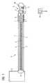
- FIG. 1shows an inventive catheter device 1 which is embodied as an atherectomy catheter.
- the inventive catheter device 1features a hollow flexible drive shaft 2 in which an OCT signal line 3 and an IVUS signal line 4 are integrated.
- a signal line 5 for a position sensor systemwhich is embodied as an electromagnetic sensor system, is arranged in the flexible drive shaft 2 .
- An IVUS sensor 6 and an OCT sensor 7are integrated into the front part of the catheter.
- an opening with a cutter 9which is embodied as a rotating knife.
- a light exit window for the OCT sensor 7At the catheter tip 8 is located a light exit window for the OCT sensor 7 .
- magnetic sensors 18 of the sensor systemare arranged there. These sensors interoperate with a position sensor 10 which is arranged outside the body of the patient.
- the position sensor 10is embodied as an electromagnetic sensor.
- the drive shaft 2is surrounded by a catheter sheath 11 . Opposite the opening is located an expandable balloon 13 for supporting positioning.
- a signal interface and a drive unit 15are connected via a rotation coupling 16 to the catheter device 1 .
- the cutter 9 for performing atherectomyis connected to the OCT sensor 7 , the IVUS sensor 6 and position sensors into an integrated unit.
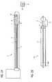
- FIGS. 2A and 2Bshow a second exemplary embodiment of a catheter device.
- FIG. 2Ashows an imaging-catheter 17 with an IVUS sensor 6 , an OCT sensor 7 with viewing window, magnetic sensors 18 , position sensors 10 , signal lines 4 for IVUS and signal lines 3 for OCT. Likewise a signal interface and a drive unit 15 are provided.
- FIG. 2Bshows an atherectomy catheter 14 which features a lumen, into which the Imaging catheter 17 is able to be introduced.
- the atherectomy catheter 14has a cutter 9 in the area of the catheter tip 8 as well as a rotatable balloon 13 .
- the lumenis transparent for OCT and IVUS.
- Within the catheter 14is located a hose 46 for a pressure medium of the balloon 13 .
- the two catheter devices 1 , 17 shown in FIGS. 1 and 2each have an OCT sensor and an IVUS sensor.
- the OCT sensordelivers especially good images of the local area
- the IVUS sensordelivers a good representation of layers further away or deeper down.
- the catheter devices 1 , 17are connected to an image processing unit which creates a common image from the images delivered by the two sensors. To do this it uses a section of the image delivered by the OCT sensor for the local area and for the remote area the complementary part of the IVUS image, the two sections are registered with each other by means of the data of the position sensors and fused together into a joint image. In this way sectional images of the vessel being examined are obtained, which can be assigned precisely to a defined position in the body. Computational methods are employed to use the data of the position sensor in order to approximate the center line and the envelope of the vessel being examined. Subsequently the individual sectional images are combined to form a volume data set so that an exact and thus especially realistic image is produced.
- the geometrical information of the center lineis used and combined with the sensor positions recorded during imaging, which greatly reduces the artifacts in 3D imaging.
- the 3D co-ordinates of the center line and the sensor positions recorded during imagingare subtracted from one other.
- the result of the subtractionis then used for each of the recorded 2D images for exact 3D reconstruction.
- This envelope of the vesselcan be used for further stages in processing the image. With the aid of the envelope the 3D reconstructed OCT-IVUS images are registered with other anatomical image data such as from a 3D angiography device of the same vessel section and subsequently fused together.
- the position sensors 10 used in the exemplary embodiments of FIGS. 1 and 2are electromagnetic position sensors to create 3D OCT-IVUS images from 2D OCT-IVUS images.
- the recording of the orientation and position of the catheter in a three-dimensional co-ordinate systemis undertaken by transmit coils in the object and receive coils in the room or conversely with receive coils in the object and transmit coils in the room.
- the electromagnetic transmitter or alternatively the electromagnetic receivercan be located in the catheter. Conversely the corresponding electromagnetic receiver or transmitter can be accommodated outside the body. Normally at least one transmitter emitting radiation in the X, Y, Z direction is assigned to a receiver or conversely a receiver with X, Y, Z receive directions is assigned to a transmitter to allow for location in the room.
- the coils of the electromagnetic position sensorsare not arranged exclusively orthogonality to each other, but at any given angle of for example 60°, to achieve a better miniaturization which allows the position sensors to be built into a catheter.
- the image information of the catheter which is recorded with the sensorsis combined or overlaid with other medical images such as 2D or 3D recordings.
- the OCT-IVUS images of the catheterare displayed jointly with the x-ray images. This makes the information about the images of the catheter device and the x-ray images visible jointly for the user and makes more rapid and better diagnosis possible.
- 2D-2D, 2D-3D, 3D-3D and 3D-4D and 4D-4D overlaysare possible, in which case the angiographic x-ray images are combined in each case with the images of the catheter device by segmentation, registration and image fusion.
- Images of the following modalities and methodscan be used for overlaying: Sonography including IVUS, radiography, fluoroscopy, angiography, OCT, discrete tomography, Positron Emission Tomography, nuclear medical diagnostics, computer tomography, nuclear resonance tomography including intracardial MR, optical recording including endoscopy, fluorescence and optical markers.
- the catheter deviceis part of a medical examination unit which possesses a functional unit for rectification of movement artifacts which arise as a result of breathing and the movement of the heart and the blood vessels.
- a breast bandcan also be used which determines via the appropriate sensors the breathing amplitude and frequency so that the image processing unit can make the necessary corrective calculations in order to calculate out the movement artifacts from the image information.
- the transmit coilsare operated and evaluated cyclically at specific intervals in time with different frequencies.
- the sensorsoffset in time and clocked.
- the x-ray detectors and the ECGare not read out if the transmitters of the electromagnetic positioning system are active.
- the OCT sensors and positions sensorsare not read out if the x-ray radiation is active Thus only such signals are ever read out as are not subject to disturbances and do not influence any other active sensors.
- the functional units and signal linesare equipped with devices and measures which shield the physiological signals and image signals and the signal processing and signal editing from the magnetic filters of the transmit antennas.
- the shell of the catheteris coated with a thin film layer made of conductive nanoparticles. Likewise nanoparticles can be used to effect a magnetic shielding.
- the catheter shellis provided with a coating which reduces the frictional resistance during guidance through the vessel.
- This coatingcan also be based on nanotechnology or alternatively can be made from a silicon material.
- the contrast meansis introduced directly into the vessel to be examined or the body cavity through a channel in the catheter.
- a temperature sensor or a pressure sensorcan be arranged in the tip of the catheter to monitor the temperature and the pressure in the vessel or the organ to be examined and treated.
- the temperature sensorwhich is accommodated in the tip of the catheter, enables any possible temperature increase arising as a result of friction to be detected.
- FIG. 3is a schematic diagram of the inventive treatment unit.
- the treatment device 19comprises a catheter device for performing atherectomy.
- a patient not shown in FIG. 3is supported on a patient table 20 , radiation is emitted by radiation source 21 in the direction of the patient table 20 .
- the radiationis produced via a high-voltage generator 22 controlled via a system control 23 .
- an x-ray detector 24Opposite the x-ray source 21 is arranged an x-ray detector 24 which in its turn is connected to a preprocessing unit 25 for x-ray images.
- a connection 26is provided for physiological sensors, which is coupled to a physiological signal processing 27 in order to control ECG signals or pulse signals or the breathing and the blood pressure of a patient.
- the data bus 30There are also preprocessing units 31 , 32 and 33 provided for OCT, IVUS and the position sensors. Associated image processing units 34 , 35 and 36 for OCT, IVUS and the position sensors are also connected to the data bus 30 .
- the poweris supplied via a power supply unit 37 .
- an image processing unit 38 for the x-ray imagesis connected to the data bus 30 , which features a connection to an image data store 39 for archiving and storing the recorded images.
- a calibration unit 40 as well as an image correction unit 41enable interference fields or artifacts of the imaging to be taken into account.
- the imagesare fused and reconstructed in an image fusion and/or reconstruction unit 42 .
- an interface 43to a patient data or image data system.
- the image data obtained from OCT, IVUS and the position sensor system as well as the x-ray images and possible fusion images of the different imaging techniquesare shown on a display unit 44 in two dimensions, three dimensions or four dimensions.
- the display unit 44is connected to an input 45 for input by a user.
- FIG. 4is a schematic diagram or the sensor read-out of the treatment unit during execution of the inventive method.
- a typical execution sequenceis as follows: Introducing he catheter under x-ray control, possibly with contrast means, creating the angiographic overview image, creating the images of the position sensors, overlaying the images of the position sensors with the overview angiography by segmentation, registration and image fusion, navigating the catheter based on the images obtained up to the destination position, these steps are in some cases executed in parallel and automatically without the interaction of the user.
- the flushing fluid for OCTis injected and the stenosis is observed with the OCT-IVUS images in two dimensions or three dimensions at high resolution. Subsequently the OCT-IVUS images are created.
- the OCT-IVUS imagesare overlaid with the overview angiography by segmentation, registration and image fusion.
- a 3D reconstruction of the OCT-IVUS imagesis undertaken based on the data of the position sensors.
- the atherectomy catheteris placed and temporarily fixed for example by inflating the balloon accommodated at the catheter tip.
- Performing the atherectomywhich means shaving off the plaque from the vessel wall with the rotating knife. If a specific amount of plaque is removed, the OCT sensor is used to check this point in the vessel wall. The process is repeated until the plaque is removed at all points. Final check of the atherectomy and removal of the catheter.
- the inventive devicereduces the number of steps required.
- the OCT sensordelivers good images in the local area, the IVUS sensor sufficiently good images of tissue layers located further down.
- the electromagnetic position sensorsallow 3D images to be created from the OCT and IVUS images.
- the passage of the cathetercan be mapped solely on the basis of the IVUS, OCT and electromagnetic signals, which means that x-ray radiation can be saved.
- the systemdelivers important additional medical information about the arteriosclerotic plaque.
- itallows the correct position of the tip of the catheter to be better checked.
- a further advantage with the integration of atherectomy and OCTalso lies in the fact that in this case no separate flushing facility has to be provided for OCT, since a flushing means is already used for the drill head.
- the sensors of the medical treatment devicewhich in the exemplary embodiment presented is an x-ray device, are read out partly offset in time and clocked. Initially a system clock is predetermined in which individual system impulses are created, with this pulsed generation being followed by the switching-on of the x-ray radiation and the activation of the magnetic location. After the x-ray radiation is switched off, the x-ray detector readout occurs and at the same time the IVUS data is read out. Subsequently the OCT data is read out, with this occurring at the same time as the readout of the ECG and the data for respiration. This means that the individual sensor is read out or the components of the catheter device are activated in such a way that a mutual fault can be excluded.
- the time-offset and clocked readout shown hereis to be seen as an example for a readout avoiding interference influences.
Landscapes
- Health & Medical Sciences (AREA)
- Life Sciences & Earth Sciences (AREA)
- Engineering & Computer Science (AREA)
- Surgery (AREA)
- Veterinary Medicine (AREA)
- General Health & Medical Sciences (AREA)
- Public Health (AREA)
- Biomedical Technology (AREA)
- Heart & Thoracic Surgery (AREA)
- Medical Informatics (AREA)
- Molecular Biology (AREA)
- Animal Behavior & Ethology (AREA)
- Physics & Mathematics (AREA)
- Biophysics (AREA)
- Pathology (AREA)
- Nuclear Medicine, Radiotherapy & Molecular Imaging (AREA)
- Radiology & Medical Imaging (AREA)
- Computer Vision & Pattern Recognition (AREA)
- Vascular Medicine (AREA)
- Immunology (AREA)
- Surgical Instruments (AREA)
- Endoscopes (AREA)
- Ultra Sonic Daignosis Equipment (AREA)
- Media Introduction/Drainage Providing Device (AREA)
- Magnetic Resonance Imaging Apparatus (AREA)
Abstract
Description
Claims (18)
Applications Claiming Priority (4)
| Application Number | Priority Date | Filing Date | Title |
|---|---|---|---|
| DE102005045373.2 | 2005-09-22 | ||
| DE102005045373 | 2005-09-22 | ||
| DE102005045373ADE102005045373A1 (en) | 2005-09-22 | 2005-09-22 | catheter device |
| DE102005048892ADE102005048892B4 (en) | 2005-09-22 | 2005-09-22 | Device for carrying out rotablation and medical treatment device |
Publications (2)
| Publication Number | Publication Date |
|---|---|
| US20070066890A1 US20070066890A1 (en) | 2007-03-22 |
| US7753852B2true US7753852B2 (en) | 2010-07-13 |
Family
ID=38235413
Family Applications (2)
| Application Number | Title | Priority Date | Filing Date |
|---|---|---|---|
| US11/526,178Expired - Fee RelatedUS7753852B2 (en) | 2005-09-22 | 2006-09-22 | Atherectomy catheter with combined OCT/IVUS imaging |
| US11/526,249Expired - Fee RelatedUS7729745B2 (en) | 2005-09-22 | 2006-09-22 | Device for carrying out rotablation |
Family Applications After (1)
| Application Number | Title | Priority Date | Filing Date |
|---|---|---|---|
| US11/526,249Expired - Fee RelatedUS7729745B2 (en) | 2005-09-22 | 2006-09-22 | Device for carrying out rotablation |
Country Status (3)
| Country | Link |
|---|---|
| US (2) | US7753852B2 (en) |
| JP (2) | JP4993981B2 (en) |
| DE (2) | DE102005045373A1 (en) |
Cited By (77)
| Publication number | Priority date | Publication date | Assignee | Title |
|---|---|---|---|---|
| US20100041948A1 (en)* | 2008-08-15 | 2010-02-18 | Fujifilm Corporation | Optical probe and three-dimensional image acquisition apparatus |
| US20100317964A1 (en)* | 2008-03-03 | 2010-12-16 | Koninklijke Philips Electronics N.V. | Biopsy guidance by electromagnetic tracking and photonic needle |
| US8192452B2 (en) | 2009-05-14 | 2012-06-05 | Tyco Healthcare Group Lp | Easily cleaned atherectomy catheters and methods of use |
| US8226674B2 (en) | 2000-12-20 | 2012-07-24 | Tyco Healthcare Group Lp | Debulking catheters and methods |
| US8246640B2 (en) | 2003-04-22 | 2012-08-21 | Tyco Healthcare Group Lp | Methods and devices for cutting tissue at a vascular location |
| US8328829B2 (en) | 1999-08-19 | 2012-12-11 | Covidien Lp | High capacity debulking catheter with razor edge cutting window |
| US8414604B2 (en) | 2008-10-13 | 2013-04-09 | Covidien Lp | Devices and methods for manipulating a catheter shaft |
| US8469979B2 (en) | 2000-12-20 | 2013-06-25 | Covidien Lp | High capacity debulking catheter with distal driven cutting wheel |
| US8496677B2 (en) | 2009-12-02 | 2013-07-30 | Covidien Lp | Methods and devices for cutting tissue |
| US8548571B2 (en) | 2009-12-08 | 2013-10-01 | Avinger, Inc. | Devices and methods for predicting and preventing restenosis |
| US8597315B2 (en) | 1999-08-19 | 2013-12-03 | Covidien Lp | Atherectomy catheter with first and second imaging devices |
| US8644913B2 (en) | 2011-03-28 | 2014-02-04 | Avinger, Inc. | Occlusion-crossing devices, imaging, and atherectomy devices |
| US8696695B2 (en) | 2009-04-28 | 2014-04-15 | Avinger, Inc. | Guidewire positioning catheter |
| US8784440B2 (en) | 2008-02-25 | 2014-07-22 | Covidien Lp | Methods and devices for cutting tissue |
| US8808186B2 (en) | 2010-11-11 | 2014-08-19 | Covidien Lp | Flexible debulking catheters with imaging and methods of use and manufacture |
| US8906334B2 (en) | 2007-05-14 | 2014-12-09 | Invista North America S.A R.L. | High efficiency reactor and process |
| US8920450B2 (en) | 2010-10-28 | 2014-12-30 | Covidien Lp | Material removal device and method of use |
| US8992717B2 (en) | 2011-09-01 | 2015-03-31 | Covidien Lp | Catheter with helical drive shaft and methods of manufacture |
| US8998937B2 (en) | 1999-08-19 | 2015-04-07 | Covidien Lp | Methods and devices for cutting tissue |
| US9028512B2 (en) | 2009-12-11 | 2015-05-12 | Covidien Lp | Material removal device having improved material capture efficiency and methods of use |
| US9078561B2 (en) | 2008-10-02 | 2015-07-14 | Vascular Imaging Corporation | Optical ultrasound receiver |
| WO2015120146A1 (en)* | 2014-02-06 | 2015-08-13 | Avinger, Inc. | Atherectomy catheters and occlusion crossing devices |
| US9119662B2 (en) | 2010-06-14 | 2015-09-01 | Covidien Lp | Material removal device and method of use |
| US9125562B2 (en) | 2009-07-01 | 2015-09-08 | Avinger, Inc. | Catheter-based off-axis optical coherence tomography imaging system |
| US9192307B2 (en) | 2002-10-07 | 2015-11-24 | Vascular Imaging Corporation | Systems and methods for minimally-invasive optical-acoustic imaging |
| US9198581B2 (en) | 2005-11-22 | 2015-12-01 | Vascular Imaging Corporation | Optical imaging probe |
| US9345406B2 (en) | 2011-11-11 | 2016-05-24 | Avinger, Inc. | Occlusion-crossing devices, atherectomy devices, and imaging |
| US9345510B2 (en) | 2010-07-01 | 2016-05-24 | Avinger, Inc. | Atherectomy catheters with longitudinally displaceable drive shafts |
| US9345398B2 (en) | 2012-05-14 | 2016-05-24 | Avinger, Inc. | Atherectomy catheter drive assemblies |
| US9468417B1 (en)* | 2011-06-15 | 2016-10-18 | Acist Medical Systems, Inc. | Stenotic lesion characterization |
| US9498600B2 (en) | 2009-07-01 | 2016-11-22 | Avinger, Inc. | Atherectomy catheter with laterally-displaceable tip |
| US9498247B2 (en) | 2014-02-06 | 2016-11-22 | Avinger, Inc. | Atherectomy catheters and occlusion crossing devices |
| US9533123B2 (en) | 2008-10-31 | 2017-01-03 | Vascular Imaging Corporation | Optical imaging probe connector method by deforming a cross section and cutting at an oblique angle |
| US9532766B2 (en) | 1998-03-05 | 2017-01-03 | Vascular Imaging Corporation | Optical-acoustic imaging device |
| US9532844B2 (en) | 2012-09-13 | 2017-01-03 | Covidien Lp | Cleaning device for medical instrument and method of use |
| US9557156B2 (en) | 2012-05-14 | 2017-01-31 | Avinger, Inc. | Optical coherence tomography with graded index fiber for biological imaging |
| US9687266B2 (en) | 2009-04-29 | 2017-06-27 | Covidien Lp | Methods and devices for cutting and abrading tissue |
| US9788790B2 (en) | 2009-05-28 | 2017-10-17 | Avinger, Inc. | Optical coherence tomography for biological imaging |
| US9801647B2 (en) | 2006-05-26 | 2017-10-31 | Covidien Lp | Catheter including cutting element and energy emitting element |
| US9829766B2 (en) | 2009-02-17 | 2017-11-28 | Analog Devices, Inc. | Electro-optic beam deflector device |
| US9854979B2 (en) | 2013-03-15 | 2018-01-02 | Avinger, Inc. | Chronic total occlusion crossing devices with imaging |
| US9918734B2 (en) | 2008-04-23 | 2018-03-20 | Avinger, Inc. | Catheter system and method for boring through blocked vascular passages |
| US9936881B2 (en) | 2012-10-04 | 2018-04-10 | Vascular Imaging Corporation | Polarization scrambling for intra-body fiber optic sensor |
| US9943329B2 (en) | 2012-11-08 | 2018-04-17 | Covidien Lp | Tissue-removing catheter with rotatable cutter |
| US9949754B2 (en) | 2011-03-28 | 2018-04-24 | Avinger, Inc. | Occlusion-crossing devices |
| US10130386B2 (en) | 2013-07-08 | 2018-11-20 | Avinger, Inc. | Identification of elastic lamina to guide interventional therapy |
| US10175421B2 (en) | 2013-03-14 | 2019-01-08 | Vascular Imaging Corporation | Optical fiber ribbon imaging guidewire and methods |
| US10213224B2 (en) | 2014-06-27 | 2019-02-26 | Covidien Lp | Cleaning device for catheter and catheter including the same |
| US10258240B1 (en) | 2014-11-24 | 2019-04-16 | Vascular Imaging Corporation | Optical fiber pressure sensor |
| US10292721B2 (en) | 2015-07-20 | 2019-05-21 | Covidien Lp | Tissue-removing catheter including movable distal tip |
| US10314667B2 (en) | 2015-03-25 | 2019-06-11 | Covidien Lp | Cleaning device for cleaning medical instrument |
| US10314664B2 (en) | 2015-10-07 | 2019-06-11 | Covidien Lp | Tissue-removing catheter and tissue-removing element with depth stop |
| US10327645B2 (en) | 2013-10-04 | 2019-06-25 | Vascular Imaging Corporation | Imaging techniques using an imaging guidewire |
| US10335173B2 (en) | 2012-09-06 | 2019-07-02 | Avinger, Inc. | Re-entry stylet for catheter |
| US10335042B2 (en) | 2013-06-28 | 2019-07-02 | Cardiovascular Systems, Inc. | Methods, devices and systems for sensing, measuring and/or characterizing vessel and/or lesion compliance and/or elastance changes during vascular procedures |
| US10357277B2 (en) | 2014-07-08 | 2019-07-23 | Avinger, Inc. | High speed chronic total occlusion crossing devices |
| US10363062B2 (en) | 2011-10-17 | 2019-07-30 | Avinger, Inc. | Atherectomy catheters and non-contact actuation mechanism for catheters |
| US10506934B2 (en) | 2012-05-25 | 2019-12-17 | Phyzhon Health Inc. | Optical fiber pressure sensor |
| US10537255B2 (en) | 2013-11-21 | 2020-01-21 | Phyzhon Health Inc. | Optical fiber pressure sensor |
| US10548478B2 (en) | 2010-07-01 | 2020-02-04 | Avinger, Inc. | Balloon atherectomy catheters with imaging |
| US10568520B2 (en) | 2015-07-13 | 2020-02-25 | Avinger, Inc. | Micro-molded anamorphic reflector lens for image guided therapeutic/diagnostic catheters |
| US10675003B2 (en) | 2014-07-11 | 2020-06-09 | Acist Medical Systems, Inc. | Intravascular imaging |
| CN111298271A (en)* | 2014-02-06 | 2020-06-19 | 尼普洛株式会社 | Catheter tube |
| US10932670B2 (en) | 2013-03-15 | 2021-03-02 | Avinger, Inc. | Optical pressure sensor assembly |
| RU205369U1 (en)* | 2020-12-14 | 2021-07-12 | Федеральное государственное бюджетное учреждение высшего образования "Тамбовский государственный технический университет" (ФГБОУ ВО "ТГТУ") | INTRAVASCULAR PROBE DEVICE FOR JOINT USE OF ROTARY ATHERECTOMY AND OPTICAL COHERENT TOMOGRAPHY |
| US11096717B2 (en) | 2013-03-15 | 2021-08-24 | Avinger, Inc. | Tissue collection device for catheter |
| US11224459B2 (en) | 2016-06-30 | 2022-01-18 | Avinger, Inc. | Atherectomy catheter with shapeable distal tip |
| US11278248B2 (en) | 2016-01-25 | 2022-03-22 | Avinger, Inc. | OCT imaging catheter with lag correction |
| US11284916B2 (en) | 2012-09-06 | 2022-03-29 | Avinger, Inc. | Atherectomy catheters and occlusion crossing devices |
| US11344327B2 (en) | 2016-06-03 | 2022-05-31 | Avinger, Inc. | Catheter device with detachable distal end |
| US11382653B2 (en) | 2010-07-01 | 2022-07-12 | Avinger, Inc. | Atherectomy catheter |
| US11399863B2 (en) | 2016-04-01 | 2022-08-02 | Avinger, Inc. | Atherectomy catheter with serrated cutter |
| US11406412B2 (en) | 2012-05-14 | 2022-08-09 | Avinger, Inc. | Atherectomy catheters with imaging |
| US20220359978A1 (en)* | 2015-03-16 | 2022-11-10 | St. Jude Medical International Holding S.â r.l. | Field concentrating antennas for magnetic position sensors |
| US11793400B2 (en) | 2019-10-18 | 2023-10-24 | Avinger, Inc. | Occlusion-crossing devices |
| US12167867B2 (en) | 2018-04-19 | 2024-12-17 | Avinger, Inc. | Occlusion-crossing devices |
| US12420069B2 (en) | 2018-12-19 | 2025-09-23 | Covidien Lp | Internal carotid artery thrombectomy devices and methods |
Families Citing this family (125)
| Publication number | Priority date | Publication date | Assignee | Title |
|---|---|---|---|---|
| US7555333B2 (en)* | 2000-06-19 | 2009-06-30 | University Of Washington | Integrated optical scanning image acquisition and display |
| US7455666B2 (en) | 2001-07-13 | 2008-11-25 | Board Of Regents, The University Of Texas System | Methods and apparatuses for navigating the subarachnoid space |
| FR2850966B1 (en) | 2003-02-10 | 2005-03-18 | Rhodia Polyamide Intermediates | PROCESS FOR PRODUCING DINITRIL COMPOUNDS |
| FR2854891B1 (en) | 2003-05-12 | 2006-07-07 | Rhodia Polyamide Intermediates | PROCESS FOR PREPARING DINITRILES |
| DE10354496B4 (en)* | 2003-11-21 | 2011-03-31 | Siemens Ag | Medical examination and / or treatment system |
| US7901348B2 (en) | 2003-12-12 | 2011-03-08 | University Of Washington | Catheterscope 3D guidance and interface system |
| US7530948B2 (en)* | 2005-02-28 | 2009-05-12 | University Of Washington | Tethered capsule endoscope for Barrett's Esophagus screening |
| WO2007046799A1 (en) | 2005-10-18 | 2007-04-26 | Invista Technologies S.A R.L. | Process of making 3-aminopentanenitrile |
| WO2007067163A1 (en) | 2005-11-23 | 2007-06-14 | University Of Washington | Scanning beam with variable sequential framing using interrupted scanning resonance |
| DE102005059262B4 (en)* | 2005-12-12 | 2008-02-07 | Siemens Ag | catheter device |
| EP2967809A4 (en)* | 2006-01-03 | 2017-03-15 | Volcano Corporation | Endoluminal filter having enhanced echogenic properties |
| US9561078B2 (en)* | 2006-03-03 | 2017-02-07 | University Of Washington | Multi-cladding optical fiber scanner |
| CA2644961A1 (en) | 2006-03-17 | 2007-09-27 | Invista Technologies S.A.R.L. | Method for the purification of triorganophosphites by treatment with a basic additive |
| US7919646B2 (en) | 2006-07-14 | 2011-04-05 | Invista North America S.A R.L. | Hydrocyanation of 2-pentenenitrile |
| US9867530B2 (en) | 2006-08-14 | 2018-01-16 | Volcano Corporation | Telescopic side port catheter device with imaging system and method for accessing side branch occlusions |
| US20080058629A1 (en)* | 2006-08-21 | 2008-03-06 | University Of Washington | Optical fiber scope with both non-resonant illumination and resonant collection/imaging for multiple modes of operation |
| US7935060B2 (en) | 2006-11-08 | 2011-05-03 | Lightlab Imaging, Inc. | Opto-acoustic imaging devices and methods |
| US20080132834A1 (en)* | 2006-12-04 | 2008-06-05 | University Of Washington | Flexible endoscope tip bending mechanism using optical fibers as tension members |
| US20080221388A1 (en)* | 2007-03-09 | 2008-09-11 | University Of Washington | Side viewing optical fiber endoscope |
| US20080243030A1 (en)* | 2007-04-02 | 2008-10-02 | University Of Washington | Multifunction cannula tools |
| WO2008121143A1 (en)* | 2007-04-02 | 2008-10-09 | University Of Washington | Catheter with imaging capability acts as guidewire for cannula tools |
| US8840566B2 (en)* | 2007-04-02 | 2014-09-23 | University Of Washington | Catheter with imaging capability acts as guidewire for cannula tools |
| US7952718B2 (en)* | 2007-05-03 | 2011-05-31 | University Of Washington | High resolution optical coherence tomography based imaging for intraluminal and interstitial use implemented with a reduced form factor |
| EP2164587B1 (en) | 2007-06-13 | 2018-04-04 | INVISTA Textiles (U.K.) Limited | Process for improving adiponitrile quality |
| DE102007032530B4 (en)* | 2007-07-12 | 2011-08-25 | Siemens AG, 80333 | Method for creating a medical image and imaging device |
| US9596993B2 (en) | 2007-07-12 | 2017-03-21 | Volcano Corporation | Automatic calibration systems and methods of use |
| WO2009009802A1 (en) | 2007-07-12 | 2009-01-15 | Volcano Corporation | Oct-ivus catheter for concurrent luminal imaging |
| EP2178442B1 (en) | 2007-07-12 | 2017-09-06 | Volcano Corporation | Catheter for in vivo imaging |
| US20090069743A1 (en)* | 2007-09-11 | 2009-03-12 | Baxter International Inc. | Infusion therapy sensor system |
| US20090137893A1 (en)* | 2007-11-27 | 2009-05-28 | University Of Washington | Adding imaging capability to distal tips of medical tools, catheters, and conduits |
| EP2229353B1 (en) | 2008-01-15 | 2018-01-03 | INVISTA Textiles (U.K.) Limited | Hydrocyanation of pentenenitriles |
| CN101910119B (en) | 2008-01-15 | 2013-05-29 | 因温斯特技术公司 | Process for preparing and refining 3-pentenenitrile, and for refining 2-methyl-3-butenenitrile |
| US9713448B2 (en) | 2008-04-03 | 2017-07-25 | Infraredx, Inc. | System and method for intravascular structural analysis compensation of chemical analysis modality |
| US8052605B2 (en)* | 2008-05-07 | 2011-11-08 | Infraredx | Multimodal catheter system and method for intravascular analysis |
| WO2010045131A1 (en) | 2008-10-14 | 2010-04-22 | Invista Technologies S.A.R.L. | Process for making 2-secondary-alkyl-4,5-di-(normal-alkyl)phenols |
| US8764666B2 (en)* | 2008-10-28 | 2014-07-01 | The Regents Of The University Of California | Ultrasound guided optical coherence tomography, photoacoustic probe for biomedical imaging |
| DE102008054297A1 (en)* | 2008-11-03 | 2010-05-06 | Siemens Aktiengesellschaft | A catheter assembly for insertion into a blood vessel, medical examination and treatment device comprising such a catheter assembly and method for minimally invasive intervention on a blood vessel in the brain |
| EP2358278B1 (en) | 2008-12-08 | 2021-05-12 | Acist Medical Systems, Inc. | System and catheter for image guidance and methods thereof |
| DE102009014489B4 (en)* | 2009-03-23 | 2011-03-10 | Siemens Aktiengesellschaft | Catheter and medical device |
| US20100268225A1 (en)* | 2009-04-15 | 2010-10-21 | Tyco Healthcare Group Lp | Methods for Image Analysis and Visualization of Medical Image Data Suitable for Use in Assessing Tissue Ablation and Systems and Methods for Controlling Tissue Ablation Using Same |
| US20100268223A1 (en)* | 2009-04-15 | 2010-10-21 | Tyco Health Group Lp | Methods for Image Analysis and Visualization of Medical Image Data Suitable for Use in Assessing Tissue Ablation and Systems and Methods for Controlling Tissue Ablation Using Same |
| CN102471218B (en) | 2009-08-07 | 2014-11-05 | 因温斯特技术公司 | Hydrogenation and esterification to form diesters |
| US8340577B2 (en)* | 2009-09-24 | 2012-12-25 | Research In Motion Limited | Communications device using electromagnet and activated communications circuit |
| US20110092955A1 (en)* | 2009-10-07 | 2011-04-21 | Purdy Phillip D | Pressure-Sensing Medical Devices, Systems and Methods, and Methods of Forming Medical Devices |
| US11141063B2 (en) | 2010-12-23 | 2021-10-12 | Philips Image Guided Therapy Corporation | Integrated system architectures and methods of use |
| US11040140B2 (en) | 2010-12-31 | 2021-06-22 | Philips Image Guided Therapy Corporation | Deep vein thrombosis therapeutic methods |
| US20130023912A1 (en)* | 2010-12-31 | 2013-01-24 | Volcano Corporation | Multiple Sclerosis Therapeutic Methods Using Therapeutic Cutting Devices and Systems |
| US20140094697A1 (en) | 2011-05-27 | 2014-04-03 | Lightlab Imaging, Inc. | Optical coherence tomography and pressure based systems and methods |
| CN103959043B (en) | 2011-05-31 | 2016-11-02 | 光学实验室成像公司 | Multimodal imaging systems, devices and methods |
| US9360630B2 (en) | 2011-08-31 | 2016-06-07 | Volcano Corporation | Optical-electrical rotary joint and methods of use |
| US9237851B2 (en)* | 2012-02-03 | 2016-01-19 | Ninepoint Medical, Inc. | Imaging system producing multiple registered images of a body lumen |
| US10070827B2 (en) | 2012-10-05 | 2018-09-11 | Volcano Corporation | Automatic image playback |
| US10568586B2 (en) | 2012-10-05 | 2020-02-25 | Volcano Corporation | Systems for indicating parameters in an imaging data set and methods of use |
| US9307926B2 (en) | 2012-10-05 | 2016-04-12 | Volcano Corporation | Automatic stent detection |
| US9858668B2 (en) | 2012-10-05 | 2018-01-02 | Volcano Corporation | Guidewire artifact removal in images |
| US9324141B2 (en) | 2012-10-05 | 2016-04-26 | Volcano Corporation | Removal of A-scan streaking artifact |
| US20140100454A1 (en) | 2012-10-05 | 2014-04-10 | Volcano Corporation | Methods and systems for establishing parameters for three-dimensional imaging |
| US9292918B2 (en) | 2012-10-05 | 2016-03-22 | Volcano Corporation | Methods and systems for transforming luminal images |
| CA2887421A1 (en) | 2012-10-05 | 2014-04-10 | David Welford | Systems and methods for amplifying light |
| US11272845B2 (en) | 2012-10-05 | 2022-03-15 | Philips Image Guided Therapy Corporation | System and method for instant and automatic border detection |
| US9286673B2 (en) | 2012-10-05 | 2016-03-15 | Volcano Corporation | Systems for correcting distortions in a medical image and methods of use thereof |
| US9367965B2 (en) | 2012-10-05 | 2016-06-14 | Volcano Corporation | Systems and methods for generating images of tissue |
| US9840734B2 (en) | 2012-10-22 | 2017-12-12 | Raindance Technologies, Inc. | Methods for analyzing DNA |
| EP2919658B1 (en) | 2012-11-19 | 2024-03-20 | Lightlab Imaging, Inc. | Interface devices, systems and methods for multimodal probes |
| US9717422B2 (en) | 2012-12-12 | 2017-08-01 | Volcano Corporation | Sheath with optically interrogatable sensors |
| EP2931132B1 (en) | 2012-12-13 | 2023-07-05 | Philips Image Guided Therapy Corporation | System for targeted cannulation |
| EP2934311B1 (en) | 2012-12-20 | 2020-04-15 | Volcano Corporation | Smooth transition catheters |
| WO2014113188A2 (en) | 2012-12-20 | 2014-07-24 | Jeremy Stigall | Locating intravascular images |
| US11406498B2 (en) | 2012-12-20 | 2022-08-09 | Philips Image Guided Therapy Corporation | Implant delivery system and implants |
| US10942022B2 (en) | 2012-12-20 | 2021-03-09 | Philips Image Guided Therapy Corporation | Manual calibration of imaging system |
| EP2934310A4 (en) | 2012-12-20 | 2016-10-12 | Nathaniel J Kemp | Optical coherence tomography system that is reconfigurable between different imaging modes |
| US10939826B2 (en) | 2012-12-20 | 2021-03-09 | Philips Image Guided Therapy Corporation | Aspirating and removing biological material |
| US10413317B2 (en) | 2012-12-21 | 2019-09-17 | Volcano Corporation | System and method for catheter steering and operation |
| US10058284B2 (en) | 2012-12-21 | 2018-08-28 | Volcano Corporation | Simultaneous imaging, monitoring, and therapy |
| US9486143B2 (en) | 2012-12-21 | 2016-11-08 | Volcano Corporation | Intravascular forward imaging device |
| EP2936241B1 (en) | 2012-12-21 | 2020-10-21 | Nathaniel J. Kemp | Power-efficient optical buffering using a polarisation-maintaining active optical switch |
| US10332228B2 (en) | 2012-12-21 | 2019-06-25 | Volcano Corporation | System and method for graphical processing of medical data |
| US9612105B2 (en) | 2012-12-21 | 2017-04-04 | Volcano Corporation | Polarization sensitive optical coherence tomography system |
| CA2895769A1 (en) | 2012-12-21 | 2014-06-26 | Douglas Meyer | Rotational ultrasound imaging catheter with extended catheter body telescope |
| JP2016507892A (en) | 2012-12-21 | 2016-03-10 | デイビッド ウェルフォード, | System and method for narrowing the wavelength emission of light |
| EP2934323A4 (en) | 2012-12-21 | 2016-08-17 | Andrew Hancock | SYSTEM AND METHOD FOR MULTIPLE PROCESSING OF IMAGE SIGNALS |
| JP2016501625A (en) | 2012-12-21 | 2016-01-21 | ジェローム マイ, | Ultrasound imaging with variable line density |
| EP2934305B1 (en)* | 2012-12-21 | 2018-02-21 | Volcano Corporation | System for multi-site intravascular measurement |
| US10226597B2 (en) | 2013-03-07 | 2019-03-12 | Volcano Corporation | Guidewire with centering mechanism |
| WO2014138555A1 (en) | 2013-03-07 | 2014-09-12 | Bernhard Sturm | Multimodal segmentation in intravascular images |
| US9351698B2 (en) | 2013-03-12 | 2016-05-31 | Lightlab Imaging, Inc. | Vascular data processing and image registration systems, methods, and apparatuses |
| CN111652917B (en)* | 2013-03-12 | 2024-07-09 | 光学实验室成像公司 | Vascular data processing and image registration system, method and device |
| US20140276923A1 (en) | 2013-03-12 | 2014-09-18 | Volcano Corporation | Vibrating catheter and methods of use |
| EP2967391A4 (en) | 2013-03-12 | 2016-11-02 | Donna Collins | SYSTEMS AND METHODS FOR DIAGNOSING CORONARY MICROVASCULAR DISEASE |
| US9301687B2 (en) | 2013-03-13 | 2016-04-05 | Volcano Corporation | System and method for OCT depth calibration |
| US11026591B2 (en) | 2013-03-13 | 2021-06-08 | Philips Image Guided Therapy Corporation | Intravascular pressure sensor calibration |
| WO2014159819A1 (en) | 2013-03-13 | 2014-10-02 | Jinhyoung Park | System and methods for producing an image from a rotational intravascular ultrasound device |
| US10219887B2 (en) | 2013-03-14 | 2019-03-05 | Volcano Corporation | Filters with echogenic characteristics |
| US12343198B2 (en) | 2013-03-14 | 2025-07-01 | Philips Image Guided Therapy Corporation | Delivery catheter having imaging capabilities |
| US10292677B2 (en) | 2013-03-14 | 2019-05-21 | Volcano Corporation | Endoluminal filter having enhanced echogenic properties |
| US20160030151A1 (en) | 2013-03-14 | 2016-02-04 | Volcano Corporation | Filters with echogenic characteristics |
| US9833221B2 (en) | 2013-03-15 | 2017-12-05 | Lightlab Imaging, Inc. | Apparatus and method of image registration |
| US9655524B2 (en) | 2013-09-13 | 2017-05-23 | Novartis Ag | OCT probe with bowing flexor |
| US9517014B2 (en) | 2013-09-16 | 2016-12-13 | Novartis Ag | OCT probe with pivoting fiber |
| US9713456B2 (en)* | 2013-12-30 | 2017-07-25 | Acist Medical Systems, Inc. | Position sensing in intravascular imaging |
| US11260160B2 (en)* | 2014-01-14 | 2022-03-01 | Philips Image Guided Therapy Corporation | Systems and methods for improving an AV access site |
| US9788853B2 (en) | 2014-01-15 | 2017-10-17 | Cardio Flow, Inc. | Atherectomy devices and methods |
| US10835313B2 (en)* | 2014-01-30 | 2020-11-17 | Medlumics S.L. | Radiofrequency ablation catheter with optical tissue evaluation |
| JP6354324B2 (en)* | 2014-05-20 | 2018-07-11 | ニプロ株式会社 | catheter |
| WO2015136854A1 (en)* | 2014-03-12 | 2015-09-17 | テルモ株式会社 | Control device, and operation method and diagnosis system for same |
| US10499813B2 (en) | 2014-09-12 | 2019-12-10 | Lightlab Imaging, Inc. | Methods, systems and apparatus for temporal calibration of an intravascular imaging system |
| US10109058B2 (en) | 2015-05-17 | 2018-10-23 | Lightlab Imaging, Inc. | Intravascular imaging system interfaces and stent detection methods |
| US9996921B2 (en) | 2015-05-17 | 2018-06-12 | LIGHTLAB IMAGING, lNC. | Detection of metal stent struts |
| US10222956B2 (en) | 2015-05-17 | 2019-03-05 | Lightlab Imaging, Inc. | Intravascular imaging user interface systems and methods |
| US10646198B2 (en) | 2015-05-17 | 2020-05-12 | Lightlab Imaging, Inc. | Intravascular imaging and guide catheter detection methods and systems |
| CA2985992A1 (en)* | 2015-07-16 | 2017-01-19 | Cardiovascular Systems, Inc. | Methods, devices and systems for sensing, measuring and/or characterizing vessel and/or lesion compliance and/or elastance changes during vascular procedures |
| EP3324830B1 (en) | 2015-07-25 | 2023-01-04 | Lightlab Imaging, Inc. | Intravascular data visualization method and device |
| JP6894896B2 (en) | 2015-11-18 | 2021-06-30 | ライトラボ・イメージング・インコーポレーテッド | X-ray image feature detection and alignment systems and methods |
| ES2851548T3 (en) | 2015-11-23 | 2021-09-07 | Lightlab Imaging Inc | Shadow detection and validation in intravascular images |
| JP7027331B2 (en) | 2016-04-14 | 2022-03-01 | ライトラボ・イメージング・インコーポレーテッド | Identification of blood vessel branches |
| US10631754B2 (en) | 2016-05-16 | 2020-04-28 | Lightlab Imaging, Inc. | Intravascular absorbable stent detection and diagnostic methods and systems |
| WO2017201287A1 (en) | 2016-05-19 | 2017-11-23 | Acist Medical Systems, Inc. | Position sensing in intravascular processes |
| US11109833B2 (en) | 2016-05-19 | 2021-09-07 | Acist Medical Systems, Inc. | Position sensing in intravascular processes |
| US10588656B2 (en) | 2017-11-10 | 2020-03-17 | Penumbra, Inc. | Thrombectomy catheter |
| CN108464817A (en)* | 2018-03-28 | 2018-08-31 | 深圳英美达医疗技术有限公司 | A kind of double-mode imaging system and its imaging method |
| JP6726714B2 (en)* | 2018-08-14 | 2020-07-22 | ライトラボ・イメージング・インコーポレーテッド | Method of operating system for detecting intravascular probe marker, and method of operating system for superimposing and registering angiographic data and intravascular data acquired for a blood vessel |
| WO2020141721A1 (en) | 2018-12-31 | 2020-07-09 | 한양대학교 산학협력단 | Tube body cleaning apparatus |
| US11344221B2 (en)* | 2019-09-16 | 2022-05-31 | Biosense Webster (Israel) Ltd. | Flexible shielded position sensor |
| JP7049402B2 (en)* | 2020-06-29 | 2022-04-06 | ライトラボ・イメージング・インコーポレーテッド | How to operate the processor device |
| US11701140B1 (en) | 2021-06-01 | 2023-07-18 | Parker Brewster | Catheter apparatus for arterial plaque removal |
Citations (42)
| Publication number | Priority date | Publication date | Assignee | Title |
|---|---|---|---|---|
| DE4037586A1 (en) | 1990-11-26 | 1992-05-27 | Siemens Ag | METHOD FOR REAL-TIME REPRESENTATION OF A MEDICAL PROBE AND PROBE FOR IMPLEMENTING THE PROCESS |
| US5287858A (en)* | 1992-09-23 | 1994-02-22 | Pilot Cardiovascular Systems, Inc. | Rotational atherectomy guidewire |
| US5391199A (en) | 1993-07-20 | 1995-02-21 | Biosense, Inc. | Apparatus and method for treating cardiac arrhythmias |
| US5540959A (en) | 1995-02-21 | 1996-07-30 | Howard J. Greenwald | Process for preparing a coated substrate |
| US5638819A (en) | 1995-08-29 | 1997-06-17 | Manwaring; Kim H. | Method and apparatus for guiding an instrument to a target |
| US5752513A (en) | 1995-06-07 | 1998-05-19 | Biosense, Inc. | Method and apparatus for determining position of object |
| US5769087A (en)* | 1993-11-12 | 1998-06-23 | Fresenius Ag | Urine measurement apparatus and method for the determination of the density of urine |
| US5830145A (en)* | 1996-09-20 | 1998-11-03 | Cardiovascular Imaging Systems, Inc. | Enhanced accuracy of three-dimensional intraluminal ultrasound (ILUS) image reconstruction |
| DE19827460A1 (en) | 1997-06-19 | 1998-12-24 | Medinol Ltd | Improved intravascular ultrasound image and signal processing |
| US5865748A (en) | 1998-01-16 | 1999-02-02 | Guidant Corporation | Guided directional coronary atherectomy distal linear encoder |
| US5895402A (en) | 1994-12-16 | 1999-04-20 | Hundertmark; Ron Ray | Intravascular catheter with guiding structure |
| US5897529A (en)* | 1997-09-05 | 1999-04-27 | Cordis Webster, Inc. | Steerable deflectable catheter having improved flexibility |
| DE19852467A1 (en) | 1997-11-15 | 1999-07-01 | Roke Manor Research | Catheter tracking system |
| EP0933804A2 (en) | 1998-01-28 | 1999-08-04 | International Business Machines Corporation | Process for the formation of a collar oxide in a trench in a semiconductor substrate |
| DE69514238T2 (en) | 1994-08-19 | 2000-05-11 | Biosense Inc., Orangeburg | MEDICAL DIAGNOSTIC, TREATMENT AND DISPLAY SYSTEM |
| US6148095A (en)* | 1997-09-08 | 2000-11-14 | University Of Iowa Research Foundation | Apparatus and method for determining three-dimensional representations of tortuous vessels |
| US6217527B1 (en)* | 1998-09-30 | 2001-04-17 | Lumend, Inc. | Methods and apparatus for crossing vascular occlusions |
| US6233476B1 (en) | 1999-05-18 | 2001-05-15 | Mediguide Ltd. | Medical positioning system |
| US6299622B1 (en)* | 1999-08-19 | 2001-10-09 | Fox Hollow Technologies, Inc. | Atherectomy catheter with aligned imager |
| US20010031919A1 (en) | 1999-05-18 | 2001-10-18 | Mediguide Ltd | Medical imaging and navigation system |
| US20020019644A1 (en)* | 1999-07-12 | 2002-02-14 | Hastings Roger N. | Magnetically guided atherectomy |
| US6366799B1 (en) | 1996-02-15 | 2002-04-02 | Biosense, Inc. | Movable transmit or receive coils for location system |
| US20020049375A1 (en) | 1999-05-18 | 2002-04-25 | Mediguide Ltd. | Method and apparatus for real time quantitative three-dimensional image reconstruction of a moving organ and intra-body navigation |
| DE10051244A1 (en) | 2000-10-17 | 2002-05-16 | Philips Corp Intellectual Pty | X-ray free intravascular localization and imaging procedure |
| US20020087208A1 (en)* | 1997-12-03 | 2002-07-04 | Scimed Life Systems, Inc. | Devices and methods for creating lesions in endocardial and surrounding tissue to isolate focal arrhythmia substrates |
| US6506972B1 (en) | 2002-01-22 | 2003-01-14 | Nanoset, Llc | Magnetically shielded conductor |
| US6546271B1 (en) | 1999-10-01 | 2003-04-08 | Bioscience, Inc. | Vascular reconstruction |
| US20030080284A1 (en)* | 2001-10-29 | 2003-05-01 | Toru Wake | Photoelectric sensor |
| US20030195419A1 (en)* | 2002-04-11 | 2003-10-16 | Terumo Kabushiki Kaisha. | Ultrasonic probe |
| DE10224011A1 (en) | 2002-05-29 | 2003-12-24 | Siemens Ag | Computer-aided reconstruction method for a three-dimensional object |
| US6713671B1 (en) | 2002-01-22 | 2004-03-30 | Nanoset, Llc | Magnetically shielded assembly |
| US20040133106A1 (en)* | 2002-10-10 | 2004-07-08 | Kabushiki Kaisha Toshiba | Ultrasonic imaging apparatus and method |
| US6772001B2 (en) | 2002-01-29 | 2004-08-03 | Siemens Aktiengesellschaft | Medical examination and/or treatment system |
| US6788967B2 (en) | 1997-05-14 | 2004-09-07 | Biosense, Inc. | Medical diagnosis, treatment and imaging systems |
| EP1034738B1 (en) | 1999-03-11 | 2004-09-29 | Biosense Webster, Inc. | Position sensing based on ultrasound emission |
| US20050101859A1 (en) | 2003-09-22 | 2005-05-12 | Michael Maschke | System for medical examination or treatment |
| US20050113685A1 (en) | 2003-11-21 | 2005-05-26 | Michael Maschke | Medical system for examination or treatment |
| US20050203553A1 (en)* | 2004-02-20 | 2005-09-15 | Siemens Aktiengesellschaft | Device for the performance and monitoring of rotablation |
| US20050222595A1 (en) | 2004-03-31 | 2005-10-06 | Siemens Aktiengesellschaft | Device for removing a total vascular occlusion with OCT monitoring |
| US20050234343A1 (en) | 2004-03-31 | 2005-10-20 | Siemens Aktiengesellschaft | Medical device for removing a vascular occlusion |
| US20060015126A1 (en)* | 2002-10-18 | 2006-01-19 | Arieh Sher | Atherectomy system with imaging guidewire |
| US20060081031A1 (en)* | 2004-10-18 | 2006-04-20 | Jonathan Dale Anderson | Medical coating test apparatus and method |
Family Cites Families (24)
| Publication number | Priority date | Publication date | Assignee | Title |
|---|---|---|---|---|
| AU607692B2 (en)* | 1986-01-06 | 1991-03-14 | Boston Scientific Corporation Northwest Technology Center, Inc. | Transluminal microdissection device |
| CA1293663C (en)* | 1986-01-06 | 1991-12-31 | David Christopher Auth | Transluminal microdissection device |
| BR8705796A (en) | 1986-11-12 | 1988-06-14 | Squibb & Sons Inc | TRANSLUMINAL MICRODISSECTION INSTRUMENT |
| DE3743578A1 (en)* | 1987-12-22 | 1989-07-13 | Andreas Dr Zeiher | BALLOON CATHETER FOR RECANALIZING STENOSES IN BODY CHANNELS, IN PARTICULAR CORONARY VESSELS AND PERIPHERAL ARTERIAL VESSELS |
| US5193546A (en)* | 1991-05-15 | 1993-03-16 | Alexander Shaknovich | Coronary intravascular ultrasound imaging method and apparatus |
| US5356418A (en)* | 1992-10-28 | 1994-10-18 | Shturman Cardiology Systems, Inc. | Apparatus and method for rotational atherectomy |
| US5312427A (en)* | 1992-10-16 | 1994-05-17 | Shturman Cardiology Systems, Inc. | Device and method for directional rotational atherectomy |
| US5827997A (en)* | 1994-09-30 | 1998-10-27 | Chung; Deborah D. L. | Metal filaments for electromagnetic interference shielding |
| US5921926A (en)* | 1997-07-28 | 1999-07-13 | University Of Central Florida | Three dimensional optical imaging colposcopy |
| US6147480A (en) | 1997-10-23 | 2000-11-14 | Biosense, Inc. | Detection of metal disturbance |
| EP1027088B1 (en)* | 1997-10-27 | 2004-09-15 | Datascope Investment Corp. | Improved intra-aortic balloon catheter |
| US6445939B1 (en) | 1999-08-09 | 2002-09-03 | Lightlab Imaging, Llc | Ultra-small optical probes, imaging optics, and methods for using same |
| US6152787A (en)* | 1999-08-30 | 2000-11-28 | Delphi Technologies, Inc. | One piece terminal |
| US6511478B1 (en)* | 2000-06-30 | 2003-01-28 | Scimed Life Systems, Inc. | Medical probe with reduced number of temperature sensor wires |
| DE10045036C1 (en)* | 2000-09-12 | 2002-07-04 | Polydiagnost Gmbh | Therapeutic endoscope, for use at gall bladder, has a setting unit through a longitudinal guide channel, which can manipulate the bending end which carries the probe |
| US7179220B2 (en)* | 2001-02-07 | 2007-02-20 | Siemens Corporate Research, Inc. | Method for guiding flexible instrument procedures |
| ITSV20010020A1 (en)* | 2001-06-08 | 2002-12-08 | Esaote Spa | MACHINE FOR THE ACQUISITION OF IMAGES OF THE INTERNAL AREA OF A BODY IN PARTICULAR FOR THE ACQUISITION OF DIAGNOSTIC IMAGES |
| JP2003047653A (en)* | 2001-05-28 | 2003-02-18 | Terumo Corp | Medical composite material, medical tubular body and medical instrument |
| JP3849867B2 (en)* | 2002-07-24 | 2006-11-22 | ソニー株式会社 | Liquid detection device and liquid amount detection device |
| JP4203358B2 (en)* | 2002-08-08 | 2008-12-24 | テルモ株式会社 | Guide wire |
| DE10325003A1 (en)* | 2003-06-03 | 2004-12-30 | Siemens Ag | Visualization of 2D / 3D-merged image data for catheter angiography |
| DE202004021953U1 (en)* | 2003-09-12 | 2013-06-19 | Vessix Vascular, Inc. | Selectable eccentric remodeling and / or ablation of atherosclerotic material |
| DE102004008368B4 (en)* | 2004-02-20 | 2006-05-24 | Siemens Ag | Catheter for performing and monitoring rotablation |
| JP3989917B2 (en)* | 2004-04-27 | 2007-10-10 | ボストン サイエンティフィック サイムド, インコーポレイテッド | Balloon catheter guide wire assembly |
- 2005
- 2005-09-22DEDE102005045373Apatent/DE102005045373A1/ennot_activeWithdrawn
- 2005-09-22DEDE102005048892Apatent/DE102005048892B4/ennot_activeExpired - Fee Related
- 2006
- 2006-09-21JPJP2006255464Apatent/JP4993981B2/ennot_activeExpired - Fee Related
- 2006-09-22USUS11/526,178patent/US7753852B2/ennot_activeExpired - Fee Related
- 2006-09-22USUS11/526,249patent/US7729745B2/ennot_activeExpired - Fee Related
- 2006-09-22JPJP2006257195Apatent/JP4993982B2/ennot_activeExpired - Fee Related
Patent Citations (51)
| Publication number | Priority date | Publication date | Assignee | Title |
|---|---|---|---|---|
| DE4037586A1 (en) | 1990-11-26 | 1992-05-27 | Siemens Ag | METHOD FOR REAL-TIME REPRESENTATION OF A MEDICAL PROBE AND PROBE FOR IMPLEMENTING THE PROCESS |
| US5287858A (en)* | 1992-09-23 | 1994-02-22 | Pilot Cardiovascular Systems, Inc. | Rotational atherectomy guidewire |
| US5391199A (en) | 1993-07-20 | 1995-02-21 | Biosense, Inc. | Apparatus and method for treating cardiac arrhythmias |
| US5769087A (en)* | 1993-11-12 | 1998-06-23 | Fresenius Ag | Urine measurement apparatus and method for the determination of the density of urine |
| DE69514238T2 (en) | 1994-08-19 | 2000-05-11 | Biosense Inc., Orangeburg | MEDICAL DIAGNOSTIC, TREATMENT AND DISPLAY SYSTEM |
| US5895402A (en) | 1994-12-16 | 1999-04-20 | Hundertmark; Ron Ray | Intravascular catheter with guiding structure |
| US5540959A (en) | 1995-02-21 | 1996-07-30 | Howard J. Greenwald | Process for preparing a coated substrate |
| US5752513A (en) | 1995-06-07 | 1998-05-19 | Biosense, Inc. | Method and apparatus for determining position of object |
| US5638819A (en) | 1995-08-29 | 1997-06-17 | Manwaring; Kim H. | Method and apparatus for guiding an instrument to a target |
| US6366799B1 (en) | 1996-02-15 | 2002-04-02 | Biosense, Inc. | Movable transmit or receive coils for location system |
| US5830145A (en)* | 1996-09-20 | 1998-11-03 | Cardiovascular Imaging Systems, Inc. | Enhanced accuracy of three-dimensional intraluminal ultrasound (ILUS) image reconstruction |
| US6788967B2 (en) | 1997-05-14 | 2004-09-07 | Biosense, Inc. | Medical diagnosis, treatment and imaging systems |
| DE19827460A1 (en) | 1997-06-19 | 1998-12-24 | Medinol Ltd | Improved intravascular ultrasound image and signal processing |
| US6152878A (en) | 1997-06-19 | 2000-11-28 | Medinol Ltd. | Intravascular ultrasound enhanced image and signal processing |
| US5897529A (en)* | 1997-09-05 | 1999-04-27 | Cordis Webster, Inc. | Steerable deflectable catheter having improved flexibility |
| US6148095A (en)* | 1997-09-08 | 2000-11-14 | University Of Iowa Research Foundation | Apparatus and method for determining three-dimensional representations of tortuous vessels |
| US6298261B1 (en) | 1997-11-15 | 2001-10-02 | Roke Manor Research Limited | Catheter tracking system |
| DE19852467A1 (en) | 1997-11-15 | 1999-07-01 | Roke Manor Research | Catheter tracking system |
| US20020087208A1 (en)* | 1997-12-03 | 2002-07-04 | Scimed Life Systems, Inc. | Devices and methods for creating lesions in endocardial and surrounding tissue to isolate focal arrhythmia substrates |
| US5865748A (en) | 1998-01-16 | 1999-02-02 | Guidant Corporation | Guided directional coronary atherectomy distal linear encoder |
| EP0933804A2 (en) | 1998-01-28 | 1999-08-04 | International Business Machines Corporation | Process for the formation of a collar oxide in a trench in a semiconductor substrate |
| US6217527B1 (en)* | 1998-09-30 | 2001-04-17 | Lumend, Inc. | Methods and apparatus for crossing vascular occlusions |
| EP1034738B1 (en) | 1999-03-11 | 2004-09-29 | Biosense Webster, Inc. | Position sensing based on ultrasound emission |
| US20020049375A1 (en) | 1999-05-18 | 2002-04-25 | Mediguide Ltd. | Method and apparatus for real time quantitative three-dimensional image reconstruction of a moving organ and intra-body navigation |
| US20010031919A1 (en) | 1999-05-18 | 2001-10-18 | Mediguide Ltd | Medical imaging and navigation system |
| US6233476B1 (en) | 1999-05-18 | 2001-05-15 | Mediguide Ltd. | Medical positioning system |
| US20020019644A1 (en)* | 1999-07-12 | 2002-02-14 | Hastings Roger N. | Magnetically guided atherectomy |
| US6299622B1 (en)* | 1999-08-19 | 2001-10-09 | Fox Hollow Technologies, Inc. | Atherectomy catheter with aligned imager |
| US6546271B1 (en) | 1999-10-01 | 2003-04-08 | Bioscience, Inc. | Vascular reconstruction |
| DE10051244A1 (en) | 2000-10-17 | 2002-05-16 | Philips Corp Intellectual Pty | X-ray free intravascular localization and imaging procedure |
| US6813512B2 (en) | 2000-10-17 | 2004-11-02 | Koninklijke Philips Electronics, N.V. | Method and apparatus for intravascular localization and imaging without X-rays |
| US20030080284A1 (en)* | 2001-10-29 | 2003-05-01 | Toru Wake | Photoelectric sensor |
| US6506972B1 (en) | 2002-01-22 | 2003-01-14 | Nanoset, Llc | Magnetically shielded conductor |
| US6673999B1 (en) | 2002-01-22 | 2004-01-06 | Nanoset Llc | Magnetically shielded assembly |
| US6713671B1 (en) | 2002-01-22 | 2004-03-30 | Nanoset, Llc | Magnetically shielded assembly |
| US6772001B2 (en) | 2002-01-29 | 2004-08-03 | Siemens Aktiengesellschaft | Medical examination and/or treatment system |
| US20030195419A1 (en)* | 2002-04-11 | 2003-10-16 | Terumo Kabushiki Kaisha. | Ultrasonic probe |
| US20040008882A1 (en) | 2002-05-29 | 2004-01-15 | Joachim Hornegger | Computer-supported image reconstruction method for a three-dimensional subject |
| DE10224011A1 (en) | 2002-05-29 | 2003-12-24 | Siemens Ag | Computer-aided reconstruction method for a three-dimensional object |
| US20040133106A1 (en)* | 2002-10-10 | 2004-07-08 | Kabushiki Kaisha Toshiba | Ultrasonic imaging apparatus and method |
| US20060015126A1 (en)* | 2002-10-18 | 2006-01-19 | Arieh Sher | Atherectomy system with imaging guidewire |
| US20050101859A1 (en) | 2003-09-22 | 2005-05-12 | Michael Maschke | System for medical examination or treatment |
| US20050113685A1 (en) | 2003-11-21 | 2005-05-26 | Michael Maschke | Medical system for examination or treatment |
| DE10354496A1 (en) | 2003-11-21 | 2005-07-07 | Siemens Ag | Medical examination and / or treatment system |
| US20050203553A1 (en)* | 2004-02-20 | 2005-09-15 | Siemens Aktiengesellschaft | Device for the performance and monitoring of rotablation |
| DE102004008370A1 (en) | 2004-02-20 | 2005-09-15 | Siemens Ag | Implementing and monitoring apparatus for removal of plaque in blood vessel wall has removing catheter and optical coherence tomographic catheter which are built into one structure unit |
| US20050222595A1 (en) | 2004-03-31 | 2005-10-06 | Siemens Aktiengesellschaft | Device for removing a total vascular occlusion with OCT monitoring |
| US20050234343A1 (en) | 2004-03-31 | 2005-10-20 | Siemens Aktiengesellschaft | Medical device for removing a vascular occlusion |
| DE102004015642B3 (en) | 2004-03-31 | 2006-02-02 | Siemens Ag | Device for elimination of complete occlusion with OCT monitoring |
| DE102004015641B3 (en) | 2004-03-31 | 2006-03-09 | Siemens Ag | Device for elimination of complete occlusion with IVUS monitoring |
| US20060081031A1 (en)* | 2004-10-18 | 2006-04-20 | Jonathan Dale Anderson | Medical coating test apparatus and method |
Non-Patent Citations (3)
| Title |
|---|
| Biophan Technologies Inc., "MRI Shielding for Medical Devices", p. 1-5, Retrieved from Internet on Nov. 7, 2005, http://www.biophan.com/shielding.php. |
| H. Von Bibra, D. Bone, J.-U. Voigt, U. Niklasson, B. Wranne, L. Rydén, "Kontrastechokardiographie", Z Kardiol, 2000, pp. I/86-I/96, vol. 89, Suppl. 1. |
| R J Dickinson and R I Kitney, "Miniature Ultrasonic Probe Construction for Minimal Access Surgery", Phys. Med. Biol. 49, 2004, pp. 3527-3538. |
Cited By (168)
| Publication number | Priority date | Publication date | Assignee | Title |
|---|---|---|---|---|
| US9532766B2 (en) | 1998-03-05 | 2017-01-03 | Vascular Imaging Corporation | Optical-acoustic imaging device |
| US8911459B2 (en) | 1999-08-19 | 2014-12-16 | Covidien Lp | Debulking catheters and methods |
| US9615850B2 (en) | 1999-08-19 | 2017-04-11 | Covidien Lp | Atherectomy catheter with aligned imager |
| US9532799B2 (en) | 1999-08-19 | 2017-01-03 | Covidien Lp | Method and devices for cutting tissue |
| US8328829B2 (en) | 1999-08-19 | 2012-12-11 | Covidien Lp | High capacity debulking catheter with razor edge cutting window |
| US8998937B2 (en) | 1999-08-19 | 2015-04-07 | Covidien Lp | Methods and devices for cutting tissue |
| US9486237B2 (en) | 1999-08-19 | 2016-11-08 | Covidien Lp | Methods and devices for cutting tissue |
| US10022145B2 (en) | 1999-08-19 | 2018-07-17 | Covidien Lp | Methods and devices for cutting tissue |
| US8597315B2 (en) | 1999-08-19 | 2013-12-03 | Covidien Lp | Atherectomy catheter with first and second imaging devices |
| US9788854B2 (en) | 1999-08-19 | 2017-10-17 | Covidien Lp | Debulking catheters and methods |
| US8226674B2 (en) | 2000-12-20 | 2012-07-24 | Tyco Healthcare Group Lp | Debulking catheters and methods |
| US8469979B2 (en) | 2000-12-20 | 2013-06-25 | Covidien Lp | High capacity debulking catheter with distal driven cutting wheel |
| US9241733B2 (en) | 2000-12-20 | 2016-01-26 | Covidien Lp | Debulking catheter |
| US9192307B2 (en) | 2002-10-07 | 2015-11-24 | Vascular Imaging Corporation | Systems and methods for minimally-invasive optical-acoustic imaging |
| US9339192B2 (en) | 2002-10-07 | 2016-05-17 | Vascular Imaging Corporation | Systems and methods for minimally-invasive optical-acoustic imaging |
| US9999438B2 (en) | 2003-04-22 | 2018-06-19 | Covidien Lp | Methods and devices for cutting tissue at a vascular location |
| US8961546B2 (en) | 2003-04-22 | 2015-02-24 | Covidien Lp | Methods and devices for cutting tissue at a vascular location |
| US8246640B2 (en) | 2003-04-22 | 2012-08-21 | Tyco Healthcare Group Lp | Methods and devices for cutting tissue at a vascular location |
| US9557490B2 (en) | 2005-11-22 | 2017-01-31 | Vascular Imaging Corporation | Optical imaging probe |
| US9198581B2 (en) | 2005-11-22 | 2015-12-01 | Vascular Imaging Corporation | Optical imaging probe |
| US10588653B2 (en) | 2006-05-26 | 2020-03-17 | Covidien Lp | Catheter including cutting element and energy emitting element |
| US11666355B2 (en) | 2006-05-26 | 2023-06-06 | Covidien Lp | Catheter including cutting element and energy emitting element |
| US9801647B2 (en) | 2006-05-26 | 2017-10-31 | Covidien Lp | Catheter including cutting element and energy emitting element |
| US8906334B2 (en) | 2007-05-14 | 2014-12-09 | Invista North America S.A R.L. | High efficiency reactor and process |
| US10219824B2 (en) | 2008-02-25 | 2019-03-05 | Covidien Lp | Methods and devices for cutting tissue |
| US8784440B2 (en) | 2008-02-25 | 2014-07-22 | Covidien Lp | Methods and devices for cutting tissue |
| US9445834B2 (en) | 2008-02-25 | 2016-09-20 | Covidien Lp | Methods and devices for cutting tissue |
| US9179985B2 (en) | 2008-03-03 | 2015-11-10 | Koninklijke Philips N.V. | Biopsy guidance by electromagnetic tracking and photonic needle |
| US20100317964A1 (en)* | 2008-03-03 | 2010-12-16 | Koninklijke Philips Electronics N.V. | Biopsy guidance by electromagnetic tracking and photonic needle |
| US9572492B2 (en) | 2008-04-23 | 2017-02-21 | Avinger, Inc. | Occlusion-crossing devices, imaging, and atherectomy devices |
| US10869685B2 (en) | 2008-04-23 | 2020-12-22 | Avinger, Inc. | Catheter system and method for boring through blocked vascular passages |
| US9918734B2 (en) | 2008-04-23 | 2018-03-20 | Avinger, Inc. | Catheter system and method for boring through blocked vascular passages |
| US20100041948A1 (en)* | 2008-08-15 | 2010-02-18 | Fujifilm Corporation | Optical probe and three-dimensional image acquisition apparatus |
| US9078561B2 (en) | 2008-10-02 | 2015-07-14 | Vascular Imaging Corporation | Optical ultrasound receiver |
| US9579026B2 (en) | 2008-10-02 | 2017-02-28 | Vascular Imaging Corporation | Optical ultrasound receiver |
| US9192406B2 (en) | 2008-10-13 | 2015-11-24 | Covidien Lp | Method for manipulating catheter shaft |
| US10507037B2 (en) | 2008-10-13 | 2019-12-17 | Covidien Lp | Method for manipulating catheter shaft |
| US8414604B2 (en) | 2008-10-13 | 2013-04-09 | Covidien Lp | Devices and methods for manipulating a catheter shaft |
| US9533123B2 (en) | 2008-10-31 | 2017-01-03 | Vascular Imaging Corporation | Optical imaging probe connector method by deforming a cross section and cutting at an oblique angle |
| US9820632B2 (en) | 2008-10-31 | 2017-11-21 | Vascular Imaging Corporation | Optical imaging probe having a handle with a cleaning mechanism |
| US9885892B2 (en) | 2009-02-17 | 2018-02-06 | Analog Devices, Inc. | Electro-optic beam deflector device |
| US9880443B2 (en)* | 2009-02-17 | 2018-01-30 | Analog Devices, Inc. | Electro-optic beam deflector device having adjustable in-plane beam control |
| US9829766B2 (en) | 2009-02-17 | 2017-11-28 | Analog Devices, Inc. | Electro-optic beam deflector device |
| US11076773B2 (en) | 2009-04-28 | 2021-08-03 | Avinger, Inc. | Guidewire positioning catheter |
| US9642646B2 (en) | 2009-04-28 | 2017-05-09 | Avinger, Inc. | Guidewire positioning catheter |
| US11998311B2 (en) | 2009-04-28 | 2024-06-04 | Avinger, Inc. | Guidewire positioning catheter |
| US8696695B2 (en) | 2009-04-28 | 2014-04-15 | Avinger, Inc. | Guidewire positioning catheter |
| US9687266B2 (en) | 2009-04-29 | 2017-06-27 | Covidien Lp | Methods and devices for cutting and abrading tissue |
| US10555753B2 (en) | 2009-04-29 | 2020-02-11 | Covidien Lp | Methods and devices for cutting and abrading tissue |
| US8192452B2 (en) | 2009-05-14 | 2012-06-05 | Tyco Healthcare Group Lp | Easily cleaned atherectomy catheters and methods of use |
| US8574249B2 (en) | 2009-05-14 | 2013-11-05 | Covidien Lp | Easily cleaned atherectomy catheters and methods of use |
| US9220530B2 (en) | 2009-05-14 | 2015-12-29 | Covidien Lp | Easily cleaned atherectomy catheters and methods of use |
| US10342491B2 (en) | 2009-05-28 | 2019-07-09 | Avinger, Inc. | Optical coherence tomography for biological imaging |
| US12178613B2 (en) | 2009-05-28 | 2024-12-31 | Avinger, Inc. | Optical coherence tomography for biological imaging |
| US11284839B2 (en) | 2009-05-28 | 2022-03-29 | Avinger, Inc. | Optical coherence tomography for biological imaging |
| US9788790B2 (en) | 2009-05-28 | 2017-10-17 | Avinger, Inc. | Optical coherence tomography for biological imaging |
| US11839493B2 (en) | 2009-05-28 | 2023-12-12 | Avinger, Inc. | Optical coherence tomography for biological imaging |
| US9125562B2 (en) | 2009-07-01 | 2015-09-08 | Avinger, Inc. | Catheter-based off-axis optical coherence tomography imaging system |
| US12053260B2 (en) | 2009-07-01 | 2024-08-06 | Avinger, Inc. | Catheter-based off-axis optical coherence tomography imaging system |
| US11717314B2 (en) | 2009-07-01 | 2023-08-08 | Avinger, Inc. | Atherectomy catheter with laterally-displaceable tip |
| US10729326B2 (en) | 2009-07-01 | 2020-08-04 | Avinger, Inc. | Catheter-based off-axis optical coherence tomography imaging system |
| US10052125B2 (en) | 2009-07-01 | 2018-08-21 | Avinger, Inc. | Atherectomy catheter with laterally-displaceable tip |
| US12089868B2 (en) | 2009-07-01 | 2024-09-17 | Avinger, Inc. | Methods of using atherectomy catheter with deflectable distal tip |
| US9498600B2 (en) | 2009-07-01 | 2016-11-22 | Avinger, Inc. | Atherectomy catheter with laterally-displaceable tip |
| US10499947B2 (en) | 2009-12-02 | 2019-12-10 | Covidien Lp | Device for cutting tissue |
| US9687267B2 (en) | 2009-12-02 | 2017-06-27 | Covidien Lp | Device for cutting tissue |
| US8496677B2 (en) | 2009-12-02 | 2013-07-30 | Covidien Lp | Methods and devices for cutting tissue |
| US8548571B2 (en) | 2009-12-08 | 2013-10-01 | Avinger, Inc. | Devices and methods for predicting and preventing restenosis |
| US9028512B2 (en) | 2009-12-11 | 2015-05-12 | Covidien Lp | Material removal device having improved material capture efficiency and methods of use |
| US9913659B2 (en) | 2009-12-11 | 2018-03-13 | Covidien Lp | Material removal device having improved material capture efficiency and methods of use |
| US10751082B2 (en) | 2009-12-11 | 2020-08-25 | Covidien Lp | Material removal device having improved material capture efficiency and methods of use |
| US9119662B2 (en) | 2010-06-14 | 2015-09-01 | Covidien Lp | Material removal device and method of use |
| US9855072B2 (en) | 2010-06-14 | 2018-01-02 | Covidien Lp | Material removal device and method of use |
| US10349974B2 (en) | 2010-07-01 | 2019-07-16 | Avinger, Inc. | Atherectomy catheters with longitudinally displaceable drive shafts |
| US9345510B2 (en) | 2010-07-01 | 2016-05-24 | Avinger, Inc. | Atherectomy catheters with longitudinally displaceable drive shafts |
| US10548478B2 (en) | 2010-07-01 | 2020-02-04 | Avinger, Inc. | Balloon atherectomy catheters with imaging |
| US11382653B2 (en) | 2010-07-01 | 2022-07-12 | Avinger, Inc. | Atherectomy catheter |
| US9717520B2 (en) | 2010-10-28 | 2017-08-01 | Covidien Lp | Material removal device and method of use |
| US10952762B2 (en) | 2010-10-28 | 2021-03-23 | Covidien Lp | Material removal device and method of use |
| US8920450B2 (en) | 2010-10-28 | 2014-12-30 | Covidien Lp | Material removal device and method of use |
| US8808186B2 (en) | 2010-11-11 | 2014-08-19 | Covidien Lp | Flexible debulking catheters with imaging and methods of use and manufacture |
| US9326789B2 (en) | 2010-11-11 | 2016-05-03 | Covidien Lp | Flexible debulking catheters with imaging and methods of use and manufacture |
| US9949754B2 (en) | 2011-03-28 | 2018-04-24 | Avinger, Inc. | Occlusion-crossing devices |
| US10952763B2 (en) | 2011-03-28 | 2021-03-23 | Avinger, Inc. | Occlusion-crossing devices |
| US11903677B2 (en) | 2011-03-28 | 2024-02-20 | Avinger, Inc. | Occlusion-crossing devices, imaging, and atherectomy devices |
| US8644913B2 (en) | 2011-03-28 | 2014-02-04 | Avinger, Inc. | Occlusion-crossing devices, imaging, and atherectomy devices |
| US12257029B2 (en) | 2011-03-28 | 2025-03-25 | Avinger, Inc. | Occlusion-crossing devices, imaging, and atherectomy devices |
| US12137931B2 (en) | 2011-03-28 | 2024-11-12 | Avinger, Inc. | Occlusion-crossing devices |
| US11134849B2 (en) | 2011-03-28 | 2021-10-05 | Avinger, Inc. | Occlusion-crossing devices, imaging, and atherectomy devices |
| US9468417B1 (en)* | 2011-06-15 | 2016-10-18 | Acist Medical Systems, Inc. | Stenotic lesion characterization |
| US10335188B2 (en) | 2011-09-01 | 2019-07-02 | Covidien Lp | Methods of manufacture of catheter with helical drive shaft |
| US8992717B2 (en) | 2011-09-01 | 2015-03-31 | Covidien Lp | Catheter with helical drive shaft and methods of manufacture |
| US9770259B2 (en) | 2011-09-01 | 2017-09-26 | Covidien Lp | Catheter with helical drive shaft and methods of manufacture |
| US10363062B2 (en) | 2011-10-17 | 2019-07-30 | Avinger, Inc. | Atherectomy catheters and non-contact actuation mechanism for catheters |
| US11135019B2 (en) | 2011-11-11 | 2021-10-05 | Avinger, Inc. | Occlusion-crossing devices, atherectomy devices, and imaging |
| US9345406B2 (en) | 2011-11-11 | 2016-05-24 | Avinger, Inc. | Occlusion-crossing devices, atherectomy devices, and imaging |
| US12257003B2 (en) | 2011-11-11 | 2025-03-25 | Avinger, Inc. | Occlusion-crossing devices, atherectomy devices, and imaging |
| US10952615B2 (en) | 2012-05-14 | 2021-03-23 | Avinger, Inc. | Optical coherence tomography with graded index fiber for biological imaging |
| US11206975B2 (en) | 2012-05-14 | 2021-12-28 | Avinger, Inc. | Atherectomy catheter drive assemblies |
| US11406412B2 (en) | 2012-05-14 | 2022-08-09 | Avinger, Inc. | Atherectomy catheters with imaging |
| US11647905B2 (en) | 2012-05-14 | 2023-05-16 | Avinger, Inc. | Optical coherence tomography with graded index fiber for biological imaging |
| US10244934B2 (en) | 2012-05-14 | 2019-04-02 | Avinger, Inc. | Atherectomy catheter drive assemblies |
| US9345398B2 (en) | 2012-05-14 | 2016-05-24 | Avinger, Inc. | Atherectomy catheter drive assemblies |
| US9557156B2 (en) | 2012-05-14 | 2017-01-31 | Avinger, Inc. | Optical coherence tomography with graded index fiber for biological imaging |
| US12171407B2 (en) | 2012-05-14 | 2024-12-24 | Avinger, Inc. | Atherectomy catheter drive assemblies |
| US11172833B2 (en) | 2012-05-25 | 2021-11-16 | Phyzhon Health Inc. | Optical fiber pressure sensor guidewire |
| US10506934B2 (en) | 2012-05-25 | 2019-12-17 | Phyzhon Health Inc. | Optical fiber pressure sensor |
| US11284916B2 (en) | 2012-09-06 | 2022-03-29 | Avinger, Inc. | Atherectomy catheters and occlusion crossing devices |
| US10335173B2 (en) | 2012-09-06 | 2019-07-02 | Avinger, Inc. | Re-entry stylet for catheter |
| US9579157B2 (en) | 2012-09-13 | 2017-02-28 | Covidien Lp | Cleaning device for medical instrument and method of use |
| US10406316B2 (en) | 2012-09-13 | 2019-09-10 | Covidien Lp | Cleaning device for medical instrument and method of use |
| US10434281B2 (en) | 2012-09-13 | 2019-10-08 | Covidien Lp | Cleaning device for medical instrument and method of use |
| US9532844B2 (en) | 2012-09-13 | 2017-01-03 | Covidien Lp | Cleaning device for medical instrument and method of use |
| US9936881B2 (en) | 2012-10-04 | 2018-04-10 | Vascular Imaging Corporation | Polarization scrambling for intra-body fiber optic sensor |
| US10932811B2 (en) | 2012-11-08 | 2021-03-02 | Covidien Lp | Tissue-removing catheter with rotatable cutter |
| US9943329B2 (en) | 2012-11-08 | 2018-04-17 | Covidien Lp | Tissue-removing catheter with rotatable cutter |
| US10983270B2 (en) | 2013-03-14 | 2021-04-20 | Phyzhon Health foc. | Optical fiber ribbon imaging guidewire and methods |
| US10175421B2 (en) | 2013-03-14 | 2019-01-08 | Vascular Imaging Corporation | Optical fiber ribbon imaging guidewire and methods |
| US11567259B2 (en) | 2013-03-14 | 2023-01-31 | Phyzhon Health Inc. | Optical fiber ribbon imaging guidewire and methods |
| US10578798B2 (en) | 2013-03-14 | 2020-03-03 | Phyzhon Health Inc. | Optical fiber ribbon imaging guidewire and methods |
| US11980386B2 (en) | 2013-03-15 | 2024-05-14 | Avinger, Inc. | Tissue collection device for catheter |
| US11890076B2 (en) | 2013-03-15 | 2024-02-06 | Avinger, Inc. | Chronic total occlusion crossing devices with imaging |
| US11096717B2 (en) | 2013-03-15 | 2021-08-24 | Avinger, Inc. | Tissue collection device for catheter |
| US10932670B2 (en) | 2013-03-15 | 2021-03-02 | Avinger, Inc. | Optical pressure sensor assembly |
| US9854979B2 (en) | 2013-03-15 | 2018-01-02 | Avinger, Inc. | Chronic total occlusion crossing devices with imaging |
| US10722121B2 (en) | 2013-03-15 | 2020-07-28 | Avinger, Inc. | Chronic total occlusion crossing devices with imaging |
| US11723538B2 (en) | 2013-03-15 | 2023-08-15 | Avinger, Inc. | Optical pressure sensor assembly |
| US10335042B2 (en) | 2013-06-28 | 2019-07-02 | Cardiovascular Systems, Inc. | Methods, devices and systems for sensing, measuring and/or characterizing vessel and/or lesion compliance and/or elastance changes during vascular procedures |
| US10806484B2 (en) | 2013-07-08 | 2020-10-20 | Avinger, Inc. | Identification of elastic lamina to guide interventional therapy |
| US10130386B2 (en) | 2013-07-08 | 2018-11-20 | Avinger, Inc. | Identification of elastic lamina to guide interventional therapy |
| US11944342B2 (en) | 2013-07-08 | 2024-04-02 | Avinger, Inc. | Identification of elastic lamina to guide interventional therapy |
| US10327645B2 (en) | 2013-10-04 | 2019-06-25 | Vascular Imaging Corporation | Imaging techniques using an imaging guidewire |
| US11298026B2 (en) | 2013-10-04 | 2022-04-12 | Phyzhon Health Inc. | Imaging techniques using an imaging guidewire |
| US10537255B2 (en) | 2013-11-21 | 2020-01-21 | Phyzhon Health Inc. | Optical fiber pressure sensor |
| US11696692B2 (en) | 2013-11-21 | 2023-07-11 | Phyzhon Health Inc. | Optical fiber pressure sensor |
| US9592075B2 (en) | 2014-02-06 | 2017-03-14 | Avinger, Inc. | Atherectomy catheters devices having multi-channel bushings |
| US9498247B2 (en) | 2014-02-06 | 2016-11-22 | Avinger, Inc. | Atherectomy catheters and occlusion crossing devices |
| US10470795B2 (en) | 2014-02-06 | 2019-11-12 | Avinger, Inc. | Atherectomy catheters and occlusion crossing devices |
| US11529165B2 (en) | 2014-02-06 | 2022-12-20 | Nipro Corporation | Catheter |
| WO2015120146A1 (en)* | 2014-02-06 | 2015-08-13 | Avinger, Inc. | Atherectomy catheters and occlusion crossing devices |
| US10568655B2 (en) | 2014-02-06 | 2020-02-25 | Avinger, Inc. | Atherectomy catheters devices having multi-channel bushings |
| CN111298271A (en)* | 2014-02-06 | 2020-06-19 | 尼普洛株式会社 | Catheter tube |
| US12048453B2 (en) | 2014-06-27 | 2024-07-30 | Covidien Lp | Cleaning device for catheter and catheter including the same |
| US10213224B2 (en) | 2014-06-27 | 2019-02-26 | Covidien Lp | Cleaning device for catheter and catheter including the same |
| US11931061B2 (en) | 2014-07-08 | 2024-03-19 | Avinger, Inc. | High speed chronic total occlusion crossing devices |
| US11147583B2 (en) | 2014-07-08 | 2021-10-19 | Avinger, Inc. | High speed chronic total occlusion crossing devices |
| US10357277B2 (en) | 2014-07-08 | 2019-07-23 | Avinger, Inc. | High speed chronic total occlusion crossing devices |
| US10675003B2 (en) | 2014-07-11 | 2020-06-09 | Acist Medical Systems, Inc. | Intravascular imaging |
| US10258240B1 (en) | 2014-11-24 | 2019-04-16 | Vascular Imaging Corporation | Optical fiber pressure sensor |
| US20220359978A1 (en)* | 2015-03-16 | 2022-11-10 | St. Jude Medical International Holding S.â r.l. | Field concentrating antennas for magnetic position sensors |
| US10314667B2 (en) | 2015-03-25 | 2019-06-11 | Covidien Lp | Cleaning device for cleaning medical instrument |
| US10568520B2 (en) | 2015-07-13 | 2020-02-25 | Avinger, Inc. | Micro-molded anamorphic reflector lens for image guided therapeutic/diagnostic catheters |
| US11627881B2 (en) | 2015-07-13 | 2023-04-18 | Avinger, Inc. | Micro-molded anamorphic reflector lens for image guided therapeutic/diagnostic catheters |
| US11033190B2 (en) | 2015-07-13 | 2021-06-15 | Avinger, Inc. | Micro-molded anamorphic reflector lens for image guided therapeutic/diagnostic catheters |
| US11974830B2 (en) | 2015-07-13 | 2024-05-07 | Avinger, Inc. | Micro-molded anamorphic reflector lens for image guided therapeutic/diagnostic catheters |
| US10292721B2 (en) | 2015-07-20 | 2019-05-21 | Covidien Lp | Tissue-removing catheter including movable distal tip |
| US10314664B2 (en) | 2015-10-07 | 2019-06-11 | Covidien Lp | Tissue-removing catheter and tissue-removing element with depth stop |
| US11278248B2 (en) | 2016-01-25 | 2022-03-22 | Avinger, Inc. | OCT imaging catheter with lag correction |
| US11399863B2 (en) | 2016-04-01 | 2022-08-02 | Avinger, Inc. | Atherectomy catheter with serrated cutter |
| US11957376B2 (en) | 2016-04-01 | 2024-04-16 | Avinger, Inc. | Atherectomy catheter with serrated cutter |
| US11344327B2 (en) | 2016-06-03 | 2022-05-31 | Avinger, Inc. | Catheter device with detachable distal end |
| US12279789B2 (en) | 2016-06-03 | 2025-04-22 | Avinger, Inc. | Catheter device with detachable distal end |
| US11224459B2 (en) | 2016-06-30 | 2022-01-18 | Avinger, Inc. | Atherectomy catheter with shapeable distal tip |
| US12161360B2 (en) | 2016-06-30 | 2024-12-10 | Avinger, Inc. | Atherectomy catheter with shaped distal tip |
| US12167867B2 (en) | 2018-04-19 | 2024-12-17 | Avinger, Inc. | Occlusion-crossing devices |
| US12420069B2 (en) | 2018-12-19 | 2025-09-23 | Covidien Lp | Internal carotid artery thrombectomy devices and methods |
| US11793400B2 (en) | 2019-10-18 | 2023-10-24 | Avinger, Inc. | Occlusion-crossing devices |
| RU205369U1 (en)* | 2020-12-14 | 2021-07-12 | Федеральное государственное бюджетное учреждение высшего образования "Тамбовский государственный технический университет" (ФГБОУ ВО "ТГТУ") | INTRAVASCULAR PROBE DEVICE FOR JOINT USE OF ROTARY ATHERECTOMY AND OPTICAL COHERENT TOMOGRAPHY |
Also Published As
| Publication number | Publication date |
|---|---|
| US20070066890A1 (en) | 2007-03-22 |
| JP2007083054A (en) | 2007-04-05 |
| JP4993981B2 (en) | 2012-08-08 |
| US7729745B2 (en) | 2010-06-01 |
| DE102005048892B4 (en) | 2009-01-15 |
| DE102005045373A1 (en) | 2007-04-05 |
| JP4993982B2 (en) | 2012-08-08 |
| JP2007083057A (en) | 2007-04-05 |
| DE102005048892A1 (en) | 2007-04-05 |
| US20070066983A1 (en) | 2007-03-22 |
Similar Documents
| Publication | Publication Date | Title |
|---|---|---|
| US7753852B2 (en) | Atherectomy catheter with combined OCT/IVUS imaging | |
| US8208990B2 (en) | Catheter device | |
| US20070135886A1 (en) | Catheter device | |
| US8335557B2 (en) | System for carrying out and monitoring minimally-invasive interventions | |
| JP5057732B2 (en) | Catheter device | |
| JP5294545B2 (en) | Medical system for inserting a catheter into a vessel | |
| JP5142719B2 (en) | Method and system for transferring a medical device to a selected location within a lumen | |
| US8442618B2 (en) | Method and system for delivering a medical device to a selected position within a lumen | |
| US20180228554A1 (en) | System and method for delivering a stent to a selected position within a lumen | |
| JP4878823B2 (en) | Catheter guidewire | |
| JP5345782B2 (en) | Blood vessel information acquisition device | |
| US8529450B2 (en) | Device for performing a cutting-balloon intervention | |
| US20090105579A1 (en) | Method and apparatus for remotely controlled navigation using diagnostically enhanced intra-operative three-dimensional image data | |
| JP2009517177A (en) | Method and apparatus for image guided medical procedures | |
| US20070038075A1 (en) | Catheter, catheter device, and imaging diagnostic device | |
| CN117295467A (en) | Intraluminal treatment guidance from prior extra-luminal imaging, intraluminal data and co-registration | |
| JP2017522068A (en) | Control of X-ray system operation and image acquisition for 3D / 4D aligned rendering of the targeted anatomy | |
| US20160361019A1 (en) | Device and method for virtual angiography |
Legal Events
| Date | Code | Title | Description |
|---|---|---|---|
| AS | Assignment | Owner name:SIEMENS AKTIENGESELLSCHAFT, GERMANY Free format text:ASSIGNMENT OF ASSIGNORS INTEREST;ASSIGNOR:MASCHKE, MICHAEL;REEL/FRAME:018337/0390 Effective date:20060912 | |
| STCF | Information on status: patent grant | Free format text:PATENTED CASE | |
| FPAY | Fee payment | Year of fee payment:4 | |
| AS | Assignment | Owner name:SIEMENS HEALTHCARE GMBH, GERMANY Free format text:ASSIGNMENT OF ASSIGNORS INTEREST;ASSIGNOR:SIEMENS AKTIENGESELLSCHAFT;REEL/FRAME:039271/0561 Effective date:20160610 | |
| MAFP | Maintenance fee payment | Free format text:PAYMENT OF MAINTENANCE FEE, 8TH YEAR, LARGE ENTITY (ORIGINAL EVENT CODE: M1552) Year of fee payment:8 | |
| FEPP | Fee payment procedure | Free format text:MAINTENANCE FEE REMINDER MAILED (ORIGINAL EVENT CODE: REM.); ENTITY STATUS OF PATENT OWNER: LARGE ENTITY | |
| LAPS | Lapse for failure to pay maintenance fees | Free format text:PATENT EXPIRED FOR FAILURE TO PAY MAINTENANCE FEES (ORIGINAL EVENT CODE: EXP.); ENTITY STATUS OF PATENT OWNER: LARGE ENTITY | |
| STCH | Information on status: patent discontinuation | Free format text:PATENT EXPIRED DUE TO NONPAYMENT OF MAINTENANCE FEES UNDER 37 CFR 1.362 | |
| FP | Lapsed due to failure to pay maintenance fee | Effective date:20220713 |