US7751868B2 - Integrated skin-mounted multifunction device for use in image-guided surgery - Google Patents
Integrated skin-mounted multifunction device for use in image-guided surgeryDownload PDFInfo
- Publication number
- US7751868B2 US7751868B2US11/271,899US27189905AUS7751868B2US 7751868 B2US7751868 B2US 7751868B2US 27189905 AUS27189905 AUS 27189905AUS 7751868 B2US7751868 B2US 7751868B2
- Authority
- US
- United States
- Prior art keywords
- imageable
- patient
- layer
- position indicating
- multifunction device
- Prior art date
- Legal status (The legal status is an assumption and is not a legal conclusion. Google has not performed a legal analysis and makes no representation as to the accuracy of the status listed.)
- Active, expires
Links
Images


Classifications
- A—HUMAN NECESSITIES
- A61—MEDICAL OR VETERINARY SCIENCE; HYGIENE
- A61B—DIAGNOSIS; SURGERY; IDENTIFICATION
- A61B5/00—Measuring for diagnostic purposes; Identification of persons
- A61B5/06—Devices, other than using radiation, for detecting or locating foreign bodies ; Determining position of diagnostic devices within or on the body of the patient
- A61B5/061—Determining position of a probe within the body employing means separate from the probe, e.g. sensing internal probe position employing impedance electrodes on the surface of the body
- A61B5/062—Determining position of a probe within the body employing means separate from the probe, e.g. sensing internal probe position employing impedance electrodes on the surface of the body using magnetic field
- A—HUMAN NECESSITIES
- A61—MEDICAL OR VETERINARY SCIENCE; HYGIENE
- A61B—DIAGNOSIS; SURGERY; IDENTIFICATION
- A61B5/00—Measuring for diagnostic purposes; Identification of persons
- A61B5/06—Devices, other than using radiation, for detecting or locating foreign bodies ; Determining position of diagnostic devices within or on the body of the patient
- A61B5/061—Determining position of a probe within the body employing means separate from the probe, e.g. sensing internal probe position employing impedance electrodes on the surface of the body
- A61B5/064—Determining position of a probe within the body employing means separate from the probe, e.g. sensing internal probe position employing impedance electrodes on the surface of the body using markers
- A—HUMAN NECESSITIES
- A61—MEDICAL OR VETERINARY SCIENCE; HYGIENE
- A61B—DIAGNOSIS; SURGERY; IDENTIFICATION
- A61B2560/00—Constructional details of operational features of apparatus; Accessories for medical measuring apparatus
- A61B2560/04—Constructional details of apparatus
- A61B2560/0406—Constructional details of apparatus specially shaped apparatus housings
- A61B2560/0412—Low-profile patch shaped housings
- A—HUMAN NECESSITIES
- A61—MEDICAL OR VETERINARY SCIENCE; HYGIENE
- A61B—DIAGNOSIS; SURGERY; IDENTIFICATION
- A61B2562/00—Details of sensors; Constructional details of sensor housings or probes; Accessories for sensors
- A61B2562/08—Sensors provided with means for identification, e.g. barcodes or memory chips
- A—HUMAN NECESSITIES
- A61—MEDICAL OR VETERINARY SCIENCE; HYGIENE
- A61B—DIAGNOSIS; SURGERY; IDENTIFICATION
- A61B5/00—Measuring for diagnostic purposes; Identification of persons
- A61B5/72—Signal processing specially adapted for physiological signals or for diagnostic purposes
- A61B5/7271—Specific aspects of physiological measurement analysis
- A61B5/7285—Specific aspects of physiological measurement analysis for synchronizing or triggering a physiological measurement or image acquisition with a physiological event or waveform, e.g. an ECG signal
- A—HUMAN NECESSITIES
- A61—MEDICAL OR VETERINARY SCIENCE; HYGIENE
- A61B—DIAGNOSIS; SURGERY; IDENTIFICATION
- A61B90/00—Instruments, implements or accessories specially adapted for surgery or diagnosis and not covered by any of the groups A61B1/00 - A61B50/00, e.g. for luxation treatment or for protecting wound edges
- A61B90/39—Markers, e.g. radio-opaque or breast lesions markers
Definitions
- This inventionrelates to an integrated skin mounted multi-function device for use in image guided surgery.
- Image guided surgery or computer assisted surgerytypically utilizes a pre-operative or intraoperative scan or image of a patient to position a device, surgical instrument, or tool during surgery.
- These imagesmay take various forms and may be acquired using various imaging modalities such as, for example, computerized tomography (CT), magnetic resonance (MR), positron emission tomography (PET), fluoroscopy, x-ray, or other imaging modality.
- CTcomputerized tomography
- MRmagnetic resonance
- PETpositron emission tomography
- fluoroscopyfluoroscopy
- x-rayor other imaging modality.
- These imagesare usually taken and transferred to a computer system.
- the surgical patientmay then be moved to an operating room, and a tracking device such as, for example, those using a camera, magnetic field generator, or other device may be connected to the computer system with the images loaded onto it.
- the tracking devicemay then track position indicating elements attached to the patient's anatomy to determine the location and orientation of the anatomy.
- Registrationmay be performed after pre-operative scanning/imaging. Registration may be performed using various methods depending on the surgical environment, surgical application, or other factors.
- the patient coordinate systemmay be referred to as the “patient space,” and may be measured by a tracking device.
- the image coordinate systemmay be referred to herein as the “image space,” and may be provided by an imaging modality.
- a first registration proceduremay include “point registration.”
- point registrationa physician selects landmarks or particular points identified in the pre-acquired images and touches corresponding landmarks or points in the patient's anatomy with a pointer probe having a position indicating element that is tracked by a tracking device.
- pointer probehaving a position indicating element that is tracked by a tracking device.
- markingsBy selecting internal or external anatomical “landmarks” that are identifiable in the pre-acquired images, it is possible to establish a relationship between the two coordinate systems (e.g., the image space of the pre-acquired image, and the patient space of the tracking system). This enables accurate navigation.
- skin marksare used for this purpose, applied as discrete markers such as ball bearings stuck to the skin.
- Another type of registrationis a “surface registration” technique.
- This techniqueis similar to point registration, except that a tracked pointer probe is dragged or passed along the patient's anatomy, either internally or externally.
- the sampled surfacecan be automatically matched with the corresponding surface in a pre-acquired image.
- Surface registrationmay be combined with point registration, as a follow-on step to increase accuracy.
- Another technique for registering two different modalitiesis by a “path registration” technique.
- the three dimensional path of a natural or artificial conduit in an organ or region of a patientis obtained in a pre-operative scan using an imaging device.
- a position indicating elementis then dragged through the pathway and its coordinates sampled in the patient space by a tracking device. It is then possible to register the shape of the path from the pre-operative image (image space) to the measured path of the sampled coordinates of the position indicating element (patient space) to get an automatic path registration.
- This method and apparatushas been disclosed in U.S. patent application Ser. No. 11/059,336 (U.S. Patent Publication No. 20050182319), entitled “Method and Apparatus for Registration, Verification, and Referencing of Internal Organs,” which is hereby incorporated by reference herein in its entirety.
- Intrinsic registrationAnother type of registration is called “intrinsic registration,” in which a patient is essentially imaged in the same position as the procedure being performed on him or her. Examples of this type of registration are the so-called “fluoroscopic navigation” techniques.
- a further type of registration processinvolves 2D/3D registration of a fluoroscopic image and a pre-operative scan.
- This minimally-invasive methodmerges the 3D pre-operative scan with the 2D image.
- one way to automatically register a patient's heart with fluoroscopy and CT or other imaging modalitiesis to use nearby bones as anatomical landmarks, or to place objects or devices (such as skin markers) preoperatively so that they are visible in both the 2D and 3D images.
- imagesare not used and therefore no registration is required.
- these “imageless” techniquesmay operate by determining the proximity of one tracked instrument relative to another or by using a tracked instrument to digitize a surface or a path.
- Dynamic referencingTracking the motion of the registered anatomical object may also be advantageous to assist with registration. It would be advantageous to provide this tracking in the same device that is used for registration. Dynamic referencing may also be helpful for imageless techniques as it may compensate for gross patient motion which might be mistaken for other movement such as, for example, movement of one instrument toward another instrument, movement of an instrument toward a surface point, or other type of movement.
- a process of “gating”may also be used to assist in imaging, registration, imageless computer assisted surgery, or for other purposes.
- a signal from a device capable of measuring physiological functionsmay be used to trigger acquisition of an image from a scanner or acquisition of a sample from a position sensor or both (e.g., to selectively “gate” data acquisition according to the physiological function).
- Gatingmay be combined with traditional dynamic referencing to generate better anatomical dynamic referencing than simple dynamic referencing or gating alone.
- multiple additional devicesmay be attached to the skin of a patient to dynamically reference, register, gate, monitor, image, etc. This may cause problems, as these different devices compete for space in or on a patient.
- an initial estimate of registrationis useful for use in conjunction with other registration techniques that prove greater accuracy.
- methodssuch as surface matching or path registration may make use of a mathematical technique known as “iterative closest points” (ICP). This technique is computationally intensive and converges much more rapidly and accurately when supplied with an initial estimate of registration. Such initial estimates may require a manual point-based registration which can be time consuming.
- ICPiterative closest points
- hand-eye coordinationis improved when a tracked surgical instrument moves on a computer display in the same direction as the surgeon expects it to move (e.g., on a coordinate system that is not rotated at an arbitrary angle relative to the patient).
- a dynamic referenceis used as the coordinate system experienced by the surgeon. If a dynamic reference is placed in an arbitrary arrangement (as it usually is), the instrument “sense” (e.g., coordinate orientation) may be arbitrary, leading to difficulty manipulating the instrument (e.g., unexpected directional movement).
- arbitrary placement of the dynamic referencemay require an additional step of rotating surgical images to compensate for such an arbitrary sense or to indicate to a computer the direction of the patient's head and feet so that the image sense is correctly displayed.
- a device that automatically corrects these problemswould be desirable.
- the devicemay comprise multiple layers that can be combined as needed during assembly and packaged into a multifunction device that is convenient and inexpensive to manufacture.
- the multifunction devicemay be disposable.
- the multifunction devicemay take the place of several independent devices currently in use in the field of image guided surgery to register, dynamically reference, verify, gate, assist in imaging the patient, or perform other functions.
- the multifunction devicemay comprise two or more portions that can be removably connected together.
- the multifunction devicemay comprise a patient mountable portion which is capable of being attached to the patient mated to one or more imageable portions containing fiducial marks or imageable pathways visible via an imaging modality.
- the one or more imageable portionsare imageable using two or more different-imaging modalities.
- one or more “operative portions”e.g., portions containing position indicating elements
- the patient mountable portions or operative portionsmay be connected to a “monitoring portion” such as, for example, an ECG monitor.
- the imageable portionmay include a grid of radio-opaque and visible markings that can also act as an aid to position a needle or other device.
- the multifunction devicemay include a circuit board.
- the circuit boardmay include a flexible circuit board that is etched with a pattern of conductors so as to form an electronic circuit for distributing power and signals to and from one or more embedded position indicating elements.
- the circuit boardmay be attached to a base layer and a cover layer that form a patient mountable portion.
- the circuit board, the base layer, and the cover layermay be attached together by way of, for example, suitable adhesive, welding processes, or other attachment methods, so as to render the assembled patient mountable portion impervious to fluids and consistent with a device that can be used in a sterile environment.
- the multifunction devicemay include fiducial marks.
- the fiducial marksmay include marks made of a material that is imageable by an imaging modality (e.g., a radio-opaque material that is visible on an x-ray).
- the fiducial marksmay be embedded within the multifunctional device such as, for example, attached to the circuit board.
- the fiducial marksmay include different, distinguishable flat patterns (e.g., dash, plus sign, triangle, circle or other shapes) or three-dimensional shapes (e.g., spheres, cubes, pyramids, cylinders, or other three-dimensional shapes).
- the fiducial marksmay be attached to the circuit board, the cover layer, base layer, or other part of the multifunction device, so long as the relative location of the fiducial marks remains fixed relative to the origin of a coordinate system of the multifunction device and the position indicating elements.
- the fiducial marksmay be arranged in an asymmetric manner so that they can be readily identified on planar images by comparing the distance between marks and the angles between lines created by joining two marks to a third apex mark.
- the multifunction devicemay include one or more “imageable patterns.”
- the one or more imageable patternsmay take the form of imageable pathways or imageable regions.
- the fiducial marksmay also be used in, as, or along with the imageable patterns.
- Imageable pathwaysmay include conduits that are visible to an imaging modality.
- the imageable pathwaysmay be made through the use of an imageable material (e.g., radio-opaque material that is visible via an x-ray) placed on the multifunction device.
- an imageable pathwaymay take the form of a tube or groove in the multifunction device that has been filled with a barium compound.
- the imageable pathwaysmay be multiply curved paths whose geometry is well established at the time of manufacture. These multiply curved paths may define a unique orientation and location of the multifunction device.
- the imageable regionsmay include zones of unique geometry that are filled with an appropriate material (e.g., one that renders them visible to an imaging modality).
- Complex imageable patternse.g., imageable pathways and imageable regions may provide superior registration to fiducial marks alone.
- the multifunction deviceincludes a layer that contains an integrated monitor capable of measuring a physiological parameter such as, for example, heart contractions (an electrocardiograph [ECG] electrode), breathing motion, or other physiological parameter.
- the integrated monitormay not only be used for gathering data regarding a physiological parameter, but also for gating the acquisition of image data (e.g., computerized tomography data during a CT scan or other data gathered by an imaging modality) or gating the use of the multifunction device as a position indicating element in a tracking device.
- Information regarding the properties of the integrated monitormay be programmed into a memory device of the multifunction device.
- additional functional and/or passive layersmay be integrated into the basic design of the multifunction device described herein by adding additional layers.
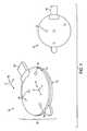
- FIG. 1illustrates a multifunction device according to an embodiment of the invention.
- FIG. 2illustrates a multifunction device according to an embodiment of the invention.
- FIG. 3Aillustrates a circuit board according to an embodiment of the invention.
- FIG. 3Billustrates a circuit board according to an embodiment of the invention.
- FIG. 4Aillustrates a multifunction device according to an embodiment of the invention.
- FIG. 4Billustrates a-multifunction device according to an embodiment of the invention.
- FIG. 5illustrates a multifunction device according to an embodiment of the invention.
- FIG. 6illustrates a multifunction device according to an embodiment of the invention.
- FIG. 7illustrates a system for performing computer-assisted surgery according to an embodiment of the invention.
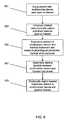
- FIG. 8illustrates a process for imaging a surgical instrument within a patient during a surgical intervention according to an embodiment of the invention.
- FIG. 9illustrates a process for utilizing a multifunction device as a proxy-target according to an embodiment of the invention.
- FIG. 1illustrates a multifunction device 100 according to an embodiment of the invention.
- Multifunction device 100may include a patient mountable portion 101 that may include internal electronics housed within a disposable package.
- Patient mountable portion 101 of multifunction device 100may be made of plastic commonly used in medical devices such as, for example, a polycarbonate, ABS, or other material.
- the plasticmay be radiolucent to assist in visualization of any intrinsic fiducials (discussed below).
- patient mountable portion 101may be made of a material suitable for fabrication by injection molding.
- the patient mountable portionmay have a pressure sensitive adhesive applied to a patient mountable surface 103 (e.g., the “underside” or bottom of the base layer of patient mountable portion 101 ).
- the pressure sensitive adhesivemay be capable of securely fastening multifunctional device 100 to a patient's skin surface, thus, enabling multifunction device 100 to act as a “stick on” patch.
- multifunctional device 100may also include surface features in the form of grooves or divots 105 that may be palpated using a probe or pointer. These surface features may exist on a palpable surface 107 (e.g., the “top” side or cover layer of patient mountable portion 101 ). In one embodiment, divots 105 may be asymmetrically arranged, so that they are distinguishable from one another.
- multifunctional device 100may also include an exit point 107 for a cable, wire, or other connector.
- multifunctional device 100may be associated with an intrinsic coordinate system 111 , which defines the location and orientation of multifunctional device 100 and all features contained therein. In some embodiments, the location and orientation of multifunctional device 100 may be determined in terms of the reference frame 150 of a tracking device.
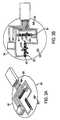
- FIG. 2illustrates an embodiment of the internal construction of multifunction device 100 in detail.
- multifunction device 100may include a circuit board 201 .
- circuit board 201may comprise a flexible circuit board that is etched with a pattern of conductors so as to form an electronic circuit for distributing power and signals to one or more embedded position indicating elements 203 and 205 . While position indicating elements 203 and 205 are shown here as coils, other types of configurations may be used. Additionally, while position indicating elements 203 and 205 are shown here as discrete components, In some embodiments, they could equally be etched into the circuit board itself.
- Circuit board 201may also include an intrinsic memory device 207 integrated into it. Circuit board 201 may be attached to a base layer 209 and a cover layer 211 that form patient mountable portion 101 (illustrated in FIG. 1 ). Circuit board 201 , base layer 209 , and cover layer 211 may be attached together by way of, for example, suitable adhesive, welding processes, or other attachment methods, so as to render the assembled patient mountable portion 101 impervious to fluids and consistent with a device that can be used in a sterile environment.
- base layer 209 , cover layer 211 , or an intermediate layer (not shown) integrated in or attached to multifunction device 100may serve as a shielding layer designed to isolate position indicating elements 203 and 205 from stray electromagnetic signals such as, for example, those that might be generated by the heart or other stray electrical signals.
- memory device 207may comprise generally a preprogrammed device such as, for example, a read-only memory (ROM) or serial ROM (SROM). In some embodiments, the memory device may not be capable of being erased or re-programmed. In some embodiments memory device may be programmed with some or all of the following information:
- memory device 207may be incorporated into a cable or plug associated with multifunction device 100 .
- memory device 207is an integral part of the assembled device that cannot be separated or removed from the assembly without damaging or otherwise rendering the assembly inoperable.
- memory device 207may include a radio frequency identification (RFID) type of device that is capable of wirelessly transmitting the aforementioned information using methods that are known in the art.
- RFIDradio frequency identification
- FIGS. 3A and 3Billustrate perspective and planar views of circuit board 201 according to an embodiment of the invention.
- multifunction device 100may include fiducial marks 301 .
- Fiducial marks 301may include marks made of a material that is imageable by an imaging modality (e.g., a radio-opaque material that is visible on an x-ray).
- Fiducial marks 301may be embedded within multifunctional device 100 and are shown in FIGS. 3A and 3B attached to circuit board 201 .
- Fiducial marks 301are shown in FIGS. 3A and 3B as different, distinguishable flat patterns (dash, plus sign, triangle, circle—other shapes may be used).
- Fiducial marks 301are shown in FIGS. 3A and 3B as attached to circuit board 201 , but could equally be attached to another part of multifunction device such as, for example, cover layer 211 , base layer 209 , or other part of multifunction device 100 , so long as the relative location of the fiducial marks remains fixed relative to the origin of coordinate system 111 of multifunction device and position indicating elements 203 and 205 .
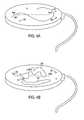
- FIGS. 4A and 4Billustrates multifunction device 100 according to an embodiment of the invention, wherein multifunction device 100 includes one or more “imageable patterns.”
- the one or more imageable patternsmay take the form of imageable pathways 401 or imageable regions 403 .
- Fiducial marks 301 , 303 or other fiducial marksmay also be used in or as imageable patterns.
- Imageable pathways 401may include conduits that are visible to an imaging modality.
- imageable pathways 401 and/or imageable regions 403may be used instead of or in addition to fiducial marks 301 .
- Imageable pathways 402may be made through the use of an imageable material (e.g., radio-opaque material that is visible to via an x-ray) placed on multifunction device 100 .
- imageable materiale.g., radio-opaque material that is visible to via an x-ray
- imageable pathway 401may take the form of a tube or groove in multifunction device 100 that has been filled with a barium compound.
- imageable pathways 401may include three-dimensional paths.
- imageable pathways 401may be multiply curved paths whose geometry is well established at the time of manufacture. These multiply curved paths may define a unique orientation and location of multifunction device 100 .
- complex imageable pathways 401may provide superior registration to fiducial marks alone because they include a continuum of points rather than the discrete set of points provided by fiducial marks alone.
- Imageable regions 403may also be used in a similar fashion and may include zones of unique geometry that are filled with an appropriate material (e.g., one that renders them visible to an imaging modality).
- fiducial marks 301may be deposited directly onto circuit board 201 .
- fiducial marks 301may be deposited near the ends of position indicating elements 203 and 205 as shown in FIGS. 3A and 3B .
- fiducial marks 301may be identifiable uniquely from their geometric shape (e.g., crosses, dashes, triangles, circles, or other shape). Additionally, fiducial marks 301 may be arranged in an asymmetric manner so that they may be readily identified on planar images by comparing the distance between marks and the angles between lines created by joining two marks to a third apex mark.
- fiducial marks 303may exist on circuit board 201 or elsewhere on multifunction device 100 .
- one or more fiducial marks 301 and/or 303may be represented out of the plane formed by the generally two-dimensional circuit board 201 .
- one or more fiducial marks 301 and/or 303may be represented in a special location relative to divots 105 .
- divots 105may enjoy a special relationship with the location of position indicating elements 203 and/or 205 , such as, for example, a divot may be located near the origin of coordinate system 111 of multifunction device 100 , the location of individual sensor origins 305 and 307 of sensors 203 and 205 respectively, and/or other feature of multifunction device 100 .
- fiducial marks 301 and/or 303may be visible from substantially perpendicular views taken by an imaging modality such as, for example, a fluoroscope, to assist in “fluoroscopic image guided surgery,” also known as “fluoronav” image guided surgery.
- an imaging modalitysuch as, for example, a fluoroscope
- parameters of a fluoroscopeare determined by examining shadows cast by fiducial marks 301 and/or 303 as x-rays pass from the fluoroscope emitter through multifunction device 100 to the fluoroscope image intensifier. This may be done in several fluoroscope positions and orientations.
- the location of multifunction device 100(and therefore any fiducial marks 301 / 303 contained therein) in image space (e.g., the frame of reference of the fluoroscope) and sensor space (e.g., the frame of reference of a tracking device) is determined to correctly determine the parameters necessary for fluoroscopic image guided surgery.
- image spacee.g., the frame of reference of the fluoroscope
- sensor spacee.g., the frame of reference of a tracking device
- FIG. 5illustrates multifunction device 100 according to another embodiment of the invention, wherein multifunction device 100 includes a layer that contains an integrated monitor 501 capable of measuring a physiological parameter such as, for example, heart contractions (an electrocardiograph [ECG] electrode), breathing motion, or other physiological parameter.
- a physiological parametersuch as, for example, heart contractions (an electrocardiograph [ECG] electrode), breathing motion, or other physiological parameter.
- ECGelectrocardiograph
- Integrated monitor 501may not only be used for gathering data regarding a physiological parameter, but also for gating the acquisition of image data (e.g., computerized tomography data during a CT scan or other data gathered by an imaging modality) or gating the use of multifunction device 100 as a position indicating element in a tracking device.
- FIG. 5illustrates a cardiac monitoring electrode as integrated monitor 501 .
- a similar device and appropriate circuit board layermay be incorporated to monitor respiratory motion using technology that is known in the art.
- signals or other data gathered by integrated monitor 501may be fed back through the common connector 109 .
- the layer containing integrated monitor 501may be composed of carbon fiber and may include an integrated conductive gel containing propylene glycol or similar material. Information regarding the properties of integrated monitor 501 may be programmed into memory device 207 .
- connectors for integrated monitor 501may be similar to those commonly used in the art such as, for example, a “snap” connector. In some embodiments, connectors for integrated monitor may be integrated into the same plug utilized by position indicating elements 203 and 205 .
- FIG. 6illustrates multifunction device 100 according to another embodiment of the invention, wherein multifunction device 100 includes a layer that contains antennae 601 , 603 , and 605 .
- Antennae 601 , 603 , and 605may serve numerous purposes such as, for example, energizing a wireless transmitter, receiving signals from a wireless transmitter (such as, for example, an RFID tag or wireless transceiver) that is capable of indicating the position and orientation of a separate position indicating element embedded in the body of a patient (e.g., a “surgical” position indicating element).
- a wireless transmittersuch as, for example, an RFID tag or wireless transceiver
- numerous individual multifunction devices or a single large multifunction devicemay take the place of the excitation/receive coils of a field generator of a tracking device.
- multifunction device 100acts as a field generator in place of (or in addition to) the separate field generation unit. This embodiment may increase the sensitivity of the tracking device and reduce the space requirements of the field generator. It may also be used as an intrinsic dynamic reference, as a field generator integrated into multifunction device 100 that is placed on the patient will move with the patient.
- additional functional and/or passive layersmay be integrated into the basic design of multifunction device 100 described herein by adding additional layers.
- additional functional and/or passive layersmay be integrated into the basic design of multifunction device 100 described herein by adding additional layers.
- MRmagnetic resonance
- a layer containing a three dimensional patterncomprising, for example, ball bearings that can be used to calculate fluoroscope parameters (or other parameters for an imaging modality), wherein the three-dimensional pattern is unique when viewed from multiple orientations.
- a layer containing a pattern of at least three radiodense regions forming a checkerboard with non radiodense regions(this could also be a complex two dimensional pattern). Such a pattern may be used to accurately track multifunction device 100 fluoroscopically (to assist in tracking patient motion in a second manner).
- multifunctional device 100may be manufactured using simple techniques and may be calibrated and programmed after it is assembled, obviating the need for high tolerance assembly techniques.
- the surface of multifunction device 100may be pre-imprinted with a body-centric coordinate system such as, for example, arrows, lines, dashes, or other indicators pointing toward, for example, the head, feet, left, and/or right of the patient.
- a body-centric coordinate systemsuch as, for example, arrows, lines, dashes, or other indicators pointing toward, for example, the head, feet, left, and/or right of the patient.
- These markingsmay visible to a surgeon and used by him or her to roughly orient the patch on the patient, enabling software to provide more relevant feedback to the surgeon. Examples of such marks are indicated as items 405 in FIGS. 4A and 4B .
- multifunction device 100may be composed of a plurality of individual patches containing at least one position indicating element and/or other devices discussed above.
- a calibration stepmay be performed to determine the exact locations of the position indicating elements so that tracking can occur. This calibration step may be done either using an imaging modality or by moving a field generator associated with the tracking device of the position indicating elements to multiple locations.
- multifunction device 100may be utilized in performing different roles in image guided surgery.
- divots 105 or other surface features of multifunction device 105may be used as discrete markings or “landmarks” for a point registration.
- position indicating elements 203 and 205may be used for other types of registration, dynamic referencing, verification of registration, and/or other purposes.
- position indicating elementsmay be used to provide position information regarding one or more points on the surface of a patient's skin in the frame of reference of a tracking device.
- Fiducial marks 301 and 303may be used in conjunction with an imaging modality to provide position information in an image space. This position information may then be used in the registration of the corresponding area of the patient's anatomy.
- position indicating elements 203 and 205may be used to track movement of a patient for use in dynamic referencing during image guided surgery.
- multifunction device 100may be connected to or used in conjunction with a radio-opaque grid that may be applied to a patient. This enables devices to be guided by the grid/multifunction device 100 combination by imaging the grid and multifunction device 100 simultaneously. Once the system is registered and a target area or point (e.g., an area or point of interest within a patient) is located on associated images of the patient, the closest grid-mark may be identified and used to assist in placement of a therapeutic or diagnostic device (e.g., a needle) at the target area or point due to the fact that the grid marks are known relative to multifunction device 100 .
- a therapeutic or diagnostic devicee.g., a needle
- Imageable pathways 401 and/or imageable regions 403may also be used to obtain, via an imaging modality, image space position information regarding a portion of the patient's anatomy for use in registration, verification of registration, or for other purposes.
- These complex three-dimensional imageable patternsmay be readily and unambiguously identified on images. Additionally, they provide a plethora of position information, which assists in performing a high quality registration. Furthermore, the three-dimensional patterns may be visible from a number of different positions/orientations of an imaging modality such as, for example, a fluoroscope or other imaging modality.
- Imageable pathways 401 and/or imageable regions 403may also be used in conjunction with fiducial marks 301 and 303 to obtain image space information.
- imageable pathways 401 , imageable regions 403 , and or fiducial marks 301 and 303may exist on multifunctional device 100 in a known spatial relationship to position indicating element 203 , position indicating element 205 , and/or coordinate system 111 of multifunction device 100 .
- the coordinates of fiducial markers 301 and 303may be known in coordinate system 111 . Determination of the location of position indicating elements 203 and 205 in the coordinate system 150 of a tracking device (e.g., the patient space) may be used to determine the location of fiducial marks 301 and 303 in the patient space (because of the known relationship between position indicating elements 203 / 205 and fiducial marks 301 / 303 ).
- Images that contain fiducial marks 301 and 303enable their locations to be determined in image space, thus, allowing registration to be performed between the image space and the patient space. In general, this known relationship may aid in registering patient-space position information provided by position indicating elements 203 and 205 and/or coordinate system 111 to image-space information provided by imageable pathways 401 , imageable regions 403 , and/or fiducial marks 301 and 303 .
- integrated monitor 501may be used to obtain physiological parameters from a patient and/or for gating the sampling of position information or image information used in image guided surgery according to those physiological parameters.
- antennae 601 , 603 , and 605may be used to provide various features of multifunction device such as, for example, a magnetic field for use with position indicating elements within the patient as part of a tracking device, as part of a CT, MR, ultrasound or other imaging modality, or for other uses.
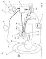
- FIG. 7illustrates a computer assisted surgery system 700 according to one embodiment of the invention.
- system 700may include an imaging device 701 , a tracking device 703 , one or more physiological measuring devices 705 , a computer 707 , and/or other elements.
- physiological measuring devices 705may be connected to computer 707 directly.
- physiological measuring device 705may be connected through tracking device 703 (not illustrated) which may then relay information to computer 707 or use the information, for example, to gate position data from position indicating elements 203 and/or 205 in multifunction device 100 .
- tracking device 703may comprise a field generator 713 and a control unit 715 .
- tracking device 703may comprise, for example, an electromagnetic tracking device.
- field generator 713may include a magnetic field generator and any associated position indicating elements 203 and/or 205 may comprise magnetic position sensors.
- Tracking device 703may be associated with a coordinate system 717 (similar to or the same as coordinate system 150 of FIG. 1 ).
- multifunction device 100such as, for example, wiring elements 719 , which may connect multifunction device 100 to tracking device 703 or physiological measuring devices 705 .
- the layer of the multifunction device containing position indicating elements 203 and 205e.g., circuit board 201 of FIG. 2
- Position indicating elements 203 and 205may be sampled by tracking device 703 to provide position and/or orientation information regarding position indicating elements 203 and 205 to computer 707 in the frame of reference of tracking device 703 (e.g., coordinate system 717 ).
- physiological parameters of a patient 709may be measured by physiological measuring devices 705 and transferred to computer 707 .
- FIG. 8illustrates a process 800 , wherein multifunction device 100 may be used as part of system 700 for performing image guided surgery.
- multifunction device 100may be connected to a patient 709 .
- one or more images 711 of patient 709may be obtained using imaging device 701 .
- Imaging device 701may include, for example, a computerized tomography (CT) device, a magnetic resonance (MR) device, a positron emission tomography (PET) device, a fluoroscope, an x-ray device, or other imaging modality.
- Images 711may then be loaded onto or stored in computer 707 .
- images 711may be obtained when patient 709 is in a known physiological state (e.g. peak inspiration).
- the physiological state of patient 709may be measured/monitored during imaging by multifunction device 100 .
- the location/geometry of fiducial markse.g., fiducial marks 301 and/or 303
- imageable patternse.g., imageable pathways 401 and/or imageable regions 403
- multifunction device 100may be determined using images 711 in coordinate system 111 of images 711 (e.g., the image space).
- tracking device 702may sample position indicating elements 203 and/or 205 of multifunction device 100 . This may produce position and/or orientation information of position indicating sensors 203 and/or 205 in the coordinate system 717 of tracking device 703 (e.g., the patient space). This position and/or orientation information may then be provided to computer 707 .
- a registration between the image space and the patient spacemay be calculated. This may be accomplished by combining the location of fiducial marks 301 / 303 and/or imageable patterns of multifunction device 100 in coordinate system 111 of images 711 (image space) with the location of position indicating elements 203 / 205 in coordinate system 717 of the tracking device 702 (patient space). This is possible because the location of fiducial marks 301 / 303 or imageable patterns (imageable paths 401 or regions 403 ) are known relative to position indicating elements 203 / 205 . In an embodiment, these relationships/relative locations may be programmed into and/or stored into memory device 207 that is associated with multifunction device 100 .
- reattachment positionsmust be consistent and known or it will not be possible to determine the location of fiducial marks 301 / 303 or imageable patterns from the location of position indicating elements 203 / 205 .
- multifunction device 100may begin to function as a dynamic reference device in which, together with the physiological measurements provided by physiological measuring devices 705 , more accurate referencing can be obtained than could be otherwise obtained using multifunction device 100 alone. This is because each time a particular gating signal is received, the organs in the patient's body are in the same state, and the position of them is not merely compensated for as would be using dynamic referencing alone.
- a medical instrument/devicesuch as a guidewire 721 (or other instrument such as, for example, a catheter) in which at least one position indicating element (e.g., a “surgical” position indicating element) has been attached, may be navigated in the patient's body and the location of guidewire 721 may be displayed on images 711 (or versions or derivations thereof) relative to multifunction device 100 . Further accuracy may be obtained if the position of guidewire 721 is gated according to at least one physiological parameter measured by physiological measuring devices 705 (which, in some embodiments, operate in conjunction with sensors 501 of multifunction device 100 ).
- physiological measuring devices 705which, in some embodiments, operate in conjunction with sensors 501 of multifunction device 100 .
- Such gatingmay act to display the position of guidewire 721 on images 711 only when the physiological parameters match those under which images 711 were obtained (e.g. peak inspiration corresponding to breath hold during the scan).
- guidewire 721 or other surgical instrumentmay be navigated in the patient to perform an intervention (e.g., surgical procedure, diagnostic procedure, or other procedure).
- FIG. 9illustrates a process 900 according to an embodiment of the invention, wherein multifunction device 100 may act as a “proxy target” that can be navigated toward in conjunction with an imageless computer assisted surgery system.
- patient 709may first be equipped with multifunction device 100 , placed directly over a point of interest 723 (e.g., a “target”) such as, for example, the superior vena cava of the heart, with the aim of, for example, placing the exit of a peripherally inserted central catheter (PICC) at point of interest 723 .
- a point of interest 723e.g., a “target”
- PICCperipherally inserted central catheter
- a medical instrumentsuch as, for example, vascular guidewire 721 (or other instrument such as for example, a catheter), that has been modified to contain a position-indicating element (e.g., a “surgical” position indicating element) near its tip 725 , is introduced into the patient (for example into a vein or artery connected to the point of interest 723 ) and directed toward the point of interest 723 .
- a position-indicating elemente.g., a “surgical” position indicating element
- the position of multifunction device 100the position of guidewire 721 , and the physiological parameters measured by physiological measuring devices 705 can be determined and transferred to computer 707 .
- the relative location between the position of multifunction device 100 and the position of guidewire 723may then can be calculated.
- tip 725 of guidewire 721may also be graphically depicted relative to the multifunction device 100 (acting as a “proxy target”) and therefore graphically depicted relative to point of interest 723 .
- Knowledge of the physiological parametersmay increase accuracy as before by enabling sampling only in particular physiological states of the patient (e.g., gating). If multifunction device 100 is oriented such that the body-centric coordinate system markings 405 (see FIGS. 4 and 7 ) on the multifunction device correspond in an intuitive way to the patient position, the physician performing the intervention will furthermore be able to understand the direction of any deviations of guidewire 721 from the intended direction, which is toward point of interest 723 .
- a guidewire 721(or other instrument such as, for example, a catheter) may be used to determine its proximity to the heart by measuring its motion during different phases of the heart cycle as determined by physiological parameter measurement by multifunction device 100 .
- guidewire 721may be used to measure the size of a vessel or conduit of patient 709 in which it is located by placing a position indicating element of guidewire 721 near its bent tip 725 and axially spinning the guidewire. Inside small vessels it's rotation will prescribe a line as bent tip 725 will be mostly constrained by the vessel walls. Inside larger vessels where bent tip 725 is allowed to take its natural shape, it will prescribe a circle, the diameter of which is an indication of the vessel size up to a maximum size determined by the amount of bend of bent tip 725 .
- Imageless computer assisted surgical methodsneed not involve the heart, but may be used for numerous medical procedures such as, for example assisting in placing a feeding tube into the stomach or small intestine of a patient or for other purposes.
- computer 707may include one or more software modules 727 a - n for performing various features and functions of the invention.
- computer 707may include a module that determines, from images 711 , the locations/geometry of the fiducial marks 301 , 303 , imageable paths 401 , imageable regions 403 and or other imageable patterns or elements in the coordinate system of images 711 and/or in the coordinate system of tracking device 713 .
- computer 707may include a module that calculates a registration between images 711 (and/or data therefrom) and tracking device 703 (and/or data therefrom).
- computer 707may include a module that calculates and/or depicts the relative location between a position of multifunction device 100 and a position of vascular guidewire 721 .
- Computer 707may also include a module that graphically depicts guidewire 721 (and/or the tip 725 of guidewire 721 ) on images 711 relative to multifunction device 100 and therefore to object of interest 723 .
- Computer 707may also include other modules for performing other features or functions of the invention described herein. In some embodiments, some of these tasks may be performed on or transferred to control unit 715 of tracking device 703 .
Landscapes
- Health & Medical Sciences (AREA)
- Life Sciences & Earth Sciences (AREA)
- Engineering & Computer Science (AREA)
- Heart & Thoracic Surgery (AREA)
- Molecular Biology (AREA)
- Biophysics (AREA)
- Pathology (AREA)
- Biomedical Technology (AREA)
- Human Computer Interaction (AREA)
- Medical Informatics (AREA)
- Physics & Mathematics (AREA)
- Surgery (AREA)
- Animal Behavior & Ethology (AREA)
- General Health & Medical Sciences (AREA)
- Public Health (AREA)
- Veterinary Medicine (AREA)
- Apparatus For Radiation Diagnosis (AREA)
- Magnetic Resonance Imaging Apparatus (AREA)
Abstract
Description
- a. properties such as impedance, resistance, magnetic moment, or other properties, thereby reducing the tolerances by which
position indicating elements - b. location and orientation of
position indicating elements system 111 that is associated withmultifunction device 100, thereby reducing the mechanical tolerances required in manufacturing necessary to produce a device that achieves a consistent accuracy;
- a. properties such as impedance, resistance, magnetic moment, or other properties, thereby reducing the tolerances by which
Claims (23)
Priority Applications (1)
| Application Number | Priority Date | Filing Date | Title |
|---|---|---|---|
| US11/271,899US7751868B2 (en) | 2004-11-12 | 2005-11-14 | Integrated skin-mounted multifunction device for use in image-guided surgery |
Applications Claiming Priority (2)
| Application Number | Priority Date | Filing Date | Title |
|---|---|---|---|
| US62694604P | 2004-11-12 | 2004-11-12 | |
| US11/271,899US7751868B2 (en) | 2004-11-12 | 2005-11-14 | Integrated skin-mounted multifunction device for use in image-guided surgery |
Publications (2)
| Publication Number | Publication Date |
|---|---|
| US20060173269A1 US20060173269A1 (en) | 2006-08-03 |
| US7751868B2true US7751868B2 (en) | 2010-07-06 |
Family
ID=36757534
Family Applications (1)
| Application Number | Title | Priority Date | Filing Date |
|---|---|---|---|
| US11/271,899Active2026-08-01US7751868B2 (en) | 2004-11-12 | 2005-11-14 | Integrated skin-mounted multifunction device for use in image-guided surgery |
Country Status (1)
| Country | Link |
|---|---|
| US (1) | US7751868B2 (en) |
Cited By (26)
| Publication number | Priority date | Publication date | Assignee | Title |
|---|---|---|---|---|
| US20080033344A1 (en)* | 2006-08-04 | 2008-02-07 | Mantell Robert R | In-Dwelling Port For Access Into A Body |
| US20080091101A1 (en)* | 2006-10-16 | 2008-04-17 | Perfint Engineering Services | Needle positioning apparatus and method |
| US20100298704A1 (en)* | 2009-05-20 | 2010-11-25 | Laurent Pelissier | Freehand ultrasound imaging systems and methods providing position quality feedback |
| US20110015521A1 (en)* | 2003-03-27 | 2011-01-20 | Boulder Innovation Group, Inc. | Means of Tracking Movement of Bodies During Medical Treatment |
| US8213693B1 (en)* | 2007-05-16 | 2012-07-03 | General Electric Company | System and method to track and navigate a tool through an imaged subject |
| US8613748B2 (en) | 2010-11-10 | 2013-12-24 | Perfint Healthcare Private Limited | Apparatus and method for stabilizing a needle |
| US8687172B2 (en) | 2011-04-13 | 2014-04-01 | Ivan Faul | Optical digitizer with improved distance measurement capability |
| US20140222205A1 (en)* | 2013-02-02 | 2014-08-07 | Perfint Healthcare Private Limited | Electronic docking system and method for robotic positioning system |
| US8864670B2 (en) | 2011-01-28 | 2014-10-21 | Hospira, Inc. | Ultrasonic monitoring device for measuring physiological parameters of a mammal |
| US9186225B1 (en) | 2014-06-09 | 2015-11-17 | Christopher Pettis | Skin marking device for radiographic imaging |
| US9295449B2 (en) | 2012-01-23 | 2016-03-29 | Ultrasonix Medical Corporation | Landmarks for ultrasound imaging |
| US20160242855A1 (en)* | 2015-01-23 | 2016-08-25 | Queen's University At Kingston | Real-Time Surgical Navigation |
| US9572595B1 (en) | 2014-03-05 | 2017-02-21 | Northgate Technologies Inc. | In-dwelling port for access into a body |
| US9681919B2 (en) | 2014-07-09 | 2017-06-20 | Neil Glossop | Systems, methods, and devices for assisting or performing guided interventional procedures using custom templates |
| EP3305202A1 (en) | 2016-10-06 | 2018-04-11 | Biosense Webster (Israel), Ltd. | Pre-operative registration of anatomical images with a position-tracking system using ultrasound |
| US20180168735A1 (en)* | 2016-05-02 | 2018-06-21 | Dorothy LUI | Methods for improving patient registration |
| US10039527B2 (en) | 2009-05-20 | 2018-08-07 | Analogic Canada Corporation | Ultrasound systems incorporating spatial position sensors and associated methods |
| US10074199B2 (en) | 2013-06-27 | 2018-09-11 | Tractus Corporation | Systems and methods for tissue mapping |
| US10265137B2 (en) | 2014-07-09 | 2019-04-23 | Neil Glossop | Systems, methods, and devices for assisting or performing guided interventional procedures using custom templates |
| US10390892B2 (en)* | 2017-08-24 | 2019-08-27 | Synaptive Medical Inc. | System and methods for updating patient registration during surface trace acquisition |
| US10510171B2 (en) | 2016-11-29 | 2019-12-17 | Biosense Webster (Israel) Ltd. | Visualization of anatomical cavities |
| US10575756B2 (en) | 2014-05-14 | 2020-03-03 | Stryker European Holdings I, Llc | Navigation system for and method of tracking the position of a work target |
| US11006853B2 (en) | 2015-09-04 | 2021-05-18 | Biosense Webster (Israel) Ltd. | Field-based location coordinate correction |
| US20220022968A1 (en)* | 2018-11-30 | 2022-01-27 | Think Surgical, Inc. | Computer input method using a digitizer as an input device |
| US11701188B2 (en) | 2017-05-10 | 2023-07-18 | Mako Surgical Corp. | Robotic spine surgery system and methods |
| US11766298B2 (en) | 2019-05-03 | 2023-09-26 | Neil Glossop | Systems, methods, and devices for registering and tracking organs during interventional procedures |
Families Citing this family (50)
| Publication number | Priority date | Publication date | Assignee | Title |
|---|---|---|---|---|
| US7398116B2 (en) | 2003-08-11 | 2008-07-08 | Veran Medical Technologies, Inc. | Methods, apparatuses, and systems useful in conducting image guided interventions |
| US8150495B2 (en) | 2003-08-11 | 2012-04-03 | Veran Medical Technologies, Inc. | Bodily sealants and methods and apparatus for image-guided delivery of same |
| EP1924198B1 (en) | 2005-09-13 | 2019-04-03 | Veran Medical Technologies, Inc. | Apparatus for image guided accuracy verification |
| US20070066881A1 (en) | 2005-09-13 | 2007-03-22 | Edwards Jerome R | Apparatus and method for image guided accuracy verification |
| US20070081703A1 (en)* | 2005-10-12 | 2007-04-12 | Industrial Widget Works Company | Methods, devices and systems for multi-modality integrated imaging |
| EP2001363B1 (en)* | 2006-03-31 | 2017-09-27 | Philips Electronics LTD | System and instrumentation for image guided prostate treatment |
| DE102006026490B4 (en)* | 2006-06-07 | 2010-03-18 | Siemens Ag | Radiotherapy device with angiography CT device |
| IL188262A (en)* | 2007-01-10 | 2011-10-31 | Mediguide Ltd | System and method for superimposing a representation of the tip of a catheter on an image acquired by a moving imager |
| EP2111163B1 (en)* | 2007-02-14 | 2019-04-24 | Koninklijke Philips N.V. | System, a method and a computer program for determining a functional property of a moving object |
| US20090209849A1 (en)* | 2007-05-02 | 2009-08-20 | Philip Stephen Rowe | Medical Device Placement and Monitoring System Utilizing Radio Frequency Identification |
| US8690768B2 (en)* | 2007-07-26 | 2014-04-08 | David Amitai | Patient operable data collection system |
| EP2194858B1 (en) | 2007-09-14 | 2017-11-22 | Corventis, Inc. | Medical device automatic start-up upon contact to patient tissue |
| US8116841B2 (en) | 2007-09-14 | 2012-02-14 | Corventis, Inc. | Adherent device with multiple physiological sensors |
| WO2009036316A1 (en) | 2007-09-14 | 2009-03-19 | Corventis, Inc. | Energy management, tracking and security for adherent patient monitor |
| EP3922171A1 (en) | 2007-09-14 | 2021-12-15 | Medtronic Monitoring, Inc. | Adherent cardiac monitor with advanced sensing capabilities |
| US9186089B2 (en) | 2007-09-14 | 2015-11-17 | Medtronic Monitoring, Inc. | Injectable physiological monitoring system |
| WO2009036369A1 (en) | 2007-09-14 | 2009-03-19 | Corventis, Inc. | System and methods for wireless body fluid monitoring |
| WO2009036327A1 (en) | 2007-09-14 | 2009-03-19 | Corventis, Inc. | Adherent device for respiratory monitoring and sleep disordered breathing |
| JP5250251B2 (en)* | 2007-12-17 | 2013-07-31 | イマグノーシス株式会社 | Medical imaging marker and its utilization program |
| US20090221908A1 (en)* | 2008-03-01 | 2009-09-03 | Neil David Glossop | System and Method for Alignment of Instrumentation in Image-Guided Intervention |
| EP2257216B1 (en) | 2008-03-12 | 2021-04-28 | Medtronic Monitoring, Inc. | Heart failure decompensation prediction based on cardiac rhythm |
| WO2009146214A1 (en) | 2008-04-18 | 2009-12-03 | Corventis, Inc. | Method and apparatus to measure bioelectric impedance of patient tissue |
| US8876830B2 (en)* | 2009-08-13 | 2014-11-04 | Zimmer, Inc. | Virtual implant placement in the OR |
| RU2012116894A (en)* | 2009-10-06 | 2013-11-20 | Смит Энд Нефью, Инк. | GUIDANCE ON ORTHOPEDIC DEVICES |
| WO2011050283A2 (en) | 2009-10-22 | 2011-04-28 | Corventis, Inc. | Remote detection and monitoring of functional chronotropic incompetence |
| US9451897B2 (en) | 2009-12-14 | 2016-09-27 | Medtronic Monitoring, Inc. | Body adherent patch with electronics for physiologic monitoring |
| US8965498B2 (en) | 2010-04-05 | 2015-02-24 | Corventis, Inc. | Method and apparatus for personalized physiologic parameters |
| JP2013530028A (en) | 2010-05-04 | 2013-07-25 | パスファインダー セラピューティクス,インコーポレイテッド | System and method for abdominal surface matching using pseudo features |
| EP3659490B1 (en) | 2010-08-20 | 2025-10-01 | Veran Medical Technologies, Inc. | Apparatus and method for four dimensional soft tissue navigation |
| WO2012149548A2 (en)* | 2011-04-29 | 2012-11-01 | The Johns Hopkins University | System and method for tracking and navigation |
| EP2812047B1 (en)* | 2012-02-09 | 2022-06-08 | Bluegrass Vascular Technologies, Inc. | Occlusion access system |
| US10092726B2 (en) | 2012-02-09 | 2018-10-09 | Bluegrass Vascular Technologies, Inc. | Occlusion access system |
| EP2816966B1 (en) | 2012-02-22 | 2023-10-25 | Veran Medical Technologies, Inc. | Steerable surgical catheter comprising a biopsy device at the distal end portion thereof |
| US9510772B2 (en) | 2012-04-10 | 2016-12-06 | Cardionxt, Inc. | System and method for localizing medical instruments during cardiovascular medical procedures |
| US20140005527A1 (en)* | 2012-06-29 | 2014-01-02 | General Electric Company | Method and system for dynamic referencing and registration used with surgical and interventional procedures |
| DE102013201701A1 (en)* | 2013-02-01 | 2014-08-07 | Friedrich-Alexander-Universität Erlangen-Nürnberg | A method of providing attenuation correction for a combined magnetic resonance positron emission tomography apparatus |
| CN105050525B (en)* | 2013-03-15 | 2018-07-31 | 直观外科手术操作公司 | Shape sensor system for tracking interventional instruments and method of use |
| EP3035843B1 (en) | 2013-08-22 | 2021-11-03 | AFTx, Inc. | Methods, systems, and apparatus for identification and characterization of rotors associated with atrial fibrillation |
| WO2015143136A1 (en)* | 2014-03-19 | 2015-09-24 | Cardionxt, Inc. | System and methods for using body surface cardiac electrogram information combined with internal information to deliver therapy |
| US20150305612A1 (en) | 2014-04-23 | 2015-10-29 | Mark Hunter | Apparatuses and methods for registering a real-time image feed from an imaging device to a steerable catheter |
| US20150305650A1 (en) | 2014-04-23 | 2015-10-29 | Mark Hunter | Apparatuses and methods for endobronchial navigation to and confirmation of the location of a target tissue and percutaneous interception of the target tissue |
| EP3139830A4 (en) | 2014-05-05 | 2018-01-24 | Cardionxt, Inc. | Methods, systems, and apparatus for identification, characterization, and treatment of rotors associated with fibrillation |
| US10856892B2 (en) | 2016-02-29 | 2020-12-08 | Bluegrass Vascular Technologies, Inc. | Catheter systems, kits, and methods for gaining access to a vessel |
| US11589926B2 (en)* | 2017-01-04 | 2023-02-28 | Medivation Ag | Mobile surgical tracking system with an integrated fiducial marker for image guided interventions |
| US10782316B2 (en) | 2017-01-09 | 2020-09-22 | Delta Design, Inc. | Socket side thermal system |
| US20200405395A1 (en)* | 2017-07-03 | 2020-12-31 | Spine Align, Llc | Intraoperative alignment assessment system and method |
| US20200237459A1 (en)* | 2019-01-25 | 2020-07-30 | Biosense Webster (Israel) Ltd. | Flexible multi-coil tracking sensor |
| US11887347B2 (en)* | 2020-08-06 | 2024-01-30 | Canon U.S.A., Inc. | Device-to-image registration method, apparatus, and storage medium |
| CN113288355A (en)* | 2021-05-17 | 2021-08-24 | 苏州心速度医疗技术有限公司 | Puncture auxiliary guiding device |
| WO2023278632A1 (en) | 2021-06-30 | 2023-01-05 | Delta Design, Inc. | Temperature control system including contactor assembly |
Citations (209)
| Publication number | Priority date | Publication date | Assignee | Title |
|---|---|---|---|---|
| US3021842A (en) | 1958-11-05 | 1962-02-20 | John F Flood | Hypodermic needle guide |
| US4080706A (en) | 1975-04-22 | 1978-03-28 | Medrad, Inc. | Method of manufacturing catheter guidewire |
| US4279252A (en) | 1979-08-24 | 1981-07-21 | Martin Michael T | X-ray scaling catheter |
| US4697595A (en) | 1984-07-24 | 1987-10-06 | Telectronics N.V. | Ultrasonically marked cardiac catheters |
| US4722056A (en) | 1986-02-18 | 1988-01-26 | Trustees Of Dartmouth College | Reference display systems for superimposing a tomagraphic image onto the focal plane of an operating microscope |
| US4777951A (en) | 1986-09-19 | 1988-10-18 | Mansfield Scientific, Inc. | Procedure and catheter instrument for treating patients for aortic stenosis |
| US4887606A (en) | 1986-09-18 | 1989-12-19 | Yock Paul G | Apparatus for use in cannulation of blood vessels |
| US4895168A (en) | 1988-01-21 | 1990-01-23 | Schneider (Usa) Inc., A Pfizer Company | Guidewire with movable core and external tubular safety cover |
| US4935019A (en) | 1986-12-22 | 1990-06-19 | Johnson & Johnson Medical, Inc. | Radiopaque polymeric composition |
| US4961433A (en) | 1988-11-02 | 1990-10-09 | Cardiometrics, Inc. | Guide wire assembly with electrical functions and male and female connectors for use therewith |
| US5014708A (en) | 1988-09-14 | 1991-05-14 | Olympus Optical Co. | Radioactive ray detecting therapeutic apparatus |
| US5042486A (en) | 1989-09-29 | 1991-08-27 | Siemens Aktiengesellschaft | Catheter locatable with non-ionizing field and method for locating same |
| US5045080A (en) | 1986-12-22 | 1991-09-03 | Johnson & Johnson Medical, Inc. | Surgical fabric with printed X-ray marker |
| US5085659A (en) | 1990-11-21 | 1992-02-04 | Everest Medical Corporation | Biopsy device with bipolar coagulation capability |
| US5116345A (en) | 1990-11-28 | 1992-05-26 | Ohio Medical Instrument Co., Inc. | Stereotactically implanting an intracranial device |
| US5187658A (en) | 1990-01-17 | 1993-02-16 | General Electric Company | System and method for segmenting internal structures contained within the interior region of a solid object |
| US5204625A (en) | 1990-12-20 | 1993-04-20 | General Electric Company | Segmentation of stationary and vascular surfaces in magnetic resonance imaging |
| US5207675A (en) | 1991-07-15 | 1993-05-04 | Jerome Canady | Surgical coagulation device |
| US5211165A (en) | 1991-09-03 | 1993-05-18 | General Electric Company | Tracking system to follow the position and orientation of a device with radiofrequency field gradients |
| US5221283A (en) | 1992-05-15 | 1993-06-22 | General Electric Company | Apparatus and method for stereotactic surgery |
| US5247935A (en) | 1992-03-19 | 1993-09-28 | General Electric Company | Magnetic resonance guided focussed ultrasound surgery |
| US5251127A (en) | 1988-02-01 | 1993-10-05 | Faro Medical Technologies Inc. | Computer-aided surgery apparatus |
| US5251635A (en) | 1991-09-03 | 1993-10-12 | General Electric Company | Stereoscopic X-ray fluoroscopy system using radiofrequency fields |
| US5255680A (en) | 1991-09-03 | 1993-10-26 | General Electric Company | Automatic gantry positioning for imaging systems |
| US5265610A (en) | 1991-09-03 | 1993-11-30 | General Electric Company | Multi-planar X-ray fluoroscopy system using radiofrequency fields |
| US5271400A (en) | 1992-04-01 | 1993-12-21 | General Electric Company | Tracking system to monitor the position and orientation of a device using magnetic resonance detection of a sample contained within the device |
| US5275165A (en) | 1992-11-06 | 1994-01-04 | General Electric Company | Magnetic resonance guided ultrasound therapy system with inclined track to move transducers in a small vertical space |
| US5290266A (en) | 1992-08-14 | 1994-03-01 | General Electric Company | Flexible coating for magnetic resonance imaging compatible invasive devices |
| US5291010A (en) | 1990-10-04 | 1994-03-01 | Olympus Optical Co., Ltd. | Solid state imaging device having a chambered imaging chip corner |
| US5291890A (en) | 1991-08-29 | 1994-03-08 | General Electric Company | Magnetic resonance surgery using heat waves produced with focussed ultrasound |
| US5304933A (en) | 1991-08-01 | 1994-04-19 | General Electric Company | Surgical local gradient coil |
| US5305203A (en) | 1988-02-01 | 1994-04-19 | Faro Medical Technologies Inc. | Computer-aided surgery apparatus |
| US5307812A (en) | 1993-03-26 | 1994-05-03 | General Electric Company | Heat surgery system monitored by real-time magnetic resonance profiling |
| US5318025A (en) | 1992-04-01 | 1994-06-07 | General Electric Company | Tracking system to monitor the position and orientation of a device using multiplexed magnetic resonance detection |
| US5353808A (en) | 1992-03-04 | 1994-10-11 | Cordis Corporation | Guidewire having distally located marker segment |
| US5365927A (en) | 1993-11-02 | 1994-11-22 | General Electric Company | Magnetic resonance imaging system with pointing device |
| US5368032A (en) | 1993-11-09 | 1994-11-29 | General Electric Company | Manually positioned focussed energy system guided by medical imaging |
| US5368031A (en) | 1993-08-29 | 1994-11-29 | General Electric Company | Magnetic resonance surgery using heat waves produced with a laser fiber |
| US5377678A (en) | 1991-09-03 | 1995-01-03 | General Electric Company | Tracking system to follow the position and orientation of a device with radiofrequency fields |
| US5383454A (en) | 1990-10-19 | 1995-01-24 | St. Louis University | System for indicating the position of a surgical probe within a head on an image of the head |
| US5383465A (en) | 1989-12-18 | 1995-01-24 | Lesny; Jan | Ultrasonic instrument |
| US5386828A (en) | 1991-12-23 | 1995-02-07 | Sims Deltec, Inc. | Guide wire apparatus with location sensing member |
| US5389101A (en) | 1992-04-21 | 1995-02-14 | University Of Utah | Apparatus and method for photogrammetric surgical localization |
| US5391199A (en) | 1993-07-20 | 1995-02-21 | Biosense, Inc. | Apparatus and method for treating cardiac arrhythmias |
| US5396905A (en) | 1994-03-29 | 1995-03-14 | General Electric Company | Surgical drape with integral MRI coil |
| US5400383A (en) | 1991-12-09 | 1995-03-21 | General Electric Company | Fluoroscopic imager with frame-filling apparatus |
| US5437277A (en) | 1991-11-18 | 1995-08-01 | General Electric Company | Inductively coupled RF tracking system for use in invasive imaging of a living body |
| US5443066A (en) | 1991-11-18 | 1995-08-22 | General Electric Company | Invasive system employing a radiofrequency tracking system |
| US5443068A (en) | 1994-09-26 | 1995-08-22 | General Electric Company | Mechanical positioner for magnetic resonance guided ultrasound therapy |
| US5465732A (en) | 1992-03-31 | 1995-11-14 | Boston Scientific Corporation | Fluoroscopically viewable multifilar calibrated guidewire and method of measuring occlusions with calibrated guidewires |
| US5480382A (en) | 1989-01-09 | 1996-01-02 | Pilot Cardiovascular Systems, Inc. | Steerable medical device |
| US5490840A (en) | 1994-09-26 | 1996-02-13 | General Electric Company | Targeted thermal release of drug-polymer conjugates |
| US5526812A (en) | 1993-06-21 | 1996-06-18 | General Electric Company | Display system for enhancing visualization of body structures during medical procedures |
| US5526814A (en) | 1993-11-09 | 1996-06-18 | General Electric Company | Automatically positioned focussed energy system guided by medical imaging |
| US5558091A (en) | 1993-10-06 | 1996-09-24 | Biosense, Inc. | Magnetic determination of position and orientation |
| CA2226938A1 (en) | 1995-07-16 | 1997-02-06 | Yoav Paltieli | Free-hand aiming of a needle guide |
| US5603318A (en) | 1992-04-21 | 1997-02-18 | University Of Utah Research Foundation | Apparatus and method for photogrammetric surgical localization |
| US5645065A (en) | 1991-09-04 | 1997-07-08 | Navion Biomedical Corporation | Catheter depth, position and orientation location system |
| US5646524A (en) | 1992-06-16 | 1997-07-08 | Elbit Ltd. | Three dimensional tracking system employing a rotating field |
| US5646525A (en) | 1992-06-16 | 1997-07-08 | Elbit Ltd. | Three dimensional tracking system employing a rotating field |
| US5647373A (en) | 1993-11-07 | 1997-07-15 | Ultra-Guide Ltd. | Articulated needle guide for ultrasound imaging and method of using same |
| US5705014A (en) | 1996-03-22 | 1998-01-06 | General Electric Company | Carbon fiber magnetic resonance compatible instruments |
| US5713858A (en) | 1995-04-28 | 1998-02-03 | Medtronic, Inc. | Permanently implantable guiding catheter |
| US5715166A (en) | 1992-03-02 | 1998-02-03 | General Motors Corporation | Apparatus for the registration of three-dimensional shapes |
| US5715822A (en) | 1995-09-28 | 1998-02-10 | General Electric Company | Magnetic resonance devices suitable for both tracking and imaging |
| US5740802A (en) | 1993-04-20 | 1998-04-21 | General Electric Company | Computer graphic and live video system for enhancing visualization of body structures during surgery |
| US5749835A (en) | 1994-09-06 | 1998-05-12 | Sims Deltec, Inc. | Method and apparatus for location of a catheter tip |
| US5769790A (en) | 1996-10-25 | 1998-06-23 | General Electric Company | Focused ultrasound surgery system guided by ultrasound imaging |
| US5769861A (en) | 1995-09-28 | 1998-06-23 | Brainlab Med. Computersysteme Gmbh | Method and devices for localizing an instrument |
| US5848969A (en) | 1996-10-28 | 1998-12-15 | Ep Technologies, Inc. | Systems and methods for visualizing interior tissue regions using expandable imaging structures |
| US5857032A (en) | 1997-04-25 | 1999-01-05 | General Electric Company | System and method for measuring and monitoring three-dimensional shaped objects |
| US5868673A (en) | 1995-03-28 | 1999-02-09 | Sonometrics Corporation | System for carrying out surgery, biopsy and ablation of a tumor or other physical anomaly |
| US5873845A (en) | 1997-03-17 | 1999-02-23 | General Electric Company | Ultrasound transducer with focused ultrasound refraction plate |
| US5880976A (en) | 1997-02-21 | 1999-03-09 | Carnegie Mellon University | Apparatus and method for facilitating the implantation of artificial components in joints |
| US5931786A (en) | 1997-06-13 | 1999-08-03 | Barzell Whitmore Maroon Bells, Inc. | Ultrasound probe support and stepping device |
| US5944023A (en) | 1995-12-07 | 1999-08-31 | Sims Deltec, Inc. | Systems and methods for determining the location of an implanted device including a magnet |
| US5978696A (en) | 1997-10-06 | 1999-11-02 | General Electric Company | Real-time image-guided placement of anchor devices |
| US6016439A (en) | 1996-10-15 | 2000-01-18 | Biosense, Inc. | Method and apparatus for synthetic viewpoint imaging |
| US6036682A (en) | 1997-12-02 | 2000-03-14 | Scimed Life Systems, Inc. | Catheter having a plurality of integral radiopaque bands |
| DE19845267C1 (en) | 1998-10-01 | 2000-05-04 | Siemens Ag | Navigation support system for medical instruments, endoscopes, biopsy instrument, bronchoscope |
| US6073043A (en) | 1997-12-22 | 2000-06-06 | Cormedica Corporation | Measuring position and orientation using magnetic fields |
| US6097978A (en) | 1997-07-03 | 2000-08-01 | Medtronic Inc. | Measurement confirmation devices and methods for fluoroscopically directed surgery |
| US6106476A (en) | 1994-09-02 | 2000-08-22 | Endosonics Corporation | Ultra miniature pressure sensor and guide wire using the same and method |
| US6141576A (en) | 1993-01-29 | 2000-10-31 | Cardima, Inc. | Intravascular sensing device |
| US6147480A (en) | 1997-10-23 | 2000-11-14 | Biosense, Inc. | Detection of metal disturbance |
| US6165184A (en) | 1996-11-18 | 2000-12-26 | Smith & Nephew, Inc. | Systems methods and instruments for minimally invasive surgery |
| US6188355B1 (en) | 1997-12-12 | 2001-02-13 | Super Dimension Ltd. | Wireless six-degree-of-freedom locator |
| US6196980B1 (en) | 1997-09-10 | 2001-03-06 | Radi Medical System Ab | Male connector with a continuous surface for a guide wire, and method therefor |
| US6205411B1 (en) | 1997-02-21 | 2001-03-20 | Carnegie Mellon University | Computer-assisted surgery planner and intra-operative guidance system |
| US6203493B1 (en) | 1996-02-15 | 2001-03-20 | Biosense, Inc. | Attachment with one or more sensors for precise position determination of endoscopes |
| US6203543B1 (en) | 1999-06-21 | 2001-03-20 | Neil David Glossop | Device for releasably securing objects to bones |
| US6210339B1 (en) | 1999-03-03 | 2001-04-03 | Endosonics Corporation | Flexible elongate member having one or more electrical contacts |
| US6226543B1 (en) | 1998-09-24 | 2001-05-01 | Super Dimension Ltd. | System and method of recording and displaying in context of an image a location of at least one point-of-interest in a body during an intra-body medical procedure |
| US6233476B1 (en) | 1999-05-18 | 2001-05-15 | Mediguide Ltd. | Medical positioning system |
| US6235038B1 (en) | 1999-10-28 | 2001-05-22 | Medtronic Surgical Navigation Technologies | System for translation of electromagnetic and optical localization systems |
| US6241690B1 (en) | 1998-05-26 | 2001-06-05 | Advanced Cardiovascular Systems, Inc. | Guidewire having exchangeable inner member |
| US20010008972A1 (en) | 1998-11-05 | 2001-07-19 | Medtronic, Inc. | System and method for optimized brain stimulation |
| US6266552B1 (en) | 1996-06-28 | 2001-07-24 | Siemens-Elema Ab | Method and arrangement for locating a measurement and/or treatment catheter in a vessel or organ of a patient |
| US6266551B1 (en) | 1996-02-15 | 2001-07-24 | Biosense, Inc. | Catheter calibration and usage monitoring system |
| US6272371B1 (en) | 1997-01-03 | 2001-08-07 | Biosense Inc. | Bend-responsive catheter |
| US6272370B1 (en) | 1998-08-07 | 2001-08-07 | The Regents Of University Of Minnesota | MR-visible medical device for neurological interventions using nonlinear magnetic stereotaxis and a method imaging |
| US6285898B1 (en) | 1993-07-20 | 2001-09-04 | Biosense, Inc. | Cardiac electromechanics |
| US6285903B1 (en) | 1998-06-30 | 2001-09-04 | Eclipse Surgical Technologies, Inc. | Intracorporeal device with radiopaque marker |
| US6288785B1 (en) | 1999-10-28 | 2001-09-11 | Northern Digital, Inc. | System for determining spatial position and/or orientation of one or more objects |
| US20010031919A1 (en) | 1999-05-18 | 2001-10-18 | Mediguide Ltd | Medical imaging and navigation system |
| US6308089B1 (en)* | 1999-04-14 | 2001-10-23 | O.B. Scientific, Inc. | Limited use medical probe |
| US20010036245A1 (en) | 1999-02-10 | 2001-11-01 | Kienzle Thomas C. | Computer assisted targeting device for use in orthopaedic surgery |
| US6314310B1 (en) | 1997-02-14 | 2001-11-06 | Biosense, Inc. | X-ray guided surgical location system with extended mapping volume |
| US20010039419A1 (en) | 2000-04-27 | 2001-11-08 | Medtronic, Inc. | Vibration sensitive ablation device and method |
| US20010038354A1 (en) | 1997-02-13 | 2001-11-08 | Super Dimension Ltd. | Six-degree tracking system |
| US6317621B1 (en) | 1999-04-30 | 2001-11-13 | Siemens Aktiengesellschaft | Method and device for catheter navigation in three-dimensional vascular tree exposures |
| US20010047133A1 (en) | 1998-08-02 | 2001-11-29 | Super Dimension Ltd. | Imaging device with magnetically permeable compensator |
| US6332089B1 (en) | 1996-02-15 | 2001-12-18 | Biosense, Inc. | Medical procedures and apparatus using intrabody probes |
| US6336906B1 (en) | 1998-12-23 | 2002-01-08 | Radi Medical Systems Ab | Sensor and guide wire assembly |
| US6338716B1 (en) | 1999-11-24 | 2002-01-15 | Acuson Corporation | Medical diagnostic ultrasonic transducer probe and imaging system for use with a position and orientation sensor |
| US6356783B1 (en) | 1997-11-20 | 2002-03-12 | David R. Hubbard, Jr. | Multi-electrode and needle injection device for diagnosis and treatment of muscle injury and pain |
| US20020038102A1 (en) | 2000-09-24 | 2002-03-28 | Medtronic, Inc. | Motor control system for a surgical handpiece |
| US20020049451A1 (en) | 2000-08-17 | 2002-04-25 | Kari Parmer | Trajectory guide with instrument immobilizer |
| US20020049375A1 (en) | 1999-05-18 | 2002-04-25 | Mediguide Ltd. | Method and apparatus for real time quantitative three-dimensional image reconstruction of a moving organ and intra-body navigation |
| US6381485B1 (en)* | 1999-10-28 | 2002-04-30 | Surgical Navigation Technologies, Inc. | Registration of human anatomy integrated for electromagnetic localization |
| US6383174B1 (en) | 1997-08-05 | 2002-05-07 | Scimed Life Systems, Inc. | Detachable aneurysm neck bridge (II) |
| US6385482B1 (en) | 1996-03-27 | 2002-05-07 | Mednetix Ag | Device and process for determining position |
| US20020062203A1 (en) | 2000-03-09 | 2002-05-23 | Super Dimension Ltd. | Object tracking using a single sensor or a pair of sensors |
| US20020074005A1 (en) | 2000-06-07 | 2002-06-20 | Hogg Bevil J. | Guide for medical devices |
| US20020087101A1 (en) | 2000-01-04 | 2002-07-04 | Barrick Earl Frederick | System and method for automatic shape registration and instrument tracking |
| US6427079B1 (en) | 1999-08-09 | 2002-07-30 | Cormedica Corporation | Position and orientation measuring with magnetic fields |
| US20020115931A1 (en)* | 2001-02-21 | 2002-08-22 | Strauss H. William | Localizing intravascular lesions on anatomic images |
| US6442417B1 (en) | 1999-11-29 | 2002-08-27 | The Board Of Trustees Of The Leland Stanford Junior University | Method and apparatus for transforming view orientations in image-guided surgery |
| US20020143317A1 (en) | 2001-03-30 | 2002-10-03 | Glossop Neil David | Device and method for registering a position sensor in an anatomical body |
| US6468265B1 (en) | 1998-11-20 | 2002-10-22 | Intuitive Surgical, Inc. | Performing cardiac surgery without cardioplegia |
| US20020156417A1 (en) | 2001-01-22 | 2002-10-24 | Rich Collin A. | Sensing catheter system and method of fabrication |
| US6473635B1 (en) | 1999-09-30 | 2002-10-29 | Koninkiljke Phillip Electronics N.V. | Method of and device for determining the position of a medical instrument |
| US20020165468A1 (en) | 2001-05-01 | 2002-11-07 | Super Dimension Ltd. | Combined catheter and input device |
| US20020165448A1 (en) | 1997-05-14 | 2002-11-07 | Shlomo Ben-Haim | Medical diagnosis, treatment and imaging systems |
| US6484118B1 (en) | 2000-07-20 | 2002-11-19 | Biosense, Inc. | Electromagnetic position single axis system |
| US6493589B1 (en) | 1998-05-07 | 2002-12-10 | Medtronic, Inc. | Methods and apparatus for treatment of pulmonary conditions |
| US6498944B1 (en) | 1996-02-01 | 2002-12-24 | Biosense, Inc. | Intrabody measurement |
| US6500114B1 (en) | 1993-11-23 | 2002-12-31 | Dofi Technologies, Inc. | Method of extracting biopsy cells from the breast |
| US6499488B1 (en) | 1999-10-28 | 2002-12-31 | Winchester Development Associates | Surgical sensor |
| US20030018251A1 (en) | 2001-04-06 | 2003-01-23 | Stephen Solomon | Cardiological mapping and navigation system |
| US6512958B1 (en) | 2001-04-26 | 2003-01-28 | Medtronic, Inc. | Percutaneous medical probe and flexible guide wire |
| US20030021455A1 (en) | 2001-01-31 | 2003-01-30 | General Electric Company | Imaging system including detector framing node |
| US20030028233A1 (en) | 1996-11-04 | 2003-02-06 | Vardi Gil M. | Catheter with attached flexible side sheath |
| US20030030004A1 (en) | 2001-01-31 | 2003-02-13 | General Electric Company | Shared memory control between detector framing node and processor |
| US6529758B2 (en) | 1996-06-28 | 2003-03-04 | The Board Of Trustees Of The Leland Stanford Junior University | Method and apparatus for volumetric image navigation |
| US6547782B1 (en) | 1991-06-13 | 2003-04-15 | International Business Machines, Corp. | System and method for augmentation of surgery |
| US20030074011A1 (en) | 1998-09-24 | 2003-04-17 | Super Dimension Ltd. | System and method of recording and displaying in context of an image a location of at least one point-of-interest in a body during an intra-body medical procedure |
| US20030092988A1 (en) | 2001-05-29 | 2003-05-15 | Makin Inder Raj S. | Staging medical treatment using ultrasound |
| US6574498B1 (en) | 1999-09-16 | 2003-06-03 | Super Dimension Ltd. | Linking of an intra-body tracking system to external reference coordinates |
| US6574492B1 (en) | 1996-01-08 | 2003-06-03 | Biosense, Inc. | Catheter having multiple arms with electrode and position sensor |
| US6580938B1 (en) | 1997-02-25 | 2003-06-17 | Biosense, Inc. | Image-guided thoracic therapy and apparatus therefor |
| US20030114778A1 (en) | 2001-12-14 | 2003-06-19 | Stefan Vilsmeier | Magnetic catheter navigation |
| US20030114846A1 (en) | 2001-12-14 | 2003-06-19 | Fuimaono Kristine B. | Basket catheter with multiple location sensors |
| US6585654B2 (en) | 1997-09-29 | 2003-07-01 | Scimed Life Systems, Inc. | Ultrasound imaging guidewire with static central core and tip |
| US6591129B1 (en) | 1996-02-15 | 2003-07-08 | Biosense, Inc. | Method for treating tissue through injection of a therapeutic agent |
| US6588333B1 (en) | 1999-10-15 | 2003-07-08 | Renishaw Plc | Method for producing an optical scale |
| US20030130576A1 (en)* | 2000-04-28 | 2003-07-10 | Teresa Seeley | Fluoroscopic tracking and visualization system |
| US6593127B1 (en) | 2002-02-04 | 2003-07-15 | Council Of Science And Industrial Research | Composition for early and profuse sporulation in fungi and a method thereof |
| US20030171739A1 (en) | 1998-09-04 | 2003-09-11 | Richard Murphy | Detachable aneurysm neck bridge |
| US20030171680A1 (en) | 1998-10-26 | 2003-09-11 | Yoav Paltieli | Needle and sensor adapters for medical systems |
| US6619838B2 (en) | 2001-08-22 | 2003-09-16 | Scimed Life Systems, Inc. | Two-piece sensor assembly |
| US6628987B1 (en) | 2000-09-26 | 2003-09-30 | Medtronic, Inc. | Method and system for sensing cardiac contractions during vagal stimulation-induced cardiopalegia |
| US20030208296A1 (en) | 2002-05-03 | 2003-11-06 | Carnegie Mellon University | Methods and systems to control a shaping tool |
| US20030208102A1 (en) | 2002-05-03 | 2003-11-06 | Super Dimension Ltd. | Multiple-electrode catheter assembly and method of operating such a catheter assembly |
| US20030220557A1 (en) | 2002-03-01 | 2003-11-27 | Kevin Cleary | Image guided liver interventions based on magnetic tracking of internal organ motion |
| US20040019274A1 (en) | 2001-06-27 | 2004-01-29 | Vanderbilt University | Method and apparatus for collecting and processing physical space data for use while performing image-guided surgery |
| US20040024309A1 (en) | 1994-09-15 | 2004-02-05 | Ferre Maurice R. | System for monitoring the position of a medical instrument with respect to a patient's body |
| US20040034297A1 (en) | 2002-08-13 | 2004-02-19 | General Electric Company | Medical device positioning system and method |
| US20040034300A1 (en) | 2002-08-19 | 2004-02-19 | Laurent Verard | Method and apparatus for virtual endoscopy |
| US20040036867A1 (en) | 2000-05-15 | 2004-02-26 | Ralf Jedamzik | One-dimensional calibration standard |
| US6702780B1 (en) | 1999-09-08 | 2004-03-09 | Super Dimension Ltd. | Steering configuration for catheter with rigid distal device |
| US6711429B1 (en) | 1998-09-24 | 2004-03-23 | Super Dimension Ltd. | System and method for determining the location of a catheter during an intra-body medical procedure |
| US6719700B1 (en)* | 2002-12-13 | 2004-04-13 | Scimed Life Systems, Inc. | Ultrasound ranging for localization of imaging transducer |
| US20040077942A1 (en) | 2002-03-11 | 2004-04-22 | Hall Andrew F. | 3D imaging for catheter interventions by use of positioning system |
| US20040078036A1 (en) | 2002-10-21 | 2004-04-22 | Yaron Keidar | Real-time monitoring and mapping of ablation lesion formation in the heart |
| US6735471B2 (en) | 1996-04-30 | 2004-05-11 | Medtronic, Inc. | Method and system for endotracheal/esophageal stimulation prior to and during a medical procedure |
| US20040097806A1 (en) | 2002-11-19 | 2004-05-20 | Mark Hunter | Navigation system for cardiac therapies |
| US20040097804A1 (en) | 2002-11-18 | 2004-05-20 | Mediguide Ltd. | Method and system for mounting an MPS sensor on a catheter |
| US20040097805A1 (en) | 2002-11-19 | 2004-05-20 | Laurent Verard | Navigation system for cardiac therapies |
| US6748112B1 (en) | 1998-07-28 | 2004-06-08 | General Electric Company | Method and apparatus for finding shape deformations in objects having smooth surfaces |
| US20040138548A1 (en) | 2003-01-13 | 2004-07-15 | Mediguide Ltd. | Method and system for registering a medical situation associated with a first coordinate system, in second coordinate system using an MPS system |
| US20040143188A1 (en) | 2002-08-07 | 2004-07-22 | Barzell Winston E. | Ultrasound probe support and stepping device |
| US20040147837A1 (en) | 2001-02-06 | 2004-07-29 | Macaulay Patrick E | Methods and apparatus for guided transluminal interventions using vessel wall penetrating catheters and other apparatus |
| US20040147939A1 (en) | 2001-04-27 | 2004-07-29 | Intek Technology, L.L.C. | Apparatus for delivering, repositioning and/or retrieving self-expanding stents |
| US20040158146A1 (en) | 2001-06-08 | 2004-08-12 | Mate Timothy P. | Guided radiation therapy system |
| US6782287B2 (en) | 2000-06-27 | 2004-08-24 | The Board Of Trustees Of The Leland Stanford Junior University | Method and apparatus for tracking a medical instrument based on image registration |
| US6792303B2 (en) | 2001-05-11 | 2004-09-14 | Scimed Life Systems, Inc. | Apparatus for improved sensor accuracy |
| US20040221853A1 (en) | 2003-05-08 | 2004-11-11 | Plasiatek, Llc | Ultrasonic placement and monitoring of a tube within the body |
| US20040234933A1 (en) | 2001-09-07 | 2004-11-25 | Dawson Steven L. | Medical procedure training system |
| US20040249267A1 (en) | 2002-04-17 | 2004-12-09 | Pinhas Gilboa | Endoscope structures and techniques for navigating to a target in branched structure |
| US20040254458A1 (en) | 2003-05-29 | 2004-12-16 | Assaf Govari | Ultrasound catheter calibration system |
| US20050038337A1 (en) | 2003-08-11 | 2005-02-17 | Edwards Jerome R. | Methods, apparatuses, and systems useful in conducting image guided interventions |
| US20050049520A1 (en) | 2003-09-03 | 2005-03-03 | Nakao Naomi L. | Needle biopsy forceps with integral sample ejector |
| US20050054900A1 (en) | 2003-07-21 | 2005-03-10 | Vanderbilt University | Ophthalmic orbital surgery apparatus and method and image-guided navigation system |
| US20050059886A1 (en) | 1998-07-24 | 2005-03-17 | Webber Richard L. | Method and system for creating task-dependent three-dimensional images |
| US20050085715A1 (en) | 2003-10-17 | 2005-04-21 | Dukesherer John H. | Method and apparatus for surgical navigation |
| US6895268B1 (en) | 1999-06-28 | 2005-05-17 | Siemens Aktiengesellschaft | Medical workstation, imaging system, and method for mixing two images |
| US6893429B2 (en) | 2001-08-30 | 2005-05-17 | Medtronic, Inc. | Convection enhanced delivery catheter to treat brain and other tumors |
| US20050107688A1 (en) | 1999-05-18 | 2005-05-19 | Mediguide Ltd. | System and method for delivering a stent to a selected position within a lumen |
| JP2005152463A (en) | 2003-11-27 | 2005-06-16 | Olympus Corp | Treating instrument for endoscope |
| US6916290B2 (en) | 2000-08-11 | 2005-07-12 | General Electric Company | In situ tumor temperature profile measuring probe and method |
| US20050182295A1 (en) | 2003-12-12 | 2005-08-18 | University Of Washington | Catheterscope 3D guidance and interface system |
| US20050182319A1 (en) | 2004-02-17 | 2005-08-18 | Glossop Neil D. | Method and apparatus for registration, verification, and referencing of internal organs |
| US20050228270A1 (en)* | 2004-04-02 | 2005-10-13 | Lloyd Charles F | Method and system for geometric distortion free tracking of 3-dimensional objects from 2-dimensional measurements |
| US20060147100A1 (en) | 2003-02-04 | 2006-07-06 | Vanderbilt University | Apparatus and methods of determining marker orientation in fiducial registration |
| US7085400B1 (en) | 2000-06-14 | 2006-08-01 | Surgical Navigation Technologies, Inc. | System and method for image based sensor calibration |
| EP1466552B1 (en) | 2002-07-31 | 2007-01-03 | Olympus Corporation | Endoscope |
| US20070032862A1 (en) | 2005-08-08 | 2007-02-08 | Jan Weber | Medical devices |
| US7570791B2 (en) | 2003-04-25 | 2009-08-04 | Medtronic Navigation, Inc. | Method and apparatus for performing 2D to 3D registration |
- 2005
- 2005-11-14USUS11/271,899patent/US7751868B2/enactiveActive
Patent Citations (239)
| Publication number | Priority date | Publication date | Assignee | Title |
|---|---|---|---|---|
| US3021842A (en) | 1958-11-05 | 1962-02-20 | John F Flood | Hypodermic needle guide |
| US4080706A (en) | 1975-04-22 | 1978-03-28 | Medrad, Inc. | Method of manufacturing catheter guidewire |
| US4279252A (en) | 1979-08-24 | 1981-07-21 | Martin Michael T | X-ray scaling catheter |
| US4697595A (en) | 1984-07-24 | 1987-10-06 | Telectronics N.V. | Ultrasonically marked cardiac catheters |
| US4722056A (en) | 1986-02-18 | 1988-01-26 | Trustees Of Dartmouth College | Reference display systems for superimposing a tomagraphic image onto the focal plane of an operating microscope |
| US4887606A (en) | 1986-09-18 | 1989-12-19 | Yock Paul G | Apparatus for use in cannulation of blood vessels |
| US4777951A (en) | 1986-09-19 | 1988-10-18 | Mansfield Scientific, Inc. | Procedure and catheter instrument for treating patients for aortic stenosis |
| US5045080A (en) | 1986-12-22 | 1991-09-03 | Johnson & Johnson Medical, Inc. | Surgical fabric with printed X-ray marker |
| US4935019A (en) | 1986-12-22 | 1990-06-19 | Johnson & Johnson Medical, Inc. | Radiopaque polymeric composition |
| US4895168A (en) | 1988-01-21 | 1990-01-23 | Schneider (Usa) Inc., A Pfizer Company | Guidewire with movable core and external tubular safety cover |
| US5305203A (en) | 1988-02-01 | 1994-04-19 | Faro Medical Technologies Inc. | Computer-aided surgery apparatus |
| US5251127A (en) | 1988-02-01 | 1993-10-05 | Faro Medical Technologies Inc. | Computer-aided surgery apparatus |
| US5014708A (en) | 1988-09-14 | 1991-05-14 | Olympus Optical Co. | Radioactive ray detecting therapeutic apparatus |
| US4961433A (en) | 1988-11-02 | 1990-10-09 | Cardiometrics, Inc. | Guide wire assembly with electrical functions and male and female connectors for use therewith |
| US5480382A (en) | 1989-01-09 | 1996-01-02 | Pilot Cardiovascular Systems, Inc. | Steerable medical device |
| US5042486A (en) | 1989-09-29 | 1991-08-27 | Siemens Aktiengesellschaft | Catheter locatable with non-ionizing field and method for locating same |
| US5383465A (en) | 1989-12-18 | 1995-01-24 | Lesny; Jan | Ultrasonic instrument |
| US5187658A (en) | 1990-01-17 | 1993-02-16 | General Electric Company | System and method for segmenting internal structures contained within the interior region of a solid object |
| US5291010A (en) | 1990-10-04 | 1994-03-01 | Olympus Optical Co., Ltd. | Solid state imaging device having a chambered imaging chip corner |
| US5383454B1 (en) | 1990-10-19 | 1996-12-31 | Univ St Louis | System for indicating the position of a surgical probe within a head on an image of the head |
| US5383454A (en) | 1990-10-19 | 1995-01-24 | St. Louis University | System for indicating the position of a surgical probe within a head on an image of the head |
| US5085659A (en) | 1990-11-21 | 1992-02-04 | Everest Medical Corporation | Biopsy device with bipolar coagulation capability |
| US5116345A (en) | 1990-11-28 | 1992-05-26 | Ohio Medical Instrument Co., Inc. | Stereotactically implanting an intracranial device |
| US5204625A (en) | 1990-12-20 | 1993-04-20 | General Electric Company | Segmentation of stationary and vascular surfaces in magnetic resonance imaging |
| US6547782B1 (en) | 1991-06-13 | 2003-04-15 | International Business Machines, Corp. | System and method for augmentation of surgery |
| US5207675A (en) | 1991-07-15 | 1993-05-04 | Jerome Canady | Surgical coagulation device |
| US5304933A (en) | 1991-08-01 | 1994-04-19 | General Electric Company | Surgical local gradient coil |
| US5291890A (en) | 1991-08-29 | 1994-03-08 | General Electric Company | Magnetic resonance surgery using heat waves produced with focussed ultrasound |
| US5377678A (en) | 1991-09-03 | 1995-01-03 | General Electric Company | Tracking system to follow the position and orientation of a device with radiofrequency fields |
| US5211165A (en) | 1991-09-03 | 1993-05-18 | General Electric Company | Tracking system to follow the position and orientation of a device with radiofrequency field gradients |
| US5251635A (en) | 1991-09-03 | 1993-10-12 | General Electric Company | Stereoscopic X-ray fluoroscopy system using radiofrequency fields |
| US5255680A (en) | 1991-09-03 | 1993-10-26 | General Electric Company | Automatic gantry positioning for imaging systems |
| US5265610A (en) | 1991-09-03 | 1993-11-30 | General Electric Company | Multi-planar X-ray fluoroscopy system using radiofrequency fields |
| US5645065A (en) | 1991-09-04 | 1997-07-08 | Navion Biomedical Corporation | Catheter depth, position and orientation location system |
| US5445150A (en) | 1991-11-18 | 1995-08-29 | General Electric Company | Invasive system employing a radiofrequency tracking system |
| US5443066A (en) | 1991-11-18 | 1995-08-22 | General Electric Company | Invasive system employing a radiofrequency tracking system |
| US5437277A (en) | 1991-11-18 | 1995-08-01 | General Electric Company | Inductively coupled RF tracking system for use in invasive imaging of a living body |
| US5493598A (en) | 1991-12-09 | 1996-02-20 | General Electric Company | Fluoroscopic imager with frame-filling apparatus |
| US5400383A (en) | 1991-12-09 | 1995-03-21 | General Electric Company | Fluoroscopic imager with frame-filling apparatus |
| US5386828A (en) | 1991-12-23 | 1995-02-07 | Sims Deltec, Inc. | Guide wire apparatus with location sensing member |
| US5715166A (en) | 1992-03-02 | 1998-02-03 | General Motors Corporation | Apparatus for the registration of three-dimensional shapes |
| US5353808A (en) | 1992-03-04 | 1994-10-11 | Cordis Corporation | Guidewire having distally located marker segment |
| US5247935A (en) | 1992-03-19 | 1993-09-28 | General Electric Company | Magnetic resonance guided focussed ultrasound surgery |
| US5465732A (en) | 1992-03-31 | 1995-11-14 | Boston Scientific Corporation | Fluoroscopically viewable multifilar calibrated guidewire and method of measuring occlusions with calibrated guidewires |
| US5271400A (en) | 1992-04-01 | 1993-12-21 | General Electric Company | Tracking system to monitor the position and orientation of a device using magnetic resonance detection of a sample contained within the device |
| US5318025A (en) | 1992-04-01 | 1994-06-07 | General Electric Company | Tracking system to monitor the position and orientation of a device using multiplexed magnetic resonance detection |
| US5389101A (en) | 1992-04-21 | 1995-02-14 | University Of Utah | Apparatus and method for photogrammetric surgical localization |
| US5603318A (en) | 1992-04-21 | 1997-02-18 | University Of Utah Research Foundation | Apparatus and method for photogrammetric surgical localization |
| US5221283A (en) | 1992-05-15 | 1993-06-22 | General Electric Company | Apparatus and method for stereotactic surgery |
| US5646525A (en) | 1992-06-16 | 1997-07-08 | Elbit Ltd. | Three dimensional tracking system employing a rotating field |
| US5646524A (en) | 1992-06-16 | 1997-07-08 | Elbit Ltd. | Three dimensional tracking system employing a rotating field |
| US5290266A (en) | 1992-08-14 | 1994-03-01 | General Electric Company | Flexible coating for magnetic resonance imaging compatible invasive devices |
| US5275165A (en) | 1992-11-06 | 1994-01-04 | General Electric Company | Magnetic resonance guided ultrasound therapy system with inclined track to move transducers in a small vertical space |
| US6141576A (en) | 1993-01-29 | 2000-10-31 | Cardima, Inc. | Intravascular sensing device |
| US5323779A (en) | 1993-03-26 | 1994-06-28 | General Electric Company | Heat surgery system monitored by real-time magnetic resonance temperature profiling |
| US5307812A (en) | 1993-03-26 | 1994-05-03 | General Electric Company | Heat surgery system monitored by real-time magnetic resonance profiling |
| US5327884A (en) | 1993-03-26 | 1994-07-12 | General Electric Company | Heat surgery system monitored by real-time magnetic resonance temperature profiling |
| US5740802A (en) | 1993-04-20 | 1998-04-21 | General Electric Company | Computer graphic and live video system for enhancing visualization of body structures during surgery |
| US5526812A (en) | 1993-06-21 | 1996-06-18 | General Electric Company | Display system for enhancing visualization of body structures during medical procedures |
| US6285898B1 (en) | 1993-07-20 | 2001-09-04 | Biosense, Inc. | Cardiac electromechanics |
| US5391199A (en) | 1993-07-20 | 1995-02-21 | Biosense, Inc. | Apparatus and method for treating cardiac arrhythmias |
| US5368031A (en) | 1993-08-29 | 1994-11-29 | General Electric Company | Magnetic resonance surgery using heat waves produced with a laser fiber |
| US5558091A (en) | 1993-10-06 | 1996-09-24 | Biosense, Inc. | Magnetic determination of position and orientation |
| US5365927A (en) | 1993-11-02 | 1994-11-22 | General Electric Company | Magnetic resonance imaging system with pointing device |
| EP0654244B1 (en) | 1993-11-07 | 1999-08-25 | Ultra-Guide Ltd. | Articulated needle guide for ultrasound imaging |
| US5647373A (en) | 1993-11-07 | 1997-07-15 | Ultra-Guide Ltd. | Articulated needle guide for ultrasound imaging and method of using same |
| US5526814A (en) | 1993-11-09 | 1996-06-18 | General Electric Company | Automatically positioned focussed energy system guided by medical imaging |
| US5368032A (en) | 1993-11-09 | 1994-11-29 | General Electric Company | Manually positioned focussed energy system guided by medical imaging |
| US6500114B1 (en) | 1993-11-23 | 2002-12-31 | Dofi Technologies, Inc. | Method of extracting biopsy cells from the breast |
| US5396905A (en) | 1994-03-29 | 1995-03-14 | General Electric Company | Surgical drape with integral MRI coil |
| US6106476A (en) | 1994-09-02 | 2000-08-22 | Endosonics Corporation | Ultra miniature pressure sensor and guide wire using the same and method |
| US5749835A (en) | 1994-09-06 | 1998-05-12 | Sims Deltec, Inc. | Method and apparatus for location of a catheter tip |
| US20040024309A1 (en) | 1994-09-15 | 2004-02-05 | Ferre Maurice R. | System for monitoring the position of a medical instrument with respect to a patient's body |
| US5443068A (en) | 1994-09-26 | 1995-08-22 | General Electric Company | Mechanical positioner for magnetic resonance guided ultrasound therapy |
| US5490840A (en) | 1994-09-26 | 1996-02-13 | General Electric Company | Targeted thermal release of drug-polymer conjugates |
| US6690963B2 (en) | 1995-01-24 | 2004-02-10 | Biosense, Inc. | System for determining the location and orientation of an invasive medical instrument |
| US5868673A (en) | 1995-03-28 | 1999-02-09 | Sonometrics Corporation | System for carrying out surgery, biopsy and ablation of a tumor or other physical anomaly |
| US5713858A (en) | 1995-04-28 | 1998-02-03 | Medtronic, Inc. | Permanently implantable guiding catheter |
| AU722539B2 (en) | 1995-07-16 | 2000-08-03 | Ultra-Guide Ltd. | Free-hand aiming of a needle guide |
| CA2226938A1 (en) | 1995-07-16 | 1997-02-06 | Yoav Paltieli | Free-hand aiming of a needle guide |
| AU6367896A (en) | 1995-07-16 | 1997-02-18 | Ultra-Guide Ltd. | Free-hand aiming of a needle guide |
| EP0845959A1 (en) | 1995-07-16 | 1998-06-10 | Ultra-Guide Ltd. | Free-hand aiming of a needle guide |
| US6216029B1 (en) | 1995-07-16 | 2001-04-10 | Ultraguide Ltd. | Free-hand aiming of a needle guide |
| BR9609484A (en) | 1995-07-16 | 1999-12-14 | Yoav Paltieli | Process and apparatus for freehand targeting of a needle towards a target located in a body volume and needle apparatus |
| US5715822A (en) | 1995-09-28 | 1998-02-10 | General Electric Company | Magnetic resonance devices suitable for both tracking and imaging |
| US5769861A (en) | 1995-09-28 | 1998-06-23 | Brainlab Med. Computersysteme Gmbh | Method and devices for localizing an instrument |
| US5944023A (en) | 1995-12-07 | 1999-08-31 | Sims Deltec, Inc. | Systems and methods for determining the location of an implanted device including a magnet |
| US6574492B1 (en) | 1996-01-08 | 2003-06-03 | Biosense, Inc. | Catheter having multiple arms with electrode and position sensor |
| US6498944B1 (en) | 1996-02-01 | 2002-12-24 | Biosense, Inc. | Intrabody measurement |
| US6332089B1 (en) | 1996-02-15 | 2001-12-18 | Biosense, Inc. | Medical procedures and apparatus using intrabody probes |
| US6266551B1 (en) | 1996-02-15 | 2001-07-24 | Biosense, Inc. | Catheter calibration and usage monitoring system |
| US6203493B1 (en) | 1996-02-15 | 2001-03-20 | Biosense, Inc. | Attachment with one or more sensors for precise position determination of endoscopes |
| US6591129B1 (en) | 1996-02-15 | 2003-07-08 | Biosense, Inc. | Method for treating tissue through injection of a therapeutic agent |
| US5705014A (en) | 1996-03-22 | 1998-01-06 | General Electric Company | Carbon fiber magnetic resonance compatible instruments |
| US6385482B1 (en) | 1996-03-27 | 2002-05-07 | Mednetix Ag | Device and process for determining position |
| US6735471B2 (en) | 1996-04-30 | 2004-05-11 | Medtronic, Inc. | Method and system for endotracheal/esophageal stimulation prior to and during a medical procedure |
| US6266552B1 (en) | 1996-06-28 | 2001-07-24 | Siemens-Elema Ab | Method and arrangement for locating a measurement and/or treatment catheter in a vessel or organ of a patient |
| US6529758B2 (en) | 1996-06-28 | 2003-03-04 | The Board Of Trustees Of The Leland Stanford Junior University | Method and apparatus for volumetric image navigation |
| US6016439A (en) | 1996-10-15 | 2000-01-18 | Biosense, Inc. | Method and apparatus for synthetic viewpoint imaging |
| US5769790A (en) | 1996-10-25 | 1998-06-23 | General Electric Company | Focused ultrasound surgery system guided by ultrasound imaging |
| US5848969A (en) | 1996-10-28 | 1998-12-15 | Ep Technologies, Inc. | Systems and methods for visualizing interior tissue regions using expandable imaging structures |
| US20030028233A1 (en) | 1996-11-04 | 2003-02-06 | Vardi Gil M. | Catheter with attached flexible side sheath |
| US6165184A (en) | 1996-11-18 | 2000-12-26 | Smith & Nephew, Inc. | Systems methods and instruments for minimally invasive surgery |
| US6272371B1 (en) | 1997-01-03 | 2001-08-07 | Biosense Inc. | Bend-responsive catheter |
| US20010038354A1 (en) | 1997-02-13 | 2001-11-08 | Super Dimension Ltd. | Six-degree tracking system |
| US6380732B1 (en) | 1997-02-13 | 2002-04-30 | Super Dimension Ltd. | Six-degree of freedom tracking system having a passive transponder on the object being tracked |
| US6314310B1 (en) | 1997-02-14 | 2001-11-06 | Biosense, Inc. | X-ray guided surgical location system with extended mapping volume |
| US6205411B1 (en) | 1997-02-21 | 2001-03-20 | Carnegie Mellon University | Computer-assisted surgery planner and intra-operative guidance system |
| US5880976A (en) | 1997-02-21 | 1999-03-09 | Carnegie Mellon University | Apparatus and method for facilitating the implantation of artificial components in joints |
| US6580938B1 (en) | 1997-02-25 | 2003-06-17 | Biosense, Inc. | Image-guided thoracic therapy and apparatus therefor |
| US5873845A (en) | 1997-03-17 | 1999-02-23 | General Electric Company | Ultrasound transducer with focused ultrasound refraction plate |
| US5857032A (en) | 1997-04-25 | 1999-01-05 | General Electric Company | System and method for measuring and monitoring three-dimensional shaped objects |
| US6788967B2 (en) | 1997-05-14 | 2004-09-07 | Biosense, Inc. | Medical diagnosis, treatment and imaging systems |
| US20020165448A1 (en) | 1997-05-14 | 2002-11-07 | Shlomo Ben-Haim | Medical diagnosis, treatment and imaging systems |
| US5931786A (en) | 1997-06-13 | 1999-08-03 | Barzell Whitmore Maroon Bells, Inc. | Ultrasound probe support and stepping device |
| US6097978A (en) | 1997-07-03 | 2000-08-01 | Medtronic Inc. | Measurement confirmation devices and methods for fluoroscopically directed surgery |
| US6383174B1 (en) | 1997-08-05 | 2002-05-07 | Scimed Life Systems, Inc. | Detachable aneurysm neck bridge (II) |
| US6196980B1 (en) | 1997-09-10 | 2001-03-06 | Radi Medical System Ab | Male connector with a continuous surface for a guide wire, and method therefor |
| US6585654B2 (en) | 1997-09-29 | 2003-07-01 | Scimed Life Systems, Inc. | Ultrasound imaging guidewire with static central core and tip |
| US5978696A (en) | 1997-10-06 | 1999-11-02 | General Electric Company | Real-time image-guided placement of anchor devices |
| US6147480A (en) | 1997-10-23 | 2000-11-14 | Biosense, Inc. | Detection of metal disturbance |
| US6356783B1 (en) | 1997-11-20 | 2002-03-12 | David R. Hubbard, Jr. | Multi-electrode and needle injection device for diagnosis and treatment of muscle injury and pain |
| US6036682A (en) | 1997-12-02 | 2000-03-14 | Scimed Life Systems, Inc. | Catheter having a plurality of integral radiopaque bands |
| US6188355B1 (en) | 1997-12-12 | 2001-02-13 | Super Dimension Ltd. | Wireless six-degree-of-freedom locator |
| US6073043A (en) | 1997-12-22 | 2000-06-06 | Cormedica Corporation | Measuring position and orientation using magnetic fields |
| US6493589B1 (en) | 1998-05-07 | 2002-12-10 | Medtronic, Inc. | Methods and apparatus for treatment of pulmonary conditions |
| US6241690B1 (en) | 1998-05-26 | 2001-06-05 | Advanced Cardiovascular Systems, Inc. | Guidewire having exchangeable inner member |
| US6285903B1 (en) | 1998-06-30 | 2001-09-04 | Eclipse Surgical Technologies, Inc. | Intracorporeal device with radiopaque marker |
| US20050059886A1 (en) | 1998-07-24 | 2005-03-17 | Webber Richard L. | Method and system for creating task-dependent three-dimensional images |
| US6748112B1 (en) | 1998-07-28 | 2004-06-08 | General Electric Company | Method and apparatus for finding shape deformations in objects having smooth surfaces |
| US20010047133A1 (en) | 1998-08-02 | 2001-11-29 | Super Dimension Ltd. | Imaging device with magnetically permeable compensator |
| US20020042571A1 (en) | 1998-08-02 | 2002-04-11 | Super Dimension Ltd. | Navigable catheter |
| US20020005719A1 (en) | 1998-08-02 | 2002-01-17 | Super Dimension Ltd . | Intrabody navigation and imaging system for medical applications |
| US6593884B1 (en) | 1998-08-02 | 2003-07-15 | Super Dimension Ltd. | Intrabody navigation system for medical applications |
| US20030216639A1 (en) | 1998-08-02 | 2003-11-20 | Pinhas Gilboa | Intrabody navigation system for medical applications |
| US6272370B1 (en) | 1998-08-07 | 2001-08-07 | The Regents Of University Of Minnesota | MR-visible medical device for neurological interventions using nonlinear magnetic stereotaxis and a method imaging |
| US20030171739A1 (en) | 1998-09-04 | 2003-09-11 | Richard Murphy | Detachable aneurysm neck bridge |
| US6711429B1 (en) | 1998-09-24 | 2004-03-23 | Super Dimension Ltd. | System and method for determining the location of a catheter during an intra-body medical procedure |
| US6558333B2 (en) | 1998-09-24 | 2003-05-06 | Super Dimension Ltd | System and method of recording and displaying in context of an image a location of at least one point-of-interest in a body during an intra-body medical procedure |
| US20010031985A1 (en) | 1998-09-24 | 2001-10-18 | Was Filed In Parent Case | System and method of recording and displaying in context of an image a location of at least one point-of -interest in a body during an intra-body medical procedure |
| US20030074011A1 (en) | 1998-09-24 | 2003-04-17 | Super Dimension Ltd. | System and method of recording and displaying in context of an image a location of at least one point-of-interest in a body during an intra-body medical procedure |
| US6226543B1 (en) | 1998-09-24 | 2001-05-01 | Super Dimension Ltd. | System and method of recording and displaying in context of an image a location of at least one point-of-interest in a body during an intra-body medical procedure |
| DE19845267C1 (en) | 1998-10-01 | 2000-05-04 | Siemens Ag | Navigation support system for medical instruments, endoscopes, biopsy instrument, bronchoscope |
| US20030171680A1 (en) | 1998-10-26 | 2003-09-11 | Yoav Paltieli | Needle and sensor adapters for medical systems |
| US20010008972A1 (en) | 1998-11-05 | 2001-07-19 | Medtronic, Inc. | System and method for optimized brain stimulation |
| US6468265B1 (en) | 1998-11-20 | 2002-10-22 | Intuitive Surgical, Inc. | Performing cardiac surgery without cardioplegia |
| US6336906B1 (en) | 1998-12-23 | 2002-01-08 | Radi Medical Systems Ab | Sensor and guide wire assembly |
| US20010036245A1 (en) | 1999-02-10 | 2001-11-01 | Kienzle Thomas C. | Computer assisted targeting device for use in orthopaedic surgery |
| US6210339B1 (en) | 1999-03-03 | 2001-04-03 | Endosonics Corporation | Flexible elongate member having one or more electrical contacts |
| US6308089B1 (en)* | 1999-04-14 | 2001-10-23 | O.B. Scientific, Inc. | Limited use medical probe |
| US6317621B1 (en) | 1999-04-30 | 2001-11-13 | Siemens Aktiengesellschaft | Method and device for catheter navigation in three-dimensional vascular tree exposures |
| US20020049375A1 (en) | 1999-05-18 | 2002-04-25 | Mediguide Ltd. | Method and apparatus for real time quantitative three-dimensional image reconstruction of a moving organ and intra-body navigation |
| US6233476B1 (en) | 1999-05-18 | 2001-05-15 | Mediguide Ltd. | Medical positioning system |
| US20050107688A1 (en) | 1999-05-18 | 2005-05-19 | Mediguide Ltd. | System and method for delivering a stent to a selected position within a lumen |
| US7386339B2 (en) | 1999-05-18 | 2008-06-10 | Mediguide Ltd. | Medical imaging and navigation system |
| US20010031919A1 (en) | 1999-05-18 | 2001-10-18 | Mediguide Ltd | Medical imaging and navigation system |
| US6203543B1 (en) | 1999-06-21 | 2001-03-20 | Neil David Glossop | Device for releasably securing objects to bones |
| US6895268B1 (en) | 1999-06-28 | 2005-05-17 | Siemens Aktiengesellschaft | Medical workstation, imaging system, and method for mixing two images |
| US6427079B1 (en) | 1999-08-09 | 2002-07-30 | Cormedica Corporation | Position and orientation measuring with magnetic fields |
| US6702780B1 (en) | 1999-09-08 | 2004-03-09 | Super Dimension Ltd. | Steering configuration for catheter with rigid distal device |
| US6574498B1 (en) | 1999-09-16 | 2003-06-03 | Super Dimension Ltd. | Linking of an intra-body tracking system to external reference coordinates |
| US6473635B1 (en) | 1999-09-30 | 2002-10-29 | Koninkiljke Phillip Electronics N.V. | Method of and device for determining the position of a medical instrument |
| US6588333B1 (en) | 1999-10-15 | 2003-07-08 | Renishaw Plc | Method for producing an optical scale |
| US6235038B1 (en) | 1999-10-28 | 2001-05-22 | Medtronic Surgical Navigation Technologies | System for translation of electromagnetic and optical localization systems |
| US6288785B1 (en) | 1999-10-28 | 2001-09-11 | Northern Digital, Inc. | System for determining spatial position and/or orientation of one or more objects |
| US20010011175A1 (en) | 1999-10-28 | 2001-08-02 | Medtronic Surgical Navigation Technologies | System for translation of electromagnetic and optical localization systems |
| US20020156363A1 (en)* | 1999-10-28 | 2002-10-24 | Hunter Mark W. | Registration of human anatomy integrated for electromagnetic localization |
| US6499488B1 (en) | 1999-10-28 | 2002-12-31 | Winchester Development Associates | Surgical sensor |
| US6381485B1 (en)* | 1999-10-28 | 2002-04-30 | Surgical Navigation Technologies, Inc. | Registration of human anatomy integrated for electromagnetic localization |
| US6338716B1 (en) | 1999-11-24 | 2002-01-15 | Acuson Corporation | Medical diagnostic ultrasonic transducer probe and imaging system for use with a position and orientation sensor |
| US6442417B1 (en) | 1999-11-29 | 2002-08-27 | The Board Of Trustees Of The Leland Stanford Junior University | Method and apparatus for transforming view orientations in image-guided surgery |
| US20020087101A1 (en) | 2000-01-04 | 2002-07-04 | Barrick Earl Frederick | System and method for automatic shape registration and instrument tracking |
| US6615155B2 (en) | 2000-03-09 | 2003-09-02 | Super Dimension Ltd. | Object tracking using a single sensor or a pair of sensors |
| US20020062203A1 (en) | 2000-03-09 | 2002-05-23 | Super Dimension Ltd. | Object tracking using a single sensor or a pair of sensors |
| US20010039419A1 (en) | 2000-04-27 | 2001-11-08 | Medtronic, Inc. | Vibration sensitive ablation device and method |
| US20030130576A1 (en)* | 2000-04-28 | 2003-07-10 | Teresa Seeley | Fluoroscopic tracking and visualization system |
| US20040036867A1 (en) | 2000-05-15 | 2004-02-26 | Ralf Jedamzik | One-dimensional calibration standard |
| US20020074005A1 (en) | 2000-06-07 | 2002-06-20 | Hogg Bevil J. | Guide for medical devices |
| US7085400B1 (en) | 2000-06-14 | 2006-08-01 | Surgical Navigation Technologies, Inc. | System and method for image based sensor calibration |
| US6782287B2 (en) | 2000-06-27 | 2004-08-24 | The Board Of Trustees Of The Leland Stanford Junior University | Method and apparatus for tracking a medical instrument based on image registration |
| US6484118B1 (en) | 2000-07-20 | 2002-11-19 | Biosense, Inc. | Electromagnetic position single axis system |
| US6916290B2 (en) | 2000-08-11 | 2005-07-12 | General Electric Company | In situ tumor temperature profile measuring probe and method |
| US20020049451A1 (en) | 2000-08-17 | 2002-04-25 | Kari Parmer | Trajectory guide with instrument immobilizer |
| US20020038102A1 (en) | 2000-09-24 | 2002-03-28 | Medtronic, Inc. | Motor control system for a surgical handpiece |
| US6628987B1 (en) | 2000-09-26 | 2003-09-30 | Medtronic, Inc. | Method and system for sensing cardiac contractions during vagal stimulation-induced cardiopalegia |
| US20020156417A1 (en) | 2001-01-22 | 2002-10-24 | Rich Collin A. | Sensing catheter system and method of fabrication |
| US6753873B2 (en) | 2001-01-31 | 2004-06-22 | General Electric Company | Shared memory control between detector framing node and processor |
| US20030030004A1 (en) | 2001-01-31 | 2003-02-13 | General Electric Company | Shared memory control between detector framing node and processor |
| US20030021455A1 (en) | 2001-01-31 | 2003-01-30 | General Electric Company | Imaging system including detector framing node |
| US20040147837A1 (en) | 2001-02-06 | 2004-07-29 | Macaulay Patrick E | Methods and apparatus for guided transluminal interventions using vessel wall penetrating catheters and other apparatus |
| US20020115931A1 (en)* | 2001-02-21 | 2002-08-22 | Strauss H. William | Localizing intravascular lesions on anatomic images |
| US20050085793A1 (en) | 2001-03-30 | 2005-04-21 | Glossop Neil D. | Device and method for registering a position sensor in an anatomical body |
| US20020143317A1 (en) | 2001-03-30 | 2002-10-03 | Glossop Neil David | Device and method for registering a position sensor in an anatomical body |
| US6785571B2 (en) | 2001-03-30 | 2004-08-31 | Neil David Glossop | Device and method for registering a position sensor in an anatomical body |
| US20030018251A1 (en) | 2001-04-06 | 2003-01-23 | Stephen Solomon | Cardiological mapping and navigation system |
| US6512958B1 (en) | 2001-04-26 | 2003-01-28 | Medtronic, Inc. | Percutaneous medical probe and flexible guide wire |
| US20040147939A1 (en) | 2001-04-27 | 2004-07-29 | Intek Technology, L.L.C. | Apparatus for delivering, repositioning and/or retrieving self-expanding stents |
| US20020165468A1 (en) | 2001-05-01 | 2002-11-07 | Super Dimension Ltd. | Combined catheter and input device |
| US6792303B2 (en) | 2001-05-11 | 2004-09-14 | Scimed Life Systems, Inc. | Apparatus for improved sensor accuracy |
| US20030092988A1 (en) | 2001-05-29 | 2003-05-15 | Makin Inder Raj S. | Staging medical treatment using ultrasound |
| US20040158146A1 (en) | 2001-06-08 | 2004-08-12 | Mate Timothy P. | Guided radiation therapy system |
| US20040019274A1 (en) | 2001-06-27 | 2004-01-29 | Vanderbilt University | Method and apparatus for collecting and processing physical space data for use while performing image-guided surgery |
| US6619838B2 (en) | 2001-08-22 | 2003-09-16 | Scimed Life Systems, Inc. | Two-piece sensor assembly |
| US6893429B2 (en) | 2001-08-30 | 2005-05-17 | Medtronic, Inc. | Convection enhanced delivery catheter to treat brain and other tumors |
| US20040234933A1 (en) | 2001-09-07 | 2004-11-25 | Dawson Steven L. | Medical procedure training system |
| US20030114778A1 (en) | 2001-12-14 | 2003-06-19 | Stefan Vilsmeier | Magnetic catheter navigation |
| US6783536B2 (en) | 2001-12-14 | 2004-08-31 | Brainlab Ag | Magnetic catheter navigation |
| US20030114846A1 (en) | 2001-12-14 | 2003-06-19 | Fuimaono Kristine B. | Basket catheter with multiple location sensors |
| US6593127B1 (en) | 2002-02-04 | 2003-07-15 | Council Of Science And Industrial Research | Composition for early and profuse sporulation in fungi and a method thereof |
| US20030220557A1 (en) | 2002-03-01 | 2003-11-27 | Kevin Cleary | Image guided liver interventions based on magnetic tracking of internal organ motion |
| US20040077942A1 (en) | 2002-03-11 | 2004-04-22 | Hall Andrew F. | 3D imaging for catheter interventions by use of positioning system |
| US20040249267A1 (en) | 2002-04-17 | 2004-12-09 | Pinhas Gilboa | Endoscope structures and techniques for navigating to a target in branched structure |
| US20030208296A1 (en) | 2002-05-03 | 2003-11-06 | Carnegie Mellon University | Methods and systems to control a shaping tool |
| US20030208102A1 (en) | 2002-05-03 | 2003-11-06 | Super Dimension Ltd. | Multiple-electrode catheter assembly and method of operating such a catheter assembly |
| EP1466552B1 (en) | 2002-07-31 | 2007-01-03 | Olympus Corporation | Endoscope |
| US20040143188A1 (en) | 2002-08-07 | 2004-07-22 | Barzell Winston E. | Ultrasound probe support and stepping device |
| US20040034297A1 (en) | 2002-08-13 | 2004-02-19 | General Electric Company | Medical device positioning system and method |
| US20040034300A1 (en) | 2002-08-19 | 2004-02-19 | Laurent Verard | Method and apparatus for virtual endoscopy |
| US20040078036A1 (en) | 2002-10-21 | 2004-04-22 | Yaron Keidar | Real-time monitoring and mapping of ablation lesion formation in the heart |
| US20040097804A1 (en) | 2002-11-18 | 2004-05-20 | Mediguide Ltd. | Method and system for mounting an MPS sensor on a catheter |
| US20040097806A1 (en) | 2002-11-19 | 2004-05-20 | Mark Hunter | Navigation system for cardiac therapies |
| US20040097805A1 (en) | 2002-11-19 | 2004-05-20 | Laurent Verard | Navigation system for cardiac therapies |
| US6719700B1 (en)* | 2002-12-13 | 2004-04-13 | Scimed Life Systems, Inc. | Ultrasound ranging for localization of imaging transducer |
| US20040138548A1 (en) | 2003-01-13 | 2004-07-15 | Mediguide Ltd. | Method and system for registering a medical situation associated with a first coordinate system, in second coordinate system using an MPS system |
| US20050033149A1 (en) | 2003-01-13 | 2005-02-10 | Mediguide Ltd. | Method and system for registering a medical situation associated with a first coordinate system, in a second coordinate system using an MPS system |
| US20060147100A1 (en) | 2003-02-04 | 2006-07-06 | Vanderbilt University | Apparatus and methods of determining marker orientation in fiducial registration |
| US7570791B2 (en) | 2003-04-25 | 2009-08-04 | Medtronic Navigation, Inc. | Method and apparatus for performing 2D to 3D registration |
| US20040221853A1 (en) | 2003-05-08 | 2004-11-11 | Plasiatek, Llc | Ultrasonic placement and monitoring of a tube within the body |
| US20040254458A1 (en) | 2003-05-29 | 2004-12-16 | Assaf Govari | Ultrasound catheter calibration system |
| US20050054900A1 (en) | 2003-07-21 | 2005-03-10 | Vanderbilt University | Ophthalmic orbital surgery apparatus and method and image-guided navigation system |
| US20050038337A1 (en) | 2003-08-11 | 2005-02-17 | Edwards Jerome R. | Methods, apparatuses, and systems useful in conducting image guided interventions |
| US20050049520A1 (en) | 2003-09-03 | 2005-03-03 | Nakao Naomi L. | Needle biopsy forceps with integral sample ejector |
| US20050085720A1 (en)* | 2003-10-17 | 2005-04-21 | Jascob Bradley A. | Method and apparatus for surgical navigation |
| US20050085715A1 (en) | 2003-10-17 | 2005-04-21 | Dukesherer John H. | Method and apparatus for surgical navigation |
| JP2005152463A (en) | 2003-11-27 | 2005-06-16 | Olympus Corp | Treating instrument for endoscope |
| US20050182295A1 (en) | 2003-12-12 | 2005-08-18 | University Of Washington | Catheterscope 3D guidance and interface system |
| US20050182319A1 (en) | 2004-02-17 | 2005-08-18 | Glossop Neil D. | Method and apparatus for registration, verification, and referencing of internal organs |
| US20050228270A1 (en)* | 2004-04-02 | 2005-10-13 | Lloyd Charles F | Method and system for geometric distortion free tracking of 3-dimensional objects from 2-dimensional measurements |
| US20070032862A1 (en) | 2005-08-08 | 2007-02-08 | Jan Weber | Medical devices |
Non-Patent Citations (32)
| Title |
|---|
| "Semi-Automatic Registration for Image Guided Surgery", Traxtal poster presented at CAOS '99 (Computer Assisted Orthopaedic Surgery, 4th International Symposium), MICCAI, Mar. 17-19, 1999, Davos, Switzerland, 1 page. |
| Baert, Shirley A. M., et al., "Endpoint Localization in Guide Wire Tracking During Endovascular Interventions", Academic Radiology, vol. 10, No. 12, Dec. 2003, pp. 1424-1432. |
| Baert, Shirley A. M., et al., "Guide-Wire Tracking During Endovascular Interventions", IEEE Transactions on Medical Imaging, vol. 22, No. 8, Aug. 2003, pp. 965-972. |
| Baert, Shirley A. M., et al., "Three-Dimensional Guide-Wire Reconstruction from Biplane Image Sequences for Integrated Display in 3-D Vasculature", IEEE Transactions on Medical Imaging, vol. 22, No. 10, Oct. 2003, pp. 1252-1258. |
| Bradford J. Wood et al., "Navigation with Electromagnetic Tracking for Interventional Radiology Procedures: A Feasibility Study", Laboratory Investigations, Journal of Vasc. Interv. Radiol., vol. 16, 2005, pp. 493-505. |
| Chassat, Fabrice, et al., "Experimental Protocol of Accuracy Evaluation of 6-D Localizers for Computer-Integrated Surgery: Application to Four Optical Localizers", MICCAI 98, vol. 1496, Oct. 1998, Cambridge, Massachusetts U.S.A., p. 277-284. |
| Cool-tip RF Tissue Ablation System, Cool-tip RF System, and Cool-tip Electrodes, http://www.valleylab.com/static/cooltip/products.html, Valleylab, copyright 2004, 4 pages. |
| Ellsmere, James, et al., "A Navigation System for Augmenting Laparoscopic Ultrasound", Center for Integration of Medicine and Innovative Technology, Cambridge, Massachusetts, 8 pages. |
| FUCHS, Henry, et al., "Towards Performing Ultrasound-Guided Needle Biopsies from Within a Head-Mounted Display", University of North Carolina, Department of Computer Science, 1996, 10 pages; [Lecture Notes in Computer Science; vol. 1131 archive Proceedings of the 4th International Conference on Visualization in Biomedical Computing table of contents, pp. 591-600 Year of Publication: 1996, ISBN:3-540-61649-7; Hamburg, Germany, Sep. 22-25, 1996).]. |
| Gee, A. H., et al., "3D Ultrasound Probe Calibration Without a Position Sensor", CUED/F-INFENG/TR 488, University of Cambridge, Department of Engineering, Sep. 2004, 21 pages. |
| Hara, A. K., et al., "Reducing Data Size and Radiation Dose for CT Colonography", AJR, vol. 168, May 1997, pp. 1181-1184. |
| Henry Fuchs, Andrei State, Mark A. Livingston, William F. Garrett, Gentaro Hirota, Mary Whitton and Etta D. Pisano (MD). "Virtual Environments Technology to Aid Needle Biopsies of the Breast: An Example of Real-Time Data Fusion." Proceedings of Medicine Meets Virtual Reality:4 (Jan. 17-20, 1996, San Diego, California), IOS Press, Amsterdam, Jan 1996. |
| Hofstetter, R., et al., "Fluoroscopy Based Surgical Navigation-Concept and Clinical Applications", Maurice E. Muller Institute for Biomechanics, University of Bern, Switzerland, 1997, 3 pages. |
| Knaan, Dotan, et al., Effective Intensity-Based 2D/3D Rigid Registration Between Fluoroscopic X-Ray and CT, MICCAI, vol. 1, 2003, pp. 351-358. |
| Kobashi, Keiji, et al., "A New Biomechanical Model Based Approach on Brain Shift Compensation", MICCAI 2003, LNCS 2878, 2003, pp. 59-66. |
| LeVeen Needle Electrode, Boston Scientific, printed from http://www.bostonscientific.com/med-specialty/deviceDetail.jhtml?task= tskBasicDevice..., printed on Sep. 13, 2004, 1 page. |
| Lindseth, Frank, et al., "Probe Calibration for Freehand 3D Ultrasound Reconstruction and Surgical Navigation", Dec. 2002, 27 pages. |
| Livyatan, Harel, "Calibration and Gradient-Based Rigid Registration of Fluoroscopic X-raysto CT, for Intra Operative Navigation", Master of Science Thesis, supervised by Prof. Leo Joskowicz, School of Computer Science and Engineering, The Hebrew University of Jerusalem, Israel, Jul. 27, 2003, 92 pages. |
| Lorigo, Liana M., et al., "Curves: Curve Evolution for Vessel Segmentation", Medical Image Analysis, vol. 5, 2001, pp. 195-206 (pp. 1-14). |
| Palti-Wasserman, Daphna, et al., "Identifying and Tracking a Guide Wire in the Coronary Arteries During Angioplasty from X-Ray Images", IEEE Transactions on Biomedical Engineering, vol. 44, No. 2, Feb. 1997, pp. 152-164. |
| RITA StarBurst Soft Tissue Access System and RITA StarBurst Hard Tissue Access System, http://www.ritamedical.com, Rita Medical Systems, Inc., copyright 2002, , 8 pages. |
| Schweikard, Achim, et al., "Robotic Motion Compensation for Respiratory Movement During Radiosurgery", Computer Aided Surgery, vol. 5, 2000, pp. 263-277. |
| Solomon, Stephen B., et al., "Real-Time Bronchoscope Tip Localization Enables Three-Dimensional CT Image Guidance for Transbronchial Needle Aspiration in Swine", Chest, vol. 114, No. 5, Nov. 1998, pp. 1405-1410. |
| Solomon, Stephen B., et al., "Real-Time Cardiac Catheter Navigation on Three-Dimensional CT Images", Journal of Interventional Cardiac Electrophysiology, vol. 8, 2003, pp. 27-36. |
| Solomon, Stephen B., et al., "Three-Dimensional CT-Guided Bronchoscopy with a Real-Time Electromagnetic Position Sensor: A Comparison of Two Image Registration Methods", Chest, vol. 118, No. 6, Dec. 2000, pp. 1783-1787. |
| Summers, Ronald M., et al., "Colonic Polyps: Complementary Role of Computer-Aided Detection in CT Colonography", Radiology, vol. 225, No. 2, Nov. 2002, pp. 391-399. |
| SuperDimension, Ltd, web page, updated in Sep. 2005, 1 page. |
| Tanase, Dafina, et al., "Magnetic Sensors for Use on Guide Wires or Catheters", in SeSens 2001, in press 2002, pp. 868-872. |
| Tapper, Michael, et al., "Problems Encountered in the Implementation of Tsai's Algorithm for Camera Calibration", Proc. 2002 Australasian Conference on Robotics and Automation, Auckland, Nov. 27-29, 2002, pp. 66-70. |
| Timinger, Holger, et al., "Motion Compensation for Interventional Navigation on 3D Static Roadmaps Based on an Affine Model and Gating", Physics in Medicine and Biology, vol. 49, 2004, pp. 719-732. |
| Tsai, Roger Y., "A Versatile Camera Calibration Technique for High-Accuracy 3D Machine Vision Metrology Using Off-the-Shelf TV Cameras and Lenses", IEEE Journal of Robotics and Automation, vol. RA-3, No. 4, Aug. 1987, pp. 323-344. |
| Wu, Xiaohui, et al., "A Direction Space Interpolation Technique for Calibration of Electromagnetic Surgical Navigation Systems", Lecture Notes in Computer Science Medical Image Computing and Computer-Assisted Intervention, MICCAI 2003, LNCS 2879, Publisher: Springer-Verlag Heidelberg, 2003, pp. 215-222. |
Cited By (42)
| Publication number | Priority date | Publication date | Assignee | Title |
|---|---|---|---|---|
| US20110015521A1 (en)* | 2003-03-27 | 2011-01-20 | Boulder Innovation Group, Inc. | Means of Tracking Movement of Bodies During Medical Treatment |
| US8663271B2 (en) | 2006-08-04 | 2014-03-04 | Northgate Technologies, Inc. | In-dwelling port for access into a body |
| US9345870B2 (en) | 2006-08-04 | 2016-05-24 | Northgate Technologies Inc. | In-dwelling port for access into a body |
| US20080033344A1 (en)* | 2006-08-04 | 2008-02-07 | Mantell Robert R | In-Dwelling Port For Access Into A Body |
| US20080091101A1 (en)* | 2006-10-16 | 2008-04-17 | Perfint Engineering Services | Needle positioning apparatus and method |
| US8774901B2 (en) | 2006-10-16 | 2014-07-08 | Perfint Healthcare Private Limited | Needle positioning apparatus and method |
| US8401620B2 (en) | 2006-10-16 | 2013-03-19 | Perfint Healthcare Private Limited | Needle positioning apparatus and method |
| US8213693B1 (en)* | 2007-05-16 | 2012-07-03 | General Electric Company | System and method to track and navigate a tool through an imaged subject |
| US20100298704A1 (en)* | 2009-05-20 | 2010-11-25 | Laurent Pelissier | Freehand ultrasound imaging systems and methods providing position quality feedback |
| US8556815B2 (en) | 2009-05-20 | 2013-10-15 | Laurent Pelissier | Freehand ultrasound imaging systems and methods for guiding fine elongate instruments |
| US9895135B2 (en) | 2009-05-20 | 2018-02-20 | Analogic Canada Corporation | Freehand ultrasound imaging systems and methods providing position quality feedback |
| US20100298705A1 (en)* | 2009-05-20 | 2010-11-25 | Laurent Pelissier | Freehand ultrasound imaging systems and methods for guiding fine elongate instruments |
| US10039527B2 (en) | 2009-05-20 | 2018-08-07 | Analogic Canada Corporation | Ultrasound systems incorporating spatial position sensors and associated methods |
| US8613748B2 (en) | 2010-11-10 | 2013-12-24 | Perfint Healthcare Private Limited | Apparatus and method for stabilizing a needle |
| US8864670B2 (en) | 2011-01-28 | 2014-10-21 | Hospira, Inc. | Ultrasonic monitoring device for measuring physiological parameters of a mammal |
| US8687172B2 (en) | 2011-04-13 | 2014-04-01 | Ivan Faul | Optical digitizer with improved distance measurement capability |
| US9295449B2 (en) | 2012-01-23 | 2016-03-29 | Ultrasonix Medical Corporation | Landmarks for ultrasound imaging |
| US20140222205A1 (en)* | 2013-02-02 | 2014-08-07 | Perfint Healthcare Private Limited | Electronic docking system and method for robotic positioning system |
| US8958913B2 (en)* | 2013-02-02 | 2015-02-17 | Perfint Healthcare Private Limited | Electronic docking system and method for robotic positioning system |
| US10074199B2 (en) | 2013-06-27 | 2018-09-11 | Tractus Corporation | Systems and methods for tissue mapping |
| US9572595B1 (en) | 2014-03-05 | 2017-02-21 | Northgate Technologies Inc. | In-dwelling port for access into a body |
| US10575756B2 (en) | 2014-05-14 | 2020-03-03 | Stryker European Holdings I, Llc | Navigation system for and method of tracking the position of a work target |
| US12408843B2 (en) | 2014-05-14 | 2025-09-09 | Stryker European Operations Holdings Llc | Navigation system for and method of tracking the position of a work target |
| US11540742B2 (en) | 2014-05-14 | 2023-01-03 | Stryker European Operations Holdings Llc | Navigation system for and method of tracking the position of a work target |
| US9186225B1 (en) | 2014-06-09 | 2015-11-17 | Christopher Pettis | Skin marking device for radiographic imaging |
| US9681919B2 (en) | 2014-07-09 | 2017-06-20 | Neil Glossop | Systems, methods, and devices for assisting or performing guided interventional procedures using custom templates |
| US11883244B2 (en) | 2014-07-09 | 2024-01-30 | Neil Glossop | Systems, methods, and devices for assisting or performing guided interventional procedures using custom templates |
| US10265137B2 (en) | 2014-07-09 | 2019-04-23 | Neil Glossop | Systems, methods, and devices for assisting or performing guided interventional procedures using custom templates |
| US11369451B2 (en) | 2014-07-09 | 2022-06-28 | Neil Glossop | Systems, methods, and devices for assisting or performing guided interventional procedures using custom templates |
| US11026750B2 (en)* | 2015-01-23 | 2021-06-08 | Queen's University At Kingston | Real-time surgical navigation |
| US20160242855A1 (en)* | 2015-01-23 | 2016-08-25 | Queen's University At Kingston | Real-Time Surgical Navigation |
| US11006853B2 (en) | 2015-09-04 | 2021-05-18 | Biosense Webster (Israel) Ltd. | Field-based location coordinate correction |
| US10588702B2 (en) | 2016-05-02 | 2020-03-17 | Synaptive Medical Inc. | System and methods for updating patient registration during surface trace acquisition |
| US10111717B2 (en)* | 2016-05-02 | 2018-10-30 | Synaptive Medical (Barbados) Inc. | System and methods for improving patent registration |
| US20180168735A1 (en)* | 2016-05-02 | 2018-06-21 | Dorothy LUI | Methods for improving patient registration |
| EP3305202A1 (en) | 2016-10-06 | 2018-04-11 | Biosense Webster (Israel), Ltd. | Pre-operative registration of anatomical images with a position-tracking system using ultrasound |
| US10510171B2 (en) | 2016-11-29 | 2019-12-17 | Biosense Webster (Israel) Ltd. | Visualization of anatomical cavities |
| US11701188B2 (en) | 2017-05-10 | 2023-07-18 | Mako Surgical Corp. | Robotic spine surgery system and methods |
| US12035985B2 (en) | 2017-05-10 | 2024-07-16 | Mako Surgical Corp. | Robotic spine surgery system and methods |
| US10390892B2 (en)* | 2017-08-24 | 2019-08-27 | Synaptive Medical Inc. | System and methods for updating patient registration during surface trace acquisition |
| US20220022968A1 (en)* | 2018-11-30 | 2022-01-27 | Think Surgical, Inc. | Computer input method using a digitizer as an input device |
| US11766298B2 (en) | 2019-05-03 | 2023-09-26 | Neil Glossop | Systems, methods, and devices for registering and tracking organs during interventional procedures |
Also Published As
| Publication number | Publication date |
|---|---|
| US20060173269A1 (en) | 2006-08-03 |
Similar Documents
| Publication | Publication Date | Title |
|---|---|---|
| US7751868B2 (en) | Integrated skin-mounted multifunction device for use in image-guided surgery | |
| US11432896B2 (en) | Flexible skin based patient tracker for optical navigation | |
| US6996430B1 (en) | Method and system for displaying cross-sectional images of a body | |
| JP6008960B2 (en) | Needle length determination and calibration for insertion guidance systems | |
| CN114733046B (en) | Tracking a catheter from an insertion point to a heart using impedance measurements | |
| EP2331001B1 (en) | System for tracking a patient | |
| US20180125604A1 (en) | Automatic Identification Of Tracked Surgical Devices Using An Electromagnetic Localization System | |
| US8364242B2 (en) | System and method of combining ultrasound image acquisition with fluoroscopic image acquisition | |
| US8483434B2 (en) | Technique for registering image data of an object | |
| EP2996610B1 (en) | System to diminish effects of distortion of a magnetic field with integrated navigation array | |
| US20080287805A1 (en) | System and method to guide an instrument through an imaged subject | |
| EP3417769A1 (en) | Combination torso vest to map cardiac electrophysiology | |
| IL188569A (en) | Method and system for registering a 3d pre-acquired image coordinate system with a medical positioning system coordinate system and with a 2d image coordinate system | |
| JP2002143179A (en) | Rendering of diagnostic image data in three-dimensional map | |
| WO2008142629A2 (en) | Multifunctional marker | |
| WO2008035271A2 (en) | Device for registering a 3d model |
Legal Events
| Date | Code | Title | Description |
|---|---|---|---|
| AS | Assignment | Owner name:TRAXTAL TECHNOLOGIES INC., CANADA Free format text:ASSIGNMENT OF ASSIGNORS INTEREST;ASSIGNOR:GLOSSOP, NEIL DAVID;REEL/FRAME:017493/0173 Effective date:20060321 | |
| AS | Assignment | Owner name:TRAXTAL INC., CANADA Free format text:CHANGE OF NAME;ASSIGNOR:TRAXTAL TECHNOLOGIES INC.;REEL/FRAME:019514/0893 Effective date:20070201 | |
| AS | Assignment | Owner name:PHILIPS ELECTRONICS LTD,CANADA Free format text:MERGER;ASSIGNOR:TRAXTAL INC.;REEL/FRAME:024363/0651 Effective date:20091231 Owner name:PHILIPS ELECTRONICS LTD, CANADA Free format text:MERGER;ASSIGNOR:TRAXTAL INC.;REEL/FRAME:024363/0651 Effective date:20091231 | |
| STCF | Information on status: patent grant | Free format text:PATENTED CASE | |
| FPAY | Fee payment | Year of fee payment:4 | |
| FEPP | Fee payment procedure | Free format text:ENTITY STATUS SET TO UNDISCOUNTED (ORIGINAL EVENT CODE: BIG.) | |
| MAFP | Maintenance fee payment | Free format text:PAYMENT OF MAINTENANCE FEE, 8TH YEAR, LARGE ENTITY (ORIGINAL EVENT CODE: M1552) Year of fee payment:8 | |
| MAFP | Maintenance fee payment | Free format text:PAYMENT OF MAINTENANCE FEE, 12TH YEAR, LARGE ENTITY (ORIGINAL EVENT CODE: M1553); ENTITY STATUS OF PATENT OWNER: LARGE ENTITY Year of fee payment:12 |