US7731835B2 - Electrochemical sensor and method for continuous analyte monitoring - Google Patents
Electrochemical sensor and method for continuous analyte monitoringDownload PDFInfo
- Publication number
- US7731835B2 US7731835B2US11/038,970US3897005AUS7731835B2US 7731835 B2US7731835 B2US 7731835B2US 3897005 AUS3897005 AUS 3897005AUS 7731835 B2US7731835 B2US 7731835B2
- Authority
- US
- United States
- Prior art keywords
- analyte
- mediator
- electrode
- potential
- sensor
- Prior art date
- Legal status (The legal status is an assumption and is not a legal conclusion. Google has not performed a legal analysis and makes no representation as to the accuracy of the status listed.)
- Expired - Fee Related, expires
Links
- XWEFFMMOPCUQTP-UHFFFAOYSA-JC1=CC=C(C2=NC=CC=C2)N=C1.C1=CN(CCCCCCNCC2=CC=NC=C2)C=C1.COC1CCC(OC)O1.Cl[Os]12(Cl)(N3=CC=CC=C3C3=N1C=CC=C3)N1=CC=CC=C1C1=N2C=CC=C1.Cl[Os]12(N3=CC=C(CNCCCCCCC4C=CC=C4)C=C3)(N3=CC=CC=C3C3=N1C=CC=C3)N1=CC=CC=C1C1=N2C=CC=C1.FP(F)(F)(F)F.NC(F)(F)F.NCCCCCCN.NCCCCCN1C=CC=C1.OCCO.[F-].[H]C(=O)C1=CC=NC=C1.[K+].[K]Cl1Cl2Cl3Cl4Cl5Cl[Os]15243Chemical compoundC1=CC=C(C2=NC=CC=C2)N=C1.C1=CN(CCCCCCNCC2=CC=NC=C2)C=C1.COC1CCC(OC)O1.Cl[Os]12(Cl)(N3=CC=CC=C3C3=N1C=CC=C3)N1=CC=CC=C1C1=N2C=CC=C1.Cl[Os]12(N3=CC=C(CNCCCCCCC4C=CC=C4)C=C3)(N3=CC=CC=C3C3=N1C=CC=C3)N1=CC=CC=C1C1=N2C=CC=C1.FP(F)(F)(F)F.NC(F)(F)F.NCCCCCCN.NCCCCCN1C=CC=C1.OCCO.[F-].[H]C(=O)C1=CC=NC=C1.[K+].[K]Cl1Cl2Cl3Cl4Cl5Cl[Os]15243XWEFFMMOPCUQTP-UHFFFAOYSA-J0.000description1
- QIVLFUZYYXYWGR-UHFFFAOYSA-KCl[Os]12(N3=CC=C(CNCCCCCCN4C=CC=C4)C=C3)(N3=CC=CC=C3C3=N1C=CC=C3)N1=CC=CC=C1C1=N2C=CC=C1.Cl[Os]12(N3=CC=C(CNCCCCCCN4C=CC=C4)C=C3)(N3=CC=CC=C3C3=N1C=CC=C3)N1=CC=CC=C1C1=N2C=CC=C1.F[P-](F)(F)(F)(F)F.[Cl-]Chemical compoundCl[Os]12(N3=CC=C(CNCCCCCCN4C=CC=C4)C=C3)(N3=CC=CC=C3C3=N1C=CC=C3)N1=CC=CC=C1C1=N2C=CC=C1.Cl[Os]12(N3=CC=C(CNCCCCCCN4C=CC=C4)C=C3)(N3=CC=CC=C3C3=N1C=CC=C3)N1=CC=CC=C1C1=N2C=CC=C1.F[P-](F)(F)(F)(F)F.[Cl-]QIVLFUZYYXYWGR-UHFFFAOYSA-K0.000description1
Images


Classifications
- C—CHEMISTRY; METALLURGY
- C12—BIOCHEMISTRY; BEER; SPIRITS; WINE; VINEGAR; MICROBIOLOGY; ENZYMOLOGY; MUTATION OR GENETIC ENGINEERING
- C12Q—MEASURING OR TESTING PROCESSES INVOLVING ENZYMES, NUCLEIC ACIDS OR MICROORGANISMS; COMPOSITIONS OR TEST PAPERS THEREFOR; PROCESSES OF PREPARING SUCH COMPOSITIONS; CONDITION-RESPONSIVE CONTROL IN MICROBIOLOGICAL OR ENZYMOLOGICAL PROCESSES
- C12Q1/00—Measuring or testing processes involving enzymes, nucleic acids or microorganisms; Compositions therefor; Processes of preparing such compositions
- C12Q1/001—Enzyme electrodes
- C12Q1/005—Enzyme electrodes involving specific analytes or enzymes
- C12Q1/006—Enzyme electrodes involving specific analytes or enzymes for glucose
- A—HUMAN NECESSITIES
- A61—MEDICAL OR VETERINARY SCIENCE; HYGIENE
- A61B—DIAGNOSIS; SURGERY; IDENTIFICATION
- A61B5/00—Measuring for diagnostic purposes; Identification of persons
- A61B5/145—Measuring characteristics of blood in vivo, e.g. gas concentration or pH-value ; Measuring characteristics of body fluids or tissues, e.g. interstitial fluid or cerebral tissue
- A61B5/14532—Measuring characteristics of blood in vivo, e.g. gas concentration or pH-value ; Measuring characteristics of body fluids or tissues, e.g. interstitial fluid or cerebral tissue for measuring glucose, e.g. by tissue impedance measurement
- A—HUMAN NECESSITIES
- A61—MEDICAL OR VETERINARY SCIENCE; HYGIENE
- A61B—DIAGNOSIS; SURGERY; IDENTIFICATION
- A61B5/00—Measuring for diagnostic purposes; Identification of persons
- A61B5/145—Measuring characteristics of blood in vivo, e.g. gas concentration or pH-value ; Measuring characteristics of body fluids or tissues, e.g. interstitial fluid or cerebral tissue
- A61B5/1468—Measuring characteristics of blood in vivo, e.g. gas concentration or pH-value ; Measuring characteristics of body fluids or tissues, e.g. interstitial fluid or cerebral tissue using chemical or electrochemical methods, e.g. by polarographic means
- A61B5/1486—Measuring characteristics of blood in vivo, e.g. gas concentration or pH-value ; Measuring characteristics of body fluids or tissues, e.g. interstitial fluid or cerebral tissue using chemical or electrochemical methods, e.g. by polarographic means using enzyme electrodes, e.g. with immobilised oxidase
- C—CHEMISTRY; METALLURGY
- C12—BIOCHEMISTRY; BEER; SPIRITS; WINE; VINEGAR; MICROBIOLOGY; ENZYMOLOGY; MUTATION OR GENETIC ENGINEERING
- C12Q—MEASURING OR TESTING PROCESSES INVOLVING ENZYMES, NUCLEIC ACIDS OR MICROORGANISMS; COMPOSITIONS OR TEST PAPERS THEREFOR; PROCESSES OF PREPARING SUCH COMPOSITIONS; CONDITION-RESPONSIVE CONTROL IN MICROBIOLOGICAL OR ENZYMOLOGICAL PROCESSES
- C12Q1/00—Measuring or testing processes involving enzymes, nucleic acids or microorganisms; Compositions therefor; Processes of preparing such compositions
- C12Q1/001—Enzyme electrodes
- C12Q1/004—Enzyme electrodes mediator-assisted
Definitions
- This inventionrelates to sensors for detecting the presence or quantity of an analyte in a biological fluid. More particularly, this invention is directed to a sensor adapted particularly for indwelling or implanted use. Analyte concentrations are measured electrochemically in a contained detection retention volume of an analyte-permeable medium separated from the biological fluid with a semi-permeable membrane.
- analyte sensing devicesare in the form of a test strip comprising a test fluid containment space pretreated with an analyte-dependent detection composition and electrodes for contact with test fluid delivered into the test fluid containment space. Electrical conductors extend from the electrodes to an area on the test strip for connection to a hand held or table mounted preprogrammed sensor reading device.
- a biological fluidis delivered to the sample fluid containment area or volume and the sensor reading device is programmed to apply a predetermined potential to the electrodes after a predetermined period of time following delivery of the fluid sample to the sample containment space. Current flow is then measured responsive to said applied potential to provide an indication of the presence and/or concentration of the target analyte.
- Electrochemical sensorsare constructed to prevent direct contact of the sample fluid with the electrodes by covering the electrodes with a semipermeable membrane or gel matrix material, which is insoluble in the test medium and permeable at least to the analyte of interest when in contact with said test medium.
- the active componentscan be immobilized, for example, by covalent bonding to non-leachable components of the biosensor or by confining the biologically/electrically active components in a testing zone or volume by means of a membrane permeable at least by the analyte, but not by the contained, optionally covalently bound, enzymes, coenzymes, and/or electron mediators.
- the implantable and/or reusable biosensors in accordance with the present inventionare designed to retain the active sensor-dependent chemical components, typically in a hydrophilic matrix in an analyte retention volume.
- the active electrochemical species that cooperate in the sensor responsive to an applied potential to provide a current flow signal proportionate to the concentration of analyte diffused into the retention volumecan optionally be covalently bound to non-leachable components of the retention volume including, but not limited to, an electrode of an electrode system, a wall of the enclosure portion of the sensor for defining, at least in part, the retention volume, to microspheres or other microparticulate solids contained in the retention volume, to the retention volume contacting the side of a membrane, or to polymer components of the retention volume matrix.
- the enzyme(s), the enzyme cofactor(s) and the electron mediator(s)can be selected to have a molecular weight sufficiently high to preclude any substantial diffusion of such components from the retention volume into the biological fluid being analyzed.
- the retention volume mediumis in contact with the electrode system comprising an electrode capable of receiving electrons from or delivering electrons to the enzyme(s) via the electron mediator(s).
- Conductor elementsextend from the electrode to a point on the device for allowing electrical communication of the electrode with a programmable controller.
- the controllercan be programmed to apply a predetermined potential sequence to the electrode system including variable potential including either a mediator oxidizing potential or mediator reducing potential, variable pulse width and variable pulse intervals.
- the controlleris also capable of sensing current flow responsive to applied potential(s) to the electrode system and comparing such data with control data previously obtained for said system to calculate and report analyte concentrations in the biological sample being analyzed and, optionally, to use such data to sense the performance status of the device and use such for modifying the then existing potential sequence protocol to optimize device function.
- the sensor controlcan be modified periodically to adjust for differences in analyte diffusion efficiency across the membrane and/or changes in concentration of the active electron mediator and/or enzyme component(s) of the device without use of classical recalibration techniques.
- the retention volumeis defined or enclosed, at least in part, by an analyte-permeable membrane and the ratio of the retention volume to the surface area of the semipermeable membrane defining that volume, at least in part, is less than 2 mm, more preferably less than 1 mm.
- the low volume to surface area ratiosare preferred in that they improve the rate of diffusional equilibrium between the fluid being tested and the retention volume medium, and thereby it works to minimize the refractory period (the recovery period) of the sensor.
- the enzyme componentis selected so that it is substantially not capable of transferring electrons to or from any endogenous substance other than said analyte. Under such conditions the enzyme reaction responsible for providing a signal of analyte concentration cannot take place without a predetermined threshold potential being applied to the electrode system.
- the sensorcan therefore be turned off to stop enzyme activity, optionally following a pulse of reducing potential to “deactivate” the mediator, and allow predictable concentration-gradient-based diffusion to work to rapidly “reset” the analyte concentration in the analyte detection/retention volume for the next programmed pulsed potential detection sequence.
- a method for monitoring analyte concentration using the sensor of this inventionby contacting the sensor with the biological fluid being analyzed. Initially at predetermined intervals a potential is applied intermittently to the electrode system sufficient to oxidize the electron mediator in the retention volume, and the current flow through the electrode is sensed as a function of the duration of the applied potential. The applied mediator oxidizing potential is maintained at least for a period of time sufficient to determine the rate of change of current through the electrode as a function of duration of the applied potential. Values for the sensed current are correlated with values of current flow for known concentrations of the analyte.
- the sensing protocolcan comprise adjusting the potential to establish a predetermined current flow and thereafter sensing the rate of change of potential required to maintain said current flow for a predetermined time period.
- analyte concentration in a biological sampleis measured as a function of the time dependent concentration of analyte in the retention volume following analyte depleting potential pulses.
- the rate of recovery concentration in the retention/depletion volumecan be readily correlated with analyte concentration in the biological fluid contacting the sensor.
- the “diffusion status” of the membranecan be checked by a preprogrammed sequence from time-to-time during sensor use and numerical values associated with the sensed status can be used as input to modify the preprogrammed pulse sequence algorithms for subsequent sensor operation.
- FIG. 1is a plan view of a sensor in accordance with the present invention.
- FIG. 2is a cross-sectional view of the sensor along lines 2 - 2 of FIG. 1 .
- FIG. 3illustrates a cross-sectional view similar to FIG. 2 but the electrode is of different construction.
- FIG. 4is similar to FIG. 2 illustrating a cross-sectional view of a sensor in accordance with this invention having a diffusion-limiting membrane with large pores.
- FIG. 5is similar to FIG. 4 illustrating a cross-sectional view of a sensor embodiment of this invention wherein diffusion into the retention volume is via pores on the periphery of an otherwise non-permeable membrane component.
- FIG. 6is a graphic representation of a pulse sequence in which two pulses of oxidative potential of different duration are applied to the sensor, interspersed with recovery intervals with reducing potential.
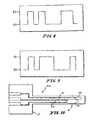
- FIG. 7is a graphic representation of a pulse sequence in which the duration of the intervals between the pulses is changed.
- FIGS. 8-9are graphic representations of measurement protocols, which combine changes in the pulse interval with changes in the pulse width.
- FIG. 10is similar to FIG. 1 and is a plan view of a sensor in accordance with this invention.
- FIG. 11is a graph showing the measured response for the Pyrrole-3-acetic acid/Mediator/glucose dehydrogenase (GDH) sensor.
- FIG. 12is a graph showing the measured response for the Pyrrole-3-carboxylic acid/mediator/GDH sensor.
- FIG. 13is a graph showing the response of the Pyrrole-3-acetic acid/Mediator/GDH and the Pyrrole-3-carboxylic acid/mediator/GDH sensors.
- the electrochemical sensor of this inventionis designed to provide signals indicative of analyte concentration in a biological fluid.
- the sensorcomprises an electrode in contact with a low volume of a hydrophilic medium for retaining an amount of analyte proportionate to the concentration of analyte in a biological fluid in contact with the sensor.
- the mediumis selected to allow facile analyte diffusion through said medium alone or upon hydration of said medium prior to or consequent to sensor use.
- the sensorcomprises an enclosure for the volume of hydrophilic medium. The enclosure is formed to expose the hydrophilic medium to the biological fluid so that analyte in the biological fluid diffuses into the hydrophilic medium until the concentration of analyte in the medium is equivalent to the concentration of analyte in the biological fluid.
- the rate of analyte mass diffusion into the retention volumeis dependent on the analyte concentration gradient.
- concentration of analyte in the retention/depletion volumemay be measured electrochemically by cooperation of an electron mediator and a redox enzyme specific for the analyte, each forming part of or being in contact with the hydrophilic medium.
- the hydrophilic mediumcan have a water concentration level less than, equal to or greater than the water content of the biological fluid.
- the components of the hydrophilic mediumincluding the enzyme and electron mediator components and a hydrophilic polymer can be used in construction of the sensor in a substantially dehydrated state, ready for rehydration prior to use or upon contact of the sensor with a biological fluid. It is important for sensor function that the analyte of interest is readily diffusible through the hydrophilic medium to enable a substantially homogeneous concentration of the analyte in the analyte retention volume, and a concentration that closely corresponds to analyte concentration in the biological fluid in contact with the sensor.
- the sensoris constructed to have an enclosure or compartment for holding the analyte retention volume of the hydrophilic medium.
- the enclosure compartmentis formed to expose the hydrophilic medium to the biological fluid when the sensor is in use.
- the enclosure compartmentis defined at least in part by a wall comprising an area of an analyte permeable membrane having a first side in contact with the hydrophilic medium and an opposite side for contact with the biological fluid when the sensor is in use.
- analyte of interest, water, and other membrane permeable components of the biological fluiddiffuses through the membrane and into the retention volume of hydrophilic medium until the concentration of analyte in the medium is proportionate to the concentration of analyte in the biological fluid contacting the analyte permeable membrane component of the sensor.
- a sensor in accordance with the present inventionmay include an analyte permeable membrane, such a membrane is not necessary to the operation of the sensor and as such is not required.
- the sensoris constructed to provide an electrode in electrical contact with the hydrophilic medium.
- the electrodeis typically formed of a conductive element such as carbon, silver, gold, platinum, palladium and the like and typically extends into or forms part of the walls of the container or chamber for the analyte retention volume of hydrophilic medium.
- the electrodeis formed of platinum and the retention volume is defined as the space overlying the electrode.
- the electrodecan be formed of a graphite powder and the retention volume is defined by the intraparticulate spaces and, in at least one embodiment, an overlying analyte permeable membrane.
- the electrodeis a component of an electrode system comprising a reference electrode and optionally an auxiliary electrode, which may be different or identical to the reference electrode.
- the electrode systemcan also comprise conductor elements for providing electrical communication between the electrode components of the system and a programmable controller to control the electrical potentials in said electrode system and to sense current flow through at least one of said electrodes responsive to said electrical potentials.
- the programmable controlleris constructed as a separate unit with electrical connectors adapted particularly for electrical communication between the controller and the electrode system of the sensor.
- the controlleris typically a hand-held or table mounted unit capable of being reversibly connected to one or more biosensors and having data storage and data display elements.
- the sensor for electrochemical analysis in accordance with this inventionfurther comprises a redox enzyme and an electron mediator in contact with the hydrophilic medium.
- the enzymeis selected for its capacity to oxidize or reduce the analyte of interest.
- the enzymeis preferably selected as well for its lack of capacity for transferring electrons to or from substances other than said analyte that are capable of diffusing from the biological fluid into the analyte retention volume.
- suitable enzymesinclude pyrroloquinoline quinone (PQQ)-dependent glucose dehydrogenase (GDH) (EC 1.1.99.17) or Hydroxybutyrate dehydrogenase (HBDH) (EC 1.1.1.30).
- PQQpyrroloquinoline quinone
- GDHpyrroloquinoline quinone
- HBDHHydroxybutyrate dehydrogenase
- Dehydrogenase enzymes for other diffusible analytesare known in the art and can be substituted dependent on the analyte of
- the electron mediatorcan be selected from any of a wide variety of electron mediators capable of facilitating transfer of electrons between the redox enzyme and a sensor electrode in contact with the medium containing or in contact with said enzyme and said mediator.
- a non-limiting example of a suitable mediatorincludes osmium (bis-bipyridyl) pyridinium chloride. It is appreciated, however, that a number of commercially available mediators, non-limiting examples of which are described in U.S. Pat. No. 5,589,326, the disclosure of which is expressly incorporated herein by reference, may be used in accordance with the present invention.
- the enzyme and electron mediatorcan be entrapped in a polymer matrix on the electrode.
- the enzyme and electron mediatorcan be selected to minimize their diffusion through an analyte permeable membrane during sensor use or can be covalently bound to the walls of the enclosure or to hydrophilic polymer components of the retention medium to minimize, if not prevent, their diffusion from the retention medium volume through the membrane and into the biological fluid during sensor use.
- hydrophilic polymerstypically having a molecular weight in excess of 5000 Daltons and having polyanionic, polycationic, or polyhydric functionality can be used as a carrier or carrier matrix forming part of the hydrophilic medium component of the sensor.
- examples of such polymersinclude cellulosic polymers such as cellulose acetate, hydroxy ethyl cellulose, polyethylene glycols, synthetic or natural gums such as guar or xanthan gums, alginic acid, poly (meth) acrylic acids and copolymers of acrylic acids and acrylic esters, glycosaminoglycans, and the like which polymers can be used.
- electrically polymerized matrices from monomerssuch as pyrrole-3-acetic acid and pyrrole-3-carboxylic acid can serve as the hydrophilic matrix and/or enzyme entrapping matrix.
- hydrophilic polymerscan be used alone or in combination to provide a hydrated or hydratable matrix through which the targeted analyte is readily diffusible. Further, such polyfunctional hydrophilic polymers can be used to “anchor” or otherwise impair the diffusion of the enzyme or electron mediator components of the medium to minimize loss of such components from the retention volume during sensor use.
- art-recognized electron mediatorshaving, for example hydroxy, carboxy or amino functionality for example ferrocene carboxylic acid, can be coupled using art-recognized ester-forming or amide-forming coupling methodologies to form the hydrophilic medium for use in preparation of the present sensors.
- Additional mediator compounds that can be tethered to a polymeric matrixinclude redox reversible imidazole-osmium complexes.
- Non-limiting examples of such complexesinclude osmium-bipyridyl conjugates such as tris(bipyridyl) osmium complex characterized by fast mediation kinetics and low redox potential (+150 mV vs. Ag/AgCl).
- Another group of osmium complex labeled, electrochemically detectable conjugatesinclude bis(bipyridyl) imidazolyl haloosmium complexes, which, like the tris(bipyridyl) osmium complexes are characterized by fast mediation kinetics and low redox potential (+150 mV vs. Ag/AgCl).
- the tris(bipyridyl) complexeshave a redox potential sufficiently different from bis(pyridyl)imidazolyl chloroosmium complexes.
- the redox enzyme and an electron mediatorcan thus be inherently non-diffusible or chemically coupled with the high molecular weight components of the medium to render the enzyme and electron mediator components substantially not capable of diffusing through the analyte permeable membrane during sensor use in contact with a biological fluid.
- the enzyme component of the sensoris typically of sufficient molecular weight that diffusional loss of that component through the semipermeable membrane is marginal over the typical period of sensor use.
- Such enzymescan be incorporated into the hydrophilic medium during device construction, as, for example, an enzyme lyophilizate formed by freeze drying a solution of enzyme in the presence of a hydrophilic monomer, for example, maltose or trehalose, or other enzyme stabilizing hydrophilic composition.
- the lyophilized enzymecan be retained in said medium during sensor manufacture and storage in a dehydrated state until rehydration prior to or during initial use of said sensor, thereby providing longer sensor shelf life.
- Electron mediator components of the sensors of the present inventionare not critical except for the fact that they should be selected or modified, for example by covalent bonding of polymer components of the hydrophilic matrix or the hydrophilic medium, to prevent or minimize diffusional loss of the electron mediator component from the analyte retention volume medium during the course of sensor use.
- the prior artis replete with reference to a wide variety of compounds including metal chelates and other metal complexes such as ferrocene, and more particularly carboxy ferrocene that can be readily coupled covalently to non-diffusible components of the hydrophilic medium.
- the membrane components of the sensor constructs of this inventioncan be any biocompatible analyte permeable membrane, including for example cellulose acetate, polyurethane, and polycarbonate.
- Other polymeric biocompatible membranes suitable for use in biosensor constructionare also well known in the art and any of such art-recognized analyte permeable membrane/membrane materials may be used in manufacture of the present sensors.
- Example of analyte permeable membranes, and as well electron mediators and redox enzymesare describe in U.S. Pat. No. 5,264,105, the specification of which is expressly incorporated herein by reference.
- the hydrophilic matrixis bound on the surface of at least one of the electrodes of the electrode system.
- the hydrophilic matrixcomprises an electron mediator covalently bound to a non-diffusible or poorly diffusible hydrophilic polymer component of the matrix.
- the redox enzymeis bound to a polymer component of the hydrophilic matrix as well.
- a sensor of the present inventionpreferably already includes the electron mediator and redox enzyme components, which are exposed to analyte present in the retention volume of the sensor. Initially at predetermined intervals a potential is applied intermittently to the electrode system sufficient to oxidize the electron mediator and the current flow through the electrode is sensed as a function of the duration of the applied potential.
- the applied mediator oxidizing potentialis maintained at least for a period of time sufficient to determine the rate of change of current through the electrode as a function of duration of the applied potential.
- Values for the sensed currentare correlated with values of current flow for known concentrations of analyte.
- the hydrophilic mediumcomprises either a polymeric electron mediator or an electron mediator covalently bound to a non-diffusible or poorly diffusible hydrophilic polymer component of the medium.
- the redox enzymeis included as a stabilized lyophilizate or is covalently bound itself to a polymer component of the hydrophilic medium.
- the hydrophilic mediumalso comprises polyfunctional components that can be reacted with difunctional crosslinking agents contacted with the surface of said hydrophilic medium to form in situ an analyte permeable membrane on the surface of the hydrophilic medium.
- a polyhydric polymer or a di- or trihydric, preferably high molecular weight, monomer component of the hydrophilic mediumcan be reacted, for example, with a polyisocyanate, for example a diisocyanate in the vapor phase, to form a polymer skin or membrane on the surface of the hydrophilic medium.
- a polyisocyanatefor example a diisocyanate in the vapor phase
- the permeability of the membranecan be controlled by the length of exposure of the polyhydric medium surface to the multifunctional crosslinker.
- 1,4-benzene diisocyanatecan be vaporized in a chamber.
- Sensor constructscomprising the hydrophilic medium, preferably already including the electron mediator and redox enzyme components, having an exposed surface is introduced into the chamber for a period of time sufficient to form a biocompatible membrane on the surface of the hydrophilic medium to define in conjunction with other sensor components for example, a simple planar non-conductive substrate, the enclosure for the analyte retention volume component of the sensor.
- a sensor 10utilizing a thin-film gold electrode 12 on an inert substrate 14 .
- a sensing portion 16includes gold electrode 12 on a surface 13 of inert substrate 14 , spacer layers 18 and a diffusion-limiting membrane 20 having small or no pores.
- Membrane 20can be formed as a separate sheet and applied to spacer layers 18 resting on gold electrode 12 and inert substrate 14 to complete definition of an enclosure for a depletion volume 22 filled with a hydrophilic medium 24 .
- the enzyme and mediatormay be immobilized, for example by entrapment in a hydrophilic matrix on the gold electrode 12 .
- the enzyme and mediatormay be immobilized by covalent bonding to a wall 26 of enclosure 22 or to overlying membrane 20 or such components can be freely diffusing and selected to have minimal membrane permeability.
- Diffusion-limiting membrane 20can be selected from art-recognized analyte permeable membranes and adhered to a surface 28 of spacer layer 18 to complete enclosure 22 for the analyte retention/depletion volume.
- the analyte permeable membranecan be formed in situ by cross-linking polyfunctional hydrophilic polymer and/or monomer components of the enzyme/mediator reagent layer.
- sensor 10may be formed without membrane 20 . It is understood that as used throughout the disclosure, that like reference numerals are used to denote like components.
- sensor 110is provided in accordance with this invention.
- Sensor 110includes an electrode formed of a porous/particulate carbon layer wherein the depletion volume is defined by the interstitial spaces between carbon/graphite particles (not shown).
- a carbon-enzyme-mediator reagent layer 122can be formed, for example by screen printing, a carbon/graphite suspension comprising a redox enzyme, and a nondiffusible or poorly diffusible, for example a polymeric, electron mediator in combination with one or more optionally non-electron mediating, polyfunctional polymers.
- the suspensionis typically deposited on a conductor element (not shown) on an inert substrate.
- the diffusion-limiting analyte permeable membrane 20can be applied, similar to that mentioned above, as a preformed polymer sheet applied and sealed over a surface 123 of the carbon electrode or membrane 20 can be formed by coating the exposed printed electrode having an intraparticulate polyfunctional polymer matrix with a polymer in solution. See, for example, International Patent Application No. WO 98/17995 for non-limiting examples of polymer membranes for coating biosensors.
- sensor 210 of the present inventionincludes a diffusion-limiting membrane 220 that is formed to have large pores 223 for enhanced glucose diffusion.
- the mediator and enzyme componentsare preferably immobilized by entrapment in a hydrophilic matrix on electrode 12 , by covalent bonding to nondiffusible polymeric components of hydrophilic medium 24 , or to surfaces 26 of sensor enclosure 22 for the retention volume.
- Sensor 310is similar to that illustrated in FIGS. 1 and 4 with the exception that membrane 320 overlying hydrophilic medium 24 is nonpermeable itself. Membrane 320 is provided with peripheral porosity allowing diffusion of glucose or other analytes into the retention/depletion volume.
- the biochemical sensor of this inventioncan be constructed in any form adapted for the intended use.
- sensors intended for repeated laboratory usecan be constructed in the form of an elongated probe having the sensor element itself located at one end and electrical conductors connecting the electrode component of the sensing element to points of electrical attachment of the probe sensor to a programmable sensor controller.
- the present electrochemical sensorcan be constructed using art recognized micro scale manufacturing techniques to produce needle-like sensors capable of being implanted or injected into a site for indwelling sensor applications.
- the electrochemical sensor of this inventioncan be utilized for monitoring analyte concentrations in biological fluids.
- the methodcomprises the steps of contacting the biological fluid with the sensor and at initially predetermined intervals intermittently applying a potential to the electrode sufficient to oxidize the electron mediator.
- the current passing through the electrodeis then sensed as a function of the duration of the applied potential.
- the applied mediator-oxidizing potentialis maintained for at least a period of time sufficient to determine the rate of change of current with time through said electrode.
- the sensed current flowis then correlated with current flow for known concentrations of said analyte in the retention/detection medium.
- the sensorcan be constructed to comprise at least a working electrode and a reference electrode and optionally an auxiliary electrode, which may be identical with the referenced electrode.
- a potentialis applied sufficient to establish a predetermined level of current flow between the working electrode and the auxiliary electrode. And the potential difference between the working and reference electrodes necessary to establish said level of current flow is measured and maintained for a period of time sufficient to determine the rate of change of potential necessary to maintain said current flow through said electrode. The potential measurements are then correlated with potential measurements recorded for known concentrations of analyte in the biological fluid.
- the applied potentials, the duration of the potential pulses, and the intervals between potential pulsesare entered into a programmable controller used in conjunction with the sensor for analyte measurements.
- the intervals between the intermittent applied potentialsare less than the time necessary for the concentration of the analyte in the retention volume to equilibrate with that in the biological fluid in contact with the analyte permeable membrane.
- the intervalsare increased incrementally for a series of applied potentials, and the concentration of the analyte and the biological fluid is determined as a function of the rate of increase in analyte concentration in the retention volume.
- the intervals between applied potential pulsescan be modified based on previous measurements to adjust for variations in sensor performance deriving from loss or degradation of redox enzyme and electron mediator components and/or change in diffusion characteristics of either the analyte permeable membrane or the retention volume medium.
- the duration of the applied potential pulseis modified based on previous measurements of sensed sensor performance status.
- the intervals between measurementsis substantially equal to or greater than the time required for the analyte concentration in the retention volume to equilibrate with that in the biological fluid.
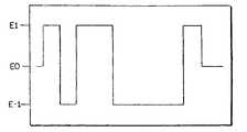
- FIGS. 6-9present graphic illustrations of sample measurement algorithms wherein the potential of the electrode is controlled over a period of time to vary between a potential at which no oxidation of mediator occurs (E 0 ) to a mediator oxidizing potential (E1) and a potential at which reduction of the mediator takes place (E ⁇ 1).
- E 0a potential at which no oxidation of mediator occurs
- E1mediator oxidizing potential
- E ⁇ 1a potential at which reduction of the mediator takes place
- FIG. 6shows a pulse sequence in which two pulses of oxidative potential of different duration are applied to the sensor, interspersed with recovery intervals with reducing potential.
- FIG. 7shows a sequence in which the duration of the intervals between the pulses is changed.
- FIGS. 8-9demonstrate measurement protocols, which combine changes in the pulse interval with changes in the pulse width.
- the controllercan select from a variety of sequences and durations of amperometric measurement intervals and recovery intervals to determine not only the analyte concentration, but also probe the condition of the sensor for enzyme activity, diffusion of substrate and mediator within the sensor, and diffusion of substrate into the sensor.
- controllerused with the present electrochemical sensor is not critical provided that the controller can be programmed to apply the appropriate potential pulse profiles to the sensor electrode or electrode system and to sense the current flow as a function of potential in time.
- a separate sensor status assessment protocolcan be implemented in which the controller implements a protocol that allows computation of diffusion characteristics of the depletion volume and membrane from current data.
- the controllercan be programmed to adjust measurement intervals and pulse widths based on computed diffusion times and determined substrate concentration. Both chronoamperometry and chronocolometry may be used to determine sensor status and substrate concentration.
- the controllershould also be capable of applying a reducing potential to the electrode to reduce the mediator and thereby decrease consumption of analyte between measurements.
- a sensor in accordance with the present inventionis formed similarly to sensor 10 of FIGS. 1-2 , except that it does not include membrane 20 .
- the enzyme and electron mediatoris entrapped in a polymer matrix on the electrode.
- This polymer matrixwas prepared by electropolymerization of pyrrole and pyrrole-mediator derivatives. This method entrapped the enzyme in a polymer matrix on the electrode, and, by incorporating mediator-derivatized pyrrole into the polymer, provided for an immobilized mediator for transfer of electrons within the sensor.
- Platinum disc electrodeswere suitable for preparing sensors in accordance with this method.
- a mediator which was suitable for copolymerization in a matrixwas be prepared by the following reaction sequence:
- the platinum disc electrodeswere polished with Al 2 O 3 -paste of decreasing roughness (3 ⁇ m, 1 ⁇ m, 0.1 ⁇ m) on a polishing cloth and were washed with distilled water. Next, the electrodes were cleaned in an ultrasonic bath first in 10 M NaOH and then in 5 M H 2 SO 4 —ten minutes each.
- the electrodesunderwent electrochemical cycling in oxygen-free 0.5 M H 2 SO 4 :
- the last stepwas the polarization at ⁇ 210 mV for five minutes.
- a pyrrole filmwas polymerized to contain active Osmium (bis-bipyridyl) pyridinium Chloride as follows:
- Co-polymerizationwas carried out with a mixture of 2 mM pyrrole, 8 mM (9) and 25 mM Tetramethyl ammonium perchlorate (as electrolyte) in 1:1 mixture of acetonitrile: water. 100 Pulses with potential/time of 0 V for 5 sec/1.5 V 1 sec. were carried out. The polymerization was carried out in the absence of oxygen.
- Enzyme electrodeswere washed with deionized water. It is appreciated, however, that the enzyme electrodes may be washed with a buffer solution or, if necessary, with 3M KCl solution to remove adsorbed GDH and afterwards stored in 0.1 M phosphate buffer pH 7. If electrodes are stored over-night they are kept at 4° C. otherwise at RT.
- Sensor responsewas measured by placing the sensor into a stirred PBS (Phosphate buffered saline) buffer along with a reference electrode and a counter electrode.
- a potential (E) sufficient to oxidize the immobilized mediatorwas applied, and the solution stirred for a time (t) until the current (I) stabilized to a low value. See FIG. 11 .
- Aliquots of 1 M glucose solution in PBSwere added in a step-wise manner, as shown by arrows in FIG. 12 , to the PBS buffer to increase the concentration of glucose in the stirred PBS buffer solution, and the current (I) was measured on the plateau region of the response curve.
- the responsewas measured for the Pyrrole-3-acetic acid/Mediator/GDH and pyrrole-3-carboxylic acid/mediator/GDH sensors.
- the responseshows good electron transfer through the immobilized mediator in the polypyrrole film.
- a sensor 410consists of conductors 12 , 15 and reagents deposited on a flat polymeric substrate 14 . Materials for encapsulation of the conductors 12 , 15 are provided, and reagents that form a semipermeable biocompatible layer over the reagent-containing sensing area 16 . See FIG. 10 .
- This method for the fabrication of sensors suitable for use according to the current inventionutilizes the so-called “wired-enzyme” technology for the immobilization of enzyme and mediator in active relationship within a sensor film.
- the GDH/PQQsubstantially does not react with any endogenous substrate other than the analyte of interest. This allows the sensor to remain inactive, or “off” so long as the electrode is not regenerating the mediator to allow further activity. Sensors produced by this method demonstrate very high sensitivity to glucose and very high current density.
Landscapes
- Life Sciences & Earth Sciences (AREA)
- Health & Medical Sciences (AREA)
- Chemical & Material Sciences (AREA)
- Organic Chemistry (AREA)
- Engineering & Computer Science (AREA)
- Physics & Mathematics (AREA)
- Zoology (AREA)
- Wood Science & Technology (AREA)
- Proteomics, Peptides & Aminoacids (AREA)
- General Health & Medical Sciences (AREA)
- Molecular Biology (AREA)
- Biophysics (AREA)
- Bioinformatics & Cheminformatics (AREA)
- Microbiology (AREA)
- Genetics & Genomics (AREA)
- General Engineering & Computer Science (AREA)
- Biochemistry (AREA)
- Biotechnology (AREA)
- Immunology (AREA)
- Analytical Chemistry (AREA)
- Veterinary Medicine (AREA)
- Pathology (AREA)
- Medical Informatics (AREA)
- Heart & Thoracic Surgery (AREA)
- Public Health (AREA)
- Emergency Medicine (AREA)
- Surgery (AREA)
- Optics & Photonics (AREA)
- Biomedical Technology (AREA)
- Animal Behavior & Ethology (AREA)
- Chemical Kinetics & Catalysis (AREA)
- General Chemical & Material Sciences (AREA)
- Apparatus Associated With Microorganisms And Enzymes (AREA)
- Measuring Or Testing Involving Enzymes Or Micro-Organisms (AREA)
Abstract
Description


- Substrate: Polyimid (such as Kapton® polyimide film, which is commercially available from E.I. DuPont de Nemours, Wilmington, Del., and Upilex® polimide film which is commercially available from UBE Industries Ltd, Japan) 0.005″ (0.127 mm) thick with gold electrodes and conducting tracks.
- Processing: Material is washed with water, acetone, and methylene chloride, then dried at 180° C. for 20 hours. Material is placed in a vacuum chamber and metalized with 50 Å Chromium followed by 500 Å Gold. Metalized material is removed. A laminated photoresist is applied. The resist is exposed, and developed in an aqueous salt solution. Then the metal pattern is developed in a metal etchent solution (typically HNO3
— HCl). The patterned material is rinsed with water, and the remaining photomask is removed with a solvent (typically N-Methylpyrrolidone (NMP). A photodefinable polyimidlayer 2 um thick is applied with a spin coater, and solidified by baking at 80° C. for 20 minutes. It is exposed and then developed with a solvent (NMP). It is then baked at 200° C. for 30 minutes to harden the remaining polymer. - Reference/Counter: A silver/silver chloride ink preparation consisting of suspension of silver particles, partially converted to silver chloride on their surfaces, in an organic solvent, i.e. cyclohexanone, with a polymeric suspending agent, such as alginate.
- Processing: The ink mixture is applied to the opening in the polyimid and dried for 30 minutes at 80° C. to remove all solvent.
- Sensor Area: The opening through the polyimid to the gold electrode is covered with a multilayer reagent area consisting of the following reagents.
- Active Enzyme Layer: A solution of Glucose Dehydrogenase/pyrroloquinoline quinone (PQQ) (Enzyme Commission No. 1.1.99.17) and a redox polymer consisting of polyvinylimidazole with bis (bipyridyl) chloro-Osmium in phosphate buffer, optionally with a polyhydric synthetic, natural or semisynthetic polymer, is applied to one electrode opening and dried. A solution of polyethylene glycol diglycidyl ether in aqueous buffer (i.e. 10 mM NaPO4, 150 mM NaCl) is applied to the area, and allowed to react for 10 minutes. The electrodes are then rinsed in physiological saline solution and allowed to dry.
- Biocompatibility Layer: The entire structure is placed in a vacuum chamber. A plasma of diglyme is created in the chamber by introducing a low pressure vapor, and applying an RF field to dissociate the diglyme and cause polymerization. After 5 minutes, the diglyme addition is stopped, and the plasma continued for a further 5 minutes. Then the vacuum is broken and the sheet removed from the chamber.
- Post-Processing: The
sensors 410 are punched from the sheet and mounted into small polycarbonate holders which protect the sensing area and serve as the insertion device. The packaged sensors are sealed into polyethylene bags. The bagged sensors are radiation-sterilized. Ten sterilized sensors are packed into a larger plastic box containing desiccant in the lid. The box is closed and packed into a cardboard outer pack that serves as the final consumer package.
Claims (27)
Priority Applications (1)
| Application Number | Priority Date | Filing Date | Title |
|---|---|---|---|
| US11/038,970US7731835B2 (en) | 1999-09-20 | 2005-01-19 | Electrochemical sensor and method for continuous analyte monitoring |
Applications Claiming Priority (4)
| Application Number | Priority Date | Filing Date | Title |
|---|---|---|---|
| US15473199P | 1999-09-20 | 1999-09-20 | |
| PCT/US2000/025631WO2001021827A1 (en) | 1999-09-20 | 2000-09-19 | Small volume biosensor for continuous analyte monitoring |
| US10/069,308US7045054B1 (en) | 1999-09-20 | 2000-09-19 | Small volume biosensor for continuous analyte monitoring |
| US11/038,970US7731835B2 (en) | 1999-09-20 | 2005-01-19 | Electrochemical sensor and method for continuous analyte monitoring |
Related Parent Applications (3)
| Application Number | Title | Priority Date | Filing Date |
|---|---|---|---|
| PCT/US2000/025631ContinuationWO2001021827A1 (en) | 1999-09-20 | 2000-09-19 | Small volume biosensor for continuous analyte monitoring |
| US10069308Continuation | 2000-09-19 | ||
| US10/069,308ContinuationUS7045054B1 (en) | 1999-09-20 | 2000-09-19 | Small volume biosensor for continuous analyte monitoring |
Publications (2)
| Publication Number | Publication Date |
|---|---|
| US20050211572A1 US20050211572A1 (en) | 2005-09-29 |
| US7731835B2true US7731835B2 (en) | 2010-06-08 |
Family
ID=36318060
Family Applications (2)
| Application Number | Title | Priority Date | Filing Date |
|---|---|---|---|
| US10/069,308Expired - Fee RelatedUS7045054B1 (en) | 1999-09-20 | 2000-09-19 | Small volume biosensor for continuous analyte monitoring |
| US11/038,970Expired - Fee RelatedUS7731835B2 (en) | 1999-09-20 | 2005-01-19 | Electrochemical sensor and method for continuous analyte monitoring |
Family Applications Before (1)
| Application Number | Title | Priority Date | Filing Date |
|---|---|---|---|
| US10/069,308Expired - Fee RelatedUS7045054B1 (en) | 1999-09-20 | 2000-09-19 | Small volume biosensor for continuous analyte monitoring |
Country Status (1)
| Country | Link |
|---|---|
| US (2) | US7045054B1 (en) |
Cited By (17)
| Publication number | Priority date | Publication date | Assignee | Title |
|---|---|---|---|---|
| US20080302660A1 (en)* | 2007-06-07 | 2008-12-11 | Kahn Carolyn R | Silicon Electrochemical Sensors |
| US8758584B2 (en) | 2010-12-16 | 2014-06-24 | Sensor Innovations, Inc. | Electrochemical sensors |
| WO2014140172A1 (en) | 2013-03-15 | 2014-09-18 | Roche Diagnostics Gmbh | Methods of failsafing electrochemical measurements of an analyte as well as devices, apparatuses and systems incorporating the same |
| WO2014140164A1 (en) | 2013-03-15 | 2014-09-18 | Roche Diagnostics Gmbh | Methods of using information from recovery pulses in electrochemical analyte measurements as well as devices, apparatuses and systems incorporating the same |
| WO2014140177A2 (en) | 2013-03-15 | 2014-09-18 | Roche Diagnostics Gmbh | Methods of detecting high antioxidant levels during electrochemical measurements and failsafing an analyte concentration therefrom as well as devices, apparatuses and systems incorporting the same |
| WO2014140170A1 (en) | 2013-03-15 | 2014-09-18 | Roche Diagnostics Gmbh | Methods of scaling data used to construct biosensor algorithms as well as devices, apparatuses and systems incorporating the same |
| US8858884B2 (en) | 2013-03-15 | 2014-10-14 | American Sterilizer Company | Coupled enzyme-based method for electronic monitoring of biological indicator |
| US9121050B2 (en) | 2013-03-15 | 2015-09-01 | American Sterilizer Company | Non-enzyme based detection method for electronic monitoring of biological indicator |
| US9551680B2 (en) | 2013-06-28 | 2017-01-24 | Verily Life Sciences Llc | Chemically reactive enzyme immobilization |
| US9617578B2 (en) | 2013-12-06 | 2017-04-11 | Verily Life Sciences Llc | Sensor membrane with low temperature coefficient |
| US9750445B2 (en) | 2013-06-28 | 2017-09-05 | Verily Life Sciences Llc | Porous polymeric formulation prepared using porogens |
| WO2018067235A1 (en) | 2016-10-05 | 2018-04-12 | Roche Diabetes Care, Inc. | Detection reagents and electrode arrangements for multi-analyte diagnostic test elements, as well as methods of using the same |
| US10190100B1 (en) | 2015-12-28 | 2019-01-29 | Verily Life Sciences Llc | Chemical modification of glucose oxidase and its application to biosensors |
| US20200100711A1 (en)* | 2018-10-01 | 2020-04-02 | The Florida International University Board Of Trustees | Wound monitoring sensors and use thereof |
| US10926523B2 (en)* | 2018-06-19 | 2021-02-23 | Sensel, Inc. | Performance enhancement of sensors through surface processing |
| EP4033235A1 (en) | 2014-11-03 | 2022-07-27 | Roche Diabetes Care GmbH | Methods of use of electrode arrangements for electrochemical test elements |
| US12345672B2 (en) | 2016-10-24 | 2025-07-01 | Roche Diabetes Care, Inc. | Methods of correcting for uncompensated resistances in the conductive elements of biosensors, as well as devices and systems incorporating the same |
Families Citing this family (113)
| Publication number | Priority date | Publication date | Assignee | Title |
|---|---|---|---|---|
| US6036924A (en) | 1997-12-04 | 2000-03-14 | Hewlett-Packard Company | Cassette of lancet cartridges for sampling blood |
| US6391005B1 (en) | 1998-03-30 | 2002-05-21 | Agilent Technologies, Inc. | Apparatus and method for penetration with shaft having a sensor for sensing penetration depth |
| US8480580B2 (en) | 1998-04-30 | 2013-07-09 | Abbott Diabetes Care Inc. | Analyte monitoring device and methods of use |
| US6175752B1 (en) | 1998-04-30 | 2001-01-16 | Therasense, Inc. | Analyte monitoring device and methods of use |
| US8346337B2 (en) | 1998-04-30 | 2013-01-01 | Abbott Diabetes Care Inc. | Analyte monitoring device and methods of use |
| US6949816B2 (en) | 2003-04-21 | 2005-09-27 | Motorola, Inc. | Semiconductor component having first surface area for electrically coupling to a semiconductor chip and second surface area for electrically coupling to a substrate, and method of manufacturing same |
| US8465425B2 (en) | 1998-04-30 | 2013-06-18 | Abbott Diabetes Care Inc. | Analyte monitoring device and methods of use |
| US8688188B2 (en) | 1998-04-30 | 2014-04-01 | Abbott Diabetes Care Inc. | Analyte monitoring device and methods of use |
| US8974386B2 (en) | 1998-04-30 | 2015-03-10 | Abbott Diabetes Care Inc. | Analyte monitoring device and methods of use |
| US9066695B2 (en) | 1998-04-30 | 2015-06-30 | Abbott Diabetes Care Inc. | Analyte monitoring device and methods of use |
| DE10057832C1 (en) | 2000-11-21 | 2002-02-21 | Hartmann Paul Ag | Blood analysis device has syringe mounted in casing, annular mounting carrying needles mounted behind test strip and being swiveled so that needle can be pushed through strip and aperture in casing to take blood sample |
| US8641644B2 (en) | 2000-11-21 | 2014-02-04 | Sanofi-Aventis Deutschland Gmbh | Blood testing apparatus having a rotatable cartridge with multiple lancing elements and testing means |
| US6560471B1 (en) | 2001-01-02 | 2003-05-06 | Therasense, Inc. | Analyte monitoring device and methods of use |
| JP4272051B2 (en) | 2001-06-12 | 2009-06-03 | ペリカン テクノロジーズ インコーポレイテッド | Blood sampling apparatus and method |
| JP4209767B2 (en) | 2001-06-12 | 2009-01-14 | ペリカン テクノロジーズ インコーポレイテッド | Self-optimized cutting instrument with adaptive means for temporary changes in skin properties |
| US9427532B2 (en) | 2001-06-12 | 2016-08-30 | Sanofi-Aventis Deutschland Gmbh | Tissue penetration device |
| US9795747B2 (en) | 2010-06-02 | 2017-10-24 | Sanofi-Aventis Deutschland Gmbh | Methods and apparatus for lancet actuation |
| US7344507B2 (en) | 2002-04-19 | 2008-03-18 | Pelikan Technologies, Inc. | Method and apparatus for lancet actuation |
| US7041068B2 (en) | 2001-06-12 | 2006-05-09 | Pelikan Technologies, Inc. | Sampling module device and method |
| US9226699B2 (en) | 2002-04-19 | 2016-01-05 | Sanofi-Aventis Deutschland Gmbh | Body fluid sampling module with a continuous compression tissue interface surface |
| AU2002344825A1 (en) | 2001-06-12 | 2002-12-23 | Pelikan Technologies, Inc. | Method and apparatus for improving success rate of blood yield from a fingerstick |
| US8337419B2 (en) | 2002-04-19 | 2012-12-25 | Sanofi-Aventis Deutschland Gmbh | Tissue penetration device |
| US7749174B2 (en) | 2001-06-12 | 2010-07-06 | Pelikan Technologies, Inc. | Method and apparatus for lancet launching device intergrated onto a blood-sampling cartridge |
| US7981056B2 (en) | 2002-04-19 | 2011-07-19 | Pelikan Technologies, Inc. | Methods and apparatus for lancet actuation |
| WO2002101359A2 (en) | 2001-06-12 | 2002-12-19 | Pelikan Technologies, Inc. | Integrated blood sampling analysis system with multi-use sampling module |
| EP1395185B1 (en) | 2001-06-12 | 2010-10-27 | Pelikan Technologies Inc. | Electric lancet actuator |
| US7344894B2 (en) | 2001-10-16 | 2008-03-18 | Agilent Technologies, Inc. | Thermal regulation of fluidic samples within a diagnostic cartridge |
| US8152991B2 (en)* | 2005-10-27 | 2012-04-10 | Nanomix, Inc. | Ammonia nanosensors, and environmental control system |
| US8154093B2 (en) | 2002-01-16 | 2012-04-10 | Nanomix, Inc. | Nano-electronic sensors for chemical and biological analytes, including capacitance and bio-membrane devices |
| US7956525B2 (en) | 2003-05-16 | 2011-06-07 | Nanomix, Inc. | Flexible nanostructure electronic devices |
| US7229458B2 (en) | 2002-04-19 | 2007-06-12 | Pelikan Technologies, Inc. | Method and apparatus for penetrating tissue |
| US7892183B2 (en) | 2002-04-19 | 2011-02-22 | Pelikan Technologies, Inc. | Method and apparatus for body fluid sampling and analyte sensing |
| US9314194B2 (en) | 2002-04-19 | 2016-04-19 | Sanofi-Aventis Deutschland Gmbh | Tissue penetration device |
| US8579831B2 (en) | 2002-04-19 | 2013-11-12 | Sanofi-Aventis Deutschland Gmbh | Method and apparatus for penetrating tissue |
| US8267870B2 (en) | 2002-04-19 | 2012-09-18 | Sanofi-Aventis Deutschland Gmbh | Method and apparatus for body fluid sampling with hybrid actuation |
| US7717863B2 (en) | 2002-04-19 | 2010-05-18 | Pelikan Technologies, Inc. | Method and apparatus for penetrating tissue |
| US8784335B2 (en) | 2002-04-19 | 2014-07-22 | Sanofi-Aventis Deutschland Gmbh | Body fluid sampling device with a capacitive sensor |
| US7582099B2 (en) | 2002-04-19 | 2009-09-01 | Pelikan Technologies, Inc | Method and apparatus for penetrating tissue |
| US7648468B2 (en) | 2002-04-19 | 2010-01-19 | Pelikon Technologies, Inc. | Method and apparatus for penetrating tissue |
| US9248267B2 (en) | 2002-04-19 | 2016-02-02 | Sanofi-Aventis Deustchland Gmbh | Tissue penetration device |
| US7481776B2 (en) | 2002-04-19 | 2009-01-27 | Pelikan Technologies, Inc. | Method and apparatus for penetrating tissue |
| US7331931B2 (en) | 2002-04-19 | 2008-02-19 | Pelikan Technologies, Inc. | Method and apparatus for penetrating tissue |
| US7674232B2 (en) | 2002-04-19 | 2010-03-09 | Pelikan Technologies, Inc. | Method and apparatus for penetrating tissue |
| US7976476B2 (en) | 2002-04-19 | 2011-07-12 | Pelikan Technologies, Inc. | Device and method for variable speed lancet |
| US7297122B2 (en) | 2002-04-19 | 2007-11-20 | Pelikan Technologies, Inc. | Method and apparatus for penetrating tissue |
| US7524293B2 (en) | 2002-04-19 | 2009-04-28 | Pelikan Technologies, Inc. | Method and apparatus for penetrating tissue |
| US8221334B2 (en) | 2002-04-19 | 2012-07-17 | Sanofi-Aventis Deutschland Gmbh | Method and apparatus for penetrating tissue |
| US7563232B2 (en) | 2002-04-19 | 2009-07-21 | Pelikan Technologies, Inc. | Method and apparatus for penetrating tissue |
| US7547287B2 (en) | 2002-04-19 | 2009-06-16 | Pelikan Technologies, Inc. | Method and apparatus for penetrating tissue |
| US7708701B2 (en) | 2002-04-19 | 2010-05-04 | Pelikan Technologies, Inc. | Method and apparatus for a multi-use body fluid sampling device |
| US9795334B2 (en) | 2002-04-19 | 2017-10-24 | Sanofi-Aventis Deutschland Gmbh | Method and apparatus for penetrating tissue |
| US7901362B2 (en) | 2002-04-19 | 2011-03-08 | Pelikan Technologies, Inc. | Method and apparatus for penetrating tissue |
| US7232451B2 (en) | 2002-04-19 | 2007-06-19 | Pelikan Technologies, Inc. | Method and apparatus for penetrating tissue |
| US7374544B2 (en) | 2002-04-19 | 2008-05-20 | Pelikan Technologies, Inc. | Method and apparatus for penetrating tissue |
| US7291117B2 (en) | 2002-04-19 | 2007-11-06 | Pelikan Technologies, Inc. | Method and apparatus for penetrating tissue |
| US7491178B2 (en) | 2002-04-19 | 2009-02-17 | Pelikan Technologies, Inc. | Method and apparatus for penetrating tissue |
| US7909778B2 (en) | 2002-04-19 | 2011-03-22 | Pelikan Technologies, Inc. | Method and apparatus for penetrating tissue |
| US7141058B2 (en) | 2002-04-19 | 2006-11-28 | Pelikan Technologies, Inc. | Method and apparatus for a body fluid sampling device using illumination |
| US7371247B2 (en) | 2002-04-19 | 2008-05-13 | Pelikan Technologies, Inc | Method and apparatus for penetrating tissue |
| US7410468B2 (en) | 2002-04-19 | 2008-08-12 | Pelikan Technologies, Inc. | Method and apparatus for penetrating tissue |
| US8702624B2 (en) | 2006-09-29 | 2014-04-22 | Sanofi-Aventis Deutschland Gmbh | Analyte measurement device with a single shot actuator |
| US7948041B2 (en) | 2005-05-19 | 2011-05-24 | Nanomix, Inc. | Sensor having a thin-film inhibition layer |
| US8574895B2 (en) | 2002-12-30 | 2013-11-05 | Sanofi-Aventis Deutschland Gmbh | Method and apparatus using optical techniques to measure analyte levels |
| US7850621B2 (en) | 2003-06-06 | 2010-12-14 | Pelikan Technologies, Inc. | Method and apparatus for body fluid sampling and analyte sensing |
| WO2006001797A1 (en) | 2004-06-14 | 2006-01-05 | Pelikan Technologies, Inc. | Low pain penetrating |
| EP1635700B1 (en) | 2003-06-13 | 2016-03-09 | Sanofi-Aventis Deutschland GmbH | Apparatus for a point of care device |
| US8282576B2 (en) | 2003-09-29 | 2012-10-09 | Sanofi-Aventis Deutschland Gmbh | Method and apparatus for an improved sample capture device |
| EP1680014A4 (en) | 2003-10-14 | 2009-01-21 | Pelikan Technologies Inc | METHOD AND DEVICE FOR A VARIABLE USER INTERFACE |
| US7822454B1 (en) | 2005-01-03 | 2010-10-26 | Pelikan Technologies, Inc. | Fluid sampling device with improved analyte detecting member configuration |
| US8668656B2 (en) | 2003-12-31 | 2014-03-11 | Sanofi-Aventis Deutschland Gmbh | Method and apparatus for improving fluidic flow and sample capture |
| US8628690B2 (en)* | 2004-02-23 | 2014-01-14 | The Texas A&M University System | Nanoemulsion compositions and methods of use thereof |
| US7780873B2 (en)* | 2004-02-23 | 2010-08-24 | Texas A&M University System | Bioactive complexes compositions and methods of use thereof |
| WO2006011062A2 (en) | 2004-05-20 | 2006-02-02 | Albatros Technologies Gmbh & Co. Kg | Printable hydrogel for biosensors |
| JP5215661B2 (en)* | 2004-05-21 | 2013-06-19 | アガマトリックス インコーポレーテッド | Electrochemical cell and method for making an electrochemical cell |
| WO2005120365A1 (en) | 2004-06-03 | 2005-12-22 | Pelikan Technologies, Inc. | Method and apparatus for a fluid sampling device |
| US9636450B2 (en) | 2007-02-19 | 2017-05-02 | Udo Hoss | Pump system modular components for delivering medication and analyte sensing at seperate insertion sites |
| US8652831B2 (en) | 2004-12-30 | 2014-02-18 | Sanofi-Aventis Deutschland Gmbh | Method and apparatus for analyte measurement test time |
| CN101454667B (en) | 2005-12-27 | 2013-04-24 | 拜尔保健有限公司 | Electrochemical sensor system using a substrate with at least one well and method of manufacturing the same |
| US7826879B2 (en) | 2006-02-28 | 2010-11-02 | Abbott Diabetes Care Inc. | Analyte sensors and methods of use |
| US20100202966A1 (en)* | 2006-03-28 | 2010-08-12 | Yossi Gross | Implantable Sensor |
| WO2007147475A1 (en)* | 2006-06-19 | 2007-12-27 | Roche Diagnostics Gmbh | Amperometric sensor and method for its manufacturing |
| US9700252B2 (en) | 2006-06-19 | 2017-07-11 | Roche Diabetes Care, Inc. | Amperometric sensor and method for its manufacturing |
| ES2533722T3 (en)* | 2007-05-30 | 2015-04-14 | Bayer Healthcare Llc | Multilayer pad and procedures for its use |
| US7972866B2 (en)* | 2007-06-18 | 2011-07-05 | Nipro Diagnostics, Inc. | Biosensor and ultrasonic method of making a biosensor |
| US20100268043A1 (en)* | 2007-11-07 | 2010-10-21 | Ofer Yodfat | Device and Method for Preventing Diabetic Complications |
| US8320983B2 (en) | 2007-12-17 | 2012-11-27 | Palo Alto Research Center Incorporated | Controlling transfer of objects affecting optical characteristics |
| EP2265324B1 (en) | 2008-04-11 | 2015-01-28 | Sanofi-Aventis Deutschland GmbH | Integrated analyte measurement system |
| US8876755B2 (en) | 2008-07-14 | 2014-11-04 | Abbott Diabetes Care Inc. | Closed loop control system interface and methods |
| WO2010027771A1 (en)* | 2008-08-27 | 2010-03-11 | Edwards Lifesciences Corporation | Analyte sensor |
| US8986208B2 (en)* | 2008-09-30 | 2015-03-24 | Abbott Diabetes Care Inc. | Analyte sensor sensitivity attenuation mitigation |
| US20100160749A1 (en)* | 2008-12-24 | 2010-06-24 | Glusense Ltd. | Implantable optical glucose sensing |
| EP2378954A4 (en)* | 2008-12-24 | 2013-05-15 | Glusense Ltd | Implantable optical glucose sensing |
| GB0900421D0 (en)* | 2009-01-12 | 2009-02-11 | Whatman Internat Ltd | Sample holder for use in biological testing |
| US9375169B2 (en) | 2009-01-30 | 2016-06-28 | Sanofi-Aventis Deutschland Gmbh | Cam drive for managing disposable penetrating member actions with a single motor and motor and control system |
| LT3912551T (en) | 2009-02-26 | 2023-12-11 | Abbott Diabetes Care, Inc. | Method of calibrating an analyte sensor |
| DK3689237T3 (en) | 2009-07-23 | 2021-08-16 | Abbott Diabetes Care Inc | Method of preparation and system for continuous analyte measurement |
| CN102471051B (en) | 2009-08-07 | 2014-06-11 | 纳诺米克斯公司 | Magnetic carbon nanotube based biodetection |
| GB2476057B (en)* | 2009-12-09 | 2012-05-30 | Schlumberger Holdings | Electro-chemical sensor |
| US8691075B2 (en)* | 2009-12-30 | 2014-04-08 | Roche Diagnostics Operations, Inc. | Method for measuring analyte concentration in a liquid sample |
| US8101065B2 (en)* | 2009-12-30 | 2012-01-24 | Lifescan, Inc. | Systems, devices, and methods for improving accuracy of biosensors using fill time |
| US8877034B2 (en) | 2009-12-30 | 2014-11-04 | Lifescan, Inc. | Systems, devices, and methods for measuring whole blood hematocrit based on initial fill velocity |
| US8965476B2 (en) | 2010-04-16 | 2015-02-24 | Sanofi-Aventis Deutschland Gmbh | Tissue penetration device |
| US8617370B2 (en) | 2010-09-30 | 2013-12-31 | Cilag Gmbh International | Systems and methods of discriminating between a control sample and a test fluid using capacitance |
| US8932445B2 (en) | 2010-09-30 | 2015-01-13 | Cilag Gmbh International | Systems and methods for improved stability of electrochemical sensors |
| US9037205B2 (en) | 2011-06-30 | 2015-05-19 | Glusense, Ltd | Implantable optical glucose sensing |
| US9535027B2 (en)* | 2012-07-25 | 2017-01-03 | Abbott Diabetes Care Inc. | Analyte sensors and methods of using same |
| US9492118B1 (en)* | 2013-06-28 | 2016-11-15 | Life Sciences Llc | Pre-treatment process for electrochemical amperometric sensor |
| EP3106877B1 (en) | 2014-02-24 | 2021-04-07 | Mocon, Inc. | Protocol adaptive computer controlled target-analyte permeation testing instrument |
| CN106999118B (en) | 2014-10-13 | 2020-07-17 | 葡萄糖传感器公司 | Analyte sensing device |
| JP6817111B2 (en)* | 2016-03-16 | 2021-01-20 | アークレイ株式会社 | Material measurement method and measuring device using electrochemical biosensor |
| US10871487B2 (en) | 2016-04-20 | 2020-12-22 | Glusense Ltd. | FRET-based glucose-detection molecules |
| CA3155968C (en) | 2018-11-02 | 2023-07-18 | Cardiai Technologies Ltd. | Portable electrochemical-sensor system for analyzing user health conditions and method thereof |
| WO2020235919A1 (en)* | 2019-05-20 | 2020-11-26 | 서강대학교 산학협력단 | Electrochemical biosensor comprising carbon nanotube for measuring biosignals and method for manufacturing same |
Citations (41)
| Publication number | Priority date | Publication date | Assignee | Title |
|---|---|---|---|---|
| GB1318815A (en) | 1970-06-08 | 1973-05-31 | Miles Lab | Specific chemical probes |
| US4224125A (en) | 1977-09-28 | 1980-09-23 | Matsushita Electric Industrial Co., Ltd. | Enzyme electrode |
| US4225410A (en) | 1978-12-04 | 1980-09-30 | Technicon Instruments Corporation | Integrated array of electrochemical sensors |
| US4376689A (en) | 1978-04-21 | 1983-03-15 | Matsushita Electric Industrial Co., Ltd. | Coenzyme immobilized electrode |
| JPS62133937A (en)* | 1985-12-04 | 1987-06-17 | 株式会社日立製作所 | Percataneous sensor |
| JPS6412257U (en) | 1987-07-07 | 1989-01-23 | ||
| EP0396788A1 (en) | 1989-05-08 | 1990-11-14 | Dräger Nederland B.V. | Process and sensor for measuring the glucose content of glucosecontaining fluids |
| US4999632A (en) | 1989-12-15 | 1991-03-12 | Boehringer Mannheim Corporation | Analog to digital conversion with noise reduction |
| WO1991009302A1 (en) | 1989-12-14 | 1991-06-27 | The Regents Of The University Of California | Method for increasing the service life of an implantable sensor |
| US5120420A (en) | 1988-03-31 | 1992-06-09 | Matsushita Electric Industrial Co., Ltd. | Biosensor and a process for preparation thereof |
| US5134057A (en)* | 1988-10-10 | 1992-07-28 | 501 Ppg Biomedical Systems, Inc. | Method of providing a substrate with a layer comprising a polyvinyl based hydrogel and a biochemically active material |
| US5141868A (en) | 1984-06-13 | 1992-08-25 | Internationale Octrooi Maatschappij "Octropa" Bv | Device for use in chemical test procedures |
| US5192415A (en) | 1991-03-04 | 1993-03-09 | Matsushita Electric Industrial Co., Ltd. | Biosensor utilizing enzyme and a method for producing the same |
| WO1993006237A1 (en) | 1991-09-13 | 1993-04-01 | Allage Associates, Inc. | Analytical method for chemical and biosensor devices formed from electroactive polymer thin films |
| US5232574A (en) | 1990-08-21 | 1993-08-03 | Daiso Co. Ltd. | Polyviologen modified electrode and use thereof |
| US5243516A (en) | 1989-12-15 | 1993-09-07 | Boehringer Mannheim Corporation | Biosensing instrument and method |
| US5262035A (en) | 1989-08-02 | 1993-11-16 | E. Heller And Company | Enzyme electrodes |
| US5264103A (en) | 1991-10-18 | 1993-11-23 | Matsushita Electric Industrial Co., Ltd. | Biosensor and a method for measuring a concentration of a substrate in a sample |
| US5264104A (en) | 1989-08-02 | 1993-11-23 | Gregg Brian A | Enzyme electrodes |
| US5264105A (en) | 1989-08-02 | 1993-11-23 | Gregg Brian A | Enzyme electrodes |
| US5288636A (en) | 1989-12-15 | 1994-02-22 | Boehringer Mannheim Corporation | Enzyme electrode system |
| US5352351A (en) | 1993-06-08 | 1994-10-04 | Boehringer Mannheim Corporation | Biosensing meter with fail/safe procedures to prevent erroneous indications |
| US5366609A (en) | 1993-06-08 | 1994-11-22 | Boehringer Mannheim Corporation | Biosensing meter with pluggable memory key |
| US5405511A (en) | 1993-06-08 | 1995-04-11 | Boehringer Mannheim Corporation | Biosensing meter with ambient temperature estimation method and system |
| US5413690A (en) | 1993-07-23 | 1995-05-09 | Boehringer Mannheim Corporation | Potentiometric biosensor and the method of its use |
| US5438271A (en) | 1993-06-08 | 1995-08-01 | Boehringer Mannheim Corporation | Biosensing meter which detects proper electrode engagement and distinguishes sample and check strips |
| US5437999A (en) | 1994-02-22 | 1995-08-01 | Boehringer Mannheim Corporation | Electrochemical sensor |
| US5494831A (en) | 1993-08-30 | 1996-02-27 | Hughes Aircraft Company | Electrochemical immunosensor system and methods |
| WO1996006947A1 (en) | 1994-09-01 | 1996-03-07 | Adam Heller | Subcutaneous glucose electrode |
| US5575895A (en) | 1994-06-02 | 1996-11-19 | Matsushita Electric Industrial Co., Ltd. | Biosensor and method for producing the same |
| US5589326A (en) | 1993-12-30 | 1996-12-31 | Boehringer Mannheim Corporation | Osmium-containing redox mediator |
| JPH0933533A (en) | 1995-07-21 | 1997-02-07 | Kdk Corp | Method for measuring concentration of glucose |
| US5735273A (en) | 1995-09-12 | 1998-04-07 | Cygnus, Inc. | Chemical signal-impermeable mask |
| US5741284A (en) | 1994-02-04 | 1998-04-21 | Cma/Microdialysis Holding Ab | Dialysis combination and microdialysis probe and insertion device |
| US5786439A (en) | 1996-10-24 | 1998-07-28 | Minimed Inc. | Hydrophilic, swellable coatings for biosensors |
| WO1998035225A1 (en) | 1997-02-06 | 1998-08-13 | E. Heller & Company | Small volume in vitro analyte sensor |
| US5820551A (en) | 1983-05-05 | 1998-10-13 | Hill; Hugh Allen Oliver | Strip electrode with screen printing |
| WO1998058250A2 (en) | 1997-06-16 | 1998-12-23 | Elan Corporation, Plc | Methods of calibrating and testing a sensor for in vivo measurement of an analyte and devices for use in such methods |
| US5906921A (en) | 1997-09-29 | 1999-05-25 | Matsushita Electric Industrial Co., Ltd. | Biosensor and method for quantitative measurement of a substrate using the same |
| US6096825A (en) | 1994-04-22 | 2000-08-01 | Bio Merieux | Electrically conductive electroactive functionalized conjugated polymers, and uses thereof |
| US6312888B1 (en) | 1998-06-10 | 2001-11-06 | Abbott Laboratories | Diagnostic assay for a sample of biological fluid |
Family Cites Families (2)
| Publication number | Priority date | Publication date | Assignee | Title |
|---|---|---|---|---|
| JPH07122624B2 (en) | 1987-07-06 | 1995-12-25 | ダイキン工業株式会社 | Biosensor |
| JP2836607B2 (en)* | 1996-08-29 | 1998-12-14 | 住友電気工業株式会社 | Stainless steel wire and its manufacturing method |
- 2000
- 2000-09-19USUS10/069,308patent/US7045054B1/ennot_activeExpired - Fee Related
- 2005
- 2005-01-19USUS11/038,970patent/US7731835B2/ennot_activeExpired - Fee Related
Patent Citations (43)
| Publication number | Priority date | Publication date | Assignee | Title |
|---|---|---|---|---|
| GB1318815A (en) | 1970-06-08 | 1973-05-31 | Miles Lab | Specific chemical probes |
| US4224125A (en) | 1977-09-28 | 1980-09-23 | Matsushita Electric Industrial Co., Ltd. | Enzyme electrode |
| US4376689A (en) | 1978-04-21 | 1983-03-15 | Matsushita Electric Industrial Co., Ltd. | Coenzyme immobilized electrode |
| US4225410A (en) | 1978-12-04 | 1980-09-30 | Technicon Instruments Corporation | Integrated array of electrochemical sensors |
| US5820551A (en) | 1983-05-05 | 1998-10-13 | Hill; Hugh Allen Oliver | Strip electrode with screen printing |
| US5141868A (en) | 1984-06-13 | 1992-08-25 | Internationale Octrooi Maatschappij "Octropa" Bv | Device for use in chemical test procedures |
| JPS62133937A (en)* | 1985-12-04 | 1987-06-17 | 株式会社日立製作所 | Percataneous sensor |
| JPS6412257U (en) | 1987-07-07 | 1989-01-23 | ||
| US5120420B1 (en) | 1988-03-31 | 1999-11-09 | Matsushita Electric Industrial Co Ltd | Biosensor and a process for preparation thereof |
| US5120420A (en) | 1988-03-31 | 1992-06-09 | Matsushita Electric Industrial Co., Ltd. | Biosensor and a process for preparation thereof |
| US5134057A (en)* | 1988-10-10 | 1992-07-28 | 501 Ppg Biomedical Systems, Inc. | Method of providing a substrate with a layer comprising a polyvinyl based hydrogel and a biochemically active material |
| EP0396788A1 (en) | 1989-05-08 | 1990-11-14 | Dräger Nederland B.V. | Process and sensor for measuring the glucose content of glucosecontaining fluids |
| US5262035A (en) | 1989-08-02 | 1993-11-16 | E. Heller And Company | Enzyme electrodes |
| US5264105A (en) | 1989-08-02 | 1993-11-23 | Gregg Brian A | Enzyme electrodes |
| US5264104A (en) | 1989-08-02 | 1993-11-23 | Gregg Brian A | Enzyme electrodes |
| WO1991009302A1 (en) | 1989-12-14 | 1991-06-27 | The Regents Of The University Of California | Method for increasing the service life of an implantable sensor |
| US5288636A (en) | 1989-12-15 | 1994-02-22 | Boehringer Mannheim Corporation | Enzyme electrode system |
| US5243516A (en) | 1989-12-15 | 1993-09-07 | Boehringer Mannheim Corporation | Biosensing instrument and method |
| US4999632A (en) | 1989-12-15 | 1991-03-12 | Boehringer Mannheim Corporation | Analog to digital conversion with noise reduction |
| US5232574A (en) | 1990-08-21 | 1993-08-03 | Daiso Co. Ltd. | Polyviologen modified electrode and use thereof |
| US5192415A (en) | 1991-03-04 | 1993-03-09 | Matsushita Electric Industrial Co., Ltd. | Biosensor utilizing enzyme and a method for producing the same |
| WO1993006237A1 (en) | 1991-09-13 | 1993-04-01 | Allage Associates, Inc. | Analytical method for chemical and biosensor devices formed from electroactive polymer thin films |
| US5264103A (en) | 1991-10-18 | 1993-11-23 | Matsushita Electric Industrial Co., Ltd. | Biosensor and a method for measuring a concentration of a substrate in a sample |
| US5352351A (en) | 1993-06-08 | 1994-10-04 | Boehringer Mannheim Corporation | Biosensing meter with fail/safe procedures to prevent erroneous indications |
| US5438271A (en) | 1993-06-08 | 1995-08-01 | Boehringer Mannheim Corporation | Biosensing meter which detects proper electrode engagement and distinguishes sample and check strips |
| US5405511A (en) | 1993-06-08 | 1995-04-11 | Boehringer Mannheim Corporation | Biosensing meter with ambient temperature estimation method and system |
| US5366609A (en) | 1993-06-08 | 1994-11-22 | Boehringer Mannheim Corporation | Biosensing meter with pluggable memory key |
| US5413690A (en) | 1993-07-23 | 1995-05-09 | Boehringer Mannheim Corporation | Potentiometric biosensor and the method of its use |
| US5494831A (en) | 1993-08-30 | 1996-02-27 | Hughes Aircraft Company | Electrochemical immunosensor system and methods |
| US5593852A (en) | 1993-12-02 | 1997-01-14 | Heller; Adam | Subcutaneous glucose electrode |
| US5589326A (en) | 1993-12-30 | 1996-12-31 | Boehringer Mannheim Corporation | Osmium-containing redox mediator |
| US5741284A (en) | 1994-02-04 | 1998-04-21 | Cma/Microdialysis Holding Ab | Dialysis combination and microdialysis probe and insertion device |
| US5437999A (en) | 1994-02-22 | 1995-08-01 | Boehringer Mannheim Corporation | Electrochemical sensor |
| US6096825A (en) | 1994-04-22 | 2000-08-01 | Bio Merieux | Electrically conductive electroactive functionalized conjugated polymers, and uses thereof |
| US5575895A (en) | 1994-06-02 | 1996-11-19 | Matsushita Electric Industrial Co., Ltd. | Biosensor and method for producing the same |
| WO1996006947A1 (en) | 1994-09-01 | 1996-03-07 | Adam Heller | Subcutaneous glucose electrode |
| JPH0933533A (en) | 1995-07-21 | 1997-02-07 | Kdk Corp | Method for measuring concentration of glucose |
| US5735273A (en) | 1995-09-12 | 1998-04-07 | Cygnus, Inc. | Chemical signal-impermeable mask |
| US5786439A (en) | 1996-10-24 | 1998-07-28 | Minimed Inc. | Hydrophilic, swellable coatings for biosensors |
| WO1998035225A1 (en) | 1997-02-06 | 1998-08-13 | E. Heller & Company | Small volume in vitro analyte sensor |
| WO1998058250A2 (en) | 1997-06-16 | 1998-12-23 | Elan Corporation, Plc | Methods of calibrating and testing a sensor for in vivo measurement of an analyte and devices for use in such methods |
| US5906921A (en) | 1997-09-29 | 1999-05-25 | Matsushita Electric Industrial Co., Ltd. | Biosensor and method for quantitative measurement of a substrate using the same |
| US6312888B1 (en) | 1998-06-10 | 2001-11-06 | Abbott Laboratories | Diagnostic assay for a sample of biological fluid |
Non-Patent Citations (12)
| Title |
|---|
| Adelou et al. ("Pulsed-amperometric detection of urea in blood samples on a conducting polypyrrole-urease biosensor," analytica Chimica Acta 341 (1997) 155-160). |
| CAPLUS abstract of Tang et al. ("Oxygen effects on glucose measurements with glucose dehydrogenase- and oxidase-based test strips for point-of-care testing," Critical Care Medicine (2001), 29(5), 1062-1070). |
| Csoregi et al., "Design and Optimization of a Selective Subcutaneously Implantable Glucose Electrode Based on Wired Glucose Oxidase", Anal. Chem. 1995, 67. 1240-1244. |
| Derwent English language abstract of Hitachi JP 62-133937, patent published on Jun. 17, 1987.* |
| Gough et al. "Development of the Implantable Glucose Sensor What are the Prospects and Why is it Taking so Long", Prospectives in Diabetes, D93-511, Apr. 7, 1995 (14pp). |
| Hill, "The exploitation of the electrochemistry of proteins", Biochemical Society Transactions, vol. 11 (453-455). |
| Koryta, "Electroanalytical Methods Used in Medicine and Biology", Medical and Biological Applications of Electrochemical Devices, 1980, A Wiley-Interscience Publication, 7-11. |
| Liu et al. "Fabrication of Miniature PO2 and pH Sensors Using Microelectronic Techniques", Diabetes Care 5: 275-277, May-Jun. 1982. |
| Turner et al., "Applications of electron transfer between biological systems and electrodes", Biochemical Society Transactions 11.445-448 (1983). |
| Updike et al., "The Enzyme Electrode", Nature, vol. 214, 986-988, Jun. 3, 1967. |
| Wang et al. "Improved Ruggedness for Membrane-Based Amperometric Sensors Using a Pulsed Amperometric Method", Anal. Chem. 1997, 69, 4482-4489. |
| Yang et al. "Development of a Dual Glucose-Oxygen Sensor System for Continuous In Vivo Monitoring", Journal of Clinical Engineering, vol. 22, No. 1, Jan.-Feb. 1997 (55-63). |
Cited By (27)
| Publication number | Priority date | Publication date | Assignee | Title |
|---|---|---|---|---|
| US20080302660A1 (en)* | 2007-06-07 | 2008-12-11 | Kahn Carolyn R | Silicon Electrochemical Sensors |
| US8197650B2 (en) | 2007-06-07 | 2012-06-12 | Sensor Innovations, Inc. | Silicon electrochemical sensors |
| US8506779B2 (en) | 2007-06-07 | 2013-08-13 | Sensor Innovations, Inc. | Electrochemical sensors |
| US8758584B2 (en) | 2010-12-16 | 2014-06-24 | Sensor Innovations, Inc. | Electrochemical sensors |
| WO2014140177A2 (en) | 2013-03-15 | 2014-09-18 | Roche Diagnostics Gmbh | Methods of detecting high antioxidant levels during electrochemical measurements and failsafing an analyte concentration therefrom as well as devices, apparatuses and systems incorporting the same |
| WO2014140164A1 (en) | 2013-03-15 | 2014-09-18 | Roche Diagnostics Gmbh | Methods of using information from recovery pulses in electrochemical analyte measurements as well as devices, apparatuses and systems incorporating the same |
| EP3385707A1 (en) | 2013-03-15 | 2018-10-10 | Roche Diabetes Care GmbH | Methods of scaling data used to construct biosensor algorithms as well as devices, apparatuses and systems incorporating the same |
| WO2014140170A1 (en) | 2013-03-15 | 2014-09-18 | Roche Diagnostics Gmbh | Methods of scaling data used to construct biosensor algorithms as well as devices, apparatuses and systems incorporating the same |
| US8858884B2 (en) | 2013-03-15 | 2014-10-14 | American Sterilizer Company | Coupled enzyme-based method for electronic monitoring of biological indicator |
| US9121050B2 (en) | 2013-03-15 | 2015-09-01 | American Sterilizer Company | Non-enzyme based detection method for electronic monitoring of biological indicator |
| EP3388824A1 (en) | 2013-03-15 | 2018-10-17 | Roche Diabetes Care GmbH | Methods of detecting high antioxidant levels during electrochemical measurements and failsafing an analyte concentration therefrom as well as devices, apparatuses and systems incorporting the same |
| EP3388823A1 (en) | 2013-03-15 | 2018-10-17 | Roche Diabetes Care GmbH | Methods of scaling data used to construct biosensor algorithms as well as devices, apparatuses and systems incorporating the same |
| WO2014140172A1 (en) | 2013-03-15 | 2014-09-18 | Roche Diagnostics Gmbh | Methods of failsafing electrochemical measurements of an analyte as well as devices, apparatuses and systems incorporating the same |
| EP3385706A1 (en) | 2013-03-15 | 2018-10-10 | Roche Diabetes Care GmbH | Methods of scaling data used to construct biosensor algorithms as well as devices, apparatuses and systems incorporating the same |
| US9750445B2 (en) | 2013-06-28 | 2017-09-05 | Verily Life Sciences Llc | Porous polymeric formulation prepared using porogens |
| US9551680B2 (en) | 2013-06-28 | 2017-01-24 | Verily Life Sciences Llc | Chemically reactive enzyme immobilization |
| US9617578B2 (en) | 2013-12-06 | 2017-04-11 | Verily Life Sciences Llc | Sensor membrane with low temperature coefficient |
| EP4033235A1 (en) | 2014-11-03 | 2022-07-27 | Roche Diabetes Care GmbH | Methods of use of electrode arrangements for electrochemical test elements |
| US10190100B1 (en) | 2015-12-28 | 2019-01-29 | Verily Life Sciences Llc | Chemical modification of glucose oxidase and its application to biosensors |
| WO2018067235A1 (en) | 2016-10-05 | 2018-04-12 | Roche Diabetes Care, Inc. | Detection reagents and electrode arrangements for multi-analyte diagnostic test elements, as well as methods of using the same |
| US11230727B2 (en) | 2016-10-05 | 2022-01-25 | Roche Diabetes Care, Inc. | Detection reagents and electrode arrangements for multi-analyte diagnostic test elements, as well as methods of using the same |
| US12024735B2 (en) | 2016-10-05 | 2024-07-02 | Roche Diabetes Care, Inc. | Detection reagents and electrode arrangements for multi-analyte diagnostic test elements, as well as methods of using the same |
| EP4481375A2 (en) | 2016-10-05 | 2024-12-25 | F. Hoffmann-La Roche AG | Detection reagents and electrode arrangements for multi-analyte diagnostic test elements, as well as methods of using the same |
| US12345672B2 (en) | 2016-10-24 | 2025-07-01 | Roche Diabetes Care, Inc. | Methods of correcting for uncompensated resistances in the conductive elements of biosensors, as well as devices and systems incorporating the same |
| US10926523B2 (en)* | 2018-06-19 | 2021-02-23 | Sensel, Inc. | Performance enhancement of sensors through surface processing |
| US20200100711A1 (en)* | 2018-10-01 | 2020-04-02 | The Florida International University Board Of Trustees | Wound monitoring sensors and use thereof |
| US11076786B2 (en)* | 2018-10-01 | 2021-08-03 | The Florida International University Board Of Trustees | Wound monitoring sensors and use thereof |
Also Published As
| Publication number | Publication date |
|---|---|
| US20050211572A1 (en) | 2005-09-29 |
| US7045054B1 (en) | 2006-05-16 |
Similar Documents
| Publication | Publication Date | Title |
|---|---|---|
| US7731835B2 (en) | Electrochemical sensor and method for continuous analyte monitoring | |
| CA2385842C (en) | Small volume biosensor for continuous analyte monitoring | |
| AU2006272909B2 (en) | Gated amperometry | |
| ES2779630T3 (en) | Multiple Electrode Biosensor System | |
| ES2223185T5 (en) | SMALL VOLUME IN VITRO ANALYTES DETECTOR USING A DIFUSIBLE OR NON-LEADING REDOX MEDIATOR. | |
| EP1395813B1 (en) | Cross-linked enzyme matrix | |
| EP1909097A1 (en) | Biosensor and biosensor cell | |
| Pravda et al. | Evaluation of amperometric glucose biosensors based on co-immobilisation of glucose oxidase with an osmium redox polymer in electrochemically generated polyphenol films | |
| US20020175087A1 (en) | Electrochemical method for measuring chemical reaction rates | |
| EP1303758A2 (en) | Electrochemical method for measuring chemical reaction rates | |
| AU2001273197A1 (en) | Electrochemical method for measuring chemical reaction rates | |
| AU2001273197A2 (en) | Electrochemical method for measuring chemical reaction rates | |
| EP1472537A2 (en) | Electrochemical biosensor strip for analysis of liquid samples | |
| Ambrózy et al. | Protective membranes at electrochemical biosensors | |
| Liu et al. | Enzyme biosensors for point-of-care testing | |
| CA2742377C (en) | Low total salt reagent compositions and systems for biosensors | |
| WO2019176339A1 (en) | Protective film material for biosensor probe | |
| EP0308514B1 (en) | Method of fabrication of a biomicroelectrode | |
| Reynolds et al. | Platinized carbon ultramicroelectrodes as glucose biosensors | |
| AU2014218413B2 (en) | Gated amperometry | |
| AU2013200069B2 (en) | Gated amperometry | |
| AU2016213744B2 (en) | Gated amperometry | |
| AU2007209797B2 (en) | Electrochemical method for measuring chemical reaction rates | |
| Rhemrev-Boom | Design and development of a miniaturised flow-through measuring device for the in vivo monitoring of glucose and lactate | |
| HK1229422A1 (en) | Test sensor for determining the concentration of an analyte in a sample |
Legal Events
| Date | Code | Title | Description |
|---|---|---|---|
| AS | Assignment | Owner name:ROCHE DIAGNOSTICS CORPORATION, INC.,INDIANA Free format text:ASSIGNMENT OF ASSIGNORS INTEREST;ASSIGNOR:ROCHE DIAGNOSTICS GMBH;REEL/FRAME:017011/0536 Effective date:20051102 Owner name:ROCHE DIAGNOSTICS CORPORATION, INC., INDIANA Free format text:ASSIGNMENT OF ASSIGNORS INTEREST;ASSIGNOR:ROCHE DIAGNOSTICS GMBH;REEL/FRAME:017011/0536 Effective date:20051102 | |
| AS | Assignment | Owner name:ROCHE DIAGNOSTICS OPERATIONS, INC., INDIANA Free format text:RE-RECORD TO CORRECT THE NAME OF THE ASSIGNEE, PREVIOUSLY RECORDED ON REEL 017011 FRAME 0536.;ASSIGNOR:ROCHE DIAGNOSTICS GMBH;REEL/FRAME:022024/0445 Effective date:20051102 Owner name:ROCHE DIAGNOSTICS OPERATIONS, INC.,INDIANA Free format text:RE-RECORD TO CORRECT THE NAME OF THE ASSIGNEE, PREVIOUSLY RECORDED ON REEL 017011 FRAME 0536;ASSIGNOR:ROCHE DIAGNOSTICS GMBH;REEL/FRAME:022024/0445 Effective date:20051102 | |
| CC | Certificate of correction | ||
| FPAY | Fee payment | Year of fee payment:4 | |
| AS | Assignment | Owner name:ROCHE DIABETES CARE, INC., INDIANA Free format text:ASSIGNMENT OF ASSIGNORS INTEREST;ASSIGNOR:ROCHE DIAGNOSTICS OPERATIONS, INC.;REEL/FRAME:036008/0670 Effective date:20150302 | |
| FEPP | Fee payment procedure | Free format text:MAINTENANCE FEE REMINDER MAILED (ORIGINAL EVENT CODE: REM.) | |
| LAPS | Lapse for failure to pay maintenance fees | Free format text:PATENT EXPIRED FOR FAILURE TO PAY MAINTENANCE FEES (ORIGINAL EVENT CODE: EXP.) | |
| STCH | Information on status: patent discontinuation | Free format text:PATENT EXPIRED DUE TO NONPAYMENT OF MAINTENANCE FEES UNDER 37 CFR 1.362 | |
| FP | Expired due to failure to pay maintenance fee | Effective date:20180608 |