US7725175B2 - System and method for neuromuscular reeducation - Google Patents
System and method for neuromuscular reeducationDownload PDFInfo
- Publication number
- US7725175B2 US7725175B2US10/727,212US72721203AUS7725175B2US 7725175 B2US7725175 B2US 7725175B2US 72721203 AUS72721203 AUS 72721203AUS 7725175 B2US7725175 B2US 7725175B2
- Authority
- US
- United States
- Prior art keywords
- muscle
- patient
- air
- neuromuscular
- joint
- Prior art date
- Legal status (The legal status is an assumption and is not a legal conclusion. Google has not performed a legal analysis and makes no representation as to the accuracy of the status listed.)
- Active, expires
Links
Images


Classifications
- A—HUMAN NECESSITIES
- A61—MEDICAL OR VETERINARY SCIENCE; HYGIENE
- A61N—ELECTROTHERAPY; MAGNETOTHERAPY; RADIATION THERAPY; ULTRASOUND THERAPY
- A61N1/00—Electrotherapy; Circuits therefor
- A61N1/18—Applying electric currents by contact electrodes
- A61N1/32—Applying electric currents by contact electrodes alternating or intermittent currents
- A61N1/36—Applying electric currents by contact electrodes alternating or intermittent currents for stimulation
- A61N1/36003—Applying electric currents by contact electrodes alternating or intermittent currents for stimulation of motor muscles, e.g. for walking assistance
- A—HUMAN NECESSITIES
- A61—MEDICAL OR VETERINARY SCIENCE; HYGIENE
- A61H—PHYSICAL THERAPY APPARATUS, e.g. DEVICES FOR LOCATING OR STIMULATING REFLEX POINTS IN THE BODY; ARTIFICIAL RESPIRATION; MASSAGE; BATHING DEVICES FOR SPECIAL THERAPEUTIC OR HYGIENIC PURPOSES OR SPECIFIC PARTS OF THE BODY
- A61H1/00—Apparatus for passive exercising; Vibrating apparatus; Chiropractic devices, e.g. body impacting devices, external devices for briefly extending or aligning unbroken bones
- A61H1/02—Stretching or bending or torsioning apparatus for exercising
- A—HUMAN NECESSITIES
- A61—MEDICAL OR VETERINARY SCIENCE; HYGIENE
- A61B—DIAGNOSIS; SURGERY; IDENTIFICATION
- A61B5/00—Measuring for diagnostic purposes; Identification of persons
- A61B5/103—Measuring devices for testing the shape, pattern, colour, size or movement of the body or parts thereof, for diagnostic purposes
- A61B5/11—Measuring movement of the entire body or parts thereof, e.g. head or hand tremor or mobility of a limb
- A61B5/1107—Measuring contraction of parts of the body, e.g. organ or muscle
- A—HUMAN NECESSITIES
- A61—MEDICAL OR VETERINARY SCIENCE; HYGIENE
- A61B—DIAGNOSIS; SURGERY; IDENTIFICATION
- A61B5/00—Measuring for diagnostic purposes; Identification of persons
- A61B5/45—For evaluating or diagnosing the musculoskeletal system or teeth
- A61B5/4519—Muscles
- A—HUMAN NECESSITIES
- A61—MEDICAL OR VETERINARY SCIENCE; HYGIENE
- A61B—DIAGNOSIS; SURGERY; IDENTIFICATION
- A61B5/00—Measuring for diagnostic purposes; Identification of persons
- A61B5/45—For evaluating or diagnosing the musculoskeletal system or teeth
- A61B5/4528—Joints
- A—HUMAN NECESSITIES
- A61—MEDICAL OR VETERINARY SCIENCE; HYGIENE
- A61B—DIAGNOSIS; SURGERY; IDENTIFICATION
- A61B5/00—Measuring for diagnostic purposes; Identification of persons
- A61B5/48—Other medical applications
- A61B5/486—Biofeedback
- A—HUMAN NECESSITIES
- A61—MEDICAL OR VETERINARY SCIENCE; HYGIENE
- A61H—PHYSICAL THERAPY APPARATUS, e.g. DEVICES FOR LOCATING OR STIMULATING REFLEX POINTS IN THE BODY; ARTIFICIAL RESPIRATION; MASSAGE; BATHING DEVICES FOR SPECIAL THERAPEUTIC OR HYGIENIC PURPOSES OR SPECIFIC PARTS OF THE BODY
- A61H2230/00—Measuring physical parameters of the user
- A61H2230/60—Muscle strain, i.e. measured on the user, e.g. Electromyography [EMG]
- A—HUMAN NECESSITIES
- A63—SPORTS; GAMES; AMUSEMENTS
- A63B—APPARATUS FOR PHYSICAL TRAINING, GYMNASTICS, SWIMMING, CLIMBING, OR FENCING; BALL GAMES; TRAINING EQUIPMENT
- A63B2230/00—Measuring physiological parameters of the user
- A63B2230/60—Measuring physiological parameters of the user muscle strain, i.e. measured on the user
Definitions
- the present inventionbroadly relates to bio-monitoring in patients, and deals more particularly with a system and a method for implementing a protocol for restoring physical functions of the neuromuscular system through bio-feedback.
- CVAcerebrovascular accident or stroke
- Strokeis the leading cause of disability in the United States with at least 700,000 new cases each year. Over half of these people have residual physical disability.
- Current stroke therapyis labor-intensive and costly. Often insurance does not cover the cost of full therapy.
- One estimateis that the United States spends $30 billion per year to take care of stroke survivors. Seventeen billion dollars of this cost is direct medical expenditures and thirteen billion dollars represent an indirect cost due to lost productivity. Another estimate is that the total direct and indirect costs of stroke are $43.3 billion per year.
- the number of strokesis projected to increase because of the increase in the over 50 “baby boom” population.
- new pharmaceutical treatments for strokeare projected to increase the number of patients surviving a stroke and increase the percentage of stroke survivors requiring rehabilitation. Therefore, it is not surprising that a recent estimate indicates the prevalence of stroke will more than double over the next 50 years.
- the initial treatmentis to stabilize the patient. This usually occurs in an emergency room and critical care unit. Following stabilization, the patient is typically transferred to a hospital rehab unit.
- the time in a rehab unithas been significantly reduced in recent years by the pressures of health care reimbursement but could be as much 14 days. Because of the reduced time, very little therapy aimed at restoring function is applied.
- Patient activitiesinvolve learning toileting, transfers between locations and how to perform functions with the unaffected limb which reinforces learned non-use and hinders restoration of function of the affected limb.
- the level of disability and availability of health care fundsdetermines where the patient goes following discharge from the rehab unit. The most severely afflicted go to a nursing home.
- EMG biofeedback treatment of stroke patientshas also shown some success. This treatment uses surface electrodes to capture the electrical activity of a selected muscle group. An electronic unit converts the signals into visual or audio information for the patient. This information is used by the patient to augment or decrease muscle activity.
- the systemshould be inexpensive, portable, comfortable, and easy to use either by the patient or by a therapist.
- the system according to the present inventionwill assist in therapy by supplying increased amounts of information to the physician/therapist while reducing the amount of patient contact time.
- the systemis adaptable to accommodate the changing paradigm of cerebrovascular accident (CVA) rehabilitation service delivery and to assist in studies designed to refine therapy protocols.
- CVAcerebrovascular accident
- the system for neuromuscular function reeducation and restoring physical function of a neuromuscular systemassociated with a joint in a patient, includes at least one sensor for measuring an electrical signal associated with an agonist muscle in the neuromuscular system of the patient.
- the electrical signal associated with the agonist muscleis used to provide visual feedback for controlling the joint extension and flexion thereby enabling reeducation and restoration of the physical function of the neuromuscular system.
- the system for neuromuscular function reeducation and restoring physical function of a neuromuscular systemassociated with a joint in a patient, includes at least one sensor for measuring an electrical signal associated with an agonist muscle in the neuromuscular system of the patient, at least one sensor for measuring an electrical signal associated with an antagonist resisting muscle.
- the electrical signal associated with the agonist muscle and the electrical signal associated with the antagonist resisting muscleare combined to form a net signal which is used to provide visual feedback for controlling the joint extension and flexion thereby enabling reeducation and restoration of the physical function of the neuromuscular system.
- the system for neuromuscular function reeducation and restoring physical function of a neuromuscular systemincludes at least one sensor for measuring an electrical signal associated with an agonist muscle in the neuromuscular system of the patient, at least one sensor for measuring an electrical signal associated with an antagonist resisting muscle, at least one electrode for providing a low-level neuromuscular stimulation to the neuromuscular system.
- the electrical signal associated with the agonist muscle and the electrical signal associated with the antagonist resisting musclecan be viewed separately or combined to form a net signal which is used to provide visual feedback, the visual feedback and the low-level neuromuscular stimulation being used for controlling the joint extension and flexion thereby enabling reeducation and restoration of the physical function of the at least one neuromuscular system.
- the system for neuromuscular function reeducation and restoring physical function of a neuromuscular system, associated with a joint in a patientincludes at least one continuous passive device for allowing the extension and flexion of the joint, wherein the continuous passive device permitting self-actuation of the neuromuscular system, at least one force sensor for measuring a parameter indicative of resistance of an antagonist resisting muscle, the antagonist resisting muscle associated with the neuromuscular system.
- the parameter which indicates the resistance of the antagonist resisting muscleis used for controlling the joint extension and flexion thereby enabling reeducation and restoration of the physical function of the at least one neuromuscular system.
- the system for neuromuscular function reeducation and restoring physical function of at least one neuromuscular system, associated with an at least one joint in a patientincludes: (i) a motion causing device adjacent to the joint, the motion causing device permitting self-actuation of the neuromuscular system; (ii) at least one force sensor for measuring a parameter indicative of the muscle resistance; (iii) at least one joint position sensor for measuring joint movement; (iv) at least one neuromuscular electrical stimulating (NMES) system; (v) an electronic memory system that stores information of the patient; (vi) at least one EMG sensor for measuring the electrical activity of the neuromuscular system; and (vii) a controller that implements a protocol for controlling the joint motion based on the measurements from the sensors thereby restoring physical function of the neuromuscular system associated with the joint.
- NMESneuromuscular electrical stimulating
- the motion causing devicecould be an inflatable device, such as a pneumatic air-muscle, having two ends, wherein one end is connected to a distal element of the joint and the other end to a proximal element of the joint.
- a devicehas the property that increasing the diameter, by supplying pressurized air for it to inflate, causes it to shorten thereby causing the joint to pivot about at least one axis.
- this devicehas spring-like characteristics, is flexible, and is lightweight.
- the force-deflection characteristicscan be made substantially similar to those of human muscle.
- the present inventionincludes a method for implementing a protocol for restoring physical function of the neuromuscular system associated with an at least one joint in a patient.
- This methodcomprises: (i) measuring a first signal indicative of the activity of a muscle, in the neuromuscular system, through an EMG sensor; (ii) measuring a second signal indicative of the joint motion through a joint position sensor; (iii) measuring a third signal indicative of the muscle resistance through a force sensor; (iv) mapping the measured signals to at least one parameter; and (v) controlling the air level in an inflatable device (such as the pneumatic air-muscle) in order to optimize the parameter for restoring physical function of the muscle, in the neuromuscular system, associated with the joint in the patient.
- an inflatable devicesuch as the pneumatic air-muscle
- TVRTonic Vibration Reflex
- the system for neuromuscular function reeducation and restoring physical function of a neuromuscular systemassociated with a joint in a patient, includes at least one sensor for measuring an electrical signal associated with an agonist muscle in the neuromuscular system of the patient, at least one sensor for measuring an electrical signal associated with an antagonist resisting muscle, at least one vibrator to excite the TVR.
- the electrical signal associated with the agonist muscle and the electrical signal associated with the antagonist resisting musclecan be viewed separately or combined to form a net signal which is used to provide visual feedback, the visual feedback and the low-level neuromuscular stimulation being used for controlling the joint extension and flexion thereby enabling reeducation and restoration of the physical function of the at least one neuromuscular system.
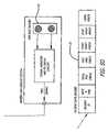
- FIG. 1shows one embodiment of the motion causing device, which is the air-muscle described herein, attached to the proximal forearm along with various sensors and a controller box;
- FIG. 2depicts an exemplary procedure for attaching EMG electrodes for measuring joint extensor activity
- FIG. 3shows one embodiment of the motion causing device, which is the air-muscle described herein, in at least two operational modes, namely the deflated mode and the inflated mode which flexes and extends, respectively, the wrist joint;
- FIG. 4shows another embodiment of the motion causing device, which is the air-muscle described herein permitting the wrist joint to be in flexion, neutral, and extended position respectively;
- FIG. 5is a block diagram showing one embodiment of the present system for neuromuscular function reeducation and restoring physical function of at least one neuromuscular system
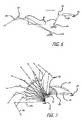
- FIG. 6shows the pivot points and trajectory taken by the motion causing device, such as an air-muscle, according to one aspect of the present invention
- FIG. 7shows the pivot points and trajectory taken by the motion causing device, such as an air-muscle, according to another aspect of the present invention.
- FIG. 8is a flow chart depicting one embodiment for the protocol for neuromuscular function reeducation and restoring physical function of at least one neuromuscular system
- FIG. 9is one embodiment of the invention using a continuous passive motion (CPM) device in conjunction with an air-muscle device;
- CPMcontinuous passive motion
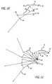
- FIG. 10is an alternative embodiment, with reduced linkages, showing the pivot points and trajectory taken by the air-muscle;
- FIG. 11is yet another embodiment, with reduced linkages, showing the pivot points and trajectory taken by the air-muscle;
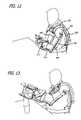
- FIG. 12is an initial state of the air-muscle device used in conjunction with a robotic system for providing assistive training
- FIG. 13is an intermediate state of the air-muscle device used in conjunction with a robotic system for providing assistive training
- FIG. 14is a final state of the air-muscle device used in conjunction with a robotic system for providing assistive training.
- FIG. 15is an exemplary plot depicting the torque calibration curves for two strap conditions.
- FIG. 1there is shown motion causing device or an actuator 10 attached to the proximal forearm 12 along with various sensors 14 , 15 for monitoring activity along the proximal forearm 12 and the at least one joint 16 , at least one electrostim electrode 22 for providing a low-level neuromuscular stimulation, and a controller box 18 with a display port 20 for providing visual feedback for a patient/therapist.
- the system shown in FIG. 1is used for neuromuscular function reeducation and restoring physical function of at least one neuromuscular system.
- the actuator 10permit relative motion between the proximal and distal portions of the joint 16 in a manner to minimize any hindrance or interference with any motion generated by the patient's neuromuscular system.
- the actuator 10is a pneumatically operated air-muscle.
- a prior art artificial muscle deviceexhibits many of the properties of human muscle.
- the artificial muscle deviceconsists of an expandable internal bladder (e.g., a rubber tube) surrounded by a braided shell. When the internal bladder is pressurized, it expands radially against the braided shell causing the muscle to contract. Braided finger traps used to hold fingers on traction devices contract radially when pulled.
- the air-muscleaccording to one embodiment in the present invention, works in the same manner but in the opposite direction (i.e., increasing the diameter causes it to shorten).
- the devicehas spring-like characteristics, is flexible, and is lightweight.
- the force-deflection characteristicscan be made similar to those of human muscle.
- Pressurized air canisters or accumulators that are recharged by air compressorscan be used as a source for supplying air to the air-muscle.
- Major advantages of the air muscleare its flexibility and ease of adaptation to address the specific loss of function exhibited by a patient.
- the device of the preferred embodimenthas three times the pull force of an air piston of the same cross sectional area. The utility of this device resides in its unique combination of attributes: low cost, light-weight, low profile, and low noise operation.
- the sensorsinclude at least one joint position sensor 15 for measuring joint movement, at least one force sensor 48 (shown in FIG. 5A ) for measuring a parameter indicative of the muscle resistance, at least one EMG sensor 14 for measuring the electrical activity of the neuromuscular system. Furthermore, there is at least one neuromuscular electrical stimulating (NMES) device, such as an electrostim electrode 22 , for providing low-level neuromuscular stimulation.
- NMESneuromuscular electrical stimulating
- the controller box 18connected through cable 24 to the air-muscle 10 , includes electronics (i) for recording and displaying 20 measurements obtained through various sensors 14 , 15 , and (ii) for controlling the air-muscle 10 operation (e.g., by controlling the supply of air into the muscle).
- This controller box 18is a self-contained, mobile device that provides sensory (e.g., visual) feedback of wrist and finger position, EMG extensor activity and wrist flexor resistive torque.
- the sensory feedbackcould be provided through tactile sensing through piezoelectric transducer induced vibrations on the skin, or through audio signals (e.g., beeps through an auditory display).
- the firmware in the microprocessor of the controller system 18has been designed to be well-structured using object-oriented programming techniques. Use of these techniques yields more reliable code having fewer discrepancies and problems.
- the controller box 18is shown as a separate device, however it is to be understood that the box could be a sleeve wrapped around the arm of a patient and as shown in FIG. 2 .
- FIG. 2depicts an exemplary procedure for attaching at least one EMG electrode 15 for measuring joint extensor activity (also known as the agonist muscle activity).
- EMG electrode 15for measuring joint extensor activity.
- the location of the EMG electrodes 15is determined for each patient by the therapist.
- the skinmay be rubbed several times with alcohol soaked gauze pads.
- the EMG electrode outputis used to measure the relative recruitment of selected extensor muscles and may be used to feedback the information to the patient to reinforce correct recruitment. Session to session variation in EMG values may be recorded for monitoring patient performance and/or compliance.
- the EMG sensormay be used for measuring the electrical activity of an agonist muscle (e.g., the extensor muscle) and/or an antagonist resisting muscle (e.g., the flexor muscle) in the neuromuscular system of a patient.
- Two measuring channelsmay be used to capture EMG activity of the muscle sets of the neuromuscular system.
- the air-muscleis supplied by compressed air by means of a source ( FIG. 5 , 42 ), and the air supply may be controlled by the controller 18 that includes a microprocessor ( FIG. 5B ) for controlling at least one valve ( FIG. 5C , 46 ) that controls the supply of pressurized air to the air-muscle.
- a sourceFIG. 5 , 42
- the air supplymay be controlled by the controller 18 that includes a microprocessor ( FIG. 5B ) for controlling at least one valve ( FIG. 5C , 46 ) that controls the supply of pressurized air to the air-muscle.
- one end of the air-muscle 10is connected to a distal element 34 of the wrist joint 16 and the other end of the air-muscle 10 is connected to a proximal element 32 of the wrist joint 16 .
- Activation of the air muscle 10by inflating it through a supply source 42 ( FIG. 5C ) of air, may cause the joint to pivot about at least one axis and causing the joint to go from a flexed position to the extended position as shown in FIG. 3 .
- thiscould be achieved by means of rotation of a bar that extends the wrist and operates a mechanism that extends the fingers and wrist between a flexed, neutral, and extended positions as shown in FIG. 4 .
- An optional data port 36may be provided for downloading stored information from the memory 50 ( FIG. 5 ) of controller 18 to a PC (not shown).
- FIG. 4an alternative embodiment of the present invention is shown therein.
- the wrist jointis shown in three states (flexion, straight, and extended).
- the air-muscle device 10(shown with a braided type covering), having strap 330 for permitting air from a source 42 , is connected at one end to the forearm support structure 300 via connector 326 .
- the support 300wraps around the forearm and is held in place by means of straps 322 .
- the other end of the air-muscle 10is connected to a bar 324 which also provides a pulley for the extension strip 336 .
- One end 320 of the strip 336is connected to a bar 338 with a corresponding joint 332 .
- the other end of strap 336is attached to the support structure 300 .
- the joints, 342 and 340 , of the support structure 300are positioned substantially adjacent the knuckles 344 and the finger joints 346 , respectively, of the patient's hand.
- the structure 300may also include tubes 352 for allowing the finger ends to be housed.
- the wrist jointmay achieve: (i) flexion, (ii) normal, and (iii) extension.
- the air-muscle 10extends, relatively, in length, upon deflation (as shown in the top figure of FIG. 4 ), which causes the bar 338 to rotate/pivot clockwise (looking from the top and as depicted by the arrow 400 ) about joint 310 .
- This causal effectis established by means of the strip 336 that connects the bar 338 to the air-muscle 10 .
- This pivot action of the bar 338causes the wrist to achieve the flexion state.
- rotation of the joints 342 and 340may allow the knuckle joints and the finger joints to assume curled/retracted positions.
- the air-muscle 10shortens, relatively, in length, upon reasonable inflation (as shown in the middle figure of FIG. 4 ), which causes the bar 338 to rotate/pivot counter-clockwise (looking from the side and as depicted by arrow 420 ) about joint 310 .
- This causal effectis established by means of the strip 336 that connects the bar 338 to the air-muscle 10 .
- This pivot action of the bar 338causes the wrist to achieve the normal or straight state.
- rotation of the joints 342 and 340may allow the knuckle joints and the finger joints to assume substantially straightened positions.
- the air-muscle 10shortens, relatively, much more in length, upon a higher degree of inflation (as shown in the bottom figure of FIG. 4 ), which causes the bar 338 to further rotate/pivot counter-clockwise (looking from the top and as depicted by the arrow 440 ) about joint 310 .
- This causal effectis established by means of the strip 336 that connects the bar 338 to the air-muscle 10 .
- This pivot action of the bar 338causes the wrist to achieve the extension state.
- rotation of the joints 342 and 340may allow the knuckle joints and the finger joints to assume substantially extended positions.
- Joint extension position or displacementis measured by the at least one joint position sensor 15 , such as a potentiometer, that is incorporated in the motion causing device.
- Resistance to extensionthat may be due to the antagonist resisting muscle, is measured by the at least one force sensor 48 ( FIG. 5A ), such as a force sensitive resistors (FSRs), that is placed on the device.
- the FSR outputis a measure of the resistance of finger and wrist flexor muscles.
- the resistance to extensionmay be measured by at least one EMG sensor.
- the resistance to extensionmay be determined by measuring the maximum extension of a patient's hand under a specified activation pressure. The compliant property of the air muscle allows the lack of full extension to be calibrated versus the resisting load or torque.
- the at least one force sensor 48i.e., the FSR
- the FSRis calibrated after each device is assembled.
- a load cell(not shown) is inserted between an activation bar on the device and muscle.
- the mechanismis fixed in six different degrees of flexion-extension.
- the output of the FSRmay be compared to the load cell output.
- the torque about the joint at each wrist positionis calculated by multiplying the muscle force by the distance to the line of action of the air muscle.
- the force sensing aspectmay be simplified by simply measuring the performance of the air muscle. Specifically, when the air muscle is pressurized, external loads cause it to extend. Thus, the amount of extension caused by the resistance of spastic resisting muscles may be sufficiently large to be measured with the joint position potentiometer. The calibration of the amount of lengthening of the muscle may be achieved by determining the change in joint position potentiometer reading versus the resistive load. This calibration result may then stored in the microprocessor. When the hand is extended by a prescribed pressure, the amount by which the extension motion is reduced from full extension is a measure of the resisting torque.
- protection against overloading a limbis provided by the compliant nature of the pneumatic muscle. Because of this compliance, it may not to be necessary to provide an auxiliary exhaust if excessive resistance is encountered.
- the compliancealso provides a method of measuring the resistive load, as confirmed through Mentor testing by means of a load cell inserted between the air muscle and the actuating bar of the hand mechanism. A data acquisition system recorded the load and the position as measured by the pivot potentiometer at 100 Hz. Resistive forces were generated by hanging weights on the hand piece and also by wearing the Mentor and manually resisting the motion with different levels of force. The resistive load versus the amount of extension is shown in FIG. 15 .
- the motion causing device 10(i.e., the air-muscle) drives the fingers and wrist into extension by moving a mechanical linkage.
- the linkageis designed to move the fingers and/or wrist in a spiral fashion.
- a therapistcan also program the air-muscle(s) so that a desired motion pattern is followed for fastest and effective recovery.
- a micro-compressorwas chosen that has a maximum output pressure which limits the maximum force supplied by the air muscle.
- the air muscleis a very compliant drive with the maximum force output at the fully flexed position where the stretch reflex resistance of the flexor muscles is minimum.
- the stretch reflexincreases resistance to motion. If a large resistance is encountered during extension, the air muscle stretches and limits the range of motion.
- spastic flexor musclesare velocity sensitive, the velocity of actuation was chosen to be about 5 degrees per second with no loading. With the weight of a flaccid hand this rate decreases to about 3.8 degrees per second and with mild resistance the rate is approximately 2.7 degrees per second.
- the actuatorto be an air-muscle
- the air-musclepermits relative motion between the proximal and distal portions of the joint, due to its high mechanical compliance, in a manner that does not hinder or interfere with any motion generated by the neuromuscular system. Allowing self-actuated motion of the joint by means of the neuromuscular system is an important element in the design. Specifically, the air-muscle allows unfettered self-actuated motion of the joint.
- a continuous passive devicewith a high mechanical compliance, that permits extension and flexion of the joint through self-actuation could be used for neuromuscular function reeducation and restoring physical function of the neuromuscular system.
- FIG. 5is a block diagram showing one embodiment of the present system for neuromuscular function reeducation and restoring physical function of at least one neuromuscular system showing the controller 18 with a microprocessor 44 and an electronic memory 50 , the air-muscle 10 , at least one display 20 , at least one joint position sensor 15 , at least one force sensor 48 , at least one EMG sensor 14 , at least one neuromuscular electrical stimulation (NEMS) device 22 for providing low-level neuromuscular stimulation.
- the real-time clock/calendar 52is powered by a battery mounted on the printed circuit board when the power is off. The clock 52 maintains the time and date continuously.
- Records of patient use, active range of motion, extensor resistive torque, and EMG activityare recorded with a time stamp in a non-volatile serial EEPROM memory device 50 . Data is kept safe, even when no power is applied to the memory 50 . These records can be downloaded to a therapist's or patient's personal computer. A patient record can be printed that provides a performance history and compliance by date. A display 20 is made available for viewing and monitoring joint/muscle activity during therapy.
- the microprocessor 44controls the activation of the air muscle by operating the microcompressor and/or the air valves.
- Wrist/joint positionmay be displayed as a bar graph or as numbers on the LCD display 20 .
- the degree of flexor resistance torque measured by the at least one force sensor, and/or electrical activity from the EMG sensormay be displayed as a variable length line of lit light emitting diodes (LEDs) incorporated next to the LCD display 20 .
- the changing goal for active wrist motionmay be displayed as a line on the LCD graph.
- One line of multi-color light emitting diodes (LEDs)may indicate the degree of flexor resistance torque as measured by the force sensitive resistors.
- a second line of LEDsmay indicate the EMG activity of the wrist or finger extensors.
- the microcompressor, air valves, microprocessor, and the LCDmay be portably located on or beside the patient during therapy sessions.
- feedbackmay be provided to the patient/therapist through audio signals (e.g., by varying the tonal frequencies, amplitudes, etc.).
- Records of patient use, active range of motion, extensor resistive torque, and EMG activityare recorded with a time stamp in an electronic memory device (e.g., a non-volatile serial EEPROM).
- the electronic memory systemcan provide stored information, such as patient compliance and patient performance, from the electronic memory to a therapist/patient on command.
- the systemincludes a mechanism for the patient to monitor the compliance and performance through visual and possibly aural means.
- the controllermay update the displays in a predetermined manner to provide a mechanism for the patient to improve performance and compliance for neuromuscular function reeducation and for restoring physical function of the neuromuscular system in the patient.
- the present inventionprovides neuromuscular reeducation and restoration of physical function of the neuromuscular system in the patient through (1) biofeedback, (2) repetitive task practice, and (3) neuromuscular electrical stimulation (NMES). Furthermore, psychological research has shown that an important part of biofeedback stimulated neuromuscular reeducation is shaping, which is defined by psychologists as establishing goals just beyond current capability and changing goals as progress is made. The present invention also provides shaping through measurements obtained during therapy session. Additionally, research has shown that many repetitions are necessary for permanent neuromuscular reeducation. To encourage practice of the repetitions, the present invention records compliance of a patient with the repetitive therapy and provides a report back to the patient's therapist.
- NMESneuromuscular electrical stimulation
- One method for implementing a protocol, for the system as described, for restoring physical function of the neuromuscular systemcomprises measuring a signal indicative of the activity of the muscle through the EMG sensor(s) 14 ; measuring a signal indicative of the joint motion through a joint position sensor 15 , where the joint is associated with the neuromuscular system; measuring another signal indicative of the muscle resistance through the force sensor 48 ; mapping the measured signals to at least one parameter; and controlling the air level in the air-muscle 10 in order to optimize the parameter for restoring physical function of the neuromuscular system associated with the joint in the patient.
- FIGS. 6 , 7depict one arrangement of pivot points 60 and linkages 64 associated with the air-muscle 10 attached to forearm supports (as depicted by 62 ).
- the air-muscle 10upon inflation and deflation, permits the arm and joints to achieve pre-determined trajectories (shown as spiral type in the figures).
- the pivot point 60 positions and the trajectories of the arm (or linkages 64 )can be controlled by inflating/deflating the air-muscle 10 in a manner to get optimal motion for the arm and joint thereby providing means for effective restoration of the physical function of the neuromuscular system.
- the air muscle 10is shown connected at one end to the forearm support 62 while at the other end it is depicted as being attached to pivot point or driving point 60 .
- the air-muscle 10is inflated/deflated in a specific manner optimal motion is achieved for reeducation and restoration of the neuromuscular system.
- FIG. 8is a flow chart depicting another embodiment of the protocol for restoring physical function of the neuromuscular system. Specifically, the patient is instructed to try to extend the wrist when a beep is heard. The EMG activity of the wrist extensors and the motion of the wrist are recorded in the memory of the device and displayed for the patient. The patient could be instructed to use the device for 6 hours a day, although more usage may be allowed, and the treatments need not be continuous. The patient could start and stop the device at any time. The first 2 hours could focus on EMG and joint position feedback. During the second 2 hours the flexor resistance torque from the flexors could be used as the feedback signal to help the patient reduce any flexor spasticity.
- the final 2 hours of therapywould include both extensor EMG and flexor resistance torque as feedback.
- the number of completed cyclescould be recorded for each day as well as the time for each cycle and the total treatment time for each day.
- the patientcould be encouraged to grasp a block and lift it several times during a day and at the end of each therapy session. After grasping, the patient may be encouraged to try and lift and subsequently move the block. The patient may also be encouraged to keep a record of successful attempts.
- the level of extensor EMG activitymay be indicated by LEDs on the display 20 .
- a level equal to that obtained at the last clinic therapy sessionwould show as a yellow light.
- a level belowwould generate a buzzing sound.
- a green LEDwould indicate a higher level.
- Faster flashing LEDsindicate higher levels of EMG activity.
- the output of the joint position sensoris displayed on the LCD by a bar. If motion exceeds this line a pleasant sound is heard. After every day, the line that represents the goal could be increased/updated by a certain amount (e.g., 1% of the highest joint motion achieved in the previous day). Thus, joint position could serve as the basis for subsequent training each day.
- the air-musclecould be activated and the wrist and finger extension may be completed.
- the extensioncould be held for a certain amount of time (e.g., 3 seconds) and then released.
- the torque of the flexor resistanceis measured and displayed on flashing red LEDs during the process (the higher the force, the faster the blinking).
- the patientcould be instructed to try to minimize this force by thinking about relaxing the flexors.
- a system delay(for e.g., of about 10 seconds) may be introduced and the process would be started over with an auditory beep.
- the number of hours of operation of the device and the active motion achieved at the beginning of a day and at the end of each daymay be recorded in memory. This information would provide fairly accurate information about patient compliance and permits matching of timed data from patient reports of self-documentation of training. When the device is turned on for the first time each day, these parameters are displayed for the patient on the LCD.
- the memorymay be downloaded onto a PC in the clinic.
- a summary chart graphically displaying the number of hours of use a day by the patient, the range of motion change per day and the change of active range of motion day to daymay be displayed and the charts printed for the patient file.
- the therapistcan prescribe the patient as to how much time he or she can spend on a specific program.
- the amount of emphasis on each programdepends on the symptoms of the patient.
- the programcould require work on the extensor muscle recruitment.
- the extensor EMG signalmay be displayed as a line of LED lights on a relative scale.
- a single color coded or intensity coded LEDcould be used.
- the range between the minimum and maximum signal measuredmay be about 60% of the height of the LED line.
- Messagesmay be provided on the LCD display 20 to encourage the patient to increase the display level.
- a second programthat could be selected by the patient, includes measurement of the resistance offered by the flexor muscles (antagonist resisting muscles) to an extension process. It is common for patients with neuromuscular diseases to develop a constant muscle contraction. Specifically, the flexors are substantially stronger than the extensors which causes the characteristic flexed fingers and wrist that can be seen in many patients. The reason for this is the lack of inhibition present in the central nervous system. To retrain the neuromuscular system, the program could display the relative magnitude of the measured resistance and request the patient to consciously try and reduce the signal on the display. EMG sensors 14 located adjacent the flexor muscles and/or FSRs 48 or lack of full extension may be used for measuring the resistance.
- the programmay also require the patient to extend the wrist with the air muscle to about neutral position. At this point EMG activity or the force output of the FSRs may be measured for the flexor muscles (antagonist resisting muscles). Subsequent calculation of the active stiffness generated by the flexors may be achieved by dividing the measured force by the amount of joint displacement (as measured by the potentiometer 15 ).
- a third program that can be selected for the patientpermits (i) increasing the activity of the extensors, and (ii) simultaneously decreasing the activity of the flexors.
- the signal to one of the LED linesmay be a combination of the magnitude of the extensor signal, and the flexor signal as measured either via the EMG sensor 14 or the FSR 48 .
- the patientmay be asked to extend their fingers and wrist and then to think about their forearm so as to minimize the signal displayed via the LED lines.
- a continuous passive motion (CPM) device 204in conjunction with an air-muscle device 206 and 206 ′, may be used for providing flexion and extension to the joint 208 of the patient for reeducation and restoration of the physical functions of the neuromuscular system in a patient.
- the upper air-muscle device 206may be controllably activated, whereas during flexion, the lower air-muscle device 206 ′ may be controllably activated.
- the air-muscleis modified so that it is capable of interacting with a personal computer based virtual reality program for capturing the interest of the user.
- the resulting systemallows extension of the existing wrist/hand device through coordinated elbow and shoulder motions.
- the therapeutic protocol chosenfor exemplary purposes according to one aspect of the invention, is to focus on two important tasks; reaching and eating. Specifically, the patient is requested to achieve either a reaching task or an eating task. After the patient has achieved his/her maximal motion, the device assists the limb to complete the task. Feedback of self-actuated progress is recorded and fed back to the patient.
- FIGS. 12-14shows various states of the air-muscle device used in conjunction with a robotic system for providing assistive training.
- the design philosophyis such that it encourages volitional activation of joint movement through feedback range of motion, muscle activity and resistance to movement and then completing the task for the patient.
- the systemis adapted to provide training of two tasks (viz., feeding and reaching). It is anticipated that training of coordinated joint movement for these two tasks will lead to more general improvement in related functional tasks as found in other feedback therapies.
- the robot systemabducts the shoulder in the flex direction, extends the elbow, supinates the forearm, and extends the wrist and fingers. Training begins with the patient facing a table with her/his forearm resting on the table transverse to the torso (in a plane parallel to the frontal plane) as shown in FIG. 12 .
- the shoulder motionis restricted to flexion in the sagittal plane.
- the intermediate reaching positionis shown in FIG. 13 .
- the robotic systemflexes the shoulder, supinates the forearm, and extends the wrist as shown in FIG. 14 .
- Most stroke patientshave the ability to flex their elbow, so this function may be left to the patient.
- the assisted movementsmay be made in a slow manner to protect against significant increases in muscle tone.
- the air muscle 500rotates the shoulder in abduction and flexion.
- a plastic hinge 502only allows motion in the sagittal plane. The normal internal rotation of the humerus is held in neutral by the distal and proximal strapping system.
- the proximal air musclerides on a plate 504 that rotates about the center of rotation of the humerus.
- a foam-lined sub-plate 506that is attached to the torso with Velcro straps, distributes the vertical reaction of the air muscle.
- a plastic cuff 508is attached to the upper humerus with Velcro straps. This serves as an attachment point for a driver 510 that is connected at its other end to the sliding plate that rests on top of the sub-plate. Contraction of the air muscle rotates the upper plate 504 in an arc about the center of rotation of the humerus.
- a potentiometer(not shown) centered on the axis of rotation records the amount of shoulder motion.
- a hinge(not shown) is attached at the elbow with straps to the distal humerus and proximal forearm.
- a potentiometeris contained in the center of rotation of the hinge to measure elbow flexion and extension.
- the forearm piece 512has an extension 514 that is used an attachment point for an air muscle. The proximal end of this air muscle is attached to the center of rotation of the humerus.
- Supination/pronationplus coordinated extension of the wrist and fingers is obtained by a modification of the wrist/hand device that has been clinically tested and currently is in commercial distribution.
- Two musclesare attached to the hand/wrist device. The proximal end of one is attached to the inside of the forearm at the elbow and the other proximal end of the other one is attached to the outside of the forearm at the elbow. If the latter muscle is contracted, combined wrist and finger extension occurs along with supination of the forearm. If the other muscles contracts, combined wrist and finger extension occurs along with pronation of the forearm.
- a potentiometeris located at the center of rotation of the wrist. Initially, no measurement of pronation/supination will be included.
- the above described system and programsthus provide effective and easy to use functionality for reeducation and restoration of the physical functions of the neuromuscular system in a patient.
- the systemis inexpensive, portable, comfortable, and easy to use either by the patient or by a therapist.
- the system and method according to the present inventionis a self-contained, mobile device, implementing a protocol, and which provides visual and/or aural feedback of wrist and finger position, measurement of extensor and flexor activity for neuromuscular function reeducation and restoring physical function of at least one neuromuscular system in a patient.
- a suitable inflatable device or mechanical drivecan be used in replacement for the air-muscle.
- Additional sensorscould be used for measuring critical parameters for restoring physical function.
- Multiple motion causing devicescould be simultaneously connected at various positions on the patient's body, and the devices could be controlled by multiple or single controllers.
- the therapistcould be allowed to select, from a menu provided through the display, unique therapeutic protocol(s) for a specific patient.
- the frequency of patient use and the ability to reach performance goalscan be recorded in memory and the record played back for the therapist/patient. While the specification describes particular embodiments of the present invention, those of ordinary skill can devise variations of the present invention without departing from the inventive concept.
Landscapes
- Health & Medical Sciences (AREA)
- General Health & Medical Sciences (AREA)
- Veterinary Medicine (AREA)
- Public Health (AREA)
- Physical Education & Sports Medicine (AREA)
- Life Sciences & Earth Sciences (AREA)
- Animal Behavior & Ethology (AREA)
- Rehabilitation Therapy (AREA)
- Pain & Pain Management (AREA)
- Epidemiology (AREA)
- Engineering & Computer Science (AREA)
- Biomedical Technology (AREA)
- Nuclear Medicine, Radiotherapy & Molecular Imaging (AREA)
- Radiology & Medical Imaging (AREA)
- Rehabilitation Tools (AREA)
- Measurement Of The Respiration, Hearing Ability, Form, And Blood Characteristics Of Living Organisms (AREA)
Abstract
Description
Claims (11)
Priority Applications (3)
| Application Number | Priority Date | Filing Date | Title |
|---|---|---|---|
| US10/727,212US7725175B2 (en) | 2002-12-04 | 2003-12-02 | System and method for neuromuscular reeducation |
| AU2003297652AAU2003297652A1 (en) | 2002-12-04 | 2003-12-03 | A system and method for neuromuscular reeducation |
| US12/758,706US8214029B2 (en) | 2002-12-04 | 2010-04-12 | System and method for neuromuscular reeducation |
Applications Claiming Priority (2)
| Application Number | Priority Date | Filing Date | Title |
|---|---|---|---|
| US43070002P | 2002-12-04 | 2002-12-04 | |
| US10/727,212US7725175B2 (en) | 2002-12-04 | 2003-12-02 | System and method for neuromuscular reeducation |
Related Child Applications (1)
| Application Number | Title | Priority Date | Filing Date |
|---|---|---|---|
| US12/758,706DivisionUS8214029B2 (en) | 2002-12-04 | 2010-04-12 | System and method for neuromuscular reeducation |
Publications (2)
| Publication Number | Publication Date |
|---|---|
| US20040267331A1 US20040267331A1 (en) | 2004-12-30 |
| US7725175B2true US7725175B2 (en) | 2010-05-25 |
Family
ID=32469510
Family Applications (2)
| Application Number | Title | Priority Date | Filing Date |
|---|---|---|---|
| US10/727,212Active2027-01-23US7725175B2 (en) | 2002-12-04 | 2003-12-02 | System and method for neuromuscular reeducation |
| US12/758,706Expired - LifetimeUS8214029B2 (en) | 2002-12-04 | 2010-04-12 | System and method for neuromuscular reeducation |
Family Applications After (1)
| Application Number | Title | Priority Date | Filing Date |
|---|---|---|---|
| US12/758,706Expired - LifetimeUS8214029B2 (en) | 2002-12-04 | 2010-04-12 | System and method for neuromuscular reeducation |
Country Status (3)
| Country | Link |
|---|---|
| US (2) | US7725175B2 (en) |
| AU (1) | AU2003297652A1 (en) |
| WO (1) | WO2004050172A1 (en) |
Cited By (23)
| Publication number | Priority date | Publication date | Assignee | Title |
|---|---|---|---|---|
| US20090131225A1 (en)* | 2007-08-15 | 2009-05-21 | Burdea Grigore C | Rehabilitation systems and methods |
| US20100069798A1 (en)* | 2008-09-15 | 2010-03-18 | Ching-Hsiang Cheng | Wearable device to assist with the movement of limbs |
| US20110137196A1 (en)* | 2007-08-31 | 2011-06-09 | Tokyo Metropolitan Organization For Medical Research | Quantitative motor function evaluation system |
| US20110137138A1 (en)* | 2008-05-29 | 2011-06-09 | Per Johansson | Patient Management Device, System And Method |
| US20110257566A1 (en)* | 2010-02-12 | 2011-10-20 | Bright Cloud International Corp | Instrumented therapy table and system |
| US8117047B1 (en) | 2007-04-16 | 2012-02-14 | Insight Diagnostics Inc. | Healthcare provider organization |
| US20120209145A1 (en)* | 2009-10-22 | 2012-08-16 | Anatoliy Ivanovich Grigoriev | Medical suit of axial loading with automatic control system |
| US20150173918A1 (en)* | 2013-10-22 | 2015-06-25 | Massachusetts Institute Of Technology | Peripheral Neural Interface Via Nerve Regeneration to Distal Tissues |
| US20150290076A1 (en)* | 2012-11-23 | 2015-10-15 | Flinders University Of South Australia | Method of therapy and haptic gaming system for sensory agnosia |
| US20150359698A1 (en)* | 2014-06-13 | 2015-12-17 | Worcester Polytechnic Institute | Actuators and Methods of Use |
| US20160199246A1 (en)* | 2015-01-14 | 2016-07-14 | Yeung Ki Kim | Upper limb rehabilitation training apparatus |
| US9925034B2 (en)* | 2011-09-30 | 2018-03-27 | Verily Life Sciences Llc | Stabilizing unintentional muscle movements |
| US9943430B2 (en) | 2015-03-25 | 2018-04-17 | Verily Life Sciences Llc | Handheld tool for leveling uncoordinated motion |
| US10271770B2 (en) | 2015-02-20 | 2019-04-30 | Verily Life Sciences Llc | Measurement and collection of human tremors through a handheld tool |
| US10271768B1 (en)* | 2010-12-23 | 2019-04-30 | Jogohealth Inc. | Method for determining rehab protocol and behavior shaping target for rehabilitation of neuromuscular disorders |
| US10368669B2 (en) | 2011-09-30 | 2019-08-06 | Verily Life Sciences Llc | System and method for stabilizing unintentional muscle movements |
| US10420663B2 (en) | 2017-05-01 | 2019-09-24 | Verily Life Sciences Llc | Handheld articulated user-assistive device with behavior control modes |
| US10600596B2 (en) | 2014-04-21 | 2020-03-24 | Verily Life Sciences Llc | Adapter to attach implements to an actively controlled human tremor cancellation platform |
| US10786415B2 (en) | 2015-03-20 | 2020-09-29 | Regents Of The University Of Minnesota | Systems and methods for assessing and training wrist joint proprioceptive function |
| US10994416B2 (en)* | 2017-12-21 | 2021-05-04 | Southeast University | Method for controlling a limb motion intention understanding and upper limb rehabilitation training robot based on force sense information and posture information |
| WO2021118382A1 (en)* | 2019-12-09 | 2021-06-17 | Федеральное государственное бюджетное научное учреждение "НАУЧНЫЙ ЦЕНТР НЕВРОЛОГИИ" | Method for increasing the effectiveness of arm motor rehabilitation after stroke |
| US11136234B2 (en) | 2007-08-15 | 2021-10-05 | Bright Cloud International Corporation | Rehabilitation systems and methods |
| US11179251B2 (en) | 2016-01-08 | 2021-11-23 | Massachusetts Institute Of Technology | Method and system for providing proprioceptive feedback and functionality mitigating limb pathology |
Families Citing this family (103)
| Publication number | Priority date | Publication date | Assignee | Title |
|---|---|---|---|---|
| US7447542B2 (en)* | 2003-07-11 | 2008-11-04 | Ob Tools Ltd. | Three-dimensional monitoring of myographic activity |
| JP4178186B2 (en)* | 2003-08-21 | 2008-11-12 | 国立大学法人 筑波大学 | Wearable motion assist device, control method for wearable motion assist device, and control program |
| US8888723B2 (en) | 2004-02-05 | 2014-11-18 | Motorika Limited | Gait rehabilitation methods and apparatuses |
| US8915871B2 (en) | 2004-02-05 | 2014-12-23 | Motorika Limited | Methods and apparatuses for rehabilitation exercise and training |
| WO2005075155A2 (en)* | 2004-02-05 | 2005-08-18 | Motorika Inc. | Fine motor control rehabilitation |
| WO2005074372A2 (en) | 2004-02-05 | 2005-08-18 | Motorika Inc. | Methods and apparatus for rehabilitation and training |
| JP4695605B2 (en)* | 2004-02-05 | 2011-06-08 | モトリカ リミテッド | Neuromuscular stimulation |
| US7979137B2 (en)* | 2004-02-11 | 2011-07-12 | Ethicon, Inc. | System and method for nerve stimulation |
| US7647112B2 (en) | 2004-02-11 | 2010-01-12 | Ethicon, Inc. | System and method for selectively stimulating different body parts |
| US7402145B1 (en)* | 2004-06-09 | 2008-07-22 | Woggon Dennis A | Method of neuromusculoskeletal proprioceptive re-education and development of a living body using corrective chair and vibration |
| EP1838270B1 (en) | 2004-08-25 | 2009-07-22 | Motorika Limited | Motor training with brain plasticity |
| US7618381B2 (en)* | 2004-10-27 | 2009-11-17 | Massachusetts Institute Of Technology | Wrist and upper extremity motion |
| DE102005022005B4 (en)* | 2005-05-09 | 2014-10-30 | Anna Gutmann | Method and device for facilitating the movement control of body parts |
| US20060287614A1 (en)* | 2005-06-16 | 2006-12-21 | Cornell Research Foundation, Inc. | Testing therapy efficacy with extremity and/or joint attachments |
| US8388561B2 (en) | 2005-07-01 | 2013-03-05 | The United States Of America, As Represented By The Secretary, Department Of Health And Human Services | Systems and methods for recovery from motor control via stimulation to a substituted site to an affected area |
| US8579839B2 (en) | 2005-07-01 | 2013-11-12 | The United States Of America, As Represented By The Secretary, Department Of Health And Human Services | Methods for recovery from motor control via stimulation to a substituted site to an affected area |
| KR20080027378A (en) | 2005-08-05 | 2008-03-26 | 코닌클리케 필립스 일렉트로닉스 엔.브이. | Measurement and stimulation of muscle tissue |
| US8834169B2 (en)* | 2005-08-31 | 2014-09-16 | The Regents Of The University Of California | Method and apparatus for automating arm and grasping movement training for rehabilitation of patients with motor impairment |
| US8165685B1 (en)* | 2005-09-29 | 2012-04-24 | Case Western Reserve University | System and method for therapeutic neuromuscular electrical stimulation |
| AT502521B1 (en)* | 2005-09-30 | 2011-12-15 | Paolo Dipl Ing Ferrara | DEVICE FOR FLEXIBLY CONTROLLABLE MOVEMENT OF PEOPLE OR OBJECTS |
| EP1929158B1 (en)* | 2005-09-30 | 2013-07-10 | Paolo Ferrara | Device for moving people or objects in a flexible controllable manner |
| CN101282696B (en)* | 2005-10-11 | 2012-12-05 | 松下电器产业株式会社 | Motion assistance apparatus and method |
| US7856264B2 (en)* | 2005-10-19 | 2010-12-21 | Advanced Neuromodulation Systems, Inc. | Systems and methods for patient interactive neural stimulation and/or chemical substance delivery |
| US8360997B2 (en)* | 2006-02-24 | 2013-01-29 | Ferrobotics Compliant Robot Technology Gmbh | Robot arm |
| AT504269B1 (en)* | 2006-09-27 | 2011-07-15 | Paolo Dr Ferrara | DEVICE FOR THE PASSIVE HEALING GYM |
| US8449445B2 (en) | 2006-03-30 | 2013-05-28 | The United States Of America, As Represented By The Secretary, Department Of Health And Human Services | Device for volitional swallowing with a substitute sensory system |
| CN101061984B (en) | 2006-04-29 | 2012-02-08 | 香港理工大学 | Rehabilitation robot system providing mechanical assistance by using electromyographic signals |
| KR100772908B1 (en)* | 2006-05-15 | 2007-11-05 | 삼성전자주식회사 | Muscle exercise aids |
| US20080114271A1 (en)* | 2006-11-13 | 2008-05-15 | David Rubenstein | Method of neuromuscular calibration |
| US8352026B2 (en) | 2007-10-03 | 2013-01-08 | Ethicon, Inc. | Implantable pulse generators and methods for selective nerve stimulation |
| US10002545B2 (en)* | 2007-10-10 | 2018-06-19 | Jennifer Robin Hanners | System and method for controlling gaming technology, musical instruments and environmental settings via detection of neuromuscular activity |
| US8260425B2 (en)* | 2007-10-12 | 2012-09-04 | Intelect Medical, Inc. | Deep brain stimulation system with inputs |
| JP4530025B2 (en) | 2007-10-30 | 2010-08-25 | セイコーエプソン株式会社 | Projection system, information processing apparatus, and projector |
| JP5173404B2 (en)* | 2007-12-28 | 2013-04-03 | パナソニック株式会社 | Muscle assist device and method of operating the same |
| US20100280423A1 (en)* | 2007-12-28 | 2010-11-04 | Panasonic Corporation | Muscle force assisting device (as amended) |
| US20100280424A1 (en)* | 2008-03-27 | 2010-11-04 | Panasonic Corporation | Muscle force assisting device |
| FR2930139B1 (en)* | 2008-04-22 | 2022-12-30 | Centre Nat Rech Scient | FUNCTIONAL REHABILITATION DEVICE. |
| US20090326612A1 (en)* | 2008-06-26 | 2009-12-31 | Michael J. Distler | Electronic biofeedback stimulation device |
| US20090321483A1 (en)* | 2008-06-30 | 2009-12-31 | Walt Froloff | Universal wrist-forearm docking station for mobile electronic devices |
| JP5243159B2 (en)* | 2008-09-12 | 2013-07-24 | パナソニック株式会社 | Wrist assist device |
| DE102008057789B4 (en)* | 2008-11-17 | 2013-07-04 | Christian Teschner | A method of controlling a display element on a display and biofeedback input device |
| AU2008246284A1 (en)* | 2008-11-19 | 2010-06-10 | Zao, Ritm Okb | Method for electrical influance on a living organism and device thereof |
| US8615301B2 (en)* | 2008-12-23 | 2013-12-24 | Robotic Integrated Technology Development Corporation | Muscle therapy system |
| US8612010B2 (en)* | 2008-12-23 | 2013-12-17 | Robotic Integrated Technology Development Corporation | Upper extremity muscle therapy system |
| US20100262047A1 (en)* | 2009-04-08 | 2010-10-14 | Drexel University | Physical therapy systems and methods |
| US9757266B2 (en)* | 2010-06-01 | 2017-09-12 | Saebo, Inc. | Orthotic device |
| US20130172901A1 (en)* | 2010-07-19 | 2013-07-04 | Institut National De La Sante Et De La Recherche Medicale (Inserm) | Tool for inserting fine tubular objects into the cochlea |
| US9464642B2 (en)* | 2010-11-19 | 2016-10-11 | President And Fellows Of Harvard College | Soft robotic actuators |
| WO2012120393A2 (en)* | 2011-03-04 | 2012-09-13 | Koninklijke Philips Electronics N.V. | Device for hand rehabilitation |
| CN103764021B (en)* | 2011-05-20 | 2015-11-25 | 南洋理工大学 | A kind ofly to repair for collaborative neuro-physiological and/or system, instrument, the apparatus and method of functional promotion |
| IL213756A (en) | 2011-06-23 | 2016-02-29 | Rehabit-Tec Ltd | Apparatus for rehabilitating an injured limb |
| US9107614B2 (en) | 2011-07-12 | 2015-08-18 | Xanadu Christina Halkias | Systems, methods, and media for finding and matching tremor signals |
| US9144529B2 (en)* | 2011-07-27 | 2015-09-29 | Stephen Lynn Culver | Range of motion assistant |
| US11904101B2 (en)* | 2012-06-27 | 2024-02-20 | Vincent John Macri | Digital virtual limb and body interaction |
| US11673042B2 (en) | 2012-06-27 | 2023-06-13 | Vincent John Macri | Digital anatomical virtual extremities for pre-training physical movement |
| US10096265B2 (en) | 2012-06-27 | 2018-10-09 | Vincent Macri | Methods and apparatuses for pre-action gaming |
| WO2014186739A1 (en) | 2013-05-17 | 2014-11-20 | Macri Vincent J | System and method for pre-movement and action training and control |
| BR112015017042B1 (en) | 2013-01-21 | 2022-03-03 | Cala Health, Inc | Device to treat tremor |
| US9387112B2 (en) | 2013-02-28 | 2016-07-12 | Marvin Frank Bryant | Myoelectric hand orthosis |
| EP2964427B1 (en) | 2013-03-04 | 2020-11-18 | President and Fellows of Harvard College | Magnetic assembly of soft robots with hard components |
| US20140276270A1 (en) | 2013-03-13 | 2014-09-18 | Passy-Muir, Inc. | Systems and methods for stimulating swallowing |
| US9539118B2 (en) | 2013-03-15 | 2017-01-10 | Neurolutions, Inc. | Brain-controlled body movement assistance devices and methods |
| WO2015003203A1 (en)* | 2013-07-11 | 2015-01-15 | Analytica Limited | Stimulation and electromyography detection |
| ES2856342T3 (en)* | 2013-10-18 | 2021-09-27 | Harvard College | Soft, mechanically programmed actuators with adaptable sleeves |
| US10046161B2 (en) | 2013-10-23 | 2018-08-14 | Ecole Polytechnique Federale De Lausanne (Epfl) | Neuroprosthetic system restoring upper limb function through coordinated electrical stimulation |
| WO2015066143A1 (en)* | 2013-10-29 | 2015-05-07 | President And Fellows Of Harvard College | Multi-segment reinforced actuators and applications |
| WO2015080955A1 (en)* | 2013-11-27 | 2015-06-04 | Oregon Health & Science University | Biofeedback during assisted movement rehabilitation therapy |
| US10111603B2 (en) | 2014-01-13 | 2018-10-30 | Vincent James Macri | Apparatus, method and system for pre-action therapy |
| CA2949843A1 (en) | 2014-06-02 | 2015-12-10 | Cala Health, Inc. | Systems and methods for peripheral nerve stimulation to treat tremor |
| US10231851B2 (en)* | 2014-08-29 | 2019-03-19 | Conor J. MADDRY | Pneumatic electromyographic exoskeleton |
| EP3203909B1 (en)* | 2014-10-17 | 2025-09-10 | Kynetyka Technologies Pty Ltd | A screening test for detection of deep vein thrombosis |
| KR20160060823A (en) | 2014-11-20 | 2016-05-31 | 순천향대학교 산학협력단 | Exercise equipment for rehabilitation of hand grip and wrist |
| JP6489422B2 (en)* | 2015-01-28 | 2019-03-27 | パナソニックIpマネジメント株式会社 | Assist wear, assist wear operating method, and control program |
| WO2016183460A1 (en)* | 2015-05-13 | 2016-11-17 | Hive Concepts, LLC | Devices, systems, and methods for muscle recovery |
| EP4342516A3 (en) | 2015-06-10 | 2024-07-10 | Cala Health, Inc. | Systems and methods for peripheral nerve stimulation to treat tremor with detachable therapy and monitoring units |
| WO2017053847A1 (en) | 2015-09-23 | 2017-03-30 | Cala Health, Inc. | Systems and methods for peripheral nerve stimulation in the finger or hand to treat hand tremors |
| CA3011993C (en) | 2016-01-21 | 2024-05-14 | Cala Health, Inc. | Systems, methods and devices for peripheral neuromodulation for treating diseases related to overactive bladder |
| JP6334588B2 (en)* | 2016-03-10 | 2018-05-30 | H2L株式会社 | Electrical stimulation system |
| US9861856B1 (en) | 2016-06-21 | 2018-01-09 | Boston Biomotion, Inc. | Computerized exercise apparatus |
| WO2018009680A1 (en) | 2016-07-08 | 2018-01-11 | Cala Health, Inc. | Systems and methods for stimulating n nerves with exactly n electrodes and improved dry electrodes |
| JP6742196B2 (en)* | 2016-08-24 | 2020-08-19 | Cyberdyne株式会社 | Life activity detection device and life activity detection system |
| CA3033662A1 (en) | 2016-08-25 | 2018-03-01 | Cala Health, Inc. | Systems and methods for treating cardiac dysfunction through peripheral nerve stimulation |
| US10408698B2 (en)* | 2017-03-09 | 2019-09-10 | Barrett Productions, LLC | Electronic force dynamometer and control system |
| EP3606604A4 (en) | 2017-04-03 | 2020-12-16 | Cala Health, Inc. | SYSTEMS, METHODS AND DEVICES FOR PERIPHERAL NEUROMODULATION FOR THE TREATMENT OF DISEASES RELATED TO OVERACTIVE BLADDER |
| CN107320285A (en)* | 2017-07-28 | 2017-11-07 | 上海逸动医学科技有限公司 | A kind of multifunctional intellectual rehabilitation training and assessment system |
| US10888732B2 (en) | 2017-11-01 | 2021-01-12 | Proteus Motion Inc. | Exercise device limb interface |
| CN108143402A (en)* | 2017-12-22 | 2018-06-12 | 芜湖牧云电子科技有限公司 | Suitable for the monitor of fracture fixation treatment |
| EP3740274A4 (en) | 2018-01-17 | 2021-10-27 | Cala Health, Inc. | Systems and methods for treating inflammatory bowel disease through peripheral nerve stimulation |
| USD852970S1 (en)* | 2018-01-29 | 2019-07-02 | Charles A. Goodman | Sexual stimulation control box |
| US11024195B2 (en) | 2018-04-10 | 2021-06-01 | Imam Abdulrahman Bin Faisal University | Diagnostic and therapeutic system for manual therapy |
| FR3093022B1 (en)* | 2019-02-25 | 2021-04-30 | Adiv Dev | ASSISTANCE DEVICE FOR AT LEAST ONE ROTATION MOVEMENT OF A USER'S WRIST |
| CN109925166B (en)* | 2019-03-14 | 2020-10-02 | 清华大学 | Exoskeleton rehabilitation system and rehabilitation method |
| US20200330323A1 (en) | 2019-04-19 | 2020-10-22 | Alex Jolly | Vibratory Nerve Exciter |
| US20220257924A1 (en)* | 2019-07-23 | 2022-08-18 | Nippon Telegraph And Telephone Corporation | Rehabilitation enlightening device, system, and method |
| US12251560B1 (en) | 2019-08-13 | 2025-03-18 | Cala Health, Inc. | Connection quality determination for wearable neurostimulation systems |
| US11890468B1 (en) | 2019-10-03 | 2024-02-06 | Cala Health, Inc. | Neurostimulation systems with event pattern detection and classification |
| RU2734406C1 (en)* | 2019-10-09 | 2020-10-15 | Сергей Арутюнович Будагян | Method for rehabilitation of functional disorders of locomotor apparatus in hemiparesis |
| CN113260340A (en)* | 2019-10-11 | 2021-08-13 | 神经解决方案股份有限公司 | Orthopedic system and rehabilitation of injured body parts |
| KR102327103B1 (en)* | 2020-02-24 | 2021-11-16 | 고려대학교 산학협력단 | Upper limb assistance system for assisting throw motion |
| US10779740B1 (en)* | 2020-02-28 | 2020-09-22 | Center For Quantitative Cytometry | System and method to enhance self-recovery of nerve-muscle damage |
| CN111659010B (en)* | 2020-07-16 | 2023-02-28 | 郑州大学 | Limb functional electrical stimulation control method and limb rehabilitation robot |
| CN112155944B (en)* | 2020-10-13 | 2024-12-27 | 河南理工大学 | A multi-pneumatically driven rehabilitation robot device for wrist joints |
| WO2024234060A1 (en)* | 2023-05-17 | 2024-11-21 | Neumo Tech Pty Ltd | Neuro-modulation therapy method, system, and apparatus |
Citations (12)
| Publication number | Priority date | Publication date | Assignee | Title |
|---|---|---|---|---|
| US4582049A (en) | 1983-09-12 | 1986-04-15 | Ylvisaker Carl J | Patient initiated response method |
| US5012820A (en)* | 1985-11-12 | 1991-05-07 | Niels Meyer | Device for investigation of muscular contraction |
| US5112296A (en)* | 1991-04-30 | 1992-05-12 | The Board Of Supervisors Of Louisiana State University | Biofeedback activated orthosis for foot-drop rehabilitation |
| US5562707A (en) | 1993-10-13 | 1996-10-08 | Sim & Mcburney | Garment for applying controlled electrical stimulation to restore motor function |
| US5860941A (en) | 1995-11-14 | 1999-01-19 | Orthologic Corp. | Active/passive device for rehabilitation of upper and lower extremities |
| US5951499A (en) | 1996-09-27 | 1999-09-14 | Orthologic Corp. | Continuous passive motion device for upper extremity forearm therapy |
| US6010468A (en) | 1998-03-05 | 2000-01-04 | The Discovery Group, Llc | Foot flexion device |
| US6064912A (en) | 1997-03-28 | 2000-05-16 | Kenney; John P. | Orthotic/electrotherapy for treating contractures due to immobility |
| WO2002013673A2 (en) | 2000-08-15 | 2002-02-21 | Stimel Ltd. | Electrostimulation system with electromyographic and visual biofeedback |
| US6456885B1 (en) | 1999-03-29 | 2002-09-24 | Kurume University | Apparatus for strengthening muscles |
| US20020143277A1 (en)* | 1999-07-27 | 2002-10-03 | Enhanced Mobility Technologies | Rehabilitation apparatus and method |
| US7396337B2 (en)* | 2002-11-21 | 2008-07-08 | Massachusetts Institute Of Technology | Powered orthotic device |
Family Cites Families (21)
| Publication number | Priority date | Publication date | Assignee | Title |
|---|---|---|---|---|
| US3983865A (en) | 1975-02-05 | 1976-10-05 | Shepard Richard S | Method and apparatus for myofunctional biofeedback |
| US4683891A (en) | 1982-04-26 | 1987-08-04 | Vincent Cornellier | Biomonitoring stress management method and device |
| US4724842A (en) | 1982-05-19 | 1988-02-16 | Charters Thomas H | Method and apparatus for muscle stimulation |
| US4653479A (en) | 1985-01-17 | 1987-03-31 | Empi, Inc. | Interrupted drive limb motion apparatus |
| US4824103A (en) | 1988-03-14 | 1989-04-25 | Smidt Gary L | Muscle Testing and exercising apparatus |
| GB2227173A (en) | 1989-01-12 | 1990-07-25 | Hugh Wilmott Grenfell | Stimulation of muscles |
| SE464557B (en) | 1989-07-31 | 1991-05-13 | Biolin Ab | ELECTROMYOGRAPHY SETTING AND DEVICE |
| IL97701A (en) | 1991-03-28 | 1995-06-29 | Univ Ben Gurion | Device for generating hand function |
| DK0541338T3 (en) | 1991-11-04 | 1996-12-02 | Cardiac Pacemakers Inc | Implantable device for monitoring and stimulating the heart for diagnosis and therapy |
| GB9224965D0 (en) | 1992-11-28 | 1993-01-20 | Kodak Ltd | Antenatal screening for chromosomal abnormalities |
| US5466213A (en) | 1993-07-06 | 1995-11-14 | Massachusetts Institute Of Technology | Interactive robotic therapist |
| US5549656A (en) | 1993-08-16 | 1996-08-27 | Med Serve Group, Inc. | Combination neuromuscular stimulator and electromyograph system |
| US5755745A (en) | 1995-09-29 | 1998-05-26 | International Rehabilitative Sciences, Inc. | Portable muscle stimulator with removable data storage card |
| US5755674A (en) | 1995-10-25 | 1998-05-26 | Watson; Steven R. | Abdominal exerciser |
| US5674262A (en) | 1996-01-26 | 1997-10-07 | Kinetic Concepts, Inc. | Pneumatic compression and functional electric stimulation device and method using the same |
| US6146341A (en) | 1998-07-15 | 2000-11-14 | M-E-System Inc. | Continuously and externally driven motion training device of joint |
| US6040912A (en)* | 1998-09-30 | 2000-03-21 | Advanced Micro Devices, Inc. | Method and apparatus for detecting process sensitivity to integrated circuit layout using wafer to wafer defect inspection device |
| AU1116000A (en) | 1998-10-15 | 2000-05-01 | Cardio Technologies, Inc. | Passive defibrillation electrodes for use with cardiac assist device |
| BR9915313A (en) | 1998-11-04 | 2004-01-13 | Cardio Tech Inc | System and method for watching a heart pump blood |
| US6155993A (en) | 1999-03-31 | 2000-12-05 | Queen's University At Kingston | Kinesiological instrument for limb movements |
| US6684276B2 (en) | 2001-03-28 | 2004-01-27 | Thomas M. Walker | Patient encounter electronic medical record system, method, and computer product |
- 2003
- 2003-12-02USUS10/727,212patent/US7725175B2/enactiveActive
- 2003-12-03AUAU2003297652Apatent/AU2003297652A1/ennot_activeAbandoned
- 2003-12-03WOPCT/US2003/038579patent/WO2004050172A1/ennot_activeApplication Discontinuation
- 2010
- 2010-04-12USUS12/758,706patent/US8214029B2/ennot_activeExpired - Lifetime
Patent Citations (12)
| Publication number | Priority date | Publication date | Assignee | Title |
|---|---|---|---|---|
| US4582049A (en) | 1983-09-12 | 1986-04-15 | Ylvisaker Carl J | Patient initiated response method |
| US5012820A (en)* | 1985-11-12 | 1991-05-07 | Niels Meyer | Device for investigation of muscular contraction |
| US5112296A (en)* | 1991-04-30 | 1992-05-12 | The Board Of Supervisors Of Louisiana State University | Biofeedback activated orthosis for foot-drop rehabilitation |
| US5562707A (en) | 1993-10-13 | 1996-10-08 | Sim & Mcburney | Garment for applying controlled electrical stimulation to restore motor function |
| US5860941A (en) | 1995-11-14 | 1999-01-19 | Orthologic Corp. | Active/passive device for rehabilitation of upper and lower extremities |
| US5951499A (en) | 1996-09-27 | 1999-09-14 | Orthologic Corp. | Continuous passive motion device for upper extremity forearm therapy |
| US6064912A (en) | 1997-03-28 | 2000-05-16 | Kenney; John P. | Orthotic/electrotherapy for treating contractures due to immobility |
| US6010468A (en) | 1998-03-05 | 2000-01-04 | The Discovery Group, Llc | Foot flexion device |
| US6456885B1 (en) | 1999-03-29 | 2002-09-24 | Kurume University | Apparatus for strengthening muscles |
| US20020143277A1 (en)* | 1999-07-27 | 2002-10-03 | Enhanced Mobility Technologies | Rehabilitation apparatus and method |
| WO2002013673A2 (en) | 2000-08-15 | 2002-02-21 | Stimel Ltd. | Electrostimulation system with electromyographic and visual biofeedback |
| US7396337B2 (en)* | 2002-11-21 | 2008-07-08 | Massachusetts Institute Of Technology | Powered orthotic device |
Non-Patent Citations (1)
| Title |
|---|
| International Search Report dated May 24, 2003. |
Cited By (43)
| Publication number | Priority date | Publication date | Assignee | Title |
|---|---|---|---|---|
| US8117047B1 (en) | 2007-04-16 | 2012-02-14 | Insight Diagnostics Inc. | Healthcare provider organization |
| US11136234B2 (en) | 2007-08-15 | 2021-10-05 | Bright Cloud International Corporation | Rehabilitation systems and methods |
| US20150105222A1 (en)* | 2007-08-15 | 2015-04-16 | Grigore C. Burdea | Rehabilitation systems and methods |
| US9868012B2 (en)* | 2007-08-15 | 2018-01-16 | Bright Cloud International Corp. | Rehabilitation systems and methods |
| US20090131225A1 (en)* | 2007-08-15 | 2009-05-21 | Burdea Grigore C | Rehabilitation systems and methods |
| US8792977B2 (en)* | 2007-08-31 | 2014-07-29 | Tokyo Metropolitan Institute Of Medical Science | Quantitative motor function evaluation system |
| US20110137196A1 (en)* | 2007-08-31 | 2011-06-09 | Tokyo Metropolitan Organization For Medical Research | Quantitative motor function evaluation system |
| US8821416B2 (en)* | 2008-05-29 | 2014-09-02 | Cunctus Ab | Patient management device, system and method |
| US20110137138A1 (en)* | 2008-05-29 | 2011-06-09 | Per Johansson | Patient Management Device, System And Method |
| US9307941B2 (en) | 2008-05-29 | 2016-04-12 | Bläckbild | Patient management device, system and method |
| US8409117B2 (en)* | 2008-09-15 | 2013-04-02 | The Hong Kong Polytechnic University | Wearable device to assist with the movement of limbs |
| US20100069798A1 (en)* | 2008-09-15 | 2010-03-18 | Ching-Hsiang Cheng | Wearable device to assist with the movement of limbs |
| US20120209145A1 (en)* | 2009-10-22 | 2012-08-16 | Anatoliy Ivanovich Grigoriev | Medical suit of axial loading with automatic control system |
| US20110257566A1 (en)* | 2010-02-12 | 2011-10-20 | Bright Cloud International Corp | Instrumented therapy table and system |
| US9522316B2 (en)* | 2010-02-12 | 2016-12-20 | Bright Cloud International Corp. | Instrumented therapy table and system |
| US10271768B1 (en)* | 2010-12-23 | 2019-04-30 | Jogohealth Inc. | Method for determining rehab protocol and behavior shaping target for rehabilitation of neuromuscular disorders |
| US9925034B2 (en)* | 2011-09-30 | 2018-03-27 | Verily Life Sciences Llc | Stabilizing unintentional muscle movements |
| US10455963B2 (en) | 2011-09-30 | 2019-10-29 | Verily Life Sciences, LLC | System and method for stabilizing unintentional muscle movements |
| US11944216B2 (en) | 2011-09-30 | 2024-04-02 | Verily Life Sciences Llc | System and method for stabilizing unintentional muscle movements |
| US10368669B2 (en) | 2011-09-30 | 2019-08-06 | Verily Life Sciences Llc | System and method for stabilizing unintentional muscle movements |
| US10004660B2 (en)* | 2012-11-23 | 2018-06-26 | Flinders University Of South Australia | Method of therapy and haptic gaming system for sensory agnosia |
| US20150290076A1 (en)* | 2012-11-23 | 2015-10-15 | Flinders University Of South Australia | Method of therapy and haptic gaming system for sensory agnosia |
| US12245956B2 (en)* | 2013-10-22 | 2025-03-11 | Massachusetts Institute Of Technology | Peripheral neural interface via nerve regeneration to distal tissues |
| US10898351B2 (en)* | 2013-10-22 | 2021-01-26 | Massachusetts Institute Of Technology | Peripheral neural interface via nerve regeneration to distal tissues |
| US20150173918A1 (en)* | 2013-10-22 | 2015-06-25 | Massachusetts Institute Of Technology | Peripheral Neural Interface Via Nerve Regeneration to Distal Tissues |
| US20210093470A1 (en)* | 2013-10-22 | 2021-04-01 | Massachusetts Institute Of Technology | Peripheral Neural Interface Via Nerve Regeneration To Distal Tissues |
| US20160346099A1 (en)* | 2013-10-22 | 2016-12-01 | Massachusetts Institute Of Technology | Peripheral neural interface via nerve regeneration to distal tissues |
| US9474634B2 (en)* | 2013-10-22 | 2016-10-25 | Massachusetts Institute Of Technology | Peripheral neural interface via nerve regeneration to distal tissues |
| US10600596B2 (en) | 2014-04-21 | 2020-03-24 | Verily Life Sciences Llc | Adapter to attach implements to an actively controlled human tremor cancellation platform |
| US10456316B2 (en)* | 2014-06-13 | 2019-10-29 | Worcester Polytechnic Institute | Actuators and methods of use |
| US20150359698A1 (en)* | 2014-06-13 | 2015-12-17 | Worcester Polytechnic Institute | Actuators and Methods of Use |
| US11925594B2 (en) | 2014-06-13 | 2024-03-12 | Worcester Polytechnic Institute | Actuators and methods of use |
| US20160199246A1 (en)* | 2015-01-14 | 2016-07-14 | Yeung Ki Kim | Upper limb rehabilitation training apparatus |
| US10271770B2 (en) | 2015-02-20 | 2019-04-30 | Verily Life Sciences Llc | Measurement and collection of human tremors through a handheld tool |
| US10786415B2 (en) | 2015-03-20 | 2020-09-29 | Regents Of The University Of Minnesota | Systems and methods for assessing and training wrist joint proprioceptive function |
| US11510840B2 (en) | 2015-03-20 | 2022-11-29 | Regents Of The University Of Minnesota | Systems and methods for assessing and training wrist joint proprioceptive function |
| US10532465B2 (en) | 2015-03-25 | 2020-01-14 | Verily Life Sciences Llc | Handheld tool for leveling uncoordinated motion |
| US9943430B2 (en) | 2015-03-25 | 2018-04-17 | Verily Life Sciences Llc | Handheld tool for leveling uncoordinated motion |
| US11179251B2 (en) | 2016-01-08 | 2021-11-23 | Massachusetts Institute Of Technology | Method and system for providing proprioceptive feedback and functionality mitigating limb pathology |
| US11369500B2 (en) | 2017-05-01 | 2022-06-28 | Verily Life Sciences Llc | Handheld articulated user-assistive device with behavior control modes |
| US10420663B2 (en) | 2017-05-01 | 2019-09-24 | Verily Life Sciences Llc | Handheld articulated user-assistive device with behavior control modes |
| US10994416B2 (en)* | 2017-12-21 | 2021-05-04 | Southeast University | Method for controlling a limb motion intention understanding and upper limb rehabilitation training robot based on force sense information and posture information |
| WO2021118382A1 (en)* | 2019-12-09 | 2021-06-17 | Федеральное государственное бюджетное научное учреждение "НАУЧНЫЙ ЦЕНТР НЕВРОЛОГИИ" | Method for increasing the effectiveness of arm motor rehabilitation after stroke |
Also Published As
| Publication number | Publication date |
|---|---|
| US20100198115A1 (en) | 2010-08-05 |
| AU2003297652A1 (en) | 2004-06-23 |
| US8214029B2 (en) | 2012-07-03 |
| US20040267331A1 (en) | 2004-12-30 |
| WO2004050172A1 (en) | 2004-06-17 |
Similar Documents
| Publication | Publication Date | Title |
|---|---|---|
| US7725175B2 (en) | System and method for neuromuscular reeducation | |
| Yoo et al. | Development of 3D-printed myoelectric hand orthosis for patients with spinal cord injury | |
| US10238318B2 (en) | Treadmill training device adapted to provide targeted resistance to leg movement | |
| US7396337B2 (en) | Powered orthotic device | |
| Chen et al. | A review of lower extremity assistive robotic exoskeletons in rehabilitation therapy | |
| US6666831B1 (en) | Method, apparatus and system for automation of body weight support training (bwst) of biped locomotion over a treadmill using a programmable stepper device (psd) operating like an exoskeleton drive system from a fixed base | |
| Aqueveque et al. | After stroke movement impairments: a review of current technologies for rehabilitation | |
| KR101667179B1 (en) | User-coupled human-machine interface system and a control method of an exoskeleton thereof | |
| US8083694B2 (en) | Multi joint orthodynamic rehabilitator, assistive orthotic device and methods for actuation controlling | |
| US20060167564A1 (en) | Limb and digit movement system | |
| JP2005529684A (en) | Paralytic / non-paralytic neuromusculoskeletal therapeutic system and method | |
| JP2014519945A (en) | Apparatus and method for healing trauma limbs | |
| WO2001036051A2 (en) | Motor learning enhancement system for optimal rehabilitation of neurological disorders and orthopedic limb injuries | |
| WO2021117063A1 (en) | A multi-joint rehabilitation system | |
| RU2735986C1 (en) | Exerciser with biological feedback for joints and muscles of hands and fingers rehabilitation | |
| Phillips et al. | Functional electrical stimulation and reciprocating gait orthosis for ambulation exercise in a tetraplegic patient: a case study | |
| Marquez-Chin et al. | Brain–computer interfaces: Neurorehabilitation of voluntary movement after stroke and spinal cord injury | |
| RU2720323C1 (en) | Exerciser with biological feedback for joints and hands rehabilitation and method of its operation | |
| Bryden et al. | An implanted neuroprosthesis for high tetraplegia | |
| EP3579742A1 (en) | Device for measuring and teaching human position and/or movement | |
| Prochazka | Technology to enhance arm and hand function | |
| RU2739699C1 (en) | Method of personalized physical rehabilitation of patients with motor disorders of wrist joints and fingers | |
| US12419800B2 (en) | Orthopedic system and method of controlling the same | |
| Reiner | Neurorehabilitation robotics and neuroprosthetics | |
| Cannaviello et al. | Wearable Sensors and Soft Robotics |
Legal Events
| Date | Code | Title | Description |
|---|---|---|---|
| STCF | Information on status: patent grant | Free format text:PATENTED CASE | |
| AS | Assignment | Owner name:FARRELL, GRANT, ARIZONA Free format text:SECURITY AGREEMENT;ASSIGNOR:KINETIC MUSCLES, INC.;REEL/FRAME:029747/0426 Effective date:20090930 Owner name:FARRELL, KAREN A. E., ARIZONA Free format text:SECURITY AGREEMENT;ASSIGNOR:KINETIC MUSCLES, INC.;REEL/FRAME:029747/0426 Effective date:20090930 | |
| FPAY | Fee payment | Year of fee payment:4 | |
| SULP | Surcharge for late payment | ||
| AS | Assignment | Owner name:KOENEMAN, PAUL, FLORIDA Free format text:ASSIGNMENT OF ASSIGNORS INTEREST;ASSIGNOR:KINETIC MUSCLES, INC.;REEL/FRAME:035006/0132 Effective date:20150126 Owner name:KOENEMAN, EDWARD, ARIZONA Free format text:ASSIGNMENT OF ASSIGNORS INTEREST;ASSIGNOR:KINETIC MUSCLES, INC.;REEL/FRAME:035006/0132 Effective date:20150126 | |
| AS | Assignment | Owner name:K-MED HOLDINGS, LLC, ARIZONA Free format text:ASSIGNMENT OF ASSIGNORS INTEREST;ASSIGNORS:KOENEMAN, EDWARD;KOENEMAN, PAUL;REEL/FRAME:035031/0205 Effective date:20150127 | |
| AS | Assignment | Owner name:MOTUS NOVA, LLC, GEORGIA Free format text:ASSIGNMENT OF ASSIGNORS INTEREST;ASSIGNOR:K-MED HOLDINGS, LLC;REEL/FRAME:037218/0262 Effective date:20150612 | |
| FEPP | Fee payment procedure | Free format text:PAYOR NUMBER ASSIGNED (ORIGINAL EVENT CODE: ASPN); ENTITY STATUS OF PATENT OWNER: SMALL ENTITY | |
| FEPP | Fee payment procedure | Free format text:PAYOR NUMBER ASSIGNED (ORIGINAL EVENT CODE: ASPN); ENTITY STATUS OF PATENT OWNER: SMALL ENTITY Free format text:PAYER NUMBER DE-ASSIGNED (ORIGINAL EVENT CODE: RMPN); ENTITY STATUS OF PATENT OWNER: SMALL ENTITY | |
| MAFP | Maintenance fee payment | Free format text:PAYMENT OF MAINTENANCE FEE, 8TH YR, SMALL ENTITY (ORIGINAL EVENT CODE: M2552) Year of fee payment:8 | |
| MAFP | Maintenance fee payment | Free format text:PAYMENT OF MAINTENANCE FEE, 12TH YR, SMALL ENTITY (ORIGINAL EVENT CODE: M2553); ENTITY STATUS OF PATENT OWNER: SMALL ENTITY Year of fee payment:12 |