US7723311B2 - Delivery of bioactive substances to target cells - Google Patents
Delivery of bioactive substances to target cellsDownload PDFInfo
- Publication number
- US7723311B2 US7723311B2US10/871,243US87124304AUS7723311B2US 7723311 B2US7723311 B2US 7723311B2US 87124304 AUS87124304 AUS 87124304AUS 7723311 B2US7723311 B2US 7723311B2
- Authority
- US
- United States
- Prior art keywords
- nanoparticle
- bioactive substance
- magnetic field
- target cell
- nanosphere
- Prior art date
- Legal status (The legal status is an assumption and is not a legal conclusion. Google has not performed a legal analysis and makes no representation as to the accuracy of the status listed.)
- Expired - Fee Related, expires
Links
- 239000000126substanceSubstances0.000titleclaimsabstractdescription82
- 230000000975bioactive effectEffects0.000titleclaimsabstractdescription80
- 239000002105nanoparticleSubstances0.000claimsabstractdescription109
- 239000002077nanosphereSubstances0.000claimsabstractdescription57
- VYPSYNLAJGMNEJ-UHFFFAOYSA-NSilicium dioxideChemical compoundO=[Si]=OVYPSYNLAJGMNEJ-UHFFFAOYSA-N0.000claimsdescription43
- 108090000623proteins and genesProteins0.000claimsdescription22
- 239000000377silicon dioxideSubstances0.000claimsdescription21
- 102000004169proteins and genesHuman genes0.000claimsdescription20
- SZVJSHCCFOBDDC-UHFFFAOYSA-Niron(II,III) oxideInorganic materialsO=[Fe]O[Fe]O[Fe]=OSZVJSHCCFOBDDC-UHFFFAOYSA-N0.000claimsdescription11
- 125000003277amino groupChemical group0.000claimsdescription7
- 230000021164cell adhesionEffects0.000claimsdescription4
- 230000005672electromagnetic fieldEffects0.000claimsdescription4
- 229920000954PolyglycolidePolymers0.000claimsdescription2
- AEMRFAOFKBGASW-UHFFFAOYSA-NGlycolic acidPolymersOCC(O)=OAEMRFAOFKBGASW-UHFFFAOYSA-N0.000claims1
- 239000011159matrix materialSubstances0.000abstractdescription18
- 210000004027cellAnatomy0.000description64
- 238000000034methodMethods0.000description17
- 229920000642polymerPolymers0.000description14
- 210000002768hair cellAnatomy0.000description13
- 210000003027ear innerAnatomy0.000description11
- 210000003454tympanic membraneAnatomy0.000description10
- 210000003477cochleaAnatomy0.000description9
- 239000002245particleSubstances0.000description9
- 210000001785incusAnatomy0.000description8
- 239000000725suspensionSubstances0.000description8
- MHMNJMPURVTYEJ-UHFFFAOYSA-Nfluorescein-5-isothiocyanateChemical compoundO1C(=O)C2=CC(N=C=S)=CC=C2C21C1=CC=C(O)C=C1OC1=CC(O)=CC=C21MHMNJMPURVTYEJ-UHFFFAOYSA-N0.000description7
- 230000001953sensory effectEffects0.000description7
- 239000000243solutionSubstances0.000description6
- 150000001412aminesChemical class0.000description5
- 241000124008MammaliaSpecies0.000description4
- GWEVSGVZZGPLCZ-UHFFFAOYSA-NTitan oxideChemical compoundO=[Ti]=OGWEVSGVZZGPLCZ-UHFFFAOYSA-N0.000description4
- 210000000959ear middleAnatomy0.000description4
- 239000012528membraneSubstances0.000description4
- 230000008929regenerationEffects0.000description4
- 238000011069regeneration methodMethods0.000description4
- 241000700605VirusesSpecies0.000description3
- 239000012153distilled waterSubstances0.000description3
- 239000002244precipitateSubstances0.000description3
- 230000008439repair processEffects0.000description3
- 230000032258transportEffects0.000description3
- XLYOFNOQVPJJNP-UHFFFAOYSA-NwaterChemical compoundOXLYOFNOQVPJJNP-UHFFFAOYSA-N0.000description3
- SJECZPVISLOESU-UHFFFAOYSA-N3-trimethoxysilylpropan-1-amineChemical compoundCO[Si](OC)(OC)CCCNSJECZPVISLOESU-UHFFFAOYSA-N0.000description2
- VHUUQVKOLVNVRT-UHFFFAOYSA-NAmmonium hydroxideChemical compound[NH4+].[OH-]VHUUQVKOLVNVRT-UHFFFAOYSA-N0.000description2
- 102000008186CollagenHuman genes0.000description2
- 108010035532CollagenProteins0.000description2
- UQSXHKLRYXJYBZ-UHFFFAOYSA-NIron oxideChemical compound[Fe]=OUQSXHKLRYXJYBZ-UHFFFAOYSA-N0.000description2
- 241000878128MalleusSpecies0.000description2
- 239000004115Sodium SilicateSubstances0.000description2
- 238000002441X-ray diffractionMethods0.000description2
- 239000000908ammonium hydroxideSubstances0.000description2
- 108010015046cell aggregation factorsProteins0.000description2
- 230000030833cell deathEffects0.000description2
- 230000001413cellular effectEffects0.000description2
- 239000003795chemical substances by applicationSubstances0.000description2
- 229920001436collagenPolymers0.000description2
- 238000010276constructionMethods0.000description2
- 210000000805cytoplasmAnatomy0.000description2
- 210000000883ear externalAnatomy0.000description2
- 230000000694effectsEffects0.000description2
- 238000005538encapsulationMethods0.000description2
- 230000012202endocytosisEffects0.000description2
- 210000003128headAnatomy0.000description2
- 208000014674injuryDiseases0.000description2
- 239000002122magnetic nanoparticleSubstances0.000description2
- 239000002069magnetite nanoparticleSubstances0.000description2
- 210000002331malleusAnatomy0.000description2
- NTHWMYGWWRZVTN-UHFFFAOYSA-Nsodium silicateChemical compound[Na+].[Na+].[O-][Si]([O-])=ONTHWMYGWWRZVTN-UHFFFAOYSA-N0.000description2
- 229910052911sodium silicateInorganic materials0.000description2
- 210000001050stapeAnatomy0.000description2
- 239000000758substrateSubstances0.000description2
- WGTYBPLFGIVFAS-UHFFFAOYSA-Mtetramethylammonium hydroxideChemical compound[OH-].C[N+](C)(C)CWGTYBPLFGIVFAS-UHFFFAOYSA-M0.000description2
- 238000004627transmission electron microscopyMethods0.000description2
- 230000008733traumaEffects0.000description2
- 230000003612virological effectEffects0.000description2
- 241000700198CaviaSpecies0.000description1
- 206010010356Congenital anomalyDiseases0.000description1
- 206010011878DeafnessDiseases0.000description1
- 206010011891Deafness neurosensoryDiseases0.000description1
- 108091034117OligonucleotideProteins0.000description1
- 208000009966Sensorineural Hearing LossDiseases0.000description1
- FAPWRFPIFSIZLT-UHFFFAOYSA-MSodium chlorideChemical compound[Na+].[Cl-]FAPWRFPIFSIZLT-UHFFFAOYSA-M0.000description1
- JLCPHMBAVCMARE-UHFFFAOYSA-N[3-[[3-[[3-[[3-[[3-[[3-[[3-[[3-[[3-[[3-[[3-[[5-(2-amino-6-oxo-1H-purin-9-yl)-3-[[3-[[3-[[3-[[3-[[3-[[5-(2-amino-6-oxo-1H-purin-9-yl)-3-[[5-(2-amino-6-oxo-1H-purin-9-yl)-3-hydroxyoxolan-2-yl]methoxy-hydroxyphosphoryl]oxyoxolan-2-yl]methoxy-hydroxyphosphoryl]oxy-5-(5-methyl-2,4-dioxopyrimidin-1-yl)oxolan-2-yl]methoxy-hydroxyphosphoryl]oxy-5-(6-aminopurin-9-yl)oxolan-2-yl]methoxy-hydroxyphosphoryl]oxy-5-(6-aminopurin-9-yl)oxolan-2-yl]methoxy-hydroxyphosphoryl]oxy-5-(6-aminopurin-9-yl)oxolan-2-yl]methoxy-hydroxyphosphoryl]oxy-5-(6-aminopurin-9-yl)oxolan-2-yl]methoxy-hydroxyphosphoryl]oxyoxolan-2-yl]methoxy-hydroxyphosphoryl]oxy-5-(5-methyl-2,4-dioxopyrimidin-1-yl)oxolan-2-yl]methoxy-hydroxyphosphoryl]oxy-5-(4-amino-2-oxopyrimidin-1-yl)oxolan-2-yl]methoxy-hydroxyphosphoryl]oxy-5-(5-methyl-2,4-dioxopyrimidin-1-yl)oxolan-2-yl]methoxy-hydroxyphosphoryl]oxy-5-(5-methyl-2,4-dioxopyrimidin-1-yl)oxolan-2-yl]methoxy-hydroxyphosphoryl]oxy-5-(6-aminopurin-9-yl)oxolan-2-yl]methoxy-hydroxyphosphoryl]oxy-5-(6-aminopurin-9-yl)oxolan-2-yl]methoxy-hydroxyphosphoryl]oxy-5-(4-amino-2-oxopyrimidin-1-yl)oxolan-2-yl]methoxy-hydroxyphosphoryl]oxy-5-(4-amino-2-oxopyrimidin-1-yl)oxolan-2-yl]methoxy-hydroxyphosphoryl]oxy-5-(4-amino-2-oxopyrimidin-1-yl)oxolan-2-yl]methoxy-hydroxyphosphoryl]oxy-5-(6-aminopurin-9-yl)oxolan-2-yl]methoxy-hydroxyphosphoryl]oxy-5-(4-amino-2-oxopyrimidin-1-yl)oxolan-2-yl]methyl [5-(6-aminopurin-9-yl)-2-(hydroxymethyl)oxolan-3-yl] hydrogen phosphatePolymersCc1cn(C2CC(OP(O)(=O)OCC3OC(CC3OP(O)(=O)OCC3OC(CC3O)n3cnc4c3nc(N)[nH]c4=O)n3cnc4c3nc(N)[nH]c4=O)C(COP(O)(=O)OC3CC(OC3COP(O)(=O)OC3CC(OC3COP(O)(=O)OC3CC(OC3COP(O)(=O)OC3CC(OC3COP(O)(=O)OC3CC(OC3COP(O)(=O)OC3CC(OC3COP(O)(=O)OC3CC(OC3COP(O)(=O)OC3CC(OC3COP(O)(=O)OC3CC(OC3COP(O)(=O)OC3CC(OC3COP(O)(=O)OC3CC(OC3COP(O)(=O)OC3CC(OC3COP(O)(=O)OC3CC(OC3COP(O)(=O)OC3CC(OC3COP(O)(=O)OC3CC(OC3COP(O)(=O)OC3CC(OC3COP(O)(=O)OC3CC(OC3CO)n3cnc4c(N)ncnc34)n3ccc(N)nc3=O)n3cnc4c(N)ncnc34)n3ccc(N)nc3=O)n3ccc(N)nc3=O)n3ccc(N)nc3=O)n3cnc4c(N)ncnc34)n3cnc4c(N)ncnc34)n3cc(C)c(=O)[nH]c3=O)n3cc(C)c(=O)[nH]c3=O)n3ccc(N)nc3=O)n3cc(C)c(=O)[nH]c3=O)n3cnc4c3nc(N)[nH]c4=O)n3cnc4c(N)ncnc34)n3cnc4c(N)ncnc34)n3cnc4c(N)ncnc34)n3cnc4c(N)ncnc34)O2)c(=O)[nH]c1=OJLCPHMBAVCMARE-UHFFFAOYSA-N0.000description1
- 230000002411adverseEffects0.000description1
- 230000002776aggregationEffects0.000description1
- 238000004220aggregationMethods0.000description1
- 238000004458analytical methodMethods0.000description1
- 238000010171animal modelMethods0.000description1
- 125000000129anionic groupChemical group0.000description1
- 239000007864aqueous solutionSubstances0.000description1
- 238000003556assayMethods0.000description1
- 210000004556brainAnatomy0.000description1
- 238000006243chemical reactionMethods0.000description1
- 210000000860cochlear nerveAnatomy0.000description1
- 238000004624confocal microscopyMethods0.000description1
- 230000007797corrosionEffects0.000description1
- 238000005260corrosionMethods0.000description1
- 238000002716delivery methodMethods0.000description1
- 230000001419dependent effectEffects0.000description1
- 201000010099diseaseDiseases0.000description1
- 208000037265diseases, disorders, signs and symptomsDiseases0.000description1
- 238000004090dissolutionMethods0.000description1
- 239000003814drugSubstances0.000description1
- 229940079593drugDrugs0.000description1
- 210000000613ear canalAnatomy0.000description1
- 238000002149energy-dispersive X-ray emission spectroscopyMethods0.000description1
- 238000001317epifluorescence microscopyMethods0.000description1
- 210000002919epithelial cellAnatomy0.000description1
- 210000000981epitheliumAnatomy0.000description1
- 239000012530fluidSubstances0.000description1
- 231100000888hearing lossToxicity0.000description1
- 230000010370hearing lossEffects0.000description1
- 208000016354hearing loss diseaseDiseases0.000description1
- 238000002173high-resolution transmission electron microscopyMethods0.000description1
- 230000002163immunogenEffects0.000description1
- 230000009851immunogenic responseEffects0.000description1
- 208000015181infectious diseaseDiseases0.000description1
- 230000000266injurious effectEffects0.000description1
- 230000003993interactionEffects0.000description1
- 230000003834intracellular effectEffects0.000description1
- NQXWGWZJXJUMQB-UHFFFAOYSA-Kiron trichloride hexahydrateChemical compoundO.O.O.O.O.O.[Cl-].Cl[Fe+]ClNQXWGWZJXJUMQB-UHFFFAOYSA-K0.000description1
- VBMVTYDPPZVILR-UHFFFAOYSA-Niron(2+);oxygen(2-)Chemical group[O-2].[Fe+2]VBMVTYDPPZVILR-UHFFFAOYSA-N0.000description1
- XNCMOUSLNOHBKY-UHFFFAOYSA-Hiron(3+);trisulfate;heptahydrateChemical compoundO.O.O.O.O.O.O.[Fe+3].[Fe+3].[O-]S([O-])(=O)=O.[O-]S([O-])(=O)=O.[O-]S([O-])(=O)=OXNCMOUSLNOHBKY-UHFFFAOYSA-H0.000description1
- 230000007246mechanismEffects0.000description1
- 238000012986modificationMethods0.000description1
- 230000004048modificationEffects0.000description1
- 230000035772mutationEffects0.000description1
- 229920005615natural polymerPolymers0.000description1
- 231100000252nontoxicToxicity0.000description1
- 230000003000nontoxic effectEffects0.000description1
- 210000004049perilymphAnatomy0.000description1
- 239000013612plasmidSubstances0.000description1
- 229920000747poly(lactic acid)Polymers0.000description1
- 230000002028prematureEffects0.000description1
- 230000001105regulatory effectEffects0.000description1
- 231100000879sensorineural hearing lossToxicity0.000description1
- 208000023573sensorineural hearing loss diseaseDiseases0.000description1
- 239000011780sodium chlorideSubstances0.000description1
- 238000000527sonicationMethods0.000description1
- 238000001356surgical procedureMethods0.000description1
- 230000004083survival effectEffects0.000description1
- 229920001059synthetic polymerPolymers0.000description1
- 230000009885systemic effectEffects0.000description1
- 210000003582temporal boneAnatomy0.000description1
- 230000001960triggered effectEffects0.000description1
- 241000701161unidentified adenovirusSpecies0.000description1
- 238000005406washingMethods0.000description1
- 229910000859α-FeInorganic materials0.000description1
Images
Classifications
- A—HUMAN NECESSITIES
- A61—MEDICAL OR VETERINARY SCIENCE; HYGIENE
- A61K—PREPARATIONS FOR MEDICAL, DENTAL OR TOILETRY PURPOSES
- A61K9/00—Medicinal preparations characterised by special physical form
- A61K9/0012—Galenical forms characterised by the site of application
- A61K9/0019—Injectable compositions; Intramuscular, intravenous, arterial, subcutaneous administration; Compositions to be administered through the skin in an invasive manner
- A—HUMAN NECESSITIES
- A61—MEDICAL OR VETERINARY SCIENCE; HYGIENE
- A61K—PREPARATIONS FOR MEDICAL, DENTAL OR TOILETRY PURPOSES
- A61K41/00—Medicinal preparations obtained by treating materials with wave energy or particle radiation ; Therapies using these preparations
- A—HUMAN NECESSITIES
- A61—MEDICAL OR VETERINARY SCIENCE; HYGIENE
- A61K—PREPARATIONS FOR MEDICAL, DENTAL OR TOILETRY PURPOSES
- A61K9/00—Medicinal preparations characterised by special physical form
- A61K9/0002—Galenical forms characterised by the drug release technique; Application systems commanded by energy
- A61K9/0009—Galenical forms characterised by the drug release technique; Application systems commanded by energy involving or responsive to electricity, magnetism or acoustic waves; Galenical aspects of sonophoresis, iontophoresis, electroporation or electroosmosis
- A—HUMAN NECESSITIES
- A61—MEDICAL OR VETERINARY SCIENCE; HYGIENE
- A61K—PREPARATIONS FOR MEDICAL, DENTAL OR TOILETRY PURPOSES
- A61K9/00—Medicinal preparations characterised by special physical form
- A61K9/48—Preparations in capsules, e.g. of gelatin, of chocolate
- A61K9/50—Microcapsules having a gas, liquid or semi-solid filling; Solid microparticles or pellets surrounded by a distinct coating layer, e.g. coated microspheres, coated drug crystals
- A61K9/5094—Microcapsules containing magnetic carrier material, e.g. ferrite for drug targeting
- C—CHEMISTRY; METALLURGY
- C12—BIOCHEMISTRY; BEER; SPIRITS; WINE; VINEGAR; MICROBIOLOGY; ENZYMOLOGY; MUTATION OR GENETIC ENGINEERING
- C12N—MICROORGANISMS OR ENZYMES; COMPOSITIONS THEREOF; PROPAGATING, PRESERVING, OR MAINTAINING MICROORGANISMS; MUTATION OR GENETIC ENGINEERING; CULTURE MEDIA
- C12N15/00—Mutation or genetic engineering; DNA or RNA concerning genetic engineering, vectors, e.g. plasmids, or their isolation, preparation or purification; Use of hosts therefor
- C12N15/09—Recombinant DNA-technology
- C12N15/87—Introduction of foreign genetic material using processes not otherwise provided for, e.g. co-transformation
- C12N15/88—Introduction of foreign genetic material using processes not otherwise provided for, e.g. co-transformation using microencapsulation, e.g. using amphiphile liposome vesicle
- A—HUMAN NECESSITIES
- A61—MEDICAL OR VETERINARY SCIENCE; HYGIENE
- A61K—PREPARATIONS FOR MEDICAL, DENTAL OR TOILETRY PURPOSES
- A61K9/00—Medicinal preparations characterised by special physical form
- A61K9/48—Preparations in capsules, e.g. of gelatin, of chocolate
- A61K9/50—Microcapsules having a gas, liquid or semi-solid filling; Solid microparticles or pellets surrounded by a distinct coating layer, e.g. coated microspheres, coated drug crystals
- A61K9/51—Nanocapsules; Nanoparticles
- A61K9/5107—Excipients; Inactive ingredients
- A61K9/5115—Inorganic compounds
Definitions
- the present inventionrelates generally to the delivery of bioactive substances to target cells within a body, and more particularly, to the delivery of genetic material to the inner ear sensory cells of the inner ear using superparamagnetic nanoparticles.
- the present inventionis directed to a method of introducing a bioactive substance into a target cell within a body.
- the bioactive substanceis associated with a superparamagnetic nanoparticle.
- the methodcomprises introducing the bioactive substance and the nanoparticle into the body and moving the bioactive substance and the nanoparticle into the target cell using a controllable external magnetic field.
- the controllable external magnetic fieldis adapted to move the nanosphere in three dimensions.
- the present inventionfurther includes a method for introducing a bioactive substance into a target cell within a body wherein the bioactive substance is supported within a nanosphere.
- the nanospherecomprises at least one superparamagnetic nanoparticle and an outer bioerodable shell.
- the outer bioerodable shellsupports the nanoparticle and the bioactive substance.
- the methodcomprises introducing the nanosphere into the body and moving the nanosphere into the target cell using a controllable external magnetic field.
- the controllable external magnetic fieldis adapted to move the nanosphere within the body in three dimensions.
- the present inventionincludes a system for introducing a bioactive substance into a target cell within a body.
- the systemcomprises a superparamagnetic nanoparticle, a biocompatible shell covering the nanoparticle and a magnetic field generator.
- the biocompatible shellis adapted to bond the bioactive substance with the nanoparticle.
- the magnetic field generatoris adapted to move the nanoparticle to the target cell in three dimensions.
- the present inventionincludes a method for introducing a bioactive substance into a target cell within a body wherein the bioactive substance is supported within a nanosphere.
- the nanospherecomprises a superparamagnetic nanoparticle and a bioerodable matrix.
- the bioerodable matrixsupports the nanoparticle and the bioactive substance.
- the methodcomprises introducing the nanosphere into the body and moving the nanosphere into the target cell.
- the nanosphereis moved into the target cell using a controllable magnetic field adapted to move the nanosphere within the body in three dimensions.
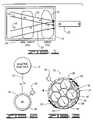
- FIG. 1is a diagrammatic illustration of the present invention showing the use of a magnetic field to move superparamagnetic nanoparticles and their associated bioactive substance through a non-target cell and into a target cell.
- FIG. 1further shows the release of the bioactive substance from the nanoparticle inside the target cell.
- FIG. 2is a diagrammatic illustration of a nanoparticle having a biocompatible shell comprised of silica. The nanoparticle is shown bound to a bioactive substance via a covalent bond.
- FIG. 3is a diagrammatic representation of nanosphere delivery system constructed in accordance with the present invention.
- the nanosphere of FIG. 3comprises a plurality of nanoparticles each having a biocompatible shell.
- the nanoparticlesare encapsulated within an outer biocompatible shell.
- the nanosphereis shown having a bioactive substance comprising a genetic material bonded to the outer biocompatible shell.
- FIG. 4is a diagrammatic representation of an alternative embodiment of a nanosphere constructed in accordance with the present invention.
- the nanosphere of FIG. 4comprises a plurality of silica coated nanoparticles bonded to a bioerodable polymer.
- the bioerodable polymeris shown supporting a genetic material.
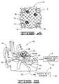
- FIG. 5is an illustration of a human ear showing the movement of nanospheres through the round window membrane and into the inner ear.
- the magnetic field generatoris shown in a plurality of positions to illustrate guided movement of the nanospheres by moving the magnetic field generator.
- bioactive substancessuch as drugs or genetic material
- Delivery of bioactive substances, such as drugs or genetic material, to specific cellsallows for the treatment of diseases and conditions that affect the human body.
- Several methods and systemshave been developed to accomplish delivery of the bioactive substance.
- Targeted delivery of bioactive substances using nanospheres and/or nanoparticles to a specific site within a bodyprovides advantages over systemic or oral administration of the bioactive substance to the body.
- effective doses of bioactive substancemay be delivered at varying doses to a desired target cell without exposing the entire body to adverse conditions or side effects.
- the present method and systemallows for the delivery of bioactive substance into sensitive or remote areas of the body in a non-invasive manner using an externally controlled magnetic field adapted to move the nanoparticle in three dimensions.
- Viral agentshave been used for targeted delivery of genetic material to specific cells within the body.
- a viral agent that has an affinity for the target cellsis chosen to transport the genetic material to the target cells.
- viruses to transport genetic material to specific cellspresents difficulties such as infection of the host body, mutation of the virus, and incitement of harmful immunogenic reactions.
- virusesare of such a size that there use may cause damaging trauma to the body by requiring invasive procedures.
- the present inventionis useful in that it minimizes trauma to the body and can use non-immunogenic substances.
- FIG. 1there is shown therein a system for introducing a bioactive substance 10 into a target cell 12 disposed within a body 14 .
- the bioactive substance 10is shown bonded to a superparamagnetic nanoparticle 16 .
- the nanoparticle 16may be covered by a biocompatible shell 18 ( FIG. 2 ) that is adapted to bond the bioactive substance 10 to the nanoparticle 16 .
- a magnetic field generator 20is positioned outside the body 14 to move the nanoparticles 16 in three dimensions and into the target cell 12 .
- the magnetic field generator 20To move the nanoparticle 16 and the bioactive substance 10 into the target cell 12 the magnetic field generator 20 generates a gradient, represented by arrows 22 , which attracts the nanoparticle to the magnetic field generator and into the target cell.
- the use of a magnetic field gradient 22facilitates internalization of the nanoparticle 16 and bioactive substance 10 by the target cell 12 .
- Facilitating uptake of the nanoparticle 16 and bioactive substance 10 using the magnetic field generatormay prevent premature release of the bioactive substance from the nanoparticle.
- the magnetic field generator 20may comprise a plurality of magnets (not shown) that are arranged such that a magnetic field is generated, within which numerous gradients 22 may be created to three-dimensionally direct the nanoparticles 16 to the target cell 12 .
- An alternative magnetic field generatormay comprise an electromagnetic field generating coil that is movable in three dimensions and adapted to create a gradient 22 that moves the nanoparticle 16 through a non-target cell 24 and into the target cell 12 . It will be appreciated that the electromagnetic field generating coil may be moved by any means that permits three-dimensional movement of the nanoparticle 16 through the body 14 .
- the electromagnetic field generating coilmay be supported on the end of a robotic arm (not shown) that is programmed to move around the body 14 so that the nanoparticle 16 is directed in three dimensions to the target cell 12 .
- the nanoparticle 16may be comprised of a ferrite such as magnetite and is preferably superparamagnetic. Because the nanoparticles 16 are superparamagnetic, the nanoparticles will only be attracted to the strongest side of the magnetic field gradient 22 and will not be attracted by other or similar nanoparticles when in a magnetic field. Thus, particle to particle interactions resulting in aggregation or other undesirable effects are minimized. Once the magnetic field is removed, the nanoparticles 16 lose their magnetic remanence.
- the nanoparticle 16 of FIG. 2is shown encapsulated in a biocompatible shell 18 .
- the biocompatible shell 18may comprise silica (SiO 2 ) or titania (TiO 2 ). Encapsulation of the nanoparticle 16 in the biocompatible shell 18 hermetically seals the nanoparticle to help prevent corrosion of the nanoparticle and provides a surface charge to promote suspension of the nanoparticle in solution to facilitate uptake of the nanoparticle by non-target 24 and target cells 12 .
- the biocompatible shell 18also provides a substrate for the attachment of amines 26 that can serve as linkers to other molecules.
- the biocompatible shell 18 of FIG. 2is shown to provide a covalent bond 30 such as a Sulfhydryl bond between the bioactive substance 10 and the nanoparticle 16 .
- FIG. 3there is shown therein a diagrammatic representation of a nanosphere 32 prepared using the methods and systems described in co-pending U.S. patent application Ser. No. 10/724,563, the contents of which are incorporated herein by reference.
- the nanosphere 32 of FIG. 3comprises a plurality of superparamagnetic nanoparticles 16 supported within the nanosphere by an erodable polymer matrix (not shown). Each nanoparticle 16 may be encapsulated within the previously described biocompatible silica shell 18 .
- the nanosphere 32has an outer shell 34 that may be adapted to support the bioactive substance 10 .
- the nanospheregenerally has a diameter of less than 300 nanometers, and more preferably a diameter of 100 nanometers or less.
- the nanoparticles 16may be arranged within the outer shell 34 such that they have uniformly aligned magnetic moments 36 . Uniform alignment of the nanoparticles' magnetic moments 36 increases the magnetic susceptibility of the nanosphere 32 thus providing more efficient transport of the nanosphere and the bioactive substance 10 through the body 14 and into the target cell 12 .
- the outer shell 34generally encapsulates the nanoparticle 16 and provides a support mechanism for the bioactive substance 10 so that it may be transported with the nanoparticles to the target cell 12 .
- the outer shell 34may comprise a bioerodable polymer that is adapted to release an encapsulated bioactive substance 38 .
- the outer bioerodable shell 34may comprise any erodable synthetic or natural polymer that is biocompatible. Polylactides, polyglycolides and collagen have been found to be acceptable for use as the outer bioerodable shell 34 of the nanosphere 32 .
- the nanosphere 32may form a reservoir 40 that encapsulates the bioactive substance 38 and the nanoparticles 16 within the nanosphere. As the outer shell 34 is dissolved, the bioactive substance 38 is released from the nanosphere 32 and dispersed into the cytoplasm (not shown) of the target cell 12 . The inclusion of the erodable polymer matrix further aids in regulating release of the bioactive substance 38 .
- the bioerodable polymer matrixmay be used to entrap the bioactive substance 38 within the outer bioerodable shell 38 .
- the erodable polymer matrixis non-toxic and capable of being consumed, metabolized or expelled by the target cell 12 .
- FIG. 1An example of such an erodable polymer matrix is collagen. A tightly cross-linked matrix will exhibit a slow release rate providing low doses of bioactive substance 38 over longer periods of time. When no bioerodable matrix is present rapid release of the bioactive substance 38 can be expected.
- the bioactive substance 10may alternatively be supported on the outer shell 34 .
- the outer shell 34may be formed from either the bioerodable polymer or a biostable polymer.
- the outer shell 34 of the nanosphere 32may comprise a silica matrix.
- the silica matrixmay have a plurality of amine groups 26 attached to the outer surface 42 of the outer shell 34 that functionalize the nanosphere 32 . These amine groups 26 give the outer surface 42 of the shell 34 a net positive charge.
- a positively charged outer shell 34has an affinity for bioactive substance 10 comprising genetic material that has a generally negative net charge.
- bioactive substance 10 or 38may itself form the outer shell by attaching the bioactive substance directly to the silica coated nanoparticles 16 or alternatively to the previously described silica matrix.
- the outer shell 34 of the nanosphere 32may have a cell adhesion factor (not shown) supported on the outer surface 42 of the shell 34 .
- the use of cell adhesion factorsenhances endocytosis of the bioactive substance 10 or 38 supported by the nanosphere 32 by the target cell 12 .
- the cell adhesion factormay comprise a protein having an affinity for a predetermined type of cell. It will be appreciated that a wide array of cell adhesion factors may be used with nanospheres 32 of the present invention without departing from the spirit of the invention.
- FIG. 4there is shown therein an alternative nanosphere 44 of the present invention that may be used to deliver the bioactive substance 10 to the target cell 12 .
- the nanosphere 44 of FIG. 4comprises a plurality of superparamagnetic nanoparticles 16 supported by a bioerodable polymer matrix 46 .
- the nanoparticles 16are shown with the biocompatible shell 18 .
- the nanoparticles 16may be supported by the bioerodable polymer matrix 46 so that they have substantially aligned magnetic moments 36 .
- the bioactive substance 10is likewise supported by the bioerodable polymer matrix 46 so that the amount of bioactive substance 10 released from the nanosphere 44 and into the target cell 12 may be controlled over time.
- FIG. 5there is shown therein an illustration of a human ear 48 .
- the ear 48 shown in FIG. 5comprises an outer ear 50 , a middle ear 52 and an inner ear 54 .
- the outer ear 50has an ear canal 56 that is closed at one end by a tympanic membrane 58 , or eardrum.
- the middle earcomprises an ossicular chain that normally connects the ear drum 58 to a cochlea 60 .
- the ossicular chainincludes a malleus 62 , an incus 64 , and a stapes 66 .
- a properly functioning ossicular chaintransmits and amplifies sound vibrations from the ear drum 58 through the malleus 62 , incus 64 and stapes 66 to vibrate an oval window (not shown) of the inner ear 54 . Vibration of the oval window is transmitted to the fluid of the inner ear to cause movement of ear sensory cells within the cochlea 60 of the inner ear 54 . Electrical impulses from the ear sensory cells are sent from the cochlea 60 along an auditory nerve 68 to the brain of the mammal where the signals are processed for hearing.
- Damage to the ear sensory cells, or hair cells, of the cochlea 60is the leading cause of sensorineural hearing loss. Congenital conditions and/or exposure to injurious levels of noise may be the cause of damage to the hair cells. After the hair cells are initially damaged, a number of inner ear cell death programs are activated that result in eventual hair cell death and permanent hearing loss. However, the supporting cells may remain alive with the capacity to regenerate hair cells and restore hearing when triggered by the appropriate bioactive substance 10 .
- FIG. 5illustrates a method of moving magnetically responsive nanospheres 32 or 44 , as described herein, into the inner ear 54 for regeneration or repair of hair cells.
- Nanosphere 32is used herein for illustration purposes, it will be appreciated that nanospheres having different constructions and configurations and individual nanoparticles 16 as previously described herein may be used to treat the target cells without departing from the spirit of the invention.
- the nanospheres 32are placed near the round window membrane 70 of the inner ear 54 and pulled through the round window membrane using the gradient 22 generated by the magnetic field generator 20 in position A. Once inside the cochlea 60 , the nanospheres 32 are moved is three dimensions through the perilymph to hair cell supporting cells using the external magnetic field generator 20 .
- the diagrammatic magnetic field generator 20is shown, in FIG. 5 , in an alternative position B to facilitate movement of the nanosphere 32 through the basal turn 72 of the cochlea 60 .
- the magnetic field generator 20may be moved to an alternative position to facilitate magnetofection of the nanosphere into the cell.
- the bioactive substance 10is released into the cytoplasm of the target cell to begin repair or regeneration of the hair cells.
- the bioactive substance 10 released into the hair cellsmay comprise a genetic material such as the Hath-1 gene.
- the Hath-1 genehas been shown to stimulate regeneration of hair cells in mammals. See, “Robust Generation of New Hair Cells in the Mature Mammalian Inner Ear by Adenovirus Expression of Hath-1,” J. Shou, J. L. Zheng, W. Q. Gao, Molecular and Cellular Neuroscience 2003; 23:169-170, the contents of which are incorporated herein by reference.
- the present inventionalso comprises a method for introducing a bioactive substance 10 into a target cell 12 within a body 14 .
- the bioactive substance 10is generally associated with a superparamagnetic nanoparticle 16 .
- the bioactive substance 10is introduced into the target cell 12 by introducing the bioactive substance and the nanoparticle 16 into the body 14 and moving the bioactive substance into the target cell.
- the bioactive substance 10is moved into the target cell 12 using an externally controlled magnetic field that is adapted to move the nanoparticle 16 and bioactive substance through the body 14 and any non-target cells 24 .
- Movement of the nanoparticle 16may comprise generating a gradient 22 in the external magnetic field.
- one of the nanoparticles 16 or nanospheres 32 or 44 as described hereinmay be used for this purpose.
- the bioactive substance 10may comprise genetic materials, such as DNA, RNA, plasmids, oligonucleotides or proteins, which are bonded to the biocompatible silica shell 18 that covers the nanoparticle 16 .
- the bond between the genetic material 10 and the silica shell 18is adapted to release the genetic material after the nanoparticle 16 and genetic material are pulled into the target cell 12 .
- the body 14may comprise a mammal having a target cell 12 disposed within the cochlea 60 of the mammal's ear 50 .
- the externally controlled magnetic fieldmay be used to move the genetic material 10 and nanoparticle 16 into the cochlea 16 , then to disperse the genetic material throughout the cochlea and across the cellular membrane (not shown) of the ear sensory cells.
- the genetic materialOnce inside the target ear sensory cell 12 , the genetic material may be released from the nanoparticle 16 or nanosphere 32 .
- the genetic material 10may then transfect the ear sensory cell or the supporting cell to cause repair or regeneration of the cells.
- Superparamagnetic nanoparticles having a silica shellwere synthesized using the modified Massart procedures described in co-pending U.S. patent application Ser. No. 10/724,563.
- the nanoparticleswere made of magnetite (Fe3O4) and synthesized to have a diameter of less than 30-50 nanometers.
- a two Molar iron (III) sulfate heptahydrate solutionwas prepared in two (2) Molar HCl and combined with one Molar iron (III) chloride hexahydrate aqueous solution. The solutions were mixed and washed in a 0.7 Molar ammonium hydroxide solution and rapidly stirred. The resulting precipitate was stirred for thirty (30) minutes then collected using a magnet.
- the precipitatewas re-suspended in 0.7 Molar ammonium hydroxide and peptized by the addition of one (1) Molar tetramethylammonium hydroxide aliquots. The volume of the resulting suspension was then taken to 250 ml for processing to add the silica shell to the nanoparticles.
- Encapsulation of the nanoparticle with silicaprovides an anionic surface charge that promotes endocytosis as well as a substrate for attachment of amines adapted to link the bioactive substance to the nanoparticle.
- the suspension of magnetite nanoparticleswas stirred and a 4 ml aliquot was taken up to 100 ml with distilled water.
- a solution of 0.54% sodium silicatewas prepared at a pH of 10.5, and 4 ml of the sodium silicate was added to the magnetite nanoparticle suspension.
- the pH of the resulting suspensionwas adjusted to 10.0 and stirred for an extended period of time. After settling for several hours, the silica-coated nanoparticles were removed from the excess silica using a magnet to pull the particles out of the solution and by washing the precipitate several times with distilled water.
- silica-coated nanoparticleswere analyzed using TEM to determine the size and structure of the nanoparticles produced in the above procedure. Analysis of the coated nanoparticles revealed an average diameter of approximately sixteen (16) nanometers with a standard deviation of 2.3 nanometers. The presence of the silica shell and iron oxide core was confirmed by energy-dispersive X-ray spectrometry (“EDS”).
- EDSenergy-dispersive X-ray spectrometry
- Silica-coated nanoparticleswere then functionalized by the addition of amine groups to the surface of the silica shell.
- the nanoparticleswere treated with 3-aminopropyl trimethoxy silane and a 1 ml aliquot of the resulting suspension was brought to a volume of 5 ml with distilled water. Additional 3-aminopropyl trimethoxy silane was added to the suspension to bring the final concentration to five percent (5%).
- the reaction systemwas stirred and the resulting nanoparticles were washed and collected.
- a Kaiser assaywas performed on several of the functionalized nanoparticles to confirm the presence of amine groups on the surface of the silica-coated nanoparticles.
- Fluorescein isothiocyanatewas used to label the nanoparticle for subsequent location of the nanoparticle using confocal microscopy.
- the particleswere conjugated with FITC using standard protocols to attach the FITC to the amine functional groups.
- Guinea pigswere anesthetized and positioned such that an experimental ear was facing upward and parallel to the operating table. A retro-articular incision was made to expose the temporal bone over the middle ear cavity. The middle ear space was opened using an otological surgical drill system (MicroCraftTM, Xomed Inc., Jacksonville, Fla.) to expose the ossicular chain of the subjects.
- otological surgical drill systemMocroCraftTM, Xomed Inc., Jacksonville, Fla.
- the silica-coated magnetic nanoparticleswere suspended in saline at a pH of 7.4 and sonicated for several minutes. Sonication was performed to disperse the nanoparticles before placement onto the ossicular epithelium. A volume of 50-75 microliters of the nanoparticle suspension was applied to the target cells in 25 microliter doses. The operative site was closed and the subjects recovered during application of an external magnetic field to the their heads.
- An externally vectored magnetic forcewas applied to the heads of the experimental animals using an external magnet so that the nanoparticles were pulled downward into the epithelia of the incus and tympanic membrane.
- the magnetcreated a magnetic field of approximately 0.35 Tesla at one inch from the experimental incus and tympanic membrane.
- Each subjectwas exposed to the external magnetic field for 20 to 30 minutes and subsequently monitored for survival for several days.
Landscapes
- Health & Medical Sciences (AREA)
- Life Sciences & Earth Sciences (AREA)
- Engineering & Computer Science (AREA)
- Chemical & Material Sciences (AREA)
- General Health & Medical Sciences (AREA)
- Bioinformatics & Cheminformatics (AREA)
- Pharmacology & Pharmacy (AREA)
- Epidemiology (AREA)
- Medicinal Chemistry (AREA)
- Animal Behavior & Ethology (AREA)
- Public Health (AREA)
- Veterinary Medicine (AREA)
- Genetics & Genomics (AREA)
- Biomedical Technology (AREA)
- Wood Science & Technology (AREA)
- Organic Chemistry (AREA)
- Biotechnology (AREA)
- General Engineering & Computer Science (AREA)
- Zoology (AREA)
- Microbiology (AREA)
- Physics & Mathematics (AREA)
- Plant Pathology (AREA)
- Molecular Biology (AREA)
- Biochemistry (AREA)
- Biophysics (AREA)
- Dermatology (AREA)
- Medicinal Preparation (AREA)
- Medicines That Contain Protein Lipid Enzymes And Other Medicines (AREA)
Abstract
Description
Claims (19)
Priority Applications (3)
| Application Number | Priority Date | Filing Date | Title |
|---|---|---|---|
| US10/871,243US7723311B2 (en) | 2003-06-18 | 2004-06-18 | Delivery of bioactive substances to target cells |
| PCT/US2004/027225WO2006009559A1 (en) | 2004-06-18 | 2004-08-20 | Delivery of bioactive substances to target cells |
| US12/505,111US8651113B2 (en) | 2003-06-18 | 2009-07-17 | Magnetically responsive nanoparticle therapeutic constructs and methods of making and using |
Applications Claiming Priority (2)
| Application Number | Priority Date | Filing Date | Title |
|---|---|---|---|
| US47938103P | 2003-06-18 | 2003-06-18 | |
| US10/871,243US7723311B2 (en) | 2003-06-18 | 2004-06-18 | Delivery of bioactive substances to target cells |
Related Parent Applications (1)
| Application Number | Title | Priority Date | Filing Date |
|---|---|---|---|
| US71211207AContinuation-In-Part | 2003-06-18 | 2007-02-28 |
Related Child Applications (1)
| Application Number | Title | Priority Date | Filing Date |
|---|---|---|---|
| US11/400,620Continuation-In-PartUS8001977B2 (en) | 2003-06-18 | 2006-04-06 | Device for moving magnetic nanoparticles through tissue |
Publications (2)
| Publication Number | Publication Date |
|---|---|
| US20050271732A1 US20050271732A1 (en) | 2005-12-08 |
| US7723311B2true US7723311B2 (en) | 2010-05-25 |
Family
ID=34958639
Family Applications (1)
| Application Number | Title | Priority Date | Filing Date |
|---|---|---|---|
| US10/871,243Expired - Fee RelatedUS7723311B2 (en) | 2003-06-18 | 2004-06-18 | Delivery of bioactive substances to target cells |
Country Status (2)
| Country | Link |
|---|---|
| US (1) | US7723311B2 (en) |
| WO (1) | WO2006009559A1 (en) |
Cited By (7)
| Publication number | Priority date | Publication date | Assignee | Title |
|---|---|---|---|---|
| US20090287036A1 (en)* | 2008-05-19 | 2009-11-19 | University Of Maryland | Methods And Systems For Using Therapeutic, Diagnostic or Prophylactic Magnetic Agents |
| US20110105825A1 (en)* | 2009-10-31 | 2011-05-05 | Qteris Inc. | Nanoparticle-sized magnetic absorption enhancers having three-dimensional geometries adapted for improved diagnostics and hyperthermic treatment |
| WO2011153348A2 (en) | 2010-06-04 | 2011-12-08 | Hough Ear Institute | Composition and method for inner ear sensory hair cell regeneration and replacement |
| US8740872B2 (en) | 2010-10-19 | 2014-06-03 | The Board Of Regents Of The University Of Oklahoma | Magnetically-targeted treatment for cardiac disorders |
| EP3144033A1 (en) | 2015-09-17 | 2017-03-22 | Sociedade Beneficente Israelita Brasileira Hospital Albert Einstein | Method and system for configuring a magnetic field generating device for directioning magnetic substances |
| US9744235B2 (en) | 2010-10-19 | 2017-08-29 | The Board Of Regents Of The University Of Oklahoma | Treatment of cardiovascular disorders with targeted nanoparticles |
| US10398668B2 (en) | 2010-10-19 | 2019-09-03 | The Board Of Regents Of The University Of Oklahoma | Glutamate treatment of cardiovascular disorders |
Families Citing this family (24)
| Publication number | Priority date | Publication date | Assignee | Title |
|---|---|---|---|---|
| US8651113B2 (en) | 2003-06-18 | 2014-02-18 | Swr&D Inc. | Magnetically responsive nanoparticle therapeutic constructs and methods of making and using |
| US8001977B2 (en)* | 2005-04-08 | 2011-08-23 | Nanobiomagnetics, Inc. | Device for moving magnetic nanoparticles through tissue |
| ES2313350T3 (en) | 2004-05-12 | 2009-03-01 | Baxter International Inc. | MICROSPHERAS OF NUCLEIC ACID, PRODUCTION AND SUPPLY OF THE SAME. |
| US7833016B2 (en)* | 2006-05-23 | 2010-11-16 | Dentatek Corporation | Root canal filling materials and methods |
| WO2008019346A2 (en) | 2006-08-04 | 2008-02-14 | Baxter International Inc. | Microsphere-based composition for preventing and/or reversing new-onset autoimmune diabetes |
| EP2137533A2 (en)* | 2007-04-19 | 2009-12-30 | 3M Innovative Properties Company | Uses of water-dispersible silica nanoparticles for attaching biomolecules |
| WO2009086071A2 (en)* | 2007-12-20 | 2009-07-09 | Mayo Foundation For Medical Education And Research | Systems and methods for magnetic-assisted therapeutic agent delivery |
| US8367427B2 (en) | 2008-08-20 | 2013-02-05 | Baxter International Inc. | Methods of processing compositions containing microparticles |
| US8323615B2 (en) | 2008-08-20 | 2012-12-04 | Baxter International Inc. | Methods of processing multi-phasic dispersions |
| US8323685B2 (en) | 2008-08-20 | 2012-12-04 | Baxter International Inc. | Methods of processing compositions containing microparticles |
| US11890226B2 (en)* | 2009-02-25 | 2024-02-06 | University Of Maryland, College Park | Device and methods for directing agents into an eye |
| US8316862B2 (en)* | 2009-02-25 | 2012-11-27 | University Of Maryland | Devices, systems and methods for magnetic-assisted therapeutic agent delivery |
| US10576295B2 (en)* | 2009-02-25 | 2020-03-03 | University Of Maryland | Device and methods for directing agents to the middle ear and the inner ear |
| US8715150B2 (en) | 2009-11-02 | 2014-05-06 | Pulse Therapeutics, Inc. | Devices for controlling magnetic nanoparticles to treat fluid obstructions |
| US9380959B2 (en)* | 2011-08-18 | 2016-07-05 | Weinberg Medical Physics Llc | MRI-guided nanoparticle cancer therapy apparatus and methodology |
| EP2758036A2 (en)* | 2011-09-21 | 2014-07-30 | Yissum Research and Development Company of The Hebrew University of Jerusalem | Nano delivery systems |
| KR20130084091A (en) | 2012-01-16 | 2013-07-24 | 삼성전자주식회사 | Image forming apparatus |
| US9883878B2 (en) | 2012-05-15 | 2018-02-06 | Pulse Therapeutics, Inc. | Magnetic-based systems and methods for manipulation of magnetic particles |
| US20140322137A1 (en)* | 2013-04-25 | 2014-10-30 | Edward R. Flynn | Detection Of Targeted Biological Substances Using Magnetic Relaxation Of Individual Nanoparticles |
| CN103980365A (en)* | 2014-06-03 | 2014-08-13 | 国家纳米科学中心 | Magnetic nanoparticle immobilized with anthrax lethal factor on surface as well as preparation method and application of magnetic nanoparticle |
| WO2016007629A2 (en) | 2014-07-08 | 2016-01-14 | University Of Maryland, Baltimore | Compositions and delivery methods for treating dental infections, inflammation, sensitivity, and for use in dental restorations |
| JP6730294B2 (en)* | 2014-10-30 | 2020-07-29 | オトマグネティクス,リミテッド・ライアビリティ・カンパニー | Magnetic injection of therapeutic agent by adding protrusions of material with different magnetization and magnetic permeability |
| US11918315B2 (en) | 2018-05-03 | 2024-03-05 | Pulse Therapeutics, Inc. | Determination of structure and traversal of occlusions using magnetic particles |
| US12171443B1 (en) | 2021-03-09 | 2024-12-24 | Pulse Therapeutics, Inc. | Magnetically controlled flow generation |
Citations (19)
| Publication number | Priority date | Publication date | Assignee | Title |
|---|---|---|---|---|
| US4501726A (en) | 1981-11-12 | 1985-02-26 | Schroeder Ulf | Intravascularly administrable, magnetically responsive nanosphere or nanoparticle, a process for the production thereof, and the use thereof |
| US4652257A (en) | 1985-03-21 | 1987-03-24 | The United States Of America As Represented By The Secretary Of The Navy | Magnetically-localizable, polymerized lipid vesicles and method of disrupting same |
| US4690130A (en) | 1985-12-19 | 1987-09-01 | Mirell Stuart G | Electromagnetic therapy control system |
| WO1998001160A2 (en) | 1996-07-10 | 1998-01-15 | Danbiosyst Uk Limited | Compositions suitable for delivery of genes to epithelial cells |
| US5916539A (en) | 1993-03-02 | 1999-06-29 | Silica Gel Ges. M.B.H. | Superparamagnetic particles, process for producing the same and their use |
| US5928958A (en) | 1994-07-27 | 1999-07-27 | Pilgrimm; Herbert | Superparamagnetic particles, process for their manufacture and usage |
| WO1999060998A1 (en) | 1998-05-27 | 1999-12-02 | Euroceltique S.A. | Preparations for the application of anti-inflammatory, especially antiseptic agents and/or agents promoting the healing of wounds, to the upper respiratory tract and/or the ear |
| US6014580A (en)* | 1997-11-12 | 2000-01-11 | Stereotaxis, Inc. | Device and method for specifying magnetic field for surgical applications |
| US6274554B1 (en)* | 1997-07-30 | 2001-08-14 | Amgen Inc. | Method for preventing and treating hearing loss using a neurturin protein product |
| US6344357B1 (en) | 1998-06-10 | 2002-02-05 | Immunoporation Ltd | Treating cells |
| US20020086842A1 (en) | 2000-06-26 | 2002-07-04 | Christian Plank | Method for transfecting cells using a magnetic field |
| WO2002056890A1 (en) | 2001-01-19 | 2002-07-25 | Synphora Ab | Novel method and compositions for local treatment of meniere's disease, tinnitus and/or hearing loss |
| US6436028B1 (en) | 1999-12-28 | 2002-08-20 | Soundtec, Inc. | Direct drive movement of body constituent |
| US6548264B1 (en)* | 2000-05-17 | 2003-04-15 | University Of Florida | Coated nanoparticles |
| WO2003059194A2 (en) | 2001-12-21 | 2003-07-24 | Alcon, Inc. | Use of synthetic inorganic nanoparticles as carriers for ophthalmic and otic drugs |
| US20030215394A1 (en)* | 2002-05-17 | 2003-11-20 | Short Robert E. | Microparticles having a matrix interior useful for ultrasound triggered delivery of drugs into the bloodstream |
| WO2004006765A1 (en) | 2002-07-17 | 2004-01-22 | Dailey James P | Delivery of therapeutic agent affixed to magnetic particle |
| US6767635B1 (en) | 1999-09-14 | 2004-07-27 | Biomedical Apherese Systeme Gmbh | Magnetic nanoparticles having biochemical activity, method for the production thereof and their use |
| US7189198B2 (en)* | 2002-07-03 | 2007-03-13 | Stereotaxis, Inc. | Magnetically guidable carriers and methods for the targeted magnetic delivery of substances in the body |
Family Cites Families (46)
| Publication number | Priority date | Publication date | Assignee | Title |
|---|---|---|---|---|
| US86842A (en)* | 1869-02-09 | Thomas b | ||
| US4376740A (en)* | 1981-01-05 | 1983-03-15 | National Research Institute For Metals | Process for production fine metal particles |
| US4356029A (en)* | 1981-12-23 | 1982-10-26 | Westinghouse Electric Corp. | Titanium product collection in a plasma reactor |
| US4526922A (en)* | 1983-04-15 | 1985-07-02 | Union Carbide Corporation | Organofunctional silane-siloxane oligomer coupling compositions, curable and cured elastomeric compositions containing same and novel electric cable containing said cured elastomeric compositions |
| US4466896A (en)* | 1983-07-29 | 1984-08-21 | Texaco Inc. | Ethylenediamine triacetic acid siloxane stabilizers for inorganic silicates in antifreeze/coolant formulations |
| SE463651B (en)* | 1983-12-21 | 1991-01-07 | Nycomed As | DIAGNOSTIC AND CONTRACTOR |
| US6203777B1 (en)* | 1983-12-21 | 2001-03-20 | Nycomed Imaging As | Method of contrast enhanced magnetic resonance imaging using carbohydrate particles |
| US4687511A (en)* | 1986-05-15 | 1987-08-18 | Gte Products Corporation | Metal matrix composite powders and process for producing same |
| DE3709851A1 (en)* | 1987-03-24 | 1988-10-06 | Silica Gel Gmbh Adsorptions Te | NMR DIAGNOSTIC LIQUID COMPOSITIONS |
| US5069936A (en)* | 1987-06-25 | 1991-12-03 | Yen Richard C K | Manufacturing protein microspheres |
| JP2790486B2 (en)* | 1988-11-08 | 1998-08-27 | 日東紡績株式会社 | Silane coupling agents and glass fiber products for laminates |
| DE4117782C2 (en)* | 1991-05-28 | 1997-07-17 | Diagnostikforschung Inst | Nanocrystalline magnetic iron oxide particles, processes for their production and diagnostic and / or therapeutic agents |
| US5512474A (en)* | 1992-05-29 | 1996-04-30 | Bsi Corporation | Cell culture support containing a cell adhesion factor and a positively-charged molecule |
| US5349957A (en)* | 1992-12-02 | 1994-09-27 | Sterling Winthrop Inc. | Preparation and magnetic properties of very small magnetite-dextran particles |
| ATE210967T1 (en)* | 1993-01-29 | 2002-01-15 | Ferx Inc | MAGNETICALLY REACTING COMPOSITION AS A CARRIER FOR BIOLOGICALLY ACTIVE SUBSTANCES AND METHOD FOR THE PRODUCTION AND USE THEREOF |
| US6482436B1 (en)* | 1993-01-29 | 2002-11-19 | Ferx Incorporated | Magnetically responsive composition |
| US6200547B1 (en)* | 1994-01-26 | 2001-03-13 | Ferx Incorporated | Magnetically responsive compositions for carrying biologically active substances and methods of production and use |
| DE4309333A1 (en)* | 1993-03-17 | 1994-09-22 | Silica Gel Gmbh | Superparamagnetic particles, process for their production and use thereof |
| US5565215A (en)* | 1993-07-23 | 1996-10-15 | Massachusettes Institute Of Technology | Biodegradable injectable particles for imaging |
| US6007845A (en)* | 1994-07-22 | 1999-12-28 | Massachusetts Institute Of Technology | Nanoparticles and microparticles of non-linear hydrophilic-hydrophobic multiblock copolymers |
| DE4428851C2 (en)* | 1994-08-04 | 2000-05-04 | Diagnostikforschung Inst | Nanoparticles containing iron, their production and application in diagnostics and therapy |
| US5749937A (en)* | 1995-03-14 | 1998-05-12 | Lockheed Idaho Technologies Company | Fast quench reactor and method |
| WO1997005994A1 (en)* | 1995-08-04 | 1997-02-20 | Microcoating Technologies Inc | Chemical vapor deposition and powder formation using thermal spray with near supercritical and supercritical fluid solutions |
| US5711803A (en)* | 1995-09-29 | 1998-01-27 | Midwest Research Institute | Preparation of a semiconductor thin film |
| US5876683A (en)* | 1995-11-02 | 1999-03-02 | Glumac; Nicholas | Combustion flame synthesis of nanophase materials |
| US5695901A (en)* | 1995-12-21 | 1997-12-09 | Colorado School Of Mines | Nano-size magnetic particles for reprographic processes and method of manufacturing the same |
| GB9600427D0 (en)* | 1996-01-10 | 1996-03-13 | Nycomed Imaging As | Contrast media |
| US6441025B2 (en)* | 1996-03-12 | 2002-08-27 | Pg-Txl Company, L.P. | Water soluble paclitaxel derivatives |
| JP2862509B2 (en)* | 1996-05-28 | 1999-03-03 | 東洋電化工業株式会社 | Carrier for lipase immobilization and immobilized lipase |
| CA2259691A1 (en)* | 1996-07-11 | 1998-01-22 | The University Of Cincinnati | Electrically assisted synthesis of particles and films with precisely controlled characteristics |
| US5788738A (en)* | 1996-09-03 | 1998-08-04 | Nanomaterials Research Corporation | Method of producing nanoscale powders by quenching of vapors |
| US5851507A (en)* | 1996-09-03 | 1998-12-22 | Nanomaterials Research Corporation | Integrated thermal process for the continuous synthesis of nanoscale powders |
| US6344271B1 (en)* | 1998-11-06 | 2002-02-05 | Nanoenergy Corporation | Materials and products using nanostructured non-stoichiometric substances |
| CA2303268A1 (en)* | 1997-06-13 | 1998-12-17 | Scott Walsh | Therapeutic nanospheres |
| US5984997A (en)* | 1997-08-29 | 1999-11-16 | Nanomaterials Research Corporation | Combustion of emulsions: A method and process for producing fine powders |
| IL123210A0 (en)* | 1998-02-06 | 1998-09-24 | Gombinsky Moshe | A device and system for the collection of magnetic particles |
| US6623982B1 (en)* | 1999-07-12 | 2003-09-23 | Immunivest Corporation | Increased separation efficiency via controlled aggregation of magnetic nanoparticles |
| US6472632B1 (en)* | 1999-09-15 | 2002-10-29 | Nanoscale Engineering And Technology Corporation | Method and apparatus for direct electrothermal-physical conversion of ceramic into nanopowder |
| US6600127B1 (en)* | 1999-09-15 | 2003-07-29 | Nanotechnologies, Inc. | Method and apparatus for direct electrothermal-physical conversion of ceramic into nanopowder |
| WO2001037721A2 (en)* | 1999-11-22 | 2001-05-31 | The Research Foundation Of State University Of New York | Magnetic nanoparticles for selective therapy |
| US7169618B2 (en)* | 2000-06-28 | 2007-01-30 | Skold Technology | Magnetic particles and methods of producing coated magnetic particles |
| US20020046993A1 (en)* | 2000-10-24 | 2002-04-25 | Peterson Dennis Roger | Electrothermal gun for direct electrothermal-physical conversion of precursor into nanopowder |
| US6767637B2 (en)* | 2000-12-13 | 2004-07-27 | Purdue Research Foundation | Microencapsulation using ultrasonic atomizers |
| US20020155059A1 (en)* | 2001-04-24 | 2002-10-24 | Tekna Plasma Systems Inc. | Plasma synthesis of titanium dioxide nanopowder and powder doping and surface modification process |
| PL372247A1 (en)* | 2002-02-01 | 2005-07-11 | Pfizer Products Inc. | Method for making homogeneous spray-dried solid amorphous drug dispersions utilizing modified spray-drying apparatus |
| WO2004066975A1 (en)* | 2002-12-18 | 2004-08-12 | Hough Ear Institute | Otologic nanotechnology |
- 2004
- 2004-06-18USUS10/871,243patent/US7723311B2/ennot_activeExpired - Fee Related
- 2004-08-20WOPCT/US2004/027225patent/WO2006009559A1/enactiveApplication Filing
Patent Citations (19)
| Publication number | Priority date | Publication date | Assignee | Title |
|---|---|---|---|---|
| US4501726A (en) | 1981-11-12 | 1985-02-26 | Schroeder Ulf | Intravascularly administrable, magnetically responsive nanosphere or nanoparticle, a process for the production thereof, and the use thereof |
| US4652257A (en) | 1985-03-21 | 1987-03-24 | The United States Of America As Represented By The Secretary Of The Navy | Magnetically-localizable, polymerized lipid vesicles and method of disrupting same |
| US4690130A (en) | 1985-12-19 | 1987-09-01 | Mirell Stuart G | Electromagnetic therapy control system |
| US5916539A (en) | 1993-03-02 | 1999-06-29 | Silica Gel Ges. M.B.H. | Superparamagnetic particles, process for producing the same and their use |
| US5928958A (en) | 1994-07-27 | 1999-07-27 | Pilgrimm; Herbert | Superparamagnetic particles, process for their manufacture and usage |
| WO1998001160A2 (en) | 1996-07-10 | 1998-01-15 | Danbiosyst Uk Limited | Compositions suitable for delivery of genes to epithelial cells |
| US6274554B1 (en)* | 1997-07-30 | 2001-08-14 | Amgen Inc. | Method for preventing and treating hearing loss using a neurturin protein product |
| US6014580A (en)* | 1997-11-12 | 2000-01-11 | Stereotaxis, Inc. | Device and method for specifying magnetic field for surgical applications |
| WO1999060998A1 (en) | 1998-05-27 | 1999-12-02 | Euroceltique S.A. | Preparations for the application of anti-inflammatory, especially antiseptic agents and/or agents promoting the healing of wounds, to the upper respiratory tract and/or the ear |
| US6344357B1 (en) | 1998-06-10 | 2002-02-05 | Immunoporation Ltd | Treating cells |
| US6767635B1 (en) | 1999-09-14 | 2004-07-27 | Biomedical Apherese Systeme Gmbh | Magnetic nanoparticles having biochemical activity, method for the production thereof and their use |
| US6436028B1 (en) | 1999-12-28 | 2002-08-20 | Soundtec, Inc. | Direct drive movement of body constituent |
| US6548264B1 (en)* | 2000-05-17 | 2003-04-15 | University Of Florida | Coated nanoparticles |
| US20020086842A1 (en) | 2000-06-26 | 2002-07-04 | Christian Plank | Method for transfecting cells using a magnetic field |
| WO2002056890A1 (en) | 2001-01-19 | 2002-07-25 | Synphora Ab | Novel method and compositions for local treatment of meniere's disease, tinnitus and/or hearing loss |
| WO2003059194A2 (en) | 2001-12-21 | 2003-07-24 | Alcon, Inc. | Use of synthetic inorganic nanoparticles as carriers for ophthalmic and otic drugs |
| US20030215394A1 (en)* | 2002-05-17 | 2003-11-20 | Short Robert E. | Microparticles having a matrix interior useful for ultrasound triggered delivery of drugs into the bloodstream |
| US7189198B2 (en)* | 2002-07-03 | 2007-03-13 | Stereotaxis, Inc. | Magnetically guidable carriers and methods for the targeted magnetic delivery of substances in the body |
| WO2004006765A1 (en) | 2002-07-17 | 2004-01-22 | Dailey James P | Delivery of therapeutic agent affixed to magnetic particle |
Non-Patent Citations (25)
| Title |
|---|
| "Magnets Help Target Gene Therapy", Press release, 2002, Bioelectromagnetics Society. |
| "The Basics of Silane Chemistry", A Guide to Silane Solutions from Dow Corning, pp. 7-8, 2005, Dow Corning Corporation. |
| Brigger et al. "Nanoparticles in cancer therapy and diagnosis," Advanced Drug Delivery Reviews, vol. 54, (2002) pp. 631-651, Elsevier. |
| Bruce M. Moskowitz, "Domain Theory", Hitchhiker's Guide to Magnetism, Enviromental Magnetism Workshop, pp. 21-33, Jun. 1991. |
| C. Wilhelm, C. Billotey, J. Roger, J.N. Pons, J.-C. Bacri, F. Gazeau, Intracellular uptake of anionic superparamagnetic nanoparticles as a function of their surface coating, article, Sep. 9, 2002,Laboratoire des Milieux Desordonnes et Heterogenes, Universite Pierre et Marie Curie, Parris, France. |
| Caruso et al. Chem. Mater. 13:109-116; 2001.* |
| Chang "Adriamycin-loaded immunological magnetic nanoparticles: Site-specific targeting . . . ," Chinese Journal of Biomedical Eng., vol. 15, No. 4 (1996) pp. 354-359, abstract only. |
| Chantal A. Lackey, Oliver W. Press, Allan S. Hoffman, and Patrick S. Stayton, A Biomimetic pH-Responsive Polymer Directs Endosomal Release and Intracellular Delivery of an Endocytosed Antibody Complex, article, Jul. 25, 2002, Department of Bioengineering, University of Washington. |
| Christian Plank, Ulrike Schillinger, Franz Scherer, Christian Bergemann, Jean-Serge Remy, Florian Krotz, Martina Anton, Jim Lausier and Joseph Rosenecker, "The Magnetofection method: Using Magnetic Force to Enhance Gene Delivery", Biol. Chem., vol. 384, pp. 737-747, May 2003. |
| Duclairoir et al. "Alpha-Tocopherol encapsulation and in vitro release from wheat gliadin nanoparticles," J. Microencapsulation, vol. 19 (2002), No. 1, pp. 53-60. |
| Electronic Publication by Business Communications Company, Inc., "Nanoparticle News", issued Oct. 2002. |
| Fadee Mondalek, Concerns Regarding the Permeability of the Round Window Membrane (RWM) to Magnetite Nanoparticles Attached to a Drug/Gene, article, Oct. 28, 2003, OU Health Sciences Center, Oklahoma City, Oklahoma. |
| Ge Liu, Deshan Li, Murali K. Pasumarthy, Tomasz H. Kowalczyk, Christopher R. Gedeon, Susannah L. Hyatt, Jennifer M. Payne, Timothy J. Miller, Peter Brunovskis, Tamara L. Fink, Osman Muhammad, Robert C. Moen, Richard W. Hanson, and Mark J. Cooper, Nanoparticles of Compacted DNA Transfect Postmitotic Cells, article, Jun. 14, 2003, Department of Biochemistry, Case Western Reserve University School of Medicine, Cleveland, Ohio. |
| Jayanth Panyam, Vinod Labhasetwar, Biodegradable nanoparticles for drug and gene delivery to cells and tissue, article, Sep. 16, 2002, Department of Pharmaceutical Sciences, University of Nebraska. |
| Jayanth Panyam, Wen-Zhong Zhou, Swayam Prabha, Sanjeeb K. Sahoo, and Vinod Labhasetwar, Rapid endo-lysosomal escape of poly(DL-lactide-co-glycolide) nanoparticles: implications for drug and gene delivery, article, Apr. 18, 2002, Department of Pharmaceutical Sciences, University of Nebraska Medical Center. |
| Junghae Suh, Denis Wirtz, and Justin Hanes, Efficient active transport of gene nanocarriers to the cell nucleus, article, Apr. 1, 2003, Departments of Biomedical Engineering, Chemical and Biomolecular Engineering, and Materials Science and Engineering, Molecular Biophysics Program, The Johns Hopkins University, Baltimore, Maryland. |
| N. Buske, C. Gansau, T. Rheinlander and B. Kroll, "Magnetic Sizing of Magnetic Nanoparticles",Mediport Kardiotechnik GmBH, 2000. |
| Nicoli et al. "Design of triptorelin loaded nanospheres for transdermal iontophoretic administration," International Journal of Pharmaceutics, No. 214 (2001) pp. 31-35. |
| Niren Murthy, Jean Campbell, Nelson Fausto, Allan S. Hoffman, and Patrick S. Stayton, Bioinspired pH-Responsive Polymers for the Intracellular Delivery of Biomolecular Drugs, article, Jan. 15, 2003, Department of Bioengineering and Department of Pathology, University of Washington. |
| Rachel A. Jones, Charles Y. Cheung, Fiona E. Black, Jasmine K. Zia, Patrick S. Stayton, Allan S. Hoffman, and Mark R. Wilson, Poly(2-alkyacrylic acid) polymers deliver molecules to the cytosol by pH-sensitive disruption of endosomal vesicles, article, 2003, Department of Biological Sciences, University of Wollongong, Wollongong, Australia and University of Washington. |
| Schutt et al. "Biocompatible Magnetic Polymer Carriers for In Vivo Radionuclide Delivery," International Society for Artificial Organs, vol. 23, No. 1 (1999) pp. 98-103. |
| Swayam Prabha, Wen-Zhong Zhou, Jayanth Panyam, Vinod Labhasetwar, Size-dependency of nanoparticle-mediated gene transfection: studies with fractionated nanoparticles, article, Jun. 6, 2002, Department of Pharmaceutical Sciences, University of Nebraska Medical Center. |
| Utreja et al. "Lipoprotein-mimicking biovectorized systems for methotrexate delivery," Pharmaceutica Acta Helvetiae, No. 73 (1999) pp. 275-279 (Abstract XP-002383903). |
| X.X. He, K.M. Wang, W.H. Tan, X. Lin, L. Chen and X.H. Chen, A Novel Method For Efficient Gene Delivery Using Amino-Modified Silica Coated Magnetic Nanoparticles, article, Jul. 27, 2003, State Key Laboratory of Chemo/Biosensing and Chemometrics, College of Chemistry & Chemical Engineering, Institute of Biological Science and Biological Technology, Hunan University, P.R. China. |
| Yong Zhang, Nathan Kohler, Miqin Zhang, Surface modification of superparamagnetic magnetite nanoparticles and their intracellular uptake, article, Aug. 8, 2001, Department of Materials Science & Engineering. University of Washington. |
Cited By (11)
| Publication number | Priority date | Publication date | Assignee | Title |
|---|---|---|---|---|
| US20090287036A1 (en)* | 2008-05-19 | 2009-11-19 | University Of Maryland | Methods And Systems For Using Therapeutic, Diagnostic or Prophylactic Magnetic Agents |
| US8579787B2 (en)* | 2008-05-19 | 2013-11-12 | University Of Maryland College Park | Methods and systems for using therapeutic, diagnostic or prophylactic magnetic agents |
| US9108035B2 (en) | 2008-05-19 | 2015-08-18 | University Of Maryland, College Park | Methods and systems for using therapeutic, diagnostic or prophylactic magnetic agents |
| US20110105825A1 (en)* | 2009-10-31 | 2011-05-05 | Qteris Inc. | Nanoparticle-sized magnetic absorption enhancers having three-dimensional geometries adapted for improved diagnostics and hyperthermic treatment |
| US8565892B2 (en) | 2009-10-31 | 2013-10-22 | Qteris, Inc. | Nanoparticle-sized magnetic absorption enhancers having three-dimensional geometries adapted for improved diagnostics and hyperthermic treatment |
| US9844679B2 (en) | 2009-10-31 | 2017-12-19 | Qteris, Inc. | Nanoparticle-sized magnetic absorption enhancers having three-dimensional geometries adapted for improved diagnostics and hyperthermic treatment |
| WO2011153348A2 (en) | 2010-06-04 | 2011-12-08 | Hough Ear Institute | Composition and method for inner ear sensory hair cell regeneration and replacement |
| US8740872B2 (en) | 2010-10-19 | 2014-06-03 | The Board Of Regents Of The University Of Oklahoma | Magnetically-targeted treatment for cardiac disorders |
| US9744235B2 (en) | 2010-10-19 | 2017-08-29 | The Board Of Regents Of The University Of Oklahoma | Treatment of cardiovascular disorders with targeted nanoparticles |
| US10398668B2 (en) | 2010-10-19 | 2019-09-03 | The Board Of Regents Of The University Of Oklahoma | Glutamate treatment of cardiovascular disorders |
| EP3144033A1 (en) | 2015-09-17 | 2017-03-22 | Sociedade Beneficente Israelita Brasileira Hospital Albert Einstein | Method and system for configuring a magnetic field generating device for directioning magnetic substances |
Also Published As
| Publication number | Publication date |
|---|---|
| WO2006009559A1 (en) | 2006-01-26 |
| US20050271732A1 (en) | 2005-12-08 |
Similar Documents
| Publication | Publication Date | Title |
|---|---|---|
| US7723311B2 (en) | Delivery of bioactive substances to target cells | |
| Arsianti et al. | Assembly of polyethylenimine-based magnetic iron oxide vectors: insights into gene delivery | |
| Li et al. | Advances in nano-based inner ear delivery systems for the treatment of sensorineural hearing loss | |
| Kopke et al. | Magnetic nanoparticles: inner ear targeted molecule delivery and middle ear implant | |
| Kenneth et al. | Epithelial internalization of superparamagnetic nanoparticles and response to external magnetic field | |
| AU2010332221B2 (en) | Uniform field magnetization and targeting of therapeutic formulations | |
| JP6232487B2 (en) | Compositions and methods for regeneration or replacement of inner ear sensory hair cells | |
| Xiao et al. | PAMAM dendrimer/pDNA functionalized-magnetic iron oxide nanoparticles for gene delivery | |
| Pyykkö et al. | An overview of nanoparticle based delivery for treatment of inner ear disorders | |
| CN107847429B (en) | Method for targeting or stimulating cells or organisms using nanoparticles and external field | |
| De Temmerman et al. | Tailoring layer-by-layer capsules for biomedical applications | |
| AU2018337076B2 (en) | Method of treatment | |
| Cheng et al. | Rabies virus glycoprotein-mediated transportation and T cell infiltration to brain tumor by magnetoelectric gold yarnballs | |
| JP2022180476A (en) | Combination therapies for inner ear sensory hair cell regeneration/replacement | |
| Lu et al. | A photothermal nanoplatform with sugar-triggered cleaning ability for high-efficiency intracellular delivery | |
| US11103720B2 (en) | Methods for stimulating cells using nanoparticles and external field | |
| US7344491B1 (en) | Method and apparatus for improving hearing | |
| Byun et al. | Bioassembly of multicellular spheroids to mimic complex tissue structure using surface-modified magnetized nanofibers | |
| US20060093611A1 (en) | Method | |
| Pyykkö et al. | Nanoparticle based inner ear therapy | |
| Poe et al. | Nanotechnology and the treatment of inner ear diseases | |
| Bakhtiar et al. | Intracellular delivery of p53 gene and MAPK siRNA into breast cancer cells utilizing barium salt nanoparticles | |
| WO2007116808A1 (en) | Virus particle-like structure and pharmaceutical preparation comprising the same | |
| US20240149035A1 (en) | Conductive Biocompatible Scaffold For Electroporation | |
| WO2020045162A1 (en) | Drug delivery carrier |
Legal Events
| Date | Code | Title | Description |
|---|---|---|---|
| AS | Assignment | Owner name:NAVY, UNITED STATES OF AMERICA, THE, AS REPRESENTE Free format text:ASSIGNMENT OF ASSIGNORS INTEREST;ASSIGNOR:KOPKE, RICHARD D.;REEL/FRAME:015296/0721 Effective date:20041021 | |
| AS | Assignment | Owner name:NANOBIOMAGNETICS, INC., OKLAHOMA Free format text:ASSIGNMENT OF ASSIGNORS INTEREST;ASSIGNORS:SEENEY, CHARLES E.;DORMER, KENNETH J.;KOPKE, RICHARD D.;REEL/FRAME:015949/0851 Effective date:20041021 Owner name:OKLAHOMA, THE UNIVERSITY OF, OKLAHOMA Free format text:ASSIGNMENT OF ASSIGNORS INTEREST;ASSIGNORS:SEENEY, CHARLES E.;DORMER, KENNETH J.;KOPKE, RICHARD D.;REEL/FRAME:015949/0851 Effective date:20041021 Owner name:HOUGH EAR INSTITUTE, OKLAHOMA Free format text:ASSIGNMENT OF ASSIGNORS INTEREST;ASSIGNORS:SEENEY, CHARLES E.;DORMER, KENNETH J.;KOPKE, RICHARD D.;REEL/FRAME:015949/0851 Effective date:20041021 Owner name:NAVAL MEDICAL CENTER SAN DIEGO, CALIFORNIA Free format text:ASSIGNMENT OF ASSIGNORS INTEREST;ASSIGNORS:SEENEY, CHARLES E.;DORMER, KENNETH J.;KOPKE, RICHARD D.;REEL/FRAME:015949/0851 Effective date:20041021 Owner name:NANOBIOMAGNETICS, INC.,OKLAHOMA Free format text:ASSIGNMENT OF ASSIGNORS INTEREST;ASSIGNORS:SEENEY, CHARLES E.;DORMER, KENNETH J.;KOPKE, RICHARD D.;REEL/FRAME:015949/0851 Effective date:20041021 Owner name:OKLAHOMA, THE UNIVERSITY OF,OKLAHOMA Free format text:ASSIGNMENT OF ASSIGNORS INTEREST;ASSIGNORS:SEENEY, CHARLES E.;DORMER, KENNETH J.;KOPKE, RICHARD D.;REEL/FRAME:015949/0851 Effective date:20041021 Owner name:HOUGH EAR INSTITUTE,OKLAHOMA Free format text:ASSIGNMENT OF ASSIGNORS INTEREST;ASSIGNORS:SEENEY, CHARLES E.;DORMER, KENNETH J.;KOPKE, RICHARD D.;REEL/FRAME:015949/0851 Effective date:20041021 Owner name:NAVAL MEDICAL CENTER SAN DIEGO,CALIFORNIA Free format text:ASSIGNMENT OF ASSIGNORS INTEREST;ASSIGNORS:SEENEY, CHARLES E.;DORMER, KENNETH J.;KOPKE, RICHARD D.;REEL/FRAME:015949/0851 Effective date:20041021 | |
| STCF | Information on status: patent grant | Free format text:PATENTED CASE | |
| AS | Assignment | Owner name:SWR&D INC., OKLAHOMA Free format text:ASSIGNMENT OF ASSIGNORS INTEREST;ASSIGNOR:NANOBIOMAGNETICS, INC;REEL/FRAME:029780/0404 Effective date:20120328 | |
| FPAY | Fee payment | Year of fee payment:4 | |
| FPAY | Fee payment | Year of fee payment:8 | |
| FEPP | Fee payment procedure | Free format text:MAINTENANCE FEE REMINDER MAILED (ORIGINAL EVENT CODE: REM.); ENTITY STATUS OF PATENT OWNER: SMALL ENTITY | |
| LAPS | Lapse for failure to pay maintenance fees | Free format text:PATENT EXPIRED FOR FAILURE TO PAY MAINTENANCE FEES (ORIGINAL EVENT CODE: EXP.); ENTITY STATUS OF PATENT OWNER: SMALL ENTITY | |
| STCH | Information on status: patent discontinuation | Free format text:PATENT EXPIRED DUE TO NONPAYMENT OF MAINTENANCE FEES UNDER 37 CFR 1.362 | |
| FP | Lapsed due to failure to pay maintenance fee | Effective date:20220525 |