US7722676B2 - Articulating implant system - Google Patents
Articulating implant systemDownload PDFInfo
- Publication number
- US7722676B2 US7722676B2US10/772,129US77212904AUS7722676B2US 7722676 B2US7722676 B2US 7722676B2US 77212904 AUS77212904 AUS 77212904AUS 7722676 B2US7722676 B2US 7722676B2
- Authority
- US
- United States
- Prior art keywords
- stem
- distal
- head
- implant
- extension
- Prior art date
- Legal status (The legal status is an assumption and is not a legal conclusion. Google has not performed a legal analysis and makes no representation as to the accuracy of the status listed.)
- Expired - Fee Related, expires
Links
- 239000007943implantSubstances0.000titleclaimsabstractdescription77
- 210000000623ulnaAnatomy0.000claimsabstractdescription79
- 238000003780insertionMethods0.000claimsabstractdescription7
- 230000037431insertionEffects0.000claimsabstractdescription7
- 210000004872soft tissueAnatomy0.000claimsdescription27
- 238000002271resectionMethods0.000claimsdescription18
- 238000000034methodMethods0.000claimsdescription16
- 210000000186triangular fibrocartilageAnatomy0.000claimsdescription10
- 230000013011matingEffects0.000claimsdescription8
- 210000005115ulnar canalAnatomy0.000claimsdescription7
- 210000003484anatomyAnatomy0.000claimsdescription4
- 239000002775capsuleSubstances0.000claimsdescription4
- 239000011248coating agentSubstances0.000claimsdescription4
- 238000000576coating methodMethods0.000claimsdescription4
- 230000003278mimic effectEffects0.000claims2
- 210000000988bone and boneAnatomy0.000abstractdescription27
- 210000000845cartilageAnatomy0.000abstractdescription4
- 206010017076FractureDiseases0.000description17
- 210000000707wristAnatomy0.000description12
- 210000003041ligamentAnatomy0.000description10
- 210000001519tissueAnatomy0.000description10
- 208000010392Bone FracturesDiseases0.000description9
- 210000000245forearmAnatomy0.000description8
- 208000014674injuryDiseases0.000description6
- 238000000926separation methodMethods0.000description6
- 230000006378damageEffects0.000description5
- 206010037802Radius fractureDiseases0.000description4
- 208000027418Wounds and injuryDiseases0.000description4
- 206010039073rheumatoid arthritisDiseases0.000description4
- 238000001356surgical procedureMethods0.000description4
- 206010003246arthritisDiseases0.000description3
- 230000033001locomotionEffects0.000description3
- 201000008482osteoarthritisDiseases0.000description3
- 239000004606Fillers/ExtendersSubstances0.000description2
- 208000002565Open FracturesDiseases0.000description2
- 239000004568cementSubstances0.000description2
- 210000004439collateral ligamentAnatomy0.000description2
- 238000013461designMethods0.000description2
- 210000003811fingerAnatomy0.000description2
- 230000035876healingEffects0.000description2
- 238000002513implantationMethods0.000description2
- 238000001727in vivoMethods0.000description2
- 238000012546transferMethods0.000description2
- 230000008733traumaEffects0.000description2
- 210000003857wrist jointAnatomy0.000description2
- 208000009692Colles' FractureDiseases0.000description1
- 208000006670Multiple fracturesDiseases0.000description1
- 206010031264OsteonecrosisDiseases0.000description1
- 208000002607PseudarthrosisDiseases0.000description1
- 206010048873Traumatic arthritisDiseases0.000description1
- 230000006978adaptationEffects0.000description1
- 210000003423ankleAnatomy0.000description1
- 238000011882arthroplastyMethods0.000description1
- 230000009286beneficial effectEffects0.000description1
- 239000002639bone cementSubstances0.000description1
- 210000003557bones of lower extremityAnatomy0.000description1
- 210000003199bones of upper extremityAnatomy0.000description1
- 238000005266castingMethods0.000description1
- 208000037976chronic inflammationDiseases0.000description1
- 208000037893chronic inflammatory disorderDiseases0.000description1
- 230000000295complement effectEffects0.000description1
- 230000000694effectsEffects0.000description1
- 210000001513elbowAnatomy0.000description1
- 239000000835fiberSubstances0.000description1
- 230000004927fusionEffects0.000description1
- 210000004349growth plateAnatomy0.000description1
- 208000015181infectious diseaseDiseases0.000description1
- 210000003127kneeAnatomy0.000description1
- 230000014759maintenance of locationEffects0.000description1
- 238000012986modificationMethods0.000description1
- 230000004048modificationEffects0.000description1
- 210000003205muscleAnatomy0.000description1
- 206010033675panniculitisDiseases0.000description1
- 230000001575pathological effectEffects0.000description1
- 230000035515penetrationEffects0.000description1
- 238000000554physical therapyMethods0.000description1
- 239000011505plasterSubstances0.000description1
- 229920003229poly(methyl methacrylate)Polymers0.000description1
- 239000004926polymethyl methacrylateSubstances0.000description1
- 229920001296polysiloxanePolymers0.000description1
- 238000011084recoveryMethods0.000description1
- 238000004904shorteningMethods0.000description1
- 230000000087stabilizing effectEffects0.000description1
- 210000004304subcutaneous tissueAnatomy0.000description1
- 230000008961swellingEffects0.000description1
- 208000011580syndromic diseaseDiseases0.000description1
- 210000001258synovial membraneAnatomy0.000description1
- 230000000451tissue damageEffects0.000description1
- 231100000827tissue damageToxicity0.000description1
Images


Classifications
- A—HUMAN NECESSITIES
- A61—MEDICAL OR VETERINARY SCIENCE; HYGIENE
- A61F—FILTERS IMPLANTABLE INTO BLOOD VESSELS; PROSTHESES; DEVICES PROVIDING PATENCY TO, OR PREVENTING COLLAPSING OF, TUBULAR STRUCTURES OF THE BODY, e.g. STENTS; ORTHOPAEDIC, NURSING OR CONTRACEPTIVE DEVICES; FOMENTATION; TREATMENT OR PROTECTION OF EYES OR EARS; BANDAGES, DRESSINGS OR ABSORBENT PADS; FIRST-AID KITS
- A61F2/00—Filters implantable into blood vessels; Prostheses, i.e. artificial substitutes or replacements for parts of the body; Appliances for connecting them with the body; Devices providing patency to, or preventing collapsing of, tubular structures of the body, e.g. stents
- A61F2/02—Prostheses implantable into the body
- A61F2/30—Joints
- A61F2/42—Joints for wrists or ankles; for hands, e.g. fingers; for feet, e.g. toes
- A61F2/4261—Joints for wrists or ankles; for hands, e.g. fingers; for feet, e.g. toes for wrists
- A—HUMAN NECESSITIES
- A61—MEDICAL OR VETERINARY SCIENCE; HYGIENE
- A61B—DIAGNOSIS; SURGERY; IDENTIFICATION
- A61B17/00—Surgical instruments, devices or methods
- A61B17/04—Surgical instruments, devices or methods for suturing wounds; Holders or packages for needles or suture materials
- A61B17/06—Needles ; Sutures; Needle-suture combinations; Holders or packages for needles or suture materials
- A61B17/06166—Sutures
- A—HUMAN NECESSITIES
- A61—MEDICAL OR VETERINARY SCIENCE; HYGIENE
- A61F—FILTERS IMPLANTABLE INTO BLOOD VESSELS; PROSTHESES; DEVICES PROVIDING PATENCY TO, OR PREVENTING COLLAPSING OF, TUBULAR STRUCTURES OF THE BODY, e.g. STENTS; ORTHOPAEDIC, NURSING OR CONTRACEPTIVE DEVICES; FOMENTATION; TREATMENT OR PROTECTION OF EYES OR EARS; BANDAGES, DRESSINGS OR ABSORBENT PADS; FIRST-AID KITS
- A61F2/00—Filters implantable into blood vessels; Prostheses, i.e. artificial substitutes or replacements for parts of the body; Appliances for connecting them with the body; Devices providing patency to, or preventing collapsing of, tubular structures of the body, e.g. stents
- A61F2/02—Prostheses implantable into the body
- A61F2/30—Joints
- A61F2/46—Special tools for implanting artificial joints
- A61F2/4603—Special tools for implanting artificial joints for insertion or extraction of endoprosthetic joints or of accessories thereof
- A61F2/4606—Special tools for implanting artificial joints for insertion or extraction of endoprosthetic joints or of accessories thereof of wrists or ankles; of hands, e.g. fingers; of feet, e.g. toes
- A—HUMAN NECESSITIES
- A61—MEDICAL OR VETERINARY SCIENCE; HYGIENE
- A61F—FILTERS IMPLANTABLE INTO BLOOD VESSELS; PROSTHESES; DEVICES PROVIDING PATENCY TO, OR PREVENTING COLLAPSING OF, TUBULAR STRUCTURES OF THE BODY, e.g. STENTS; ORTHOPAEDIC, NURSING OR CONTRACEPTIVE DEVICES; FOMENTATION; TREATMENT OR PROTECTION OF EYES OR EARS; BANDAGES, DRESSINGS OR ABSORBENT PADS; FIRST-AID KITS
- A61F2/00—Filters implantable into blood vessels; Prostheses, i.e. artificial substitutes or replacements for parts of the body; Appliances for connecting them with the body; Devices providing patency to, or preventing collapsing of, tubular structures of the body, e.g. stents
- A61F2/02—Prostheses implantable into the body
- A61F2/30—Joints
- A61F2/30767—Special external or bone-contacting surface, e.g. coating for improving bone ingrowth
- A—HUMAN NECESSITIES
- A61—MEDICAL OR VETERINARY SCIENCE; HYGIENE
- A61F—FILTERS IMPLANTABLE INTO BLOOD VESSELS; PROSTHESES; DEVICES PROVIDING PATENCY TO, OR PREVENTING COLLAPSING OF, TUBULAR STRUCTURES OF THE BODY, e.g. STENTS; ORTHOPAEDIC, NURSING OR CONTRACEPTIVE DEVICES; FOMENTATION; TREATMENT OR PROTECTION OF EYES OR EARS; BANDAGES, DRESSINGS OR ABSORBENT PADS; FIRST-AID KITS
- A61F2/00—Filters implantable into blood vessels; Prostheses, i.e. artificial substitutes or replacements for parts of the body; Appliances for connecting them with the body; Devices providing patency to, or preventing collapsing of, tubular structures of the body, e.g. stents
- A61F2/02—Prostheses implantable into the body
- A61F2/30—Joints
- A61F2/46—Special tools for implanting artificial joints
- A61F2/4637—Special tools for implanting artificial joints for connecting or disconnecting two parts of a prosthesis
- A—HUMAN NECESSITIES
- A61—MEDICAL OR VETERINARY SCIENCE; HYGIENE
- A61F—FILTERS IMPLANTABLE INTO BLOOD VESSELS; PROSTHESES; DEVICES PROVIDING PATENCY TO, OR PREVENTING COLLAPSING OF, TUBULAR STRUCTURES OF THE BODY, e.g. STENTS; ORTHOPAEDIC, NURSING OR CONTRACEPTIVE DEVICES; FOMENTATION; TREATMENT OR PROTECTION OF EYES OR EARS; BANDAGES, DRESSINGS OR ABSORBENT PADS; FIRST-AID KITS
- A61F2/00—Filters implantable into blood vessels; Prostheses, i.e. artificial substitutes or replacements for parts of the body; Appliances for connecting them with the body; Devices providing patency to, or preventing collapsing of, tubular structures of the body, e.g. stents
- A61F2/02—Prostheses implantable into the body
- A61F2/30—Joints
- A61F2/46—Special tools for implanting artificial joints
- A61F2/4684—Trial or dummy prostheses
- A—HUMAN NECESSITIES
- A61—MEDICAL OR VETERINARY SCIENCE; HYGIENE
- A61F—FILTERS IMPLANTABLE INTO BLOOD VESSELS; PROSTHESES; DEVICES PROVIDING PATENCY TO, OR PREVENTING COLLAPSING OF, TUBULAR STRUCTURES OF THE BODY, e.g. STENTS; ORTHOPAEDIC, NURSING OR CONTRACEPTIVE DEVICES; FOMENTATION; TREATMENT OR PROTECTION OF EYES OR EARS; BANDAGES, DRESSINGS OR ABSORBENT PADS; FIRST-AID KITS
- A61F2/00—Filters implantable into blood vessels; Prostheses, i.e. artificial substitutes or replacements for parts of the body; Appliances for connecting them with the body; Devices providing patency to, or preventing collapsing of, tubular structures of the body, e.g. stents
- A61F2/02—Prostheses implantable into the body
- A61F2/30—Joints
- A61F2002/30001—Additional features of subject-matter classified in A61F2/28, A61F2/30 and subgroups thereof
- A61F2002/30108—Shapes
- A61F2002/3011—Cross-sections or two-dimensional shapes
- A61F2002/30138—Convex polygonal shapes
- A61F2002/30156—Convex polygonal shapes triangular
- A—HUMAN NECESSITIES
- A61—MEDICAL OR VETERINARY SCIENCE; HYGIENE
- A61F—FILTERS IMPLANTABLE INTO BLOOD VESSELS; PROSTHESES; DEVICES PROVIDING PATENCY TO, OR PREVENTING COLLAPSING OF, TUBULAR STRUCTURES OF THE BODY, e.g. STENTS; ORTHOPAEDIC, NURSING OR CONTRACEPTIVE DEVICES; FOMENTATION; TREATMENT OR PROTECTION OF EYES OR EARS; BANDAGES, DRESSINGS OR ABSORBENT PADS; FIRST-AID KITS
- A61F2/00—Filters implantable into blood vessels; Prostheses, i.e. artificial substitutes or replacements for parts of the body; Appliances for connecting them with the body; Devices providing patency to, or preventing collapsing of, tubular structures of the body, e.g. stents
- A61F2/02—Prostheses implantable into the body
- A61F2/30—Joints
- A61F2002/30001—Additional features of subject-matter classified in A61F2/28, A61F2/30 and subgroups thereof
- A61F2002/30108—Shapes
- A61F2002/30199—Three-dimensional shapes
- A61F2002/302—Three-dimensional shapes toroidal, e.g. rings
- A—HUMAN NECESSITIES
- A61—MEDICAL OR VETERINARY SCIENCE; HYGIENE
- A61F—FILTERS IMPLANTABLE INTO BLOOD VESSELS; PROSTHESES; DEVICES PROVIDING PATENCY TO, OR PREVENTING COLLAPSING OF, TUBULAR STRUCTURES OF THE BODY, e.g. STENTS; ORTHOPAEDIC, NURSING OR CONTRACEPTIVE DEVICES; FOMENTATION; TREATMENT OR PROTECTION OF EYES OR EARS; BANDAGES, DRESSINGS OR ABSORBENT PADS; FIRST-AID KITS
- A61F2/00—Filters implantable into blood vessels; Prostheses, i.e. artificial substitutes or replacements for parts of the body; Appliances for connecting them with the body; Devices providing patency to, or preventing collapsing of, tubular structures of the body, e.g. stents
- A61F2/02—Prostheses implantable into the body
- A61F2/30—Joints
- A61F2002/30001—Additional features of subject-matter classified in A61F2/28, A61F2/30 and subgroups thereof
- A61F2002/30316—The prosthesis having different structural features at different locations within the same prosthesis; Connections between prosthetic parts; Special structural features of bone or joint prostheses not otherwise provided for
- A61F2002/30329—Connections or couplings between prosthetic parts, e.g. between modular parts; Connecting elements
- A61F2002/30331—Connections or couplings between prosthetic parts, e.g. between modular parts; Connecting elements made by longitudinally pushing a protrusion into a complementarily-shaped recess, e.g. held by friction fit
- A61F2002/30332—Conically- or frustoconically-shaped protrusion and recess
- A—HUMAN NECESSITIES
- A61—MEDICAL OR VETERINARY SCIENCE; HYGIENE
- A61F—FILTERS IMPLANTABLE INTO BLOOD VESSELS; PROSTHESES; DEVICES PROVIDING PATENCY TO, OR PREVENTING COLLAPSING OF, TUBULAR STRUCTURES OF THE BODY, e.g. STENTS; ORTHOPAEDIC, NURSING OR CONTRACEPTIVE DEVICES; FOMENTATION; TREATMENT OR PROTECTION OF EYES OR EARS; BANDAGES, DRESSINGS OR ABSORBENT PADS; FIRST-AID KITS
- A61F2/00—Filters implantable into blood vessels; Prostheses, i.e. artificial substitutes or replacements for parts of the body; Appliances for connecting them with the body; Devices providing patency to, or preventing collapsing of, tubular structures of the body, e.g. stents
- A61F2/02—Prostheses implantable into the body
- A61F2/30—Joints
- A61F2002/30001—Additional features of subject-matter classified in A61F2/28, A61F2/30 and subgroups thereof
- A61F2002/30316—The prosthesis having different structural features at different locations within the same prosthesis; Connections between prosthetic parts; Special structural features of bone or joint prostheses not otherwise provided for
- A61F2002/30535—Special structural features of bone or joint prostheses not otherwise provided for
- A61F2002/30574—Special structural features of bone or joint prostheses not otherwise provided for with an integral complete or partial collar or flange
- A—HUMAN NECESSITIES
- A61—MEDICAL OR VETERINARY SCIENCE; HYGIENE
- A61F—FILTERS IMPLANTABLE INTO BLOOD VESSELS; PROSTHESES; DEVICES PROVIDING PATENCY TO, OR PREVENTING COLLAPSING OF, TUBULAR STRUCTURES OF THE BODY, e.g. STENTS; ORTHOPAEDIC, NURSING OR CONTRACEPTIVE DEVICES; FOMENTATION; TREATMENT OR PROTECTION OF EYES OR EARS; BANDAGES, DRESSINGS OR ABSORBENT PADS; FIRST-AID KITS
- A61F2/00—Filters implantable into blood vessels; Prostheses, i.e. artificial substitutes or replacements for parts of the body; Appliances for connecting them with the body; Devices providing patency to, or preventing collapsing of, tubular structures of the body, e.g. stents
- A61F2/02—Prostheses implantable into the body
- A61F2/30—Joints
- A61F2002/30001—Additional features of subject-matter classified in A61F2/28, A61F2/30 and subgroups thereof
- A61F2002/30316—The prosthesis having different structural features at different locations within the same prosthesis; Connections between prosthetic parts; Special structural features of bone or joint prostheses not otherwise provided for
- A61F2002/30535—Special structural features of bone or joint prostheses not otherwise provided for
- A61F2002/30604—Special structural features of bone or joint prostheses not otherwise provided for modular
- A—HUMAN NECESSITIES
- A61—MEDICAL OR VETERINARY SCIENCE; HYGIENE
- A61F—FILTERS IMPLANTABLE INTO BLOOD VESSELS; PROSTHESES; DEVICES PROVIDING PATENCY TO, OR PREVENTING COLLAPSING OF, TUBULAR STRUCTURES OF THE BODY, e.g. STENTS; ORTHOPAEDIC, NURSING OR CONTRACEPTIVE DEVICES; FOMENTATION; TREATMENT OR PROTECTION OF EYES OR EARS; BANDAGES, DRESSINGS OR ABSORBENT PADS; FIRST-AID KITS
- A61F2/00—Filters implantable into blood vessels; Prostheses, i.e. artificial substitutes or replacements for parts of the body; Appliances for connecting them with the body; Devices providing patency to, or preventing collapsing of, tubular structures of the body, e.g. stents
- A61F2/02—Prostheses implantable into the body
- A61F2/30—Joints
- A61F2002/30001—Additional features of subject-matter classified in A61F2/28, A61F2/30 and subgroups thereof
- A61F2002/30667—Features concerning an interaction with the environment or a particular use of the prosthesis
- A61F2002/30691—Drainage means, e.g. for evacuating blood or other fluids
- A—HUMAN NECESSITIES
- A61—MEDICAL OR VETERINARY SCIENCE; HYGIENE
- A61F—FILTERS IMPLANTABLE INTO BLOOD VESSELS; PROSTHESES; DEVICES PROVIDING PATENCY TO, OR PREVENTING COLLAPSING OF, TUBULAR STRUCTURES OF THE BODY, e.g. STENTS; ORTHOPAEDIC, NURSING OR CONTRACEPTIVE DEVICES; FOMENTATION; TREATMENT OR PROTECTION OF EYES OR EARS; BANDAGES, DRESSINGS OR ABSORBENT PADS; FIRST-AID KITS
- A61F2/00—Filters implantable into blood vessels; Prostheses, i.e. artificial substitutes or replacements for parts of the body; Appliances for connecting them with the body; Devices providing patency to, or preventing collapsing of, tubular structures of the body, e.g. stents
- A61F2/02—Prostheses implantable into the body
- A61F2/30—Joints
- A61F2/30767—Special external or bone-contacting surface, e.g. coating for improving bone ingrowth
- A61F2/30771—Special external or bone-contacting surface, e.g. coating for improving bone ingrowth applied in original prostheses, e.g. holes or grooves
- A61F2002/30772—Apertures or holes, e.g. of circular cross section
- A61F2002/30784—Plurality of holes
- A—HUMAN NECESSITIES
- A61—MEDICAL OR VETERINARY SCIENCE; HYGIENE
- A61F—FILTERS IMPLANTABLE INTO BLOOD VESSELS; PROSTHESES; DEVICES PROVIDING PATENCY TO, OR PREVENTING COLLAPSING OF, TUBULAR STRUCTURES OF THE BODY, e.g. STENTS; ORTHOPAEDIC, NURSING OR CONTRACEPTIVE DEVICES; FOMENTATION; TREATMENT OR PROTECTION OF EYES OR EARS; BANDAGES, DRESSINGS OR ABSORBENT PADS; FIRST-AID KITS
- A61F2/00—Filters implantable into blood vessels; Prostheses, i.e. artificial substitutes or replacements for parts of the body; Appliances for connecting them with the body; Devices providing patency to, or preventing collapsing of, tubular structures of the body, e.g. stents
- A61F2/02—Prostheses implantable into the body
- A61F2/30—Joints
- A61F2/30767—Special external or bone-contacting surface, e.g. coating for improving bone ingrowth
- A61F2/30771—Special external or bone-contacting surface, e.g. coating for improving bone ingrowth applied in original prostheses, e.g. holes or grooves
- A61F2002/3082—Grooves
- A61F2002/30827—Plurality of grooves
- A—HUMAN NECESSITIES
- A61—MEDICAL OR VETERINARY SCIENCE; HYGIENE
- A61F—FILTERS IMPLANTABLE INTO BLOOD VESSELS; PROSTHESES; DEVICES PROVIDING PATENCY TO, OR PREVENTING COLLAPSING OF, TUBULAR STRUCTURES OF THE BODY, e.g. STENTS; ORTHOPAEDIC, NURSING OR CONTRACEPTIVE DEVICES; FOMENTATION; TREATMENT OR PROTECTION OF EYES OR EARS; BANDAGES, DRESSINGS OR ABSORBENT PADS; FIRST-AID KITS
- A61F2/00—Filters implantable into blood vessels; Prostheses, i.e. artificial substitutes or replacements for parts of the body; Appliances for connecting them with the body; Devices providing patency to, or preventing collapsing of, tubular structures of the body, e.g. stents
- A61F2/02—Prostheses implantable into the body
- A61F2/30—Joints
- A61F2/30767—Special external or bone-contacting surface, e.g. coating for improving bone ingrowth
- A61F2/30771—Special external or bone-contacting surface, e.g. coating for improving bone ingrowth applied in original prostheses, e.g. holes or grooves
- A61F2002/30878—Special external or bone-contacting surface, e.g. coating for improving bone ingrowth applied in original prostheses, e.g. holes or grooves with non-sharp protrusions, for instance contacting the bone for anchoring, e.g. keels, pegs, pins, posts, shanks, stems, struts
- A—HUMAN NECESSITIES
- A61—MEDICAL OR VETERINARY SCIENCE; HYGIENE
- A61F—FILTERS IMPLANTABLE INTO BLOOD VESSELS; PROSTHESES; DEVICES PROVIDING PATENCY TO, OR PREVENTING COLLAPSING OF, TUBULAR STRUCTURES OF THE BODY, e.g. STENTS; ORTHOPAEDIC, NURSING OR CONTRACEPTIVE DEVICES; FOMENTATION; TREATMENT OR PROTECTION OF EYES OR EARS; BANDAGES, DRESSINGS OR ABSORBENT PADS; FIRST-AID KITS
- A61F2/00—Filters implantable into blood vessels; Prostheses, i.e. artificial substitutes or replacements for parts of the body; Appliances for connecting them with the body; Devices providing patency to, or preventing collapsing of, tubular structures of the body, e.g. stents
- A61F2/02—Prostheses implantable into the body
- A61F2/30—Joints
- A61F2/42—Joints for wrists or ankles; for hands, e.g. fingers; for feet, e.g. toes
- A61F2/4261—Joints for wrists or ankles; for hands, e.g. fingers; for feet, e.g. toes for wrists
- A61F2002/4269—Joints for wrists or ankles; for hands, e.g. fingers; for feet, e.g. toes for wrists for distal radio-ulnar joints, i.e. DRU joints
- A—HUMAN NECESSITIES
- A61—MEDICAL OR VETERINARY SCIENCE; HYGIENE
- A61F—FILTERS IMPLANTABLE INTO BLOOD VESSELS; PROSTHESES; DEVICES PROVIDING PATENCY TO, OR PREVENTING COLLAPSING OF, TUBULAR STRUCTURES OF THE BODY, e.g. STENTS; ORTHOPAEDIC, NURSING OR CONTRACEPTIVE DEVICES; FOMENTATION; TREATMENT OR PROTECTION OF EYES OR EARS; BANDAGES, DRESSINGS OR ABSORBENT PADS; FIRST-AID KITS
- A61F2/00—Filters implantable into blood vessels; Prostheses, i.e. artificial substitutes or replacements for parts of the body; Appliances for connecting them with the body; Devices providing patency to, or preventing collapsing of, tubular structures of the body, e.g. stents
- A61F2/02—Prostheses implantable into the body
- A61F2/30—Joints
- A61F2/46—Special tools for implanting artificial joints
- A61F2002/4631—Special tools for implanting artificial joints the prosthesis being specially adapted for being cemented
- A—HUMAN NECESSITIES
- A61—MEDICAL OR VETERINARY SCIENCE; HYGIENE
- A61F—FILTERS IMPLANTABLE INTO BLOOD VESSELS; PROSTHESES; DEVICES PROVIDING PATENCY TO, OR PREVENTING COLLAPSING OF, TUBULAR STRUCTURES OF THE BODY, e.g. STENTS; ORTHOPAEDIC, NURSING OR CONTRACEPTIVE DEVICES; FOMENTATION; TREATMENT OR PROTECTION OF EYES OR EARS; BANDAGES, DRESSINGS OR ABSORBENT PADS; FIRST-AID KITS
- A61F2/00—Filters implantable into blood vessels; Prostheses, i.e. artificial substitutes or replacements for parts of the body; Appliances for connecting them with the body; Devices providing patency to, or preventing collapsing of, tubular structures of the body, e.g. stents
- A61F2/02—Prostheses implantable into the body
- A61F2/30—Joints
- A61F2/46—Special tools for implanting artificial joints
- A61F2002/4681—Special tools for implanting artificial joints by applying mechanical shocks, e.g. by hammering
- A—HUMAN NECESSITIES
- A61—MEDICAL OR VETERINARY SCIENCE; HYGIENE
- A61F—FILTERS IMPLANTABLE INTO BLOOD VESSELS; PROSTHESES; DEVICES PROVIDING PATENCY TO, OR PREVENTING COLLAPSING OF, TUBULAR STRUCTURES OF THE BODY, e.g. STENTS; ORTHOPAEDIC, NURSING OR CONTRACEPTIVE DEVICES; FOMENTATION; TREATMENT OR PROTECTION OF EYES OR EARS; BANDAGES, DRESSINGS OR ABSORBENT PADS; FIRST-AID KITS
- A61F2220/00—Fixations or connections for prostheses classified in groups A61F2/00 - A61F2/26 or A61F2/82 or A61F9/00 or A61F11/00 or subgroups thereof
- A61F2220/0025—Connections or couplings between prosthetic parts, e.g. between modular parts; Connecting elements
- A61F2220/0033—Connections or couplings between prosthetic parts, e.g. between modular parts; Connecting elements made by longitudinally pushing a protrusion into a complementary-shaped recess, e.g. held by friction fit
- A—HUMAN NECESSITIES
- A61—MEDICAL OR VETERINARY SCIENCE; HYGIENE
- A61F—FILTERS IMPLANTABLE INTO BLOOD VESSELS; PROSTHESES; DEVICES PROVIDING PATENCY TO, OR PREVENTING COLLAPSING OF, TUBULAR STRUCTURES OF THE BODY, e.g. STENTS; ORTHOPAEDIC, NURSING OR CONTRACEPTIVE DEVICES; FOMENTATION; TREATMENT OR PROTECTION OF EYES OR EARS; BANDAGES, DRESSINGS OR ABSORBENT PADS; FIRST-AID KITS
- A61F2230/00—Geometry of prostheses classified in groups A61F2/00 - A61F2/26 or A61F2/82 or A61F9/00 or A61F11/00 or subgroups thereof
- A61F2230/0002—Two-dimensional shapes, e.g. cross-sections
- A61F2230/0017—Angular shapes
- A61F2230/0023—Angular shapes triangular
- A—HUMAN NECESSITIES
- A61—MEDICAL OR VETERINARY SCIENCE; HYGIENE
- A61F—FILTERS IMPLANTABLE INTO BLOOD VESSELS; PROSTHESES; DEVICES PROVIDING PATENCY TO, OR PREVENTING COLLAPSING OF, TUBULAR STRUCTURES OF THE BODY, e.g. STENTS; ORTHOPAEDIC, NURSING OR CONTRACEPTIVE DEVICES; FOMENTATION; TREATMENT OR PROTECTION OF EYES OR EARS; BANDAGES, DRESSINGS OR ABSORBENT PADS; FIRST-AID KITS
- A61F2230/00—Geometry of prostheses classified in groups A61F2/00 - A61F2/26 or A61F2/82 or A61F9/00 or A61F11/00 or subgroups thereof
- A61F2230/0063—Three-dimensional shapes
- A61F2230/0065—Three-dimensional shapes toroidal, e.g. ring-shaped, doughnut-shaped
Definitions
- the present inventionrelates to an articulating implant system for fixation to a bone. Specifically, the present invention provides an articulating implant system for replacing the distal ulna.
- Both the proximal and distal radioulnar jointsare synovial joints.
- the proximal jointlies between the head of the radius and the radial notch of the ulna.
- the distal radioulnar jointis separated from the wrist by an articular disc that extends from the base of the ulnar styloid process to the radius.
- the distal radioulnar jointis a pivot-joint formed between the head of the ulna and the ulnar notch on the lower end of the radius.
- the articular surfacesare connected together by the volar radioulnar ligament, the dorsal radioulnar ligament, and the articular disk.
- the volar radioulnar ligamentis a narrow band of fibers extending from the anterior margin of the ulnar notch of the radius to the front of the head of the ulna.
- the dorsal radioulnar ligamentextends between corresponding surfaces on the dorsal aspect of the articulation.
- the articular diskis triangular in shape, and is placed transversely beneath the head of the ulna, binding the lower ends of the ulna and radius firmly together. Its periphery is thicker than its center, which is occasionally perforated. It is attached by its apex to a depression between the styloid process and the head of the ulna; and by its base, which is thin, to the prominent edge of the radius, which separates the ulnar notch from the carpal articular surface. Its margins are united to the ligaments of the wrist-joint.
- the radiusarticulates in pronation and supination on the distal ulna.
- the ulnaa relatively straight forearm bone linked to the wrist, translates dorsal-palmarly to accept the modestly bowed radius. Since the sigmoid fossa socket in most wrists is relatively flat, ligaments are required to support the distal ulna. These ligaments include the triangular fibrocartilage (TFC), the extensor carpi ulnaris (ECU) subsheath, and the ulnar collateral ligament complex.
- TFCtriangular fibrocartilage
- ECUextensor carpi ulnaris
- the stabilizing elements of the triangular fibrocartilage (TFC), extensor carpi ulnaris (ECU) subsheath, and the ulnar collateral complexare well recognized along with the importance of a distal ulna component (ulnar head) for transfer of compressive loads between the ulnar carpus and the distal ulna across the distal radioulnar joint.
- the distal radioulnar jointshares loading forces that occur with forearm rotation and gripping.
- the arc of pronation and supinationaverages 150 to 160 degrees with the most useful portion being between 80 degrees pronation and 45 degrees supination.
- Distal radius fracturesare usually caused by a fall on an outstretched hand. When a person falls on an outstretched hand, the hand suddenly becomes rigid, and the momentum from the fall will cause both a twisting force and a compressing force on the forearm. The kind of injury these forces are likely to cause depends on the age of the person who is injured. In children, and in older adults, such a fall is likely to result in a fracture of the radius. Distal radius fractures may also result from direct trauma such as might occur during an auto accident.
- a non-displaced fractureis one in which the bone cracks and the broken pieces stay in alignment.
- a torus or ripple fracturebends the back of the radius away from the growth plate.
- a displaced fractureis one in which the bone breaks in two or more pieces that move out of alignment. Such a break may be extremely painful and produces a deformity that is easily seen.
- An open or compound fractureis one in which the ends of the bone are displaced and pierce the skin. In these cases, there is a significant risk of infection.
- musclesmay gradually weaken from lack of use during bone healing.
- a patientmay need physical therapy in order to regain proper use of the wrist.
- Ligament disruption, ulnar styloid fractures, and fractures into the distal radioulnar jointare common occurrences following fractures of the distal radius and other rotational instability injuries of the forearm. Fracture or dislocation involving the distal radioulnar joint often results in a loss of forearm rotation related to either instability or incongruity between the sigmoid fossa of the distal radius and the ulnar head. A variety of different fractures involving the distal radius may cause this condition including the Colles' fracture and the Galeazzi fractures.
- a joint replacementmay be necessary. Specifically, this may include either joint replacement of the distal ulna or operative procedures designed to shorten the ulna or resect all or part of the distal ulna (i.e. Darrach, Bowers, or matched resection procedures).
- Implants or prosthesesare employed for restoring damaged upper and lower extremity bones such as fingers, wrists, elbows, knees and ankles of human patients. These prostheses are especially useful in the reconstruction of joints which, for example, have been damaged by pathological conditions such as rheumatoid arthritis, degenerative arthritis, aseptic necrosis, and for treating trauma which may have a debilitating effect on articular joints.
- arthroplastiesThere are three types of arthroplasties: 1) unconstrained, 2) semi-constrained and 3) fully constrained.
- a common flaw with all of these current joint replacement designsis the inability to reconstruct and re-attach the replaced joint's vital native capsular and ligamentous restraints, which dictate, in large measure, the behavior and stability of the joint (i.e., its kinematics).
- the primary reasons for wrist replacement surgeryare to relieve pain and to maintain function in the wrist and hand.
- the primary indications, therefore, for reconstruction of the distal radioulnar joint by prosthetic replacementare generally related to a fracture of the distal ulna or a fracture extending into the distal radioulnar joint producing post-traumatic arthritis.
- Degenerative arthritis from other causesis also a primary indication. This is considered if there is associated arthritis and an ulnar shortening procedure is contraindicated.
- Osteoarthritisthe most common form of arthritis, results from a gradual wearing away of the cartilage covering on bones.
- a third condition for primary ulna replacementis rheumatoid arthritis with a painful and unstable distal radioulnar joint.
- Rheumatoid arthritisis a chronic inflammatory disease of the joints that results in pain, stiffness and swelling. Rheumatoid arthritis usually affects several joints on both the right and left sides of the body. Both forms of arthritis may affect the strength of the fingers and hand, making it difficult to grip or pinch. In some cases, fusing the wrist bones together will reduce or eliminate pain and improve grip strength. However, if the bones are fused together, the ability of the wrist to move and bend is lost. Wrist replacement surgery may enable retention or recovery of wrist movements. In these situations, prosthetic replacement of the distal ulna with soft tissue advancement may be beneficial.
- a distal ulnar prosthesisis also suitable to correct a previous resection of the distal ulna that has failed. Such will be the case for both partial resection of the joint articular surface and complete resection of the distal ulna.
- DRUJdistal radioulnar joint
- a failed distal ulnamay be corrected by a pronator quadratus interposition, or, if there has been only a partial resection, a fusion of the distal radioulnar joint combined with a proximal pseudarthrosis (Suave-Kapandji procedure).
- a distal ulna prosthesisis generally preferable.
- a distal ulnar prosthesisis also suitable to correct a previous prosthetic replacement such as a silicone ulnar head replacement which has failed.
- the present inventionis directed to an articulating implant system for fixation to a bone.
- the implant systemcomprises two components: a fixation component and an articulating component.
- the fixation componenthas first and second ends.
- the first end of the fixation componentis configured for fixation to a bone. This may be, for example, a stem.
- the second end of the fixation componentis configured for operative attachment to the articulating component.
- Suture attachment meansare provided at or near the second end of the fixation component. This may be, for example, by provision of holes for receiving sutures, the holes positioned through an extension provided at the second end of the fixation component.
- the articulating componentis configured for articulating against bone or cartilage.
- an area of the articulating componentmay have a porous surface for ingrowth. This area would preferably be near the suture attachment means of the fixation component.
- a connecting taper meansmay be provided at a first end thereof.
- the articulating componentis configured for operative attachment to the fixation component. This may be done by, for example, a Morse taper. It is preferable that the suture attachment means of the fixation component cooperate with the articulating component such that the attachment means is provided at a suitable location near the articulating surface.
- an extensionis provided at the second end of the fixation component.
- Suture holesare provided at both the distal and the proximal ends of the extension.
- a boreis provided through the articulating component such that the extension of the fixation component passes therethrough, the suture hole provided at the distal end of the extension extending through the bore, the suture holes provided at the proximal end of the extension failing to pass through the bore.
- the articulating implant system of the present inventionis particularly suited to a modular ulnar implant for implantation after a resection of the distal ulna.
- the fixation componentis a stem and the articulating component is a head.
- the modular ulnar implantcomprises an eccentric head and a stem, the stem having suture holes for receiving sutures to anchor the implant to soft tissues that are exposed after resection of the distal ulna.
- the stemattaches to the head via a morse taper.
- These soft tissuesinclude the ulna collateral capsule, the triangular fibrocartilage, and the extensor carpi ulnaris subsheath.
- the headis offset from the stem, is triangulated to reproduce normal anatomy, and has an approximately 200° arc for mating with the radial sigmoid notch.
- the headincludes a bore extending completely therethrough for receiving an extension from the stem. Additionally, the head may include a drainage hole and instrument interface on its distal surface to allow effective in vivo assembly and rotational positioning.
- the headis covered, at least near the triangulated portion, with an ingrowth coating to promote ingrowth with the soft tissues.
- the stemhas first and second ends.
- the first stem endis configured for fixation to a bone, is tapered to match the ulnar canal anatomy and is preferably fluted for effective fixation in the canal.
- the second stem endis configured for operative attachment to the head.
- the stemincludes a platform near the head interface at the second stem end to prevent subsidence into the ulnar canal.
- the stemincludes suture holes for receiving sutures to anchor the implant to soft tissues. The suture holes anchor the implant to the triangular soft tissues.
- a stem extender collarmay be used to add additional resection height.
- the stemmay include an instrument interface for positioning control.
- the stemincludes an extension at the second end, preferably centrally located on a morse taper.
- the extensionis configured for receipt by the bore in the head.
- At least one suture holeis provided at the distal end of the extension for receiving sutures, the suture hole being accessible after the head has been placed on the stem.
- Suture holesmay also be provided on the platform near the stem second end, near the head interface. In this embodiment, the suture holes in the platform and in the extension anchor the implant to the triangular soft tissues.
- the articulating implant systemallows for attachment of tissues near a surface or location of an articulating component without attaching the suture to that component. This allows independent rotation and orientation of the articulating component, a head in a modular ulnar implant system, with respect to the tissue suture attachment. Forces or constraints of the tissue attachment do not affect the orientation or behavior of the articulating component.
- the implantalso allows for more versatility of the suture attachment by not being constrained to the non-articulating area of the articulating component.
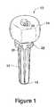
- FIG. 1illustrates an embodiment of a modular ulnar implant in accordance with the present invention.
- FIG. 2illustrates the stem of the modular ulnar implant of FIG. 1 .
- FIG. 3illustrates the head of the modular ulnar implant of FIG. 1 .
- the present inventionis directed to an articulating implant system for fixation to a bone.
- the implant systemcomprises two components: a fixation component and an articulating component.
- FIG. 1illustrates a modular ulnar implant 10 in accordance with the articulating implant system of the present invention.
- the implant 10is intended to be an anatomical replacement for the distal ulna after its resection.
- the modular ulnar implant 10includes a fixation component and an articulating component.
- the fixation componentis a stem 12 and the articulating component is a head 14 .
- the stem 12attaches to the head 14 via a morse taper.
- the stem 12is elongated and formed with first and second ends, 16 and 18 .
- the first stem end 16is configured for fixation to a bone, specifically, for insertion into the intramedullary canal of the distal ulna to thereby anchor the modular ulnar implant to the distal ulna.
- the stem 12is tapered to match the ulnar canal anatomy and facilitate insertion of the first stem end 16 into the intramedullary canal of the distal ulna.
- the stem 12is provided with flutes 20 for to prevent rotation of the stem 12 in the intramedullary canal of the distal ulna, thereby facilitating effective fixation of the implant 10 in the canal.
- the second stem end 18may be formed with a roughened or porous surface to enable a cement-free joint between the stem and the distal ulna. Alternately, or in addition to such surface, cement may be used to anchor the stem 12 in the intramedullary canal of the distal ulna.
- the second stem end 18is configured for operative attachment to the head 14 .
- the stem 12includes a platform 22 near the head interface at the second stem end 18 to prevent subsidence into the ulnar canal or excessive penetration of the stem 12 into the intramedullary canal of the distal ulna.
- the stem 12includes, at or near the second stem end 18 , suture holes 24 and 26 for receiving sutures to anchor the implant 10 to soft tissues. Sutures threaded through the suture holes 24 and 26 anchor the implant to the triangular soft tissues that are exposed after resection of the distal ulna. These soft tissues include the ulna collateral capsule, the triangular fibrocartilage, and the extensor carpi ulnaris subsheath. A stem extender collar may be used to add additional resection height.
- the stem 12may include an instrument interface for positioning control.
- the stem 12includes an extension 28 at the second end 18 , preferably centrally located and formed as a morse taper.
- the extension 28is configured for receipt by a bore in the head 14 .
- At least one suture hole 24is provided at a distal end 30 of the extension 28 for receiving sutures, the suture hole 24 being accessible after the head 14 has been placed on the stem 12 .
- Suture holes 26may also be provided on the platform 22 near the stem second end 18 , near the head interface. In this embodiment, the suture holes 26 and 24 in the platform and in the extension, respectively, anchor the implant 10 to the triangular soft tissues.
- the head 14is configured for articulating against bone or cartilage.
- the head 14is offset from the stem 12 after the implant 10 has been assembled.
- the head 14is triangulated to reproduce normal anatomy, and has an approximately 200° arc for mating with the radial sigmoid notch.
- the headincludes a bore 32 extending completely therethrough for receiving an extension from the stem.
- the head 14may include a bore extending less than completely therethrough for mating with the second end 18 of the stem 12 .
- the head 14may include a drainage hole and instrument interface on its distal surface to allow effective in vivo assembly and rotational positioning.
- the head 14is covered, at least near the triangulated portion, with an ingrowth coating, or is otherwise provided with a porous surface, to promote ingrowth with the soft tissues.
- the second end 18 of the stem 12 and the bore 32 of the head 14are complementary so as to provide a secure fit between the head 14 and the stem 12 when the second stem end 18 is inserted in the bore 32 of the head 14 .
- the bore 32is provided through the head 14 such that the extension 28 of the stem 12 passes completely therethrough, with both the extension 28 of the stem 12 and the bore 14 being Morse tapers.
- insertion of the stem extension 28 through the bore 32results in the suture hole 24 provided at the distal end 30 of the extension 28 extending through the bore 32 while the suture holes 26 provided at the proximal end of the extension 34 fail to pass through the bore 32 .
- the implant 10has suture holes at both ends of the head 14 .
- the implantis attached to the soft tissues via the stem (or fixation component).
- the stemor fixation component
- forces from the suture tissue attachmentare transferred directly through the fixation component to the bone and not through the connection of the articulating component to the fixation component.
- there is no risk of separation of the head and stemdue to biomechanical forces from the tissues attached by suture to the implant.
- the distal ulnais first exposed.
- the distal ulnamay be exposed by making an incision along the medial shaft of the distal ulna in line with the ulnar styloid.
- a dorsal incision centered over the distal radioulnar joint in line with the fourth metacarpalmay be used to expose the distal ulna.
- a templatemay be placed against the distal ulna and located distally over the articular surface of the distal ulna to mark the prescribed resection length.
- the distal ulnais resected, by, for example, using an oscillating saw, exposing the intramedullary canal and the soft tissues that formerly surrounded the distal ulna. Once exposed, the intramedullary canal is identified and reamed to accommodate an appropriately sized stem.
- a trial stem and trial headmay be used to verify anatomical alignment and to ensure that the proper resection length has been achieved.
- the stem of the stemmay be inserted into the intramedullary canal of the distal ulna to anchor the implant to the distal ulna.
- the first end of the stemis inserted into the intramedullary canal, for example by using an impactor until the platform contacts the distal ulna.
- the fit between the stem and the distal ulnamay be assessed by applying a distal traction on the stem. Any movement of the stem in the intramedullary canal of the distal ulna indicates that a firm fit has not been obtained. If a firm fit is not obtained between the stem and the distal ulna after impaction of the stem, a bone cement such as polymethylmethacrylate may be used to cement the stem to the distal ulna.
- the headmay be impacted onto the stem. Specifically, the extension at the second stem end may be inserted into the bore formed in the head. The head may be advanced onto the stem with an impactor until a secure fit is obtained between the head and the stem.
- the stemmay then be sutured to the soft tissue formerly surrounding the distal ulna, specifically the ulna collateral capsule, the triangular fibrocartilage, and the extensor carpi ulnaris subsheath using the suture holes formed in the stem.
- Non-absorbable suturesmay be used.
- the suturesattach to the implant through the fixation component, forces from the suture tissue attachment are transferred directly through the fixation component to the bone, thereby reducing or eliminating the risk of component separation due to such forces.
- the suturesmay be tied into the capsular sleeve surrounding the implant and the subcutaneous tissues and skin may be closed over the implant.
Landscapes
- Health & Medical Sciences (AREA)
- Orthopedic Medicine & Surgery (AREA)
- Transplantation (AREA)
- Life Sciences & Earth Sciences (AREA)
- General Health & Medical Sciences (AREA)
- Public Health (AREA)
- Heart & Thoracic Surgery (AREA)
- Biomedical Technology (AREA)
- Veterinary Medicine (AREA)
- Animal Behavior & Ethology (AREA)
- Engineering & Computer Science (AREA)
- Vascular Medicine (AREA)
- Cardiology (AREA)
- Oral & Maxillofacial Surgery (AREA)
- Physical Education & Sports Medicine (AREA)
- Surgery (AREA)
- Molecular Biology (AREA)
- Nuclear Medicine, Radiotherapy & Molecular Imaging (AREA)
- Medical Informatics (AREA)
- Prostheses (AREA)
- Surgical Instruments (AREA)
- Paper (AREA)
Abstract
Description
Claims (20)
Priority Applications (2)
| Application Number | Priority Date | Filing Date | Title |
|---|---|---|---|
| US10/772,129US7722676B2 (en) | 2003-02-05 | 2004-02-04 | Articulating implant system |
| US12/786,101US8512412B2 (en) | 2003-02-05 | 2010-05-24 | Articulating implant system |
Applications Claiming Priority (2)
| Application Number | Priority Date | Filing Date | Title |
|---|---|---|---|
| US44547403P | 2003-02-05 | 2003-02-05 | |
| US10/772,129US7722676B2 (en) | 2003-02-05 | 2004-02-04 | Articulating implant system |
Related Child Applications (1)
| Application Number | Title | Priority Date | Filing Date |
|---|---|---|---|
| US12/786,101ContinuationUS8512412B2 (en) | 2003-02-05 | 2010-05-24 | Articulating implant system |
Publications (2)
| Publication Number | Publication Date |
|---|---|
| US20040230312A1 US20040230312A1 (en) | 2004-11-18 |
| US7722676B2true US7722676B2 (en) | 2010-05-25 |
Family
ID=32869368
Family Applications (2)
| Application Number | Title | Priority Date | Filing Date |
|---|---|---|---|
| US10/772,129Expired - Fee RelatedUS7722676B2 (en) | 2003-02-05 | 2004-02-04 | Articulating implant system |
| US12/786,101Expired - Fee RelatedUS8512412B2 (en) | 2003-02-05 | 2010-05-24 | Articulating implant system |
Family Applications After (1)
| Application Number | Title | Priority Date | Filing Date |
|---|---|---|---|
| US12/786,101Expired - Fee RelatedUS8512412B2 (en) | 2003-02-05 | 2010-05-24 | Articulating implant system |
Country Status (11)
| Country | Link |
|---|---|
| US (2) | US7722676B2 (en) |
| EP (1) | EP1596769B1 (en) |
| JP (1) | JP4465350B2 (en) |
| KR (1) | KR100825192B1 (en) |
| CN (1) | CN100488470C (en) |
| AT (1) | ATE392869T1 (en) |
| AU (1) | AU2004211945B2 (en) |
| BR (1) | BRPI0406686B8 (en) |
| CA (1) | CA2513917C (en) |
| DE (1) | DE602004013281T2 (en) |
| WO (1) | WO2004071357A2 (en) |
Cited By (43)
| Publication number | Priority date | Publication date | Assignee | Title |
|---|---|---|---|---|
| US20070118136A1 (en)* | 2002-12-03 | 2007-05-24 | Arthrosurface, Inc. | Tibial resurfacing system |
| US20080249631A1 (en)* | 2005-11-17 | 2008-10-09 | Bioprofile | Implant, more particularly partial ulnar head implant |
| US20090281632A1 (en)* | 2008-05-09 | 2009-11-12 | Remi Sciences, Inc. | Ulnar head prosthesis system |
| US20090281631A1 (en)* | 2008-05-09 | 2009-11-12 | Remi Sciences, Inc. | Ulnar head prosthesis system |
| US20100131069A1 (en)* | 2007-08-01 | 2010-05-27 | Jeffrey Halbrecht | Method and system for patella tendon realignment |
| US20100185294A1 (en)* | 2002-06-04 | 2010-07-22 | Arthrosurface Incorporated | Nanorough Alloy Substrate |
| US20100198354A1 (en)* | 2007-08-01 | 2010-08-05 | Jeffrey Halbrecht | Method and system for patella tendon realignment |
| US20100204701A1 (en)* | 2000-05-01 | 2010-08-12 | Arthrosurface Incorporated | System and Method for Joint Resurface Repair |
| US20100217402A1 (en)* | 2007-10-18 | 2010-08-26 | Inbone Technologies, Inc. | Fibular stiffener and bony defect replacer |
| US20100234960A1 (en)* | 2003-02-05 | 2010-09-16 | Shaun Hanson | Articulating Implant System |
| US20110213466A1 (en)* | 2009-08-27 | 2011-09-01 | The Foundry Llc | Method and Apparatus for Force Redistribution in Articular Joints |
| US8361159B2 (en) | 2002-12-03 | 2013-01-29 | Arthrosurface, Inc. | System for articular surface replacement |
| US8388624B2 (en) | 2003-02-24 | 2013-03-05 | Arthrosurface Incorporated | Trochlear resurfacing system and method |
| US8556902B2 (en) | 2002-12-03 | 2013-10-15 | Arthrosurface Incorporated | System and method for retrograde procedure |
| US8663230B2 (en) | 2002-12-03 | 2014-03-04 | Arthrosurface Incorporated | Retrograde delivery of resurfacing devices |
| US8845724B2 (en) | 2009-08-27 | 2014-09-30 | Cotera, Inc. | Method and apparatus for altering biomechanics of the articular joints |
| US8852285B2 (en) | 2012-03-13 | 2014-10-07 | Jeffrey A Greenberg | Prosthesis for distal radioulnar joint |
| US8864827B2 (en) | 2000-05-01 | 2014-10-21 | Arthrosurface Inc. | System and method for joint resurface repair |
| US8926615B2 (en) | 2002-12-03 | 2015-01-06 | Arthrosurface, Inc. | System and method for retrograde procedure |
| US8961614B2 (en) | 2004-11-22 | 2015-02-24 | Arthrosurface, Inc. | Articular surface implant and delivery system |
| US9055955B2 (en) | 2000-05-01 | 2015-06-16 | Arthrosurface Inc. | Bone resurfacing system and method |
| US9066716B2 (en) | 2011-03-30 | 2015-06-30 | Arthrosurface Incorporated | Suture coil and suture sheath for tissue repair |
| US9204873B2 (en) | 2000-05-01 | 2015-12-08 | Arthrosurface Incorporated | System and method for joint resurface repair |
| US9283076B2 (en) | 2009-04-17 | 2016-03-15 | Arthrosurface Incorporated | Glenoid resurfacing system and method |
| US9357989B2 (en) | 2000-05-01 | 2016-06-07 | Arthrosurface Incorporated | System and method for joint resurface repair |
| US9358029B2 (en) | 2006-12-11 | 2016-06-07 | Arthrosurface Incorporated | Retrograde resection apparatus and method |
| US9468466B1 (en) | 2012-08-24 | 2016-10-18 | Cotera, Inc. | Method and apparatus for altering biomechanics of the spine |
| US9468448B2 (en) | 2012-07-03 | 2016-10-18 | Arthrosurface Incorporated | System and method for joint resurfacing and repair |
| US9492200B2 (en) | 2013-04-16 | 2016-11-15 | Arthrosurface Incorporated | Suture system and method |
| US9662126B2 (en) | 2009-04-17 | 2017-05-30 | Arthrosurface Incorporated | Glenoid resurfacing system and method |
| US9668868B2 (en) | 2009-08-27 | 2017-06-06 | Cotera, Inc. | Apparatus and methods for treatment of patellofemoral conditions |
| US9861408B2 (en) | 2009-08-27 | 2018-01-09 | The Foundry, Llc | Method and apparatus for treating canine cruciate ligament disease |
| US9861492B2 (en) | 2014-03-07 | 2018-01-09 | Arthrosurface Incorporated | Anchor for an implant assembly |
| US10349980B2 (en) | 2009-08-27 | 2019-07-16 | The Foundry, Llc | Method and apparatus for altering biomechanics of the shoulder |
| US10624748B2 (en) | 2014-03-07 | 2020-04-21 | Arthrosurface Incorporated | System and method for repairing articular surfaces |
| US10624752B2 (en) | 2006-07-17 | 2020-04-21 | Arthrosurface Incorporated | Tibial resurfacing system and method |
| US10945743B2 (en) | 2009-04-17 | 2021-03-16 | Arthrosurface Incorporated | Glenoid repair system and methods of use thereof |
| US11160663B2 (en) | 2017-08-04 | 2021-11-02 | Arthrosurface Incorporated | Multicomponent articular surface implant |
| US11478358B2 (en) | 2019-03-12 | 2022-10-25 | Arthrosurface Incorporated | Humeral and glenoid articular surface implant systems and methods |
| US11607319B2 (en) | 2014-03-07 | 2023-03-21 | Arthrosurface Incorporated | System and method for repairing articular surfaces |
| US11712276B2 (en) | 2011-12-22 | 2023-08-01 | Arthrosurface Incorporated | System and method for bone fixation |
| US11896476B2 (en) | 2020-01-02 | 2024-02-13 | Zkr Orthopedics, Inc. | Patella tendon realignment implant with changeable shape |
| US12303396B2 (en) | 2020-05-11 | 2025-05-20 | Zkr Orthopedics, Inc. | Adjustable patellar tendon realignment implant |
Families Citing this family (38)
| Publication number | Priority date | Publication date | Assignee | Title |
|---|---|---|---|---|
| US8052756B2 (en) | 2002-10-24 | 2011-11-08 | Biomet Manufacturing Corp. | Method and apparatus for wrist arthroplasty |
| US8105390B2 (en) | 2002-10-24 | 2012-01-31 | Biomet Manufacturing Corp. | Method and apparatus for wrist arthroplasty |
| US8105388B2 (en)* | 2002-10-24 | 2012-01-31 | Biomet Manufacturing Corp. | Method and apparatus for wrist arthroplasty |
| US6746486B1 (en)* | 2002-10-24 | 2004-06-08 | Biomet, Inc. | Method and apparatus for total wrist angled back carpal plate |
| US8940055B2 (en) | 2002-10-24 | 2015-01-27 | Biomet Manufacturing, Llc | Method and apparatus for wrist arthroplasty |
| US8066777B2 (en)* | 2002-10-24 | 2011-11-29 | Biomet Manufacturing Corp. | Method and apparatus for wrist arthroplasty |
| US8105389B2 (en)* | 2002-10-24 | 2012-01-31 | Biomet Manufacturing Corp. | Method and apparatus for wrist arthroplasty |
| US8821581B2 (en) | 2002-10-24 | 2014-09-02 | Biomet Manufacturing, Llc | Method and apparatus for wrist arthroplasty |
| US8372154B2 (en) | 2002-10-24 | 2013-02-12 | Biomet Manufacturing Corp. | Method and apparatus for wrist arthroplasty |
| US7625408B2 (en)* | 2003-07-22 | 2009-12-01 | Avanta Orthopaedics, Llc | Prosthetic wrist implant |
| US7892287B2 (en)* | 2004-09-27 | 2011-02-22 | Depuy Products, Inc. | Glenoid augment and associated method |
| US7922769B2 (en) | 2004-09-27 | 2011-04-12 | Depuy Products, Inc. | Modular glenoid prosthesis and associated method |
| US7927335B2 (en) | 2004-09-27 | 2011-04-19 | Depuy Products, Inc. | Instrument for preparing an implant support surface and associated method |
| US7909883B2 (en)* | 2007-02-21 | 2011-03-22 | Sidebotham Christopher G | Percutaneous implant for limb salvage |
| US8652179B2 (en)* | 2008-05-02 | 2014-02-18 | The Cleveland Clinic Foundation | Bone plate extender and extension system for bone restoration and methods of use thereof |
| US8915918B2 (en)* | 2008-05-02 | 2014-12-23 | Thomas James Graham | Bone plate system for bone restoration and methods of use thereof |
| US8608783B2 (en)* | 2008-05-08 | 2013-12-17 | The Cleveland Clinic Foundation | Bone plate with flange member and methods of use thereof |
| US8628533B2 (en)* | 2008-05-08 | 2014-01-14 | The Cleveland Clinic Foundation | Bone plate with reduction aids and methods of use thereof |
| US8241365B2 (en) | 2008-12-23 | 2012-08-14 | Depuy Products, Inc. | Shoulder prosthesis with vault-filling structure having bone-sparing configuration |
| US8366784B2 (en)* | 2009-09-17 | 2013-02-05 | Biomet Manufacturing Corp. | Distal radioulnar joint device |
| US8152854B2 (en)* | 2009-09-30 | 2012-04-10 | Imbriglia Joseph E | Resurfacing implant for the wrist and method of implantation thereof |
| US8231683B2 (en) | 2009-12-08 | 2012-07-31 | Depuy Products, Inc. | Shoulder prosthesis assembly having glenoid rim replacement structure |
| US8480750B2 (en) | 2010-11-24 | 2013-07-09 | DePuy Synthes Products, LLC | Modular glenoid prosthesis |
| US8465548B2 (en) | 2010-11-24 | 2013-06-18 | DePuy Synthes Products, LLC | Modular glenoid prosthesis |
| WO2012154920A1 (en) | 2011-05-12 | 2012-11-15 | Small Bone Innovations, Inc. | Wrist implant for carpal hemiarthroplasty |
| WO2014008229A1 (en)* | 2012-07-03 | 2014-01-09 | Smith & Nephew, Inc. | Orthopedic prosthesis with suture anchor features |
| CN108095860A (en)* | 2013-03-13 | 2018-06-01 | 瑞特医疗技术公司 | Correct implantation piece enhanced portion, system and method |
| US9949839B2 (en) | 2013-03-13 | 2018-04-24 | Wright Medical Technology, Inc. | Revision implant augments, systems, and methods |
| US20140277553A1 (en)* | 2013-03-14 | 2014-09-18 | Ascension Orthopedics, Inc. | Ulnar Head Replacement Implant System |
| WO2016044053A1 (en) | 2014-09-19 | 2016-03-24 | Crossroads Extremity Systems, Llc | Bone fixation implant and means of fixation |
| CN104665960A (en)* | 2015-02-15 | 2015-06-03 | 张洋 | Orthopedic prosthesis for proximal radioulnar joint fusion |
| FR3037803B1 (en)* | 2015-06-23 | 2017-07-07 | I Ceram | IMPLANT OF SUBSTITUTION OF STERNUM |
| US20170056188A1 (en)* | 2015-08-25 | 2017-03-02 | Wright Medical Technology, Inc. | Total ankle talar prosthesis with anchor holes and grooves |
| US10940023B2 (en) | 2016-12-15 | 2021-03-09 | Stryker European Holdings I, Llc | Bone plate trial |
| RU186939U1 (en)* | 2018-03-20 | 2019-02-11 | Общество с ограниченной ответственностью "Мойе Керамик-Имплантате" | DENTAL HEAD OF THE ELBOW |
| ES3004736T3 (en) | 2019-06-24 | 2025-03-13 | Skeletal Dynamics Inc | Distal radioulnar joint prosthesis system |
| CN112386375A (en)* | 2020-07-07 | 2021-02-23 | 内蒙古医科大学第二附属医院(内蒙古自治区骨科研究所) | Palm and finger and interphalangeal joint prosthesis |
| US20220047311A1 (en)* | 2020-08-17 | 2022-02-17 | Ahmad N. Ali | Surgical Method of Stabilizing Bone or Joint Fracture Using Flexible Line |
Citations (10)
| Publication number | Priority date | Publication date | Assignee | Title |
|---|---|---|---|---|
| US3745590A (en) | 1971-06-25 | 1973-07-17 | Cutter Lab | Articulating prosthesis with ligamentous attachment |
| FR2660856A1 (en) | 1990-04-12 | 1991-10-18 | Kapandji Adalbert | Lower radioulnar prosthesis |
| US5108444A (en) | 1990-04-26 | 1992-04-28 | Branemark Per Ingvar | System for reconstructing the distal radioulnar joints (dru-joints) in wrists |
| US5782926A (en) | 1995-06-21 | 1998-07-21 | Sulzer Orthopaedie Ag | Wrist prosthesis |
| US5938699A (en) | 1997-04-18 | 1999-08-17 | Campbell; G. Stewart | Distal radial ulnar joint reconstruction system |
| US5951604A (en) | 1997-12-31 | 1999-09-14 | Avanta Orthopedics, Inc. | Distal radioulnar joint prosthesis |
| US6027534A (en)* | 1997-11-03 | 2000-02-22 | Deputy Orthopaedics, Inc. | Modular elbow |
| WO2001001892A1 (en) | 1999-07-02 | 2001-01-11 | Van Straten Research & Development B.V. | Distal radioulnar joint prosthesis |
| WO2001070138A1 (en) | 2000-03-23 | 2001-09-27 | Philippe Kopylov Ab | Joint surface replacement of the distal radioulnar joint |
| US6302915B1 (en) | 2000-08-30 | 2001-10-16 | The Mayo Foundation For Medical Education & Research | Ulnar implant system |
Family Cites Families (5)
| Publication number | Priority date | Publication date | Assignee | Title |
|---|---|---|---|---|
| JPH06168039A (en) | 1992-11-30 | 1994-06-14 | Toshiba Corp | Power system |
| US5888220A (en)* | 1994-05-06 | 1999-03-30 | Advanced Bio Surfaces, Inc. | Articulating joint repair |
| EP0883389A1 (en)* | 1996-02-29 | 1998-12-16 | Ahmet Ege | The anatomic replacement prosthesis system in the hand and wrist |
| US6890358B2 (en)* | 2002-03-29 | 2005-05-10 | Depuy Products, Inc. | Distal component for wrist prosthesis |
| US7722676B2 (en)* | 2003-02-05 | 2010-05-25 | Wright Medical Technology, Inc. | Articulating implant system |
- 2004
- 2004-02-04USUS10/772,129patent/US7722676B2/ennot_activeExpired - Fee Related
- 2004-02-05JPJP2006503392Apatent/JP4465350B2/ennot_activeExpired - Fee Related
- 2004-02-05CNCNB2004800036898Apatent/CN100488470C/ennot_activeExpired - Fee Related
- 2004-02-05BRBRPI0406686Apatent/BRPI0406686B8/ennot_activeIP Right Cessation
- 2004-02-05WOPCT/US2004/003517patent/WO2004071357A2/enactiveApplication Filing
- 2004-02-05EPEP04708649Apatent/EP1596769B1/ennot_activeExpired - Lifetime
- 2004-02-05DEDE602004013281Tpatent/DE602004013281T2/ennot_activeExpired - Lifetime
- 2004-02-05AUAU2004211945Apatent/AU2004211945B2/ennot_activeCeased
- 2004-02-05KRKR1020057014265Apatent/KR100825192B1/ennot_activeExpired - Fee Related
- 2004-02-05CACA2513917Apatent/CA2513917C/ennot_activeExpired - Fee Related
- 2004-02-05ATAT04708649Tpatent/ATE392869T1/ennot_activeIP Right Cessation
- 2010
- 2010-05-24USUS12/786,101patent/US8512412B2/ennot_activeExpired - Fee Related
Patent Citations (10)
| Publication number | Priority date | Publication date | Assignee | Title |
|---|---|---|---|---|
| US3745590A (en) | 1971-06-25 | 1973-07-17 | Cutter Lab | Articulating prosthesis with ligamentous attachment |
| FR2660856A1 (en) | 1990-04-12 | 1991-10-18 | Kapandji Adalbert | Lower radioulnar prosthesis |
| US5108444A (en) | 1990-04-26 | 1992-04-28 | Branemark Per Ingvar | System for reconstructing the distal radioulnar joints (dru-joints) in wrists |
| US5782926A (en) | 1995-06-21 | 1998-07-21 | Sulzer Orthopaedie Ag | Wrist prosthesis |
| US5938699A (en) | 1997-04-18 | 1999-08-17 | Campbell; G. Stewart | Distal radial ulnar joint reconstruction system |
| US6027534A (en)* | 1997-11-03 | 2000-02-22 | Deputy Orthopaedics, Inc. | Modular elbow |
| US5951604A (en) | 1997-12-31 | 1999-09-14 | Avanta Orthopedics, Inc. | Distal radioulnar joint prosthesis |
| WO2001001892A1 (en) | 1999-07-02 | 2001-01-11 | Van Straten Research & Development B.V. | Distal radioulnar joint prosthesis |
| WO2001070138A1 (en) | 2000-03-23 | 2001-09-27 | Philippe Kopylov Ab | Joint surface replacement of the distal radioulnar joint |
| US6302915B1 (en) | 2000-08-30 | 2001-10-16 | The Mayo Foundation For Medical Education & Research | Ulnar implant system |
Non-Patent Citations (2)
| Title |
|---|
| International Preliminary Report on Patentability mailed Aug. 18, 2005 for PCT/US2004/003517 (filed Feb. 5, 2004). |
| International Search Report, PCT International Search Report mailed Jul. 23, 2004 for PCT/US2004/003517 (filed Feb. 5, 2004). |
Cited By (84)
| Publication number | Priority date | Publication date | Assignee | Title |
|---|---|---|---|---|
| US20100204701A1 (en)* | 2000-05-01 | 2010-08-12 | Arthrosurface Incorporated | System and Method for Joint Resurface Repair |
| US9055955B2 (en) | 2000-05-01 | 2015-06-16 | Arthrosurface Inc. | Bone resurfacing system and method |
| US9204873B2 (en) | 2000-05-01 | 2015-12-08 | Arthrosurface Incorporated | System and method for joint resurface repair |
| US8540717B2 (en) | 2000-05-01 | 2013-09-24 | Arthrosurface Incorporated | System and method for joint resurface repair |
| US9357989B2 (en) | 2000-05-01 | 2016-06-07 | Arthrosurface Incorporated | System and method for joint resurface repair |
| US8864827B2 (en) | 2000-05-01 | 2014-10-21 | Arthrosurface Inc. | System and method for joint resurface repair |
| US20100185294A1 (en)* | 2002-06-04 | 2010-07-22 | Arthrosurface Incorporated | Nanorough Alloy Substrate |
| US20070118136A1 (en)* | 2002-12-03 | 2007-05-24 | Arthrosurface, Inc. | Tibial resurfacing system |
| US8926615B2 (en) | 2002-12-03 | 2015-01-06 | Arthrosurface, Inc. | System and method for retrograde procedure |
| US8663230B2 (en) | 2002-12-03 | 2014-03-04 | Arthrosurface Incorporated | Retrograde delivery of resurfacing devices |
| US8556902B2 (en) | 2002-12-03 | 2013-10-15 | Arthrosurface Incorporated | System and method for retrograde procedure |
| US10076343B2 (en) | 2002-12-03 | 2018-09-18 | Arthrosurface Incorporated | System for articular surface replacement |
| US9044343B2 (en) | 2002-12-03 | 2015-06-02 | Arthrosurface Incorporated | System for articular surface replacement |
| US8361159B2 (en) | 2002-12-03 | 2013-01-29 | Arthrosurface, Inc. | System for articular surface replacement |
| US8523872B2 (en) | 2002-12-03 | 2013-09-03 | Arthrosurface Incorporated | Tibial resurfacing system |
| US20100234960A1 (en)* | 2003-02-05 | 2010-09-16 | Shaun Hanson | Articulating Implant System |
| US8512412B2 (en)* | 2003-02-05 | 2013-08-20 | Wright Medical Technology, Inc. | Articulating implant system |
| US9931211B2 (en) | 2003-02-24 | 2018-04-03 | Arthrosurface Incorporated | Trochlear resurfacing system and method |
| US8388624B2 (en) | 2003-02-24 | 2013-03-05 | Arthrosurface Incorporated | Trochlear resurfacing system and method |
| US9351745B2 (en) | 2003-02-24 | 2016-05-31 | Arthrosurface Incorporated | Trochlear resurfacing system and method |
| US11337819B2 (en) | 2003-02-24 | 2022-05-24 | Arthrosurface Incorporated | Trochlear resurfacing system and method |
| US10624749B2 (en) | 2003-02-24 | 2020-04-21 | Arthrosurface Incorporated | Trochlear resurfacing system and method |
| US8961614B2 (en) | 2004-11-22 | 2015-02-24 | Arthrosurface, Inc. | Articular surface implant and delivery system |
| US8088168B2 (en)* | 2005-11-17 | 2012-01-03 | Tornier Sas | Implant, more particularly partial ulnar head implant |
| US20080249631A1 (en)* | 2005-11-17 | 2008-10-09 | Bioprofile | Implant, more particularly partial ulnar head implant |
| US11471289B2 (en) | 2006-07-17 | 2022-10-18 | Arthrosurface Incorporated | Tibial resurfacing system and method |
| US10624752B2 (en) | 2006-07-17 | 2020-04-21 | Arthrosurface Incorporated | Tibial resurfacing system and method |
| US9358029B2 (en) | 2006-12-11 | 2016-06-07 | Arthrosurface Incorporated | Retrograde resection apparatus and method |
| US10959740B2 (en) | 2006-12-11 | 2021-03-30 | Arthrosurface Incorporated | Retrograde resection apparatus and method |
| US10045788B2 (en) | 2006-12-11 | 2018-08-14 | Arthrosurface Incorporated | Retrograde resection apparatus and method |
| US10918416B2 (en) | 2007-08-01 | 2021-02-16 | Zkr Orthopedics, Inc. | Method and system for patella tendon realignment |
| US20100131069A1 (en)* | 2007-08-01 | 2010-05-27 | Jeffrey Halbrecht | Method and system for patella tendon realignment |
| US10918415B2 (en) | 2007-08-01 | 2021-02-16 | Zkr Orthopedics, Inc. | Method and system for patella tendon realignment |
| US9808287B2 (en) | 2007-08-01 | 2017-11-07 | Jeffrey Halbrecht | Method and system for patella tendon realignment |
| US20100198354A1 (en)* | 2007-08-01 | 2010-08-05 | Jeffrey Halbrecht | Method and system for patella tendon realignment |
| US8110006B2 (en)* | 2007-10-18 | 2012-02-07 | Inbone Technologies, Inc. | Fibular stiffener and bony defect replacer |
| US20100217402A1 (en)* | 2007-10-18 | 2010-08-26 | Inbone Technologies, Inc. | Fibular stiffener and bony defect replacer |
| US20090281632A1 (en)* | 2008-05-09 | 2009-11-12 | Remi Sciences, Inc. | Ulnar head prosthesis system |
| US8052755B2 (en)* | 2008-05-09 | 2011-11-08 | Remi Sciences, Inc. | Ulnar head prosthesis system |
| US7875082B2 (en)* | 2008-05-09 | 2011-01-25 | Remi Sciences, Inc. | Ulnar head prosthesis system |
| US20090281631A1 (en)* | 2008-05-09 | 2009-11-12 | Remi Sciences, Inc. | Ulnar head prosthesis system |
| US9283076B2 (en) | 2009-04-17 | 2016-03-15 | Arthrosurface Incorporated | Glenoid resurfacing system and method |
| US9662126B2 (en) | 2009-04-17 | 2017-05-30 | Arthrosurface Incorporated | Glenoid resurfacing system and method |
| US10478200B2 (en) | 2009-04-17 | 2019-11-19 | Arthrosurface Incorporated | Glenoid resurfacing system and method |
| US11478259B2 (en) | 2009-04-17 | 2022-10-25 | Arthrosurface, Incorporated | Glenoid resurfacing system and method |
| US10945743B2 (en) | 2009-04-17 | 2021-03-16 | Arthrosurface Incorporated | Glenoid repair system and methods of use thereof |
| US9668868B2 (en) | 2009-08-27 | 2017-06-06 | Cotera, Inc. | Apparatus and methods for treatment of patellofemoral conditions |
| US20110213466A1 (en)* | 2009-08-27 | 2011-09-01 | The Foundry Llc | Method and Apparatus for Force Redistribution in Articular Joints |
| US11730519B2 (en) | 2009-08-27 | 2023-08-22 | The Foundry, Llc | Method and apparatus for force redistribution in articular joints |
| US11517360B2 (en) | 2009-08-27 | 2022-12-06 | The Foundry, Llc | Method and apparatus for treating canine cruciate ligament disease |
| US9931136B2 (en) | 2009-08-27 | 2018-04-03 | The Foundry, Llc | Method and apparatus for altering biomechanics of articular joints |
| US9861408B2 (en) | 2009-08-27 | 2018-01-09 | The Foundry, Llc | Method and apparatus for treating canine cruciate ligament disease |
| US9795410B2 (en) | 2009-08-27 | 2017-10-24 | Cotera, Inc. | Method and apparatus for force redistribution in articular joints |
| US8597362B2 (en) | 2009-08-27 | 2013-12-03 | Cotera, Inc. | Method and apparatus for force redistribution in articular joints |
| US10349980B2 (en) | 2009-08-27 | 2019-07-16 | The Foundry, Llc | Method and apparatus for altering biomechanics of the shoulder |
| US8845724B2 (en) | 2009-08-27 | 2014-09-30 | Cotera, Inc. | Method and apparatus for altering biomechanics of the articular joints |
| US9114016B2 (en) | 2009-08-27 | 2015-08-25 | Cotera, Inc. | Method and apparatus for altering biomechanics of the articular joints |
| US9278004B2 (en) | 2009-08-27 | 2016-03-08 | Cotera, Inc. | Method and apparatus for altering biomechanics of the articular joints |
| US10695094B2 (en) | 2009-08-27 | 2020-06-30 | The Foundry, Llc | Method and apparatus for altering biomechanics of articular joints |
| US9066716B2 (en) | 2011-03-30 | 2015-06-30 | Arthrosurface Incorporated | Suture coil and suture sheath for tissue repair |
| US11712276B2 (en) | 2011-12-22 | 2023-08-01 | Arthrosurface Incorporated | System and method for bone fixation |
| US8852285B2 (en) | 2012-03-13 | 2014-10-07 | Jeffrey A Greenberg | Prosthesis for distal radioulnar joint |
| US11191552B2 (en) | 2012-07-03 | 2021-12-07 | Arthrosurface, Incorporated | System and method for joint resurfacing and repair |
| US9468448B2 (en) | 2012-07-03 | 2016-10-18 | Arthrosurface Incorporated | System and method for joint resurfacing and repair |
| US10307172B2 (en) | 2012-07-03 | 2019-06-04 | Arthrosurface Incorporated | System and method for joint resurfacing and repair |
| US9468466B1 (en) | 2012-08-24 | 2016-10-18 | Cotera, Inc. | Method and apparatus for altering biomechanics of the spine |
| US10898237B2 (en) | 2012-08-24 | 2021-01-26 | The Foundry, Llc | Method and apparatus for altering biomechanics of the spine |
| US10695096B2 (en) | 2013-04-16 | 2020-06-30 | Arthrosurface Incorporated | Suture system and method |
| US11648036B2 (en) | 2013-04-16 | 2023-05-16 | Arthrosurface Incorporated | Suture system and method |
| US9492200B2 (en) | 2013-04-16 | 2016-11-15 | Arthrosurface Incorporated | Suture system and method |
| US11607319B2 (en) | 2014-03-07 | 2023-03-21 | Arthrosurface Incorporated | System and method for repairing articular surfaces |
| US10624754B2 (en) | 2014-03-07 | 2020-04-21 | Arthrosurface Incorporated | System and method for repairing articular surfaces |
| US11766334B2 (en) | 2014-03-07 | 2023-09-26 | Arthrosurface Incorporated | System and method for repairing articular surfaces |
| US11083587B2 (en) | 2014-03-07 | 2021-08-10 | Arthrosurface Incorporated | Implant and anchor assembly |
| US9962265B2 (en) | 2014-03-07 | 2018-05-08 | Arthrosurface Incorporated | System and method for repairing articular surfaces |
| US9931219B2 (en) | 2014-03-07 | 2018-04-03 | Arthrosurface Incorporated | Implant and anchor assembly |
| US9861492B2 (en) | 2014-03-07 | 2018-01-09 | Arthrosurface Incorporated | Anchor for an implant assembly |
| US10624748B2 (en) | 2014-03-07 | 2020-04-21 | Arthrosurface Incorporated | System and method for repairing articular surfaces |
| US10575957B2 (en) | 2014-03-07 | 2020-03-03 | Arthrosurface Incoporated | Anchor for an implant assembly |
| US11241256B2 (en) | 2015-10-15 | 2022-02-08 | The Foundry, Llc | Method and apparatus for altering biomechanics of the shoulder |
| US11160663B2 (en) | 2017-08-04 | 2021-11-02 | Arthrosurface Incorporated | Multicomponent articular surface implant |
| US11478358B2 (en) | 2019-03-12 | 2022-10-25 | Arthrosurface Incorporated | Humeral and glenoid articular surface implant systems and methods |
| US11896476B2 (en) | 2020-01-02 | 2024-02-13 | Zkr Orthopedics, Inc. | Patella tendon realignment implant with changeable shape |
| US12303396B2 (en) | 2020-05-11 | 2025-05-20 | Zkr Orthopedics, Inc. | Adjustable patellar tendon realignment implant |
Also Published As
| Publication number | Publication date |
|---|---|
| CN1753651A (en) | 2006-03-29 |
| WO2004071357A3 (en) | 2004-11-11 |
| CN100488470C (en) | 2009-05-20 |
| KR20050101194A (en) | 2005-10-20 |
| CA2513917C (en) | 2010-05-04 |
| DE602004013281D1 (en) | 2008-06-05 |
| AU2004211945B2 (en) | 2008-04-24 |
| US20100234960A1 (en) | 2010-09-16 |
| BRPI0406686B8 (en) | 2021-06-22 |
| WO2004071357A2 (en) | 2004-08-26 |
| DE602004013281T2 (en) | 2009-03-05 |
| US20040230312A1 (en) | 2004-11-18 |
| KR100825192B1 (en) | 2008-04-24 |
| JP2006516469A (en) | 2006-07-06 |
| EP1596769B1 (en) | 2008-04-23 |
| CA2513917A1 (en) | 2004-08-26 |
| EP1596769A2 (en) | 2005-11-23 |
| US8512412B2 (en) | 2013-08-20 |
| BRPI0406686B1 (en) | 2015-02-18 |
| JP4465350B2 (en) | 2010-05-19 |
| AU2004211945A1 (en) | 2004-08-26 |
| ATE392869T1 (en) | 2008-05-15 |
| BRPI0406686A (en) | 2005-12-20 |
Similar Documents
| Publication | Publication Date | Title |
|---|---|---|
| US7722676B2 (en) | Articulating implant system | |
| US6302915B1 (en) | Ulnar implant system | |
| CN1780595B (en) | Prosthetic device for total joint replacement in facet joint arthroplasty | |
| US7160329B2 (en) | Radial-capitellar implant | |
| Dee | Total replacement of the elbow joint | |
| Volz | Total Wrist Arthroplasty A Clinical Review | |
| US20120191199A1 (en) | Method and apparatus for distal radioulnar joint (druj) arthroplasty | |
| CN101361684A (en) | knee prosthesis | |
| US8152854B2 (en) | Resurfacing implant for the wrist and method of implantation thereof | |
| SwANSON et al. | Reconstruction of the Thumb Basal Joints Development and Current Status of Implant Techniques. | |
| Croog et al. | Newest advances in the operative treatment of basal joint arthritis | |
| Burton | Basal joint implant arthroplasty in osteoarthritis: indications, techniques, pitfalls, and problems | |
| SWANSON et al. | Arthroplasty of the thumb basal joints. | |
| Tang et al. | The radioulnar joints and forearm axis: surgeons' perspective | |
| Celli et al. | The Elbow Joint | |
| JP2025513086A (en) | Stemless Semi-Constrained Implantable Joint Replacement Device | |
| Varitimidis et al. | Reconstruction of the elbow: Surgeons' perspective | |
| ADALBERT | An Original Prosthesis of the Distal Radioulnar Joint | |
| Swanson et al. | Flexible Im lant Arthroplasty of the Radiocarpa Joint P | |
| Martín-Ferrero et al. | Tips, Trics and Long-Term Results of the Treatment of Trapezometacarpal Joint Osteoarthritis with Total Arthroplasty | |
| Bhat et al. | Principles of surgery in the rheumatoid hand | |
| Stanley | 14 Arthroplasty and Arthrodesis of | |
| Gschwend et al. | Elbow Arthroplasty, with Particular Regard to the Gschwend-Scheier-Bähler III Elbow Joint Prosthesis | |
| WO2011040914A1 (en) | Resurfacing implant for the wrist and method of implantation thereof |
Legal Events
| Date | Code | Title | Description |
|---|---|---|---|
| AS | Assignment | Owner name:WRIGHT MEDICAL TECHNOLOGY, INC., TENNESSEE Free format text:ASSIGNMENT OF ASSIGNORS INTEREST;ASSIGNORS:HANSON, SHAUN;KING, GRAHAM J.W.;PATTERSON, STUART D.;AND OTHERS;REEL/FRAME:015564/0888;SIGNING DATES FROM 20040601 TO 20040712 Owner name:WRIGHT MEDICAL TECHNOLOGY, INC.,TENNESSEE Free format text:ASSIGNMENT OF ASSIGNORS INTEREST;ASSIGNORS:HANSON, SHAUN;KING, GRAHAM J.W.;PATTERSON, STUART D.;AND OTHERS;SIGNING DATES FROM 20040601 TO 20040712;REEL/FRAME:015564/0888 | |
| STCF | Information on status: patent grant | Free format text:PATENTED CASE | |
| FPAY | Fee payment | Year of fee payment:4 | |
| AS | Assignment | Owner name:MIDCAP FINANCIAL TRUST, AS AGENT, MARYLAND Free format text:SECURITY INTEREST;ASSIGNOR:WRIGHT MEDICAL TECHNOLOGY, INC.;REEL/FRAME:041257/0126 Effective date:20161223 | |
| MAFP | Maintenance fee payment | Free format text:PAYMENT OF MAINTENANCE FEE, 8TH YEAR, LARGE ENTITY (ORIGINAL EVENT CODE: M1552) Year of fee payment:8 | |
| AS | Assignment | Owner name:BIOMIMETIC THERAPEUTICS, LLC, TENNESSEE Free format text:RELEASE BY SECURED PARTY;ASSIGNOR:MIDCAP FUNDING IV TRUST;REEL/FRAME:054480/0001 Effective date:20201112 Owner name:SOLANA SURGICAL, LLC, TENNESSEE Free format text:RELEASE BY SECURED PARTY;ASSIGNOR:MIDCAP FUNDING IV TRUST;REEL/FRAME:054480/0001 Effective date:20201112 Owner name:WRIGHT MEDICAL TECHNOLOGY, INC., TENNESSEE Free format text:RELEASE BY SECURED PARTY;ASSIGNOR:MIDCAP FUNDING IV TRUST;REEL/FRAME:054480/0001 Effective date:20201112 Owner name:INBONE TECHNOLOGIES, INC., TENNESSEE Free format text:RELEASE BY SECURED PARTY;ASSIGNOR:MIDCAP FUNDING IV TRUST;REEL/FRAME:054480/0001 Effective date:20201112 Owner name:BIOMIMETIC THERAPEUTICS CANADA, INC., TENNESSEE Free format text:RELEASE BY SECURED PARTY;ASSIGNOR:MIDCAP FUNDING IV TRUST;REEL/FRAME:054480/0001 Effective date:20201112 Owner name:TROOPER HOLDINGS INC., MINNESOTA Free format text:RELEASE BY SECURED PARTY;ASSIGNOR:MIDCAP FUNDING IV TRUST;REEL/FRAME:054480/0001 Effective date:20201112 Owner name:WRIGHT MEDICAL GROUP N.V., NETHERLANDS Free format text:RELEASE BY SECURED PARTY;ASSIGNOR:MIDCAP FUNDING IV TRUST;REEL/FRAME:054480/0001 Effective date:20201112 Owner name:BIOMIMETIC THERAPEUTICS USA, INC., TENNESSEE Free format text:RELEASE BY SECURED PARTY;ASSIGNOR:MIDCAP FUNDING IV TRUST;REEL/FRAME:054480/0001 Effective date:20201112 Owner name:WRIGHT MEDICAL GROUP INTELLECTUAL PROPERTY, INC., TENNESSEE Free format text:RELEASE BY SECURED PARTY;ASSIGNOR:MIDCAP FUNDING IV TRUST;REEL/FRAME:054480/0001 Effective date:20201112 Owner name:TORNIER US HOLDINGS, INC., MINNESOTA Free format text:RELEASE BY SECURED PARTY;ASSIGNOR:MIDCAP FUNDING IV TRUST;REEL/FRAME:054480/0001 Effective date:20201112 Owner name:TORNIER, INC., MINNESOTA Free format text:RELEASE BY SECURED PARTY;ASSIGNOR:MIDCAP FUNDING IV TRUST;REEL/FRAME:054480/0001 Effective date:20201112 Owner name:WRIGHT MEDICAL GROUP, INC., TENNESSEE Free format text:RELEASE BY SECURED PARTY;ASSIGNOR:MIDCAP FUNDING IV TRUST;REEL/FRAME:054480/0001 Effective date:20201112 Owner name:ORTHOPRO, L.L.C., TENNESSEE Free format text:RELEASE BY SECURED PARTY;ASSIGNOR:MIDCAP FUNDING IV TRUST;REEL/FRAME:054480/0001 Effective date:20201112 Owner name:WRIGHT MEDICAL CAPITAL, INC., TENNESSEE Free format text:RELEASE BY SECURED PARTY;ASSIGNOR:MIDCAP FUNDING IV TRUST;REEL/FRAME:054480/0001 Effective date:20201112 Owner name:WHITE BOX ORTHOPEDICS, LLC, TENNESSEE Free format text:RELEASE BY SECURED PARTY;ASSIGNOR:MIDCAP FUNDING IV TRUST;REEL/FRAME:054480/0001 Effective date:20201112 Owner name:ORTHOHELIX SURGICAL DESIGNS, INC., MINNESOTA Free format text:RELEASE BY SECURED PARTY;ASSIGNOR:MIDCAP FUNDING IV TRUST;REEL/FRAME:054480/0001 Effective date:20201112 | |
| FEPP | Fee payment procedure | Free format text:MAINTENANCE FEE REMINDER MAILED (ORIGINAL EVENT CODE: REM.); ENTITY STATUS OF PATENT OWNER: LARGE ENTITY | |
| LAPS | Lapse for failure to pay maintenance fees | Free format text:PATENT EXPIRED FOR FAILURE TO PAY MAINTENANCE FEES (ORIGINAL EVENT CODE: EXP.); ENTITY STATUS OF PATENT OWNER: LARGE ENTITY | |
| STCH | Information on status: patent discontinuation | Free format text:PATENT EXPIRED DUE TO NONPAYMENT OF MAINTENANCE FEES UNDER 37 CFR 1.362 | |
| FP | Lapsed due to failure to pay maintenance fee | Effective date:20220525 |