US7697966B2 - Noninvasive targeting system method and apparatus - Google Patents
Noninvasive targeting system method and apparatusDownload PDFInfo
- Publication number
- US7697966B2 US7697966B2US11/361,143US36114306AUS7697966B2US 7697966 B2US7697966 B2US 7697966B2US 36114306 AUS36114306 AUS 36114306AUS 7697966 B2US7697966 B2US 7697966B2
- Authority
- US
- United States
- Prior art keywords
- sample
- signal
- tissue
- measuring system
- targeting
- Prior art date
- Legal status (The legal status is an assumption and is not a legal conclusion. Google has not performed a legal analysis and makes no representation as to the accuracy of the status listed.)
- Active, expires
Links
- 230000008685targetingEffects0.000titleclaimsabstractdescription118
- 238000000034methodMethods0.000titleclaimsdescription33
- 239000012491analyteSubstances0.000claimsabstractdescription36
- 239000000523sampleSubstances0.000claimsdescription426
- 238000005259measurementMethods0.000claimsdescription54
- 230000003287optical effectEffects0.000claimsdescription46
- 230000003595spectral effectEffects0.000claimsdescription22
- 230000033001locomotionEffects0.000claimsdescription21
- 238000012545processingMethods0.000claimsdescription13
- 238000006073displacement reactionMethods0.000claimsdescription11
- 239000000126substanceSubstances0.000claimsdescription10
- 238000013480data collectionMethods0.000claimsdescription3
- 210000004207dermisAnatomy0.000claimsdescription3
- 238000007920subcutaneous administrationMethods0.000claimsdescription3
- 238000005070samplingMethods0.000abstractdescription42
- 210000001519tissueAnatomy0.000description145
- 210000003491skinAnatomy0.000description80
- WQZGKKKJIJFFOK-GASJEMHNSA-NGlucoseNatural productsOC[C@H]1OC(O)[C@H](O)[C@@H](O)[C@@H]1OWQZGKKKJIJFFOK-GASJEMHNSA-N0.000description31
- 239000008103glucoseSubstances0.000description31
- 230000008878couplingEffects0.000description26
- 238000010168coupling processMethods0.000description26
- 238000005859coupling reactionMethods0.000description26
- 239000012530fluidSubstances0.000description25
- 230000035515penetrationEffects0.000description24
- 239000000835fiberSubstances0.000description22
- 230000036571hydrationEffects0.000description22
- 238000006703hydration reactionMethods0.000description22
- 230000003044adaptive effectEffects0.000description20
- 238000005286illuminationMethods0.000description17
- 238000001228spectrumMethods0.000description17
- XLYOFNOQVPJJNP-UHFFFAOYSA-NwaterSubstancesOXLYOFNOQVPJJNP-UHFFFAOYSA-N0.000description16
- 238000002835absorbanceMethods0.000description15
- 239000003990capacitorSubstances0.000description14
- 210000004369bloodAnatomy0.000description12
- 239000008280bloodSubstances0.000description12
- 238000004891communicationMethods0.000description12
- 230000008901benefitEffects0.000description10
- 239000000463materialSubstances0.000description9
- 210000000245forearmAnatomy0.000description8
- 230000008569processEffects0.000description8
- 238000010586diagramMethods0.000description7
- 238000005516engineering processMethods0.000description7
- 238000010438heat treatmentMethods0.000description7
- 238000001514detection methodMethods0.000description6
- 238000003384imaging methodMethods0.000description6
- 210000000434stratum corneumAnatomy0.000description6
- XUIMIQQOPSSXEZ-UHFFFAOYSA-NSiliconChemical compound[Si]XUIMIQQOPSSXEZ-UHFFFAOYSA-N0.000description5
- 238000012937correctionMethods0.000description5
- 230000001419dependent effectEffects0.000description5
- 230000004044responseEffects0.000description5
- 229910052710siliconInorganic materials0.000description5
- 239000010703siliconSubstances0.000description5
- 125000006850spacer groupChemical group0.000description5
- 238000013459approachMethods0.000description4
- 238000005284basis setMethods0.000description4
- 238000012544monitoring processMethods0.000description4
- 238000013450outlier detectionMethods0.000description4
- 238000004458analytical methodMethods0.000description3
- 230000008859changeEffects0.000description3
- 239000000470constituentSubstances0.000description3
- 230000007423decreaseEffects0.000description3
- 230000002500effect on skinEffects0.000description3
- 230000000670limiting effectEffects0.000description3
- 230000007246mechanismEffects0.000description3
- 239000000203mixtureSubstances0.000description3
- 230000000149penetrating effectEffects0.000description3
- 230000009467reductionEffects0.000description3
- 239000000243solutionSubstances0.000description3
- 238000002604ultrasonographyMethods0.000description3
- 102000008186CollagenHuman genes0.000description2
- 108010035532CollagenProteins0.000description2
- 238000001069Raman spectroscopyMethods0.000description2
- VYPSYNLAJGMNEJ-UHFFFAOYSA-NSilicium dioxideChemical compoundO=[Si]=OVYPSYNLAJGMNEJ-UHFFFAOYSA-N0.000description2
- 238000000862absorption spectrumMethods0.000description2
- 230000004888barrier functionEffects0.000description2
- 230000009286beneficial effectEffects0.000description2
- 210000004204blood vesselAnatomy0.000description2
- 239000011248coating agentSubstances0.000description2
- 238000000576coating methodMethods0.000description2
- 229920001436collagenPolymers0.000description2
- 210000003722extracellular fluidAnatomy0.000description2
- 238000001914filtrationMethods0.000description2
- 238000002595magnetic resonance imagingMethods0.000description2
- 238000012423maintenanceMethods0.000description2
- 238000012014optical coherence tomographyMethods0.000description2
- 230000010412perfusionEffects0.000description2
- 230000002829reductive effectEffects0.000description2
- 230000035945sensitivityEffects0.000description2
- 238000000926separation methodMethods0.000description2
- 238000004611spectroscopical analysisMethods0.000description2
- 230000000699topical effectEffects0.000description2
- 230000036572transepidermal water lossEffects0.000description2
- 238000002834transmittanceMethods0.000description2
- 238000012935AveragingMethods0.000description1
- 229920000298CellophanePolymers0.000description1
- 102000016942ElastinHuman genes0.000description1
- 108010014258ElastinProteins0.000description1
- 229920000544Gore-TexPolymers0.000description1
- 208000003351MelanosisDiseases0.000description1
- -1PyrexTMSubstances0.000description1
- 208000000260WartsDiseases0.000description1
- 230000003187abdominal effectEffects0.000description1
- 238000010521absorption reactionMethods0.000description1
- 238000004847absorption spectroscopyMethods0.000description1
- 230000009471actionEffects0.000description1
- 239000000853adhesiveSubstances0.000description1
- 230000001070adhesive effectEffects0.000description1
- 230000002411adverseEffects0.000description1
- 229940061720alpha hydroxy acidDrugs0.000description1
- 150000001280alpha hydroxy acidsChemical class0.000description1
- 230000000202analgesic effectEffects0.000description1
- 230000002547anomalous effectEffects0.000description1
- 230000003667anti-reflective effectEffects0.000description1
- 230000003881arterial anastomosisEffects0.000description1
- 230000002238attenuated effectEffects0.000description1
- 230000033228biological regulationEffects0.000description1
- 239000012472biological sampleSubstances0.000description1
- 230000000903blocking effectEffects0.000description1
- 244000309466calfSpecies0.000description1
- 230000015556catabolic processEffects0.000description1
- 238000006243chemical reactionMethods0.000description1
- 238000005253claddingMethods0.000description1
- 239000004020conductorSubstances0.000description1
- 238000007405data analysisMethods0.000description1
- 238000006731degradation reactionMethods0.000description1
- 238000011161developmentMethods0.000description1
- 230000018109developmental processEffects0.000description1
- 235000014113dietary fatty acidsNutrition0.000description1
- 238000009792diffusion processMethods0.000description1
- 239000000975dyeSubstances0.000description1
- 210000000624ear auricleAnatomy0.000description1
- 230000000694effectsEffects0.000description1
- 229920002549elastinPolymers0.000description1
- 230000005672electromagnetic fieldEffects0.000description1
- 230000007613environmental effectEffects0.000description1
- 230000002255enzymatic effectEffects0.000description1
- 230000007717exclusionEffects0.000description1
- 239000000194fatty acidSubstances0.000description1
- 229930195729fatty acidNatural products0.000description1
- 150000004665fatty acidsChemical class0.000description1
- 239000010685fatty oilSubstances0.000description1
- 210000003811fingerAnatomy0.000description1
- 229920005570flexible polymerPolymers0.000description1
- NBVXSUQYWXRMNV-UHFFFAOYSA-NfluoromethaneChemical compoundFCNBVXSUQYWXRMNV-UHFFFAOYSA-N0.000description1
- 230000004907fluxEffects0.000description1
- 238000009472formulationMethods0.000description1
- 210000004907glandAnatomy0.000description1
- 239000011521glassSubstances0.000description1
- 125000002791glucosyl groupChemical groupC1([C@H](O)[C@@H](O)[C@H](O)[C@H](O1)CO)*0.000description1
- 210000004209hairAnatomy0.000description1
- 210000003780hair follicleAnatomy0.000description1
- 210000003128headAnatomy0.000description1
- 230000002209hydrophobic effectEffects0.000description1
- 230000006872improvementEffects0.000description1
- 238000001727in vivoMethods0.000description1
- 238000011065in-situ storageMethods0.000description1
- 238000002347injectionMethods0.000description1
- 239000007924injectionSubstances0.000description1
- 230000001788irregularEffects0.000description1
- 238000002386leachingMethods0.000description1
- 239000004973liquid crystal related substanceSubstances0.000description1
- 238000007726management methodMethods0.000description1
- 239000003550markerSubstances0.000description1
- 239000012528membraneSubstances0.000description1
- 238000003032molecular dockingMethods0.000description1
- 231100000344non-irritatingToxicity0.000description1
- 231100000252nontoxicToxicity0.000description1
- 230000003000nontoxic effectEffects0.000description1
- 238000005457optimizationMethods0.000description1
- 238000013021overheatingMethods0.000description1
- 230000036961partial effectEffects0.000description1
- 230000007903penetration abilityEffects0.000description1
- 230000000704physical effectEffects0.000description1
- 230000010399physical interactionEffects0.000description1
- 230000035790physiological processes and functionsEffects0.000description1
- 210000002381plasmaAnatomy0.000description1
- 239000004033plasticSubstances0.000description1
- 229920003023plasticPolymers0.000description1
- 238000005381potential energyMethods0.000description1
- 238000007781pre-processingMethods0.000description1
- 238000003672processing methodMethods0.000description1
- 230000001737promoting effectEffects0.000description1
- 102000004169proteins and genesHuman genes0.000description1
- 108090000623proteins and genesProteins0.000description1
- 230000005855radiationEffects0.000description1
- 238000001055reflectance spectroscopyMethods0.000description1
- 238000000985reflectance spectrumMethods0.000description1
- 230000002441reversible effectEffects0.000description1
- 229910052594sapphireInorganic materials0.000description1
- 239000010980sapphireSubstances0.000description1
- 210000002966serumAnatomy0.000description1
- 239000000377silicon dioxideSubstances0.000description1
- 230000037067skin hydrationEffects0.000description1
- 201000010153skin papillomaDiseases0.000description1
- 239000002904solventSubstances0.000description1
- 230000000087stabilizing effectEffects0.000description1
- 230000003068static effectEffects0.000description1
- 230000001629suppressionEffects0.000description1
- 238000010809targeting techniqueMethods0.000description1
- 230000001225therapeutic effectEffects0.000description1
- 210000003813thumbAnatomy0.000description1
- 238000012546transferMethods0.000description1
- 210000000689upper legAnatomy0.000description1
- 230000037221weight managementEffects0.000description1
- 230000037303wrinklesEffects0.000description1
- 210000000707wristAnatomy0.000description1
Images


Classifications
- A—HUMAN NECESSITIES
- A61—MEDICAL OR VETERINARY SCIENCE; HYGIENE
- A61B—DIAGNOSIS; SURGERY; IDENTIFICATION
- A61B5/00—Measuring for diagnostic purposes; Identification of persons
- A61B5/145—Measuring characteristics of blood in vivo, e.g. gas concentration or pH-value ; Measuring characteristics of body fluids or tissues, e.g. interstitial fluid or cerebral tissue
- A61B5/14532—Measuring characteristics of blood in vivo, e.g. gas concentration or pH-value ; Measuring characteristics of body fluids or tissues, e.g. interstitial fluid or cerebral tissue for measuring glucose, e.g. by tissue impedance measurement
- A—HUMAN NECESSITIES
- A61—MEDICAL OR VETERINARY SCIENCE; HYGIENE
- A61B—DIAGNOSIS; SURGERY; IDENTIFICATION
- A61B5/00—Measuring for diagnostic purposes; Identification of persons
- A61B5/06—Devices, other than using radiation, for detecting or locating foreign bodies ; Determining position of diagnostic devices within or on the body of the patient
- A61B5/061—Determining position of a probe within the body employing means separate from the probe, e.g. sensing internal probe position employing impedance electrodes on the surface of the body
- A—HUMAN NECESSITIES
- A61—MEDICAL OR VETERINARY SCIENCE; HYGIENE
- A61B—DIAGNOSIS; SURGERY; IDENTIFICATION
- A61B5/00—Measuring for diagnostic purposes; Identification of persons
- A61B5/145—Measuring characteristics of blood in vivo, e.g. gas concentration or pH-value ; Measuring characteristics of body fluids or tissues, e.g. interstitial fluid or cerebral tissue
- A61B5/1455—Measuring characteristics of blood in vivo, e.g. gas concentration or pH-value ; Measuring characteristics of body fluids or tissues, e.g. interstitial fluid or cerebral tissue using optical sensors, e.g. spectral photometrical oximeters
- A—HUMAN NECESSITIES
- A61—MEDICAL OR VETERINARY SCIENCE; HYGIENE
- A61B—DIAGNOSIS; SURGERY; IDENTIFICATION
- A61B5/00—Measuring for diagnostic purposes; Identification of persons
- A61B5/145—Measuring characteristics of blood in vivo, e.g. gas concentration or pH-value ; Measuring characteristics of body fluids or tissues, e.g. interstitial fluid or cerebral tissue
- A61B5/1495—Calibrating or testing of in-vivo probes
- A—HUMAN NECESSITIES
- A61—MEDICAL OR VETERINARY SCIENCE; HYGIENE
- A61B—DIAGNOSIS; SURGERY; IDENTIFICATION
- A61B5/00—Measuring for diagnostic purposes; Identification of persons
- A61B5/48—Other medical applications
- A61B5/4887—Locating particular structures in or on the body
- A61B5/489—Blood vessels
- A—HUMAN NECESSITIES
- A61—MEDICAL OR VETERINARY SCIENCE; HYGIENE
- A61B—DIAGNOSIS; SURGERY; IDENTIFICATION
- A61B2562/00—Details of sensors; Constructional details of sensor housings or probes; Accessories for sensors
- A61B2562/02—Details of sensors specially adapted for in-vivo measurements
- A61B2562/0233—Special features of optical sensors or probes classified in A61B5/00
- A61B2562/0242—Special features of optical sensors or probes classified in A61B5/00 for varying or adjusting the optical path length in the tissue
- A—HUMAN NECESSITIES
- A61—MEDICAL OR VETERINARY SCIENCE; HYGIENE
- A61B—DIAGNOSIS; SURGERY; IDENTIFICATION
- A61B2562/00—Details of sensors; Constructional details of sensor housings or probes; Accessories for sensors
- A61B2562/14—Coupling media or elements to improve sensor contact with skin or tissue
- A61B2562/146—Coupling media or elements to improve sensor contact with skin or tissue for optical coupling
- A—HUMAN NECESSITIES
- A61—MEDICAL OR VETERINARY SCIENCE; HYGIENE
- A61B—DIAGNOSIS; SURGERY; IDENTIFICATION
- A61B5/00—Measuring for diagnostic purposes; Identification of persons
- A61B5/72—Signal processing specially adapted for physiological signals or for diagnostic purposes
- A61B5/7235—Details of waveform analysis
- A61B5/7264—Classification of physiological signals or data, e.g. using neural networks, statistical classifiers, expert systems or fuzzy systems
Definitions
- the inventionrelates to noninvasive sampling. More particularly, the invention relates to a sample probe interface method and apparatus for use in conjunction with a noninvasive analyzer. More particularly, the invention relates to a targeting system used to control sampling position of a measuring system where control of positioning of the measuring system enhances noninvasive analyte property determination.
- a wide range of technologiesserve to analyze the chemical make-up of the body. These techniques are broadly categorized into two groups, invasive and noninvasive.
- a technologyis referred to as invasive if the measurement process acquires a biosample from the body for analysis or if any part of the measuring apparatus penetrates through the outer layers of skin into the body.
- Noninvasive proceduresdo not penetrate into the body or acquire a biosample outside of their calibration and calibration maintenance steps.
- Some examples of invasive technologies for glucose concentration determination in the bodyare those that analyze the biosamples of whole blood, serum, plasma, interstitial fluid, and mixtures or selectively sampled components of the aforementioned. Typically, these samples are analyzed with electrochemical, electroenzymatic, and/or colorimetric approaches. For example, enzymatic and colorimetric approaches are used to determine the glucose concentration in interstitial fluid samples.
- Noninvasive analyzersdeliver external energy in the form of light to a sample site, region, or volume of the human body where the photons interact with a tissue sample, thus probing chemical and physical features. Some of the incident photons are specularly reflected, diffusely reflected, scattered and/or transmitted out of the body where they are detected. Based upon knowledge of the incident photons and detected photons, the chemical and/or structural basis of the sampled site is deduced.
- a distinct advantage of a noninvasive analyzeris the analysis of chemical and structural constituents in the body without the generation of a biohazard in a pain-free manner with limited consumables. Additionally, noninvasive analyzers allow multiple analytes or structural features to be determined at one time.
- noninvasive analyzersare magnetic resonance imaging (MRI's), X-rays, pulse oximeters, and noninvasive glucose concentration analyzers. With the exception of X-rays, these determinations are performed with relatively harmless wavelengths of radiation. Examples herein focus on noninvasive glucose concentration estimation using near-infrared vibrational absorption spectroscopy, but the principles apply to other noninvasive measurements and/or estimation of additional blood and/or tissue analytes.
- MRImagnetic resonance imaging
- X-raysX-rays
- pulse oximeterspulse oximeters
- noninvasive glucose concentration analyzersWith the exception of X-rays, these determinations are performed with relatively harmless wavelengths of radiation. Examples herein focus on noninvasive glucose concentration estimation using near-infrared vibrational absorption spectroscopy, but the principles apply to other noninvasive measurements and/or estimation of additional blood and/or tissue analytes.
- noninvasive glucose concentration analyzeris a system performing glucose concentration estimations from spectra.
- a noninvasive apparatususes some form of spectroscopy to acquire a signal, such as a spectrum, from the body.
- a particular range for noninvasive glucose concentration estimation in diffuse reflectance modeis in the near-infrared from approximately 1100 to 2500 nm or one or more ranges therein.
- Optical based glucose concentration analyzersrequire calibration. This is true for all types of glucose concentration analyzers, such as traditional invasive, alternative invasive, noninvasive, and implantable analyzers.
- a fundamental feature of noninvasive glucose analyzersis that they are secondary in nature, that is, they do not measure blood glucose concentrations directly. Therefore, a primary method is required to calibrate these devices to measure blood glucose concentrations properly.
- Wavelengthsinclude 1560 to 1590, 1750 to 1780, 2085 to 2115, and 2255 to 2285 nm with at least one additional reference signal from 1000 to 2700 nm.
- U.S. Pat. No. 5,361,758(Nov. 8, 1994) describe a noninvasive device and method for determining analyte concentrations within a living subject using polychromatic light, a wavelength separation device, and an array detector.
- the apparatususes a receptor shaped to accept a fingertip with means for blocking extraneous light.
- R. Messerschmidt, M. Robinson Diffuse reflectance monitoring apparatus, U.S. Pat. No. 5,935,062 (Aug. 10, 1999) and R. Messerschmidt, M. Robinson Diffuse reflectance monitoring apparatus, U.S. Pat. No. 6,230,034 (May 8, 2001)describe a diffuse reflectance control device that discriminates between diffusely reflected light that is reflected from selected depths. This control device additionally acts as a blocker to prevent specularly reflected light from reaching the detector.
- the arm sample site platformis moved along the z-axis that is perpendicular to the plane defined by the sample surface by raising or lowering the sample holder platform relative to the analyzer probe tip.
- the '012 patentfurther teaches proper contact to be the moment specularly reflected light is about zero at the water bands about 1950 and 2500 nm.
- Glycerolis a common index matching fluid for optics to skin.
- the index-matching mediumis a composition containing perfluorocarbons and chlorofluorocarbons.
- the index-matching mediumis preferably a composition containing chlorofluorocarbons with optional added perfluorocarbons.
- Optical sampling interface system for in-vivo measurement of tissueworld patent publication no. WO 2003/105664 (filed Jun. 11, 2003) describe an optical sampling interface system that includes an optical probe placement guide, a means for stabilizing the sampled tissue, and an optical coupler for repeatably sampling a tissue measurement site in-vivo.
- a solution to the problemis to use a targeting system for precisely locating a sample volume and an adaptive measuring system to relieve induced strain on the sample site between and during sampling.
- the adaptive measuring systemreduces stress and strain and/or has improved sampling precision and accuracy leading to enhanced noninvasive analyte property estimation.
- the method and apparatusresult in:
- the inventionprovides a targeting system used to direct a measuring system to a targeted sample site or volume.
- the targeting systemincreases analyte estimation performance by increasing precision and accuracy of sampling and/or by targeting an analyte rich tissue volume.
- FIG. 1provides a block diagram of an analyzer with a targeting system and a measuring system according to the invention
- FIG. 2provides a block diagram of a measuring system according to the invention
- FIG. 3is an example of an analyzer having a targeting system and a measuring system according to the invention
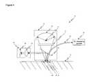
- FIG. 4is a second example of an analyzer having a targeting system and a measuring system according to the invention.
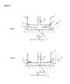
- FIG. 5is a third example of an analyzer having a targeting system and a measuring system according to the invention.
- FIG. 6provides a block diagram of a two probe analyzer according to the invention.
- FIG. 7illustrates an embodiment of a dynamic mount according to the invention
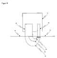
- FIG. 8illustrates control of depth of penetration and pathlength according to the invention.
- FIG. 9provides a block diagram of processing spectra according to the invention.
- Samplingis controlled to enhance analyte concentration estimation derived from noninvasive sampling.
- a targeting systemis used to direct a measuring system to a targeted tissue sample site or tissue volume.
- the targeting systemincreases analyte estimation performance by increasing precision and/or accuracy of sampling and/or by targeting an analyte rich tissue volume.
- An analyzerincludes two major elements, a targeting system and a measuring system.
- the measuring systemis integral to the analyzer.
- the targeting systemis optionally internal to the analyzer, semi-coupled to the analyzer, or is used separately from the analyzer in terms of time of use or in the space that is occupied.
- an x, y, and z-axes coordinate system relative to a given body partis defined.
- An x,y,z coordinate systemis used to define the sample site, movement of objects about the sample site, changes in the sample site, and physical interactions with the sample site.
- the x-axisis defined along the length of a body part and the y-axis is defined across the body part. As an illustrative example using a sample site on the forearm, the x-axis runs between the elbow and the wrist and the y-axis runs across the axis of the forearm.
- the x-axisruns between the base and tip of the digit and the y-axis runs across the digit.
- the x,y plane tangentialtouches the skin surface, such as at a sample site.
- the z-axisis defined as orthogonal to the plane defined by the x- and y-axes.
- a sample site on the forearmis defined by an x,y plane tangential to the sample site.
- An object, such as a sample probe, moving along an axis perpendicular to the x,y planeis moving along the z-axis.
- Rotation of an object about one or a combination of axesis further used to define the orientation of an object, such as a sample probe, relative to the sample site.
- Tiltrefers to an off z-axis alignment of the longitudinal orientation of the sample probe where the longitudinal axis extends from the sample probe tip interfacing with a sample site to the opposite end of the sample probe.
- the analyzerhas two primary systems, a targeting system 15 and a measuring system 16 .
- the targeting systemtargets a tissue area or volume of the sample 14 .
- the targeting systemtargets a surface feature 141 , one or more volumes or layers 142 , and/or an underlying feature 143 , such as a capillary, blood vessel, and/or a distance between a portion of the measuring system, such as a sample probe tip and the sample site.
- the measuring systemcontains a sample probe 303 , which is optionally separate from or integrated into the targeting system.
- the sample probe of the measuring systemis preferably directed to the targeted region or to a location relative to the targeted region either while the targeting system is active or subsequent to targeting. Less preferably, use of the measuring system is followed by use of the targeting system and a targeting image is used to post process the measuring system data.
- a controller 17is used to direct the movement of the sample probe 303 in at least one of the x-, y-, and z-axes via one or more actuators 18 . Optionally the controller directs a part of the analyzer that changes the observed tissue sample in terms of surface area or volume.
- the controllercommunicates with the targeting system, measuring system, and/or controller.
- the targeting system and measuring systemoptionally use a single source that is shared or have separate sources.
- the targetingis optionally used to first target a region and the measuring system is used to subsequently sample at or near the targeted region. Alternatively, the targeting and measuring system are used over the same period of time so that targeting is active during sampling by the measuring system.
- the targeting system and measuring systemoptionally share optics and/or probe the same tissue area and/or volume. Alternatively, the targeting and measuring system use separate optics and/or probe different or overlapping tissue volumes.
- neither, one, or both of the targeting system and measuring systemare brought into contact with the skin tissue 14 at or about the sample site.
- Each of these parametersare further considered, infra.
- Targetsinclude any of:
- marking features added to the skininclude a tattoo, one or more dyes, one or more reflectors, a crosshair marking, and positional markers, such as one or more dots or lines.
- a skin surface featureinclude a wart, hair follicle, hair, freckle, wrinkle, and gland.
- Tissue morphologyincludes surface shape of the skin, such as curvature and flatness.
- specifications for a dermis thicknessinclude a minimal thickness and a maximum depth.
- the targetis a volume of skin wherein the analyte, such as glucose, concentration is higher.
- the measuring systemis directed to image photons at a depth of the enhanced analyte concentration.
- a targeting systemtargets a target.
- a targeting systemtypically includes a controller, an actuator, and a sample probe that are each described infra.
- Examples of targeting systemsinclude a planarity detection system, optical coherence tomography (OCT), a proximity detector and/or targeting system, an imaging system, a two-detector system, and a single detector system.
- Examples of targeting system technologyinclude: capacitance, impedence, acoustic signature, ultrasound, use of a pulsed laser to detect and determine distance, and the use of an electromagnetic field, such as radar and high frequency radio-frequency waves.
- Sources of the targeting systeminclude a laser scanner, ultrasound, and light, such as ultraviolet, visible, near-infrared, mid-infrared, and far-infrared light.
- Detectors of the targeting systemare optionally a single element, a two detector system, an imaging system, or a detector array, such as a charge coupled detector (CCD) or charge injection device or detector (CID).
- CCDcharge coupled detector
- CIDcharge injection device or detector
- One use of a targeting systemis to control movement of a sample probe to a sample site or location.
- a second example of use of a targeting systemis to make its own measurement.
- a third useis as a primary or secondary outlier detection determination. In its broadest sense, one or more targeting systems are used in conjunction with or independently from a measurement system.
- mid-infrared lightsamples tissue surface features to the exclusion of features at a depth due to the large absorbance of water in the mid-infrared.
- a second exampleuses the therapeutic window in the near-infrared to image a tissue feature at a depth due to the light penetration ability from 700 to 1100 nm. Additional examples are targeting with light from about 1100 to 1450, about 1450 to 1900, and/or about 1900 to 2500 nm, which have progressively shallower penetration depths of about 10, 5, and 2 mm in tissue, respectively.
- a further exampleis use of visible light for targeting or imaging greater depths, such as tens of millimeters.
- Raman targeting systemsuch as in WIPO international publication number WO 2005/009236 (Feb. 3, 2005), which is incorporated herein in its entirety by this reference thereto.
- a Raman systemis capable of targeting capillaries. Multiple permutations and combinations of optical system components are available for use in a targeting system.
- the targeting systemuses capacitance sensors or touch sensors for determining any of:
- capacitanceFor a capacitance based targeting system, capacitance, C, is calculated according to equation 1
- capacitance, Cis proportional to the area, A, of the capacitor divided by the distance, d, between the capacitor plates.
- the capacitorhas two plates.
- the first capacitor plateis integrated or connected to the measuring system, such as at the sample module and preferably at the sample module sample probe tip.
- the second capacitoris the deformable material, such as a skin sample, body part, or a tissue sample site. The assumption is that the person is a capacitor. A typical adult has a capacitance of about 120 pF.
- the distance between the capacitor platesis calculated through the combination of equations 1 and 2 through the measurement of the circuit time constant.
- the time constantis the time required to trip a set voltage level, such as about 2.2 volts, given a power supply of known power, such as about 3.3 volts.
- the time constantis used to calculate the capacitance using equation 2.
- the capacitanceis then used to calculate the distance or relative distance through equation 1. For example, as a distance between a sample site, such as a forearm or digit of a hand, and the capacitor plate decreases, the time constant increases and the capacitance increases.
- the measure of distanceis used in positioning the probe at or in proximate contact with the sample site without disturbing the sample site.
- the distance or relative distance between the sample probe tip and the sample siteis determined, preferably before the tip of the sample probe displaces localized sample site skin/tissue, which can lead to degradation of the sample integrity in terms of collected signal-to-noise ratios and/or sampling precision. Examples are used to illustrate the use of the capacitance sensor in the context of a noninvasive analyte property determination.
- the distance or relative distance between the sample probe tip and the sample siteis determined using a single capacitor.
- the sample probeis brought into close proximity with the sample site using the time constant/distance measurement as a metric. In this manner, the sample probe is brought into close proximity to the sample site without displacing the sample site. Due to the inverse relationship between capacitance and distance, the sensitivity to distance between the sample site and the sample probe increases as the distance between the sample probe and the sample site decreases.
- capacitance sensorsthe distance between the sample site and the tip of the sample probe is readily directed to a distance of less than about one millimeter.
- Capacitance sensors as used hereinare also readily used to place the sample probe tip with a distance of less than about 0.1 millimeter to the sample site. In this example, multiple capacitors are optionally used to yield more than one distance reading between the sample probe tip and the sample site. Multiple capacitive sensors are optionally used to control tilt along x- and/or y-axes.
- two or more capacitance sensorsare optionally used for leveling the tip of the sample probe relative to the morphology of the sample site.
- the distance between the sample site and the probe tipis measured using two or more capacitor pairs. For example, if one capacitor reads a larger distance to the sample site than the second capacitor, then the probe tip is moved to level the probe by moving the larger distance side toward the sample, the smaller distance side away from the sample, or both.
- the sample probe tip tilt or angleis either moved manually or by mechanical means.
- a controllercontrols the movement of one or more sample probes via one or more actuators.
- the controlleroptionally uses an intelligent system for locating the sample site and/or for determining surface morphology. For example, the controller hunts in the x- and y-axes for a spectral signature.
- the controllermoves a sample probe via the actuator toward or away from the sample along the z-axis.
- the controlleroptionally uses feedback from the targeting system, from the measurement system, or from an outside sensor in a closed-loop mechanism for deciding on targeting probe movement and for sample probe movement.
- the controlleroptimizes a multivariate response, such as response due to chemical features or physical features.
- Examples of chemical featuresinclude blood/tissue constituents, such as water, protein, collagen, elastin, and fat.
- physical featuresinclude temperature, pressure, and tissue strain. Combinations of features are used to determine features, such as specular reflectance.
- specular reflectanceis a physical feature optionally measured with a chemical signature, such as water absorbance bands centered at about 1450, 1900, or 2600 nm.
- Controlled elementsinclude any of the x-, y-, z-axis position of sampling along with rotation or tilt of the sample probe. Also optionally controlled are periods of light launch, intensity of light launch, depth of focus, and surface temperature.
- the controllercontrols elements resulting in pathlength and/or depth of penetration variation.
- the controllercontrols an iris, rotating wheel, backreflector, or incident optic, which are each described infra.
- the controlleroptionally moves the targeting probe and/or sample probe so as to make minimal and/or controlled contact with the sample.
- Strainis the elongation of material under load. Stress is a force that produces strain on a physical body. Strain is the deformation of a physical body under the action of applied force. In order for an elongated material to have strain there must be resistance to stretching. For example, an elongated spring has strain characterized by percent elongation, such as percent increase in length.
- Skincontains constituents, such as collagen, that have spring-like properties. That is, elongation causes an increase in potential energy of the skin. Strain induced stress changes optical properties of skin, such as absorbance and scattering. Therefore, it is undesirable to make optical spectroscopy measurements on skin with various stress states. Stressed skin also causes fluid movements that are not reversible on a short timescale. The most precise optical measurements would therefore be conducted on skin in the natural strain state, such as minimally stretched skin. Skin is stretched or elongated by applying loads to skin along any of the x-, y-, and z-axes, described infra. Controlled contact reduces stress and strain on the sample. Reducing stress and strain on the sample results in more precise sampling and more accurate and precise glucose concentration estimations.
- An example of using light to measure a physical property, such as contact, stress, and/or strain, in tissueis provided.
- Incident photonsare directed at a sample and a portion of the photons returning from the sample are collected and detected.
- the detected photonsare detected at various times, such as when no stress is applied to the tissue and when stress is applied to the tissue. For example, measurements are made when a sample probe is not yet in contact with the tissue and at various times when the sample probe is in contact with the tissue, such as immediately upon contact and with varying displacement of the sample probe into the tissue.
- the displacement into the tissueis optionally at a controlled or variable rate.
- the collected lightis used to determine properties.
- One exemplary propertyis establishing contact of the sample probe with the tissue.
- a second exemplary propertyis strain.
- Changes in the ratioare indicative of hydration.
- data collection routinesare varied depending upon the determined state of the tissue.
- the probing tissue displacementis varied with change in hydration.
- the strain measurementis optionally made with either the targeting system or measurement system.
- the tissue state probe describe hereinis optionally used in conjunction with a dynamic probe, described infra.
- An actuatormoves the sample probe relative to the tissue sample.
- One or more actuatorsare used to move the sample probe along one or more of the x-, y-, and z-axes.
- the tilt of the sample probe relative to the xy-plane tangential to the tissue sampleis optionally controlled.
- the targeting systemoperates in conjunction with the measurement system, described, infra.
- the combined base module 11 , communication bundle 12 , sample module 13 , and processing centerare referred to as a measuring system 16 .
- the combined measuring system 16 and targeting system 15are referred to as an analyzer 10 .
- FIG. 2a block diagram of an exemplar measuring system 16 of the analyzer 10 is presented that includes a base module 11 and sample module 13 connected via communication means 12 , such as integrated optics or a communication bundle.
- communication means 12such as integrated optics or a communication bundle.
- analysis means 21are incorporated into the analyzer.
- the communication bundleis replaced with wireless communication technology between the sample module and base module or the communication bundle is integrated into the analyzer.
- the measuring system 16 of the noninvasive glucose analyzer 10is included in a single unit, such as a handheld unit or a unit.
- the measuring system 16 of the analyzer 10is physically separated into elements, such as a base module in a first housing 11 , a communication bundle 12 , and a sample module in a second housing 13 .
- Advantages of separate unitsinclude heat, size, and weight management.
- a separated base moduleallows for support of the bulk of the analyzer on a stable surface, such as a tabletop or floor. This allows a smaller sample module to interface with a sample, such as human skin tissue. Separation allows a more flexible and/or lighter sample module for use in sampling by an individual.
- a split analyzerresults in less of a physical impact, in terms of mass and/or tissue displacement, on the sample site by the sample module.
- the sample module, base module, communication bundle, display module, processing center, and tracking systemare further described, infra.
- the base module or semi-remote systemincludes a wavelength selection device, such as a grating, and a detector that is preferably a detector array.
- the remote base moduleis preferably coupled to the sample via a wired or wireless communication bundle that carries at least the optical signal and optionally power.
- the communication bundletransmits control and monitoring signals between the sample module and the base module.
- the base modulepreferably contains at least one of an embedded computer, a display, and an interface to an external computer system.
- the sample/sampling moduleinterfaces to a tissue sample site.
- the sample moduleis used to deliver photons to the sample site and to collect photons from the sample site.
- any element of the analyzeris contained within the sample module.
- a source, guiding optics, proximity sensor, tilt sensor, and collection opticare contained within the sample module.
- the base modulecouples directly to the sample module without a communication bundle.
- the combined base module and sample moduleare integrated into a handheld analyzer, such as a handheld near-infrared based glucose analyzer that couples to the sampling site through an optional guide.
- the base moduleresides on a table, the sample module interfaces through a semi-permanently attached guide to the dorsal aspect of the forearm, and a communication bundle carries power and optical signal between the two modules.
- the base moduleis worn on the person, for example on a belt or as a watch is worn.
- the sample modulealternatively couples to any of a hand, finger, palmar region, base of thumb, forearm, volar aspect of the forearm, dorsal aspect of the forearm, upper arm, head, earlobe, eye, tongue, chest, torso, abdominal region, thigh, calf, foot, plantar region, and toe.
- poweris preferably from a standard wall outlet for power.
- the moduleis preferably battery powered.
- an optional docking stationis provided for power and data analysis.
- any of the embodiments described hereinare operable in a home environment, public facility, or in a medical environment, such as an emergency room, critical care facility, intensive care unit, hospital room, or medical professional patient treatment area.
- the split analyzeris operable in a critical care facility where the sample module is positioned in proximate contact with a subject or patient during use and where the base module is positioned on a support surface, such as a rack, medical instrumentation rack, table, or wall mount.
- Optical componentssuch as a source, backreflector, guiding optics, lenses, filters, mirrors, a wavelength separation device, and at least one detector are optionally positioned in the base module and/or sample module.
- a single sourceis used for the targeting system and the measuring system.
- a sample module 13 portion of an analyzer 10is presented.
- photons from source 31are directed to a sample 14 either directly or via one or more optics, such as a backreflector 32 or a lens.
- the incident photonspass through a dichroic filter 33 .
- a portion of the incident photonseither reflect off of the surface or are diffusely reflected from a volume of the tissue sample 14 .
- a portion of the specular and/or diffusely reflected photonsare directed to a targeting system 15 .
- the collection opticsuses a dichroic filter 33 that reflects a portion of the specular or diffusely reflected to the targeting or imaging system 15 .
- a collection optic 34such as a fiber optic, is used to collect diffusely reflected photons.
- the end of the fiber opticis preferably in close proximity to the surface of the tissue sample 14 .
- the housing or casing of the fiber opticis used to block specularly reflected light.
- the collected lightis directed to the remainder of the measuring system 16 .
- coupling fluidis used at the sample module 13 skin tissue 14 interface. This example is illustrative of a system that uses a single source for the targeting system and measuring system.
- this exampleis illustrative of a system where the targeting system is used to target a sample prior to measurement or at the same time of operation of the measurement system. Still further, this example is illustrative of a targeting system that images substantially the same volume that the measuring system observes.
- a sample module 13 portion of an analyzer 10is presented.
- a source 31emits light. At least part of the emitted light is incident upon a sample tissue site 14 .
- a backreflector 32focuses a portion of the emitted light 31 through an optional first optic 41 , through an optional second optic 42 , and optionally through a fluid coupler.
- the incident photonsare optionally controlled by an aperture defined by an outer radial distance of a incident light blocker. A portion of the incident photons penetrate into the sample 14 where they are transmitted, scattered, diffusely reflected, and/or absorbed.
- the targeting system 15light is optionally directed via optics or mirrors 43 to a detector array 44 .
- the measuring system 16light is collected with one or more collection optics 34 , such as a fiber optic.
- An optional guide element 45is used to control the positioning of the incident photons.
- a first optic and a second opticare used in the optical path between the source element 31 and the tissue sample 14 .
- An optional first optic 41is placed in the optical path after the source element 31 and preferably before the tissue sample 14 .
- the first opticincludes at least one of the following parameters: optically passes desirable wavelengths of light, optically blocks at least one region of undesirable wavelengths of light, limits radiative heat transmitted to the tissue sample, and is not in contact with the tissue sample.
- the first opticpasses desirable wavelengths of light, such as about 1200 to 1850 nm, or sub-regions therein, such as about 1300 to 1700 nm. Within the transmissive region, high transmittance, such as greater than ninety percent, is desirable, but any transmittance is acceptable as long as sufficient net analyte signal is achieved.
- the first opticis, optionally, anti-reflective coated or is index of refraction matched to adjoining surfaces in the optical path. In some embodiments, such as in Example IV, the first optic also passes light used for imaging, such as a region in the visible or in the near-infrared from about 700 to 1100 nm.
- the first opticpreferably blocks or strongly diminishes light throughput in at least one undesirable spectral region emitted by the source or entering through ambient conditions.
- the first opticis used to remove unwanted ultraviolet (UV), visible (VIS), and/or near-infrared light from about 700 to 1000 nm.
- UVultraviolet
- VISvisible
- near-infrared lightfrom about 700 to 1000 nm.
- light of higher energy than the spectral region collected and analyzedis removed in order to remove unwanted heat resulting from photon flux onto the sample and to reduce heating of optics later in the optical path.
- Photons removed by the filter that result in the heating of the filterdo not result in direct heating of the sample site via radiative heating or photonic heating. Rather, the much slower and less efficient conduction or convection processes convey this heat. This reduces the risk of over heating the skin at or about the sample site.
- a second optic 42is optionally placed in the optical path after the source element 31 and before the tissue sample 14 .
- the second opticpasses desirable wavelengths of light and/or optically blocks at least one region of undesirable wavelengths of light.
- The, optional, second opticis in close proximity to the tissue sample. This allows control of radiative and/or conductive heat transmitted to the tissue sample and or control of specular reflectance as described, infra.
- the second optic 42is, optionally, used to control thermal transfer to the tissue sample.
- the second opticis of low thermal conductivity.
- the low thermal conductivityminimizes conductive heating of the sample by the raised temperature of the sample module 13 due to heating by the source.
- Examples of low thermal conductivity materials that are transmissive in the spectral region of interestinclude, silica, PyrexTM, sapphire, and some glasses and plastics.
- the second optichas higher thermal conductivity and is used to more rapidly adjust the tissue sample 14 temperature to that of the tissue sample contacting area of the sample module 13 .
- An example of a higher thermally conductive materialis silicon.
- the second opticoptionally surrounds a detector or a detection optic 34 , such as a fiber.
- An optional spaceris provided between the fiber core and the incident photons.
- the fiber coating and/or spacerprovide specular reflectance blocking and/or depth of penetration and pathlength control as described, infra.
- the maximum penetration of the photons into the tissue samplepreferably exceeds the radial dimension of the spacer.
- the second opticaids in mechanically placing or stabilizing the tip of the sample probe, preferably containing a collection fiber optic, in close proximity to the sample site.
- the measuring systemis used as a targeting system.
- the measuring systemin this example, has targeting system capabilities.
- the measuring systemis used to both target the sample and to subsequently or concurrently measure the sample.
- a separate targeting systemis not needed in this example.
- the targeting system 15such as a camera system or endoscope, targets a first tissue site or tissue volume.
- the measuring system 16targets a second site or volume.
- the two sample sitesoptionally overlap, partially overlap, or are separated.
- the first site and second siteoverlap so that the targeted site is the site sampled.
- the first siteis separated from the second site.
- the controlleris used to adjust a sample probe of the measuring system relative to the targeted volume or area. This allows the targeting system to find and target one feature and the measuring system to measure a separate feature.
- the targeting system and measuring systemhave separate sources and optical trains. Additionally, in this example the targeting system is used before and/or concurrently in time with the measuring system.
- a first sample probe 61is part of a targeting system 15 .
- a second sample probe 62is part of a measuring system 16 .
- the sample probes 61 , 62each are independently controlled via a controller 17 .
- the sample probesmove along any of the x-, y-, and z-axes and each have optional rotation and/or tilt control.
- the sample probes 61 , 62are used at the same or different times.
- the sample probessample different tissue sample 14 locations or the same tissue sample location at different times.
- the two sample probes 61 , 62move in synchronization or are moved independently of each other.
- One embodiment of the method and/or apparatus of the inventionincludes a targeting system used to direct sampling of a measurement system.
- Optional components and/or controls of the apparatusinclude any of:
- the targeting system and/or measuring systemare optionally controlled in at least one of x-, y-, and z-axes and optionally in rotation or tilt. This allows the probing system to adapt to the skin tissue surface.
- the sample probeis an adaptive probe with the benefit of reducing stress/strain upon sampling, as described supra.
- An adaptive sample probe of the targeting and/or measuring systempositions the corresponding sample probe tip at varying positions relative to a tissue sample. As the state of the skin changes, the adaptive probe adjusts the position of the sample probe tip or imaging interface relative to the tissue sample site.
- a first characteristic of the adaptive mountis achievement of highly repeatable sampling by limiting stress and strain on and about the median targeted tissue measurement site. In this manner, the skin undergoes minimal stress as the skin is not deformed to force the exact same position of the tissue to be sampled with each measurement. This leads to more reproducible sampling and hence better accuracy and precision of determined analyte properties.
- an adaptive probeoptionally provides a means for locally registering the location of the targeted and or measured tissue volume with respect to the optical probe and/or tip of a sample module, such that a narrow range of tissue volumes are sampled by the optical system(s).
- Local registrationrefers to controlling the position of the optical probe relative to a target and/or measurement location of the tissue.
- the adaptive probeallows flexibility in terms of the exact position of the tissue that is sampled.
- Means for registering the sample probe to the tissueare preferably optical, but are optionally mechanical and/or electromechanical.
- the sample probeis optionally dynamic.
- the targeting system sample probe 61 , the measuring system sample probe 62 , and/or a shared sample probe 303are optionally dynamic.
- a dynamic probeis moved in a controlled fashion relative to a tissue sample in order to control spectral variations resulting from the sample probe displacement of the tissue sample during a sampling process.
- a noninvasive analyzer 10controls movement of a dynamic sample probe along any of the x-, y-, and z-axes and optionally controls tilt and/or rotation of the sample probe relative to a sampled tissue 14 .
- the ability to move the sample probe relative to the tissue sample as a function of timeallows a dynamic tissue measurement.
- a dynamic tissue measurementis designed to collect time serial spectral data that contains the dynamic tissue response of the tissue sample as the sample probe is brought into contact with the tissue sample. In this measurement process spectral raster scans are collected continuously or semi-continuously as the sample probe is moved into contact with the tissue sample and/or as the sample probe displaces the tissue sample.
- the sample probeis lowered slowly onto the targeted measurement site with or without an optical probe placement guide while the instrument acquires signal.
- a sample probeis controlled at least along the z-axis perpendicular to the x, y plane tangential to the surface of the sampled site thereby controlling displacement of the sample probe relative to a sample.
- the z-axis control of the displaced sample probe element of the sample moduleprovides for collection of noninvasive spectra with a given displacement of a tissue sample and for collection of noninvasive spectra with varying applied displacement positions of the sample probe relative to the nominal plane of the sample tissue surface.
- the interface between the optical probe and the skin surface at the tissue measurement siteis potentially a significant source of sampling error due to:
- Incident light normal to the surfacepenetrates into the skin sample based upon the difference in refractive index, Snell's Law.
- refractive index of skinapproximately 1.38
- refractive index of airapproximately 1.0
- approximately four percent of the normally incident lightis reflected and ninety-six percent of the light penetrates into the skin if the surface is smooth.
- the rough tissue surfaceresults in an increased percentage of specularly reflected light.
- the percentage of light penetrating into skinvaries as the index of refraction of the interfacing material to skin changes.
- the couplingchanges with the use of an intermediate material, such as water or a coupling fluid.
- the amount of light that is specularly reflectedis determined to degrade noninvasive estimations of low signal to noise analytes.
- a targeting or measuring sample probethat does not contact the surface of the skin or is not coupled to the skin via a coupling fluid, results in specular reflectance off of the diffusely reflecting skin surface that is partially caught in collection optics. This specular reflectance is difficult to remove once captured by the collection optic system and subsequently observed with the detector system.
- the specular signalis often much larger in magnitude across the desired spectral region compared with the analyte signal. For example, four-percent specular light is orders of magnitude larger than a noninvasive glucose signal from the glucose molecule that is present in about the 30 to 600 mg/dL range.
- One method for removing specular lightis to have part of the sample probe contact the skin surface. For example, having an optically opaque part contact the skin between the incident and collection photons forces the collected photons to have gone through at least a portion of the skin.
- specular blockersinclude a thin or thick blade blocker or a fiber optic cladding or buffer.
- One or both of the targeting system and measurement systemoptionally has a specular blocker.
- An optional aspect of the optical sampling system of one or both of the targeting and measurement systemis the maintenance of an optimal level of hydration of the surface tissue at the measurement site for enhancement of the optical signal, sample reproducibility, and suppression of surface reflectance.
- Skin hydration meansare optionally used with the targeting and/or measuring system. Skin surface irregularities result in an increase in surface reflection of the incident light. Surface irregularities of skin mean that the incident light is not normal to the surface. This results in more reflected light, and less penetrating light. In addition, air gaps in the outer layers of skin result in more reflected light that does not penetrate to the analyte containing region. A fraction of the light penetrating into an outermost layer of skin hits one or more air pockets and is reflected off of each surface of the air pocket. Many air pockets or poor hydration lead to a significant reduction in the percentage of incident photons that penetrate through the outermost skin layers, such as the stratum corneum, to the inner skin layers.
- Hydrationis achieved through a variety of means, such as occlusion, direct water contact, and increasing localized perfusion.
- a preferred means of the optional hydration stepis hydration by occlusive blockage of trans-epidermal water loss. This blockage ensures a steady state hydration as water diffusing from interior tissue is trapped in the stratum corneum. Attainment of high hydration levels reduces the water concentration gradient that provides the driving force for this trans-epidermal water movement.
- an occlusive plugfits snugly into a guide aperture during periods between measurements, acting to insulate the tissue in the guide aperture from trans-epidermal water loss and the environmental effects of temperature and humidity that are known to influence the stratum corneum hydration state.
- an occlusive patchis used, such as wrapping or covering the tissue sample site with a flexible polymer sheet.
- a window or opticis contacted with the sample site to increase the localized skin surface and shallow depth hydration and/or to stabilize the tissue by providing the same tissue displacement as the probe.
- the opticis continuously, replaceably, or intermittently attached to the sample site. Examples of optics include a window, a longpass filter, and a bandpass filter.
- hydration meansinclude a material that provides a hydration barrier, thus promoting the full and stable hydration of the stratum corneum.
- the occlusion meansuse a hydrophobic material, such as cellophane.
- optional perfusion enhancement or regulation meansare used to increased precision and accuracy in analyte property estimation by the removal or reduction of dry or pocketed skin at the sampling site.
- a coupling fluidis optionally used with the targeting and/or measuring system.
- An optical coupling fluid with a refractive index between that of the skin surface and the contacting mediumis preferably used.
- a coupling fluidneed not be a refractive index matching fluid in order to increase light throughput.
- the coupling fluidneed not have a refractive index between that of skin and the optic to be beneficial.
- the percentage of incident photons passing through a silicon lens into skinis increased even with use of a coupling fluid that does not have a refractive index between that of silicon and skin.
- FC-40(a fluorocarbon) has an index of refraction of 1.290 that is not between that of skin, 1.38, and silicon, approximately 3.45. However, the FC-40 still increases incident photon penetration by displacement of air.
- FC-40is not an index-matching medium, optical coupling fluid, or refractive-index matching coupling fluid; however, FC-40 is a coupling fluid that aids in light coupling by displacing the air.
- Preferable coupling fluidsare minimally inactive or inactive in terms of absorbance in the spectral region of interest.
- the near-infrared fluorocarbonssuch as FC-40
- FC-40near-infrared fluorocarbons
- a coupling mediumis used to fill these air gaps.
- the coupling fluidfor an application, such as noninvasive glucose estimation, the coupling fluid:
- a coupling fluidis preheated to between about 90 and 95° F., preferably about 92° F. Preheating the coupling fluid minimizes changes to the surface temperature of the contacted site, thus minimizing spectral changes observed from the sampled tissue.
- the coupling fluidis optionally delivered in a manual, semi-automated, or automated fashion.
- neither the targeting system nor the measurement systemuse a mount in the sampling process.
- a guide or optionally a mountis optionally used with one or both of the targeting system and measurement system.
- a key characteristic of an optional adaptive mountis achievement of highly repeatable sampling by limiting stress and strain on and/or about the median targeted tissue measurement site. To achieve this, the mount adapts to physical changes in the sample.
- An additional benefit of the adaptive mountis that it optionally provides a means for locally registering the location of the targeted tissue volume with respect to the optical probe and/or tip of a sample module, such that a narrow range of tissue volumes are sampled by the optical system. Local registration refers to controlling the position of the optical probe relative to a target location on the tissue.
- the adaptive mountallows flexibility in terms of the exact position of the tissue that is sampled.
- the mountadapts to the new state of the sample by mounting a sample probe to a slightly new position in terms of x-position and y-position, described infra.
- Means for registering the mount and the optical probeare optionally mechanical, optical, electrical, and/or magnetic.
- an adaptive mountthat increases precision and accuracy of noninvasive sampling, which results in increased sensitivity, precision, and accuracy of subsequent analyte property estimation derived from the sampling.
- the adaptive mountis placed onto the skin of a person. Between uses, opposing ends of the adaptive mount move relative to each other as the skin tissue state changes.
- the adaptive mountis designed to minimize skin deformation during placement of a sample probe of an analyzer or during placement of a plug.
- the adaptive mountsamples a dynamic x-, y-position at or about a central sample site.
- the adaptive mountis deformable, which distributes applied forces during sample about the sample site.
- At least one axis of the sample probeis allowed to float relative to a fixed x,y-point that defines a given sample site.
- FIG. 7an example of an adaptive mount with freedom of motion along the x-axis is presented at two moments in time.
- the tissue 14has a distance, d 1 , between a first alignment piece 71 and a second alignment piece 72 .
- the two alignment pieces 71 , 72have corresponding means for registration 73 , 74 .
- the two registration pieces 73 , 74 piecesare integral to the alignment pieces 71 , 72 or are separate pieces.
- the two registration pieces 73 , 74have a distance, d 3 , between them. In this case, the registration pieces protrude from the alignment pieces.
- a portion of a sample module 13is represented near the tissue 14 .
- Registration pieces 75 , 76correspond to the registration pieces on the mount 73 , 74 , respectively. In this case, registration piece 75 acts as one-half of a lock and key element corresponding to the second half of a lock and key element 73 .
- a sample probe 303is situated at a given x-, y-position relative to the tissue 14 .
- the tissue 14has changed state.
- the tissuehas elongated resulting in the distance between the first and second alignment pieces 71 , 72 to expand in distance from d 1 to d 2 .
- the corresponding distance between the first and second registration pieces 73 , 74has similarly expanded in distance from d 3 to d 4 .
- the sample module 13includes one registration piece 75 that couples with a corresponding registration piece 71 on the mount 70 .
- a second registration piece 76 on the sample module 13has freedom of movement in at least one-dimension relative to the alignment piece 72 and/or registration piece 74 .
- the tip of the sample probe 303mounts to a slightly different x-, y-position of the tissue 14 as the tissue state changes in a manner that effects the tissue size, shape, and/or torque. This results in at least a portion of the sample module 13 and/or sample probe 303 to mount on the mount 70 via one or more alignment pieces and/or one or more registration pieces with minimal deformation or strain on the tissue 14 .
- the mounting of the sample probe 303 to the mount 70 with minimal strainresults in noninvasive spectra with fewer spectral interferences and hence corresponding analyte property estimation is more precise and accurate.
- the sample probe 303is movable along the z-axis, so that the tip of the sample probe results in minimal stress on the sample tissue volume. In the pictured instance, the sample probe is shown as extended to the tissue 14 at time 2 .
- a movable z-axis sample probeis optionally used with this system, supra.
- the variable placement of the sample probe relative to the tissueis performed along the y-axis or through a combination of x- and y-axis.
- the alignment piece 72optionally contains means, such as groove along the y-axis for y-axis freedom of movement or a slide, such as a planar surface, for x- and y-axis freedom of movement.
- one or both of the targeting and measuring systemstarget a depth of skin tissue.
- the measuring systemis adjusted to a pathlength or depth in the absence of a targeting system.
- the targeting systemtargets a depth.
- a targeted depthis the cutaneous layer of skin tissue.
- a hydrophilic analytesuch as glucose
- the depth targeting of photons into a perfused or aqueous rich layerincreases the sampling photon density in the analyte rich region and minimizes photon density in the adipose layer.
- the depth of penetration and pathlength of collected photonsis dependent upon the tissue state and properties of the tissue, such as scattering and absorbance. Generally, lower scattering results in deeper maximum photon depth of penetration. As absorption increases, the photons traveling deeper have a smaller probability of returning to the incident surface. Thus, effective depth of penetration of collected photons is dependent upon both parameters. In addition, scattering and depth of penetration affect the optical pathlength. Generally, photons collected at an incident surface with deeper penetration and/or greater radial diffusion have, on average, longer pathlengths. Since scattering and absorbance are wavelength dependent, the average depth of penetration and pathlength are also wavelength dependent.
- a cutaneous sampling optical probelimits the radial distance between incident photons directed at the skin and the collected photons coming from the skin.
- Optional radial range limitsinclude a minimum range, a maximum range, or both a minimum and maximum range.
- very short pathlengthsare effectively blocked using a spacer between a region of incident photons contacting the skin and a region where photons are collected from the skin.
- Exampleinclude a thin or thick blocker, such as a blade, a gap, and an optically opaque sheath, such as a fiber optic coating. This spacer is optionally used to block specular light in embodiments where the optics do not come into close proximity with the skin.
- a maximum rangeis defined by the far reaches of the incident illumination area and collection area.
- An illumination probe 81delivers photons to the tissue sample 14 .
- a collection probe 82collects light emerging from a collection area.
- Photons having the longest radial distance to travel between the illumination area and collection areatypically have the largest average depth of penetration and pathlength 84 .
- Intermediate radial distancestypically result in intermediate depths of penetration and pathlength 85 .
- the average pathlength and depth of penetrationis increased by moving the illumination area further from the collection area.
- controlled distance between the illumination area and detection areais optionally static, dynamic, or adjustable to a person or to a tissue state.
- a fiber bundle or a plurality of bundletsare used.
- the spacing between the illumination and collection fibers of each bundlet, and the spacing between bundletsis optimized to minimize sampling of the adipose subcutaneous layer and to maximize collection of light that has been backscattered from the cutaneous layer.
- This exampleoptimizes penetration depth by limiting the range of distances between illumination fibers and detection fibers.
- the pathlength and depth of penetration of photons in a tissue sampleare optionally controlled through others means.
- mechanical and/or optical meansare used to change the illumination area lit by a source and/or the collection area observed by a detector. As described, infra, this changes the average pathlength and depth of penetration.
- a changing blocker thickness or iris diameterare used to expand or contract the illumination and/or detector area.
- the irisis optionally mechanically opened and shut or is optically expanded or contracted.
- a liquid crystalis used to black out or make opaque regions of the illumination and/or collection area.
- a reflector shapeis changed with time causing the illumination area lit or detection area observed to expand or contract.
- a shape of a back reflector behind a sourceis changed to create larger or smaller illumination areas, such as a circle with a different diameter, on the sample.
- the incident angle of the photonsis changed. This alters the initial angle of the photons entering the sample. This initial angle operates in conjunction with scattering and absorbance to result in an altered average depth of penetration and/or pathlength of the photons into the sample.
- fiber ringsare used for illumination and/or collection.
- a wheelis rotated in the optical train prior to the sample. The wheel has transmissive, semi-transmissive, or opaque regions as a function of wavelength and/or position.
- the average pathlengthis varied.
- the wheelis spun in a light source that the average distance of the open areas varies as a function of time.
- a second wheelis used so that only the open areas of interest are viewed at a given time by the detecting system. This allows a detector to see different depths of the same sample through time or for an array to see different depths and pathlengths of a sample at a single point in time or through time.
- part of an analyzeris redirected to a new sample site.
- part of a sample probeis used to aim the sample probe to a new sampled area.
- probed tissue pathlengthis controlled by tailoring the distance distribution between optical illuminator conduits and the detector conduit using a digital mirror array.
- lightpasses from a multiplicity of illuminator conduits into the skin and from the skin into a centrally located detector conduit.
- Source lightis separated into different optical channels defined by individual fibers in a short fiber bundle into which the source light is focused.
- a digital mirror array, or DLP chipis used to separate the source light into individual fibers or a few fibers in an illumination bundle. Focused light is reflected off of the mirror array onto the fiber optic and individual mirror angles on the chip are controlled to reflect full, partial, or no intensity onto individual illumination fibers. Since each fiber represents an element in the source/detector distance distribution, manipulation of the reflected light allows for tailoring or even optimization of the light launch distribution into the tissue. Such flexibility allows for pathlength correction of the measured diffuse reflectance signal.
- the analyzerpreferably includes a data processing module, which is used in generation of an analyte property using signal generated from the targeting system and/or the measuring system.
- the data processing modulepreferably uses data preprocessing and/or data processing techniques in combination with the invention.
- a method and apparatuscorrect for tissue related interference for the purpose of calibration and measurement of biological parameters noninvasively. The method is described in terms of outlier identification, filtering, spectral correction, and baseline subtraction steps that, when used together, enable the noninvasive measurement of a biological parameter, such as glucose concentration.
- FIG. 9a block diagram summarizing processing 90 of the near-infrared signal is presented.
- the stepsare all preferably used in the order illustrated. Alternatively, one or more steps are omitted and/or the steps are performed in an alternative order.
- the methodoptionally includes both gross 91 and detailed 96 methods for detecting outliers or anomalous measurements that are incompatible with the processing methods or are the result of sampling or instrument errors.
- Spectral correctioninvolving the steps of filtering 92 and/or correction 93 , is applied to compensate for noise and interference and to adjust a spectrum according to local or minor changes in the optical properties of the tissue sample.
- the step of background removal 95reduces variation in the measurement, such as variation associated with sample-site differences, dynamic tissue changes, and subject-to-subject variation.
- An optional tissue template 94is used to remove background 95 .
- Examples of a tissue templateinclude a spectrum of the subject being measured, a basis set, or a computed spectrum of a cluster of spectral data.
- a background removal steppreferably follows the steps defined above and uses a spectral background or tissue template.
- the spectrally corrected signal, zis used for calibration development or measurement of a target analyte.
- the backgroundis estimated on the basis of an optimal selection of spectrally corrected measurements collected prior to the measurement, m.
- the variables c and dare preferably determined on the basis of features related to the dynamic variation of the tissue.
- x 1is a spectrally corrected spectral measurement collected on tissue at the beginning of a measurement period.
- the measurement periodis defined as a time period during which the state of the tissue sample is uniform. Further detail on basis sets is provided in U.S. Pat. No. 6,115,673, which is incorporated herein in its entirety by this reference thereto.
- the background removal stepuses a basis set of spectral interferences to remove the signals that are specific to a given sampled tissue volume, such as the background.
- the optical estimate of the backgroundare performed subsequent to the removal of noise and the correction of the spectrum.
- the order of the stepsis optionally varied.
- the wavelength selection stepis optionally performed out of sequence, such as after the second outlier detection or before any of the earlier steps.
- not all stepsare required. For example, correcting dead pixels is not appropriate to some analyzers.
- conversion to absorbanceis not always required, nor are other steps.
- An optional intelligent system for measuring blood analyte propertiesis used in combination with the invention.
- the systemoperates on near-infrared absorbance spectra of in-vivo skin tissue.
- the architectureemploys a pattern classification engine to adapt the calibration to the structural properties and physiological state of the patient as manifested in the absorbance spectrum.
- a priori information about the primary sources of sample variabilityare used to establish general categories of patients. Application of calibration schemes specific to the various categories results in improved calibration and prediction.
- Two classification methodsare optionally used. The first assumes the classes are mutually exclusive and applies specific calibration models to the various patient categories.
- the second methoduses fuzzy set theory to develop calibration models and blood analyte predictions. In the second, each calibration sample has the opportunity to influence more than one calibration model according to its class membership. Similarly, the predictions from more than one calibration are combined through defuzzification to produce the final blood analyte property estimation.
- the two implementations of the intelligent measurement systemare detailed for the noninvasive estimation of the concentration of blood glucose.
- the firstuses spectral features related to gross tissue properties to determine which of several prediction models is the most likely to produce an accurate blood glucose estimation.
- the extracted featuresare representative of the actual tissue volume irradiated.
- the secondemploys a fuzzy classification system to assign a degree of membership in each of several classes to the spectral measurement.
- the membershipis used to aggregate the predictions of calibrations associated with each class to produce the final blood glucose prediction.
- the membership strategyis employed during calibration in modified form of weighted principal components regression to produce calibrations from the entire population of samples. Additional description of an intelligent system is provided in U.S. Pat. No. 6,280,381, which is incorporated herein in its entirety by this reference thereto.
Landscapes
- Health & Medical Sciences (AREA)
- Life Sciences & Earth Sciences (AREA)
- Physics & Mathematics (AREA)
- Engineering & Computer Science (AREA)
- Molecular Biology (AREA)
- General Health & Medical Sciences (AREA)
- Biophysics (AREA)
- Biomedical Technology (AREA)
- Heart & Thoracic Surgery (AREA)
- Medical Informatics (AREA)
- Veterinary Medicine (AREA)
- Surgery (AREA)
- Animal Behavior & Ethology (AREA)
- Pathology (AREA)
- Public Health (AREA)
- Optics & Photonics (AREA)
- Emergency Medicine (AREA)
- Vascular Medicine (AREA)
- Spectroscopy & Molecular Physics (AREA)
- Human Computer Interaction (AREA)
- Measurement Of The Respiration, Hearing Ability, Form, And Blood Characteristics Of Living Organisms (AREA)
- Investigating Or Analysing Materials By Optical Means (AREA)
Abstract
Description
- claims benefit of U.S. provisional patent application No. 60/656,727 filed Feb. 25, 2005;
- claims benefit of U.S. provisional patent application No. 60/658,708 filed Mar. 3, 2005; and
- claims benefit of U.S. provisional patent application No. 60/658,821 filed Mar. 4, 2005; and
all of which are incorporated as if fully set forth herein in their entirety by this reference thereto.
- increased precision and accuracy of noninvasive sampling;
- a means of assuring that the similar tissue sample volumes are repeatably sampled; and
- minimizing sampling errors due to mechanical tissue distortion and probe placement.
- a natural tissue component;
- a chemical feature;
- a physical feature;
- an abstract feature;
- a marking feature added to the skin;
- a skin surface feature;
- a measurement of tissue strain;
- tissue morphology;
- a target below the skin surface;
- a manmade target;
- a fluorescent marker;
- a subcutaneous feature;
- a dermis thickness within a specification;
- capillary beds;
- a capillary;
- a blood vessel; and
- arterial anastomoses.
- tilt of a sample probe relative to a sample site;
- distance of a sample probe tip to a sample site;
- x,y-position of a sample probe tip relative to a sample site;
- relative distance of a sample probe tip to a sample site; and
- contact of a sample probe tip with a sample site.
where capacitance, C, is proportional to the area, A, of the capacitor divided by the distance, d, between the capacitor plates. The capacitor has two plates. The first capacitor plate is integrated or connected to the measuring system, such as at the sample module and preferably at the sample module sample probe tip. The second capacitor is the deformable material, such as a skin sample, body part, or a tissue sample site. The assumption is that the person is a capacitor. A typical adult has a capacitance of about 120 pF. The time constant of a capacitor/resistor is calculated according to
T=RC (2)
where the time constant, T, is equal to the resistance, R, times the capacitance, C. Hence, the distance between the capacitor plates is calculated through the combination of
- an adaptive sample probe head;
- a dynamic sampling probe;
- a specular reflectance blocker;
- occlusion and/or tissue hydration control;
- a coupling fluid;
- an automated coupling fluid delivery system;
- a guide;
- a mount;
- a system for reducing stress/strain on the tissue;
- a system for controlling skin tissue state;
- a split system;
- a system for controlling pathlength;
- a system for controlling depth of penetration;
- a system for reducing and/or controlling thermal changes of the skin tissue;
- means for minimizing sampling error;
- an intelligent system for data processing;
- a basis set; and/or
- a data processing algorithm.
- skin state change;
- skin deformation with time;
- skin stress/strain;
- temperature mismatch;
- lost dynamic range of detection system;
- air gaps; and
- refractive index mismatch.
- a vapor barrier or semi-permeable membrane (for example, GORE-TEX, manufactured by W. L. Gore and Associates of Newark, Del. as the mount) in the form of a wrap or a patch configured to cover the site target for measurement. In this latter embodiment the patch is affixed to the tissue site through an adhesive or other attachment mechanism such as a strap or a wrap;
- non-occlusive mechanisms for hydration of the stratum corneum may also be used, including:
- an application of water that is pneumatically driven into the skin;
- ultrasound energy applications to accelerate passive occlusion;
- topical application of skin toners and other water/solute mixtures such as alpha hydroxy acid solutions that serve to drive water and solute into the dry outer skin layer; and
- topical analgesic formulations that enhance and/or stimulate local circulation at the measurement site leading to an improvement in surface hydration.
Coupling Fluid
- is spectrally inactive;
- is non-irritating;
- is nontoxic;
- has low viscosity for good surface coverage properties;
- has poor solvent properties with respect to leaching fatty acids and oils from the skin upon repeated application; and
- is thermally compatible with the measurement system.
z=x−(cxt+d) (3)
where xtis the estimated background or tissue template, c and d are slope and intercept adjustments to the tissue template. Direct subtraction is just one form of background removal. The spectrally corrected signal, z, is used for calibration development or measurement of a target analyte. The background is estimated on the basis of an optimal selection of spectrally corrected measurements collected prior to the measurement, m. The variables c and d are preferably determined on the basis of features related to the dynamic variation of the tissue.
- averaging spectra;
- correcting dead pixels;
- calculating absorbance;
- performing x-axis standardization;
- uniformly re-sampling the spectrum to standardize the x-axis;
- performing a first (gross) outlier detection;
- correcting the spectrum;
- performing a wavelength selection;
- removing interference; and
- performing a second (fine) outlier detection
Claims (22)
Priority Applications (4)
| Application Number | Priority Date | Filing Date | Title |
|---|---|---|---|
| US11/361,143US7697966B2 (en) | 2002-03-08 | 2006-02-23 | Noninvasive targeting system method and apparatus |
| EP06721018AEP1850736A4 (en) | 2005-02-25 | 2006-02-24 | Noninvasive targeting system method and apparatus |
| JP2007557171AJP2008531133A (en) | 2005-02-25 | 2006-02-24 | Non-invasive targeting system method and apparatus |
| PCT/US2006/006496WO2006093816A2 (en) | 2005-02-25 | 2006-02-24 | Noninvasive targeting system method and apparatus |
Applications Claiming Priority (11)
| Application Number | Priority Date | Filing Date | Title |
|---|---|---|---|
| US36288502P | 2002-03-08 | 2002-03-08 | |
| PCT/US2003/007065WO2003076883A2 (en) | 2002-03-08 | 2003-03-07 | Compact apparatus for noninvasive measurement of glucose through near-infrared spectroscopy |
| US10/472,856US7133710B2 (en) | 2002-03-08 | 2003-03-07 | Compact apparatus for noninvasive measurement of glucose through near-infrared spectroscopy |
| US56656804P | 2004-04-28 | 2004-04-28 | |
| US60501704P | 2004-08-27 | 2004-08-27 | |
| US65672705P | 2005-02-25 | 2005-02-25 | |
| US65870805P | 2005-03-03 | 2005-03-03 | |
| US65882105P | 2005-03-04 | 2005-03-04 | |
| US11/117,104US7519406B2 (en) | 2004-04-28 | 2005-04-27 | Noninvasive analyzer sample probe interface method and apparatus |
| US76148606P | 2006-01-23 | 2006-01-23 | |
| US11/361,143US7697966B2 (en) | 2002-03-08 | 2006-02-23 | Noninvasive targeting system method and apparatus |
Related Parent Applications (3)
| Application Number | Title | Priority Date | Filing Date |
|---|---|---|---|
| US10/472,856Continuation-In-PartUS7133710B2 (en) | 1999-10-08 | 2003-03-07 | Compact apparatus for noninvasive measurement of glucose through near-infrared spectroscopy |
| PCT/US2003/007065Continuation-In-PartWO2003076883A2 (en) | 1999-10-08 | 2003-03-07 | Compact apparatus for noninvasive measurement of glucose through near-infrared spectroscopy |
| US11/117,104Continuation-In-PartUS7519406B2 (en) | 2000-05-02 | 2005-04-27 | Noninvasive analyzer sample probe interface method and apparatus |
Publications (2)
| Publication Number | Publication Date |
|---|---|
| US20060200017A1 US20060200017A1 (en) | 2006-09-07 |
| US7697966B2true US7697966B2 (en) | 2010-04-13 |
Family
ID=36941660
Family Applications (1)
| Application Number | Title | Priority Date | Filing Date |
|---|---|---|---|
| US11/361,143Active2029-02-13US7697966B2 (en) | 2002-03-08 | 2006-02-23 | Noninvasive targeting system method and apparatus |
Country Status (4)
| Country | Link |
|---|---|
| US (1) | US7697966B2 (en) |
| EP (1) | EP1850736A4 (en) |
| JP (1) | JP2008531133A (en) |
| WO (1) | WO2006093816A2 (en) |
Cited By (276)
| Publication number | Priority date | Publication date | Assignee | Title |
|---|---|---|---|---|
| US20090036759A1 (en)* | 2007-08-01 | 2009-02-05 | Ault Timothy E | Collapsible noninvasive analyzer method and apparatus |
| US20110125004A1 (en)* | 2008-07-31 | 2011-05-26 | Koninklikjke Philips Electronics N.V. | Analysis by photo acoustic displacement and interferometryl |
| WO2012134515A1 (en)* | 2011-04-01 | 2012-10-04 | Verifica-Bg Inc. | Method and apparatus for in vivo optical measurement of blood glucose concentration |
| US20130072771A1 (en)* | 2011-09-21 | 2013-03-21 | Pixart Imaging Inc. | Optical finger mouse, electronic device and physiological characteristics detection device |
| US20130131473A1 (en)* | 2011-11-18 | 2013-05-23 | Pixart Imaging Inc. | Optical distance measurement system and operation method thereof |
| US20130131474A1 (en)* | 2011-11-22 | 2013-05-23 | Pixart Imaging Inc. | Remote controller and display system |
| US8514067B2 (en) | 2011-08-16 | 2013-08-20 | Elwha Llc | Systematic distillation of status data relating to regimen compliance |
| US8670642B2 (en) | 2005-11-18 | 2014-03-11 | Omni Medsci, Inc. | Broadband or mid-infrared fiber light sources |
| US8848282B2 (en) | 2002-09-03 | 2014-09-30 | Omni Medsci, Inc. | System and method for voice control of medical devices |
| US9042967B2 (en) | 2008-05-20 | 2015-05-26 | University Health Network | Device and method for wound imaging and monitoring |
| US20150157269A1 (en)* | 2013-12-06 | 2015-06-11 | Covidien Lp | Capacitance enhanced physiological measurements |
| US20150241401A1 (en)* | 2014-02-18 | 2015-08-27 | Aemtek, Inc. | Methods, systems and devices for batch sampling |
| US9164032B2 (en) | 2012-12-31 | 2015-10-20 | Omni Medsci, Inc. | Short-wave infrared super-continuum lasers for detecting counterfeit or illicit drugs and pharmaceutical process control |
| US9351671B2 (en) | 2012-07-16 | 2016-05-31 | Timothy Ruchti | Multiplexed pathlength resolved noninvasive analyzer apparatus and method of use thereof |
| US9351672B2 (en) | 2012-07-16 | 2016-05-31 | Timothy Ruchti | Multiplexed pathlength resolved noninvasive analyzer apparatus with stacked filters and method of use thereof |
| US9442065B2 (en) | 2014-09-29 | 2016-09-13 | Zyomed Corp. | Systems and methods for synthesis of zyotons for use in collision computing for noninvasive blood glucose and other measurements |
| US9554738B1 (en) | 2016-03-30 | 2017-01-31 | Zyomed Corp. | Spectroscopic tomography systems and methods for noninvasive detection and measurement of analytes using collision computing |
| US9585604B2 (en) | 2012-07-16 | 2017-03-07 | Zyomed Corp. | Multiplexed pathlength resolved noninvasive analyzer apparatus with dynamic optical paths and method of use thereof |
| US9766126B2 (en) | 2013-07-12 | 2017-09-19 | Zyomed Corp. | Dynamic radially controlled light input to a noninvasive analyzer apparatus and method of use thereof |
| US9846933B2 (en) | 2015-11-16 | 2017-12-19 | General Electric Company | Systems and methods for monitoring components |
| US9869545B2 (en) | 2015-04-15 | 2018-01-16 | General Electric Company | Data acquisition devices, systems and method for analyzing strain sensors and monitoring turbine component strain |
| US9879981B1 (en) | 2016-12-02 | 2018-01-30 | General Electric Company | Systems and methods for evaluating component strain |
| US9897584B2 (en) | 2012-12-31 | 2018-02-20 | Omni Medsci, Inc. | Short-wave infrared super-continuum lasers for natural gas leak detection, exploration, and other active remote sensing applications |
| US9909860B2 (en) | 2015-04-15 | 2018-03-06 | General Electric Company | Systems and methods for monitoring component deformation |
| US9932853B2 (en) | 2015-04-28 | 2018-04-03 | General Electric Company | Assemblies and methods for monitoring turbine component strain |
| US9953408B2 (en) | 2015-11-16 | 2018-04-24 | General Electric Company | Methods for monitoring components |
| US9993159B2 (en) | 2012-12-31 | 2018-06-12 | Omni Medsci, Inc. | Near-infrared super-continuum lasers for early detection of breast and other cancers |
| US10012552B2 (en) | 2015-11-23 | 2018-07-03 | General Electric Company | Systems and methods for monitoring component strain |
| US10076277B2 (en) | 2015-01-22 | 2018-09-18 | Covidien Lp | Pain level detection and characterization using capacitive sensors |
| US10126119B2 (en) | 2017-01-17 | 2018-11-13 | General Electric Company | Methods of forming a passive strain indicator on a preexisting component |
| US10132615B2 (en) | 2016-12-20 | 2018-11-20 | General Electric Company | Data acquisition devices, systems and method for analyzing passive strain indicators and monitoring turbine component strain |
| US10136819B2 (en) | 2012-12-31 | 2018-11-27 | Omni Medsci, Inc. | Short-wave infrared super-continuum lasers and similar light sources for imaging applications |
| US10159412B2 (en) | 2010-12-01 | 2018-12-25 | Cercacor Laboratories, Inc. | Handheld processing device including medical applications for minimally and non invasive glucose measurements |
| US10213113B2 (en) | 2012-12-31 | 2019-02-26 | Omni Medsci, Inc. | Physiological measurement device using light emitting diodes |
| US10345179B2 (en) | 2017-02-14 | 2019-07-09 | General Electric Company | Passive strain indicator |
| US10438356B2 (en) | 2014-07-24 | 2019-10-08 | University Health Network | Collection and analysis of data for diagnostic purposes |
| US10451499B2 (en) | 2017-04-06 | 2019-10-22 | General Electric Company | Methods for applying passive strain indicators to components |
| US10502551B2 (en) | 2017-03-06 | 2019-12-10 | General Electric Company | Methods for monitoring components using micro and macro three-dimensional analysis |
| US10660526B2 (en) | 2012-12-31 | 2020-05-26 | Omni Medsci, Inc. | Near-infrared time-of-flight imaging using laser diodes with Bragg reflectors |
| US10697760B2 (en) | 2015-04-15 | 2020-06-30 | General Electric Company | Data acquisition devices, systems and method for analyzing strain sensors and monitoring component strain |
| US10736518B2 (en) | 2015-08-31 | 2020-08-11 | Masimo Corporation | Systems and methods to monitor repositioning of a patient |
| US10765367B2 (en) | 2014-10-07 | 2020-09-08 | Masimo Corporation | Modular physiological sensors |
| US10779098B2 (en) | 2018-07-10 | 2020-09-15 | Masimo Corporation | Patient monitor alarm speaker analyzer |
| US10784634B2 (en) | 2015-02-06 | 2020-09-22 | Masimo Corporation | Pogo pin connector |
| USD897098S1 (en) | 2018-10-12 | 2020-09-29 | Masimo Corporation | Card holder set |
| US10799163B2 (en) | 2006-10-12 | 2020-10-13 | Masimo Corporation | Perfusion index smoother |
| US10799160B2 (en) | 2013-10-07 | 2020-10-13 | Masimo Corporation | Regional oximetry pod |
| US10825568B2 (en) | 2013-10-11 | 2020-11-03 | Masimo Corporation | Alarm notification system |
| US10842422B2 (en) | 2016-07-21 | 2020-11-24 | University Of Kentucky Research Foundation | Compact low-cost fiberless diffuse speckle contrast flow-oximeter |
| US10849554B2 (en) | 2017-04-18 | 2020-12-01 | Masimo Corporation | Nose sensor |
| US10856750B2 (en) | 2017-04-28 | 2020-12-08 | Masimo Corporation | Spot check measurement system |
| US10856788B2 (en) | 2005-03-01 | 2020-12-08 | Cercacor Laboratories, Inc. | Noninvasive multi-parameter patient monitor |
| US10863938B2 (en) | 2006-10-12 | 2020-12-15 | Masimo Corporation | System and method for monitoring the life of a physiological sensor |
| US10872176B2 (en) | 2017-01-23 | 2020-12-22 | General Electric Company | Methods of making and monitoring a component with an integral strain indicator |
| US10869602B2 (en) | 2002-03-25 | 2020-12-22 | Masimo Corporation | Physiological measurement communications adapter |
| US10912500B2 (en) | 2008-07-03 | 2021-02-09 | Masimo Corporation | Multi-stream data collection system for noninvasive measurement of blood constituents |
| US10912524B2 (en) | 2006-09-22 | 2021-02-09 | Masimo Corporation | Modular patient monitor |
| US10918281B2 (en) | 2017-04-26 | 2021-02-16 | Masimo Corporation | Medical monitoring device having multiple configurations |
| US10925550B2 (en) | 2011-10-13 | 2021-02-23 | Masimo Corporation | Medical monitoring hub |
| US10932705B2 (en) | 2017-05-08 | 2021-03-02 | Masimo Corporation | System for displaying and controlling medical monitoring data |
| US10932729B2 (en) | 2018-06-06 | 2021-03-02 | Masimo Corporation | Opioid overdose monitoring |
| US10939877B2 (en) | 2005-10-14 | 2021-03-09 | Masimo Corporation | Robust alarm system |
| US10943450B2 (en) | 2009-12-21 | 2021-03-09 | Masimo Corporation | Modular patient monitor |
| US10956950B2 (en) | 2017-02-24 | 2021-03-23 | Masimo Corporation | Managing dynamic licenses for physiological parameters in a patient monitoring environment |
| US10952641B2 (en) | 2008-09-15 | 2021-03-23 | Masimo Corporation | Gas sampling line |
| US10959652B2 (en) | 2001-07-02 | 2021-03-30 | Masimo Corporation | Low power pulse oximeter |
| US10973447B2 (en) | 2003-01-24 | 2021-04-13 | Masimo Corporation | Noninvasive oximetry optical sensor including disposable and reusable elements |
| USD916135S1 (en) | 2018-10-11 | 2021-04-13 | Masimo Corporation | Display screen or portion thereof with a graphical user interface |
| US10980457B2 (en) | 2007-04-21 | 2021-04-20 | Masimo Corporation | Tissue profile wellness monitor |
| US10980432B2 (en) | 2013-08-05 | 2021-04-20 | Masimo Corporation | Systems and methods for measuring blood pressure |
| US10987066B2 (en) | 2017-10-31 | 2021-04-27 | Masimo Corporation | System for displaying oxygen state indications |
| US10991135B2 (en) | 2015-08-11 | 2021-04-27 | Masimo Corporation | Medical monitoring analysis and replay including indicia responsive to light attenuated by body tissue |
| USD917704S1 (en) | 2019-08-16 | 2021-04-27 | Masimo Corporation | Patient monitor |
| USD917564S1 (en) | 2018-10-11 | 2021-04-27 | Masimo Corporation | Display screen or portion thereof with graphical user interface |
| USD917550S1 (en) | 2018-10-11 | 2021-04-27 | Masimo Corporation | Display screen or portion thereof with a graphical user interface |
| US10993643B2 (en) | 2006-10-12 | 2021-05-04 | Masimo Corporation | Patient monitor capable of monitoring the quality of attached probes and accessories |
| US10993662B2 (en) | 2016-03-04 | 2021-05-04 | Masimo Corporation | Nose sensor |
| USD919094S1 (en) | 2019-08-16 | 2021-05-11 | Masimo Corporation | Blood pressure device |
| US11000232B2 (en) | 2014-06-19 | 2021-05-11 | Masimo Corporation | Proximity sensor in pulse oximeter |
| USD919100S1 (en) | 2019-08-16 | 2021-05-11 | Masimo Corporation | Holder for a patient monitor |
| US11022466B2 (en) | 2013-07-17 | 2021-06-01 | Masimo Corporation | Pulser with double-bearing position encoder for non-invasive physiological monitoring |
| USD921202S1 (en) | 2019-08-16 | 2021-06-01 | Masimo Corporation | Holder for a blood pressure device |
| US11020029B2 (en) | 2003-07-25 | 2021-06-01 | Masimo Corporation | Multipurpose sensor port |
| US11020084B2 (en) | 2012-09-20 | 2021-06-01 | Masimo Corporation | Acoustic patient sensor coupler |
| US11026604B2 (en) | 2017-07-13 | 2021-06-08 | Cercacor Laboratories, Inc. | Medical monitoring device for harmonizing physiological measurements |
| US11033210B2 (en) | 2008-03-04 | 2021-06-15 | Masimo Corporation | Multispot monitoring for use in optical coherence tomography |
| US11069461B2 (en) | 2012-08-01 | 2021-07-20 | Masimo Corporation | Automated assembly sensor cable |
| USD925597S1 (en) | 2017-10-31 | 2021-07-20 | Masimo Corporation | Display screen or portion thereof with graphical user interface |
| US11071480B2 (en) | 2012-04-17 | 2021-07-27 | Masimo Corporation | Hypersaturation index |
| US11076777B2 (en) | 2016-10-13 | 2021-08-03 | Masimo Corporation | Systems and methods for monitoring orientation to reduce pressure ulcer formation |
| US11083397B2 (en) | 2012-02-09 | 2021-08-10 | Masimo Corporation | Wireless patient monitoring device |
| US11087875B2 (en) | 2009-03-04 | 2021-08-10 | Masimo Corporation | Medical monitoring system |
| US11086609B2 (en) | 2017-02-24 | 2021-08-10 | Masimo Corporation | Medical monitoring hub |
| USD927699S1 (en) | 2019-10-18 | 2021-08-10 | Masimo Corporation | Electrode pad |
| US11095068B2 (en) | 2017-08-15 | 2021-08-17 | Masimo Corporation | Water resistant connector for noninvasive patient monitor |
| US11089982B2 (en) | 2011-10-13 | 2021-08-17 | Masimo Corporation | Robust fractional saturation determination |
| US11096631B2 (en) | 2017-02-24 | 2021-08-24 | Masimo Corporation | Modular multi-parameter patient monitoring device |
| US11103134B2 (en) | 2014-09-18 | 2021-08-31 | Masimo Semiconductor, Inc. | Enhanced visible near-infrared photodiode and non-invasive physiological sensor |
| US11114188B2 (en) | 2009-10-06 | 2021-09-07 | Cercacor Laboratories, Inc. | System for monitoring a physiological parameter of a user |
| US11109770B2 (en) | 2011-06-21 | 2021-09-07 | Masimo Corporation | Patient monitoring system |
| US11109818B2 (en) | 2018-04-19 | 2021-09-07 | Masimo Corporation | Mobile patient alarm display |
| US11133105B2 (en) | 2009-03-04 | 2021-09-28 | Masimo Corporation | Medical monitoring system |
| US11132117B2 (en) | 2012-03-25 | 2021-09-28 | Masimo Corporation | Physiological monitor touchscreen interface |
| US11145408B2 (en) | 2009-03-04 | 2021-10-12 | Masimo Corporation | Medical communication protocol translator |
| USD933232S1 (en) | 2020-05-11 | 2021-10-12 | Masimo Corporation | Blood pressure monitor |
| US11153089B2 (en) | 2016-07-06 | 2021-10-19 | Masimo Corporation | Secure and zero knowledge data sharing for cloud applications |
| US11147518B1 (en) | 2013-10-07 | 2021-10-19 | Masimo Corporation | Regional oximetry signal processor |
| US11176801B2 (en) | 2011-08-19 | 2021-11-16 | Masimo Corporation | Health care sanitation monitoring system |
| US11178776B2 (en) | 2015-02-06 | 2021-11-16 | Masimo Corporation | Fold flex circuit for LNOP |
| US11172890B2 (en) | 2012-01-04 | 2021-11-16 | Masimo Corporation | Automated condition screening and detection |
| US11179111B2 (en) | 2012-01-04 | 2021-11-23 | Masimo Corporation | Automated CCHD screening and detection |
| US11185262B2 (en) | 2017-03-10 | 2021-11-30 | Masimo Corporation | Pneumonia screener |
| US11191485B2 (en) | 2006-06-05 | 2021-12-07 | Masimo Corporation | Parameter upgrade system |
| US11191484B2 (en) | 2016-04-29 | 2021-12-07 | Masimo Corporation | Optical sensor tape |
| US11202571B2 (en) | 2016-07-07 | 2021-12-21 | Masimo Corporation | Wearable pulse oximeter and respiration monitor |
| US11224363B2 (en) | 2013-01-16 | 2022-01-18 | Masimo Corporation | Active-pulse blood analysis system |
| US11229374B2 (en) | 2006-12-09 | 2022-01-25 | Masimo Corporation | Plethysmograph variability processor |
| US11234655B2 (en) | 2007-01-20 | 2022-02-01 | Masimo Corporation | Perfusion trend indicator |
| US11241199B2 (en) | 2011-10-13 | 2022-02-08 | Masimo Corporation | System for displaying medical monitoring data |
| US11259745B2 (en) | 2014-01-28 | 2022-03-01 | Masimo Corporation | Autonomous drug delivery system |
| US11272852B2 (en) | 2011-06-21 | 2022-03-15 | Masimo Corporation | Patient monitoring system |
| US11272883B2 (en) | 2016-03-04 | 2022-03-15 | Masimo Corporation | Physiological sensor |
| US11272839B2 (en) | 2018-10-12 | 2022-03-15 | Ma Simo Corporation | System for transmission of sensor data using dual communication protocol |
| US11289199B2 (en) | 2010-01-19 | 2022-03-29 | Masimo Corporation | Wellness analysis system |
| US11291061B2 (en) | 2017-01-18 | 2022-03-29 | Masimo Corporation | Patient-worn wireless physiological sensor with pairing functionality |
| US11291415B2 (en) | 2015-05-04 | 2022-04-05 | Cercacor Laboratories, Inc. | Noninvasive sensor system with visual infographic display |
| USRE49007E1 (en) | 2010-03-01 | 2022-04-05 | Masimo Corporation | Adaptive alarm system |
| US11298021B2 (en) | 2017-10-19 | 2022-04-12 | Masimo Corporation | Medical monitoring system |
| USRE49034E1 (en) | 2002-01-24 | 2022-04-19 | Masimo Corporation | Physiological trend monitor |
| CN114376515A (en)* | 2020-10-22 | 2022-04-22 | 三星电子株式会社 | Device for estimating biological information and sensor for measuring multiple signals |
| US11313673B2 (en) | 2017-01-24 | 2022-04-26 | General Electric Company | Methods of making a component with an integral strain indicator |
| US11331013B2 (en) | 2014-09-04 | 2022-05-17 | Masimo Corporation | Total hemoglobin screening sensor |
| US11330996B2 (en) | 2010-05-06 | 2022-05-17 | Masimo Corporation | Patient monitor for determining microcirculation state |
| US11367529B2 (en) | 2012-11-05 | 2022-06-21 | Cercacor Laboratories, Inc. | Physiological test credit method |
| US11363960B2 (en) | 2011-02-25 | 2022-06-21 | Masimo Corporation | Patient monitor for monitoring microcirculation |
| US11389093B2 (en) | 2018-10-11 | 2022-07-19 | Masimo Corporation | Low noise oximetry cable |
| US11399774B2 (en) | 2010-10-13 | 2022-08-02 | Masimo Corporation | Physiological measurement logic engine |
| US11399722B2 (en) | 2010-03-30 | 2022-08-02 | Masimo Corporation | Plethysmographic respiration rate detection |
| US11406286B2 (en) | 2018-10-11 | 2022-08-09 | Masimo Corporation | Patient monitoring device with improved user interface |
| US11410507B2 (en) | 2017-02-24 | 2022-08-09 | Masimo Corporation | Localized projection of audible noises in medical settings |
| US11412964B2 (en) | 2008-05-05 | 2022-08-16 | Masimo Corporation | Pulse oximetry system with electrical decoupling circuitry |
| US11417426B2 (en) | 2017-02-24 | 2022-08-16 | Masimo Corporation | System for displaying medical monitoring data |
| US11426125B2 (en) | 2009-02-16 | 2022-08-30 | Masimo Corporation | Physiological measurement device |
| US11426104B2 (en) | 2004-08-11 | 2022-08-30 | Masimo Corporation | Method for data reduction and calibration of an OCT-based physiological monitor |
| US11439329B2 (en) | 2011-07-13 | 2022-09-13 | Masimo Corporation | Multiple measurement mode in a physiological sensor |
| US11445948B2 (en) | 2018-10-11 | 2022-09-20 | Masimo Corporation | Patient connector assembly with vertical detents |
| US11452449B2 (en) | 2012-10-30 | 2022-09-27 | Masimo Corporation | Universal medical system |
| US11464410B2 (en) | 2018-10-12 | 2022-10-11 | Masimo Corporation | Medical systems and methods |
| US11488715B2 (en) | 2011-02-13 | 2022-11-01 | Masimo Corporation | Medical characterization system |
| US11484231B2 (en) | 2010-03-08 | 2022-11-01 | Masimo Corporation | Reprocessing of a physiological sensor |
| US11504066B1 (en) | 2015-09-04 | 2022-11-22 | Cercacor Laboratories, Inc. | Low-noise sensor system |
| US11504002B2 (en) | 2012-09-20 | 2022-11-22 | Masimo Corporation | Physiological monitoring system |
| US11504062B2 (en) | 2013-03-14 | 2022-11-22 | Masimo Corporation | Patient monitor placement indicator |
| US11504058B1 (en) | 2016-12-02 | 2022-11-22 | Masimo Corporation | Multi-site noninvasive measurement of a physiological parameter |
| US11515664B2 (en) | 2009-03-11 | 2022-11-29 | Masimo Corporation | Magnetic connector |
| USD973072S1 (en) | 2020-09-30 | 2022-12-20 | Masimo Corporation | Display screen or portion thereof with graphical user interface |
| USD973686S1 (en) | 2020-09-30 | 2022-12-27 | Masimo Corporation | Display screen or portion thereof with graphical user interface |
| US11534087B2 (en) | 2009-11-24 | 2022-12-27 | Cercacor Laboratories, Inc. | Physiological measurement system with automatic wavelength adjustment |
| USD973685S1 (en) | 2020-09-30 | 2022-12-27 | Masimo Corporation | Display screen or portion thereof with graphical user interface |
| USD974193S1 (en) | 2020-07-27 | 2023-01-03 | Masimo Corporation | Wearable temperature measurement device |
| US11559275B2 (en) | 2008-12-30 | 2023-01-24 | Masimo Corporation | Acoustic sensor assembly |
| US11571152B2 (en) | 2009-12-04 | 2023-02-07 | Masimo Corporation | Calibration for multi-stage physiological monitors |
| US11581091B2 (en) | 2014-08-26 | 2023-02-14 | Vccb Holdings, Inc. | Real-time monitoring systems and methods in a healthcare environment |
| USD979516S1 (en) | 2020-05-11 | 2023-02-28 | Masimo Corporation | Connector |
| USD980091S1 (en) | 2020-07-27 | 2023-03-07 | Masimo Corporation | Wearable temperature measurement device |
| US11596363B2 (en) | 2013-09-12 | 2023-03-07 | Cercacor Laboratories, Inc. | Medical device management system |
| US11602289B2 (en) | 2015-02-06 | 2023-03-14 | Masimo Corporation | Soft boot pulse oximetry sensor |
| US11607139B2 (en) | 2006-09-20 | 2023-03-21 | Masimo Corporation | Congenital heart disease monitor |
| US11622733B2 (en) | 2008-05-02 | 2023-04-11 | Masimo Corporation | Monitor configuration system |
| US11637437B2 (en) | 2019-04-17 | 2023-04-25 | Masimo Corporation | Charging station for physiological monitoring device |
| US11638532B2 (en) | 2008-07-03 | 2023-05-02 | Masimo Corporation | User-worn device for noninvasively measuring a physiological parameter of a user |
| US11645905B2 (en) | 2013-03-13 | 2023-05-09 | Masimo Corporation | Systems and methods for monitoring a patient health network |
| USD985498S1 (en) | 2019-08-16 | 2023-05-09 | Masimo Corporation | Connector |
| US11653862B2 (en) | 2015-05-22 | 2023-05-23 | Cercacor Laboratories, Inc. | Non-invasive optical physiological differential pathlength sensor |
| US11673041B2 (en) | 2013-12-13 | 2023-06-13 | Masimo Corporation | Avatar-incentive healthcare therapy |
| US11672447B2 (en) | 2006-10-12 | 2023-06-13 | Masimo Corporation | Method and apparatus for calibration to reduce coupling between signals in a measurement system |
| US11679579B2 (en) | 2015-12-17 | 2023-06-20 | Masimo Corporation | Varnish-coated release liner |
| US11684296B2 (en) | 2018-12-21 | 2023-06-27 | Cercacor Laboratories, Inc. | Noninvasive physiological sensor |
| US11690574B2 (en) | 2003-11-05 | 2023-07-04 | Masimo Corporation | Pulse oximeter access apparatus and method |
| US11696712B2 (en) | 2014-06-13 | 2023-07-11 | Vccb Holdings, Inc. | Alarm fatigue management systems and methods |
| US11721105B2 (en) | 2020-02-13 | 2023-08-08 | Masimo Corporation | System and method for monitoring clinical activities |
| US11717210B2 (en) | 2010-09-28 | 2023-08-08 | Masimo Corporation | Depth of consciousness monitor including oximeter |
| US11724031B2 (en) | 2006-01-17 | 2023-08-15 | Masimo Corporation | Drug administration controller |
| US11730379B2 (en) | 2020-03-20 | 2023-08-22 | Masimo Corporation | Remote patient management and monitoring systems and methods |
| USD997365S1 (en) | 2021-06-24 | 2023-08-29 | Masimo Corporation | Physiological nose sensor |
| US11744471B2 (en) | 2009-09-17 | 2023-09-05 | Masimo Corporation | Optical-based physiological monitoring system |
| US11747178B2 (en) | 2011-10-27 | 2023-09-05 | Masimo Corporation | Physiological monitor gauge panel |
| USD998631S1 (en) | 2018-10-11 | 2023-09-12 | Masimo Corporation | Display screen or portion thereof with a graphical user interface |
| USD998630S1 (en) | 2018-10-11 | 2023-09-12 | Masimo Corporation | Display screen or portion thereof with a graphical user interface |
| US11752262B2 (en) | 2009-05-20 | 2023-09-12 | Masimo Corporation | Hemoglobin display and patient treatment |
| USD999246S1 (en) | 2018-10-11 | 2023-09-19 | Masimo Corporation | Display screen or portion thereof with a graphical user interface |
| US11766198B2 (en) | 2018-02-02 | 2023-09-26 | Cercacor Laboratories, Inc. | Limb-worn patient monitoring device |
| US11779247B2 (en) | 2009-07-29 | 2023-10-10 | Masimo Corporation | Non-invasive physiological sensor cover |
| USD1000975S1 (en) | 2021-09-22 | 2023-10-10 | Masimo Corporation | Wearable temperature measurement device |
| US11803623B2 (en) | 2019-10-18 | 2023-10-31 | Masimo Corporation | Display layout and interactive objects for patient monitoring |
| US20230355142A1 (en)* | 2020-09-30 | 2023-11-09 | Light Touch Technology Incorporated | Substance-in-blood concentration measurement device and substance-in-blood concentration measurement method |
| US11816771B2 (en) | 2017-02-24 | 2023-11-14 | Masimo Corporation | Augmented reality system for displaying patient data |
| US11832940B2 (en) | 2019-08-27 | 2023-12-05 | Cercacor Laboratories, Inc. | Non-invasive medical monitoring device for blood analyte measurements |
| US11864890B2 (en) | 2016-12-22 | 2024-01-09 | Cercacor Laboratories, Inc. | Methods and devices for detecting intensity of light with translucent detector |
| US11872156B2 (en) | 2018-08-22 | 2024-01-16 | Masimo Corporation | Core body temperature measurement |
| US11879960B2 (en) | 2020-02-13 | 2024-01-23 | Masimo Corporation | System and method for monitoring clinical activities |
| US11877824B2 (en) | 2011-08-17 | 2024-01-23 | Masimo Corporation | Modulated physiological sensor |
| US11887728B2 (en) | 2012-09-20 | 2024-01-30 | Masimo Corporation | Intelligent medical escalation process |
| US11883129B2 (en) | 2018-04-24 | 2024-01-30 | Cercacor Laboratories, Inc. | Easy insert finger sensor for transmission based spectroscopy sensor |
| US11937949B2 (en) | 2004-03-08 | 2024-03-26 | Masimo Corporation | Physiological parameter system |
| US11944431B2 (en) | 2006-03-17 | 2024-04-02 | Masimo Corportation | Apparatus and method for creating a stable optical interface |
| US11951186B2 (en) | 2019-10-25 | 2024-04-09 | Willow Laboratories, Inc. | Indicator compounds, devices comprising indicator compounds, and methods of making and using the same |
| US11963749B2 (en) | 2013-03-13 | 2024-04-23 | Masimo Corporation | Acoustic physiological monitoring system |
| US11963736B2 (en) | 2009-07-20 | 2024-04-23 | Masimo Corporation | Wireless patient monitoring system |
| US11974841B2 (en) | 2009-10-16 | 2024-05-07 | Masimo Corporation | Respiration processor |
| US11986067B2 (en) | 2020-08-19 | 2024-05-21 | Masimo Corporation | Strap for a wearable device |
| US11986289B2 (en) | 2018-11-27 | 2024-05-21 | Willow Laboratories, Inc. | Assembly for medical monitoring device with multiple physiological sensors |
| US11990706B2 (en) | 2012-02-08 | 2024-05-21 | Masimo Corporation | Cable tether system |
| US11992342B2 (en) | 2013-01-02 | 2024-05-28 | Masimo Corporation | Acoustic respiratory monitoring sensor with probe-off detection |
| US11998362B2 (en) | 2009-10-15 | 2024-06-04 | Masimo Corporation | Acoustic respiratory monitoring sensor having multiple sensing elements |
| US12004869B2 (en) | 2018-11-05 | 2024-06-11 | Masimo Corporation | System to monitor and manage patient hydration via plethysmograph variablity index in response to the passive leg raising |
| US12004881B2 (en) | 2012-01-04 | 2024-06-11 | Masimo Corporation | Automated condition screening and detection |
| USD1031729S1 (en) | 2017-08-15 | 2024-06-18 | Masimo Corporation | Connector |
| US12014328B2 (en) | 2005-07-13 | 2024-06-18 | Vccb Holdings, Inc. | Medicine bottle cap with electronic embedded curved display |
| US12016661B2 (en) | 2011-01-10 | 2024-06-25 | Masimo Corporation | Non-invasive intravascular volume index monitor |
| US12016721B2 (en) | 2013-10-11 | 2024-06-25 | Masimo Corporation | Acoustic sensor with attachment portion |
| US12029844B2 (en) | 2020-06-25 | 2024-07-09 | Willow Laboratories, Inc. | Combination spirometer-inhaler |
| US12029586B2 (en) | 2006-10-12 | 2024-07-09 | Masimo Corporation | Oximeter probe off indicator defining probe off space |
| US12036014B2 (en) | 2015-01-23 | 2024-07-16 | Masimo Corporation | Nasal/oral cannula system and manufacturing |
| US12042285B1 (en) | 2012-08-29 | 2024-07-23 | Masimo Corporation | Physiological measurement calibration |
| USD1036293S1 (en) | 2021-08-17 | 2024-07-23 | Masimo Corporation | Straps for a wearable device |
| US12048534B2 (en) | 2020-03-04 | 2024-07-30 | Willow Laboratories, Inc. | Systems and methods for securing a tissue site to a sensor |
| US12066426B1 (en) | 2019-01-16 | 2024-08-20 | Masimo Corporation | Pulsed micro-chip laser for malaria detection |
| US12076159B2 (en) | 2019-02-07 | 2024-09-03 | Masimo Corporation | Combining multiple QEEG features to estimate drug-independent sedation level using machine learning |
| USD1041511S1 (en) | 2018-10-11 | 2024-09-10 | Masimo Corporation | Display screen or portion thereof with a graphical user interface |
| US12082926B2 (en) | 2020-08-04 | 2024-09-10 | Masimo Corporation | Optical sensor with multiple detectors or multiple emitters |
| USD1042596S1 (en) | 2022-12-12 | 2024-09-17 | Masimo Corporation | Monitoring camera |
| US12089968B2 (en) | 2006-12-22 | 2024-09-17 | Masimo Corporation | Optical patient monitor |
| US12097043B2 (en) | 2018-06-06 | 2024-09-24 | Masimo Corporation | Locating a locally stored medication |
| US12114974B2 (en) | 2020-01-13 | 2024-10-15 | Masimo Corporation | Wearable device with physiological parameters monitoring |
| USD1048571S1 (en) | 2021-10-07 | 2024-10-22 | Masimo Corporation | Bite block |
| US12126683B2 (en) | 2021-08-31 | 2024-10-22 | Masimo Corporation | Privacy switch for mobile communications device |
| US12128213B2 (en) | 2020-01-30 | 2024-10-29 | Willow Laboratories, Inc. | Method of operating redundant staggered disease management systems |
| US12131661B2 (en) | 2019-10-03 | 2024-10-29 | Willow Laboratories, Inc. | Personalized health coaching system |
| USD1048908S1 (en) | 2022-10-04 | 2024-10-29 | Masimo Corporation | Wearable sensor |
| US12127838B2 (en) | 2020-04-22 | 2024-10-29 | Willow Laboratories, Inc. | Self-contained minimal action invasive blood constituent system |
| US12138079B2 (en) | 2016-11-30 | 2024-11-12 | Masimo Corporation | Haemodynamic monitor with improved filtering |
| US12178852B2 (en) | 2020-09-30 | 2024-12-31 | Willow Laboratories, Inc. | Insulin formulations and uses in infusion devices |
| US12178572B1 (en) | 2013-06-11 | 2024-12-31 | Masimo Corporation | Blood glucose sensing system |
| USD1057159S1 (en) | 2022-03-29 | 2025-01-07 | Masimo Corporation | Electronic measurement device |
| USD1057160S1 (en) | 2022-03-29 | 2025-01-07 | Masimo Corporation | Electronic measurement device |
| US12198790B1 (en) | 2010-10-07 | 2025-01-14 | Masimo Corporation | Physiological monitor sensor systems and methods |
| US12201702B1 (en) | 2016-02-12 | 2025-01-21 | Masimo Corporation | Diagnosis, removal, or mechanical damaging of tumor using plasmonic nanobubbles |
| US12207901B1 (en) | 2019-08-16 | 2025-01-28 | Masimo Corporation | Optical detection of transient vapor nanobubbles in a microfluidic device |
| US12220207B2 (en) | 2019-02-26 | 2025-02-11 | Masimo Corporation | Non-contact core body temperature measurement systems and methods |
| USD1061585S1 (en) | 2020-10-16 | 2025-02-11 | Masimo Corporation | Display screen or portion thereof with graphical user interface |
| US12236767B2 (en) | 2022-01-11 | 2025-02-25 | Masimo Corporation | Machine learning based monitoring system |
| USD1063893S1 (en) | 2022-03-11 | 2025-02-25 | Masimo Corporation | Electronic device |
| USD1066244S1 (en) | 2023-05-11 | 2025-03-11 | Masimo Corporation | Charger |
| US12257081B2 (en) | 2009-10-15 | 2025-03-25 | Masimo Corporation | Bidirectional physiological information display |
| USD1068656S1 (en) | 2023-05-11 | 2025-04-01 | Masimo Corporation | Charger |
| US12268475B2 (en) | 2012-12-31 | 2025-04-08 | Omni Medsci, Inc. | Wearable device for differential measurement on pulse rate and blood flow |
| US12272445B1 (en) | 2019-12-05 | 2025-04-08 | Masimo Corporation | Automated medical coding |
| USD1071195S1 (en) | 2022-10-06 | 2025-04-15 | Masimo Corporation | Mounting device for a medical transducer |
| USD1072837S1 (en) | 2020-10-27 | 2025-04-29 | Masimo Corporation | Display screen or portion thereof with graphical user interface |
| USD1072836S1 (en) | 2020-10-16 | 2025-04-29 | Masimo Corporation | Display screen or portion thereof with graphical user interface |
| USD1078689S1 (en) | 2022-12-12 | 2025-06-10 | Masimo Corporation | Electronic device |
| US12336796B2 (en) | 2021-07-13 | 2025-06-24 | Masimo Corporation | Wearable device with physiological parameters monitoring |
| US12343108B2 (en) | 2013-03-15 | 2025-07-01 | Willow Laboratories, Inc. | Cloud-based physiological monitoring system |
| USD1083653S1 (en) | 2022-09-09 | 2025-07-15 | Masimo Corporation | Band |
| US12362596B2 (en) | 2021-08-19 | 2025-07-15 | Masimo Corporation | Wearable physiological monitoring devices |
| US12367973B2 (en) | 2013-09-12 | 2025-07-22 | Willow Laboratories, Inc. | Medical device calibration |
| USD1085102S1 (en) | 2021-03-19 | 2025-07-22 | Masimo Corporation | Display screen or portion thereof with graphical user interface |
| US12383194B2 (en) | 2012-06-07 | 2025-08-12 | Masimo Corporation | Depth of consciousness monitor |
| US12408869B2 (en) | 2009-05-19 | 2025-09-09 | Masimo Corporation | Disposable components for reusable physiological sensor |
| USD1092244S1 (en) | 2023-07-03 | 2025-09-09 | Masimo Corporation | Band for an electronic device |
| USD1094735S1 (en) | 2023-05-25 | 2025-09-23 | Masimo Corporation | Wearable device for physiological monitoring |
| US12419588B2 (en) | 2009-10-15 | 2025-09-23 | Masimo Corporation | System and method for monitoring respiratory rate measurements |
| USD1095288S1 (en) | 2022-07-20 | 2025-09-30 | Masimo Corporation | Set of straps for a wearable device |
| USD1095483S1 (en) | 2022-09-23 | 2025-09-30 | Masimo Corporation | Caregiver notification device |
| US12440128B2 (en) | 2022-12-23 | 2025-10-14 | Masimo Corporation | Wrist and finger worn pulse oximetry system |
Families Citing this family (40)
| Publication number | Priority date | Publication date | Assignee | Title |
|---|---|---|---|---|
| US7606608B2 (en)* | 2000-05-02 | 2009-10-20 | Sensys Medical, Inc. | Optical sampling interface system for in-vivo measurement of tissue |
| US7519406B2 (en)* | 2004-04-28 | 2009-04-14 | Sensys Medical, Inc. | Noninvasive analyzer sample probe interface method and apparatus |
| US20060211931A1 (en)* | 2000-05-02 | 2006-09-21 | Blank Thomas B | Noninvasive analyzer sample probe interface method and apparatus |
| US8504128B2 (en) | 2002-03-08 | 2013-08-06 | Glt Acquisition Corp. | Method and apparatus for coupling a channeled sample probe to tissue |
| US20050054908A1 (en)* | 2003-03-07 | 2005-03-10 | Blank Thomas B. | Photostimulation method and apparatus in combination with glucose determination |
| US8718738B2 (en) | 2002-03-08 | 2014-05-06 | Glt Acquisition Corp. | Method and apparatus for coupling a sample probe with a sample site |
| JP4189322B2 (en)* | 2002-03-08 | 2008-12-03 | センシス メディカル インク | Compact instrument for non-invasive measurement of glucose by near infrared spectroscopy |
| US7697966B2 (en) | 2002-03-08 | 2010-04-13 | Sensys Medical, Inc. | Noninvasive targeting system method and apparatus |
| US20070149868A1 (en)* | 2002-03-08 | 2007-06-28 | Blank Thomas B | Method and Apparatus for Photostimulation Enhanced Analyte Property Estimation |
| US20070234300A1 (en)* | 2003-09-18 | 2007-10-04 | Leake David W | Method and Apparatus for Performing State-Table Driven Regression Testing |
| US20080033275A1 (en)* | 2004-04-28 | 2008-02-07 | Blank Thomas B | Method and Apparatus for Sample Probe Movement Control |
| US8868147B2 (en) | 2004-04-28 | 2014-10-21 | Glt Acquisition Corp. | Method and apparatus for controlling positioning of a noninvasive analyzer sample probe |
| US20060217602A1 (en)* | 2005-03-04 | 2006-09-28 | Alan Abul-Haj | Method and apparatus for noninvasive targeting |
| US20100087739A1 (en)* | 2006-09-26 | 2010-04-08 | Koninklijke Philips Electronics N.V. | Apparatus for optical body analysis |
| EP2077750A4 (en)* | 2006-11-03 | 2010-03-31 | Sensys Medical Inc | Method and apparatus for noninvasive probe/skin tissue contact sensing |
| JP5219440B2 (en)* | 2007-09-12 | 2013-06-26 | キヤノン株式会社 | measuring device |
| JP5521172B2 (en)* | 2008-09-22 | 2014-06-11 | 公益財団法人ヒューマンサイエンス振興財団 | Image printing device |
| JP2010240151A (en)* | 2009-04-06 | 2010-10-28 | Sumitomo Electric Ind Ltd | OCT device for dental diagnosis |
| JP5394501B2 (en)* | 2009-10-02 | 2014-01-22 | シャープ株式会社 | Blood vessel condition monitoring method |
| US8101065B2 (en) | 2009-12-30 | 2012-01-24 | Lifescan, Inc. | Systems, devices, and methods for improving accuracy of biosensors using fill time |
| US8877034B2 (en) | 2009-12-30 | 2014-11-04 | Lifescan, Inc. | Systems, devices, and methods for measuring whole blood hematocrit based on initial fill velocity |
| KR101661290B1 (en)* | 2010-02-18 | 2016-09-29 | 엘지전자 주식회사 | Method For Non-Invasive Glucose Measurement And Non-Invasive Glucose Measuring Apparatus using the same Method |
| JP5386634B2 (en)* | 2010-03-19 | 2014-01-15 | シャープ株式会社 | Measurement result processing apparatus, measurement system, measurement result processing method, control program, and recording medium |
| US8932445B2 (en) | 2010-09-30 | 2015-01-13 | Cilag Gmbh International | Systems and methods for improved stability of electrochemical sensors |
| US8617370B2 (en) | 2010-09-30 | 2013-12-31 | Cilag Gmbh International | Systems and methods of discriminating between a control sample and a test fluid using capacitance |
| US10548490B2 (en)* | 2012-03-01 | 2020-02-04 | Pixart Imaging Inc. | Physiological detection device and operating method thereof |
| US20160242682A1 (en)* | 2012-07-16 | 2016-08-25 | Sandeep Gulati | Noninvasive analyzer apparatus and method of use thereof for separating distributed probing photons emerging from a sample |
| US20150018644A1 (en)* | 2012-07-16 | 2015-01-15 | Sandeep Gulati | Multiplexed pathlength resolved noninvasive analyzer apparatus with non-uniform detector array and method of use thereof |
| US20160249836A1 (en)* | 2012-07-16 | 2016-09-01 | Sandeep Gulati | Sample optical pathlength control using a noninvasive analyzer apparatus and method of use thereof |
| US10201291B2 (en)* | 2012-10-26 | 2019-02-12 | Varian Medical Systems, Inc. | Apparatus and method for real-time tracking of bony structures |
| US11076776B2 (en) | 2012-10-26 | 2021-08-03 | Varian Medical Systems Particle Therapy Gmbh | Apparatus and method for real-time tracking of bony structures |
| US9924894B2 (en)* | 2015-06-03 | 2018-03-27 | Hong Kong Applied Science And Technology Research Institute Co. Ltd. | Non-invasive measurement of skin thickness and glucose concentration with Raman spectroscopy and method of calibration thereof |
| US9967523B2 (en)* | 2015-12-16 | 2018-05-08 | General Electric Company | Locating systems and methods for components |
| US11585963B2 (en)* | 2016-08-12 | 2023-02-21 | Optotune Consumer Ag | Optical device, particularly camera, particularly comprising autofocus and image stabilization |
| US20220022784A1 (en)* | 2018-12-14 | 2022-01-27 | Ecole Polytechnique Federale De Lausanne (Epfl) | Non-invasive Glucose Sensor |
| WO2021071954A1 (en)* | 2019-10-09 | 2021-04-15 | University Of Washington | Reference canceling systems, devices, and methods for determining tissue characteristics in vitro |
| US11879626B2 (en)* | 2020-06-18 | 2024-01-23 | Covidien Lp | Reduction of temperature from high power LED in a medical sensor |
| CN118695809A (en) | 2022-02-09 | 2024-09-24 | 库瑞生物公司 | Apparatus and method for magnetic detection of tissue movement |
| EP4230131A1 (en)* | 2022-02-22 | 2023-08-23 | DiaMonTech AG | Wearable device and method for detecting an analyte in tissue of a human or animal subject |
| US12357843B2 (en)* | 2022-03-25 | 2025-07-15 | Cutera, Inc. | Methods of skin treatment with laser light |
Citations (135)
| Publication number | Priority date | Publication date | Assignee | Title |
|---|---|---|---|---|
| US4033054A (en) | 1975-08-11 | 1977-07-05 | Tatsuo Fukuoka | Footwear |
| US4213462A (en) | 1977-08-25 | 1980-07-22 | Nobuhiro Sato | Optical assembly for detecting an abnormality of an organ or tissue and method |
| US4272040A (en) | 1978-07-14 | 1981-06-09 | General Dynamics, Pomona Division | Aerodynamic control mechanism for thrust vector control |
| US4321930A (en) | 1977-06-28 | 1982-03-30 | Duke University, Inc. | Apparatus for monitoring metabolism in body organs |
| DE2640987C3 (en) | 1976-09-11 | 1982-06-09 | Drägerwerk AG, 2400 Lübeck | Transducer for the transcutaneous measurement of gases in the blood |
| US4548505A (en) | 1981-04-22 | 1985-10-22 | Sumitomo Electric Industries, Ltd. | Sensor for spectral analyzer for living tissues |
| US4674338A (en) | 1984-12-31 | 1987-06-23 | Lake Charles Instruments, Inc. | Flow volume detection device |
| US4685464A (en) | 1985-07-05 | 1987-08-11 | Nellcor Incorporated | Durable sensor for detecting optical pulses |
| US4798955A (en) | 1987-09-23 | 1989-01-17 | Futrex, Inc. | Measurement locator and light shield for use in interactance testing of body composition and method for use thereof |
| US4830014A (en) | 1983-05-11 | 1989-05-16 | Nellcor Incorporated | Sensor having cutaneous conformance |
| US4866644A (en) | 1986-08-29 | 1989-09-12 | Shenk John S | Optical instrument calibration system |
| US4882492A (en) | 1988-01-19 | 1989-11-21 | Biotronics Associates, Inc. | Non-invasive near infrared measurement of blood analyte concentrations |
| US5007423A (en) | 1989-10-04 | 1991-04-16 | Nippon Colin Company Ltd. | Oximeter sensor temperature control |
| US5068536A (en) | 1989-01-19 | 1991-11-26 | Futrex, Inc. | Method for providing custom calibration for near infrared instruments for measurement of blood glucose |
| US5070874A (en) | 1990-01-30 | 1991-12-10 | Biocontrol Technology, Inc. | Non-invasive determination of glucose concentration in body of patients |
| US5131391A (en) | 1989-06-22 | 1992-07-21 | Colin Electronics Co., Ltd. | Pulse oxymeter having probe with warming means |
| US5170786A (en) | 1990-09-28 | 1992-12-15 | Novametrix Medical Systems, Inc. | Reusable probe system |
| US5285783A (en) | 1990-02-15 | 1994-02-15 | Hewlett-Packard Company | Sensor, apparatus and method for non-invasive measurement of oxygen saturation |
| US5299570A (en) | 1991-08-12 | 1994-04-05 | Avl Medical Instruments Ag | System for measuring the saturation of at least one gas, particularly the oxygen saturation of blood |
| US5348003A (en) | 1992-09-03 | 1994-09-20 | Sirraya, Inc. | Method and apparatus for chemical analysis |
| US5361758A (en) | 1988-06-09 | 1994-11-08 | Cme Telemetrix Inc. | Method and device for measuring concentration levels of blood constituents non-invasively |
| US5398681A (en) | 1992-12-10 | 1995-03-21 | Sunshine Medical Instruments, Inc. | Pocket-type instrument for non-invasive measurement of blood glucose concentration |
| US5448662A (en) | 1992-02-12 | 1995-09-05 | Hughes Aircraft Company | Apparatus for coupling an optical fiber to a structure at a desired angle |
| US5492118A (en) | 1993-12-16 | 1996-02-20 | Board Of Trustees Of The University Of Illinois | Determining material concentrations in tissues |
| US5506482A (en) | 1993-08-05 | 1996-04-09 | Mitsubishi Denki Kabushiki Kaisha | Magnetic focusing system with improved symmetry and manufacturability |
| US5507288A (en) | 1994-05-05 | 1996-04-16 | Boehringer Mannheim Gmbh | Analytical system for monitoring a substance to be analyzed in patient-blood |
| US5517301A (en) | 1993-07-27 | 1996-05-14 | Hughes Aircraft Company | Apparatus for characterizing an optic |
| US5548674A (en) | 1989-08-29 | 1996-08-20 | Fibotech, Inc. | High precision fiberoptic alignment spring receptacle and fiberoptic probe |
| US5574855A (en) | 1995-05-15 | 1996-11-12 | Emc Corporation | Method and apparatus for testing raid systems |
| US5596987A (en) | 1988-11-02 | 1997-01-28 | Noninvasive Technology, Inc. | Optical coupler for in vivo examination of biological tissue |
| US5619195A (en) | 1995-12-29 | 1997-04-08 | Charles D. Hayes | Multi-axial position sensing apparatus |
| US5632273A (en) | 1994-02-04 | 1997-05-27 | Hamamatsu Photonics K.K. | Method and means for measurement of biochemical components |
| US5636634A (en) | 1993-03-16 | 1997-06-10 | Ep Technologies, Inc. | Systems using guide sheaths for introducing, deploying, and stabilizing cardiac mapping and ablation probes |
| US5655530A (en) | 1995-08-09 | 1997-08-12 | Rio Grande Medical Technologies, Inc. | Method for non-invasive blood analyte measurement with improved optical interface |
| US5661843A (en) | 1996-01-30 | 1997-08-26 | Rifocs Corporation | Fiber optic probe |
| US5671317A (en) | 1996-07-16 | 1997-09-23 | Health Research, Inc. | Fiber optic positioner |
| US5687717A (en) | 1996-08-06 | 1997-11-18 | Tremont Medical, Inc. | Patient monitoring system with chassis mounted or remotely operable modules and portable computer |
| US5725480A (en) | 1996-03-06 | 1998-03-10 | Abbott Laboratories | Non-invasive calibration and categorization of individuals for subsequent non-invasive detection of biological compounds |
| US5730140A (en) | 1995-04-28 | 1998-03-24 | Fitch; William Tecumseh S. | Sonification system using synthesized realistic body sounds modified by other medically-important variables for physiological monitoring |
| US5747806A (en) | 1996-02-02 | 1998-05-05 | Instrumentation Metrics, Inc | Method and apparatus for multi-spectral analysis in noninvasive nir spectroscopy |
| US5750994A (en) | 1995-07-31 | 1998-05-12 | Instrumentation Metrics, Inc. | Positive correlation filter systems and methods of use thereof |
| US5769076A (en) | 1995-05-02 | 1998-06-23 | Toa Medical Electronics Co., Ltd. | Non-invasive blood analyzer and method using the same |
| US5770454A (en) | 1994-05-19 | 1998-06-23 | Boehringer Mannheim Gmbh | Method and aparatus for determining an analyte in a biological sample |
| US5825488A (en) | 1995-11-18 | 1998-10-20 | Boehringer Mannheim Gmbh | Method and apparatus for determining analytical data concerning the inside of a scattering matrix |
| US5830132A (en) | 1993-08-24 | 1998-11-03 | Robinson; Mark R. | Robust accurate non-invasive analyte monitor |
| US5869075A (en) | 1997-08-15 | 1999-02-09 | Kimberly-Clark Worldwide, Inc. | Soft tissue achieved by applying a solid hydrophilic lotion |
| US5877664A (en) | 1996-05-08 | 1999-03-02 | Jackson, Jr.; John T. | Magnetic proximity switch system |
| US5879373A (en) | 1994-12-24 | 1999-03-09 | Boehringer Mannheim Gmbh | System and method for the determination of tissue properties |
| US5891021A (en) | 1998-06-03 | 1999-04-06 | Perdue Holdings, Inc. | Partially rigid-partially flexible electro-optical sensor for fingertip transillumination |
| US5912656A (en) | 1994-07-01 | 1999-06-15 | Ohmeda Inc. | Device for producing a display from monitored data |
| US5935062A (en) | 1995-08-09 | 1999-08-10 | Rio Grande Medical Technologies, Inc. | Diffuse reflectance monitoring apparatus |
| US5956150A (en) | 1998-02-02 | 1999-09-21 | Motorola, Inc. | Laser mount positioning device and method of using same |
| US5978691A (en) | 1996-07-19 | 1999-11-02 | Mills; Alexander Knight | Device and method for noninvasive continuous determination of blood gases, pH, hemoglobin level, and oxygen content |
| US6014756A (en) | 1995-04-18 | 2000-01-11 | International Business Machines Corporation | High availability error self-recovering shared cache for multiprocessor systems |
| US6040578A (en) | 1996-02-02 | 2000-03-21 | Instrumentation Metrics, Inc. | Method and apparatus for multi-spectral analysis of organic blood analytes in noninvasive infrared spectroscopy |
| US6045511A (en) | 1995-02-24 | 2000-04-04 | Dipl-Ing. Lutz Ott | Device and evaluation procedure for the depth-selective, noninvasive detection of the blood flow and/or intra and/or extra-corporeally flowing liquids in biological tissue |
| US6067463A (en) | 1999-01-05 | 2000-05-23 | Abbott Laboratories | Method and apparatus for non-invasively measuring the amount of glucose in blood |
| US6088605A (en) | 1996-02-23 | 2000-07-11 | Diasense, Inc. | Method and apparatus for non-invasive blood glucose sensing |
| US6093156A (en) | 1996-12-06 | 2000-07-25 | Abbott Laboratories | Method and apparatus for obtaining blood for diagnostic tests |
| US6095974A (en) | 1995-07-21 | 2000-08-01 | Respironics, Inc. | Disposable fiber optic probe |
| US6115673A (en) | 1997-08-14 | 2000-09-05 | Instrumentation Metrics, Inc. | Method and apparatus for generating basis sets for use in spectroscopic analysis |
| US6144868A (en) | 1998-10-15 | 2000-11-07 | Sensidyne, Inc. | Reusable pulse oximeter probe and disposable bandage apparatus |
| US6147749A (en) | 1995-08-07 | 2000-11-14 | Kyoto Daiichi Kagaku Co., Ltd | Method and apparatus for measuring concentration by light projection |
| US6152876A (en) | 1997-04-18 | 2000-11-28 | Rio Grande Medical Technologies, Inc. | Method for non-invasive blood analyte measurement with improved optical interface |
| US6157041A (en) | 1998-10-13 | 2000-12-05 | Rio Grande Medical Technologies, Inc. | Methods and apparatus for tailoring spectroscopic calibration models |
| US6180416B1 (en) | 1998-09-30 | 2001-01-30 | Cygnus, Inc. | Method and device for predicting physiological values |
| JP2001037741A (en) | 1999-07-27 | 2001-02-13 | Matsushita Electric Works Ltd | Noninvasive blood glucose measurement method and noninvasive glycemic meter |
| US6233471B1 (en) | 1998-05-13 | 2001-05-15 | Cygnus, Inc. | Signal processing for measurement of physiological analysis |
| US6240306B1 (en) | 1995-08-09 | 2001-05-29 | Rio Grande Medical Technologies, Inc. | Method and apparatus for non-invasive blood analyte measurement with fluid compartment equilibration |
| US6253097B1 (en) | 1996-03-06 | 2001-06-26 | Datex-Ohmeda, Inc. | Noninvasive medical monitoring instrument using surface emitting laser devices |
| US6272364B1 (en) | 1998-05-13 | 2001-08-07 | Cygnus, Inc. | Method and device for predicting physiological values |
| US6280381B1 (en) | 1999-07-22 | 2001-08-28 | Instrumentation Metrics, Inc. | Intelligent system for noninvasive blood analyte prediction |
| US6289230B1 (en) | 1998-07-07 | 2001-09-11 | Lightouch Medical, Inc. | Tissue modulation process for quantitative noninvasive in vivo spectroscopic analysis of tissues |
| WO2001072222A1 (en)* | 2000-03-24 | 2001-10-04 | Medick S.A. | Non-invasive measurement of skin bilirubin level |
| US6304766B1 (en) | 1998-08-26 | 2001-10-16 | Sensors For Medicine And Science | Optical-based sensing devices, especially for in-situ sensing in humans |
| JP2001299727A (en) | 2000-04-25 | 2001-10-30 | Matsushita Electric Works Ltd | Apparatus for measuring concentration of glucose in organism |
| US6326160B1 (en) | 1998-09-30 | 2001-12-04 | Cygnus, Inc. | Microprocessors for use in a device for predicting physiological values |
| US6330464B1 (en) | 1998-08-26 | 2001-12-11 | Sensors For Medicine & Science | Optical-based sensing devices |
| US6334360B1 (en) | 2000-05-09 | 2002-01-01 | Po-Huei Chen | Water level controller with conductance terminals |
| US20020026106A1 (en) | 1998-05-18 | 2002-02-28 | Abbots Laboratories | Non-invasive sensor having controllable temperature feature |
| US6381489B1 (en) | 1995-10-31 | 2002-04-30 | Kyoto Daiichi Kagaku Co., Ltd. | Measuring condition setting jig, measuring condition setting method and biological information measuring instrument |
| US20020058864A1 (en) | 2000-11-13 | 2002-05-16 | Mansfield James R. | Reduction of spectral site to site variation |
| US6400974B1 (en) | 2000-06-29 | 2002-06-04 | Sensors For Medicine And Science, Inc. | Implanted sensor processing system and method for processing implanted sensor output |
| US6405065B1 (en) | 1999-01-22 | 2002-06-11 | Instrumentation Metrics, Inc. | Non-invasive in vivo tissue classification using near-infrared measurements |
| US6411373B1 (en) | 1999-10-08 | 2002-06-25 | Instrumentation Metrics, Inc. | Fiber optic illumination and detection patterns, shapes, and locations for use in spectroscopic analysis |
| US6411838B1 (en) | 1998-12-23 | 2002-06-25 | Medispectra, Inc. | Systems and methods for optical examination of samples |
| US6415167B1 (en) | 2000-05-02 | 2002-07-02 | Instrumentation Metrics, Inc. | Fiber optic probe placement guide |
| US20020087949A1 (en) | 2000-03-03 | 2002-07-04 | Valery Golender | System and method for software diagnostics using a combination of visual and dynamic tracing |
| US6421549B1 (en) | 1999-07-14 | 2002-07-16 | Providence Health System-Oregon | Adaptive calibration pulsed oximetry method and device |
| US6441388B1 (en) | 1998-10-13 | 2002-08-27 | Rio Grande Medical Technologies, Inc. | Methods and apparatus for spectroscopic calibration model transfer |
| US6442408B1 (en) | 1999-07-22 | 2002-08-27 | Instrumentation Metrics, Inc. | Method for quantification of stratum corneum hydration using diffuse reflectance spectroscopy |
| US6449500B1 (en) | 1999-07-23 | 2002-09-10 | Kurabo Industries Ltd. | Probe for optical measurement |
| US6456870B1 (en) | 1999-07-22 | 2002-09-24 | Sensys Medical, Inc. | Non-invasive method of determining skin thickness and characterizing layers of skin tissue in vivo |
| JP2002535023A (en) | 1999-01-22 | 2002-10-22 | インストルメンテーション メトリックス インコーポレイテッド | Systems and methods for non-invasive blood analysis measurements |
| US6475800B1 (en) | 1999-07-22 | 2002-11-05 | Instrumentation Metrics, Inc. | Intra-serum and intra-gel for modeling human skin tissue |
| EP1254631A1 (en) | 2000-02-07 | 2002-11-06 | Matsushita Electric Industrial Co., Ltd. | Biological information collecting probe, biological information measuring instrument, method for producing biological information collecting probe, and biological information measuring method |
| US6487429B2 (en) | 2000-05-30 | 2002-11-26 | Sensys Medical, Inc. | Use of targeted glycemic profiles in the calibration of a noninvasive blood glucose monitor |
| US6493566B1 (en) | 1999-01-22 | 2002-12-10 | Instrumentation Metrics, Inc. | Classification system for sex determination and tissue characterization |
| US6501982B1 (en) | 1999-01-22 | 2002-12-31 | Sensys Medical, Inc. | System for the noninvasive estimation of relative age |
| US6507687B1 (en) | 1998-04-09 | 2003-01-14 | Isis Innovation Limited | Imaging apparatus |
| US6512937B2 (en) | 1999-07-22 | 2003-01-28 | Sensys Medical, Inc. | Multi-tier method of developing localized calibration models for non-invasive blood analyte prediction |
| US6512982B2 (en) | 2000-12-20 | 2003-01-28 | General Electric Company | Methods and systems for evaluating defects in metals |
| US20030040663A1 (en) | 1999-03-10 | 2003-02-27 | Peter Rule | Device for capturing thermal spectra from tissue |
| US6558320B1 (en) | 2000-01-20 | 2003-05-06 | Medtronic Minimed, Inc. | Handheld personal data assistant (PDA) with a medical device and method of using the same |
| US6574490B2 (en) | 2001-04-11 | 2003-06-03 | Rio Grande Medical Technologies, Inc. | System for non-invasive measurement of glucose in humans |
| US6585370B2 (en) | 1998-11-02 | 2003-07-01 | Gary M. Zelman | Removable lens frame mounted to an eyewear platform |
| US20030156270A1 (en) | 2002-02-15 | 2003-08-21 | Hunter Bradley L. | Multiple degree of freedom substrate manipulator |
| US6631282B2 (en) | 2001-08-09 | 2003-10-07 | Optiscan Biomedical Corporation | Device for isolating regions of living tissue |
| US20030208113A1 (en) | 2001-07-18 | 2003-11-06 | Mault James R | Closed loop glycemic index system |
| US20030216630A1 (en) | 2002-01-25 | 2003-11-20 | Inotech Medical Systems, Inc. | Conductivity reconstruction based on inverse finite element measurements in a tissue monitoring system |
| US20030216627A1 (en) | 1997-08-14 | 2003-11-20 | Lorenz Alexander D. | Measurement site dependent data preprocessing method for robust calibration and prediction |
| US6690958B1 (en)* | 2002-05-07 | 2004-02-10 | Nostix Llc | Ultrasound-guided near infrared spectrophotometer |
| US20040068163A1 (en) | 2001-01-26 | 2004-04-08 | Ruchti Timothy L. | Noninvasive measurement of glucose through the optical properties of tissue |
| US20040077937A1 (en) | 2002-10-21 | 2004-04-22 | Remon Medical Technologies Ltd | Apparatus and method for coupling a medical device to a body surface |
| US20040127777A1 (en) | 2001-01-26 | 2004-07-01 | Ruchti Timothy L. | Indirect measurement of tissue analytes through tissue properties |
| US20040163032A1 (en) | 2002-12-17 | 2004-08-19 | Jin Guo | Ambiguity resolution for predictive text entry |
| US20040167473A1 (en) | 2000-02-23 | 2004-08-26 | Moenning Stephen P. | Trocar-cannula complex, cannula and method for delivering fluids during minimally invasive surgery |
| US6788965B2 (en) | 2001-08-03 | 2004-09-07 | Sensys Medical, Inc. | Intelligent system for detecting errors and determining failure modes in noninvasive measurement of blood and tissue analytes |
| US6839583B1 (en) | 1999-06-03 | 2005-01-04 | Hutchinson Technology Corporation | Disposable tissue probe tip |
| US20050007125A1 (en) | 2003-07-11 | 2005-01-13 | Heger Charles E. | Dual axis capacitive level sensor |
| US20050034102A1 (en) | 2003-08-06 | 2005-02-10 | Peck Joseph E. | Emulation of a programmable hardware element |
| US20050054908A1 (en) | 2003-03-07 | 2005-03-10 | Blank Thomas B. | Photostimulation method and apparatus in combination with glucose determination |
| US6920345B2 (en) | 2003-01-24 | 2005-07-19 | Masimo Corporation | Optical sensor including disposable and reusable elements |
| CN1214768C (en) | 2000-05-15 | 2005-08-17 | 博士伦公司 | Foldable iris fixated intraocular lenses |
| US20050187439A1 (en) | 2003-03-07 | 2005-08-25 | Blank Thomas B. | Sampling interface system for in-vivo estimation of tissue analyte concentration |
| US20050267342A1 (en) | 2004-04-28 | 2005-12-01 | Blank Thomas B | Noninvasive analyzer sample probe interface method and apparatus |
| US20060200017A1 (en) | 2002-03-08 | 2006-09-07 | Monfre Stephen L | Noninvasive targeting system method and apparatus |
| US20060211931A1 (en) | 2000-05-02 | 2006-09-21 | Blank Thomas B | Noninvasive analyzer sample probe interface method and apparatus |
| US20060217602A1 (en) | 2005-03-04 | 2006-09-28 | Alan Abul-Haj | Method and apparatus for noninvasive targeting |
| US7133710B2 (en) | 2002-03-08 | 2006-11-07 | Sensys Medical, Inc. | Compact apparatus for noninvasive measurement of glucose through near-infrared spectroscopy |
| US7178063B1 (en) | 2003-07-22 | 2007-02-13 | Hewlett-Packard Development Company, L.P. | Method and apparatus for ordering test cases for regression testing |
| US20080009835A1 (en) | 2005-02-17 | 2008-01-10 | Kriesel Marshall S | Fluid dispensing apparatus with flow rate control |
| US20080033275A1 (en) | 2004-04-28 | 2008-02-07 | Blank Thomas B | Method and Apparatus for Sample Probe Movement Control |
| US7409330B2 (en) | 2005-06-16 | 2008-08-05 | Kabushiki Kaisha Toshiba | Method and system for software debugging using a simulator |
| US20090062635A1 (en) | 2003-12-09 | 2009-03-05 | Dexcom, Inc. | Signal processing for continuous analyte sensor |
Family Cites Families (6)
| Publication number | Priority date | Publication date | Assignee | Title |
|---|---|---|---|---|
| CA1340588C (en)* | 1988-06-13 | 1999-06-08 | Balraj Krishan Handa | Amino acid derivatives |
| US5230034A (en)* | 1991-09-20 | 1993-07-20 | Bottoms Jack Jr | All dielectric self-supporting fiber optic cable |
| US6534012B1 (en)* | 2000-08-02 | 2003-03-18 | Sensys Medical, Inc. | Apparatus and method for reproducibly modifying localized absorption and scattering coefficients at a tissue measurement site during optical sampling |
| US6441838B1 (en)* | 2001-01-19 | 2002-08-27 | Hewlett-Packard Company | Method of treating a metal surface to increase polymer adhesion |
| EP1635696A2 (en)* | 2003-06-09 | 2006-03-22 | Glucon Inc. | Wearable glucometer |
| EP1651109A1 (en)* | 2003-07-31 | 2006-05-03 | Koninklijke Philips Electronics N.V. | Method and apparatus for determining a property of a fluid which flows through a biological tubular structure with variable numerical aperture |
- 2006
- 2006-02-23USUS11/361,143patent/US7697966B2/enactiveActive
- 2006-02-24EPEP06721018Apatent/EP1850736A4/ennot_activeWithdrawn
- 2006-02-24WOPCT/US2006/006496patent/WO2006093816A2/enactiveApplication Filing
- 2006-02-24JPJP2007557171Apatent/JP2008531133A/enactivePending
Patent Citations (144)
| Publication number | Priority date | Publication date | Assignee | Title |
|---|---|---|---|---|
| US4033054A (en) | 1975-08-11 | 1977-07-05 | Tatsuo Fukuoka | Footwear |
| DE2640987C3 (en) | 1976-09-11 | 1982-06-09 | Drägerwerk AG, 2400 Lübeck | Transducer for the transcutaneous measurement of gases in the blood |
| US4321930A (en) | 1977-06-28 | 1982-03-30 | Duke University, Inc. | Apparatus for monitoring metabolism in body organs |
| US4213462A (en) | 1977-08-25 | 1980-07-22 | Nobuhiro Sato | Optical assembly for detecting an abnormality of an organ or tissue and method |
| US4272040A (en) | 1978-07-14 | 1981-06-09 | General Dynamics, Pomona Division | Aerodynamic control mechanism for thrust vector control |
| US4548505A (en) | 1981-04-22 | 1985-10-22 | Sumitomo Electric Industries, Ltd. | Sensor for spectral analyzer for living tissues |
| US4830014A (en) | 1983-05-11 | 1989-05-16 | Nellcor Incorporated | Sensor having cutaneous conformance |
| US4674338A (en) | 1984-12-31 | 1987-06-23 | Lake Charles Instruments, Inc. | Flow volume detection device |
| US4685464A (en) | 1985-07-05 | 1987-08-11 | Nellcor Incorporated | Durable sensor for detecting optical pulses |
| US4866644A (en) | 1986-08-29 | 1989-09-12 | Shenk John S | Optical instrument calibration system |
| US4798955A (en) | 1987-09-23 | 1989-01-17 | Futrex, Inc. | Measurement locator and light shield for use in interactance testing of body composition and method for use thereof |
| US4882492A (en) | 1988-01-19 | 1989-11-21 | Biotronics Associates, Inc. | Non-invasive near infrared measurement of blood analyte concentrations |
| US5361758A (en) | 1988-06-09 | 1994-11-08 | Cme Telemetrix Inc. | Method and device for measuring concentration levels of blood constituents non-invasively |
| US5596987A (en) | 1988-11-02 | 1997-01-28 | Noninvasive Technology, Inc. | Optical coupler for in vivo examination of biological tissue |
| US5068536A (en) | 1989-01-19 | 1991-11-26 | Futrex, Inc. | Method for providing custom calibration for near infrared instruments for measurement of blood glucose |
| US5131391A (en) | 1989-06-22 | 1992-07-21 | Colin Electronics Co., Ltd. | Pulse oxymeter having probe with warming means |
| US5548674A (en) | 1989-08-29 | 1996-08-20 | Fibotech, Inc. | High precision fiberoptic alignment spring receptacle and fiberoptic probe |
| US5007423A (en) | 1989-10-04 | 1991-04-16 | Nippon Colin Company Ltd. | Oximeter sensor temperature control |
| US5070874A (en) | 1990-01-30 | 1991-12-10 | Biocontrol Technology, Inc. | Non-invasive determination of glucose concentration in body of patients |
| US5285783A (en) | 1990-02-15 | 1994-02-15 | Hewlett-Packard Company | Sensor, apparatus and method for non-invasive measurement of oxygen saturation |
| US5170786A (en) | 1990-09-28 | 1992-12-15 | Novametrix Medical Systems, Inc. | Reusable probe system |
| US5299570A (en) | 1991-08-12 | 1994-04-05 | Avl Medical Instruments Ag | System for measuring the saturation of at least one gas, particularly the oxygen saturation of blood |
| US5448662A (en) | 1992-02-12 | 1995-09-05 | Hughes Aircraft Company | Apparatus for coupling an optical fiber to a structure at a desired angle |
| US5348003A (en) | 1992-09-03 | 1994-09-20 | Sirraya, Inc. | Method and apparatus for chemical analysis |
| US5398681A (en) | 1992-12-10 | 1995-03-21 | Sunshine Medical Instruments, Inc. | Pocket-type instrument for non-invasive measurement of blood glucose concentration |
| US5636634A (en) | 1993-03-16 | 1997-06-10 | Ep Technologies, Inc. | Systems using guide sheaths for introducing, deploying, and stabilizing cardiac mapping and ablation probes |
| US5517301A (en) | 1993-07-27 | 1996-05-14 | Hughes Aircraft Company | Apparatus for characterizing an optic |
| US5506482A (en) | 1993-08-05 | 1996-04-09 | Mitsubishi Denki Kabushiki Kaisha | Magnetic focusing system with improved symmetry and manufacturability |
| US5830132A (en) | 1993-08-24 | 1998-11-03 | Robinson; Mark R. | Robust accurate non-invasive analyte monitor |
| US5492118A (en) | 1993-12-16 | 1996-02-20 | Board Of Trustees Of The University Of Illinois | Determining material concentrations in tissues |
| US5632273A (en) | 1994-02-04 | 1997-05-27 | Hamamatsu Photonics K.K. | Method and means for measurement of biochemical components |
| US5507288B1 (en) | 1994-05-05 | 1997-07-08 | Boehringer Mannheim Gmbh | Analytical system for monitoring a substance to be analyzed in patient-blood |
| US5507288A (en) | 1994-05-05 | 1996-04-16 | Boehringer Mannheim Gmbh | Analytical system for monitoring a substance to be analyzed in patient-blood |
| US5770454A (en) | 1994-05-19 | 1998-06-23 | Boehringer Mannheim Gmbh | Method and aparatus for determining an analyte in a biological sample |
| US5912656A (en) | 1994-07-01 | 1999-06-15 | Ohmeda Inc. | Device for producing a display from monitored data |
| US5879373A (en) | 1994-12-24 | 1999-03-09 | Boehringer Mannheim Gmbh | System and method for the determination of tissue properties |
| US6045511A (en) | 1995-02-24 | 2000-04-04 | Dipl-Ing. Lutz Ott | Device and evaluation procedure for the depth-selective, noninvasive detection of the blood flow and/or intra and/or extra-corporeally flowing liquids in biological tissue |
| US6014756A (en) | 1995-04-18 | 2000-01-11 | International Business Machines Corporation | High availability error self-recovering shared cache for multiprocessor systems |
| US5730140A (en) | 1995-04-28 | 1998-03-24 | Fitch; William Tecumseh S. | Sonification system using synthesized realistic body sounds modified by other medically-important variables for physiological monitoring |
| US5769076A (en) | 1995-05-02 | 1998-06-23 | Toa Medical Electronics Co., Ltd. | Non-invasive blood analyzer and method using the same |
| US5574855A (en) | 1995-05-15 | 1996-11-12 | Emc Corporation | Method and apparatus for testing raid systems |
| US6095974A (en) | 1995-07-21 | 2000-08-01 | Respironics, Inc. | Disposable fiber optic probe |
| US5750994A (en) | 1995-07-31 | 1998-05-12 | Instrumentation Metrics, Inc. | Positive correlation filter systems and methods of use thereof |
| US6147749A (en) | 1995-08-07 | 2000-11-14 | Kyoto Daiichi Kagaku Co., Ltd | Method and apparatus for measuring concentration by light projection |
| US5935062A (en) | 1995-08-09 | 1999-08-10 | Rio Grande Medical Technologies, Inc. | Diffuse reflectance monitoring apparatus |
| US5823951A (en) | 1995-08-09 | 1998-10-20 | Rio Grande Medical Technologies, Inc. | Method for non-invasive blood analyte measurement with improved optical interface |
| US5655530A (en) | 1995-08-09 | 1997-08-12 | Rio Grande Medical Technologies, Inc. | Method for non-invasive blood analyte measurement with improved optical interface |
| US6240306B1 (en) | 1995-08-09 | 2001-05-29 | Rio Grande Medical Technologies, Inc. | Method and apparatus for non-invasive blood analyte measurement with fluid compartment equilibration |
| US6230034B1 (en) | 1995-08-09 | 2001-05-08 | Rio Grande Medical Technologies, Inc. | Diffuse reflectance monitoring apparatus |
| US6381489B1 (en) | 1995-10-31 | 2002-04-30 | Kyoto Daiichi Kagaku Co., Ltd. | Measuring condition setting jig, measuring condition setting method and biological information measuring instrument |
| US5825488A (en) | 1995-11-18 | 1998-10-20 | Boehringer Mannheim Gmbh | Method and apparatus for determining analytical data concerning the inside of a scattering matrix |
| US5619195A (en) | 1995-12-29 | 1997-04-08 | Charles D. Hayes | Multi-axial position sensing apparatus |
| US5661843A (en) | 1996-01-30 | 1997-08-26 | Rifocs Corporation | Fiber optic probe |
| US5945676A (en) | 1996-02-02 | 1999-08-31 | Instrumentation Metrics, Inc. | Method and apparatus for multi-spectral analysis in noninvasive NIR spectroscopy |
| US6236047B1 (en) | 1996-02-02 | 2001-05-22 | Instrumentation Metrics, Inc. | Method for multi-spectral analysis of organic blood analytes in noninvasive infrared spectroscopy |
| US5747806A (en) | 1996-02-02 | 1998-05-05 | Instrumentation Metrics, Inc | Method and apparatus for multi-spectral analysis in noninvasive nir spectroscopy |
| US6040578A (en) | 1996-02-02 | 2000-03-21 | Instrumentation Metrics, Inc. | Method and apparatus for multi-spectral analysis of organic blood analytes in noninvasive infrared spectroscopy |
| US6088605A (en) | 1996-02-23 | 2000-07-11 | Diasense, Inc. | Method and apparatus for non-invasive blood glucose sensing |
| US5725480A (en) | 1996-03-06 | 1998-03-10 | Abbott Laboratories | Non-invasive calibration and categorization of individuals for subsequent non-invasive detection of biological compounds |
| US6253097B1 (en) | 1996-03-06 | 2001-06-26 | Datex-Ohmeda, Inc. | Noninvasive medical monitoring instrument using surface emitting laser devices |
| US5877664A (en) | 1996-05-08 | 1999-03-02 | Jackson, Jr.; John T. | Magnetic proximity switch system |
| US5671317A (en) | 1996-07-16 | 1997-09-23 | Health Research, Inc. | Fiber optic positioner |
| US5978691A (en) | 1996-07-19 | 1999-11-02 | Mills; Alexander Knight | Device and method for noninvasive continuous determination of blood gases, pH, hemoglobin level, and oxygen content |
| US5687717A (en) | 1996-08-06 | 1997-11-18 | Tremont Medical, Inc. | Patient monitoring system with chassis mounted or remotely operable modules and portable computer |
| US6093156A (en) | 1996-12-06 | 2000-07-25 | Abbott Laboratories | Method and apparatus for obtaining blood for diagnostic tests |
| US6152876A (en) | 1997-04-18 | 2000-11-28 | Rio Grande Medical Technologies, Inc. | Method for non-invasive blood analyte measurement with improved optical interface |
| US6115673A (en) | 1997-08-14 | 2000-09-05 | Instrumentation Metrics, Inc. | Method and apparatus for generating basis sets for use in spectroscopic analysis |
| US20030216627A1 (en) | 1997-08-14 | 2003-11-20 | Lorenz Alexander D. | Measurement site dependent data preprocessing method for robust calibration and prediction |
| US5869075A (en) | 1997-08-15 | 1999-02-09 | Kimberly-Clark Worldwide, Inc. | Soft tissue achieved by applying a solid hydrophilic lotion |
| US5956150A (en) | 1998-02-02 | 1999-09-21 | Motorola, Inc. | Laser mount positioning device and method of using same |
| US6507687B1 (en) | 1998-04-09 | 2003-01-14 | Isis Innovation Limited | Imaging apparatus |
| US6546269B1 (en) | 1998-05-13 | 2003-04-08 | Cygnus, Inc. | Method and device for predicting physiological values |
| US6272364B1 (en) | 1998-05-13 | 2001-08-07 | Cygnus, Inc. | Method and device for predicting physiological values |
| US6233471B1 (en) | 1998-05-13 | 2001-05-15 | Cygnus, Inc. | Signal processing for measurement of physiological analysis |
| US20020026106A1 (en) | 1998-05-18 | 2002-02-28 | Abbots Laboratories | Non-invasive sensor having controllable temperature feature |
| US5891021A (en) | 1998-06-03 | 1999-04-06 | Perdue Holdings, Inc. | Partially rigid-partially flexible electro-optical sensor for fingertip transillumination |
| US6289230B1 (en) | 1998-07-07 | 2001-09-11 | Lightouch Medical, Inc. | Tissue modulation process for quantitative noninvasive in vivo spectroscopic analysis of tissues |
| US6641533B2 (en) | 1998-08-18 | 2003-11-04 | Medtronic Minimed, Inc. | Handheld personal data assistant (PDA) with a medical device and method of using the same |
| US6304766B1 (en) | 1998-08-26 | 2001-10-16 | Sensors For Medicine And Science | Optical-based sensing devices, especially for in-situ sensing in humans |
| US6330464B1 (en) | 1998-08-26 | 2001-12-11 | Sensors For Medicine & Science | Optical-based sensing devices |
| US6326160B1 (en) | 1998-09-30 | 2001-12-04 | Cygnus, Inc. | Microprocessors for use in a device for predicting physiological values |
| US6180416B1 (en) | 1998-09-30 | 2001-01-30 | Cygnus, Inc. | Method and device for predicting physiological values |
| US6528809B1 (en) | 1998-10-13 | 2003-03-04 | Rio Grande Medical Technologies, Inc. | Methods and apparatus for tailoring spectroscopic calibration models |
| US6157041A (en) | 1998-10-13 | 2000-12-05 | Rio Grande Medical Technologies, Inc. | Methods and apparatus for tailoring spectroscopic calibration models |
| US6441388B1 (en) | 1998-10-13 | 2002-08-27 | Rio Grande Medical Technologies, Inc. | Methods and apparatus for spectroscopic calibration model transfer |
| US6144868A (en) | 1998-10-15 | 2000-11-07 | Sensidyne, Inc. | Reusable pulse oximeter probe and disposable bandage apparatus |
| US6585370B2 (en) | 1998-11-02 | 2003-07-01 | Gary M. Zelman | Removable lens frame mounted to an eyewear platform |
| US6411838B1 (en) | 1998-12-23 | 2002-06-25 | Medispectra, Inc. | Systems and methods for optical examination of samples |
| US6067463A (en) | 1999-01-05 | 2000-05-23 | Abbott Laboratories | Method and apparatus for non-invasively measuring the amount of glucose in blood |
| US6501982B1 (en) | 1999-01-22 | 2002-12-31 | Sensys Medical, Inc. | System for the noninvasive estimation of relative age |
| US6493566B1 (en) | 1999-01-22 | 2002-12-10 | Instrumentation Metrics, Inc. | Classification system for sex determination and tissue characterization |
| US6405065B1 (en) | 1999-01-22 | 2002-06-11 | Instrumentation Metrics, Inc. | Non-invasive in vivo tissue classification using near-infrared measurements |
| JP2002535023A (en) | 1999-01-22 | 2002-10-22 | インストルメンテーション メトリックス インコーポレイテッド | Systems and methods for non-invasive blood analysis measurements |
| US20030040663A1 (en) | 1999-03-10 | 2003-02-27 | Peter Rule | Device for capturing thermal spectra from tissue |
| US6839583B1 (en) | 1999-06-03 | 2005-01-04 | Hutchinson Technology Corporation | Disposable tissue probe tip |
| US6421549B1 (en) | 1999-07-14 | 2002-07-16 | Providence Health System-Oregon | Adaptive calibration pulsed oximetry method and device |
| US6512937B2 (en) | 1999-07-22 | 2003-01-28 | Sensys Medical, Inc. | Multi-tier method of developing localized calibration models for non-invasive blood analyte prediction |
| US6442408B1 (en) | 1999-07-22 | 2002-08-27 | Instrumentation Metrics, Inc. | Method for quantification of stratum corneum hydration using diffuse reflectance spectroscopy |
| US6280381B1 (en) | 1999-07-22 | 2001-08-28 | Instrumentation Metrics, Inc. | Intelligent system for noninvasive blood analyte prediction |
| US6456870B1 (en) | 1999-07-22 | 2002-09-24 | Sensys Medical, Inc. | Non-invasive method of determining skin thickness and characterizing layers of skin tissue in vivo |
| US6475800B1 (en) | 1999-07-22 | 2002-11-05 | Instrumentation Metrics, Inc. | Intra-serum and intra-gel for modeling human skin tissue |
| US6449500B1 (en) | 1999-07-23 | 2002-09-10 | Kurabo Industries Ltd. | Probe for optical measurement |
| JP2001037741A (en) | 1999-07-27 | 2001-02-13 | Matsushita Electric Works Ltd | Noninvasive blood glucose measurement method and noninvasive glycemic meter |
| US6411373B1 (en) | 1999-10-08 | 2002-06-25 | Instrumentation Metrics, Inc. | Fiber optic illumination and detection patterns, shapes, and locations for use in spectroscopic analysis |
| US6558320B1 (en) | 2000-01-20 | 2003-05-06 | Medtronic Minimed, Inc. | Handheld personal data assistant (PDA) with a medical device and method of using the same |
| EP1254631A1 (en) | 2000-02-07 | 2002-11-06 | Matsushita Electric Industrial Co., Ltd. | Biological information collecting probe, biological information measuring instrument, method for producing biological information collecting probe, and biological information measuring method |
| US20040167473A1 (en) | 2000-02-23 | 2004-08-26 | Moenning Stephen P. | Trocar-cannula complex, cannula and method for delivering fluids during minimally invasive surgery |
| US20020087949A1 (en) | 2000-03-03 | 2002-07-04 | Valery Golender | System and method for software diagnostics using a combination of visual and dynamic tracing |
| US6927843B2 (en)* | 2000-03-24 | 2005-08-09 | Medick S.A. | Non-invasive measurement of skin bilirubin level |
| WO2001072222A1 (en)* | 2000-03-24 | 2001-10-04 | Medick S.A. | Non-invasive measurement of skin bilirubin level |
| JP2001299727A (en) | 2000-04-25 | 2001-10-30 | Matsushita Electric Works Ltd | Apparatus for measuring concentration of glucose in organism |
| US6415167B1 (en) | 2000-05-02 | 2002-07-02 | Instrumentation Metrics, Inc. | Fiber optic probe placement guide |
| US20060211931A1 (en) | 2000-05-02 | 2006-09-21 | Blank Thomas B | Noninvasive analyzer sample probe interface method and apparatus |
| US6334360B1 (en) | 2000-05-09 | 2002-01-01 | Po-Huei Chen | Water level controller with conductance terminals |
| CN1214768C (en) | 2000-05-15 | 2005-08-17 | 博士伦公司 | Foldable iris fixated intraocular lenses |
| US6487429B2 (en) | 2000-05-30 | 2002-11-26 | Sensys Medical, Inc. | Use of targeted glycemic profiles in the calibration of a noninvasive blood glucose monitor |
| US6400974B1 (en) | 2000-06-29 | 2002-06-04 | Sensors For Medicine And Science, Inc. | Implanted sensor processing system and method for processing implanted sensor output |
| US20020058864A1 (en) | 2000-11-13 | 2002-05-16 | Mansfield James R. | Reduction of spectral site to site variation |
| US6512982B2 (en) | 2000-12-20 | 2003-01-28 | General Electric Company | Methods and systems for evaluating defects in metals |
| US20040127777A1 (en) | 2001-01-26 | 2004-07-01 | Ruchti Timothy L. | Indirect measurement of tissue analytes through tissue properties |
| US20040068163A1 (en) | 2001-01-26 | 2004-04-08 | Ruchti Timothy L. | Noninvasive measurement of glucose through the optical properties of tissue |
| US6574490B2 (en) | 2001-04-11 | 2003-06-03 | Rio Grande Medical Technologies, Inc. | System for non-invasive measurement of glucose in humans |
| US20030208113A1 (en) | 2001-07-18 | 2003-11-06 | Mault James R | Closed loop glycemic index system |
| US6788965B2 (en) | 2001-08-03 | 2004-09-07 | Sensys Medical, Inc. | Intelligent system for detecting errors and determining failure modes in noninvasive measurement of blood and tissue analytes |
| US6631282B2 (en) | 2001-08-09 | 2003-10-07 | Optiscan Biomedical Corporation | Device for isolating regions of living tissue |
| US20030216630A1 (en) | 2002-01-25 | 2003-11-20 | Inotech Medical Systems, Inc. | Conductivity reconstruction based on inverse finite element measurements in a tissue monitoring system |
| US20030156270A1 (en) | 2002-02-15 | 2003-08-21 | Hunter Bradley L. | Multiple degree of freedom substrate manipulator |
| US7133710B2 (en) | 2002-03-08 | 2006-11-07 | Sensys Medical, Inc. | Compact apparatus for noninvasive measurement of glucose through near-infrared spectroscopy |
| US20060200017A1 (en) | 2002-03-08 | 2006-09-07 | Monfre Stephen L | Noninvasive targeting system method and apparatus |
| US6690958B1 (en)* | 2002-05-07 | 2004-02-10 | Nostix Llc | Ultrasound-guided near infrared spectrophotometer |
| US20040077937A1 (en) | 2002-10-21 | 2004-04-22 | Remon Medical Technologies Ltd | Apparatus and method for coupling a medical device to a body surface |
| US20040163032A1 (en) | 2002-12-17 | 2004-08-19 | Jin Guo | Ambiguity resolution for predictive text entry |
| US6920345B2 (en) | 2003-01-24 | 2005-07-19 | Masimo Corporation | Optical sensor including disposable and reusable elements |
| US20050187439A1 (en) | 2003-03-07 | 2005-08-25 | Blank Thomas B. | Sampling interface system for in-vivo estimation of tissue analyte concentration |
| US20050054908A1 (en) | 2003-03-07 | 2005-03-10 | Blank Thomas B. | Photostimulation method and apparatus in combination with glucose determination |
| US20050007125A1 (en) | 2003-07-11 | 2005-01-13 | Heger Charles E. | Dual axis capacitive level sensor |
| US7178063B1 (en) | 2003-07-22 | 2007-02-13 | Hewlett-Packard Development Company, L.P. | Method and apparatus for ordering test cases for regression testing |
| US20050034102A1 (en) | 2003-08-06 | 2005-02-10 | Peck Joseph E. | Emulation of a programmable hardware element |
| US20090062635A1 (en) | 2003-12-09 | 2009-03-05 | Dexcom, Inc. | Signal processing for continuous analyte sensor |
| US20050267342A1 (en) | 2004-04-28 | 2005-12-01 | Blank Thomas B | Noninvasive analyzer sample probe interface method and apparatus |
| US20080033275A1 (en) | 2004-04-28 | 2008-02-07 | Blank Thomas B | Method and Apparatus for Sample Probe Movement Control |
| US20080009835A1 (en) | 2005-02-17 | 2008-01-10 | Kriesel Marshall S | Fluid dispensing apparatus with flow rate control |
| US20060217602A1 (en) | 2005-03-04 | 2006-09-28 | Alan Abul-Haj | Method and apparatus for noninvasive targeting |
| US7409330B2 (en) | 2005-06-16 | 2008-08-05 | Kabushiki Kaisha Toshiba | Method and system for software debugging using a simulator |
Non-Patent Citations (26)
| Title |
|---|
| Barnes, R.J. et al., "Standard Normal Variate Transformation and De-trending of Near-Infrared Diffuse Reflectance Spectra", Applied Spectroscopy, 1989, pp. 772-777, vol. 43, No. 5. |
| Beebe, K.R., et al., Chemometrics: A Practical Guide, 1998, pp. 26-55, John Wiley & Sons, Inc., New York. |
| Blank, T.B., et al., "Transfer of Near-Infrared Multivariate Calibrations without Standards", Analytical Chemistry, 1996, pp. 2987-2995, vol. 68. |
| Cygnus, Inc., "GlucoWatch Automatic Glucose Biographer and AutoSensors", Cygnus Inc., Revision date: Mar. 2001, Document #1992-00. |
| Diabetes Statistics, NIDDK National Diabetes Information Clearinghouse, Nov. 1997, National Institute of Health, Bethesda, MD; NIH Publication No. 98-3926, http://www.niddk.nih.gov; Retrieved Jan. 22, 1999. |
| Geladi, P., et al., "Linearization and Scatter-Corrections for Near-Infrared Reflectance Spectra of Meat", Applied Spectroscopy, 1985, pp. 491-500, vol. 39, No. 3. |
| Gross, T.M., et al., "Performance Evaluation of the MiniMed Continuous Glucose Monitoring System During Patient Home Use", Diabetes Technology & Therapeutics, 2000, pp. 49-56, vol. 2, No. 1. |
| Hazen, K.H., "Glucose Determination in Biological Matrices Using Near-Infrared Spectroscopy", Doctoral Dissertation, University of Iowa, Aug. 1995, pp. 193-203 & 242-249. |
| Isaksson, T., et al., "Optimised Scaling (OS-2) Regression Applied to Near Infrared Diffuse Spectroscopy Data from Food Products", J. Near Infrared Spectroscopy, 1993, pp. 85-97, vol. 1. |
| Isaksson, T., et al., "Piece-Wise Multiplicative Scatter Correction Applied to Near-Infrared Diffuse Transmittance Data from Meat Products", Applied Spectroscopy, 1993, pp. 702-709, vol. 47, No. 6. |
| Martens, H., et al., "Extended Multiplicative Signal Correction and Spectral Interference Subtraction: New Preprocessing Methods of Near-Infrared Spectroscopy", Journal of Pharmaceutical & Biomedical Analysis, 1991, pp. 625-635, vol. 9, No. 8. |
| Massart, D.L., et al., "Data Handling in Science and Technology-vol. 2", Chemometrics: a textbook, 1988, pp. 215-253, Elsevier Science Publishing Company, Inc., New York. |
| Ohkudo, Y., et al., "Intensive Insulin Therapy Prevents the Progression of Diabetic Microvascular Complications in Japanese Patients with Non-Insulin-Dependent Diabetes Mellitus: A Randomized Prospective 6-Year Study", Diabetes Research and Clinical Practice,1995, pp. 103-117, vol. 28. |
| Oppenheim, A.V., et al., "Digital Filter Design Techniques", Digital Signal Processing, 1975, pp. 195-271, Prentice Hall, Englewood Cliffs, New Jersey. |
| Otto, M., Chemometrics, Statistics and Computer Application in Analytical Chemistry, 1999, pp. 51-78, Wiley-VCH, New York. |
| Rebrin, K., et al., "Subcutaneous Glucose Predicts Plasma Glucose Independent of Insulin: Implications for Continuous Monitoring", American Journal of Physiology, 1999, vol. 277, pp. E561-E571, 0193-1849/99, The American Physiological Society. |
| Savitsky, A., et al., "Smoothing and Differentiation of Data by Simplified Least Squares Procedures", Analytical Chemistry, Jul. 1964, pp. 1627-1639, vol. 36, No. 8. |
| Sharaf, M.A., et al., Chemometrics, 1986, pp. 102-109, John Wiley & Sons, New York. |
| Sum, S.T., "Spectral Signal Correction for Multivariate Calibration", Doctoral Dissertation, University of Delaware, 1998, pp. 92-136. |
| Sum, S.T., et al., "Standardization of Fiber-Optic Probes for Near-Infrared Multivariate Calibrations", Applied Spectroscopy, 1998, pp. 869-877, vol. 52, No. 6. |
| Tamada, J.A., et al., "Noninvasive Glucose Monitoring Comprehensive Clinical Results", JAMA, Nov. 17, 1999, pp. 1839-1844, vol. 282, No. 19. |
| The Diabetes Control and Complications Trial Research Group, "The Effect of Intensive Treatment of Diabetes on the Development and Progression of Long-Term Complications in Insulin-Dependent Diabetes Mellitus", N Eng J of Med, Sep. 30, 1993, pp. 977-986, vol. 329, No. 14. |
| Tranjanoski, Z., et al., "Open-Flow Microperfusion of Subcutaneous Adipose Tissue for On-Line Continuous Ex Vivo Measurement of Glucose Concentration", Diabetes Care, Jul. 1997, pp. 1114-1120, vol. 20, No. 7. |
| Tranjanoski, Z., et al., "Portable Device for Continuous Fractionated Blood Sampling and Continuous Ex Vivo Blood Glucose Monitoring", Biosensors & Bioelectronics, 1996, vol. 11, No. 5, pp. 479-487. |
| U.K. Prospective Diabetes Study (UKPDS) Group, "Intensive Blood-Glucose Control with Sulphonylureas or Insulin Compared with Conventional Treatment and Risk of Complications in Patients with Type 2 Diabetes (UKPDS 33)", Lancet, Sep. 12, 1998, pp. 837-853, vol. 352. |
| Webster's II New Riverside University Dictionary: The Riverside Publishing Company, 1994, p. 1000. |
Cited By (542)
| Publication number | Priority date | Publication date | Assignee | Title |
|---|---|---|---|---|
| US10980455B2 (en) | 2001-07-02 | 2021-04-20 | Masimo Corporation | Low power pulse oximeter |
| US11219391B2 (en) | 2001-07-02 | 2022-01-11 | Masimo Corporation | Low power pulse oximeter |
| US10959652B2 (en) | 2001-07-02 | 2021-03-30 | Masimo Corporation | Low power pulse oximeter |
| USRE49034E1 (en) | 2002-01-24 | 2022-04-19 | Masimo Corporation | Physiological trend monitor |
| US10869602B2 (en) | 2002-03-25 | 2020-12-22 | Masimo Corporation | Physiological measurement communications adapter |
| US11484205B2 (en) | 2002-03-25 | 2022-11-01 | Masimo Corporation | Physiological measurement device |
| US9456750B2 (en) | 2002-09-03 | 2016-10-04 | Omni Medsci, Inc. | System and method for voice control of medical devices |
| US9456751B2 (en) | 2002-09-03 | 2016-10-04 | Omni Medsci, Inc. | System and method for voice control of medical devices |
| US8848282B2 (en) | 2002-09-03 | 2014-09-30 | Omni Medsci, Inc. | System and method for voice control of medical devices |
| US10004402B2 (en) | 2002-09-03 | 2018-06-26 | Omni Medsci, Inc. | Measurement apparatus for physiological parameters |
| US9055868B2 (en) | 2002-09-03 | 2015-06-16 | Omni Medsci, Inc. | System and method for voice control of medical devices |
| US9770174B2 (en) | 2002-09-03 | 2017-09-26 | Omni Medsci, Inc. | System and method for voice control of measurement apparatus |
| US10973447B2 (en) | 2003-01-24 | 2021-04-13 | Masimo Corporation | Noninvasive oximetry optical sensor including disposable and reusable elements |
| US11020029B2 (en) | 2003-07-25 | 2021-06-01 | Masimo Corporation | Multipurpose sensor port |
| US11690574B2 (en) | 2003-11-05 | 2023-07-04 | Masimo Corporation | Pulse oximeter access apparatus and method |
| US11937949B2 (en) | 2004-03-08 | 2024-03-26 | Masimo Corporation | Physiological parameter system |
| US11426104B2 (en) | 2004-08-11 | 2022-08-30 | Masimo Corporation | Method for data reduction and calibration of an OCT-based physiological monitor |
| US10856788B2 (en) | 2005-03-01 | 2020-12-08 | Cercacor Laboratories, Inc. | Noninvasive multi-parameter patient monitor |
| US11545263B2 (en) | 2005-03-01 | 2023-01-03 | Cercacor Laboratories, Inc. | Multiple wavelength sensor emitters |
| US12283374B2 (en) | 2005-03-01 | 2025-04-22 | Willow Laboratories, Inc. | Noninvasive multi-parameter patient monitor |
| US10984911B2 (en) | 2005-03-01 | 2021-04-20 | Cercacor Laboratories, Inc. | Multiple wavelength sensor emitters |
| US11430572B2 (en) | 2005-03-01 | 2022-08-30 | Cercacor Laboratories, Inc. | Multiple wavelength sensor emitters |
| US12230393B2 (en) | 2005-03-01 | 2025-02-18 | Willow Laboratories, Inc. | Multiple wavelength sensor emitters |
| US12014328B2 (en) | 2005-07-13 | 2024-06-18 | Vccb Holdings, Inc. | Medicine bottle cap with electronic embedded curved display |
| US10939877B2 (en) | 2005-10-14 | 2021-03-09 | Masimo Corporation | Robust alarm system |
| US11839498B2 (en) | 2005-10-14 | 2023-12-12 | Masimo Corporation | Robust alarm system |
| US12178620B2 (en) | 2005-10-14 | 2024-12-31 | Masimo Corporation | Robust alarm system |
| US9400215B2 (en) | 2005-11-18 | 2016-07-26 | Omni Medsci, Inc. | Broadband or mid-infrared fiber light sources |
| US10942064B2 (en) | 2005-11-18 | 2021-03-09 | Omni Medsci, Inc. | Diagnostic system with broadband light source |
| US9077146B2 (en) | 2005-11-18 | 2015-07-07 | Omni Medsci, Inc. | Broadband or mid-infrared fiber light sources |
| US9726539B2 (en) | 2005-11-18 | 2017-08-08 | Omni Medsci, Inc. | Broadband or mid-infrared fiber light sources |
| US8971681B2 (en) | 2005-11-18 | 2015-03-03 | Omni Medsci, Inc. | Broadband or mid-infrared fiber light sources |
| US9476769B2 (en) | 2005-11-18 | 2016-10-25 | Omni Medsci, Inc. | Broadband or mid-infrared fiber light sources |
| US8670642B2 (en) | 2005-11-18 | 2014-03-11 | Omni Medsci, Inc. | Broadband or mid-infrared fiber light sources |
| US10041832B2 (en) | 2005-11-18 | 2018-08-07 | Omni Medsci, Inc. | Mid-infrared super-continuum laser |
| US10466102B2 (en) | 2005-11-18 | 2019-11-05 | Omni Medsci, Inc. | Spectroscopy system with laser and pulsed output beam |
| US11724031B2 (en) | 2006-01-17 | 2023-08-15 | Masimo Corporation | Drug administration controller |
| US11944431B2 (en) | 2006-03-17 | 2024-04-02 | Masimo Corportation | Apparatus and method for creating a stable optical interface |
| US12109048B2 (en) | 2006-06-05 | 2024-10-08 | Masimo Corporation | Parameter upgrade system |
| US11191485B2 (en) | 2006-06-05 | 2021-12-07 | Masimo Corporation | Parameter upgrade system |
| US11607139B2 (en) | 2006-09-20 | 2023-03-21 | Masimo Corporation | Congenital heart disease monitor |
| US10912524B2 (en) | 2006-09-22 | 2021-02-09 | Masimo Corporation | Modular patient monitor |
| US12127835B2 (en) | 2006-10-12 | 2024-10-29 | Masimo Corporation | System and method for monitoring the life of a physiological sensor |
| US11672447B2 (en) | 2006-10-12 | 2023-06-13 | Masimo Corporation | Method and apparatus for calibration to reduce coupling between signals in a measurement system |
| US10863938B2 (en) | 2006-10-12 | 2020-12-15 | Masimo Corporation | System and method for monitoring the life of a physiological sensor |
| US11857315B2 (en) | 2006-10-12 | 2024-01-02 | Masimo Corporation | Patient monitor capable of monitoring the quality of attached probes and accessories |
| US10799163B2 (en) | 2006-10-12 | 2020-10-13 | Masimo Corporation | Perfusion index smoother |
| US11857319B2 (en) | 2006-10-12 | 2024-01-02 | Masimo Corporation | System and method for monitoring the life of a physiological sensor |
| US11317837B2 (en) | 2006-10-12 | 2022-05-03 | Masimo Corporation | System and method for monitoring the life of a physiological sensor |
| US12029586B2 (en) | 2006-10-12 | 2024-07-09 | Masimo Corporation | Oximeter probe off indicator defining probe off space |
| US10993643B2 (en) | 2006-10-12 | 2021-05-04 | Masimo Corporation | Patient monitor capable of monitoring the quality of attached probes and accessories |
| US11006867B2 (en) | 2006-10-12 | 2021-05-18 | Masimo Corporation | Perfusion index smoother |
| US12171552B2 (en) | 2006-10-12 | 2024-12-24 | Masimo Corporation | Method and apparatus for calibration to reduce coupling between signals in a measurement system |
| US11759130B2 (en) | 2006-10-12 | 2023-09-19 | Masimo Corporation | Perfusion index smoother |
| US12109012B2 (en) | 2006-12-09 | 2024-10-08 | Masimo Corporation | Plethysmograph variability processor |
| US11229374B2 (en) | 2006-12-09 | 2022-01-25 | Masimo Corporation | Plethysmograph variability processor |
| US12089968B2 (en) | 2006-12-22 | 2024-09-17 | Masimo Corporation | Optical patient monitor |
| US12357243B2 (en) | 2006-12-22 | 2025-07-15 | Masimo Corporation | Physiological parameter system |
| US11234655B2 (en) | 2007-01-20 | 2022-02-01 | Masimo Corporation | Perfusion trend indicator |
| US10980457B2 (en) | 2007-04-21 | 2021-04-20 | Masimo Corporation | Tissue profile wellness monitor |
| US12156733B2 (en) | 2007-04-21 | 2024-12-03 | Masimo Corporation | Tissue profile wellness monitor |
| US11647923B2 (en) | 2007-04-21 | 2023-05-16 | Masimo Corporation | Tissue profile wellness monitor |
| US20090036759A1 (en)* | 2007-08-01 | 2009-02-05 | Ault Timothy E | Collapsible noninvasive analyzer method and apparatus |
| US11660028B2 (en) | 2008-03-04 | 2023-05-30 | Masimo Corporation | Multispot monitoring for use in optical coherence tomography |
| US11033210B2 (en) | 2008-03-04 | 2021-06-15 | Masimo Corporation | Multispot monitoring for use in optical coherence tomography |
| US11622733B2 (en) | 2008-05-02 | 2023-04-11 | Masimo Corporation | Monitor configuration system |
| US11412964B2 (en) | 2008-05-05 | 2022-08-16 | Masimo Corporation | Pulse oximetry system with electrical decoupling circuitry |
| US11154198B2 (en) | 2008-05-20 | 2021-10-26 | University Health Network | Method and system for imaging and collection of data for diagnostic purposes |
| US9042967B2 (en) | 2008-05-20 | 2015-05-26 | University Health Network | Device and method for wound imaging and monitoring |
| US11284800B2 (en) | 2008-05-20 | 2022-03-29 | University Health Network | Devices, methods, and systems for fluorescence-based endoscopic imaging and collection of data with optical filters with corresponding discrete spectral bandwidth |
| US12251191B2 (en) | 2008-05-20 | 2025-03-18 | University Health Network | Diagnostic method and system with optical and temperature sensors for imaging and mapping fluorescence intensities of tissue |
| US11375898B2 (en) | 2008-05-20 | 2022-07-05 | University Health Network | Method and system with spectral filtering and thermal mapping for imaging and collection of data for diagnostic purposes from bacteria |
| US12226186B2 (en) | 2008-05-20 | 2025-02-18 | University Health Network | Devices, methods, and systems with spectral filtering for detecting wound and identifying bacteria based on fluorescence signature |
| US11426103B2 (en) | 2008-07-03 | 2022-08-30 | Masimo Corporation | Multi-stream data collection system for noninvasive measurement of blood constituents |
| US11484229B2 (en) | 2008-07-03 | 2022-11-01 | Masimo Corporation | User-worn device for noninvasively measuring a physiological parameter of a user |
| US10912500B2 (en) | 2008-07-03 | 2021-02-09 | Masimo Corporation | Multi-stream data collection system for noninvasive measurement of blood constituents |
| US11638532B2 (en) | 2008-07-03 | 2023-05-02 | Masimo Corporation | User-worn device for noninvasively measuring a physiological parameter of a user |
| US11642037B2 (en) | 2008-07-03 | 2023-05-09 | Masimo Corporation | User-worn device for noninvasively measuring a physiological parameter of a user |
| US11642036B2 (en) | 2008-07-03 | 2023-05-09 | Masimo Corporation | User-worn device for noninvasively measuring a physiological parameter of a user |
| US12036009B1 (en) | 2008-07-03 | 2024-07-16 | Masimo Corporation | User-worn device for noninvasively measuring a physiological parameter of a user |
| US10912501B2 (en) | 2008-07-03 | 2021-02-09 | Masimo Corporation | User-worn device for noninvasively measuring a physiological parameter of a user |
| US11647914B2 (en) | 2008-07-03 | 2023-05-16 | Masimo Corporation | User-worn device for noninvasively measuring a physiological parameter of a user |
| US10912502B2 (en) | 2008-07-03 | 2021-02-09 | Masimo Corporation | User-worn device for noninvasively measuring a physiological parameter of a user |
| US11484230B2 (en) | 2008-07-03 | 2022-11-01 | Masimo Corporation | User-worn device for noninvasively measuring a physiological parameter of a user |
| US12023139B1 (en) | 2008-07-03 | 2024-07-02 | Masimo Corporation | User-worn device for noninvasively measuring a physiological parameter of a user |
| US11751773B2 (en) | 2008-07-03 | 2023-09-12 | Masimo Corporation | Emitter arrangement for physiological measurements |
| US10945648B2 (en) | 2008-07-03 | 2021-03-16 | Masimo Corporation | User-worn device for noninvasively measuring a physiological parameter of a user |
| US20110125004A1 (en)* | 2008-07-31 | 2011-05-26 | Koninklikjke Philips Electronics N.V. | Analysis by photo acoustic displacement and interferometryl |
| US10952641B2 (en) | 2008-09-15 | 2021-03-23 | Masimo Corporation | Gas sampling line |
| US11564593B2 (en) | 2008-09-15 | 2023-01-31 | Masimo Corporation | Gas sampling line |
| US12414711B2 (en) | 2008-09-15 | 2025-09-16 | Masimo Corporation | Gas sampling line |
| US11559275B2 (en) | 2008-12-30 | 2023-01-24 | Masimo Corporation | Acoustic sensor assembly |
| US12232905B2 (en) | 2008-12-30 | 2025-02-25 | Masimo Corporation | Acoustic sensor assembly |
| US11432771B2 (en) | 2009-02-16 | 2022-09-06 | Masimo Corporation | Physiological measurement device |
| US11877867B2 (en) | 2009-02-16 | 2024-01-23 | Masimo Corporation | Physiological measurement device |
| US11426125B2 (en) | 2009-02-16 | 2022-08-30 | Masimo Corporation | Physiological measurement device |
| US11923080B2 (en) | 2009-03-04 | 2024-03-05 | Masimo Corporation | Medical monitoring system |
| US11133105B2 (en) | 2009-03-04 | 2021-09-28 | Masimo Corporation | Medical monitoring system |
| US11158421B2 (en) | 2009-03-04 | 2021-10-26 | Masimo Corporation | Physiological parameter alarm delay |
| US11087875B2 (en) | 2009-03-04 | 2021-08-10 | Masimo Corporation | Medical monitoring system |
| US12057222B2 (en) | 2009-03-04 | 2024-08-06 | Masimo Corporation | Physiological alarm threshold determination |
| US11145408B2 (en) | 2009-03-04 | 2021-10-12 | Masimo Corporation | Medical communication protocol translator |
| US11515664B2 (en) | 2009-03-11 | 2022-11-29 | Masimo Corporation | Magnetic connector |
| US11848515B1 (en) | 2009-03-11 | 2023-12-19 | Masimo Corporation | Magnetic connector |
| US12408869B2 (en) | 2009-05-19 | 2025-09-09 | Masimo Corporation | Disposable components for reusable physiological sensor |
| US11752262B2 (en) | 2009-05-20 | 2023-09-12 | Masimo Corporation | Hemoglobin display and patient treatment |
| US12318580B2 (en) | 2009-05-20 | 2025-06-03 | Masimo Corporation | Hemoglobin display and patient treatment |
| US11963736B2 (en) | 2009-07-20 | 2024-04-23 | Masimo Corporation | Wireless patient monitoring system |
| US11779247B2 (en) | 2009-07-29 | 2023-10-10 | Masimo Corporation | Non-invasive physiological sensor cover |
| US12042283B2 (en) | 2009-07-29 | 2024-07-23 | Masimo Corporation | Non-invasive physiological sensor cover |
| US11744471B2 (en) | 2009-09-17 | 2023-09-05 | Masimo Corporation | Optical-based physiological monitoring system |
| US11114188B2 (en) | 2009-10-06 | 2021-09-07 | Cercacor Laboratories, Inc. | System for monitoring a physiological parameter of a user |
| US11342072B2 (en) | 2009-10-06 | 2022-05-24 | Cercacor Laboratories, Inc. | Optical sensing systems and methods for detecting a physiological condition of a patient |
| US12419588B2 (en) | 2009-10-15 | 2025-09-23 | Masimo Corporation | System and method for monitoring respiratory rate measurements |
| US11998362B2 (en) | 2009-10-15 | 2024-06-04 | Masimo Corporation | Acoustic respiratory monitoring sensor having multiple sensing elements |
| US12257081B2 (en) | 2009-10-15 | 2025-03-25 | Masimo Corporation | Bidirectional physiological information display |
| US11974841B2 (en) | 2009-10-16 | 2024-05-07 | Masimo Corporation | Respiration processor |
| US11534087B2 (en) | 2009-11-24 | 2022-12-27 | Cercacor Laboratories, Inc. | Physiological measurement system with automatic wavelength adjustment |
| US12127833B2 (en) | 2009-11-24 | 2024-10-29 | Willow Laboratories, Inc. | Physiological measurement system with automatic wavelength adjustment |
| US12186079B2 (en) | 2009-12-04 | 2025-01-07 | Masimo Corporation | Calibration for multi-stage physiological monitors |
| US11571152B2 (en) | 2009-12-04 | 2023-02-07 | Masimo Corporation | Calibration for multi-stage physiological monitors |
| US10943450B2 (en) | 2009-12-21 | 2021-03-09 | Masimo Corporation | Modular patient monitor |
| US11900775B2 (en) | 2009-12-21 | 2024-02-13 | Masimo Corporation | Modular patient monitor |
| US11289199B2 (en) | 2010-01-19 | 2022-03-29 | Masimo Corporation | Wellness analysis system |
| USRE49007E1 (en) | 2010-03-01 | 2022-04-05 | Masimo Corporation | Adaptive alarm system |
| US12109021B2 (en) | 2010-03-08 | 2024-10-08 | Masimo Corporation | Reprocessing of a physiological sensor |
| US11484231B2 (en) | 2010-03-08 | 2022-11-01 | Masimo Corporation | Reprocessing of a physiological sensor |
| US11399722B2 (en) | 2010-03-30 | 2022-08-02 | Masimo Corporation | Plethysmographic respiration rate detection |
| US11330996B2 (en) | 2010-05-06 | 2022-05-17 | Masimo Corporation | Patient monitor for determining microcirculation state |
| US12178559B2 (en) | 2010-05-06 | 2024-12-31 | Masimo Corporation | Patient monitor for determining microcirculation state |
| US11717210B2 (en) | 2010-09-28 | 2023-08-08 | Masimo Corporation | Depth of consciousness monitor including oximeter |
| US12198790B1 (en) | 2010-10-07 | 2025-01-14 | Masimo Corporation | Physiological monitor sensor systems and methods |
| US11399774B2 (en) | 2010-10-13 | 2022-08-02 | Masimo Corporation | Physiological measurement logic engine |
| US10729335B2 (en) | 2010-12-01 | 2020-08-04 | Cercacor Laboratories, Inc. | Handheld processing device including medical applications for minimally and non invasive glucose measurements |
| US10159412B2 (en) | 2010-12-01 | 2018-12-25 | Cercacor Laboratories, Inc. | Handheld processing device including medical applications for minimally and non invasive glucose measurements |
| US12121333B2 (en) | 2010-12-01 | 2024-10-22 | Willow Laboratories, Inc. | Handheld processing device including medical applications for minimally and non invasive glucose measurements |
| US12016661B2 (en) | 2011-01-10 | 2024-06-25 | Masimo Corporation | Non-invasive intravascular volume index monitor |
| US11488715B2 (en) | 2011-02-13 | 2022-11-01 | Masimo Corporation | Medical characterization system |
| US11363960B2 (en) | 2011-02-25 | 2022-06-21 | Masimo Corporation | Patient monitor for monitoring microcirculation |
| WO2012134515A1 (en)* | 2011-04-01 | 2012-10-04 | Verifica-Bg Inc. | Method and apparatus for in vivo optical measurement of blood glucose concentration |
| US11109770B2 (en) | 2011-06-21 | 2021-09-07 | Masimo Corporation | Patient monitoring system |
| US11272852B2 (en) | 2011-06-21 | 2022-03-15 | Masimo Corporation | Patient monitoring system |
| US12318175B2 (en) | 2011-06-21 | 2025-06-03 | Masimo Corporation | Patient monitoring system |
| US11925445B2 (en) | 2011-06-21 | 2024-03-12 | Masimo Corporation | Patient monitoring system |
| US11439329B2 (en) | 2011-07-13 | 2022-09-13 | Masimo Corporation | Multiple measurement mode in a physiological sensor |
| US8723640B2 (en) | 2011-08-16 | 2014-05-13 | Elwha Llc | Distillation of status data relating to regimen compliance responsive to the presence and absence of wireless signals relating to one or more threshold frequencies |
| US8599009B2 (en) | 2011-08-16 | 2013-12-03 | Elwha Llc | Systematic distillation of status data relating to regimen compliance |
| US8816814B2 (en) | 2011-08-16 | 2014-08-26 | Elwha Llc | Systematic distillation of status data responsive to whether or not a wireless signal has been received and relating to regimen compliance |
| US9770189B2 (en) | 2011-08-16 | 2017-09-26 | Elwha Llc | Systematic distillation of status data relating to regimen compliance |
| US8514067B2 (en) | 2011-08-16 | 2013-08-20 | Elwha Llc | Systematic distillation of status data relating to regimen compliance |
| US12357181B2 (en) | 2011-08-17 | 2025-07-15 | Masimo Coporation | Modulated physiological sensor |
| US11877824B2 (en) | 2011-08-17 | 2024-01-23 | Masimo Corporation | Modulated physiological sensor |
| US11816973B2 (en) | 2011-08-19 | 2023-11-14 | Masimo Corporation | Health care sanitation monitoring system |
| US11176801B2 (en) | 2011-08-19 | 2021-11-16 | Masimo Corporation | Health care sanitation monitoring system |
| US9301694B2 (en)* | 2011-09-21 | 2016-04-05 | Pixart Imaging Inc. | Optical finger mouse, electronic device and physiological characteristics detection device |
| US20130072771A1 (en)* | 2011-09-21 | 2013-03-21 | Pixart Imaging Inc. | Optical finger mouse, electronic device and physiological characteristics detection device |
| US11241199B2 (en) | 2011-10-13 | 2022-02-08 | Masimo Corporation | System for displaying medical monitoring data |
| US11089982B2 (en) | 2011-10-13 | 2021-08-17 | Masimo Corporation | Robust fractional saturation determination |
| US12402843B2 (en) | 2011-10-13 | 2025-09-02 | Masimo Corporation | System for displaying medical monitoring data |
| US11786183B2 (en) | 2011-10-13 | 2023-10-17 | Masimo Corporation | Medical monitoring hub |
| US12226206B2 (en) | 2011-10-13 | 2025-02-18 | Masimo Corporation | Robust fractional saturation determination |
| US11179114B2 (en) | 2011-10-13 | 2021-11-23 | Masimo Corporation | Medical monitoring hub |
| US10925550B2 (en) | 2011-10-13 | 2021-02-23 | Masimo Corporation | Medical monitoring hub |
| US12329548B2 (en) | 2011-10-13 | 2025-06-17 | Masimo Corporation | Medical monitoring hub |
| US11747178B2 (en) | 2011-10-27 | 2023-09-05 | Masimo Corporation | Physiological monitor gauge panel |
| US20130131473A1 (en)* | 2011-11-18 | 2013-05-23 | Pixart Imaging Inc. | Optical distance measurement system and operation method thereof |
| US9204843B2 (en)* | 2011-11-18 | 2015-12-08 | Pixart Imaging Inc. | Optical distance measurement system and operation method thereof |
| US9314173B2 (en)* | 2011-11-22 | 2016-04-19 | Pixart Imaging Inc. | Remote controller and display system |
| US20130131474A1 (en)* | 2011-11-22 | 2013-05-23 | Pixart Imaging Inc. | Remote controller and display system |
| US12004881B2 (en) | 2012-01-04 | 2024-06-11 | Masimo Corporation | Automated condition screening and detection |
| US12011300B2 (en) | 2012-01-04 | 2024-06-18 | Masimo Corporation | Automated condition screening and detection |
| US11179111B2 (en) | 2012-01-04 | 2021-11-23 | Masimo Corporation | Automated CCHD screening and detection |
| US11172890B2 (en) | 2012-01-04 | 2021-11-16 | Masimo Corporation | Automated condition screening and detection |
| US11990706B2 (en) | 2012-02-08 | 2024-05-21 | Masimo Corporation | Cable tether system |
| US11083397B2 (en) | 2012-02-09 | 2021-08-10 | Masimo Corporation | Wireless patient monitoring device |
| US11918353B2 (en) | 2012-02-09 | 2024-03-05 | Masimo Corporation | Wireless patient monitoring device |
| US12109022B2 (en) | 2012-02-09 | 2024-10-08 | Masimo Corporation | Wireless patient monitoring device |
| US11132117B2 (en) | 2012-03-25 | 2021-09-28 | Masimo Corporation | Physiological monitor touchscreen interface |
| US12167913B2 (en) | 2012-04-17 | 2024-12-17 | Masimo Corporation | Hypersaturation index |
| US11071480B2 (en) | 2012-04-17 | 2021-07-27 | Masimo Corporation | Hypersaturation index |
| US12383194B2 (en) | 2012-06-07 | 2025-08-12 | Masimo Corporation | Depth of consciousness monitor |
| US9351671B2 (en) | 2012-07-16 | 2016-05-31 | Timothy Ruchti | Multiplexed pathlength resolved noninvasive analyzer apparatus and method of use thereof |
| US9351672B2 (en) | 2012-07-16 | 2016-05-31 | Timothy Ruchti | Multiplexed pathlength resolved noninvasive analyzer apparatus with stacked filters and method of use thereof |
| US9375170B2 (en) | 2012-07-16 | 2016-06-28 | Timothy Ruchti | Multiplexed pathlength resolved noninvasive analyzer apparatus with stacked filters and method of use thereof |
| US9585604B2 (en) | 2012-07-16 | 2017-03-07 | Zyomed Corp. | Multiplexed pathlength resolved noninvasive analyzer apparatus with dynamic optical paths and method of use thereof |
| US11557407B2 (en) | 2012-08-01 | 2023-01-17 | Masimo Corporation | Automated assembly sensor cable |
| US11069461B2 (en) | 2012-08-01 | 2021-07-20 | Masimo Corporation | Automated assembly sensor cable |
| US12042285B1 (en) | 2012-08-29 | 2024-07-23 | Masimo Corporation | Physiological measurement calibration |
| US11992361B2 (en) | 2012-09-20 | 2024-05-28 | Masimo Corporation | Acoustic patient sensor coupler |
| US11504002B2 (en) | 2012-09-20 | 2022-11-22 | Masimo Corporation | Physiological monitoring system |
| US11887728B2 (en) | 2012-09-20 | 2024-01-30 | Masimo Corporation | Intelligent medical escalation process |
| USD989112S1 (en) | 2012-09-20 | 2023-06-13 | Masimo Corporation | Display screen or portion thereof with a graphical user interface for physiological monitoring |
| US11020084B2 (en) | 2012-09-20 | 2021-06-01 | Masimo Corporation | Acoustic patient sensor coupler |
| US11452449B2 (en) | 2012-10-30 | 2022-09-27 | Masimo Corporation | Universal medical system |
| US12310695B2 (en) | 2012-10-30 | 2025-05-27 | Masimo Corporation | Universal medical system |
| US11367529B2 (en) | 2012-11-05 | 2022-06-21 | Cercacor Laboratories, Inc. | Physiological test credit method |
| US12230391B2 (en) | 2012-11-05 | 2025-02-18 | Willow Laboratories, Inc. | Physiological test credit method |
| US9993159B2 (en) | 2012-12-31 | 2018-06-12 | Omni Medsci, Inc. | Near-infrared super-continuum lasers for early detection of breast and other cancers |
| US11241156B2 (en) | 2012-12-31 | 2022-02-08 | Omni Medsci, Inc. | Time-of-flight imaging and physiological measurements |
| US9651533B2 (en) | 2012-12-31 | 2017-05-16 | Omni Medsci, Inc. | Short-wave infrared super-continuum lasers for detecting counterfeit or illicit drugs and pharmaceutical process control |
| US9757040B2 (en) | 2012-12-31 | 2017-09-12 | Omni Medsci, Inc. | Short-wave infrared super-continuum lasers for early detection of dental caries |
| US10660526B2 (en) | 2012-12-31 | 2020-05-26 | Omni Medsci, Inc. | Near-infrared time-of-flight imaging using laser diodes with Bragg reflectors |
| US9797876B2 (en) | 2012-12-31 | 2017-10-24 | Omni Medsci, Inc. | Short-wave infrared super-continuum lasers for natural gas leak detection, exploration, and other active remote sensing applications |
| US9164032B2 (en) | 2012-12-31 | 2015-10-20 | Omni Medsci, Inc. | Short-wave infrared super-continuum lasers for detecting counterfeit or illicit drugs and pharmaceutical process control |
| US10517484B2 (en) | 2012-12-31 | 2019-12-31 | Omni Medsci, Inc. | Semiconductor diodes-based physiological measurement device with improved signal-to-noise ratio |
| US9861286B1 (en) | 2012-12-31 | 2018-01-09 | Omni Medsci, Inc. | Short-wave infrared super-continuum lasers for early detection of dental caries |
| US10820807B2 (en) | 2012-12-31 | 2020-11-03 | Omni Medsci, Inc. | Time-of-flight measurement of skin or blood using array of laser diodes with Bragg reflectors |
| US12426788B2 (en) | 2012-12-31 | 2025-09-30 | Omni Medsci, Inc. | Active remote sensing of atmospheric gases or smoke using a time-of-flight sensor |
| US9885698B2 (en) | 2012-12-31 | 2018-02-06 | Omni Medsci, Inc. | Near-infrared lasers for non-invasive monitoring of glucose, ketones, HbA1C, and other blood constituents |
| US9897584B2 (en) | 2012-12-31 | 2018-02-20 | Omni Medsci, Inc. | Short-wave infrared super-continuum lasers for natural gas leak detection, exploration, and other active remote sensing applications |
| US9500635B2 (en) | 2012-12-31 | 2016-11-22 | Omni Medsci, Inc. | Short-wave infrared super-continuum lasers for early detection of dental caries |
| US11353440B2 (en) | 2012-12-31 | 2022-06-07 | Omni Medsci, Inc. | Time-of-flight physiological measurements and cloud services |
| US9500634B2 (en) | 2012-12-31 | 2016-11-22 | Omni Medsci, Inc. | Short-wave infrared super-continuum lasers for natural gas leak detection, exploration, and other active remote sensing applications |
| US10677774B2 (en) | 2012-12-31 | 2020-06-09 | Omni Medsci, Inc. | Near-infrared time-of-flight cameras and imaging |
| US11160455B2 (en) | 2012-12-31 | 2021-11-02 | Omni Medsci, Inc. | Multi-wavelength wearable device for non-invasive blood measurements in tissue |
| US10441176B2 (en) | 2012-12-31 | 2019-10-15 | Omni Medsci, Inc. | Imaging using near-infrared laser diodes with distributed bragg reflectors |
| US10386230B1 (en) | 2012-12-31 | 2019-08-20 | Omni Medsci, Inc. | Near-infrared time-of-flight remote sensing |
| US9494567B2 (en) | 2012-12-31 | 2016-11-15 | Omni Medsci, Inc. | Near-infrared lasers for non-invasive monitoring of glucose, ketones, HBA1C, and other blood constituents |
| US9995722B2 (en) | 2012-12-31 | 2018-06-12 | Omni Medsci, Inc. | Short-wave infrared super-continuum lasers for natural gas leak detection, exploration, and other active remote sensing applications |
| US12268475B2 (en) | 2012-12-31 | 2025-04-08 | Omni Medsci, Inc. | Wearable device for differential measurement on pulse rate and blood flow |
| US11109761B2 (en) | 2012-12-31 | 2021-09-07 | Omni Medsci, Inc. | High signal-to-noise ratio light spectroscopy of tissue |
| US10098546B2 (en) | 2012-12-31 | 2018-10-16 | Omni Medsci, Inc. | Wearable devices using near-infrared light sources |
| US10126283B2 (en) | 2012-12-31 | 2018-11-13 | Omni Medsci, Inc. | Near-infrared time-of-flight imaging |
| US10213113B2 (en) | 2012-12-31 | 2019-02-26 | Omni Medsci, Inc. | Physiological measurement device using light emitting diodes |
| US10136819B2 (en) | 2012-12-31 | 2018-11-27 | Omni Medsci, Inc. | Short-wave infrared super-continuum lasers and similar light sources for imaging applications |
| US10201283B2 (en) | 2012-12-31 | 2019-02-12 | Omni Medsci, Inc. | Near-infrared laser diodes used in imaging applications |
| US10928374B2 (en) | 2012-12-31 | 2021-02-23 | Omni Medsci, Inc. | Non-invasive measurement of blood within the skin using array of laser diodes with Bragg reflectors and a camera system |
| US10874304B2 (en) | 2012-12-31 | 2020-12-29 | Omni Medsci, Inc. | Semiconductor source based near infrared measurement device with improved signal-to-noise ratio |
| US10918287B2 (en) | 2012-12-31 | 2021-02-16 | Omni Medsci, Inc. | System for non-invasive measurement using cameras and time of flight detection |
| US10172523B2 (en) | 2012-12-31 | 2019-01-08 | Omni Medsci, Inc. | Light-based spectroscopy with improved signal-to-noise ratio |
| US10188299B2 (en) | 2012-12-31 | 2019-01-29 | Omni Medsci, Inc. | System configured for measuring physiological parameters |
| US11992342B2 (en) | 2013-01-02 | 2024-05-28 | Masimo Corporation | Acoustic respiratory monitoring sensor with probe-off detection |
| US11224363B2 (en) | 2013-01-16 | 2022-01-18 | Masimo Corporation | Active-pulse blood analysis system |
| US11839470B2 (en) | 2013-01-16 | 2023-12-12 | Masimo Corporation | Active-pulse blood analysis system |
| US12193813B2 (en) | 2013-01-16 | 2025-01-14 | Masimo Corporation | Active-pulse blood analysis system |
| US11645905B2 (en) | 2013-03-13 | 2023-05-09 | Masimo Corporation | Systems and methods for monitoring a patient health network |
| US12142136B2 (en) | 2013-03-13 | 2024-11-12 | Masimo Corporation | Systems and methods for monitoring a patient health network |
| US11963749B2 (en) | 2013-03-13 | 2024-04-23 | Masimo Corporation | Acoustic physiological monitoring system |
| US12042300B2 (en) | 2013-03-14 | 2024-07-23 | Masimo Corporation | Patient monitor placement indicator |
| US11504062B2 (en) | 2013-03-14 | 2022-11-22 | Masimo Corporation | Patient monitor placement indicator |
| US12343108B2 (en) | 2013-03-15 | 2025-07-01 | Willow Laboratories, Inc. | Cloud-based physiological monitoring system |
| US12178572B1 (en) | 2013-06-11 | 2024-12-31 | Masimo Corporation | Blood glucose sensing system |
| US9766126B2 (en) | 2013-07-12 | 2017-09-19 | Zyomed Corp. | Dynamic radially controlled light input to a noninvasive analyzer apparatus and method of use thereof |
| US11988532B2 (en) | 2013-07-17 | 2024-05-21 | Masimo Corporation | Pulser with double-bearing position encoder for non-invasive physiological monitoring |
| US11022466B2 (en) | 2013-07-17 | 2021-06-01 | Masimo Corporation | Pulser with double-bearing position encoder for non-invasive physiological monitoring |
| US12318176B2 (en) | 2013-08-05 | 2025-06-03 | Masimo Corporation | Systems and methods for measuring blood pressure |
| US11944415B2 (en) | 2013-08-05 | 2024-04-02 | Masimo Corporation | Systems and methods for measuring blood pressure |
| US10980432B2 (en) | 2013-08-05 | 2021-04-20 | Masimo Corporation | Systems and methods for measuring blood pressure |
| US12367973B2 (en) | 2013-09-12 | 2025-07-22 | Willow Laboratories, Inc. | Medical device calibration |
| US11596363B2 (en) | 2013-09-12 | 2023-03-07 | Cercacor Laboratories, Inc. | Medical device management system |
| US11717194B2 (en) | 2013-10-07 | 2023-08-08 | Masimo Corporation | Regional oximetry pod |
| US11076782B2 (en) | 2013-10-07 | 2021-08-03 | Masimo Corporation | Regional oximetry user interface |
| US12318196B2 (en) | 2013-10-07 | 2025-06-03 | Masimo Corporation | Regional oximetry user interface |
| US10799160B2 (en) | 2013-10-07 | 2020-10-13 | Masimo Corporation | Regional oximetry pod |
| US11147518B1 (en) | 2013-10-07 | 2021-10-19 | Masimo Corporation | Regional oximetry signal processor |
| US11751780B2 (en) | 2013-10-07 | 2023-09-12 | Masimo Corporation | Regional oximetry sensor |
| US12357203B2 (en) | 2013-10-07 | 2025-07-15 | Masimo Corporation | Regional oximetry pod |
| US12357237B1 (en) | 2013-10-07 | 2025-07-15 | Masimo Corporation | Regional oximetry signal processor |
| US10832818B2 (en) | 2013-10-11 | 2020-11-10 | Masimo Corporation | Alarm notification system |
| US11488711B2 (en) | 2013-10-11 | 2022-11-01 | Masimo Corporation | Alarm notification system |
| US11699526B2 (en) | 2013-10-11 | 2023-07-11 | Masimo Corporation | Alarm notification system |
| US12009098B2 (en) | 2013-10-11 | 2024-06-11 | Masimo Corporation | Alarm notification system |
| US12230396B2 (en) | 2013-10-11 | 2025-02-18 | Masimo Corporation | Alarm notification system |
| US10825568B2 (en) | 2013-10-11 | 2020-11-03 | Masimo Corporation | Alarm notification system |
| US12016721B2 (en) | 2013-10-11 | 2024-06-25 | Masimo Corporation | Acoustic sensor with attachment portion |
| US9462979B2 (en)* | 2013-12-06 | 2016-10-11 | Covidien Lp | Capacitance enhanced physiological measurements |
| US20150157269A1 (en)* | 2013-12-06 | 2015-06-11 | Covidien Lp | Capacitance enhanced physiological measurements |
| US11969645B2 (en) | 2013-12-13 | 2024-04-30 | Masimo Corporation | Avatar-incentive healthcare therapy |
| US12214274B2 (en) | 2013-12-13 | 2025-02-04 | Masimo Corporation | Avatar-incentive healthcare therapy |
| US11673041B2 (en) | 2013-12-13 | 2023-06-13 | Masimo Corporation | Avatar-incentive healthcare therapy |
| US12402832B2 (en) | 2014-01-28 | 2025-09-02 | Masimo Corporation | Autonomous drug delivery system |
| US11883190B2 (en) | 2014-01-28 | 2024-01-30 | Masimo Corporation | Autonomous drug delivery system |
| US11259745B2 (en) | 2014-01-28 | 2022-03-01 | Masimo Corporation | Autonomous drug delivery system |
| US10663446B2 (en)* | 2014-02-18 | 2020-05-26 | Fremonta Corporation | Methods, systems and devices for batch sampling |
| US20150241401A1 (en)* | 2014-02-18 | 2015-08-27 | Aemtek, Inc. | Methods, systems and devices for batch sampling |
| US12396667B2 (en) | 2014-06-13 | 2025-08-26 | Vccb Holdings, Inc. | Alarm fatigue management systems and methods |
| US11696712B2 (en) | 2014-06-13 | 2023-07-11 | Vccb Holdings, Inc. | Alarm fatigue management systems and methods |
| US11000232B2 (en) | 2014-06-19 | 2021-05-11 | Masimo Corporation | Proximity sensor in pulse oximeter |
| US12011292B2 (en) | 2014-06-19 | 2024-06-18 | Masimo Corporation | Proximity sensor in pulse oximeter |
| US12387335B2 (en) | 2014-07-24 | 2025-08-12 | University Health Network | Systems, devices, and methods for visualization of tissue and collection and analysis of data regarding same |
| US12169935B2 (en) | 2014-07-24 | 2024-12-17 | University Health Network | Systems, devices, and methods for visualization of tissue and collection and analysis of data regarding same |
| US11961236B2 (en) | 2014-07-24 | 2024-04-16 | University Health Network | Collection and analysis of data for diagnostic purposes |
| US10438356B2 (en) | 2014-07-24 | 2019-10-08 | University Health Network | Collection and analysis of data for diagnostic purposes |
| US11954861B2 (en) | 2014-07-24 | 2024-04-09 | University Health Network | Systems, devices, and methods for visualization of tissue and collection and analysis of data regarding same |
| US11676276B2 (en) | 2014-07-24 | 2023-06-13 | University Health Network | Collection and analysis of data for diagnostic purposes |
| US11961616B2 (en) | 2014-08-26 | 2024-04-16 | Vccb Holdings, Inc. | Real-time monitoring systems and methods in a healthcare environment |
| US11581091B2 (en) | 2014-08-26 | 2023-02-14 | Vccb Holdings, Inc. | Real-time monitoring systems and methods in a healthcare environment |
| US12237081B2 (en) | 2014-08-26 | 2025-02-25 | Vccb Holdings, Inc. | Real-time monitoring systems and methods in a healthcare environment |
| US11331013B2 (en) | 2014-09-04 | 2022-05-17 | Masimo Corporation | Total hemoglobin screening sensor |
| US12201420B2 (en) | 2014-09-04 | 2025-01-21 | Masimo Corporation | Total hemoglobin screening sensor |
| US11103134B2 (en) | 2014-09-18 | 2021-08-31 | Masimo Semiconductor, Inc. | Enhanced visible near-infrared photodiode and non-invasive physiological sensor |
| US12220205B2 (en) | 2014-09-18 | 2025-02-11 | Masimo Semiconductor, Inc. | Enhanced visible near-infrared photodiode and non-invasive physiological sensor |
| US11850024B2 (en) | 2014-09-18 | 2023-12-26 | Masimo Semiconductor, Inc. | Enhanced visible near-infrared photodiode and non-invasive physiological sensor |
| US9610018B2 (en) | 2014-09-29 | 2017-04-04 | Zyomed Corp. | Systems and methods for measurement of heart rate and other heart-related characteristics from photoplethysmographic (PPG) signals using collision computing |
| US9459203B2 (en) | 2014-09-29 | 2016-10-04 | Zyomed, Corp. | Systems and methods for generating and using projector curve sets for universal calibration for noninvasive blood glucose and other measurements |
| US9459202B2 (en) | 2014-09-29 | 2016-10-04 | Zyomed Corp. | Systems and methods for collision computing for detection and noninvasive measurement of blood glucose and other substances and events |
| US9459201B2 (en) | 2014-09-29 | 2016-10-04 | Zyomed Corp. | Systems and methods for noninvasive blood glucose and other analyte detection and measurement using collision computing |
| US9453794B2 (en) | 2014-09-29 | 2016-09-27 | Zyomed Corp. | Systems and methods for blood glucose and other analyte detection and measurement using collision computing |
| US9442065B2 (en) | 2014-09-29 | 2016-09-13 | Zyomed Corp. | Systems and methods for synthesis of zyotons for use in collision computing for noninvasive blood glucose and other measurements |
| US9448164B2 (en) | 2014-09-29 | 2016-09-20 | Zyomed Corp. | Systems and methods for noninvasive blood glucose and other analyte detection and measurement using collision computing |
| US9448165B2 (en) | 2014-09-29 | 2016-09-20 | Zyomed Corp. | Systems and methods for control of illumination or radiation collection for blood glucose and other analyte detection and measurement using collision computing |
| US10765367B2 (en) | 2014-10-07 | 2020-09-08 | Masimo Corporation | Modular physiological sensors |
| US11717218B2 (en) | 2014-10-07 | 2023-08-08 | Masimo Corporation | Modular physiological sensor |
| US10076277B2 (en) | 2015-01-22 | 2018-09-18 | Covidien Lp | Pain level detection and characterization using capacitive sensors |
| US12036014B2 (en) | 2015-01-23 | 2024-07-16 | Masimo Corporation | Nasal/oral cannula system and manufacturing |
| US11903140B2 (en) | 2015-02-06 | 2024-02-13 | Masimo Corporation | Fold flex circuit for LNOP |
| US11894640B2 (en) | 2015-02-06 | 2024-02-06 | Masimo Corporation | Pogo pin connector |
| US12015226B2 (en) | 2015-02-06 | 2024-06-18 | Masimo Corporation | Pogo pin connector |
| US12207419B2 (en) | 2015-02-06 | 2025-01-21 | Masimo Corporation | Fold flex circuit for LNOP |
| US12127834B2 (en) | 2015-02-06 | 2024-10-29 | Masimo Corporation | Soft boot pulse oximetry sensor |
| US11437768B2 (en) | 2015-02-06 | 2022-09-06 | Masimo Corporation | Pogo pin connector |
| US11602289B2 (en) | 2015-02-06 | 2023-03-14 | Masimo Corporation | Soft boot pulse oximetry sensor |
| US12374843B2 (en) | 2015-02-06 | 2025-07-29 | Masimo Corporation | Pogo pin connector |
| US10784634B2 (en) | 2015-02-06 | 2020-09-22 | Masimo Corporation | Pogo pin connector |
| US11178776B2 (en) | 2015-02-06 | 2021-11-16 | Masimo Corporation | Fold flex circuit for LNOP |
| US10697760B2 (en) | 2015-04-15 | 2020-06-30 | General Electric Company | Data acquisition devices, systems and method for analyzing strain sensors and monitoring component strain |
| US9869545B2 (en) | 2015-04-15 | 2018-01-16 | General Electric Company | Data acquisition devices, systems and method for analyzing strain sensors and monitoring turbine component strain |
| US9909860B2 (en) | 2015-04-15 | 2018-03-06 | General Electric Company | Systems and methods for monitoring component deformation |
| US9932853B2 (en) | 2015-04-28 | 2018-04-03 | General Electric Company | Assemblies and methods for monitoring turbine component strain |
| US12004883B2 (en) | 2015-05-04 | 2024-06-11 | Willow Laboratories, Inc. | Noninvasive sensor system with visual infographic display |
| US11291415B2 (en) | 2015-05-04 | 2022-04-05 | Cercacor Laboratories, Inc. | Noninvasive sensor system with visual infographic display |
| US12150760B2 (en) | 2015-05-22 | 2024-11-26 | Willow Laboratories, Inc. | Non-invasive optical physiological differential pathlength sensor |
| US11653862B2 (en) | 2015-05-22 | 2023-05-23 | Cercacor Laboratories, Inc. | Non-invasive optical physiological differential pathlength sensor |
| US10991135B2 (en) | 2015-08-11 | 2021-04-27 | Masimo Corporation | Medical monitoring analysis and replay including indicia responsive to light attenuated by body tissue |
| US11967009B2 (en) | 2015-08-11 | 2024-04-23 | Masimo Corporation | Medical monitoring analysis and replay including indicia responsive to light attenuated by body tissue |
| US11605188B2 (en) | 2015-08-11 | 2023-03-14 | Masimo Corporation | Medical monitoring analysis and replay including indicia responsive to light attenuated by body tissue |
| US10736518B2 (en) | 2015-08-31 | 2020-08-11 | Masimo Corporation | Systems and methods to monitor repositioning of a patient |
| US11089963B2 (en) | 2015-08-31 | 2021-08-17 | Masimo Corporation | Systems and methods for patient fall detection |
| US12133717B2 (en) | 2015-08-31 | 2024-11-05 | Masimo Corporation | Systems and methods for patient fall detection |
| US12150739B2 (en) | 2015-08-31 | 2024-11-26 | Masimo Corporation | Systems and methods for patient fall detection |
| US11576582B2 (en) | 2015-08-31 | 2023-02-14 | Masimo Corporation | Patient-worn wireless physiological sensor |
| US11504066B1 (en) | 2015-09-04 | 2022-11-22 | Cercacor Laboratories, Inc. | Low-noise sensor system |
| US11864922B2 (en) | 2015-09-04 | 2024-01-09 | Cercacor Laboratories, Inc. | Low-noise sensor system |
| US9846933B2 (en) | 2015-11-16 | 2017-12-19 | General Electric Company | Systems and methods for monitoring components |
| US9953408B2 (en) | 2015-11-16 | 2018-04-24 | General Electric Company | Methods for monitoring components |
| US10012552B2 (en) | 2015-11-23 | 2018-07-03 | General Electric Company | Systems and methods for monitoring component strain |
| US11679579B2 (en) | 2015-12-17 | 2023-06-20 | Masimo Corporation | Varnish-coated release liner |
| US12201702B1 (en) | 2016-02-12 | 2025-01-21 | Masimo Corporation | Diagnosis, removal, or mechanical damaging of tumor using plasmonic nanobubbles |
| US10993662B2 (en) | 2016-03-04 | 2021-05-04 | Masimo Corporation | Nose sensor |
| US11272883B2 (en) | 2016-03-04 | 2022-03-15 | Masimo Corporation | Physiological sensor |
| US11931176B2 (en) | 2016-03-04 | 2024-03-19 | Masimo Corporation | Nose sensor |
| US9554738B1 (en) | 2016-03-30 | 2017-01-31 | Zyomed Corp. | Spectroscopic tomography systems and methods for noninvasive detection and measurement of analytes using collision computing |
| US11191484B2 (en) | 2016-04-29 | 2021-12-07 | Masimo Corporation | Optical sensor tape |
| US12004877B2 (en) | 2016-04-29 | 2024-06-11 | Masimo Corporation | Optical sensor tape |
| US12107960B2 (en) | 2016-07-06 | 2024-10-01 | Masimo Corporation | Secure and zero knowledge data sharing for cloud applications |
| US11153089B2 (en) | 2016-07-06 | 2021-10-19 | Masimo Corporation | Secure and zero knowledge data sharing for cloud applications |
| US11706029B2 (en) | 2016-07-06 | 2023-07-18 | Masimo Corporation | Secure and zero knowledge data sharing for cloud applications |
| US12070293B2 (en) | 2016-07-07 | 2024-08-27 | Masimo Corporation | Wearable pulse oximeter and respiration monitor |
| US11202571B2 (en) | 2016-07-07 | 2021-12-21 | Masimo Corporation | Wearable pulse oximeter and respiration monitor |
| US10842422B2 (en) | 2016-07-21 | 2020-11-24 | University Of Kentucky Research Foundation | Compact low-cost fiberless diffuse speckle contrast flow-oximeter |
| US11076777B2 (en) | 2016-10-13 | 2021-08-03 | Masimo Corporation | Systems and methods for monitoring orientation to reduce pressure ulcer formation |
| US12138079B2 (en) | 2016-11-30 | 2024-11-12 | Masimo Corporation | Haemodynamic monitor with improved filtering |
| US11504058B1 (en) | 2016-12-02 | 2022-11-22 | Masimo Corporation | Multi-site noninvasive measurement of a physiological parameter |
| US12232888B2 (en) | 2016-12-02 | 2025-02-25 | Masimo Corporation | Multi-site noninvasive measurement of a physiological parameter |
| US9879981B1 (en) | 2016-12-02 | 2018-01-30 | General Electric Company | Systems and methods for evaluating component strain |
| US10132615B2 (en) | 2016-12-20 | 2018-11-20 | General Electric Company | Data acquisition devices, systems and method for analyzing passive strain indicators and monitoring turbine component strain |
| US11864890B2 (en) | 2016-12-22 | 2024-01-09 | Cercacor Laboratories, Inc. | Methods and devices for detecting intensity of light with translucent detector |
| US10126119B2 (en) | 2017-01-17 | 2018-11-13 | General Electric Company | Methods of forming a passive strain indicator on a preexisting component |
| US11291061B2 (en) | 2017-01-18 | 2022-03-29 | Masimo Corporation | Patient-worn wireless physiological sensor with pairing functionality |
| US12302426B2 (en) | 2017-01-18 | 2025-05-13 | Masimo Corporation | Patient-worn wireless physiological sensor with pairing functionality |
| US11825536B2 (en) | 2017-01-18 | 2023-11-21 | Masimo Corporation | Patient-worn wireless physiological sensor with pairing functionality |
| US10872176B2 (en) | 2017-01-23 | 2020-12-22 | General Electric Company | Methods of making and monitoring a component with an integral strain indicator |
| US11313673B2 (en) | 2017-01-24 | 2022-04-26 | General Electric Company | Methods of making a component with an integral strain indicator |
| US10345179B2 (en) | 2017-02-14 | 2019-07-09 | General Electric Company | Passive strain indicator |
| US10956950B2 (en) | 2017-02-24 | 2021-03-23 | Masimo Corporation | Managing dynamic licenses for physiological parameters in a patient monitoring environment |
| US11096631B2 (en) | 2017-02-24 | 2021-08-24 | Masimo Corporation | Modular multi-parameter patient monitoring device |
| US11969269B2 (en) | 2017-02-24 | 2024-04-30 | Masimo Corporation | Modular multi-parameter patient monitoring device |
| US11410507B2 (en) | 2017-02-24 | 2022-08-09 | Masimo Corporation | Localized projection of audible noises in medical settings |
| US11417426B2 (en) | 2017-02-24 | 2022-08-16 | Masimo Corporation | System for displaying medical monitoring data |
| US12318229B2 (en) | 2017-02-24 | 2025-06-03 | Masimo Corporation | Modular multi-parameter patient monitoring device |
| US11816771B2 (en) | 2017-02-24 | 2023-11-14 | Masimo Corporation | Augmented reality system for displaying patient data |
| US12205208B2 (en) | 2017-02-24 | 2025-01-21 | Masimo Corporation | Augmented reality system for displaying patient data |
| US11596365B2 (en) | 2017-02-24 | 2023-03-07 | Masimo Corporation | Modular multi-parameter patient monitoring device |
| US12394285B2 (en) | 2017-02-24 | 2025-08-19 | Masimo Corporation | Localized projection of audible noises in medical settings |
| US12211617B2 (en) | 2017-02-24 | 2025-01-28 | Masimo Corporation | System for displaying medical monitoring data |
| US11886858B2 (en) | 2017-02-24 | 2024-01-30 | Masimo Corporation | Medical monitoring hub |
| US11901070B2 (en) | 2017-02-24 | 2024-02-13 | Masimo Corporation | System for displaying medical monitoring data |
| US11086609B2 (en) | 2017-02-24 | 2021-08-10 | Masimo Corporation | Medical monitoring hub |
| US11830349B2 (en) | 2017-02-24 | 2023-11-28 | Masimo Corporation | Localized projection of audible noises in medical settings |
| US10502551B2 (en) | 2017-03-06 | 2019-12-10 | General Electric Company | Methods for monitoring components using micro and macro three-dimensional analysis |
| US11185262B2 (en) | 2017-03-10 | 2021-11-30 | Masimo Corporation | Pneumonia screener |
| US10451499B2 (en) | 2017-04-06 | 2019-10-22 | General Electric Company | Methods for applying passive strain indicators to components |
| US12220257B2 (en) | 2017-04-18 | 2025-02-11 | Masimo Corporation | Nose sensor |
| US11534110B2 (en) | 2017-04-18 | 2022-12-27 | Masimo Corporation | Nose sensor |
| US12004875B2 (en) | 2017-04-18 | 2024-06-11 | Masimo Corporation | Nose sensor |
| US10849554B2 (en) | 2017-04-18 | 2020-12-01 | Masimo Corporation | Nose sensor |
| US11813036B2 (en) | 2017-04-26 | 2023-11-14 | Masimo Corporation | Medical monitoring device having multiple configurations |
| US10918281B2 (en) | 2017-04-26 | 2021-02-16 | Masimo Corporation | Medical monitoring device having multiple configurations |
| US12263018B2 (en) | 2017-04-28 | 2025-04-01 | Masimo Corporation | Spot check measurement system |
| US10856750B2 (en) | 2017-04-28 | 2020-12-08 | Masimo Corporation | Spot check measurement system |
| US12011264B2 (en) | 2017-05-08 | 2024-06-18 | Masimo Corporation | System for displaying and controlling medical monitoring data |
| US12343142B2 (en) | 2017-05-08 | 2025-07-01 | Masimo Corporation | System for displaying and controlling medical monitoring data |
| US10932705B2 (en) | 2017-05-08 | 2021-03-02 | Masimo Corporation | System for displaying and controlling medical monitoring data |
| US11026604B2 (en) | 2017-07-13 | 2021-06-08 | Cercacor Laboratories, Inc. | Medical monitoring device for harmonizing physiological measurements |
| US11992311B2 (en) | 2017-07-13 | 2024-05-28 | Willow Laboratories, Inc. | Medical monitoring device for harmonizing physiological measurements |
| US12402816B2 (en) | 2017-07-13 | 2025-09-02 | Willow Laboratories, Inc. | Medical monitoring device for harmonizing physiological measurements |
| USD1031729S1 (en) | 2017-08-15 | 2024-06-18 | Masimo Corporation | Connector |
| US11705666B2 (en) | 2017-08-15 | 2023-07-18 | Masimo Corporation | Water resistant connector for noninvasive patient monitor |
| US12142875B2 (en) | 2017-08-15 | 2024-11-12 | Masimo Corporation | Water resistant connector for noninvasive patient monitor |
| US11095068B2 (en) | 2017-08-15 | 2021-08-17 | Masimo Corporation | Water resistant connector for noninvasive patient monitor |
| US11298021B2 (en) | 2017-10-19 | 2022-04-12 | Masimo Corporation | Medical monitoring system |
| US10987066B2 (en) | 2017-10-31 | 2021-04-27 | Masimo Corporation | System for displaying oxygen state indications |
| US12059274B2 (en) | 2017-10-31 | 2024-08-13 | Masimo Corporation | System for displaying oxygen state indications |
| USD1044828S1 (en) | 2017-10-31 | 2024-10-01 | Masimo Corporation | Display screen or portion thereof with graphical user interface |
| USD925597S1 (en) | 2017-10-31 | 2021-07-20 | Masimo Corporation | Display screen or portion thereof with graphical user interface |
| US11766198B2 (en) | 2018-02-02 | 2023-09-26 | Cercacor Laboratories, Inc. | Limb-worn patient monitoring device |
| US12193849B2 (en) | 2018-04-19 | 2025-01-14 | Masimo Corporation | Mobile patient alarm display |
| US11844634B2 (en) | 2018-04-19 | 2023-12-19 | Masimo Corporation | Mobile patient alarm display |
| US11109818B2 (en) | 2018-04-19 | 2021-09-07 | Masimo Corporation | Mobile patient alarm display |
| US11883129B2 (en) | 2018-04-24 | 2024-01-30 | Cercacor Laboratories, Inc. | Easy insert finger sensor for transmission based spectroscopy sensor |
| US10939878B2 (en) | 2018-06-06 | 2021-03-09 | Masimo Corporation | Opioid overdose monitoring |
| US11564642B2 (en) | 2018-06-06 | 2023-01-31 | Masimo Corporation | Opioid overdose monitoring |
| US12097043B2 (en) | 2018-06-06 | 2024-09-24 | Masimo Corporation | Locating a locally stored medication |
| US11627919B2 (en) | 2018-06-06 | 2023-04-18 | Masimo Corporation | Opioid overdose monitoring |
| US10932729B2 (en) | 2018-06-06 | 2021-03-02 | Masimo Corporation | Opioid overdose monitoring |
| US11082786B2 (en) | 2018-07-10 | 2021-08-03 | Masimo Corporation | Patient monitor alarm speaker analyzer |
| US12238489B2 (en) | 2018-07-10 | 2025-02-25 | Masimo Corporation | Patient monitor alarm speaker analyzer |
| US10779098B2 (en) | 2018-07-10 | 2020-09-15 | Masimo Corporation | Patient monitor alarm speaker analyzer |
| US11812229B2 (en) | 2018-07-10 | 2023-11-07 | Masimo Corporation | Patient monitor alarm speaker analyzer |
| US12257183B2 (en) | 2018-08-22 | 2025-03-25 | Masimo Corporation | Core body temperature measurement |
| US11872156B2 (en) | 2018-08-22 | 2024-01-16 | Masimo Corporation | Core body temperature measurement |
| USD999245S1 (en) | 2018-10-11 | 2023-09-19 | Masimo Corporation | Display screen or portion thereof with graphical user interface |
| USD998630S1 (en) | 2018-10-11 | 2023-09-12 | Masimo Corporation | Display screen or portion thereof with a graphical user interface |
| USD999246S1 (en) | 2018-10-11 | 2023-09-19 | Masimo Corporation | Display screen or portion thereof with a graphical user interface |
| US12156732B2 (en) | 2018-10-11 | 2024-12-03 | Masimo Corporation | Patient connector assembly with vertical detents |
| US11992308B2 (en) | 2018-10-11 | 2024-05-28 | Masimo Corporation | Patient monitoring device with improved user interface |
| USD1041511S1 (en) | 2018-10-11 | 2024-09-10 | Masimo Corporation | Display screen or portion thereof with a graphical user interface |
| USD917550S1 (en) | 2018-10-11 | 2021-04-27 | Masimo Corporation | Display screen or portion thereof with a graphical user interface |
| US11445948B2 (en) | 2018-10-11 | 2022-09-20 | Masimo Corporation | Patient connector assembly with vertical detents |
| USD916135S1 (en) | 2018-10-11 | 2021-04-13 | Masimo Corporation | Display screen or portion thereof with a graphical user interface |
| USD998625S1 (en) | 2018-10-11 | 2023-09-12 | Masimo Corporation | Display screen or portion thereof with a graphical user interface |
| USD998631S1 (en) | 2018-10-11 | 2023-09-12 | Masimo Corporation | Display screen or portion thereof with a graphical user interface |
| USD999244S1 (en) | 2018-10-11 | 2023-09-19 | Masimo Corporation | Display screen or portion thereof with a graphical user interface |
| US11406286B2 (en) | 2018-10-11 | 2022-08-09 | Masimo Corporation | Patient monitoring device with improved user interface |
| US12053280B2 (en) | 2018-10-11 | 2024-08-06 | Masimo Corporation | Low noise oximetry cable |
| USD917564S1 (en) | 2018-10-11 | 2021-04-27 | Masimo Corporation | Display screen or portion thereof with graphical user interface |
| US11389093B2 (en) | 2018-10-11 | 2022-07-19 | Masimo Corporation | Low noise oximetry cable |
| US12042245B2 (en) | 2018-10-12 | 2024-07-23 | Masimo Corporation | Medical systems and methods |
| USD897098S1 (en) | 2018-10-12 | 2020-09-29 | Masimo Corporation | Card holder set |
| USD989327S1 (en) | 2018-10-12 | 2023-06-13 | Masimo Corporation | Holder |
| US11464410B2 (en) | 2018-10-12 | 2022-10-11 | Masimo Corporation | Medical systems and methods |
| US12257022B2 (en) | 2018-10-12 | 2025-03-25 | Masimo Corporation | System for transmission of sensor data using dual communication protocol |
| US11272839B2 (en) | 2018-10-12 | 2022-03-15 | Ma Simo Corporation | System for transmission of sensor data using dual communication protocol |
| US12004869B2 (en) | 2018-11-05 | 2024-06-11 | Masimo Corporation | System to monitor and manage patient hydration via plethysmograph variablity index in response to the passive leg raising |
| US11986289B2 (en) | 2018-11-27 | 2024-05-21 | Willow Laboratories, Inc. | Assembly for medical monitoring device with multiple physiological sensors |
| US11684296B2 (en) | 2018-12-21 | 2023-06-27 | Cercacor Laboratories, Inc. | Noninvasive physiological sensor |
| US12064240B2 (en) | 2018-12-21 | 2024-08-20 | Willow Laboratories, Inc. | Noninvasive physiological sensor |
| US12066426B1 (en) | 2019-01-16 | 2024-08-20 | Masimo Corporation | Pulsed micro-chip laser for malaria detection |
| US12076159B2 (en) | 2019-02-07 | 2024-09-03 | Masimo Corporation | Combining multiple QEEG features to estimate drug-independent sedation level using machine learning |
| US12220207B2 (en) | 2019-02-26 | 2025-02-11 | Masimo Corporation | Non-contact core body temperature measurement systems and methods |
| US12178581B2 (en) | 2019-04-17 | 2024-12-31 | Masimo Corporation | Patient monitoring systems, devices, and methods |
| US11678829B2 (en) | 2019-04-17 | 2023-06-20 | Masimo Corporation | Physiological monitoring device attachment assembly |
| US12390140B2 (en) | 2019-04-17 | 2025-08-19 | Masimo Corporation | Blood pressure cuff |
| US11986305B2 (en) | 2019-04-17 | 2024-05-21 | Masimo Corporation | Liquid inhibiting air intake for blood pressure monitor |
| US11637437B2 (en) | 2019-04-17 | 2023-04-25 | Masimo Corporation | Charging station for physiological monitoring device |
| US12433524B2 (en) | 2019-04-17 | 2025-10-07 | Masimo Corporation | Electrocardiogram device |
| US11701043B2 (en) | 2019-04-17 | 2023-07-18 | Masimo Corporation | Blood pressure monitor attachment assembly |
| USD919094S1 (en) | 2019-08-16 | 2021-05-11 | Masimo Corporation | Blood pressure device |
| USD919100S1 (en) | 2019-08-16 | 2021-05-11 | Masimo Corporation | Holder for a patient monitor |
| USD921202S1 (en) | 2019-08-16 | 2021-06-01 | Masimo Corporation | Holder for a blood pressure device |
| USD933234S1 (en) | 2019-08-16 | 2021-10-12 | Masimo Corporation | Patient monitor |
| US12207901B1 (en) | 2019-08-16 | 2025-01-28 | Masimo Corporation | Optical detection of transient vapor nanobubbles in a microfluidic device |
| USD1066672S1 (en) | 2019-08-16 | 2025-03-11 | Masimo Corporation | Patient monitor and holder |
| USD967433S1 (en) | 2019-08-16 | 2022-10-18 | Masimo Corporation | Patient monitor |
| USD917704S1 (en) | 2019-08-16 | 2021-04-27 | Masimo Corporation | Patient monitor |
| USD985498S1 (en) | 2019-08-16 | 2023-05-09 | Masimo Corporation | Connector |
| USD933233S1 (en) | 2019-08-16 | 2021-10-12 | Masimo Corporation | Blood pressure device |
| USD1037462S1 (en) | 2019-08-16 | 2024-07-30 | Masimo Corporation | Holder for a patient monitor |
| US11832940B2 (en) | 2019-08-27 | 2023-12-05 | Cercacor Laboratories, Inc. | Non-invasive medical monitoring device for blood analyte measurements |
| US12131661B2 (en) | 2019-10-03 | 2024-10-29 | Willow Laboratories, Inc. | Personalized health coaching system |
| USD950738S1 (en) | 2019-10-18 | 2022-05-03 | Masimo Corporation | Electrode pad |
| US12235947B2 (en) | 2019-10-18 | 2025-02-25 | Masimo Corporation | Display layout and interactive objects for patient monitoring |
| USD927699S1 (en) | 2019-10-18 | 2021-08-10 | Masimo Corporation | Electrode pad |
| US11803623B2 (en) | 2019-10-18 | 2023-10-31 | Masimo Corporation | Display layout and interactive objects for patient monitoring |
| US12235941B2 (en) | 2019-10-18 | 2025-02-25 | Masimo Corporation | Display layout and interactive objects for patient monitoring |
| US11951186B2 (en) | 2019-10-25 | 2024-04-09 | Willow Laboratories, Inc. | Indicator compounds, devices comprising indicator compounds, and methods of making and using the same |
| US12272445B1 (en) | 2019-12-05 | 2025-04-08 | Masimo Corporation | Automated medical coding |
| US12114974B2 (en) | 2020-01-13 | 2024-10-15 | Masimo Corporation | Wearable device with physiological parameters monitoring |
| US12128213B2 (en) | 2020-01-30 | 2024-10-29 | Willow Laboratories, Inc. | Method of operating redundant staggered disease management systems |
| US11721105B2 (en) | 2020-02-13 | 2023-08-08 | Masimo Corporation | System and method for monitoring clinical activities |
| US12322185B2 (en) | 2020-02-13 | 2025-06-03 | Masimo Corporation | System and method for monitoring clinical activities |
| US12347202B2 (en) | 2020-02-13 | 2025-07-01 | Masimo Corporation | System and method for monitoring clinical activities |
| US12067783B2 (en) | 2020-02-13 | 2024-08-20 | Masimo Corporation | System and method for monitoring clinical activities |
| US11879960B2 (en) | 2020-02-13 | 2024-01-23 | Masimo Corporation | System and method for monitoring clinical activities |
| US12048534B2 (en) | 2020-03-04 | 2024-07-30 | Willow Laboratories, Inc. | Systems and methods for securing a tissue site to a sensor |
| US12364403B2 (en) | 2020-03-20 | 2025-07-22 | Masimo Corporation | Wearable device for noninvasive body temperature measurement |
| US11730379B2 (en) | 2020-03-20 | 2023-08-22 | Masimo Corporation | Remote patient management and monitoring systems and methods |
| US12042252B2 (en) | 2020-03-20 | 2024-07-23 | Masimo Corporation | Remote patient management and monitoring systems and methods |
| US12390114B2 (en) | 2020-03-20 | 2025-08-19 | Masimo Corporation | Wearable device for monitoring health status |
| US12064217B2 (en) | 2020-03-20 | 2024-08-20 | Masimo Corporation | Remote patient management and monitoring systems and methods |
| US12295708B2 (en) | 2020-03-20 | 2025-05-13 | Masimo Corporation | Remote patient management and monitoring systems and methods |
| US11974833B2 (en) | 2020-03-20 | 2024-05-07 | Masimo Corporation | Wearable device for noninvasive body temperature measurement |
| US12127838B2 (en) | 2020-04-22 | 2024-10-29 | Willow Laboratories, Inc. | Self-contained minimal action invasive blood constituent system |
| USD965789S1 (en) | 2020-05-11 | 2022-10-04 | Masimo Corporation | Blood pressure monitor |
| USD933232S1 (en) | 2020-05-11 | 2021-10-12 | Masimo Corporation | Blood pressure monitor |
| USD1060680S1 (en) | 2020-05-11 | 2025-02-04 | Masimo Corporation | Blood pressure monitor |
| USD979516S1 (en) | 2020-05-11 | 2023-02-28 | Masimo Corporation | Connector |
| US12029844B2 (en) | 2020-06-25 | 2024-07-09 | Willow Laboratories, Inc. | Combination spirometer-inhaler |
| USD1022729S1 (en) | 2020-07-27 | 2024-04-16 | Masimo Corporation | Wearable temperature measurement device |
| USD974193S1 (en) | 2020-07-27 | 2023-01-03 | Masimo Corporation | Wearable temperature measurement device |
| USD980091S1 (en) | 2020-07-27 | 2023-03-07 | Masimo Corporation | Wearable temperature measurement device |
| US12082926B2 (en) | 2020-08-04 | 2024-09-10 | Masimo Corporation | Optical sensor with multiple detectors or multiple emitters |
| US11986067B2 (en) | 2020-08-19 | 2024-05-21 | Masimo Corporation | Strap for a wearable device |
| USD973686S1 (en) | 2020-09-30 | 2022-12-27 | Masimo Corporation | Display screen or portion thereof with graphical user interface |
| US12178852B2 (en) | 2020-09-30 | 2024-12-31 | Willow Laboratories, Inc. | Insulin formulations and uses in infusion devices |
| USD973072S1 (en) | 2020-09-30 | 2022-12-20 | Masimo Corporation | Display screen or portion thereof with graphical user interface |
| USD973685S1 (en) | 2020-09-30 | 2022-12-27 | Masimo Corporation | Display screen or portion thereof with graphical user interface |
| US20230355142A1 (en)* | 2020-09-30 | 2023-11-09 | Light Touch Technology Incorporated | Substance-in-blood concentration measurement device and substance-in-blood concentration measurement method |
| USD1061585S1 (en) | 2020-10-16 | 2025-02-11 | Masimo Corporation | Display screen or portion thereof with graphical user interface |
| USD1072836S1 (en) | 2020-10-16 | 2025-04-29 | Masimo Corporation | Display screen or portion thereof with graphical user interface |
| US20220125321A1 (en)* | 2020-10-22 | 2022-04-28 | Samsung Electronics Co., Ltd. | Apparatus for estimating biological-information and sensor for measuring multi-signal |
| CN114376515A (en)* | 2020-10-22 | 2022-04-22 | 三星电子株式会社 | Device for estimating biological information and sensor for measuring multiple signals |
| US12239422B2 (en)* | 2020-10-22 | 2025-03-04 | Samsung Electronics Co., Ltd. | Apparatus for estimating biological-information and sensor for measuring multi-signal |
| USD1072837S1 (en) | 2020-10-27 | 2025-04-29 | Masimo Corporation | Display screen or portion thereof with graphical user interface |
| US12440171B2 (en) | 2021-01-05 | 2025-10-14 | Masimo Corporation | Modular patient monitor |
| USD1085102S1 (en) | 2021-03-19 | 2025-07-22 | Masimo Corporation | Display screen or portion thereof with graphical user interface |
| USD1079020S1 (en) | 2021-06-24 | 2025-06-10 | Masimo Corporation | Physiological nose sensor |
| USD997365S1 (en) | 2021-06-24 | 2023-08-29 | Masimo Corporation | Physiological nose sensor |
| USD1042852S1 (en) | 2021-06-24 | 2024-09-17 | Masimo Corporation | Physiological nose sensor |
| US12336796B2 (en) | 2021-07-13 | 2025-06-24 | Masimo Corporation | Wearable device with physiological parameters monitoring |
| USD1036293S1 (en) | 2021-08-17 | 2024-07-23 | Masimo Corporation | Straps for a wearable device |
| US12362596B2 (en) | 2021-08-19 | 2025-07-15 | Masimo Corporation | Wearable physiological monitoring devices |
| US12126683B2 (en) | 2021-08-31 | 2024-10-22 | Masimo Corporation | Privacy switch for mobile communications device |
| USD1050910S1 (en) | 2021-09-22 | 2024-11-12 | Masimo Corporation | Portion of a wearable temperature measurement device |
| USD1000975S1 (en) | 2021-09-22 | 2023-10-10 | Masimo Corporation | Wearable temperature measurement device |
| USD1048571S1 (en) | 2021-10-07 | 2024-10-22 | Masimo Corporation | Bite block |
| US12236767B2 (en) | 2022-01-11 | 2025-02-25 | Masimo Corporation | Machine learning based monitoring system |
| USD1063893S1 (en) | 2022-03-11 | 2025-02-25 | Masimo Corporation | Electronic device |
| USD1057160S1 (en) | 2022-03-29 | 2025-01-07 | Masimo Corporation | Electronic measurement device |
| USD1057159S1 (en) | 2022-03-29 | 2025-01-07 | Masimo Corporation | Electronic measurement device |
| USD1095288S1 (en) | 2022-07-20 | 2025-09-30 | Masimo Corporation | Set of straps for a wearable device |
| USD1083653S1 (en) | 2022-09-09 | 2025-07-15 | Masimo Corporation | Band |
| USD1095483S1 (en) | 2022-09-23 | 2025-09-30 | Masimo Corporation | Caregiver notification device |
| USD1048908S1 (en) | 2022-10-04 | 2024-10-29 | Masimo Corporation | Wearable sensor |
| USD1071195S1 (en) | 2022-10-06 | 2025-04-15 | Masimo Corporation | Mounting device for a medical transducer |
| USD1042596S1 (en) | 2022-12-12 | 2024-09-17 | Masimo Corporation | Monitoring camera |
| USD1078689S1 (en) | 2022-12-12 | 2025-06-10 | Masimo Corporation | Electronic device |
| US12440128B2 (en) | 2022-12-23 | 2025-10-14 | Masimo Corporation | Wrist and finger worn pulse oximetry system |
| USD1068656S1 (en) | 2023-05-11 | 2025-04-01 | Masimo Corporation | Charger |
| USD1066244S1 (en) | 2023-05-11 | 2025-03-11 | Masimo Corporation | Charger |
| USD1094735S1 (en) | 2023-05-25 | 2025-09-23 | Masimo Corporation | Wearable device for physiological monitoring |
| USD1092244S1 (en) | 2023-07-03 | 2025-09-09 | Masimo Corporation | Band for an electronic device |
Also Published As
| Publication number | Publication date |
|---|---|
| EP1850736A4 (en) | 2010-03-17 |
| EP1850736A2 (en) | 2007-11-07 |
| JP2008531133A (en) | 2008-08-14 |
| WO2006093816A2 (en) | 2006-09-08 |
| US20060200017A1 (en) | 2006-09-07 |
| WO2006093816A3 (en) | 2006-11-09 |
| WO2006093816B1 (en) | 2006-12-28 |
Similar Documents
| Publication | Publication Date | Title |
|---|---|---|
| US7697966B2 (en) | Noninvasive targeting system method and apparatus | |
| US20060217602A1 (en) | Method and apparatus for noninvasive targeting | |
| US7299080B2 (en) | Compact apparatus for noninvasive measurement of glucose through near-infrared spectroscopy | |
| US7519406B2 (en) | Noninvasive analyzer sample probe interface method and apparatus | |
| US8868147B2 (en) | Method and apparatus for controlling positioning of a noninvasive analyzer sample probe | |
| US20080033275A1 (en) | Method and Apparatus for Sample Probe Movement Control | |
| US6990364B2 (en) | Noninvasive measurement of glucose through the optical properties of tissue | |
| US20060211931A1 (en) | Noninvasive analyzer sample probe interface method and apparatus | |
| US7133710B2 (en) | Compact apparatus for noninvasive measurement of glucose through near-infrared spectroscopy | |
| US7606608B2 (en) | Optical sampling interface system for in-vivo measurement of tissue | |
| US20050187439A1 (en) | Sampling interface system for in-vivo estimation of tissue analyte concentration | |
| AU2002249985A1 (en) | Noninvasive measurement of glucose through the optical properties of tissue | |
| US20150018646A1 (en) | Dynamic sample mapping noninvasive analyzer apparatus and method of use thereof | |
| US20050043630A1 (en) | Thermal Emission Non-Invasive Analyte Monitor | |
| WO2008058014A2 (en) | Method and apparatus for noninvasive probe/skin tissue contact sensing | |
| CA2471564A1 (en) | Indirect measurement of tissue analytes through tissue properties |
Legal Events
| Date | Code | Title | Description |
|---|---|---|---|
| AS | Assignment | Owner name:SENSYS MEDICAL, INC.,ARIZONA Free format text:ASSIGNMENT OF ASSIGNORS INTEREST;ASSIGNORS:MONFRE, STEPHEN L.;BLANK, THOMAS B.;HAZEN, KEVIN H.;AND OTHERS;SIGNING DATES FROM 20060515 TO 20060516;REEL/FRAME:017667/0779 Owner name:SENSYS MEDICAL, INC., ARIZONA Free format text:ASSIGNMENT OF ASSIGNORS INTEREST;ASSIGNORS:MONFRE, STEPHEN L.;BLANK, THOMAS B.;HAZEN, KEVIN H.;AND OTHERS;REEL/FRAME:017667/0779;SIGNING DATES FROM 20060515 TO 20060516 | |
| AS | Assignment | Owner name:GLENN PATENT GROUP, CALIFORNIA Free format text:LIEN;ASSIGNOR:SENSYS MEDICAL, INC.;REEL/FRAME:022117/0887 Effective date:20090120 Owner name:GLENN PATENT GROUP,CALIFORNIA Free format text:LIEN;ASSIGNOR:SENSYS MEDICAL, INC.;REEL/FRAME:022117/0887 Effective date:20090120 | |
| AS | Assignment | Owner name:SENSYS MEDICAL, INC., ARIZONA Free format text:LIEN RELEASE;ASSIGNOR:GLENN PATENT GROUP;REEL/FRAME:022542/0360 Effective date:20090414 Owner name:SENSYS MEDICAL, INC.,ARIZONA Free format text:LIEN RELEASE;ASSIGNOR:GLENN PATENT GROUP;REEL/FRAME:022542/0360 Effective date:20090414 | |
| STCF | Information on status: patent grant | Free format text:PATENTED CASE | |
| FEPP | Fee payment procedure | Free format text:PAYOR NUMBER ASSIGNED (ORIGINAL EVENT CODE: ASPN); ENTITY STATUS OF PATENT OWNER: LARGE ENTITY | |
| AS | Assignment | Owner name:SENSYS MEDICAL, LTD, MALTA Free format text:ASSIGNMENT OF ASSIGNORS INTEREST;ASSIGNOR:SENSYS MEDICAL, INC.;REEL/FRAME:028714/0623 Effective date:20120428 | |
| AS | Assignment | Owner name:GLT ACQUISITION CORP., CALIFORNIA Free format text:ASSIGNMENT OF ASSIGNORS INTEREST;ASSIGNOR:SENSYS MEDICAL, LIMITED;REEL/FRAME:028912/0036 Effective date:20120829 | |
| FEPP | Fee payment procedure | Free format text:PAT HOLDER NO LONGER CLAIMS SMALL ENTITY STATUS, ENTITY STATUS SET TO UNDISCOUNTED (ORIGINAL EVENT CODE: STOL); ENTITY STATUS OF PATENT OWNER: LARGE ENTITY | |
| FPAY | Fee payment | Year of fee payment:4 | |
| MAFP | Maintenance fee payment | Free format text:PAYMENT OF MAINTENANCE FEE, 8TH YEAR, LARGE ENTITY (ORIGINAL EVENT CODE: M1552) Year of fee payment:8 | |
| MAFP | Maintenance fee payment | Free format text:PAYMENT OF MAINTENANCE FEE, 12TH YEAR, LARGE ENTITY (ORIGINAL EVENT CODE: M1553); ENTITY STATUS OF PATENT OWNER: LARGE ENTITY Year of fee payment:12 | |
| AS | Assignment | Owner name:WILLOW LABORATORIES, INC., CALIFORNIA Free format text:CHANGE OF NAME;ASSIGNOR:CERCACOR LABORATORIES, INC.;REEL/FRAME:067456/0297 Effective date:20240117 |