US7691639B2 - Misfolded protein sensor method - Google Patents
Misfolded protein sensor methodDownload PDFInfo
- Publication number
- US7691639B2 US7691639B2US10/494,906US49490604AUS7691639B2US 7691639 B2US7691639 B2US 7691639B2US 49490604 AUS49490604 AUS 49490604AUS 7691639 B2US7691639 B2US 7691639B2
- Authority
- US
- United States
- Prior art keywords
- sample
- peptide
- protein
- secondary structure
- beta
- Prior art date
- Legal status (The legal status is an assumption and is not a legal conclusion. Google has not performed a legal analysis and makes no representation as to the accuracy of the status listed.)
- Active, expires
Links
Images


Classifications
- G—PHYSICS
- G01—MEASURING; TESTING
- G01N—INVESTIGATING OR ANALYSING MATERIALS BY DETERMINING THEIR CHEMICAL OR PHYSICAL PROPERTIES
- G01N33/00—Investigating or analysing materials by specific methods not covered by groups G01N1/00 - G01N31/00
- G01N33/48—Biological material, e.g. blood, urine; Haemocytometers
- G01N33/50—Chemical analysis of biological material, e.g. blood, urine; Testing involving biospecific ligand binding methods; Immunological testing
- G01N33/53—Immunoassay; Biospecific binding assay; Materials therefor
- G01N33/536—Immunoassay; Biospecific binding assay; Materials therefor with immune complex formed in liquid phase
- G01N33/542—Immunoassay; Biospecific binding assay; Materials therefor with immune complex formed in liquid phase with steric inhibition or signal modification, e.g. fluorescent quenching
- G—PHYSICS
- G01—MEASURING; TESTING
- G01N—INVESTIGATING OR ANALYSING MATERIALS BY DETERMINING THEIR CHEMICAL OR PHYSICAL PROPERTIES
- G01N33/00—Investigating or analysing materials by specific methods not covered by groups G01N1/00 - G01N31/00
- G01N33/48—Biological material, e.g. blood, urine; Haemocytometers
- G01N33/50—Chemical analysis of biological material, e.g. blood, urine; Testing involving biospecific ligand binding methods; Immunological testing
- G01N33/68—Chemical analysis of biological material, e.g. blood, urine; Testing involving biospecific ligand binding methods; Immunological testing involving proteins, peptides or amino acids
- G01N33/6893—Chemical analysis of biological material, e.g. blood, urine; Testing involving biospecific ligand binding methods; Immunological testing involving proteins, peptides or amino acids related to diseases not provided for elsewhere
- G01N33/6896—Neurological disorders, e.g. Alzheimer's disease
- G—PHYSICS
- G01—MEASURING; TESTING
- G01N—INVESTIGATING OR ANALYSING MATERIALS BY DETERMINING THEIR CHEMICAL OR PHYSICAL PROPERTIES
- G01N2800/00—Detection or diagnosis of diseases
- G01N2800/28—Neurological disorders
- G01N2800/2814—Dementia; Cognitive disorders
- G01N2800/2828—Prion diseases
Definitions
- This inventionrelates generally to a catalytic conformational sensor method and application of such method for detecting proteins and proteinaceous particles; and more particularly to detecting misfolded or disease-associated proteins and proteinaceous particles.
- the present inventiondetects misfolded or abnormal conformations of proteins or peptides such as those contributing to “folding diseases”.
- the “folding diseases”are characterized by proteins with destabilizing conformers which tend to aggregate and eventually form toxic plaques in brain and other tissue. See Bucciantini, M., et al. (2002) Inherent Toxicity of Aggregates Implies a Common Mechanism for Protein Misfolding Diseases . Nature 416:507-511.
- misfolding diseasescan be hard to diagnose since the disease symptoms may be latent where the aggregates are slowly building up over time and go through stages of increased aggregation leading to fibril formation and eventual plaque deposition leading to impairment of cellular viability.
- Such misfolding of peptides and aggregate formationis believed to play a key role in Alzheimer's disease where beta-amyloid protein (or A beta, a 39-42 residue peptide) forms fibrillar deposits upon a conformer change; Huntington's disease where insoluble protein aggregates are formed by expansion of poly-glutamine tracts in the N-terminus of huntingtin (Htt), an antiapoptotic neuronal protein; and noninfectious cancers such as in cases where tumor-associated cell surface NADH oxidase (tNOX) has prion-like properties such as proteinase R , ability to form amyloid filaments and the ability to convert the normal NOX protein into tNOX.
- Htthuntingtin
- tNOXtumor-associated cell surface
- Prionsare small proteinaceous particles with no nucleic acids, thus are resistant to most nucleic-acid modifying procedures and proteases.
- the normal prion (PrP) proteinis a cell-surface metallo-glyroprotein that is mostly an alpha-helix and loop structure as shown in FIG. 8 , and is usually expressed in the central nevrvous and lymph systems. It's proposed function is that of an antioxidant and cellular homeostasis.
- the abnormal form of the PrPis a conformer which is resistant to proteases and is predominantly beta-sheet in its secondary structure as shown in FIG. 9 . It is believed that this conformational change in secondary structure is what leads to the aggregate and eventual neurotoxic plaque deposition in the prion-disease process.
- the abnormal prionare infectious particles that play key roles in the transmission of several diseases such as Creutzfeldt-Jakob syndrome, chronic wasting disease (CWD), nvCJD, transmissible spongiform encephalopathy (TSE), Mad Cow disease (BSE) and scrapie a neurological disorder in sheep and goats 1 .
- CWDchronic wasting disease
- nvCJDtransmissible spongiform encephalopathy
- TSEtransmissible spongiform encephalopathy
- BSEMad Cow disease
- scrapiea neurological disorder in sheep and goats 1 . 1 Clayton Thomas, Tabor's Cyclopedic Medical Dictionary (Phil., F. A. Davis Company, 1989), at 1485.
- Prionsdiseases caused by prions can be hard to diagnose since the disease may be latent where the infection is dormant, or may even be subclinical where abnormal prion is demonstrable but the disease remains an acute or chronic symptomless infection.
- normal homologues of a prion-associated proteinexist in the brains of uninfected organisms, further complicating detection. 2 Prions associate with a protein referred to as PrP 27-30, a 28 kdalton hydrophobic glycoprotein, that polymerizes (aggregates) into rod-like filaments, plaques of which are found in infected brains.
- PrP 27-30a protein referred to as PrP 27-30, a 28 kdalton hydrophobic glycoprotein, that polymerizes (aggregates) into rod-like filaments, plaques of which are found in infected brains.
- the normal protein homologuediffers from prions in that it is readily degradable as opposed to prions which are highly resistant to proteases.
- prionsmay contain extremely small amounts of highly infectious nucleic acid, undetectable by conventional assay methods.
- 3many current techniques used to detect the presence of prion-related infections rely on the gross morphology changes in the brain and immunochemistry techniques that are generally applied only after symptoms have already manifest themselves. Many of the current detection methods rely on antibody-based assays or affinity chromatography using brain tissue from dead animals and in some cases capillary immunoelectrophoresis using blood samples. 2 Ivan Roitt, et al., Immunology (Mosby-Year Book Europe Limited, 1993), at 15.1. 3 Benjamin Lewin, Genes IV (Oxford Univ. Press, New York, 1990), at 108.
- the EU Commission's evaluation protocolhas sensitivity, specificity and detection limits and titre.
- the sensitivity of a testis the proportion of infected reference animals that test positive in the assay. It previously used 300 samples from individual animals to assess this element.
- the specificity of a testis the proportion of uninfected reference animals that test negative in the assay. Previously used 1,000 samples from individual animals for this purpose. In order to test detection limits, various dilutions ranging from 10 0 to 10 ⁇ 5 of positive brain homogenate were used.
- a table showing an evaluation of EU test resultsis shown in FIG. 12 . Even with high degrees of sensitivity and specificity, however, the fact remains that these tests must be performed post-mortem and require working with large amounts of highly infectious biohazard materials.
- the Center for Disease Controlclassifies prions as Risk Group 2 agents requiring Biosafety Level 2 (BSL2) containment.
- BSL2Biosafety Level 2
- Prionscan be inactivated by fresh household bleach, 1 molar NaOH, 4 molar guanidine reagents, or phenol followed by 4.5 hours of autoclaving at 132° C.
- Procedures involving brain tissue from human patients with neurological degenerative disorderspose special challenges and should be handled with the same precautions as HIV+ human tissue.
- working with large amounts of such biohazardous materialscan be an obstacle to quick and simple testing of mass quantities or assembly-line samples as well as cumbersome even for small applications.
- the present inventionis based on the interaction between low concentration levels of abnormal proteinaceous particles and a peptide fragment or probe to induce transformation and propagation of the probe bound to the abnormal proteinaceous particles initially present within a test sample.
- infectious levels of a test samplecan be propagated even from low concentrations.
- the present inventionuses catalytic propagation to exploit conformational changes in proteins associated with a particular disease process, such as transmissible spongiform encephalopathy (TSE).
- Catalytic propagationbasically amplifies the number of existing protein fragments causing aggregates to form. The aggregates of conformationally changed protein fragments are then easily detected using common analytical techniques.
- the present inventionallows testing to be done using rapid and cost-effective analytical techniques, even on, heretofore difficult to detect, small sample sizes and is widely applicable to tissues and body fluids other than those found in brain. Results of the present invention can easily and immediately interpreted using familiar analytical instrumentation. Additionally, the present invention can amplify a weak signal, thus can be successfully applied to small or weak samples such as those associated with body fluids; thereby opening the door to analysis of tissues and fluids for the elusive diseases discussed above. Moreover, this allows the method to be relatively noninvasive in that it does not need to be performed post-mortem; and because it does not need to be performed post-mortem it can be applied to presymptomatically.
- the present inventionallows detection using samples with very low levels of infectious agents and involves amplifying a peptide probe as opposed to a whole potentially infectious protein, many of the previous biohazard-handling concerns are reduced.
- the peptide probesare designed for the detection of a desired sequence and so have adaptable levels of selectivity and specificity built into the method. Also, intrinsic optical fluors such as pyrene can be designed into the peptide probe allowing simple, single step optical detection of the abnormal proteinaceous particles.
- FIG. 1is a pictoral representation of conformers of transmissible spongiform encephalopathies (TSE) and probes in the form of labeled peptides and labeled dendrimers;
- TSEtransmissible spongiform encephalopathies
- FIG. 2is a pictoral representation of TSE protein detection schema
- FIG. 3is a graph showing the conformational changes associated with a poly-L-lysine test peptide using circular dichroism
- FIG. 4is a graph comparing the circular dichroism results of the poly-L-lysine test peptide at different temperatures and pH;
- FIG. 5is a table comparing the circular dichroism results of the poly-L-lysine test peptide at different temperatures and pH;
- FIG. 6is a graph of data for fluorescence resonance energy transfer (FRET) experiments for proximal and distal locations in an ⁇ -helical bundle structure undergoing conformational change;
- FRETfluorescence resonance energy transfer
- FIG. 7is a graph of the driving force necessary to overcome the energy difference between two different
- FIG. 8is a structural diagram of a normal PcP protein, a cell-surface metallo-glycoprotein that is expressed in the central nervous and lymphatic systems, and that is characterized as having mostly an alpha-helix and loop structure;
- FIG. 9is a structural diagram of the PcP protein that has shifted to a predominately beta structure in which it is likely for form aggregates and neurotoxic fibrils eventually leading to plaque deposition;
- FIG. 10is a pictoral representation of amplification of signal and propagation of conformational change without increased aggregation by the addition of dendrimers of the invention to a test sample;
- FIG. 11is a structural diagram of proteins used in the current prior art prion-diagnostic market; wherein FIG. 11 a on the left shows the PrPsens protein molecule and FIG. 11 b on the right shows a PrPres protein molecule;
- FIG. 12is a table evaluating the current prior art in European Union certified prion-diagnostic tests
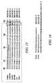
- FIG. 13is a comparison showing selected PrP sequences among six different species, i.e., Seq. Id. No. 1 through Seq. Id. No. 6;
- FIG. 14shows peptide sequences for the synthetic peptide probes 19-mer Seq. Id. No. 7, and 14-mer, Seq. Id. No. 8;
- FIG. 15is a graph of fluorescence detection experimental results showing the effects of peptide concentration
- FIG. 16is a graph of fluorescence detection experimental results showing the effects of peptide concentration likely showing excimer emission at approximately 460 nanometers (nm);
- FIG. 17is a graph of fluorescence detection experimental results showing pyrene's excitation of fluorescence
- FIG. 18is a graph of fluorescence detection experimental results showing pyrene's excitation spectra for fluorescence at 398 and approximately 460 nm;
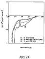
- FIG. 19is a graph comparing the circular dichroism results of several peptides ranging in concentration from 20 to 100 milli Molar (mM) under varying buffer conditions;
- FIG. 20is a graph comparing the circular dichroism results of several peptides including the synthetic peptides of Seq. Id. No. 7 and Seq. Id. No. 8 under varying buffer conditions;
- FIG. 21shows experimental results of the conformational lability of the synthetic peptides.
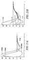
- FIG. 21 a on the leftshow that 14-mer, Seq. Id. No. 8, assumes a beta-sheet conformer while the longer analog, 19-mer, Seq. Id. No. 7, remains coiled.
- FIG. 21 b on the rightshows that addition of 14-mer, Seq. Id. No. 8, to 19-mer, Seq. Id. No. 7, initiates a phase shift to beta-sheet form;
- FIG. 22is a conceptual illustration of a comparison of where Seq. Id. No. 7 and Seq. Id. No. 8 overlap in structure;
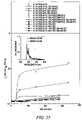
- FIG. 23is a graph of experimental results showing that peptides can self-associate
- FIG. 24is a graph of fluorescence data showing the efficiency of excimer formation under low concentrations
- FIG. 25is a graph of fluorescence experimental results showing the effect of nuclei on self-association due to catalytic conformational transition
- FIG. 26contains two graphs of fluorescence experimental results showing the interaction of Seq. Id. No. 7 and Seq. Id. No. 8 at different ratios; wherein FIG. 26 a on the left shows a 1:1 mixture and FIG. 26 b on the right shows a 100:1 mixture;
- FIG. 27contains four graphs of fluorescence experimental results showing the effect of nuclei on self-association.
- FIGS. 27 a, b, c and dshow the results at 24 hours, 48 hours, 144 hours and 336 hours, respectively;
- FIG. 28is a graph of fluorescence experimental results showing the effect of nuclei on self-association due to catalytic conformational transition at 1 hour in FIG. 28 a on the left and at 150 hours in FIG. 28 b on the right;
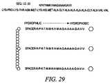
- FIG. 29shows peptide Seq. Id. No. 9, which is used to form sequences for a generalized dendrimer structure of this invention.
- FIG. 30shows a peptide sequence, i.e., Seq. Id. No. 10, for a preferred embodiment of a specific dendrimer structure of this invention
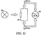
- FIG. 31is a conceptual diagram of an experimental device.
- FIG. 32is a system diagram of preferred embodiments of the invention.
- the present inventiondetects the presence of abnormal proteins and proteinaceous particles based on a method that utilizes catalytic propagation.
- a samplecontaining abnormal proteins or proteinaceous particles
- the peptide probeundergoes conformational changes resulting in the formation of aggregates.
- the addition of the abnormal proteins and proteinaceous particlescatalyzes the formation of the aggregates and causes further propagation of this conformational transition.
- the resulting aggregatesare then easily detected using common analytical instrumentation and techniques.
- the abnormal proteins and proteinaceous particles on which the invention focusesare proteins, protein based chemical structures such as prions and protein subunits such as peptides that are capable of conformational changes that lead to the formation of aggregates and ultimately to disease states.
- a preferred example of such proteinaceous particlesis that of a prion protein.
- Prionscan exist in one of two distinct conformations characterized by having a secondary protein structure that is either predominately alpha-helical or predominately beta-sheet; where the predominately beta-sheet conformation has a much higher preference to exist in a multimeric state.
- predominately beta-sheet (or beta rich) secondary structureis more typical of abnormally folded or disease-causing proteinaceous particles since their preference to aggregate is likely to be disruptive in an in vivo environment.
- FIG. 1shows illustrations of both the alpha-helical monomer 10 and the beta-sheet dimer 12 forms of a TSE conformer (or alternative secondary structure).
- wtnormal wild-type form of prion protein
- PrP Scabnormal, disease-causing form
- the mechanism of the inventionis shown in a schematic in FIG. 2 .
- the top row of the schematicshows an example of an unknown sample of TSE protein 20 represented as containing aggregated beta-sheets 12 .
- the beta-sheetsare then disaggregated 22 by subjecting the sample to commonly known disaggregation methods such as sonication. This is followed by the addition of labeled peptide probes 14 which are allowed to bind to the sample 20 . Presence of the beta-sheet conformation in the sample 20 induces the peptide probes to also shift to beta-sheet formation 16 . In this manner the transition to beta-sheet is propagated among the peptide probes 14 thereby causing new aggregates 18 to form.
- the resulting transition to a predominately beta-sheet form and amplified aggregate formationcan then easily be detected using common analytical techniques such as light scattering and circular dichroism (CD); and in a particularly preferred embodiment where the peptide probe is fluorescent labeled, fluorescence detection instrumentation can also be used.
- CDlight scattering and circular dichroism
- the bottom row of FIG. 2shows an alternative example in which the unknown sample of TSE protein 20 is represented in its normal alpha-helical form 10 .
- the sampleis subjected to the same disaggregation process described above.
- the labeled peptide probes 14neither a transition to beta-sheet form nor binding to the unknown samples occurs.
- unknown samplescan be tested for the presence or absence of such abnormal protein conformations or sequences.
- a preferred embodiment of the inventioninvolves the following basic procedures.
- Peptide probes 14are selected in order to be added to an unknown or test sample 20 at a later stage in the process.
- the peptide probes 14are preferably proteins or peptide sequences that have secondary structures of predominately alpha-helix or random coil.
- the peptide probes 14are peptide fragments consisting of a helix-loop-helix structure as found in lysine.
- the peptide probescan be made of a peptide sequence chosen from wild-type (wt) TSE, from a desired species-specific TSE peptide sequence, or even from a selectively mutated TSE sequence that has been mutated in such a manner as to render it destabilized and noninfectious.
- extrinsic fluorssuch as pyrene can be added or designed into the peptide probe to allow detection of anticipated conformational changes using common fluorescence detection techniques.
- a peptide probe 14is added to a test sample 20 .
- the sample 20Prior to the addition of the peptide probe 14 , however, it is preferred to have the sample 20 subjected to disaggregation techniques commonly known in the art, such as sonication.
- the disaggregation stepallows any potentially aggregated sample material 20 to break apart so that these disaggregated sample materials 22 are more free to recombine with the newly introduced peptide probes 14 ; thereby facilitating the anticipated catalytic propagation.
- test sample 20 or disaggregated test sample 22is allowed to interact with the peptide probes 14 .
- the resulting mixtureis then subjected to analytical methods commonly known in the art for the detection of aggregates and to fluorescence measurements in cases where fluorescent peptide probes 14 are used.
- Unknown or test samples 20 containing any dominant beta-sheet formation characteristic of abnormally folded or disease-causing proteinsresults in an increase in beta-sheet formation and consequently aggregate formation in the final mixture containing both the test sample 20 and the peptide probes 14 .
- unknown or test samples 20 which lack a predominantly beta-sheet secondary structurewill neither catalyze a transition to beta-sheet structure 16 nor will propagate the formation of aggregates 18 .
- the binding of a metal ligandcould direct a change in the protein scaffolding and favor aggregation.
- the expression or cleavage of different peptide sequencescan promote advanced aggregation leading to fibril and plaque formation.
- Genetic point mutationscan also alter the relative energy levels required of the two distinct conformations, resulting in midpoint shifts in structural transitions. Furthermore, an increase in concentration levels could be sufficient to favor the conformational transition.
- the disease process in many of the abnormal protein conformationssuch as in prion-related diseases always involves the catalytic propagation of the abnormal conformation, resulting in transformation of the previously normal protein.
- optical detection techniquesinclude, but are not limited to, light scattering, or hydrophobicity detection using extrinsic fluors such as 1-anilino-8-napthalene sulfonate (ANS) or Congo Red stain, fluorescence proximity probes on the peptide fragments, including fluorescence resonance energy transfer (FRET) & quenching of intrinsic tryptophan fluorescence through either conformational change of monomer or binding at interface in alpha-beta heterodimer; the N-terminal loop region is particularly interesting in this regard selective binding to target protein, circular dichroism (CD) monitoring of actual conformation, nuclear magnetic resonance (NMR).
- extrinsic fluorssuch as 1-anilino-8-napthalene sulfonate (ANS) or Congo Red stain
- FRETfluorescence resonance energy transfer
- NMRnuclear magnetic resonance
- FIG. 3shows a circular dichroism graph of experimentation with poly-L-lysine 20 micro Molar ( ⁇ M) 52,000 molecular weight (MW) as a peptide probe.
- ⁇ Mmicro Molar
- MWmolecular weight
- FIG. 4shows an absorbance graph of experimentation with poly-L-lysine 70 mircomolar ( ⁇ M) 52,000 molecular weight (MW) as a peptide probe.
- ⁇ Mpoly-L-lysine 70 mircomolar
- MWmolecular weight
- FIG. 5shows general circular dichroism results of experimentation with poly-L-lysine at varying temperatures and pH indicating its potential for transitioning from random coil to beta-sheet under the varying environmental conditions. The results indicate that both temperature and pH play an important role in the transition.
- FIG. 6shows experimentation results using pyrene as a fluorescent probe in proximal and distal locations in an alpha helical bundle structure undergoing conformational change.
- the pyrene excimer formation 15is shown at 480 nm 42 and the spectra for a predominately alpha-helical structure 17 is contrasted 40 as well.
- FITCfluorescent probes
- a primary objective of this inventionalso encompasses use of the catalytic propagation of conformational change to directly correlate the measures of abnormal prion presence with levels of infectivity. For this reason we favor implementation of the invention in a manner where there is no increase in resulting infectious products as a result of the propagation.
- Thiscan be achieved by placing a “break” in the links between the chain of infection, transmission and propagation of the abnormal form. Such a “break” must occur at the transitional stage between the dimer and multimer forms of the aggregate.
- the physical formation of the multimer formcan be blocked by simply impeding the step which leads to its formation. This may be done, preferably by using a large pendant probe or by a neutral “blocker” segment, bearing in mind that probes on linkers or “tethers” are more likely to encounter each other and thus result in amplifying the signal.
- the peptide probes 14function in the manner described above.
- the peptide probesact as “nuclei”; wherein once the peptide probe 14 binds to a test sample 20 , or a sample known to have beta-rich structure 12 , it is converted to a peptide probe conformer 16 which has the capacity to act as a trigger to bind to another peptide probe 14 and continues to induce the same conformational change. Propagation of this reaction can then be controlled by the peptide sequence chosen for the peptide probe 14 and by the experimental conditions.
- a peptide probe 14capable of rapid and continuous propagation of the reaction be chosen with which to nucleate the unknown sample 20 .
- peptide probe 14 chosenis one that is less likely to aggregate.
- Associations of peptidecan be controlled by the thermodynamics of the solution in which they are in and by the presence of amorphous nuclei which self-associate, crystalline nuclei which readily aggregate, specific peptide sequences which may aggregate, but may do so under low concentrations which are difficult to measure by conventional means, or larger peptide sequences modeled after known beta-sheet structures or proteins such as a beta-rich prion protein.
- FIG. 14shows the peptide sequences of the two synthesized peptides.
- the 19-mer sequence referred to as Seq. Id. No. 7is closely modeled after residues 104 through 122 of the human sequence.
- the 14-mer sequence referred to as Seq. Id. No. 8is closely modeled after residues 109 through 122 of the human PrP sequence.
- the synthetic peptide probes 14were also prepared with and without pyrene butyric acid as a fluorescence marker.
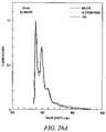
- FIG. 15shows a graph of fluorescence-spectra results at different peptide concentrations. The data were collected over times ranging form one hour to one week with no experimental changes observed after twenty-four hours. The resulting graphs show:
- FIG. 16shows a graph of the fluorescence spectra for samples 46 through 52 normalized to the intensity at 378 nm for the initial scan. It was observed that the spectrum for Sample 52 which contained the highest peptide concentration was markedly different leading to the conclusion that there is excimer emission with a maximum at approximately 460 nm.
- FIG. 17is a graph of experimental results showing pyrene's excitation of fluorescence.
- the experimentswere performed with excitation wavelengths at 365 nm to observe excimer emission at approximately 460 nm.
- the excitation at 348 nmincreases the fluorescence signal by over a hundred times with no other modifications or signal amplification.
- the excitation spectra for fluorescence at 398 nm and at approximately 460 nmwere recorded and are shown in FIG. 18 .
- FIG. 19shows experimental data obtained from circular dichroism (CD) analysis of the 19-mer under different condition.
- the CD spectrawere recorded for a number of peptide concentrations ranging from 20 to 100 mM.
- the resultsshow that the 19-mer is largely coiled and exhibits high thermodynamic stability under the experimental conditions tested such as varying pH, ionic strength and temperature.
- organicssuch as acetonitrile and trifluoroethylene (TFE) encourage the formation of the secondary structure.
- FIG. 20shows both the previous results and the results of a similar experiment in which the 19-mer was mixed with its shorter analog, the 14-mer.
- the resulting projectiondoes not entirely agree with the CD results.
- the conformations of both synthetic peptidesare clearly concentration dependent.
- the 19-merexhibits largely a coil conformation that is fairly stable under a wide variety of the experimental conditions tested, the 14-mer exhibits a transition from coil or hairpin to beta-sheet structure depending on its concentration.
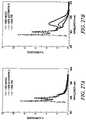
- FIG. 23shows a graph of fluorescence results showing that the 19-mer could self-associate with increasing concentration as shown in Sample curve 66 and at low concentrations with pH modifications to give a net neutral charge while using potassium chloride (KCl) to screen the charge as shown in Sample curve 68 .
- the 19-mercan also self-associate at low concentrations with the introduction of some type of nucleating agent, as discussed earlier. Thus, the conditions for self-association can be optimized to adapt to a desired type of detection.
- Sample curve 66 containing 0.1 M TRIS buffer at pH 6 to 9 and Sample curve 68 containing 0.1 M TRIS buffer at pH 10 to 11 in the presence of KCl at 100 to 500 mMare shown again in FIG. 24 to reflect the efficiency of excimer formation under low concentrations.
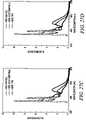
- FIG. 26 ashows more fluorescence data of the 19-mer in water 70 , acetonitrile 72 and TFE 74 after twenty-four hours.
- FIG. 26 bshows the experimental results for a 100:1 combination of the 19-mer and 14-mer in water 76 , acetonitrile 78 and TFE 80 after twenty-four hours.
- peptide associationwas monitored by the appearance of excimer emission at approximately 460 nm.
- FIGS. 27 a, b, c , and dshow four fluorescence data graphs taken at 24, 48, 144 and 336 hours, respectively. The measurements were taken to determine the effect of pH, temperature, ionic strength, and organic additives on the kinetics of the peptide associations studied for the 19-mer model peptide. The fluorescence intensities as measured at 378 nm for monomeric units and 460 nm for associations were used to characterize the I E /I M ratio or self-association of the peptide.
- FIG. 28Additional fluorescence results are shown in FIG. 28 where an insoluble fraction of the peptide was extracted and dissolved in organic solvent containing methanol/ethanol/dimethylformanide and then analyzed. Fluorescence detection results of the “insoluble” portion show high levels of peptide association wherein the I E /I M ratio equals 2. A small aliquot of “insoluble” portion was added to nucleate 20 micro Molar 19-mer peptide solutions which were then analyzed and are reported in the same graph. The results show that the presence of the nucleating fraction significantly increased the efficiency of the peptide association and this can be seen more dramatically in FIG. 28 b at 150 hours.
- the peptide probes 14can be used to detect proteinaceous particles such as in prion-like structures exhibiting coil to beta-sheet transition. According to Prusiner, et al. Prion protein peptides induce alpha - helix to beta - sheet conformational transitions . Biochemsitry. 34:4186-92 (1995). As a result, synthetic peptide probes such as the Seq. Id. No 7, 19-mer should be conformationally sensitive to the presence of prion-like substances that undergo this conformational shift.
- an intrinsic optical reportersuch as pyrene
- this embodiment of the inventionhas the added advantage of being able to detect such prion-like substances in test samples 20 such as blood, lymph, CSF and even tissues other than brain homogenate that typically contain very low levels of abnormal prion substances that are otherwise too difficult to detect.
- the intrinsic optical reporterallows optical (fluorescence) measurements to be taken of the peptide probe associates that form upon interaction with nucleating samples such as an abnormal prion.
- the peptide probes 14are synthesized based on the structure of a dendrimer; dendrimers being synthesized three-dimensional highly branched macromolecules.
- the advantages of using a dendrimer probe 15are multifold. Dendrimers should increase the speed of the assay kinetics thereby relaying quicker test results. This can be especially advantageous in assembly line applications of the invention where products or specimens in mass quantities can be quickly tested for the presence of abnormal proteinaceous particles. This embodiment is also extremely beneficial in applications where quick decisions must be based on the detection results. This embodiment is also advantageous for use in these applications as well as others since the highly branched structure of the dendrimer prevents amplification of abnormal proteinaceous particles or aggregates.
- a generalized dendrimer 15 structureis shown in FIG. 29 and is referred to as Seq. Id. No. 9.
- a specific dendrimer structurewas designed and synthesized, referred to as Seq. Id. No 10, and is shown in FIG. 30 .
- the specific dendrimer structureis basically a loop-turn-loop structure as illustrated by FIG. 30 a .
- FIG. 30 bit is shown that the sequence is modeled after the human PrP sequence shown in FIG. 14 in residues 126 through 104 plus 109 through 126 .
- This structureshows the region on the right 74 as an inverted form of the PrP sequence. This was done to take advantage of the five aminoacids which naturally form a loop in order to place hydrophobic pyrene in a corresponding hydrophobic region. Also the valine-valine fragment is essential to beta-sheet formation and so is retained in the sequence. In the figure, green denotes possible mouse variants.
- the amyloidogenic palindrome region 70may be changed to SS or SSS/AAA.
- the central region 72is a loop sequence with steric constraints, thus it is possible to add tryptophan for steric and fluorescence considerations.
- aminoacid sequencesuch as one or more deletions or insertions are possible as alluded to above, provided that the dendrimer retains its branched loop-turn-loop structure as well as aminoacids essential to beta-sheet formation, and preferably contains an optical reporter.
- FIG. 10shows a schematic diagram of how the dendrimer probes 15 amplify signal and propagate conformational change without aggregation and without increasing the biohazard or infectious nature of an abnormal protein or prion test sample 12 .
- the figureshows that once the dendrimer probes 15 come into contact with the abnormal sample 12 , the dendrimer probe 15 undergoes the conformational shift to a predominately beta-sheet structure 17 .
- the newly formed beta-rich dendrimer probe 17nucleates other dendrimer probes 15 to make the same transition. By doing so, any optical signal associated with the dendrimer probe 15 is amplified as more probes 15 shift to the beta-rich state 17 .
- the minimal detectable concentration of pyreneonly provides a number for the peptide probe 14 concentration that can be worked with; but the detection limit of the assay is not dependent on it because it is the resultant of the fluorescent ensemble that is being observed.
- the real measurement of interest and the rate limiting step in the analysisis the amount of abnormal e. g. prion protein that need to be present in the sample 20 to initiate a conformer change in the peptide probe 14 .
- Immunoassaysare typically sensitive in the picomolar range. Nevertheless, once the conformer change is initiated in a single peptide probe 14 , the catalytic propagation of its beta-rich structure allows detection in samples previously considered to have abnormal particles 12 at concentrations too low to detect.
- the method of this inventionis widely applicable to many industries.
- some of those industriesinclude the diagnostics markets in animal health and human health, the food industry, pharmaceutics, especially for screening animal by-products, transplant/transfusion and vaccine supplies, research and development in such areas as chemotherapies for TSE's, as well as national security in the area of biosensors for biowarfare agents.
- the methods discussed hereincan be applied for use with a simple detection instrument such as the one shown in FIG. 31 .
- the device shown in FIG. 31is a simple optical device that includes a light source 80 shown in blue e. g. lamp or laser; a T-format sample cell 82 shown in grey; and a photomultiplier tube 84 shown in pink.
- a light source 80 shown in bluee. g. lamp or laser
- T-format sample cell 82shown in grey
- a photomultiplier tube 84shown in pink.
Landscapes
- Health & Medical Sciences (AREA)
- Life Sciences & Earth Sciences (AREA)
- Engineering & Computer Science (AREA)
- Immunology (AREA)
- Biomedical Technology (AREA)
- Hematology (AREA)
- Chemical & Material Sciences (AREA)
- Urology & Nephrology (AREA)
- Molecular Biology (AREA)
- Analytical Chemistry (AREA)
- General Health & Medical Sciences (AREA)
- Microbiology (AREA)
- Biotechnology (AREA)
- Pathology (AREA)
- Cell Biology (AREA)
- Food Science & Technology (AREA)
- Medicinal Chemistry (AREA)
- Physics & Mathematics (AREA)
- General Physics & Mathematics (AREA)
- Biochemistry (AREA)
- Neurosurgery (AREA)
- Neurology (AREA)
- Proteomics, Peptides & Aminoacids (AREA)
- Investigating Or Analysing Biological Materials (AREA)
- Peptides Or Proteins (AREA)
- Investigating Or Analysing Materials By The Use Of Chemical Reactions (AREA)
- Investigating, Analyzing Materials By Fluorescence Or Luminescence (AREA)
Abstract
Description
- Brain Tissue Sampling. Cross-sections of brain can be used to examine and monitor gross morphology changes indicative of disease states such as the appearance of spongiform in the brain, in addition to immunohistochemistry techniques such as antibody-based assays or affinity chromatography which can detect disease-specific prion deposits. These techniques are used for a conclusive bovine spongiform encephalopathy (BSE) diagnosis after slaughter of animals displaying clinical symptoms. Drawbacks of tissue sampling include belated detection that is possible only after symptoms appear, necessary slaughter of affected animals, and results that takes days to weeks to complete.
- Prionic-Check also requires liquified-brain tissue for use with a novel antibody under the Western Blot technique. This test is as reliable as the immunochemistry technique and is more rapid, yielding results in six to seven hours, but shares the drawbacks of the six-month lag time between PrPSaccumulation (responsible for the gross morphology changes) in the brain and the display of clinical symptoms, along with the need for slaughter of the animal to obtain a sample.
- Tonsillar Biopsy Sampling. Though quite accurate, it requires surgical intervention and the requisite days to weeks to obtain results.
- Body Fluids: Blood and Cerebrospinal Sampling. As in the above detection methods, results are not immediate
- Electrospray ionization mass spectrometry (ESI-MS), nuclear magnetic resonance NMR, circular dichroism (CD) and other non-amplified structural techniques. All of these techniques require a large amount of infectious sample, and have the disadvantage of requiring off-site testing or a large financial investment in equipment.
- Prionics—in Switzerland. The test involves Western blot of monoclonal antibodies (MABs) to detect PrP in brain tissue from dead animals in seven to eight hours.
- Enfer Scientific—in Ireland. The test involves ELISA-based testing on spinal cord tissue from dead animals in under four hours.
- CEA—in France. The test involves a sandwich immunoassay using two monoclonals on brain tissue collected after death in under twenty-four hours.
Sample 24 which was maintained at pH7, 25° C. resulting in a minimum at approximately 205 namometers (nm) indicating random coil structure.Sample 26 which was maintained at pH11, 50° C. resulting in a minimum at approximately 216 namometers (nm) indicating beta-sheet structure.Sample 28 which was a 1:1 combination of samples maintained at pH7, 25° C. and at pH11, 50° C. resulting in a minimum at approximately 216 namometers (nm) indicating beta-sheet structure.Sample 30 which was a 1:1 combination of samples maintained at pH7, 50° C. and at pH11, 50° C. resulting in a minimum at approximately 216 namometers (nm) indicating beta-sheet structure.
Sample 32 which was maintained atpH Sample 34 which was maintained at pH7, 50° C. resulting in a plateau at approximately 0.22 indicating random coil structure.Sample 36 which was a 10:1 combination of samples maintained at pH7, 50° C. and at pH11, 50° C. resulting in a steeper incline from approximately 0.22 to 0.33 indicating an accelerated transition from random coil to beta-sheet structure.Sample 38 which was a 10:1 combination of samples maintained at pH7, 25° C. and at ph11, 50° C. resulting in a gradual incline from approximately 0.22 to 0.26 indicating a transition from random coil to beta-sheet structure.
Sample 46 which was at a concentration of 5 μM with a relative fluorescence peak at approximately 0.1.Sample 48 which was at a concentration of 10 μM with a relative fluorescence peak at approximately 0.4.Sample 50 which was at a concentration of 150 μM with a relative fluorescence peak at approximately 4.7.
Note: data were also collected forSample 52 at a high concentration of 800 μM, but is not shown in the figure.
Pyr*+Pyr=(Pyr—Pyr)*
where Pyr is a pyrene molecule and Pyr* is a pyrene in its excited form; the (Pyr_Pyr)* represents the formation of excited dimer. More general information on excimers can be found in Freifelder, David.Physical Biochemistry: Applications to Biochemistry and Molecular Biology, (W. H. Freeman Press, New York, 2nd ed. 1982), at 559.
- Fluorescence of pyrene, which is covalently attached to the
peptide probe 14 in preferred embodiments, allows monitoring of peptide self-association in this model system. It can also be used as an index of conformational change and especially since at low concentrations, the peptide association is difficult to measure using nonoptical techniques. - The fluorescence data shows that self-association of the Seq. Id. No 7, 19-mer, can be promoted by adjusting ionic strength or pH.
- The fluorescence data also shows that the kinetics of the conformational changes can be modulated by controlling solvent parameters and the peptide probe sequence.
- The kinetics of the self-assembly or association process can be controlled or regulated by the addition of or by preexisting nucleating associated forms. This strongly supports the conclusions that the conformational transitions of the 19-mer can be autocatalytic.
- Fluorescence of pyrene, which is covalently attached to the
Claims (16)
Priority Applications (4)
| Application Number | Priority Date | Filing Date | Title |
|---|---|---|---|
| US10/494,906US7691639B2 (en) | 2001-05-31 | 2002-05-30 | Misfolded protein sensor method |
| US11/979,226US20080171341A1 (en) | 2001-05-31 | 2007-10-31 | Detection of conformationally altered proteins and prions |
| US12/726,941US8062895B2 (en) | 2001-05-31 | 2010-03-18 | Misfolded protein sensor method |
| US14/484,683US9638702B2 (en) | 2001-05-31 | 2014-09-12 | Detection of conformationally altered proteins |
Applications Claiming Priority (4)
| Application Number | Priority Date | Filing Date | Title |
|---|---|---|---|
| US29545601P | 2001-05-31 | 2001-05-31 | |
| US60295456 | 2001-05-31 | ||
| US10/494,906US7691639B2 (en) | 2001-05-31 | 2002-05-30 | Misfolded protein sensor method |
| PCT/US2002/017212WO2002097444A2 (en) | 2001-05-31 | 2002-05-30 | Misfolded protein sensor method |
Related Parent Applications (1)
| Application Number | Title | Priority Date | Filing Date |
|---|---|---|---|
| PCT/US2002/017212A-371-Of-InternationalWO2002097444A2 (en) | 2001-05-31 | 2002-05-30 | Misfolded protein sensor method |
Related Child Applications (3)
| Application Number | Title | Priority Date | Filing Date |
|---|---|---|---|
| US10/161,061Continuation-In-PartUS7166471B2 (en) | 2001-05-31 | 2002-05-30 | Misfolded protein sensor method in body fluids |
| US11/979,226Continuation-In-PartUS20080171341A1 (en) | 2001-05-31 | 2007-10-31 | Detection of conformationally altered proteins and prions |
| US12/726,941ContinuationUS8062895B2 (en) | 2001-05-31 | 2010-03-18 | Misfolded protein sensor method |
Publications (2)
| Publication Number | Publication Date |
|---|---|
| US20060286672A1 US20060286672A1 (en) | 2006-12-21 |
| US7691639B2true US7691639B2 (en) | 2010-04-06 |
Family
ID=23137808
Family Applications (4)
| Application Number | Title | Priority Date | Filing Date |
|---|---|---|---|
| US10/161,061Expired - LifetimeUS7166471B2 (en) | 2001-05-31 | 2002-05-30 | Misfolded protein sensor method in body fluids |
| US10/494,906Active2026-04-02US7691639B2 (en) | 2001-05-31 | 2002-05-30 | Misfolded protein sensor method |
| US11/504,692AbandonedUS20060275910A1 (en) | 2001-05-31 | 2006-08-16 | Misfolded protein sensor method in body fluids |
| US12/726,941Expired - LifetimeUS8062895B2 (en) | 2001-05-31 | 2010-03-18 | Misfolded protein sensor method |
Family Applications Before (1)
| Application Number | Title | Priority Date | Filing Date |
|---|---|---|---|
| US10/161,061Expired - LifetimeUS7166471B2 (en) | 2001-05-31 | 2002-05-30 | Misfolded protein sensor method in body fluids |
Family Applications After (2)
| Application Number | Title | Priority Date | Filing Date |
|---|---|---|---|
| US11/504,692AbandonedUS20060275910A1 (en) | 2001-05-31 | 2006-08-16 | Misfolded protein sensor method in body fluids |
| US12/726,941Expired - LifetimeUS8062895B2 (en) | 2001-05-31 | 2010-03-18 | Misfolded protein sensor method |
Country Status (7)
| Country | Link |
|---|---|
| US (4) | US7166471B2 (en) |
| EP (1) | EP1395833B1 (en) |
| JP (1) | JP4235544B2 (en) |
| AU (2) | AU2002320045B2 (en) |
| CA (1) | CA2448981C (en) |
| MX (1) | MXPA03011000A (en) |
| WO (1) | WO2002097444A2 (en) |
Cited By (9)
| Publication number | Priority date | Publication date | Assignee | Title |
|---|---|---|---|---|
| US20060057671A1 (en)* | 2004-09-10 | 2006-03-16 | Orser Cindy S | Immobilized probes and methods of detecting conformationally altered prion proteins |
| US20080095706A1 (en)* | 2006-07-28 | 2008-04-24 | Adlyfe, Inc. | Peptide probes for diagnostics and therapeutics |
| US20080171341A1 (en)* | 2001-05-31 | 2008-07-17 | Adlyfe, Inc. | Detection of conformationally altered proteins and prions |
| US20100080796A1 (en)* | 2008-04-17 | 2010-04-01 | Peptimmune, Inc. | Synthesis of directed sequence polymer compositions and antibodies thereof for the treatment of protein conformational disorders |
| US20100233095A1 (en)* | 2009-01-30 | 2010-09-16 | Adlyfe, Inc. | Conformationally dynamic peptides |
| US20110081660A1 (en)* | 2005-02-15 | 2011-04-07 | Adlyfe, Inc. | Method for Detecting Misfolded Proteins and Prions |
| US8062895B2 (en) | 2001-05-31 | 2011-11-22 | Adlyfe, Inc. | Misfolded protein sensor method |
| US9556247B2 (en) | 2010-05-25 | 2017-01-31 | System Of Systems Analytics, Inc. | Stabilized amyloid-beta oligomers and uses thereof |
| US9795692B2 (en) | 2011-04-27 | 2017-10-24 | System Of Systems Analytics, Inc. | Ocular detection of amyloid proteins |
Families Citing this family (19)
| Publication number | Priority date | Publication date | Assignee | Title |
|---|---|---|---|---|
| EP1380290A1 (en) | 2002-07-09 | 2004-01-14 | Universitair Medisch Centrum Utrecht | Cross-beta structure pathway and its therapeutic relevance |
| JP4709149B2 (en) | 2003-08-13 | 2011-06-22 | ノバルティス バクシンズ アンド ダイアグノスティックス,インコーポレーテッド | Prion-specific peptide reagents |
| US20060035242A1 (en)* | 2004-08-13 | 2006-02-16 | Michelitsch Melissa D | Prion-specific peptide reagents |
| CA2561246A1 (en)* | 2004-03-25 | 2005-10-06 | Fuence Co., Ltd. | Method of detecting conformational change of an amyloid protein, a method of searching a substance having an activity that affects to conformational change of an amyloid protein, and a method of searching a therapeutic or diagnostic agent for amyloid-related diseases |
| WO2006076687A2 (en)* | 2005-01-13 | 2006-07-20 | Novartis Vaccines And Diagnostics Inc. | Elisa assays using prion-specific peptide reagents |
| WO2006076683A2 (en)* | 2005-01-13 | 2006-07-20 | Novartis Vaccines And Diagnostics Inc. | Isolation and detection of pathogenic prions |
| US8114832B2 (en) | 2005-07-13 | 2012-02-14 | Crossbeta Biosciences B.V. | Method for detecting and/or removing a protein comprising a cross-beta structure from a pharmaceutical composition |
| EP2386861A3 (en) | 2005-07-13 | 2012-07-18 | Crossbeta Biosciences B.V. | Cross-ß structure binding compounds |
| US20070015133A1 (en)* | 2005-07-13 | 2007-01-18 | Umc Utrecht Holding B.V. | Method for detecting and/or removing protein and/or peptide comprising a cross-beta structure from an aqueous solution comprising a protein |
| CA2621767A1 (en) | 2005-09-09 | 2007-03-15 | Novartis Ag | Prion-specific peptoid reagents |
| GB2453191A (en)* | 2005-10-18 | 2009-04-01 | Brigham & Womens Hospital | Diagnosis of transmissible spongiform encephalopathy |
| JP5164971B2 (en)* | 2006-04-21 | 2013-03-21 | ピープルバイオ,アイ エヌ シー | A method for differential detection of multimers from monomers of multimer-forming polypeptides using three-dimensional interactions |
| WO2008029965A1 (en)* | 2006-09-08 | 2008-03-13 | Peoplebio, Inc. | Simultaneous reaction assay for differentially detecting multimeric form |
| US20110189692A1 (en)* | 2008-04-30 | 2011-08-04 | Novartis Ag | Assay for pathogenic conformers |
| CN107677828A (en)* | 2008-10-31 | 2018-02-09 | 耶鲁大学 | Preeclampsia detects and the method and composition for the treatment of |
| JP6310922B2 (en) | 2012-09-25 | 2018-04-11 | フォー−ウェブ・インコーポレイテッド | Programmable graft and method of using a programmable graft to repair bone structure |
| US9588129B2 (en)* | 2013-03-15 | 2017-03-07 | Amira Medical Technologies Inc. | Methods for analyzing blood to detect diseases associated with abnormal protein aggregation |
| CA3051839A1 (en) | 2017-02-17 | 2018-08-23 | Bristol-Myers Squibb Company | Antibodies to alpha-synuclein and uses thereof |
| TWI698641B (en)* | 2017-12-28 | 2020-07-11 | 大陸商浙江數問生物技術有限公司 | Device, kit and method for detecting misfolded protein |
Citations (53)
| Publication number | Priority date | Publication date | Assignee | Title |
|---|---|---|---|---|
| US4444879A (en) | 1981-01-29 | 1984-04-24 | Science Research Center, Inc. | Immunoassay with article having support film and immunological counterpart of analyte |
| US5565186A (en) | 1994-05-13 | 1996-10-15 | The Regents Of The University Of California | Method of detecting prions in a sample and transgenic animal used for same |
| WO1997016728A1 (en) | 1995-11-02 | 1997-05-09 | The Regents Of The University Of California | FORMATION AND USE OF PRION PROTEIN (PrP) COMPLEXES |
| WO1997043649A1 (en) | 1996-05-14 | 1997-11-20 | Winnacker Ernst Ludwig | CHARPERONES CAPABLE OF BINDING TO PRION PROTEINS AND DISTINGUISHING THE ISOFORMS PrPc AND PrP?sc¿ |
| US5721106A (en) | 1991-08-13 | 1998-02-24 | Regents Of The University Of Minnesota | In Vitro method for screening β-amyloid deposition |
| US5773572A (en) | 1991-12-03 | 1998-06-30 | Proteus Molecular Design Limited | Fragments of prion proteins |
| WO1998037411A1 (en) | 1997-02-21 | 1998-08-27 | The Regents Of The University Of California | Assay for disease related conformation of a protein |
| US5854204A (en) | 1995-03-14 | 1998-12-29 | Praecis Pharmaceuticals, Inc. | Aβ peptides that modulate β-amyloid aggregation |
| WO1999041279A2 (en) | 1998-02-13 | 1999-08-19 | Arch Development Corporation | Methods and compositions comprising the use of blocked b-amyloid peptide |
| US5955343A (en) | 1992-12-28 | 1999-09-21 | Massachusetts Institute Of Technology | Stable macroscopic membranes formed by self-assembly of amphiphilic peptides and uses therefor |
| US5977324A (en)* | 1998-02-20 | 1999-11-02 | The Regents Of The University Of California | Process for concentrating protein with disease-related conformation |
| WO2000002575A1 (en) | 1998-07-09 | 2000-01-20 | V.I. Technologies, Inc. | Prion protein and uses thereof |
| WO2000026238A2 (en) | 1998-11-04 | 2000-05-11 | D-Gen Limited | Biological materials and methods useful in the diagnosis and treatment of prion diseases |
| WO2000043791A2 (en) | 1999-01-25 | 2000-07-27 | Minerva Biotechnologies Corporation | Rapid and sensitive detection of aberrant protein aggregation in neurodegenerative diseases |
| WO2000069900A2 (en) | 1999-05-17 | 2000-11-23 | Conjuchem, Inc. | Protection of endogenous therapeutic peptides from peptidase activity through conjugation to blood components |
| US6166187A (en) | 1999-03-05 | 2000-12-26 | The Regents Of The University Of California | Method of concentrating prion proteins in blood samples |
| WO2001007479A2 (en) | 1999-07-27 | 2001-02-01 | Imperial College Innovations Limited | Fragments of cellular prion protein and methods useful in the diagnosis and treatment of prion diseases |
| WO2001007473A1 (en) | 1999-07-28 | 2001-02-01 | Kelvin Stott | Peptides containing n-substituted l-amino acids for preventing beta-strand association |
| WO2001014412A1 (en) | 1999-08-23 | 2001-03-01 | The Regents Of The University Of California | Compounds useful to mimic peptide beta-strands |
| US6214565B1 (en) | 1998-10-09 | 2001-04-10 | The Regents Of The University Of California | Assay for disease related conformation of a protein and isolating same |
| US20010001061A1 (en) | 1997-02-21 | 2001-05-10 | Prusiner Stanley B. | Assay for disease related conformation of a protein |
| WO2001050134A2 (en) | 1999-12-29 | 2001-07-12 | American Cyanamid Company | Methods of detection of amyloidogenic proteins |
| US6290954B1 (en) | 1995-09-14 | 2001-09-18 | The Scripps Research Institute | Antibodies specific for native PrPSc |
| WO2001077687A2 (en) | 2000-04-05 | 2001-10-18 | V.I Technologies, Inc. | Prion-binding peptidic ligands and methods of using same |
| WO2002004954A2 (en) | 2000-07-07 | 2002-01-17 | Applied Research Systems Ars Holding N.V. | Early diagnosis of conformational diseases |
| WO2002004604A2 (en) | 2000-07-10 | 2002-01-17 | University Of British Columbia | Immortalized human microglia cell and continuous cell line |
| US20020042121A1 (en) | 1997-09-19 | 2002-04-11 | Detlev Riesner | Method for measuring the association of substructures of pathological protein depositions |
| WO2002053723A2 (en) | 2001-01-08 | 2002-07-11 | Health Protection Agency | Degradation and detection of tse infectivity |
| US20020137112A1 (en) | 2000-12-07 | 2002-09-26 | Mario Chojkier | Compositions and methods for diagnosing alzheimer's disease |
| CA2443929A1 (en) | 2001-04-17 | 2002-10-24 | Ista, S.P.A. | Detection and quantification of prion isoforms in neurodegenerative diseases using mass spectrometry |
| WO2002097444A2 (en) | 2001-05-31 | 2002-12-05 | Arete Associates | Misfolded protein sensor method |
| WO2003085086A2 (en) | 2002-04-09 | 2003-10-16 | The Scripps Research Institute | Motif-grafted hybrid polypeptides and uses thereof |
| WO2004018511A2 (en) | 2002-08-23 | 2004-03-04 | Copenhagen Biotech Assets Aps | Composite peptide compounds for diagnosis and treatment of diseases caused by prion proteins |
| US20040052928A1 (en) | 2002-09-06 | 2004-03-18 | Ehud Gazit | Peptides and methods using same for diagnosing and treating amyloid-associated diseases |
| WO2004029072A2 (en) | 2002-09-27 | 2004-04-08 | Caprion Pharmaceuticals Inc. | PrPsc -INTERACTING MOLECULES AND USES THEREOF |
| JP2004155688A (en) | 2002-04-30 | 2004-06-03 | Biofrontier Kenkyusho:Kk | Synthetic peptide having chaperone activity, method for measuring decarbonation activity, medicine for transmissible spongiform encephalopathy, and its searching method |
| US20040224365A1 (en) | 1997-08-14 | 2004-11-11 | Charles Glabe | Fluorescent amyloid Abeta peptides and uses thereof |
| US20040229280A1 (en) | 2002-12-03 | 2004-11-18 | Hammond David J. | Prion protein ligands and methods of use |
| US6821504B2 (en) | 2001-05-23 | 2004-11-23 | New York University | Detection of alzheimer's amyloid by magnetic resonance imaging |
| WO2005010533A2 (en) | 2003-07-31 | 2005-02-03 | Hadasit Medical Research Services & Development Ltd. | Methods and kits for the detection of prion diseases |
| US20050026165A1 (en) | 2001-05-31 | 2005-02-03 | Cindy Orser | Detection of conformationally altered proteins and prions |
| US20050112607A1 (en) | 1999-01-23 | 2005-05-26 | Bamdad Cynthia C. | Rapid and sensitive detection of protein aggregation |
| US20050118645A1 (en) | 2003-08-13 | 2005-06-02 | Michelitsch Melissa D. | Prion-specific peptide reagents |
| US20050181998A1 (en) | 2001-12-10 | 2005-08-18 | Applied Research Systems Ars Holding N.V. | Prion inhibiting peptides and derivatives thereof |
| US20050221404A1 (en) | 2002-02-28 | 2005-10-06 | Lane Amin R | Binding of pathological forms of prion proteins |
| US20060035242A1 (en) | 2004-08-13 | 2006-02-16 | Michelitsch Melissa D | Prion-specific peptide reagents |
| US20060057671A1 (en) | 2004-09-10 | 2006-03-16 | Orser Cindy S | Immobilized probes and methods of detecting conformationally altered prion proteins |
| US20060078892A1 (en) | 2003-04-04 | 2006-04-13 | Prometic Biosciences, Ltd | Prion protein binding materials and methods of use |
| US20060178302A1 (en) | 1997-02-05 | 2006-08-10 | Northwestern University & The University Of Southern California | Amyloid beta protein (globular assembly and uses thereof) |
| WO2006088823A2 (en) | 2005-02-15 | 2006-08-24 | Adlyfe, Inc. | Method for detecting misfolded proteins and prions |
| US20060235199A1 (en) | 2003-08-19 | 2006-10-19 | Hisakazu Mihara | Reagent for amplifying amyloid fibrosis of amyloid ss-protein |
| US7349041B2 (en) | 2001-10-05 | 2008-03-25 | Samsung Electronics Co., Ltd. | Liquid crystal display with light guiding plate removed from LCD panel and polarizing plate inside LCD panel |
| US20080095706A1 (en) | 2006-07-28 | 2008-04-24 | Adlyfe, Inc. | Peptide probes for diagnostics and therapeutics |
Family Cites Families (7)
| Publication number | Priority date | Publication date | Assignee | Title |
|---|---|---|---|---|
| US1001061A (en)* | 1907-03-12 | 1911-08-22 | Joseph Mcc Michaelson | Logarithmic scale. |
| US4293221A (en)* | 1979-04-17 | 1981-10-06 | Research Corporation | Multidimensional slit-scan flow system |
| US5948763A (en)* | 1995-06-07 | 1999-09-07 | New York University | Peptides and pharmaceutical compositions thereof for treatment of disorders or diseases associated with abnormal protein folding into amyloid or amyloid-like deposits |
| US6186659B1 (en)* | 1998-08-21 | 2001-02-13 | Agilent Technologies Inc. | Apparatus and method for mixing a film of fluid |
| CA2451795A1 (en) | 2001-06-26 | 2003-01-09 | Paul M. Mathews | Cell-based high-throughput screening methods |
| WO2009117042A1 (en) | 2008-03-21 | 2009-09-24 | Adlyfe, Inc. | Use of pyrene to carry non-peptide agents across the blood brain barrier |
| JP2011517666A (en) | 2008-03-21 | 2011-06-16 | エイディーライフ インコーポレイティッド | Use of pyrene to transport peptides across the blood-brain barrier |
- 2002
- 2002-05-30MXMXPA03011000Apatent/MXPA03011000A/enactiveIP Right Grant
- 2002-05-30WOPCT/US2002/017212patent/WO2002097444A2/enactiveApplication Filing
- 2002-05-30CACA2448981Apatent/CA2448981C/ennot_activeExpired - Lifetime
- 2002-05-30USUS10/161,061patent/US7166471B2/ennot_activeExpired - Lifetime
- 2002-05-30AUAU2002320045Apatent/AU2002320045B2/ennot_activeCeased
- 2002-05-30USUS10/494,906patent/US7691639B2/enactiveActive
- 2002-05-30JPJP2003500572Apatent/JP4235544B2/ennot_activeExpired - Fee Related
- 2002-05-30EPEP02749544Apatent/EP1395833B1/ennot_activeExpired - Lifetime
- 2006
- 2006-08-16USUS11/504,692patent/US20060275910A1/ennot_activeAbandoned
- 2008
- 2008-08-07AUAU2008203542Apatent/AU2008203542B2/ennot_activeCeased
- 2010
- 2010-03-18USUS12/726,941patent/US8062895B2/ennot_activeExpired - Lifetime
Patent Citations (63)
| Publication number | Priority date | Publication date | Assignee | Title |
|---|---|---|---|---|
| US6498017B2 (en) | 1907-11-28 | 2002-12-24 | Evotec Biosystems Ag | Method for measuring the association of substructures of pathological protein depositions |
| US4444879A (en) | 1981-01-29 | 1984-04-24 | Science Research Center, Inc. | Immunoassay with article having support film and immunological counterpart of analyte |
| US5721106A (en) | 1991-08-13 | 1998-02-24 | Regents Of The University Of Minnesota | In Vitro method for screening β-amyloid deposition |
| US5773572A (en) | 1991-12-03 | 1998-06-30 | Proteus Molecular Design Limited | Fragments of prion proteins |
| US5955343A (en) | 1992-12-28 | 1999-09-21 | Massachusetts Institute Of Technology | Stable macroscopic membranes formed by self-assembly of amphiphilic peptides and uses therefor |
| US5565186A (en) | 1994-05-13 | 1996-10-15 | The Regents Of The University Of California | Method of detecting prions in a sample and transgenic animal used for same |
| US5854204A (en) | 1995-03-14 | 1998-12-29 | Praecis Pharmaceuticals, Inc. | Aβ peptides that modulate β-amyloid aggregation |
| US6290954B1 (en) | 1995-09-14 | 2001-09-18 | The Scripps Research Institute | Antibodies specific for native PrPSc |
| WO1997016728A1 (en) | 1995-11-02 | 1997-05-09 | The Regents Of The University Of California | FORMATION AND USE OF PRION PROTEIN (PrP) COMPLEXES |
| WO1997043649A1 (en) | 1996-05-14 | 1997-11-20 | Winnacker Ernst Ludwig | CHARPERONES CAPABLE OF BINDING TO PRION PROTEINS AND DISTINGUISHING THE ISOFORMS PrPc AND PrP?sc¿ |
| US20060178302A1 (en) | 1997-02-05 | 2006-08-10 | Northwestern University & The University Of Southern California | Amyloid beta protein (globular assembly and uses thereof) |
| WO1998037411A1 (en) | 1997-02-21 | 1998-08-27 | The Regents Of The University Of California | Assay for disease related conformation of a protein |
| US20010001061A1 (en) | 1997-02-21 | 2001-05-10 | Prusiner Stanley B. | Assay for disease related conformation of a protein |
| US20040224365A1 (en) | 1997-08-14 | 2004-11-11 | Charles Glabe | Fluorescent amyloid Abeta peptides and uses thereof |
| US20020042121A1 (en) | 1997-09-19 | 2002-04-11 | Detlev Riesner | Method for measuring the association of substructures of pathological protein depositions |
| WO1999041279A2 (en) | 1998-02-13 | 1999-08-19 | Arch Development Corporation | Methods and compositions comprising the use of blocked b-amyloid peptide |
| US6677125B2 (en) | 1998-02-20 | 2004-01-13 | The Regents Of The University Of California | Assay for disease related conformation of a protein and isolating same |
| US5977324A (en)* | 1998-02-20 | 1999-11-02 | The Regents Of The University Of California | Process for concentrating protein with disease-related conformation |
| US6750025B1 (en) | 1998-07-09 | 2004-06-15 | V.I. Technologies, Inc. | Method of detecting and isolating prion protein and variants thereof |
| WO2000002575A1 (en) | 1998-07-09 | 2000-01-20 | V.I. Technologies, Inc. | Prion protein and uses thereof |
| US6214565B1 (en) | 1998-10-09 | 2001-04-10 | The Regents Of The University Of California | Assay for disease related conformation of a protein and isolating same |
| US6534036B1 (en) | 1998-11-04 | 2003-03-18 | D. Gen Limited | Biological materials and methods useful in the diagnosis and treatment of diseases |
| WO2000026238A2 (en) | 1998-11-04 | 2000-05-11 | D-Gen Limited | Biological materials and methods useful in the diagnosis and treatment of prion diseases |
| US20050112607A1 (en) | 1999-01-23 | 2005-05-26 | Bamdad Cynthia C. | Rapid and sensitive detection of protein aggregation |
| WO2000043791A2 (en) | 1999-01-25 | 2000-07-27 | Minerva Biotechnologies Corporation | Rapid and sensitive detection of aberrant protein aggregation in neurodegenerative diseases |
| US6166187A (en) | 1999-03-05 | 2000-12-26 | The Regents Of The University Of California | Method of concentrating prion proteins in blood samples |
| WO2000069900A2 (en) | 1999-05-17 | 2000-11-23 | Conjuchem, Inc. | Protection of endogenous therapeutic peptides from peptidase activity through conjugation to blood components |
| WO2001007479A2 (en) | 1999-07-27 | 2001-02-01 | Imperial College Innovations Limited | Fragments of cellular prion protein and methods useful in the diagnosis and treatment of prion diseases |
| WO2001007473A1 (en) | 1999-07-28 | 2001-02-01 | Kelvin Stott | Peptides containing n-substituted l-amino acids for preventing beta-strand association |
| WO2001014412A1 (en) | 1999-08-23 | 2001-03-01 | The Regents Of The University Of California | Compounds useful to mimic peptide beta-strands |
| US6399314B1 (en) | 1999-12-29 | 2002-06-04 | American Cyanamid Company | Methods of detection of amyloidogenic proteins |
| WO2001050134A2 (en) | 1999-12-29 | 2001-07-12 | American Cyanamid Company | Methods of detection of amyloidogenic proteins |
| WO2001077687A2 (en) | 2000-04-05 | 2001-10-18 | V.I Technologies, Inc. | Prion-binding peptidic ligands and methods of using same |
| WO2002004954A2 (en) | 2000-07-07 | 2002-01-17 | Applied Research Systems Ars Holding N.V. | Early diagnosis of conformational diseases |
| WO2002004604A2 (en) | 2000-07-10 | 2002-01-17 | University Of British Columbia | Immortalized human microglia cell and continuous cell line |
| US20020137112A1 (en) | 2000-12-07 | 2002-09-26 | Mario Chojkier | Compositions and methods for diagnosing alzheimer's disease |
| WO2002053723A2 (en) | 2001-01-08 | 2002-07-11 | Health Protection Agency | Degradation and detection of tse infectivity |
| CA2443929A1 (en) | 2001-04-17 | 2002-10-24 | Ista, S.P.A. | Detection and quantification of prion isoforms in neurodegenerative diseases using mass spectrometry |
| US6821504B2 (en) | 2001-05-23 | 2004-11-23 | New York University | Detection of alzheimer's amyloid by magnetic resonance imaging |
| US20050026165A1 (en) | 2001-05-31 | 2005-02-03 | Cindy Orser | Detection of conformationally altered proteins and prions |
| US20080171341A1 (en) | 2001-05-31 | 2008-07-17 | Adlyfe, Inc. | Detection of conformationally altered proteins and prions |
| US7166471B2 (en) | 2001-05-31 | 2007-01-23 | Arete Associates | Misfolded protein sensor method in body fluids |
| US20060275910A1 (en) | 2001-05-31 | 2006-12-07 | Arete Associates | Misfolded protein sensor method in body fluids |
| WO2002097444A2 (en) | 2001-05-31 | 2002-12-05 | Arete Associates | Misfolded protein sensor method |
| US7349041B2 (en) | 2001-10-05 | 2008-03-25 | Samsung Electronics Co., Ltd. | Liquid crystal display with light guiding plate removed from LCD panel and polarizing plate inside LCD panel |
| US20050181998A1 (en) | 2001-12-10 | 2005-08-18 | Applied Research Systems Ars Holding N.V. | Prion inhibiting peptides and derivatives thereof |
| US20050221404A1 (en) | 2002-02-28 | 2005-10-06 | Lane Amin R | Binding of pathological forms of prion proteins |
| WO2003085086A2 (en) | 2002-04-09 | 2003-10-16 | The Scripps Research Institute | Motif-grafted hybrid polypeptides and uses thereof |
| US20030215880A1 (en) | 2002-04-09 | 2003-11-20 | Burton Dennis R. | Motif-grafted hybrid polypeptides and uses thereof |
| JP2004155688A (en) | 2002-04-30 | 2004-06-03 | Biofrontier Kenkyusho:Kk | Synthetic peptide having chaperone activity, method for measuring decarbonation activity, medicine for transmissible spongiform encephalopathy, and its searching method |
| US20060057636A1 (en) | 2002-08-21 | 2006-03-16 | Peter Heegaard | Composite peptide compounds for diagnosis and treatment of diseases caused by prion proteins |
| WO2004018511A2 (en) | 2002-08-23 | 2004-03-04 | Copenhagen Biotech Assets Aps | Composite peptide compounds for diagnosis and treatment of diseases caused by prion proteins |
| US20040052928A1 (en) | 2002-09-06 | 2004-03-18 | Ehud Gazit | Peptides and methods using same for diagnosing and treating amyloid-associated diseases |
| WO2004029072A2 (en) | 2002-09-27 | 2004-04-08 | Caprion Pharmaceuticals Inc. | PrPsc -INTERACTING MOLECULES AND USES THEREOF |
| US20040229280A1 (en) | 2002-12-03 | 2004-11-18 | Hammond David J. | Prion protein ligands and methods of use |
| US20060078892A1 (en) | 2003-04-04 | 2006-04-13 | Prometic Biosciences, Ltd | Prion protein binding materials and methods of use |
| WO2005010533A2 (en) | 2003-07-31 | 2005-02-03 | Hadasit Medical Research Services & Development Ltd. | Methods and kits for the detection of prion diseases |
| US20050118645A1 (en) | 2003-08-13 | 2005-06-02 | Michelitsch Melissa D. | Prion-specific peptide reagents |
| US20060235199A1 (en) | 2003-08-19 | 2006-10-19 | Hisakazu Mihara | Reagent for amplifying amyloid fibrosis of amyloid ss-protein |
| US20060035242A1 (en) | 2004-08-13 | 2006-02-16 | Michelitsch Melissa D | Prion-specific peptide reagents |
| US20060057671A1 (en) | 2004-09-10 | 2006-03-16 | Orser Cindy S | Immobilized probes and methods of detecting conformationally altered prion proteins |
| WO2006088823A2 (en) | 2005-02-15 | 2006-08-24 | Adlyfe, Inc. | Method for detecting misfolded proteins and prions |
| US20080095706A1 (en) | 2006-07-28 | 2008-04-24 | Adlyfe, Inc. | Peptide probes for diagnostics and therapeutics |
Non-Patent Citations (77)
| Title |
|---|
| Anantharamaiah, G.M., et al., "Studies of Synthetic Peptide Analogs of the Amphipathic Helix", J. Biol. Chem. 260(18):10248-10255, 1985. |
| Anfinsen, C.B., "Principles that Govern the Folding of Protein Chains", Science 181(4096):223-230, 1973. |
| Baba, M., et al., "Aggregation of alpha-Synuclein in Lewy Bodies of Sporadic Parkinson's Disease and Dementia with Lewy Bodies", Am. J. Patthology 152(4):879-885, 1998. |
| Baba, M., et al., "Aggregation of α-Synuclein in Lewy Bodies of Sporadic Parkinson's Disease and Dementia with Lewy Bodies", Am. J. Patthology 152(4):879-885, 1998. |
| Baker, D., "A surprising simplicity to protein folding", Nature 405:39-42, 2000. |
| Booth, D.R., et al., "Instability, unfolding and aggregation of human lysozyme variants underlying amyloid fibrillogenesis", Nature 385:787-793, 1997. |
| Buschmann et al., "Detection of cattle-derived BSE prions using transgenic mice overexpressing bovine PrPC"; Archives of Virology, Supplement 16:75-86 (2000). |
| Carrell, R.W. et al., "Conformational Disease", The Lancet 350:134-138, 1997. |
| Chiti, F., et al., "Designing conditions for in vitro formation of amyloid protofilaments and fibrils", Proc. Natl. Acad. Sci. USA 96:3590-3594, 1999. |
| Chitnumsub et al. (1999) "The Nucleation of Monomeric Parallel Beta-Sheet-Like Structures and Their Self-Assembly in Aqueous Solution" Bioorganic & Medicinal Chemistry 7 (1): 39-59. |
| Daura, X., et al., "Reversible Peptide Folding in Solution by Molecular Dynamics Simulation", J. Mol. Biol. 280:925-932, 1998. |
| Dobson, C.M. et al., "Kinetic studies of protein folding using NMR spectroscopy", Nature Structural Biology Suppl:504-507, Jul. 1998. |
| Dobson, C.M., "Protein misfolding, evolution and disease", TIBS 24:329-332, 1999. |
| Dobson, C.M., "The structural basis of protein folding and its links with human disease", Phil. Trans. R. Soc. London B 356:133-145, 2001. |
| Epstein, F.H., "Molecular Basis Of The Neurodegenerative Disorders", New. Eng. J. Med. 340(25):1970-1980, 1999. |
| Fraser et al. (1994) "Conformation and Fibrillogenesis of Alzheimer A-beta Peptides with Selected Substitution of Charged Residues" Journal of Molecular Biology 244 (1): 64-73. |
| Fraser P E et al: "Conformation and fibrillogenesis of Alzheimer A-beta peptides with selected substitution of charged residues" Journal of Molecular Biology, London, GB, vol. 244, No. 1, 1994, pp. 64-73, XP002957211 ISSN:0022-2836. |
| Graceffa et al., The Excimer Fluorescence of Pyrene-labeled Tropomyosin, 1980, The Journal of Biological Chemistry, vol. 255, No. 23, pp. 11296-11300.* |
| Grosset et al: "Rapid presymptomatic detection fo PrP via conformationally responsive palindromic PrP peptides" Peptides, Elsevier, Amsterdam, US, vol. 26, No. 11, Nov. 2005, pp. 2193-2200, XP005137424 ISSN: 0196-9781 *the whole document*.* |
| Grosset et al: "Rapid presymptomatic detection fo PrP<Sc> via conformationally responsive palindromic PrP peptides" Peptides, Elsevier, Amsterdam, US, vol. 26, No. 11, Nov. 2005, pp. 2193-2200, XP005137424 ISSN: 0196-9781 *the whole document*. |
| Hachiya et al., Biochemical and Biophysical Research Communications, 323:339-344 (2004), © Elsevier, Inc. |
| Isenman, D.E., et al., "The Structure and Function of Immunoglobulin Domains", Proc. Natl. Acad. Sci. USA 72(2):548-552, 1975. |
| Ishii et al., "Fluorescence Studies of the Conformation of Pyrene-labeled Tropomyosin: Effects of F-actin and Myosin Subfragment 1," Biochemistry 24(23):6631-6638 (Nov. 1985) (Abstract Only). |
| Koclsko et al.; "Cell-Free Formation of Protease-Resistant Prion Protein"; Nature, 370:471-474 (Aug. 11, 1994). |
| Krawczak, M., et al., "Human Gene Mutation Database-A Biomedical Information and Research Resource", Human Mutation 15:45-51, 2000. |
| Lansbury, P.T., "Evolution of amyloid: What normal protein folding may tell us about fibrillogenesis and disease", Proc. Natl. Acad. Sci. USA 96:3342-3344, 1999. |
| Levy, E., et al., "Stroke in Icelandic Patients With Hereditary Amyloid Angiopathy Is Related To A Mutation In The Cystatin C Gene, An Inhibitor of Cysteine Proteases", J. Exp. Med. 169:1771-1778, 1989. |
| Liao, Y-C.J., et al., "Human Prion Protein cDNA: Molecular Cloning, Chromosomal Mapping, and Biological Implications", Science 233:364-367, 1986. |
| Lu et al. "Structural Determinants For Ligand-Receptor Conformational Selection In A Peptide G Protein-coupled Receptor," The Journal Of Biological Chemistry 282:17921-17929 (2007). |
| MacPhee, C.E., et al., "Chemical Dissection and Reassembly of Amyloid Fibrils Formed by a Peptide Fragment of Transthyretin", J. Mol. Biol. 297:1203-1215, 2000. |
| Matouschek, A., et al., "Mapping the transition state and pathway of protein folding by protein engineering", Nature 340:122-126, 1989. |
| Maxson et al.; "A solid-phase assay for identification of modulators of prion protein interactions"; Analytical Biochemistry, 323(1): 54-64 (Dec. 1, 2003). |
| Mihara et al. "Synthesis, Receptor Binding Activity and Fluorescence Property of Fluorescent Enkephalin Analogs Containing L-1-pyrenylalanine," Int. J. Pept Protein Res. 30(5):605-612 (Nov. 1987) (Abstract Only). |
| Nguyen et al: "Prion Protein Peptides Induce alpha-Helix to beta-Sheet Conformational Transitions" American Chemical Society, Biochemistry 1995, 34, 4186-4192; Departments of Neurology, Medicine, Pharmaceutical Chemistry, and Biochemistry and Biophysics, University of California, San Francisco, California 94143. |
| Nguyen et al: "Prion Protein Peptides Induce α-Helix to β-Sheet Conformational Transitions" American Chemical Society, Biochemistry 1995, 34, 4186-4192; Departments of Neurology, Medicine, Pharmaceutical Chemistry, and Biochemistry and Biophysics, University of California, San Francisco, California 94143. |
| Nguyen, J., et al., "Prion Protein Peptides Induce alpha-Helix to beta-Sheet Conformational Transitions", Biochemistry 34:4186-4192, 1995. |
| Nguyen, J., et al., "Prion Protein Peptides Induce α-Helix to β-Sheet Conformational Transitions", Biochemistry 34:4186-4192, 1995. |
| Nicotera, P. "A Route For Prion Neuroinvasion," Neuron 31:345-348 (Aug. 16, 2001). |
| Oesch, B., et al., "A Cellular Gene Encodes Scrapie PrP 27-30 Protein", Cell 40:735-746, 1985. |
| Office Action dated Apr. 13, 2007, issued by the Examiner in U.S. Appl. No. 11/030,300 (14 pgs.). |
| Office Action dated Apr. 5, 2007, issued by the Examiner in U.S. Appl. No. 10/728,246 (8 pgs.). |
| Office Action dated Dec. 21, 2007, issued by the Examiner in U.S. Appl. No. 11/030,300 (11 pgs.) (9 pgs.). |
| Office Action dated Dec. 4, 2007, issued by the Examiner in U.S. Appl. No. 10/728,246 (7 pgs.). |
| Office Action dated Feb. 23, 2005, issued by the Examiner in U.S. Appl. No. 10/161,061, now U.S. Patent No. 7,166,471 (7 pgs.). |
| Office Action dated Jan. 17, 2006, issued by the Examiner in U.S. Appl. No. 10/161,061, now U.S. Patent No. 7,166,471 (8 pgs.). |
| Office Action dated Jun. 15, 2005, issued by the Examiner in U.S. Appl. No. 10/161,061, now U.S. Patent No. 7,166,471 (5 pgs.). |
| Office Action dated Mar. 18, 2009 issued by the Examiner in U.S. Appl. No. 11/030,300. |
| Office Action dated Oct. 14, 2008 issued by the Examiner in U.S. Appl. No. 11/030,300. |
| Office Action dated Sep. 27, 2005, issued by the Examiner in U.S. Appl. No. 10/161,061, now U.S. Patent No. 7,166,471 (8 pgs.). |
| Office Action dated Sep. 8, 2004, issued by the Examiner in U.S. Appl. No. 10/161,061, now U.S. Patent No. 7,166,471 (5 pgs.). |
| Office Action issued on Jun. 11, 2009, by the Examiner in U.S. Appl. No. 10/494,906 (US 2006/0275910). |
| Pan et al., Conversion of alpha-helices into beta-sheets features in the formation of the scrapie prion proteins, Biochemistry, Dec. 1993, vol. 90, 10962-10966.* |
| Pan, K-M et al., "Conversion of alpha-helices into beta-sheets features in the formation of the scrapie prion proteins", Proc. Natl. Acad. Sci. USA 90:10962-10966, Dec. 1993. |
| Pan, K-M et al., "Conversion of α-helices into β-sheets features in the formation of the scrapie prion proteins", Proc. Natl. Acad. Sci. USA 90:10962-10966, Dec. 1993. |
| Perutz, M.F., "Glutamine repeats and neurodegenerative disease: molecular aspects", TIBS 24:58-63, 1999. |
| Pillot et al., The 118-135 Peptide of the Human Prion Protein forms Amyloid Fibrils and Induces Liposome Function, J. Mol. Biol., 1997, 274, 381-393.* |
| Prior, R., et al., "Selective binding of Soluble Abeta1-40 and Abeta1-42 to a Subset of Senile Plaques", Am. J. Pathology 148(6):1740-1756, 1996. |
| Prior, R., et al., "Selective binding of Soluble Aβ1-40 and Aβ1-42 to a Subset of Senile Plaques", Am. J. Pathology 148(6):1740-1756, 1996. |
| Prusiner, S.B., et al., "Prion Protein Biology", Cell 93:337-348, 1998. |
| Riordan, J.R., "Identification of the Cystic Fibrosis Gene: Cloning and Characterization of Complementary DNA", Science 245:1066-1073, 1989. |
| Ruiz et al., "Monomer and Excimer Fluorescence of Horse Plasma Gelsolin Labelled with N-(1-pyrenyl)iodoacetamide," Biochem Cell Biol 70(7):573-578 (Jul. 1992) (Abstract Only). |
| Salmona, M., et al., "Molecular determinants of the physicochemical properties of a critical protein region comprising residues 106-126", Biochemical Journal 342:207-214, 1999. |
| Schatzl, H.M., "Prion Protein Gene Variation Among Primates", J. Mol. Biol. 245:362-374, 1995. |
| Shaked, GM et al., "A Protease-resistant Prion Protein Isoform Is Present in Urine of Animals and Humans Affected with Prion Disease", J.B.C. 276(34):31479-31482, Aug. 24, 2001. |
| Soto, C., "Protein misfolding and disease; protein refolding and therapy", FEBS Letters 498:204-207, 2001. |
| Speed, M.A,, et al., "Specific aggregation of partially folded polypeptide chains: The molecular basis of inclusion body composition", Nature Biotechnology 14:1283-1287, 1996. |
| Speed, M.A., et al., "Polymerization Mechanism of Polypeptide Chain Aggregation", Biotechnology and Bioengineering 54(4):333-343, 1997. |
| Spillantini, M.G., "alpha-Synuclein in filamentous inclusions of Lewy bodies from Parkinson's disease and dementia with Lewy bodies", Proc. Natl. Acad. Sci. USA 95:6469-6473, 1998. |
| Spillantini, M.G., "alpha-Synuclein in Lewy bodies" Nature 388:839-840, 1997. |
| Spillantini, M.G., "α-Synuclein in filamentous inclusions of Lewy bodies from Parkinson's disease and dementia with Lewy bodies", Proc. Natl. Acad. Sci. USA 95:6469-6473, 1998. |
| Spillantini, M.G., "α-Synuclein in Lewy bodies" Nature 388:839-840, 1997. |
| Stahl, N., et al., "Prions and prion proteins", The FASEB Journal 5:2799-2807, 1991. |
| Surewicz, W.K., et al., "Infrared spectroscopic evidence of conformational transitions of an atrial natriuretic peptide", Proc. Natl. Acad. Sci. USA 84:7028-7030, 1987. |
| Tcherkasskaya et al. J. of Biomolecular Structure & Dynamics 21(3):353-365 (2003) © Adenine Press. |
| Thomas, P.J., et al., "Defective protein folding as a basis of human disease", TIBS 20:456-459, 1995. |
| Westaway, D., et al., "Distinct Prion Proteins in Short and Long Scrapie Incubation Period Mice", Cell 51:651-662, 1987. |
| Wilson et al. (2000) "Conformational Transitions in Model Silk Peptides" Biophysical Journal 78 (5): 2690-2701. |
Cited By (14)
| Publication number | Priority date | Publication date | Assignee | Title |
|---|---|---|---|---|
| US8062895B2 (en) | 2001-05-31 | 2011-11-22 | Adlyfe, Inc. | Misfolded protein sensor method |
| US9638702B2 (en) | 2001-05-31 | 2017-05-02 | System Of Systems Analytics, Inc. | Detection of conformationally altered proteins |
| US20080171341A1 (en)* | 2001-05-31 | 2008-07-17 | Adlyfe, Inc. | Detection of conformationally altered proteins and prions |
| US20060057671A1 (en)* | 2004-09-10 | 2006-03-16 | Orser Cindy S | Immobilized probes and methods of detecting conformationally altered prion proteins |
| US8372593B2 (en) | 2005-02-15 | 2013-02-12 | Adlyfe, Inc. | Method for detecting misfolded proteins and prions |
| US20110081660A1 (en)* | 2005-02-15 | 2011-04-07 | Adlyfe, Inc. | Method for Detecting Misfolded Proteins and Prions |
| US8673579B2 (en) | 2006-07-28 | 2014-03-18 | Adlyfe, Inc. | Peptide probes for diagnostics and therapeutics |
| US20080095706A1 (en)* | 2006-07-28 | 2008-04-24 | Adlyfe, Inc. | Peptide probes for diagnostics and therapeutics |
| US8546532B2 (en) | 2008-04-17 | 2013-10-01 | Declion Pharmaceuticals, Inc. | Synthesis of directed sequence polymer compositions and antibodies thereof for the treatment of protein conformational disorders |
| US20100080796A1 (en)* | 2008-04-17 | 2010-04-01 | Peptimmune, Inc. | Synthesis of directed sequence polymer compositions and antibodies thereof for the treatment of protein conformational disorders |
| US20100233095A1 (en)* | 2009-01-30 | 2010-09-16 | Adlyfe, Inc. | Conformationally dynamic peptides |
| US9696316B2 (en) | 2009-01-30 | 2017-07-04 | System Of Systems Analytics, Inc. | Conformationally dynamic peptides |
| US9556247B2 (en) | 2010-05-25 | 2017-01-31 | System Of Systems Analytics, Inc. | Stabilized amyloid-beta oligomers and uses thereof |
| US9795692B2 (en) | 2011-04-27 | 2017-10-24 | System Of Systems Analytics, Inc. | Ocular detection of amyloid proteins |
Also Published As
| Publication number | Publication date |
|---|---|
| US20060275910A1 (en) | 2006-12-07 |
| CA2448981C (en) | 2014-03-18 |
| US8062895B2 (en) | 2011-11-22 |
| US20060286672A1 (en) | 2006-12-21 |
| AU2008203542B2 (en) | 2011-01-20 |
| EP1395833A2 (en) | 2004-03-10 |
| MXPA03011000A (en) | 2004-02-27 |
| US7166471B2 (en) | 2007-01-23 |
| JP2004536296A (en) | 2004-12-02 |
| AU2002320045B2 (en) | 2008-05-08 |
| US20030104633A1 (en) | 2003-06-05 |
| US20100267151A1 (en) | 2010-10-21 |
| EP1395833B1 (en) | 2013-02-27 |
| WO2002097444A2 (en) | 2002-12-05 |
| AU2008203542A1 (en) | 2008-08-28 |
| JP4235544B2 (en) | 2009-03-11 |
| WO2002097444A3 (en) | 2003-10-30 |
| CA2448981A1 (en) | 2002-12-05 |
Similar Documents
| Publication | Publication Date | Title |
|---|---|---|
| US8062895B2 (en) | Misfolded protein sensor method | |
| AU2002320045A1 (en) | Misfolded protein sensor method | |
| US9638702B2 (en) | Detection of conformationally altered proteins | |
| NO331624B1 (en) | Method for the diagnosis or detection of a conformational disease in a sample, test of a marker for a conformational disease, diagnostic set for use in the test, method for identifying compounds that modulate the protein conformational transition, method for detecting pathogenic form of prion protein, apparatus for use in the methods and methods for diagnosing CJD and Alzheimer's in a sample. | |
| JP2007536502A5 (en) | ||
| WO2015175769A1 (en) | Methods for the detection of amyloid beta oligomers in biological samples | |
| EP2312313A2 (en) | Immobilized probes and methods of detecting conformationally altered prion proteins | |
| JP2003508788A (en) | Minimal tau peptide for nucleation of paired helical filaments | |
| Kulenkampff et al. | An antibody scanning method for the detection of α-synuclein oligomers in the serum of Parkinson's disease patients | |
| US20090042211A1 (en) | Method for the Selective Detection of Pathological Protein Depositions | |
| JP4568840B2 (en) | Testing method for Alzheimer's disease | |
| Wirthensohn | Biochemical and Structural Studies of Amyloid Proteins | |
| Kulenkampff et al. | An antibody scanning method for the detection of a-synuclein oligomers in the serum of Parkinson's disease patients | |
| HK1156106A (en) | Detection of conformationally altered proteins and prions | |
| Praseetha et al. | Molecular Nanobiotechnological approaches for the detection and therapy of prion related diseases. |
Legal Events
| Date | Code | Title | Description |
|---|---|---|---|
| AS | Assignment | Owner name:BANK OF THE WEST, CALIFORNIA Free format text:SECURITY INTEREST;ASSIGNOR:ARETE ASSOCIATES;REEL/FRAME:014468/0895 Effective date:20031212 Owner name:BANK OF THE WEST,CALIFORNIA Free format text:SECURITY INTEREST;ASSIGNOR:ARETE ASSOCIATES;REEL/FRAME:014468/0895 Effective date:20031212 | |
| AS | Assignment | Owner name:ARETE ASSOCIATES, INC.,CALIFORNIA Free format text:ASSIGNMENT OF ASSIGNORS INTEREST;ASSIGNORS:ORSER, CINDY;GROSSET, ANNE;DAVIDSON, EUGENE;SIGNING DATES FROM 20051220 TO 20060125;REEL/FRAME:018447/0840 Owner name:ARETE ASSOCIATES, INC., CALIFORNIA Free format text:ASSIGNMENT OF ASSIGNORS INTEREST;ASSIGNORS:ORSER, CINDY;GROSSET, ANNE;DAVIDSON, EUGENE;REEL/FRAME:018447/0840;SIGNING DATES FROM 20051220 TO 20060125 | |
| AS | Assignment | Owner name:ADLYFE, INC.,MARYLAND Free format text:ASSIGNMENT OF ASSIGNORS INTEREST;ASSIGNOR:ARETE ASSOCIATES;REEL/FRAME:018867/0579 Effective date:20061017 Owner name:ADLYFE, INC., MARYLAND Free format text:ASSIGNMENT OF ASSIGNORS INTEREST;ASSIGNOR:ARETE ASSOCIATES;REEL/FRAME:018867/0579 Effective date:20061017 | |
| AS | Assignment | Owner name:SQUARE 1 BANK, NORTH CAROLINA Free format text:SECURITY INTEREST;ASSIGNOR:ADLYFE, INC.;REEL/FRAME:020849/0707 Effective date:20080421 Owner name:SQUARE 1 BANK,NORTH CAROLINA Free format text:SECURITY INTEREST;ASSIGNOR:ADLYFE, INC.;REEL/FRAME:020849/0707 Effective date:20080421 | |
| AS | Assignment | Owner name:CANAAN VII L.P., AS ADMINISTRATIVE AGENT, CONNECTI Free format text:SECURITY INTEREST;ASSIGNOR:ADLYFE, INC.;REEL/FRAME:020955/0126 Effective date:20080421 Owner name:CANAAN VII L.P., AS ADMINISTRATIVE AGENT,CONNECTIC Free format text:SECURITY INTEREST;ASSIGNOR:ADLYFE, INC.;REEL/FRAME:020955/0126 Effective date:20080421 | |
| FEPP | Fee payment procedure | Free format text:PAYOR NUMBER ASSIGNED (ORIGINAL EVENT CODE: ASPN); ENTITY STATUS OF PATENT OWNER: LARGE ENTITY | |
| AS | Assignment | Owner name:ADLYFE, INC.,MARYLAND Free format text:RELEASE OF SECURITY INTEREST;ASSIGNOR:SQUARE 1 BANK;REEL/FRAME:024103/0184 Effective date:20100316 Owner name:ADLYFE, INC., MARYLAND Free format text:RELEASE OF SECURITY INTEREST;ASSIGNOR:SQUARE 1 BANK;REEL/FRAME:024103/0184 Effective date:20100316 | |
| STCF | Information on status: patent grant | Free format text:PATENTED CASE | |
| FEPP | Fee payment procedure | Free format text:PAT HOLDER NO LONGER CLAIMS SMALL ENTITY STATUS, ENTITY STATUS SET TO UNDISCOUNTED (ORIGINAL EVENT CODE: STOL); ENTITY STATUS OF PATENT OWNER: LARGE ENTITY | |
| SULP | Surcharge for late payment | ||
| FPAY | Fee payment | Year of fee payment:4 | |
| AS | Assignment | Owner name:ADLYFE, INC., MARYLAND Free format text:RELEASE BY SECURED PARTY;ASSIGNOR:BURRILL LIFE SCIENCES CAPITAL FUND, L.P.;REEL/FRAME:033980/0075 Effective date:20080421 | |
| AS | Assignment | Owner name:SYSTEM OF SYSTEMS ANALYTICS, INC., VIRGINIA Free format text:ASSIGNMENT OF ASSIGNORS INTEREST;ASSIGNOR:ADLYFE, INC.;REEL/FRAME:034053/0128 Effective date:20141027 | |
| AS | Assignment | Owner name:SYSTEM OF SYSTEMS ANALYTICS, INC., VIRGINIA Free format text:ASSIGNMENT OF ASSIGNORS INTEREST;ASSIGNOR:ADLYFE, INC.;REEL/FRAME:037103/0760 Effective date:20150917 | |
| MAFP | Maintenance fee payment | Free format text:PAYMENT OF MAINTENANCE FEE, 8TH YEAR, LARGE ENTITY (ORIGINAL EVENT CODE: M1552) Year of fee payment:8 | |
| MAFP | Maintenance fee payment | Free format text:PAYMENT OF MAINTENANCE FEE, 12TH YEAR, LARGE ENTITY (ORIGINAL EVENT CODE: M1553); ENTITY STATUS OF PATENT OWNER: LARGE ENTITY Year of fee payment:12 | |
| AS | Assignment | Owner name:PRESYMPTO, INC., MARYLAND Free format text:ASSIGNMENT OF ASSIGNORS INTEREST;ASSIGNOR:SYSTEM OF SYSTEMS ANALYTICS, INC.;REEL/FRAME:058782/0842 Effective date:20211229 |