US7689019B2 - Method and device for registering 2D projection images relative to a 3D image data record - Google Patents
Method and device for registering 2D projection images relative to a 3D image data recordDownload PDFInfo
- Publication number
- US7689019B2 US7689019B2US11/437,089US43708906AUS7689019B2US 7689019 B2US7689019 B2US 7689019B2US 43708906 AUS43708906 AUS 43708906AUS 7689019 B2US7689019 B2US 7689019B2
- Authority
- US
- United States
- Prior art keywords
- image data
- data record
- projection images
- target structure
- registration
- Prior art date
- Legal status (The legal status is an assumption and is not a legal conclusion. Google has not performed a legal analysis and makes no representation as to the accuracy of the status listed.)
- Active, expires
Links
Images


Classifications
- G—PHYSICS
- G03—PHOTOGRAPHY; CINEMATOGRAPHY; ANALOGOUS TECHNIQUES USING WAVES OTHER THAN OPTICAL WAVES; ELECTROGRAPHY; HOLOGRAPHY
- G03B—APPARATUS OR ARRANGEMENTS FOR TAKING PHOTOGRAPHS OR FOR PROJECTING OR VIEWING THEM; APPARATUS OR ARRANGEMENTS EMPLOYING ANALOGOUS TECHNIQUES USING WAVES OTHER THAN OPTICAL WAVES; ACCESSORIES THEREFOR
- G03B42/00—Obtaining records using waves other than optical waves; Visualisation of such records by using optical means
- G03B42/02—Obtaining records using waves other than optical waves; Visualisation of such records by using optical means using X-rays
- G03B42/021—Apparatus for direct X-ray cinematography
- G03B42/023—Apparatus for indirect X-ray cinematography, i.e. by taking pictures on ordinary film from the images on the fluorescent screen
- G—PHYSICS
- G06—COMPUTING OR CALCULATING; COUNTING
- G06T—IMAGE DATA PROCESSING OR GENERATION, IN GENERAL
- G06T7/00—Image analysis
- G06T7/30—Determination of transform parameters for the alignment of images, i.e. image registration
- G06T7/33—Determination of transform parameters for the alignment of images, i.e. image registration using feature-based methods
- G—PHYSICS
- G06—COMPUTING OR CALCULATING; COUNTING
- G06T—IMAGE DATA PROCESSING OR GENERATION, IN GENERAL
- G06T7/00—Image analysis
- G06T7/30—Determination of transform parameters for the alignment of images, i.e. image registration
- G06T7/38—Registration of image sequences
- G—PHYSICS
- G06—COMPUTING OR CALCULATING; COUNTING
- G06T—IMAGE DATA PROCESSING OR GENERATION, IN GENERAL
- G06T2207/00—Indexing scheme for image analysis or image enhancement
- G06T2207/30—Subject of image; Context of image processing
- G06T2207/30004—Biomedical image processing
- G06T2207/30048—Heart; Cardiac
- G—PHYSICS
- G06—COMPUTING OR CALCULATING; COUNTING
- G06T—IMAGE DATA PROCESSING OR GENERATION, IN GENERAL
- G06T2207/00—Indexing scheme for image analysis or image enhancement
- G06T2207/30—Subject of image; Context of image processing
- G06T2207/30004—Biomedical image processing
- G06T2207/30101—Blood vessel; Artery; Vein; Vascular
- Y—GENERAL TAGGING OF NEW TECHNOLOGICAL DEVELOPMENTS; GENERAL TAGGING OF CROSS-SECTIONAL TECHNOLOGIES SPANNING OVER SEVERAL SECTIONS OF THE IPC; TECHNICAL SUBJECTS COVERED BY FORMER USPC CROSS-REFERENCE ART COLLECTIONS [XRACs] AND DIGESTS
- Y10—TECHNICAL SUBJECTS COVERED BY FORMER USPC
- Y10S—TECHNICAL SUBJECTS COVERED BY FORMER USPC CROSS-REFERENCE ART COLLECTIONS [XRACs] AND DIGESTS
- Y10S128/00—Surgery
- Y10S128/92—Computer assisted medical diagnostics
- Y10S128/922—Computer assisted medical diagnostics including image analysis
Definitions
- the inventionrelates to a method as well as to a device for registering 2D projection images of an object relative to a 3D image data record of the same object.
- a catheteris introduced during the recording of real time x-ray images, so-called fluoroscopy images, via veins or arteries into a heart chamber.
- fluoroscopy imagesvia veins or arteries into a heart chamber.
- the tissue causing the arrhythmiais ablated by the application of high-frequency current, i.e. left behind as necrotic tissue.
- the medical/technical difficulty with such interventionslies in that fact that, although the catheter can be visualized very exactly and at high resolution in the fluoroscopy images during the x-ray checking, the anatomy of the patient is only shown insufficiently in the fluoroscopy images.
- the precisely-positioned inclusion of the catheter tip in the 3D data recordrequires that the 3D image data record as well as the two 2D fluoroscopy images are registered with each other, i.e., that their coordinate systems are correlated to each other via a transformation matrix.
- 2D-3D registrationvarious methods known from the prior art are named in the US 2003/0220555 A1.
- image-based registrationfor example an “artificial” projection image is calculated in each case iteratively from the 3D image record and compared to the 2D image fluoroscopy record actually obtained. This process is repeated while varying the angle of projection until such time as a sufficient match between the artificial projection image, the so-called “digitally reconstructed radiogram (DRR)”, and the true 2D fluoroscopy image is obtained.
- DRRdigitalally reconstructed radiogram
- Another 2D-3D registration algorithmuses a landmark-based registration: To this end special anatomical features such as for example the heart surface or specific vessel branching points etc. are used, which are recognizable both in the fluoscopic images and also in the 3D image data record. Further 2D-3D registration algorithms are described are in the Article of J. Weese, T. M. Buzug, G. P. Penney and P. Desmedt “2D/3D Registration and Motion Tracking for Surgical Interventions”, Philips J. Res. 51 (1998), pages 299 to 316. In this method too so-called pseudo-projections are calculated from the 3D image data and compared to the x-ray projection images.
- DE 102 01 644 A1discloses a method for registering an intraoperatively recorded 3D image data record with the patient coordinate system, with the 3D image data record having been calculated from a series of projection images recorded for example with a C-arm device.
- Marker points attached to the patientwhich are arranged at least partly outside the reconstructable 3D volume are used for the registration.
- the marker pointsare however recorded in at least two 2D projection images, on which the 3D image is calculated, and their spatial position is computed with the aid of the known projection geometry. These are related to the marker coordinates in the patient coordinate system. This allows a registration of the 3D image volume to the patient coordinate system.
- the methodrequires however, that the 2D projection images used are already registered to the 3D image volume.
- the inventionhas thus set itself the object of achieving an improved method for registering 2D projection images of an object relative to a 3D image data record which does not exhibit the above-mentioned disadvantages of the 2D-3D registration algorithms.
- the inventiongets around the above difficulties of 2D-3D registration by spatially reconstructing a target structure contained in the object from at least two 2D projection images of the object which were recorded from different angles.
- a reconstruction which uses only a few projection imagesis also referred to as a “symbolic reconstruction”.
- a symbolic reconstruction from very few 2D imagesis only possible if certain characteristics of the image to be reconstructed are predetermined and are used for the reconstruction.
- a model functionis used in this case for the form of the target structure to be reconstructed.
- the reconstructed 3D model of the structure contained in the objectwhich is identifiable both in the 3D image data record and also in the 2D projection images, is then registered in a so-called 3D-3D registration with the 3D image data record.
- Such 3D-3D registrationsare known in the prior art and are typically more stable than 2D-3D registration algorithms.
- the result of the 3D-3D registrationcan preferably be used to register further 2D projection images of the object from any other projection angles to the 3D image data record of the object and to display the co-registered images jointly on a screen, to allow the doctor a good orientation in the object (e.g. in the investigation area) during a minimally-invasive treatment of the patient for example.
- Thisrequires the device with which the 2D fluoroscopy images are recorded, e.g. a C-arm x-ray device, to be calibrated, i.e. for the spatial relation to each other of images recorded with different angulations to be known.
- the inventive registration methodis repeated after an intentional or unintentional movement of the patient.
- three, four or five 2D fluoroscopy imagesare recorded with different angulations, the spatial position and orientation of the target structure recognizable on these images symbolically reconstructed and the 3D model thus obtained registered by 3D-3D registration relative to the 3D image data record. From this registration the spatial orientation of the 2D projection images and all further 2D projection images recorded with the same device from the same object/patient for the 3D image data record can then be determined.
- the target structure which is spatially reconstructed from the 2D projection images by symbolic reconstructionis also referred to below as the “3D feature”.
- the method in accordance with the inventionrequires that this feature is to be recognized both in the 2D projection images and also in the 3D image data record.
- thisinvolves an anatomical structure such as a characteristic vessel section for example, especially with branches, or a characteristic bone structure.
- another organ or another sufficiently clearly delineated structure, such as the ventricle system in the brainis also possible.
- the structureis able to be delineated relatively clearly, extends over a significant part of the 3D image data record and has a layout which is not too complex, which can be reproduced approximately with a simple model function.
- Contrasted vessel sectionsare particularly suitable as target structure for the following surgical interventions: For cardiological or electrophysiological interventions of the aortic arc, a coronary sine section or a pulmonary arterial section; for neurological interventions a contrasted carotis section (with branches), for intervention in the abdomen area a contrasted aorta section or a contrasted section of an abdominal vessel.
- the target structurecan involve a non-anatomical structure, such as an implant for example, a screw inserted into a bone or a landmark located outside the body, which can be used for orientation.
- a non-anatomical structuresuch as an implant for example, a screw inserted into a bone or a landmark located outside the body, which can be used for orientation.
- a rotation matrix and a translation vectorare determined, which correspond to the relative alignments of the imaging system.
- This transformationis used to set up a relationship between the vessel centerlines belonging to the different projection images, so that each data point belonging to a projection image is coupled to a data point in the other projection images, so that the coupled data points represent the same location in the vessel of the target structure in each case.
- a three-dimensional treecan be calculated from the vessel centerlines and a corresponding three-dimensional visual presentation of the target structure constructed, e.g. a graphic reproduction which can be rotated using a cursor on the screen. This method could also be applied to an embodiment of the present invention.
- Different methodscan also be employed for the subsequent 3D-3D registration of the spatially reconstructed structure with the 3D image data record.
- a basic distinctionis made here between voxel-based registration methods and feature-based registration methods.
- the latter methodsrequire that the target structure is also identified in the 3D image data record, so that with 3D-3D registration only the symbolically reconstructed target structure is harmonized with the target structure contained in the 3D image data record. This identification is also referred to as “extraction”.
- a specific characteristic of the target structureespecially the surface or the centerlines of the target structure from the 3D image data record is extracted and compared to the corresponding feature of the target structure reconstructed from the 2D projection images.
- voxel-based 3D-3D registration methodsnot only individually extracted features, but the entire 3D image data record are used.
- a measure of similarity between the 3D image data record and a three-dimensional data record containing the symbolically reconstructed target structureis calculated and the relative orientation, position and where necessary compression of the two 3D data records to each other is varied until a sufficiently high value for the measure of similarity is obtained.
- the degree of matchingcan for example be calculated using the “mutual information” or “normalized mutual information” measure of quality. These measures of quality are known to the person skilled in the art through histograms for gray value distribution.
- the registrationcan be a “rigid” registration, or also take account of a change in the form of the object between the recording of the 3D image data record and the 2D fluoroscopy images.
- the object or the examination areais subject to a rhythmic or arrhythmic physiological movement (e.g. through heartbeat, breathing) to achieve an optimum registration in accordance with a preferred embodiment it is ensured that the 3D image data record and the 2D fluoroscopy images are each recorded in the same movement phase.
- a rhythmic or arrhythmic physiological movemente.g. through heartbeat, breathing
- the 2D projection images for the symbolic reconstructionare recorded in the same breathing phase and at a matching ECG point in time where possible or those of a series of 2D projection images are selected in which the movement state matching the 3D image data record was recorded. This means that both in the recording of the 3D image data record and also of the 2D projection images it is necessary to record the movement phase.
- the movement phase of the heartcan e.g. be recorded by an ECG recorded in parallel.
- the relevant image datacan then be selected on the basis of the ECG.
- the recording of the 2D projection images like that of the 3D image data recordcan be triggered by the ECG, so that all images are recorded in the same movement phase.
- the 3D image data recordis preferably a data record obtained pre-operatively.
- Any 3D image data recordcan be used, independent of recording modality used, such as CT, Computer Tomography Angiography (CTA), Magnetic resonance tomography (MRT), 3D ultrasound, possibly also Positron Emission Tomography (PET).
- CTComputer Tomography Angiography
- MRTMagnetic resonance tomography
- PETPositron Emission Tomography
- the first-mentioned recording modalitiesespecially allow an anatomically exact and high-resolution presentation of the area to be investigated. The only important factor is that the target structure used in the reconstruction is able to be identified in the 3D image data record.
- the inventionis also intended for use on a device for executing the registration method described above.
- Thisincludes an x-ray device which allows 2D projection images to be recorded from different projection angles, a data memory and also computing means for executing the spatial reconstruction and the 3D-3D registration.
- the x-ray deviceis preferably a C-arm device. This can be a monoplane device, with which the 2D projection images are recorded with different angulations of the C-arm. Especially preferably an x-ray biplane device is used with which two 2D projection images can be simultaneously recorded in each case. This naturally simplifies the symbolic 3D reconstruction, since the two projection images are guaranteed to be recorded in the same movement state of the object.
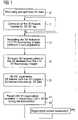
- FIG. 1shows a flowchart of an exemplary embodiment of the method in accordance with the invention
- FIGS. 2 a, 2 bshow two typical 2D projection images which show the same vessel tree from two different projection directions;
- FIG. 3shows a device for executing the method in accordance with the invention.
- FIGS. 2 a and 2 bshow the same vessel tree as a 2D projection image from two different projection angles in each case. These images have been recorded for example with the C-arm x-ray device shown in FIG. 3 with different C-arm angulations, with the visibility having being enhanced for example by contrast means injection.
- the individual vessel arms 2are thus easy to recognize, and the vessel branches 6 can also be identified. A type of tree structure of this vessel section is thus already able to be identified from FIG. 2 a without the precise location of the individual vessel branches in the space being able to be determined.
- each vesselis approximated in each 2D projection image by a “centerline” 4 , which is marked as a dotted line in the largest vessel.
- the vessel diametercould also be averaged over the visible vessel length or be extracted at each individual point along the centerline from each of the two Figures FIG. 2 a and FIG. 2 b .
- the positions of the vessel branches 6are now for example extracted automatically from the individual images and each branch 6 in the image FIG. 2 a is assigned to a branch 6 ′ in the image FIG. 2 b . This can be done on the basis of the height of the vessel branches in the image (if the angulation direction of the C-arm is known), the diameter of the diverging vessels or on the basis of the tree structure determined.
- each vessel branchas well as the course of the vessel between the individual branches can then be determined by means of back projection.
- a three-dimensional model of the vessel structureis obtained which contains the position and orientation of each vessel arm 2 in relation to the geometry of the 2D fluoroscopy images and thereby of the x-ray device used.
- a pre-operative 3D image data recordis recorded. This can be done at any point in time before the planned diagnostic or surgical intervention. Alternatively the 3D image data record can also be acquired directly before the execution of the further method steps on the same x-ray device. In particular C-arm devices also partly allow the recording of three-dimensionally resolved images similar to CT images.
- step 32 Cthe 3D feature (the target structure) needed for 3D-3D registration is extracted from the 3D image data record, a process which can be undertaken using surface, centerline or landmark extraction. This step is only necessary if the 3D-3D registration actually needs an extraction of the 3D feature, which is not the case for example with voxel based registration algorithms. Step 32 can thus optionally be skipped.
- step 34two or more 2D fluoroscopy images are recorded from different C-arm angulations, in which case the 3D feature to be subsequently reconstructed is easily visible.
- step 36the 3D feature of the vessel section for example is symbolically reconstructed, so that its spatial position and orientation relative to the coordinate system of the C-arm system is known (registered).
- Step 38is the actual 3D-3D registration between the 3D feature originating from the pre-operative 3D image data record and the 3D feature which was symbolically reconstructed from the two or more 2D x-ray fluoroscopy images (see step 36 ).
- the result soughtnamely the registration of the pre-operative 3D image data (preferably CT/MR) relative to the coordinate system of the C-arm system, is available.
- the 2D x-ray fluoroscopy imagescan thus be visualized during an intervention together with the 3D image data with the same orientation and scaling/projection direction (step 40 ).
- the methodis repeated, beginning at step 34 , in order to update the registration.
- FIG. 3shows an example for a device for executing this method.
- a significant element of this deviceis the x-ray device 8 with a C-arm 10 , attached to the arms of which are an x-ray tube 12 and an x-ray detector 14 .
- the angular position of the C-armcan be varied (angulated) around a bed 16 with the patient 18 supported on it to obtain fluoroscopy images from different projection directions.
- the images obtained in this wayare forwarded to a control and image evaluation device 20 which comprises at least one data memory 22 and a computing means 24 with a processor or suchlike.
- the computing meansis programmed to spatially reconstruct the 3D feature from the at least two x-ray images and to execute the 3D model thus obtained with the 3D image data record stored in the memory 22 .
- this processis fully automatic. If necessary the user must specify in advance the type of destination structure (vessel section, bone).
- the joint visualization of the 2D fluoroscopy images with the pre-operative 3D image data recordis reproduce
- a particular application of the method described abovelies in also establishing a fixed spatial relationship to the pre-operative 3D image data to further systems as well. This is possible for all systems which for example through their off-line calibration with the C-arm x-ray system have a fixed spatial relationship to the coordinate system of the C-arm x-ray system.
- the pre-operative 3D image datacan also be used for control of an active instrument such a catheter (e.g. the Niobe system from Stereotaxis).
- an active instrumentsuch a catheter (e.g. the Niobe system from Stereotaxis).
- Examples of such systemsare localization systems for surgery (e.g. CAPPA from CAS Innovations, Erlangen), for, interventional cardiology (e.g. the MPS system from Mediguide, Israel) or the electrophysiological mapping systems.
- the last-named groupincludes for example the CARTO system from Biosense Webster, CA, USA and the Ensite-system from Endocardial Solutions, MN, USA, which are used for performing electrophysiological procedures such as ablation for auricular fibrillation.
- the present inventiondescribes an approach to a solution for registering 2D projection images, which can be obtained for example during an operation in real time, but containing little anatomical information for pre-operative morphologically detailed 3D image data, which was obtained by CT or MR for example.
- 2D-3D registration algorithmsare not used, but instead the more stable and widely-available 3D-3D registration algorithms. This requires the previous symbolic reconstruction of a target structure from two or more 2D x-ray fluoroscopy images which is also identifiable in the 3D images.
Landscapes
- Engineering & Computer Science (AREA)
- Physics & Mathematics (AREA)
- General Physics & Mathematics (AREA)
- Computer Vision & Pattern Recognition (AREA)
- Theoretical Computer Science (AREA)
- Apparatus For Radiation Diagnosis (AREA)
Abstract
Description
Claims (20)
Applications Claiming Priority (3)
| Application Number | Priority Date | Filing Date | Title |
|---|---|---|---|
| DE102005023167ADE102005023167B4 (en) | 2005-05-19 | 2005-05-19 | Method and device for registering 2D projection images relative to a 3D image data set |
| DE102005023167 | 2005-05-19 | ||
| DE102005023167.5 | 2005-05-19 |
Publications (2)
| Publication Number | Publication Date |
|---|---|
| US20060262970A1 US20060262970A1 (en) | 2006-11-23 |
| US7689019B2true US7689019B2 (en) | 2010-03-30 |
Family
ID=37311067
Family Applications (1)
| Application Number | Title | Priority Date | Filing Date |
|---|---|---|---|
| US11/437,089Active2029-01-08US7689019B2 (en) | 2005-05-19 | 2006-05-19 | Method and device for registering 2D projection images relative to a 3D image data record |
Country Status (3)
| Country | Link |
|---|---|
| US (1) | US7689019B2 (en) |
| CN (1) | CN100581478C (en) |
| DE (1) | DE102005023167B4 (en) |
Cited By (58)
| Publication number | Priority date | Publication date | Assignee | Title |
|---|---|---|---|---|
| US20080175455A1 (en)* | 2006-09-29 | 2008-07-24 | Matthias John | Method and device for the combined representation of 2D fluoroscopic images and a static 3D image data set |
| US20090123046A1 (en)* | 2006-05-11 | 2009-05-14 | Koninklijke Philips Electronics N.V. | System and method for generating intraoperative 3-dimensional images using non-contrast image data |
| US20090161939A1 (en)* | 2007-12-21 | 2009-06-25 | General Electric Company | System and method for extracting features of interest from an image |
| US20100189337A1 (en)* | 2007-07-11 | 2010-07-29 | Koninklijke Philips Electronics N.V. | Method for acquiring 3-dimensional images of coronary vessels, particularly of coronary veins |
| US20100272315A1 (en)* | 2009-04-24 | 2010-10-28 | Siemens Corporation | Automatic Measurement of Morphometric and Motion Parameters of the Coronary Tree From A Rotational X-Ray Sequence |
| US20100316278A1 (en)* | 2009-06-12 | 2010-12-16 | Ulrich Hartung | High-resolution three-dimensional medical imaging with dynamic real-time information |
| EP2468208A2 (en) | 2010-12-22 | 2012-06-27 | Biosense Webster (Israel), Ltd. | Compensation for magnetic disturbance due to fluoroscope |
| US20120289826A1 (en)* | 2011-05-12 | 2012-11-15 | Siemens Aktiengesellschaft | Method for localization and identification of structures in projection images |
| WO2013118047A1 (en)* | 2012-02-06 | 2013-08-15 | Koninklijke Philips Electronics N.V. | Invisible bifurcation detection within vessel tree images |
| US8659764B2 (en) | 2009-02-27 | 2014-02-25 | Body Surface Translations, Inc. | Estimating physical parameters using three dimensional representations |
| US8750568B2 (en) | 2012-05-22 | 2014-06-10 | Covidien Lp | System and method for conformal ablation planning |
| US20140355855A1 (en)* | 2013-05-30 | 2014-12-04 | Siemens Aktiengesellschaft | System and Method for Magnetic Resonance Imaging Based Respiratory Motion Correction for PET/MRI |
| US9105200B2 (en) | 2011-10-04 | 2015-08-11 | Quantant Technology, Inc. | Semi-automated or fully automated, network and/or web-based, 3D and/or 4D imaging of anatomy for training, rehearsing and/or conducting medical procedures, using multiple standard X-ray and/or other imaging projections, without a need for special hardware and/or systems and/or pre-processing/analysis of a captured image data |
| US9265468B2 (en) | 2011-05-11 | 2016-02-23 | Broncus Medical, Inc. | Fluoroscopy-based surgical device tracking method |
| US20160132663A1 (en)* | 2003-11-03 | 2016-05-12 | Tech Pharmacy Services, Llc | System and method of enhanced distribution of pharmaceuticals in long-term care facilities |
| US9439627B2 (en) | 2012-05-22 | 2016-09-13 | Covidien Lp | Planning system and navigation system for an ablation procedure |
| US9439622B2 (en) | 2012-05-22 | 2016-09-13 | Covidien Lp | Surgical navigation system |
| US9439623B2 (en) | 2012-05-22 | 2016-09-13 | Covidien Lp | Surgical planning system and navigation system |
| US9498182B2 (en) | 2012-05-22 | 2016-11-22 | Covidien Lp | Systems and methods for planning and navigation |
| US9510771B1 (en) | 2011-10-28 | 2016-12-06 | Nuvasive, Inc. | Systems and methods for performing spine surgery |
| US9633431B2 (en) | 2014-07-02 | 2017-04-25 | Covidien Lp | Fluoroscopic pose estimation |
| US9848922B2 (en) | 2013-10-09 | 2017-12-26 | Nuvasive, Inc. | Systems and methods for performing spine surgery |
| US9875544B2 (en) | 2013-08-09 | 2018-01-23 | Broncus Medical Inc. | Registration of fluoroscopic images of the chest and corresponding 3D image data based on the ribs and spine |
| US9972120B2 (en) | 2012-03-22 | 2018-05-15 | University Of Notre Dame Du Lac | Systems and methods for geometrically mapping two-dimensional images to three-dimensional surfaces |
| US9974525B2 (en) | 2014-10-31 | 2018-05-22 | Covidien Lp | Computed tomography enhanced fluoroscopic system, device, and method of utilizing the same |
| US20180280727A1 (en)* | 2017-03-30 | 2018-10-04 | Shimadzu Corporation | Positioning apparatus and method of positioning |
| US10164776B1 (en) | 2013-03-14 | 2018-12-25 | goTenna Inc. | System and method for private and point-to-point communication between computing devices |
| US10674982B2 (en) | 2015-08-06 | 2020-06-09 | Covidien Lp | System and method for local three dimensional volume reconstruction using a standard fluoroscope |
| US10699448B2 (en) | 2017-06-29 | 2020-06-30 | Covidien Lp | System and method for identifying, marking and navigating to a target using real time two dimensional fluoroscopic data |
| US10702226B2 (en) | 2015-08-06 | 2020-07-07 | Covidien Lp | System and method for local three dimensional volume reconstruction using a standard fluoroscope |
| US10716525B2 (en) | 2015-08-06 | 2020-07-21 | Covidien Lp | System and method for navigating to target and performing procedure on target utilizing fluoroscopic-based local three dimensional volume reconstruction |
| US10734116B2 (en) | 2011-10-04 | 2020-08-04 | Quantant Technology, Inc. | Remote cloud based medical image sharing and rendering semi-automated or fully automated network and/or web-based, 3D and/or 4D imaging of anatomy for training, rehearsing and/or conducting medical procedures, using multiple standard X-ray and/or other imaging projections, without a need for special hardware and/or systems and/or pre-processing/analysis of a captured image data |
| US10818019B2 (en) | 2017-08-14 | 2020-10-27 | Siemens Healthcare Gmbh | Dilated fully convolutional network for multi-agent 2D/3D medical image registration |
| US10893843B2 (en) | 2017-10-10 | 2021-01-19 | Covidien Lp | System and method for identifying and marking a target in a fluoroscopic three-dimensional reconstruction |
| US10905498B2 (en) | 2018-02-08 | 2021-02-02 | Covidien Lp | System and method for catheter detection in fluoroscopic images and updating displayed position of catheter |
| US11051886B2 (en) | 2016-09-27 | 2021-07-06 | Covidien Lp | Systems and methods for performing a surgical navigation procedure |
| US11172895B2 (en) | 2015-12-07 | 2021-11-16 | Covidien Lp | Visualization, navigation, and planning with electromagnetic navigation bronchoscopy and cone beam computed tomography integrated |
| US11282170B2 (en) | 2017-03-30 | 2022-03-22 | Koninklijke Philips N.V. | Contrast injection imaging |
| US11470303B1 (en) | 2010-06-24 | 2022-10-11 | Steven M. Hoffberg | Two dimensional to three dimensional moving image converter |
| US11483531B2 (en) | 2020-05-26 | 2022-10-25 | Unify Medical, Inc. | Generation of three-dimensional images with digital magnification |
| US11671703B2 (en) | 2017-04-14 | 2023-06-06 | Unify Medical, Inc. | System and apparatus for co-registration and correlation between multi-modal imagery and method for same |
| US11707329B2 (en) | 2018-08-10 | 2023-07-25 | Covidien Lp | Systems and methods for ablation visualization |
| US11710249B2 (en) | 2019-12-20 | 2023-07-25 | Unify Medical, Inc. | Generation of three-dimensional scans for intraoperative imaging |
| US11750794B2 (en) | 2015-03-24 | 2023-09-05 | Augmedics Ltd. | Combining video-based and optic-based augmented reality in a near eye display |
| US11766296B2 (en) | 2018-11-26 | 2023-09-26 | Augmedics Ltd. | Tracking system for image-guided surgery |
| US11801115B2 (en) | 2019-12-22 | 2023-10-31 | Augmedics Ltd. | Mirroring in image guided surgery |
| US11896445B2 (en) | 2021-07-07 | 2024-02-13 | Augmedics Ltd. | Iliac pin and adapter |
| US11974887B2 (en) | 2018-05-02 | 2024-05-07 | Augmedics Ltd. | Registration marker for an augmented reality system |
| US11980506B2 (en) | 2019-07-29 | 2024-05-14 | Augmedics Ltd. | Fiducial marker |
| US12044856B2 (en) | 2022-09-13 | 2024-07-23 | Augmedics Ltd. | Configurable augmented reality eyewear for image-guided medical intervention |
| US12106504B2 (en) | 2019-12-20 | 2024-10-01 | Unify Medical, Inc. | Generation of three-dimensional scans for intraoperative imaging |
| US12150821B2 (en) | 2021-07-29 | 2024-11-26 | Augmedics Ltd. | Rotating marker and adapter for image-guided surgery |
| US12178666B2 (en) | 2019-07-29 | 2024-12-31 | Augmedics Ltd. | Fiducial marker |
| US12186028B2 (en) | 2020-06-15 | 2025-01-07 | Augmedics Ltd. | Rotating marker for image guided surgery |
| US12239385B2 (en) | 2020-09-09 | 2025-03-04 | Augmedics Ltd. | Universal tool adapter |
| US12354227B2 (en) | 2022-04-21 | 2025-07-08 | Augmedics Ltd. | Systems for medical image visualization |
| US12357393B2 (en) | 2014-06-17 | 2025-07-15 | Nuvasive, Inc. | Systems and methods for planning, performing, and assessing spinal correction during surgery |
| US12417595B2 (en) | 2021-08-18 | 2025-09-16 | Augmedics Ltd. | Augmented-reality surgical system using depth sensing |
Families Citing this family (42)
| Publication number | Priority date | Publication date | Assignee | Title |
|---|---|---|---|---|
| DE102006012943B4 (en)* | 2006-03-21 | 2009-11-19 | Siemens Ag | Method for automatically evaluating an imaging data set of an object and medical imaging system |
| DE102006012945B4 (en)* | 2006-03-21 | 2014-10-02 | Siemens Aktiengesellschaft | A method of virtual layer positioning in a 3D volume data set and medical imaging system |
| US20080131029A1 (en)* | 2006-10-10 | 2008-06-05 | Coleby Stanley E | Systems and methods for visualizing and measuring real world 3-d spatial data |
| US8126239B2 (en)* | 2006-10-20 | 2012-02-28 | Siemens Aktiengesellschaft | Registering 2D and 3D data using 3D ultrasound data |
| IL188569A (en) | 2007-01-17 | 2014-05-28 | Mediguide Ltd | Method and system for registering a 3d pre-acquired image coordinate system with a medical positioning system coordinate system and with a 2d image coordinate system |
| DE102007009179A1 (en)* | 2007-02-26 | 2008-06-26 | Siemens Ag | Registering method for data set of creature, involves deforming one of two-dimensional actual representation corresponding to determined deformation so that deformed representation is registered relative to actual representation |
| US7773719B2 (en)* | 2007-03-26 | 2010-08-10 | Siemens Medical Solutions Usa, Inc. | Model-based heart reconstruction and navigation |
| WO2008120136A1 (en)* | 2007-03-30 | 2008-10-09 | Koninklijke Philips Electronics N.V. | 2d/3d image registration |
| US20080300478A1 (en)* | 2007-05-30 | 2008-12-04 | General Electric Company | System and method for displaying real-time state of imaged anatomy during a surgical procedure |
| EP2207483B1 (en)* | 2007-10-19 | 2016-06-01 | Metritrack, Inc. | Three dimensional mapping display system for diagnostic ultrasound machines and method |
| WO2009077971A1 (en)* | 2007-12-18 | 2009-06-25 | Koninklijke Philips Electronics, N.V. | Fusion of cardiac 3d ultrasound and x-ray information by means of epicardial surfaces and landmarks |
| JP5906015B2 (en)* | 2007-12-18 | 2016-04-20 | コーニンクレッカ フィリップス エヌ ヴェKoninklijke Philips N.V. | 2D / 3D image registration based on features |
| US20090198126A1 (en)* | 2008-02-05 | 2009-08-06 | Klaus Klingenbeck-Regn | Imaging system |
| DE102008018445A1 (en)* | 2008-04-07 | 2009-10-15 | Carl Zeiss Industrielle Messtechnik Gmbh | Method for the tomographic measurement of mechanical workpieces |
| DE102008020780B4 (en)* | 2008-04-25 | 2010-12-02 | Siemens Aktiengesellschaft | Correction method for correcting an ECG signal data set of an ECG signal |
| JP5675056B2 (en)* | 2008-07-04 | 2015-02-25 | 株式会社東芝 | X-ray imaging apparatus and image processing apparatus |
| US8131046B2 (en)* | 2008-10-29 | 2012-03-06 | Allegheny-Singer Research Institute | Magnetic resonance imager using cylindrical offset region of excitation, and method |
| EP2370953A1 (en)* | 2008-11-25 | 2011-10-05 | Koninklijke Philips Electronics N.V. | Image provision for registration |
| US8675996B2 (en)* | 2009-07-29 | 2014-03-18 | Siemens Aktiengesellschaft | Catheter RF ablation using segmentation-based 2D-3D registration |
| JP2011175477A (en)* | 2010-02-24 | 2011-09-08 | Canon Inc | Three-dimensional measurement apparatus, processing method and program |
| DE102010018872A1 (en)* | 2010-04-30 | 2011-11-03 | Siemens Aktiengesellschaft | An imaging method for highlighting vessels in an examination area of a patient and a medical system for performing the method |
| DE102010020783B4 (en)* | 2010-05-18 | 2022-05-12 | Siemens Healthcare Gmbh | Method and system for determining 3D geometry data of objects |
| US8675939B2 (en) | 2010-07-13 | 2014-03-18 | Stryker Leibinger Gmbh & Co. Kg | Registration of anatomical data sets |
| US9295435B2 (en) | 2011-02-07 | 2016-03-29 | Koninklijke Philips N.V. | Image representation supporting the accurate positioning of an intervention device in vessel intervention procedures |
| JP6166663B2 (en)* | 2011-03-02 | 2017-07-19 | コーニンクレッカ フィリップス エヌ ヴェKoninklijke Philips N.V. | Visualization for navigation guidance |
| EP2852326B1 (en) | 2012-05-22 | 2018-12-05 | Mazor Robotics Ltd. | On-site verification of implant positioning |
| CN103505288B (en)* | 2012-06-29 | 2017-11-17 | 通用电气公司 | Ultrasonic imaging method and supersonic imaging apparatus |
| CN104574329B (en)* | 2013-10-09 | 2018-03-09 | 深圳迈瑞生物医疗电子股份有限公司 | Ultrasonic fusion of imaging method, ultrasonic fusion of imaging navigation system |
| US11213220B2 (en) | 2014-08-11 | 2022-01-04 | Cubisme, Inc. | Method for determining in vivo tissue biomarker characteristics using multiparameter MRI matrix creation and big data analytics |
| US10453269B2 (en) | 2014-12-08 | 2019-10-22 | Align Technology, Inc. | Intraoral scanning using ultrasound and optical scan data |
| JP6476041B2 (en)* | 2015-03-31 | 2019-02-27 | 株式会社Aze | MEDICAL IMAGE DIAGNOSIS DEVICE, ITS CONTROL METHOD, AND PROGRAM |
| KR101797042B1 (en)* | 2015-05-15 | 2017-11-13 | 삼성전자주식회사 | Method and apparatus for synthesizing medical images |
| FI20155856A7 (en)* | 2015-11-18 | 2017-05-19 | Teknologian Tutkimuskeskus Vtt Oy | Methods and apparatuses for penetrating imaging |
| WO2018006058A1 (en) | 2016-07-01 | 2018-01-04 | Cubisme, Inc. | System and method for forming a super-resolution biomarker map image |
| DE102016219817B4 (en)* | 2016-10-12 | 2018-05-30 | Siemens Healthcare Gmbh | Method for determining an X-ray image data set and X-ray device |
| TWI624243B (en)* | 2016-12-15 | 2018-05-21 | 神農資訊股份有限公司 | Surgical navigation system and instrument guiding method thereof |
| WO2018195501A2 (en)* | 2017-04-21 | 2018-10-25 | Cubisme, Inc. | System and method for creating, querying, and displaying a miba master file |
| US10432913B2 (en)* | 2017-05-31 | 2019-10-01 | Proximie, Inc. | Systems and methods for determining three dimensional measurements in telemedicine application |
| EP3618715A4 (en)* | 2017-06-19 | 2021-02-17 | Mohamed R. Mahfouz | HIP SURGICAL NAVIGATION USING FLUOROSCOPY AND TRACKING SENSORS |
| EP3522116A1 (en)* | 2018-01-31 | 2019-08-07 | Koninklijke Philips N.V. | Device, system and method for determining the position of stents in an image of vasculature structure |
| DE102020003366A1 (en)* | 2020-06-04 | 2021-12-23 | Ziehm Imaging Gmbh | Method and device for image monitoring by means of an X-ray device during a surgical procedure |
| CN114332372B (en)* | 2021-12-28 | 2024-11-12 | 北京阅影科技有限公司 | Method and device for determining a three-dimensional model of a blood vessel |
Citations (9)
| Publication number | Priority date | Publication date | Assignee | Title |
|---|---|---|---|---|
| US6047080A (en)* | 1996-06-19 | 2000-04-04 | Arch Development Corporation | Method and apparatus for three-dimensional reconstruction of coronary vessels from angiographic images |
| US20030013966A1 (en) | 1996-06-28 | 2003-01-16 | Sonosite, Inc. | Balance body ultrasound system |
| DE10201644A1 (en) | 2002-01-17 | 2003-08-07 | Siemens Ag | Registration procedure for projective intraoperative 3D imaging |
| US20030181809A1 (en)* | 2002-03-11 | 2003-09-25 | Hall Andrew F. | 3D imaging for catheter interventions by use of 2D/3D image fusion |
| US6628977B2 (en)* | 1999-12-28 | 2003-09-30 | Siemens Aktiengesellschaft | Method and system for visualizing an object |
| US20030220555A1 (en) | 2002-03-11 | 2003-11-27 | Benno Heigl | Method and apparatus for image presentation of a medical instrument introduced into an examination region of a patent |
| US20040077942A1 (en)* | 2002-03-11 | 2004-04-22 | Hall Andrew F. | 3D imaging for catheter interventions by use of positioning system |
| US20040170311A1 (en) | 1999-10-15 | 2004-09-02 | Vittorio Accomazzi | Perspective with shear warp |
| US20050004449A1 (en)* | 2003-05-20 | 2005-01-06 | Matthias Mitschke | Method for marker-less navigation in preoperative 3D images using an intraoperatively acquired 3D C-arm image |
- 2005
- 2005-05-19DEDE102005023167Apatent/DE102005023167B4/ennot_activeExpired - Fee Related
- 2006
- 2006-05-19CNCN200610082786Apatent/CN100581478C/enactiveActive
- 2006-05-19USUS11/437,089patent/US7689019B2/enactiveActive
Patent Citations (9)
| Publication number | Priority date | Publication date | Assignee | Title |
|---|---|---|---|---|
| US6047080A (en)* | 1996-06-19 | 2000-04-04 | Arch Development Corporation | Method and apparatus for three-dimensional reconstruction of coronary vessels from angiographic images |
| US20030013966A1 (en) | 1996-06-28 | 2003-01-16 | Sonosite, Inc. | Balance body ultrasound system |
| US20040170311A1 (en) | 1999-10-15 | 2004-09-02 | Vittorio Accomazzi | Perspective with shear warp |
| US6628977B2 (en)* | 1999-12-28 | 2003-09-30 | Siemens Aktiengesellschaft | Method and system for visualizing an object |
| DE10201644A1 (en) | 2002-01-17 | 2003-08-07 | Siemens Ag | Registration procedure for projective intraoperative 3D imaging |
| US20030181809A1 (en)* | 2002-03-11 | 2003-09-25 | Hall Andrew F. | 3D imaging for catheter interventions by use of 2D/3D image fusion |
| US20030220555A1 (en) | 2002-03-11 | 2003-11-27 | Benno Heigl | Method and apparatus for image presentation of a medical instrument introduced into an examination region of a patent |
| US20040077942A1 (en)* | 2002-03-11 | 2004-04-22 | Hall Andrew F. | 3D imaging for catheter interventions by use of positioning system |
| US20050004449A1 (en)* | 2003-05-20 | 2005-01-06 | Matthias Mitschke | Method for marker-less navigation in preoperative 3D images using an intraoperatively acquired 3D C-arm image |
Non-Patent Citations (2)
| Title |
|---|
| J. Weese, T.M. Buzug, G.P. Penney and P. Desmedt, "2D/3D Registration and Motion Tracking for Surgical Interventions", Philips Journal of Research, 1998, pp. 299-316, vol. 51, No. 2. |
| Paieon Medical Ltd.; [Retrieved from internet on] Jul. 25, 2006; [Retrieved from internet at] http://www.matimop.org.il/newrdinf/company/c5292.htm; pp. 1-2; Rosh Haaiyn, Israel. |
Cited By (113)
| Publication number | Priority date | Publication date | Assignee | Title |
|---|---|---|---|---|
| US9747422B2 (en)* | 2003-11-03 | 2017-08-29 | Tech Pharmacy Services, Llc | System and method of enhanced distribution of pharmaceuticals in long-term care facilities |
| US20160203293A1 (en)* | 2003-11-03 | 2016-07-14 | Tech Pharmacy Services, Llc | System of enhanced distribution of pharmaceuticals in long-term care facilities |
| US9710609B2 (en)* | 2003-11-03 | 2017-07-18 | Tech Pharmacy Services, Llc | System of enhanced distribution of pharmaceuticals in long-term care facilities |
| US9740830B2 (en)* | 2003-11-03 | 2017-08-22 | Tech Pharmacy Services, Llc | Method of enhanced distribution of pharmaceuticals in long-term care facilities |
| US20160203294A1 (en)* | 2003-11-03 | 2016-07-14 | Tech Pharmacy Services, Llc | Method of enhanced distribution of pharmaceuticals in long-term care facilities |
| US20160132663A1 (en)* | 2003-11-03 | 2016-05-12 | Tech Pharmacy Services, Llc | System and method of enhanced distribution of pharmaceuticals in long-term care facilities |
| US20090123046A1 (en)* | 2006-05-11 | 2009-05-14 | Koninklijke Philips Electronics N.V. | System and method for generating intraoperative 3-dimensional images using non-contrast image data |
| US8005283B2 (en)* | 2006-09-29 | 2011-08-23 | Siemens Aktiengesellschaft | Method and device for the combined representation of 2D fluoroscopic images and a static 3D image data set |
| US20080175455A1 (en)* | 2006-09-29 | 2008-07-24 | Matthias John | Method and device for the combined representation of 2D fluoroscopic images and a static 3D image data set |
| US20100189337A1 (en)* | 2007-07-11 | 2010-07-29 | Koninklijke Philips Electronics N.V. | Method for acquiring 3-dimensional images of coronary vessels, particularly of coronary veins |
| US20090161939A1 (en)* | 2007-12-21 | 2009-06-25 | General Electric Company | System and method for extracting features of interest from an image |
| US9070181B2 (en)* | 2007-12-21 | 2015-06-30 | General Electric Company | System and method for extracting features of interest from an image |
| US8659764B2 (en) | 2009-02-27 | 2014-02-25 | Body Surface Translations, Inc. | Estimating physical parameters using three dimensional representations |
| US20100272315A1 (en)* | 2009-04-24 | 2010-10-28 | Siemens Corporation | Automatic Measurement of Morphometric and Motion Parameters of the Coronary Tree From A Rotational X-Ray Sequence |
| US8428319B2 (en)* | 2009-04-24 | 2013-04-23 | Siemens Aktiengesellschaft | Automatic measurement of morphometric and motion parameters of the coronary tree from a rotational X-ray sequence |
| US20100316278A1 (en)* | 2009-06-12 | 2010-12-16 | Ulrich Hartung | High-resolution three-dimensional medical imaging with dynamic real-time information |
| US9036880B2 (en) | 2009-06-12 | 2015-05-19 | Siemens Aktiengesellschaft | High-resolution three-dimensional medical imaging with dynamic real-time information |
| US11470303B1 (en) | 2010-06-24 | 2022-10-11 | Steven M. Hoffberg | Two dimensional to three dimensional moving image converter |
| EP3403610A1 (en) | 2010-12-22 | 2018-11-21 | Biosense Webster (Israel) Ltd. | Compensation for magnetic disturbance due to fluoroscope |
| EP3241495A1 (en) | 2010-12-22 | 2017-11-08 | Biosense Webster (Israel), Ltd. | Compensation for magnetic disturbance due to fluoroscope |
| US8812079B2 (en) | 2010-12-22 | 2014-08-19 | Biosense Webster (Israel), Ltd. | Compensation for magnetic disturbance due to fluoroscope |
| EP2468208A2 (en) | 2010-12-22 | 2012-06-27 | Biosense Webster (Israel), Ltd. | Compensation for magnetic disturbance due to fluoroscope |
| US9265468B2 (en) | 2011-05-11 | 2016-02-23 | Broncus Medical, Inc. | Fluoroscopy-based surgical device tracking method |
| US11284846B2 (en)* | 2011-05-12 | 2022-03-29 | The John Hopkins University | Method for localization and identification of structures in projection images |
| US20120289826A1 (en)* | 2011-05-12 | 2012-11-15 | Siemens Aktiengesellschaft | Method for localization and identification of structures in projection images |
| US9105200B2 (en) | 2011-10-04 | 2015-08-11 | Quantant Technology, Inc. | Semi-automated or fully automated, network and/or web-based, 3D and/or 4D imaging of anatomy for training, rehearsing and/or conducting medical procedures, using multiple standard X-ray and/or other imaging projections, without a need for special hardware and/or systems and/or pre-processing/analysis of a captured image data |
| US10734116B2 (en) | 2011-10-04 | 2020-08-04 | Quantant Technology, Inc. | Remote cloud based medical image sharing and rendering semi-automated or fully automated network and/or web-based, 3D and/or 4D imaging of anatomy for training, rehearsing and/or conducting medical procedures, using multiple standard X-ray and/or other imaging projections, without a need for special hardware and/or systems and/or pre-processing/analysis of a captured image data |
| USRE49094E1 (en) | 2011-10-28 | 2022-06-07 | Nuvasive, Inc. | Systems and methods for performing spine surgery |
| US9510771B1 (en) | 2011-10-28 | 2016-12-06 | Nuvasive, Inc. | Systems and methods for performing spine surgery |
| WO2013118047A1 (en)* | 2012-02-06 | 2013-08-15 | Koninklijke Philips Electronics N.V. | Invisible bifurcation detection within vessel tree images |
| US9280823B2 (en) | 2012-02-06 | 2016-03-08 | Koninklijke Philips N.V. | Invisible bifurcation detection within vessel tree images |
| US9972120B2 (en) | 2012-03-22 | 2018-05-15 | University Of Notre Dame Du Lac | Systems and methods for geometrically mapping two-dimensional images to three-dimensional surfaces |
| US9439627B2 (en) | 2012-05-22 | 2016-09-13 | Covidien Lp | Planning system and navigation system for an ablation procedure |
| US9498182B2 (en) | 2012-05-22 | 2016-11-22 | Covidien Lp | Systems and methods for planning and navigation |
| US8750568B2 (en) | 2012-05-22 | 2014-06-10 | Covidien Lp | System and method for conformal ablation planning |
| US9439622B2 (en) | 2012-05-22 | 2016-09-13 | Covidien Lp | Surgical navigation system |
| US9439623B2 (en) | 2012-05-22 | 2016-09-13 | Covidien Lp | Surgical planning system and navigation system |
| US10164776B1 (en) | 2013-03-14 | 2018-12-25 | goTenna Inc. | System and method for private and point-to-point communication between computing devices |
| US9398855B2 (en)* | 2013-05-30 | 2016-07-26 | Siemens Aktiengesellschaft | System and method for magnetic resonance imaging based respiratory motion correction for PET/MRI |
| US20140355855A1 (en)* | 2013-05-30 | 2014-12-04 | Siemens Aktiengesellschaft | System and Method for Magnetic Resonance Imaging Based Respiratory Motion Correction for PET/MRI |
| US9875544B2 (en) | 2013-08-09 | 2018-01-23 | Broncus Medical Inc. | Registration of fluoroscopic images of the chest and corresponding 3D image data based on the ribs and spine |
| US9848922B2 (en) | 2013-10-09 | 2017-12-26 | Nuvasive, Inc. | Systems and methods for performing spine surgery |
| US12357393B2 (en) | 2014-06-17 | 2025-07-15 | Nuvasive, Inc. | Systems and methods for planning, performing, and assessing spinal correction during surgery |
| US11798178B2 (en) | 2014-07-02 | 2023-10-24 | Covidien Lp | Fluoroscopic pose estimation |
| US9959620B2 (en) | 2014-07-02 | 2018-05-01 | Covidien Lp | Fluoroscopic pose estimation |
| US10706540B2 (en) | 2014-07-02 | 2020-07-07 | Covidien Lp | Fluoroscopic pose estimation |
| US9633431B2 (en) | 2014-07-02 | 2017-04-25 | Covidien Lp | Fluoroscopic pose estimation |
| US10163207B2 (en) | 2014-07-02 | 2018-12-25 | Covidien Lp | Fluoroscopic pose estimation |
| US9974525B2 (en) | 2014-10-31 | 2018-05-22 | Covidien Lp | Computed tomography enhanced fluoroscopic system, device, and method of utilizing the same |
| US10314564B2 (en) | 2014-10-31 | 2019-06-11 | Covidien Lp | Computed tomography enhanced fluoroscopic system, device, and method of utilizing the same |
| US9986983B2 (en) | 2014-10-31 | 2018-06-05 | Covidien Lp | Computed tomography enhanced fluoroscopic system, device, and method of utilizing the same |
| US11871913B2 (en) | 2014-10-31 | 2024-01-16 | Covidien Lp | Computed tomography enhanced fluoroscopic system, device, and method of utilizing the same |
| US10321898B2 (en) | 2014-10-31 | 2019-06-18 | Covidien Lp | Computed tomography enhanced fluoroscopic system, device, and method of utilizing the same |
| US12063345B2 (en) | 2015-03-24 | 2024-08-13 | Augmedics Ltd. | Systems for facilitating augmented reality-assisted medical procedures |
| US12206837B2 (en) | 2015-03-24 | 2025-01-21 | Augmedics Ltd. | Combining video-based and optic-based augmented reality in a near eye display |
| US11750794B2 (en) | 2015-03-24 | 2023-09-05 | Augmedics Ltd. | Combining video-based and optic-based augmented reality in a near eye display |
| US12069233B2 (en) | 2015-03-24 | 2024-08-20 | Augmedics Ltd. | Head-mounted augmented reality near eye display device |
| US10674982B2 (en) | 2015-08-06 | 2020-06-09 | Covidien Lp | System and method for local three dimensional volume reconstruction using a standard fluoroscope |
| US11547377B2 (en) | 2015-08-06 | 2023-01-10 | Covidien Lp | System and method for navigating to target and performing procedure on target utilizing fluoroscopic-based local three dimensional volume reconstruction |
| US11559266B2 (en) | 2015-08-06 | 2023-01-24 | Covidien Lp | System and method for local three dimensional volume reconstruction using a standard fluoroscope |
| US10716525B2 (en) | 2015-08-06 | 2020-07-21 | Covidien Lp | System and method for navigating to target and performing procedure on target utilizing fluoroscopic-based local three dimensional volume reconstruction |
| US10702226B2 (en) | 2015-08-06 | 2020-07-07 | Covidien Lp | System and method for local three dimensional volume reconstruction using a standard fluoroscope |
| US11707241B2 (en) | 2015-08-06 | 2023-07-25 | Covidien Lp | System and method for local three dimensional volume reconstruction using a standard fluoroscope |
| US11992349B2 (en) | 2015-08-06 | 2024-05-28 | Covidien Lp | System and method for local three dimensional volume reconstruction using a standard fluoroscope |
| US11925493B2 (en) | 2015-12-07 | 2024-03-12 | Covidien Lp | Visualization, navigation, and planning with electromagnetic navigation bronchoscopy and cone beam computed tomography integrated |
| US11172895B2 (en) | 2015-12-07 | 2021-11-16 | Covidien Lp | Visualization, navigation, and planning with electromagnetic navigation bronchoscopy and cone beam computed tomography integrated |
| US11051886B2 (en) | 2016-09-27 | 2021-07-06 | Covidien Lp | Systems and methods for performing a surgical navigation procedure |
| US20180280727A1 (en)* | 2017-03-30 | 2018-10-04 | Shimadzu Corporation | Positioning apparatus and method of positioning |
| US11282170B2 (en) | 2017-03-30 | 2022-03-22 | Koninklijke Philips N.V. | Contrast injection imaging |
| US10434335B2 (en)* | 2017-03-30 | 2019-10-08 | Shimadzu Corporation | Positioning apparatus and method of positioning by generation of DRR image from X-ray CT image data |
| US11671703B2 (en) | 2017-04-14 | 2023-06-06 | Unify Medical, Inc. | System and apparatus for co-registration and correlation between multi-modal imagery and method for same |
| US12342072B2 (en) | 2017-04-14 | 2025-06-24 | Unify Medical, Inc. | System and apparatus for co-registration and correlation between multi-modal imagery and method for same |
| US10699448B2 (en) | 2017-06-29 | 2020-06-30 | Covidien Lp | System and method for identifying, marking and navigating to a target using real time two dimensional fluoroscopic data |
| US11341692B2 (en) | 2017-06-29 | 2022-05-24 | Covidien Lp | System and method for identifying, marking and navigating to a target using real time two dimensional fluoroscopic data |
| US10846893B2 (en) | 2017-06-29 | 2020-11-24 | Covidien Lp | System and method for identifying, marking and navigating to a target using real time three dimensional fluoroscopic data |
| US11354813B2 (en) | 2017-08-14 | 2022-06-07 | Siemens Healthcare Gmbh | Dilated fully convolutional network for 2D/3D medical image registration |
| US10818019B2 (en) | 2017-08-14 | 2020-10-27 | Siemens Healthcare Gmbh | Dilated fully convolutional network for multi-agent 2D/3D medical image registration |
| US10893843B2 (en) | 2017-10-10 | 2021-01-19 | Covidien Lp | System and method for identifying and marking a target in a fluoroscopic three-dimensional reconstruction |
| US12064280B2 (en) | 2017-10-10 | 2024-08-20 | Covidien Lp | System and method for identifying and marking a target in a fluoroscopic three-dimensional reconstruction |
| US11564649B2 (en) | 2017-10-10 | 2023-01-31 | Covidien Lp | System and method for identifying and marking a target in a fluoroscopic three-dimensional reconstruction |
| US11701184B2 (en) | 2018-02-08 | 2023-07-18 | Covidien Lp | System and method for catheter detection in fluoroscopic images and updating displayed position of catheter |
| US10905498B2 (en) | 2018-02-08 | 2021-02-02 | Covidien Lp | System and method for catheter detection in fluoroscopic images and updating displayed position of catheter |
| US11253325B2 (en) | 2018-02-08 | 2022-02-22 | Covidien Lp | System and method for catheter detection in fluoroscopic images and updating displayed position of catheter |
| US12144566B2 (en) | 2018-02-08 | 2024-11-19 | Covidien Lp | System and method for catheter detection in fluoroscopic images and updating displayed position of catheter |
| US12290416B2 (en) | 2018-05-02 | 2025-05-06 | Augmedics Ltd. | Registration of a fiducial marker for an augmented reality system |
| US11974887B2 (en) | 2018-05-02 | 2024-05-07 | Augmedics Ltd. | Registration marker for an augmented reality system |
| US11980507B2 (en) | 2018-05-02 | 2024-05-14 | Augmedics Ltd. | Registration of a fiducial marker for an augmented reality system |
| US11980508B2 (en) | 2018-05-02 | 2024-05-14 | Augmedics Ltd. | Registration of a fiducial marker for an augmented reality system |
| US12207891B2 (en) | 2018-08-10 | 2025-01-28 | Covidien Lp | Systems and methods for ablation visualization |
| US11707329B2 (en) | 2018-08-10 | 2023-07-25 | Covidien Lp | Systems and methods for ablation visualization |
| US12201384B2 (en) | 2018-11-26 | 2025-01-21 | Augmedics Ltd. | Tracking systems and methods for image-guided surgery |
| US11766296B2 (en) | 2018-11-26 | 2023-09-26 | Augmedics Ltd. | Tracking system for image-guided surgery |
| US11980429B2 (en) | 2018-11-26 | 2024-05-14 | Augmedics Ltd. | Tracking methods for image-guided surgery |
| US11980506B2 (en) | 2019-07-29 | 2024-05-14 | Augmedics Ltd. | Fiducial marker |
| US12178666B2 (en) | 2019-07-29 | 2024-12-31 | Augmedics Ltd. | Fiducial marker |
| US12118738B2 (en) | 2019-12-20 | 2024-10-15 | Unify Medical, Inc. | Generation of three-dimensional scans for intraoperative imaging |
| US12106504B2 (en) | 2019-12-20 | 2024-10-01 | Unify Medical, Inc. | Generation of three-dimensional scans for intraoperative imaging |
| US11710249B2 (en) | 2019-12-20 | 2023-07-25 | Unify Medical, Inc. | Generation of three-dimensional scans for intraoperative imaging |
| US12383369B2 (en) | 2019-12-22 | 2025-08-12 | Augmedics Ltd. | Mirroring in image guided surgery |
| US12076196B2 (en) | 2019-12-22 | 2024-09-03 | Augmedics Ltd. | Mirroring in image guided surgery |
| US11801115B2 (en) | 2019-12-22 | 2023-10-31 | Augmedics Ltd. | Mirroring in image guided surgery |
| US12355931B2 (en) | 2020-05-26 | 2025-07-08 | Unify Medical, Inc. | Generation of three-dimensional images with digital magnification |
| US11483531B2 (en) | 2020-05-26 | 2022-10-25 | Unify Medical, Inc. | Generation of three-dimensional images with digital magnification |
| US11800078B2 (en) | 2020-05-26 | 2023-10-24 | Unify Medical, Inc. | Generation of three-dimensional images with digital magnification |
| US12186028B2 (en) | 2020-06-15 | 2025-01-07 | Augmedics Ltd. | Rotating marker for image guided surgery |
| US12239385B2 (en) | 2020-09-09 | 2025-03-04 | Augmedics Ltd. | Universal tool adapter |
| US11896445B2 (en) | 2021-07-07 | 2024-02-13 | Augmedics Ltd. | Iliac pin and adapter |
| US12150821B2 (en) | 2021-07-29 | 2024-11-26 | Augmedics Ltd. | Rotating marker and adapter for image-guided surgery |
| US12417595B2 (en) | 2021-08-18 | 2025-09-16 | Augmedics Ltd. | Augmented-reality surgical system using depth sensing |
| US12354227B2 (en) | 2022-04-21 | 2025-07-08 | Augmedics Ltd. | Systems for medical image visualization |
| US12412346B2 (en) | 2022-04-21 | 2025-09-09 | Augmedics Ltd. | Methods for medical image visualization |
| US12044856B2 (en) | 2022-09-13 | 2024-07-23 | Augmedics Ltd. | Configurable augmented reality eyewear for image-guided medical intervention |
| US12044858B2 (en) | 2022-09-13 | 2024-07-23 | Augmedics Ltd. | Adjustable augmented reality eyewear for image-guided medical intervention |
Also Published As
| Publication number | Publication date |
|---|---|
| CN100581478C (en) | 2010-01-20 |
| CN1864635A (en) | 2006-11-22 |
| US20060262970A1 (en) | 2006-11-23 |
| DE102005023167A1 (en) | 2006-11-23 |
| DE102005023167B4 (en) | 2008-01-03 |
Similar Documents
| Publication | Publication Date | Title |
|---|---|---|
| US7689019B2 (en) | Method and device for registering 2D projection images relative to a 3D image data record | |
| CN102196768B (en) | Cardiac- and/or respiratory-gated image acquisition system and method for virtual anatomy enriched real-time 2D imaging in interventional radiofrequency ablation or pacemaker placement procedures | |
| US6923768B2 (en) | Method and apparatus for acquiring and displaying a medical instrument introduced into a cavity organ of a patient to be examined or treated | |
| US8548567B2 (en) | System for performing and monitoring minimally invasive interventions | |
| JP4854915B2 (en) | Method for detecting and rendering a medical catheter introduced in an examination area of a patient | |
| JP4606703B2 (en) | Medical examination and / or treatment equipment | |
| US20030220555A1 (en) | Method and apparatus for image presentation of a medical instrument introduced into an examination region of a patent | |
| US8195271B2 (en) | Method and system for performing ablation to treat ventricular tachycardia | |
| EP1715788B1 (en) | Method and apparatus for registration, verification, and referencing of internal organs | |
| US10163204B2 (en) | Tracking-based 3D model enhancement | |
| CN103687541B (en) | Visualization for navigation guidance | |
| US7760926B2 (en) | Method and device for marking three-dimensional structures on two-dimensional projection images | |
| JP7157074B2 (en) | Navigation platform for medical devices, especially cardiac catheters | |
| EP2193499A2 (en) | Detection and tracking of interventional tools | |
| JP2014509895A (en) | Diagnostic imaging system and method for providing an image display to assist in the accurate guidance of an interventional device in a vascular intervention procedure | |
| US20090088633A1 (en) | Method for the combined image display of a catheter inserted into the heart area of a patient with electrophysiological cardiological data | |
| US20080306378A1 (en) | Method and system for images registration | |
| Manzke et al. | Intra-operative volume imaging of the left atrium and pulmonary veins with rotational X-ray angiography | |
| US9036880B2 (en) | High-resolution three-dimensional medical imaging with dynamic real-time information | |
| Feuerstein et al. | Registration-free laparoscope augmentation for intra-operative liver resection planning | |
| Linte et al. | From pre-operative cardiac modeling to intra-operative virtual environments for surgical guidance: an in vivo study | |
| Rhode et al. | Application of XMR 2D-3D registration to cardiac interventional guidance | |
| Thornton | Imaging in electrophysiology |
Legal Events
| Date | Code | Title | Description |
|---|---|---|---|
| AS | Assignment | Owner name:SIEMENS AKTIENGESELLSCHAFT,GERMANY Free format text:ASSIGNMENT OF ASSIGNORS INTEREST;ASSIGNORS:BOESE, JAN;RAHN, NORBERT;SIGNING DATES FROM 20060510 TO 20060515;REEL/FRAME:018001/0824 Owner name:SIEMENS AKTIENGESELLSCHAFT, GERMANY Free format text:ASSIGNMENT OF ASSIGNORS INTEREST;ASSIGNORS:BOESE, JAN;RAHN, NORBERT;REEL/FRAME:018001/0824;SIGNING DATES FROM 20060510 TO 20060515 | |
| STCF | Information on status: patent grant | Free format text:PATENTED CASE | |
| FPAY | Fee payment | Year of fee payment:4 | |
| AS | Assignment | Owner name:SIEMENS HEALTHCARE GMBH, GERMANY Free format text:ASSIGNMENT OF ASSIGNORS INTEREST;ASSIGNOR:SIEMENS AKTIENGESELLSCHAFT;REEL/FRAME:039271/0561 Effective date:20160610 | |
| FPAY | Fee payment | Year of fee payment:8 | |
| MAFP | Maintenance fee payment | Free format text:PAYMENT OF MAINTENANCE FEE, 12TH YEAR, LARGE ENTITY (ORIGINAL EVENT CODE: M1553); ENTITY STATUS OF PATENT OWNER: LARGE ENTITY Year of fee payment:12 | |
| AS | Assignment | Owner name:SIEMENS HEALTHINEERS AG, GERMANY Free format text:ASSIGNMENT OF ASSIGNORS INTEREST;ASSIGNOR:SIEMENS HEALTHCARE GMBH;REEL/FRAME:066088/0256 Effective date:20231219 | |
| AS | Assignment | Owner name:SIEMENS HEALTHINEERS AG, GERMANY Free format text:CORRECTIVE ASSIGNMENT TO CORRECT THE ASSIGNEE PREVIOUSLY RECORDED AT REEL: 066088 FRAME: 0256. ASSIGNOR(S) HEREBY CONFIRMS THE ASSIGNMENT;ASSIGNOR:SIEMENS HEALTHCARE GMBH;REEL/FRAME:071178/0246 Effective date:20231219 |