US7654958B2 - Method and apparatus for ultrasound imaging with autofrequency selection - Google Patents
Method and apparatus for ultrasound imaging with autofrequency selectionDownload PDFInfo
- Publication number
- US7654958B2 US7654958B2US10/827,520US82752004AUS7654958B2US 7654958 B2US7654958 B2US 7654958B2US 82752004 AUS82752004 AUS 82752004AUS 7654958 B2US7654958 B2US 7654958B2
- Authority
- US
- United States
- Prior art keywords
- frequency
- image
- imaging
- catheter
- ultrasound probe
- Prior art date
- Legal status (The legal status is an assumption and is not a legal conclusion. Google has not performed a legal analysis and makes no representation as to the accuracy of the status listed.)
- Active, expires
Links
- 238000012285ultrasound imagingMethods0.000titleclaimsabstractdescription34
- 238000000034methodMethods0.000titleclaimsdescription43
- 238000003384imaging methodMethods0.000claimsabstractdescription193
- 238000002604ultrasonographyMethods0.000claimsabstractdescription79
- 239000000523sampleSubstances0.000claimsabstractdescription49
- 230000004044responseEffects0.000claimsabstractdescription5
- 230000008569processEffects0.000claimsdescription17
- 230000008859changeEffects0.000claimsdescription11
- 238000012545processingMethods0.000claimsdescription10
- 230000000747cardiac effectEffects0.000claimsdescription8
- 238000002059diagnostic imagingMethods0.000description36
- 210000001519tissueAnatomy0.000description9
- 238000005516engineering processMethods0.000description7
- 238000004422calculation algorithmMethods0.000description6
- 230000003247decreasing effectEffects0.000description6
- 210000004369bloodAnatomy0.000description5
- 239000008280bloodSubstances0.000description5
- 230000003993interactionEffects0.000description4
- 238000005457optimizationMethods0.000description4
- 230000015556catabolic processEffects0.000description3
- 230000001276controlling effectEffects0.000description3
- 230000000875corresponding effectEffects0.000description3
- 238000006731degradation reactionMethods0.000description3
- 239000012530fluidSubstances0.000description3
- 230000000704physical effectEffects0.000description3
- 230000009467reductionEffects0.000description3
- 230000002308calcificationEffects0.000description2
- 230000002596correlated effectEffects0.000description2
- 238000010586diagramMethods0.000description2
- 230000006872improvementEffects0.000description2
- 238000005259measurementMethods0.000description2
- 238000012986modificationMethods0.000description2
- 230000004048modificationEffects0.000description2
- 230000003321amplificationEffects0.000description1
- 230000005540biological transmissionEffects0.000description1
- 238000012790confirmationMethods0.000description1
- 210000002808connective tissueAnatomy0.000description1
- 238000003745diagnosisMethods0.000description1
- 238000002592echocardiographyMethods0.000description1
- 230000000694effectsEffects0.000description1
- 238000002001electrophysiologyMethods0.000description1
- 230000007831electrophysiologyEffects0.000description1
- 238000002595magnetic resonance imagingMethods0.000description1
- 210000003205muscleAnatomy0.000description1
- 210000004165myocardiumAnatomy0.000description1
- 238000003199nucleic acid amplification methodMethods0.000description1
- 230000001960triggered effectEffects0.000description1
Images


Classifications
- G—PHYSICS
- G01—MEASURING; TESTING
- G01S—RADIO DIRECTION-FINDING; RADIO NAVIGATION; DETERMINING DISTANCE OR VELOCITY BY USE OF RADIO WAVES; LOCATING OR PRESENCE-DETECTING BY USE OF THE REFLECTION OR RERADIATION OF RADIO WAVES; ANALOGOUS ARRANGEMENTS USING OTHER WAVES
- G01S15/00—Systems using the reflection or reradiation of acoustic waves, e.g. sonar systems
- G01S15/88—Sonar systems specially adapted for specific applications
- G01S15/89—Sonar systems specially adapted for specific applications for mapping or imaging
- G01S15/8906—Short-range imaging systems; Acoustic microscope systems using pulse-echo techniques
- G01S15/895—Short-range imaging systems; Acoustic microscope systems using pulse-echo techniques characterised by the transmitted frequency spectrum
- G01S15/8952—Short-range imaging systems; Acoustic microscope systems using pulse-echo techniques characterised by the transmitted frequency spectrum using discrete, multiple frequencies
- A—HUMAN NECESSITIES
- A61—MEDICAL OR VETERINARY SCIENCE; HYGIENE
- A61B—DIAGNOSIS; SURGERY; IDENTIFICATION
- A61B8/00—Diagnosis using ultrasonic, sonic or infrasonic waves
- A61B8/12—Diagnosis using ultrasonic, sonic or infrasonic waves in body cavities or body tracts, e.g. by using catheters
- G—PHYSICS
- G01—MEASURING; TESTING
- G01S—RADIO DIRECTION-FINDING; RADIO NAVIGATION; DETERMINING DISTANCE OR VELOCITY BY USE OF RADIO WAVES; LOCATING OR PRESENCE-DETECTING BY USE OF THE REFLECTION OR RERADIATION OF RADIO WAVES; ANALOGOUS ARRANGEMENTS USING OTHER WAVES
- G01S7/00—Details of systems according to groups G01S13/00, G01S15/00, G01S17/00
- G01S7/52—Details of systems according to groups G01S13/00, G01S15/00, G01S17/00 of systems according to group G01S15/00
- G01S7/52017—Details of systems according to groups G01S13/00, G01S15/00, G01S17/00 of systems according to group G01S15/00 particularly adapted to short-range imaging
- G01S7/52023—Details of receivers
- G01S7/52033—Gain control of receivers
- G—PHYSICS
- G01—MEASURING; TESTING
- G01S—RADIO DIRECTION-FINDING; RADIO NAVIGATION; DETERMINING DISTANCE OR VELOCITY BY USE OF RADIO WAVES; LOCATING OR PRESENCE-DETECTING BY USE OF THE REFLECTION OR RERADIATION OF RADIO WAVES; ANALOGOUS ARRANGEMENTS USING OTHER WAVES
- G01S7/00—Details of systems according to groups G01S13/00, G01S15/00, G01S17/00
- G01S7/52—Details of systems according to groups G01S13/00, G01S15/00, G01S17/00 of systems according to group G01S15/00
- G01S7/52017—Details of systems according to groups G01S13/00, G01S15/00, G01S17/00 of systems according to group G01S15/00 particularly adapted to short-range imaging
- G01S7/5205—Means for monitoring or calibrating
- A—HUMAN NECESSITIES
- A61—MEDICAL OR VETERINARY SCIENCE; HYGIENE
- A61B—DIAGNOSIS; SURGERY; IDENTIFICATION
- A61B8/00—Diagnosis using ultrasonic, sonic or infrasonic waves
- A61B8/46—Ultrasonic, sonic or infrasonic diagnostic devices with special arrangements for interfacing with the operator or the patient
- A61B8/461—Displaying means of special interest
- A—HUMAN NECESSITIES
- A61—MEDICAL OR VETERINARY SCIENCE; HYGIENE
- A61B—DIAGNOSIS; SURGERY; IDENTIFICATION
- A61B8/00—Diagnosis using ultrasonic, sonic or infrasonic waves
- A61B8/46—Ultrasonic, sonic or infrasonic diagnostic devices with special arrangements for interfacing with the operator or the patient
- A61B8/467—Ultrasonic, sonic or infrasonic diagnostic devices with special arrangements for interfacing with the operator or the patient characterised by special input means
Definitions
- the present inventionis directed at medical imaging technology, and more particularly to a method and apparatus for ultrasound imaging with autofrequency selection.
- Medical imaging technologyis used to improve the diagnosis and treatment of medical conditions.
- Presently available medical imaging technologyincludes a wide variety of ultrasound, X-ray, nuclear, magnetic resonance imaging (MRI) and other imaging systems.
- MRImagnetic resonance imaging
- various parametersmay be controlled that affect the resultant image.
- the imaging beam aperture size, imaging beam frequency, and apodization parametersmay be adjusted as described in U.S. Pat. No. 6,629,929 to Jago (“Jago” hereafter) and U.S. Pat. No. 6,354,997 to Holley (“Holley” hereafter), which are incorporated by reference herein in their entirety.
- Other adjustable parameters for ultrasound and non-ultrasound imaging technologiesalso exist.
- a userTo adjust a parameter in a typical catheter-based ultrasound imaging system, a user inputs a desired parameter change, which is then implemented by the particular imaging system. This requires the user to know how the parameter change will affect the image, and may require the user to iteratively try a number of different parameter changes to achieve a desired result. Thus, this process may be tedious, time consuming, and involve significant user-system interaction.
- An ultrasound imaging systemincludes an interface for receiving user input and a controller coupled to the interface, the controller being adapted and configured to adjust parameters for a catheter-based ultrasound probe in response to received user input.
- User inputmay be in the form of a desired imaging depth or user designation of a feature within an image, such as by means of a touch screen.
- the controlleris programmed to receive a user request for a desired imaging depth, automatically determine an imaging frequency that corresponds to the desired imaging depth, and adjust the imaging frequency of the catheter-based ultrasound probe to the determined imaging frequency that corresponds to the desired imaging depth.
- the controllermay be further programmed so the imaging frequency is selected from a range of incremented frequencies separated by increments of about 0.1 MHz to about 0.5 MHz within a range of about 2 MHz to about 20 MHz.
- the controllermay be further programmed so the imaging frequency is set to scan through a range of frequencies.
- the controllermay be further programmed to receive an ultrasound image from the catheter-based ultrasound probe, determine a signal attenuation in the received ultrasound image at the determined imaging frequency, determine an imaging frequency that corresponds to the determined signal attenuation, and adjust the imaging frequency of the catheter-based ultrasound probe to the determined imaging frequency that corresponds to the determined signal attenuation.
- the controllermay be further programmed to compare the determined signal attenuation to a predicted signal attenuation, and adjust the imaging frequency to the determined imaging frequency that corresponds to the determined signal attenuation if the determined signal attenuation diverges from the predicted signal attenuation by at least a known value.
- the controllermay be further programmed to process a first image of a feature of interest imaged at the determined imaging frequency, adjust the imaging frequency of the catheter-based ultrasound probe by a delta-frequency, process a second image of the feature of interest imaged at the delta-frequency adjusted imaging frequency, compare a resolution of the first image to a resolution of the second image, and adjust the imaging frequency to the determined imaging frequency if the resolution of the first image is better than the resolution of the second image.
- a method of controlling an ultrasound imaging systemincludes receiving a user request for a desired imaging depth or change in the present imaging depth, automatically determining an imaging frequency that corresponds to the desired imaging depth, and adjusting the imaging frequency of a catheter-based ultrasound probe to the determined imaging frequency that corresponds to the desired imaging depth.
- the desired imaging depthmay be received as a user request for a scan through a range of frequencies to identify features at various depths.
- the desired imaging depthmay be received as a user designation of a feature within an image, and determining the imaging frequency involves determining an imaging frequency that corresponds to the user designated feature.
- the imaging frequencymay be selected from a range of incremented frequencies separated by increments of about 0.1 MHz to about 0.5 MHz within a range of about 2 MHz to about 20 MHz.
- the imaging frequency selectionmay be conducted as a scan through the range of frequencies.
- the methodmay include receiving an ultrasound image from the catheter-based ultrasound probe, determining a signal attenuation in the received ultrasound image at the determined imaging frequency, determining an imaging frequency that corresponds to the determined signal attenuation, and adjusting the imaging frequency of the catheter-based ultrasound probe to the determined imaging frequency that corresponds to the determined signal attenuation.
- the methodmay further include comparing the determined signal attenuation to a predicted signal attenuation, and adjusting the imaging frequency to the determined imaging frequency that corresponds to the determined signal attenuation if the determined signal attenuation diverges from the predicted signal attenuation by at least a known value.
- the methodmay further include processing a first image of a feature of interest imaged at the determined imaging frequency, adjusting the imaging frequency of the catheter-based ultrasound probe by a delta-frequency, processing a second image of the feature of interest imaged at the delta-frequency adjusted imaging frequency, comparing a resolution of the first image to a resolution of the second image, and adjusting the imaging frequency to the determined imaging frequency if the resolution of the first image is better than the resolution of the second image.
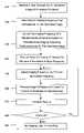
- FIG. 1is a flowchart of a method of controlling an ultrasound imaging system according to an embodiment of the present invention.
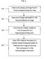
- FIG. 2is a flowchart of a method of compensating for signal attenuation in an ultrasound imaging system according to an embodiment of the present invention.
- FIG. 3is a flowchart of a method of auto-scanning a plurality of imaging frequencies in an ultrasound imaging system according to an embodiment of the present invention.
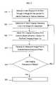
- FIG. 4is a flowchart of a method of optimizing an image in an ultrasound imaging system according to an embodiment of the present invention.
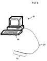
- FIG. 5is a block diagram of an exemplary ultrasound imaging system usable with various embodiments of the present invention.
- the exemplary ultrasound imaging systemincludes a workstation 30 , having an interface 35 (e.g., a keyboard, mouse, touchscreen display, etc.), a controller, and a display.
- the workstation 30is coupled to an ultrasound probe 10 via a percutaneous catheter 20 .
- the controllermay be a computer, such as a personal computer, an internal microprocessor, or an application specific integrated circuit (ASIC) operating software that causes the controller to perform the control functions described herein.
- ASICapplication specific integrated circuit
- the controlleris preferably programmable so as to perform various processes and method steps described in greater detail below.
- Other configurationsare also contemplated, and the system may or may not include further components as would be readily apparent to one of ordinary skill in the art after reading this disclosure.
- the ultrasound imaging systemreceives a user request for a desired imaging depth.
- a usermay: (1) enter a desired imaging depth (e.g., 5 cm) into a keyboard type interface, such as or including keys, buttons, toggle switches, rotary knobs, or various keypads, for example; (2) select an increase or decrease (i.e., a change) in a present imaging depth on a touchscreen display type interface; (3) issue a voice command to increase or decrease the present imaging depth interpreted by a voice recognition type interface; (4) select one of a list of possible imaging depths listed on a display using a mouse type interface; or (5) select any imaged feature or position on a video display showing the real-time image received from the ultrasound imaging system, such as by touching a touch-screen display or using a pixel-detecting pen coupled to the system to indicate the depth (and/or feature) for
- Step 120may include processes such as retrieving a corresponding frequency from an electronic lookup table or database based upon the desired depth, or calculating a corresponding frequency using any one of a number of algorithms as would be readily apparent to one of ordinary skill in the art after reading this disclosure.
- the attenuations of sound in various tissues, including in blood, as a function of frequencyhave been measured and therefore are known and can be reduced to a look up table.
- the measured attenuation of sound in bloodmay be transformed into a computational algorithm, such as by a curve fit, that the imaging system can perform using the indicated imaging depth as an input.
- the imaging depth of an ultrasound imager due to attenuation of sound in bloodis an approximately linear gradient (classically expressed in dB/cm/MHz), such that the imaging depth at 4.5 MHz is about 11 cm and the imaging depth at 7.0 MHz is about 6 cm.
- Step 120may be based on assumed physical properties or may include measuring physical properties of the fluids and/or tissues between the ultrasound sensor and the desired depth using data obtained from the ultrasound sensor or other sensors.
- the look up table or algorithmmay include attenuation effects of tissues, such as heart muscle or connective tissue in addition to blood.
- the medical imaging systemthen adjusts the imaging frequency of the system to the imaging frequency determined in step 120 .
- the medical imaging systemautomatically adjusts the imaging frequency in accordance with manual adjustment of the desired imaging depth.
- the medical imaging systemcan optimize the imaging frequency to match the desired imaging depth without requiring any further user interaction than the request for the desired imaging depth. As such, user interaction is minimized and the most optimized image is generated.
- the imaging systemmay also automatically adjust the time-gain compensation (up or down) in conjunction with adjusting the imaging frequency.
- the time-gain compensationalso compensates for the attenuation of sound by blood or tissues. Since the time at which an echo signal is received is directly related to the distance the associated sound traveled (i.e., to and from the echoing structure), sound attenuation can be compensated for by amplifying the echo signals by an increasing amount based upon the time after the transmission pulse that the echo signal is received.
- the time-gain compensationmay be adjusted according to an algorithm (such as a linear adjustment). However, applying too high a level of amplification in order to capture distant echo signals may result in greater noise in the image. Also, the appropriate time-gain may vary with imaging frequency. In this embodiment, when the imaging system receives a request for a new imaging depth, the system automatically adjusts the time-gain compensation to provide appropriate signal gain at the approximate time echo signals from that distance will be received at the ultrasound transducers.
- the catheter-based ultrasound probeincludes an array of ultrasound transducers for generating ultrasound pulse(s), the array of ultrasound transducers, such that the system has an imaging frequency range of about 2 MHz to about 20 MHz.
- the systemis adjustable from about 2 MHz to about 20 MHz in about 0.5 MHz intervals.
- the medical imaging systemmay adjust the imaging frequency to an imaging frequency selected from the group consisting essentially of 2.0 MHz, 2.5 MHz, 3.0 MHz . . . 19.0 MHz, 19.5 MHz, 20.0 MHz.
- the increment of imaging frequencyis more or less than about 0.5 MHz intervals.
- the increment of imaging frequencyis about 0.1 MHz.
- the medical imaging systemmay further be provided with signal attenuation compensation capabilities.
- Signal attenuationgenerally refers to a reduction in signal quality, which may be caused by ultrasound pulses passing through different body tissues, structures, and fluids, such as for example calcification layers that absorb or scatter ultrasound energy. The resulting reduction in reflected ultrasound energy may result in degradation of the received echo signal quality.
- image signal qualitymay be compensated for by increasing or decreasing the imaging frequency from the frequency that typically provides an optimized image of a given imaging depth.
- the imaging frequency determined in step 120may be further adjusted upon or after being implemented in step 130 to compensate for signal attenuation.
- an ultrasound imageis received from the catheter-based ultrasound probe in step 210 .
- the medical imaging systemdetermines a signal attenuation in the received ultrasound image.
- the systemmay compare the measured received echo energy to the energy that would be expected if the attenuation matched expected values for the imaging depth. If the received energy is less than the expected energy, attenuation over the path length may be greater than the prediction or assumption. Similarly, if the received energy exceeds the expected energy, the actual attenuation may be less than the prediction or assumption. Attenuation may also be calculated using other methods, including an electronic table look up using the received or measured path length as an input, or an algorithm, such as a linear gradient, using the received or measured path length as an independent variable.
- the medical imaging systemthen automatically determines an imaging frequency that corresponds to a reduced signal attenuation in step 230 .
- the determined imaging frequencymay be an imaging frequency one (or more) increments (e.g., about 0.5 MHz or about 0.1 MHz, according to various embodiments) below (i.e., a lower frequency) that determined in step 120 . If measured signal attenuation is less than expected, the imaging frequency may be increased in order to provide finer resolution of features at the selected imaging depth.
- the medical imaging systemthen adjusts the imaging frequency of the system in step 240 to the determined imaging frequency that corresponds to the measured signal attenuation.
- the medical imaging systemmay then verify that the change has improved the signal attenuation condition by re-running steps 220 , 230 , 240 . In this manner, the medical imaging system may automatically re-adjust for signal attenuation with minimal user interaction required.
- the medical imaging systemmay be provided with a frequency scanning capability. More specifically, in step 310 the medical imaging system receives a user request for a scan through a range of frequencies to identify features at various distances, features including any number of viewable structures such as tissue masses, anomalies, etc. In step 320 the medical imaging system determines a “next” imaging frequency from the range of available frequencies. By way of example, if the scan is operating at its first cycle, the “next” imaging frequency may be the first available imaging frequency (e.g., 2.0 MHz for the medical imaging system previously discussed or the current imaging frequency+/ ⁇ the delta frequency). The medical imaging system then adjusts the imaging frequency in step 330 to the frequency determined in step 320 .
- the “next” imaging frequencymay be the first available imaging frequency (e.g., 2.0 MHz for the medical imaging system previously discussed or the current imaging frequency+/ ⁇ the delta frequency).
- step 340After an ultrasound image from the catheter-based ultrasound probe is received in step 340 , the medical imaging system then determines whether the frequency determined in step 320 is the last imaging frequency in the range of available frequencies. If not, the medical imaging system re-runs step 320 , else the process ends in step 360 . Alternatively, steps 310 through 350 may be performed until stopped by the operator.
- the medical imaging systemmay be provided with an optimization feature as shown in FIG. 4 .
- the medical imaging systemreceives a user request for an optimized image of a feature of interest, indicated by the point of focus set by the user, or assumed by default to be at 75% of the imaging depth.
- a usermay desire an optimized image of one of the features discovered during the scan and presented on the ultrasound image display.
- an “auto-recognize” featuremay be provided that automatically recognizes features and initiates the request received in step 410 .
- the medical imaging system in step 420determines an imaging frequency that corresponds to the optimized image based on the measured depth to the feature.
- the determination of an imaging frequencymay assume physical properties for intervening tissues, including in an embodiment, assuming properties based upon detected intervening structures and fluids and measured parameters (e.g., continuity of imaged tissue, received echo signal strength, etc.).
- the medical imaging systemmay select 4.0 MHz in step 420 .
- step 430the medical imaging system then adjusts the imaging frequency to the frequency determined in step 420 . This may be followed by a confirmation step that queries the user whether the image has been sufficiently optimized. If the user responds that further optimization is required, then the process shown in FIG. 4 may repeat (even with a smaller delta frequency).
- the medical imaging systemmay include an image recognition and processing capability that assists in the optimization process.
- the image processing capabilitydetermines a measure of the image quality of the feature selected for imaging, such as by calculating a measure of resolution by measuring the definition of a boundary.
- the image processing capabilitymay determine the range over which echoes from a surface are received along a vector, which may be combined with statistical measures of the changes in intensity along the vector in the vicinity of the structure.
- the medical imaging systemadjusts the imaging frequency of the catheter-based ultrasound probe in step 450 to a higher or lower frequency (F 1 ) and obtains another image.
- the frequencymay be adjusted up or down as determined by the imaging processing system as necessary to determine if optimum image quality (e.g., resolution) is achieved.
- optimum image qualitye.g., resolution
- the subsequent discussionassumes step 450 increases frequency the first time through the process (default), but the process may be implemented by decreasing the frequency the first time through.
- the second imagemay need to be taken (timed or triggered) so as to correspond to a similar configuration as in the first image so that image quality measurements can be compared.
- the first and second imagesare timed or initiated based upon an input (e.g., an ECG signal) to occur at the same point in the cardiac cycle.
- step 460the image obtained at the new frequency F 1 is processed to determine a measure of the image quality (e.g., resolution) of the feature selected for imaging. Then, in step 470 , the two measures of resolution for images taken at F 0 and F 1 are compared to determine if the image quality (e.g., resolution) is improved or degraded as a result of the change in imaging frequency.
- a measure of the image qualitye.g., resolution
- step 480the medical imaging system determines whether to further adjust the frequency or whether an optimum frequency was obtained. If there is an improvement in image quality (e.g., resolution) when the imaging frequency is increased from F 0 to F 1 , then the process returns to step 450 , sequentially increasing (or decreasing) the frequency to new frequency F i and comparing the resulting image quality (e.g., resolution) measurements. Steps 450 through 480 are repeated until the system determines there is no change or a degradation in image quality (e.g., resolution) when frequency is increased from F i-1 to F i . When that determination is made, the medical imaging system sets the imaging frequency to the frequency that provided the best measure of image quality (e.g., F i-1 ) in step 490 .
- image qualitye.g., resolution
- step 480the medical imaging system determines that the optimum frequency may be lower than the initial frequency (F 0 ). In that case, the process returns to step 450 where the imaging frequency is decreased to F 1 . Then steps 460 through 480 are performed to determine if lowering the imaging frequency improved the image quality (e.g., resolution) of the desired feature. Steps 450 through 480 are repeated until the system determines there is no change or a degradation in image quality when frequency is decreased from F i-1 to F i . When that determination is made, the medical imaging system sets the imaging frequency to the frequency that provided the best measure of image quality (e.g., F i-1 ) in step 490 .
- the best measure of image qualitye.g., F i-1
- a medical imaging systemmay merge or correlate ultrasound images with other medical information, including concurrent instrumentation data, such as electrocardiogram (ECG) data.
- ECGelectrocardiogram
- intracardiac electrophysiology cathetersmay also be present in the heart.
- displaying ECG data(such as a trace moving across the screen) on the same monitor that displays ultrasound images would aid the physician.
- Such ECG datamay be correlated to the ultrasound images so the current ECG trace(s) is displayed along with the current ultrasound image.
- the ECG datamay be further correlated to the image so the display shows only the ECG trace of the ECG catheter that is presently imaged by imaging system.
Landscapes
- Engineering & Computer Science (AREA)
- Physics & Mathematics (AREA)
- Radar, Positioning & Navigation (AREA)
- Remote Sensing (AREA)
- Health & Medical Sciences (AREA)
- Life Sciences & Earth Sciences (AREA)
- Computer Networks & Wireless Communication (AREA)
- General Physics & Mathematics (AREA)
- Acoustics & Sound (AREA)
- Nuclear Medicine, Radiotherapy & Molecular Imaging (AREA)
- Molecular Biology (AREA)
- Pathology (AREA)
- Radiology & Medical Imaging (AREA)
- Biomedical Technology (AREA)
- Heart & Thoracic Surgery (AREA)
- Medical Informatics (AREA)
- Biophysics (AREA)
- Surgery (AREA)
- Animal Behavior & Ethology (AREA)
- General Health & Medical Sciences (AREA)
- Public Health (AREA)
- Veterinary Medicine (AREA)
- Ultra Sonic Daignosis Equipment (AREA)
Abstract
Description
Claims (22)
Priority Applications (1)
| Application Number | Priority Date | Filing Date | Title |
|---|---|---|---|
| US10/827,520US7654958B2 (en) | 2004-04-20 | 2004-04-20 | Method and apparatus for ultrasound imaging with autofrequency selection |
Applications Claiming Priority (1)
| Application Number | Priority Date | Filing Date | Title |
|---|---|---|---|
| US10/827,520US7654958B2 (en) | 2004-04-20 | 2004-04-20 | Method and apparatus for ultrasound imaging with autofrequency selection |
Publications (2)
| Publication Number | Publication Date |
|---|---|
| US20050240103A1 US20050240103A1 (en) | 2005-10-27 |
| US7654958B2true US7654958B2 (en) | 2010-02-02 |
Family
ID=35137426
Family Applications (1)
| Application Number | Title | Priority Date | Filing Date |
|---|---|---|---|
| US10/827,520Active2027-06-07US7654958B2 (en) | 2004-04-20 | 2004-04-20 | Method and apparatus for ultrasound imaging with autofrequency selection |
Country Status (1)
| Country | Link |
|---|---|
| US (1) | US7654958B2 (en) |
Cited By (5)
| Publication number | Priority date | Publication date | Assignee | Title |
|---|---|---|---|---|
| US20090069682A1 (en)* | 2007-01-24 | 2009-03-12 | Hastings Harold M | Simplified controls for implementing depth-based gain control in ultrasound systems |
| US20100234730A1 (en)* | 2006-03-31 | 2010-09-16 | National University Corporation Kyoto Institute Of Technology | Image processing device, ultrasonic imaging apparatus including the same, and image processing method |
| US20110293152A1 (en)* | 2010-06-01 | 2011-12-01 | Samsung Medison Co., Ltd. | Medical imaging system and image processing method |
| US9152834B2 (en) | 2012-08-09 | 2015-10-06 | Symbol Technologies, Llc | Image capture based on scanning resolution setting compared to determined scanning resolution relative to target distance in barcode reading |
| US20230148382A1 (en)* | 2020-04-01 | 2023-05-11 | Koninklijke Philips N.V. | Focus optimization for prediction in multi-frequency ultrasound imaging |
Families Citing this family (26)
| Publication number | Priority date | Publication date | Assignee | Title |
|---|---|---|---|---|
| JP2003513691A (en) | 1999-10-25 | 2003-04-15 | シーラス、コーポレイション | Use of focused ultrasound to seal blood vessels |
| US6626855B1 (en) | 1999-11-26 | 2003-09-30 | Therus Corpoation | Controlled high efficiency lesion formation using high intensity ultrasound |
| US20070213616A1 (en) | 2005-10-20 | 2007-09-13 | Thomas Anderson | Systems and methods for arteriotomy localization |
| CN101330875A (en)* | 2005-12-19 | 2008-12-24 | 皇家飞利浦电子股份有限公司 | Automatic ultrasound scanning initiated by protocol stage |
| EP2138099A3 (en)* | 2008-06-25 | 2010-01-06 | FUJIFILM Corporation | Ultrasound observation device and method for controlling operation thereof |
| KR101139123B1 (en)* | 2008-07-10 | 2012-04-30 | 삼성메디슨 주식회사 | Ultrasound apparatus and method for controlling image depth |
| US8295912B2 (en) | 2009-10-12 | 2012-10-23 | Kona Medical, Inc. | Method and system to inhibit a function of a nerve traveling with an artery |
| US9119951B2 (en) | 2009-10-12 | 2015-09-01 | Kona Medical, Inc. | Energetic modulation of nerves |
| US11998266B2 (en) | 2009-10-12 | 2024-06-04 | Otsuka Medical Devices Co., Ltd | Intravascular energy delivery |
| US8986211B2 (en) | 2009-10-12 | 2015-03-24 | Kona Medical, Inc. | Energetic modulation of nerves |
| US20160059044A1 (en) | 2009-10-12 | 2016-03-03 | Kona Medical, Inc. | Energy delivery to intraparenchymal regions of the kidney to treat hypertension |
| US20110092880A1 (en) | 2009-10-12 | 2011-04-21 | Michael Gertner | Energetic modulation of nerves |
| US8517962B2 (en) | 2009-10-12 | 2013-08-27 | Kona Medical, Inc. | Energetic modulation of nerves |
| US8986231B2 (en) | 2009-10-12 | 2015-03-24 | Kona Medical, Inc. | Energetic modulation of nerves |
| US20110118600A1 (en) | 2009-11-16 | 2011-05-19 | Michael Gertner | External Autonomic Modulation |
| US8469904B2 (en) | 2009-10-12 | 2013-06-25 | Kona Medical, Inc. | Energetic modulation of nerves |
| US9174065B2 (en) | 2009-10-12 | 2015-11-03 | Kona Medical, Inc. | Energetic modulation of nerves |
| EP2519158A1 (en)* | 2009-12-29 | 2012-11-07 | Boston Scientific Scimed, Inc. | Systems and methods for multi-frequency imaging of patient tissue using intravascular ultrasound imaging systems |
| WO2011138783A1 (en)* | 2010-05-05 | 2011-11-10 | Technion Research & Development Foundation Ltd. | Method and system of manipulating bilayer membranes |
| EP2521593B1 (en)* | 2011-03-15 | 2015-12-09 | Kona Medical, Inc. | Energetic modulation of nerves |
| JP2013172791A (en)* | 2012-02-24 | 2013-09-05 | Sony Corp | Ultrasonic inspection apparatus, ultrasonic inspection method and program |
| US12357274B2 (en)* | 2013-12-20 | 2025-07-15 | Raghu Raghavan | Systems and methods for acquiring ultrasonic data |
| JP6351365B2 (en)* | 2014-05-14 | 2018-07-04 | キヤノン株式会社 | Photoacoustic apparatus, information processing method, program |
| US10925579B2 (en) | 2014-11-05 | 2021-02-23 | Otsuka Medical Devices Co., Ltd. | Systems and methods for real-time tracking of a target tissue using imaging before and during therapy delivery |
| WO2022011327A1 (en)* | 2020-07-10 | 2022-01-13 | Secondwave Systems, Inc. | Systems and methods for targeting an organ with ultrasound stimulation for treating inflammation |
| DE112021006546T5 (en)* | 2020-12-18 | 2023-11-16 | Koninklijke Philips N.V. | ULTRASONIC IMAGING USING ANATOMY-BASED ACOUSTIC SETTINGS |
Citations (113)
| Publication number | Priority date | Publication date | Assignee | Title |
|---|---|---|---|---|
| US4161121A (en) | 1976-04-05 | 1979-07-17 | Varian Associates, Inc. | Ultrasonic imaging system |
| US4241610A (en) | 1979-02-05 | 1980-12-30 | Varian Associates, Inc. | Ultrasonic imaging system utilizing dynamic and pseudo-dynamic focusing |
| US4442713A (en)* | 1982-03-09 | 1984-04-17 | Sri International | Frequency varied ultrasonic imaging array |
| US4462408A (en) | 1982-05-17 | 1984-07-31 | Advanced Technology Laboratories, Inc. | Ultrasonic endoscope having elongated array mounted in manner allowing it to remain flexible |
| US4519260A (en) | 1982-02-18 | 1985-05-28 | The Board Of Trustees Of The Leland Stanford Junior University | Ultrasonic transducers and applications thereof |
| US4534221A (en)* | 1982-09-27 | 1985-08-13 | Technicare Corporation | Ultrasonic diagnostic imaging systems for varying depths of field |
| US4576177A (en) | 1983-02-18 | 1986-03-18 | Webster Wilton W Jr | Catheter for removing arteriosclerotic plaque |
| US4605009A (en) | 1983-04-06 | 1986-08-12 | Universite Francois Rabelais | Ultrasonic sweep echography and display endoscopic probe |
| US4841977A (en) | 1987-05-26 | 1989-06-27 | Inter Therapy, Inc. | Ultra-thin acoustic transducer and balloon catheter using same in imaging array subassembly |
| US4890268A (en) | 1988-12-27 | 1989-12-26 | General Electric Company | Two-dimensional phased array of ultrasonic transducers |
| US4917097A (en) | 1987-10-27 | 1990-04-17 | Endosonics Corporation | Apparatus and method for imaging small cavities |
| US4951677A (en) | 1988-03-21 | 1990-08-28 | Prutech Research And Development Partnership Ii | Acoustic imaging catheter and the like |
| US5002059A (en) | 1989-07-26 | 1991-03-26 | Boston Scientific Corporation | Tip filled ultrasound catheter |
| US5090956A (en) | 1983-10-31 | 1992-02-25 | Catheter Research, Inc. | Catheter with memory element-controlled steering |
| US5105819A (en) | 1988-09-01 | 1992-04-21 | Kon-Tron Elektronik AG | Ultrasound endoscope device |
| US5152294A (en) | 1989-12-14 | 1992-10-06 | Aloka Co., Ltd. | Three-dimensional ultrasonic scanner |
| US5158087A (en) | 1992-01-31 | 1992-10-27 | Hewlett-Packard Company | Differential temperature measurement for ultrasound transducer thermal control |
| US5170793A (en) | 1990-02-07 | 1992-12-15 | Kabushiki Kaisha Toshiba | Ultrasonic probe |
| US5195968A (en) | 1990-02-02 | 1993-03-23 | Ingemar Lundquist | Catheter steering mechanism |
| US5254088A (en) | 1990-02-02 | 1993-10-19 | Ep Technologies, Inc. | Catheter steering mechanism |
| US5279559A (en) | 1992-03-06 | 1994-01-18 | Aai Corporation | Remote steering system for medical catheter |
| US5301674A (en)* | 1992-03-27 | 1994-04-12 | Diasonics, Inc. | Method and apparatus for focusing transmission and reception of ultrasonic beams |
| US5307816A (en) | 1991-08-21 | 1994-05-03 | Kabushiki Kaisha Toshiba | Thrombus resolving treatment apparatus |
| US5309914A (en) | 1991-04-17 | 1994-05-10 | Kabushiki Kaisha Toshiba | Ultrasonic imaging apparatus |
| US5325860A (en) | 1991-11-08 | 1994-07-05 | Mayo Foundation For Medical Education And Research | Ultrasonic and interventional catheter and method |
| US5345938A (en) | 1991-09-30 | 1994-09-13 | Kabushiki Kaisha Toshiba | Diagnostic apparatus for circulatory systems |
| US5357550A (en) | 1991-09-09 | 1994-10-18 | Kabushiki Kaisha Toshiba | Apparatus for diagnosing vascular systems in organism |
| US5358478A (en) | 1990-02-02 | 1994-10-25 | Ep Technologies, Inc. | Catheter steering assembly providing asymmetric left and right curve configurations |
| US5361767A (en)* | 1993-01-25 | 1994-11-08 | Igor Yukov | Tissue characterization method and apparatus |
| US5364351A (en) | 1992-11-13 | 1994-11-15 | Ep Technologies, Inc. | Catheter steering mechanism |
| US5372138A (en) | 1988-03-21 | 1994-12-13 | Boston Scientific Corporation | Acousting imaging catheters and the like |
| US5385148A (en) | 1993-07-30 | 1995-01-31 | The Regents Of The University Of California | Cardiac imaging and ablation catheter |
| US5438997A (en) | 1991-03-13 | 1995-08-08 | Sieben; Wayne | Intravascular imaging apparatus and methods for use and manufacture |
| US5456258A (en) | 1993-12-20 | 1995-10-10 | Fuji Photo Optical Co., Ltd. | Catheter type ultrasound probe |
| US5470350A (en) | 1993-04-02 | 1995-11-28 | Siemens Aktiengesellschaft | System for the treatment of pathological tissue having a catheter with a pressure sensor |
| US5499630A (en) | 1993-11-22 | 1996-03-19 | Kabushiki Kaisha Toshiba | Catheter type ultrasound probe |
| US5515853A (en) | 1995-03-28 | 1996-05-14 | Sonometrics Corporation | Three-dimensional digital ultrasound tracking system |
| US5515856A (en) | 1994-08-30 | 1996-05-14 | Vingmed Sound A/S | Method for generating anatomical M-mode displays |
| US5560362A (en) | 1994-06-13 | 1996-10-01 | Acuson Corporation | Active thermal control of ultrasound transducers |
| US5588432A (en) | 1988-03-21 | 1996-12-31 | Boston Scientific Corporation | Catheters for imaging, sensing electrical potentials, and ablating tissue |
| US5622174A (en) | 1992-10-02 | 1997-04-22 | Kabushiki Kaisha Toshiba | Ultrasonic diagnosis apparatus and image displaying system |
| US5662116A (en) | 1995-09-12 | 1997-09-02 | Fuji Photo Optical Co., Ltd. | Multi-plane electronic scan ultrasound probe |
| US5697965A (en) | 1996-04-01 | 1997-12-16 | Procath Corporation | Method of making an atrial defibrillation catheter |
| US5699805A (en) | 1996-06-20 | 1997-12-23 | Mayo Foundation For Medical Education And Research | Longitudinal multiplane ultrasound transducer underfluid catheter system |
| US5704361A (en) | 1991-11-08 | 1998-01-06 | Mayo Foundation For Medical Education And Research | Volumetric image ultrasound transducer underfluid catheter system |
| US5713363A (en) | 1991-11-08 | 1998-02-03 | Mayo Foundation For Medical Education And Research | Ultrasound catheter and method for imaging and hemodynamic monitoring |
| US5715817A (en) | 1993-06-29 | 1998-02-10 | C.R. Bard, Inc. | Bidirectional steering catheter |
| US5722403A (en) | 1996-10-28 | 1998-03-03 | Ep Technologies, Inc. | Systems and methods using a porous electrode for ablating and visualizing interior tissue regions |
| US5749364A (en) | 1996-06-21 | 1998-05-12 | Acuson Corporation | Method and apparatus for mapping pressure and tissue properties |
| US5788636A (en) | 1997-02-25 | 1998-08-04 | Acuson Corporation | Method and system for forming an ultrasound image of a tissue while simultaneously ablating the tissue |
| US5795299A (en) | 1997-01-31 | 1998-08-18 | Acuson Corporation | Ultrasonic transducer assembly with extended flexible circuits |
| US5797848A (en) | 1997-01-31 | 1998-08-25 | Acuson Corporation | Ultrasonic transducer assembly with improved electrical interface |
| US5800356A (en) | 1997-05-29 | 1998-09-01 | Advanced Technology Laboratories, Inc. | Ultrasonic diagnostic imaging system with doppler assisted tracking of tissue motion |
| US5807324A (en) | 1996-04-01 | 1998-09-15 | Procath Corporation | Steerable catheter |
| US5846205A (en) | 1997-01-31 | 1998-12-08 | Acuson Corporation | Catheter-mounted, phased-array ultrasound transducer with improved imaging |
| US5888577A (en) | 1997-06-30 | 1999-03-30 | Procath Corporation | Method for forming an electrophysiology catheter |
| US5891088A (en) | 1990-02-02 | 1999-04-06 | Ep Technologies, Inc. | Catheter steering assembly providing asymmetric left and right curve configurations |
| US5906579A (en) | 1996-08-16 | 1999-05-25 | Smith & Nephew Endoscopy, Inc. | Through-wall catheter steering and positioning |
| US5916168A (en) | 1997-05-29 | 1999-06-29 | Advanced Technology Laboratories, Inc. | Three dimensional M-mode ultrasonic diagnostic imaging system |
| US5921978A (en) | 1997-06-20 | 1999-07-13 | Ep Technologies, Inc. | Catheter tip steering plane marker |
| US5928276A (en) | 1998-06-11 | 1999-07-27 | Griffin, Iii; Joseph C. | Combined cable and electrophysiology catheters |
| US5931863A (en) | 1997-12-22 | 1999-08-03 | Procath Corporation | Electrophysiology catheter |
| US5935102A (en) | 1993-05-14 | 1999-08-10 | C. R. Bard | Steerable electrode catheter |
| US5938616A (en) | 1997-01-31 | 1999-08-17 | Acuson Corporation | Steering mechanism and steering line for a catheter-mounted ultrasonic transducer |
| US5954654A (en) | 1997-01-31 | 1999-09-21 | Acuson Corporation | Steering mechanism and steering line for a catheter-mounted ultrasonic transducer |
| US6013072A (en) | 1997-07-09 | 2000-01-11 | Intraluminal Therapeutics, Inc. | Systems and methods for steering a catheter through body tissue |
| US6033378A (en) | 1990-02-02 | 2000-03-07 | Ep Technologies, Inc. | Catheter steering mechanism |
| US6095976A (en)* | 1997-06-19 | 2000-08-01 | Medinol Ltd. | Method for enhancing an image derived from reflected ultrasound signals produced by an ultrasound transmitter and detector inserted in a bodily lumen |
| US6144870A (en) | 1996-10-21 | 2000-11-07 | Procath Corporation | Catheter with improved electrodes and method of fabrication |
| US6171248B1 (en) | 1997-02-27 | 2001-01-09 | Acuson Corporation | Ultrasonic probe, system and method for two-dimensional imaging or three-dimensional reconstruction |
| US6190353B1 (en) | 1995-10-13 | 2001-02-20 | Transvascular, Inc. | Methods and apparatus for bypassing arterial obstructions and/or performing other transvascular procedures |
| US6210333B1 (en) | 1999-10-12 | 2001-04-03 | Acuson Corporation | Medical diagnostic ultrasound system and method for automated triggered intervals |
| US6224556B1 (en) | 1998-11-25 | 2001-05-01 | Acuson Corporation | Diagnostic medical ultrasound system and method for using a sparse array |
| US6228028B1 (en) | 1996-11-07 | 2001-05-08 | Tomtec Imaging Systems Gmbh | Method and apparatus for ultrasound image reconstruction |
| US6261246B1 (en) | 1997-09-29 | 2001-07-17 | Scimed Life Systems, Inc. | Intravascular imaging guidewire |
| US20010020126A1 (en)* | 1996-10-28 | 2001-09-06 | David K. Swanson | Systems and methods for visualizing tissue during diagnostic or therapeutic procedures |
| US6293943B1 (en) | 1995-06-07 | 2001-09-25 | Ep Technologies, Inc. | Tissue heating and ablation systems and methods which predict maximum tissue temperature |
| US6306097B1 (en) | 1999-06-17 | 2001-10-23 | Acuson Corporation | Ultrasound imaging catheter guiding assembly with catheter working port |
| US6310828B1 (en) | 1997-07-18 | 2001-10-30 | Tomtec Imaging Systems Gmbh | Method and device for sensing ultrasound images |
| US6322507B1 (en)* | 1998-10-26 | 2001-11-27 | Medson Ltd. | Ultrasonic apparatus and method for evaluation of bone tissue |
| US6358208B1 (en)* | 1998-11-21 | 2002-03-19 | Philipp Lang | Assessment of cardiovascular performance using ultrasound methods and devices that interrogate interstitial fluid |
| US6360027B1 (en) | 1996-02-29 | 2002-03-19 | Acuson Corporation | Multiple ultrasound image registration system, method and transducer |
| US6368275B1 (en) | 1999-10-07 | 2002-04-09 | Acuson Corporation | Method and apparatus for diagnostic medical information gathering, hyperthermia treatment, or directed gene therapy |
| US6385489B1 (en) | 1998-09-25 | 2002-05-07 | Ep Medsystems, Inc. | Triple array defibrillation catheter and method of using the same |
| US6398731B1 (en) | 1997-07-25 | 2002-06-04 | Tomtec Imaging Systems Gmbh | Method for recording ultrasound images of moving objects |
| US6423002B1 (en) | 1999-06-24 | 2002-07-23 | Acuson Corporation | Intra-operative diagnostic ultrasound multiple-array transducer probe and optional surgical tool |
| US6440488B2 (en) | 1999-12-03 | 2002-08-27 | Ep Medsystems, Inc. | Flexible electrode catheter and process for manufacturing the same |
| US6443894B1 (en) | 1999-09-29 | 2002-09-03 | Acuson Corporation | Medical diagnostic ultrasound system and method for mapping surface data for three dimensional imaging |
| US6475148B1 (en) | 2000-10-25 | 2002-11-05 | Acuson Corporation | Medical diagnostic ultrasound-aided drug delivery system and method |
| US6475149B1 (en) | 2001-09-21 | 2002-11-05 | Acuson Corporation | Border detection method and system |
| US6482161B1 (en) | 2000-06-29 | 2002-11-19 | Acuson Corporation | Medical diagnostic ultrasound system and method for vessel structure analysis |
| US6491633B1 (en) | 2000-03-10 | 2002-12-10 | Acuson Corporation | Medical diagnostic ultrasound system and method for contrast agent image beamformation |
| US6503202B1 (en) | 2000-06-29 | 2003-01-07 | Acuson Corp. | Medical diagnostic ultrasound system and method for flow analysis |
| US6517488B1 (en) | 2000-06-29 | 2003-02-11 | Acuson Corporation | Medical diagnostic ultrasound system and method for identifying constrictions |
| US6527717B1 (en) | 2000-03-10 | 2003-03-04 | Acuson Corporation | Tissue motion analysis medical diagnostic ultrasound system and method |
| US20030045796A1 (en) | 2001-08-31 | 2003-03-06 | Friedman Zvi M. | Ultrasonic monitoring system and method |
| US6532378B2 (en) | 2000-01-14 | 2003-03-11 | Ep Medsystems, Inc. | Pulmonary artery catheter for left and right atrial recording |
| US6554770B1 (en) | 1998-11-20 | 2003-04-29 | Acuson Corporation | Medical diagnostic ultrasound imaging methods for extended field of view |
| US6589182B1 (en) | 2001-02-12 | 2003-07-08 | Acuson Corporation | Medical diagnostic ultrasound catheter with first and second tip portions |
| US6605043B1 (en) | 1998-11-19 | 2003-08-12 | Acuson Corp. | Diagnostic medical ultrasound systems and transducers utilizing micro-mechanical components |
| US6607528B1 (en) | 1999-06-22 | 2003-08-19 | Senorx, Inc. | Shapeable electrosurgical scalpel |
| US6607488B1 (en) | 2000-03-02 | 2003-08-19 | Acuson Corporation | Medical diagnostic ultrasound system and method for scanning plane orientation |
| US6612992B1 (en) | 2000-03-02 | 2003-09-02 | Acuson Corp | Medical diagnostic ultrasound catheter and method for position determination |
| US6645147B1 (en) | 1998-11-25 | 2003-11-11 | Acuson Corporation | Diagnostic medical ultrasound image and system for contrast agent imaging |
| US6648875B2 (en) | 2001-05-04 | 2003-11-18 | Cardiac Pacemakers, Inc. | Means for maintaining tension on a steering tendon in a steerable catheter |
| US20040039286A1 (en)* | 2002-08-26 | 2004-02-26 | The Cleveland Clinic Foundation | System and method of aquiring blood-vessel data |
| US6709396B2 (en) | 2002-07-17 | 2004-03-23 | Vermon | Ultrasound array transducer for catheter use |
| US20040097805A1 (en) | 2002-11-19 | 2004-05-20 | Laurent Verard | Navigation system for cardiac therapies |
| US20040249282A1 (en) | 2003-06-09 | 2004-12-09 | Bjorn Olstad | System and method for extracting information based on ultrasound-located landmarks |
| US6908434B1 (en) | 2002-01-16 | 2005-06-21 | Ep Medsystems, Inc. | Ultrasound imaging catheter isolation system with temperature sensor |
| US6923768B2 (en) | 2002-03-11 | 2005-08-02 | Siemens Aktiengesellschaft | Method and apparatus for acquiring and displaying a medical instrument introduced into a cavity organ of a patient to be examined or treated |
| US20050203390A1 (en) | 1999-08-23 | 2005-09-15 | Hans Torp | Method and apparatus for providing real-time calculation and display of tissue deformation in ultrasound imaging |
| US7396332B2 (en)* | 2002-06-10 | 2008-07-08 | Scimed Life Systems, Inc. | Transducer with multiple resonant frequencies for an imaging catheter |
- 2004
- 2004-04-20USUS10/827,520patent/US7654958B2/enactiveActive
Patent Citations (126)
| Publication number | Priority date | Publication date | Assignee | Title |
|---|---|---|---|---|
| US4161121A (en) | 1976-04-05 | 1979-07-17 | Varian Associates, Inc. | Ultrasonic imaging system |
| US4241610A (en) | 1979-02-05 | 1980-12-30 | Varian Associates, Inc. | Ultrasonic imaging system utilizing dynamic and pseudo-dynamic focusing |
| US4519260A (en) | 1982-02-18 | 1985-05-28 | The Board Of Trustees Of The Leland Stanford Junior University | Ultrasonic transducers and applications thereof |
| US4442713A (en)* | 1982-03-09 | 1984-04-17 | Sri International | Frequency varied ultrasonic imaging array |
| US4462408A (en) | 1982-05-17 | 1984-07-31 | Advanced Technology Laboratories, Inc. | Ultrasonic endoscope having elongated array mounted in manner allowing it to remain flexible |
| US4534221A (en)* | 1982-09-27 | 1985-08-13 | Technicare Corporation | Ultrasonic diagnostic imaging systems for varying depths of field |
| US4576177A (en) | 1983-02-18 | 1986-03-18 | Webster Wilton W Jr | Catheter for removing arteriosclerotic plaque |
| US4605009A (en) | 1983-04-06 | 1986-08-12 | Universite Francois Rabelais | Ultrasonic sweep echography and display endoscopic probe |
| US5090956A (en) | 1983-10-31 | 1992-02-25 | Catheter Research, Inc. | Catheter with memory element-controlled steering |
| US4841977A (en) | 1987-05-26 | 1989-06-27 | Inter Therapy, Inc. | Ultra-thin acoustic transducer and balloon catheter using same in imaging array subassembly |
| US4917097A (en) | 1987-10-27 | 1990-04-17 | Endosonics Corporation | Apparatus and method for imaging small cavities |
| US4951677A (en) | 1988-03-21 | 1990-08-28 | Prutech Research And Development Partnership Ii | Acoustic imaging catheter and the like |
| US5588432A (en) | 1988-03-21 | 1996-12-31 | Boston Scientific Corporation | Catheters for imaging, sensing electrical potentials, and ablating tissue |
| US5372138A (en) | 1988-03-21 | 1994-12-13 | Boston Scientific Corporation | Acousting imaging catheters and the like |
| US5105819A (en) | 1988-09-01 | 1992-04-21 | Kon-Tron Elektronik AG | Ultrasound endoscope device |
| US4890268A (en) | 1988-12-27 | 1989-12-26 | General Electric Company | Two-dimensional phased array of ultrasonic transducers |
| US5002059A (en) | 1989-07-26 | 1991-03-26 | Boston Scientific Corporation | Tip filled ultrasound catheter |
| US5152294A (en) | 1989-12-14 | 1992-10-06 | Aloka Co., Ltd. | Three-dimensional ultrasonic scanner |
| US6033378A (en) | 1990-02-02 | 2000-03-07 | Ep Technologies, Inc. | Catheter steering mechanism |
| US5891088A (en) | 1990-02-02 | 1999-04-06 | Ep Technologies, Inc. | Catheter steering assembly providing asymmetric left and right curve configurations |
| US5531686A (en) | 1990-02-02 | 1996-07-02 | Ep Technologies, Inc. | Catheter steering mechanism |
| US6485455B1 (en) | 1990-02-02 | 2002-11-26 | Ep Technologies, Inc. | Catheter steering assembly providing asymmetric left and right curve configurations |
| US5395327A (en) | 1990-02-02 | 1995-03-07 | Ep Technologies, Inc. | Catheter steering mechanism |
| US5254088A (en) | 1990-02-02 | 1993-10-19 | Ep Technologies, Inc. | Catheter steering mechanism |
| US5195968A (en) | 1990-02-02 | 1993-03-23 | Ingemar Lundquist | Catheter steering mechanism |
| US5336182A (en) | 1990-02-02 | 1994-08-09 | Ep Technologies, Inc. | Catheter steering mechanism |
| US5358478A (en) | 1990-02-02 | 1994-10-25 | Ep Technologies, Inc. | Catheter steering assembly providing asymmetric left and right curve configurations |
| US5170793A (en) | 1990-02-07 | 1992-12-15 | Kabushiki Kaisha Toshiba | Ultrasonic probe |
| US5438997A (en) | 1991-03-13 | 1995-08-08 | Sieben; Wayne | Intravascular imaging apparatus and methods for use and manufacture |
| US5309914A (en) | 1991-04-17 | 1994-05-10 | Kabushiki Kaisha Toshiba | Ultrasonic imaging apparatus |
| US5307816A (en) | 1991-08-21 | 1994-05-03 | Kabushiki Kaisha Toshiba | Thrombus resolving treatment apparatus |
| US5357550A (en) | 1991-09-09 | 1994-10-18 | Kabushiki Kaisha Toshiba | Apparatus for diagnosing vascular systems in organism |
| US5345938A (en) | 1991-09-30 | 1994-09-13 | Kabushiki Kaisha Toshiba | Diagnostic apparatus for circulatory systems |
| US5713363A (en) | 1991-11-08 | 1998-02-03 | Mayo Foundation For Medical Education And Research | Ultrasound catheter and method for imaging and hemodynamic monitoring |
| US5345940A (en) | 1991-11-08 | 1994-09-13 | Mayo Foundation For Medical Education And Research | Transvascular ultrasound hemodynamic and interventional catheter and method |
| US6039693A (en) | 1991-11-08 | 2000-03-21 | Mayo Foundation For Medical Education And Research | Volumetric image ultrasound transducer underfluid catheter system |
| US6306096B1 (en) | 1991-11-08 | 2001-10-23 | Mayo Foundation For Medical Education And Research | Volumetric image ultrasound transducer underfluid catheter system |
| US5325860A (en) | 1991-11-08 | 1994-07-05 | Mayo Foundation For Medical Education And Research | Ultrasonic and interventional catheter and method |
| US5704361A (en) | 1991-11-08 | 1998-01-06 | Mayo Foundation For Medical Education And Research | Volumetric image ultrasound transducer underfluid catheter system |
| US5158087A (en) | 1992-01-31 | 1992-10-27 | Hewlett-Packard Company | Differential temperature measurement for ultrasound transducer thermal control |
| US5279559A (en) | 1992-03-06 | 1994-01-18 | Aai Corporation | Remote steering system for medical catheter |
| US5301674A (en)* | 1992-03-27 | 1994-04-12 | Diasonics, Inc. | Method and apparatus for focusing transmission and reception of ultrasonic beams |
| US5701897A (en) | 1992-10-02 | 1997-12-30 | Kabushiki Kaisha Toshiba | Ultrasonic diagnosis apparatus and image displaying system |
| US5622174A (en) | 1992-10-02 | 1997-04-22 | Kabushiki Kaisha Toshiba | Ultrasonic diagnosis apparatus and image displaying system |
| US5364351A (en) | 1992-11-13 | 1994-11-15 | Ep Technologies, Inc. | Catheter steering mechanism |
| US5456664A (en) | 1992-11-13 | 1995-10-10 | Ep Technologies, Inc. | Catheter steering mechanism |
| US5361767A (en)* | 1993-01-25 | 1994-11-08 | Igor Yukov | Tissue characterization method and apparatus |
| US5470350A (en) | 1993-04-02 | 1995-11-28 | Siemens Aktiengesellschaft | System for the treatment of pathological tissue having a catheter with a pressure sensor |
| US5935102A (en) | 1993-05-14 | 1999-08-10 | C. R. Bard | Steerable electrode catheter |
| US5715817A (en) | 1993-06-29 | 1998-02-10 | C.R. Bard, Inc. | Bidirectional steering catheter |
| US5385148A (en) | 1993-07-30 | 1995-01-31 | The Regents Of The University Of California | Cardiac imaging and ablation catheter |
| US5499630A (en) | 1993-11-22 | 1996-03-19 | Kabushiki Kaisha Toshiba | Catheter type ultrasound probe |
| US5456258A (en) | 1993-12-20 | 1995-10-10 | Fuji Photo Optical Co., Ltd. | Catheter type ultrasound probe |
| US5560362A (en) | 1994-06-13 | 1996-10-01 | Acuson Corporation | Active thermal control of ultrasound transducers |
| US5515856A (en) | 1994-08-30 | 1996-05-14 | Vingmed Sound A/S | Method for generating anatomical M-mode displays |
| US5515853A (en) | 1995-03-28 | 1996-05-14 | Sonometrics Corporation | Three-dimensional digital ultrasound tracking system |
| US6293943B1 (en) | 1995-06-07 | 2001-09-25 | Ep Technologies, Inc. | Tissue heating and ablation systems and methods which predict maximum tissue temperature |
| US5662116A (en) | 1995-09-12 | 1997-09-02 | Fuji Photo Optical Co., Ltd. | Multi-plane electronic scan ultrasound probe |
| US6190353B1 (en) | 1995-10-13 | 2001-02-20 | Transvascular, Inc. | Methods and apparatus for bypassing arterial obstructions and/or performing other transvascular procedures |
| US6360027B1 (en) | 1996-02-29 | 2002-03-19 | Acuson Corporation | Multiple ultrasound image registration system, method and transducer |
| US5807324A (en) | 1996-04-01 | 1998-09-15 | Procath Corporation | Steerable catheter |
| US5697965A (en) | 1996-04-01 | 1997-12-16 | Procath Corporation | Method of making an atrial defibrillation catheter |
| US5699805A (en) | 1996-06-20 | 1997-12-23 | Mayo Foundation For Medical Education And Research | Longitudinal multiplane ultrasound transducer underfluid catheter system |
| US5749364A (en) | 1996-06-21 | 1998-05-12 | Acuson Corporation | Method and apparatus for mapping pressure and tissue properties |
| US5906579A (en) | 1996-08-16 | 1999-05-25 | Smith & Nephew Endoscopy, Inc. | Through-wall catheter steering and positioning |
| US6144870A (en) | 1996-10-21 | 2000-11-07 | Procath Corporation | Catheter with improved electrodes and method of fabrication |
| US5722403A (en) | 1996-10-28 | 1998-03-03 | Ep Technologies, Inc. | Systems and methods using a porous electrode for ablating and visualizing interior tissue regions |
| US20010020126A1 (en)* | 1996-10-28 | 2001-09-06 | David K. Swanson | Systems and methods for visualizing tissue during diagnostic or therapeutic procedures |
| US6228028B1 (en) | 1996-11-07 | 2001-05-08 | Tomtec Imaging Systems Gmbh | Method and apparatus for ultrasound image reconstruction |
| US5795299A (en) | 1997-01-31 | 1998-08-18 | Acuson Corporation | Ultrasonic transducer assembly with extended flexible circuits |
| US5938616A (en) | 1997-01-31 | 1999-08-17 | Acuson Corporation | Steering mechanism and steering line for a catheter-mounted ultrasonic transducer |
| US5954654A (en) | 1997-01-31 | 1999-09-21 | Acuson Corporation | Steering mechanism and steering line for a catheter-mounted ultrasonic transducer |
| US5797848A (en) | 1997-01-31 | 1998-08-25 | Acuson Corporation | Ultrasonic transducer assembly with improved electrical interface |
| US5846205A (en) | 1997-01-31 | 1998-12-08 | Acuson Corporation | Catheter-mounted, phased-array ultrasound transducer with improved imaging |
| US6228032B1 (en) | 1997-01-31 | 2001-05-08 | Acuson Corporation | Steering mechanism and steering line for a catheter-mounted ultrasonic transducer |
| US5788636A (en) | 1997-02-25 | 1998-08-04 | Acuson Corporation | Method and system for forming an ultrasound image of a tissue while simultaneously ablating the tissue |
| US6171248B1 (en) | 1997-02-27 | 2001-01-09 | Acuson Corporation | Ultrasonic probe, system and method for two-dimensional imaging or three-dimensional reconstruction |
| US5800356A (en) | 1997-05-29 | 1998-09-01 | Advanced Technology Laboratories, Inc. | Ultrasonic diagnostic imaging system with doppler assisted tracking of tissue motion |
| US5916168A (en) | 1997-05-29 | 1999-06-29 | Advanced Technology Laboratories, Inc. | Three dimensional M-mode ultrasonic diagnostic imaging system |
| US6095976A (en)* | 1997-06-19 | 2000-08-01 | Medinol Ltd. | Method for enhancing an image derived from reflected ultrasound signals produced by an ultrasound transmitter and detector inserted in a bodily lumen |
| US5921978A (en) | 1997-06-20 | 1999-07-13 | Ep Technologies, Inc. | Catheter tip steering plane marker |
| US5888577A (en) | 1997-06-30 | 1999-03-30 | Procath Corporation | Method for forming an electrophysiology catheter |
| US6013072A (en) | 1997-07-09 | 2000-01-11 | Intraluminal Therapeutics, Inc. | Systems and methods for steering a catheter through body tissue |
| US6310828B1 (en) | 1997-07-18 | 2001-10-30 | Tomtec Imaging Systems Gmbh | Method and device for sensing ultrasound images |
| US6398731B1 (en) | 1997-07-25 | 2002-06-04 | Tomtec Imaging Systems Gmbh | Method for recording ultrasound images of moving objects |
| US6261246B1 (en) | 1997-09-29 | 2001-07-17 | Scimed Life Systems, Inc. | Intravascular imaging guidewire |
| US6173205B1 (en) | 1997-12-22 | 2001-01-09 | Procath Corporation | Electrophysiology catheter |
| US6085117A (en) | 1997-12-22 | 2000-07-04 | Procath Corporation | Method of defibrillating employing coronary sinus and external patch electrodes |
| US5931863A (en) | 1997-12-22 | 1999-08-03 | Procath Corporation | Electrophysiology catheter |
| US5928276A (en) | 1998-06-11 | 1999-07-27 | Griffin, Iii; Joseph C. | Combined cable and electrophysiology catheters |
| US6385489B1 (en) | 1998-09-25 | 2002-05-07 | Ep Medsystems, Inc. | Triple array defibrillation catheter and method of using the same |
| US6322507B1 (en)* | 1998-10-26 | 2001-11-27 | Medson Ltd. | Ultrasonic apparatus and method for evaluation of bone tissue |
| US6605043B1 (en) | 1998-11-19 | 2003-08-12 | Acuson Corp. | Diagnostic medical ultrasound systems and transducers utilizing micro-mechanical components |
| US6554770B1 (en) | 1998-11-20 | 2003-04-29 | Acuson Corporation | Medical diagnostic ultrasound imaging methods for extended field of view |
| US6358208B1 (en)* | 1998-11-21 | 2002-03-19 | Philipp Lang | Assessment of cardiovascular performance using ultrasound methods and devices that interrogate interstitial fluid |
| US6224556B1 (en) | 1998-11-25 | 2001-05-01 | Acuson Corporation | Diagnostic medical ultrasound system and method for using a sparse array |
| US6645147B1 (en) | 1998-11-25 | 2003-11-11 | Acuson Corporation | Diagnostic medical ultrasound image and system for contrast agent imaging |
| US6306097B1 (en) | 1999-06-17 | 2001-10-23 | Acuson Corporation | Ultrasound imaging catheter guiding assembly with catheter working port |
| US6607528B1 (en) | 1999-06-22 | 2003-08-19 | Senorx, Inc. | Shapeable electrosurgical scalpel |
| US6423002B1 (en) | 1999-06-24 | 2002-07-23 | Acuson Corporation | Intra-operative diagnostic ultrasound multiple-array transducer probe and optional surgical tool |
| US20050203390A1 (en) | 1999-08-23 | 2005-09-15 | Hans Torp | Method and apparatus for providing real-time calculation and display of tissue deformation in ultrasound imaging |
| US6443894B1 (en) | 1999-09-29 | 2002-09-03 | Acuson Corporation | Medical diagnostic ultrasound system and method for mapping surface data for three dimensional imaging |
| US6368275B1 (en) | 1999-10-07 | 2002-04-09 | Acuson Corporation | Method and apparatus for diagnostic medical information gathering, hyperthermia treatment, or directed gene therapy |
| US6210333B1 (en) | 1999-10-12 | 2001-04-03 | Acuson Corporation | Medical diagnostic ultrasound system and method for automated triggered intervals |
| US6440488B2 (en) | 1999-12-03 | 2002-08-27 | Ep Medsystems, Inc. | Flexible electrode catheter and process for manufacturing the same |
| US6532378B2 (en) | 2000-01-14 | 2003-03-11 | Ep Medsystems, Inc. | Pulmonary artery catheter for left and right atrial recording |
| US6607488B1 (en) | 2000-03-02 | 2003-08-19 | Acuson Corporation | Medical diagnostic ultrasound system and method for scanning plane orientation |
| US6612992B1 (en) | 2000-03-02 | 2003-09-02 | Acuson Corp | Medical diagnostic ultrasound catheter and method for position determination |
| US6491633B1 (en) | 2000-03-10 | 2002-12-10 | Acuson Corporation | Medical diagnostic ultrasound system and method for contrast agent image beamformation |
| US20030158483A1 (en) | 2000-03-10 | 2003-08-21 | Acuson Corporation | Tissue motion analysis medical diagnostic ultrasound system and method |
| US6527717B1 (en) | 2000-03-10 | 2003-03-04 | Acuson Corporation | Tissue motion analysis medical diagnostic ultrasound system and method |
| US6503202B1 (en) | 2000-06-29 | 2003-01-07 | Acuson Corp. | Medical diagnostic ultrasound system and method for flow analysis |
| US6517488B1 (en) | 2000-06-29 | 2003-02-11 | Acuson Corporation | Medical diagnostic ultrasound system and method for identifying constrictions |
| US6482161B1 (en) | 2000-06-29 | 2002-11-19 | Acuson Corporation | Medical diagnostic ultrasound system and method for vessel structure analysis |
| US6475148B1 (en) | 2000-10-25 | 2002-11-05 | Acuson Corporation | Medical diagnostic ultrasound-aided drug delivery system and method |
| US6589182B1 (en) | 2001-02-12 | 2003-07-08 | Acuson Corporation | Medical diagnostic ultrasound catheter with first and second tip portions |
| US6648875B2 (en) | 2001-05-04 | 2003-11-18 | Cardiac Pacemakers, Inc. | Means for maintaining tension on a steering tendon in a steerable catheter |
| US20030045796A1 (en) | 2001-08-31 | 2003-03-06 | Friedman Zvi M. | Ultrasonic monitoring system and method |
| US6475149B1 (en) | 2001-09-21 | 2002-11-05 | Acuson Corporation | Border detection method and system |
| US6908434B1 (en) | 2002-01-16 | 2005-06-21 | Ep Medsystems, Inc. | Ultrasound imaging catheter isolation system with temperature sensor |
| US6923768B2 (en) | 2002-03-11 | 2005-08-02 | Siemens Aktiengesellschaft | Method and apparatus for acquiring and displaying a medical instrument introduced into a cavity organ of a patient to be examined or treated |
| US7396332B2 (en)* | 2002-06-10 | 2008-07-08 | Scimed Life Systems, Inc. | Transducer with multiple resonant frequencies for an imaging catheter |
| US6709396B2 (en) | 2002-07-17 | 2004-03-23 | Vermon | Ultrasound array transducer for catheter use |
| US20040039286A1 (en)* | 2002-08-26 | 2004-02-26 | The Cleveland Clinic Foundation | System and method of aquiring blood-vessel data |
| US20040097805A1 (en) | 2002-11-19 | 2004-05-20 | Laurent Verard | Navigation system for cardiac therapies |
| US20040249282A1 (en) | 2003-06-09 | 2004-12-09 | Bjorn Olstad | System and method for extracting information based on ultrasound-located landmarks |
Non-Patent Citations (45)
Cited By (6)
| Publication number | Priority date | Publication date | Assignee | Title |
|---|---|---|---|---|
| US20100234730A1 (en)* | 2006-03-31 | 2010-09-16 | National University Corporation Kyoto Institute Of Technology | Image processing device, ultrasonic imaging apparatus including the same, and image processing method |
| US20090069682A1 (en)* | 2007-01-24 | 2009-03-12 | Hastings Harold M | Simplified controls for implementing depth-based gain control in ultrasound systems |
| US20110293152A1 (en)* | 2010-06-01 | 2011-12-01 | Samsung Medison Co., Ltd. | Medical imaging system and image processing method |
| US8509508B2 (en)* | 2010-06-01 | 2013-08-13 | Samsung Medison Co., Ltd. | Medical imaging system and image processing method |
| US9152834B2 (en) | 2012-08-09 | 2015-10-06 | Symbol Technologies, Llc | Image capture based on scanning resolution setting compared to determined scanning resolution relative to target distance in barcode reading |
| US20230148382A1 (en)* | 2020-04-01 | 2023-05-11 | Koninklijke Philips N.V. | Focus optimization for prediction in multi-frequency ultrasound imaging |
Also Published As
| Publication number | Publication date |
|---|---|
| US20050240103A1 (en) | 2005-10-27 |
Similar Documents
| Publication | Publication Date | Title |
|---|---|---|
| US7654958B2 (en) | Method and apparatus for ultrasound imaging with autofrequency selection | |
| US12419606B2 (en) | Ultrasound diagnosis apparatus and controlling method | |
| US9918701B2 (en) | Methods and systems for automatic control of subjective image quality in imaging of objects | |
| EP2014237B1 (en) | Ultrasonograph | |
| US10743845B2 (en) | Ultrasound diagnostic apparatus and method for distinguishing a low signal/noise area in an ultrasound image | |
| US12089994B2 (en) | Ultrasound elasticity measuring devices and elasticity comparative measuring methods | |
| US9241689B2 (en) | Ultrasonic diagnostic equipment and imaging processing apparatus | |
| US9814447B2 (en) | Ultrasonic diagnostic apparatus | |
| JP5171610B2 (en) | Ultrasonic diagnostic equipment | |
| CN102596048B (en) | Ultrasonographic device, ultrasonic image processing device, medical image diagnostic device, and medical image processing device | |
| JP2007513726A (en) | Ultrasound imaging system with automatic control of penetration, resolution and frame rate | |
| US20180214134A1 (en) | Ultrasound diagnosis apparatus and method of operating the same | |
| US11534143B2 (en) | Acoustic wave diagnostic apparatus and control method of acoustic wave diagnostic apparatus | |
| US20120136248A1 (en) | Ultrasound diagnosis apparatus, image generating method, and image processing apparatus | |
| US9186124B2 (en) | Ultrasonic diagnostic apparatus, ultrasonic image processing apparatus, and ultrasonic image processing method | |
| US11619728B2 (en) | Attenuation estimation using ultrasound | |
| CN111970973A (en) | Ultrasonic diagnostic apparatus and method for controlling ultrasonic diagnostic apparatus | |
| JP4711583B2 (en) | Ultrasonic imaging device | |
| JPH08280681A (en) | Ultrasonic diagnostic equipment | |
| WO2018130370A1 (en) | Methods and systems for automatic control of subjective image quality in imaging of objects | |
| US12298445B2 (en) | Method and system for adjusting image gain | |
| JP5931414B2 (en) | Ultrasonic diagnostic apparatus, image generation method, and image processing apparatus | |
| CN111035410B (en) | Ultrasound system, method of generating image data, and storage medium | |
| JP7424003B2 (en) | Medical image display device, area display method, and area display program | |
| US20150080732A1 (en) | Ultrasound diagnostic apparatus and data processing method |
Legal Events
| Date | Code | Title | Description |
|---|---|---|---|
| AS | Assignment | Owner name:EP MEDSYSTEMS, INC., NEW JERSEY Free format text:ASSIGNMENT OF ASSIGNORS INTEREST;ASSIGNORS:BYRD, CHARLES BRYAN;KRISHNA, PRAVEEN DALA;REEL/FRAME:015292/0518 Effective date:20041004 Owner name:EP MEDSYSTEMS, INC.,NEW JERSEY Free format text:ASSIGNMENT OF ASSIGNORS INTEREST;ASSIGNORS:BYRD, CHARLES BRYAN;KRISHNA, PRAVEEN DALA;REEL/FRAME:015292/0518 Effective date:20041004 | |
| AS | Assignment | Owner name:KELTIC FINANCIAL PARTNERS, LP, NEW YORK Free format text:SECURITY AGREEMENT;ASSIGNOR:EP MEDSYSTEMS, INC.;REEL/FRAME:020599/0373 Effective date:20080228 Owner name:KELTIC FINANCIAL PARTNERS, LP,NEW YORK Free format text:SECURITY AGREEMENT;ASSIGNOR:EP MEDSYSTEMS, INC.;REEL/FRAME:020599/0373 Effective date:20080228 | |
| AS | Assignment | Owner name:KELTIC FINANCIAL PARTNERS, LP, NEW YORK Free format text:RELEASE BY SECURED PARTY;ASSIGNOR:EP MEDSYSTEMS, INC.;REEL/FRAME:021243/0030 Effective date:20080710 Owner name:KELTIC FINANCIAL PARTNERS, LP,NEW YORK Free format text:RELEASE BY SECURED PARTY;ASSIGNOR:EP MEDSYSTEMS, INC.;REEL/FRAME:021243/0030 Effective date:20080710 | |
| AS | Assignment | Owner name:EP MEDSYSTEMS LLC, MINNESOTA Free format text:MERGER;ASSIGNOR:EP MEDSYSTEMS, INC.;REEL/FRAME:023493/0815 Effective date:20080703 Owner name:EP MEDSYSTEMS LLC,MINNESOTA Free format text:MERGER;ASSIGNOR:EP MEDSYSTEMS, INC.;REEL/FRAME:023493/0815 Effective date:20080703 | |
| AS | Assignment | Owner name:ST. JUDE MEDICAL, ATRIAL FIBRILLATION DIVISION, IN Free format text:ASSIGNMENT OF ASSIGNORS INTEREST;ASSIGNOR:EP MEDSYSTEMS LLC;REEL/FRAME:023502/0337 Effective date:20080708 | |
| STCF | Information on status: patent grant | Free format text:PATENTED CASE | |
| CC | Certificate of correction | ||
| FPAY | Fee payment | Year of fee payment:4 | |
| FPAY | Fee payment | Year of fee payment:8 | |
| MAFP | Maintenance fee payment | Free format text:PAYMENT OF MAINTENANCE FEE, 12TH YEAR, LARGE ENTITY (ORIGINAL EVENT CODE: M1553); ENTITY STATUS OF PATENT OWNER: LARGE ENTITY Year of fee payment:12 |