US7637919B2 - Anastomosis system for performing anastomosis in body - Google Patents
Anastomosis system for performing anastomosis in bodyDownload PDFInfo
- Publication number
- US7637919B2 US7637919B2US10/353,865US35386503AUS7637919B2US 7637919 B2US7637919 B2US 7637919B2US 35386503 AUS35386503 AUS 35386503AUS 7637919 B2US7637919 B2US 7637919B2
- Authority
- US
- United States
- Prior art keywords
- housing
- anastomosis
- distal end
- guide
- button
- Prior art date
- Legal status (The legal status is an assumption and is not a legal conclusion. Google has not performed a legal analysis and makes no representation as to the accuracy of the status listed.)
- Expired - Lifetime, expires
Links
Images


Classifications
- A—HUMAN NECESSITIES
- A61—MEDICAL OR VETERINARY SCIENCE; HYGIENE
- A61B—DIAGNOSIS; SURGERY; IDENTIFICATION
- A61B17/00—Surgical instruments, devices or methods
- A61B17/11—Surgical instruments, devices or methods for performing anastomosis; Buttons for anastomosis
- A61B17/1114—Surgical instruments, devices or methods for performing anastomosis; Buttons for anastomosis of the digestive tract, e.g. bowels or oesophagus
- A—HUMAN NECESSITIES
- A61—MEDICAL OR VETERINARY SCIENCE; HYGIENE
- A61B—DIAGNOSIS; SURGERY; IDENTIFICATION
- A61B17/00—Surgical instruments, devices or methods
- A61B17/064—Surgical staples, i.e. penetrating the tissue
- A61B17/0643—Surgical staples, i.e. penetrating the tissue with separate closing member, e.g. for interlocking with staple
- A—HUMAN NECESSITIES
- A61—MEDICAL OR VETERINARY SCIENCE; HYGIENE
- A61B—DIAGNOSIS; SURGERY; IDENTIFICATION
- A61B17/00—Surgical instruments, devices or methods
- A61B17/064—Surgical staples, i.e. penetrating the tissue
- A61B2017/0647—Surgical staples, i.e. penetrating the tissue having one single leg, e.g. tacks
- A—HUMAN NECESSITIES
- A61—MEDICAL OR VETERINARY SCIENCE; HYGIENE
- A61B—DIAGNOSIS; SURGERY; IDENTIFICATION
- A61B17/00—Surgical instruments, devices or methods
- A61B17/11—Surgical instruments, devices or methods for performing anastomosis; Buttons for anastomosis
- A61B2017/1139—Side-to-side connections, e.g. shunt or X-connections
Definitions
- the present inventionrelates to an anastomosis system and, more particularly, to an anastomosis system used for, e.g., a gastrointestinal anastomosis to couple two organs or lumen portions in a living body to each other.
- U.S. Pat. No. 5,425,738discloses an endoscopic insertion device for inserting an anastomosis ring including two members, which can be engaged with each other, into a tubular tissue such as a blood vessel or a large intestine.
- the insertion deviceincludes an outer sleeve which has a stop portion at its distal end and an obturator, which is inserted through the outer sleeve, and has an inflatable cuff at its distal end.
- the anastomosis ringcan be inserted into the tubular tissue. After the tubular tissue is fixed to the members of the anastomosis ring, the members are closed to anastomose the tubular tissue fixed to the members.
- U.S. Pat. No. 5,441,507discloses a technique for anastomosing tubular organs under observation via an endoscope. According to the disclosed technique, one end of a first severed intestinal segment is purse-string sutured, an end cap of an anastomosis device is inserted into the first intestinal segment, and after that, the first intestinal segment is anastomosed to a second intestinal segment.
- Jpn. Pat. Appln. KOKAI Publication No. 6-47050discloses a tissue suture ligature device to be inserted to a tubular organ.
- the ligature devicehas an annular staple releasing section and an anvil opposite thereto. The anvil is axially moved to bend the legs of staples penetrating a tissue, thereby performing a suture.
- the anastomosis ring disclosed in U.S. Pat. No. 5,425,738has a relatively large diameter. Accordingly, after the abdomen is pierced, it is necessary to insert the ring through a formed orifice into a coelom. Accordingly, a plurality of orifices formed by piercing the abdomen are required to insert a celoscope in addition to the insertion device. In the technique disclosed in U.S. Pat. No. 5,441,507, the anastomosis ring is not used. However, since the anastomosis device is relatively large and hard, it is necessary to form a plurality of orifices in the abdomen. As to the ligature device disclosed in Jpn. Pat. Appln. KOKAI Publication No. 6-47050, since the ligature is accomplished by using the staples, it is difficult to reduce the diameter of the ligature device.
- anastomosis systemwhich is inserted together with an endoscope through the mouth or the anus into the coelom to further reduce the burden on a patient.
- the present inventionis made in consideration of the related arts and it is an object of the present invention to provide an anastomosis system in which an anastomosis button is inserted through the mouth or the anus into the coelom to further reduce the burden on the patient.
- the anastomosis system to perform an anastomosis in a bodyincludes an anastomosis button, which has two foldable flange portions and a tubular main body portion extending between the flange portions for coupling two lumen portions in the body to each other.
- the anastomosis buttonis received in a housing as the flange portions are folded and is inserted through at least one of the mouth or the anus to a target portion in the body.
- a guidewhich guides the housing to the target portion in the body, includes a distal end to be inserted into the body and a proximal end arranged on the outside of the body.
- the anastomosis buttonfurther includes a release line, which includes a distal end to be inserted to the target portion along the guide and a proximal end held on the outside of the body, for ejecting the anastomosis button from the housing to be held in the target portion by operating the proximal end on the outside of the body.
- the anastomosis buttonis received in the housing as the flange portions are folded, even when the flange portions of the anastomosis button are large, the constitution of the anastomosis button is compact upon insertion.
- the anastomosis buttonis ejected from the housing by operating the release line on the outside of the body, the anastomosis button is returned to the original form, whereby the two lumen portions can be coupled to each other.
- the ejection of the anastomosis button from the housingcan be surely disposed at a correct position under observation via the endoscope inserted through the guide.
- an improved anastomosisfor anastomosing two lumen portions in a body.
- the anastomosisincludes the steps of: providing an anastomosis button, which includes two foldable flange portions and a tubular main body portion extending between the flange portions, for coupling the two lumen portions in the body to each other; receiving the anastomosis button in a housing as the flange portions are folded; inserting a multi-lumen tube through either the mouth or the anus until the distal end of the tube reaches a target portion in the body; inserting an endoscope into the tube; piercing a coelom wall and holding the corresponding coelom wall to be anastomosed with a forceps or a wire with a needle inserted through either the tube or the endoscope to pierce the corresponding organic wall under observation via the endoscope; ejecting the anastomosis button from the housing
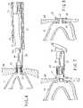
- FIG. 1is a schematic diagram showing the entire structure of an anastomosis system according to a preferred embodiment of the present invention
- FIG. 2Ais a schematic diagram explaining the arrangement of members in the distal end of a guide tube of the anastomosis system in FIG. 1 ;
- FIG. 2Bis a perspective view showing a developed anastomosis button
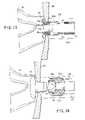
- FIG. 3Ais a sectional view showing an internal portion of the guide tube in FIG. 2A ;
- FIG. 3Bis a cross-sectional view taken along a line B-B in FIG. 3A ;
- FIG. 3Cis a cross-sectional view taken along a line C-C in FIG. 3A ;
- FIG. 4is an explanatory diagram showing a state in which a part of an intestine is pulled into a stomach through a stomach wall pierced by the anastomosis system in FIG. 1 ;
- FIG. 5is an explanatory diagram showing a state in which, in the state shown in FIG. 4 , the intestine is pierced and a housing is inserted thereto;
- FIG. 6is an explanatory diagram showing a state in which a part of the anastomosis button is ejected from the housing;
- FIG. 7is an explanatory diagram showing a state in which the ejection of the anastomosis button from the housing is completed
- FIG. 8is a sectional view showing a state in which the stomach wall and an intestine wall are subjected to the anastomosis with the anastomosis button;
- FIG. 9Ais a sectional view showing a part of an anastomosis system according to another embodiment.
- FIG. 9Bis a schematic perspective view of an anastomosis button used in the anastomosis system in FIG. 9A ;
- FIG. 10is an explanatory diagram showing a state according to the anastomosis system in FIG. 9A , the state being similar to that in FIG. 5 ;
- FIG. 11is an explanatory diagram showing a state in which the anastomosis button is ejected from a housing in the anastomosis system in FIG. 9A ;
- FIG. 12is a sectional view showing a state in which an anastomosis is performed with the anastomosis button shown in FIG. 9B , the state being similar to that shown in FIG. 8 ;
- FIG. 13is an explanatory diagram showing a state in which an intestine wall is held by a wire with a needle;
- FIG. 14Ais a sectional view showing a state in which an anastomosis is performed by using an anastomosis system according to further another embodiment
- FIG. 14Bis a schematic exploded perspective view of an anastomosis button used in the anastomosis system in FIG. 14A ;
- FIG. 15is an explanatory diagram showing the insertion of a member of the anastomosis button shown in FIG. 14B ;
- FIG. 16is an explanatory diagram showing a state in which the other member of the anastomosis button is attached to the member, which is previously inserted;
- FIG. 17is an explanatory diagram showing the ejection of the other member shown in FIG. 14B ;
- FIG. 18is a sectional view showing a state in which the anastomosis is performed by the anastomosis button shown in FIG. 14B ;
- FIG. 19is an explanatory diagram showing a state in which the anastomosis is performed by using the anastomosis system shown in FIG. 14A ;
- FIG. 20is a sectional view of a modified anastomosis button.
- an anastomosis system 10is suitable for a gastroenterostomy in a treatment for, particularly, obesity.
- the systemis not limited thereto.
- the anastomosis system 10has a guide 12 shaped into a flexible tube which can be inserted through a mouth.
- the guide 12at least its distal end is made of transparent resin.
- a plane at the distal endis inclined in the longitudinal direction of the axis.
- an orifice 14 to be opened on the sideis formed.
- a holding instrument 18such as a grasping forceps (in FIG.
- a housing operation shaft 20 for operating a housing, which will be described below, and an endoscope 22are inserted through an operation section main body 16 arranged on the outside of a body into the guide, so that the distal ends thereof are guided to a target portion in the coelom. Since at least the distal end of the guide 12 is made of the transparent resin, even when the distal end 22 a of the endoscope 22 is not protruded from the guide 12 , the coelom can be observed through the endoscope 22 .
- the distal end 22 a of the endoscope 22 inserted through the guide 12can be protruded outwardly in the radial direction from the inside of the guide 12 through the side orifice 14 opened in the vicinity of the plane at the distal end of the guide 12 .
- illumination light guided through a light guide(not shown) is irradiated from an illumination window 24 a , which is formed on the surface at the distal end 22 a , to a predetermined portion in the coelom, so that reflected light incident on an observation window 24 b can be guided to an endoscope operation section 26 arranged on the outside of the body through an image guide (not shown).
- Reference numeral 28 a shown in FIG. 2Adenotes a channel through which an operative instrument such as a forceps or a high-frequency knife is inserted.
- Reference numeral 28 bdenotes a nozzle for ejecting water or air.
- a large-diameter lumen 30 through which the endoscope 22 is inserted and a plurality of small-diameter lumens 32 and 34are formed.
- a shaft of the holding instrument 18 having, for example, a grasping forceps 19 at the distal endis inserted.
- the housing operation shaft 20is inserted through the small-diameter lumen 34 .
- the lumens 30 to 34constitute a common lumen 36 as a space with a large diameter.
- reference symbol ⁇designates an area where the common lumen 36 is formed and reference symbol ⁇ designates an area where the lumens are formed.
- the side orifice 14is not always formed in the area ⁇ .
- the orifice 14is formed in the vicinity of the distal end of the guide 12 .
- a housing 44 receiving an anastomosis button 42 having two flange portions 38 and a tubular main body portion 40 extending between the flange portions (refer to FIG. 2B )is arranged. Accordingly, the dimension of the common lumen 36 in the axial direction is preferably set so that the housing 44 can be received fully.
- the anastomosis button 42is made of a flexible or elastic material which can be deformed. Particularly, the flange portions 38 are folded to be received in the housing 44 . When they are ejected from the housing 44 , the flange portions 38 can be immediately returned to the original form. Moreover, as will be explained below, the anastomosis button 42 has such strength that, for example, when a stomach wall is anastomosed to an intestine wall, the two wall portions can be tightly held until they adhere to each other.
- the housing 44 receiving the above-mentioned anastomosis button 42is constituted of two cylindrical portions 44 a and 44 b which are engaged with each other in a telescopic manner in the present embodiment. Particularly as shown in FIG. 3A , the cylindrical portions 44 a and 44 b are formed so that the elongated cylindrical portions are substantially equivalent to the axial length of the anastomosis button 42 elongated in an hourglass-shaped form as the two flange portions 38 are folded oppositely.
- the operation shaft 20is fixed to either one of the cylindrical portions 44 a and 44 b .
- the end of the cylindrical portion 44 awhich is located at the distal end of the housing 44 , is tapered so as to be easily inserted into a pierced coelom wall. Since the sloped plane is formed at the distal end, when the anastomosis button 42 is ejected from the housing 44 , the position of the anastomosis button 42 can be easily confirmed through the endoscope 22 .
- the anastomosis button 42is folded in an hourglass-shaped form and is then stored in the elongated housing 44 .
- the housing 44 receiving the anastomosis button 42 thereinis disposed in the large-diameter common lumen 36 formed at the distal end of the guide 12 .
- the operation shaft 20is inserted through the lumen 34 .
- an operation section of the shaftis disposed on the outside of the operation section main body 16 . If necessary, the holding instruments 18 having the grasping forceps 19 at the distal end are also received in the common lumen 36 at the distal end of the guide 12 and the shafts thereof are inserted to the lumens 32 .
- the operatorinserts their thumb into a ring shown by reference numeral 18 a in FIG. 1 and then moves the thumb with respect to a slider 18 b and the ring 18 a , so that a pair of jaws of each grasping forceps 19 can be opened or closed.
- the distal end of the guide 12 prepared as mentioned aboveis inserted into the stomach of a patient through their mouth and esophagus and is then disposed at a desired portion.
- the statecan be confirmed via the endoscope 22 previously inserted in the lumen 30 , the endoscope being inserted subsequent to the guide 12 .
- the confirmationcan be easily made because the guide 12 is made of the transparent material.
- the distal end 22 a of the endoscope 22is extended outwardly through the side orifice 14 of the guide 12 and the end surface of the distal end is aimed to a portion 92 to be pierced (refer to FIG. 4 ) of the stomach wall 90 .
- an incision instrumentsuch as a high-frequency knife is inserted through, for example, the channel 28 a ( FIG. 2A ) in the endoscope 22 to pierce the portion 92 .
- the desired portionis held or pulled by the grasping forceps 19 while the portion 92 to be pierced of the stomach wall 90 is monitored by the endoscope 22 , so that the portion can be surely easily pierced.
- FIGS. 4 through 8show a processing of anastomosing a small intestine 94 to the above-mentioned pierced portion 92 of the stomach wall.
- the two grasping forceps 19are advanced from the guide 12 under observation via the endoscope 22 to grasp the small intestine 94 and pull it into the stomach through the pierced portion 92 formed in the stomach wall.
- the small intestine 94is also pierced by the incision instrument such as a high-frequency knife inserted through the channel in the endoscope 22 .
- the operating section of the operation shaft 20is operated outside the body to protrude the housing 44 from the distal end of the guide 12 and then insert the housing 44 through the pierced portion in the small intestine 94 .
- the cylindrical portion 44 a at the distal end in which one of the flange portions 38 is receivedis inserted into the small intestine 94 , the insertion of the housing 44 is stopped.
- the common channel 36serves as a free space.
- the distal end 22 a of the endoscope 22is inserted into the common channel serving as a free space.
- a release line illustrated by a grasping forceps 46 shown in FIG. 6is advanced toward the housing 44 through the channel 28 a in the endoscope 22 .
- the operation shaft 20 or grasping forceps 46is moved to telescope the cylindrical portions 44 a and 44 b of the housing 44 , thereby extruding one of the flange portions 38 of the anastomosis button 42 into the small intestine 94 .
- the flange portion 38 extruded in the small intestine 94is returned to the original circular form by elasticity, so that the flange portion is brought into contact with the inner surface of the small intestine 94 .
- FIG. 7shows such a state.
- the main body portion 40 of the anastomosis button 42is shaped tubularly, two lumen portions of the stomach and the small intestine are coupled to each other through an orifice 41 . Consequently, food fed to the stomach is immediately transferred to the small intestine 94 , so that digestive and assimilative operations are not performed in the stomach.
- the anastomosis button 42 for anastomosing the stomach to the small intestineis held in the patient's body in this state for, e.g., about one week. After the pierced portions have adhered to each other, the button is extracted from the body.
- the anastomosis button 42particularly, the flange portions 38 thereof are formed flexibly.
- the anastomosis buttoncan be easily removed from the pierced portion 92 in the stomach wall 90 .
- the anastomosis button 42can be easily removed by merely inserting the grasping forceps through the normal endoscope.
- the anastomosis button 42can be inserted together with the endoscope 22 through the mouth into the coelom.
- the gastroenterostomycan be performed remarkably easily.
- the patientis not burdened substantially.
- the housing 44 receiving the anastomosis button 42 thereinis constituted of the remarkably simple cylindrical members, a special device is not needed, so that the anastomosis system is formed at extremely low cost.
- FIGS. 9A through 12show an anastomosis system according to another embodiment. Since the fundamental principle of the present embodiment described hereinafter is the same as that of the foregoing embodiment, the same components are designated by the same reference numerals and the detailed description is omitted.
- a housing 54includes a cylindrical portion 54 a having a tapered distal end and a bottom wall portion 54 b provided at the proximal end of the cylindrical portion.
- at least one slit 56is formed (it is preferable to form plural slits). Accordingly, the diameter of the tapered distal end can be enlarged.
- a flexible guide tube 60through which a guide wire 58 can be inserted, is fixed and an opening portion 62 is formed.
- the guide tube 60is protruded from the distal end of the housing 54 so that it can guide the housing 54 while sliding along the guide wire 58 inserted therethrough.
- the housing 54is held more surely than the case where the housing is held by the operation shaft 20 alone.
- the housing 54is surely and easily guided to a desired position.
- An anastomosis button in the housing 54is received on the guide tube 60 .
- a rim 39is provided for the periphery of each flange portion 38 to increase the bending strength of the flange portion 38 . Accordingly, even when the diameter of the penetrating orifice 41 is increased, the stomach wall 90 and the intestine wall 94 can be surely held without increasing the outer diameter or thickness of the flange portion 38 .
- the rims 39can be protruded so as to face each other. To allow the rims 49 to be smoothly in contact with the stomach wall 90 and the intestine wall 94 , preferably, they are protruded opposite to each other as shown in FIG. 9B .
- the housing 54 according to the present embodimentafter the housing 54 according to the present embodiment is protruded from the common lumen 36 of the guide 12 , it can be offset outwardly in the radial direction from the axis in the longitudinal direction of the guide 12 by the operation shaft 20 and the guide wire 58 . Consequently, the distal end 22 a of the endoscope 22 can be linearly advanced from the common lumen 36 of the guide 12 past the housing 54 . In this case, it is unnecessary to form the side orifice 14 in the foregoing embodiment.
- the anastomosis system according to the present embodimentcan be used in a manner similar to the foregoing embodiment. According to the present embodiment, since the distal end 22 a of the endoscope 22 can be linearly advanced past the housing 54 , the stomach wall 90 and the intestine wall 94 can be pierced in a state in which the surface at the distal end of the endoscope is disposed substantially parallel to the walls.
- FIG. 10shows the state in which the distal end of the housing 54 is inserted through the pierced portion.
- the distal end of a release linesuch as a grasping forceps extending through the channel in the endoscope 22 is inserted into the housing 54 through the opening portion 62 formed in the bottom wall portion 54 b .
- the anastomosis button 42 Ais moved to a front portion in the housing 54 .
- the slits 56 formed at the tapered distal endare opened to enlarge the diameter of the tapered distal end.
- FIG. 20shows a modified button 42 C with a rounded shape.
- FIGS. 13 through 19show further another embodiment.
- FIG. 13shows a technique for pulling the intestine wall 94 of the small intestine in the present embodiment.
- the intestine wall 94is drawn to the stomach wall 90 by a piercing needle 66 inserted through the channel 28 a in the endoscope 22 .
- the piercing needle 66is formed as a long drawing line formed by inserting a needle wire 66 b through a needle sheath 66 a .
- the distal end of the needle wire 66 bis coiled to form a needle portion.
- the needle wire 66 bis inserted together with the needle sheath 66 a into the intestine wall and the needle sheath 66 a alone is removed. Consequently, the coiled needle at the distal end of the needle wire 66 b is held in the intestine wall 94 . Alternatively, the coiled needle pierces the intestine wall 94 to prevent the needle wire 66 b from being removed from the intestine wall 94 . In this state, the needle wire 66 b is drawn in the direction toward the patient's mouth and is then fixed to an external portion of the body, so that it is possible to hold the state in which the intestine wall 94 is pulled to the stomach wall 90 .
- the intestine wall 94be drawn to the stomach wall 90 at portions opposite to the diameter of the pierced portion 92 by using two piercing needles 66 .
- an anastomosis button 42 Bis constituted of a first segment 43 , which has the tubular main body portion 40 and the flange portion 38 , and a second disc-like segment 38 a that is fitted to the main body portion 40 of the first segment.
- the end of the main body portion 40has a tapered shape so that the second segment 38 a can be easily attached thereto.
- the second segment 38 ais fitted to the first segment 43 , thereby forming the flange portion opposite to the flange portion 38 of the first segment 43 .
- the distance between the flange portionscan be controlled in accordance with the thickness of the patient's stomach wall 90 and intestine wall 94 .
- the entire anastomosis button 42 B according to the present embodimentis round, so that it comes smoothly into contact with the stomach wall and the small intestine wall.
- the anastomosis buttonhas a configuration that is friendly to a mucous membrane.
- a housing 64 receiving the anastomosis button 42 B thereinhas an inner cylindrical portion 64 a that is attached to the distal end of the endoscope 22 and an outer cylindrical portion 64 b that is slidably attached onto the inner cylindrical portion 64 a . Between the two cylindrical portions, either the first segment or the second segment of the anastomosis button 42 B is folded and they can be received.
- two operation wires 68 as release linesare connected to the outer cylindrical portion 64 b .
- Each operation wire 68is inserted through a tube sheath 70 and extends to an operation section of the endoscope 22 shown in FIG. 19 .
- reference numeral 72denotes an operation knob for the outer cylindrical portion which is connected to the operation wire 68
- reference numeral 74denotes an operation main body for the outer cylindrical portion which is connected to the tube sheath 70 .
- FIG. 14Ashows a state in which the first segment 64 a of the anastomosis button 42 B is received.
- FIG. 16shows a state in which the second segment 64 b is received.
- the housing 64can receive the first segment 43 and the second segment 38 a of the anastomosis button 42 B in an annular gap between the inner and outer cylindrical portions 64 a and 64 b .
- button discharging threads 78in which spherical beads 76 are connected to the distal ends are preferably used as release lines in addition to the operation wire 68 .
- each discharging thread 78is inserted through the channel 28 a in the endoscope 22 into the inner cylindrical portion 64 a to dispose the beads 76 at both the distal ends on the outer peripheral surface of the cylindrical portion.
- the second segment 38 ais attached to the reduced diameter portion of the inner cylindrical portion 64 a and is then covered with the outer cylindrical portion 64 b .
- the proximal end of each discharging thread 78extends outwardly from the operation section of the endoscope 22 and is then fixed to a button operation main body 82 , which is rotated by a rotation knob 80 , as shown in FIG. 19 .
- the rotation knob 80is rotated to wind the discharging threads 78 around the button operation main body 82 . Consequently, the discharging threads 78 are pulled toward the proximal ends, so that the beads 76 discharge the second segment 74 b out of the housing 64 .
- a plurality of discharge threads 78are used as shown in FIGS. 16 and 17 .
- One discharging threadcan be also used.
- the attachment of the anastomosis button 42 B according to the present embodimentis performed as follows.
- the endoscope 22is inserted through a mouthpiece 98 ( FIG. 19 ) attached to the patient's mouth.
- the distal end of the endoscope 22is disposed opposite to the pierced portion 92 and the intestine wall 94 is pierced by an incision instrument such as a high-frequency knife inserted through the channel in the endoscope 22 .
- an incision instrumentsuch as a high-frequency knife inserted through the channel in the endoscope 22 .
- the distal end of the housing 64is inserted through an orifice formed in the intestine wall 94 and the operation knob 72 for the outer cylindrical portion 64 b shown in FIG. 19 is drawn to move the outer cylindrical portion 64 b backward. Consequently, the first segment 43 of the anastomosis button 42 B is exposed and the flange portion 38 is developed in the original circular shape due to elasticity.
- the endoscope 22is withdrawn to pull the inner cylindrical portion 64 a of the housing 64 out of the first segment 43 .
- the flange portion 38is brought into contact with the inner surface of the intestine wall 94 , the first segment 43 is held in the small intestine.
- the endoscope 22in which the housing 64 receiving the second disc-like segment 38 a is attached to the distal end 22 a , is inserted into the stomach to allow the distal end to face the first segment 43 of the anastomosis button previously attached.
- the housing 64is allowed to coaxially match the end, in which the diameter is reduced, of the main body portion 40 of the first segment 43 and is then close thereto.
- the outer cylindrical portion 64 b of the housing 64is withdrawn and the rotation knob 80 provided for the operation section of the endoscope 22 is rotated to wind the discharging threads 78 around the button operation main body 8 . Consequently, as shown in FIG.
- the second segment 38 ais returned to the original disc shape on the inner cylindrical portion 64 a and is then discharged by the beads 76 to be attached onto the main body portion 40 of the first segment 43 .
- the first segment 43can be held by a grasping forceps (not shown) inserted through the channel in the endoscope 22 .
- the present embodimenthas been explained in association with the gastroenterostomy, the example in which the anastomosis button was inserted through the mouth has been described.
- the anastomosis buttoncan be inserted through the anus.
- the members in the embodimentscan be properly combined to each other and they are not limited to any embodiment.
- the anastomosis buttoncan be easily inserted through the mouth or the anus into the coelom, the burden on the patient is extremely little. Moreover, since it takes a remarkably short time, the system can be generally used in other applications in addition to the above-mentioned gastroenterostomy.
Landscapes
- Health & Medical Sciences (AREA)
- Surgery (AREA)
- Life Sciences & Earth Sciences (AREA)
- Medical Informatics (AREA)
- Nuclear Medicine, Radiotherapy & Molecular Imaging (AREA)
- Engineering & Computer Science (AREA)
- Biomedical Technology (AREA)
- Heart & Thoracic Surgery (AREA)
- Physiology (AREA)
- Molecular Biology (AREA)
- Animal Behavior & Ethology (AREA)
- General Health & Medical Sciences (AREA)
- Public Health (AREA)
- Veterinary Medicine (AREA)
- Surgical Instruments (AREA)
- Endoscopes (AREA)
Abstract
Description
Claims (7)
Priority Applications (2)
| Application Number | Priority Date | Filing Date | Title |
|---|---|---|---|
| US10/353,865US7637919B2 (en) | 2002-01-30 | 2003-01-29 | Anastomosis system for performing anastomosis in body |
| US10/957,911US7654951B2 (en) | 2002-01-30 | 2004-10-04 | Anastomosis system for performing anastomosis in body |
Applications Claiming Priority (2)
| Application Number | Priority Date | Filing Date | Title |
|---|---|---|---|
| US35272702P | 2002-01-30 | 2002-01-30 | |
| US10/353,865US7637919B2 (en) | 2002-01-30 | 2003-01-29 | Anastomosis system for performing anastomosis in body |
Related Child Applications (1)
| Application Number | Title | Priority Date | Filing Date |
|---|---|---|---|
| US10/957,911ContinuationUS7654951B2 (en) | 2002-01-30 | 2004-10-04 | Anastomosis system for performing anastomosis in body |
Publications (2)
| Publication Number | Publication Date |
|---|---|
| US20030216749A1 US20030216749A1 (en) | 2003-11-20 |
| US7637919B2true US7637919B2 (en) | 2009-12-29 |
Family
ID=29420296
Family Applications (2)
| Application Number | Title | Priority Date | Filing Date |
|---|---|---|---|
| US10/353,865Expired - LifetimeUS7637919B2 (en) | 2002-01-30 | 2003-01-29 | Anastomosis system for performing anastomosis in body |
| US10/957,911Expired - LifetimeUS7654951B2 (en) | 2002-01-30 | 2004-10-04 | Anastomosis system for performing anastomosis in body |
Family Applications After (1)
| Application Number | Title | Priority Date | Filing Date |
|---|---|---|---|
| US10/957,911Expired - LifetimeUS7654951B2 (en) | 2002-01-30 | 2004-10-04 | Anastomosis system for performing anastomosis in body |
Country Status (2)
| Country | Link |
|---|---|
| US (2) | US7637919B2 (en) |
| JP (2) | JP4197963B2 (en) |
Cited By (32)
| Publication number | Priority date | Publication date | Assignee | Title |
|---|---|---|---|---|
| US20060036267A1 (en)* | 2004-08-11 | 2006-02-16 | Usgi Medical Inc. | Methods and apparatus for performing malabsorptive bypass procedures within a patient's gastro-intestinal lumen |
| US20060089707A1 (en)* | 2004-08-11 | 2006-04-27 | Emory University | Vascular conduit device and system for implanting |
| US20100331866A1 (en)* | 2009-06-26 | 2010-12-30 | Vihar Surti | Linear clamps for anastomosis |
| US20130231689A1 (en)* | 2004-04-12 | 2013-09-05 | Kenneth F. Binmoeller | Luminal structure anchoring devices and methods |
| US8545525B2 (en) | 2009-11-03 | 2013-10-01 | Cook Medical Technologies Llc | Planar clamps for anastomosis |
| US8603121B2 (en) | 2010-04-14 | 2013-12-10 | Cook Medical Technologies Llc | Systems and methods for creating anastomoses |
| US8617159B2 (en) | 2006-09-08 | 2013-12-31 | Ethicon Endo-Surgery, Inc. | Surgical instrumentation for performing endoluminal and/or transluminal anastomosis |
| US8834361B2 (en) | 2009-05-15 | 2014-09-16 | Cook Medical Technologies Llc | Systems, devices and methods for accessing a bodily opening |
| US8858489B2 (en) | 2007-04-24 | 2014-10-14 | Emory University | Conduit device and system for implanting a conduit device in a tissue wall |
| US8864781B2 (en)* | 2007-02-28 | 2014-10-21 | Cook Medical Technologies Llc | Intestinal bypass using magnets |
| US8974379B2 (en) | 2008-03-06 | 2015-03-10 | Cook Medical Technologies Llc | Medical systems for accessing an internal bodily opening |
| US9028523B2 (en) | 2008-05-15 | 2015-05-12 | Cook Medical Technologies Llc | Systems, devices and methods for accessing a bodily opening |
| US9320875B2 (en) | 2011-02-01 | 2016-04-26 | Emory University | Systems for implanting and using a conduit within a tissue wall |
| US9364259B2 (en) | 2009-04-21 | 2016-06-14 | Xlumena, Inc. | System and method for delivering expanding trocar through a sheath |
| US9381041B2 (en) | 2009-04-21 | 2016-07-05 | Xlumena, Inc. | Methods and devices for access across adjacent tissue layers |
| US9526648B2 (en) | 2010-06-13 | 2016-12-27 | Synerz Medical, Inc. | Intragastric device for treating obesity |
| US9532773B2 (en) | 2011-01-28 | 2017-01-03 | Apica Cardiovascular Limited | Systems for sealing a tissue wall puncture |
| US9888926B2 (en) | 2009-05-29 | 2018-02-13 | Boston Scientific Scimed, Inc. | Apparatus and method for deploying stent across adjacent tissue layers |
| US10028741B2 (en) | 2013-01-25 | 2018-07-24 | Apica Cardiovascular Limited | Systems and methods for percutaneous access, stabilization and closure of organs |
| WO2018138614A2 (en) | 2017-01-30 | 2018-08-02 | Ethicon Llc | Tissue compression assemblies with biodegradable interlinks |
| WO2018138616A1 (en) | 2017-01-30 | 2018-08-02 | Ethicon Llc | Non-magnetic fragmentable tissue compression devices |
| US10076330B2 (en) | 2008-05-12 | 2018-09-18 | Xlumena, Inc. | Tissue anchor for securing tissue layers |
| US10342544B2 (en) | 2013-04-16 | 2019-07-09 | Ethicon Endo-Surgery, Inc. | Method and apparatus for joining hollow organ sections in anastomosis |
| US10413436B2 (en) | 2010-06-13 | 2019-09-17 | W. L. Gore & Associates, Inc. | Intragastric device for treating obesity |
| US10420665B2 (en) | 2010-06-13 | 2019-09-24 | W. L. Gore & Associates, Inc. | Intragastric device for treating obesity |
| US10485909B2 (en) | 2014-10-31 | 2019-11-26 | Thoratec Corporation | Apical connectors and instruments for use in a heart wall |
| US10518012B2 (en) | 2013-03-15 | 2019-12-31 | Apk Advanced Medical Technologies, Inc. | Devices, systems, and methods for implanting and using a connector in a tissue wall |
| US10779980B2 (en) | 2016-04-27 | 2020-09-22 | Synerz Medical, Inc. | Intragastric device for treating obesity |
| US10952732B2 (en) | 2013-02-21 | 2021-03-23 | Boston Scientific Scimed Inc. | Devices and methods for forming an anastomosis |
| US11033272B2 (en) | 2013-04-16 | 2021-06-15 | Ethicon Endo-Surgery, Inc. | Methods for partial diversion of the intestinal tract |
| US11135078B2 (en) | 2010-06-13 | 2021-10-05 | Synerz Medical, Inc. | Intragastric device for treating obesity |
| US12303105B2 (en) | 2004-04-12 | 2025-05-20 | Boston Scientific Scimed, Inc. | Luminal structure anchoring devices and methods |
Families Citing this family (104)
| Publication number | Priority date | Publication date | Assignee | Title |
|---|---|---|---|---|
| US7527590B2 (en)* | 2002-03-19 | 2009-05-05 | Olympus Corporation | Anastomosis system |
| JP2004024331A (en)* | 2002-06-21 | 2004-01-29 | Vayu:Kk | Catheter |
| US7351247B2 (en) | 2002-09-04 | 2008-04-01 | Bioconnect Systems, Inc. | Devices and methods for interconnecting body conduits |
| US7695446B2 (en) | 2002-12-02 | 2010-04-13 | Gi Dynamics, Inc. | Methods of treatment using a bariatric sleeve |
| US7678068B2 (en) | 2002-12-02 | 2010-03-16 | Gi Dynamics, Inc. | Atraumatic delivery devices |
| WO2004049982A2 (en) | 2002-12-02 | 2004-06-17 | Gi Dynamics, Inc. | Bariatric sleeve |
| US7608114B2 (en) | 2002-12-02 | 2009-10-27 | Gi Dynamics, Inc. | Bariatric sleeve |
| US7025791B2 (en)* | 2002-12-02 | 2006-04-11 | Gi Dynamics, Inc. | Bariatric sleeve |
| US7766973B2 (en) | 2005-01-19 | 2010-08-03 | Gi Dynamics, Inc. | Eversion resistant sleeves |
| US7960935B2 (en) | 2003-07-08 | 2011-06-14 | The Board Of Regents Of The University Of Nebraska | Robotic devices with agent delivery components and related methods |
| US20050075656A1 (en)* | 2003-09-30 | 2005-04-07 | Jean Beaupre | Applier for a surgical device |
| CA2482707C (en)* | 2003-09-30 | 2013-07-02 | Ethicon Endo-Surgery, Inc. | Applier having automated release of surgical device |
| CA2482697C (en)* | 2003-09-30 | 2012-11-20 | Ethicon Endo-Surgery, Inc. | Applier for a surgical device |
| AU2004305450B2 (en) | 2003-12-09 | 2009-01-08 | Gi Dynamics, Inc. | Intestinal sleeve |
| US8057420B2 (en) | 2003-12-09 | 2011-11-15 | Gi Dynamics, Inc. | Gastrointestinal implant with drawstring |
| US7618427B2 (en)* | 2003-12-29 | 2009-11-17 | Ethicon Endo-Surgery, Inc. | Device and method for intralumenal anastomosis |
| US8992420B2 (en)* | 2004-04-14 | 2015-03-31 | Usgi Medical, Inc. | Methods and apparatus for off-axis visualization |
| US8277373B2 (en)* | 2004-04-14 | 2012-10-02 | Usgi Medical, Inc. | Methods and apparaus for off-axis visualization |
| US20050251091A1 (en)* | 2004-05-10 | 2005-11-10 | Usgi Medical Inc. | Apparatus and methods for transgastric tissue manipulation |
| US7803195B2 (en) | 2004-06-03 | 2010-09-28 | Mayo Foundation For Medical Education And Research | Obesity treatment and device |
| US7931661B2 (en)* | 2004-06-14 | 2011-04-26 | Usgi Medical, Inc. | Apparatus and methods for performing transluminal gastrointestinal procedures |
| ATE506042T1 (en) | 2004-07-09 | 2011-05-15 | Gi Dynamics Inc | DEVICES FOR PLACEMENT OF A GASTROINTESTINAL SLEEVE |
| EP1799145B1 (en) | 2004-09-17 | 2016-12-21 | GI Dynamics, Inc. | Gastrointestinal anchor |
| US7922743B2 (en)* | 2004-10-18 | 2011-04-12 | Tyco Healthcare Group Lp | Structure for applying sprayable wound treatment material |
| ITMI20042131A1 (en)* | 2004-11-05 | 2005-02-05 | Ethicon Endo Surgery Inc | DEVICE AND METHOD FOR OBESITY THERAPY |
| ITMI20042129A1 (en)* | 2004-11-05 | 2005-02-05 | Ethicon Endo Surgery Inc | DEVICE AND METHOD FOR OBESITY THERAPY |
| US7771382B2 (en) | 2005-01-19 | 2010-08-10 | Gi Dynamics, Inc. | Resistive anti-obesity devices |
| JP2008536552A (en)* | 2005-04-11 | 2008-09-11 | ユーエスジーアイ メディカル インク. | Method and apparatus for off-axis visualization |
| US7534247B2 (en)* | 2005-05-03 | 2009-05-19 | Ethicon Endo-Surgery, Inc. | Sheathless anastomotic ring applier device |
| RU2290103C1 (en)* | 2005-05-11 | 2006-12-27 | Дмитрий Андреевич Чичеватов | Method for developing esophago-small-intestinal and esophago-gastric anastomosis |
| US7976488B2 (en) | 2005-06-08 | 2011-07-12 | Gi Dynamics, Inc. | Gastrointestinal anchor compliance |
| US8641729B2 (en)* | 2005-07-13 | 2014-02-04 | Creighton University | Systems and techniques for minimally invasive gastrointestinal procedures |
| US8906040B2 (en)* | 2005-07-13 | 2014-12-09 | Creighton University | Systems and techniques for minimally invasive gastrointestinal procedures |
| US20070051375A1 (en)* | 2005-09-06 | 2007-03-08 | Milliman Keith L | Instrument introducer |
| EP1948078A2 (en)* | 2005-11-14 | 2008-07-30 | Sentinel Group, LLC | Gastro-intestinal therapeutic device and method |
| WO2007063904A1 (en)* | 2005-12-01 | 2007-06-07 | Olympus Medical Systems Corp. | Guiding long medical member and long medical device |
| ITMI20060062A1 (en)* | 2006-01-16 | 2007-07-17 | Ethicon Endo Surgery Inc | ANASTOMOTIC DEVICE SUITABLE FOR CLOSING AND PREVIOUSLY KEEPING A FIRST PORTION OF FABRIC AND A SECOND PORTION OF FABRICS FOR THE FORMATION OF ANASTICOSUS |
| US8726909B2 (en)* | 2006-01-27 | 2014-05-20 | Usgi Medical, Inc. | Methods and apparatus for revision of obesity procedures |
| BRPI0602735A (en)* | 2006-06-06 | 2008-01-29 | Luiz Gonzaga Granja Jr | anastomosis prosthesis |
| US9579088B2 (en) | 2007-02-20 | 2017-02-28 | Board Of Regents Of The University Of Nebraska | Methods, systems, and devices for surgical visualization and device manipulation |
| WO2007149559A2 (en) | 2006-06-22 | 2007-12-27 | Board Of Regents Of The University Of Nebraska | Magnetically coupleable robotic devices and related methods |
| US8679096B2 (en) | 2007-06-21 | 2014-03-25 | Board Of Regents Of The University Of Nebraska | Multifunctional operational component for robotic devices |
| US7819836B2 (en) | 2006-06-23 | 2010-10-26 | Gi Dynamics, Inc. | Resistive anti-obesity devices |
| US8801647B2 (en) | 2007-02-22 | 2014-08-12 | Gi Dynamics, Inc. | Use of a gastrointestinal sleeve to treat bariatric surgery fistulas and leaks |
| US20080208214A1 (en) | 2007-02-26 | 2008-08-28 | Olympus Medical Systems Corp. | Applicator and tissue fastening method through natural orifice |
| US20080208239A1 (en)* | 2007-02-27 | 2008-08-28 | Gary Annunziata | Method for treating obesity using an implantable weight loss device |
| US10238518B2 (en)* | 2007-02-27 | 2019-03-26 | Agt Inc. | Implantable weight control device |
| US8343171B2 (en) | 2007-07-12 | 2013-01-01 | Board Of Regents Of The University Of Nebraska | Methods and systems of actuation in robotic devices |
| EP2173259A4 (en) | 2007-08-02 | 2015-07-08 | Bio Connect Systems | Implantable flow connector |
| US20130197546A1 (en) | 2007-08-02 | 2013-08-01 | Bioconnect Systems, Inc. | Implantable flow connector |
| JP2010536435A (en) | 2007-08-15 | 2010-12-02 | ボード オブ リージェンツ オブ ザ ユニバーシティ オブ ネブラスカ | Medical inflation, attachment and delivery devices and associated methods |
| CA2695619C (en) | 2007-08-15 | 2015-11-24 | Board Of Regents Of The University Of Nebraska | Modular and cooperative medical devices and related systems and methods |
| US10238392B2 (en)* | 2009-12-29 | 2019-03-26 | Cvdevices, Llc | Methods for diagnosing and delivering therapeutic interventions in the peritoneal cavity |
| US20100191264A1 (en)* | 2007-09-21 | 2010-07-29 | Cvd Devices, Llc | Devices, systems and methods for diagnosing and delivering therapeutic interventions in the peritoneal cavity |
| AU2008310975B2 (en)* | 2007-10-09 | 2013-08-22 | Cook Medical Technologies Llc | Systems, devices and methods having an overtube for accessing a bodily opening |
| WO2009082710A1 (en) | 2007-12-21 | 2009-07-02 | Endometabolic Solutions, Inc. | Methods and devices for endoscopically creating an anastomosis |
| US8870778B2 (en)* | 2007-12-24 | 2014-10-28 | Olympus Medical Systems Corp. | Tranlumen endoscope insertion surgery |
| EP2240083B8 (en)* | 2008-01-10 | 2015-08-19 | Covidien LP | Imaging system for a surgical device |
| JP5053904B2 (en)* | 2008-03-31 | 2012-10-24 | オリンパスメディカルシステムズ株式会社 | Endoscope, endoscope with tip cap, and cleaning sheath for endoscope |
| US20090281379A1 (en) | 2008-05-12 | 2009-11-12 | Xlumena, Inc. | System and method for transluminal access |
| US8562513B2 (en)* | 2008-05-20 | 2013-10-22 | Olympus Medical Systems Corp. | Endoscope device |
| US20100041952A1 (en)* | 2008-08-12 | 2010-02-18 | Omnimed Enterprises LLC | Medical Instrument Light Source Connection Device |
| JP5407036B2 (en)* | 2008-09-02 | 2014-02-05 | オリンパスメディカルシステムズ株式会社 | Treatment endoscope |
| JP4642936B2 (en)* | 2009-02-09 | 2011-03-02 | オリンパスメディカルシステムズ株式会社 | Medical tube |
| EP3973892B1 (en)* | 2009-07-15 | 2024-10-23 | GT Metabolic Solutions, Inc. | Incisionless gastric bypass devices |
| EP2600758A1 (en) | 2010-08-06 | 2013-06-12 | Board of Regents of the University of Nebraska | Methods and systems for handling or delivering materials for natural orifice surgery |
| JP5437300B2 (en)* | 2011-03-17 | 2014-03-12 | 富士フイルム株式会社 | Endoscope insertion aid |
| JP6174017B2 (en) | 2011-06-10 | 2017-08-02 | ボード オブ リージェンツ オブ ザ ユニバーシティ オブ ネブラスカ | In vivo vascular seal end effector and in vivo robotic device |
| JP5866131B2 (en) | 2011-06-15 | 2016-02-17 | フラクシス インコーポレイテッド | Anastomotic connector |
| EP2720626B1 (en) | 2011-06-15 | 2017-06-14 | Phraxis Inc. | Anastomotic connector and system for delivery |
| WO2013009887A1 (en) | 2011-07-11 | 2013-01-17 | Board Of Regents Of The University Of Nebraska | Robotic surgical devices, systems and related methods |
| WO2013052137A2 (en) | 2011-10-03 | 2013-04-11 | Board Of Regents Of The University Of Nebraska | Robotic surgical devices, systems and related methods |
| WO2013106569A2 (en) | 2012-01-10 | 2013-07-18 | Board Of Regents Of The University Of Nebraska | Methods, systems, and devices for surgical access and insertion |
| US9314600B2 (en) | 2012-04-15 | 2016-04-19 | Bioconnect Systems, Inc. | Delivery system for implantable flow connector |
| US10434293B2 (en) | 2012-04-15 | 2019-10-08 | Tva Medical, Inc. | Implantable flow connector |
| EP2844181B1 (en) | 2012-05-01 | 2021-03-10 | Board of Regents of the University of Nebraska | Single site robotic device and related systems |
| EP2861182B1 (en) | 2012-06-15 | 2019-02-20 | Phraxis Inc. | Arterial and venous anchor devices forming an anastomotic connector |
| EP3189948B1 (en) | 2012-06-22 | 2018-10-17 | Board of Regents of the University of Nebraska | Local control robotic surgical devices |
| US9770305B2 (en) | 2012-08-08 | 2017-09-26 | Board Of Regents Of The University Of Nebraska | Robotic surgical devices, systems, and related methods |
| US12295680B2 (en) | 2012-08-08 | 2025-05-13 | Board Of Regents Of The University Of Nebraska | Robotic surgical devices, systems and related methods |
| EP2882331A4 (en) | 2012-08-08 | 2016-03-23 | Univ Nebraska | ROBOTIC SURGICAL SYSTEMS AND DEVICES, AND ASSOCIATED METHODS |
| CA2905948C (en) | 2013-03-14 | 2022-01-11 | Board Of Regents Of The University Of Nebraska | Methods, systems, and devices relating to robotic surgical devices, end effectors, and controllers |
| CA2906672C (en) | 2013-03-14 | 2022-03-15 | Board Of Regents Of The University Of Nebraska | Methods, systems, and devices relating to force control surgical systems |
| CA2906772C (en) | 2013-03-15 | 2021-09-21 | Board Of Regents Of The University Of Nebraska | Robotic surgical devices, systems and related methods |
| US10966700B2 (en) | 2013-07-17 | 2021-04-06 | Virtual Incision Corporation | Robotic surgical devices, systems and related methods |
| JP6329560B2 (en)* | 2013-10-29 | 2018-05-23 | オリンパス株式会社 | Endoscopic treatment tool and endoscope system |
| WO2015134747A1 (en) | 2014-03-06 | 2015-09-11 | Mayo Foundation For Medical Education And Research | Apparatus and methods of inducing weight loss using blood flow control |
| CA2961213A1 (en) | 2014-09-12 | 2016-03-17 | Board Of Regents Of The University Of Nebraska | Quick-release end effectors and related systems and methods |
| EP3217890B1 (en) | 2014-11-11 | 2020-04-08 | Board of Regents of the University of Nebraska | Robotic device with compact joint design |
| WO2017024081A1 (en) | 2015-08-03 | 2017-02-09 | Board Of Regents Of The University Of Nebraska | Robotic surgical devices systems and related methods |
| WO2017124014A1 (en) | 2016-01-13 | 2017-07-20 | Agt Inc. | Implantable weight control device to promote early and prolonged satiety in a bariatric patient |
| CA3024623A1 (en) | 2016-05-18 | 2017-11-23 | Virtual Incision Corporation | Robotic surgical devices, systems and related methods |
| CA3034671A1 (en) | 2016-08-25 | 2018-03-01 | Shane Farritor | Quick-release tool coupler and related systems and methods |
| WO2018045036A1 (en) | 2016-08-30 | 2018-03-08 | Board Of Regents Of The University Of Nebraska | Robotic device with compact joint design and an additional degree of freedom and related systems and methods |
| CN115337111B (en) | 2016-11-22 | 2025-04-25 | 内布拉斯加大学董事会 | Improved coarse positioning device and related system and method |
| CN115553922A (en) | 2016-11-29 | 2023-01-03 | 虚拟切割有限公司 | User controller with user presence detection and related systems and methods |
| WO2018112199A1 (en) | 2016-12-14 | 2018-06-21 | Virtual Incision Corporation | Releasable attachment device for coupling to medical devices and related systems and methods |
| CN117017492A (en) | 2017-09-27 | 2023-11-10 | 虚拟切割有限公司 | Robotic surgical device with tracking camera technology and related systems and methods |
| CA3087672A1 (en) | 2018-01-05 | 2019-07-11 | Board Of Regents Of The University Of Nebraska | Single-arm robotic device with compact joint design and related systems and methods |
| US20200107902A1 (en)* | 2018-10-03 | 2020-04-09 | Daniel Ezra Walzman | Osteotomy Device |
| US11065021B2 (en) | 2018-10-03 | 2021-07-20 | Daniel Ezra Walzman | Osteotomy device |
| WO2020146348A1 (en) | 2019-01-07 | 2020-07-16 | Virtual Incision Corporation | Robotically assisted surgical system and related devices and methods |
| JP2023532977A (en) | 2020-07-06 | 2023-08-01 | バーチャル インシジョン コーポレイション | Surgical robotic positioning system and related apparatus and methods |
| USD1081998S1 (en) | 2022-03-17 | 2025-07-01 | Gt Metabolic Solutions, Inc. | Anastomosis formation device |
Citations (12)
| Publication number | Priority date | Publication date | Assignee | Title |
|---|---|---|---|---|
| US2127903A (en)* | 1936-05-05 | 1938-08-23 | Davis & Geck Inc | Tube for surgical purposes and method of preparing and using the same |
| US4964863A (en)* | 1987-06-10 | 1990-10-23 | Moskovsky Gorodskoi Nauchno-Issledovatelsky Institut Skoroi Pomoschi Imeni N.V. Sklifosovskogo | Device for establishing esophagoenterostomies |
| JPH0354603A (en) | 1989-07-24 | 1991-03-08 | Zexel Corp | Multitask processing method |
| JPH0647050A (en) | 1992-06-04 | 1994-02-22 | Olympus Optical Co Ltd | Tissue suture and ligature device |
| US5425738A (en) | 1992-04-08 | 1995-06-20 | American Cyanamid Company | Endoscopic anastomosis ring insertion device and method of use thereof |
| US5441507A (en) | 1992-07-29 | 1995-08-15 | Wilk; Peter J. | Laparoscopic or endoscopic anastomosis technique and associated instruments |
| US5813973A (en)* | 1996-05-30 | 1998-09-29 | Gloth; David | Device and method for alleviating female urinary incontinence |
| JP2000037347A (en) | 1998-07-21 | 2000-02-08 | Olympus Optical Co Ltd | Endoscope treatment system |
| US6059719A (en)* | 1997-08-06 | 2000-05-09 | Olympus Optical Co., Ltd. | Endoscope system |
| JP2000166936A (en) | 1998-12-09 | 2000-06-20 | Olympus Optical Co Ltd | Endoscopic treatment instrument |
| US6458140B2 (en)* | 1999-07-28 | 2002-10-01 | Vasconnect, Inc. | Devices and methods for interconnecting vessels |
| US7175644B2 (en)* | 2001-02-14 | 2007-02-13 | Broncus Technologies, Inc. | Devices and methods for maintaining collateral channels in tissue |
Family Cites Families (4)
| Publication number | Priority date | Publication date | Assignee | Title |
|---|---|---|---|---|
| US4807593A (en)* | 1987-05-08 | 1989-02-28 | Olympus Optical Co. Ltd. | Endoscope guide tube |
| US5643174A (en)* | 1993-08-18 | 1997-07-01 | Sumitomo Bakelite Company Limited | Endoscopic guide tube with embedded coil spring |
| DE19715507C1 (en)* | 1997-04-14 | 1999-02-04 | Storz Karl Gmbh & Co | Medical instrument with a tube-like element and an angled handle, in particular mediastinoscope, laryngoscope, diverticuloscope |
| US6863651B2 (en)* | 2001-10-19 | 2005-03-08 | Visionscope, Llc | Miniature endoscope with imaging fiber system |
- 2003
- 2003-01-29USUS10/353,865patent/US7637919B2/ennot_activeExpired - Lifetime
- 2003-01-30JPJP2003021542Apatent/JP4197963B2/ennot_activeExpired - Lifetime
- 2004
- 2004-10-04USUS10/957,911patent/US7654951B2/ennot_activeExpired - Lifetime
- 2004-12-24JPJP2004374890Apatent/JP4340224B2/ennot_activeExpired - Lifetime
Patent Citations (12)
| Publication number | Priority date | Publication date | Assignee | Title |
|---|---|---|---|---|
| US2127903A (en)* | 1936-05-05 | 1938-08-23 | Davis & Geck Inc | Tube for surgical purposes and method of preparing and using the same |
| US4964863A (en)* | 1987-06-10 | 1990-10-23 | Moskovsky Gorodskoi Nauchno-Issledovatelsky Institut Skoroi Pomoschi Imeni N.V. Sklifosovskogo | Device for establishing esophagoenterostomies |
| JPH0354603A (en) | 1989-07-24 | 1991-03-08 | Zexel Corp | Multitask processing method |
| US5425738A (en) | 1992-04-08 | 1995-06-20 | American Cyanamid Company | Endoscopic anastomosis ring insertion device and method of use thereof |
| JPH0647050A (en) | 1992-06-04 | 1994-02-22 | Olympus Optical Co Ltd | Tissue suture and ligature device |
| US5441507A (en) | 1992-07-29 | 1995-08-15 | Wilk; Peter J. | Laparoscopic or endoscopic anastomosis technique and associated instruments |
| US5813973A (en)* | 1996-05-30 | 1998-09-29 | Gloth; David | Device and method for alleviating female urinary incontinence |
| US6059719A (en)* | 1997-08-06 | 2000-05-09 | Olympus Optical Co., Ltd. | Endoscope system |
| JP2000037347A (en) | 1998-07-21 | 2000-02-08 | Olympus Optical Co Ltd | Endoscope treatment system |
| JP2000166936A (en) | 1998-12-09 | 2000-06-20 | Olympus Optical Co Ltd | Endoscopic treatment instrument |
| US6458140B2 (en)* | 1999-07-28 | 2002-10-01 | Vasconnect, Inc. | Devices and methods for interconnecting vessels |
| US7175644B2 (en)* | 2001-02-14 | 2007-02-13 | Broncus Technologies, Inc. | Devices and methods for maintaining collateral channels in tissue |
Cited By (53)
| Publication number | Priority date | Publication date | Assignee | Title |
|---|---|---|---|---|
| US12303105B2 (en) | 2004-04-12 | 2025-05-20 | Boston Scientific Scimed, Inc. | Luminal structure anchoring devices and methods |
| US20130231689A1 (en)* | 2004-04-12 | 2013-09-05 | Kenneth F. Binmoeller | Luminal structure anchoring devices and methods |
| US11857160B2 (en) | 2004-04-12 | 2024-01-02 | Boston Scientific Scimed, Inc. | Luminal structure anchoring devices and methods |
| US10945735B2 (en)* | 2004-04-12 | 2021-03-16 | Boston Scientific Scimed, Inc. | Luminal structure anchoring devices and methods |
| US20060089707A1 (en)* | 2004-08-11 | 2006-04-27 | Emory University | Vascular conduit device and system for implanting |
| US20060036267A1 (en)* | 2004-08-11 | 2006-02-16 | Usgi Medical Inc. | Methods and apparatus for performing malabsorptive bypass procedures within a patient's gastro-intestinal lumen |
| US9138228B2 (en)* | 2004-08-11 | 2015-09-22 | Emory University | Vascular conduit device and system for implanting |
| US8617159B2 (en) | 2006-09-08 | 2013-12-31 | Ethicon Endo-Surgery, Inc. | Surgical instrumentation for performing endoluminal and/or transluminal anastomosis |
| US8864781B2 (en)* | 2007-02-28 | 2014-10-21 | Cook Medical Technologies Llc | Intestinal bypass using magnets |
| US9226753B2 (en) | 2007-02-28 | 2016-01-05 | Cook Medical Technologies Llc | Intestinal bypass using magnets |
| US11027103B2 (en) | 2007-04-24 | 2021-06-08 | Emory University | Conduit device and system for implanting a conduit device in a tissue wall |
| US9950146B2 (en) | 2007-04-24 | 2018-04-24 | Emory Univeristy | Conduit device and system for implanting a conduit device in a tissue wall |
| US8858489B2 (en) | 2007-04-24 | 2014-10-14 | Emory University | Conduit device and system for implanting a conduit device in a tissue wall |
| US9308015B2 (en) | 2007-04-24 | 2016-04-12 | Emory University | Conduit device and system for implanting a conduit device in a tissue wall |
| US8974379B2 (en) | 2008-03-06 | 2015-03-10 | Cook Medical Technologies Llc | Medical systems for accessing an internal bodily opening |
| US10076330B2 (en) | 2008-05-12 | 2018-09-18 | Xlumena, Inc. | Tissue anchor for securing tissue layers |
| US9028523B2 (en) | 2008-05-15 | 2015-05-12 | Cook Medical Technologies Llc | Systems, devices and methods for accessing a bodily opening |
| US9364259B2 (en) | 2009-04-21 | 2016-06-14 | Xlumena, Inc. | System and method for delivering expanding trocar through a sheath |
| US9381041B2 (en) | 2009-04-21 | 2016-07-05 | Xlumena, Inc. | Methods and devices for access across adjacent tissue layers |
| US8834361B2 (en) | 2009-05-15 | 2014-09-16 | Cook Medical Technologies Llc | Systems, devices and methods for accessing a bodily opening |
| US9888926B2 (en) | 2009-05-29 | 2018-02-13 | Boston Scientific Scimed, Inc. | Apparatus and method for deploying stent across adjacent tissue layers |
| US8728103B2 (en) | 2009-06-26 | 2014-05-20 | Cook Medical Technologies Llc | Linear clamps for anastomosis |
| US20100331866A1 (en)* | 2009-06-26 | 2010-12-30 | Vihar Surti | Linear clamps for anastomosis |
| US8545525B2 (en) | 2009-11-03 | 2013-10-01 | Cook Medical Technologies Llc | Planar clamps for anastomosis |
| US8603121B2 (en) | 2010-04-14 | 2013-12-10 | Cook Medical Technologies Llc | Systems and methods for creating anastomoses |
| US10420665B2 (en) | 2010-06-13 | 2019-09-24 | W. L. Gore & Associates, Inc. | Intragastric device for treating obesity |
| US10413436B2 (en) | 2010-06-13 | 2019-09-17 | W. L. Gore & Associates, Inc. | Intragastric device for treating obesity |
| US9526648B2 (en) | 2010-06-13 | 2016-12-27 | Synerz Medical, Inc. | Intragastric device for treating obesity |
| US11607329B2 (en) | 2010-06-13 | 2023-03-21 | Synerz Medical, Inc. | Intragastric device for treating obesity |
| US10512557B2 (en) | 2010-06-13 | 2019-12-24 | W. L. Gore & Associates, Inc. | Intragastric device for treating obesity |
| US11596538B2 (en) | 2010-06-13 | 2023-03-07 | Synerz Medical, Inc. | Intragastric device for treating obesity |
| US11351050B2 (en) | 2010-06-13 | 2022-06-07 | Synerz Medical, Inc. | Intragastric device for treating obesity |
| US11135078B2 (en) | 2010-06-13 | 2021-10-05 | Synerz Medical, Inc. | Intragastric device for treating obesity |
| US9532773B2 (en) | 2011-01-28 | 2017-01-03 | Apica Cardiovascular Limited | Systems for sealing a tissue wall puncture |
| US10357232B2 (en) | 2011-01-28 | 2019-07-23 | Apica Cardiovascular Limited | Systems for sealing a tissue wall puncture |
| US9320875B2 (en) | 2011-02-01 | 2016-04-26 | Emory University | Systems for implanting and using a conduit within a tissue wall |
| US10499949B2 (en) | 2011-02-01 | 2019-12-10 | Emory University | Systems for implanting and using a conduit within a tissue wall |
| US12343072B2 (en) | 2012-05-17 | 2025-07-01 | Boston Scientific Scimed, Inc. | Methods and devices for access across adjacent tissue layers |
| US10028741B2 (en) | 2013-01-25 | 2018-07-24 | Apica Cardiovascular Limited | Systems and methods for percutaneous access, stabilization and closure of organs |
| US11116542B2 (en) | 2013-01-25 | 2021-09-14 | Apica Cardiovascular Limited | Systems and methods for percutaneous access, stabilization and closure of organs |
| US12201299B2 (en) | 2013-02-21 | 2025-01-21 | Boston Scientific Scimed, Inc. | Devices and methods for forming an anastomosis |
| US10952732B2 (en) | 2013-02-21 | 2021-03-23 | Boston Scientific Scimed Inc. | Devices and methods for forming an anastomosis |
| US10518012B2 (en) | 2013-03-15 | 2019-12-31 | Apk Advanced Medical Technologies, Inc. | Devices, systems, and methods for implanting and using a connector in a tissue wall |
| US11395659B2 (en) | 2013-04-16 | 2022-07-26 | Ethicon Endo-Surgery, Inc. | Method and apparatus for joining hollow organ sections in anastomosis |
| US10342544B2 (en) | 2013-04-16 | 2019-07-09 | Ethicon Endo-Surgery, Inc. | Method and apparatus for joining hollow organ sections in anastomosis |
| US11033272B2 (en) | 2013-04-16 | 2021-06-15 | Ethicon Endo-Surgery, Inc. | Methods for partial diversion of the intestinal tract |
| US10485909B2 (en) | 2014-10-31 | 2019-11-26 | Thoratec Corporation | Apical connectors and instruments for use in a heart wall |
| US10779980B2 (en) | 2016-04-27 | 2020-09-22 | Synerz Medical, Inc. | Intragastric device for treating obesity |
| US11369382B2 (en) | 2017-01-30 | 2022-06-28 | Cilag Gmbh International | Tissue compression assemblies with biodegradable interlinks |
| US10555735B2 (en) | 2017-01-30 | 2020-02-11 | Ethicon Llc | Tissue compression assemblies with biodegradable interlinks |
| WO2018138614A2 (en) | 2017-01-30 | 2018-08-02 | Ethicon Llc | Tissue compression assemblies with biodegradable interlinks |
| US10376265B2 (en) | 2017-01-30 | 2019-08-13 | Ethicon Llc | Non-magnetic fragmentable tissue compression devices |
| WO2018138616A1 (en) | 2017-01-30 | 2018-08-02 | Ethicon Llc | Non-magnetic fragmentable tissue compression devices |
Also Published As
| Publication number | Publication date |
|---|---|
| JP4340224B2 (en) | 2009-10-07 |
| US7654951B2 (en) | 2010-02-02 |
| US20030216749A1 (en) | 2003-11-20 |
| US20050043720A1 (en) | 2005-02-24 |
| JP2003220065A (en) | 2003-08-05 |
| JP2005095673A (en) | 2005-04-14 |
| JP4197963B2 (en) | 2008-12-17 |
Similar Documents
| Publication | Publication Date | Title |
|---|---|---|
| US7637919B2 (en) | Anastomosis system for performing anastomosis in body | |
| US10076334B2 (en) | Surgical instrument and method for performing a resection | |
| JP4754574B2 (en) | Apparatus and method for obesity treatment | |
| US6988987B2 (en) | Guide tube | |
| JP5215371B2 (en) | Apparatus and method for performing a bypass procedure in the digestive system | |
| US8758375B2 (en) | Method for suturing perforation | |
| US8425538B2 (en) | Suturing device for sealing a puncture in an anatomical structure | |
| US8142448B2 (en) | Endoscopic instruments for suturing tissues in a body cavity | |
| JP5037353B2 (en) | Equipment for the treatment of obesity | |
| JP4964660B2 (en) | Triple-bending sphincterotome | |
| US8728121B2 (en) | Puncture needle and medical procedure using puncture needle that is performed via natural orifice | |
| US20060253144A1 (en) | Anastomosis instrument and method of excising wall portion of hollow organ within a living body | |
| US8911458B2 (en) | Device for performing end-to-end anastomosis | |
| US20060167473A1 (en) | Instrument for use in the treatment of prolapsed hemorrhoids | |
| JP5090174B2 (en) | Equipment for the treatment of obesity | |
| JPH0647050A (en) | Tissue suture and ligature device | |
| JP2007296348A (en) | Folding sphincter incision tool | |
| JP2010502326A (en) | Surgical instrument for performing intraluminal and / or transluminal anastomosis | |
| US20040167547A1 (en) | Surgical suture placement device | |
| US11504127B2 (en) | Method for anastomosing alimentary tract | |
| JP2003019138A (en) | Tissue suturing ligator | |
| JP2003111763A (en) | Surgical device for stapling and fastening body tissues | |
| CN112741664B (en) | Hose type tubular anastomat | |
| US20210059659A1 (en) | Devices and methods for closure of openings in tissue | |
| AU2015261713B2 (en) | Surgical instrument and method for performing a resection |
Legal Events
| Date | Code | Title | Description |
|---|---|---|---|
| AS | Assignment | Owner name:OLYMPUS OPTICAL CO., LTD., JAPAN Free format text:ASSIGNMENT OF ASSIGNORS INTEREST;ASSIGNOR:ISHIKAWA, MASAHIRO;REEL/FRAME:014095/0431 Effective date:20030318 | |
| AS | Assignment | Owner name:OLYMPUS CORPORATION, JAPAN Free format text:CHANGE OF NAME;ASSIGNOR:OLYMPUS OPTICAL CO., LTD.;REEL/FRAME:017291/0822 Effective date:20031001 | |
| FEPP | Fee payment procedure | Free format text:PAYOR NUMBER ASSIGNED (ORIGINAL EVENT CODE: ASPN); ENTITY STATUS OF PATENT OWNER: LARGE ENTITY | |
| STCF | Information on status: patent grant | Free format text:PATENTED CASE | |
| AS | Assignment | Owner name:COMERICA BANK, A TEXAS BANKING ASSOCIATION, MICHIG Free format text:SECURITY AGREEMENT;ASSIGNOR:APOLLO ENDOSURGERY, INC.;REEL/FRAME:026530/0286 Effective date:20110622 | |
| FPAY | Fee payment | Year of fee payment:4 | |
| AS | Assignment | Owner name:APOLLO ENDOSURGERY, INC., TEXAS Free format text:RELEASE BY SECURED PARTY;ASSIGNOR:COMERICA BANK;REEL/FRAME:031699/0959 Effective date:20131202 | |
| AS | Assignment | Owner name:OXFORD FINANCE LLC, AS AGENT, VIRGINIA Free format text:SECURITY AGREEMENT;ASSIGNOR:APOLLO ENDOSURGERY, INC.;REEL/FRAME:031756/0729 Effective date:20131202 | |
| AS | Assignment | Owner name:THE JOHNS HOPKINS UNIVERSITY, MARYLAND Free format text:ASSIGNMENT OF ASSIGNORS INTEREST;ASSIGNORS:KALLOO, ANTHONY;KANTSEVOY, SERGEY;REEL/FRAME:031710/0873 Effective date:20091104 | |
| AS | Assignment | Owner name:APOLLO ENDOSURGERY, INC., TEXAS Free format text:ASSIGNMENT OF ASSIGNORS INTEREST;ASSIGNOR:JOHNS HOPKINS UNIVERSITY;REEL/FRAME:031722/0579 Effective date:20131126 | |
| AS | Assignment | Owner name:ATHYRIUM OPPORTUNITIES II ACQUISITION LP, AS ADMINISTRATIVE AGENT, NEW YORK Free format text:NOTICE OF GRANT OF SECURITY INTEREST IN PATENTS;ASSIGNOR:APOLLO ENDOSURGERY, INC.;REEL/FRAME:035120/0843 Effective date:20150227 Owner name:ATHYRIUM OPPORTUNITIES II ACQUISITION LP, AS ADMIN Free format text:NOTICE OF GRANT OF SECURITY INTEREST IN PATENTS;ASSIGNOR:APOLLO ENDOSURGERY, INC.;REEL/FRAME:035120/0843 Effective date:20150227 Owner name:APOLLO ENDOSURGERY, INC., TEXAS Free format text:TERMINATION OF PATENT SECURITY INTEREST (RECORDED ON 12/3/13 AT REEL/FRAME 031756/0729 AND ON 1/2/14 AT REEL/FRAME 031910/0460);ASSIGNOR:OXFORD FINANCE LLC, AS AGENT;REEL/FRAME:035120/0740 Effective date:20150227 | |
| AS | Assignment | Owner name:OLYMPUS CORPORATION, JAPAN Free format text:CHANGE OF ADDRESS;ASSIGNOR:OLYMPUS CORPORATION;REEL/FRAME:039344/0502 Effective date:20160401 | |
| AS | Assignment | Owner name:ATHYRIUM OPPORTUNITIES II ACQUISITION LP, AS ADMINISTRATIVE AGENT, NEW YORK Free format text:NOTICE OF GRANT OF SECURITY INTEREST IN PATENTS;ASSIGNOR:APOLLO ENDOSURGERY US, INC.;REEL/FRAME:041224/0821 Effective date:20150227 Owner name:ATHYRIUM OPPORTUNITIES II ACQUISITION LP, AS ADMIN Free format text:NOTICE OF GRANT OF SECURITY INTEREST IN PATENTS;ASSIGNOR:APOLLO ENDOSURGERY US, INC.;REEL/FRAME:041224/0821 Effective date:20150227 | |
| AS | Assignment | Owner name:APOLLO ENDOSURGERY US, INC., TEXAS Free format text:CHANGE OF NAME;ASSIGNOR:APOLLO ENDOSURGERY, INC.;REEL/FRAME:042106/0912 Effective date:20161229 | |
| FPAY | Fee payment | Year of fee payment:8 | |
| AS | Assignment | Owner name:APOLLO ENDOSURGERY US, INC. (F/K/A APOLLO ENDOSURG Free format text:TERMINATION AND RELEASE OF SECURITY INTEREST IN PATENTS;ASSIGNOR:ATHYRIUM OPPORTUNITIES II ACQUISITION LP, AS ADMINISTRATIVE AGENT;REEL/FRAME:048622/0328 Effective date:20190315 Owner name:APOLLO ENDOSURGERY US, INC. (F/K/A APOLLO ENDOSURG Free format text:TERMINATION AND RELEASE OF SECURITY INTEREST IN PATENTS;ASSIGNOR:ATHYRIUM OPPORTUNITIES II ACQUISITION LP, AS ADMINISTRATIVE AGENT;REEL/FRAME:048622/0343 Effective date:20190315 Owner name:APOLLO ENDOSURGERY US, INC. (F/K/A APOLLO ENDOSURGERY, INC.), TEXAS Free format text:TERMINATION AND RELEASE OF SECURITY INTEREST IN PATENTS;ASSIGNOR:ATHYRIUM OPPORTUNITIES II ACQUISITION LP, AS ADMINISTRATIVE AGENT;REEL/FRAME:048622/0328 Effective date:20190315 Owner name:APOLLO ENDOSURGERY US, INC. (F/K/A APOLLO ENDOSURGERY, INC.), TEXAS Free format text:TERMINATION AND RELEASE OF SECURITY INTEREST IN PATENTS;ASSIGNOR:ATHYRIUM OPPORTUNITIES II ACQUISITION LP, AS ADMINISTRATIVE AGENT;REEL/FRAME:048622/0343 Effective date:20190315 | |
| MAFP | Maintenance fee payment | Free format text:PAYMENT OF MAINTENANCE FEE, 12TH YEAR, LARGE ENTITY (ORIGINAL EVENT CODE: M1553); ENTITY STATUS OF PATENT OWNER: LARGE ENTITY Year of fee payment:12 | |
| AS | Assignment | Owner name:INNOVATUS LIFE SCIENCES LENDING FUND I, LP, NEW YORK Free format text:SECURITY INTEREST;ASSIGNORS:APOLLO ENDOSURGERY, INC.;APOLLO ENDOSURGERY US, INC. (F/K/A LPATH, INC.);REEL/FRAME:058980/0199 Effective date:20211221 | |
| AS | Assignment | Owner name:INNOVATUS LIFE SCIENCES LENDING FUND I, LP, NEW YORK Free format text:TERMINATION AND RELEASE OF INTELLECTUAL PROPERTY SECURITY AGREEMENT;ASSIGNORS:APOLLO ENDOSURGERY, INC.;APOLLO ENDOSURGERY US, INC.;APOLLO ENDOSURGERY INTERNATIONAL, LLC;AND OTHERS;REEL/FRAME:063264/0046 Effective date:20230404 | |
| AS | Assignment | Owner name:APOLLO ENDOSURGERY, INC., TEXAS Free format text:ASSIGNMENT OF ASSIGNORS INTEREST;ASSIGNOR:APOLLO ENDOSURGERY US, INC.;REEL/FRAME:066132/0801 Effective date:20230404 Owner name:BOSTON SCIENTIFIC SCIMED, INC., MINNESOTA Free format text:ASSIGNMENT OF ASSIGNORS INTEREST;ASSIGNOR:APOLLO ENDOSURGERY, INC.;REEL/FRAME:066132/0914 Effective date:20230404 | |
| AS | Assignment | Owner name:BOSTON SCIENTIFIC SCIMED, INC., MINNESOTA Free format text:CORRECTIVE ASSIGNMENT TO CORRECT THE EXPUNGED PROPERTY 12406874 PREVIOUSLY RECORDED AT REEL: 066132 FRAME: 0914. ASSIGNOR(S) HEREBY CONFIRMS THE ASSIGNMENT;ASSIGNOR:APOLLO ENDOSURGERY, INC.;REEL/FRAME:066739/0804 Effective date:20230404 |