US7569233B2 - Hybrid biologic-synthetic bioabsorbable scaffolds - Google Patents
Hybrid biologic-synthetic bioabsorbable scaffoldsDownload PDFInfo
- Publication number
- US7569233B2 US7569233B2US11/110,169US11016905AUS7569233B2US 7569233 B2US7569233 B2US 7569233B2US 11016905 AUS11016905 AUS 11016905AUS 7569233 B2US7569233 B2US 7569233B2
- Authority
- US
- United States
- Prior art keywords
- sis
- layer
- assembly
- synthetic
- tissue
- Prior art date
- Legal status (The legal status is an assumption and is not a legal conclusion. Google has not performed a legal analysis and makes no representation as to the accuracy of the status listed.)
- Active, expires
Links
Images


Classifications
- A—HUMAN NECESSITIES
- A61—MEDICAL OR VETERINARY SCIENCE; HYGIENE
- A61L—METHODS OR APPARATUS FOR STERILISING MATERIALS OR OBJECTS IN GENERAL; DISINFECTION, STERILISATION OR DEODORISATION OF AIR; CHEMICAL ASPECTS OF BANDAGES, DRESSINGS, ABSORBENT PADS OR SURGICAL ARTICLES; MATERIALS FOR BANDAGES, DRESSINGS, ABSORBENT PADS OR SURGICAL ARTICLES
- A61L27/00—Materials for grafts or prostheses or for coating grafts or prostheses
- A61L27/36—Materials for grafts or prostheses or for coating grafts or prostheses containing ingredients of undetermined constitution or reaction products thereof, e.g. transplant tissue, natural bone, extracellular matrix
- A61L27/3683—Materials for grafts or prostheses or for coating grafts or prostheses containing ingredients of undetermined constitution or reaction products thereof, e.g. transplant tissue, natural bone, extracellular matrix subjected to a specific treatment prior to implantation, e.g. decellularising, demineralising, grinding, cellular disruption/non-collagenous protein removal, anti-calcification, crosslinking, supercritical fluid extraction, enzyme treatment
- A61L27/3691—Materials for grafts or prostheses or for coating grafts or prostheses containing ingredients of undetermined constitution or reaction products thereof, e.g. transplant tissue, natural bone, extracellular matrix subjected to a specific treatment prior to implantation, e.g. decellularising, demineralising, grinding, cellular disruption/non-collagenous protein removal, anti-calcification, crosslinking, supercritical fluid extraction, enzyme treatment characterised by physical conditions of the treatment, e.g. applying a compressive force to the composition, pressure cycles, ultrasonic/sonication or microwave treatment, lyophilisation
- A—HUMAN NECESSITIES
- A61—MEDICAL OR VETERINARY SCIENCE; HYGIENE
- A61L—METHODS OR APPARATUS FOR STERILISING MATERIALS OR OBJECTS IN GENERAL; DISINFECTION, STERILISATION OR DEODORISATION OF AIR; CHEMICAL ASPECTS OF BANDAGES, DRESSINGS, ABSORBENT PADS OR SURGICAL ARTICLES; MATERIALS FOR BANDAGES, DRESSINGS, ABSORBENT PADS OR SURGICAL ARTICLES
- A61L27/00—Materials for grafts or prostheses or for coating grafts or prostheses
- A61L27/36—Materials for grafts or prostheses or for coating grafts or prostheses containing ingredients of undetermined constitution or reaction products thereof, e.g. transplant tissue, natural bone, extracellular matrix
- A61L27/3604—Materials for grafts or prostheses or for coating grafts or prostheses containing ingredients of undetermined constitution or reaction products thereof, e.g. transplant tissue, natural bone, extracellular matrix characterised by the human or animal origin of the biological material, e.g. hair, fascia, fish scales, silk, shellac, pericardium, pleura, renal tissue, amniotic membrane, parenchymal tissue, fetal tissue, muscle tissue, fat tissue, enamel
- A61L27/3629—Intestinal tissue, e.g. small intestinal submucosa
- A—HUMAN NECESSITIES
- A61—MEDICAL OR VETERINARY SCIENCE; HYGIENE
- A61L—METHODS OR APPARATUS FOR STERILISING MATERIALS OR OBJECTS IN GENERAL; DISINFECTION, STERILISATION OR DEODORISATION OF AIR; CHEMICAL ASPECTS OF BANDAGES, DRESSINGS, ABSORBENT PADS OR SURGICAL ARTICLES; MATERIALS FOR BANDAGES, DRESSINGS, ABSORBENT PADS OR SURGICAL ARTICLES
- A61L27/00—Materials for grafts or prostheses or for coating grafts or prostheses
- A61L27/36—Materials for grafts or prostheses or for coating grafts or prostheses containing ingredients of undetermined constitution or reaction products thereof, e.g. transplant tissue, natural bone, extracellular matrix
- A61L27/3604—Materials for grafts or prostheses or for coating grafts or prostheses containing ingredients of undetermined constitution or reaction products thereof, e.g. transplant tissue, natural bone, extracellular matrix characterised by the human or animal origin of the biological material, e.g. hair, fascia, fish scales, silk, shellac, pericardium, pleura, renal tissue, amniotic membrane, parenchymal tissue, fetal tissue, muscle tissue, fat tissue, enamel
- A61L27/3633—Extracellular matrix [ECM]
Definitions
- the present inventionrelates to bioprosthetics and particularly to the use of bioprosthetics for the repair and replacement of connective tissue. More particularly, the present invention relates to the use of a composite bioprosthetic device made up of a synthetic portion and heterologous animal tissue.
- ECMsextracellular matrices
- SISsmall intestine submucosa
- SIShas been used to repair, support, and stabilize a wide variety of anatomical defects and traumatic injuries.
- Commercially-available SIS materialis derived from porcine small intestinal submucosa that remodels the qualities of its host when implanted in human soft tissues. Further, it is taught that the SIS material provides a natural matrix with a three-dimensional microstructure and biochemical composition that facilitates host cell proliferation and supports tissue remodeling.
- SIS productssuch as Oasis material and Surgisis material, are commercially available from Cook Biotech, Bloomington, Ind.
- RESTORE Orthobiologic ImplantAn SIS product referred to as RESTORE Orthobiologic Implant is available from DePuy Orthopaedics, Inc. in Warsaw, Indiana. The DePuy product is described for use during rotator cuff surgery, and is provided as a resorbable framework that allows the rotator cuff tendon to regenerate itself.
- the RESTORE Implantis derived from porcine small intestine submucosa that has been cleaned, disinfected, and sterilized. Small intestine submucosa (SIS) has been described as a naturally-occurring ECM composed primarily of collagenous proteins. Other biological molecules, such as growth factors, glycosaminoglycans, etc., have also been identified in SIS.
- the ECM materialmay also include partial layers of laminar muscular is mucosa, muscular is mucosoa, lamina basementum, stratum compactum and/or other tissue materials depending upon factors such as the source from which the ECM material was derived and the delamination procedure.
- a naturally occurring ECMto clean, delaminate, and/or comminute the ECM, or even to cross-link the collagen fibers within the ECM. It is also within the definition of naturally occurring ECM to fully or partially remove one or more sub-components of the naturally occurring ECM. However, it is not within the definition of a naturally occurring ECM to separate and purify the natural collagen or other components or sub-components of the ECM and reform a matrix material from the purified natural collagen or other components or sub-components of the ECM.
- Naturally occurring extracellular matrixor “naturally occurring ECM” are intended to refer to extracellular matrix material that has been cleaned, disinfected, sterilized, and optionally cross-linked.
- naturally occurring extracellular matrixand “naturally occurring ECM” are also intended to include ECM foam material prepared as described in U.S. Patent Application No. 60/388,761 entitled “Extracellular Matrix Scaffold and Method for Making the Same”.
- tissuesuch as ligaments and tendons, for example
- Suturing the tom or ruptured ends of the tissueis one method of attempting to restore function to the injured tissue.
- Suturesmay also be reinforced through the use of synthetic non-bioabsorbable or bioabsorbable materials.
- Autograftingwhere tissue is taken from another site on the patient's body, is another means of soft tissue reconstruction.
- Yet another means of repair or reconstructioncan be achieved through allografting, where tissue from a donor of the same species is used.
- Still another means of repair or reconstruction of soft tissueis through xenografting in which tissue from a donor of a different species is used.
- a bioprosthetic devicefor soft tissue attachment, reinforcement, and/or reconstruction.
- the bioprosthetic devicecomprises SIS or other ECM formed to include a tissue layer, and a synthetic portion coupled to the tissue layer.
- the tissue layermay also be dehydrated.
- the SIS portion of the bioprosthetic deviceincludes a top tissue layer of SIS material and a bottom tissue layer of SIS material coupled to the top tissue layer.
- the synthetic portion of the bioprosthetic deviceincludes a row of fibers positioned to lie between the top and bottom tissue layers of the SIS portion.
- the fibersare positioned to lie in a spaced-apart coplanar relation to one another along a length, L, of the SIS portion.
- the fibersare each formed to include a length L2, where L2 is longer than L so that an outer end portion of each fiber extends beyond the SIS portion in order to anchor the bioprosthetic device to the surrounding soft tissue.
- the synthetic reinforcing portion of the bioprosthetic deviceincludes a mesh member formed to define the same length, L, as the SIS portion, or may include a mesh member having a body portion coupled to the SIS portion and outer wing members coupled to the body portion and positioned to extend beyond the length, L, and a width, W, of the SIS portion in order to provide more material for anchoring the bioprosthetic device to the surrounding soft tissue.
- the synthetic reinforcing portion of the deviceenhances the mechanical integrity of the construct in one (for fiber reinforcements) or two (for fiber or mesh reinforcements) dimensions.
- integrity in three dimensionsis desirable for the implant to withstand the shear forces that will be present after implantation.
- the absorbable synthetic portion of the deviceis in a three-dimensional form, to provide mechanical strength in three dimensions.
- the absorbable syntheticmay be a fibrous nonwoven construct or a three-dimensional woven mesh, for example.
- tissue infiltration and repair in three dimensionsis desirable, although three-dimensional enhanced mechanical integrity of the implant is not necessary.
- another embodiment of this inventionis a composite device comprised of an SIS portion and an absorbable synthetic foam.
- the absorbable synthetic foamin one example, is made of a biocompatible polymer that has a degradation profile that exceeds that of the SIS portion of the device.
- the SIS portion of the deviceprovides the initial suturability of the product, and the synthetic foam provides an increased surface area in three dimensions for enhanced tissue infiltration.
- that synthetic foamis made of 65/35 polyglycolic acid/polycaprolactone, or 60/40 polylactic acid/polycaprolactone, or a 50:50 mix of the two.
- the ECM portion of the compositemay be provided as a single, hydrated sheet of SIS.
- the single sheet of SISis lyophilized (freeze-dried). Such a treatment renders increased porosity to the SIS sheet, thereby enhancing it's capacity for allowing tissue ingrowth.
- this SIS portionmay comprise multiple sheets of SIS that have been laminated together by mechanical pressure while hydrated.
- the laminated SIS assemblyoptionally further physically crosslinked by partially or fully drying (down to less than 15% moisture content) under vacuum pressure.
- the laminated SIS assemblyis lyophilized, instead of being vacuum dried, to increase its porosity.
- the SIS sheet or laminateis perforated by mechanical means, to create holes ranging, for example, from 1 mm to 1 cm.
- Another embodimentuses woven textiles of single or multi-layer SIS strips that have been optionally vacuum dried or lyophilized, to create meshes having different-sized openings.
- the woven mesh SISoptionally is assembled while the SIS is still hydrated and then the whole assembly vacuum-dried or lyophilized.
- Such a constructis suturable in the short term, and has the advantage of having a very open structure for tissue ingrowth over time.
- the three-dimensional synthetic portion of the deviceis illustratively provided in the form of a fibrous nonwoven or foam material.
- the synthetic portion of the devicepreferably has interconnecting pores or voids to facilitate the transport of nutrients and/or invasion of cells into the scaffold.
- the interconnected voidsrange in size, for example, from about 20 to 400 microns, preferably 50 to 250 microns, and constitute about 70 to 95 percent of the total volume of the construct.
- the range of the void size in the constructcan be manipulated by changing process steps during construct fabrication.
- the foamoptionally may be formed around a reinforcing material, for example, a knitted mesh.
- the synthetic reinforcing portion of the deviceis made of a fibrous matrix made, for example, of threads, yarns, nets, laces, felts, and nonwovens.
- An illustrated method of combining the bioabsorbable fibrous materials, e.g. fibers, to make the fibrous matrix for use in devices of the present inventionis known to one skilled in the art as the wet lay process of forming nonwovens.
- the wet lay methodhas been described in “Nonwoven Textiles,” by Radko Krcma, Textile Trade Press, Manchester, England, 1967 pages 175-176.
- the synthetic reinforcing portion of the deviceis made of a three-dimensional mesh or textile.
- a preferred method of combining the bioabsorbable fibrous materials, e.g. fibers, to make the fibrous matrix for use in devices of the present inventionis known to one skilled in the art as three-dimensional weaving or knitting.
- the three-dimensional weaving/knitting or braiding methodhas been described by several groups who have used the constructs for tissue engineering applications including Chen et al. in “Collagen Hybridization with Poly(1-Lactic Acid) Braid Promotes Ligament Cell Migration,” Mater. Sci. Eng.
- Such a three-dimensional materialcan provide both reinforcement and three-dimensional form.
- the synthetic reinforcing portion of the tissue implant of the present inventionmay include textiles with woven, knitted, warped knitted (i.e., lace-like), nonwoven, and braided structures.
- the reinforcing componenthas a mesh-like structure.
- mechanical properties of the materialcan be altered by changing the density or texture of the material.
- the fibers used to make the reinforcing componentcan be for example, monofilaments, yarns, threads, braids, or bundles of fibers.
- fiberscan be made of any biocompatible material, including bioabsorbable materials such as polylactic acid (PLA), polyglycolic acid (PGA), polycaprolactone (PCL), polydioxanone (PDO), trimethylene carbonate (TMC), polyvinyl alcohol (PVA), copolymers or blends thereof.
- bioabsorbable materialssuch as polylactic acid (PLA), polyglycolic acid (PGA), polycaprolactone (PCL), polydioxanone (PDO), trimethylene carbonate (TMC), polyvinyl alcohol (PVA), copolymers or blends thereof.
- the fibers that comprise the nonwoven or three-dimensional meshare formed of a polylactic acid and polyglycolic acid copolymer at a 95:5 mole ratio.
- the ECM and the synthetic three-dimensional portionare provided in layers. It is understood for the purposes of this invention that the term “coupled to” describes a relationship wherein a surface of one layer is in contact with a surface of another layer and the two surfaces are connected through mechanical or chemical means, such as through lamination, crosslinking, diffusion of the material of one layer into interstices of the adjacent layer, stitching, and the like. “Sandwiched between” describes a relationship wherein a middle layer has a first surface in contact with a surface of an adjacent layer, and a second opposite-facing surface in contact with a surface of a second adjacent layer. Again, it is understood that the sandwiched layers are connected through mechanical or chemical means.
- the synthetic reinforcing portionmay be provided as individual fibers or as layers. The synthetic reinforcing portion may be imbedded within a foam layer, provided between two other layers that are otherwise coupled together, or may form a layer that is coupled to one or more adjacent layers.
- the devices of the present inventioncan be combined with one or more bioactive agents (in addition to those already present in naturally occurring ECM), one or more biologically-derived agents or substances, one or more cell types, one or more biological lubricants, one or more biocompatible inorganic materials, one or more biocompatible synthetic polymers and one or more biopolymers. Moreover, the devices of the present invention can be combined with devices containing such materials.
- Bioactive agentsinclude one or more of the following: chemotactic agents; therapeutic agents (e.g. antibiotics, steroidal and non-steroidal analgesics and anti-inflammatories, anti-rejection agents such as immunosuppressants and anti-cancer drugs); various proteins (e.g. short chain peptides, bone morphogenic proteins, glycoprotein and lipoprotein); cell attachment mediators; biologically active ligands; integrin binding sequence; ligands; various growth and/or differentiation agents (e.g.
- epidermal growth factorIGF-I, IGF-II, TGF- ⁇ I-III, growth and differentiation factors, vascular endothelial growth factors, fibroblast growth factors, platelet derived growth factors, insulin derived growth factor and transforming growth factors, parathyroid hormone, parathyroid hormone related peptide, bFGF; TGF ⁇ superfamily factors; BMP-2; BMP-4; BMP-6; BMP-12; sonic hedgehog; GDF5; GDF6; GDF8; PDGF); small molecules that affect the upregulation of specific growth factors; tenascin-C; hyaluronic acid; chondroitin sulfate; fibronectin; decorin; thromboelastin; thrombin-derived peptides; heparin-binding domains; heparin; heparan sulfate; DNA fragments and DNA plasmids. If other such substances have therapeutic value in the orthopaedic field, it is anticipated that at least some of these
- Bioly derived agentsinclude one or more of the following: bone (autograft, allograft, and xenograft) and derivates of bone; cartilage (autograft, allograft, and xenograft), including, for example, meniscal tissue, and derivatives; ligament (autograft, allograft, and xenograft) and derivatives; derivatives of intestinal tissue (autograft, allograft, and xenograft), including for example submucosa; derivatives of stomach tissue (autograft, allograft, and xenograft), including for example submucosa; derivatives of bladder tissue (autograft, allograft, and xenograft), including for example submucosa; derivatives of alimentary tissue (autograft, allograft, and xenograft), including for example submucosa; derivatives of respiratory tissue (autograft, allograft, and xenograft), including
- biologically derived agentsPurified ECM and other collagen sources are also intended to be included within “biologically derived agents.” If other such substances have therapeutic value in the orthopaedic field, it is anticipated that at least some of these substances will have use in the present invention, and such substances should be included in the meaning of “biologically-derived agent” and “biologically-derived agents” unless expressly limited otherwise.
- Bioremodelable collageneous tissue matricesalso include bioremodelable collageneous tissue matrices.
- the expressions “bioremodelable collagenous tissue matrix” and “naturally occurring bioremodelable collageneous tissue matrix”include matrices derived from native tissue selected from the group consisting of skin, artery, vein, pericardium, heart valve, dura mater, ligament, bone, cartilage, bladder, liver, stomach, fascia and intestine, tendon, whatever the source.
- bioremodelable collageneous tissue matrixis intended to refer to matrix material that has been cleaned, processed, sterilized, and optionally crosslinked, it is not within the definition of a naturally occurring bioremodelable collageneous tissue matrix to purify the natural fibers and reform a matrix material from purified natural fibers.
- bioremodelable collageneous tissue matricesincludes “extracellular matrices” within its definition.
- Cellsinclude one or more of the following: chondrocytes; fibrochondrocytes; osteocytes; osteoblasts; osteoclasts; synoviocytes; bone marrow cells; mesenchymal cells; stromal cells; stem cells; embryonic stem cells; precursor cells derived from adipose tissue; peripheral blood progenitor cells; stem cells isolated from adult tissue; genetically transformed cells; a combination of chondrocytes and other cells; a combination of osteocytes and other cells; a combination of synoviocytes and other cells; a combination of bone marrow cells and other cells; a combination of mesenchymal cells and other cells; a combination of stromal cells and other cells; a combination of stem cells and other cells; a combination of embryonic stem cells and other cells; a combination of precursor cells isolated from adult tissue and other cells; a combination of peripheral blood progenitor cells and other cells; a combination of stem cells isolated from adult tissue and other cells; and a combination of
- a sterilized implantmay be subsequently seeded with living cells and packaged in an appropriate medium for the cell type used.
- a cell culture mediumcomprising Dulbecco's Modified Eagles Medium (DMEM) can be used with standard additives such as non-essential amino acids, glucose, ascorbic acid, sodium pyrovate, fungicides, antibiotics, etc., in concentrations deemed appropriate for cell type, shipping conditions, etc.
- DMEMDulbecco's Modified Eagles Medium
- Bio lubricantsinclude: hyaluronic acid and its salts, such as sodium hyaluronate; glycosaminoglycans such as dermatan sulfate, heparan sulfate, chondroiton sulfate and keratan sulfate; synovial fluid and components of synovial fluid, including mucinous glycoproteins (e.g. lubricin), tribonectins, articular cartilage superficial zone proteins, surface-active phospholipids, lubricating glycoproteins I, II; vitronectin; and rooster comb hyaluronate.
- mucinous glycoproteinse.g. lubricin
- tribonectinse.g. lubricin
- articular cartilage superficial zone proteinse.g. articular cartilage superficial zone proteins
- surface-active phospholipidse.g. lubricating glycoproteins I, II
- vitronectin
- Bio lubricantis also intended to include commercial products such as ARTHREASETM high molecular weight sodium hyaluronate, available in Europe from DePuy International, Ltd. of Leeds, England, and manufactured by Bio-Technology General (Israel) Ltd., of Rehovot, Israel; SYNVISC® Hylan G-F 20, manufactured by Biomatrix, Inc., of Ridgefield, N.J.
- HYLAGAN® sodium hyaluronateavailable from Sanofi-Synthelabo, Inc., of New York, N.Y., manufactured by FIDIA S.p.A., of Padua, Italy
- HEALON® sodium hyaluronateavailable from Pharmacia Corporation of Peapack, N.J. in concentrations of 1%, 1.4% and 2.3% (for opthalmologic uses).
- Biocompatible polymersis intended to include both synthetic polymers and biopolymers (e.g. collagen).
- biocompatible polymersinclude: polyesters of [alpha]-hydroxycarboxylic acids, such as poly(L-lactide) (PLLA) and polyglycolide (PGA); poly-p-dioxanone (PDO); polycaprolactone (PCL); polyvinyl alcohol (PVA); polyethylene oxide (PEO); polymers disclosed in U.S. Pat. Nos. 6,333,029 and 6,355,699; and any other bioresorbable and biocompatible polymer, co-polymer or mixture of polymers or co-polymers that are utilized in the construction of prosthetic implants.
- polyesters of [alpha]-hydroxycarboxylic acidssuch as poly(L-lactide) (PLLA) and polyglycolide (PGA); poly-p-dioxanone (PDO); polycaprolactone (PCL); polyvinyl alcohol (PVA); polyethylene oxide (PEO); polymers
- Biocompatible inorganic materialsinclude materials such as hydroxyapatite, all calcium phosphates, alpha-tricalcium phosphate, beta-tricalcium phosphate, calcium carbonate, barium carbonate, calcium sulfate, barium sulfate, polymorphs of calcium phosphate, sintered and non-sintered ceramic particles, and combinations of such materials. If other such substances have therapeutic value in the orthopaedic field, it is anticipated that at least some of these substances will have use in the present invention, and such substances should be included in the meaning of “biocompatible inorganic material” and “biocompatible inorganic materials” unless expressly limited otherwise.
- bioactive agentsbiologically derived agents, cells, biological lubricants, biocompatible inorganic materials, biocompatible polymers can be used with the devices of the present invention.
- a bioprosthetic devicecomprising a layer of ECM material having a first surface, and a three-dimensional synthetic portion having a first surface, wherein the first surface of the ECM layer is coupled to the first surface of the three-dimensional synthetic portion.
- the three-dimensional synthetic portionmay be a fibrous material, illustratively selected from the group consisting of mesh, textile, and felt.
- the three-dimensional synthetic portionmay be a synthetic foam.
- a prosthetic devicecomprising one or more layers of bioremodelable collageneous tissue matrices material coupled to one or more three-dimensional synthetic bodies to provide a three-dimensional composite for tissue attachment, reinforcement, or reconstruction.
- a method for making a bioprosthetic devicecomprising the steps of providing a layer of ECM material having a first surface, placing a polymer solution in contact the first surface of the ECM material to make an assembly, wherein the polymer is selected to form a foam upon lyophilization, and lyophilizing the assembly.
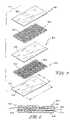
- FIG. 1is a perspective view showing a composite bioprosthetic device of the present invention formed to include a small intestinal submucosa (SIS) portion and a synthetic portion and showing the SIS portion including a top tissue layer of SIS material and a bottom tissue layer of SIS material and further showing the synthetic portion including a row of four fibers positioned to lie in coplanar relation to each other between the top and bottom tissue layers of the SIS portion and positioned to run longitudinally along a length of the SIS portion and extend beyond a first and second end of the SIS portion in order to anchor the bioprosthetic device to surrounding soft tissue;
- SISsmall intestinal submucosa
- FIG. 2is a perspective view similar to FIG. 1 showing an SIS portion of another bioprosthetic device of the present invention being formed to include a top layer, a bottom layer, and two middle layers positioned to lie between the top and the bottom layers and a synthetic device being formed to include three rows of four fibers so that each row is positioned to lie between each of the adjacent tissue layers of the SIS portion so that each fiber is positioned to run longitudinally along a length, L, of the SIS portion;
- FIG. 3is a sectional view taken along line 3 - 3 of FIG. 2 showing the top, bottom, and middle tissue layers of the SIS portion and also showing the three rows of fibers of the synthetic portion of the bioprosthetic device;
- FIG. 4is a perspective view showing an SIS portion of yet another bioprosthetic device of the present invention being formed to include four tissue layers, similar to FIG. 2 , and also showing a synthetic portion of the bioprosthetic device including a first row of multiple fibers positioned to lie between two tissue layers of the SIS portion along a length, L, of the SIS portion and a second row of multiple fibers positioned to lie between two other tissue layers of the SIS portion along a width, W, of the SIS portion;
- FIG. 5is an exploded perspective view of another bioprosthetic device of the present invention showing an SIS portion of the prosthetic device including top, bottom, and middle tissue layers and showing a synthetic portion including a first and a second mesh member positioned to lie between the top and middle tissue layers of and the middle and bottom tissue layers of the SIS portion, respectively;
- FIG. 6is a sectional view of the bioprosthetic device of FIG. 5 showing first and second mesh members “sandwiched” between the tissue layers of the SIS portion of the device;
- FIG. 7is a perspective view showing an SIS portion of another bioprosthetic device being formed to include a top and a bottom tissue layer and further showing a synthetic portion being formed to include a mesh member having a body portion positioned to lie between the top and bottom tissue layers and outer wing portions provided for anchoring the device to surrounding soft tissue;
- FIG. 8is a perspective view showing an SIS portion of yet another bioprosthetic device being formed to include a circularly shaped top and bottom tissue layers each having a diameter, D 1 , and further showing a synthetic portion of the device being formed to include a circular mesh member positioned to lie between the top and bottom tissue layers and having a diameter, D 2 , which is larger than D 1 so that an outer rim portion of the mesh member is formed to extend beyond the top and bottom tissue layers for anchoring the bioprosthetic device to the host tissue during surgery;
- FIG. 9is a sectional view of a bioprosthetic device similar to the bioprosthetic device of FIG. 5 , having two SIS layers, a reinforcing mesh material between the SIS layers, and a reinforced three-dimensional foam portion adjacent one of the SIS layers;
- FIG. 10is sectional view of another bioprosthetic device, wherein the SIS layer is sandwiched between two foam layers;
- FIG. 11is sectional view of another bioprosthetic device, wherein a foam layer is sandwiched between SIS layers;
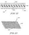
- FIG. 12is a sectional view of another bioprosthetic device, wherein a three-dimensional synthetic layer is sandwiched between two SIS layers;
- FIG. 13is a perspective view showing an SIS portion for use in another bioprosthetic device, wherein the SIS layer is made from weaving strips of SIS.
- a composite bioprosthetic device 10is provided for the purposes of soft tissue attachment, reinforcement, and/or reconstruction.
- Bioprosthetic device 10includes a small intestinal submucosa (SIS) portion 12 and a synthetic portion 14 .
- SIS portion 12is provided to be absorbed into the body and replaced by host tissue.
- SIS portion 12acts as a scaffold for tissue ingrowth and remodeling.
- Synthetic portion 14 of bioprosthetic device 10provides additional initial mechanical strength to bioprosthetic device 10 . Because device 10 includes SIS portion 12 and synthetic portion 14 , bioprosthetic device 10 is provided with a differential in biodegradation and bioremodeling rates.
- Synthetic portion 14for example, can be configured to degrade at a slower rate than SIS portion 12 .
- synthetic portion 14may act as an anchor to couple bioprosthetic device 10 to the surrounding soft tissue (not shown) during surgery.
- the SIS portionmay be sutured to couple the bioprosthetic device to the surrounding tissue.
- SIS portion 12 of bioprosthetic device 10includes a top tissue layer 16 and a bottom tissue layer 18 coupled to top tissue layer 16 mechanically or through a dehydration process.
- top and bottom tissue layers 16 , 18are provided in bioprosthetic device 10 shown in FIG. 1 , it is within the scope of this disclosure, as will be described in more detail later, to include SIS portions 12 having any number of tissue layers. It is also included within the scope of this disclosure to provide perforated tissue layers or any other physical configuration of SIS. See FIGS. 2-4 , for example. Further, it is within the scope of this disclosure to define top and bottom tissue layers 16 , 18 as including multiple tissue layers each.
- top and bottom tissue layers 16 , 18each include three to four layers of SIS tissue.
- SIS portion 12further includes a first end 20 , a second end 22 spaced-apart from first end 20 , and sides 24 coupled to and positioned to lie between first and second ends 20 , 22 .
- a length, L,is defined as the distance between first end 20 and second end 22 and a width, W, is defined as the distance between sides 24 .
- Synthetic portion 14 of bioprosthetic device 10includes row 26 of four fibers 28 , as shown in FIG. 1 . It is within the scope of the disclosure to define fibers to include fibers or any fibrous material. Fibers 28 are positioned to lie along length L between top and bottom tissue layers 16 , 18 and are further positioned to lie in coplanar relation to one another. When making bioprosthetic device 10 , fibers 28 of synthetic portion 14 are placed between top and bottom tissue layers 16 , 18 prior to dehydration. Although row 26 of four fibers 28 is provided in bioprosthetic device 10 shown in FIG. 1 , it is within the scope of this disclosure to include synthetic portions 14 formed to include any number of rows 26 having any number of fibers 28 .
- fibers 28 made from bioabsorbable and non-bioabsorbable materialsare included in the scope of this disclosure.
- fibers 28 made from polylactic acid (PLA) or polyglycolic (PGA) acida combination of the two, PanacrylTM absorbable suture (Ethicon, Inc, Somerville, N.J.), other bioabsorbable materials, nylon, polyethylene, KevlarTM, DacronTM, PTFE, carbon fiber, or other non-bioabsorbable materials.
- each fiber 28 of bioprosthetic device 10includes two outer end portions 30 a middle portion 32 coupled to and positioned to lie between outer end portions 30 .
- Middle portion 32is positioned to lie between top tissue layer 16 and bottom tissue layer 18 of SIS portion 12 .
- Middle portion 32 of fibers 28helps to provide strength along length, L, of bioprosthetic device 10 .
- One or more outer end portions 30 of fibers 28can be used for anchoring bioprosthetic device 10 to surrounding soft tissue (not shown).
- the combination of SIS portion 12 and fibers 28further provide bioprosthetic device 10 with differential biodegradation rates.
- fibers 28 of synthetic portion 14can be made to be non-bioabsorbable or can be made with material which absorbs into the body at a slower rate than SIS portion 12 .
- Uses for bioprosthetic device 10 shown in FIG. 1include, but are not limited to, ligament or tendon repair.
- Bioprosthetic device 110include an alternate SIS portion 112 of having top tissue layer 16 , bottom tissue layer 18 , and two middle tissue layers 115 .
- Top, bottom, and middle tissue layers 16 , 18 , 115include one or more layers of SIS tissue each.
- SIS portion 112similar to SIS portion 12 , also includes a first end 20 , a second end 22 spaced-apart from first end 20 , and sides 24 .
- Bioprosthetic device 110further includes an alternate synthetic portion 114 having three rows 26 of four fibers 28 . One row 26 is positioned to lie between top tissue layer 16 and one of the middle tissue layers 115 .
- Another row 26is positioned to lie between the two middle tissue layers 115 , and the final row 26 of fibers 28 is positioned to lie between another one of the middle tissue layers 115 and bottom tissue layer 16 , as shown in FIG. 3 .
- Fibers 28 of bioprosthetic device 110similar to fibers 28 of bioprosthetic device 10 , are positioned to lie along length, L, of SIS portion 112 .
- fibers 28 of bioprosthetic devices 10 , 110are positioned to lie along length, L, of each respective SIS portion 12 , 112 , it is within the scope of this disclosure to include a synthetic portion 214 of an alternate bioprosthetic device 210 , as shown in FIG. 4 , having multi-directional fibers 28 positioned to lie along a length, L, of an SIS portion 212 and along width, W, of SIS portion 212 .
- Synthetic portion 214 of bioprosthetic device 210includes a first row 226 having seventeen fibers 28 positioned to lie along length, L, of SIS portion 212 .
- Synthetic portion 214further includes a second row 227 having eighteen fibers 28 positioned to lie along width, W, of SIS portion 212 so that the fibers 28 of first row 226 and second row 227 are positioned to lie orthogonally with respect to each other.
- rows 226 and 227are positioned to lie in orthogonal relation to one another, it is within the scope of this disclosure to include synthetic portion 214 having first and second rows 226 and 227 which lie at any angular relation to one another. It is also within the scope of this disclosure to include rows 226 and 227 each having any number of fibers 28 .
- bioprosthetic device 210includes a top tissue layer 216 , a bottom tissue layer 218 , and two middle tissue layers 215 , positioned to lie between top and bottom tissue layers 216 , 218 .
- top, bottom, and middle tissue layers 216 , 218 , 215are each formed to include one or more layers of SIS tissue.
- SIS portion 212 of bioprosthetic device 210is shown to include four tissue layers, it is within the scope of the disclosure to include bioprosthetic device 210 having any number of tissue layers. As shown in FIG.
- first row 226is positioned to lie between top tissue layer 216 and one of the two middle tissue layers 215 positioned to lie adjacent to top tissue layer 216 .
- Second row 227is positioned to lie between the other middle tissue layer 215 and bottom tissue layer 218 . It is within the scope of this disclosure, however, to include rows 226 , 227 positioned to lie between any tissue layer of device 210 .
- Bioprosthetic device 310is similar to devices, 10 , 110 , and 210 and includes an SIS portion 312 having a top tissue layer 316 , a bottom tissue layer 318 , and a middle tissue layer 315 positioned to lie between top and bottom tissue layers 316 , 318 .
- Top, bottom, and middle tissue layers 316 , 318 , 315each include one or more layers of SIS tissue.
- Bioprosthetic device 310further includes a synthetic portion 314 including first mesh member 320 and second mesh member 322 . It is within the scope of this disclosure to include any type of synthetic mesh member.
- bioabsorbable and/or non-bioabsorbable mesh members 320 , 322made of either woven or nonwoven PGA and/or PLA mixtures are included within the scope of disclosure of this invention.
- First mesh member 320is coupled to and positioned to lie between top tissue layer 316 and middle tissue layer 315
- second mesh member 322is coupled to and positioned to lie between middle tissue layer 315 and bottom tissue layer 318 , as shown in FIGS. 5 and 6 .
- Each of the first and second mesh members 320 , 322has a length, L, and a width, W, approximately equal to length, L, and width, W, of tissue layers 315 , 316 , 318 , of SIS portion 312 . It is understood that in some embodiments, it may be preferable for the mesh to be slightly smaller than the SIS portion.
- second mesh member 322is shown partially coated in comminuted SIS 340 .
- Comminuted SISmay be used to fill the interstices of second mesh member 322 to provide a stronger device.
- Other means for reinforcing bioprosthetic device 10may be employed, including suturing or tacking the various layers together.
- comminuted SISis discussed with respect to the embodiment shown in FIG. 5 , it is understood that comminuted SIS may be used to coat the mesh or fibers for any embodiment.
- bioprosthetic device 410having a synthetic portion 414 including a mesh member 420 , as shown in FIG. 7 .
- bioprosthetic device 410includes an SIS portion 412 having a top tissue layer 416 and a bottom tissue layer 418 coupled to top tissue layer 416 .
- Top and bottom tissue layers 416 , 418each include one or more layers of SIS tissue.
- Mesh member 420includes a central body portion (not shown) and outer wing portions 430 , as shown in FIG. 7 .
- Outer wing portions 430are extensions of the central body portion. Although four outer wing portions 430 are shown in FIG.
- a mesh memberhaving a body portion and any number of wing portions 430 coupled to the body portion.
- the central body portion of mesh member 420is formed to include a length and a width equal to length, L, and width, W, of SIS portion 412 .
- the central body portionis coupled to and positioned to lie between top tissue layer 416 and bottom tissue layer 418 of SIS portion 420 .
- Each wing portion 430is coupled to the central body portion of mesh member 420 and is positioned to extend beyond the length, L, and width, W, of SIS portion 412 , as shown in FIG. 7 .
- outer wing portions 430are extensions of the central body portion.
- Wing portions 430provide additional material for anchoring bioprosthetic device 410 to the surrounding soft tissue. Because outer wing portions 430 extend beyond central body portion of mesh member 420 , mesh member 420 has a length and a width greater than length, L, and width, W, of SIS portion 412 .
- Bioprosthetic device 510includes an SIS portion 512 and a synthetic portion 514 coupled to SIS portion 512 .
- SIS portion 512includes a top tissue layer 516 which is circular in shape and a bottom tissue layer 518 which is also circular in shape.
- Each of the top and bottom tissue layers 516 , 518include one or more layers of SIS tissue.
- Top and bottom tissue layers 516 , 518each have a diameter, D 1 .
- the synthetic portion 514 of bioprosthetic device 510includes a mesh member 520 coupled to and positioned to lie between top and bottom tissue layers 516 , 518 .
- Mesh member 520is circular in shape and has a diameter, D 2 , which is greater than diameter, D 1 , of synthetic portion 512 . Therefore, as shown in FIG. 8 , an outer rim portion 530 of mesh member 520 is provided. Similar to outer wing portions 430 of bioprosthetic device 410 , shown in FIG. 7 , outer rim portion 530 of bioprosthetic device 510 provides additional material for anchoring bioprosthetic device 510 to the surrounding soft tissue during surgery.
- FIG. 9shows a three-dimensional prosthetic device 610 , having several SIS layers 612 , a synthetic reinforcing material 614 positioned to lie between the SIS layers 612 , and a three-dimensional synthetic portion 624 .
- the SIS layer 612may comprise any number of tissue layers.
- the layersmay be laminated together. It is included within the scope of this disclosure to provide perforated tissue layers or any other physical configuration of SIS. As with the embodiments shown in FIGS. 5-8 , any number of SIS and reinforcing layers may be used, depending on the application.
- Synthetic reinforcing material 614illustratively comprises a two-dimensional fibrous matrix construct, as shown in FIGS. 5-8 , and may have the same length and width as the SIS layer, as shown in FIG. 5 , may be slightly smaller, or may extend beyond the ends of the SIS layer, as shown in FIGS. 7-8 .
- synthetic reinforcing materialmay comprise a three-dimensional mesh, textile, felt, or other fibrous nonwoven construct, which may be shaped or formed for the particular application.
- the fiberscomprise any biocompatible material, including PLA, PGA, PCL, PDO, TMC, PVA, or copolymers or blends thereof.
- mesh materialis a 95:5 copolymer of PLA/PGA.
- Three-dimensional synthetic portion 624is a nonwoven material prepared to have numerous interconnecting pores or voids 626 .
- the size of the voidsmay range from 20 to 400 microns. However, the size of the voids may be adjusted depending on the application, and the size may be manipulated by changing process steps during construction by altering freezing temperature, rate of temperature change and vacuum profile. Examples of various polymers that may be used for the foam, as well as various lyophilization profiles to control porosity, are described in U.S. Pat. Nos. 6,333,029 and 6,355,699, hereby incorporated by reference.
- three-dimensional synthetic portion 624further comprises a synthetic reinforcing layer 628 embedded within the foam. Reinforcing layer 628 illustratively provides enhanced mechanical integrity to the three-dimensional synthetic portion. In an illustrated embodiment, a Vicryl knitted mesh is used. However, other reinforcing layers may be used.
- three-dimensional synthetic portion 624may be a hybrid ECM/synthetic foam portion.
- the polymer solutionis mixed with a slurry of comminuted SIS prior to lyophilization. See U.S. Application No. 60/388,761 entitled “Extracellular Matrix Scaffold and Method for Making the Same”, hereby incorporated by reference.
- FIG. 10shows a bioprosthetic device 710 that is similar to that of FIG. 9 .
- the SIS layer 712is sandwiched between two three-dimensional synthetic portions 724 , 730 .
- both three-dimensional synthetic portionsare foams, having voids 726 .
- three-dimensional synthetic portion 724has a reinforcing mesh 728
- three-dimensional synthetic portion 730does not have a reinforcing member.
- FIG. 11shows an embodiment 810 where the SIS layer 812 is sandwiched between two three-dimensional synthetic portions 824 , 830 , neither of which has reinforcing members.
- FIG. 12shows another embodiment 910 , wherein a single three-dimensional synthetic portion 964 is sandwiched between two SIS layers 952 , 953 .
- three-dimensional synthetic portion 964is a foam, with voids 966 , but other three-dimensional synthetic portions may be used.
- FIG. 13shows a woven mesh 912 made from strips 928 of SIS.
- Fresh, lyophilized, or laminated strips of SISmay be cut into narrower strips and woven into a mesh.
- the stripsmay be of any width, depending on the application, for example 0.1 to 20 mm, more particularly 1.0 mm wide strips.
- the woven stripsmay be laminated together to provide enhanced mechanical support.
- the SIS woven meshmay be used as the SIS layer in any of the above embodiments. When used with the synthetic foams, if sufficient space is provided in the weaving, the foams will form through the spaces within the mesh.
- Reinforced SIS devicesmay also be fabricated using other processes.
- a synthetic polymer mesh coated with comminuted SIS(or other ECM) may be sandwiched in the middle of twenty strips of SIS (10 layers on each side), laminated under high pressure, and subsequently dried under vacuum pressure in a flat-bed gel drier system.
- the comminuted SIS(or other comminuted ECM) may be prepared in the manner described in U.S. patent application Publication No. 20030044444 A1 entitled “Porous Extracellular Matrix Scaffold and Method” by P. Malaviya et al., the entirety of which is hereby incorporated by reference.
- Such a laminated and dried implantis significantly more resistant to delamination (175 minutes to delaminate using a water bath delamination protocol described below) as compared to implants made either with high pressure lamination but without the comminuted SIS coating (60 minutes to delaminate) or with the comminuted SIS coating but without the high pressure lamination (20-30 minutes to delaminate). It is believed that coating the synthetic mesh with comminuted SIS and then initiating lamination under high pressure has a synergistic effect on the resistance to delamination.
- Such relatively high, positive pressuremay be applied in a number of different manners.
- the relatively high, positive pressureis applied by use of a pneumatic cylinder press assembly.
- Other positive pressure sourcesmay also be used.
- the pressuremay be applied in a wide range of magnitudes.
- Implants fabricated in such a mannermay have higher, and perhaps significantly higher, mechanical properties.
- such implantsmay be used where diseased/damaged tissue needs to be regenerated under high load conditions.
- such implantsmay be used for the augmentation of damaged/resected hip capsule following primary or revision hip surgery, for patellar tendon regeneration, for the repair of large rotator cuff tears, for spinal ligament regeneration, and the like.
- the surface of the synthetic polymeris characteristically hydrophobic in nature, while the SIS surface is hydrophilic.
- the surface of the synthetic polymer componentmay be modified to render it more hydrophilic, and, as a result, more compatible with the SIS surface.
- the more hydrophilic polymer surfacecreates a like-like attraction (e.g., weak force and hydrogen bonding) between the synthetic polymer component and the SIS thereby reducing the occurrences of delamination of the device.
- Such modification of the surface of the polymer componentmay also be used in conjunction with concepts described above for fabricating a pressure-laminated and vacuum-dehydrated composite.
- Surface modification of the synthetic polymer componentmay be accomplished by numerous techniques such as, for example, traditional wet chemistry or gas plasma processing.
- Traditional chemistriesmay include surface hydrolysis and amidation techniques.
- Base or acid catalyzed hydrolysis of the synthetic polymere.g., polyester
- a bifunctional aminemay have an amine on both ends thereof, or, alternatively, may have an amine on one end with any type of hydrophilic group on the other end.
- Gas plasma treatment of the synthetic polymergenerates high energy reactive species that bond to surfaces.
- treatment of a polymer surface with ammonia plasmagenerates an amine functionalized surface.
- an oxidative plasmamay be produced by filtering aqueous hydrogen peroxide into the plasma chamber at approximately 400 mTorr and applying an approximately 200 Watt radio frequency for approximately 3, 5, or 10 minutes.
- crosslinking of the SIS materialmay be avoided, thus retaining more of its biochemical and biological properties.
- crosslinking of the SIS materialmay be used in conjunction with the herein described strengthening techniques.
- the composite implants described hereinmay be used where diseased or damaged tissue needs to be regenerated under high load conditions, for example, for the augmentation of damaged/resected hip capsule following primary or revision hip surgery, for patellar or Achilles tendon regeneration, for the repair of large rotator cuff tears, for spinal ligament regeneration, etcetera.
- devicesmay be fabricated which include a combination of both surface treatment and coating of the synthetic polymer component.
- the synthetic polymer componentmay first be treated to enhance the hydrophilicity of it surface (e.g., by use of wet chemistry or gas plasma treatment). Once treated, the synthetic polymer component may be coated in comminuted SIS (or other naturally occurring extracellular matrix material) in the manner described above. Thereafter, the synthetic polymer component may be secured to layers of SIS (or other ECM).
- the treated and coated synthetic polymer layermay be laminated to one or more SIS layers under high pressure and subsequently dried under vacuum pressure in the manner described above.
- FIGS. 9-13specific embodiments, it is understood that other arrangements are within the scope of this invention.
- an SIS layeris sandwiched between two three-dimensional foam sections, with or without a reinforcing material embedded within the foam. Additional reinforcing layers, as shown in FIG. 9 may be used with these embodiments.
- a layer of reinforcing materialmay be used, depending upon the application.
- the reinforcing portionmay comprise a three-dimensional mesh or textile, and the three-dimensional foam portion may be omitted.
- the synthetic portionmay comprise ProleneTM (Ethicon, Inc, Somerville, N.J.) meshes and/or sutures, VicrylTM (Ethicon, Inc, Somerville, N.J.) meshes and/or sutures, MersileneTM (Ethicon, Inc, Somerville, N.J.) meshes, PDS IITM (Ethicon, Inc., Somerville, N.J.) meshes or sutures, PanacrylTM (Ethicon, Inc., Somerville, N.J.) meshes or sutures, and MonocrylTM meshes or sutures, for example.
- ProleneTMEthicon, Inc, Somerville, N.J.
- VicrylTMEthicon, Inc, Somerville, N.J.
- MersileneTMEthicon, Inc, Somerville, N.J.
- PDS IITMEthicon, Inc., Somerville, N.J.
- PanacrylTMEthicon, Inc., Somerville, N.
- Additional two or three-dimensional meshesmay be constructed for particular applications. Further it is within the scope of this disclosure to include bioprosthetic devices where the SIS portion includes any number of tissue layers and where multiple tissue layers are positioned to lie along each synthetic layer. The SIS layers may be dehydrated prior to or subsequent to assembly of the device. Further, any shape and/or orientation of the SIS portion and the synthetic portion of the bioprosthetic device is within the scope of this disclosure; FIGS. 1-13 are merely examples of various embodiments of the present invention.
- Sheets of clean, disinfected porcine SIS materialwere obtained as described in U.S. Pat. Nos. 4,902,508 and 4,956,178. Ten strips, 3.5 inches wide and 6 inches long were cut. The strips were hydrated by placing in RO water, at room temperature, for 5 minutes.
- SIS stripswere placed on top of each other, while ensuring no air bubbles were trapped between the strips.
- a knitted PanacrylTM mesh2 inches wide and 5 inches long, was placed centrally on the 5-layer thick SIS strip. The mesh had been pretreated to remove any traces of oil or other contaminants due to handling. This was done by a series of rinses, each 2 minutes long, in 100%, 90%, 80%, 70% ethanol (200 proof) in RO water, followed by a final 5 minute in RO water. Subsequently, a second 5-layer thick strip of SIS was assembled and placed to sandwich the mesh between the two SIS strips.
- the implantwas dried under vacuum pressure using a gel drier system (Model FB-GD-45, Fisher Scientific, Pittsburgh, Pa.) for 3 hours.
- the gel drier bed temperaturewas set at 30° C. for the procedure. This drying procedure results in “squeezing out” of the bulk water in the implant and also reduces the amount of bound water within the tissue, resulting in a final moisture of between 7%-8%. This process also results in a physical crosslinking between the laminates of SIS and between the mesh and adjacent SIS laminates.
- Non-reinforced SIS stripswere made in the same way as described, except that no mesh material was placed between the strips of SIS.
- This exampledescribes the preparation of three-dimensional composite tissue implants incorporating a biodegradable SIS laminated sheet, a synthetic reinforcement in the form of a biodegradable mesh, and a synthetic degradable foam.
- a solution of the polymer to be lyophilized to form the foam componentwas prepared in a four step process.
- a 95:5 weight ratio solution of 1,4-dioxane/(40/60 PCL/PLA)was made and poured into a flask.
- the flaskwas placed in a water bath, stirring at 60-70° C. for 5 hrs.
- the solutionwas filtered using an extraction thimble, extra coarse porosity, type ASTM 170-220 (EC) and stored in flasks.
- a three-dimensional mesh material composed of a 95:5 copolymer of polylactic/polyglycolic acid (PLA/PGA) knitted meshwas rendered flat to control curling by using a compression molder at 80° C. for 2 min.
- 0.8-mm metal shimswere placed at each end of a 4 ⁇ 4 inch aluminum mold, and the mesh was sized to fit the mold.
- the synthetic meshwas then laid into the mold, covering both shims.
- an SIS laminated sheetwas placed over the mesh followed by additional shims to cover the edges of the SIS and synthetic mesh.
- the polymer solution(40:60 PCL/PLA) was added into mold such that the solution covered the sheet of SIS as well as the mesh and reached a level of 3.0 mm in the mold.
- the mold assemblythen was placed on the shelf of the lyophilizer (Virtis, Gardiner, N.Y.) and the freeze dry sequence begun.
- the freeze dry sequence used in this examplewas: 1) ⁇ 17° C. for 60 minutes; 2) ⁇ 5° C. for 60 minutes under vacuum of 100 mT; 3) 5° C. for 60 minutes under vacuum of 20 mT; 4) 20° C. for 60 minutes under vacuum of 20 mT.
- the mold assemblywas taken out of the freeze drier and allowed to degas in a vacuum hood for 2 to 3 hours, and stored under nitrogen.
- the resultant bioprosthetic devicehas a structure as illustrated in FIG. 9 .
- the three-dimensional meshprovides both mechanical strength and three-dimensional structure to the resultant device.
- the foammay be shaped or sculpted for the particular application, and the mesh/SIS layers may be trimmed to fit. It is also understood that the mold could be provided in the desired shape, reducing or obviating the need for sculpting or trimming.
- Example 2uses the process outlined in Example 2 to fabricate a biodegradable composite scaffold of the present invention where the foam component is a 65:35 PGA/PCL copolymer.
- Example 2uses the process outlined in Example 2 to fabricate a biodegradable composite scaffold of the present invention where the synthetic knitted mesh component is composed of 100% PDO.
- Example 2uses the process outlined in Example 2 to fabricate a biodegradable composite scaffold of the present invention where in place of a three-dimensional mesh, the synthetic component is a nonwoven fibrous structure composed of either 100% PDO, 100% 90/10 PGA/PLA or a combination of the two.
- This exampleuses the process outlined in Example 2 to fabricate a biodegradable composite scaffold of the present invention where the SIS component is soaked overnight in the polymer solution (5% wt 60/40 PLA/PCL in dioxane) prior to placement over the synthetic mesh. Enhanced lamination between the components was found when this additional soaking step was added to the process as evidenced by a composite with a greater degree of handlability.
- Example 2uses the process outlined in Example 2 to fabricate a biodegradable composite scaffold of the present invention where the SIS component is a single layer sheet rather than a laminated sheet.
- This exampleuses the process outlined in Example 2 to fabricate a biodegradable composite scaffold of the present invention where the SIS laminated sheet is perforated with holes ranging from 1 mm-1 cm. These perforations allow for enhanced penetration of the polymer solution through the SIS sheet.
- Example 2uses the process outlined in Example 2 to fabricate a biodegradable composite scaffold of the present invention where the SIS reinforcing component is a “woven mesh” of laminated strips sandwiched between two layers of 60/40 PLA/PCL foam.
- FIG. 13shows such a woven mesh.
- FIG. 11wherein the SIS layer is a woven mesh of FIG. 13 , illustrates the construct of this Example.
- a soaking testwas performed to test resistance to delamination.
- Implants made as specified in Example 1both reinforced and non-reinforced were cut into several strips 1 cm wide by 5 cm long, using a #10 scalpel blade. The strips were immersed in RO water, at room temperature for 1, 2, 5, 10, 20, 30, or 60 minutes. Delamination was detected at the edges of the implants by direct visual observation. All implants showed obvious signs of delamination at 1 hour. In non-reinforced implants, delamination was first visually observed between 40-60 minutes, whereas in the reinforced samples delamination was apparent between 20-30 minutes.
- This exampleillustrates the enhanced mechanical properties of a construct reinforced with absorbable mesh. Preparation of three-dimensional elastomeric tissue implants with and without a reinforcement in the form of a biodegradable mesh are described. While a foam is used for the elastomeric tissue in this example, it is expected that similar results will be achieved with an ECM and a biodegradable mesh.
- a solution of the polymer to be lyophilized to form the foam componentwas prepared in a four step process.
- a 95/5 weight ratio solution of 1,4-dioxane/(40/60 PCL/PLA)was made and poured into a flask.
- the flaskwas placed in a water bath, stirring at 70° C. for 5 hrs.
- the solutionwas filtered using an extraction thimble, extra coarse porosity, type ASTM 170-220 (EC) and stored in flasks.
- Reinforcing mesh materials formed of a 90/10 copolymer of polyglycolic/polylactic acid (PGA/PLA) knitted (Code VKM-M) and woven (Code VWM-M), both sold under the tradename VICRYLwere rendered flat by ironing, using a compression molder at 80° C./2 min.
- PGA/PLApolyglycolic/polylactic acid
- VICRYLtradename VICRYL
- the moldwas tilted to about a 5 degree angle so that one of the non-clamping sides was higher than the other. Approximately 60 ml of the polymer solution was slowly transferred into the mold, ensuring that the solution was well dispersed in the mold. The mold was then placed on a shelf in a Virtis (Gardiner, N.Y.), Freeze Mobile G freeze dryer. The following freeze drying sequence was used: 1) 20° C. for 15 minutes; 2) ⁇ 5° C. for 120 minutes; 3) ⁇ 5° C. for 90 minutes under vacuum 100 milliTorr; 4) 5° C. for 90 minutes under vacuum 100 milliTorr; 5) 20° C. for 90 minutes under vacuum 100 milliTorr. The mold assembly was then removed from the freezer and placed in a nitrogen box overnight. Following the completion of this process the resulting implant was carefully peeled out of the mold in the form of a foam/mesh sheet.
- Nonreinforced foamswere also fabricated. To obtain non-reinforced foams, however, the steps regarding the insertion of the mesh into the mold were not performed. The lyophilization steps above were followed.
- the dimensions of the specimenswere approximately 5 cm ⁇ 9 cm. Specimens were tested for pull-out strength in the wale direction of the mesh (knitting machine axis).
- a size 0 polypropylene monofilament suture(Code 8834H), sold under the tradename PROLENE (by Ethicon, Inc., Somerville, N.J.) was passed through the mesh 6.25 mm from the edge of the specimens. The ends of the suture were clamped into the upper jaw and the mesh or the reinforced foam was clamped into the lower jaw of an Instron model 4501 (Canton, Mass.).
- the Instron machinewith a 20 lb load cell, was activated using a cross-head speed of 2.54 cm per minute. The ends of the suture were pulled at a constant rate until failure occurred. The peak load (lbs.) experienced during the pulling was recorded.
- Sheets of clean, disinfected porcine SIS materialwere obtained as described in patents U.S. Pat. Nos. 4,902,508 and 4,956,178. Twenty strips, 3.5 inches wide and 6 inches long were cut. The strips were hydrated by placing in RO water, at room temperature, for 5 minutes.
- Lamination of the thus assembled implantwas initiated under high pressure using a pneumatic cylinder press (Model BTP-501-A, TRD Manufacturing Inc., Loves Park, Ill. 61111.)
- the presswas operated at 40 psi air pressure to drive the piston, which resulted in a total compressive force of approximately 4000 lbs on the assembled implant. This force created an approximate average lamination pressure of 180 psi on the implant.
- the samplewas compressed for 15 minutes at room temperature. This process resulted in a “squeezing out” of most of the bulk water associated with the SIS laminates and comminuted SIS and created a partially wet laminated implant.
- the implantwas subsequently dried under vacuum pressure using a flat-bed gel drier system (Model FB-GD-45, Fisher Scientific, Pittsburgh, Pa.) for 3 hours.
- the gel drier bed temperaturewas set at 30° C. for the procedure.
- This drying procedureresulted in a further reduction of the bulk water associated with the implant and also reduced the amount of bound water within the implant, resulting in a final moisture content between 7%-8%.
- This processalso results in a physical crosslinking between the laminates of SIS and the comminuted SIS coating the synthetic mesh by further increasing the surface contact area of SIS material.
- Implantswere also made as described above but without coating the PanacrylTM mesh with the comminuted SIS fibers.
- high pressure laminated SIS implants reinforced with a comminuted SIS coated synthetic meshwill also have higher (and perhaps significantly higher) mechanical properties (e.g. higher ball burst strength) as compared with implants made without high pressure lamination or without a comminuted SIS coating on the synthetic mesh.
Landscapes
- Health & Medical Sciences (AREA)
- Life Sciences & Earth Sciences (AREA)
- Biomedical Technology (AREA)
- Chemical & Material Sciences (AREA)
- Engineering & Computer Science (AREA)
- Oral & Maxillofacial Surgery (AREA)
- General Health & Medical Sciences (AREA)
- Chemical Kinetics & Catalysis (AREA)
- Dermatology (AREA)
- Medicinal Chemistry (AREA)
- Molecular Biology (AREA)
- Transplantation (AREA)
- Epidemiology (AREA)
- Animal Behavior & Ethology (AREA)
- Botany (AREA)
- Public Health (AREA)
- Veterinary Medicine (AREA)
- Urology & Nephrology (AREA)
- Zoology (AREA)
- Biophysics (AREA)
- Prostheses (AREA)
- Materials For Medical Uses (AREA)
Abstract
Description
| TABLE 1 |
| Suture Pull-Out Data (lbs.) |
| Time | Foam | Mesh | Foamed Mesh | ||
| 0 Day | 0.46 | 5.3 +/− 0.8 | 5.7 +/− 0.3 | ||
| 7 Day* | — | 4.0 +/− 1.0 | 5.0 +/− 0.5 | ||
| *exposed for 7 days to phosphate buffered saline at 37° C. in a temperature controlled water bath. | |||||
Claims (25)
Priority Applications (1)
| Application Number | Priority Date | Filing Date | Title |
|---|---|---|---|
| US11/110,169US7569233B2 (en) | 2004-05-04 | 2005-04-20 | Hybrid biologic-synthetic bioabsorbable scaffolds |
Applications Claiming Priority (3)
| Application Number | Priority Date | Filing Date | Title |
|---|---|---|---|
| US56788604P | 2004-05-04 | 2004-05-04 | |
| US57176604P | 2004-05-17 | 2004-05-17 | |
| US11/110,169US7569233B2 (en) | 2004-05-04 | 2005-04-20 | Hybrid biologic-synthetic bioabsorbable scaffolds |
Publications (2)
| Publication Number | Publication Date |
|---|---|
| US20050249771A1 US20050249771A1 (en) | 2005-11-10 |
| US7569233B2true US7569233B2 (en) | 2009-08-04 |
Family
ID=35239679
Family Applications (1)
| Application Number | Title | Priority Date | Filing Date |
|---|---|---|---|
| US11/110,169Active2026-06-28US7569233B2 (en) | 2004-05-04 | 2005-04-20 | Hybrid biologic-synthetic bioabsorbable scaffolds |
Country Status (1)
| Country | Link |
|---|---|
| US (1) | US7569233B2 (en) |
Cited By (13)
| Publication number | Priority date | Publication date | Assignee | Title |
|---|---|---|---|---|
| US20090011507A1 (en)* | 2007-07-07 | 2009-01-08 | Julian Ellis | Scaffolds for Use in Tissue Engineering to Culture Cells |
| US20090280154A1 (en)* | 2005-10-27 | 2009-11-12 | Peter Sylvest Nielsen | Biodegradable Scaffold with ECM Material |
| US20100040660A1 (en)* | 2008-08-12 | 2010-02-18 | Korea Research Institute Of Chemical Technology | Development of a tissue - engineered scaffold for nerve regeneration using a biocompatible and injectable hydrogel |
| US20100063599A1 (en)* | 2008-09-05 | 2010-03-11 | Pegasus Biologics, Inc. | Device for soft tissue repair or replacement |
| US20110052526A1 (en)* | 2009-09-02 | 2011-03-03 | Khay-Yong Saw | Method and composition for neochondrogenesis |
| WO2012024390A3 (en)* | 2010-08-17 | 2012-05-31 | University Of Pittsburgh - Of The Commonwealth System Of Higher Education | Biohybrid composite scaffold |
| US20120221107A1 (en)* | 2007-07-07 | 2012-08-30 | Jmea Corporation | Disk Fusion Implant |
| KR101242656B1 (en) | 2011-03-04 | 2013-03-20 | 포항공과대학교 산학협력단 | Artificial pancreatic islet cell structure and manufacturing method thereof |
| US20150081010A1 (en)* | 2012-10-08 | 2015-03-19 | Robert G Matheny | Reinforced Vascular Prostheses |
| US9238090B1 (en) | 2014-12-24 | 2016-01-19 | Fettech, Llc | Tissue-based compositions |
| US9901457B2 (en) | 2014-10-16 | 2018-02-27 | Jmea Corporation | Coiling implantable prostheses |
| US10758644B2 (en) | 2008-03-27 | 2020-09-01 | The Cleveland Clinic Foundation | Reinforced tissue graft |
| US11013590B2 (en) | 2008-03-27 | 2021-05-25 | The Cleveland Clinic Foundation | Reinforced tissue graft |
Families Citing this family (22)
| Publication number | Priority date | Publication date | Assignee | Title |
|---|---|---|---|---|
| US8366787B2 (en)* | 2000-08-04 | 2013-02-05 | Depuy Products, Inc. | Hybrid biologic-synthetic bioabsorbable scaffolds |
| US7819918B2 (en) | 2001-07-16 | 2010-10-26 | Depuy Products, Inc. | Implantable tissue repair device |
| JP4197158B2 (en) | 2001-07-16 | 2008-12-17 | デピュイ・プロダクツ・インコーポレイテッド | Devices with naturally occurring biologically derived materials |
| WO2003007784A2 (en) | 2001-07-16 | 2003-01-30 | Depuy Products, Inc. | Meniscus regeneration device and method |
| US8012205B2 (en)* | 2001-07-16 | 2011-09-06 | Depuy Products, Inc. | Cartilage repair and regeneration device |
| US20050027307A1 (en) | 2001-07-16 | 2005-02-03 | Schwartz Herbert Eugene | Unitary surgical device and method |
| US8025896B2 (en) | 2001-07-16 | 2011-09-27 | Depuy Products, Inc. | Porous extracellular matrix scaffold and method |
| US20060095048A1 (en) | 2004-10-29 | 2006-05-04 | Zannis Anthony D | Method of repairing soft tissue using sizing templates |
| US7354627B2 (en) | 2004-12-22 | 2008-04-08 | Depuy Products, Inc. | Method for organizing the assembly of collagen fibers and compositions formed therefrom |
| US7595062B2 (en)* | 2005-07-28 | 2009-09-29 | Depuy Products, Inc. | Joint resurfacing orthopaedic implant and associated method |
| US9532943B2 (en)* | 2010-12-20 | 2017-01-03 | Cormatrix Cardiovascular, Inc. | Drug eluting patch for the treatment of localized tissue disease or defect |
| US7871440B2 (en) | 2006-12-11 | 2011-01-18 | Depuy Products, Inc. | Unitary surgical device and method |
| CN101626791A (en)* | 2007-03-07 | 2010-01-13 | 科洛普拉斯特公司 | Mesh comprising ECM |
| US9636438B2 (en)* | 2007-03-07 | 2017-05-02 | Coloplast A/S | Fistula plug comprising ECM |
| DE102008022319A1 (en)* | 2008-04-30 | 2009-11-05 | Aesculap Ag | Implant, in particular for restoring and / or regenerating human and / or animal tissue |
| US9295757B2 (en)* | 2008-06-10 | 2016-03-29 | Cook Biotech Incorporated | Quilted implantable graft |
| WO2009152215A2 (en)* | 2008-06-10 | 2009-12-17 | Cook Biotech Incorporated | Quilted implantable graft |
| US8636803B2 (en)* | 2009-04-07 | 2014-01-28 | Spinal Stabilization Technologies, Llc | Percutaneous implantable nuclear prosthesis |
| US8298586B2 (en)* | 2009-07-22 | 2012-10-30 | Acell Inc | Variable density tissue graft composition |
| JP5863064B2 (en)* | 2010-12-20 | 2016-02-16 | コーマトリックス カーディオバスキュラー, インコーポレイテッドCorMatrix Cardiovascular, Inc. | Patch extractant for localized tissue disease or defect area of the present invention |
| GB201317636D0 (en) | 2013-10-04 | 2013-11-20 | Isis Innovation | Scaffold |
| EP4142647A1 (en) | 2020-05-01 | 2023-03-08 | Harbor Medtech, Inc. | Port-accessible multidirectional reinforced minimally invasive collagen device for soft tissue repair |
Citations (250)
| Publication number | Priority date | Publication date | Assignee | Title |
|---|---|---|---|---|
| US3272204A (en) | 1965-09-22 | 1966-09-13 | Ethicon Inc | Absorbable collagen prosthetic implant with non-absorbable reinforcing strands |
| US3562820A (en) | 1966-08-22 | 1971-02-16 | Bernhard Braun | Tubular sheet and strip form prostheses on a basis of biological tissue |
| US4105034A (en) | 1977-06-10 | 1978-08-08 | Ethicon, Inc. | Poly(alkylene oxalate) absorbable coating for sutures |
| US4130639A (en) | 1977-09-28 | 1978-12-19 | Ethicon, Inc. | Absorbable pharmaceutical compositions based on isomorphic copolyoxalates |
| US4140678A (en) | 1977-06-13 | 1979-02-20 | Ethicon, Inc. | Synthetic absorbable surgical devices of poly(alkylene oxalates) |
| US4141087A (en) | 1977-01-19 | 1979-02-27 | Ethicon, Inc. | Isomorphic copolyoxalates and sutures thereof |
| FR2422386A1 (en) | 1978-04-13 | 1979-11-09 | Christian Buscayret | Device for fastening suture into tissue - has rectangular body with two perforations accommodating suture and two protuberances engaging tissue |
| US4205399A (en) | 1977-06-13 | 1980-06-03 | Ethicon, Inc. | Synthetic absorbable surgical devices of poly(alkylene oxalates) |
| US4208511A (en) | 1977-01-19 | 1980-06-17 | Ethicon, Inc. | Isomorphic copolyoxalates and sutures thereof |
| US4352463A (en) | 1979-01-18 | 1982-10-05 | Leisure Lawn, Inc. | Motorized combination wet and dry lawn treatment spreader |
| US4400833A (en) | 1981-06-10 | 1983-08-30 | Kurland Kenneth Z | Means and method of implanting bioprosthetics |
| US4418691A (en) | 1981-10-26 | 1983-12-06 | Massachusetts Institute Of Technology | Method of promoting the regeneration of tissue at a wound |
| US4610397A (en) | 1983-10-27 | 1986-09-09 | Urschel Laboratories Incorporated | Comminuting equipment |
| US4642120A (en) | 1983-03-23 | 1987-02-10 | Ramot University Authority For Applied Research And Industrial Development Ltd. | Repair of cartilage and bones |
| US4669473A (en) | 1985-09-06 | 1987-06-02 | Acufex Microsurgical, Inc. | Surgical fastener |
| US4703108A (en) | 1984-03-27 | 1987-10-27 | University Of Medicine & Dentistry Of New Jersey | Biodegradable matrix and methods for producing same |
| US4705040A (en) | 1985-11-18 | 1987-11-10 | Medi-Tech, Incorporated | Percutaneous fixation of hollow organs |
| US4741330A (en) | 1983-05-19 | 1988-05-03 | Hayhurst John O | Method and apparatus for anchoring and manipulating cartilage |
| US4750492A (en) | 1985-02-27 | 1988-06-14 | Richards Medical Company | Absorbable suture apparatus, method and installer |
| US4846835A (en) | 1987-06-15 | 1989-07-11 | Grande Daniel A | Technique for healing lesions in cartilage |
| US4873976A (en) | 1984-02-28 | 1989-10-17 | Schreiber Saul N | Surgical fasteners and method |
| US4880429A (en) | 1987-07-20 | 1989-11-14 | Stone Kevin R | Prosthetic meniscus |
| US4902508A (en) | 1988-07-11 | 1990-02-20 | Purdue Research Foundation | Tissue graft composition |
| US4919667A (en) | 1988-12-02 | 1990-04-24 | Stryker Corporation | Implant |
| WO1990009769A1 (en) | 1989-03-02 | 1990-09-07 | Regen Corporation | Prosthetic meniscus |
| US4956178A (en) | 1988-07-11 | 1990-09-11 | Purdue Research Foundation | Tissue graft composition |
| US4956179A (en) | 1971-11-01 | 1990-09-11 | Astra Lakemedel Aktiebolag | Antibacterial combination of penicillin and cephalosporin |
| US4976715A (en) | 1986-05-20 | 1990-12-11 | Concept, Inc. | Repair tack for bodily tissue |
| EP0446105A2 (en) | 1990-03-06 | 1991-09-11 | Showa Shell Sekiyu Kabushiki Kaisha | Water absorptive and retentive flexible cloth and method for producing same |
| US5061286A (en) | 1989-08-18 | 1991-10-29 | Osteotech, Inc. | Osteoprosthetic implant |
| US5102421A (en) | 1990-06-14 | 1992-04-07 | Wm. E. Anpach, III | Suture anchor and method of forming |
| US5108438A (en) | 1989-03-02 | 1992-04-28 | Regen Corporation | Prosthetic intervertebral disc |
| US5128326A (en) | 1984-12-06 | 1992-07-07 | Biomatrix, Inc. | Drug delivery systems based on hyaluronans derivatives thereof and their salts and methods of producing same |
| USRE34021E (en) | 1985-11-18 | 1992-08-04 | Abbott Laboratories | Percutaneous fixation of hollow organs |
| GB2215209B (en) | 1988-03-14 | 1992-08-26 | Osmed Inc | Method and apparatus for biodegradable, osteogenic, bone graft substitute device |
| US5236431A (en) | 1991-07-22 | 1993-08-17 | Synthes | Resorbable fixation device with controlled stiffness for treating bodily material in vivo and introducer therefor |
| US5246441A (en) | 1989-09-08 | 1993-09-21 | Linvatec Corporation | Bioabsorbable tack for joining bodily tissue |
| US5258015A (en) | 1991-05-03 | 1993-11-02 | American Cyanamid Company | Locking filament caps |
| US5269809A (en) | 1990-07-02 | 1993-12-14 | American Cyanamid Company | Locking mechanism for use with a slotted suture anchor |
| US5275826A (en) | 1992-11-13 | 1994-01-04 | Purdue Research Foundation | Fluidized intestinal submucosa and its use as an injectable tissue graft |
| US5281422A (en) | 1991-09-24 | 1994-01-25 | Purdue Research Foundation | Graft for promoting autogenous tissue growth |
| EP0591991A2 (en) | 1992-10-09 | 1994-04-13 | United States Surgical Corporation | Suture loop locking device |
| US5306311A (en) | 1987-07-20 | 1994-04-26 | Regen Corporation | Prosthetic articular cartilage |
| US5320633A (en) | 1992-12-10 | 1994-06-14 | William C. Allen | Method and system for repairing a tear in the meniscus |
| US5329846A (en) | 1991-08-12 | 1994-07-19 | Bonutti Peter M | Tissue press and system |
| US5350583A (en) | 1988-03-09 | 1994-09-27 | Terumo Kabushiki Kaisha | Cell-penetrable medical material and artificial skin |
| US5352463A (en) | 1992-11-13 | 1994-10-04 | Badylak Steven F | Tissue graft for surgical reconstruction of a collagenous meniscus and method therefor |
| US5374268A (en) | 1991-05-13 | 1994-12-20 | United States Surgical Corporation | Device and method for repairing torn tissue |
| US5376118A (en) | 1989-05-10 | 1994-12-27 | United States Surgical Corporation | Support material for cell impregnation |
| US5380334A (en) | 1993-02-17 | 1995-01-10 | Smith & Nephew Dyonics, Inc. | Soft tissue anchors and systems for implantation |
| EP0632999A1 (en) | 1993-07-01 | 1995-01-11 | United States Surgical Corporation | Soft tissue repair system and method |
| WO1995005083A1 (en) | 1993-08-13 | 1995-02-23 | Smith & Nephew Richards Inc | Microporous polymeric foams and microtextured surfaces |
| WO1995006439A1 (en) | 1993-08-31 | 1995-03-09 | Depuy Inc. | Catgut orthopaedic appliance |
| WO1995022301A1 (en) | 1994-02-18 | 1995-08-24 | Organogenesis, Inc. | Bioremodelable collagen graft prosthesis |
| US5447940A (en) | 1993-07-28 | 1995-09-05 | Johnson & Johnson Medical, Inc. | Absorbable composite materials for use in the treatment of periodontal disease |
| US5460962A (en) | 1994-01-04 | 1995-10-24 | Organogenesis Inc. | Peracetic acid sterilization of collagen or collagenous tissue |
| US5464929A (en) | 1995-03-06 | 1995-11-07 | Ethicon, Inc. | Absorbable polyoxaesters |
| WO1995032623A1 (en) | 1994-05-27 | 1995-12-07 | Regen Biologics, Inc. | Meniscal augmentation device |
| US5479033A (en) | 1994-05-27 | 1995-12-26 | Sandia Corporation | Complementary junction heterostructure field-effect transistor |
| US5514181A (en) | 1993-09-29 | 1996-05-07 | Johnson & Johnson Medical, Inc. | Absorbable structures for ligament and tendon repair |
| US5520691A (en) | 1990-11-06 | 1996-05-28 | Branch; Thomas P. | Method and apparatus for re-approximating tissue |
| US5545178A (en) | 1994-04-29 | 1996-08-13 | Kensey Nash Corporation | System for closing a percutaneous puncture formed by a trocar to prevent tissue at the puncture from herniating |
| WO1996024304A1 (en) | 1995-02-10 | 1996-08-15 | The Hospital For Joint Diseases, Orthopaedic Institute | Fixation method for the attachment of wound repair materials to cartilage defects |
| WO1996024661A1 (en) | 1995-02-10 | 1996-08-15 | Purdue Research Foundation | Submucosa as a growth substrate for cells |
| US5554389A (en) | 1995-04-07 | 1996-09-10 | Purdue Research Foundation | Urinary bladder submucosa derived tissue graft |
| EP0734736A1 (en) | 1995-03-31 | 1996-10-02 | Toyo Boseki Kabushiki Kaisha | Medical device and method for producing the same |
| US5569252A (en) | 1994-09-27 | 1996-10-29 | Justin; Daniel F. | Device for repairing a meniscal tear in a knee and method |
| US5591234A (en) | 1993-02-01 | 1997-01-07 | Axel Kirsch | Post-surgery orthopedic covering |
| US5593441A (en) | 1992-03-04 | 1997-01-14 | C. R. Bard, Inc. | Method for limiting the incidence of postoperative adhesions |
| US5595751A (en) | 1995-03-06 | 1997-01-21 | Ethicon, Inc. | Absorbable polyoxaesters containing amines and/or amido groups |
| US5597579A (en) | 1995-03-06 | 1997-01-28 | Ethicon, Inc. | Blends of absorbable polyoxaamides |
| WO1997005193A1 (en) | 1995-07-28 | 1997-02-13 | Sanitaria Scaligera Spa | A process of surface activation of biocompatible and bioabsorbable aliphatic polyesters and polyesters thus activated |
| US5607687A (en) | 1995-03-06 | 1997-03-04 | Ethicon, Inc. | Polymer blends containing absorbable polyoxaesters |
| US5618552A (en) | 1995-03-06 | 1997-04-08 | Ethicon, Inc. | Absorbable polyoxaesters |
| US5620698A (en) | 1995-03-06 | 1997-04-15 | Ethicon, Inc. | Blends of absorbable polyoxaesters containing amines and/or amido groups |
| US5626614A (en) | 1995-12-22 | 1997-05-06 | Applied Medical Resources Corporation | T-anchor suturing device and method for using same |
| US5630824A (en) | 1994-06-01 | 1997-05-20 | Innovasive Devices, Inc. | Suture attachment device |
| US5632745A (en) | 1995-02-07 | 1997-05-27 | R&D Biologicals, Inc. | Surgical implantation of cartilage repair unit |
| US5641518A (en) | 1992-11-13 | 1997-06-24 | Purdue Research Foundation | Method of repairing bone tissue |
| US5645860A (en) | 1995-04-07 | 1997-07-08 | Purdue Research Foundation | Tissue graft and method for urinary urothelium reconstruction replacement |
| US5660225A (en) | 1993-08-30 | 1997-08-26 | Saffran; Bruce Nathan | Method of fracture treatment by restraining macromolecules or macromolecular aggregates adjacent to damaged tissues |
| US5668288A (en) | 1996-04-16 | 1997-09-16 | Depuy Orthopaedics, Inc. | Polyester ionomers for implant fabrication |
| US5669912A (en) | 1994-04-26 | 1997-09-23 | Spetzler; Robert F. | Apparatus for securing a cranial piece in position |
| WO1997037613A1 (en) | 1996-04-05 | 1997-10-16 | Depuy Orthopaedics, Inc. | Tissue graft construct for replacement of cartilaginous structures |
| US5693085A (en) | 1994-04-29 | 1997-12-02 | Scimed Life Systems, Inc. | Stent with collagen |
| US5698213A (en) | 1995-03-06 | 1997-12-16 | Ethicon, Inc. | Hydrogels of absorbable polyoxaesters |
| US5700583A (en) | 1995-03-06 | 1997-12-23 | Ethicon, Inc. | Hydrogels of absorbable polyoxaesters containing amines or amido groups |
| US5702462A (en) | 1996-01-24 | 1997-12-30 | Oberlander; Michael | Method of meniscal repair |
| US5711969A (en) | 1995-04-07 | 1998-01-27 | Purdue Research Foundation | Large area submucosal tissue graft constructs |
| US5711960A (en) | 1993-09-24 | 1998-01-27 | Takiron Co., Ltd. | Biocompatible implant material comprising a tri-axial or more three-dimensional fabric |
| WO1998006445A1 (en) | 1996-08-16 | 1998-02-19 | Children's Medical Center Corporation | Bladder submucosa seeded with cells for tissue reconstruction |
| US5725556A (en) | 1995-12-15 | 1998-03-10 | M & R Medical, Inc. | Suture locking apparatus |
| US5730933A (en) | 1996-04-16 | 1998-03-24 | Depuy Orthopaedics, Inc. | Radiation sterilization of biologically active compounds |
| US5733868A (en) | 1996-04-16 | 1998-03-31 | Depuy Orthopaedics, Inc. | Poly(amino acid) adhesive tissue grafts |
| US5733337A (en) | 1995-04-07 | 1998-03-31 | Organogenesis, Inc. | Tissue repair fabric |
| US5736372A (en) | 1986-11-20 | 1998-04-07 | Massachusetts Institute Of Technology | Biodegradable synthetic polymeric fibrous matrix containing chondrocyte for in vivo production of a cartilaginous structure |
| US5735897A (en) | 1993-10-19 | 1998-04-07 | Scimed Life Systems, Inc. | Intravascular stent pump |
| US5755791A (en) | 1996-04-05 | 1998-05-26 | Purdue Research Foundation | Perforated submucosal tissue graft constructs |
| WO1998022158A2 (en) | 1996-08-23 | 1998-05-28 | Cook Biotech, Incorporated | Graft prosthesis, materials and methods |
| US5759205A (en) | 1994-01-21 | 1998-06-02 | Brown University Research Foundation | Negatively charged polymeric electret implant |
| US5759190A (en) | 1996-08-30 | 1998-06-02 | Vts Holdings Limited | Method and kit for autologous transplantation |
| US5762600A (en) | 1994-04-29 | 1998-06-09 | W. L. Gore & Associates, Inc. | Blood contact surfaces employing natural subendothelial matrix and methods for making and using the same |
| US5769899A (en) | 1994-08-12 | 1998-06-23 | Matrix Biotechnologies, Inc. | Cartilage repair unit |
| US5773577A (en) | 1994-03-03 | 1998-06-30 | Protein Polymer Technologies | Products comprising substrates capable of enzymatic cross-linking |
| WO1998030167A1 (en) | 1997-01-14 | 1998-07-16 | Research Corporation Technologies, Inc. | Bone fixation pin with rotary cutting tip |
| WO1998034569A1 (en) | 1997-02-11 | 1998-08-13 | Smith & Nephew, Inc. | Repairing cartilage |
| US5795353A (en) | 1994-05-06 | 1998-08-18 | Advanced Bio Surfaces, Inc. | Joint resurfacing system |
| US5800537A (en) | 1992-08-07 | 1998-09-01 | Tissue Engineering, Inc. | Method and construct for producing graft tissue from an extracellular matrix |
| WO1998040111A1 (en) | 1997-03-07 | 1998-09-17 | University College London | Tissue implant |
| US5830708A (en) | 1995-06-06 | 1998-11-03 | Advanced Tissue Sciences, Inc. | Methods for production of a naturally secreted extracellular matrix |
| US5834232A (en) | 1996-05-01 | 1998-11-10 | Zymogenetics, Inc. | Cross-linked gelatin gels and methods of making them |
| US5842477A (en) | 1996-02-21 | 1998-12-01 | Advanced Tissue Sciences, Inc. | Method for repairing cartilage |
| US5855619A (en) | 1994-06-06 | 1999-01-05 | Case Western Reserve University | Biomatrix for soft tissue regeneration |
| US5855613A (en) | 1995-10-13 | 1999-01-05 | Islet Sheet Medical, Inc. | Retrievable bioartificial implants having dimensions allowing rapid diffusion of oxygen and rapid biological response to physiological change |
| US5855610A (en) | 1995-05-19 | 1999-01-05 | Children's Medical Center Corporation | Engineering of strong, pliable tissues |
| US5859150A (en) | 1995-03-06 | 1999-01-12 | Ethicon, Inc. | Prepolymers of absorbable polyoxaesters |
| US5861004A (en) | 1991-11-08 | 1999-01-19 | Kensey Nash Corporation | Hemostatic puncture closure system including closure locking means and method of use |
| US5863551A (en) | 1996-10-16 | 1999-01-26 | Organogel Canada Ltee | Implantable polymer hydrogel for therapeutic uses |
| WO1999003979A1 (en) | 1997-07-17 | 1999-01-28 | Smith & Nephew Plc | Methods for the preparation of artificial cellular tissue |
| US5891558A (en) | 1994-11-22 | 1999-04-06 | Tissue Engineering, Inc. | Biopolymer foams for use in tissue repair and reconstruction |
| US5899939A (en) | 1998-01-21 | 1999-05-04 | Osteotech, Inc. | Bone-derived implant for load-supporting applications |
| US5906997A (en) | 1997-06-17 | 1999-05-25 | Fzio Med, Inc. | Bioresorbable compositions of carboxypolysaccharide polyether intermacromolecular complexes and methods for their use in reducing surgical adhesions |
| US5916265A (en) | 1994-03-30 | 1999-06-29 | Hu; Jie | Method of producing a biological extracellular matrix for use as a cell seeding scaffold and implant |
| US5939323A (en) | 1996-05-28 | 1999-08-17 | Brown University | Hyaluronan based biodegradable scaffolds for tissue repair |
| US5955100A (en) | 1992-10-27 | 1999-09-21 | Behringwerke Aktiengesellschaft | Prodrugs their preparation and use as pharmaceuticals |
| US5954723A (en) | 1994-04-26 | 1999-09-21 | Spetzler; Robert F. | Method and apparatus for securing a cranial plate with pins |
| US5954747A (en) | 1997-11-20 | 1999-09-21 | Clark; Ron | Meniscus repair anchor system |
| WO1999047188A1 (en) | 1998-03-17 | 1999-09-23 | Tissue Engineering, Inc. | Biopolymer matt for use in tissue repair and reconstruction |
| US5958874A (en) | 1998-02-18 | 1999-09-28 | The Research Foundation Of State University Of New York | Recombinant fibronectin-based extracellular matrix for wound healing |
| US5971987A (en) | 1998-09-18 | 1999-10-26 | Ethicon, Inc. | Biocompatible absorbable polymer fastener and driver for use in surgical procedures |
| US5981825A (en) | 1994-05-13 | 1999-11-09 | Thm Biomedical, Inc. | Device and methods for in vivo culturing of diverse tissue cells |
| US5980524A (en) | 1997-06-02 | 1999-11-09 | Innovasive Devices, Inc. | Device for repairing a meniscal tear in a knee and method |
| US5981802A (en) | 1995-12-14 | 1999-11-09 | The Trustees Of Columbia University In The City Of New York | Simple allylic amination procedure |
| WO1999043786A3 (en) | 1998-02-27 | 1999-11-11 | Purdue Research Foundation | Submucosa gel compositions |
| US5989280A (en) | 1993-10-22 | 1999-11-23 | Scimed Lifesystems, Inc | Stent delivery apparatus and method |
| US5989269A (en) | 1996-08-30 | 1999-11-23 | Vts Holdings L.L.C. | Method, instruments and kit for autologous transplantation |
| JPH11319068A (en) | 1998-05-12 | 1999-11-24 | Menicon Co Ltd | Base material for artificial skin and production thereof |
| US5993475A (en) | 1998-04-22 | 1999-11-30 | Bristol-Myers Squibb Co. | Tissue repair device |
| US5993844A (en) | 1997-05-08 | 1999-11-30 | Organogenesis, Inc. | Chemical treatment, without detergents or enzymes, of tissue to form an acellular, collagenous matrix |
| US6010525A (en) | 1997-08-01 | 2000-01-04 | Peter M. Bonutti | Method and apparatus for securing a suture |
| US6017348A (en) | 1995-03-07 | 2000-01-25 | Innovasive Devices, Inc. | Apparatus and methods for articular cartilage defect repair |
| US6027744A (en) | 1998-04-24 | 2000-02-22 | University Of Massachusetts Medical Center | Guided development and support of hydrogel-cell compositions |
| WO2000016822A1 (en) | 1998-09-21 | 2000-03-30 | The Brigham And Women's Hospital, Inc. | Compositions and methods for tissue repair |
| US6056777A (en) | 1998-02-27 | 2000-05-02 | Mcdowell; Charles L. | Method and device for regenerating cartilage in articulating |
| US6056752A (en) | 1997-10-24 | 2000-05-02 | Smith & Nephew, Inc. | Fixation of cruciate ligament grafts |
| US6056778A (en) | 1997-10-29 | 2000-05-02 | Arthrex, Inc. | Meniscal repair device |
| WO2000024437A2 (en) | 1998-10-28 | 2000-05-04 | Ashby Scientific Ltd. | Textured and porous silicone rubber |
| US6060640A (en) | 1995-05-19 | 2000-05-09 | Baxter International Inc. | Multiple-layer, formed-in-place immunoisolation membrane structures for implantation of cells in host tissue |
| US6066160A (en) | 1998-11-23 | 2000-05-23 | Quickie Llc | Passive knotless suture terminator for use in minimally invasive surgery and to facilitate standard tissue securing |
| US6068648A (en) | 1998-01-26 | 2000-05-30 | Orthodyne, Inc. | Tissue anchoring system and method |
| WO2000032250A1 (en) | 1998-12-01 | 2000-06-08 | Cook Biotech, Inc. | A multi-formed collagenous biomaterial medical device |
| US6077989A (en) | 1996-05-28 | 2000-06-20 | Kandel; Rita | Resorbable implant biomaterial made of condensed calcium phosphate particles |
| US6080194A (en) | 1995-02-10 | 2000-06-27 | The Hospital For Joint Disease Orthopaedic Institute | Multi-stage collagen-based template or implant for use in the repair of cartilage lesions |
| US6093201A (en) | 1999-01-19 | 2000-07-25 | Ethicon, Inc. | Biocompatible absorbable polymer plating system for tissue fixation |
| US6098347A (en) | 1998-01-21 | 2000-08-08 | Jaeger; Leroy L. | Metal framed geodesic structure |
| US6099567A (en) | 1996-12-10 | 2000-08-08 | Purdue Research Foundation | Stomach submucosa derived tissue graft |
| WO2000015765A9 (en) | 1998-09-11 | 2000-08-17 | Purdue Research Foundation | Enhanced submucosal tissue graft constructs |
| US6110212A (en) | 1994-11-15 | 2000-08-29 | Kenton W. Gregory | Elastin and elastin-based materials |
| US6117979A (en)* | 1997-08-18 | 2000-09-12 | Medtronic, Inc. | Process for making a bioprosthetic device and implants produced therefrom |
| US6126686A (en) | 1996-12-10 | 2000-10-03 | Purdue Research Foundation | Artificial vascular valves |
| US6152935A (en) | 1996-12-11 | 2000-11-28 | Ethicon, Inc. | Meniscal repair device having integral spring member |
| WO2000072782A1 (en) | 1999-05-27 | 2000-12-07 | Smith & Nephew Plc | Implants for connective tissue reconstruction |
| WO2000048550A3 (en) | 1999-02-16 | 2000-12-14 | Sulzer Biolog Inc | Device and method for regeneration and repair of cartilage lesions |
| US6166184A (en)* | 1997-08-18 | 2000-12-26 | Medtronic Inc. | Process for making a bioprosthetic device |
| US6171344B1 (en) | 1996-08-16 | 2001-01-09 | Children's Medical Center Corporation | Bladder submucosa seeded with cells for tissue reconstruction |
| US6179872B1 (en) | 1998-03-17 | 2001-01-30 | Tissue Engineering | Biopolymer matt for use in tissue repair and reconstruction |
| US6179840B1 (en) | 1999-07-23 | 2001-01-30 | Ethicon, Inc. | Graft fixation device and method |
| US6187039B1 (en) | 1996-12-10 | 2001-02-13 | Purdue Research Foundation | Tubular submucosal graft constructs |
| US6197296B1 (en) | 1993-03-29 | 2001-03-06 | National Heart Research Fund | Tissue equivalents |
| WO2001019423A1 (en) | 1999-09-15 | 2001-03-22 | Bio-Vascular, Inc. | Resorbable implant materials |
| US6214049B1 (en) | 1999-01-14 | 2001-04-10 | Comfort Biomedical, Inc. | Method and apparatus for augmentating osteointegration of prosthetic implant devices |
| US6214048B1 (en) | 1992-02-10 | 2001-04-10 | Matsumoto Dental College | Bone substitute product and method of producing the same |
| US6224892B1 (en) | 1997-03-01 | 2001-05-01 | Smith & Nephew Plc | Polyesterhydrogels |
| US6235057B1 (en) | 1995-01-24 | 2001-05-22 | Smith & Nephew, Inc. | Method for soft tissue reconstruction |
| US6242247B1 (en) | 1996-06-04 | 2001-06-05 | Sulzer Orthopedics Ltd. | Method for making cartilage and implants |
| WO2001039694A2 (en) | 1999-12-03 | 2001-06-07 | University Of Leeds | Repair of damaged tissue |
| US6251876B1 (en) | 1996-06-21 | 2001-06-26 | Fidia, S.P.A. | Autocross-linked hyaluronic acid and related pharmaceutical compositions for the treatment of arthropathies |
| US6251143B1 (en) | 1999-06-04 | 2001-06-26 | Depuy Orthopaedics, Inc. | Cartilage repair unit |
| WO2001045765A1 (en) | 1999-12-22 | 2001-06-28 | Acell, Inc. | Tissue regenerative composition |
| US6258124B1 (en) | 1999-05-10 | 2001-07-10 | C. R. Bard, Inc. | Prosthetic repair fabric |
| US6265333B1 (en) | 1998-06-02 | 2001-07-24 | Board Of Regents, University Of Nebraska-Lincoln | Delamination resistant composites prepared by small diameter fiber reinforcement at ply interfaces |
| US6264702B1 (en) | 1997-08-01 | 2001-07-24 | Sofradim Production | Composite prosthesis for preventing post-surgical adhesions |
| US6267957B1 (en) | 1998-01-20 | 2001-07-31 | Howard Green | Attaching agents to tissue with transglutaminase and a transglutaminase substrate |
| US6270530B1 (en) | 1997-05-01 | 2001-08-07 | C.R. Bard, Inc. | Prosthetic repair fabric |
| US6273893B1 (en) | 1999-11-10 | 2001-08-14 | Ethicon, Inc. | Absorbable rivet/pin applier for use in surgical procedures |
| US6280473B1 (en) | 1996-08-19 | 2001-08-28 | Macropore, Inc. | Resorbable, macro-porous, non-collapsing and flexible membrane barrier for skeletal repair and regeneration |
| US6280474B1 (en) | 1997-01-09 | 2001-08-28 | Neucoll, Inc. | Devices for tissue repair and methods for preparation and use thereof |
| US6288043B1 (en) | 1999-06-18 | 2001-09-11 | Orquest, Inc. | Injectable hyaluronate-sulfated polysaccharide conjugates |
| WO2001066159A1 (en) | 2000-03-09 | 2001-09-13 | Syntacoll Ag | Novel natural polymer-based material with improved properties for use in human and veterinary medicine and the method of manufacturing such |
| US6290711B1 (en) | 1999-08-13 | 2001-09-18 | Innovasive Devices, Inc. | Connector device and method for surgically joining and securing flexible tissue repair members |
| US6293961B2 (en) | 1998-12-30 | 2001-09-25 | Ethicon, Inc. | Suture locking device |
| US6294041B1 (en) | 1998-02-06 | 2001-09-25 | Osteotech, Inc. | Method for an osteoimplant manufacture |
| WO2001070293A1 (en) | 2000-03-18 | 2001-09-27 | The University Of Nottingham | Polymeric composite materials and their manufacture |
| US20010024658A1 (en) | 1999-08-17 | 2001-09-27 | Feng-Jing Chen | Pharmaceutical dosage form for oral administration of hydrophilic drugs, particularly low molecular weight heparin |
| US6299905B1 (en) | 1996-04-16 | 2001-10-09 | Depuy Orthopaedics, Inc. | Bioerodable polymeric adhesives for tissue repair |
| US6306159B1 (en) | 1998-12-23 | 2001-10-23 | Depuy Orthopaedics, Inc. | Meniscal repair device |
| US6306177B1 (en) | 1994-05-06 | 2001-10-23 | Advanced Bio Surfaces, Inc. | Biomaterial system for in situ tissue repair |
| US6319258B1 (en) | 1999-09-29 | 2001-11-20 | Ethicon, Inc. | Absorbable rivet/pin applier for use in surgical procedures |
| US20010043943A1 (en) | 2000-05-22 | 2001-11-22 | Coffey Arthur C. | Combination SIS and vacuum bandage and method |
| US6326025B1 (en) | 1998-12-30 | 2001-12-04 | Depuy Orthopaedics, Inc | Tissue reactive adhesive compositions |
| US6333029B1 (en) | 1999-06-30 | 2001-12-25 | Ethicon, Inc. | Porous tissue scaffoldings for the repair of regeneration of tissue |
| US6334872B1 (en) | 1994-02-18 | 2002-01-01 | Organogenesis Inc. | Method for treating diseased or damaged organs |
| US20020019649A1 (en) | 1999-12-02 | 2002-02-14 | Smith & Nephew, Inc., Delaware Corporation | Closure device and method for tissue repair |
| US6355699B1 (en) | 1999-06-30 | 2002-03-12 | Ethicon, Inc. | Process for manufacturing biomedical foams |
| US20020038151A1 (en) | 2000-08-04 | 2002-03-28 | Plouhar Pamela L. | Reinforced small intestinal submucosa (SIS) |
| US6364884B1 (en) | 1999-07-23 | 2002-04-02 | Ethicon, Inc. | Method of securing a graft using a graft fixation device |
| US6371958B1 (en) | 2000-03-02 | 2002-04-16 | Ethicon, Inc. | Scaffold fixation device for use in articular cartilage repair |
| US6373221B2 (en) | 1999-12-27 | 2002-04-16 | Kabushiki Kaisha Toyoda Jidoshokki Seisakusho | Charger coupling |
| US20020048595A1 (en) | 1995-02-22 | 2002-04-25 | Peter Geistlich | Resorbable extracellular matrix for reconstruction of cartilage tissue |
| US6379710B1 (en) | 1996-12-10 | 2002-04-30 | Purdue Research Foundation | Biomaterial derived from vertebrate liver tissue |
| US6383221B1 (en) | 1999-01-22 | 2002-05-07 | Osteotech, Inc. | Method for forming an intervertebral implant |
| US6402766B2 (en) | 1999-07-23 | 2002-06-11 | Ethicon, Inc. | Graft fixation device combination |
| US6409764B1 (en) | 1998-12-03 | 2002-06-25 | Charles F. White | Methods and articles for regenerating bone or peridontal tissue |
| US20020090725A1 (en) | 2000-11-17 | 2002-07-11 | Simpson David G. | Electroprocessed collagen |
| US20020091444A1 (en) | 2001-01-05 | 2002-07-11 | Jun Yang | Vascular tissue composition |
| US6423073B2 (en) | 1999-07-23 | 2002-07-23 | Ethicon, Inc. | Instrument for inserting graft fixation device |
| US20020099448A1 (en) | 1996-08-23 | 2002-07-25 | Michael C. Hiles | Multi-formed collagenous biomaterial medical device |
| WO2002022184A3 (en) | 2000-09-18 | 2002-08-01 | Organogenesis Inc | Bioengineered flat sheet graft prosthesis and its use |
| US6436110B2 (en) | 1999-07-23 | 2002-08-20 | Ethicon, Inc. | Method of securing a graft using a graft fixation device |
| US6440444B2 (en) | 1999-02-23 | 2002-08-27 | Osteotech, Inc. | Load bearing osteoimplant and method of repairing bone using the same |
| US6447517B1 (en) | 1999-07-23 | 2002-09-10 | Ethicon, Inc. | Instrument for inserting graft fixation device |
| US6451032B1 (en) | 1997-08-01 | 2002-09-17 | Sofradim Production | Composite prosthesis for preventing post-surgical adhesions and method for obtaining same |
| US6458158B1 (en) | 1999-01-05 | 2002-10-01 | Lifenet | Composite bone graft, method of making and using same |
| US6464729B1 (en) | 1995-11-06 | 2002-10-15 | Mount Sinai Hospital Corporation | Reconstituted mineralized cartilage tissue |
| US20020173806A1 (en) | 1996-08-30 | 2002-11-21 | Verigen Transplantation Service International (Vtsi) Ag | Method for autologous transplantation |
| US6497707B1 (en) | 1999-07-23 | 2002-12-24 | Ethicon, Inc. | Graft fixation device combination |
| US6497650B1 (en) | 1999-07-28 | 2002-12-24 | C. R. Bard, Inc. | Hernia prosthesis |
| US20030014126A1 (en) | 2001-06-28 | 2003-01-16 | Patel Umesh H. | Graft prosthesis devices containing renal capsule collagen |
| US6508821B1 (en) | 2000-01-28 | 2003-01-21 | Depuy Orthopaedics, Inc. | Soft tissue repair material fixation apparatus and method |
| WO2003007790A2 (en) | 2001-07-16 | 2003-01-30 | Depuy Products, Inc. | Hybrid biologic/synthetic porous extracellular matrix scaffolds |
| US20030023316A1 (en)* | 2000-08-04 | 2003-01-30 | Brown Laura Jean | Hybrid biologic-synthetic bioabsorable scaffolds |
| WO2003007788A2 (en) | 2001-07-16 | 2003-01-30 | Depuy Products, Inc. | Unitary surgical device and method |
| US6517564B1 (en) | 1999-02-02 | 2003-02-11 | Arthrex, Inc. | Bioabsorbable tissue tack with oval-shaped head and method of tissue fixation using same |
| US20030032961A1 (en) | 2001-07-16 | 2003-02-13 | Pelo Mark Joseph | Devices from naturally occurring biologically derived materials |
| US20030033022A1 (en) | 2001-07-16 | 2003-02-13 | Plouhar Pamela Lynn | Cartilage repair and regeneration device and method |
| US20030036797A1 (en) | 2001-07-16 | 2003-02-20 | Prasanna Malaviya | Meniscus regeneration device and method |
| US20030036801A1 (en) | 2001-07-16 | 2003-02-20 | Schwartz Herbert E. | Cartilage repair apparatus and method |
| US20030044444A1 (en) | 2001-07-16 | 2003-03-06 | Prasanna Malaviya | Porous extracellular matrix scaffold and method |
| US20030049299A1 (en) | 2001-07-16 | 2003-03-13 | Prasanna Malaviya | Porous delivery scaffold and method |
| US6566345B2 (en) | 2000-04-28 | 2003-05-20 | Fziomed, Inc. | Polyacid/polyalkylene oxide foams and gels and methods for their delivery |
| US6572650B1 (en) | 1998-06-05 | 2003-06-03 | Organogenesis Inc. | Bioengineered vascular graft support prostheses |
| US6592623B1 (en) | 1999-08-31 | 2003-07-15 | Virginia Commonwealth University Intellectual Property Foundation | Engineered muscle |
| US6652872B2 (en) | 1999-07-06 | 2003-11-25 | Ramat At Tel Aviv University Ltd. | Scaffold formed of tissue treated to eliminate cellular and cytosolic elements |
| US6692499B2 (en) | 1997-07-02 | 2004-02-17 | Linvatec Biomaterials Oy | Surgical fastener for tissue treatment |
| US20040143344A1 (en) | 2001-07-16 | 2004-07-22 | Prasanna Malaviya | Implantable tissue repair device and method |
| US6812221B2 (en) | 2000-11-21 | 2004-11-02 | The Texas A&M University System | FGF-affinity chromatography |
| US6840962B1 (en) | 1995-05-01 | 2005-01-11 | Massachusetts Institute Of Technology | Tissue engineered tendons and ligaments |
| US6869938B1 (en) | 1997-06-17 | 2005-03-22 | Fziomed, Inc. | Compositions of polyacids and polyethers and methods for their use in reducing adhesions |
| EP1593400A1 (en) | 2004-05-04 | 2005-11-09 | Depuy Products, Inc. | Hybrid biologic-synthetic bioabsorbable scaffolds |
| US6989034B2 (en) | 2002-05-31 | 2006-01-24 | Ethicon, Inc. | Attachment of absorbable tissue scaffolds to fixation devices |
Family Cites Families (3)
| Publication number | Priority date | Publication date | Assignee | Title |
|---|---|---|---|---|
| DE2715947A1 (en)* | 1977-04-09 | 1978-10-19 | Bayer Ag | DEVICE FOR DETERMINING THE WATER CONTENT OF ISOTROPIC MATERIALS WITH THE HELP OF MICROWAVE ABSORPTION |
| US5759208A (en)* | 1996-02-29 | 1998-06-02 | The Procter & Gamble Company | Laundry detergent compositions containing silicone emulsions |
| US6056753A (en)* | 1998-07-13 | 2000-05-02 | Jackson; Roger P. | Set screw for use with osteosynthesis apparatus |
- 2005
- 2005-04-20USUS11/110,169patent/US7569233B2/enactiveActive
Patent Citations (304)
| Publication number | Priority date | Publication date | Assignee | Title |
|---|---|---|---|---|
| US3272204A (en) | 1965-09-22 | 1966-09-13 | Ethicon Inc | Absorbable collagen prosthetic implant with non-absorbable reinforcing strands |
| US3562820A (en) | 1966-08-22 | 1971-02-16 | Bernhard Braun | Tubular sheet and strip form prostheses on a basis of biological tissue |
| US4956179A (en) | 1971-11-01 | 1990-09-11 | Astra Lakemedel Aktiebolag | Antibacterial combination of penicillin and cephalosporin |
| US4208511A (en) | 1977-01-19 | 1980-06-17 | Ethicon, Inc. | Isomorphic copolyoxalates and sutures thereof |
| US4141087A (en) | 1977-01-19 | 1979-02-27 | Ethicon, Inc. | Isomorphic copolyoxalates and sutures thereof |
| US4105034A (en) | 1977-06-10 | 1978-08-08 | Ethicon, Inc. | Poly(alkylene oxalate) absorbable coating for sutures |
| US4140678A (en) | 1977-06-13 | 1979-02-20 | Ethicon, Inc. | Synthetic absorbable surgical devices of poly(alkylene oxalates) |
| US4205399A (en) | 1977-06-13 | 1980-06-03 | Ethicon, Inc. | Synthetic absorbable surgical devices of poly(alkylene oxalates) |
| US4130639A (en) | 1977-09-28 | 1978-12-19 | Ethicon, Inc. | Absorbable pharmaceutical compositions based on isomorphic copolyoxalates |
| FR2422386A1 (en) | 1978-04-13 | 1979-11-09 | Christian Buscayret | Device for fastening suture into tissue - has rectangular body with two perforations accommodating suture and two protuberances engaging tissue |
| US4352463A (en) | 1979-01-18 | 1982-10-05 | Leisure Lawn, Inc. | Motorized combination wet and dry lawn treatment spreader |
| US4400833A (en) | 1981-06-10 | 1983-08-30 | Kurland Kenneth Z | Means and method of implanting bioprosthetics |
| US4418691A (en) | 1981-10-26 | 1983-12-06 | Massachusetts Institute Of Technology | Method of promoting the regeneration of tissue at a wound |
| US4642120A (en) | 1983-03-23 | 1987-02-10 | Ramot University Authority For Applied Research And Industrial Development Ltd. | Repair of cartilage and bones |
| US4741330A (en) | 1983-05-19 | 1988-05-03 | Hayhurst John O | Method and apparatus for anchoring and manipulating cartilage |
| US4610397A (en) | 1983-10-27 | 1986-09-09 | Urschel Laboratories Incorporated | Comminuting equipment |
| US4873976A (en) | 1984-02-28 | 1989-10-17 | Schreiber Saul N | Surgical fasteners and method |
| US4703108A (en) | 1984-03-27 | 1987-10-27 | University Of Medicine & Dentistry Of New Jersey | Biodegradable matrix and methods for producing same |
| US5128326A (en) | 1984-12-06 | 1992-07-07 | Biomatrix, Inc. | Drug delivery systems based on hyaluronans derivatives thereof and their salts and methods of producing same |
| US4750492A (en) | 1985-02-27 | 1988-06-14 | Richards Medical Company | Absorbable suture apparatus, method and installer |
| US4669473A (en) | 1985-09-06 | 1987-06-02 | Acufex Microsurgical, Inc. | Surgical fastener |
| US4705040A (en) | 1985-11-18 | 1987-11-10 | Medi-Tech, Incorporated | Percutaneous fixation of hollow organs |
| USRE34021E (en) | 1985-11-18 | 1992-08-04 | Abbott Laboratories | Percutaneous fixation of hollow organs |
| US4976715A (en) | 1986-05-20 | 1990-12-11 | Concept, Inc. | Repair tack for bodily tissue |
| US5736372A (en) | 1986-11-20 | 1998-04-07 | Massachusetts Institute Of Technology | Biodegradable synthetic polymeric fibrous matrix containing chondrocyte for in vivo production of a cartilaginous structure |
| US4846835A (en) | 1987-06-15 | 1989-07-11 | Grande Daniel A | Technique for healing lesions in cartilage |
| US5007934A (en) | 1987-07-20 | 1991-04-16 | Regen Corporation | Prosthetic meniscus |
| US5306311A (en) | 1987-07-20 | 1994-04-26 | Regen Corporation | Prosthetic articular cartilage |
| US5681353A (en) | 1987-07-20 | 1997-10-28 | Regen Biologics, Inc. | Meniscal augmentation device |
| US5735903A (en) | 1987-07-20 | 1998-04-07 | Li; Shu-Tung | Meniscal augmentation device |
| US6042610A (en) | 1987-07-20 | 2000-03-28 | Regen Biologics, Inc. | Meniscal augmentation device |
| US4880429A (en) | 1987-07-20 | 1989-11-14 | Stone Kevin R | Prosthetic meniscus |
| US5350583A (en) | 1988-03-09 | 1994-09-27 | Terumo Kabushiki Kaisha | Cell-penetrable medical material and artificial skin |
| GB2215209B (en) | 1988-03-14 | 1992-08-26 | Osmed Inc | Method and apparatus for biodegradable, osteogenic, bone graft substitute device |
| US4956178A (en) | 1988-07-11 | 1990-09-11 | Purdue Research Foundation | Tissue graft composition |
| US4902508A (en) | 1988-07-11 | 1990-02-20 | Purdue Research Foundation | Tissue graft composition |
| US4919667A (en) | 1988-12-02 | 1990-04-24 | Stryker Corporation | Implant |
| US5108438A (en) | 1989-03-02 | 1992-04-28 | Regen Corporation | Prosthetic intervertebral disc |
| WO1990009769A1 (en) | 1989-03-02 | 1990-09-07 | Regen Corporation | Prosthetic meniscus |
| US5376118A (en) | 1989-05-10 | 1994-12-27 | United States Surgical Corporation | Support material for cell impregnation |
| US5061286A (en) | 1989-08-18 | 1991-10-29 | Osteotech, Inc. | Osteoprosthetic implant |
| US5246441A (en) | 1989-09-08 | 1993-09-21 | Linvatec Corporation | Bioabsorbable tack for joining bodily tissue |
| EP0446105A2 (en) | 1990-03-06 | 1991-09-11 | Showa Shell Sekiyu Kabushiki Kaisha | Water absorptive and retentive flexible cloth and method for producing same |
| US5102421A (en) | 1990-06-14 | 1992-04-07 | Wm. E. Anpach, III | Suture anchor and method of forming |
| US5269809A (en) | 1990-07-02 | 1993-12-14 | American Cyanamid Company | Locking mechanism for use with a slotted suture anchor |
| US5520691A (en) | 1990-11-06 | 1996-05-28 | Branch; Thomas P. | Method and apparatus for re-approximating tissue |
| US5258015A (en) | 1991-05-03 | 1993-11-02 | American Cyanamid Company | Locking filament caps |
| US5374268A (en) | 1991-05-13 | 1994-12-20 | United States Surgical Corporation | Device and method for repairing torn tissue |
| US5236431A (en) | 1991-07-22 | 1993-08-17 | Synthes | Resorbable fixation device with controlled stiffness for treating bodily material in vivo and introducer therefor |
| US5329846A (en) | 1991-08-12 | 1994-07-19 | Bonutti Peter M | Tissue press and system |
| US5372821A (en) | 1991-09-24 | 1994-12-13 | Purdue Research Foundation | Graft for promoting autogenous tissue growth |
| US5281422A (en) | 1991-09-24 | 1994-01-25 | Purdue Research Foundation | Graft for promoting autogenous tissue growth |
| US5573784A (en) | 1991-09-24 | 1996-11-12 | Purdue Research Foundation | Graft for promoting autogenous tissue growth |
| US5445833A (en) | 1991-09-24 | 1995-08-29 | Purdue Research Foundation | Tendon or ligament graft for promoting autogenous tissue growth |
| US5861004A (en) | 1991-11-08 | 1999-01-19 | Kensey Nash Corporation | Hemostatic puncture closure system including closure locking means and method of use |
| US6214048B1 (en) | 1992-02-10 | 2001-04-10 | Matsumoto Dental College | Bone substitute product and method of producing the same |
| US5593441A (en) | 1992-03-04 | 1997-01-14 | C. R. Bard, Inc. | Method for limiting the incidence of postoperative adhesions |
| US6051750A (en) | 1992-08-07 | 2000-04-18 | Tissue Engineering, Inc. | Method and construct for producing graft tissue from an extracellular matrix |
| US5800537A (en) | 1992-08-07 | 1998-09-01 | Tissue Engineering, Inc. | Method and construct for producing graft tissue from an extracellular matrix |
| EP0591991A2 (en) | 1992-10-09 | 1994-04-13 | United States Surgical Corporation | Suture loop locking device |
| US5955100A (en) | 1992-10-27 | 1999-09-21 | Behringwerke Aktiengesellschaft | Prodrugs their preparation and use as pharmaceuticals |
| US5352463A (en) | 1992-11-13 | 1994-10-04 | Badylak Steven F | Tissue graft for surgical reconstruction of a collagenous meniscus and method therefor |
| US5641518A (en) | 1992-11-13 | 1997-06-24 | Purdue Research Foundation | Method of repairing bone tissue |
| US5516533A (en) | 1992-11-13 | 1996-05-14 | Purdue Research Foundation | Fluidized intestinal submucosa and its use as an injectable tissue graft |
| WO1994011008A1 (en) | 1992-11-13 | 1994-05-26 | Purdue Research Foundation | Fluidized intestinal submucosa and its use as an injectable tissue graft |
| US5275826A (en) | 1992-11-13 | 1994-01-04 | Purdue Research Foundation | Fluidized intestinal submucosa and its use as an injectable tissue graft |
| US5320633A (en) | 1992-12-10 | 1994-06-14 | William C. Allen | Method and system for repairing a tear in the meniscus |
| US5591234A (en) | 1993-02-01 | 1997-01-07 | Axel Kirsch | Post-surgery orthopedic covering |
| US5601558A (en) | 1993-02-17 | 1997-02-11 | Smith & Nephew Endoscopy, Inc. | Soft tissue anchors and systems for implantation |
| US5380334A (en) | 1993-02-17 | 1995-01-10 | Smith & Nephew Dyonics, Inc. | Soft tissue anchors and systems for implantation |
| US6197296B1 (en) | 1993-03-29 | 2001-03-06 | National Heart Research Fund | Tissue equivalents |
| EP0632999A1 (en) | 1993-07-01 | 1995-01-11 | United States Surgical Corporation | Soft tissue repair system and method |
| US5447940A (en) | 1993-07-28 | 1995-09-05 | Johnson & Johnson Medical, Inc. | Absorbable composite materials for use in the treatment of periodontal disease |
| WO1995005083A1 (en) | 1993-08-13 | 1995-02-23 | Smith & Nephew Richards Inc | Microporous polymeric foams and microtextured surfaces |
| US5847012A (en) | 1993-08-13 | 1998-12-08 | Smith & Nephew, Inc. | Microporous polymeric foams and microtextured surfaces |
| US5677355A (en) | 1993-08-13 | 1997-10-14 | Smith & Nephew, Inc. | Continuous open-cell polymeric foams containing living cells |
| US5660225A (en) | 1993-08-30 | 1997-08-26 | Saffran; Bruce Nathan | Method of fracture treatment by restraining macromolecules or macromolecular aggregates adjacent to damaged tissues |
| WO1995006439A1 (en) | 1993-08-31 | 1995-03-09 | Depuy Inc. | Catgut orthopaedic appliance |
| US5711960A (en) | 1993-09-24 | 1998-01-27 | Takiron Co., Ltd. | Biocompatible implant material comprising a tri-axial or more three-dimensional fabric |
| US5595621A (en) | 1993-09-29 | 1997-01-21 | Johnson & Johnson Medical, Inc. | Method of making absorbable structures for ligament and tendon repair |
| US5514181A (en) | 1993-09-29 | 1996-05-07 | Johnson & Johnson Medical, Inc. | Absorbable structures for ligament and tendon repair |
| US5735897A (en) | 1993-10-19 | 1998-04-07 | Scimed Life Systems, Inc. | Intravascular stent pump |
| US20030212447A1 (en) | 1993-10-22 | 2003-11-13 | Euteneuer Charles L. | Stent delivery apparatus and method |
| US5989280A (en) | 1993-10-22 | 1999-11-23 | Scimed Lifesystems, Inc | Stent delivery apparatus and method |
| US5460962A (en) | 1994-01-04 | 1995-10-24 | Organogenesis Inc. | Peracetic acid sterilization of collagen or collagenous tissue |
| US5759205A (en) | 1994-01-21 | 1998-06-02 | Brown University Research Foundation | Negatively charged polymeric electret implant |
| WO1995022301A1 (en) | 1994-02-18 | 1995-08-24 | Organogenesis, Inc. | Bioremodelable collagen graft prosthesis |
| US6334872B1 (en) | 1994-02-18 | 2002-01-01 | Organogenesis Inc. | Method for treating diseased or damaged organs |
| US5773577A (en) | 1994-03-03 | 1998-06-30 | Protein Polymer Technologies | Products comprising substrates capable of enzymatic cross-linking |
| US5916265A (en) | 1994-03-30 | 1999-06-29 | Hu; Jie | Method of producing a biological extracellular matrix for use as a cell seeding scaffold and implant |
| US5669912A (en) | 1994-04-26 | 1997-09-23 | Spetzler; Robert F. | Apparatus for securing a cranial piece in position |
| US5954723A (en) | 1994-04-26 | 1999-09-21 | Spetzler; Robert F. | Method and apparatus for securing a cranial plate with pins |
| US5545178A (en) | 1994-04-29 | 1996-08-13 | Kensey Nash Corporation | System for closing a percutaneous puncture formed by a trocar to prevent tissue at the puncture from herniating |
| US5762600A (en) | 1994-04-29 | 1998-06-09 | W. L. Gore & Associates, Inc. | Blood contact surfaces employing natural subendothelial matrix and methods for making and using the same |
| US5693085A (en) | 1994-04-29 | 1997-12-02 | Scimed Life Systems, Inc. | Stent with collagen |
| US6306177B1 (en) | 1994-05-06 | 2001-10-23 | Advanced Bio Surfaces, Inc. | Biomaterial system for in situ tissue repair |
| US5795353A (en) | 1994-05-06 | 1998-08-18 | Advanced Bio Surfaces, Inc. | Joint resurfacing system |
| US5981825A (en) | 1994-05-13 | 1999-11-09 | Thm Biomedical, Inc. | Device and methods for in vivo culturing of diverse tissue cells |
| US5479033A (en) | 1994-05-27 | 1995-12-26 | Sandia Corporation | Complementary junction heterostructure field-effect transistor |
| WO1995032623A1 (en) | 1994-05-27 | 1995-12-07 | Regen Biologics, Inc. | Meniscal augmentation device |
| US5630824A (en) | 1994-06-01 | 1997-05-20 | Innovasive Devices, Inc. | Suture attachment device |
| US5855619A (en) | 1994-06-06 | 1999-01-05 | Case Western Reserve University | Biomatrix for soft tissue regeneration |
| US5769899A (en) | 1994-08-12 | 1998-06-23 | Matrix Biotechnologies, Inc. | Cartilage repair unit |
| US5569252A (en) | 1994-09-27 | 1996-10-29 | Justin; Daniel F. | Device for repairing a meniscal tear in a knee and method |
| US6110212A (en) | 1994-11-15 | 2000-08-29 | Kenton W. Gregory | Elastin and elastin-based materials |
| US5891558A (en) | 1994-11-22 | 1999-04-06 | Tissue Engineering, Inc. | Biopolymer foams for use in tissue repair and reconstruction |
| US6153292A (en) | 1994-11-22 | 2000-11-28 | Tissue Engineering, Inc. | Biopolymer foams for use in tissue repair and reconstruction |
| US6235057B1 (en) | 1995-01-24 | 2001-05-22 | Smith & Nephew, Inc. | Method for soft tissue reconstruction |
| US5632745A (en) | 1995-02-07 | 1997-05-27 | R&D Biologicals, Inc. | Surgical implantation of cartilage repair unit |
| US5695998A (en) | 1995-02-10 | 1997-12-09 | Purdue Research Foundation | Submucosa as a growth substrate for islet cells |
| WO1996024304A1 (en) | 1995-02-10 | 1996-08-15 | The Hospital For Joint Diseases, Orthopaedic Institute | Fixation method for the attachment of wound repair materials to cartilage defects |
| US5753267A (en) | 1995-02-10 | 1998-05-19 | Purdue Research Foundation | Method for enhancing functional properties of submucosal tissue graft constructs |
| WO1996024661A1 (en) | 1995-02-10 | 1996-08-15 | Purdue Research Foundation | Submucosa as a growth substrate for cells |
| US5866414A (en) | 1995-02-10 | 1999-02-02 | Badylak; Stephen F. | Submucosa gel as a growth substrate for cells |
| US6080194A (en) | 1995-02-10 | 2000-06-27 | The Hospital For Joint Disease Orthopaedic Institute | Multi-stage collagen-based template or implant for use in the repair of cartilage lesions |
| US20020048595A1 (en) | 1995-02-22 | 2002-04-25 | Peter Geistlich | Resorbable extracellular matrix for reconstruction of cartilage tissue |
| US5464929A (en) | 1995-03-06 | 1995-11-07 | Ethicon, Inc. | Absorbable polyoxaesters |
| US5618552A (en) | 1995-03-06 | 1997-04-08 | Ethicon, Inc. | Absorbable polyoxaesters |
| US5595751A (en) | 1995-03-06 | 1997-01-21 | Ethicon, Inc. | Absorbable polyoxaesters containing amines and/or amido groups |
| US5597579A (en) | 1995-03-06 | 1997-01-28 | Ethicon, Inc. | Blends of absorbable polyoxaamides |
| US5607687A (en) | 1995-03-06 | 1997-03-04 | Ethicon, Inc. | Polymer blends containing absorbable polyoxaesters |
| US5620698A (en) | 1995-03-06 | 1997-04-15 | Ethicon, Inc. | Blends of absorbable polyoxaesters containing amines and/or amido groups |
| US5645850A (en) | 1995-03-06 | 1997-07-08 | Ethicon, Inc. | Blending containing absorbable polyoxaamides |
| US5859150A (en) | 1995-03-06 | 1999-01-12 | Ethicon, Inc. | Prepolymers of absorbable polyoxaesters |
| US5700583A (en) | 1995-03-06 | 1997-12-23 | Ethicon, Inc. | Hydrogels of absorbable polyoxaesters containing amines or amido groups |
| US5698213A (en) | 1995-03-06 | 1997-12-16 | Ethicon, Inc. | Hydrogels of absorbable polyoxaesters |
| US5648088A (en) | 1995-03-06 | 1997-07-15 | Ethicon, Inc. | Blends of absorbable polyoxaesters containing amines and/or amide groups |
| US6017348A (en) | 1995-03-07 | 2000-01-25 | Innovasive Devices, Inc. | Apparatus and methods for articular cartilage defect repair |
| EP0734736A1 (en) | 1995-03-31 | 1996-10-02 | Toyo Boseki Kabushiki Kaisha | Medical device and method for producing the same |
| US5711969A (en) | 1995-04-07 | 1998-01-27 | Purdue Research Foundation | Large area submucosal tissue graft constructs |
| US5885619A (en) | 1995-04-07 | 1999-03-23 | Purdue Research Foundation | Large area submucosal tissue graft constructs and method for making the same |
| US5554389A (en) | 1995-04-07 | 1996-09-10 | Purdue Research Foundation | Urinary bladder submucosa derived tissue graft |
| US5645860A (en) | 1995-04-07 | 1997-07-08 | Purdue Research Foundation | Tissue graft and method for urinary urothelium reconstruction replacement |
| US5762966A (en) | 1995-04-07 | 1998-06-09 | Purdue Research Foundation | Tissue graft and method for urinary tract urothelium reconstruction and replacement |
| US5733337A (en) | 1995-04-07 | 1998-03-31 | Organogenesis, Inc. | Tissue repair fabric |
| US6840962B1 (en) | 1995-05-01 | 2005-01-11 | Massachusetts Institute Of Technology | Tissue engineered tendons and ligaments |
| US5855610A (en) | 1995-05-19 | 1999-01-05 | Children's Medical Center Corporation | Engineering of strong, pliable tissues |
| US6060640A (en) | 1995-05-19 | 2000-05-09 | Baxter International Inc. | Multiple-layer, formed-in-place immunoisolation membrane structures for implantation of cells in host tissue |
| US5830708A (en) | 1995-06-06 | 1998-11-03 | Advanced Tissue Sciences, Inc. | Methods for production of a naturally secreted extracellular matrix |
| WO1997005193A1 (en) | 1995-07-28 | 1997-02-13 | Sanitaria Scaligera Spa | A process of surface activation of biocompatible and bioabsorbable aliphatic polyesters and polyesters thus activated |
| US5855613A (en) | 1995-10-13 | 1999-01-05 | Islet Sheet Medical, Inc. | Retrievable bioartificial implants having dimensions allowing rapid diffusion of oxygen and rapid biological response to physiological change |
| US6165225A (en) | 1995-10-13 | 2000-12-26 | Islet Sheet Medical Llc | Retrievable bioartificial implants having dimensions allowing rapid diffusion of oxygen and rapid biological response to physiological change, processes for their manufacture, and methods for their use |
| US6464729B1 (en) | 1995-11-06 | 2002-10-15 | Mount Sinai Hospital Corporation | Reconstituted mineralized cartilage tissue |
| US5981802A (en) | 1995-12-14 | 1999-11-09 | The Trustees Of Columbia University In The City Of New York | Simple allylic amination procedure |
| US5725556A (en) | 1995-12-15 | 1998-03-10 | M & R Medical, Inc. | Suture locking apparatus |
| US5626614A (en) | 1995-12-22 | 1997-05-06 | Applied Medical Resources Corporation | T-anchor suturing device and method for using same |
| US5702462A (en) | 1996-01-24 | 1997-12-30 | Oberlander; Michael | Method of meniscal repair |
| US5842477A (en) | 1996-02-21 | 1998-12-01 | Advanced Tissue Sciences, Inc. | Method for repairing cartilage |
| US5968096A (en) | 1996-04-05 | 1999-10-19 | Purdue Research Foundation | Method of repairing perforated submucosal tissue graft constructs |
| WO1997037613A1 (en) | 1996-04-05 | 1997-10-16 | Depuy Orthopaedics, Inc. | Tissue graft construct for replacement of cartilaginous structures |
| US5788625A (en) | 1996-04-05 | 1998-08-04 | Depuy Orthopaedics, Inc. | Method of making reconstructive SIS structure for cartilaginous elements in situ |
| US6176880B1 (en) | 1996-04-05 | 2001-01-23 | Depuy Orthopaedics, Inc. | Tissue graft construct for replacement of cartilaginous structures |
| US5997575A (en) | 1996-04-05 | 1999-12-07 | Purdue Research Foundation | Perforated submucosal tissue graft constructs |
| US5755791A (en) | 1996-04-05 | 1998-05-26 | Purdue Research Foundation | Perforated submucosal tissue graft constructs |
| US20010023373A1 (en) | 1996-04-05 | 2001-09-20 | Plouhar Pamela L. | Tissue graft construct for replacement of cartilaginous structures |
| US5922028A (en) | 1996-04-05 | 1999-07-13 | Depuy Orthopaedics, Inc. | Multi-layered SIS tissue graft construct for replacement of cartilaginous elements in situ |
| US20010002446A1 (en) | 1996-04-05 | 2001-05-31 | Plouhar Pamela L. | Tissue graft construct for replacement of cartilaginous structures |
| US5733868A (en) | 1996-04-16 | 1998-03-31 | Depuy Orthopaedics, Inc. | Poly(amino acid) adhesive tissue grafts |
| US20020031551A1 (en) | 1996-04-16 | 2002-03-14 | Peterson Dale R. | Bioerodable polymeric adhesives for tissue repair |
| US5730933A (en) | 1996-04-16 | 1998-03-24 | Depuy Orthopaedics, Inc. | Radiation sterilization of biologically active compounds |
| US6299905B1 (en) | 1996-04-16 | 2001-10-09 | Depuy Orthopaedics, Inc. | Bioerodable polymeric adhesives for tissue repair |
| US5668288A (en) | 1996-04-16 | 1997-09-16 | Depuy Orthopaedics, Inc. | Polyester ionomers for implant fabrication |
| US5834232A (en) | 1996-05-01 | 1998-11-10 | Zymogenetics, Inc. | Cross-linked gelatin gels and methods of making them |
| US6077989A (en) | 1996-05-28 | 2000-06-20 | Kandel; Rita | Resorbable implant biomaterial made of condensed calcium phosphate particles |
| US5939323A (en) | 1996-05-28 | 1999-08-17 | Brown University | Hyaluronan based biodegradable scaffolds for tissue repair |
| US6387693B2 (en) | 1996-06-04 | 2002-05-14 | Sulzer Orthopedics Ltd. | Method for producing cartilage tissue and implants for repairing enchondral and osteochondral defects as well as arrangement for carrying out the method |
| US6242247B1 (en) | 1996-06-04 | 2001-06-05 | Sulzer Orthopedics Ltd. | Method for making cartilage and implants |
| US6251876B1 (en) | 1996-06-21 | 2001-06-26 | Fidia, S.P.A. | Autocross-linked hyaluronic acid and related pharmaceutical compositions for the treatment of arthropathies |
| WO1998006445A1 (en) | 1996-08-16 | 1998-02-19 | Children's Medical Center Corporation | Bladder submucosa seeded with cells for tissue reconstruction |
| US6171344B1 (en) | 1996-08-16 | 2001-01-09 | Children's Medical Center Corporation | Bladder submucosa seeded with cells for tissue reconstruction |
| US6280473B1 (en) | 1996-08-19 | 2001-08-28 | Macropore, Inc. | Resorbable, macro-porous, non-collapsing and flexible membrane barrier for skeletal repair and regeneration |
| US6666892B2 (en) | 1996-08-23 | 2003-12-23 | Cook Biotech Incorporated | Multi-formed collagenous biomaterial medical device |
| US20020099448A1 (en) | 1996-08-23 | 2002-07-25 | Michael C. Hiles | Multi-formed collagenous biomaterial medical device |
| WO1998022158A2 (en) | 1996-08-23 | 1998-05-28 | Cook Biotech, Incorporated | Graft prosthesis, materials and methods |
| US6206931B1 (en) | 1996-08-23 | 2001-03-27 | Cook Incorporated | Graft prosthesis materials |
| WO1998022158A3 (en) | 1996-08-23 | 1998-10-22 | Cook Biotech Inc | Graft prosthesis, materials and methods |
| US20020173806A1 (en) | 1996-08-30 | 2002-11-21 | Verigen Transplantation Service International (Vtsi) Ag | Method for autologous transplantation |
| US5989269A (en) | 1996-08-30 | 1999-11-23 | Vts Holdings L.L.C. | Method, instruments and kit for autologous transplantation |
| US6283980B1 (en) | 1996-08-30 | 2001-09-04 | Verigen Transplantation Services Internt'l | Method, instruments, and kit for autologous transplantation |
| US5759190A (en) | 1996-08-30 | 1998-06-02 | Vts Holdings Limited | Method and kit for autologous transplantation |
| US6379367B1 (en) | 1996-08-30 | 2002-04-30 | Verigen Transplantation Service International (Vtsi) Ag | Method instruments and kit for autologous transplantation |
| US5863551A (en) | 1996-10-16 | 1999-01-26 | Organogel Canada Ltee | Implantable polymer hydrogel for therapeutic uses |
| US6187039B1 (en) | 1996-12-10 | 2001-02-13 | Purdue Research Foundation | Tubular submucosal graft constructs |
| US6099567A (en) | 1996-12-10 | 2000-08-08 | Purdue Research Foundation | Stomach submucosa derived tissue graft |
| US6379710B1 (en) | 1996-12-10 | 2002-04-30 | Purdue Research Foundation | Biomaterial derived from vertebrate liver tissue |
| US6358284B1 (en) | 1996-12-10 | 2002-03-19 | Med Institute, Inc. | Tubular grafts from purified submucosa |
| US6126686A (en) | 1996-12-10 | 2000-10-03 | Purdue Research Foundation | Artificial vascular valves |
| US6152935A (en) | 1996-12-11 | 2000-11-28 | Ethicon, Inc. | Meniscal repair device having integral spring member |
| US6156044A (en) | 1996-12-11 | 2000-12-05 | Ethicon, Inc. | Meniscal repair device |
| US6280474B1 (en) | 1997-01-09 | 2001-08-28 | Neucoll, Inc. | Devices for tissue repair and methods for preparation and use thereof |
| WO1998030167A1 (en) | 1997-01-14 | 1998-07-16 | Research Corporation Technologies, Inc. | Bone fixation pin with rotary cutting tip |
| WO1998034569A1 (en) | 1997-02-11 | 1998-08-13 | Smith & Nephew, Inc. | Repairing cartilage |
| US6146385A (en) | 1997-02-11 | 2000-11-14 | Smith & Nephew, Inc. | Repairing cartilage |
| US6224892B1 (en) | 1997-03-01 | 2001-05-01 | Smith & Nephew Plc | Polyesterhydrogels |
| WO1998040111A1 (en) | 1997-03-07 | 1998-09-17 | University College London | Tissue implant |
| US6270530B1 (en) | 1997-05-01 | 2001-08-07 | C.R. Bard, Inc. | Prosthetic repair fabric |
| US5993844A (en) | 1997-05-08 | 1999-11-30 | Organogenesis, Inc. | Chemical treatment, without detergents or enzymes, of tissue to form an acellular, collagenous matrix |
| US5980524A (en) | 1997-06-02 | 1999-11-09 | Innovasive Devices, Inc. | Device for repairing a meniscal tear in a knee and method |
| US6017301A (en) | 1997-06-17 | 2000-01-25 | Fziomed, Inc. | Bioresorbable compositions of carboxypolysaccharide polyether intermacromolecular complexes and methods for their use in reducing surgical adhesions |
| US6869938B1 (en) | 1997-06-17 | 2005-03-22 | Fziomed, Inc. | Compositions of polyacids and polyethers and methods for their use in reducing adhesions |
| US6133325A (en) | 1997-06-17 | 2000-10-17 | Fziomed, Inc. | Bioresorbable compositions of carboxypolysaccharide polyether intermacromolecular complexes and methods for their use in reducing surgical adhesions |
| US6034140A (en) | 1997-06-17 | 2000-03-07 | Fziomed, Inc. | Bioresorbable compositions of carboxypolysaccharide polyether intermacromolecular complexes and methods for their use in reducing surgical adhesions |
| US5906997A (en) | 1997-06-17 | 1999-05-25 | Fzio Med, Inc. | Bioresorbable compositions of carboxypolysaccharide polyether intermacromolecular complexes and methods for their use in reducing surgical adhesions |
| US6692499B2 (en) | 1997-07-02 | 2004-02-17 | Linvatec Biomaterials Oy | Surgical fastener for tissue treatment |
| WO1999003979A1 (en) | 1997-07-17 | 1999-01-28 | Smith & Nephew Plc | Methods for the preparation of artificial cellular tissue |
| US6010525A (en) | 1997-08-01 | 2000-01-04 | Peter M. Bonutti | Method and apparatus for securing a suture |
| US6451032B1 (en) | 1997-08-01 | 2002-09-17 | Sofradim Production | Composite prosthesis for preventing post-surgical adhesions and method for obtaining same |
| US6264702B1 (en) | 1997-08-01 | 2001-07-24 | Sofradim Production | Composite prosthesis for preventing post-surgical adhesions |
| US6117979A (en)* | 1997-08-18 | 2000-09-12 | Medtronic, Inc. | Process for making a bioprosthetic device and implants produced therefrom |
| US6166184A (en)* | 1997-08-18 | 2000-12-26 | Medtronic Inc. | Process for making a bioprosthetic device |
| US6056752A (en) | 1997-10-24 | 2000-05-02 | Smith & Nephew, Inc. | Fixation of cruciate ligament grafts |
| US6056778A (en) | 1997-10-29 | 2000-05-02 | Arthrex, Inc. | Meniscal repair device |
| US6306156B1 (en) | 1997-11-20 | 2001-10-23 | Ron Clark | Meniscus repair anchor system |
| US5954747A (en) | 1997-11-20 | 1999-09-21 | Clark; Ron | Meniscus repair anchor system |
| US6267957B1 (en) | 1998-01-20 | 2001-07-31 | Howard Green | Attaching agents to tissue with transglutaminase and a transglutaminase substrate |
| US5899939A (en) | 1998-01-21 | 1999-05-04 | Osteotech, Inc. | Bone-derived implant for load-supporting applications |
| US6098347A (en) | 1998-01-21 | 2000-08-08 | Jaeger; Leroy L. | Metal framed geodesic structure |
| US6068648A (en) | 1998-01-26 | 2000-05-30 | Orthodyne, Inc. | Tissue anchoring system and method |
| US6294041B1 (en) | 1998-02-06 | 2001-09-25 | Osteotech, Inc. | Method for an osteoimplant manufacture |
| US5958874A (en) | 1998-02-18 | 1999-09-28 | The Research Foundation Of State University Of New York | Recombinant fibronectin-based extracellular matrix for wound healing |
| WO1999043786A3 (en) | 1998-02-27 | 1999-11-11 | Purdue Research Foundation | Submucosa gel compositions |
| US6056777A (en) | 1998-02-27 | 2000-05-02 | Mcdowell; Charles L. | Method and device for regenerating cartilage in articulating |
| US6179872B1 (en) | 1998-03-17 | 2001-01-30 | Tissue Engineering | Biopolymer matt for use in tissue repair and reconstruction |
| WO1999047188A1 (en) | 1998-03-17 | 1999-09-23 | Tissue Engineering, Inc. | Biopolymer matt for use in tissue repair and reconstruction |
| US5993475A (en) | 1998-04-22 | 1999-11-30 | Bristol-Myers Squibb Co. | Tissue repair device |
| US6027744A (en) | 1998-04-24 | 2000-02-22 | University Of Massachusetts Medical Center | Guided development and support of hydrogel-cell compositions |
| JPH11319068A (en) | 1998-05-12 | 1999-11-24 | Menicon Co Ltd | Base material for artificial skin and production thereof |
| US6265333B1 (en) | 1998-06-02 | 2001-07-24 | Board Of Regents, University Of Nebraska-Lincoln | Delamination resistant composites prepared by small diameter fiber reinforcement at ply interfaces |
| US6572650B1 (en) | 1998-06-05 | 2003-06-03 | Organogenesis Inc. | Bioengineered vascular graft support prostheses |
| WO2000015765A9 (en) | 1998-09-11 | 2000-08-17 | Purdue Research Foundation | Enhanced submucosal tissue graft constructs |
| US5971987A (en) | 1998-09-18 | 1999-10-26 | Ethicon, Inc. | Biocompatible absorbable polymer fastener and driver for use in surgical procedures |
| WO2000016822A1 (en) | 1998-09-21 | 2000-03-30 | The Brigham And Women's Hospital, Inc. | Compositions and methods for tissue repair |
| WO2000024437A2 (en) | 1998-10-28 | 2000-05-04 | Ashby Scientific Ltd. | Textured and porous silicone rubber |
| WO2000024437A3 (en) | 1998-10-28 | 2000-10-19 | Ashby Scient Ltd | Textured and porous silicone rubber |
| US6066160A (en) | 1998-11-23 | 2000-05-23 | Quickie Llc | Passive knotless suture terminator for use in minimally invasive surgery and to facilitate standard tissue securing |
| WO2000032250A1 (en) | 1998-12-01 | 2000-06-08 | Cook Biotech, Inc. | A multi-formed collagenous biomaterial medical device |
| US6409764B1 (en) | 1998-12-03 | 2002-06-25 | Charles F. White | Methods and articles for regenerating bone or peridontal tissue |
| US6306159B1 (en) | 1998-12-23 | 2001-10-23 | Depuy Orthopaedics, Inc. | Meniscal repair device |
| US6293961B2 (en) | 1998-12-30 | 2001-09-25 | Ethicon, Inc. | Suture locking device |
| US6326025B1 (en) | 1998-12-30 | 2001-12-04 | Depuy Orthopaedics, Inc | Tissue reactive adhesive compositions |
| US6319271B1 (en) | 1998-12-30 | 2001-11-20 | Depuy Orthopaedics, Inc. | Suture locking device |
| US6458158B1 (en) | 1999-01-05 | 2002-10-01 | Lifenet | Composite bone graft, method of making and using same |
| US6214049B1 (en) | 1999-01-14 | 2001-04-10 | Comfort Biomedical, Inc. | Method and apparatus for augmentating osteointegration of prosthetic implant devices |
| US6093201A (en) | 1999-01-19 | 2000-07-25 | Ethicon, Inc. | Biocompatible absorbable polymer plating system for tissue fixation |
| US6383221B1 (en) | 1999-01-22 | 2002-05-07 | Osteotech, Inc. | Method for forming an intervertebral implant |
| US6517564B1 (en) | 1999-02-02 | 2003-02-11 | Arthrex, Inc. | Bioabsorbable tissue tack with oval-shaped head and method of tissue fixation using same |
| WO2000048550A3 (en) | 1999-02-16 | 2000-12-14 | Sulzer Biolog Inc | Device and method for regeneration and repair of cartilage lesions |
| US6440444B2 (en) | 1999-02-23 | 2002-08-27 | Osteotech, Inc. | Load bearing osteoimplant and method of repairing bone using the same |
| US6258124B1 (en) | 1999-05-10 | 2001-07-10 | C. R. Bard, Inc. | Prosthetic repair fabric |
| WO2000072782A1 (en) | 1999-05-27 | 2000-12-07 | Smith & Nephew Plc | Implants for connective tissue reconstruction |
| US6251143B1 (en) | 1999-06-04 | 2001-06-26 | Depuy Orthopaedics, Inc. | Cartilage repair unit |
| US6288043B1 (en) | 1999-06-18 | 2001-09-11 | Orquest, Inc. | Injectable hyaluronate-sulfated polysaccharide conjugates |
| US6355699B1 (en) | 1999-06-30 | 2002-03-12 | Ethicon, Inc. | Process for manufacturing biomedical foams |
| US6333029B1 (en) | 1999-06-30 | 2001-12-25 | Ethicon, Inc. | Porous tissue scaffoldings for the repair of regeneration of tissue |
| US6652872B2 (en) | 1999-07-06 | 2003-11-25 | Ramat At Tel Aviv University Ltd. | Scaffold formed of tissue treated to eliminate cellular and cytosolic elements |
| US6497707B1 (en) | 1999-07-23 | 2002-12-24 | Ethicon, Inc. | Graft fixation device combination |
| EP1070487B1 (en) | 1999-07-23 | 2005-09-14 | Ethicon, Inc. | Graft fixation device |
| US6402766B2 (en) | 1999-07-23 | 2002-06-11 | Ethicon, Inc. | Graft fixation device combination |
| US6447517B1 (en) | 1999-07-23 | 2002-09-10 | Ethicon, Inc. | Instrument for inserting graft fixation device |
| US6179840B1 (en) | 1999-07-23 | 2001-01-30 | Ethicon, Inc. | Graft fixation device and method |
| US6364884B1 (en) | 1999-07-23 | 2002-04-02 | Ethicon, Inc. | Method of securing a graft using a graft fixation device |
| US6423073B2 (en) | 1999-07-23 | 2002-07-23 | Ethicon, Inc. | Instrument for inserting graft fixation device |
| US6436110B2 (en) | 1999-07-23 | 2002-08-20 | Ethicon, Inc. | Method of securing a graft using a graft fixation device |
| US6497650B1 (en) | 1999-07-28 | 2002-12-24 | C. R. Bard, Inc. | Hernia prosthesis |
| US6290711B1 (en) | 1999-08-13 | 2001-09-18 | Innovasive Devices, Inc. | Connector device and method for surgically joining and securing flexible tissue repair members |
| US20010024658A1 (en) | 1999-08-17 | 2001-09-27 | Feng-Jing Chen | Pharmaceutical dosage form for oral administration of hydrophilic drugs, particularly low molecular weight heparin |
| US6458383B2 (en) | 1999-08-17 | 2002-10-01 | Lipocine, Inc. | Pharmaceutical dosage form for oral administration of hydrophilic drugs, particularly low molecular weight heparin |
| US6592623B1 (en) | 1999-08-31 | 2003-07-15 | Virginia Commonwealth University Intellectual Property Foundation | Engineered muscle |
| WO2001019423A1 (en) | 1999-09-15 | 2001-03-22 | Bio-Vascular, Inc. | Resorbable implant materials |
| US6319258B1 (en) | 1999-09-29 | 2001-11-20 | Ethicon, Inc. | Absorbable rivet/pin applier for use in surgical procedures |
| US6273893B1 (en) | 1999-11-10 | 2001-08-14 | Ethicon, Inc. | Absorbable rivet/pin applier for use in surgical procedures |
| US20020019649A1 (en) | 1999-12-02 | 2002-02-14 | Smith & Nephew, Inc., Delaware Corporation | Closure device and method for tissue repair |
| WO2001039694A3 (en) | 1999-12-03 | 2001-12-20 | Univ Leeds | Repair of damaged tissue |
| WO2001039694A2 (en) | 1999-12-03 | 2001-06-07 | University Of Leeds | Repair of damaged tissue |
| WO2001045765A1 (en) | 1999-12-22 | 2001-06-28 | Acell, Inc. | Tissue regenerative composition |
| US6373221B2 (en) | 1999-12-27 | 2002-04-16 | Kabushiki Kaisha Toyoda Jidoshokki Seisakusho | Charger coupling |
| US6508821B1 (en) | 2000-01-28 | 2003-01-21 | Depuy Orthopaedics, Inc. | Soft tissue repair material fixation apparatus and method |
| US6371958B1 (en) | 2000-03-02 | 2002-04-16 | Ethicon, Inc. | Scaffold fixation device for use in articular cartilage repair |
| WO2001066159A1 (en) | 2000-03-09 | 2001-09-13 | Syntacoll Ag | Novel natural polymer-based material with improved properties for use in human and veterinary medicine and the method of manufacturing such |
| WO2001070293A1 (en) | 2000-03-18 | 2001-09-27 | The University Of Nottingham | Polymeric composite materials and their manufacture |
| US6566345B2 (en) | 2000-04-28 | 2003-05-20 | Fziomed, Inc. | Polyacid/polyalkylene oxide foams and gels and methods for their delivery |
| US20010043943A1 (en) | 2000-05-22 | 2001-11-22 | Coffey Arthur C. | Combination SIS and vacuum bandage and method |
| US20030023316A1 (en)* | 2000-08-04 | 2003-01-30 | Brown Laura Jean | Hybrid biologic-synthetic bioabsorable scaffolds |
| US20020038151A1 (en) | 2000-08-04 | 2002-03-28 | Plouhar Pamela L. | Reinforced small intestinal submucosa (SIS) |
| US6638312B2 (en)* | 2000-08-04 | 2003-10-28 | Depuy Orthopaedics, Inc. | Reinforced small intestinal submucosa (SIS) |
| WO2002022184A3 (en) | 2000-09-18 | 2002-08-01 | Organogenesis Inc | Bioengineered flat sheet graft prosthesis and its use |
| US20020090725A1 (en) | 2000-11-17 | 2002-07-11 | Simpson David G. | Electroprocessed collagen |
| US6812221B2 (en) | 2000-11-21 | 2004-11-02 | The Texas A&M University System | FGF-affinity chromatography |
| US20020091444A1 (en) | 2001-01-05 | 2002-07-11 | Jun Yang | Vascular tissue composition |
| US20030014126A1 (en) | 2001-06-28 | 2003-01-16 | Patel Umesh H. | Graft prosthesis devices containing renal capsule collagen |
| WO2003007790A2 (en) | 2001-07-16 | 2003-01-30 | Depuy Products, Inc. | Hybrid biologic/synthetic porous extracellular matrix scaffolds |
| US20040143344A1 (en) | 2001-07-16 | 2004-07-22 | Prasanna Malaviya | Implantable tissue repair device and method |
| WO2003007784A3 (en) | 2001-07-16 | 2003-05-22 | Depuy Products Inc | Meniscus regeneration device and method |
| US20030049299A1 (en) | 2001-07-16 | 2003-03-13 | Prasanna Malaviya | Porous delivery scaffold and method |
| US20030044444A1 (en) | 2001-07-16 | 2003-03-06 | Prasanna Malaviya | Porous extracellular matrix scaffold and method |
| US20030036801A1 (en) | 2001-07-16 | 2003-02-20 | Schwartz Herbert E. | Cartilage repair apparatus and method |
| US20030036797A1 (en) | 2001-07-16 | 2003-02-20 | Prasanna Malaviya | Meniscus regeneration device and method |
| WO2003007788A2 (en) | 2001-07-16 | 2003-01-30 | Depuy Products, Inc. | Unitary surgical device and method |
| US20030033021A1 (en) | 2001-07-16 | 2003-02-13 | Plouhar Pamela Lynn | Cartilage repair and regeneration scaffold and method |
| US20030033022A1 (en) | 2001-07-16 | 2003-02-13 | Plouhar Pamela Lynn | Cartilage repair and regeneration device and method |
| US20030032961A1 (en) | 2001-07-16 | 2003-02-13 | Pelo Mark Joseph | Devices from naturally occurring biologically derived materials |
| US20030021827A1 (en) | 2001-07-16 | 2003-01-30 | Prasanna Malaviya | Hybrid biologic/synthetic porous extracellular matrix scaffolds |
| US6989034B2 (en) | 2002-05-31 | 2006-01-24 | Ethicon, Inc. | Attachment of absorbable tissue scaffolds to fixation devices |
| EP1593400A1 (en) | 2004-05-04 | 2005-11-09 | Depuy Products, Inc. | Hybrid biologic-synthetic bioabsorbable scaffolds |
Non-Patent Citations (142)
| Title |
|---|
| "Current Clinical Applications of SIS," Third SIS Symposium, Nov. 2000, USA. |
| "Small Intestinal Submucosa," Third SIS Symposium, Nov. 2000, USA. |
| "Handbook of Biodegradable Polymers" Hardwood Press 1977 (161-182). |
| "Handbook of Biodegradable Polymers" Hardwood Press 1997 (99-118). |
| "Polymer Preprints" (ACS Division of Polymer Chemistry), 1989. 30 (1): 498. |
| Aigner et al., "Collagens-major component of the physiological cartilage matrix, major target of cartilage degeneration, major tool in cartilage repair", Advanced Drug Delivery Reviews, vol. 55, No. 12, 2003, pp. 1569-1593. |
| Aiken et al., "Small Intestinal Submucosa as an Intra-Articular Ligamentous Graft Material: A Pilot Study in Dogs", Vet Comp Orthopedics Traumatology, 7:124-128, (1994). |
| Allman et al., "The Th2-Restricted Immune Response to Xenogeneic Small Intestinal Submucosa Does Not Influence Systemic Protective Immunity to Viral and Bacterial Pathogens", Tissue Engineering, vol. 8, No. 1, 2002, pp. 53-62. |
| Allman et al., Xenogeneic Extracellular Matrix Grafts Elicit a Th2-Restricted Immune Response, Transplantation, vol. 71, No. 11, Jun. 15, 2001, pp. 1631-1640. |
| Answers.com,. Definition of "freeze-dry", Accessed on May 12, 2005, 6 pgs. |
| Arnoczky et al., Meniscal repair using an exogenous fibrin clot-An experimental study of dogs, J. Bone Joint Surg. Am., 1988, 70(8), pp. 1209-1216. |
| Arnoczky et al., The microvasculature of the meniscus and its response to injury-An experimental study in the dog, Am. J. Sports Med., 1983, 11(3); pp. 131-141. |
| Arnoczky SP "Building a meniscus", Biological considerations, Clin Orthop October; 367 (suppl), S244-53, 1999. |
| Badylak , S., et al., "Naturally Occurring Extracellular Matrix as a Scaffold for Musculoskeletal Repair", Clin Orthop, 3675:S333-S3433, (1999). |
| Badylak et al., "The Use of Xenogeneic Small Intestinal Submucosa as a Biomaterial for Achille's Tendon Repair in a dog model", J Biomed Materials, 29:977-985, (1995). |
| Badylak, et al., "Different Configurations of Small Intestinal Submucosa as a Biomaterial for Achilles Tendon Repair in a Dog Model," First SIS Symposium, Dec. 1996, USA. |
| Bercovy et al., "Carbon-PGLA Prosthesis for Ligament Reconstruction Experimental Basis and Short Term Results in Man", Clin. Orthop. Relat. Res., (196), 159-68 (1985). |
| Biscarini et al., "Growth of High Vacuum Sublimed Oligomer Thin Films", ACS Polymer Preprints, vol. 37, No. 2, 1996, pp. 618-619. |
| Biscarini et al., "Morphology and roughness of high-vacuum sublimed oligomer thin films", Thin Solid Films, vol. 439-443, 1996, pp. 284-285. |
| Biscarini et al., "Scaling Behavior of Anisotropic Organic Thin Films Grown in High-Vacuum", Physical Review Letters, vol. 78, No. 12, Mar. 24, 1997, pp. 2389-2392. |
| Boss et al., "Technical innovative: creation of a peripheral vascularized trough to enhance healing in cryopreserved meniscal allograft reconstruction", Knee Surg Sports Traumatol Arthrosc., 2000, 8(3), pp. 159-162. |
| Cheng & Teoh, "Surface modification of ultra thin poly (ÿ caprolactone) films using acrylic acid and collagen", Biomaterials, v25(11), pp. 1991-2001, 2004. |
| Clarke et al., "Intestine Submucosa and Polypropylene Mesh for Abdominal Wall Repair in Dogs", J Surg Res, 60:107-114, (1996). |
| Cobb et al., "Histology after Dural Grafting with Small Intestinal Submucosa", Surgical Neurology, 46: 389-394, (1996). |
| Cobb et al., "Porcine Small Intestinal Submucosa as a Dural Substitute", Surgical Neurology, 51:99-104, (1999). |
| Cohn et al., "Biodegradable PEO/PLA block copolymers," Journal of Biomedical Materials Research, 1988, 22 (993-1009). |
| Cook(R) News Releases, "Cook Incorporated Forms Dedicated Tissue Engineered Products Group", (Feb. 16, 2000). |
| Cook(R) News Releases, "Cook(R) Introduces Innovative Surgisis(TM) Soft Tissue Repair Biomaterial", (May 21, 2000). |
| Cook(R) News Releases, "Cook(R) Oasis(TM) Wound Dressing Biomaterial From Cook(R) Remodels Partial Thickness Skin Injuries", (Dec. 23, 1999). |
| Cook(R) News Releases, "FDA Clears Oasis(TM) Wound Dressing Biomaterial From Cook(R) For Full-Thickness Skin Injuries", (Jan. 24, 2000). |
| Cook, et al., "Comparison of SIS Cancellous Bone as Substrates for Three-Dimensional Culture of Canine Articular Chondrocytes," First SIS Symposium, Dec. 1996, USA. |
| Cook, et al., "Meniscal Regeneration in dogs Using Grafts of SIS," Second SIS Symposium, Dec. 1998, USA. |
| Cook, et al., "Tissue Engineering For Meniscal Repair Using SIS," Third SIS Symposium, Nov. 2000, USA. |
| Cook, J.L. et al., "Induction of Meniscal Regeneration in Dogs Using a Novel Biomaterial", Am J Sports Med, 27: 658, (1999). |
| Cook® News Releases, "Cook Incorporated Forms Dedicated Tissue Engineered Products Group", (Feb. 16, 2000). |
| Cook® News Releases, "Cook® Introduces Innovative Surgisis™ Soft Tissue Repair Biomaterial", (May 21, 2000). |
| Cook® News Releases, "Cook® Oasis™ Wound Dressing Biomaterial From Cook® Remodels Partial Thickness Skin Injuries", (Dec. 23, 1999). |
| Cook® News Releases, "FDA Clears Oasis™ Wound Dressing Biomaterial From Cook® For Full-Thickness Skin Injuries", (Jan. 24, 2000). |
| Courtney et al., "Modification of polymer surfaces: optimization of approaches", Perfusion, v 18 (11), pp. 33-39, 2003. |
| Croll et al., "Controllable surface modification of Poly(lactic-co-glycolic acid) (PLGA) by hydrolysis or aminolysis I: physical, chemical, and theoretical aspects", Biomacromolecules, Mar.-Apr. 2004, 5(2): 463-473. |
| de Groot et al., "Use of porous polyurethanes for meniscal reconstruction and meniscal protheses", Biomaterials, 1996, 17(2), pp. 163-173. |
| Definitions from Onelook.com for "trimethylen" and "trimethylene", accessed Mar. 17, 2006, 1 page. |
| Definitions of "comminute" and "slurry", Dictionary.com; Accessed Sep. 20, 2005, 2 pgs. |
| Definitions of "intertwine" and "twine" , American Heritage Dictionary of the English Language Online, Accessed Sep. 29, 2005, 2 pgs. |
| Definitions of "intertwine" and "twine." American Heritage Dictionary of the English Language Online. Accessed Sep. 29, 2005. 2 pages. |
| Dejardin, L.M. et al., "Use of small intestinal submucosal implants for regeneration of large fascial defects: an experimental study in dogs", J Biomed Mater Res, 46:203-211, (1999). |
| Disilvestro et al., "Effects of Cross-Linking on the Mechanical Properties of a Porous Foam Scaffold of Small Intestine Submucosa", Society for Biomaterials 29th Annual Meeting Transactions, 2003, pp. 88. |
| European Search Report for European Patent Application No. 05252559.9-2107, Sep. 22, 2005, 3 pgs. |
| Ferrand et al., "Directional Porosity of Porcine Small-Intestinal Submucosa", J Biomed Materials Res, 27:1235-1241, (1993). |
| Fox et al., Trephination of incomplete meniscal tears, Arthroscopy, 1993, 9(4); pp. 451-455. |
| Friess, "Collagen in drug delivery and tissue engineering", Advanced Drug Delivery Reviews, vol. 55, No. 12, 2003, pp. 1529-1530. |
| Gastel JA, Muirhead WR, Lifrak JT, Fadale PD, Hulstyn MJ, Labrador DP "Meniscal tissue regeneration using a collagenous biomaterial derived from porcine small intestine submucosa", Arthroscopy, February; 17(2): 151-159, 2001. |
| Geiger et al., "Collagen sponges for bone regeneration with rhBMP-2", Advanced Drug Delivery Reviews, vol. 55, No. 12, 2003, pp. 1613-1629. |
| Handbook of Biodegradable Polymers Hardwood Press 1997 (251-272). |
| Hiles et al., "Mechanical properties of xenogeneic small-intestinal submucosa when used as an aortic graft in the dog", Journal of Biomedical Materials Research, vol. 29, 883-891, (1995). |
| Hiles et al., "Porosity of Porcine Small-Intestinal Submucosa for use as a Vascular Graft", J Biomed Materials Res, 27: 139-144, (1993). |
| Hodde and Hiles, "Bioactive FGF-2 in sterilized extracellular matrix", Wounds, 13(5): 195-201 (2001). |
| Hodde et al., "Fibronectin peptides mediate HMEC adhesion to porcine-derived extracellular matrix", Biomaterials, vol. 23, 2002, pp. 1841-1848. |
| Hodde et al., "Glycosaminoglycan Content of Small Intestinal Submucosa: A Bioscaffold for Tissue Replacement", Tissue Engineering, 2:3, 209-217, (1996). |
| Hodde et al., "The Effect of Range of Motion Upon Remodeling of Small Intestinal Submucosa (SIS) when used as an Achilles Tendon Repair Material in the Rabbit", Tissue Engineering 3, 1:27-37, (1997). |
| Hodde et al., "Wounds: A Compendium of Clinical Research and Practice", Website: http:www.hmpcommunications.com/WNDS, Printed: Jul. 12, 2005, 7 pgs. |
| Hodde, "Naturally Occurring Scaffolds for Soft Tissue Repair and Regeneration", Tissue Engineering, vol. 8, No. 2, 2002, pp. 295-308. |
| Hodde, et al., "Glycosaminoglycan Content of Small Intestinal Submucosa: A Potential for GAG-Growth Interactions in SIS-Mediated Healing", First Symposium, Dec. 1996, USA. |
| Hodde, et al., "Vascular Endothelial Growth Factor in Porcine-Derived Extracellular Matrix", Endothelium, vol. 8(1), 2001, pp. 11-24. |
| Hoffman, "SIS Disc Replacement For the Temporomandibular Joint," Third SIS Symposium, Nov. 2000, USA. |
| How to Cut Meat Products 2001, Urschel Corp., Accessed online at fr.urschel.com/literature/HTCMeat.pdf on Oct. 3, 2005, 8 pgs. |
| Hurst et al., "Mapping of the distribution of significant proteins and proteoglycans in small intestinal submucosa by fluorescence microscopy", J. Biomater. Sci. Polymer Edn., vol. 12, No. 11, 2001, pp. 1267-1279. |
| Ide et al., "Collagen Hybridization with Poly(I-Lactic Acid) Braid Promotes Ligament Cell Migration", Mater. Sci. Eng. C, 17(1-2), 95-99 (2001). |
| J.S. Pieper et al "Preparation and characterization of porous crosslinked collagenous matrices containing bioavailable chondroitin suplhate" Biomaterials 1999, 20: 847-858. |
| J.S. Pieper et al. Attachment of glycosaminoglycans to collangenous matrices modulates the tissue response in rats, Biomaterials 2000, 21 (16): 1689-1699. |
| Kaeding, "Use of SIS In The Surgical Treatment of Chronic Symptomatic Patella Tendinosis," Third SIS Symposium, Nov. 2000, USA. |
| Kandel, et al., "SIS and Reconstituted Cartilage and Its Use in Joint Resurfacing of Focal Defects in Rabbits," First SIS Symposium, Dec. 1996, USA. |
| Klawitter et al., "An Evaluation of Bone Growth into Porous High Density Polyethylene", J. Biomed. Materials Res., vol. 10, (1976) pp. 311-323. |
| Klompmaker et al., "Meniscal replacement using a porous polymer prosthesis: a preliminary study in the dog after dog.", Biomaterials, 1996, 17(12), pp. 1169-1175. |
| Klootwyk, et al., "The Use of Xenographic SIS as a Biomaterial for Achilles Tendon Repair in Dogs," First SIS Symposium, Dec. 1996, USA. |
| Knapp, et al., "Biocompatibility of Small-Intestine Submucosa in Urinary Tract as Augmentation Cystoplasty Graft and Injectable Suspension", J Endourology, 8:125-130, (1994). |
| Kristen Billiar et al. "Effects of carbodiimide crosslinking conditions on the physical properties of laminated intestinal submucosa", J. Biomed. Mater. Res. 2001, 51(1): 101-108. |
| Kropp et al, Characterization of Small Intestinal Submucosa Regenerated Canine Detrusor: Assessment of Reinnervation, In Vitro Compliance and contractility, J. of Urol,156:599-607, (1996). |
| Kropp et al., "Experimental Assessment of Small Intestinal Submucosa as a Bladder Wall Substitute", Urology 446:396-400, (1995). |
| Kropp et al., "Regenerative Bladder Augmentation: A Review of the Initial Preclinical Studies with Porcine Small Intestinal Submucosa", Muscle, Matrix, and Bladder Function. Plenum Press, New York, (1995). |
| Kropp et al., "Regenerative Urinary Bladder Augmentation Using Small Intestinal Submucosa: Urodynamic and Histopathologic Assessment in Long-Term Canine Bladder Augmentations", Journal of Urology, 155:2098-2104, (1996). |
| Kwon et al., "Fibroblast culture on surface-modified poly (glycolide-co- ÿ-caprolactone) scaffold for soft tissue regeneration", J. Biomater Sci Polym ed. 2001, 12(10) 1147-60. |
| Kyumin Whang "A biodegradable polymer scaffold for delivery of osteotropic factors" Biomaterials 2000, 21 (24): 2545-2551. |
| Lenz, et al., "SIS as an ACL Replacement in Dogs and Goats," First Symposium, Dec. 1996, USA. |
| Lifrak, et al., "Enhanced Repair of Goat Meniscal Defects Using Porcine Small Intestinal Submucosa (SIS)," Third SIS Symposium, Nov. 2000, USA. |
| Ma et al., "Biodegradable Polymer Scaffolds with Well-Defined Interconnected Spherical Pore Network", Tissue Engineering, vol. 7, No. 1, 2001, pp. 23-33. |
| Ma et al., "Microtubular architecture of biodegradable polymer scaffolds", J. Biomed. Materials Res., vol. 56, No. 4, 2001, pp. 469-477. |
| Matthews et al., "Electrospinning of Collagen Type II: A Feasibility Study", Journal of Bioactive and Compatible Polymers, vol. 18, Mar. 2003, pp. 125-134. |
| McPherson, Ph.D. et al., "Characterization of Fibronectin Derived from Porcine Small Intestinal Submucosa", Tissue Engineering, vol. 4, No. 1, 1998, pp. 75-83. |
| Merriam-Webster Online Dictionary definitions of "suspension", "suspend", "cohesive", "cohesion", "comminute", "pulverize", "submucosa", and "tissue". Accessed Mar. 30, 2006, 9 pgs. |
| Metcalf et al., "Surgical technique for xenograft (SIS) augmentation of rotator-cuff repairs", Op Tech Orthop, 12(3): 204-208, 2002. |
| Moore, et al., "Bridging Segmental Defects In Long Bones With Intramedullary Tubes And Periosteal Sleeves Made From Small Intestinal Submucosa (SIS)," Third SIS Symposium, Nov. 2000, USA. |
| Nehrer et al., "Matrix collagen type and pore size influence behavior of seeded canine chondrocytes", Biomaterials, vol. 18, No. 11, 1997, pp. 769-776. |
| Obermiller, et al., "Suture Retention Strength of SIS," First SIS Symposium, Dec. 1996, USA. |
| O'Grady et al., "Global regulatory registration requirements for collagen-based combination products: points to consider", Advanced Drug Delivery Reviews, vol. 55, No. 12, 2003, pp. 1699-1721. |
| Ojha, et al., "PGA-Plla Versus Small Intestinal Submucosa (SIS): A Comparison of Neo-Cartilage Grown From Two Scaffold Materials," Third SIS Symposium, Nov. 2000, USA. |
| Olsen et al., "Recombinant collagen and gelatin for drug delivery", Advanced Drug Delivery Reviews, vol. 55, No. 12, 2003, pp. 1547-1567. |
| On-line Medical Dictionary definition of "extracellular matrix" located at http://cancerweb.nci.ac.uk/cgibin/omd?extracellular+matrix, Nov. 18, 1997, downloaded from internet on Nov. 27, 1996. |
| P. K. Chu et al., "Plasma-surface modification of biomaterials", Materials Science and Engineering, Reports: A Review Journal, vol. 36, No. 5-6, Mar. 29, 2002, pp. 143-206. |
| P.B. van Wachem et al. "In vivo biocompatability of carbodiimide-crosslinked collagen matrices: Effects of crosslink density, heparin immobilization, and bFGF loading" J. Biomed. Mater. Res. 2001, 55 (3): 368-378. |
| Paulino, et al., "The Use of an SIS-PGA Composite Graft for Repair of Cartilage Defect," Dec. 1998, USA. |
| Prevel et al., "Small Intestinal Submucosa: Utilization as a Wound Dressing in Full-Thickness Rodent Wounds", Ann Plast Surg. 35:381-388, (1995). |
| Prevel et al., "Small Intestinal Submucosa: Utilization for Repair of Rodent Abdominal Wall Defects", Ann Plast Surg, 35:374-380, (1995). |
| Resin Technology Group, LLC, "Viscosity chart", http://www.resintechgroup.com/tables/viscosity.html, accessed online Mar. 30, 2006, 1pg. |
| Rodeo, "Arthroscopic meniscal repair with use of the outside-in technique", Instr. Course Lect., 2000, 49, pp. 195-206. |
| Rodeo, "Meniscal allografts-where do we stand?", Am. J. Sports Med., 2001, 29(2), pp. 246-261. |
| Rodkey et al., "A clinical study of collagen meniscus implants to restore the injured meniscus", Clin. Orthop., 1999, 49(367 Suppl.), pp. S281-S292. |
| Ruszczak et al., "Collagen as a carrier for on-site delivery of antibacterial drugs", Advanced Drug Delivery Reviews, vol. 55, No. 12, 2003, pp. 1679-1698. |
| Sacks, M.S., et al., "Quantification of the fiber architecture and biaxial mechanical behavior of porcine intestinal submucosa", J Biomed Mater Res, 46:1-10, (1999). |
| Salem et al., "Interactions of 3T3 fibroblasts and endothelial with defined pore ffeatures", J. Biomed Materials Res., vol. 61, No. 2, 2002, pp. 212-217. |
| Sandusky, et al., "Healing Comparison of Small Intestine Submucosa and ePTFE Grafts in the Canine Carotid Artery", J. Surg.Res., 58:415-420, (1995). |
| Shelton, et al., "Repair of the Canine Medial Meniscus using SIS: A Feasibility Study," Second SIS Symposium, Dec. 1998, USA. |
| Shors, Coralline Bone Graft Substitutes, Orthopaedic Clinics of North America, Bone Grafting and Bone Graft Substitutes, vol. 30, No. 4, Oct. 1999, pp. 599-613. |
| Siegel et al., "Meniscal allografts", Clin Sports Med., 1993, 12(1), pp. 59-80. |
| Solchaga, et al., "SIS as Delivery Vehicle for Mesenchymal Progenitor Cells," Dec. 1998, USA. |
| Spaans et al., "Solvent-free fabrication of micro-porous polyurethane amide and polyurethane-urea scaffolds for repair and replacement of the knee-joint meniscus", Biomaterials, 2000, 21(23), pp. 2453-2460. |
| Stollsteimer et al., "Meniscal allograft transplantation: a 1- to 5-year follow-up of 22 patients", Arthroscopy, 2000, 16(4), pp. 343-347. |
| Stone et al., "Regeneration of meniscal cartilage with use of a collagen scaffold—Analysis of Preliminary date", J. Bone Joint Surg. Am., 1997, 79(12), pp. 1770-1777. |
| Suckow, M.A., "Enhanced Bone Regeneration Using Porcine Small Intestinal Submucosa", J. Invest Surg, 12: 277, (1999). |
| Supplementary European Search Report, Appln No. 02753403.1 (PCT/US 223190) dated Dec. 21, 2006 (3 pages). |
| Sweigart et al., "Toward tissue engineering of the knee meniscus", Tissue Eng., 2001, 7(2), pp. 111-129. |
| Tan W, Krishnaraj R, Desai TA "Evaluation of nanostructured composite collagen-chitosan matrices for tissue engineering", Tissue Eng April; 7(2): 203-210, 2001. |
| The Encyclopedia of Polymer Science, 1988 (13) 31-41. |
| Tomasek and Gifford, "Small Intestinal Submucosa Matrix Regulates The Differentiation of Myofibroblasts," Third SIS Symposium, Nov. 2000, USA. |
| Tomczak and Kaeding, "Use of SIS in The Surgical Treatment of Tendinosis About The Foot And Ankle," Third SIS Symposium, Nov. 2000, USA. |
| Toombs and May, "Clinical Follow-Up of Three Canine ACL Reconstructions Using an SIS ACL Device," Dec. 1998, USA. |
| Toshimitsu Momose et al. "Surface modification of extrasynovial tendon by chemically modified hyaluronic acid coating" J. Biomed. Mater. Res. 2002, 59: 219-224. |
| Tullius, et al., "Differential Permeability of SIS," First SIS Symposium, Dec. 1996, USA. |
| Vaught et al., "Detrusor Regeneration in the Rat Using Porcine Small Intestinal Submucosa Grafts: Functional Innervation and Receptor Expression", J. Urol., 155:374-378, (1996). |
| Voytik-Harbin & Badylak, "Induction of Osteogenic Activity By Small Intestinal Submucosa in Rat Calvaria Non-union Defects," First SIS Symposium, Dec. 1996, USA. |
| Voytik-Harbin et al., "Application and Evaluation of the AlamarBlue Assay for Cell Growth and Survival of Fibroblasts", Journal of Immunological Methods, In Vitro Cell Bio-Animal, 34: 2399-246, (1998). |
| Voytik-Harbin et al., "Identification of Extractable Growth Factors from Small Intestinal Submucosa", Journal of Cellular Biochemistry, vol. 67, 1997, pp. 478-491. |
| Wan et al., "Cell adhesion on gaseous plasma modified poly-(L-lactide) surface under shear stress field", Biomaterials, v24(21), pp. 3757-3764, 2003. |
| Wang, et al., "Small Intestinal Submucosa Enhances Healing of Medical Collateral Ligament In A Rabbit Model," Third SIS Symposium, Nov. 2000, USA. |
| Wang, Experimental Study of Osteogenic Activity of Sintered Hydroxyapatite-On the Relationship of Sintering Temperature and Pore Size-, J. Jpn. Orthop. Assoc., vol. 64, 1990, pp. 847-859. |
| Welch, et al., "Healing of Canine Meniscal Defect with Small Intestinal Submucosa Implants," Dec. 1998, USA. |
| White et al., "Biomaterial Aspects of Interpore-200 Porous Hydroxyapatite", Dental Clinics of North America, Reconstructive Implant Surgery and Implant Prosthodontics 1, vol. 30, No. 1, pp. 49-67. |
| White et al., "Biomaterial Aspects of Interpore-200 Porous Hydroxyapatite", Dental Clinics of North America, Reconstructive Implant Surgery and Implant Prosthodontics 1, vol. 30, No. 1, pp. 49-67 , 1986. |
| Wiklerson, "Use of The Porcine Small Intestine Submucosal Tissue Graft And Repair of Rotator Cuff Tears," Third SIS Symposium, Nov. 2000, USA. |
| Yang et al., "Effect of surface treatment on the biocompatibility of microbial polyhydroxyalkanoates", Biomaterials, v 23 (5), pp. 1391-1397, 2002. |
| Zhang et al., Design of nanostructured biological materials through self-assembly of peptides and proteins, MIT Current Opinion in chemical Biology, 2002, 6:865-871. |
| Zhu et al, "Immobilization of Biomacromolecules onto Aminolyzed Poly(L-lactic acid) toward Acceleration of Endothelium Regeneration", Tissue Engineering, v 10, pp. 53-61, 2004. |
Cited By (30)
| Publication number | Priority date | Publication date | Assignee | Title |
|---|---|---|---|---|
| US20090280154A1 (en)* | 2005-10-27 | 2009-11-12 | Peter Sylvest Nielsen | Biodegradable Scaffold with ECM Material |
| US8053559B2 (en) | 2005-10-27 | 2011-11-08 | Coloplast A/S | Biodegradable scaffold with ECM material |
| US20120221107A1 (en)* | 2007-07-07 | 2012-08-30 | Jmea Corporation | Disk Fusion Implant |
| US10765526B2 (en) | 2007-07-07 | 2020-09-08 | Jmea Corporation | Disk fusion implant |
| US20090011507A1 (en)* | 2007-07-07 | 2009-01-08 | Julian Ellis | Scaffolds for Use in Tissue Engineering to Culture Cells |
| US10039647B2 (en) | 2007-07-07 | 2018-08-07 | Jmea Corporation | Disk fusion implant |
| US8696753B2 (en)* | 2007-07-07 | 2014-04-15 | Jmea Corporation | Disk fusion implant |
| US10758644B2 (en) | 2008-03-27 | 2020-09-01 | The Cleveland Clinic Foundation | Reinforced tissue graft |
| US11951232B2 (en) | 2008-03-27 | 2024-04-09 | The Cleveland Clinic Foundation | Reinforced tissue graft |
| US11013590B2 (en) | 2008-03-27 | 2021-05-25 | The Cleveland Clinic Foundation | Reinforced tissue graft |
| US10792393B2 (en) | 2008-03-27 | 2020-10-06 | The Cleveland Clinic Foundation | Reinforced tissue graft |
| US20100040660A1 (en)* | 2008-08-12 | 2010-02-18 | Korea Research Institute Of Chemical Technology | Development of a tissue - engineered scaffold for nerve regeneration using a biocompatible and injectable hydrogel |
| US9387280B2 (en)* | 2008-09-05 | 2016-07-12 | Synovis Orthopedic And Woundcare, Inc. | Device for soft tissue repair or replacement |
| US20100063599A1 (en)* | 2008-09-05 | 2010-03-11 | Pegasus Biologics, Inc. | Device for soft tissue repair or replacement |
| US10493180B2 (en) | 2008-09-05 | 2019-12-03 | Synovis Life Technologies, Inc. | Device for soft tissue repair or replacement |
| US20110052526A1 (en)* | 2009-09-02 | 2011-03-03 | Khay-Yong Saw | Method and composition for neochondrogenesis |
| US8377432B2 (en) | 2009-09-02 | 2013-02-19 | Khay-Yong Saw | Method and composition for neochondrogenesis |
| US9421307B2 (en) | 2010-08-17 | 2016-08-23 | University of Pittsburgh—of the Commonwealth System of Higher Education | Biohybrid composite scaffold |
| US10092676B2 (en) | 2010-08-17 | 2018-10-09 | University of Pittsburgh—of the Commonwealth System of Higher Education | Biohybrid composite scaffold |
| WO2012024390A3 (en)* | 2010-08-17 | 2012-05-31 | University Of Pittsburgh - Of The Commonwealth System Of Higher Education | Biohybrid composite scaffold |
| KR101242656B1 (en) | 2011-03-04 | 2013-03-20 | 포항공과대학교 산학협력단 | Artificial pancreatic islet cell structure and manufacturing method thereof |
| US10052189B2 (en)* | 2012-10-08 | 2018-08-21 | Cormatrix Cardiovascular, Inc. | Reinforced vascular prostheses |
| US20150081010A1 (en)* | 2012-10-08 | 2015-03-19 | Robert G Matheny | Reinforced Vascular Prostheses |
| US10751195B2 (en) | 2014-10-16 | 2020-08-25 | Jmea Corporation | Coiling implantable prostheses |
| US11331198B2 (en) | 2014-10-16 | 2022-05-17 | Jmea Corporation | Coiling implantable prostheses |
| US11672673B2 (en) | 2014-10-16 | 2023-06-13 | Jmea Corporation | Coiling implantable prostheses and methods for implanting |
| US9901457B2 (en) | 2014-10-16 | 2018-02-27 | Jmea Corporation | Coiling implantable prostheses |
| US12109128B2 (en) | 2014-10-16 | 2024-10-08 | Jmea Corporation | Coiling implantable prostheses and methods for implanting |
| US9238090B1 (en) | 2014-12-24 | 2016-01-19 | Fettech, Llc | Tissue-based compositions |
| US11938246B2 (en) | 2014-12-24 | 2024-03-26 | Fettech, Llc | Tissue-based compositions and methods of use thereof |
Also Published As
| Publication number | Publication date |
|---|---|
| US20050249771A1 (en) | 2005-11-10 |
Similar Documents
| Publication | Publication Date | Title |
|---|---|---|
| US7569233B2 (en) | Hybrid biologic-synthetic bioabsorbable scaffolds | |
| AU2002320517B2 (en) | Hybrid biologic-synthetic bioabsorable scaffolds | |
| US8366787B2 (en) | Hybrid biologic-synthetic bioabsorbable scaffolds | |
| US20050249772A1 (en) | Hybrid biologic-synthetic bioabsorbable scaffolds | |
| AU2002320517A1 (en) | Hybrid biologic-synthetic bioabsorable scaffolds | |
| US7914808B2 (en) | Hybrid biologic/synthetic porous extracellular matrix scaffolds | |
| EP1593400A1 (en) | Hybrid biologic-synthetic bioabsorbable scaffolds | |
| US7163563B2 (en) | Unitary surgical device and method | |
| US7871440B2 (en) | Unitary surgical device and method | |
| JP4194774B2 (en) | Bioprosthesis device | |
| US8025896B2 (en) | Porous extracellular matrix scaffold and method | |
| JP2004522555A5 (en) | ||
| AU2002316694A1 (en) | Hybrid biologic/synthetic porous extracellular matrix scaffolds | |
| US20030036797A1 (en) | Meniscus regeneration device and method | |
| US20030049299A1 (en) | Porous delivery scaffold and method | |
| AU2002354913A1 (en) | Unitary surgical device and method | |
| EP1618903A1 (en) | Hybrid biologic-synthetic bioabsorbable scaffolds |
Legal Events
| Date | Code | Title | Description |
|---|---|---|---|
| AS | Assignment | Owner name:DEPUY PRODUCTS, INC., INDIANA Free format text:ASSIGNMENT OF ASSIGNORS INTEREST;ASSIGNORS:MALAVIYA, PRASANNA;ORBAN, JANINE M.;JENKS, PHILIP J.;AND OTHERS;REEL/FRAME:016495/0184 Effective date:20050418 | |
| STCF | Information on status: patent grant | Free format text:PATENTED CASE | |
| CC | Certificate of correction | ||
| FPAY | Fee payment | Year of fee payment:4 | |
| AS | Assignment | Owner name:DEPUY SYNTHES PRODUCTS, LLC, MASSACHUSETTS Free format text:CHANGE OF NAME;ASSIGNOR:HAND INNOVATIONS LLC;REEL/FRAME:032998/0644 Effective date:20121231 Owner name:HAND INNOVATIONS LLC, FLORIDA Free format text:ASSIGNMENT OF ASSIGNORS INTEREST;ASSIGNOR:DEPUY SPINE, LLC;REEL/FRAME:032949/0887 Effective date:20121230 Owner name:DEPUY SPINE, LLC, MASSACHUSETTS Free format text:ASSIGNMENT OF ASSIGNORS INTEREST;ASSIGNOR:DEPUY PRODUCTS, INC.;REEL/FRAME:032949/0620 Effective date:20121230 | |
| FPAY | Fee payment | Year of fee payment:8 | |
| MAFP | Maintenance fee payment | Free format text:PAYMENT OF MAINTENANCE FEE, 12TH YEAR, LARGE ENTITY (ORIGINAL EVENT CODE: M1553); ENTITY STATUS OF PATENT OWNER: LARGE ENTITY Year of fee payment:12 |