US7559892B2 - Medical wireless imaging device - Google Patents
Medical wireless imaging deviceDownload PDFInfo
- Publication number
- US7559892B2 US7559892B2US11/298,265US29826505AUS7559892B2US 7559892 B2US7559892 B2US 7559892B2US 29826505 AUS29826505 AUS 29826505AUS 7559892 B2US7559892 B2US 7559892B2
- Authority
- US
- United States
- Prior art keywords
- image
- endoscope
- light source
- sensor
- light
- Prior art date
- Legal status (The legal status is an assumption and is not a legal conclusion. Google has not performed a legal analysis and makes no representation as to the accuracy of the status listed.)
- Expired - Fee Related, expires
Links
Images


Classifications
- A—HUMAN NECESSITIES
- A61—MEDICAL OR VETERINARY SCIENCE; HYGIENE
- A61B—DIAGNOSIS; SURGERY; IDENTIFICATION
- A61B6/00—Apparatus or devices for radiation diagnosis; Apparatus or devices for radiation diagnosis combined with radiation therapy equipment
- A61B6/48—Diagnostic techniques
- A61B6/481—Diagnostic techniques involving the use of contrast agents
- A—HUMAN NECESSITIES
- A61—MEDICAL OR VETERINARY SCIENCE; HYGIENE
- A61B—DIAGNOSIS; SURGERY; IDENTIFICATION
- A61B1/00—Instruments for performing medical examinations of the interior of cavities or tubes of the body by visual or photographical inspection, e.g. endoscopes; Illuminating arrangements therefor
- A61B1/00064—Constructional details of the endoscope body
- A61B1/00071—Insertion part of the endoscope body
- A61B1/0008—Insertion part of the endoscope body characterised by distal tip features
- A61B1/00096—Optical elements
- A—HUMAN NECESSITIES
- A61—MEDICAL OR VETERINARY SCIENCE; HYGIENE
- A61B—DIAGNOSIS; SURGERY; IDENTIFICATION
- A61B1/00—Instruments for performing medical examinations of the interior of cavities or tubes of the body by visual or photographical inspection, e.g. endoscopes; Illuminating arrangements therefor
- A61B1/00163—Optical arrangements
- A61B1/00193—Optical arrangements adapted for stereoscopic vision
- A—HUMAN NECESSITIES
- A61—MEDICAL OR VETERINARY SCIENCE; HYGIENE
- A61B—DIAGNOSIS; SURGERY; IDENTIFICATION
- A61B1/00—Instruments for performing medical examinations of the interior of cavities or tubes of the body by visual or photographical inspection, e.g. endoscopes; Illuminating arrangements therefor
- A61B1/00163—Optical arrangements
- A61B1/00194—Optical arrangements adapted for three-dimensional imaging
- A—HUMAN NECESSITIES
- A61—MEDICAL OR VETERINARY SCIENCE; HYGIENE
- A61B—DIAGNOSIS; SURGERY; IDENTIFICATION
- A61B1/00—Instruments for performing medical examinations of the interior of cavities or tubes of the body by visual or photographical inspection, e.g. endoscopes; Illuminating arrangements therefor
- A61B1/06—Instruments for performing medical examinations of the interior of cavities or tubes of the body by visual or photographical inspection, e.g. endoscopes; Illuminating arrangements therefor with illuminating arrangements
- A61B1/0623—Instruments for performing medical examinations of the interior of cavities or tubes of the body by visual or photographical inspection, e.g. endoscopes; Illuminating arrangements therefor with illuminating arrangements for off-axis illumination
- A—HUMAN NECESSITIES
- A61—MEDICAL OR VETERINARY SCIENCE; HYGIENE
- A61B—DIAGNOSIS; SURGERY; IDENTIFICATION
- A61B1/00—Instruments for performing medical examinations of the interior of cavities or tubes of the body by visual or photographical inspection, e.g. endoscopes; Illuminating arrangements therefor
- A61B1/06—Instruments for performing medical examinations of the interior of cavities or tubes of the body by visual or photographical inspection, e.g. endoscopes; Illuminating arrangements therefor with illuminating arrangements
- A61B1/0655—Control therefor
- A—HUMAN NECESSITIES
- A61—MEDICAL OR VETERINARY SCIENCE; HYGIENE
- A61B—DIAGNOSIS; SURGERY; IDENTIFICATION
- A61B6/00—Apparatus or devices for radiation diagnosis; Apparatus or devices for radiation diagnosis combined with radiation therapy equipment
- A61B6/50—Apparatus or devices for radiation diagnosis; Apparatus or devices for radiation diagnosis combined with radiation therapy equipment specially adapted for specific body parts; specially adapted for specific clinical applications
- A61B6/504—Apparatus or devices for radiation diagnosis; Apparatus or devices for radiation diagnosis combined with radiation therapy equipment specially adapted for specific body parts; specially adapted for specific clinical applications for diagnosis of blood vessels, e.g. by angiography
- A—HUMAN NECESSITIES
- A61—MEDICAL OR VETERINARY SCIENCE; HYGIENE
- A61B—DIAGNOSIS; SURGERY; IDENTIFICATION
- A61B1/00—Instruments for performing medical examinations of the interior of cavities or tubes of the body by visual or photographical inspection, e.g. endoscopes; Illuminating arrangements therefor
- A61B1/06—Instruments for performing medical examinations of the interior of cavities or tubes of the body by visual or photographical inspection, e.g. endoscopes; Illuminating arrangements therefor with illuminating arrangements
- A61B1/0638—Instruments for performing medical examinations of the interior of cavities or tubes of the body by visual or photographical inspection, e.g. endoscopes; Illuminating arrangements therefor with illuminating arrangements providing two or more wavelengths
- A—HUMAN NECESSITIES
- A61—MEDICAL OR VETERINARY SCIENCE; HYGIENE
- A61B—DIAGNOSIS; SURGERY; IDENTIFICATION
- A61B5/00—Measuring for diagnostic purposes; Identification of persons
- A61B5/0059—Measuring for diagnostic purposes; Identification of persons using light, e.g. diagnosis by transillumination, diascopy, fluorescence
- A61B5/0082—Measuring for diagnostic purposes; Identification of persons using light, e.g. diagnosis by transillumination, diascopy, fluorescence adapted for particular medical purposes
- A61B5/0084—Measuring for diagnostic purposes; Identification of persons using light, e.g. diagnosis by transillumination, diascopy, fluorescence adapted for particular medical purposes for introduction into the body, e.g. by catheters
- A—HUMAN NECESSITIES
- A61—MEDICAL OR VETERINARY SCIENCE; HYGIENE
- A61B—DIAGNOSIS; SURGERY; IDENTIFICATION
- A61B5/00—Measuring for diagnostic purposes; Identification of persons
- A61B5/02—Detecting, measuring or recording for evaluating the cardiovascular system, e.g. pulse, heart rate, blood pressure or blood flow
- A61B5/026—Measuring blood flow
- A61B5/0275—Measuring blood flow using tracers, e.g. dye dilution
- H—ELECTRICITY
- H04—ELECTRIC COMMUNICATION TECHNIQUE
- H04N—PICTORIAL COMMUNICATION, e.g. TELEVISION
- H04N2209/00—Details of colour television systems
- H04N2209/04—Picture signal generators
- H04N2209/041—Picture signal generators using solid-state devices
- H04N2209/042—Picture signal generators using solid-state devices having a single pick-up sensor
- H04N2209/047—Picture signal generators using solid-state devices having a single pick-up sensor using multispectral pick-up elements
- H—ELECTRICITY
- H04—ELECTRIC COMMUNICATION TECHNIQUE
- H04N—PICTORIAL COMMUNICATION, e.g. TELEVISION
- H04N23/00—Cameras or camera modules comprising electronic image sensors; Control thereof
- H04N23/50—Constructional details
- H04N23/555—Constructional details for picking-up images in sites, inaccessible due to their dimensions or hazardous conditions, e.g. endoscopes or borescopes
Definitions
- the present inventionrelates to an image sensor, and more particularly but not exclusively to two and three-dimensional optical processing from within restricted spaces, and an endoscope using the same.
- Endoscopyis a surgical technique that involves the use of an endoscope, to see images of the body's internal structures through very small incisions.
- Endoscopic surgeryhas been used for decades in a number of different procedures, including gall bladder removal, tubal ligation, and knee surgery, and recently in plastic surgery including both cosmetic and re-constructive procedures.
- An endoscopemay be a rigid or flexible endoscope which consists of five basic parts: a tubular probe, a small camera head, a camera control unit, a bright light source and a cable set which may include a fiber optic cable.
- the endoscopeis inserted through a small incision; and connected to a viewing screen which magnifies the transmitted images of the body's internal structures.
- the surgeonis able to view the surgical area by watching the screen while moving the tube of the endoscope through the surgical area.
- An advantage of the shorter incisions possible when using an endoscopeis reduced damage to the patient's body from the surgery. In particular, the risk of sensory loss from nerve damage is decreased.
- most current endoscopesprovide only flat, two-dimensional images which are not always sufficient for the requirements of the surgery. The ability of an endoscope to provide three-dimensional information in its output would extend the field of endoscope use within surgery.
- Patent JP8220448Adiscloses a stereoscopic adapter for a one-eye endoscope, which uses an optical assembly to divide and deflect the image to two sensors.
- a further method, disclosed in U.S. Pat. No. 6,009,189uses image acquisition from different directions using one or more cameras.
- An attempt to obtain 3D information using two light sourceswas disclosed in U.S. Pat. No. 4,714,319 in which two light sources are used to give an illusion of a stereoscopic image based upon shadows.
- JP131622Adiscloses a method for achieving the illusion of a stereoscopic image by using two light sources, which are turned on alternately.
- an additional problem with current endoscopesis the issue of lighting of the subject for imaging.
- the interior spaces of the bodyhave to be illuminated in order to be imaged and thus the endoscope generally includes an illumination source.
- Different parts of the field to be illuminatedare at different distances from the illumination source and relative reflection ratios depend strongly on relative distances to the illumination source.
- the relative distanceshowever may be very large in a typical surgical field of view, distances can easily range between 2 and 20 cm giving a distance ratio of 1:10.
- the corresponding brightness ratiomay then be 1:100, causing blinding and making the more distant object all but invisible.
- JP61018915Asuggests solving the problem of uneven lighting by using a liquid-crystal shutter element to reduce the transmitted light.
- Other citations that discuss general regulation of illumination levelsinclude U.S. Pat. No. 4,967,269, JP4236934A, JP8114755A and JP8024219A.
- JP3264043Adiscloses a sleeve that was developed in order to overcome the need to sterilize the endoscope.
- Embodiments of the present inventionprovide 3D imaging of an object based upon photometry measurements of reflected light intensity. Such a method is relatively efficient and accurate and can be implemented within the restricted dimensions of an endoscope.
- a pixilated image sensorfor insertion within a restricted space, the sensor comprising a plurality of pixels arranged in a selected image distortion pattern, said image distortion pattern being selected to project an image larger than said restricted space to within said restricted space substantially with retention of an image resolution level.
- the image distortion patternis a splitting of said image into two parts and wherein said pixilated image sensor comprises said pixels arranged in two discontinuous parts.
- the discontinuous partsare arranged in successive lengths.
- the restricted spaceis an interior longitudinal wall of an endoscope and wherein said discontinuous parts are arranged on successive lengths of said interior longitudinal wall.
- the restricted spaceis an interior longitudinal wall of an endoscope and wherein said discontinuous parts are arranged on successive lengths of said interior longitudinal wall.
- the distortion patternis an astigmatic image distortion.
- the distortion patternis a projection of an image into a rectangular shape having dimensions predetermined to fit within said restricted space.
- a preferred embodimentincludes one of a group comprising CMOS-based pixel sensors and CCD based pixel sensors.
- a preferred embodimentis controllable to co-operate with alternating image illumination sources to produce uniform illuminated images for each illumination source.
- an endoscopehaving restricted dimensions and comprising at least one image gatherer at least one image distorter and at least one image sensor shaped to fit within said restricted dimensions, and wherein said image distorter is operable to distort an image received from said image gatherer so that the image is sensible at said shaped image sensor substantially with an original image resolution level.
- the image distortercomprises an image splitter operable to split said image into two part images.
- the image sensorcomprises two sensor parts, each separately arranged along longitudinal walls of said endoscope.
- the two partsare arranged in successive lengths along opposite longitudinal walls of said endoscope.
- the distorteris an astigmatic image distorter.
- the astigmatic image distorteris an image rectangulator and said image sensor comprises sensing pixels rearranged to complement rectangulation of said image by said image rectangulator.
- the image distortercomprises at least one lens.
- the image distortercomprises at least one image-distorting mirror.
- the image distortercomprises optical fibers to guide image light substantially from said lens to said image sensor.
- the image distortercomprises a second lens.
- the image distortercomprises at least a second image-distorting mirror.
- the image distortercomprises at least one flat optical plate.
- a preferred embodimentcomprises at least one light source for illuminating an object, said light source being controllable to flash at predetermined times.
- a preferred embodimentcomprises a second light source, said first and said second light sources each separately controllable to flash.
- the first light sourceis a white light source and said second light source is an IR source.
- one light sourcebeing a right side light source for illuminating an object from a first side and the other light source being a left side light source for illuminating said object from a second side.
- one light sourcecomprising light of a first spectral response and the other light source comprising light of a second spectral response.
- a preferred embodimentfurther comprises color filters associated with said light gatherer to separate light from said image into right and left images to be fed to respective right aid left distance measurers to obtain right and left distance measurements for construction of a three-dimensional image.
- said light sourcesare configured to flash alternately or simultaneously.
- a preferred embodimentfurther comprises a relative brightness measurer for obtaining relative brightnesses of points of said object using respective right and left illumination sources, thereby to deduce 3 dimensional distance information of said object for use in construction of a 3 dimensional image thereof.
- a preferred embodimentfurther comprises a second image gatherer and a second image sensor.
- the first and said second image sensorsare arranged back to back longitudinally within said endoscope.
- the first and said second image sensorsare arranged successively longitudinally along said endoscope.
- the first and said second image sensorsare arranged along a longitudinal wall of said endoscope.
- a preferred embodimentcomprises a brightness averager operable to identify brightness differentials due to variations in distances from said endoscope of objects being illuminated, and substantially to cancel said brightness differentials.
- a preferred embodimentfurther comprises at least one illumination source for illuminating an object with controllable width light pulses and wherein said brightness averager is operable to cancel said brightness differentials by controlling said widths.
- a preferred embodimenthas at least two controllable illumination sources, one illumination source for emitting visible light to produce a visible spectrum image and one illumination source for emitting invisible (i.e. IR or UV) light to produce a corresponding spectral response image, said endoscope being controllable to produce desired ratios of visible and invisible images.
- an endoscope systemcomprising an endoscope and a controller, said endoscope comprising:
- At least one image gathererAt least one image gatherer
- At least one image sensorshaped to fit within restricted dimensions of said endoscope, said image distorter being operable to distort an image received from said image gatherer so that the image is sensible at said shaped image sensor with retention of image resolution,
- said controllercomprising a dedicated image processor for processing image output of said endoscope.
- the dedicated image processoris a motion video processor operable to produce motion video from said image output.
- the dedicated image processorcomprises a 3D modeler for generating a 3D model from said image output.
- the said dedicated image processorfurther comprises a 3D imager operable to generate a stereoscopic display from said 3D model.
- a preferred embodimentcomprises an image recorder for recording imaging.
- a preferred embodimentcomprises a control and display communication link for remote control and remote viewing of said system.
- the image distortercomprises an image splitter operable to split said image into two part images.
- the image sensorcomprises two sensor parts, each separately arranged along longitudinal walls of said endoscope.
- the two partsare arranged in successive lengths along opposite longitudinal walls of said endoscope.
- the distorteris an astigmatic image distorter.
- the astigmatic image distorteris an image rectangulator and said image sensor comprises sensing pixels rearranged to complement rectangulation of said image by said image rectangulator.
- the image distortercomprises at least one lens.
- the image distortercomprises at least one image-distorting mirror.
- the image distortercomprises optical fibers to guide image light substantially from said lens to said image sensor.
- the image distortercomprises a second lens.
- the image distortercomprises at least a second image-distorting mirror.
- the image distortercomprises at least one flat optical plate.
- a preferred embodimentfurther comprises at least one light source for illuminating an object.
- a preferred embodimentcomprises a second light source, said first and said second light sources each separately controllable to flash.
- the first light sourceis a white light source and said second light source is an invisible source.
- one light sourceis a right side light source for illuminating an object from a first side and the other light source is a left side light source for illuminating said object from a second side.
- one light sourcecomprises light of a first spectral response and the other light source comprises light of a second spectral response.
- a preferred embodimentcomprises color filters associated with said light gatherer to separate light from said image into right and left images to be fed to respective right and left distance measurers to obtain right and left distance measurements for construction of a three-dimensional image.
- the light sourcesare configured to flash alternately or simultaneously.
- a preferred embodimentfurther comprises a relative brightness measurer for obtaining relative brightnesses of points of said object using respective right and left illumination sources, thereby to deduce 3 dimensional distance information of said object for use in construction of a 3 dimensional image thereof.
- a preferred embodimentfurther comprises a second image gatherer and a second image sensor.
- the first and said second image sensorsare arranged back to back longitudinally within said endoscope.
- the first and said second image sensorsare arranged successively longitudinally along said endoscope.
- the first and said second image sensorsare arranged along a longitudinal wall of said endoscope.
- a preferred embodimentcomprises a brightness averager operable to identify brightness differentials due to variations in distances from said endoscope of objects being illuminated, and substantially to reduce said brightness differentials.
- an endoscopefor internally producing an image of a field of view, said image occupying an area larger than a cross-sectional area of said endoscope, the endoscope comprising
- an image sensorarranged in said compact shape to receive said distorted light to form an image thereon.
- a preferred embodimentcomprises longitudinal walls, wherein said image sensor is arranged along said longitudinal walls, the endoscope further comprising a light diverter for diverting said light towards said image sensor.
- the image sensorcomprises two parts
- said distortercomprises an image splitter for splitting said image into two parts
- said light diverteris arranged to send light of each image part to a respective part of said image sensor.
- the sensor partsare aligned on facing lengths of internal sides of said longitudinal walls of said endoscope.
- the sensor partsare aligned successively longitudinally along an internal side of one of said walls of said endoscope.
- a preferred embodiment of the image distortercomprises an astigmatic lens shaped to distort a square image into a rectangular shape of substantially equivalent area.
- a preferred embodimentfurther comprises a contrast equalizer for compensating for high contrasts differences due to differential distances of objects in said field of view.
- a preferred embodimentcomprises two illumination sources for illuminating said field of view.
- the illumination sourcesare controllable to illuminate alternately, and said image sensor is controllable to gather images in synchronization with said illumination sources thereby to obtain independently illuminated images.
- each illumination sourceis of a different predetermined spectral response.
- a preferred embodiment of said image sensorcomprises pixels, each pixel being responsive to one of said predetermined spectral responses.
- a preferred embodiment of the image sensorcomprises a plurality of pixels responsive to white light.
- said image sensorcomprises a plurality of pixels responsive to different wavelengths of light.
- the wavelengths usedcomprise at least three of red light green light, blue light and infra-red light.
- a second image sensorforms a second image from light obtained from said field of view.
- said second image sensoris placed in back to back relationship with said first image sensor over a longitudinal axis of said endoscope.
- the second image sensoris placed in end to end relationship with said first image sensor along a longitudinal wall of said endoscope.
- the second image sensoris placed across from said first image sensor on facing internal longitudinal walls of said endoscope.
- a compact endoscopefor producing 3D images of a field of view, comprising a first image sensor for receiving a view of said field through a first optical path and a second image sensor for receiving a view of said field through a second optical path, and wherein said first and said second image sensors are placed back to back along a longitudinal axis of said endoscope.
- a compact endoscopefor producing 3D images of a field of view, comprising a first image sensor for receiving a view of said field through a first optical path and a second image sensor for receiving a view of said field through a second optical path and wherein said first and said second image sensors are placed end to end along a longitudinal wall of said endoscope
- a compact endoscopefor producing 3D images of a field of view, comprising two illumination sources for illuminating said field of view, an image sensor for receiving a view of said field illuminated via each of said illumination sources, and a view differentiator for differentiating between each view.
- the differentiatoris a sequential control for providing sequential operation of said illumination sources.
- the illumination sourcesare each operable to produce illumination at respectively different spectral responses and said differentiator comprises a series of filters at said image sensor for differentially sensing light at said respectively different spectral responses.
- the image distortercomprises a plurality of optical fibers for guiding parts of received image to said image sensor according to said distortion pattern.
- a method of manufacturing a compact endoscopecomprising:
- FIG. 1is a simplified block diagram of an endoscope system to which embodiments of the present invention may be applied,
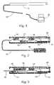
- FIG. 2is a simplified block diagram of an endoscope system according to a first embodiment of the present invention
- FIG. 3is a simplified block diagram of a wireless modification of the endoscope of FIG. 2 .
- FIG. 4is a simplified schematic block diagram of an endoscope according to a preferred embodiment of the present invention.
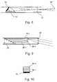
- FIG. 5is a simplified ray diagram showing optical paths within an endoscope according to a preferred embodiment of the present invention
- FIG. 6is a ray diagram view from a different angle of the embodiment of FIG. 5 .
- FIG. 7is a ray diagram showing an alternative construction of an optical assembly according to a preferred embodiment of the present invention.
- FIG. 8is a ray diagram showing a further alternative construction of an optical assembly according to a preferred embodiment of the present invention.
- FIG. 9is a ray diagram showing yet a further alternative construction of the optical assembly according to a preferred embodiment of the present invention.
- FIG. 10is a ray diagram taken from the front, of the embodiment of FIG. 9 .
- FIG. 11is a ray diagram showing yet a further alternative construction of an optical assembly according to a preferred embodiment of the present invention.
- FIG. 12is a simplified layout diagram of an image sensor according to an embodiment of the present invention.
- FIG. 13is a simplified ray diagram showing an endoscope for use in a stereoscopic mode according to a preferred embodiment of the present invention
- FIG. 14is a simplified ray diagram showing how a 3D model obtained from the embodiment of FIG. 13 can be used to construct a stereoscopic image of the field of view
- FIG. 15Ais a simplified diagram in cross-sectional view showing an arrangement of the image sensors in a stereoscopic endoscope according to a preferred embodiment of the present invention
- FIG. 15Bis a view from one end of the arrangement of FIG. 15A .
- FIG. 16is a simplified ray diagram showing an alternative arrangement of sensors for obtaining a stereoscopic image of a field of view according to a preferred embodiment of the present invention
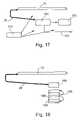
- FIG. 17is a simplified block diagram of a network portable endoscope and associated hardware usable with preferred embodiments of the present invention
- FIG. 18is a simplified block diagram of an endoscope adapted to perform minimal invasive surgery and usable with the preferred embodiments of the present invention
- FIG. 19is a simplified block diagram of an enhanced endoscope system for use in research
- FIG. 20is a simplified block diagram of a configuration of an endoscope system for obtaining stereoscopic images, and usable with the preferred embodiments of the present invention.
- FIG. 21is a simplified block diagram of a system for use in intra-vascular procedures.
- the present embodimentsprovide a diagnostic and operative system for minimally invasive diagnosis and surgery procedures, and other medical and non-medical viewing applications, in particular in which access conditions dictate the use of small-dimension viewing devices.
- FIG. 1is a basic block diagram of a basic configuration of an endoscope according to a first embodiment of the present invention.
- the figureshows a basic configuration of the endoscopic system including interconnections.
- the configurationcomprises a miniature endoscopic front-end 10 , hereinafter simply referred to as an endoscope, attached by a wire connection 20 to a processing device 30 , typically a PC, the PC having appropriate software for carrying out image processing of the output of the endoscope.
- a processing device 30typically a PC
- the wire connection 20may be an optical connection or may instead use RF or a like means of wireless communication.
- the miniature endoscopic front-end 10may be designed for connection to any standard PC input (the USB input for example).
- the software included with processing device 30processes the output of the miniature endoscopic front-end 10 .
- the softwaremay typically control transfer of the images to the monitor of the PC 30 and their display thereon including steps of 3D modeling based on stereoscopic information as will be described below, and may control internal features of the endoscopic front end including light intensity, and automatic gain control (AGC), again as will be described below.
- AGCautomatic gain control
- FIG. 2is an internal block diagram of an endoscope according to a preferred embodiment of the present invention.
- a miniature endoscope 40is connected by a wire 42 to an adapter 44 .
- the endoscope 40comprises an image sensor 46 which may typically comprise a CMOS or CCD or like sensing technology, an optical assembly 48 , a light or illumination source 50 , communication interface 52 and controller 54 .
- the wired unit of FIG. 2preferably includes a voltage regulator 56 .
- the image sensor 46is aligned along the length of a longitudinal side-wall (that is to say substantially in parallel with the wall and at least not perpendicular thereto) of the endoscope 40 .
- a longitudinal side-wallthat is to say substantially in parallel with the wall and at least not perpendicular thereto
- the sensoris arranged in two parts, as will be explained below.
- FIG. 3is an internal block diagram of a wireless equivalent of the embodiment of FIG. 2 . Parts that are identical to those shown above are given the same reference numerals and are not referred to again except as necessary for an understanding of the present embodiment.
- the wire 42is replaced by a wireless link ( 56 such as an IR or RF link with appropriate sensor, and a battery pack 58 .
- FIG. 4is an schematic block diagram of the miniature endoscope according to a preferred embodiment of the present invention. Parts that are identical to those shown above are given the same reference numerals and are not referred to again except as necessary for an understanding of the present embodiment.
- Optical assembly 48receives light, indicated by arrows 60 , from an object being viewed. The light is processed by optical assembly 48 , as will be explained below, to reach image sensor 46 were it is converted from photons into electrical signals. The electrical signals are digitized and passed to a transmitting device 62 , for example an LVDS transmitter, which drives the data through communication link 20 and adapter 44 to the processing device 30 .
- a transmitting device 62for example an LVDS transmitter
- Operating power for the endoscope 40is preferably provided, through adapter 44 , to the voltage regulator 56 .
- Control of the front-endis preferably carried out by the processor device 30 as discussed above.
- Control data from the processing device 30is preferably received at the endoscope 40 by a receiving device 64 , which may typically be an LVDS receiver.
- Hard wired logic 66preferably serves as an interface to convert the incoming control data into signals for controlling both the sensor 46 and the light source 50 .
- the light source 50preferably comprises one or more light transmitting devices such as LEDs, typically a left light source 68 and right light source 70 .
- the left and right light sourcesmay be controllable through a driver 72 .
- the functions of each of the above componentsare described in greater detail below.
- use of CMOS and similar technologies for the sensorspermit the sensor 46 , the transmitting device 62 , the receiving device 64 , the hard wired logic 66 , the driver 72 and the voltage regulator 56 to be integrated into a single semiconductor Integrated Circuit and such integration is particularly advantageous in achieving a compact design of endoscope.
- the light source 50preferably comprises an integrated array of several white light sources (LEDs for example) with energy emission in the visible light range mixed, optionally, with IR light sources (LEDs) for purposes that will be explained below.
- LEDswhite light sources
- IR light sourcesLEDs
- any combination of spectral responsesmay be used, particularly preferred combinations including red+IR and green+blue.
- An integrated array of light sourcesallows control of each light source individually facilitating the following features:
- the Systemis able to turn on the white light source and the IR Light source in sequence to generate an IR image every N (user determined) standard white images, for detection by a sensor configuration to be described below with respect to FIG. 12 .
- the objects being imagedare generally located at a range of different distances or field depths from the light source and are consequently unevenly illuminated.
- the more distant areas in the fieldare dark and are almost invisible while the nearer areas are bright and can become saturated.
- the systempreferably exerts control over the intensity of each light source individually, thereby to compensate for reflected intensity of the objects.
- An example of an algorithm for control of the illumination arrayis given as follows:
- an initialization processis carried out to generate a reference image, preferably a homogeneous white object, to be stored for each light source.
- imagingis carried out and the input image of the field (indicated by matrix II) is divided into M areas such that: M>N.
- an inner product matrixis calculated such that element Tij of the inner product matrix reflects the inner product resulting from taking the II matrix and performing matrix multiplication with the RIi matrix, in the area Sj and summing the elements of the result metrics.
- Pijthe intensity of the pixel located in (xp,yp) resulting from light source i in area j
- Rjthe intensity of the pixel located in (xp,yp) resulting from the input image in area j
- a vector vis determined that satisfies the following: Tv ⁇ k ⁇ Min, where
- the central control unitpreferably uses the above algorithm to post-process the data to reconstruct a natural look of the image, thereby to compensate for brightness non-uniformities.
- the size of the sensorprovides a limitation on the transverse diameter of the endoscope.
- the sensorin order to remove the limitation is placed along the longitudinal wall of the endoscope, again preferably substantially parallel to the wall but at least not perpendicular thereto.
- the use of the longitudinal wallnot only gives greater freedom to reduce the transverse diameter of the endoscope but also gives the freedom to increase the length of the sensor, thus increasing image resolution in the horizontal sense.
- the senormay be supplied with color filters to allow acquisition of IR images for diagnostic purposes or 3D imaging, again as will be described in detail below.
- the sensorcomprises a field of pixels arranged in an array over an image-gathering field.
- the first specific embodimentcomprises a rearrangement of the pixels in the sensor.
- the sensor widthmay be divided into say two parts, then the two parts may be placed end to end lengthwise.
- a 512 ⁇ 512 pixels' sensor with pixel dimensions of 10 ⁇ 10 micronmay be divided into two sections of width 256 pixels each to be placed end to end to give a sensor of 256 ⁇ 1024 pixels and having an overall imaging area of 2.56 mm ⁇ 10.24 mm.
- the longer dimensionis preferably placed along the lengthwise dimension of the endoscope, thus permitting reduced diameter of the endoscope with no corresponding reduction in the precision level of the image.
- the second specific embodimentlikewise relates to a geometrical rearrangement of the pixels.
- the prior art image sensorhas a round or square overall sensor or pixilated area, however, if the same number of pixels are arranged as a rectangle having the same area as the original sensor but with the height and width freely chosen then the width may be selected to be smaller than the width of the equivalent prior art sensor. More particularly, for an exemplary 512 ⁇ 512 pixels' sensor with pixel dimensions of 10 ⁇ 10 micron the standard prior art sensor (which will have a width of 5.12 mm) may be replaced by a rectangular sensor having the same overall sensing area as in the previous specific embodiment, but with specific width height dimensions of 2.56 mm ⁇ 10.24 mm, thus becoming easier to fit in the endoscope.
- FIG. 5is a ray diagram showing a simplified view from above of optical paths within the endoscope.
- each sensoris preferably associated with an optical assembly which is able to redirect image parts in accordance with the rearrangements of the pixels.
- FIG. 5shows a version of optical assembly 48 designed for the first of the two specific embodiments of the image sensor, namely that involving the widthwise transfer of pixels.
- FIG. 6shows a side view of the same optical assembly.
- FIG. 5shows a point source object 80 , from which light reaches two lenses 82 and 84 .
- the two lensesare selected and arranged to divide the light into two parts, which parts reach a front-surface-mirror 86 .
- the front surface mirrorsends each part of the image to a different part of the sensor 46 , and recovery of the image is possible by appropriate wiring or addressing of the sensor pixels to recover the original image shape.
- FIG. 7is a ray diagram showing an alternative version of optical assembly 48 , again designed for the first specific embodiment of the image sensor.
- a single lens 86is positioned in conjunction with two front-surface-mirrors 88 and 90 to deflect light from the object 80 to the mirrors.
- Each of the two front surface mirrorsrespectively transfers half of the image to the upper or lower part of the sensor 46 .
- FIG. 8is a ray diagram showing a third embodiment of the optical assembly 48 , this time for the second of the specific embodiments of the image sensor 46 , namely the embodiment in which the square shape of pixels is reduced to a rectangular shape having smaller width.
- An asymmetric or astigmatic lens 92is arranged to focus light onto a front-surface-mirror 94 . The light is distorted by the lens 92 to undo the distortion introduced into the image by the rectangular shape of the sensor 46 , and then it is reflected by the mirror 94 onto the surface of the sensor 46 .
- FIG. 9is a ray diagram taken from the side showing a further embodiment of the optical assembly 48 .
- the embodiment of FIG. 8necessitates a relatively complicated design of the mirror, and in order to obviate such complexity, additional optical design is shown.
- the same astigmatic lens 92is placed, not in front of a mirror but rather in front of a series of flat optical plates 96 . 1 . . . 96 .n, each comprising a diagonal lateral cross section, the plates each reflecting the light through the respective plate to the surface of sensor 46 .
- FIG. 10is a ray diagram, taken from the front, of the series of optical plates 96 of FIG. 9 .
- a comparison between the perspectives of FIG. 9 and FIG. 10show the layout of the plates with respect to the endoscope.
- FIG. 11is a simplified ray diagram showing a further embodiment of the optical assembly 48 .
- a single lens 98is preferably used to focus light from an object 80 to a plane 100 shown in dotted lines.
- a series of optical fibers 102are lined up over the surface of plane 100 to guide light to desired portions of the surface of the image sensor 46 .
- the fibers 102are able to direct light as desired and thus can be used in combination with any arrangement of the sensor pixels that is desired.
- FIG. 12is a layout diagram showing a layout of pixels on a sensory surface of an embodiment of the image sensor 46 .
- an arraycomprising pixels of four types is shown, red r, green g, blue b and infra-red IR.
- the pixelsare evenly spaced and allow acquisition of a colored image when used in conjunction with white light, or an IR image when used in conjunction with an IR source.
- IR wavelengthsIn many cases, important medical information is contained at IR wavelengths.
- the sensoris preferably designed as described above, and using inter alia pixels IR filters, that is to say color filters that have band passes at IR wavelengths.
- the sensoris placed in an endoscope in association with either one or both of a source of visible light and a source of infra-red light.
- Use of the appropriate one of the two light sourcespermits acquisition of either color frames or IR frames as desired.
- IR and color framesare obtained simultaneously by operating color and IR light sources together and allowing each pixel to pick up the waveband it has been designed for.
- the color and IR light sourcesare operated separately. Typically one IR frame would be prepared and sent for every several color frames.
- FIG. 13is a simplified ray diagram showing how the endoscope may be used in a stereoscopic mode.
- the stereoscopic modepermits the production of 3D images.
- the ray diagramindicates rays emanating from a single point, and the skilled person will appreciate how to extrapolate to a full image.
- an endoscopecomprises two separate white light sources 110 and 112 located at opposite sides of a front opening of the endoscope, respectively being a left light source 110 and a right light source 112 .
- the two white light sourcesare controlled to light in turn in successive short flashes to illuminate an object 114 .
- Light reflected by the object 114returns to the endoscope where it strikes a lens 115 placed across the front opening and where it is focused on to the plane of sensor 46 .
- the sensordetects the illumination level, which differs between the left and right light beams. The ratio of the illumination levels may be used to calculate the position of the object and thereby to build up a 3D distance database, as will be explained in greater detail below.
- an image 116 of object 114may be considered as comprising a series of activated x, y, locations on the detection plane of the sensor 46 . For each of the x, y locations forming the image 116 on the sensor 46 , a ratio between the Right Illuminated Image (RII) and the Left Illuminated Image (LII) may be discerned.
- RIIRight Illuminated Image
- LIILeft Illuminated Image
- the detected ratiomay differ over the image as it is a function in each case of the distances of the respective light source to the object 114 .
- the left light source 110 and the right light source 112have a distance between them which is twice d, d being the length of arrow 117 , and the lens has a focal length of 1/f, where f is the length of arrow 118 .
- the distance from the lens 115 to the plane of the object 114is denoted by Z and is indicated by arrow 120 .
- LBLLeft Beam Length
- RBLRight Beam Length
- the image 116obtained as described above may now be stored in terms of a 3D model.
- the 3D modelis preferably displayed as a 3D image by constructing therefrom two stereoscopic images.
- FIG. 13thus shows how an image of the object can be stored as a 3D data base.
- 3D data of the objectis obtained as described above and stored as a database.
- FIG. 14is a further simplified ray diagram showing, by means of rays, how the 3D model or database of FIG. 13 can be used to obtain a 3D effect at the eyes of an observer.
- the databaseis converted into two separate stereoscopic images, and a display device is used to display each one of the stereoscopic images to a different eye.
- the devicemay be a pair of glasses having a controllable shutter on each on of the eyes.
- X, Y, 114 and Z 120represents the three dimensions to be used in the image 119 , which corresponds to image 116 as stored in the previous figure, the object being to reproduce the three dimensional character of the image by showing different projections of the image to each of the two eyes of a viewer.
- line 122represents a projected location on the left image.
- Line 124represents the same projected location as it appears oil the right image.
- 1/f 118is the focal length (the amplification factor).
- D 126is the distance between the lenses 128 (representing the eyes).
- a preferred embodiment for producing a 3D model using the endoscopeuses different color left and right light sources in place of white light sources.
- a left illumination source 110may be green and right illumination source 112 may be a combination of red+blue.
- Such a two-color embodimentis advantageous in that it is simple to control and avoids image distortion problems due to the time lag between acquisitions of the two separate images.
- one of the light sources 110 , 112is a visible light source and the second light source is an IR light source.
- color filters at the sensorpreferably include an IR pass filter.
- the sensor of FIG. 12with an arrangement of IR, red, green and blue detectors as described above may be used.
- FIGS. 15A and 15Bare simplified schematic diagrams showing an endoscope according to a preferred embodiment of the present invention for obtaining dual sensor stereoscopic imaging, as will be explained below.
- FIG. 15Ais a side sectional view and FIG. 15B is a front view.
- each image sensor 140 and 142is associated with a respective optical assembly comprising a lens 150 and 152 and a mirror 154 and 156 .
- the respective light source 146 , 148illuminates the entire field of view as described above and light is gathered by the lens and directed by the mirror onto the sensor.
- the sensorsare preferably mounted on a single PCB 158 .
- FIG. 15Bis a view from the front of the endoscope of FIG. 15A . It will be noticed that a third optical light source 158 shown. Since the stereoscopic aspect of the image is obtained from the use of two optical image paths, as opposed to the previous embodiments which used different light sources and different object optical paths, there is now freedom to use any number of light sources as desired to produce desired color (or IR) information.
- the back-to-back arrangement of the sensors 140 and 142 along the central axis of the endoscope 144ensures that the endoscope dimensions are minimized both lengthwise and radially.
- An endoscope 160comprises two image sensors 162 and 164 arranged in a head to tail arrangement along one longitudinal wall of the endoscope, and again, as above, preferably parallel to the wall and at least not perpendicular thereto.
- Illumination sources 166 and 168are located at a front end 170 of the endoscope and located at the periphery thereof.
- Two lenses 172 and 174direct light received from a field of view onto respective mirrors 176 and 178 each of which is arranged to deflect the light onto one of the sensors.
- Each image sensor 162 and 164thus provides a slightly different image of the field of view.
- the dual sensor configurationdoes not decrease the overall image resolution, because, in accordance with the above configurations, two full-size image sensors may be used.
- the two versions of an endoscope for obtaining dual sensor stereoscopic imaging described abovecan make use of image sensors either with or without color filters.
- the sensor of FIG. 12could be used for one or both of the sensors in either of the embodiments above.
- a further preferred embodimentuses a monochrome sensor for one of the two image sensors and a color sensor for the second.
- a monochrome sensorfor one of the two image sensors and a color sensor for the second.
- FIG. 17is a simplified block diagram of a network portable endoscope and associated hardware. Parts that are identical to those shown above are given the same reference numerals and are not referred to again except as necessary for an understanding of the present embodiment.
- An endoscope 10is connected to a central control unit 180 where dedicated image processing takes place.
- the control unit 180allows for full motion video to be produced from the signals emitted by the endoscope.
- the control unitis connected to a local display device 182 . Additionally or alternatively, a remote control and viewing link 183 may be used to allow remote monitoring and control of the endoscope.
- the endoscope 10is preferably a portable device and may be powered from a battery pack 184 .
- FIG. 18is a simplified block diagram of an endoscope adapted to perform minimal invasive surgery (MIS). Parts that are identical to those shown above are given the same reference numerals and are not referred to again except as necessary for an understanding of the present embodiment.
- the most common use of endoscopic systemsis for the performance of MIS procedures by the surgeon in the operating room.
- the use of a reduced size endoscope according to the above embodimentsenables new procedures to be performed in which minimal dimensions of the operating equipment is important.
- the endoscope 10is connected to a rack 190 .
- the rackcontains accomodation for a full range of equipment that may be required in the course of use of the endoscope in the operating room, for example a central control unit 180 , a high quality monitor 182 , an insufflator 186 etc.
- the configuration of FIG. 18by virtue of the dedicated image processing provided with the control unit 180 , gives full motion video without requiring fiber-optic and camera head cables.
- FIG. 19is a simplified block diagram showing an enhanced version of the endoscope for use in research. Parts that are identical to those shown above are given the same reference numerals and are not referred to again except as necessary for an understanding of the present embodiment.
- the systemcomprises a miniature endoscopic front-end 10 connected to a highly integrated PC based central control unit 200 via communication link 20 .
- the central control unituses dedicated image processing and thus enables full motion video, displayable locally on display device 182 or remotely via control and display link 183 .
- An optional printer 202is provided to print documents and images, including images taken via the endoscope, of the pathologies or stages of the procedure.
- the systempreferably includes a VCR 204 for recording video produced by the endoscope and a digital storage device 206 allowing archiving of the whole video.
- the systemcan also be connected via remote control and viewing link 183 , to a remote site for teaching or for using medical help and guidance. In some hospitals and operating rooms, in addition to regular operating procedures, research is carried out. Research procedures generally require additional documentation and communication functions. In order to support those requirements a PC based system with high documentation and communication capabilities is provided by the enhanced control unit 200 .
- an image enhancement software packageis used, allowing the generation of high quality hard copies of images.
- FIG. 20is a simplified block diagram showing a configuration of endoscope for obtaining stereoscopic (3D) images. Parts that are identical to those shown above are given the same reference numerals and are not referred to again except as necessary for an understanding of the present embodiment.
- the miniature endoscope 10is connected via a communication link 20 as before to a 3D central control unit 210 , which is the same as the previous control unit 200 except that it has the additional capability to construct a 3D model from image information provided by the endoscope.
- the 3D modelcan then be projected to form a 3D image on a 3D stereoscopic display system 212 .
- the configuration of FIG. 20may be combined with features taken from any of the embodiments referred to above.
- the 3D imaging embodiments referred to abovewhich may be grouped into multiple light source based imaging and dual optical path imaging, can give the necessary information to construct a 3D model of the scene and to generate stereoscopic images therefrom.
- FIG. 21is a simplified block diagram showing a variation of an endoscope system for use in intra-vascular procedures. Parts that are identical to those shown above are given the same reference numerals and are not referred to again except as necessary for an understanding of the present embodiment.
- the systemincludes a long, flexible, thin and preferably disposable catheter 220 , a balloon/Stent 222 an endoscope imaging head 224 , an X-ray tube 226 , X-ray imaging system 228 , a video display system 230 and an injection unit 232 .
- Intravascular proceduresare widely used in the medical field.
- cardiac catheterizationis a very common diagnostic test performed thousands of times a day.
- catheter 220is inserted into an artery at the groin or arm.
- the catheteris directed retrogradely to the heart and to the origin of the coronary arteries, which supply blood to the heart muscle.
- a contrast substance (“dye”)is injected through the catheter.
- the use of an x-ray tube, and an endoscope in conjunction with the dyeenables a view of the heart chambers and coronary arteries to be obtained.
- the resulting imagesmay be recorded using an x-ray camera and/or the endoscope systems as described above.
- the obstructionmay be removed and the artery reopened using techniques such as inserting the balloon and inflating it (PTCA) or inserting a stent, as known to the person skilled in the art.
- PTCAballoon and inflating it
- a few methodsmay be used to acquire intra-vascular images in the presence of blood.
- One methodis based on the fact that certain near IR wavelengths allow viewing through blood. The method thus involves the use of an IR illumination source and a sensor with IR filters as described above.
- Another methoduses controlled injection of a transparent physiological liquid into the blood vessel in order to dilute the blood prior to the imaging.
- Yet another methoduses a conical dome, a balloon or any other rigid or flexible and inflatable transparent structure in order to improve visibility by “pushing” the blood to the walls of the vessels, thus enlarging the part of the optical path that does not include blood.
- Another way of improving visibilityis by using a post-processing algorithm after the acquiring of the image has been done. The post-processing algorithm is based on the extraction of parameters from the received image and the use of those parameters in an inverse operation to improve the image.
- an endoscope of reduced dimensionswhich is able to provide 2D and 3D images, and which is usable in a range of minimally invasive surgical procedures.
Landscapes
- Health & Medical Sciences (AREA)
- Life Sciences & Earth Sciences (AREA)
- Surgery (AREA)
- Engineering & Computer Science (AREA)
- Medical Informatics (AREA)
- Pathology (AREA)
- Molecular Biology (AREA)
- Biophysics (AREA)
- Veterinary Medicine (AREA)
- Public Health (AREA)
- Nuclear Medicine, Radiotherapy & Molecular Imaging (AREA)
- Optics & Photonics (AREA)
- General Health & Medical Sciences (AREA)
- Radiology & Medical Imaging (AREA)
- Biomedical Technology (AREA)
- Heart & Thoracic Surgery (AREA)
- Physics & Mathematics (AREA)
- Animal Behavior & Ethology (AREA)
- High Energy & Nuclear Physics (AREA)
- Vascular Medicine (AREA)
- Dentistry (AREA)
- Oral & Maxillofacial Surgery (AREA)
- Multimedia (AREA)
- Signal Processing (AREA)
- Endoscopes (AREA)
- Instruments For Viewing The Inside Of Hollow Bodies (AREA)
- Studio Devices (AREA)
Abstract
Description
This application is a Continuation of U.S. patent application Ser. No. 10/683,344, filed Oct. 14, 2003, now U.S. Pat. No. 7,241,262, granted Jul. 10, 2007, entitled AN IMAGE SENSOR AND AN ENDOSCOPE USING THE SAME by Doron Adler, et al., which is a Divisional of U.S. patent application Ser. No. 09/826,163, filed Apr. 5, 2001, now U.S. Pat. No. 6,659,940, granted Dec. 9, 2003, entitled AN IMAGE SENSOR AND AN ENDOSCOPE USING THE SAME, by Doron Adler, et al, which claims the benefit of and priority to Israel Patent Application Number 135571, filed Apr. 10, 2000, the contents of which are hereby incorporated by reference as if recited in full herein.
The present invention relates to an image sensor, and more particularly but not exclusively to two and three-dimensional optical processing from within restricted spaces, and an endoscope using the same.
Endoscopy is a surgical technique that involves the use of an endoscope, to see images of the body's internal structures through very small incisions.
Endoscopic surgery has been used for decades in a number of different procedures, including gall bladder removal, tubal ligation, and knee surgery, and recently in plastic surgery including both cosmetic and re-constructive procedures.
An endoscope may be a rigid or flexible endoscope which consists of five basic parts: a tubular probe, a small camera head, a camera control unit, a bright light source and a cable set which may include a fiber optic cable. The endoscope is inserted through a small incision; and connected to a viewing screen which magnifies the transmitted images of the body's internal structures.
During surgery, the surgeon is able to view the surgical area by watching the screen while moving the tube of the endoscope through the surgical area.
In a typical surgical procedure using an endoscope, only a few small incisions, each less than one inch long, are needed to insert the endoscope probe and other instruments. For some procedures, such as breast augmentation, only two incisions may be necessary. For others, such as a forehead lift, three or four short incisions may be needed. The tiny eye of the endoscope camera allows a surgeon to view the surgical site.
An advantage of the shorter incisions possible when using an endoscope is reduced damage to the patient's body from the surgery. In particular, the risk of sensory loss from nerve damage is decreased. However, most current endoscopes provide only flat, two-dimensional images which are not always sufficient for the requirements of the surgery. The ability of an endoscope to provide three-dimensional information in its output would extend the field of endoscope use within surgery.
The need for a 3D imaging ability within an endoscope has been addressed in the past A number of solutions that provide stereoscopic images by using two different optical paths are disclosed in U.S. Pat. Nos. 5,944,655 , 5,222,477 , 4,651,201, 5,191,203, 5,122,650 , 5,471,237, JP7163517A, .U.S. Pat. No. 5,673,147, 6,139,490, 5,603,687, WO9960916A2, and JP63244011A.
Another method, represented by U.S. Pat. No. 5,728,044 and U. S. Pat. No. 5,575,754 makes use of an additional sensor that provides location measurements of image points. Patent JP8220448A discloses a stereoscopic adapter for a one-eye endoscope, which uses an optical assembly to divide and deflect the image to two sensors. A further method, disclosed in U.S. Pat. No. 6,009,189 uses image acquisition from different directions using one or more cameras. An attempt to obtain 3D information using two light sources was disclosed in U.S. Pat. No. 4,714,319 in which two light sources are used to give an illusion of a stereoscopic image based upon shadows. JP131622A discloses a method for achieving the illusion of a stereoscopic image by using two light sources, which are turned on alternately.
An additional problem with current endoscopes is the issue of lighting of the subject for imaging. The interior spaces of the body have to be illuminated in order to be imaged and thus the endoscope generally includes an illumination source. Different parts of the field to be illuminated are at different distances from the illumination source and relative reflection ratios depend strongly on relative distances to the illumination source. The relative distances however may be very large in a typical surgical field of view, distances can easily range between 2 and 20 cm giving a distance ratio of 1:10. The corresponding brightness ratio may then be 1:100, causing blinding and making the more distant object all but invisible.
One reference, JP61018915A, suggests solving the problem of uneven lighting by using a liquid-crystal shutter element to reduce the transmitted light. Other citations that discuss general regulation of illumination levels include U.S. Pat. No. 4,967,269, JP4236934A, JP8114755A and JP8024219A.
In general it is desirable to reduce endoscope size and at the same time to improve image quality. Furthermore, it is desirable to produce a disposable endoscope, thus avoiding any need for sterilization, it being appreciated that sterilization of a complex electronic item such as an endoscope being awkward in itself.
Efforts to design new head architecture have mainly concentrated on integration of the sensor, typically a CCD based sensor, with optics at the distal end. Examples of such integration are disclosed in U.S. Pat. Nos. 4,604,992 , 4,491,865 , 4,692,608, JP60258515A, U.S. Pat. Nos. 4,746,203 , 4,720,178 , 5,166,787, 4,803,562 , 5,594,497 and EP434793B1. Reducing the overall dimensions of the distal end of the endoscope are addressed in U.S. Pat. No. 5,376,960 and U S. Pat. No. 4,819,065, and Japanese Patent Applications No. 7318815A and No. 70221A. Integration of the endoscope with other forms of imaging such as ultrasound and Optical Coherence Tomography are disclosed in U.S. Pat. No. 4,869,256, 6,129,672, 6,099,475, 6,039,693, 5,502,2399, 6,134,003 and U S. Pat. No. 6,010,449
Intra-vascular applications are disclosed in certain of the above-mentioned patents, which integrate the endoscope with an ultrasound sensor or other data acquisition devices. Patents that disclose methods for enabling visibility within opaque fluids are U.S. Pat. Nos. 4,576,146, 4,827,907, 5,010,875, 4,934,339, 6,178,346 and U S. Pat. No. 4,998,972.
Sterilization issues of different devices including endoscopes are discussed in WO9732534A1, U.S. Pat. Nos. 5,792,045 and U S. Pat. No.5,498,230. In particular JP3264043A discloses a sleeve that was developed in order to overcome the need to sterilize the endoscope.
The above-mentioned solutions are however incomplete and are difficult to integrate into a single endoscope optimized for all the above issues.
It is an aim of the present embodiments to provide solutions to the above issues that can be integrated into a single endoscope.
It is an aim of the embodiments to provide an endoscope that is smaller than current endoscopes but without any corresponding reduction in optical processing ability.
It is a further aim of the present embodiments to provide a 3D imaging facility that can be incorporated into a reduced size endoscope.
It is a further aim of the present embodiments to provide object illumination that is not subject to high contrast problems, for example by individual controlling of the light sources.
It is a further aim of the present embodiments to provide a modified endoscope that is simple and cost effective to manufacture and may therefore be treated as a disposable item.
Embodiments of the present invention provide 3D imaging of an object based upon photometry measurements of reflected light intensity. Such a method is relatively efficient and accurate and can be implemented within the restricted dimensions of an endoscope.
According to a first aspect of the present invention there is thus provided a pixilated image sensor for insertion within a restricted space, the sensor comprising a plurality of pixels arranged in a selected image distortion pattern, said image distortion pattern being selected to project an image larger than said restricted space to within said restricted space substantially with retention of an image resolution level.
Preferably, the image distortion pattern is a splitting of said image into two parts and wherein said pixilated image sensor comprises said pixels arranged in two discontinuous parts.
Preferably, the discontinuous parts are arranged in successive lengths.
Preferably, the restricted space is an interior longitudinal wall of an endoscope and wherein said discontinuous parts are arranged on successive lengths of said interior longitudinal wall.
Preferably, the restricted space is an interior longitudinal wall of an endoscope and wherein said discontinuous parts are arranged on successive lengths of said interior longitudinal wall.
Preferably, the distortion pattern is an astigmatic image distortion.
Preferably, the distortion pattern is a projection of an image into a rectangular shape having dimensions predetermined to fit within said restricted space.
A preferred embodiment includes one of a group comprising CMOS-based pixel sensors and CCD based pixel sensors.
A preferred embodiment is controllable to co-operate with alternating image illumination sources to produce uniform illuminated images for each illumination source.
According to a second aspect of the present invention there is provided an endoscope having restricted dimensions and comprising at least one image gatherer at least one image distorter and at least one image sensor shaped to fit within said restricted dimensions, and wherein said image distorter is operable to distort an image received from said image gatherer so that the image is sensible at said shaped image sensor substantially with an original image resolution level.
Preferably, the image distorter comprises an image splitter operable to split said image into two part images.
Preferably, the image sensor comprises two sensor parts, each separately arranged along longitudinal walls of said endoscope.
Preferably, the two parts are arranged in successive lengths along opposite longitudinal walls of said endoscope.
Preferably, the distorter is an astigmatic image distorter.
Preferably, the astigmatic image distorter is an image rectangulator and said image sensor comprises sensing pixels rearranged to complement rectangulation of said image by said image rectangulator.
Preferably, the image distorter comprises at least one lens.
Preferably, the image distorter comprises at least one image-distorting mirror.
Preferably, the image distorter comprises optical fibers to guide image light substantially from said lens to said image sensor.
Preferably, the image distorter comprises a second lens.
Preferably, the image distorter comprises at least a second image-distorting mirror.
Preferably, the image distorter comprises at least one flat optical plate.
A preferred embodiment comprises at least one light source for illuminating an object, said light source being controllable to flash at predetermined times.
A preferred embodiment comprises a second light source, said first and said second light sources each separately controllable to flash.
Preferably, the first light source is a white light source and said second light source is an IR source.
In a preferred embodiment, one light source being a right side light source for illuminating an object from a first side and the other light source being a left side light source for illuminating said object from a second side.
In a preferred embodiment, one light source comprising light of a first spectral response and the other light source comprising light of a second spectral response.
A preferred embodiment further comprises color filters associated with said light gatherer to separate light from said image into right and left images to be fed to respective right aid left distance measurers to obtain right and left distance measurements for construction of a three-dimensional image.
In a preferred embodiment, said light sources are configured to flash alternately or simultaneously.
A preferred embodiment further comprises a relative brightness measurer for obtaining relative brightnesses of points of said object using respective right and left illumination sources, thereby to deduce 3 dimensional distance information of said object for use in construction of a 3 dimensional image thereof.
A preferred embodiment further comprises a second image gatherer and a second image sensor.
Preferably, the first and said second image sensors are arranged back to back longitudinally within said endoscope.
Preferably, the first and said second image sensors are arranged successively longitudinally along said endoscope.
Preferably, the first and said second image sensors are arranged along a longitudinal wall of said endoscope.
A preferred embodiment comprises a brightness averager operable to identify brightness differentials due to variations in distances from said endoscope of objects being illuminated, and substantially to cancel said brightness differentials.
A preferred embodiment further comprises at least one illumination source for illuminating an object with controllable width light pulses and wherein said brightness averager is operable to cancel said brightness differentials by controlling said widths.
A preferred embodiment has at least two controllable illumination sources, one illumination source for emitting visible light to produce a visible spectrum image and one illumination source for emitting invisible (i.e. IR or UV) light to produce a corresponding spectral response image, said endoscope being controllable to produce desired ratios of visible and invisible images.
According to a third aspect of the present invention there is provided an endoscope system comprising an endoscope and a controller, said endoscope comprising:
at least one image gatherer,
at least one image distorter and
at least one image sensor shaped to fit within restricted dimensions of said endoscope, said image distorter being operable to distort an image received from said image gatherer so that the image is sensible at said shaped image sensor with retention of image resolution,
said controller comprising a dedicated image processor for processing image output of said endoscope.
Preferably, the dedicated image processor is a motion video processor operable to produce motion video from said image output.
Preferably, the dedicated image processor comprises a 3D modeler for generating a 3D model from said image output.
Preferably, the said dedicated image processor further comprises a 3D imager operable to generate a stereoscopic display from said 3D model.
A preferred embodiment comprises an image recorder for recording imaging.
A preferred embodiment comprises a control and display communication link for remote control and remote viewing of said system.
Preferably, the image distorter comprises an image splitter operable to split said image into two part images.
Preferably, the image sensor comprises two sensor parts, each separately arranged along longitudinal walls of said endoscope.
Preferably, the two parts are arranged in successive lengths along opposite longitudinal walls of said endoscope.
Preferably, the distorter is an astigmatic image distorter.
Preferably, the astigmatic image distorter is an image rectangulator and said image sensor comprises sensing pixels rearranged to complement rectangulation of said image by said image rectangulator.
Preferably, the image distorter comprises at least one lens.
Preferably, the image distorter comprises at least one image-distorting mirror.
Preferably, the image distorter comprises optical fibers to guide image light substantially from said lens to said image sensor.
Preferably, the image distorter comprises a second lens.
Preferably, the image distorter comprises at least a second image-distorting mirror.
Preferably, the image distorter comprises at least one flat optical plate.
A preferred embodiment further comprises at least one light source for illuminating an object.
A preferred embodiment comprises a second light source, said first and said second light sources each separately controllable to flash.
Preferably, the first light source is a white light source and said second light source is an invisible source.
In a preferred embodiment, one light source is a right side light source for illuminating an object from a first side and the other light source is a left side light source for illuminating said object from a second side.
In a preferred embodiment, one light source comprises light of a first spectral response and the other light source comprises light of a second spectral response.
A preferred embodiment comprises color filters associated with said light gatherer to separate light from said image into right and left images to be fed to respective right and left distance measurers to obtain right and left distance measurements for construction of a three-dimensional image.
Preferably, the light sources are configured to flash alternately or simultaneously.
A preferred embodiment further comprises a relative brightness measurer for obtaining relative brightnesses of points of said object using respective right and left illumination sources, thereby to deduce 3 dimensional distance information of said object for use in construction of a 3 dimensional image thereof.
A preferred embodiment further comprises a second image gatherer and a second image sensor.
Preferably, the first and said second image sensors are arranged back to back longitudinally within said endoscope.
Preferably, the first and said second image sensors are arranged successively longitudinally along said endoscope.
Preferably, the first and said second image sensors are arranged along a longitudinal wall of said endoscope.
A preferred embodiment comprises a brightness averager operable to identify brightness differentials due to variations in distances from said endoscope of objects being illuminated, and substantially to reduce said brightness differentials.
According to a fifth embodiment or the present invention there is provided an endoscope for internally producing an image of a field of view, said image occupying an area larger than a cross-sectional area of said endoscope, the endoscope comprising
an image distorter for distorting light received from said field of view into a compact shape, and
an image sensor arranged in said compact shape to receive said distorted light to form an image thereon.
A preferred embodiment comprises longitudinal walls, wherein said image sensor is arranged along said longitudinal walls, the endoscope further comprising a light diverter for diverting said light towards said image sensor.
Preferably, the image sensor comprises two parts, said distorter comprises an image splitter for splitting said image into two parts, and said light diverter is arranged to send light of each image part to a respective part of said image sensor.
Preferably, the sensor parts are aligned on facing lengths of internal sides of said longitudinal walls of said endoscope.
Preferably, the sensor parts are aligned successively longitudinally along an internal side of one of said walls of said endoscope.
A preferred embodiment of the image distorter comprises an astigmatic lens shaped to distort a square image into a rectangular shape of substantially equivalent area.
A preferred embodiment further comprises a contrast equalizer for compensating for high contrasts differences due to differential distances of objects in said field of view.
A preferred embodiment comprises two illumination sources for illuminating said field of view.
In a preferred embodiment, the illumination sources are controllable to illuminate alternately, and said image sensor is controllable to gather images in synchronization with said illumination sources thereby to obtain independently illuminated images.
In a preferred embodiment, each illumination source is of a different predetermined spectral response.
A preferred embodiment of said image sensor comprises pixels, each pixel being responsive to one of said predetermined spectral responses.
A preferred embodiment of the image sensor comprises a plurality of pixels responsive to white light.
In a preferred embodiment, said image sensor comprises a plurality of pixels responsive to different wavelengths of light.
In a preferred embodiment, the wavelengths used comprise at least three of red light green light, blue light and infra-red light.
In a preferred embodiment, a second image sensor forms a second image from light obtained from said field of view.
In a preferred embodiment, said second image sensor is placed in back to back relationship with said first image sensor over a longitudinal axis of said endoscope.
In a preferred embodiment, the second image sensor is placed in end to end relationship with said first image sensor along a longitudinal wall of said endoscope.
In a preferred embodiment, the second image sensor is placed across from said first image sensor on facing internal longitudinal walls of said endoscope.
According to a sixth embodiment of the present invention there is provided a compact endoscope for producing 3D images of a field of view, comprising a first image sensor for receiving a view of said field through a first optical path and a second image sensor for receiving a view of said field through a second optical path, and wherein said first and said second image sensors are placed back to back along a longitudinal axis of said endoscope.
According to a seventh embodiment of the present invention there is provided a compact endoscope for producing 3D images of a field of view, comprising a first image sensor for receiving a view of said field through a first optical path and a second image sensor for receiving a view of said field through a second optical path and wherein said first and said second image sensors are placed end to end along a longitudinal wall of said endoscope
According to an eighth embodiment of the present invention there is provided a compact endoscope for producing 3D images of a field of view, comprising two illumination sources for illuminating said field of view, an image sensor for receiving a view of said field illuminated via each of said illumination sources, and a view differentiator for differentiating between each view.
Preferably, the differentiator is a sequential control for providing sequential operation of said illumination sources.
Preferably, the illumination sources are each operable to produce illumination at respectively different spectral responses and said differentiator comprises a series of filters at said image sensor for differentially sensing light at said respectively different spectral responses.
Preferably, the image distorter comprises a plurality of optical fibers for guiding parts of received image to said image sensor according to said distortion pattern.
According to a ninth embodiment of the present invention there is provided a method of manufacturing a compact endoscope, comprising:
providing an illumination source,
providing an image distorter,
providing an image ray diverter,
providing an image sensor whose shape has been altered to correspond to a distortion built into said image distorter, said distortion being selected to reduce at least one dimension of said image sensor to less than that of an undistorted version being sensed,
assembling said image distorter, said image ray diverter and said image sensor to form an optical path within an endoscope
According to a tenth embodiment of the present invention there is provided a method of obtaining an endoscopic image comprising:
illuminating a field of view,
distorting light reflected from said field of view such as to form a distorted image of said field of view having at least one dimension reduced in comparison to an equivalent dimension of said undistorted image, and
sensing said light within said endoscope using at least one image sensor correspondingly distorted.
For a better understanding of the invention and to show how the same may be carried into effect, reference will now be made, purely by way of example, to the accompanying drawings, in which:
The present embodiments provide a diagnostic and operative system for minimally invasive diagnosis and surgery procedures, and other medical and non-medical viewing applications, in particular in which access conditions dictate the use of small-dimension viewing devices.
Reference is now made toFIG. 1 , which is a basic block diagram of a basic configuration of an endoscope according to a first embodiment of the present invention. The figure shows a basic configuration of the endoscopic system including interconnections. The configuration comprises a miniature endoscopic front-end 10, hereinafter simply referred to as an endoscope, attached by awire connection 20 to aprocessing device 30, typically a PC, the PC having appropriate software for carrying out image processing of the output of the endoscope. The skilled person will appreciate that thewire connection 20 may be an optical connection or may instead use RF or a like means of wireless communication. The miniature endoscopic front-end 10 may be designed for connection to any standard PC input (the USB input for example).
The software included withprocessing device 30 processes the output of the miniature endoscopic front-end 10. The software may typically control transfer of the images to the monitor of thePC 30 and their display thereon including steps of 3D modeling based on stereoscopic information as will be described below, and may control internal features of the endoscopic front end including light intensity, and automatic gain control (AGC), again as will be described below.
Reference is now made toFIG. 2 , which is an internal block diagram of an endoscope according to a preferred embodiment of the present invention. Aminiature endoscope 40 is connected by awire 42 to anadapter 44. Theendoscope 40 comprises animage sensor 46 which may typically comprise a CMOS or CCD or like sensing technology, anoptical assembly 48, a light orillumination source 50,communication interface 52 andcontroller 54. The wired unit ofFIG. 2 preferably includes avoltage regulator 56.
As will be explained in more detail below, theimage sensor 46 is aligned along the length of a longitudinal side-wall (that is to say substantially in parallel with the wall and at least not perpendicular thereto) of theendoscope 40. Such an alignment enables the radial dimension of the endoscope to be reduced beyond the diagonal of theimage sensor 46. Preferably the sensor is arranged in two parts, as will be explained below.
Reference is now made toFIG. 3 , which is an internal block diagram of a wireless equivalent of the embodiment ofFIG. 2 . Parts that are identical to those shown above are given the same reference numerals and are not referred to again except as necessary for an understanding of the present embodiment. In the embodiment ofFIG. 3 , thewire 42 is replaced by a wireless link (56 such as an IR or RF link with appropriate sensor, and abattery pack 58.
Reference is now made toFIG. 4 , which is an schematic block diagram of the miniature endoscope according to a preferred embodiment of the present invention. Parts that are identical to those shown above are given the same reference numerals and are not referred to again except as necessary for an understanding of the present embodiment.Optical assembly 48 receives light, indicated byarrows 60, from an object being viewed. The light is processed byoptical assembly 48, as will be explained below, to reachimage sensor 46 were it is converted from photons into electrical signals. The electrical signals are digitized and passed to a transmittingdevice 62, for example an LVDS transmitter, which drives the data throughcommunication link 20 andadapter 44 to theprocessing device 30.
Operating power for theendoscope 40 is preferably provided, throughadapter 44, to thevoltage regulator 56. Control of the front-end is preferably carried out by theprocessor device 30 as discussed above. Control data from theprocessing device 30 is preferably received at theendoscope 40 by a receivingdevice 64, which may typically be an LVDS receiver. Hardwired logic 66 preferably serves as an interface to convert the incoming control data into signals for controlling both thesensor 46 and thelight source 50.
Thelight source 50 preferably comprises one or more light transmitting devices such as LEDs, typically a leftlight source 68 and rightlight source 70. The left and right light sources may be controllable through adriver 72. The functions of each of the above components are described in greater detail below. As the skilled person will be aware, use of CMOS and similar technologies for the sensors permit thesensor 46, the transmittingdevice 62, the receivingdevice 64, the hard wiredlogic 66, thedriver 72 and thevoltage regulator 56 to be integrated into a single semiconductor Integrated Circuit and such integration is particularly advantageous in achieving a compact design of endoscope.
Considering thelight source 50 in greater detail, it preferably comprises an integrated array of several white light sources (LEDs for example) with energy emission in the visible light range mixed, optionally, with IR light sources (LEDs) for purposes that will be explained below. In fact, any combination of spectral responses may be used, particularly preferred combinations including red+IR and green+blue. An integrated array of light sources allows control of each light source individually facilitating the following features:
The System is able to turn on the white light source and the IR Light source in sequence to generate an IR image every N (user determined) standard white images, for detection by a sensor configuration to be described below with respect toFIG. 12 .
The objects being imaged are generally located at a range of different distances or field depths from the light source and are consequently unevenly illuminated. The more distant areas in the field are dark and are almost invisible while the nearer areas are bright and can become saturated. In order to compensate for the uneven illumination intensity over the field, the system preferably exerts control over the intensity of each light source individually, thereby to compensate for reflected intensity of the objects. An example of an algorithm for control of the illumination array is given as follows:
Given N individual light sources in the illumination array in the camera head, an initialization process is carried out to generate a reference image, preferably a homogeneous white object, to be stored for each light source. The stored reference mages (matrices) are identified hereinbelow by RIi where i=1,2 . . . N
Following initialization, imaging is carried out and the input image of the field (indicated by matrix II) is divided into M areas such that: M>N. The image areas are identified hereinbelow by Sj j=1,2, . . . M
Following the above imaging stage, an inner product matrix is calculated such that element Tij of the inner product matrix reflects the inner product resulting from taking the II matrix and performing matrix multiplication with the RIi matrix, in the area Sj and summing the elements of the result metrics.
The resulting inner product matrix is given by T where:
wherein
Pij—the intensity of the pixel located in (xp,yp) resulting from light source i in area j
Rj—the intensity of the pixel located in (xp,yp) resulting from the input image in area j
Sj—the total pixels in area j
xp,yp—the pixels coordinates in area j
Next, a vector v is determined that satisfies the following:
Tv−k→ Min, where
Tv−k→ Min, where
v—the vector of intensities of each source, and
k—the vector of the desired common intensity, and the solution to this requirement is given by
v=(TT·T)−1·TT·k
v=(TT·T)−1·TT·k
The central control unit preferably uses the above algorithm to post-process the data to reconstruct a natural look of the image, thereby to compensate for brightness non-uniformities.
In the case of using LEDs as the light source, their fast response time makes it possible to operate them in a “controllable-flash” mode, replacing the need for variable integration time (or AGC).
Referring now to theimage sensor 46, as observed above in respect ofFIG. 2 , in the prior art endoscope the size of the sensor provides a limitation on the transverse diameter of the endoscope. Thus, in the present embodiment, in order to remove the limitation the sensor is placed along the longitudinal wall of the endoscope, again preferably substantially parallel to the wall but at least not perpendicular thereto. The use of the longitudinal wall not only gives greater freedom to reduce the transverse diameter of the endoscope but also gives the freedom to increase the length of the sensor, thus increasing image resolution in the horizontal sense.
As will be explained below, there are two specific embodiments of the realigned sensor, each one associated with a respective design of the optical assembly as will be described in detail below.
In addition to the above-mentioned geometrical realignment, the sensor may be supplied with color filters to allow acquisition of IR images for diagnostic purposes or 3D imaging, again as will be described in detail below.
Referring now to the geometric design of the sensor, as will be appreciated, the sensor comprises a field of pixels arranged in an array over an image-gathering field. The first specific embodiment comprises a rearrangement of the pixels in the sensor. Given that for the purposes of example, the sensor width may be divided into say two parts, then the two parts may be placed end to end lengthwise. Thus, for example, a 512×512 pixels' sensor with pixel dimensions of 10×10 micron, may be divided into two sections of width 256 pixels each to be placed end to end to give a sensor of 256×1024 pixels and having an overall imaging area of 2.56 mm×10.24 mm. The longer dimension is preferably placed along the lengthwise dimension of the endoscope, thus permitting reduced diameter of the endoscope with no corresponding reduction in the precision level of the image.
The second specific embodiment likewise relates to a geometrical rearrangement of the pixels. The prior art image sensor has a round or square overall sensor or pixilated area, however, if the same number of pixels are arranged as a rectangle having the same area as the original sensor but with the height and width freely chosen then the width may be selected to be smaller than the width of the equivalent prior art sensor. More particularly, for an exemplary 512×512 pixels' sensor with pixel dimensions of 10×10 micron the standard prior art sensor (which will have a width of 5.12 mm) may be replaced by a rectangular sensor having the same overall sensing area as in the previous specific embodiment, but with specific width height dimensions of 2.56 mm×10.24 mm, thus becoming easier to fit in the endoscope.
Reference is now made toFIG. 5 , which is a ray diagram showing a simplified view from above of optical paths within the endoscope. As will be appreciated, in order for the image sensors of the specific embodiments referred to above to produce images which can be recreated in an undistorted fashion, each sensor is preferably associated with an optical assembly which is able to redirect image parts in accordance with the rearrangements of the pixels.
Reference is now made toFIG. 7 which is a ray diagram showing an alternative version ofoptical assembly 48, again designed for the first specific embodiment of the image sensor. Asingle lens 86 is positioned in conjunction with two front-surface-mirrors object 80 to the mirrors. Each of the two front surface mirrors respectively transfers half of the image to the upper or lower part of thesensor 46.
Reference is now made toFIG. 8 , which is a ray diagram showing a third embodiment of theoptical assembly 48, this time for the second of the specific embodiments of theimage sensor 46, namely the embodiment in which the square shape of pixels is reduced to a rectangular shape having smaller width. An asymmetric orastigmatic lens 92 is arranged to focus light onto a front-surface-mirror 94. The light is distorted by thelens 92 to undo the distortion introduced into the image by the rectangular shape of thesensor 46, and then it is reflected by themirror 94 onto the surface of thesensor 46.
Reference is now made toFIG. 9 , which is a ray diagram taken from the side showing a further embodiment of theoptical assembly 48. The embodiment ofFIG. 8 necessitates a relatively complicated design of the mirror, and in order to obviate such complexity, additional optical design is shown. As shown inFIG. 9 , the sameastigmatic lens 92 is placed, not in front of a mirror but rather in front of a series of flat optical plates96.1 . . .96.n, each comprising a diagonal lateral cross section, the plates each reflecting the light through the respective plate to the surface ofsensor 46.
Reference is additionally made toFIG. 10 , which is a ray diagram, taken from the front, of the series of optical plates96 ofFIG. 9 . A comparison between the perspectives ofFIG. 9 andFIG. 10 show the layout of the plates with respect to the endoscope.
Reference is now made toFIG. 11 , which is a simplified ray diagram showing a further embodiment of theoptical assembly 48. In the embodiment ofFIG. 11 , asingle lens 98 is preferably used to focus light from anobject 80 to aplane 100 shown in dotted lines. A series ofoptical fibers 102 are lined up over the surface ofplane 100 to guide light to desired portions of the surface of theimage sensor 46. Thefibers 102 are able to direct light as desired and thus can be used in combination with any arrangement of the sensor pixels that is desired.
Returning to the construction of theimage sensor 46, reference is now made toFIG. 12 , which is a layout diagram showing a layout of pixels on a sensory surface of an embodiment of theimage sensor 46. InFIG. 12 , an array comprising pixels of four types is shown, red r, green g, blue b and infra-red IR. The pixels are evenly spaced and allow acquisition of a colored image when used in conjunction with white light, or an IR image when used in conjunction with an IR source.
In many cases, important medical information is contained at IR wavelengths. In order to allow acquisition of IR images the sensor is preferably designed as described above, and using inter alia pixels IR filters, that is to say color filters that have band passes at IR wavelengths. The sensor is placed in an endoscope in association with either one or both of a source of visible light and a source of infra-red light. Use of the appropriate one of the two light sources permits acquisition of either color frames or IR frames as desired. In one preferred embodiment, IR and color frames are obtained simultaneously by operating color and IR light sources together and allowing each pixel to pick up the waveband it has been designed for. In another preferred embodiment the color and IR light sources are operated separately. Typically one IR frame would be prepared and sent for every several color frames.
Reference is now made toFIG. 13 , which is a simplified ray diagram showing how the endoscope may be used in a stereoscopic mode. The stereoscopic mode permits the production of 3D images. As with previous figures the ray diagram indicates rays emanating from a single point, and the skilled person will appreciate how to extrapolate to a full image.
InFIG. 13 , an endoscope comprises two separatewhite light sources light source 110 and a rightlight source 112. The two white light sources are controlled to light in turn in successive short flashes to illuminate anobject 114. Light reflected by theobject 114 returns to the endoscope where it strikes alens 115 placed across the front opening and where it is focused on to the plane ofsensor 46. The sensor detects the illumination level, which differs between the left and right light beams. The ratio of the illumination levels may be used to calculate the position of the object and thereby to build up a 3D distance database, as will be explained in greater detail below.
As mentioned above, in the stereoscopic mode the left and right light sources are used sequentially. Comparison between left and right illuminated images allows a 3D database to be constructed, enabling stereoscopic display of the scene. In the present embodiment, the comparison between the images is based upon photometry measurements. InFIG. 13 , animage 116 ofobject 114 may be considered as comprising a series of activated x, y, locations on the detection plane of thesensor 46. For each of the x, y locations forming theimage 116 on thesensor 46, a ratio between the Right Illuminated Image (RII) and the Left Illuminated Image (LII) may be discerned. The detected ratio may differ over the image as it is a function in each case of the distances of the respective light source to theobject 114. The leftlight source 110 and the rightlight source 112 have a distance between them which is twice d, d being the length ofarrow 117, and the lens has a focal length of 1/f, where f is the length ofarrow 118. The distance from thelens 115 to the plane of theobject 114 is denoted by Z and is indicated byarrow 120.
The Left Beam Length (LBL) can thus be expressed by:
LBL=√[Z2+(X−d)2]+√[(Z+1/f)2+(X+x)2]
LBL=√[Z2+(X−d)2]+√[(Z+1/f)2+(X+x)2]
while the Right Beam Length (RBL) is given by:
RBL=√[Z2+(X+d)2]+√[(Z+1/f)2+(X+x)2]
RBL=√[Z2+(X+d)2]+√[(Z+1/f)2+(X+x)2]
where:
X=xZf
X=xZf
Thus the ratio of the light intensity between the left and right light sources, which is the inverted square of the distance LBL/RBL, may be expressed as:
LeftToRightRatio=(LBL/RBL)(−2)
LeftToRightRatio=(LBL/RBL)(−2)
Theimage 116, obtained as described above may now be stored in terms of a 3D model. The 3D model is preferably displayed as a 3D image by constructing therefrom two stereoscopic images. The conversion may be performed using conversion formulae as follows:
yl=yr=−Y/(Z*f)
xl=(−X−D/2)/(Z*f)
xr=(−X+D/2)/(Z*f)
yl=yr=−Y/(Z*f)
xl=(−X−D/2)/(Z*f)
xr=(−X+D/2)/(Z*f)
Reference is now made toFIG. 14 , which is a further simplified ray diagram showing, by means of rays, how the 3D model or database ofFIG. 13 can be used to obtain a 3D effect at the eyes of an observer. In order to display the 3D information using a standard 2D display (monitor) the database is converted into two separate stereoscopic images, and a display device is used to display each one of the stereoscopic images to a different eye. For example the device may be a pair of glasses having a controllable shutter on each on of the eyes.
InFIG. 14 , X, Y,114 andZ 120 represents the three dimensions to be used in theimage 119, which corresponds to image116 as stored in the previous figure, the object being to reproduce the three dimensional character of the image by showing different projections of the image to each of the two eyes of a viewer.
1/f 118 is the focal length (the amplification factor).
A preferred embodiment for producing a 3D model using the endoscope uses different color left and right light sources in place of white light sources. Thus, instead of sequentially illuminating the object from either side, it is possible to illuminate the image simultaneously using both sources and to use appropriate filters to separate the left and right brightness information. For example aleft illumination source 110 may be green andright illumination source 112 may be a combination of red+blue. Such a two-color embodiment is advantageous in that it is simple to control and avoids image distortion problems due to the time lag between acquisitions of the two separate images.
In one alternative embodiment, one of thelight sources FIG. 12 , with an arrangement of IR, red, green and blue detectors as described above may be used.
Reference is now made toFIGS. 15A and 15B which are simplified schematic diagrams showing an endoscope according to a preferred embodiment of the present invention for obtaining dual sensor stereoscopic imaging, as will be explained below.FIG. 15A is a side sectional view andFIG. 15B is a front view.
In the embodiment ofFIG. 15A twoimage sensors endoscope 144. Eachimage sensor lens mirror light source single PCB 158.
The back-to-back arrangement of thesensors endoscope 144 ensures that the endoscope dimensions are minimized both lengthwise and radially.
Reference is now made toFIG. 16 , which is an alternative embodiment of an endoscope for obtaining dual sensor stereoscopic imaging. Anendoscope 160 comprises twoimage sensors 162 and164 arranged in a head to tail arrangement along one longitudinal wall of the endoscope, and again, as above, preferably parallel to the wall and at least not perpendicular thereto.Illumination sources front end 170 of the endoscope and located at the periphery thereof. Twolenses respective mirrors image sensor 162 and164 thus provides a slightly different image of the field of view.
It is emphasized that the dual sensor configuration does not decrease the overall image resolution, because, in accordance with the above configurations, two full-size image sensors may be used.
The two versions of an endoscope for obtaining dual sensor stereoscopic imaging described above can make use of image sensors either with or without color filters. However the sensor ofFIG. 12 could be used for one or both of the sensors in either of the embodiments above.
A further preferred embodiment uses a monochrome sensor for one of the two image sensors and a color sensor for the second. Such a combination of one monochrome sensor and one color-filtered sensor in the unit improves the resolution of the overall image and the sensitivity and dynamic range of the endoscope.
The above embodiments have been described in accordance with the general endoscope layout given inFIG. 1 . In the following, alternative endoscopic system configurations are described.
Reference is now made toFIG. 17 , which is a simplified block diagram of a network portable endoscope and associated hardware. Parts that are identical to those shown above are given the same reference numerals and are not referred to again except as necessary for an understanding of the present embodiment. Anendoscope 10 is connected to acentral control unit 180 where dedicated image processing takes place. Thecontrol unit 180 allows for full motion video to be produced from the signals emitted by the endoscope. The control unit is connected to alocal display device 182. Additionally or alternatively, a remote control andviewing link 183 may be used to allow remote monitoring and control of the endoscope. Theendoscope 10 is preferably a portable device and may be powered from abattery pack 184.
Reference is now made toFIG. 18 , which is a simplified block diagram of an endoscope adapted to perform minimal invasive surgery (MIS). Parts that are identical to those shown above are given the same reference numerals and are not referred to again except as necessary for an understanding of the present embodiment. The most common use of endoscopic systems is for the performance of MIS procedures by the surgeon in the operating room. The use of a reduced size endoscope according to the above embodiments enables new procedures to be performed in which minimal dimensions of the operating equipment is important. InFIG. 18 , theendoscope 10 is connected to arack 190. The rack contains accomodation for a full range of equipment that may be required in the course of use of the endoscope in the operating room, for example acentral control unit 180, ahigh quality monitor 182, aninsufflator 186 etc.
The configuration ofFIG. 18 , by virtue of the dedicated image processing provided with thecontrol unit 180, gives full motion video without requiring fiber-optic and camera head cables.
Reference is now made toFIG. 19 , which is a simplified block diagram showing an enhanced version of the endoscope for use in research. Parts that are identical to those shown above are given the same reference numerals and are not referred to again except as necessary for an understanding of the present embodiment. The system comprises a miniature endoscopic front-end 10 connected to a highly integrated PC basedcentral control unit 200 viacommunication link 20.
The central control unit uses dedicated image processing and thus enables full motion video, displayable locally ondisplay device 182 or remotely via control anddisplay link 183. Anoptional printer 202 is provided to print documents and images, including images taken via the endoscope, of the pathologies or stages of the procedure. The system preferably includes aVCR 204 for recording video produced by the endoscope and adigital storage device 206 allowing archiving of the whole video. As mentioned above, the system can also be connected via remote control andviewing link 183, to a remote site for teaching or for using medical help and guidance. In some hospitals and operating rooms, in addition to regular operating procedures, research is carried out. Research procedures generally require additional documentation and communication functions. In order to support those requirements a PC based system with high documentation and communication capabilities is provided by theenhanced control unit 200. In addition to the external devices, an image enhancement software package is used, allowing the generation of high quality hard copies of images.
Reference is now made toFIG. 20 , which is a simplified block diagram showing a configuration of endoscope for obtaining stereoscopic (3D) images. Parts that are identical to those shown above are given the same reference numerals and are not referred to again except as necessary for an understanding of the present embodiment. Theminiature endoscope 10 is connected via acommunication link 20 as before to a 3Dcentral control unit 210, which is the same as theprevious control unit 200 except that it has the additional capability to construct a 3D model from image information provided by the endoscope. The 3D model can then be projected to form a 3D image on a 3Dstereoscopic display system 212. The configuration ofFIG. 20 may be combined with features taken from any of the embodiments referred to above.
Recently, new operating procedures requiring stereoscopic (3D) display have been developed. In particular such new applications involved minimally invasive heart and brain procedures. The 3D imaging embodiments referred to above, which may be grouped into multiple light source based imaging and dual optical path imaging, can give the necessary information to construct a 3D model of the scene and to generate stereoscopic images therefrom.
Reference is now made toFIG. 21 , which is a simplified block diagram showing a variation of an endoscope system for use in intra-vascular procedures. Parts that are identical to those shown above are given the same reference numerals and are not referred to again except as necessary for an understanding of the present embodiment. The system includes a long, flexible, thin and preferablydisposable catheter 220, a balloon/Stent 222 anendoscope imaging head 224, anX-ray tube 226,X-ray imaging system 228, avideo display system 230 and aninjection unit 232.
Intra Vascular procedures are widely used in the medical field. Among various intra-vascular procedures, cardiac catheterization is a very common diagnostic test performed thousands of times a day. During the procedure,catheter 220 is inserted into an artery at the groin or arm. The catheter is directed retrogradely to the heart and to the origin of the coronary arteries, which supply blood to the heart muscle. A contrast substance (“dye”) is injected through the catheter. The use of an x-ray tube, and an endoscope in conjunction with the dye enables a view of the heart chambers and coronary arteries to be obtained. The resulting images may be recorded using an x-ray camera and/or the endoscope systems as described above. If an obstruction is detected in one or more of the coronary arteries, the obstruction may be removed and the artery reopened using techniques such as inserting the balloon and inflating it (PTCA) or inserting a stent, as known to the person skilled in the art.
In intra-vascular operation generally, a few methods may be used to acquire intra-vascular images in the presence of blood. One method is based on the fact that certain near IR wavelengths allow viewing through blood. The method thus involves the use of an IR illumination source and a sensor with IR filters as described above. Another method uses controlled injection of a transparent physiological liquid into the blood vessel in order to dilute the blood prior to the imaging. Yet another method uses a conical dome, a balloon or any other rigid or flexible and inflatable transparent structure in order to improve visibility by “pushing” the blood to the walls of the vessels, thus enlarging the part of the optical path that does not include blood. Another way of improving visibility is by using a post-processing algorithm after the acquiring of the image has been done. The post-processing algorithm is based on the extraction of parameters from the received image and the use of those parameters in an inverse operation to improve the image.
There is thus provided an endoscope of reduced dimensions which is able to provide 2D and 3D images, and which is usable in a range of minimally invasive surgical procedures.
It is appreciated that certain features of the invention, which are, for clarity, described in the context of separate embodiments, may also be provided in combination in a single embodiment. Conversely, various features of the invention which are, for brevity, described in the context of a single embodiment, may also be provided separately or in any suitable subcombination.
It will be appreciated by persons skilled in the art that the present invention is not limited to what has been particularly shown and described hereinabove. Rather the scope of the present invention is defined by the appended claims and includes both combinations and subcombinations of the various features described hereinabove as well as variations and modifications thereof which would occur to persons skilled in the art upon reading the foregoing description.
Claims (12)
1. A system for medical imaging, the system comprising:
a wireless imaging device, said imaging device comprising a plurality of light sources and a transmitter to transmit data through a wireless communication link;
a processing device to process said data received from said transmitter, and to calculate intensities of each individual light source based on a reference image and on an input image received from said transmitter; and
a control unit to wirelessly control each individual light source of said plurality of light sources based on the calculated intensities of each individual light source.
2. The system according toclaim 1 wherein the control unit controls the intensity of individual light sources.
3. The system according toclaim 1 wherein the control unit controls the light pulse width of individual light sources.
4. The system according toclaim 1 , comprising a monitor.
5. The system according toclaim 1 wherein the control unit is an external unit.
6. The system according toclaim 1 wherein the control unit is to post-process the data to reconstruct a natural look of the image, thereby to compensate for brightness non-uniformities.
7. A method for medical imaging, the method comprising:
imaging interior spaces of a body to obtain an input image of an object;
operating individual light sources during an imaging period;
generating a reference image to be stored for each light source;
based on the reference image and on the input image, calculating intensities of each light source;
wirelessly controlling the intensity of each individual light source based on the calculated intensities; and
wirelessly transmitting data of the object.
8. The method according toclaim 7 comprising displaying transmitted data.
9. The method according toclaim 7 comprising post-processing the data to compensate for uneven illumination intensity in an image.
10. The method according toclaim 7 wherein the reference image is generated based on a substantially homogeneous white object.
11. The method according toclaim 7 comprising post-processing the data to reconstruct a natural look of the object being viewed, thereby to compensate for brightness non-uniformities.
12. The method according toclaim 7 wherein a reference image is used to calculate a reference matrix.
Priority Applications (2)
| Application Number | Priority Date | Filing Date | Title |
|---|---|---|---|
| US11/298,265US7559892B2 (en) | 2000-04-10 | 2005-12-09 | Medical wireless imaging device |
| US12/166,283US20090054764A1 (en) | 2000-04-10 | 2008-07-01 | Three-dimensional image reconstruction using two light sources |
Applications Claiming Priority (5)
| Application Number | Priority Date | Filing Date | Title |
|---|---|---|---|
| IL13557100AIL135571A0 (en) | 2000-04-10 | 2000-04-10 | Minimal invasive surgery imaging system |
| IL135571 | 2000-04-10 | ||
| US09/826,163US6659940B2 (en) | 2000-04-10 | 2001-04-05 | Image sensor and an endoscope using the same |
| US10/683,344US7241262B2 (en) | 2000-04-10 | 2003-10-14 | Image-distorting endoscopes and methods of making and using such endoscope |
| US11/298,265US7559892B2 (en) | 2000-04-10 | 2005-12-09 | Medical wireless imaging device |
Related Parent Applications (1)
| Application Number | Title | Priority Date | Filing Date |
|---|---|---|---|
| US10/683,344ContinuationUS7241262B2 (en) | 2000-04-10 | 2003-10-14 | Image-distorting endoscopes and methods of making and using such endoscope |
Related Child Applications (1)
| Application Number | Title | Priority Date | Filing Date |
|---|---|---|---|
| US12/166,283ContinuationUS20090054764A1 (en) | 2000-04-10 | 2008-07-01 | Three-dimensional image reconstruction using two light sources |
Publications (2)
| Publication Number | Publication Date |
|---|---|
| US20060183976A1 US20060183976A1 (en) | 2006-08-17 |
| US7559892B2true US7559892B2 (en) | 2009-07-14 |
Family
ID=11074042
Family Applications (4)
| Application Number | Title | Priority Date | Filing Date |
|---|---|---|---|
| US09/826,163Expired - LifetimeUS6659940B2 (en) | 2000-04-10 | 2001-04-05 | Image sensor and an endoscope using the same |
| US10/683,344Expired - Fee RelatedUS7241262B2 (en) | 2000-04-10 | 2003-10-14 | Image-distorting endoscopes and methods of making and using such endoscope |
| US11/298,265Expired - Fee RelatedUS7559892B2 (en) | 2000-04-10 | 2005-12-09 | Medical wireless imaging device |
| US12/166,283AbandonedUS20090054764A1 (en) | 2000-04-10 | 2008-07-01 | Three-dimensional image reconstruction using two light sources |
Family Applications Before (2)
| Application Number | Title | Priority Date | Filing Date |
|---|---|---|---|
| US09/826,163Expired - LifetimeUS6659940B2 (en) | 2000-04-10 | 2001-04-05 | Image sensor and an endoscope using the same |
| US10/683,344Expired - Fee RelatedUS7241262B2 (en) | 2000-04-10 | 2003-10-14 | Image-distorting endoscopes and methods of making and using such endoscope |
Family Applications After (1)
| Application Number | Title | Priority Date | Filing Date |
|---|---|---|---|
| US12/166,283AbandonedUS20090054764A1 (en) | 2000-04-10 | 2008-07-01 | Three-dimensional image reconstruction using two light sources |
Country Status (7)
| Country | Link |
|---|---|
| US (4) | US6659940B2 (en) |
| EP (1) | EP1287707B2 (en) |
| JP (2) | JP4741158B2 (en) |
| AU (1) | AU2001246803A1 (en) |
| CA (1) | CA2406038A1 (en) |
| IL (2) | IL135571A0 (en) |
| WO (1) | WO2001076452A2 (en) |
Cited By (51)
| Publication number | Priority date | Publication date | Assignee | Title |
|---|---|---|---|---|
| US20070038030A1 (en)* | 2005-08-10 | 2007-02-15 | Pentax Corporation | Endoscope |
| US20080287742A1 (en)* | 2007-04-17 | 2008-11-20 | Gyrus Acmi, Inc. | Light source power based on predetermined sensed condition |
| US20080297611A1 (en)* | 2007-05-31 | 2008-12-04 | Microsoft Corporation | Computer-controlled lighting for video communication |
| US20090149713A1 (en)* | 2006-08-24 | 2009-06-11 | Olympus Medical Systems Corp. | Endoscope apparatus |
| US20090268011A1 (en)* | 2008-04-26 | 2009-10-29 | Intuitive Surgical, Inc. | Augmented stereoscopic visualization for a surgical robot using a camera unit with a modified prism |
| US20110029733A1 (en)* | 2004-09-15 | 2011-02-03 | Gyrus Acmi, Inc. | Endoscopy device supporting multiple input devices |
| US20110060184A1 (en)* | 2009-09-08 | 2011-03-10 | Gyrus Acmi, Inc. | Endoscopic illumination system, assembly and methods for staged illumination of different target areas |
| US20120121203A1 (en)* | 2009-08-07 | 2012-05-17 | Takayuki Hara | Image processing apparatus, image processing method, and computer program |
| US8316555B2 (en) | 2009-12-09 | 2012-11-27 | Honeywell International Inc. | Non-contact data transfer from moving systems |
| US8517921B2 (en) | 2004-04-16 | 2013-08-27 | Gyrus Acmi, Inc. | Endoscopic instrument having reduced diameter flexible shaft |
| US20130296652A1 (en)* | 2004-09-24 | 2013-11-07 | Vivid Medical Inc. | Solid state illumination for endoscopy |
| US8926502B2 (en) | 2011-03-07 | 2015-01-06 | Endochoice, Inc. | Multi camera endoscope having a side service channel |
| WO2015047631A1 (en)* | 2013-09-24 | 2015-04-02 | Endochoice, Inc. | Circuit board assembly of a multiple viewing elements endoscope |
| US9101266B2 (en) | 2011-02-07 | 2015-08-11 | Endochoice Innovation Center Ltd. | Multi-element cover for a multi-camera endoscope |
| US9101287B2 (en) | 2011-03-07 | 2015-08-11 | Endochoice Innovation Center Ltd. | Multi camera endoscope assembly having multiple working channels |
| US9101268B2 (en) | 2009-06-18 | 2015-08-11 | Endochoice Innovation Center Ltd. | Multi-camera endoscope |
| US9314147B2 (en) | 2011-12-13 | 2016-04-19 | Endochoice Innovation Center Ltd. | Rotatable connector for an endoscope |
| US9320419B2 (en) | 2010-12-09 | 2016-04-26 | Endochoice Innovation Center Ltd. | Fluid channeling component of a multi-camera endoscope |
| US9402533B2 (en) | 2011-03-07 | 2016-08-02 | Endochoice Innovation Center Ltd. | Endoscope circuit board assembly |
| US9492063B2 (en) | 2009-06-18 | 2016-11-15 | Endochoice Innovation Center Ltd. | Multi-viewing element endoscope |
| US9554692B2 (en) | 2009-06-18 | 2017-01-31 | EndoChoice Innovation Ctr. Ltd. | Multi-camera endoscope |
| US9560953B2 (en) | 2010-09-20 | 2017-02-07 | Endochoice, Inc. | Operational interface in a multi-viewing element endoscope |
| US9560954B2 (en) | 2012-07-24 | 2017-02-07 | Endochoice, Inc. | Connector for use with endoscope |
| US9642513B2 (en) | 2009-06-18 | 2017-05-09 | Endochoice Inc. | Compact multi-viewing element endoscope system |
| US9655502B2 (en) | 2011-12-13 | 2017-05-23 | EndoChoice Innovation Center, Ltd. | Removable tip endoscope |
| US9706903B2 (en) | 2009-06-18 | 2017-07-18 | Endochoice, Inc. | Multiple viewing elements endoscope system with modular imaging units |
| US9713417B2 (en) | 2009-06-18 | 2017-07-25 | Endochoice, Inc. | Image capture assembly for use in a multi-viewing elements endoscope |
| US9757150B2 (en) | 2011-10-18 | 2017-09-12 | Treble Innovations | Portable endoscope and method of use thereof |
| US9814374B2 (en) | 2010-12-09 | 2017-11-14 | Endochoice Innovation Center Ltd. | Flexible electronic circuit board for a multi-camera endoscope |
| US9860510B2 (en) | 2013-03-15 | 2018-01-02 | Intuitive Surgical Operations, Inc. | Depth based modification of captured images |
| US9872609B2 (en) | 2009-06-18 | 2018-01-23 | Endochoice Innovation Center Ltd. | Multi-camera endoscope |
| US9901244B2 (en) | 2009-06-18 | 2018-02-27 | Endochoice, Inc. | Circuit board assembly of a multiple viewing elements endoscope |
| US9986899B2 (en) | 2013-03-28 | 2018-06-05 | Endochoice, Inc. | Manifold for a multiple viewing elements endoscope |
| US9993142B2 (en) | 2013-03-28 | 2018-06-12 | Endochoice, Inc. | Fluid distribution device for a multiple viewing elements endoscope |
| US10029079B2 (en) | 2011-10-18 | 2018-07-24 | Treble Innovations | Endoscopic peripheral |
| US10080486B2 (en) | 2010-09-20 | 2018-09-25 | Endochoice Innovation Center Ltd. | Multi-camera endoscope having fluid channels |
| US10143358B2 (en) | 2012-02-07 | 2018-12-04 | Treble Innovations, Llc | System and method for a magnetic endoscope |
| US10165929B2 (en) | 2009-06-18 | 2019-01-01 | Endochoice, Inc. | Compact multi-viewing element endoscope system |
| US10203493B2 (en) | 2010-10-28 | 2019-02-12 | Endochoice Innovation Center Ltd. | Optical systems for multi-sensor endoscopes |
| US10499794B2 (en) | 2013-05-09 | 2019-12-10 | Endochoice, Inc. | Operational interface in a multi-viewing element endoscope |
| US10602960B2 (en) | 2013-07-24 | 2020-03-31 | Cook Medical Technologies Llc | Locating device |
| US11032481B2 (en) | 2018-07-06 | 2021-06-08 | Medos International Sarl | Camera scope electronic variable prism |
| US11202014B2 (en) | 2018-07-06 | 2021-12-14 | Medos International Sari | Camera scope electronic variable angle of view |
| US11278190B2 (en) | 2009-06-18 | 2022-03-22 | Endochoice, Inc. | Multi-viewing element endoscope |
| US11547275B2 (en) | 2009-06-18 | 2023-01-10 | Endochoice, Inc. | Compact multi-viewing element endoscope system |
| US11864734B2 (en) | 2009-06-18 | 2024-01-09 | Endochoice, Inc. | Multi-camera endoscope |
| US11889986B2 (en) | 2010-12-09 | 2024-02-06 | Endochoice, Inc. | Flexible electronic circuit board for a multi-camera endoscope |
| US11892617B2 (en) | 2021-12-21 | 2024-02-06 | Karl Storz Imaging, Inc. | Endoscope system with adaptive lighting control |
| US12137873B2 (en) | 2009-06-18 | 2024-11-12 | Endochoice, Inc. | Compact multi-viewing element endoscope system |
| US12204087B2 (en) | 2010-10-28 | 2025-01-21 | Endochoice, Inc. | Optical systems for multi-sensor endoscopes |
| US12220105B2 (en) | 2010-06-16 | 2025-02-11 | Endochoice, Inc. | Circuit board assembly of a multiple viewing elements endoscope |
Families Citing this family (599)
| Publication number | Priority date | Publication date | Assignee | Title |
|---|---|---|---|---|
| US20110034769A1 (en) | 1997-10-06 | 2011-02-10 | Micro-Imaging Solutions Llc | Reduced area imaging device incorporated within wireless endoscopic devices |
| US7030904B2 (en)* | 1997-10-06 | 2006-04-18 | Micro-Medical Devices, Inc. | Reduced area imaging device incorporated within wireless endoscopic devices |
| US8317689B1 (en) | 1999-09-13 | 2012-11-27 | Visionscope Technologies Llc | Miniature endoscope system |
| US6692430B2 (en)* | 2000-04-10 | 2004-02-17 | C2Cure Inc. | Intra vascular imaging apparatus |
| IL135571A0 (en) | 2000-04-10 | 2001-05-20 | Doron Adler | Minimal invasive surgery imaging system |
| US7625335B2 (en)* | 2000-08-25 | 2009-12-01 | 3Shape Aps | Method and apparatus for three-dimensional optical scanning of interior surfaces |
| DK1368986T3 (en)* | 2001-03-02 | 2012-02-27 | 3Shape As | Method of modeling custom made earpieces |
| JP4663230B2 (en)* | 2001-06-28 | 2011-04-06 | ギブン イメージング リミテッド | In vivo imaging device having a small cross-sectional area and method for constructing the same |
| US20030063185A1 (en)* | 2001-09-28 | 2003-04-03 | Bell Cynthia S. | Three-dimensional imaging with complementary color filter arrays |
| US10595710B2 (en) | 2001-10-19 | 2020-03-24 | Visionscope Technologies Llc | Portable imaging system employing a miniature endoscope |
| US20070167681A1 (en) | 2001-10-19 | 2007-07-19 | Gill Thomas J | Portable imaging system employing a miniature endoscope |
| US8038602B2 (en) | 2001-10-19 | 2011-10-18 | Visionscope Llc | Portable imaging system employing a miniature endoscope |
| US6863651B2 (en) | 2001-10-19 | 2005-03-08 | Visionscope, Llc | Miniature endoscope with imaging fiber system |
| EP1463441A4 (en) | 2001-12-11 | 2009-01-21 | C2Cure Inc | Apparatus, method and system for intravascular photographic imaging |
| IL153510A0 (en)* | 2001-12-18 | 2003-07-06 | Given Imaging Ltd | Device, system and method for capturing in-vivo images with three-dimensional aspects |
| WO2003098913A2 (en) | 2002-05-16 | 2003-11-27 | Cbyond Inc. | Miniature camera head |
| US20040174446A1 (en)* | 2003-02-28 | 2004-09-09 | Tinku Acharya | Four-color mosaic pattern for depth and image capture |
| US20040169748A1 (en)* | 2003-02-28 | 2004-09-02 | Tinku Acharya | Sub-sampled infrared sensor for use in a digital image capture device |
| US7274393B2 (en)* | 2003-02-28 | 2007-09-25 | Intel Corporation | Four-color mosaic pattern for depth and image capture |
| US8118732B2 (en) | 2003-04-01 | 2012-02-21 | Boston Scientific Scimed, Inc. | Force feedback control system for video endoscope |
| US7578786B2 (en) | 2003-04-01 | 2009-08-25 | Boston Scientific Scimed, Inc. | Video endoscope |
| US7591783B2 (en) | 2003-04-01 | 2009-09-22 | Boston Scientific Scimed, Inc. | Articulation joint for video endoscope |
| US20050245789A1 (en) | 2003-04-01 | 2005-11-03 | Boston Scientific Scimed, Inc. | Fluid manifold for endoscope system |
| US20050154262A1 (en)* | 2003-04-01 | 2005-07-14 | Banik Michael S. | Imaging system for video endoscope |
| US20040199052A1 (en) | 2003-04-01 | 2004-10-07 | Scimed Life Systems, Inc. | Endoscopic imaging system |
| US9060770B2 (en) | 2003-05-20 | 2015-06-23 | Ethicon Endo-Surgery, Inc. | Robotically-driven surgical instrument with E-beam driver |
| US20070084897A1 (en) | 2003-05-20 | 2007-04-19 | Shelton Frederick E Iv | Articulating surgical stapling instrument incorporating a two-piece e-beam firing mechanism |
| JP2007521477A (en)* | 2003-06-26 | 2007-08-02 | ギブン・イメージング・リミテツド | Method, apparatus and system for in vivo detection |
| US20050059859A1 (en)* | 2003-09-15 | 2005-03-17 | Gregory Konstorum | Method and system for mounting and securing parts inside an instrument housing |
| DE10358735B4 (en)* | 2003-12-15 | 2011-04-21 | Siemens Ag | Catheter device comprising a catheter, in particular an intravascular catheter |
| US7344494B2 (en)* | 2004-02-09 | 2008-03-18 | Karl Storz Development Corp. | Endoscope with variable direction of view module |
| US9072535B2 (en) | 2011-05-27 | 2015-07-07 | Ethicon Endo-Surgery, Inc. | Surgical stapling instruments with rotatable staple deployment arrangements |
| US8215531B2 (en) | 2004-07-28 | 2012-07-10 | Ethicon Endo-Surgery, Inc. | Surgical stapling instrument having a medical substance dispenser |
| US11890012B2 (en) | 2004-07-28 | 2024-02-06 | Cilag Gmbh International | Staple cartridge comprising cartridge body and attached support |
| US11998198B2 (en) | 2004-07-28 | 2024-06-04 | Cilag Gmbh International | Surgical stapling instrument incorporating a two-piece E-beam firing mechanism |
| US7300397B2 (en) | 2004-07-29 | 2007-11-27 | C2C Cure, Inc. | Endoscope electronics assembly |
| JP4823496B2 (en)* | 2004-08-23 | 2011-11-24 | オリンパス株式会社 | Observation system |
| US7597662B2 (en) | 2004-09-30 | 2009-10-06 | Boston Scientific Scimed, Inc. | Multi-fluid delivery system |
| US7241263B2 (en) | 2004-09-30 | 2007-07-10 | Scimed Life Systems, Inc. | Selectively rotatable shaft coupler |
| JP2008514363A (en) | 2004-09-30 | 2008-05-08 | ボストン サイエンティフィック リミテッド | Multifunctional endoscope system for use in electrosurgical applications |
| US7479106B2 (en) | 2004-09-30 | 2009-01-20 | Boston Scientific Scimed, Inc. | Automated control of irrigation and aspiration in a single-use endoscope |
| EP1799096A2 (en) | 2004-09-30 | 2007-06-27 | Boston Scientific Scimed, Inc. | System and method of obstruction removal |
| WO2006039522A2 (en) | 2004-09-30 | 2006-04-13 | Boston Scientific Scimed, Inc. | Adapter for use with digital imaging medical device |
| US8083671B2 (en) | 2004-09-30 | 2011-12-27 | Boston Scientific Scimed, Inc. | Fluid delivery system for use with an endoscope |
| US8797392B2 (en) | 2005-01-05 | 2014-08-05 | Avantis Medical Sytems, Inc. | Endoscope assembly with a polarizing filter |
| US8872906B2 (en) | 2005-01-05 | 2014-10-28 | Avantis Medical Systems, Inc. | Endoscope assembly with a polarizing filter |
| CA2533549C (en)* | 2005-01-21 | 2009-12-15 | Karl Storz Development Corp. | Variable direction of view instrument with distal image sensor |
| US20060164510A1 (en)* | 2005-01-24 | 2006-07-27 | Doron Adler | Sensor with narrow mounting profile |
| US8137333B2 (en) | 2005-10-25 | 2012-03-20 | Voyage Medical, Inc. | Delivery of biological compounds to ischemic and/or infarcted tissue |
| US7918787B2 (en) | 2005-02-02 | 2011-04-05 | Voyage Medical, Inc. | Tissue visualization and manipulation systems |
| US20080015569A1 (en) | 2005-02-02 | 2008-01-17 | Voyage Medical, Inc. | Methods and apparatus for treatment of atrial fibrillation |
| US7930016B1 (en) | 2005-02-02 | 2011-04-19 | Voyage Medical, Inc. | Tissue closure system |
| US8050746B2 (en) | 2005-02-02 | 2011-11-01 | Voyage Medical, Inc. | Tissue visualization device and method variations |
| US8078266B2 (en) | 2005-10-25 | 2011-12-13 | Voyage Medical, Inc. | Flow reduction hood systems |
| US7860555B2 (en) | 2005-02-02 | 2010-12-28 | Voyage Medical, Inc. | Tissue visualization and manipulation system |
| US10064540B2 (en) | 2005-02-02 | 2018-09-04 | Intuitive Surgical Operations, Inc. | Visualization apparatus for transseptal access |
| US11478152B2 (en) | 2005-02-02 | 2022-10-25 | Intuitive Surgical Operations, Inc. | Electrophysiology mapping and visualization system |
| US9510732B2 (en) | 2005-10-25 | 2016-12-06 | Intuitive Surgical Operations, Inc. | Methods and apparatus for efficient purging |
| US7860556B2 (en) | 2005-02-02 | 2010-12-28 | Voyage Medical, Inc. | Tissue imaging and extraction systems |
| EP1868486A4 (en)* | 2005-04-01 | 2012-11-14 | Verathon Medical Canada Ulc | Video retractor |
| US20060221218A1 (en)* | 2005-04-05 | 2006-10-05 | Doron Adler | Image sensor with improved color filter |
| US7846107B2 (en) | 2005-05-13 | 2010-12-07 | Boston Scientific Scimed, Inc. | Endoscopic apparatus with integrated multiple biopsy device |
| US8097003B2 (en) | 2005-05-13 | 2012-01-17 | Boston Scientific Scimed, Inc. | Endoscopic apparatus with integrated variceal ligation device |
| US7443508B1 (en) | 2005-05-18 | 2008-10-28 | Vie Group, Llc | Spectrophotometric scanner |
| JP5191090B2 (en)* | 2005-07-15 | 2013-04-24 | オリンパスメディカルシステムズ株式会社 | Endoscope device |
| US20070039077A1 (en)* | 2005-08-10 | 2007-02-15 | Pentax Corporation | Endoscope |
| US8052597B2 (en) | 2005-08-30 | 2011-11-08 | Boston Scientific Scimed, Inc. | Method for forming an endoscope articulation joint |
| US11246590B2 (en) | 2005-08-31 | 2022-02-15 | Cilag Gmbh International | Staple cartridge including staple drivers having different unfired heights |
| US10159482B2 (en) | 2005-08-31 | 2018-12-25 | Ethicon Llc | Fastener cartridge assembly comprising a fixed anvil and different staple heights |
| US11484312B2 (en) | 2005-08-31 | 2022-11-01 | Cilag Gmbh International | Staple cartridge comprising a staple driver arrangement |
| US7669746B2 (en) | 2005-08-31 | 2010-03-02 | Ethicon Endo-Surgery, Inc. | Staple cartridges for forming staples having differing formed staple heights |
| US9237891B2 (en) | 2005-08-31 | 2016-01-19 | Ethicon Endo-Surgery, Inc. | Robotically-controlled surgical stapling devices that produce formed staples having different lengths |
| US7934630B2 (en) | 2005-08-31 | 2011-05-03 | Ethicon Endo-Surgery, Inc. | Staple cartridges for forming staples having differing formed staple heights |
| US7988633B2 (en)* | 2005-10-12 | 2011-08-02 | Volcano Corporation | Apparatus and method for use of RFID catheter intelligence |
| US8221310B2 (en) | 2005-10-25 | 2012-07-17 | Voyage Medical, Inc. | Tissue visualization device and method variations |
| US20070106317A1 (en) | 2005-11-09 | 2007-05-10 | Shelton Frederick E Iv | Hydraulically and electrically actuated articulation joints for surgical instruments |
| US20090118577A9 (en)* | 2005-12-13 | 2009-05-07 | Gyrus Acmi, Inc. | Medical device made with a super alloy |
| US7967759B2 (en) | 2006-01-19 | 2011-06-28 | Boston Scientific Scimed, Inc. | Endoscopic system with integrated patient respiratory status indicator |
| WO2007087421A2 (en) | 2006-01-23 | 2007-08-02 | Avantis Medical Systems, Inc. | Endoscope |
| US7753904B2 (en) | 2006-01-31 | 2010-07-13 | Ethicon Endo-Surgery, Inc. | Endoscopic surgical instrument with a handle that can articulate with respect to the shaft |
| US8186555B2 (en) | 2006-01-31 | 2012-05-29 | Ethicon Endo-Surgery, Inc. | Motor-driven surgical cutting and fastening instrument with mechanical closure system |
| US11224427B2 (en) | 2006-01-31 | 2022-01-18 | Cilag Gmbh International | Surgical stapling system including a console and retraction assembly |
| US7845537B2 (en) | 2006-01-31 | 2010-12-07 | Ethicon Endo-Surgery, Inc. | Surgical instrument having recording capabilities |
| US20110295295A1 (en) | 2006-01-31 | 2011-12-01 | Ethicon Endo-Surgery, Inc. | Robotically-controlled surgical instrument having recording capabilities |
| US20110024477A1 (en) | 2009-02-06 | 2011-02-03 | Hall Steven G | Driven Surgical Stapler Improvements |
| US11793518B2 (en) | 2006-01-31 | 2023-10-24 | Cilag Gmbh International | Powered surgical instruments with firing system lockout arrangements |
| US8820603B2 (en) | 2006-01-31 | 2014-09-02 | Ethicon Endo-Surgery, Inc. | Accessing data stored in a memory of a surgical instrument |
| US11278279B2 (en) | 2006-01-31 | 2022-03-22 | Cilag Gmbh International | Surgical instrument assembly |
| US8708213B2 (en) | 2006-01-31 | 2014-04-29 | Ethicon Endo-Surgery, Inc. | Surgical instrument having a feedback system |
| US20120292367A1 (en) | 2006-01-31 | 2012-11-22 | Ethicon Endo-Surgery, Inc. | Robotically-controlled end effector |
| US20070213623A1 (en)* | 2006-03-07 | 2007-09-13 | William Wekell | Apparatus and method for vertically positioning a monitoring transducer relative to a patient |
| US8992422B2 (en) | 2006-03-23 | 2015-03-31 | Ethicon Endo-Surgery, Inc. | Robotically-controlled endoscopic accessory channel |
| US8888684B2 (en) | 2006-03-27 | 2014-11-18 | Boston Scientific Scimed, Inc. | Medical devices with local drug delivery capabilities |
| JP4864511B2 (en)* | 2006-03-31 | 2012-02-01 | 富士フイルム株式会社 | Electronic endoscope apparatus and program |
| US8202265B2 (en) | 2006-04-20 | 2012-06-19 | Boston Scientific Scimed, Inc. | Multiple lumen assembly for use in endoscopes or other medical devices |
| US7955255B2 (en) | 2006-04-20 | 2011-06-07 | Boston Scientific Scimed, Inc. | Imaging assembly with transparent distal cap |
| EP2023795A2 (en) | 2006-05-19 | 2009-02-18 | Avantis Medical Systems, Inc. | Device and method for reducing effects of video artifacts |
| US9055906B2 (en) | 2006-06-14 | 2015-06-16 | Intuitive Surgical Operations, Inc. | In-vivo visualization systems |
| US8322455B2 (en) | 2006-06-27 | 2012-12-04 | Ethicon Endo-Surgery, Inc. | Manually driven surgical cutting and fastening instrument |
| WO2008028149A2 (en) | 2006-09-01 | 2008-03-06 | Voyage Medical, Inc. | Electrophysiology mapping and visualization system |
| US10004388B2 (en) | 2006-09-01 | 2018-06-26 | Intuitive Surgical Operations, Inc. | Coronary sinus cannulation |
| US20080097476A1 (en) | 2006-09-01 | 2008-04-24 | Voyage Medical, Inc. | Precision control systems for tissue visualization and manipulation assemblies |
| US10568652B2 (en) | 2006-09-29 | 2020-02-25 | Ethicon Llc | Surgical staples having attached drivers of different heights and stapling instruments for deploying the same |
| US7506791B2 (en) | 2006-09-29 | 2009-03-24 | Ethicon Endo-Surgery, Inc. | Surgical stapling instrument with mechanical mechanism for limiting maximum tissue compression |
| US11980366B2 (en) | 2006-10-03 | 2024-05-14 | Cilag Gmbh International | Surgical instrument |
| US10335131B2 (en) | 2006-10-23 | 2019-07-02 | Intuitive Surgical Operations, Inc. | Methods for preventing tissue migration |
| US20080183036A1 (en) | 2006-12-18 | 2008-07-31 | Voyage Medical, Inc. | Systems and methods for unobstructed visualization and ablation |
| US8556807B2 (en)* | 2006-12-21 | 2013-10-15 | Intuitive Surgical Operations, Inc. | Hermetically sealed distal sensor endoscope |
| US8814779B2 (en) | 2006-12-21 | 2014-08-26 | Intuitive Surgical Operations, Inc. | Stereoscopic endoscope |
| US8131350B2 (en) | 2006-12-21 | 2012-03-06 | Voyage Medical, Inc. | Stabilization of visualization catheters |
| US9226648B2 (en) | 2006-12-21 | 2016-01-05 | Intuitive Surgical Operations, Inc. | Off-axis visualization systems |
| US20080161647A1 (en)* | 2006-12-27 | 2008-07-03 | Amit Pascal | Device and method for multiple illumination fields of an in-vivo imaging device |
| US11291441B2 (en) | 2007-01-10 | 2022-04-05 | Cilag Gmbh International | Surgical instrument with wireless communication between control unit and remote sensor |
| US8632535B2 (en) | 2007-01-10 | 2014-01-21 | Ethicon Endo-Surgery, Inc. | Interlock and surgical instrument including same |
| US8684253B2 (en) | 2007-01-10 | 2014-04-01 | Ethicon Endo-Surgery, Inc. | Surgical instrument with wireless communication between a control unit of a robotic system and remote sensor |
| US8652120B2 (en) | 2007-01-10 | 2014-02-18 | Ethicon Endo-Surgery, Inc. | Surgical instrument with wireless communication between control unit and sensor transponders |
| US11039836B2 (en) | 2007-01-11 | 2021-06-22 | Cilag Gmbh International | Staple cartridge for use with a surgical stapling instrument |
| US20080169333A1 (en) | 2007-01-11 | 2008-07-17 | Shelton Frederick E | Surgical stapler end effector with tapered distal end |
| US7673782B2 (en) | 2007-03-15 | 2010-03-09 | Ethicon Endo-Surgery, Inc. | Surgical stapling instrument having a releasable buttress material |
| US9730573B2 (en)* | 2007-03-20 | 2017-08-15 | Given Imaging Ltd. | Narrow band in-vivo imaging device |
| US8893946B2 (en) | 2007-03-28 | 2014-11-25 | Ethicon Endo-Surgery, Inc. | Laparoscopic tissue thickness and clamp load measuring devices |
| US8064666B2 (en) | 2007-04-10 | 2011-11-22 | Avantis Medical Systems, Inc. | Method and device for examining or imaging an interior surface of a cavity |
| DE102007018809B4 (en)* | 2007-04-20 | 2015-04-02 | Siemens Aktiengesellschaft | Medical device and associated method |
| EP2148608A4 (en) | 2007-04-27 | 2010-04-28 | Voyage Medical Inc | Complex shape steerable tissue visualization and manipulation catheter |
| US8657805B2 (en) | 2007-05-08 | 2014-02-25 | Intuitive Surgical Operations, Inc. | Complex shape steerable tissue visualization and manipulation catheter |
| WO2008141238A1 (en) | 2007-05-11 | 2008-11-20 | Voyage Medical, Inc. | Visual electrode ablation systems |
| US8931682B2 (en) | 2007-06-04 | 2015-01-13 | Ethicon Endo-Surgery, Inc. | Robotically-controlled shaft based rotary drive systems for surgical instruments |
| US11564682B2 (en) | 2007-06-04 | 2023-01-31 | Cilag Gmbh International | Surgical stapler device |
| US7753245B2 (en) | 2007-06-22 | 2010-07-13 | Ethicon Endo-Surgery, Inc. | Surgical stapling instruments |
| US11849941B2 (en) | 2007-06-29 | 2023-12-26 | Cilag Gmbh International | Staple cartridge having staple cavities extending at a transverse angle relative to a longitudinal cartridge axis |
| US20090030276A1 (en)* | 2007-07-27 | 2009-01-29 | Voyage Medical, Inc. | Tissue visualization catheter with imaging systems integration |
| US20090046171A1 (en)* | 2007-08-16 | 2009-02-19 | C2Cure, Inc. | Non-linear color correction |
| US8235985B2 (en) | 2007-08-31 | 2012-08-07 | Voyage Medical, Inc. | Visualization and ablation system variations |
| US20090213211A1 (en)* | 2007-10-11 | 2009-08-27 | Avantis Medical Systems, Inc. | Method and Device for Reducing the Fixed Pattern Noise of a Digital Image |
| US8323182B2 (en) | 2007-12-18 | 2012-12-04 | Manohara Harish M | Endoscope and system and method of operation thereof |
| US8858609B2 (en) | 2008-02-07 | 2014-10-14 | Intuitive Surgical Operations, Inc. | Stent delivery under direct visualization |
| US8636736B2 (en) | 2008-02-14 | 2014-01-28 | Ethicon Endo-Surgery, Inc. | Motorized surgical cutting and fastening instrument |
| US7819298B2 (en) | 2008-02-14 | 2010-10-26 | Ethicon Endo-Surgery, Inc. | Surgical stapling apparatus with control features operable with one hand |
| US11986183B2 (en) | 2008-02-14 | 2024-05-21 | Cilag Gmbh International | Surgical cutting and fastening instrument comprising a plurality of sensors to measure an electrical parameter |
| US9179912B2 (en) | 2008-02-14 | 2015-11-10 | Ethicon Endo-Surgery, Inc. | Robotically-controlled motorized surgical cutting and fastening instrument |
| US8573465B2 (en) | 2008-02-14 | 2013-11-05 | Ethicon Endo-Surgery, Inc. | Robotically-controlled surgical end effector system with rotary actuated closure systems |
| US8758391B2 (en) | 2008-02-14 | 2014-06-24 | Ethicon Endo-Surgery, Inc. | Interchangeable tools for surgical instruments |
| US7866527B2 (en) | 2008-02-14 | 2011-01-11 | Ethicon Endo-Surgery, Inc. | Surgical stapling apparatus with interlockable firing system |
| JP5410110B2 (en) | 2008-02-14 | 2014-02-05 | エシコン・エンド−サージェリィ・インコーポレイテッド | Surgical cutting / fixing instrument with RF electrode |
| US9585657B2 (en) | 2008-02-15 | 2017-03-07 | Ethicon Endo-Surgery, Llc | Actuator for releasing a layer of material from a surgical end effector |
| US11272927B2 (en) | 2008-02-15 | 2022-03-15 | Cilag Gmbh International | Layer arrangements for surgical staple cartridges |
| DE102008013854A1 (en)* | 2008-03-12 | 2009-09-24 | Siemens Aktiengesellschaft | Catheter and associated medical examination and treatment facility |
| DE102008013858A1 (en)* | 2008-03-12 | 2009-09-24 | Siemens Aktiengesellschaft | Catheter device and associated medical examination and treatment device |
| US9005114B2 (en)* | 2008-04-14 | 2015-04-14 | Carnegie Mellon University | Articulated device with visualization system |
| US9101735B2 (en) | 2008-07-07 | 2015-08-11 | Intuitive Surgical Operations, Inc. | Catheter control systems |
| US9005230B2 (en) | 2008-09-23 | 2015-04-14 | Ethicon Endo-Surgery, Inc. | Motorized surgical instrument |
| US9386983B2 (en) | 2008-09-23 | 2016-07-12 | Ethicon Endo-Surgery, Llc | Robotically-controlled motorized surgical instrument |
| US8210411B2 (en) | 2008-09-23 | 2012-07-03 | Ethicon Endo-Surgery, Inc. | Motor-driven surgical cutting instrument |
| US11648005B2 (en) | 2008-09-23 | 2023-05-16 | Cilag Gmbh International | Robotically-controlled motorized surgical instrument with an end effector |
| US8608045B2 (en) | 2008-10-10 | 2013-12-17 | Ethicon Endo-Sugery, Inc. | Powered surgical cutting and stapling apparatus with manually retractable firing system |
| US8894643B2 (en) | 2008-10-10 | 2014-11-25 | Intuitive Surgical Operations, Inc. | Integral electrode placement and connection systems |
| US8333012B2 (en) | 2008-10-10 | 2012-12-18 | Voyage Medical, Inc. | Method of forming electrode placement and connection systems |
| US9795442B2 (en) | 2008-11-11 | 2017-10-24 | Shifamed Holdings, Llc | Ablation catheters |
| US9468364B2 (en) | 2008-11-14 | 2016-10-18 | Intuitive Surgical Operations, Inc. | Intravascular catheter with hood and image processing systems |
| US9442019B2 (en)* | 2008-12-26 | 2016-09-13 | Fluke Corporation | Infrared imaging probe |
| CN106137138A (en)* | 2009-01-07 | 2016-11-23 | 基文影像公司 | The apparatus and method of pathological changes in detection bodies |
| WO2010086859A1 (en)* | 2009-01-29 | 2010-08-05 | Given Imaging Ltd. | Device,system and method for detection of bleeding |
| US8517239B2 (en) | 2009-02-05 | 2013-08-27 | Ethicon Endo-Surgery, Inc. | Surgical stapling instrument comprising a magnetic element driver |
| US8444036B2 (en) | 2009-02-06 | 2013-05-21 | Ethicon Endo-Surgery, Inc. | Motor driven surgical fastener device with mechanisms for adjusting a tissue gap within the end effector |
| RU2525225C2 (en) | 2009-02-06 | 2014-08-10 | Этикон Эндо-Серджери, Инк. | Improvement of drive surgical suturing instrument |
| DE102009014462B4 (en)* | 2009-03-23 | 2019-01-17 | Siemens Healthcare Gmbh | A blood pump, medical device, comprising a blood pump and methods for assisting the placement of a blood pump |
| DE102009014489B4 (en)* | 2009-03-23 | 2011-03-10 | Siemens Aktiengesellschaft | Catheter and medical device |
| WO2011060296A2 (en) | 2009-11-13 | 2011-05-19 | California Institute Of Technology | Stereo imaging miniature endoscope with single imaging chip and conjugated multi-bandpass filters |
| US8851354B2 (en) | 2009-12-24 | 2014-10-07 | Ethicon Endo-Surgery, Inc. | Surgical cutting instrument that analyzes tissue thickness |
| US8220688B2 (en) | 2009-12-24 | 2012-07-17 | Ethicon Endo-Surgery, Inc. | Motor-driven surgical cutting instrument with electric actuator directional control assembly |
| DE102010007177B4 (en)* | 2010-02-08 | 2017-06-22 | Siemens Healthcare Gmbh | Display method for an image of the interior of a vessel located in front of a widening device and display device corresponding thereto |
| US8694071B2 (en) | 2010-02-12 | 2014-04-08 | Intuitive Surgical Operations, Inc. | Image stabilization techniques and methods |
| US9814522B2 (en) | 2010-04-06 | 2017-11-14 | Intuitive Surgical Operations, Inc. | Apparatus and methods for ablation efficacy |
| US9655677B2 (en) | 2010-05-12 | 2017-05-23 | Shifamed Holdings, Llc | Ablation catheters including a balloon and electrodes |
| AU2011252976A1 (en) | 2010-05-12 | 2012-11-08 | Shifamed Holdings, Llc | Low profile electrode assembly |
| US20110292258A1 (en)* | 2010-05-28 | 2011-12-01 | C2Cure, Inc. | Two sensor imaging systems |
| US8783543B2 (en) | 2010-07-30 | 2014-07-22 | Ethicon Endo-Surgery, Inc. | Tissue acquisition arrangements and methods for surgical stapling devices |
| JP5834386B2 (en)* | 2010-08-20 | 2015-12-24 | ソニー株式会社 | Optical sensor, lens module, and camera module |
| US9016542B2 (en) | 2010-09-30 | 2015-04-28 | Ethicon Endo-Surgery, Inc. | Staple cartridge comprising compressible distortion resistant components |
| US11925354B2 (en) | 2010-09-30 | 2024-03-12 | Cilag Gmbh International | Staple cartridge comprising staples positioned within a compressible portion thereof |
| US12213666B2 (en) | 2010-09-30 | 2025-02-04 | Cilag Gmbh International | Tissue thickness compensator comprising layers |
| US9351730B2 (en) | 2011-04-29 | 2016-05-31 | Ethicon Endo-Surgery, Llc | Tissue thickness compensator comprising channels |
| US9232941B2 (en) | 2010-09-30 | 2016-01-12 | Ethicon Endo-Surgery, Inc. | Tissue thickness compensator comprising a reservoir |
| US9364233B2 (en) | 2010-09-30 | 2016-06-14 | Ethicon Endo-Surgery, Llc | Tissue thickness compensators for circular surgical staplers |
| US11298125B2 (en) | 2010-09-30 | 2022-04-12 | Cilag Gmbh International | Tissue stapler having a thickness compensator |
| US9386988B2 (en) | 2010-09-30 | 2016-07-12 | Ethicon End-Surgery, LLC | Retainer assembly including a tissue thickness compensator |
| US10945731B2 (en) | 2010-09-30 | 2021-03-16 | Ethicon Llc | Tissue thickness compensator comprising controlled release and expansion |
| US11812965B2 (en) | 2010-09-30 | 2023-11-14 | Cilag Gmbh International | Layer of material for a surgical end effector |
| US9788834B2 (en) | 2010-09-30 | 2017-10-17 | Ethicon Llc | Layer comprising deployable attachment members |
| US9629814B2 (en) | 2010-09-30 | 2017-04-25 | Ethicon Endo-Surgery, Llc | Tissue thickness compensator configured to redistribute compressive forces |
| US8695866B2 (en) | 2010-10-01 | 2014-04-15 | Ethicon Endo-Surgery, Inc. | Surgical instrument having a power control circuit |
| US20120086791A1 (en)* | 2010-10-11 | 2012-04-12 | Yu Zheng | Endoscope and Angiograph System with Options for Advantages in Signal-to-Noise and Disposability |
| DE112010006052T5 (en) | 2010-12-08 | 2013-10-10 | Industrial Technology Research Institute | Method for generating stereoscopic views of monoscopic endoscopic images and systems using them |
| KR20120073887A (en)* | 2010-12-27 | 2012-07-05 | 삼성전자주식회사 | Image processing apparatus and method for porcessing image thereof |
| JP5053468B2 (en)* | 2011-02-01 | 2012-10-17 | パナソニック株式会社 | Stereoscopic image capturing apparatus and endoscope |
| AU2012250197B2 (en) | 2011-04-29 | 2017-08-10 | Ethicon Endo-Surgery, Inc. | Staple cartridge comprising staples positioned within a compressible portion thereof |
| EP2708021B1 (en) | 2011-05-12 | 2019-07-10 | DePuy Synthes Products, Inc. | Image sensor with tolerance optimizing interconnects |
| US11207064B2 (en) | 2011-05-27 | 2021-12-28 | Cilag Gmbh International | Automated end effector component reloading system for use with a robotic system |
| KR101971211B1 (en)* | 2011-08-12 | 2019-04-23 | 인튜어티브 서지컬 오퍼레이션즈 인코포레이티드 | An apparatus for image capture in a surgical instrument |
| US8784301B2 (en) | 2011-08-12 | 2014-07-22 | Intuitive Surgical Operations, Inc. | Image capture unit and method with an extended depth of field |
| US8734328B2 (en) | 2011-08-12 | 2014-05-27 | Intuitive Surgical Operations, Inc. | Increased resolution and dynamic range image capture unit in a surgical instrument and method |
| US8672838B2 (en) | 2011-08-12 | 2014-03-18 | Intuitive Surgical Operations, Inc. | Image capture unit in a surgical instrument |
| US9295375B2 (en) | 2012-09-27 | 2016-03-29 | Hrayr Karnig Shahinian | Programmable spectral source and design tool for 3D imaging using complementary bandpass filters |
| US9456735B2 (en) | 2012-09-27 | 2016-10-04 | Shahinian Karnig Hrayr | Multi-angle rear-viewing endoscope and method of operation thereof |
| US20130162789A1 (en)* | 2011-12-22 | 2013-06-27 | Himax Imaging Limited | Endoscope with a light source |
| US20130162767A1 (en)* | 2011-12-22 | 2013-06-27 | Himax Imaging Limited | Endoscope and wireless transmission system thereof |
| CN103239201A (en)* | 2012-02-01 | 2013-08-14 | 恒景科技股份有限公司 | Endoscope with light source |
| CN103239198A (en)* | 2012-02-03 | 2013-08-14 | 恒景科技股份有限公司 | Endoscope and wireless transmission system thereof |
| US9044230B2 (en) | 2012-02-13 | 2015-06-02 | Ethicon Endo-Surgery, Inc. | Surgical cutting and fastening instrument with apparatus for determining cartridge and firing motion status |
| DE102012021736B4 (en)* | 2012-03-22 | 2021-08-05 | Volkswagen Aktiengesellschaft | Image sensor for visible and infrared light and camera system |
| BR112014024098B1 (en) | 2012-03-28 | 2021-05-25 | Ethicon Endo-Surgery, Inc. | staple cartridge |
| JP6224070B2 (en) | 2012-03-28 | 2017-11-01 | エシコン・エンド−サージェリィ・インコーポレイテッドEthicon Endo−Surgery,Inc. | Retainer assembly including tissue thickness compensator |
| MX358135B (en) | 2012-03-28 | 2018-08-06 | Ethicon Endo Surgery Inc | Tissue thickness compensator comprising a plurality of layers. |
| JP6089436B2 (en)* | 2012-04-18 | 2017-03-08 | ソニー株式会社 | Image processing apparatus, method of operating image processing apparatus, and imaging apparatus |
| JP2013252185A (en)* | 2012-06-05 | 2013-12-19 | Canon Inc | Endoscope and endoscope apparatus |
| US9101358B2 (en) | 2012-06-15 | 2015-08-11 | Ethicon Endo-Surgery, Inc. | Articulatable surgical instrument comprising a firing drive |
| BR112014032776B1 (en) | 2012-06-28 | 2021-09-08 | Ethicon Endo-Surgery, Inc | SURGICAL INSTRUMENT SYSTEM AND SURGICAL KIT FOR USE WITH A SURGICAL INSTRUMENT SYSTEM |
| US9282974B2 (en) | 2012-06-28 | 2016-03-15 | Ethicon Endo-Surgery, Llc | Empty clip cartridge lockout |
| US9408606B2 (en) | 2012-06-28 | 2016-08-09 | Ethicon Endo-Surgery, Llc | Robotically powered surgical device with manually-actuatable reversing system |
| JP6290201B2 (en) | 2012-06-28 | 2018-03-07 | エシコン・エンド−サージェリィ・インコーポレイテッドEthicon Endo−Surgery,Inc. | Lockout for empty clip cartridge |
| US11278284B2 (en) | 2012-06-28 | 2022-03-22 | Cilag Gmbh International | Rotary drive arrangements for surgical instruments |
| US20140005718A1 (en) | 2012-06-28 | 2014-01-02 | Ethicon Endo-Surgery, Inc. | Multi-functional powered surgical device with external dissection features |
| US20140001231A1 (en) | 2012-06-28 | 2014-01-02 | Ethicon Endo-Surgery, Inc. | Firing system lockout arrangements for surgical instruments |
| US12383267B2 (en) | 2012-06-28 | 2025-08-12 | Cilag Gmbh International | Robotically powered surgical device with manually-actuatable reversing system |
| US9289256B2 (en) | 2012-06-28 | 2016-03-22 | Ethicon Endo-Surgery, Llc | Surgical end effectors having angled tissue-contacting surfaces |
| MX346174B (en) | 2012-07-26 | 2017-03-10 | Depuy Synthes Products Inc | YCBCR PULSED LIGHTING SCHEME IN A DEFICIENT LIGHT ENVIRONMENT. |
| US9462234B2 (en) | 2012-07-26 | 2016-10-04 | DePuy Synthes Products, Inc. | Camera system with minimal area monolithic CMOS image sensor |
| MX389501B (en) | 2012-07-26 | 2025-03-20 | Depuy Synthes Products Inc | CONTINUOUS VIDEO IN A LIGHT-POOR ENVIRONMENT |
| US9076246B2 (en)* | 2012-08-09 | 2015-07-07 | Hologic, Inc. | System and method of overlaying images of different modalities |
| JP2014045800A (en)* | 2012-08-29 | 2014-03-17 | Canon Inc | Three-dimensional endoscope system |
| CN103957770B (en) | 2012-08-30 | 2016-05-18 | 奥林巴斯株式会社 | endoscope |
| JP2014073143A (en)* | 2012-10-02 | 2014-04-24 | Canon Inc | Endoscope system |
| KR20140102521A (en)* | 2013-02-14 | 2014-08-22 | 삼성전자주식회사 | endoscope apparatus and control method thereof |
| RU2672520C2 (en) | 2013-03-01 | 2018-11-15 | Этикон Эндо-Серджери, Инк. | Hingedly turnable surgical instruments with conducting ways for signal transfer |
| BR112015021082B1 (en) | 2013-03-01 | 2022-05-10 | Ethicon Endo-Surgery, Inc | surgical instrument |
| US9456752B2 (en) | 2013-03-14 | 2016-10-04 | Aperture Diagnostics Ltd. | Full-field three-dimensional surface measurement |
| US9808244B2 (en) | 2013-03-14 | 2017-11-07 | Ethicon Llc | Sensor arrangements for absolute positioning system for surgical instruments |
| US9629629B2 (en) | 2013-03-14 | 2017-04-25 | Ethicon Endo-Surgey, LLC | Control systems for surgical instruments |
| EP2967285B1 (en) | 2013-03-15 | 2023-08-16 | DePuy Synthes Products, Inc. | Image sensor synchronization without input clock and data transmission clock |
| BR112015022987A2 (en) | 2013-03-15 | 2017-07-18 | Olive Medical Corp | integrated prism trocar visualization for use with angled endoscope |
| AU2014237346B2 (en) | 2013-03-15 | 2020-02-27 | Hologic, Inc. | System and method for reviewing and analyzing cytological specimens |
| WO2014145249A1 (en) | 2013-03-15 | 2014-09-18 | Olive Medical Corporation | Controlling the integral light energy of a laser pulse |
| CA2906821A1 (en) | 2013-03-15 | 2014-09-18 | Olive Medical Corporation | Scope sensing in a light controlled environment |
| WO2014144947A1 (en) | 2013-03-15 | 2014-09-18 | Olive Medical Corporation | Super resolution and color motion artifact correction in a pulsed color imaging system |
| CA2906975A1 (en) | 2013-03-15 | 2014-09-18 | Olive Medical Corporation | Minimize image sensor i/o and conductor counts in endoscope applications |
| US10349824B2 (en) | 2013-04-08 | 2019-07-16 | Apama Medical, Inc. | Tissue mapping and visualization systems |
| KR20150140760A (en) | 2013-04-08 | 2015-12-16 | 아파마 메디칼, 인크. | Cardiac ablation catheters and methods of use thereof |
| US10098694B2 (en) | 2013-04-08 | 2018-10-16 | Apama Medical, Inc. | Tissue ablation and monitoring thereof |
| US9826976B2 (en) | 2013-04-16 | 2017-11-28 | Ethicon Llc | Motor driven surgical instruments with lockable dual drive shafts |
| BR112015026109B1 (en) | 2013-04-16 | 2022-02-22 | Ethicon Endo-Surgery, Inc | surgical instrument |
| JP2014216734A (en)* | 2013-04-24 | 2014-11-17 | 日立マクセル株式会社 | Imaging apparatus and imaging system |
| WO2014189091A1 (en)* | 2013-05-22 | 2014-11-27 | オリンパスメディカルシステムズ株式会社 | Endoscope |
| US9257763B2 (en) | 2013-07-02 | 2016-02-09 | Gyrus Acmi, Inc. | Hybrid interconnect |
| US9510739B2 (en) | 2013-07-12 | 2016-12-06 | Gyrus Acmi, Inc. | Endoscope small imaging system |
| US9775609B2 (en) | 2013-08-23 | 2017-10-03 | Ethicon Llc | Tamper proof circuit for surgical instrument battery pack |
| MX369362B (en) | 2013-08-23 | 2019-11-06 | Ethicon Endo Surgery Llc | Firing member retraction devices for powered surgical instruments. |
| US9962161B2 (en) | 2014-02-12 | 2018-05-08 | Ethicon Llc | Deliverable surgical instrument |
| JP6462004B2 (en) | 2014-02-24 | 2019-01-30 | エシコン エルエルシー | Fastening system with launcher lockout |
| US9861261B2 (en) | 2014-03-14 | 2018-01-09 | Hrayr Karnig Shahinian | Endoscope system and method of operation thereof |
| WO2015143453A1 (en) | 2014-03-21 | 2015-09-24 | Olive Medical Corporation | Card edge connector for an imaging sensor |
| US10013049B2 (en) | 2014-03-26 | 2018-07-03 | Ethicon Llc | Power management through sleep options of segmented circuit and wake up control |
| US20150272580A1 (en) | 2014-03-26 | 2015-10-01 | Ethicon Endo-Surgery, Inc. | Verification of number of battery exchanges/procedure count |
| US12232723B2 (en) | 2014-03-26 | 2025-02-25 | Cilag Gmbh International | Systems and methods for controlling a segmented circuit |
| BR112016021943B1 (en) | 2014-03-26 | 2022-06-14 | Ethicon Endo-Surgery, Llc | SURGICAL INSTRUMENT FOR USE BY AN OPERATOR IN A SURGICAL PROCEDURE |
| US10004497B2 (en) | 2014-03-26 | 2018-06-26 | Ethicon Llc | Interface systems for use with surgical instruments |
| US10470768B2 (en) | 2014-04-16 | 2019-11-12 | Ethicon Llc | Fastener cartridge including a layer attached thereto |
| BR112016023825B1 (en) | 2014-04-16 | 2022-08-02 | Ethicon Endo-Surgery, Llc | STAPLE CARTRIDGE FOR USE WITH A SURGICAL STAPLER AND STAPLE CARTRIDGE FOR USE WITH A SURGICAL INSTRUMENT |
| US10327764B2 (en) | 2014-09-26 | 2019-06-25 | Ethicon Llc | Method for creating a flexible staple line |
| US20150297225A1 (en) | 2014-04-16 | 2015-10-22 | Ethicon Endo-Surgery, Inc. | Fastener cartridges including extensions having different configurations |
| CN106456159B (en) | 2014-04-16 | 2019-03-08 | 伊西康内外科有限责任公司 | Fastener Cartridge Assembly and Nail Retainer Cover Arrangement |
| CN106456176B (en) | 2014-04-16 | 2019-06-28 | 伊西康内外科有限责任公司 | Fastener Cartridge Including Extensions With Different Configurations |
| CN104116496B (en)* | 2014-07-11 | 2016-06-22 | 深圳先进技术研究院 | Medical three dimension vein blood vessel augmented reality device and method |
| US10135242B2 (en) | 2014-09-05 | 2018-11-20 | Ethicon Llc | Smart cartridge wake up operation and data retention |
| BR112017004361B1 (en) | 2014-09-05 | 2023-04-11 | Ethicon Llc | ELECTRONIC SYSTEM FOR A SURGICAL INSTRUMENT |
| US11311294B2 (en) | 2014-09-05 | 2022-04-26 | Cilag Gmbh International | Powered medical device including measurement of closure state of jaws |
| US10105142B2 (en) | 2014-09-18 | 2018-10-23 | Ethicon Llc | Surgical stapler with plurality of cutting elements |
| CN107427300B (en) | 2014-09-26 | 2020-12-04 | 伊西康有限责任公司 | Surgical suture buttresses and auxiliary materials |
| US11523821B2 (en) | 2014-09-26 | 2022-12-13 | Cilag Gmbh International | Method for creating a flexible staple line |
| US10076325B2 (en) | 2014-10-13 | 2018-09-18 | Ethicon Llc | Surgical stapling apparatus comprising a tissue stop |
| US9924944B2 (en) | 2014-10-16 | 2018-03-27 | Ethicon Llc | Staple cartridge comprising an adjunct material |
| US10517594B2 (en) | 2014-10-29 | 2019-12-31 | Ethicon Llc | Cartridge assemblies for surgical staplers |
| US11141153B2 (en) | 2014-10-29 | 2021-10-12 | Cilag Gmbh International | Staple cartridges comprising driver arrangements |
| US9844376B2 (en) | 2014-11-06 | 2017-12-19 | Ethicon Llc | Staple cartridge comprising a releasable adjunct material |
| US10736636B2 (en) | 2014-12-10 | 2020-08-11 | Ethicon Llc | Articulatable surgical instrument system |
| US9943309B2 (en) | 2014-12-18 | 2018-04-17 | Ethicon Llc | Surgical instruments with articulatable end effectors and movable firing beam support arrangements |
| US10188385B2 (en) | 2014-12-18 | 2019-01-29 | Ethicon Llc | Surgical instrument system comprising lockable systems |
| US9844374B2 (en) | 2014-12-18 | 2017-12-19 | Ethicon Llc | Surgical instrument systems comprising an articulatable end effector and means for adjusting the firing stroke of a firing member |
| US9987000B2 (en) | 2014-12-18 | 2018-06-05 | Ethicon Llc | Surgical instrument assembly comprising a flexible articulation system |
| MX389118B (en) | 2014-12-18 | 2025-03-20 | Ethicon Llc | SURGICAL INSTRUMENT WITH AN ANVIL THAT CAN BE SELECTIVELY MOVED ON A DISCRETE, NON-MOBILE AXIS RELATIVE TO A STAPLE CARTRIDGE. |
| US10085748B2 (en) | 2014-12-18 | 2018-10-02 | Ethicon Llc | Locking arrangements for detachable shaft assemblies with articulatable surgical end effectors |
| US9844375B2 (en) | 2014-12-18 | 2017-12-19 | Ethicon Llc | Drive arrangements for articulatable surgical instruments |
| US10159483B2 (en) | 2015-02-27 | 2018-12-25 | Ethicon Llc | Surgical apparatus configured to track an end-of-life parameter |
| US11154301B2 (en) | 2015-02-27 | 2021-10-26 | Cilag Gmbh International | Modular stapling assembly |
| US10180463B2 (en) | 2015-02-27 | 2019-01-15 | Ethicon Llc | Surgical apparatus configured to assess whether a performance parameter of the surgical apparatus is within an acceptable performance band |
| US9901342B2 (en) | 2015-03-06 | 2018-02-27 | Ethicon Endo-Surgery, Llc | Signal and power communication system positioned on a rotatable shaft |
| US9808246B2 (en) | 2015-03-06 | 2017-11-07 | Ethicon Endo-Surgery, Llc | Method of operating a powered surgical instrument |
| JP2020121162A (en) | 2015-03-06 | 2020-08-13 | エシコン エルエルシーEthicon LLC | Time dependent evaluation of sensor data to determine stability element, creep element and viscoelastic element of measurement |
| US10245033B2 (en) | 2015-03-06 | 2019-04-02 | Ethicon Llc | Surgical instrument comprising a lockable battery housing |
| US10687806B2 (en) | 2015-03-06 | 2020-06-23 | Ethicon Llc | Adaptive tissue compression techniques to adjust closure rates for multiple tissue types |
| US10548504B2 (en) | 2015-03-06 | 2020-02-04 | Ethicon Llc | Overlaid multi sensor radio frequency (RF) electrode system to measure tissue compression |
| US10441279B2 (en) | 2015-03-06 | 2019-10-15 | Ethicon Llc | Multiple level thresholds to modify operation of powered surgical instruments |
| US9924961B2 (en) | 2015-03-06 | 2018-03-27 | Ethicon Endo-Surgery, Llc | Interactive feedback system for powered surgical instruments |
| US10617412B2 (en) | 2015-03-06 | 2020-04-14 | Ethicon Llc | System for detecting the mis-insertion of a staple cartridge into a surgical stapler |
| US9993248B2 (en) | 2015-03-06 | 2018-06-12 | Ethicon Endo-Surgery, Llc | Smart sensors with local signal processing |
| US10433844B2 (en) | 2015-03-31 | 2019-10-08 | Ethicon Llc | Surgical instrument with selectively disengageable threaded drive systems |
| WO2016174701A1 (en)* | 2015-04-27 | 2016-11-03 | オリンパス株式会社 | Endoscopic device and method for measuring three-dimensional shape |
| CN107427264A (en)* | 2015-04-30 | 2017-12-01 | 奥林巴斯株式会社 | Camera device, image processing device and image processing method |
| US10835249B2 (en) | 2015-08-17 | 2020-11-17 | Ethicon Llc | Implantable layers for a surgical instrument |
| US9829698B2 (en)* | 2015-08-31 | 2017-11-28 | Panasonic Corporation | Endoscope |
| US10105139B2 (en) | 2015-09-23 | 2018-10-23 | Ethicon Llc | Surgical stapler having downstream current-based motor control |
| US10238386B2 (en) | 2015-09-23 | 2019-03-26 | Ethicon Llc | Surgical stapler having motor control based on an electrical parameter related to a motor current |
| US10327769B2 (en) | 2015-09-23 | 2019-06-25 | Ethicon Llc | Surgical stapler having motor control based on a drive system component |
| US10363036B2 (en) | 2015-09-23 | 2019-07-30 | Ethicon Llc | Surgical stapler having force-based motor control |
| US10299878B2 (en) | 2015-09-25 | 2019-05-28 | Ethicon Llc | Implantable adjunct systems for determining adjunct skew |
| US11890015B2 (en) | 2015-09-30 | 2024-02-06 | Cilag Gmbh International | Compressible adjunct with crossing spacer fibers |
| US10478188B2 (en) | 2015-09-30 | 2019-11-19 | Ethicon Llc | Implantable layer comprising a constricted configuration |
| US10433846B2 (en) | 2015-09-30 | 2019-10-08 | Ethicon Llc | Compressible adjunct with crossing spacer fibers |
| US10980539B2 (en) | 2015-09-30 | 2021-04-20 | Ethicon Llc | Implantable adjunct comprising bonded layers |
| EP4302713A3 (en) | 2015-11-16 | 2024-03-13 | Boston Scientific Scimed, Inc. | Energy delivery devices |
| US10368865B2 (en) | 2015-12-30 | 2019-08-06 | Ethicon Llc | Mechanisms for compensating for drivetrain failure in powered surgical instruments |
| US10265068B2 (en) | 2015-12-30 | 2019-04-23 | Ethicon Llc | Surgical instruments with separable motors and motor control circuits |
| US10292704B2 (en) | 2015-12-30 | 2019-05-21 | Ethicon Llc | Mechanisms for compensating for battery pack failure in powered surgical instruments |
| US11213293B2 (en) | 2016-02-09 | 2022-01-04 | Cilag Gmbh International | Articulatable surgical instruments with single articulation link arrangements |
| BR112018016098B1 (en) | 2016-02-09 | 2023-02-23 | Ethicon Llc | SURGICAL INSTRUMENT |
| US10413291B2 (en) | 2016-02-09 | 2019-09-17 | Ethicon Llc | Surgical instrument articulation mechanism with slotted secondary constraint |
| US10258331B2 (en) | 2016-02-12 | 2019-04-16 | Ethicon Llc | Mechanisms for compensating for drivetrain failure in powered surgical instruments |
| US10448948B2 (en) | 2016-02-12 | 2019-10-22 | Ethicon Llc | Mechanisms for compensating for drivetrain failure in powered surgical instruments |
| US11224426B2 (en) | 2016-02-12 | 2022-01-18 | Cilag Gmbh International | Mechanisms for compensating for drivetrain failure in powered surgical instruments |
| US10413297B2 (en) | 2016-04-01 | 2019-09-17 | Ethicon Llc | Surgical stapling system configured to apply annular rows of staples having different heights |
| US10617413B2 (en) | 2016-04-01 | 2020-04-14 | Ethicon Llc | Closure system arrangements for surgical cutting and stapling devices with separate and distinct firing shafts |
| US11607239B2 (en) | 2016-04-15 | 2023-03-21 | Cilag Gmbh International | Systems and methods for controlling a surgical stapling and cutting instrument |
| US10405859B2 (en) | 2016-04-15 | 2019-09-10 | Ethicon Llc | Surgical instrument with adjustable stop/start control during a firing motion |
| US11179150B2 (en) | 2016-04-15 | 2021-11-23 | Cilag Gmbh International | Systems and methods for controlling a surgical stapling and cutting instrument |
| US10456137B2 (en) | 2016-04-15 | 2019-10-29 | Ethicon Llc | Staple formation detection mechanisms |
| US10492783B2 (en) | 2016-04-15 | 2019-12-03 | Ethicon, Llc | Surgical instrument with improved stop/start control during a firing motion |
| US10828028B2 (en) | 2016-04-15 | 2020-11-10 | Ethicon Llc | Surgical instrument with multiple program responses during a firing motion |
| US10357247B2 (en) | 2016-04-15 | 2019-07-23 | Ethicon Llc | Surgical instrument with multiple program responses during a firing motion |
| US10426467B2 (en) | 2016-04-15 | 2019-10-01 | Ethicon Llc | Surgical instrument with detection sensors |
| US10335145B2 (en) | 2016-04-15 | 2019-07-02 | Ethicon Llc | Modular surgical instrument with configurable operating mode |
| US11317917B2 (en) | 2016-04-18 | 2022-05-03 | Cilag Gmbh International | Surgical stapling system comprising a lockable firing assembly |
| US10363037B2 (en) | 2016-04-18 | 2019-07-30 | Ethicon Llc | Surgical instrument system comprising a magnetic lockout |
| US20170296173A1 (en) | 2016-04-18 | 2017-10-19 | Ethicon Endo-Surgery, Llc | Method for operating a surgical instrument |
| DE102016113000A1 (en)* | 2016-07-14 | 2018-01-18 | Aesculap Ag | Endoscopic device and method for endoscopic examination |
| US10500000B2 (en) | 2016-08-16 | 2019-12-10 | Ethicon Llc | Surgical tool with manual control of end effector jaws |
| US10485543B2 (en) | 2016-12-21 | 2019-11-26 | Ethicon Llc | Anvil having a knife slot width |
| US11419606B2 (en) | 2016-12-21 | 2022-08-23 | Cilag Gmbh International | Shaft assembly comprising a clutch configured to adapt the output of a rotary firing member to two different systems |
| CN110087565A (en) | 2016-12-21 | 2019-08-02 | 爱惜康有限责任公司 | Surgical stapling system |
| JP7010956B2 (en) | 2016-12-21 | 2022-01-26 | エシコン エルエルシー | How to staple tissue |
| US10980536B2 (en) | 2016-12-21 | 2021-04-20 | Ethicon Llc | No-cartridge and spent cartridge lockout arrangements for surgical staplers |
| US10973516B2 (en) | 2016-12-21 | 2021-04-13 | Ethicon Llc | Surgical end effectors and adaptable firing members therefor |
| MX2019007295A (en) | 2016-12-21 | 2019-10-15 | Ethicon Llc | Surgical instrument system comprising an end effector lockout and a firing assembly lockout. |
| US10426471B2 (en) | 2016-12-21 | 2019-10-01 | Ethicon Llc | Surgical instrument with multiple failure response modes |
| JP7010957B2 (en) | 2016-12-21 | 2022-01-26 | エシコン エルエルシー | Shaft assembly with lockout |
| US10898186B2 (en) | 2016-12-21 | 2021-01-26 | Ethicon Llc | Staple forming pocket arrangements comprising primary sidewalls and pocket sidewalls |
| US10542982B2 (en) | 2016-12-21 | 2020-01-28 | Ethicon Llc | Shaft assembly comprising first and second articulation lockouts |
| US10758229B2 (en) | 2016-12-21 | 2020-09-01 | Ethicon Llc | Surgical instrument comprising improved jaw control |
| US10813638B2 (en) | 2016-12-21 | 2020-10-27 | Ethicon Llc | Surgical end effectors with expandable tissue stop arrangements |
| US20180168625A1 (en) | 2016-12-21 | 2018-06-21 | Ethicon Endo-Surgery, Llc | Surgical stapling instruments with smart staple cartridges |
| JP2020501815A (en) | 2016-12-21 | 2020-01-23 | エシコン エルエルシーEthicon LLC | Surgical stapling system |
| US10582928B2 (en) | 2016-12-21 | 2020-03-10 | Ethicon Llc | Articulation lock arrangements for locking an end effector in an articulated position in response to actuation of a jaw closure system |
| US11090048B2 (en) | 2016-12-21 | 2021-08-17 | Cilag Gmbh International | Method for resetting a fuse of a surgical instrument shaft |
| US20180168615A1 (en) | 2016-12-21 | 2018-06-21 | Ethicon Endo-Surgery, Llc | Method of deforming staples from two different types of staple cartridges with the same surgical stapling instrument |
| US11134942B2 (en) | 2016-12-21 | 2021-10-05 | Cilag Gmbh International | Surgical stapling instruments and staple-forming anvils |
| US10568625B2 (en) | 2016-12-21 | 2020-02-25 | Ethicon Llc | Staple cartridges and arrangements of staples and staple cavities therein |
| JP6983893B2 (en) | 2016-12-21 | 2021-12-17 | エシコン エルエルシーEthicon LLC | Lockout configuration for surgical end effectors and replaceable tool assemblies |
| US10695055B2 (en) | 2016-12-21 | 2020-06-30 | Ethicon Llc | Firing assembly comprising a lockout |
| US10571679B2 (en)* | 2017-01-06 | 2020-02-25 | Karl Storz Imaging, Inc. | Endoscope incorporating multiple image sensors for increased resolution |
| WO2018152066A1 (en) | 2017-02-14 | 2018-08-23 | Ototechnologies Corp. | Ear canal modeling using pattern projection |
| EP4250721A3 (en) | 2017-02-15 | 2024-02-07 | Lazurite Holdings LLC | Wireless medical imaging system comprising a head unit and a light cable that comprises an integrated light source |
| US10327767B2 (en) | 2017-06-20 | 2019-06-25 | Ethicon Llc | Control of motor velocity of a surgical stapling and cutting instrument based on angle of articulation |
| US11517325B2 (en) | 2017-06-20 | 2022-12-06 | Cilag Gmbh International | Closed loop feedback control of motor velocity of a surgical stapling and cutting instrument based on measured displacement distance traveled over a specified time interval |
| US10646220B2 (en) | 2017-06-20 | 2020-05-12 | Ethicon Llc | Systems and methods for controlling displacement member velocity for a surgical instrument |
| US10368864B2 (en) | 2017-06-20 | 2019-08-06 | Ethicon Llc | Systems and methods for controlling displaying motor velocity for a surgical instrument |
| US10813639B2 (en) | 2017-06-20 | 2020-10-27 | Ethicon Llc | Closed loop feedback control of motor velocity of a surgical stapling and cutting instrument based on system conditions |
| USD879808S1 (en) | 2017-06-20 | 2020-03-31 | Ethicon Llc | Display panel with graphical user interface |
| US10881399B2 (en) | 2017-06-20 | 2021-01-05 | Ethicon Llc | Techniques for adaptive control of motor velocity of a surgical stapling and cutting instrument |
| USD879809S1 (en) | 2017-06-20 | 2020-03-31 | Ethicon Llc | Display panel with changeable graphical user interface |
| USD890784S1 (en) | 2017-06-20 | 2020-07-21 | Ethicon Llc | Display panel with changeable graphical user interface |
| US11653914B2 (en) | 2017-06-20 | 2023-05-23 | Cilag Gmbh International | Systems and methods for controlling motor velocity of a surgical stapling and cutting instrument according to articulation angle of end effector |
| US10307170B2 (en) | 2017-06-20 | 2019-06-04 | Ethicon Llc | Method for closed loop control of motor velocity of a surgical stapling and cutting instrument |
| US11090046B2 (en) | 2017-06-20 | 2021-08-17 | Cilag Gmbh International | Systems and methods for controlling displacement member motion of a surgical stapling and cutting instrument |
| US10779820B2 (en) | 2017-06-20 | 2020-09-22 | Ethicon Llc | Systems and methods for controlling motor speed according to user input for a surgical instrument |
| US11382638B2 (en) | 2017-06-20 | 2022-07-12 | Cilag Gmbh International | Closed loop feedback control of motor velocity of a surgical stapling and cutting instrument based on measured time over a specified displacement distance |
| US10980537B2 (en) | 2017-06-20 | 2021-04-20 | Ethicon Llc | Closed loop feedback control of motor velocity of a surgical stapling and cutting instrument based on measured time over a specified number of shaft rotations |
| US10624633B2 (en) | 2017-06-20 | 2020-04-21 | Ethicon Llc | Systems and methods for controlling motor velocity of a surgical stapling and cutting instrument |
| US10390841B2 (en) | 2017-06-20 | 2019-08-27 | Ethicon Llc | Control of motor velocity of a surgical stapling and cutting instrument based on angle of articulation |
| US11071554B2 (en) | 2017-06-20 | 2021-07-27 | Cilag Gmbh International | Closed loop feedback control of motor velocity of a surgical stapling and cutting instrument based on magnitude of velocity error measurements |
| US10881396B2 (en) | 2017-06-20 | 2021-01-05 | Ethicon Llc | Surgical instrument with variable duration trigger arrangement |
| US10888321B2 (en) | 2017-06-20 | 2021-01-12 | Ethicon Llc | Systems and methods for controlling velocity of a displacement member of a surgical stapling and cutting instrument |
| US11266405B2 (en) | 2017-06-27 | 2022-03-08 | Cilag Gmbh International | Surgical anvil manufacturing methods |
| US10772629B2 (en) | 2017-06-27 | 2020-09-15 | Ethicon Llc | Surgical anvil arrangements |
| US10856869B2 (en) | 2017-06-27 | 2020-12-08 | Ethicon Llc | Surgical anvil arrangements |
| US11324503B2 (en) | 2017-06-27 | 2022-05-10 | Cilag Gmbh International | Surgical firing member arrangements |
| US10993716B2 (en) | 2017-06-27 | 2021-05-04 | Ethicon Llc | Surgical anvil arrangements |
| US11090049B2 (en) | 2017-06-27 | 2021-08-17 | Cilag Gmbh International | Staple forming pocket arrangements |
| US11259805B2 (en) | 2017-06-28 | 2022-03-01 | Cilag Gmbh International | Surgical instrument comprising firing member supports |
| EP3420947B1 (en) | 2017-06-28 | 2022-05-25 | Cilag GmbH International | Surgical instrument comprising selectively actuatable rotatable couplers |
| USD906355S1 (en) | 2017-06-28 | 2020-12-29 | Ethicon Llc | Display screen or portion thereof with a graphical user interface for a surgical instrument |
| US11246592B2 (en) | 2017-06-28 | 2022-02-15 | Cilag Gmbh International | Surgical instrument comprising an articulation system lockable to a frame |
| US11484310B2 (en) | 2017-06-28 | 2022-11-01 | Cilag Gmbh International | Surgical instrument comprising a shaft including a closure tube profile |
| USD869655S1 (en) | 2017-06-28 | 2019-12-10 | Ethicon Llc | Surgical fastener cartridge |
| US10211586B2 (en) | 2017-06-28 | 2019-02-19 | Ethicon Llc | Surgical shaft assemblies with watertight housings |
| US10903685B2 (en) | 2017-06-28 | 2021-01-26 | Ethicon Llc | Surgical shaft assemblies with slip ring assemblies forming capacitive channels |
| US11564686B2 (en) | 2017-06-28 | 2023-01-31 | Cilag Gmbh International | Surgical shaft assemblies with flexible interfaces |
| USD851762S1 (en) | 2017-06-28 | 2019-06-18 | Ethicon Llc | Anvil |
| US10765427B2 (en) | 2017-06-28 | 2020-09-08 | Ethicon Llc | Method for articulating a surgical instrument |
| US10716614B2 (en) | 2017-06-28 | 2020-07-21 | Ethicon Llc | Surgical shaft assemblies with slip ring assemblies with increased contact pressure |
| US10758232B2 (en) | 2017-06-28 | 2020-09-01 | Ethicon Llc | Surgical instrument with positive jaw opening features |
| USD854151S1 (en) | 2017-06-28 | 2019-07-16 | Ethicon Llc | Surgical instrument shaft |
| US10932772B2 (en) | 2017-06-29 | 2021-03-02 | Ethicon Llc | Methods for closed loop velocity control for robotic surgical instrument |
| US10258418B2 (en) | 2017-06-29 | 2019-04-16 | Ethicon Llc | System for controlling articulation forces |
| US10898183B2 (en) | 2017-06-29 | 2021-01-26 | Ethicon Llc | Robotic surgical instrument with closed loop feedback techniques for advancement of closure member during firing |
| US10398434B2 (en) | 2017-06-29 | 2019-09-03 | Ethicon Llc | Closed loop velocity control of closure member for robotic surgical instrument |
| US11007022B2 (en) | 2017-06-29 | 2021-05-18 | Ethicon Llc | Closed loop velocity control techniques based on sensed tissue parameters for robotic surgical instrument |
| US11304695B2 (en) | 2017-08-03 | 2022-04-19 | Cilag Gmbh International | Surgical system shaft interconnection |
| US11944300B2 (en) | 2017-08-03 | 2024-04-02 | Cilag Gmbh International | Method for operating a surgical system bailout |
| US11471155B2 (en) | 2017-08-03 | 2022-10-18 | Cilag Gmbh International | Surgical system bailout |
| US11974742B2 (en) | 2017-08-03 | 2024-05-07 | Cilag Gmbh International | Surgical system comprising an articulation bailout |
| US10729501B2 (en) | 2017-09-29 | 2020-08-04 | Ethicon Llc | Systems and methods for language selection of a surgical instrument |
| US11399829B2 (en) | 2017-09-29 | 2022-08-02 | Cilag Gmbh International | Systems and methods of initiating a power shutdown mode for a surgical instrument |
| US10743872B2 (en) | 2017-09-29 | 2020-08-18 | Ethicon Llc | System and methods for controlling a display of a surgical instrument |
| USD917500S1 (en) | 2017-09-29 | 2021-04-27 | Ethicon Llc | Display screen or portion thereof with graphical user interface |
| USD907648S1 (en) | 2017-09-29 | 2021-01-12 | Ethicon Llc | Display screen or portion thereof with animated graphical user interface |
| US10796471B2 (en) | 2017-09-29 | 2020-10-06 | Ethicon Llc | Systems and methods of displaying a knife position for a surgical instrument |
| US10765429B2 (en) | 2017-09-29 | 2020-09-08 | Ethicon Llc | Systems and methods for providing alerts according to the operational state of a surgical instrument |
| USD907647S1 (en) | 2017-09-29 | 2021-01-12 | Ethicon Llc | Display screen or portion thereof with animated graphical user interface |
| US11090075B2 (en) | 2017-10-30 | 2021-08-17 | Cilag Gmbh International | Articulation features for surgical end effector |
| US11134944B2 (en) | 2017-10-30 | 2021-10-05 | Cilag Gmbh International | Surgical stapler knife motion controls |
| US10842490B2 (en) | 2017-10-31 | 2020-11-24 | Ethicon Llc | Cartridge body design with force reduction based on firing completion |
| US10779903B2 (en) | 2017-10-31 | 2020-09-22 | Ethicon Llc | Positive shaft rotation lock activated by jaw closure |
| US11033267B2 (en) | 2017-12-15 | 2021-06-15 | Ethicon Llc | Systems and methods of controlling a clamping member firing rate of a surgical instrument |
| US10743875B2 (en) | 2017-12-15 | 2020-08-18 | Ethicon Llc | Surgical end effectors with jaw stiffener arrangements configured to permit monitoring of firing member |
| US10743874B2 (en) | 2017-12-15 | 2020-08-18 | Ethicon Llc | Sealed adapters for use with electromechanical surgical instruments |
| US11197670B2 (en) | 2017-12-15 | 2021-12-14 | Cilag Gmbh International | Surgical end effectors with pivotal jaws configured to touch at their respective distal ends when fully closed |
| US10828033B2 (en) | 2017-12-15 | 2020-11-10 | Ethicon Llc | Handheld electromechanical surgical instruments with improved motor control arrangements for positioning components of an adapter coupled thereto |
| US10869666B2 (en) | 2017-12-15 | 2020-12-22 | Ethicon Llc | Adapters with control systems for controlling multiple motors of an electromechanical surgical instrument |
| US10966718B2 (en) | 2017-12-15 | 2021-04-06 | Ethicon Llc | Dynamic clamping assemblies with improved wear characteristics for use in connection with electromechanical surgical instruments |
| US10779826B2 (en) | 2017-12-15 | 2020-09-22 | Ethicon Llc | Methods of operating surgical end effectors |
| US11071543B2 (en) | 2017-12-15 | 2021-07-27 | Cilag Gmbh International | Surgical end effectors with clamping assemblies configured to increase jaw aperture ranges |
| US11006955B2 (en) | 2017-12-15 | 2021-05-18 | Ethicon Llc | End effectors with positive jaw opening features for use with adapters for electromechanical surgical instruments |
| US10779825B2 (en) | 2017-12-15 | 2020-09-22 | Ethicon Llc | Adapters with end effector position sensing and control arrangements for use in connection with electromechanical surgical instruments |
| US10687813B2 (en) | 2017-12-15 | 2020-06-23 | Ethicon Llc | Adapters with firing stroke sensing arrangements for use in connection with electromechanical surgical instruments |
| US10729509B2 (en) | 2017-12-19 | 2020-08-04 | Ethicon Llc | Surgical instrument comprising closure and firing locking mechanism |
| US10835330B2 (en) | 2017-12-19 | 2020-11-17 | Ethicon Llc | Method for determining the position of a rotatable jaw of a surgical instrument attachment assembly |
| US10716565B2 (en) | 2017-12-19 | 2020-07-21 | Ethicon Llc | Surgical instruments with dual articulation drivers |
| USD910847S1 (en) | 2017-12-19 | 2021-02-16 | Ethicon Llc | Surgical instrument assembly |
| US11020112B2 (en) | 2017-12-19 | 2021-06-01 | Ethicon Llc | Surgical tools configured for interchangeable use with different controller interfaces |
| US11045270B2 (en) | 2017-12-19 | 2021-06-29 | Cilag Gmbh International | Robotic attachment comprising exterior drive actuator |
| US11129680B2 (en) | 2017-12-21 | 2021-09-28 | Cilag Gmbh International | Surgical instrument comprising a projector |
| US11179151B2 (en) | 2017-12-21 | 2021-11-23 | Cilag Gmbh International | Surgical instrument comprising a display |
| US12336705B2 (en) | 2017-12-21 | 2025-06-24 | Cilag Gmbh International | Continuous use self-propelled stapling instrument |
| US11311290B2 (en) | 2017-12-21 | 2022-04-26 | Cilag Gmbh International | Surgical instrument comprising an end effector dampener |
| US11076853B2 (en) | 2017-12-21 | 2021-08-03 | Cilag Gmbh International | Systems and methods of displaying a knife position during transection for a surgical instrument |
| US10136803B1 (en) | 2018-04-26 | 2018-11-27 | InnovaQuartz LLC | Ring illuminated surgical camera |
| US10433710B1 (en) | 2018-05-22 | 2019-10-08 | Innovaquartz Inc. | In-vivo imaging and access system utilizing a canted working channel and a ring illuminated surgical camera |
| US10606062B2 (en) | 2018-06-20 | 2020-03-31 | Karl Storz Imaging, Inc. | Medical imaging device with split image on common image sensor |
| US11045192B2 (en) | 2018-08-20 | 2021-06-29 | Cilag Gmbh International | Fabricating techniques for surgical stapler anvils |
| US11207065B2 (en) | 2018-08-20 | 2021-12-28 | Cilag Gmbh International | Method for fabricating surgical stapler anvils |
| US11083458B2 (en) | 2018-08-20 | 2021-08-10 | Cilag Gmbh International | Powered surgical instruments with clutching arrangements to convert linear drive motions to rotary drive motions |
| US20200054321A1 (en) | 2018-08-20 | 2020-02-20 | Ethicon Llc | Surgical instruments with progressive jaw closure arrangements |
| US11253256B2 (en) | 2018-08-20 | 2022-02-22 | Cilag Gmbh International | Articulatable motor powered surgical instruments with dedicated articulation motor arrangements |
| US10912559B2 (en) | 2018-08-20 | 2021-02-09 | Ethicon Llc | Reinforced deformable anvil tip for surgical stapler anvil |
| US11039834B2 (en) | 2018-08-20 | 2021-06-22 | Cilag Gmbh International | Surgical stapler anvils with staple directing protrusions and tissue stability features |
| USD914878S1 (en) | 2018-08-20 | 2021-03-30 | Ethicon Llc | Surgical instrument anvil |
| US10856870B2 (en) | 2018-08-20 | 2020-12-08 | Ethicon Llc | Switching arrangements for motor powered articulatable surgical instruments |
| US11291440B2 (en) | 2018-08-20 | 2022-04-05 | Cilag Gmbh International | Method for operating a powered articulatable surgical instrument |
| US11324501B2 (en) | 2018-08-20 | 2022-05-10 | Cilag Gmbh International | Surgical stapling devices with improved closure members |
| US10842492B2 (en) | 2018-08-20 | 2020-11-24 | Ethicon Llc | Powered articulatable surgical instruments with clutching and locking arrangements for linking an articulation drive system to a firing drive system |
| US10779821B2 (en) | 2018-08-20 | 2020-09-22 | Ethicon Llc | Surgical stapler anvils with tissue stop features configured to avoid tissue pinch |
| US11497389B2 (en) | 2018-10-03 | 2022-11-15 | Karl Storz Imaging, Inc. | Attachment system for conditioning light between endoscope and camera |
| US10743755B1 (en) | 2018-10-11 | 2020-08-18 | InnovaQuartz LLC | Multi-spectrum ring illuminated surgical camera |
| EP3888348B1 (en)* | 2018-11-28 | 2025-05-21 | Lumileds Holding B.V. | Method of obtaining a digital image |
| US11172929B2 (en) | 2019-03-25 | 2021-11-16 | Cilag Gmbh International | Articulation drive arrangements for surgical systems |
| US11147553B2 (en) | 2019-03-25 | 2021-10-19 | Cilag Gmbh International | Firing drive arrangements for surgical systems |
| US11696761B2 (en) | 2019-03-25 | 2023-07-11 | Cilag Gmbh International | Firing drive arrangements for surgical systems |
| US11147551B2 (en) | 2019-03-25 | 2021-10-19 | Cilag Gmbh International | Firing drive arrangements for surgical systems |
| US11648009B2 (en) | 2019-04-30 | 2023-05-16 | Cilag Gmbh International | Rotatable jaw tip for a surgical instrument |
| US11903581B2 (en) | 2019-04-30 | 2024-02-20 | Cilag Gmbh International | Methods for stapling tissue using a surgical instrument |
| US11253254B2 (en) | 2019-04-30 | 2022-02-22 | Cilag Gmbh International | Shaft rotation actuator on a surgical instrument |
| US11452528B2 (en) | 2019-04-30 | 2022-09-27 | Cilag Gmbh International | Articulation actuators for a surgical instrument |
| US11426251B2 (en) | 2019-04-30 | 2022-08-30 | Cilag Gmbh International | Articulation directional lights on a surgical instrument |
| US11471157B2 (en) | 2019-04-30 | 2022-10-18 | Cilag Gmbh International | Articulation control mapping for a surgical instrument |
| US11432816B2 (en) | 2019-04-30 | 2022-09-06 | Cilag Gmbh International | Articulation pin for a surgical instrument |
| US11553971B2 (en) | 2019-06-28 | 2023-01-17 | Cilag Gmbh International | Surgical RFID assemblies for display and communication |
| US11638587B2 (en) | 2019-06-28 | 2023-05-02 | Cilag Gmbh International | RFID identification systems for surgical instruments |
| US11246678B2 (en) | 2019-06-28 | 2022-02-15 | Cilag Gmbh International | Surgical stapling system having a frangible RFID tag |
| US11426167B2 (en) | 2019-06-28 | 2022-08-30 | Cilag Gmbh International | Mechanisms for proper anvil attachment surgical stapling head assembly |
| US11298127B2 (en) | 2019-06-28 | 2022-04-12 | Cilag GmbH Interational | Surgical stapling system having a lockout mechanism for an incompatible cartridge |
| US12004740B2 (en) | 2019-06-28 | 2024-06-11 | Cilag Gmbh International | Surgical stapling system having an information decryption protocol |
| US11627959B2 (en) | 2019-06-28 | 2023-04-18 | Cilag Gmbh International | Surgical instruments including manual and powered system lockouts |
| US11241235B2 (en) | 2019-06-28 | 2022-02-08 | Cilag Gmbh International | Method of using multiple RFID chips with a surgical assembly |
| US11051807B2 (en) | 2019-06-28 | 2021-07-06 | Cilag Gmbh International | Packaging assembly including a particulate trap |
| US11259803B2 (en) | 2019-06-28 | 2022-03-01 | Cilag Gmbh International | Surgical stapling system having an information encryption protocol |
| US11478241B2 (en) | 2019-06-28 | 2022-10-25 | Cilag Gmbh International | Staple cartridge including projections |
| US11464601B2 (en) | 2019-06-28 | 2022-10-11 | Cilag Gmbh International | Surgical instrument comprising an RFID system for tracking a movable component |
| US11660163B2 (en) | 2019-06-28 | 2023-05-30 | Cilag Gmbh International | Surgical system with RFID tags for updating motor assembly parameters |
| US11219455B2 (en) | 2019-06-28 | 2022-01-11 | Cilag Gmbh International | Surgical instrument including a lockout key |
| US11224497B2 (en) | 2019-06-28 | 2022-01-18 | Cilag Gmbh International | Surgical systems with multiple RFID tags |
| US11399837B2 (en) | 2019-06-28 | 2022-08-02 | Cilag Gmbh International | Mechanisms for motor control adjustments of a motorized surgical instrument |
| US11684434B2 (en) | 2019-06-28 | 2023-06-27 | Cilag Gmbh International | Surgical RFID assemblies for instrument operational setting control |
| US11291451B2 (en) | 2019-06-28 | 2022-04-05 | Cilag Gmbh International | Surgical instrument with battery compatibility verification functionality |
| US11497492B2 (en) | 2019-06-28 | 2022-11-15 | Cilag Gmbh International | Surgical instrument including an articulation lock |
| US11771419B2 (en) | 2019-06-28 | 2023-10-03 | Cilag Gmbh International | Packaging for a replaceable component of a surgical stapling system |
| US11298132B2 (en) | 2019-06-28 | 2022-04-12 | Cilag GmbH Inlernational | Staple cartridge including a honeycomb extension |
| US11376098B2 (en) | 2019-06-28 | 2022-07-05 | Cilag Gmbh International | Surgical instrument system comprising an RFID system |
| US11523822B2 (en) | 2019-06-28 | 2022-12-13 | Cilag Gmbh International | Battery pack including a circuit interrupter |
| US11800206B2 (en)* | 2019-07-08 | 2023-10-24 | Calumino Pty Ltd. | Hybrid cameras |
| US11070762B2 (en)* | 2019-10-10 | 2021-07-20 | Titan Medical Inc. | Imaging apparatus for use in a robotic surgery system |
| US11931033B2 (en) | 2019-12-19 | 2024-03-19 | Cilag Gmbh International | Staple cartridge comprising a latch lockout |
| US11304696B2 (en) | 2019-12-19 | 2022-04-19 | Cilag Gmbh International | Surgical instrument comprising a powered articulation system |
| US11234698B2 (en) | 2019-12-19 | 2022-02-01 | Cilag Gmbh International | Stapling system comprising a clamp lockout and a firing lockout |
| US11576672B2 (en) | 2019-12-19 | 2023-02-14 | Cilag Gmbh International | Surgical instrument comprising a closure system including a closure member and an opening member driven by a drive screw |
| US11844520B2 (en) | 2019-12-19 | 2023-12-19 | Cilag Gmbh International | Staple cartridge comprising driver retention members |
| US11504122B2 (en) | 2019-12-19 | 2022-11-22 | Cilag Gmbh International | Surgical instrument comprising a nested firing member |
| US11529137B2 (en) | 2019-12-19 | 2022-12-20 | Cilag Gmbh International | Staple cartridge comprising driver retention members |
| US11701111B2 (en) | 2019-12-19 | 2023-07-18 | Cilag Gmbh International | Method for operating a surgical stapling instrument |
| US12035913B2 (en) | 2019-12-19 | 2024-07-16 | Cilag Gmbh International | Staple cartridge comprising a deployable knife |
| US11607219B2 (en) | 2019-12-19 | 2023-03-21 | Cilag Gmbh International | Staple cartridge comprising a detachable tissue cutting knife |
| US11464512B2 (en) | 2019-12-19 | 2022-10-11 | Cilag Gmbh International | Staple cartridge comprising a curved deck surface |
| US11529139B2 (en) | 2019-12-19 | 2022-12-20 | Cilag Gmbh International | Motor driven surgical instrument |
| US11911032B2 (en) | 2019-12-19 | 2024-02-27 | Cilag Gmbh International | Staple cartridge comprising a seating cam |
| US11446029B2 (en) | 2019-12-19 | 2022-09-20 | Cilag Gmbh International | Staple cartridge comprising projections extending from a curved deck surface |
| US11559304B2 (en) | 2019-12-19 | 2023-01-24 | Cilag Gmbh International | Surgical instrument comprising a rapid closure mechanism |
| US11291447B2 (en) | 2019-12-19 | 2022-04-05 | Cilag Gmbh International | Stapling instrument comprising independent jaw closing and staple firing systems |
| USD974560S1 (en) | 2020-06-02 | 2023-01-03 | Cilag Gmbh International | Staple cartridge |
| USD967421S1 (en) | 2020-06-02 | 2022-10-18 | Cilag Gmbh International | Staple cartridge |
| USD966512S1 (en) | 2020-06-02 | 2022-10-11 | Cilag Gmbh International | Staple cartridge |
| USD975850S1 (en) | 2020-06-02 | 2023-01-17 | Cilag Gmbh International | Staple cartridge |
| USD975851S1 (en) | 2020-06-02 | 2023-01-17 | Cilag Gmbh International | Staple cartridge |
| USD976401S1 (en) | 2020-06-02 | 2023-01-24 | Cilag Gmbh International | Staple cartridge |
| USD975278S1 (en) | 2020-06-02 | 2023-01-10 | Cilag Gmbh International | Staple cartridge |
| US11871925B2 (en) | 2020-07-28 | 2024-01-16 | Cilag Gmbh International | Surgical instruments with dual spherical articulation joint arrangements |
| US11602267B2 (en) | 2020-08-28 | 2023-03-14 | Karl Storz Imaging, Inc. | Endoscopic system incorporating multiple image sensors for increased resolution |
| US11717289B2 (en) | 2020-10-29 | 2023-08-08 | Cilag Gmbh International | Surgical instrument comprising an indicator which indicates that an articulation drive is actuatable |
| USD980425S1 (en) | 2020-10-29 | 2023-03-07 | Cilag Gmbh International | Surgical instrument assembly |
| USD1013170S1 (en) | 2020-10-29 | 2024-01-30 | Cilag Gmbh International | Surgical instrument assembly |
| US11896217B2 (en) | 2020-10-29 | 2024-02-13 | Cilag Gmbh International | Surgical instrument comprising an articulation lock |
| US11779330B2 (en) | 2020-10-29 | 2023-10-10 | Cilag Gmbh International | Surgical instrument comprising a jaw alignment system |
| US11617577B2 (en) | 2020-10-29 | 2023-04-04 | Cilag Gmbh International | Surgical instrument comprising a sensor configured to sense whether an articulation drive of the surgical instrument is actuatable |
| US12053175B2 (en) | 2020-10-29 | 2024-08-06 | Cilag Gmbh International | Surgical instrument comprising a stowed closure actuator stop |
| US11517390B2 (en) | 2020-10-29 | 2022-12-06 | Cilag Gmbh International | Surgical instrument comprising a limited travel switch |
| US11452526B2 (en) | 2020-10-29 | 2022-09-27 | Cilag Gmbh International | Surgical instrument comprising a staged voltage regulation start-up system |
| US11844518B2 (en) | 2020-10-29 | 2023-12-19 | Cilag Gmbh International | Method for operating a surgical instrument |
| US11534259B2 (en) | 2020-10-29 | 2022-12-27 | Cilag Gmbh International | Surgical instrument comprising an articulation indicator |
| US11931025B2 (en) | 2020-10-29 | 2024-03-19 | Cilag Gmbh International | Surgical instrument comprising a releasable closure drive lock |
| US11890010B2 (en) | 2020-12-02 | 2024-02-06 | Cllag GmbH International | Dual-sided reinforced reload for surgical instruments |
| US11653915B2 (en) | 2020-12-02 | 2023-05-23 | Cilag Gmbh International | Surgical instruments with sled location detection and adjustment features |
| US11678882B2 (en) | 2020-12-02 | 2023-06-20 | Cilag Gmbh International | Surgical instruments with interactive features to remedy incidental sled movements |
| US11849943B2 (en) | 2020-12-02 | 2023-12-26 | Cilag Gmbh International | Surgical instrument with cartridge release mechanisms |
| US11737751B2 (en) | 2020-12-02 | 2023-08-29 | Cilag Gmbh International | Devices and methods of managing energy dissipated within sterile barriers of surgical instrument housings |
| US11627960B2 (en) | 2020-12-02 | 2023-04-18 | Cilag Gmbh International | Powered surgical instruments with smart reload with separately attachable exteriorly mounted wiring connections |
| US11744581B2 (en) | 2020-12-02 | 2023-09-05 | Cilag Gmbh International | Powered surgical instruments with multi-phase tissue treatment |
| US11944296B2 (en) | 2020-12-02 | 2024-04-02 | Cilag Gmbh International | Powered surgical instruments with external connectors |
| US11653920B2 (en) | 2020-12-02 | 2023-05-23 | Cilag Gmbh International | Powered surgical instruments with communication interfaces through sterile barrier |
| US11586029B2 (en) | 2020-12-09 | 2023-02-21 | Karl Storz Imaging, Inc. | Medical imaging device with split image on common image sensor |
| CN116528787A (en)* | 2021-02-04 | 2023-08-01 | 瑞德医疗机器股份有限公司 | Information processing apparatus, auxiliary system |
| US11980362B2 (en) | 2021-02-26 | 2024-05-14 | Cilag Gmbh International | Surgical instrument system comprising a power transfer coil |
| US11751869B2 (en) | 2021-02-26 | 2023-09-12 | Cilag Gmbh International | Monitoring of multiple sensors over time to detect moving characteristics of tissue |
| US11744583B2 (en) | 2021-02-26 | 2023-09-05 | Cilag Gmbh International | Distal communication array to tune frequency of RF systems |
| US11925349B2 (en) | 2021-02-26 | 2024-03-12 | Cilag Gmbh International | Adjustment to transfer parameters to improve available power |
| US11730473B2 (en) | 2021-02-26 | 2023-08-22 | Cilag Gmbh International | Monitoring of manufacturing life-cycle |
| US11950779B2 (en) | 2021-02-26 | 2024-04-09 | Cilag Gmbh International | Method of powering and communicating with a staple cartridge |
| US11696757B2 (en) | 2021-02-26 | 2023-07-11 | Cilag Gmbh International | Monitoring of internal systems to detect and track cartridge motion status |
| US11950777B2 (en) | 2021-02-26 | 2024-04-09 | Cilag Gmbh International | Staple cartridge comprising an information access control system |
| US11812964B2 (en) | 2021-02-26 | 2023-11-14 | Cilag Gmbh International | Staple cartridge comprising a power management circuit |
| US12108951B2 (en) | 2021-02-26 | 2024-10-08 | Cilag Gmbh International | Staple cartridge comprising a sensing array and a temperature control system |
| US11701113B2 (en) | 2021-02-26 | 2023-07-18 | Cilag Gmbh International | Stapling instrument comprising a separate power antenna and a data transfer antenna |
| US11793514B2 (en) | 2021-02-26 | 2023-10-24 | Cilag Gmbh International | Staple cartridge comprising sensor array which may be embedded in cartridge body |
| US11749877B2 (en) | 2021-02-26 | 2023-09-05 | Cilag Gmbh International | Stapling instrument comprising a signal antenna |
| US12324580B2 (en) | 2021-02-26 | 2025-06-10 | Cilag Gmbh International | Method of powering and communicating with a staple cartridge |
| US11723657B2 (en) | 2021-02-26 | 2023-08-15 | Cilag Gmbh International | Adjustable communication based on available bandwidth and power capacity |
| US11759202B2 (en) | 2021-03-22 | 2023-09-19 | Cilag Gmbh International | Staple cartridge comprising an implantable layer |
| US11717291B2 (en) | 2021-03-22 | 2023-08-08 | Cilag Gmbh International | Staple cartridge comprising staples configured to apply different tissue compression |
| US11826012B2 (en) | 2021-03-22 | 2023-11-28 | Cilag Gmbh International | Stapling instrument comprising a pulsed motor-driven firing rack |
| US11806011B2 (en) | 2021-03-22 | 2023-11-07 | Cilag Gmbh International | Stapling instrument comprising tissue compression systems |
| US11737749B2 (en) | 2021-03-22 | 2023-08-29 | Cilag Gmbh International | Surgical stapling instrument comprising a retraction system |
| US11723658B2 (en) | 2021-03-22 | 2023-08-15 | Cilag Gmbh International | Staple cartridge comprising a firing lockout |
| US11826042B2 (en) | 2021-03-22 | 2023-11-28 | Cilag Gmbh International | Surgical instrument comprising a firing drive including a selectable leverage mechanism |
| US11832816B2 (en) | 2021-03-24 | 2023-12-05 | Cilag Gmbh International | Surgical stapling assembly comprising nonplanar staples and planar staples |
| US11849945B2 (en) | 2021-03-24 | 2023-12-26 | Cilag Gmbh International | Rotary-driven surgical stapling assembly comprising eccentrically driven firing member |
| US11744603B2 (en) | 2021-03-24 | 2023-09-05 | Cilag Gmbh International | Multi-axis pivot joints for surgical instruments and methods for manufacturing same |
| US12102323B2 (en) | 2021-03-24 | 2024-10-01 | Cilag Gmbh International | Rotary-driven surgical stapling assembly comprising a floatable component |
| US11793516B2 (en) | 2021-03-24 | 2023-10-24 | Cilag Gmbh International | Surgical staple cartridge comprising longitudinal support beam |
| US11896218B2 (en) | 2021-03-24 | 2024-02-13 | Cilag Gmbh International | Method of using a powered stapling device |
| US11944336B2 (en) | 2021-03-24 | 2024-04-02 | Cilag Gmbh International | Joint arrangements for multi-planar alignment and support of operational drive shafts in articulatable surgical instruments |
| US11786243B2 (en) | 2021-03-24 | 2023-10-17 | Cilag Gmbh International | Firing members having flexible portions for adapting to a load during a surgical firing stroke |
| US11849944B2 (en) | 2021-03-24 | 2023-12-26 | Cilag Gmbh International | Drivers for fastener cartridge assemblies having rotary drive screws |
| US11786239B2 (en) | 2021-03-24 | 2023-10-17 | Cilag Gmbh International | Surgical instrument articulation joint arrangements comprising multiple moving linkage features |
| US11903582B2 (en) | 2021-03-24 | 2024-02-20 | Cilag Gmbh International | Leveraging surfaces for cartridge installation |
| US11857183B2 (en) | 2021-03-24 | 2024-01-02 | Cilag Gmbh International | Stapling assembly components having metal substrates and plastic bodies |
| US11896219B2 (en) | 2021-03-24 | 2024-02-13 | Cilag Gmbh International | Mating features between drivers and underside of a cartridge deck |
| US11826047B2 (en) | 2021-05-28 | 2023-11-28 | Cilag Gmbh International | Stapling instrument comprising jaw mounts |
| US11980363B2 (en) | 2021-10-18 | 2024-05-14 | Cilag Gmbh International | Row-to-row staple array variations |
| US11957337B2 (en) | 2021-10-18 | 2024-04-16 | Cilag Gmbh International | Surgical stapling assembly with offset ramped drive surfaces |
| US12239317B2 (en) | 2021-10-18 | 2025-03-04 | Cilag Gmbh International | Anvil comprising an arrangement of forming pockets proximal to tissue stop |
| US11877745B2 (en) | 2021-10-18 | 2024-01-23 | Cilag Gmbh International | Surgical stapling assembly having longitudinally-repeating staple leg clusters |
| US12089841B2 (en) | 2021-10-28 | 2024-09-17 | Cilag CmbH International | Staple cartridge identification systems |
| US12432790B2 (en) | 2021-10-28 | 2025-09-30 | Cilag Gmbh International | Method and device for transmitting UART communications over a security short range wireless communication |
| US11937816B2 (en) | 2021-10-28 | 2024-03-26 | Cilag Gmbh International | Electrical lead arrangements for surgical instruments |
Citations (145)
| Publication number | Priority date | Publication date | Assignee | Title |
|---|---|---|---|---|
| US3321656A (en) | 1954-03-11 | 1967-05-23 | Edward E Sheldon | Television camera tube with lead oxide screen |
| US3971065A (en) | 1975-03-05 | 1976-07-20 | Eastman Kodak Company | Color imaging array |
| US4253447A (en) | 1978-10-16 | 1981-03-03 | Welch Allyn, Inc. | Color endoscope with charge coupled device and television viewing |
| US4261344A (en) | 1979-09-24 | 1981-04-14 | Welch Allyn, Inc. | Color endoscope |
| US4467361A (en) | 1981-08-14 | 1984-08-21 | Olympus Optical Co., Ltd. | Image pick-up apparatus |
| US4491865A (en) | 1982-09-29 | 1985-01-01 | Welch Allyn, Inc. | Image sensor assembly |
| US4555768A (en) | 1983-06-07 | 1985-11-26 | Rca Corporation | Digital signal processing system employing logarithms to multiply and divide |
| JPS60258515A (en) | 1984-05-18 | 1985-12-20 | Fuji Photo Optical Co Ltd | Optical image pickup device |
| JPS6118915A (en) | 1984-07-05 | 1986-01-27 | Olympus Optical Co Ltd | Lighting optical system for endoscope |
| US4569335A (en) | 1983-04-12 | 1986-02-11 | Sumitomo Electric Industries, Ltd. | Fiberscope |
| US4576146A (en) | 1983-03-22 | 1986-03-18 | Sumitomo Electric Industries, Ltd. | Fiberscope |
| US4602281A (en)* | 1983-09-05 | 1986-07-22 | Olympus Optical Co., Ltd. | Automatic means for controlling dosage of illuminating light for picking-up image by endoscope assembly |
| US4604992A (en) | 1983-12-27 | 1986-08-12 | Olympus Optical Company, Ltd. | Endoscope system |
| US4625236A (en)* | 1984-07-31 | 1986-11-25 | Olympus Optical Co., Ltd. | Light source means for endoscope employing solid state imaging device |
| US4633304A (en)* | 1983-08-27 | 1986-12-30 | Olympus Optical Co., Ltd. | Endoscope assembly |
| US4646721A (en) | 1984-06-26 | 1987-03-03 | Fuji Photo Optical Co., Ltd. | Light shielding construction for the forward end of an endoscope |
| US4651201A (en) | 1984-06-01 | 1987-03-17 | Arnold Schoolman | Stereoscopic endoscope arrangement |
| DE3529026C2 (en) | 1984-08-15 | 1987-05-21 | Olympus Optical Co., Ltd., Tokio/Tokyo, Jp | |
| US4682219A (en) | 1985-08-16 | 1987-07-21 | Fuji Photo Optical Co., Ltd. | Endoscope |
| US4697208A (en) | 1985-06-13 | 1987-09-29 | Olympus Optical Co., Ltd. | Color image pickup device with complementary color type mosaic filter and gamma compensation means |
| US4713683A (en)* | 1984-08-31 | 1987-12-15 | Olympus Optical Co., Ltd. | Illuminating and synchronizing device for color imaging equipment |
| US4714319A (en) | 1983-09-30 | 1987-12-22 | Zeevi Yehoshua Y | Apparatus for relief illusion |
| US4746203A (en) | 1984-08-15 | 1988-05-24 | Olympus Optical Co., Ltd. | Optical system for endoscope |
| US4757805A (en) | 1986-06-25 | 1988-07-19 | Olympus Optical Co., Ltd. | Endoscope |
| US4768513A (en) | 1986-04-21 | 1988-09-06 | Agency Of Industrial Science And Technology | Method and device for measuring and processing light |
| JPS63244011A (en) | 1987-03-31 | 1988-10-11 | Olympus Optical Co Ltd | Electronic endoscope |
| US4784133A (en) | 1987-01-28 | 1988-11-15 | Mackin Robert A | Working well balloon angioscope and method |
| US4803550A (en) | 1987-04-17 | 1989-02-07 | Olympus Optical Co., Ltd. | Imaging apparatus having illumination means |
| US4803562A (en) | 1986-06-20 | 1989-02-07 | Olympus Optical Co., Ltd. | Image sensing apparatus |
| US4809680A (en) | 1986-09-01 | 1989-03-07 | Olympus Optical Co., Ltd. | Endoscope tip |
| US4819065A (en) | 1986-05-08 | 1989-04-04 | Olympus Optical Co., Ltd. | Electronic endoscope apparatus |
| US4827907A (en) | 1986-11-28 | 1989-05-09 | Teac Optical Co., Ltd. | Intra-observation apparatus |
| US4831456A (en) | 1986-12-08 | 1989-05-16 | Olympus Optical Co., Ltd. | Imaging apparatus using a solid-state imaging element having a substrate |
| US4832003A (en) | 1986-09-12 | 1989-05-23 | Olympus Optical Co., Ltd. | Electronic endoscope tip |
| US4832033A (en) | 1985-04-29 | 1989-05-23 | Bio-Medical Research Limited | Electrical stimulation of muscle |
| US4857724A (en) | 1987-05-15 | 1989-08-15 | U.S. Philips Corp. | Picture pick-up and display system and picture pick-up device suitable for said system |
| US4866526A (en)* | 1987-07-25 | 1989-09-12 | Richard Wolf Gmbh | Video endoscope with light intensity regulation |
| US4869256A (en) | 1987-04-22 | 1989-09-26 | Olympus Optical Co., Ltd. | Endoscope apparatus |
| US4884133A (en)* | 1987-06-11 | 1989-11-28 | Olympus Optical Co., Ltd. | Endoscope light source apparatus |
| US4905670A (en) | 1988-12-28 | 1990-03-06 | Adair Edwin Lloyd | Apparatus for cervical videoscopy |
| US4926257A (en) | 1986-12-19 | 1990-05-15 | Olympus Optical Co., Ltd. | Stereoscopic electronic endoscope device |
| US4934339A (en) | 1988-10-07 | 1990-06-19 | Olympus Optical Co., Ltd. | Intravascular endoscope apparatus |
| US4939573A (en) | 1983-03-17 | 1990-07-03 | Nec Corporation | Color filter arrangement comprising transparent or white filters for solid state color imaging apparatus |
| US4953539A (en) | 1986-12-26 | 1990-09-04 | Olympus Optical Co., Ltd. | Endoscope apparatus |
| US4967269A (en) | 1988-07-28 | 1990-10-30 | Olympus Optical Co., Ltd. | Endoscope automatic light control apparatus and endoscope apparatus making use of the same |
| US4986642A (en) | 1987-11-20 | 1991-01-22 | Olympus Optical Co., Ltd. | Objective lens system for endoscopes and image pickup system equipped with said objective lens system |
| US4998972A (en) | 1988-04-28 | 1991-03-12 | Thomas J. Fogarty | Real time angioscopy imaging system |
| US5021888A (en) | 1987-12-18 | 1991-06-04 | Kabushiki Kaisha Toshiba | Miniaturized solid state imaging device |
| US5022399A (en) | 1989-05-10 | 1991-06-11 | Biegeleisen Ken P | Venoscope |
| US5029574A (en) | 1988-04-14 | 1991-07-09 | Okamoto Industries, Inc. | Endoscopic balloon with a protective film thereon |
| US5122650A (en) | 1991-04-18 | 1992-06-16 | Mckinley Optics, Inc. | Stereo video endoscope objective lens system |
| US5166787A (en) | 1989-06-28 | 1992-11-24 | Karl Storz Gmbh & Co. | Endoscope having provision for repositioning a video sensor to a location which does not provide the same cross-sectionally viewed relationship with the distal end |
| US5184223A (en) | 1990-04-28 | 1993-02-02 | Olympus Optical Co., Ltd. | Electronic imaging apparatus |
| US5191203A (en) | 1991-04-18 | 1993-03-02 | Mckinley Optics, Inc. | Stereo video endoscope objective lens system |
| US5216512A (en) | 1990-02-08 | 1993-06-01 | U.S. Philips Corp. | Imaging system including an optical system compressing a circular object plane area to an elliptical area at a detection face |
| US5222477A (en) | 1991-09-30 | 1993-06-29 | Welch Allyn, Inc. | Endoscope or borescope stereo viewing system |
| US5264925A (en)* | 1992-06-26 | 1993-11-23 | Life Surgery, Inc. | Single sensor video imaging system and method using sequential color object illumination |
| US5301090A (en)* | 1992-03-16 | 1994-04-05 | Aharon Z. Hed | Luminaire |
| US5311600A (en) | 1992-09-29 | 1994-05-10 | The Board Of Trustees Of The Leland Stanford Junior University | Method of edge detection in optical images using neural network classifier |
| US5323233A (en) | 1990-07-31 | 1994-06-21 | Canon Kabushiki Kaisha | Image signal processing apparatus having a color filter with offset luminance filter elements |
| US5325847A (en) | 1991-10-25 | 1994-07-05 | Asahi Kogaku Kogyo Kabushiki Kaisha | Distal end part of endoscope |
| US5335662A (en) | 1992-08-28 | 1994-08-09 | Olympus Optical Co., Ltd. | Image pickup system comprising signal processing device which uses exclusive adaptor in probes different in image pickup system from each other |
| US5343254A (en) | 1991-04-25 | 1994-08-30 | Olympus Optical Co., Ltd. | Image signal processing device capable of suppressing nonuniformity of illumination |
| US5376960A (en) | 1991-09-10 | 1994-12-27 | Richard Wolf Gmbh | Video endoscope with solid-state imaging device |
| US5408268A (en)* | 1992-06-26 | 1995-04-18 | Apollo Camera, L.L.C. | Video imaging system and method using a single full frame sensor and sequential color object illumination |
| US5432543A (en) | 1992-03-05 | 1995-07-11 | Olympus Optical Co., Ltd. | Endoscopic image processing device for estimating three-dimensional shape of object based on detection of same point on a plurality of different images |
| US5444574A (en) | 1991-09-19 | 1995-08-22 | Olympus Optical Co., Ltd. | Electronic image pickup apparatus equipped with means for eliminating moire |
| US5450243A (en) | 1987-02-25 | 1995-09-12 | Olympus Optical Co., Ltd. | Imaging system having a blurring optical element for minimizing moire phenomenon |
| US5471237A (en) | 1992-06-26 | 1995-11-28 | Apollo Camer, Llc | Single lens stereoscopic video camera |
| US5494483A (en) | 1992-09-30 | 1996-02-27 | Adair; Edwin L. | Stereoscopic endoscope with miniaturized electronic imaging chip |
| US5498230A (en) | 1994-10-03 | 1996-03-12 | Adair; Edwin L. | Sterile connector and video camera cover for sterile endoscope |
| JPH0882751A (en) | 1994-09-09 | 1996-03-26 | Matsushita Electric Ind Co Ltd | Electronic endoscopic device |
| US5512940A (en) | 1993-03-19 | 1996-04-30 | Olympus Optical Co., Ltd. | Image processing apparatus, endoscope image sensing and processing apparatus, and image processing method for performing different displays depending upon subject quantity |
| US5547455A (en) | 1994-03-30 | 1996-08-20 | Medical Media Systems | Electronically steerable endoscope |
| US5557324A (en) | 1992-10-29 | 1996-09-17 | The Johns Hopkins University | Polorization viewer |
| US5575754A (en) | 1995-02-24 | 1996-11-19 | Olympus Optical Co., Ltd. | Endoscopic apparatus for three dimensional instrumentation |
| US5594497A (en) | 1993-04-07 | 1997-01-14 | Ahern; John M. | Endoscope provided with a distally located color CCD |
| US5598205A (en) | 1994-04-22 | 1997-01-28 | Olympus Optical Co., Ltd. | Imaging apparatus |
| US5603687A (en) | 1992-10-28 | 1997-02-18 | Oktas General Partnership | Asymmetric stereo-optic endoscope |
| WO1997015229A1 (en) | 1995-10-23 | 1997-05-01 | Cytometrics, Inc. | Method and apparatus for reflected imaging analysis |
| WO1997032534A1 (en) | 1994-10-03 | 1997-09-12 | Adair Edwin Lloyd | Sterile surgical coupler and drape |
| US5673147A (en) | 1995-04-18 | 1997-09-30 | Mckinley Optics, Inc. | Stereo video endoscope objective lens systems |
| US5700236A (en) | 1993-10-08 | 1997-12-23 | United States Surgical Corporation | Endoscope attachment for changing angle of view |
| US5712493A (en) | 1995-03-20 | 1998-01-27 | Kabushiki Kaisha Toshiba | Display device having driving circuits at the periphery of a substrate |
| EP0630056B1 (en) | 1993-05-28 | 1998-02-18 | Toshiba Ave Co., Ltd | Use of anisotropically conductive film for connecting leads of wiring board with electrode pads of photoelectric converting device and mounting method of the device |
| EP0827908A1 (en) | 1996-09-06 | 1998-03-11 | Robotic Vision Systems Inc. | Apparatus for detecting a polarization altering substance on a surface |
| US5728044A (en) | 1995-03-10 | 1998-03-17 | Shan; Yansong | Sensor device for spacial imaging of endoscopes |
| US5751341A (en) | 1993-01-05 | 1998-05-12 | Vista Medical Technologies, Inc. | Stereoscopic endoscope system |
| US5797837A (en) | 1995-10-04 | 1998-08-25 | Fuji Photo Optical Co., Ltd. | Endoscope image unit with circuit board having a hole therethrough |
| US5847394A (en) | 1995-12-18 | 1998-12-08 | Research Foundation Of City College Of New York | Imaging of objects based upon the polarization or depolarization of light |
| JPH1119026A (en) | 1997-07-08 | 1999-01-26 | Fuji Photo Film Co Ltd | Endoscope apparatus |
| WO1999023812A2 (en) | 1997-11-04 | 1999-05-14 | Sightline Technologies Ltd. | Video rectoscope |
| US5905597A (en) | 1994-10-20 | 1999-05-18 | Canon Kabushiki Kaisha | Optical apparatus |
| US5907178A (en) | 1996-01-29 | 1999-05-25 | International Business Machines Corporation | Multi-view imaging apparatus |
| US5929901A (en) | 1997-10-06 | 1999-07-27 | Adair; Edwin L. | Reduced area imaging devices incorporated within surgical instruments |
| US5928137A (en) | 1996-05-03 | 1999-07-27 | Green; Philip S. | System and method for endoscopic imaging and endosurgery |
| US5940126A (en) | 1994-10-25 | 1999-08-17 | Kabushiki Kaisha Toshiba | Multiple image video camera apparatus |
| US5944655A (en) | 1994-07-08 | 1999-08-31 | Forschunjszentrum Karlsruhe Gmbh | 3D endoscope with optical switch and prism arrangement |
| US5984860A (en) | 1998-03-25 | 1999-11-16 | Shan; Yansong | Pass-through duodenal enteroscopic device |
| US5986693A (en) | 1997-10-06 | 1999-11-16 | Adair; Edwin L. | Reduced area imaging devices incorporated within surgical instruments |
| WO1999060916A2 (en) | 1998-05-28 | 1999-12-02 | Mckinley Optics, Inc. | Multiple magnification stereo video telescope objective lens system |
| US6001084A (en) | 1995-12-18 | 1999-12-14 | Riek; Siegfried | Medical needle for endoscopic surgery |
| US6009189A (en) | 1996-08-16 | 1999-12-28 | Schaack; David F. | Apparatus and method for making accurate three-dimensional size measurements of inaccessible objects |
| US6010449A (en) | 1997-02-28 | 2000-01-04 | Lumend, Inc. | Intravascular catheter system for treating a vascular occlusion |
| US6039693A (en) | 1991-11-08 | 2000-03-21 | Mayo Foundation For Medical Education And Research | Volumetric image ultrasound transducer underfluid catheter system |
| US6043839A (en) | 1997-10-06 | 2000-03-28 | Adair; Edwin L. | Reduced area imaging devices |
| US6075235A (en) | 1997-01-02 | 2000-06-13 | Chun; Cornell Seu Lun | High-resolution polarization-sensitive imaging sensors |
| US6134003A (en) | 1991-04-29 | 2000-10-17 | Massachusetts Institute Of Technology | Method and apparatus for performing optical measurements using a fiber optic imaging guidewire, catheter or endoscope |
| US6139490A (en) | 1996-02-22 | 2000-10-31 | Precision Optics Corporation | Stereoscopic endoscope with virtual reality viewing |
| US6142930A (en) | 1997-01-13 | 2000-11-07 | Asahi Kogaku Kogyo Kabushiki Kaisha | Electronic endoscope having compact construction |
| US6148227A (en) | 1998-01-07 | 2000-11-14 | Richard Wolf Gmbh | Diagnosis apparatus for the picture providing recording of fluorescing biological tissue regions |
| WO2000045691A3 (en) | 1999-02-04 | 2000-12-14 | Silva Branco Antonio Carlos Da | Kit for endovascular venous surgery |
| US6178346B1 (en) | 1998-10-23 | 2001-01-23 | David C. Amundson | Infrared endoscopic imaging in a liquid with suspended particles: method and apparatus |
| US6177984B1 (en) | 1998-01-23 | 2001-01-23 | Providence Health System | Video imaging of superficial biological tissue layers using polarized light |
| US6184923B1 (en) | 1994-11-25 | 2001-02-06 | Olympus Optical Co., Ltd. | Endoscope with an interchangeable distal end optical adapter |
| US6206825B1 (en) | 1995-06-29 | 2001-03-27 | Olympus Optical Co., Ltd. | Illumination system for endoscopes and an endoscope having the illumination system |
| WO2001022741A2 (en) | 1999-09-23 | 2001-03-29 | Nadeau Richard G | Medical applications of orthogonal polarization spectral imaging |
| US6240312B1 (en)* | 1997-10-23 | 2001-05-29 | Robert R. Alfano | Remote-controllable, micro-scale device for use in in vivo medical diagnosis and/or treatment |
| US6281506B1 (en) | 1995-10-05 | 2001-08-28 | Kabushiki Kaisha Toshiba | X-ray imaging apparatus |
| US20010031912A1 (en) | 2000-04-10 | 2001-10-18 | Cbeyond Inc. | Image sensor and an endoscope using the same |
| US20010040211A1 (en) | 1995-09-12 | 2001-11-15 | Olympus Optical Co., Ltd. | Objective lens system |
| US6327374B1 (en) | 1999-02-18 | 2001-12-04 | Thermo Radiometrie Oy | Arrangement and method for inspection of surface quality |
| US20010051766A1 (en) | 1999-03-01 | 2001-12-13 | Gazdzinski Robert F. | Endoscopic smart probe and method |
| US6331156B1 (en) | 1999-06-21 | 2001-12-18 | Richard Wolf Gmbh | Electronic endoscope |
| US20020089586A1 (en)* | 1996-02-26 | 2002-07-11 | Takayuki Suzuki | TV observation system for endoscopes |
| US6449006B1 (en)* | 1992-06-26 | 2002-09-10 | Apollo Camera, Llc | LED illumination system for endoscopic cameras |
| US6459919B1 (en)* | 1997-08-26 | 2002-10-01 | Color Kinetics, Incorporated | Precision illumination methods and systems |
| US6464633B1 (en)* | 1999-08-23 | 2002-10-15 | Olympus Optical Co., Ltd. | Light source device for endoscope using DMD |
| US6476851B1 (en) | 1996-12-27 | 2002-11-05 | Olympus Optical Co., Ltd. | Electronic endoscope |
| US6485414B1 (en) | 1998-07-13 | 2002-11-26 | Ceramoptec Industries, Inc. | Color video diagnostic system for mini-endoscopes |
| WO2003013624A2 (en) | 2001-08-10 | 2003-02-20 | C2Cure Inc. | Intra vascular imaging method and apparatus |
| US6533722B2 (en) | 1999-12-03 | 2003-03-18 | Pentax Corporation | Electronic endoscope having reduced diameter |
| US6670636B2 (en) | 2000-06-15 | 2003-12-30 | Seiko Epson Corporation | Substrate device, method of manufacturing the same, and electro-optical device |
| US20040019255A1 (en) | 2000-04-03 | 2004-01-29 | Olympus Optical Co., Ltd. | Measuring endoscope system |
| US6697110B1 (en) | 1997-07-15 | 2004-02-24 | Koninkl Philips Electronics Nv | Color sample interpolation |
| WO2003098913A3 (en) | 2002-05-16 | 2004-04-29 | Cbyond Inc | Miniature camera head |
| US6943837B1 (en) | 1999-12-31 | 2005-09-13 | Intel Corporation | Method and apparatus for colormetric channel balancing for solid state image sensor using time division multiplexed sampling waveforms |
| US20050259487A1 (en) | 2001-06-28 | 2005-11-24 | Arkady Glukhovsky | In vivo imaging device with a small cross sectional area |
| US20060158512A1 (en)* | 2000-03-08 | 2006-07-20 | Given Imaging Ltd. | Device and system for in vivo imaging |
| JP2006198424A (en) | 2006-04-06 | 2006-08-03 | Olympus Corp | Endoscope |
| US7123301B1 (en) | 1999-06-11 | 2006-10-17 | Analog Devices, Inc. | Pixel gain amplifier |
| US7127280B2 (en) | 2000-06-23 | 2006-10-24 | L'oreal | Apparatus and process for examining a surface |
| US7133073B1 (en) | 1999-08-19 | 2006-11-07 | Dialog Imaging Systems Gmbh | Method and apparatus for color interpolation |
| US7347817B2 (en) | 2001-08-02 | 2008-03-25 | Given Imaging Ltd. | Polarized in vivo imaging device, system and method |
| US7355625B1 (en) | 1999-03-17 | 2008-04-08 | Olympus Corporation | Endoscopic imaging system and endoscope system |
Family Cites Families (64)
| Publication number | Priority date | Publication date | Assignee | Title |
|---|---|---|---|---|
| US3683389A (en)* | 1971-01-20 | 1972-08-08 | Corning Glass Works | Omnidirectional loop antenna array |
| US3971362A (en)* | 1972-10-27 | 1976-07-27 | The United States Of America As Represented By The Administrator Of The National Aeronautics And Space Administration | Miniature ingestible telemeter devices to measure deep-body temperature |
| JPS5519124A (en)* | 1978-07-27 | 1980-02-09 | Olympus Optical Co | Camera system for medical treatment |
| US4429328A (en)* | 1981-07-16 | 1984-01-31 | Cjm Associates | Three-dimensional display methods using vertically aligned points of origin |
| JPS59129819A (en)* | 1983-01-14 | 1984-07-26 | Olympus Optical Co Ltd | Endoscope photographing device |
| US4622954A (en)† | 1984-05-15 | 1986-11-18 | Fuji Photo Optical Co., Ltd. | Endoscope having a plate-like image sensor for forming images |
| JPH0820230B2 (en)* | 1984-06-08 | 1996-03-04 | オリンパス光学工業株式会社 | Measuring endoscope |
| JPS6159308A (en)* | 1984-08-30 | 1986-03-26 | Olympus Optical Co Ltd | Endoscope device |
| JPS6215514A (en)† | 1985-07-15 | 1987-01-23 | Shoichi Tanaka | Solid-state image pickup endoscope |
| US4710807A (en)† | 1985-11-11 | 1987-12-01 | Kabushiki Kaisha Machida Seisakusho | Illuminating light supply system in electronic endoscope apparatus |
| US4689621A (en)* | 1986-03-31 | 1987-08-25 | The United States Of America As Represented By The Administrator Of The National Aeronautics And Space Administration | Temperature responsive transmitter |
| JPS6365840A (en)† | 1986-04-04 | 1988-03-24 | オリンパス光学工業株式会社 | Endoscope |
| US4882619A (en)* | 1986-04-07 | 1989-11-21 | Olympus Optical Co., Ltd. | High resolution image pickup system with color dispersion means |
| JPH0664243B2 (en)† | 1986-04-30 | 1994-08-22 | オリンパス光学工業株式会社 | Endoscope |
| US4918521A (en)* | 1987-01-20 | 1990-04-17 | Olympus Optical Co., Ltd. | Solid state imaging apparatus |
| US4873572A (en)† | 1987-02-27 | 1989-10-10 | Olympus Optical Co., Ltd. | Electronic endoscope apparatus |
| JPS63242233A (en)* | 1987-03-31 | 1988-10-07 | 株式会社東芝 | electronic endoscope device |
| DE3712453A1 (en)* | 1987-04-11 | 1988-10-20 | Wolf Gmbh Richard | WIDE-ANGLE LENS FOR ENDOSCOPES |
| JPS63294509A (en)† | 1987-05-27 | 1988-12-01 | Olympus Optical Co Ltd | Stereoscopic endoscope device |
| IL87306A0 (en)† | 1988-08-02 | 1989-01-31 | Technion Res & Dev Foundation | Wide dynamic range camera |
| JP2656951B2 (en)* | 1988-07-28 | 1997-09-24 | オリンパス光学工業株式会社 | Electronic endoscope device |
| US4844076A (en)* | 1988-08-26 | 1989-07-04 | The Johns Hopkins University | Ingestible size continuously transmitting temperature monitoring pill |
| US5430475A (en)* | 1990-06-29 | 1995-07-04 | Olympus Optical Co., Ltd. | Electronic endoscope apparatus having micro array on photoelectric conversion surface |
| US5216612A (en)* | 1990-07-16 | 1993-06-01 | R. J. Reynolds Tobacco Company | Intelligent computer integrated maintenance system and method |
| JP3164609B2 (en)† | 1990-10-31 | 2001-05-08 | オリンパス光学工業株式会社 | Endoscope device |
| JP2936791B2 (en)† | 1991-05-28 | 1999-08-23 | 松下電器産業株式会社 | Gradation correction device |
| US5279607A (en)* | 1991-05-30 | 1994-01-18 | The State University Of New York | Telemetry capsule and process |
| JP2964715B2 (en)* | 1991-07-01 | 1999-10-18 | 富士写真光機株式会社 | Electronic endoscope system |
| US5436655A (en)† | 1991-08-09 | 1995-07-25 | Olympus Optical Co., Ltd. | Endoscope apparatus for three dimensional measurement for scanning spot light to execute three dimensional measurement |
| FR2690066B1 (en)* | 1992-04-21 | 1998-08-07 | Jean Marc Inglese | IMAGE ACQUISITION DEVICE USING A SEMICONDUCTOR LIGHT SOURCE |
| JP3257640B2 (en)† | 1992-06-09 | 2002-02-18 | オリンパス光学工業株式会社 | Stereoscopic endoscope device |
| US5686693A (en)* | 1992-06-25 | 1997-11-11 | Jakobsson; Bo | Soft steel projectile |
| JPH0659195A (en)* | 1992-08-07 | 1994-03-04 | Fuji Photo Optical Co Ltd | Optical system device for endoscope |
| DE4243452A1 (en)† | 1992-12-22 | 1994-06-23 | Zeiss Carl Fa | Adaptor for stereoscopic microscope or medical endoscope |
| WO1994017493A1 (en)† | 1993-01-29 | 1994-08-04 | Q-Dot Photonics, Inc. | Methods and apparatus for image processing |
| JPH06335450A (en)* | 1993-05-31 | 1994-12-06 | Olympus Optical Co Ltd | Electronic endscope equipment |
| US5607436A (en)* | 1993-10-08 | 1997-03-04 | United States Surgical Corporation | Apparatus for applying surgical clips |
| IL108352A (en)* | 1994-01-17 | 2000-02-29 | Given Imaging Ltd | In vivo video camera system |
| CA2145232A1 (en)* | 1994-03-24 | 1995-09-25 | Arie Avny | Viewing method and apparatus particularly useful for viewing the interior of the large intestine |
| JPH07275200A (en)* | 1994-04-15 | 1995-10-24 | Asahi Optical Co Ltd | Endoscope lighting system |
| JP3580869B2 (en)* | 1994-09-13 | 2004-10-27 | オリンパス株式会社 | Stereoscopic endoscope |
| US5710884A (en)* | 1995-03-29 | 1998-01-20 | Intel Corporation | System for automatically updating personal profile server with updates to additional user information gathered from monitoring user's electronic consuming habits generated on computer during use |
| JPH08313215A (en)* | 1995-05-23 | 1996-11-29 | Olympus Optical Co Ltd | Two-dimensional distance sensor |
| AU6334896A (en)† | 1995-06-07 | 1996-12-30 | Gabriel Medical, Inc. | Imaging system with independent processing of visible and in frared light energy |
| US5776050A (en)* | 1995-07-24 | 1998-07-07 | Medical Media Systems | Anatomical visualization system |
| US5734418A (en)† | 1996-07-17 | 1998-03-31 | Welch Allyn, Inc. | Endoscope with tab imager package |
| US5721827A (en)* | 1996-10-02 | 1998-02-24 | James Logan | System for electrically distributing personalized information |
| US6100920A (en)† | 1997-01-31 | 2000-08-08 | Circon Corporation | Video signal compensator for compensating differential picture brightness of an optical image due to uneven illumination and method |
| US5908469A (en)* | 1997-02-14 | 1999-06-01 | International Business Machines Corporation | Generic user authentication for network computers |
| US5999844A (en)* | 1997-04-23 | 1999-12-07 | Accumed International, Inc. | Method and apparatus for imaging and sampling diseased tissue using autofluorescence |
| US6092197A (en)* | 1997-12-31 | 2000-07-18 | The Customer Logic Company, Llc | System and method for the secure discovery, exploitation and publication of information |
| US6128525A (en)† | 1997-07-29 | 2000-10-03 | Zeng; Haishan | Apparatus and method to monitor photodynamic therapy (PDT) |
| JPH1199126A (en) | 1997-09-29 | 1999-04-13 | Olympus Optical Co Ltd | Electronic endoscope |
| DE19851993A1 (en)† | 1997-11-24 | 1999-06-02 | Weiglhofer Gerhard | 3-D stereo real-time sensor system |
| KR100243301B1 (en)† | 1997-11-24 | 2000-02-01 | 윤종용 | Apparatus and method for expanding dynamic range |
| JPH11197098A (en)* | 1998-01-19 | 1999-07-27 | Fuji Photo Optical Co Ltd | Electronic endoscope device which forms perspective image |
| US6006119A (en)* | 1998-02-04 | 1999-12-21 | Polestar Technologies, Inc. | Non-invasive optical measurement of blood hematocrit |
| JP2000019407A (en)* | 1998-07-06 | 2000-01-21 | Olympus Optical Co Ltd | Image-formation optical system |
| US6115709A (en)* | 1998-09-18 | 2000-09-05 | Tacit Knowledge Systems, Inc. | Method and system for constructing a knowledge profile of a user having unrestricted and restricted access portions according to respective levels of confidence of content of the portions |
| US6490490B1 (en)* | 1998-11-09 | 2002-12-03 | Olympus Optical Co., Ltd. | Remote operation support system and method |
| US6734893B1 (en)† | 1998-12-04 | 2004-05-11 | Olympus Winter & Ibe Gmbh | Endoscopy illumination system for stroboscopy |
| JPH11326786A (en)* | 1999-02-19 | 1999-11-26 | Olympus Optical Co Ltd | Illumination optical system used for electronic endoscope |
| US7154527B1 (en)* | 1999-02-25 | 2006-12-26 | Visionsense Ltd. | Optical device |
| US7116352B2 (en)* | 1999-02-25 | 2006-10-03 | Visionsense Ltd. | Capsule |
- 2000
- 2000-04-10ILIL13557100Apatent/IL135571A0/enunknown
- 2001
- 2001-04-04WOPCT/IL2001/000313patent/WO2001076452A2/enactiveApplication Filing
- 2001-04-04AUAU2001246803Apatent/AU2001246803A1/ennot_activeAbandoned
- 2001-04-04JPJP2001573976Apatent/JP4741158B2/ennot_activeExpired - Lifetime
- 2001-04-04EPEP01919745.8Apatent/EP1287707B2/ennot_activeExpired - Lifetime
- 2001-04-04CACA002406038Apatent/CA2406038A1/ennot_activeAbandoned
- 2001-04-05USUS09/826,163patent/US6659940B2/ennot_activeExpired - Lifetime
- 2002
- 2002-10-06ILIL152128Apatent/IL152128A/ennot_activeIP Right Cessation
- 2003
- 2003-10-14USUS10/683,344patent/US7241262B2/ennot_activeExpired - Fee Related
- 2005
- 2005-12-09USUS11/298,265patent/US7559892B2/ennot_activeExpired - Fee Related
- 2008
- 2008-07-01USUS12/166,283patent/US20090054764A1/ennot_activeAbandoned
- 2010
- 2010-12-15JPJP2010278712Apatent/JP2011120916A/enactivePending
Patent Citations (159)
| Publication number | Priority date | Publication date | Assignee | Title |
|---|---|---|---|---|
| US3321656A (en) | 1954-03-11 | 1967-05-23 | Edward E Sheldon | Television camera tube with lead oxide screen |
| US3971065A (en) | 1975-03-05 | 1976-07-20 | Eastman Kodak Company | Color imaging array |
| US4253447A (en) | 1978-10-16 | 1981-03-03 | Welch Allyn, Inc. | Color endoscope with charge coupled device and television viewing |
| US4261344A (en) | 1979-09-24 | 1981-04-14 | Welch Allyn, Inc. | Color endoscope |
| US4467361A (en) | 1981-08-14 | 1984-08-21 | Olympus Optical Co., Ltd. | Image pick-up apparatus |
| US4491865A (en) | 1982-09-29 | 1985-01-01 | Welch Allyn, Inc. | Image sensor assembly |
| US4939573A (en) | 1983-03-17 | 1990-07-03 | Nec Corporation | Color filter arrangement comprising transparent or white filters for solid state color imaging apparatus |
| US4576146A (en) | 1983-03-22 | 1986-03-18 | Sumitomo Electric Industries, Ltd. | Fiberscope |
| US4569335A (en) | 1983-04-12 | 1986-02-11 | Sumitomo Electric Industries, Ltd. | Fiberscope |
| US4555768A (en) | 1983-06-07 | 1985-11-26 | Rca Corporation | Digital signal processing system employing logarithms to multiply and divide |
| US4633304A (en)* | 1983-08-27 | 1986-12-30 | Olympus Optical Co., Ltd. | Endoscope assembly |
| US4602281A (en)* | 1983-09-05 | 1986-07-22 | Olympus Optical Co., Ltd. | Automatic means for controlling dosage of illuminating light for picking-up image by endoscope assembly |
| US4714319A (en) | 1983-09-30 | 1987-12-22 | Zeevi Yehoshua Y | Apparatus for relief illusion |
| US4604992A (en) | 1983-12-27 | 1986-08-12 | Olympus Optical Company, Ltd. | Endoscope system |
| US4692608A (en) | 1984-05-18 | 1987-09-08 | Fuji Photo Optical Company, Ltd. | Compact optical imaging system |
| JPS60258515A (en) | 1984-05-18 | 1985-12-20 | Fuji Photo Optical Co Ltd | Optical image pickup device |
| US4651201A (en) | 1984-06-01 | 1987-03-17 | Arnold Schoolman | Stereoscopic endoscope arrangement |
| US4646721A (en) | 1984-06-26 | 1987-03-03 | Fuji Photo Optical Co., Ltd. | Light shielding construction for the forward end of an endoscope |
| JPS6118915A (en) | 1984-07-05 | 1986-01-27 | Olympus Optical Co Ltd | Lighting optical system for endoscope |
| US4625236A (en)* | 1984-07-31 | 1986-11-25 | Olympus Optical Co., Ltd. | Light source means for endoscope employing solid state imaging device |
| DE3529026C2 (en) | 1984-08-15 | 1987-05-21 | Olympus Optical Co., Ltd., Tokio/Tokyo, Jp | |
| US4720178A (en) | 1984-08-15 | 1988-01-19 | Olympus Optical Co., Ltd. | Optical system for endoscope |
| US4746203A (en) | 1984-08-15 | 1988-05-24 | Olympus Optical Co., Ltd. | Optical system for endoscope |
| US4713683A (en)* | 1984-08-31 | 1987-12-15 | Olympus Optical Co., Ltd. | Illuminating and synchronizing device for color imaging equipment |
| US4832033A (en) | 1985-04-29 | 1989-05-23 | Bio-Medical Research Limited | Electrical stimulation of muscle |
| US4697208A (en) | 1985-06-13 | 1987-09-29 | Olympus Optical Co., Ltd. | Color image pickup device with complementary color type mosaic filter and gamma compensation means |
| US4682219A (en) | 1985-08-16 | 1987-07-21 | Fuji Photo Optical Co., Ltd. | Endoscope |
| US4768513A (en) | 1986-04-21 | 1988-09-06 | Agency Of Industrial Science And Technology | Method and device for measuring and processing light |
| US4819065A (en) | 1986-05-08 | 1989-04-04 | Olympus Optical Co., Ltd. | Electronic endoscope apparatus |
| US4803562A (en) | 1986-06-20 | 1989-02-07 | Olympus Optical Co., Ltd. | Image sensing apparatus |
| US4757805A (en) | 1986-06-25 | 1988-07-19 | Olympus Optical Co., Ltd. | Endoscope |
| DE3720624C2 (en) | 1986-06-25 | 1988-07-28 | Olympus Optical Co | ENDOSCOPE |
| US4809680A (en) | 1986-09-01 | 1989-03-07 | Olympus Optical Co., Ltd. | Endoscope tip |
| US4832003A (en) | 1986-09-12 | 1989-05-23 | Olympus Optical Co., Ltd. | Electronic endoscope tip |
| US4827907A (en) | 1986-11-28 | 1989-05-09 | Teac Optical Co., Ltd. | Intra-observation apparatus |
| US4831456A (en) | 1986-12-08 | 1989-05-16 | Olympus Optical Co., Ltd. | Imaging apparatus using a solid-state imaging element having a substrate |
| US4926257A (en) | 1986-12-19 | 1990-05-15 | Olympus Optical Co., Ltd. | Stereoscopic electronic endoscope device |
| US4953539A (en) | 1986-12-26 | 1990-09-04 | Olympus Optical Co., Ltd. | Endoscope apparatus |
| US4784133A (en) | 1987-01-28 | 1988-11-15 | Mackin Robert A | Working well balloon angioscope and method |
| US5450243A (en) | 1987-02-25 | 1995-09-12 | Olympus Optical Co., Ltd. | Imaging system having a blurring optical element for minimizing moire phenomenon |
| JPS63244011A (en) | 1987-03-31 | 1988-10-11 | Olympus Optical Co Ltd | Electronic endoscope |
| US4803550A (en) | 1987-04-17 | 1989-02-07 | Olympus Optical Co., Ltd. | Imaging apparatus having illumination means |
| US4869256A (en) | 1987-04-22 | 1989-09-26 | Olympus Optical Co., Ltd. | Endoscope apparatus |
| US4857724A (en) | 1987-05-15 | 1989-08-15 | U.S. Philips Corp. | Picture pick-up and display system and picture pick-up device suitable for said system |
| US4884133A (en)* | 1987-06-11 | 1989-11-28 | Olympus Optical Co., Ltd. | Endoscope light source apparatus |
| US4866526A (en)* | 1987-07-25 | 1989-09-12 | Richard Wolf Gmbh | Video endoscope with light intensity regulation |
| US4986642A (en) | 1987-11-20 | 1991-01-22 | Olympus Optical Co., Ltd. | Objective lens system for endoscopes and image pickup system equipped with said objective lens system |
| US5021888A (en) | 1987-12-18 | 1991-06-04 | Kabushiki Kaisha Toshiba | Miniaturized solid state imaging device |
| US5029574A (en) | 1988-04-14 | 1991-07-09 | Okamoto Industries, Inc. | Endoscopic balloon with a protective film thereon |
| US4998972A (en) | 1988-04-28 | 1991-03-12 | Thomas J. Fogarty | Real time angioscopy imaging system |
| US4967269A (en) | 1988-07-28 | 1990-10-30 | Olympus Optical Co., Ltd. | Endoscope automatic light control apparatus and endoscope apparatus making use of the same |
| US5010875A (en) | 1988-10-07 | 1991-04-30 | Olympus Optical Co., Ltd. | Intravascular endoscope apparatus |
| US4934339A (en) | 1988-10-07 | 1990-06-19 | Olympus Optical Co., Ltd. | Intravascular endoscope apparatus |
| US4905670A (en) | 1988-12-28 | 1990-03-06 | Adair Edwin Lloyd | Apparatus for cervical videoscopy |
| US5022399A (en) | 1989-05-10 | 1991-06-11 | Biegeleisen Ken P | Venoscope |
| US5166787A (en) | 1989-06-28 | 1992-11-24 | Karl Storz Gmbh & Co. | Endoscope having provision for repositioning a video sensor to a location which does not provide the same cross-sectionally viewed relationship with the distal end |
| EP0434793B1 (en) | 1989-06-28 | 1995-04-05 | Karl Storz GmbH & Co. | Endoscope with a video device arranged at its distal end |
| US5216512A (en) | 1990-02-08 | 1993-06-01 | U.S. Philips Corp. | Imaging system including an optical system compressing a circular object plane area to an elliptical area at a detection face |
| US5184223A (en) | 1990-04-28 | 1993-02-02 | Olympus Optical Co., Ltd. | Electronic imaging apparatus |
| US5323233A (en) | 1990-07-31 | 1994-06-21 | Canon Kabushiki Kaisha | Image signal processing apparatus having a color filter with offset luminance filter elements |
| US5122650A (en) | 1991-04-18 | 1992-06-16 | Mckinley Optics, Inc. | Stereo video endoscope objective lens system |
| US5191203A (en) | 1991-04-18 | 1993-03-02 | Mckinley Optics, Inc. | Stereo video endoscope objective lens system |
| US5343254A (en) | 1991-04-25 | 1994-08-30 | Olympus Optical Co., Ltd. | Image signal processing device capable of suppressing nonuniformity of illumination |
| US6134003A (en) | 1991-04-29 | 2000-10-17 | Massachusetts Institute Of Technology | Method and apparatus for performing optical measurements using a fiber optic imaging guidewire, catheter or endoscope |
| US5376960A (en) | 1991-09-10 | 1994-12-27 | Richard Wolf Gmbh | Video endoscope with solid-state imaging device |
| US5444574A (en) | 1991-09-19 | 1995-08-22 | Olympus Optical Co., Ltd. | Electronic image pickup apparatus equipped with means for eliminating moire |
| US5222477A (en) | 1991-09-30 | 1993-06-29 | Welch Allyn, Inc. | Endoscope or borescope stereo viewing system |
| US5325847A (en) | 1991-10-25 | 1994-07-05 | Asahi Kogaku Kogyo Kabushiki Kaisha | Distal end part of endoscope |
| US6039693A (en) | 1991-11-08 | 2000-03-21 | Mayo Foundation For Medical Education And Research | Volumetric image ultrasound transducer underfluid catheter system |
| US6099475A (en) | 1991-11-08 | 2000-08-08 | Mayo Foundation For Medical Education And Research | Volumetric image ultrasound transducer underfluid catheter system |
| US6129672A (en) | 1991-11-08 | 2000-10-10 | Mayo Foundation For Medical Education And Research | Volumetric image ultrasound transducer underfluid catheter system |
| US5432543A (en) | 1992-03-05 | 1995-07-11 | Olympus Optical Co., Ltd. | Endoscopic image processing device for estimating three-dimensional shape of object based on detection of same point on a plurality of different images |
| US5301090A (en)* | 1992-03-16 | 1994-04-05 | Aharon Z. Hed | Luminaire |
| US5471237A (en) | 1992-06-26 | 1995-11-28 | Apollo Camer, Llc | Single lens stereoscopic video camera |
| US6449006B1 (en)* | 1992-06-26 | 2002-09-10 | Apollo Camera, Llc | LED illumination system for endoscopic cameras |
| US5408268A (en)* | 1992-06-26 | 1995-04-18 | Apollo Camera, L.L.C. | Video imaging system and method using a single full frame sensor and sequential color object illumination |
| US5264925A (en)* | 1992-06-26 | 1993-11-23 | Life Surgery, Inc. | Single sensor video imaging system and method using sequential color object illumination |
| US5335662A (en) | 1992-08-28 | 1994-08-09 | Olympus Optical Co., Ltd. | Image pickup system comprising signal processing device which uses exclusive adaptor in probes different in image pickup system from each other |
| US5311600A (en) | 1992-09-29 | 1994-05-10 | The Board Of Trustees Of The Leland Stanford Junior University | Method of edge detection in optical images using neural network classifier |
| US5494483A (en) | 1992-09-30 | 1996-02-27 | Adair; Edwin L. | Stereoscopic endoscope with miniaturized electronic imaging chip |
| US5603687A (en) | 1992-10-28 | 1997-02-18 | Oktas General Partnership | Asymmetric stereo-optic endoscope |
| US5557324A (en) | 1992-10-29 | 1996-09-17 | The Johns Hopkins University | Polorization viewer |
| US5751341A (en) | 1993-01-05 | 1998-05-12 | Vista Medical Technologies, Inc. | Stereoscopic endoscope system |
| US5512940A (en) | 1993-03-19 | 1996-04-30 | Olympus Optical Co., Ltd. | Image processing apparatus, endoscope image sensing and processing apparatus, and image processing method for performing different displays depending upon subject quantity |
| US5594497A (en) | 1993-04-07 | 1997-01-14 | Ahern; John M. | Endoscope provided with a distally located color CCD |
| EP0630056B1 (en) | 1993-05-28 | 1998-02-18 | Toshiba Ave Co., Ltd | Use of anisotropically conductive film for connecting leads of wiring board with electrode pads of photoelectric converting device and mounting method of the device |
| US5700236A (en) | 1993-10-08 | 1997-12-23 | United States Surgical Corporation | Endoscope attachment for changing angle of view |
| US5547455A (en) | 1994-03-30 | 1996-08-20 | Medical Media Systems | Electronically steerable endoscope |
| US5598205A (en) | 1994-04-22 | 1997-01-28 | Olympus Optical Co., Ltd. | Imaging apparatus |
| US5944655A (en) | 1994-07-08 | 1999-08-31 | Forschunjszentrum Karlsruhe Gmbh | 3D endoscope with optical switch and prism arrangement |
| JPH0882751A (en) | 1994-09-09 | 1996-03-26 | Matsushita Electric Ind Co Ltd | Electronic endoscopic device |
| US5792045A (en) | 1994-10-03 | 1998-08-11 | Adair; Edwin L. | Sterile surgical coupler and drape |
| WO1997032534A1 (en) | 1994-10-03 | 1997-09-12 | Adair Edwin Lloyd | Sterile surgical coupler and drape |
| US5498230A (en) | 1994-10-03 | 1996-03-12 | Adair; Edwin L. | Sterile connector and video camera cover for sterile endoscope |
| US5905597A (en) | 1994-10-20 | 1999-05-18 | Canon Kabushiki Kaisha | Optical apparatus |
| US5940126A (en) | 1994-10-25 | 1999-08-17 | Kabushiki Kaisha Toshiba | Multiple image video camera apparatus |
| US6184923B1 (en) | 1994-11-25 | 2001-02-06 | Olympus Optical Co., Ltd. | Endoscope with an interchangeable distal end optical adapter |
| US5575754A (en) | 1995-02-24 | 1996-11-19 | Olympus Optical Co., Ltd. | Endoscopic apparatus for three dimensional instrumentation |
| US5728044A (en) | 1995-03-10 | 1998-03-17 | Shan; Yansong | Sensor device for spacial imaging of endoscopes |
| US5712493A (en) | 1995-03-20 | 1998-01-27 | Kabushiki Kaisha Toshiba | Display device having driving circuits at the periphery of a substrate |
| US5673147A (en) | 1995-04-18 | 1997-09-30 | Mckinley Optics, Inc. | Stereo video endoscope objective lens systems |
| US6206825B1 (en) | 1995-06-29 | 2001-03-27 | Olympus Optical Co., Ltd. | Illumination system for endoscopes and an endoscope having the illumination system |
| US20030174409A1 (en) | 1995-09-12 | 2003-09-18 | Toshiyuki Nagaoka | Objective lens system |
| US20010040211A1 (en) | 1995-09-12 | 2001-11-15 | Olympus Optical Co., Ltd. | Objective lens system |
| US5797837A (en) | 1995-10-04 | 1998-08-25 | Fuji Photo Optical Co., Ltd. | Endoscope image unit with circuit board having a hole therethrough |
| US6281506B1 (en) | 1995-10-05 | 2001-08-28 | Kabushiki Kaisha Toshiba | X-ray imaging apparatus |
| WO1997015229A1 (en) | 1995-10-23 | 1997-05-01 | Cytometrics, Inc. | Method and apparatus for reflected imaging analysis |
| US6001084A (en) | 1995-12-18 | 1999-12-14 | Riek; Siegfried | Medical needle for endoscopic surgery |
| US5847394A (en) | 1995-12-18 | 1998-12-08 | Research Foundation Of City College Of New York | Imaging of objects based upon the polarization or depolarization of light |
| US5907178A (en) | 1996-01-29 | 1999-05-25 | International Business Machines Corporation | Multi-view imaging apparatus |
| US6139490A (en) | 1996-02-22 | 2000-10-31 | Precision Optics Corporation | Stereoscopic endoscope with virtual reality viewing |
| US20020089586A1 (en)* | 1996-02-26 | 2002-07-11 | Takayuki Suzuki | TV observation system for endoscopes |
| US5928137A (en) | 1996-05-03 | 1999-07-27 | Green; Philip S. | System and method for endoscopic imaging and endosurgery |
| US6009189A (en) | 1996-08-16 | 1999-12-28 | Schaack; David F. | Apparatus and method for making accurate three-dimensional size measurements of inaccessible objects |
| EP0827908A1 (en) | 1996-09-06 | 1998-03-11 | Robotic Vision Systems Inc. | Apparatus for detecting a polarization altering substance on a surface |
| US6476851B1 (en) | 1996-12-27 | 2002-11-05 | Olympus Optical Co., Ltd. | Electronic endoscope |
| US6075235A (en) | 1997-01-02 | 2000-06-13 | Chun; Cornell Seu Lun | High-resolution polarization-sensitive imaging sensors |
| US6142930A (en) | 1997-01-13 | 2000-11-07 | Asahi Kogaku Kogyo Kabushiki Kaisha | Electronic endoscope having compact construction |
| US6010449A (en) | 1997-02-28 | 2000-01-04 | Lumend, Inc. | Intravascular catheter system for treating a vascular occlusion |
| JPH1119026A (en) | 1997-07-08 | 1999-01-26 | Fuji Photo Film Co Ltd | Endoscope apparatus |
| US6697110B1 (en) | 1997-07-15 | 2004-02-24 | Koninkl Philips Electronics Nv | Color sample interpolation |
| US7308296B2 (en)* | 1997-08-26 | 2007-12-11 | Color Kinetics Incorporated | Precision illumination methods and systems |
| US6459919B1 (en)* | 1997-08-26 | 2002-10-01 | Color Kinetics, Incorporated | Precision illumination methods and systems |
| US6043839A (en) | 1997-10-06 | 2000-03-28 | Adair; Edwin L. | Reduced area imaging devices |
| US5986693A (en) | 1997-10-06 | 1999-11-16 | Adair; Edwin L. | Reduced area imaging devices incorporated within surgical instruments |
| US5929901A (en) | 1997-10-06 | 1999-07-27 | Adair; Edwin L. | Reduced area imaging devices incorporated within surgical instruments |
| US6240312B1 (en)* | 1997-10-23 | 2001-05-29 | Robert R. Alfano | Remote-controllable, micro-scale device for use in in vivo medical diagnosis and/or treatment |
| WO1999023812A2 (en) | 1997-11-04 | 1999-05-14 | Sightline Technologies Ltd. | Video rectoscope |
| US6148227A (en) | 1998-01-07 | 2000-11-14 | Richard Wolf Gmbh | Diagnosis apparatus for the picture providing recording of fluorescing biological tissue regions |
| US6177984B1 (en) | 1998-01-23 | 2001-01-23 | Providence Health System | Video imaging of superficial biological tissue layers using polarized light |
| US5984860A (en) | 1998-03-25 | 1999-11-16 | Shan; Yansong | Pass-through duodenal enteroscopic device |
| WO1999060916A2 (en) | 1998-05-28 | 1999-12-02 | Mckinley Optics, Inc. | Multiple magnification stereo video telescope objective lens system |
| US6485414B1 (en) | 1998-07-13 | 2002-11-26 | Ceramoptec Industries, Inc. | Color video diagnostic system for mini-endoscopes |
| US6178346B1 (en) | 1998-10-23 | 2001-01-23 | David C. Amundson | Infrared endoscopic imaging in a liquid with suspended particles: method and apparatus |
| WO2000045691A3 (en) | 1999-02-04 | 2000-12-14 | Silva Branco Antonio Carlos Da | Kit for endovascular venous surgery |
| US6327374B1 (en) | 1999-02-18 | 2001-12-04 | Thermo Radiometrie Oy | Arrangement and method for inspection of surface quality |
| US20010051766A1 (en) | 1999-03-01 | 2001-12-13 | Gazdzinski Robert F. | Endoscopic smart probe and method |
| US6984205B2 (en)* | 1999-03-01 | 2006-01-10 | Gazdzinski Robert F | Endoscopic smart probe and method |
| US7355625B1 (en) | 1999-03-17 | 2008-04-08 | Olympus Corporation | Endoscopic imaging system and endoscope system |
| US7123301B1 (en) | 1999-06-11 | 2006-10-17 | Analog Devices, Inc. | Pixel gain amplifier |
| US6331156B1 (en) | 1999-06-21 | 2001-12-18 | Richard Wolf Gmbh | Electronic endoscope |
| US7133073B1 (en) | 1999-08-19 | 2006-11-07 | Dialog Imaging Systems Gmbh | Method and apparatus for color interpolation |
| US6464633B1 (en)* | 1999-08-23 | 2002-10-15 | Olympus Optical Co., Ltd. | Light source device for endoscope using DMD |
| WO2001022741A2 (en) | 1999-09-23 | 2001-03-29 | Nadeau Richard G | Medical applications of orthogonal polarization spectral imaging |
| US6533722B2 (en) | 1999-12-03 | 2003-03-18 | Pentax Corporation | Electronic endoscope having reduced diameter |
| US6943837B1 (en) | 1999-12-31 | 2005-09-13 | Intel Corporation | Method and apparatus for colormetric channel balancing for solid state image sensor using time division multiplexed sampling waveforms |
| US20060158512A1 (en)* | 2000-03-08 | 2006-07-20 | Given Imaging Ltd. | Device and system for in vivo imaging |
| US20040019255A1 (en) | 2000-04-03 | 2004-01-29 | Olympus Optical Co., Ltd. | Measuring endoscope system |
| US6692430B2 (en) | 2000-04-10 | 2004-02-17 | C2Cure Inc. | Intra vascular imaging apparatus |
| WO2001076452A3 (en) | 2000-04-10 | 2002-02-07 | Cbyond Inc | An image sensor and an endoscope using the same |
| US6659940B2 (en) | 2000-04-10 | 2003-12-09 | C2Cure Inc. | Image sensor and an endoscope using the same |
| US20010031912A1 (en) | 2000-04-10 | 2001-10-18 | Cbeyond Inc. | Image sensor and an endoscope using the same |
| US6670636B2 (en) | 2000-06-15 | 2003-12-30 | Seiko Epson Corporation | Substrate device, method of manufacturing the same, and electro-optical device |
| US7127280B2 (en) | 2000-06-23 | 2006-10-24 | L'oreal | Apparatus and process for examining a surface |
| US20050259487A1 (en) | 2001-06-28 | 2005-11-24 | Arkady Glukhovsky | In vivo imaging device with a small cross sectional area |
| US7347817B2 (en) | 2001-08-02 | 2008-03-25 | Given Imaging Ltd. | Polarized in vivo imaging device, system and method |
| WO2003013624A2 (en) | 2001-08-10 | 2003-02-20 | C2Cure Inc. | Intra vascular imaging method and apparatus |
| WO2003098913A3 (en) | 2002-05-16 | 2004-04-29 | Cbyond Inc | Miniature camera head |
| JP2006198424A (en) | 2006-04-06 | 2006-08-03 | Olympus Corp | Endoscope |
Non-Patent Citations (15)
| Title |
|---|
| "A Review of the Optical Properties of Biological Tissues," Cheong, Praht and Welch, IEEE Journal of Quantum Electronics, vol. 26, Dec. 12, 1990. |
| "Optical Properties of Circulating Human Blood in Wavelength Range 400-2500 nm," Andre Roggan, Journal of Biomedical Optics, Jan. 1999. |
| European Examination Report for European Application No. 01919745.8, dated Jul. 20, 2006, EPO, 4 pages. |
| European Search Report dated Nov. 26, 2008 for related European patent application No. EP02758761, filed Aug. 11, 2002; 6 pages. |
| Fujipoly America Corp-General Information; http//www.fujipoly.com/general/default.asp; accessed Jun. 11, 2004. |
| Fujipoly America Corp-Zebra Elastomeric Connectors, http://www.fujipoly.com/products/genProductLine.asp?Productline=zabra; accessed Jun. 11, 2004. |
| International Search Report dated Jun. 30, 2004 for related International patent application No. PCTIL0200659, filed Aug. 11, 2002; 1 page. |
| Mizuno, K. et al. "New Percutaneous Transluminal Coronary Angioscope" Selected papers on optical fibers in Medicine; SPIE Milestone Series; Bellingham, SPIE, US, vol. MS 11, Jan. 1, 1990 pp. 150-155. |
| Office Action dated Dec. 1, 2008 for related Israel Patent Application No. 162420 (in the Hebrew language); 3 pages. |
| PCT International Search Report dated Jun. 4, 2003, for corresponding PCT International Application No. PCT/IL02/00999, filed Dec. 11, 2002. |
| PCT International Search Report dated Mar. 24, 2004, for corresponding PCT International Application No. PCT/IL03/00399, filed May 15, 2003. |
| PCT International Search Report dated Oct. 11, 2001 for corresponding PCT International Application No. PCT/IL01/00313, filed Apr. 4, 2001, 3 pages. |
| PCT International Search Report for PCT/US03/32975-3pgs. |
| Supplementary European Search Report dated Dec. 22, 2008 for related European patent application No. 02795407.2; 4 pages. |
| United States Patent and Trademark Office Action dated Aug. 9, 2008 for U.S. Appl. No. 10/759,045, filed Jan. 20, 2004, 9 pages. |
Cited By (110)
| Publication number | Priority date | Publication date | Assignee | Title |
|---|---|---|---|---|
| US8517921B2 (en) | 2004-04-16 | 2013-08-27 | Gyrus Acmi, Inc. | Endoscopic instrument having reduced diameter flexible shaft |
| US20110029733A1 (en)* | 2004-09-15 | 2011-02-03 | Gyrus Acmi, Inc. | Endoscopy device supporting multiple input devices |
| US8659646B2 (en) | 2004-09-15 | 2014-02-25 | Gyrus Acmi, Inc. | Endoscopy device supporting multiple input devices |
| US20130296652A1 (en)* | 2004-09-24 | 2013-11-07 | Vivid Medical Inc. | Solid state illumination for endoscopy |
| US9271637B2 (en)* | 2004-09-24 | 2016-03-01 | Vivid Medical Inc. | Solid state illumination for endoscopy |
| US20070038030A1 (en)* | 2005-08-10 | 2007-02-15 | Pentax Corporation | Endoscope |
| US20090149713A1 (en)* | 2006-08-24 | 2009-06-11 | Olympus Medical Systems Corp. | Endoscope apparatus |
| US20080287742A1 (en)* | 2007-04-17 | 2008-11-20 | Gyrus Acmi, Inc. | Light source power based on predetermined sensed condition |
| US8591408B2 (en) | 2007-04-17 | 2013-11-26 | Gyrus Acmi, Inc. | Light source power based on predetermined sensed condition |
| US8926503B2 (en) | 2007-04-17 | 2015-01-06 | Gyrus Acmi, Inc. | Light source power based on predetermined sensed condition |
| US8372003B2 (en) | 2007-04-17 | 2013-02-12 | Gyrus Acmi, Inc. | Light source power based on predetermined sensed condition |
| US20080297611A1 (en)* | 2007-05-31 | 2008-12-04 | Microsoft Corporation | Computer-controlled lighting for video communication |
| US7652716B2 (en)* | 2007-05-31 | 2010-01-26 | Microsoft Corporation | Computer-controlled lighting for video communication |
| US8228368B2 (en)* | 2008-04-26 | 2012-07-24 | Intuitive Surgical Operations, Inc. | Augmented stereoscopic visualization for a surgical robot using a captured fluorescence image and captured stereoscopic visible images |
| US20090268010A1 (en)* | 2008-04-26 | 2009-10-29 | Intuitive Surgical, Inc. | Augmented stereoscopic visualization for a surgical robot using a captured fluorescence image and captured stereoscopic visible images |
| US9775499B2 (en)* | 2008-04-26 | 2017-10-03 | Intuitive Surgical Operations, Inc. | Augmented visualization for a surgical robot using a captured visible image combined with a fluorescence image and a captured visible image |
| US10524644B2 (en) | 2008-04-26 | 2020-01-07 | Intuitive Surgical Operations, Inc. | Augmented visualization for a surgical robot using a captured visible image combined with a fluorescence image and a captured visible image |
| US20090268011A1 (en)* | 2008-04-26 | 2009-10-29 | Intuitive Surgical, Inc. | Augmented stereoscopic visualization for a surgical robot using a camera unit with a modified prism |
| US20090268012A1 (en)* | 2008-04-26 | 2009-10-29 | Intuitive Surgical, Inc | Augmented stereoscopic visualization for a surgical robot using a captured visible image combined with a fluorescence image and a captured visible image |
| US20150077519A1 (en)* | 2008-04-26 | 2015-03-19 | Intuitive Surgical Operations, Inc. | Augmented stereoscopic visualization for a surgical robot using a captured visible image combined with a fluoroescence image and a captured visible image |
| US8803955B2 (en)* | 2008-04-26 | 2014-08-12 | Intuitive Surgical Operations, Inc. | Augmented stereoscopic visualization for a surgical robot using a camera unit with a modified prism |
| US8810631B2 (en)* | 2008-04-26 | 2014-08-19 | Intuitive Surgical Operations, Inc. | Augmented stereoscopic visualization for a surgical robot using a captured visible image combined with a fluorescence image and a captured visible image |
| US11547275B2 (en) | 2009-06-18 | 2023-01-10 | Endochoice, Inc. | Compact multi-viewing element endoscope system |
| US11278190B2 (en) | 2009-06-18 | 2022-03-22 | Endochoice, Inc. | Multi-viewing element endoscope |
| US11864734B2 (en) | 2009-06-18 | 2024-01-09 | Endochoice, Inc. | Multi-camera endoscope |
| US12336686B2 (en) | 2009-06-18 | 2025-06-24 | Endochoice, Inc. | Multi-viewing element endoscope |
| US10905320B2 (en) | 2009-06-18 | 2021-02-02 | Endochoice, Inc. | Multi-camera endoscope |
| US10799095B2 (en) | 2009-06-18 | 2020-10-13 | Endochoice, Inc. | Multi-viewing element endoscope |
| US9101268B2 (en) | 2009-06-18 | 2015-08-11 | Endochoice Innovation Center Ltd. | Multi-camera endoscope |
| US10092167B2 (en) | 2009-06-18 | 2018-10-09 | Endochoice, Inc. | Multiple viewing elements endoscope system with modular imaging units |
| US12303106B2 (en) | 2009-06-18 | 2025-05-20 | Endochoice, Inc. | Multi-camera endoscope |
| US10791909B2 (en) | 2009-06-18 | 2020-10-06 | Endochoice, Inc. | Image capture assembly for use in a multi-viewing elements endoscope |
| US10791910B2 (en) | 2009-06-18 | 2020-10-06 | Endochoice, Inc. | Multiple viewing elements endoscope system with modular imaging units |
| US10765305B2 (en) | 2009-06-18 | 2020-09-08 | Endochoice, Inc. | Circuit board assembly of a multiple viewing elements endoscope |
| US9492063B2 (en) | 2009-06-18 | 2016-11-15 | Endochoice Innovation Center Ltd. | Multi-viewing element endoscope |
| US9554692B2 (en) | 2009-06-18 | 2017-01-31 | EndoChoice Innovation Ctr. Ltd. | Multi-camera endoscope |
| US10638922B2 (en) | 2009-06-18 | 2020-05-05 | Endochoice, Inc. | Multi-camera endoscope |
| US10912445B2 (en) | 2009-06-18 | 2021-02-09 | Endochoice, Inc. | Compact multi-viewing element endoscope system |
| US9642513B2 (en) | 2009-06-18 | 2017-05-09 | Endochoice Inc. | Compact multi-viewing element endoscope system |
| US9901244B2 (en) | 2009-06-18 | 2018-02-27 | Endochoice, Inc. | Circuit board assembly of a multiple viewing elements endoscope |
| US9706905B2 (en) | 2009-06-18 | 2017-07-18 | Endochoice Innovation Center Ltd. | Multi-camera endoscope |
| US9706903B2 (en) | 2009-06-18 | 2017-07-18 | Endochoice, Inc. | Multiple viewing elements endoscope system with modular imaging units |
| US9713417B2 (en) | 2009-06-18 | 2017-07-25 | Endochoice, Inc. | Image capture assembly for use in a multi-viewing elements endoscope |
| US11471028B2 (en) | 2009-06-18 | 2022-10-18 | Endochoice, Inc. | Circuit board assembly of a multiple viewing elements endoscope |
| US12137873B2 (en) | 2009-06-18 | 2024-11-12 | Endochoice, Inc. | Compact multi-viewing element endoscope system |
| US9872609B2 (en) | 2009-06-18 | 2018-01-23 | Endochoice Innovation Center Ltd. | Multi-camera endoscope |
| US11534056B2 (en) | 2009-06-18 | 2022-12-27 | Endochoice, Inc. | Multi-camera endoscope |
| US11986155B2 (en) | 2009-06-18 | 2024-05-21 | Endochoice, Inc. | Multi-viewing element endoscope |
| US10165929B2 (en) | 2009-06-18 | 2019-01-01 | Endochoice, Inc. | Compact multi-viewing element endoscope system |
| US20120121203A1 (en)* | 2009-08-07 | 2012-05-17 | Takayuki Hara | Image processing apparatus, image processing method, and computer program |
| US8750638B2 (en)* | 2009-08-07 | 2014-06-10 | Ricoh Company, Ltd. | Image processing apparatus, image processing method, and computer program |
| US20110060184A1 (en)* | 2009-09-08 | 2011-03-10 | Gyrus Acmi, Inc. | Endoscopic illumination system, assembly and methods for staged illumination of different target areas |
| US8512232B2 (en) | 2009-09-08 | 2013-08-20 | Gyrus Acmi, Inc. | Endoscopic illumination system, assembly and methods for staged illumination of different target areas |
| US8316555B2 (en) | 2009-12-09 | 2012-11-27 | Honeywell International Inc. | Non-contact data transfer from moving systems |
| US12220105B2 (en) | 2010-06-16 | 2025-02-11 | Endochoice, Inc. | Circuit board assembly of a multiple viewing elements endoscope |
| US10080486B2 (en) | 2010-09-20 | 2018-09-25 | Endochoice Innovation Center Ltd. | Multi-camera endoscope having fluid channels |
| US9560953B2 (en) | 2010-09-20 | 2017-02-07 | Endochoice, Inc. | Operational interface in a multi-viewing element endoscope |
| US9986892B2 (en) | 2010-09-20 | 2018-06-05 | Endochoice, Inc. | Operational interface in a multi-viewing element endoscope |
| US10203493B2 (en) | 2010-10-28 | 2019-02-12 | Endochoice Innovation Center Ltd. | Optical systems for multi-sensor endoscopes |
| US12204087B2 (en) | 2010-10-28 | 2025-01-21 | Endochoice, Inc. | Optical systems for multi-sensor endoscopes |
| US11543646B2 (en) | 2010-10-28 | 2023-01-03 | Endochoice, Inc. | Optical systems for multi-sensor endoscopes |
| US11497388B2 (en) | 2010-12-09 | 2022-11-15 | Endochoice, Inc. | Flexible electronic circuit board for a multi-camera endoscope |
| US10182707B2 (en) | 2010-12-09 | 2019-01-22 | Endochoice Innovation Center Ltd. | Fluid channeling component of a multi-camera endoscope |
| US9814374B2 (en) | 2010-12-09 | 2017-11-14 | Endochoice Innovation Center Ltd. | Flexible electronic circuit board for a multi-camera endoscope |
| US10898063B2 (en) | 2010-12-09 | 2021-01-26 | Endochoice, Inc. | Flexible electronic circuit board for a multi camera endoscope |
| US9320419B2 (en) | 2010-12-09 | 2016-04-26 | Endochoice Innovation Center Ltd. | Fluid channeling component of a multi-camera endoscope |
| US11889986B2 (en) | 2010-12-09 | 2024-02-06 | Endochoice, Inc. | Flexible electronic circuit board for a multi-camera endoscope |
| US9351629B2 (en) | 2011-02-07 | 2016-05-31 | Endochoice Innovation Center Ltd. | Multi-element cover for a multi-camera endoscope |
| US10070774B2 (en) | 2011-02-07 | 2018-09-11 | Endochoice Innovation Center Ltd. | Multi-element cover for a multi-camera endoscope |
| US9101266B2 (en) | 2011-02-07 | 2015-08-11 | Endochoice Innovation Center Ltd. | Multi-element cover for a multi-camera endoscope |
| US8926502B2 (en) | 2011-03-07 | 2015-01-06 | Endochoice, Inc. | Multi camera endoscope having a side service channel |
| US9402533B2 (en) | 2011-03-07 | 2016-08-02 | Endochoice Innovation Center Ltd. | Endoscope circuit board assembly |
| US11026566B2 (en) | 2011-03-07 | 2021-06-08 | Endochoice, Inc. | Multi camera endoscope assembly having multiple working channels |
| US10292578B2 (en) | 2011-03-07 | 2019-05-21 | Endochoice Innovation Center Ltd. | Multi camera endoscope assembly having multiple working channels |
| US9101287B2 (en) | 2011-03-07 | 2015-08-11 | Endochoice Innovation Center Ltd. | Multi camera endoscope assembly having multiple working channels |
| US9854959B2 (en) | 2011-03-07 | 2018-01-02 | Endochoice Innovation Center Ltd. | Multi camera endoscope assembly having multiple working channels |
| US9713415B2 (en) | 2011-03-07 | 2017-07-25 | Endochoice Innovation Center Ltd. | Multi camera endoscope having a side service channel |
| US10029079B2 (en) | 2011-10-18 | 2018-07-24 | Treble Innovations | Endoscopic peripheral |
| US9757150B2 (en) | 2011-10-18 | 2017-09-12 | Treble Innovations | Portable endoscope and method of use thereof |
| US10321807B2 (en) | 2011-10-18 | 2019-06-18 | Treble Innovations, Llc | Methods of performing internal body imaging |
| US10945589B2 (en) | 2011-10-18 | 2021-03-16 | Treble Innovations, Llc | Flexible endoscopic peripheral |
| US11445891B2 (en) | 2011-10-18 | 2022-09-20 | Treble Innovations, Llc | Portable wireless endoscope |
| US10682042B2 (en) | 2011-10-18 | 2020-06-16 | Treble Innovations, Llc | Wireless viewing system and method |
| US12208225B2 (en) | 2011-10-18 | 2025-01-28 | Treble Innovations, Llc | Flexible endoscopic peripheral |
| US11291357B2 (en) | 2011-12-13 | 2022-04-05 | Endochoice, Inc. | Removable tip endoscope |
| US9655502B2 (en) | 2011-12-13 | 2017-05-23 | EndoChoice Innovation Center, Ltd. | Removable tip endoscope |
| US12290241B2 (en) | 2011-12-13 | 2025-05-06 | Endochoice, Inc. | Removable tip endoscope |
| US10470649B2 (en) | 2011-12-13 | 2019-11-12 | Endochoice, Inc. | Removable tip endoscope |
| US9314147B2 (en) | 2011-12-13 | 2016-04-19 | Endochoice Innovation Center Ltd. | Rotatable connector for an endoscope |
| US10143358B2 (en) | 2012-02-07 | 2018-12-04 | Treble Innovations, Llc | System and method for a magnetic endoscope |
| US9560954B2 (en) | 2012-07-24 | 2017-02-07 | Endochoice, Inc. | Connector for use with endoscope |
| US11057602B2 (en) | 2013-03-15 | 2021-07-06 | Intuitive Surgical Operations, Inc. | Depth based modification of captured images |
| US10516871B2 (en) | 2013-03-15 | 2019-12-24 | Intuitive Surgical Operations, Inc. | Depth based modification of captured images |
| US9860510B2 (en) | 2013-03-15 | 2018-01-02 | Intuitive Surgical Operations, Inc. | Depth based modification of captured images |
| US11793393B2 (en) | 2013-03-28 | 2023-10-24 | Endochoice, Inc. | Manifold for a multiple viewing elements endoscope |
| US9993142B2 (en) | 2013-03-28 | 2018-06-12 | Endochoice, Inc. | Fluid distribution device for a multiple viewing elements endoscope |
| US9986899B2 (en) | 2013-03-28 | 2018-06-05 | Endochoice, Inc. | Manifold for a multiple viewing elements endoscope |
| US10925471B2 (en) | 2013-03-28 | 2021-02-23 | Endochoice, Inc. | Fluid distribution device for a multiple viewing elements endoscope |
| US11925323B2 (en) | 2013-03-28 | 2024-03-12 | Endochoice, Inc. | Fluid distribution device for a multiple viewing elements endoscope |
| US10905315B2 (en) | 2013-03-28 | 2021-02-02 | Endochoice, Inc. | Manifold for a multiple viewing elements endoscope |
| US12232699B2 (en) | 2013-03-28 | 2025-02-25 | Endochoice, Inc. | Manifold for a multiple viewing elements endoscope |
| US10499794B2 (en) | 2013-05-09 | 2019-12-10 | Endochoice, Inc. | Operational interface in a multi-viewing element endoscope |
| US10602960B2 (en) | 2013-07-24 | 2020-03-31 | Cook Medical Technologies Llc | Locating device |
| WO2015047631A1 (en)* | 2013-09-24 | 2015-04-02 | Endochoice, Inc. | Circuit board assembly of a multiple viewing elements endoscope |
| US12200364B2 (en) | 2018-07-06 | 2025-01-14 | Medos International Sarl | Camera scope electronic variable prism |
| US11317029B2 (en) | 2018-07-06 | 2022-04-26 | Medos International Sarl | Camera scope electronic variable prism |
| US11202014B2 (en) | 2018-07-06 | 2021-12-14 | Medos International Sari | Camera scope electronic variable angle of view |
| US12082772B2 (en) | 2018-07-06 | 2024-09-10 | Medos International Sarl | Camera scope electronic variable prism |
| US11032481B2 (en) | 2018-07-06 | 2021-06-08 | Medos International Sarl | Camera scope electronic variable prism |
| US11892617B2 (en) | 2021-12-21 | 2024-02-06 | Karl Storz Imaging, Inc. | Endoscope system with adaptive lighting control |
Also Published As
| Publication number | Publication date |
|---|---|
| IL152128A (en) | 2010-11-30 |
| EP1287707A2 (en) | 2003-03-05 |
| WO2001076452A3 (en) | 2002-02-07 |
| US20060183976A1 (en) | 2006-08-17 |
| JP4741158B2 (en) | 2011-08-03 |
| WO2001076452A2 (en) | 2001-10-18 |
| JP2003529432A (en) | 2003-10-07 |
| JP2011120916A (en) | 2011-06-23 |
| EP1287707B1 (en) | 2015-08-12 |
| AU2001246803A1 (en) | 2001-10-23 |
| US6659940B2 (en) | 2003-12-09 |
| EP1287707A4 (en) | 2003-05-14 |
| US20040082833A1 (en) | 2004-04-29 |
| US7241262B2 (en) | 2007-07-10 |
| US20010031912A1 (en) | 2001-10-18 |
| CA2406038A1 (en) | 2001-10-18 |
| IL135571A0 (en) | 2001-05-20 |
| US20090054764A1 (en) | 2009-02-26 |
| EP1287707B2 (en) | 2023-01-04 |
Similar Documents
| Publication | Publication Date | Title |
|---|---|---|
| US7559892B2 (en) | Medical wireless imaging device | |
| CN111526774B (en) | Hyperspectral imaging in light-poor environments | |
| US7448995B2 (en) | Scanning endoscope | |
| US10473911B2 (en) | Simultaneous visible and fluorescence endoscopic imaging | |
| US20030174208A1 (en) | Device, system and method for capturing in-vivo images with three-dimensional aspects | |
| JPH0879581A (en) | Visual mirror with intensified image | |
| US20090244521A1 (en) | System and method for multi-mode optical imaging | |
| CN110945399B (en) | Signal processing apparatus, imaging apparatus, signal processing method, and memory | |
| EP4531648A1 (en) | Multispectral imaging camera and methods of use | |
| JP7224963B2 (en) | Medical controller and medical observation system | |
| JPH02305543A (en) | Endoscope photographing device |
Legal Events
| Date | Code | Title | Description |
|---|---|---|---|
| STCF | Information on status: patent grant | Free format text:PATENTED CASE | |
| AS | Assignment | Owner name:GIVEN IMAGING LTD.,ISRAEL Free format text:ASSIGNMENT OF ASSIGNORS INTEREST;ASSIGNOR:C2CURE INC.;REEL/FRAME:024242/0101 Effective date:20050401 | |
| FPAY | Fee payment | Year of fee payment:4 | |
| AS | Assignment | Owner name:GYRUS ACMI, INC. D.B.A. OLYMPUS SURGICAL TECHNOLOG Free format text:MERGER;ASSIGNOR:C2CURE INC.;REEL/FRAME:033441/0068 Effective date:20140731 | |
| FPAY | Fee payment | Year of fee payment:8 | |
| FEPP | Fee payment procedure | Free format text:MAINTENANCE FEE REMINDER MAILED (ORIGINAL EVENT CODE: REM.); ENTITY STATUS OF PATENT OWNER: LARGE ENTITY | |
| LAPS | Lapse for failure to pay maintenance fees | Free format text:PATENT EXPIRED FOR FAILURE TO PAY MAINTENANCE FEES (ORIGINAL EVENT CODE: EXP.); ENTITY STATUS OF PATENT OWNER: LARGE ENTITY | |
| STCH | Information on status: patent discontinuation | Free format text:PATENT EXPIRED DUE TO NONPAYMENT OF MAINTENANCE FEES UNDER 37 CFR 1.362 | |
| FP | Lapsed due to failure to pay maintenance fee | Effective date:20210714 |