US7549984B2 - Method of compressing a portion of a lung - Google Patents
Method of compressing a portion of a lungDownload PDFInfo
- Publication number
- US7549984B2 US7549984B2US11/153,253US15325305AUS7549984B2US 7549984 B2US7549984 B2US 7549984B2US 15325305 AUS15325305 AUS 15325305AUS 7549984 B2US7549984 B2US 7549984B2
- Authority
- US
- United States
- Prior art keywords
- lung
- fluid
- pressure
- catheter
- selected lung
- Prior art date
- Legal status (The legal status is an assumption and is not a legal conclusion. Google has not performed a legal analysis and makes no representation as to the accuracy of the status listed.)
- Expired - Fee Related
Links
- 210000004072lungAnatomy0.000titleclaimsabstractdescription157
- 238000000034methodMethods0.000titleclaimsabstractdescription47
- 239000012530fluidSubstances0.000claimsabstractdescription50
- 238000013022ventingMethods0.000claimsdescription7
- 238000007906compressionMethods0.000claimsdescription3
- 230000006835compressionEffects0.000claimsdescription3
- 210000003437tracheaAnatomy0.000claimsdescription3
- 238000011038discontinuous diafiltration by volume reductionMethods0.000abstractdescription13
- 239000000463materialSubstances0.000description44
- 210000001519tissueAnatomy0.000description35
- 239000003795chemical substances by applicationSubstances0.000description31
- 230000007246mechanismEffects0.000description24
- -1polyethylenePolymers0.000description21
- 235000018102proteinsNutrition0.000description17
- 102000004169proteins and genesHuman genes0.000description17
- 108090000623proteins and genesProteins0.000description17
- 239000000565sealantSubstances0.000description17
- 239000000243solutionSubstances0.000description13
- SXRSQZLOMIGNAQ-UHFFFAOYSA-NGlutaraldehydeChemical compoundO=CCCCC=OSXRSQZLOMIGNAQ-UHFFFAOYSA-N0.000description12
- 239000007789gasSubstances0.000description12
- 102000009027AlbuminsHuman genes0.000description10
- 108010088751AlbuminsProteins0.000description10
- 239000003292glueSubstances0.000description10
- 229960000587glutaralDrugs0.000description10
- 239000007943implantSubstances0.000description10
- CURLTUGMZLYLDI-UHFFFAOYSA-NCarbon dioxideChemical compoundO=C=OCURLTUGMZLYLDI-UHFFFAOYSA-N0.000description9
- PEDCQBHIVMGVHV-UHFFFAOYSA-NGlycerineChemical compoundOCC(O)COPEDCQBHIVMGVHV-UHFFFAOYSA-N0.000description9
- 229910052751metalInorganic materials0.000description9
- 239000002184metalSubstances0.000description9
- 239000000203mixtureSubstances0.000description9
- 239000000126substanceSubstances0.000description9
- 208000007123Pulmonary AtelectasisDiseases0.000description8
- 210000000621bronchiAnatomy0.000description8
- 229910002092carbon dioxideInorganic materials0.000description8
- 229920001223polyethylene glycolPolymers0.000description8
- 229920000642polymerPolymers0.000description8
- XLYOFNOQVPJJNP-UHFFFAOYSA-NwaterSubstancesOXLYOFNOQVPJJNP-UHFFFAOYSA-N0.000description8
- 206010003598AtelectasisDiseases0.000description7
- 239000004971Cross linkerSubstances0.000description7
- 239000007864aqueous solutionSubstances0.000description7
- 210000000038chestAnatomy0.000description7
- 239000000499gelSubstances0.000description7
- 239000000017hydrogelSubstances0.000description7
- 238000003384imaging methodMethods0.000description7
- 238000002156mixingMethods0.000description7
- 208000006545Chronic Obstructive Pulmonary DiseaseDiseases0.000description6
- 108010035532CollagenProteins0.000description6
- 102000008186CollagenHuman genes0.000description6
- 230000008901benefitEffects0.000description6
- 210000004369bloodAnatomy0.000description6
- 239000008280bloodSubstances0.000description6
- 229920001436collagenPolymers0.000description6
- 230000008878couplingEffects0.000description6
- 238000010168coupling processMethods0.000description6
- 238000005859coupling reactionMethods0.000description6
- 230000000694effectsEffects0.000description6
- 238000011282treatmentMethods0.000description6
- QVGXLLKOCUKJST-UHFFFAOYSA-Natomic oxygenChemical compound[O]QVGXLLKOCUKJST-UHFFFAOYSA-N0.000description5
- 230000000903blocking effectEffects0.000description5
- 230000017531blood circulationEffects0.000description5
- 239000000872bufferSubstances0.000description5
- 235000014633carbohydratesNutrition0.000description5
- 150000001720carbohydratesChemical class0.000description5
- 229920001577copolymerPolymers0.000description5
- 239000000945fillerSubstances0.000description5
- 239000001301oxygenSubstances0.000description5
- 229910052760oxygenInorganic materials0.000description5
- 229920002635polyurethanePolymers0.000description5
- 239000007787solidSubstances0.000description5
- 238000001356surgical procedureMethods0.000description5
- 210000000115thoracic cavityAnatomy0.000description5
- 108010010803GelatinProteins0.000description4
- 229920003171Poly (ethylene oxide)Polymers0.000description4
- 239000000654additiveSubstances0.000description4
- 238000013461designMethods0.000description4
- 238000001514detection methodMethods0.000description4
- 230000003073embolic effectEffects0.000description4
- 229920000159gelatinPolymers0.000description4
- 235000019322gelatineNutrition0.000description4
- 235000011852gelatine dessertsNutrition0.000description4
- 150000002739metalsChemical class0.000description4
- 239000002245particleSubstances0.000description4
- 229920001296polysiloxanePolymers0.000description4
- 239000004814polyurethaneSubstances0.000description4
- 229920000036polyvinylpyrrolidonePolymers0.000description4
- 235000013855polyvinylpyrrolidoneNutrition0.000description4
- 230000008569processEffects0.000description4
- 230000029058respiratory gaseous exchangeEffects0.000description4
- 239000004094surface-active agentSubstances0.000description4
- 229920002101ChitinPolymers0.000description3
- RTZKZFJDLAIYFH-UHFFFAOYSA-NDiethyl etherChemical compoundCCOCCRTZKZFJDLAIYFH-UHFFFAOYSA-N0.000description3
- LFQSCWFLJHTTHZ-UHFFFAOYSA-NEthanolChemical compoundCCOLFQSCWFLJHTTHZ-UHFFFAOYSA-N0.000description3
- 229920000954PolyglycolidePolymers0.000description3
- 239000004372Polyvinyl alcoholSubstances0.000description3
- 229920002472StarchPolymers0.000description3
- 238000002835absorbanceMethods0.000description3
- 239000002253acidSubstances0.000description3
- 239000000560biocompatible materialSubstances0.000description3
- 239000002131composite materialSubstances0.000description3
- 150000001875compoundsChemical class0.000description3
- 239000003431cross linking reagentSubstances0.000description3
- 238000005520cutting processMethods0.000description3
- 230000008021depositionEffects0.000description3
- 238000005516engineering processMethods0.000description3
- RAXXELZNTBOGNW-UHFFFAOYSA-NimidazoleNatural productsC1=CNC=N1RAXXELZNTBOGNW-UHFFFAOYSA-N0.000description3
- 238000002347injectionMethods0.000description3
- 239000007924injectionSubstances0.000description3
- 238000002595magnetic resonance imagingMethods0.000description3
- 239000011859microparticleSubstances0.000description3
- 239000003607modifierSubstances0.000description3
- 210000003097mucusAnatomy0.000description3
- 230000036961partial effectEffects0.000description3
- 201000003144pneumothoraxDiseases0.000description3
- 229920000747poly(lactic acid)Polymers0.000description3
- 239000004626polylactic acidSubstances0.000description3
- 229920002451polyvinyl alcoholPolymers0.000description3
- 239000001267polyvinylpyrrolidoneSubstances0.000description3
- 230000004044responseEffects0.000description3
- 150000003839saltsChemical class0.000description3
- 230000007480spreadingEffects0.000description3
- 238000003892spreadingMethods0.000description3
- 235000019698starchNutrition0.000description3
- 239000000758substrateSubstances0.000description3
- 210000000779thoracic wallAnatomy0.000description3
- OZJPLYNZGCXSJM-UHFFFAOYSA-N5-valerolactoneChemical compoundO=C1CCCCO1OZJPLYNZGCXSJM-UHFFFAOYSA-N0.000description2
- 229920001661ChitosanPolymers0.000description2
- HEDRZPFGACZZDS-UHFFFAOYSA-NChloroformChemical compoundClC(Cl)ClHEDRZPFGACZZDS-UHFFFAOYSA-N0.000description2
- 229920001651CyanoacrylatePolymers0.000description2
- 229920002307DextranPolymers0.000description2
- 108010082495Dietary Plant ProteinsProteins0.000description2
- AEMRFAOFKBGASW-UHFFFAOYSA-NGlycolic acidChemical compoundOCC(O)=OAEMRFAOFKBGASW-UHFFFAOYSA-N0.000description2
- 206010061218InflammationDiseases0.000description2
- JVTAAEKCZFNVCJ-REOHCLBHSA-NL-lactic acidChemical compoundC[C@H](O)C(O)=OJVTAAEKCZFNVCJ-REOHCLBHSA-N0.000description2
- 241001465754MetazoaSpecies0.000description2
- GQPLMRYTRLFLPF-UHFFFAOYSA-NNitrous OxideChemical compound[O-][N+]#NGQPLMRYTRLFLPF-UHFFFAOYSA-N0.000description2
- 229920002732PolyanhydridePolymers0.000description2
- 239000004698PolyethyleneSubstances0.000description2
- 239000002202Polyethylene glycolSubstances0.000description2
- 229920001710PolyorthoesterPolymers0.000description2
- PPBRXRYQALVLMV-UHFFFAOYSA-NStyreneChemical compoundC=CC1=CC=CC=C1PPBRXRYQALVLMV-UHFFFAOYSA-N0.000description2
- 229910001069Ti alloyInorganic materials0.000description2
- MZVQCMJNVPIDEA-UHFFFAOYSA-N[CH2]CN(CC)CCChemical group[CH2]CN(CC)CCMZVQCMJNVPIDEA-UHFFFAOYSA-N0.000description2
- 239000000853adhesiveSubstances0.000description2
- 230000001070adhesive effectEffects0.000description2
- 150000001298alcoholsChemical class0.000description2
- 210000001367arteryAnatomy0.000description2
- 239000011230binding agentSubstances0.000description2
- 229920000249biocompatible polymerPolymers0.000description2
- 230000015572biosynthetic processEffects0.000description2
- 210000003123bronchioleAnatomy0.000description2
- 210000000845cartilageAnatomy0.000description2
- 239000005018caseinSubstances0.000description2
- BECPQYXYKAMYBN-UHFFFAOYSA-Ncasein, tech.Chemical compoundNCCCCC(C(O)=O)N=C(O)C(CC(O)=O)N=C(O)C(CCC(O)=N)N=C(O)C(CC(C)C)N=C(O)C(CCC(O)=O)N=C(O)C(CC(O)=O)N=C(O)C(CCC(O)=O)N=C(O)C(C(C)O)N=C(O)C(CCC(O)=N)N=C(O)C(CCC(O)=N)N=C(O)C(CCC(O)=N)N=C(O)C(CCC(O)=O)N=C(O)C(CCC(O)=O)N=C(O)C(COP(O)(O)=O)N=C(O)C(CCC(O)=N)N=C(O)C(N)CC1=CC=CC=C1BECPQYXYKAMYBN-UHFFFAOYSA-N0.000description2
- 235000021240caseinsNutrition0.000description2
- 239000001913celluloseSubstances0.000description2
- 229920002678cellulosePolymers0.000description2
- 239000000919ceramicSubstances0.000description2
- 238000006243chemical reactionMethods0.000description2
- 239000002872contrast mediaSubstances0.000description2
- 238000004132cross linkingMethods0.000description2
- 230000006378damageEffects0.000description2
- 235000014113dietary fatty acidsNutrition0.000description2
- 239000000194fatty acidSubstances0.000description2
- 229930195729fatty acidNatural products0.000description2
- 150000004665fatty acidsChemical class0.000description2
- 239000000835fiberSubstances0.000description2
- 238000009472formulationMethods0.000description2
- 239000008273gelatinSubstances0.000description2
- 239000001307heliumSubstances0.000description2
- 229910052734heliumInorganic materials0.000description2
- SWQJXJOGLNCZEY-UHFFFAOYSA-Nhelium atomChemical compound[He]SWQJXJOGLNCZEY-UHFFFAOYSA-N0.000description2
- SWQJXJOGLNCZEY-BJUDXGSMSA-Nhelium-3 atomChemical compound[3He]SWQJXJOGLNCZEY-BJUDXGSMSA-N0.000description2
- 229920002674hyaluronanPolymers0.000description2
- 230000001965increasing effectEffects0.000description2
- 230000004054inflammatory processEffects0.000description2
- JVTAAEKCZFNVCJ-UHFFFAOYSA-Nlactic acidChemical compoundCC(O)C(O)=OJVTAAEKCZFNVCJ-UHFFFAOYSA-N0.000description2
- JJTUDXZGHPGLLC-UHFFFAOYSA-NlactideChemical compoundCC1OC(=O)C(C)OC1=OJJTUDXZGHPGLLC-UHFFFAOYSA-N0.000description2
- 150000002632lipidsChemical class0.000description2
- 239000007788liquidSubstances0.000description2
- 239000003550markerSubstances0.000description2
- 229910001000nickel titaniumInorganic materials0.000description2
- HLXZNVUGXRDIFK-UHFFFAOYSA-Nnickel titaniumChemical compound[Ti].[Ti].[Ti].[Ti].[Ti].[Ti].[Ti].[Ti].[Ti].[Ti].[Ti].[Ni].[Ni].[Ni].[Ni].[Ni].[Ni].[Ni].[Ni].[Ni].[Ni].[Ni].[Ni].[Ni].[Ni]HLXZNVUGXRDIFK-UHFFFAOYSA-N0.000description2
- 230000037361pathwayEffects0.000description2
- 239000000546pharmaceutical excipientSubstances0.000description2
- 229920003023plasticPolymers0.000description2
- 239000004033plasticSubstances0.000description2
- 229920001606poly(lactic acid-co-glycolic acid)Polymers0.000description2
- 229920002627poly(phosphazenes)Polymers0.000description2
- 239000004632polycaprolactoneSubstances0.000description2
- 229920001610polycaprolactonePolymers0.000description2
- 229920000728polyesterPolymers0.000description2
- 229920000573polyethylenePolymers0.000description2
- 239000004633polyglycolic acidSubstances0.000description2
- 239000012460protein solutionSubstances0.000description2
- 238000011084recoveryMethods0.000description2
- 230000002829reductive effectEffects0.000description2
- 239000000523sampleSubstances0.000description2
- 229910001285shape-memory alloyInorganic materials0.000description2
- 238000002603single-photon emission computed tomographyMethods0.000description2
- 230000003068static effectEffects0.000description2
- 230000008719thickeningEffects0.000description2
- 210000005166vasculatureAnatomy0.000description2
- 238000009423ventilationMethods0.000description2
- FHNFHKCVQCLJFQ-YPZZEJLDSA-Nxenon-129 atomChemical compound[129Xe]FHNFHKCVQCLJFQ-YPZZEJLDSA-N0.000description2
- PAPBSGBWRJIAAV-UHFFFAOYSA-Nε-CaprolactoneChemical compoundO=C1CCCCCO1PAPBSGBWRJIAAV-UHFFFAOYSA-N0.000description2
- KIUKXJAPPMFGSW-DNGZLQJQSA-N(2S,3S,4S,5R,6R)-6-[(2S,3R,4R,5S,6R)-3-Acetamido-2-[(2S,3S,4R,5R,6R)-6-[(2R,3R,4R,5S,6R)-3-acetamido-2,5-dihydroxy-6-(hydroxymethyl)oxan-4-yl]oxy-2-carboxy-4,5-dihydroxyoxan-3-yl]oxy-5-hydroxy-6-(hydroxymethyl)oxan-4-yl]oxy-3,4,5-trihydroxyoxane-2-carboxylic acidChemical compoundCC(=O)N[C@H]1[C@H](O)O[C@H](CO)[C@@H](O)[C@@H]1O[C@H]1[C@H](O)[C@@H](O)[C@H](O[C@H]2[C@@H]([C@@H](O[C@H]3[C@@H]([C@@H](O)[C@H](O)[C@H](O3)C(O)=O)O)[C@H](O)[C@@H](CO)O2)NC(C)=O)[C@@H](C(O)=O)O1KIUKXJAPPMFGSW-DNGZLQJQSA-N0.000description1
- WRIDQFICGBMAFQ-UHFFFAOYSA-N(E)-8-Octadecenoic acidNatural productsCCCCCCCCCC=CCCCCCCC(O)=OWRIDQFICGBMAFQ-UHFFFAOYSA-N0.000description1
- RKDVKSZUMVYZHH-UHFFFAOYSA-N1,4-dioxane-2,5-dioneChemical compoundO=C1COC(=O)CO1RKDVKSZUMVYZHH-UHFFFAOYSA-N0.000description1
- LQJBNNIYVWPHFW-UHFFFAOYSA-N20:1omega9c fatty acidNatural productsCCCCCCCCCCC=CCCCCCCCC(O)=OLQJBNNIYVWPHFW-UHFFFAOYSA-N0.000description1
- QSBYPNXLFMSGKH-UHFFFAOYSA-N9-HeptadecensaeureNatural productsCCCCCCCC=CCCCCCCCC(O)=OQSBYPNXLFMSGKH-UHFFFAOYSA-N0.000description1
- 229920001817AgarPolymers0.000description1
- 229920000936AgarosePolymers0.000description1
- 229910000851Alloy steelInorganic materials0.000description1
- BVKZGUZCCUSVTD-UHFFFAOYSA-MBicarbonateChemical compoundOC([O-])=OBVKZGUZCCUSVTD-UHFFFAOYSA-M0.000description1
- 108010006654BleomycinProteins0.000description1
- BTBUEUYNUDRHOZ-UHFFFAOYSA-NBorateChemical compound[O-]B([O-])[O-]BTBUEUYNUDRHOZ-UHFFFAOYSA-N0.000description1
- OKTJSMMVPCPJKN-UHFFFAOYSA-NCarbonChemical compound[C]OKTJSMMVPCPJKN-UHFFFAOYSA-N0.000description1
- BVKZGUZCCUSVTD-UHFFFAOYSA-LCarbonateChemical compound[O-]C([O-])=OBVKZGUZCCUSVTD-UHFFFAOYSA-L0.000description1
- 229920002134Carboxymethyl cellulosePolymers0.000description1
- 108010076119CaseinsProteins0.000description1
- 102000019034ChemokinesHuman genes0.000description1
- 108010012236ChemokinesProteins0.000description1
- 240000000560Citrus x paradisiSpecies0.000description1
- LVZWSLJZHVFIQJ-UHFFFAOYSA-NCyclopropaneChemical compoundC1CC1LVZWSLJZHVFIQJ-UHFFFAOYSA-N0.000description1
- 102000004127CytokinesHuman genes0.000description1
- 108090000695CytokinesProteins0.000description1
- 229920004934Dacron®Polymers0.000description1
- 206010061818Disease progressionDiseases0.000description1
- 241000196324EmbryophytaSpecies0.000description1
- 102000009123FibrinHuman genes0.000description1
- 108010073385FibrinProteins0.000description1
- BWGVNKXGVNDBDI-UHFFFAOYSA-NFibrin monomerChemical compoundCNC(=O)CNC(=O)CNBWGVNKXGVNDBDI-UHFFFAOYSA-N0.000description1
- 206010016654FibrosisDiseases0.000description1
- 229910052688GadoliniumInorganic materials0.000description1
- DGAQECJNVWCQMB-PUAWFVPOSA-MIlexoside XXIXChemical compoundC[C@@H]1CC[C@@]2(CC[C@@]3(C(=CC[C@H]4[C@]3(CC[C@@H]5[C@@]4(CC[C@@H](C5(C)C)OS(=O)(=O)[O-])C)C)[C@@H]2[C@]1(C)O)C)C(=O)O[C@H]6[C@@H]([C@H]([C@@H]([C@H](O6)CO)O)O)O.[Na+]DGAQECJNVWCQMB-PUAWFVPOSA-M0.000description1
- PIWKPBJCKXDKJR-UHFFFAOYSA-NIsofluraneChemical compoundFC(F)OC(Cl)C(F)(F)FPIWKPBJCKXDKJR-UHFFFAOYSA-N0.000description1
- 102000011782KeratinsHuman genes0.000description1
- 108010076876KeratinsProteins0.000description1
- WHNWPMSKXPGLAX-UHFFFAOYSA-NN-Vinyl-2-pyrrolidoneChemical compoundC=CN1CCCC1=OWHNWPMSKXPGLAX-UHFFFAOYSA-N0.000description1
- 239000004677NylonSubstances0.000description1
- 239000005642Oleic acidSubstances0.000description1
- ZQPPMHVWECSIRJ-UHFFFAOYSA-NOleic acidNatural productsCCCCCCCCC=CCCCCCCCC(O)=OZQPPMHVWECSIRJ-UHFFFAOYSA-N0.000description1
- 229910019142PO4Inorganic materials0.000description1
- 229920001244Poly(D,L-lactide)Polymers0.000description1
- 229920001744PolyaldehydeChemical group0.000description1
- 239000004952PolyamideSubstances0.000description1
- 229920000331PolyhydroxybutyratePolymers0.000description1
- 108020004511Recombinant DNAProteins0.000description1
- 229920005654SephadexPolymers0.000description1
- 239000012507Sephadex™Substances0.000description1
- FAPWRFPIFSIZLT-UHFFFAOYSA-MSodium chlorideChemical compound[Na+].[Cl-]FAPWRFPIFSIZLT-UHFFFAOYSA-M0.000description1
- 108010073771Soybean ProteinsProteins0.000description1
- 239000004809TeflonSubstances0.000description1
- 229920006362Teflon®Polymers0.000description1
- 108090000190ThrombinProteins0.000description1
- 208000031737Tissue AdhesionsDiseases0.000description1
- RTAQQCXQSZGOHL-UHFFFAOYSA-NTitaniumChemical compound[Ti]RTAQQCXQSZGOHL-UHFFFAOYSA-N0.000description1
- XSTXAVWGXDQKEL-UHFFFAOYSA-NTrichloroethyleneChemical groupClC=C(Cl)ClXSTXAVWGXDQKEL-UHFFFAOYSA-N0.000description1
- 241000219094VitaceaeSpecies0.000description1
- HZEWFHLRYVTOIW-UHFFFAOYSA-N[Ti].[Ni]Chemical compound[Ti].[Ni]HZEWFHLRYVTOIW-UHFFFAOYSA-N0.000description1
- 230000002378acidificating effectEffects0.000description1
- 150000007513acidsChemical class0.000description1
- 150000003926acrylamidesChemical class0.000description1
- 150000001252acrylic acid derivativesChemical class0.000description1
- 230000009471actionEffects0.000description1
- 239000000443aerosolSubstances0.000description1
- 239000008272agarSubstances0.000description1
- 239000003570airSubstances0.000description1
- 210000004712air sacAnatomy0.000description1
- 150000001299aldehydesChemical group0.000description1
- 229920000615alginic acidPolymers0.000description1
- 235000010443alginic acidNutrition0.000description1
- 229910045601alloyInorganic materials0.000description1
- 239000000956alloySubstances0.000description1
- BZRLYGTWEDFWCR-UHFFFAOYSA-Namino 2-methylpent-2-enoateChemical compoundCCC=C(C)C(=O)ONBZRLYGTWEDFWCR-UHFFFAOYSA-N0.000description1
- 238000004873anchoringMethods0.000description1
- 229940030225antihemorrhagicsDrugs0.000description1
- 238000013459approachMethods0.000description1
- 238000005452bendingMethods0.000description1
- 239000000227bioadhesiveSubstances0.000description1
- 239000012620biological materialSubstances0.000description1
- 229960001561bleomycinDrugs0.000description1
- OYVAGSVQBOHSSS-UAPAGMARSA-Obleomycin A2Chemical compoundN([C@H](C(=O)N[C@H](C)[C@@H](O)[C@H](C)C(=O)N[C@@H]([C@H](O)C)C(=O)NCCC=1SC=C(N=1)C=1SC=C(N=1)C(=O)NCCC[S+](C)C)[C@@H](O[C@H]1[C@H]([C@@H](O)[C@H](O)[C@H](CO)O1)O[C@@H]1[C@H]([C@@H](OC(N)=O)[C@H](O)[C@@H](CO)O1)O)C=1N=CNC=1)C(=O)C1=NC([C@H](CC(N)=O)NC[C@H](N)C(N)=O)=NC(N)=C1COYVAGSVQBOHSSS-UAPAGMARSA-O0.000description1
- 229920001400block copolymerPolymers0.000description1
- 239000002981blocking agentSubstances0.000description1
- 229910052793cadmiumInorganic materials0.000description1
- BDOSMKKIYDKNTQ-UHFFFAOYSA-Ncadmium atomChemical compound[Cd]BDOSMKKIYDKNTQ-UHFFFAOYSA-N0.000description1
- 229910052799carbonInorganic materials0.000description1
- 239000001569carbon dioxideSubstances0.000description1
- 150000004649carbonic acid derivativesChemical class0.000description1
- 239000001768carboxy methyl celluloseSubstances0.000description1
- 235000010948carboxy methyl celluloseNutrition0.000description1
- 125000002057carboxymethyl groupChemical group[H]OC(=O)C([H])([H])[*]0.000description1
- 239000008112carboxymethyl-celluloseSubstances0.000description1
- 230000003197catalytic effectEffects0.000description1
- 238000004113cell cultureMethods0.000description1
- 230000001413cellular effectEffects0.000description1
- 239000013043chemical agentSubstances0.000description1
- 238000007385chemical modificationMethods0.000description1
- 229960001701chloroformDrugs0.000description1
- 238000004140cleaningMethods0.000description1
- 230000001010compromised effectEffects0.000description1
- 238000002591computed tomographyMethods0.000description1
- 238000009833condensationMethods0.000description1
- 230000005494condensationEffects0.000description1
- 238000006482condensation reactionMethods0.000description1
- 238000010276constructionMethods0.000description1
- 239000006184cosolventSubstances0.000description1
- NLCKLZIHJQEMCU-UHFFFAOYSA-Ncyano prop-2-enoateChemical classC=CC(=O)OC#NNLCKLZIHJQEMCU-UHFFFAOYSA-N0.000description1
- 229950011148cyclopropaneDrugs0.000description1
- 238000000354decomposition reactionMethods0.000description1
- 230000007423decreaseEffects0.000description1
- 230000007812deficiencyEffects0.000description1
- 238000002716delivery methodMethods0.000description1
- DPYMFVXJLLWWEU-UHFFFAOYSA-NdesfluraneChemical compoundFC(F)OC(F)C(F)(F)FDPYMFVXJLLWWEU-UHFFFAOYSA-N0.000description1
- 229960003537desfluraneDrugs0.000description1
- 229960004132diethyl etherDrugs0.000description1
- 125000002147dimethylamino groupChemical group[H]C([H])([H])N(*)C([H])([H])[H]0.000description1
- 201000010099diseaseDiseases0.000description1
- 230000005750disease progressionEffects0.000description1
- 208000037265diseases, disorders, signs and symptomsDiseases0.000description1
- 238000009826distributionMethods0.000description1
- 229920001971elastomerPolymers0.000description1
- 239000003995emulsifying agentSubstances0.000description1
- JPGQOUSTVILISH-UHFFFAOYSA-NenfluraneChemical compoundFC(F)OC(F)(F)C(F)ClJPGQOUSTVILISH-UHFFFAOYSA-N0.000description1
- 229960000305enfluraneDrugs0.000description1
- 230000002708enhancing effectEffects0.000description1
- 150000002148estersChemical group0.000description1
- 229950003499fibrinDrugs0.000description1
- 230000004761fibrosisEffects0.000description1
- 239000004811fluoropolymerSubstances0.000description1
- 229920002313fluoropolymerPolymers0.000description1
- 238000002594fluoroscopyMethods0.000description1
- DLEGDLSLRSOURQ-UHFFFAOYSA-NfluroxeneChemical compoundFC(F)(F)COC=CDLEGDLSLRSOURQ-UHFFFAOYSA-N0.000description1
- 229950010045fluroxeneDrugs0.000description1
- 239000006260foamSubstances0.000description1
- 230000006870functionEffects0.000description1
- UIWYJDYFSGRHKR-UHFFFAOYSA-Ngadolinium atomChemical compound[Gd]UIWYJDYFSGRHKR-UHFFFAOYSA-N0.000description1
- 235000021021grapesNutrition0.000description1
- BCQZXOMGPXTTIC-UHFFFAOYSA-NhalothaneChemical compoundFC(F)(F)C(Cl)BrBCQZXOMGPXTTIC-UHFFFAOYSA-N0.000description1
- 229960003132halothaneDrugs0.000description1
- 239000002874hemostatic agentSubstances0.000description1
- 229920001903high density polyethylenePolymers0.000description1
- 239000004700high-density polyethyleneSubstances0.000description1
- 229960003160hyaluronic acidDrugs0.000description1
- 239000012216imaging agentSubstances0.000description1
- 238000001727in vivoMethods0.000description1
- 238000010348incorporationMethods0.000description1
- 230000005764inhibitory processEffects0.000description1
- 230000007794irritationEffects0.000description1
- 229960002725isofluraneDrugs0.000description1
- QXJSBBXBKPUZAA-UHFFFAOYSA-Nisooleic acidNatural productsCCCCCCCC=CCCCCCCCCC(O)=OQXJSBBXBKPUZAA-UHFFFAOYSA-N0.000description1
- 239000004310lactic acidSubstances0.000description1
- 235000014655lactic acidNutrition0.000description1
- 229920000126latexPolymers0.000description1
- 239000004816latexSubstances0.000description1
- 210000000265leukocyteAnatomy0.000description1
- 239000002502liposomeSubstances0.000description1
- 229920001684low density polyethylenePolymers0.000description1
- 239000004702low-density polyethyleneSubstances0.000description1
- 210000002540macrophageAnatomy0.000description1
- FQPSGWSUVKBHSU-UHFFFAOYSA-NmethacrylamideChemical classCC(=C)C(N)=OFQPSGWSUVKBHSU-UHFFFAOYSA-N0.000description1
- RFKMCNOHBTXSMU-UHFFFAOYSA-NmethoxyfluraneChemical compoundCOC(F)(F)C(Cl)ClRFKMCNOHBTXSMU-UHFFFAOYSA-N0.000description1
- 229960002455methoxyfluraneDrugs0.000description1
- 239000004005microsphereSubstances0.000description1
- 238000002324minimally invasive surgeryMethods0.000description1
- 230000004048modificationEffects0.000description1
- 238000012986modificationMethods0.000description1
- 230000017074necrotic cell deathEffects0.000description1
- 208000015122neurodegenerative diseaseDiseases0.000description1
- 210000000440neutrophilAnatomy0.000description1
- 239000001272nitrous oxideSubstances0.000description1
- 229960001730nitrous oxideDrugs0.000description1
- 102000039446nucleic acidsHuman genes0.000description1
- 108020004707nucleic acidsProteins0.000description1
- 150000007523nucleic acidsChemical class0.000description1
- 230000000269nucleophilic effectEffects0.000description1
- 229920001778nylonPolymers0.000description1
- 239000003921oilSubstances0.000description1
- ZQPPMHVWECSIRJ-KTKRTIGZSA-Noleic acidChemical compoundCCCCCCCC\C=C/CCCCCCCC(O)=OZQPPMHVWECSIRJ-KTKRTIGZSA-N0.000description1
- 210000000056organAnatomy0.000description1
- 239000003960organic solventSubstances0.000description1
- 230000010412perfusionEffects0.000description1
- 230000002093peripheral effectEffects0.000description1
- 229940124531pharmaceutical excipientDrugs0.000description1
- 238000005191phase separationMethods0.000description1
- NBIIXXVUZAFLBC-UHFFFAOYSA-KphosphateChemical compound[O-]P([O-])([O-])=ONBIIXXVUZAFLBC-UHFFFAOYSA-K0.000description1
- 239000010452phosphateSubstances0.000description1
- 125000002525phosphocholine groupChemical classOP(=O)(OCC[N+](C)(C)C)O*0.000description1
- 239000004014plasticizerSubstances0.000description1
- 229920001432poly(L-lactide)Polymers0.000description1
- 239000002745poly(ortho ester)Substances0.000description1
- 229920002463poly(p-dioxanone) polymerPolymers0.000description1
- 229920002647polyamidePolymers0.000description1
- 229920000515polycarbonatePolymers0.000description1
- 239000004417polycarbonateSubstances0.000description1
- 239000000622polydioxanoneSubstances0.000description1
- 239000005020polyethylene terephthalateSubstances0.000description1
- 229920005594polymer fiberPolymers0.000description1
- 229920000136polysorbatePolymers0.000description1
- 229950008882polysorbateDrugs0.000description1
- 239000011148porous materialSubstances0.000description1
- 229940069328povidoneDrugs0.000description1
- 239000002243precursorSubstances0.000description1
- 208000037821progressive diseaseDiseases0.000description1
- 230000001737promoting effectEffects0.000description1
- 210000001147pulmonary arteryAnatomy0.000description1
- 230000002685pulmonary effectEffects0.000description1
- 208000002815pulmonary hypertensionDiseases0.000description1
- 210000004879pulmonary tissueAnatomy0.000description1
- 210000003492pulmonary veinAnatomy0.000description1
- 230000007115recruitmentEffects0.000description1
- 238000007634remodelingMethods0.000description1
- 230000003362replicative effectEffects0.000description1
- 230000002441reversible effectEffects0.000description1
- 238000005070samplingMethods0.000description1
- 229920006395saturated elastomerPolymers0.000description1
- 239000003229sclerosing agentSubstances0.000description1
- 238000007789sealingMethods0.000description1
- DFEYYRMXOJXZRJ-UHFFFAOYSA-NsevofluraneChemical compoundFCOC(C(F)(F)F)C(F)(F)FDFEYYRMXOJXZRJ-UHFFFAOYSA-N0.000description1
- 229960002078sevofluraneDrugs0.000description1
- 239000000779smokeSubstances0.000description1
- 239000011734sodiumSubstances0.000description1
- 229910052708sodiumInorganic materials0.000description1
- 239000002195soluble materialSubstances0.000description1
- 239000002904solventSubstances0.000description1
- 235000019710soybean proteinNutrition0.000description1
- 239000003381stabilizerSubstances0.000description1
- 239000010935stainless steelSubstances0.000description1
- 229910001220stainless steelInorganic materials0.000description1
- 239000008107starchSubstances0.000description1
- 239000010959steelSubstances0.000description1
- 238000006467substitution reactionMethods0.000description1
- 229920002994synthetic fiberPolymers0.000description1
- 229920001059synthetic polymerPolymers0.000description1
- 230000008337systemic blood flowEffects0.000description1
- 239000011269tarSubstances0.000description1
- 238000012360testing methodMethods0.000description1
- 230000009974thixotropic effectEffects0.000description1
- 229960004072thrombinDrugs0.000description1
- 230000000451tissue damageEffects0.000description1
- 231100000827tissue damageToxicity0.000description1
- 230000008354tissue degradationEffects0.000description1
- 230000009772tissue formationEffects0.000description1
- 230000007838tissue remodelingEffects0.000description1
- 239000010936titaniumSubstances0.000description1
- 229910052719titaniumInorganic materials0.000description1
- 231100000331toxicToxicity0.000description1
- 230000002588toxic effectEffects0.000description1
- 238000012546transferMethods0.000description1
- 230000009261transgenic effectEffects0.000description1
- 230000007704transitionEffects0.000description1
- UBOXGVDOUJQMTN-UHFFFAOYSA-NtrichloroethyleneNatural productsClCC(Cl)ClUBOXGVDOUJQMTN-UHFFFAOYSA-N0.000description1
- 229960002415trichloroethyleneDrugs0.000description1
- 238000000870ultraviolet spectroscopyMethods0.000description1
- 210000003934vacuoleAnatomy0.000description1
- 230000002792vascularEffects0.000description1
- 229920002554vinyl polymerPolymers0.000description1
- 239000001993waxSubstances0.000description1
Images


Classifications
- A—HUMAN NECESSITIES
- A61—MEDICAL OR VETERINARY SCIENCE; HYGIENE
- A61M—DEVICES FOR INTRODUCING MEDIA INTO, OR ONTO, THE BODY; DEVICES FOR TRANSDUCING BODY MEDIA OR FOR TAKING MEDIA FROM THE BODY; DEVICES FOR PRODUCING OR ENDING SLEEP OR STUPOR
- A61M25/00—Catheters; Hollow probes
- A61M25/0021—Catheters; Hollow probes characterised by the form of the tubing
- A61M25/0023—Catheters; Hollow probes characterised by the form of the tubing by the form of the lumen, e.g. cross-section, variable diameter
- A61M25/0026—Multi-lumen catheters with stationary elements
- A—HUMAN NECESSITIES
- A61—MEDICAL OR VETERINARY SCIENCE; HYGIENE
- A61B—DIAGNOSIS; SURGERY; IDENTIFICATION
- A61B17/00—Surgical instruments, devices or methods
- A61B17/12—Surgical instruments, devices or methods for ligaturing or otherwise compressing tubular parts of the body, e.g. blood vessels or umbilical cord
- A61B17/12022—Occluding by internal devices, e.g. balloons or releasable wires
- A61B17/12099—Occluding by internal devices, e.g. balloons or releasable wires characterised by the location of the occluder
- A61B17/12104—Occluding by internal devices, e.g. balloons or releasable wires characterised by the location of the occluder in an air passage
- A—HUMAN NECESSITIES
- A61—MEDICAL OR VETERINARY SCIENCE; HYGIENE
- A61B—DIAGNOSIS; SURGERY; IDENTIFICATION
- A61B17/00—Surgical instruments, devices or methods
- A61B17/12—Surgical instruments, devices or methods for ligaturing or otherwise compressing tubular parts of the body, e.g. blood vessels or umbilical cord
- A61B17/12022—Occluding by internal devices, e.g. balloons or releasable wires
- A61B17/12131—Occluding by internal devices, e.g. balloons or releasable wires characterised by the type of occluding device
- A61B17/12136—Balloons
- A—HUMAN NECESSITIES
- A61—MEDICAL OR VETERINARY SCIENCE; HYGIENE
- A61B—DIAGNOSIS; SURGERY; IDENTIFICATION
- A61B17/00—Surgical instruments, devices or methods
- A61B17/12—Surgical instruments, devices or methods for ligaturing or otherwise compressing tubular parts of the body, e.g. blood vessels or umbilical cord
- A61B17/12022—Occluding by internal devices, e.g. balloons or releasable wires
- A61B17/12131—Occluding by internal devices, e.g. balloons or releasable wires characterised by the type of occluding device
- A61B17/12181—Occluding by internal devices, e.g. balloons or releasable wires characterised by the type of occluding device formed by fluidized, gelatinous or cellular remodelable materials, e.g. embolic liquids, foams or extracellular matrices
- A61B17/12186—Occluding by internal devices, e.g. balloons or releasable wires characterised by the type of occluding device formed by fluidized, gelatinous or cellular remodelable materials, e.g. embolic liquids, foams or extracellular matrices liquid materials adapted to be injected
- A—HUMAN NECESSITIES
- A61—MEDICAL OR VETERINARY SCIENCE; HYGIENE
- A61M—DEVICES FOR INTRODUCING MEDIA INTO, OR ONTO, THE BODY; DEVICES FOR TRANSDUCING BODY MEDIA OR FOR TAKING MEDIA FROM THE BODY; DEVICES FOR PRODUCING OR ENDING SLEEP OR STUPOR
- A61M25/00—Catheters; Hollow probes
- A61M25/10—Balloon catheters
- A61M25/1027—Making of balloon catheters
- A—HUMAN NECESSITIES
- A61—MEDICAL OR VETERINARY SCIENCE; HYGIENE
- A61B—DIAGNOSIS; SURGERY; IDENTIFICATION
- A61B17/00—Surgical instruments, devices or methods
- A61B17/12—Surgical instruments, devices or methods for ligaturing or otherwise compressing tubular parts of the body, e.g. blood vessels or umbilical cord
- A61B17/12022—Occluding by internal devices, e.g. balloons or releasable wires
- A61B2017/1205—Introduction devices
- A61B2017/12054—Details concerning the detachment of the occluding device from the introduction device
- A61B2017/12095—Threaded connection
- A—HUMAN NECESSITIES
- A61—MEDICAL OR VETERINARY SCIENCE; HYGIENE
- A61B—DIAGNOSIS; SURGERY; IDENTIFICATION
- A61B17/00—Surgical instruments, devices or methods
- A61B17/22—Implements for squeezing-off ulcers or the like on inner organs of the body; Implements for scraping-out cavities of body organs, e.g. bones; for invasive removal or destruction of calculus using mechanical vibrations; for removing obstructions in blood vessels, not otherwise provided for
- A61B2017/22051—Implements for squeezing-off ulcers or the like on inner organs of the body; Implements for scraping-out cavities of body organs, e.g. bones; for invasive removal or destruction of calculus using mechanical vibrations; for removing obstructions in blood vessels, not otherwise provided for with an inflatable part, e.g. balloon, for positioning, blocking, or immobilisation
- A61B2017/22065—Functions of balloons
- A61B2017/22067—Blocking; Occlusion
- A—HUMAN NECESSITIES
- A61—MEDICAL OR VETERINARY SCIENCE; HYGIENE
- A61M—DEVICES FOR INTRODUCING MEDIA INTO, OR ONTO, THE BODY; DEVICES FOR TRANSDUCING BODY MEDIA OR FOR TAKING MEDIA FROM THE BODY; DEVICES FOR PRODUCING OR ENDING SLEEP OR STUPOR
- A61M25/00—Catheters; Hollow probes
- A61M25/10—Balloon catheters
- A61M2025/1043—Balloon catheters with special features or adapted for special applications
- A61M2025/1052—Balloon catheters with special features or adapted for special applications for temporarily occluding a vessel for isolating a sector
- A—HUMAN NECESSITIES
- A61—MEDICAL OR VETERINARY SCIENCE; HYGIENE
- A61M—DEVICES FOR INTRODUCING MEDIA INTO, OR ONTO, THE BODY; DEVICES FOR TRANSDUCING BODY MEDIA OR FOR TAKING MEDIA FROM THE BODY; DEVICES FOR PRODUCING OR ENDING SLEEP OR STUPOR
- A61M25/00—Catheters; Hollow probes
- A61M25/10—Balloon catheters
- A61M2025/1043—Balloon catheters with special features or adapted for special applications
- A61M2025/1054—Balloon catheters with special features or adapted for special applications having detachable or disposable balloons
- A—HUMAN NECESSITIES
- A61—MEDICAL OR VETERINARY SCIENCE; HYGIENE
- A61M—DEVICES FOR INTRODUCING MEDIA INTO, OR ONTO, THE BODY; DEVICES FOR TRANSDUCING BODY MEDIA OR FOR TAKING MEDIA FROM THE BODY; DEVICES FOR PRODUCING OR ENDING SLEEP OR STUPOR
- A61M2210/00—Anatomical parts of the body
- A61M2210/10—Trunk
- A61M2210/1025—Respiratory system
- A61M2210/1039—Lungs
- A—HUMAN NECESSITIES
- A61—MEDICAL OR VETERINARY SCIENCE; HYGIENE
- A61M—DEVICES FOR INTRODUCING MEDIA INTO, OR ONTO, THE BODY; DEVICES FOR TRANSDUCING BODY MEDIA OR FOR TAKING MEDIA FROM THE BODY; DEVICES FOR PRODUCING OR ENDING SLEEP OR STUPOR
- A61M25/00—Catheters; Hollow probes
- A61M25/0067—Catheters; Hollow probes characterised by the distal end, e.g. tips
- A61M25/0068—Static characteristics of the catheter tip, e.g. shape, atraumatic tip, curved tip or tip structure
- A61M25/0069—Tip not integral with tube
- A—HUMAN NECESSITIES
- A61—MEDICAL OR VETERINARY SCIENCE; HYGIENE
- A61M—DEVICES FOR INTRODUCING MEDIA INTO, OR ONTO, THE BODY; DEVICES FOR TRANSDUCING BODY MEDIA OR FOR TAKING MEDIA FROM THE BODY; DEVICES FOR PRODUCING OR ENDING SLEEP OR STUPOR
- A61M25/00—Catheters; Hollow probes
- A61M25/0067—Catheters; Hollow probes characterised by the distal end, e.g. tips
- A61M25/0068—Static characteristics of the catheter tip, e.g. shape, atraumatic tip, curved tip or tip structure
- A61M25/007—Side holes, e.g. their profiles or arrangements; Provisions to keep side holes unblocked
- A—HUMAN NECESSITIES
- A61—MEDICAL OR VETERINARY SCIENCE; HYGIENE
- A61M—DEVICES FOR INTRODUCING MEDIA INTO, OR ONTO, THE BODY; DEVICES FOR TRANSDUCING BODY MEDIA OR FOR TAKING MEDIA FROM THE BODY; DEVICES FOR PRODUCING OR ENDING SLEEP OR STUPOR
- A61M25/00—Catheters; Hollow probes
- A61M25/0067—Catheters; Hollow probes characterised by the distal end, e.g. tips
- A61M25/0074—Dynamic characteristics of the catheter tip, e.g. openable, closable, expandable or deformable
- A61M25/0075—Valve means
Definitions
- the primary role of the lungis to perform the function of breathing which assists in the intake of oxygen and removal of carbon dioxide from the body.
- the oxygen in airis inhaled through the mouth and trachea to the main bronchi.
- the bronchidivide at the end of the trachea into the left and right main bronchi and these respectively divide into bronchial branches, which “feed” the three lobes of the lung on the right and two on the left.
- These bronchicontinue to subdivide into bronchioles (smaller bronchi), over twenty three times in total.
- the over 100,000 bronchiolesget smaller in diameter and ultimately terminate in over 300 million air sacs, called alveoli.
- the alveoliwhich are clustered like grapes, are approximately 0.3 mm in diameter and provide a huge surface area for gas exchange to take place. There are capillaries surrounding the alveoli and this is where the inspired oxygen is diffused into the vascular system of the body. Likewise, toxic CO 2 is diffused into the alveoli from the capillaries and is removed from the body during expiration.
- the lung structureWith no external loads, the lung structure is approximately the size of a grapefruit. It is expanded larger in the chest cavity with a physiologic level of vacuum that stretches it to the chest wall. As we inhale, we are forcing the lung cavity to a larger condition by flexing the ribs and lowering the diaphragm. The vacuum around the lungs pull the lung volume larger as the chest volume is increased; air pressure in the lung is reduced and atmospheric air pressure forces air into the lung. During expiration, the diaphragm and ribs are relaxed to allow the elastic properties of the lung to pull the chest cavity to a smaller volume and to force air out of the lungs.
- COPDChronic Obstructive Pulmonary Disease
- Lung tissueis eroded to leave large holes, typically in the upper lobes. The holes do not contribute to the elastic pulling forces required during expiration. Areas adjacent to the holes are more highly stressed. The stressed tissue stretches and loses recoil properties. These stretched regions fail to pull on and thus fail to suspend the major airways in a radial fashion to hold them open. As the disease progresses, the patient will eventually need to force expiration, which causes the major airways to collapse and block air flow. This effect is exacerbated with additional applied expiration pressure since the airways are ill-supported. During inspiration, these unsupported regions fill preferentially since they are floppy and have no resistance to expand (no elasticity). They preferentially consume the oxygenated air even though there is little remaining surface area to exchange O 2 to the bloodstream.
- Lung volume reduction surgeryis a procedure where the chest is opened and a target region of lung is cut out. This accomplishes several things. It removes damaged regions that contribute very little to gas exchange. More importantly, it removes lung volume so that the healthy portion of the lung that remains can be expanded beyond typical physiologic volume (expand healthy functioning alveoli) to fill the chest cavity with functioning lung.
- the procedureincreases surface area of healthy tissue to increase gas exchange. It also stretches the remaining tissue to restore support of the major airways, and it improves expiration mechanics.
- the procedurealso cuts off blood circulation through the removed regions that had little effective gas exchange. This prevents CO 2 laden blood from mixing back into the left side of the heart and to the arteries.
- LVRS procedureWhile the LVRS procedure is ideal in many ways, it requires major chest intervention that requires cutting the chest plate or major spreading of ribs. Pain associated with this causes interruption of normal breathing and difficulty to revive the patient from forced ventilation to normal breathing after the procedure. The procedure presents with high mortality rates and long recovery times.
- LVRSAnother risk with LVRS is associated with cutting too much volume out.
- the tissueBy cutting more than approximately one third of the expanded lung volume per side (one third of the chest cavity volume per side), the tissue may be over-stressed and rupture with expansion. These ruptures culminate as spontaneous pneumothorax events (leaks that vent vacuum holding the lung up to the chest wall and allow collapse of the lung). Also, adhesions between the lung and chest wall that occur naturally present stress points upon expansion that can cause ruptures.
- Tension pneumothorax complicationscan also be caused by the surgery. This is a condition that causes central chest organs to shift. The imbalance of force in the chest after expanding highly elastic lung tissue pulls the mediastinal region of the central thorax sufficiently to actually shift large vessels and cause flow restrictions. This condition can be very serious and warrant further surgeries.
- LVRlung volume reduction
- Bronchoscopically-placed LVR deviceshave been described which may be implanted into the lungs to block airways in an attempt to create a volume reduction effect distal to the blocking device to emulate LVRS.
- plug and one-way air directing devicesare introduced to block an area of the lung to cause oxygen depletion distally to cause volume reduction through a process known as atelectasis.
- These devicesmay provide some relief to the patient by blocking preferential filling of damaged lung tissue. All of these devices are inserted through the working channel of a flexible bronchoscope and are placed only as far as the third to the fifth subdivision or segment of bronchi.
- bronchoscope working channelsare typically 2.0 mm in diameter; the blocking and one—way valve devices must be expanded to seat in airways that are as large as 15 mm in diameter. Therefore, the expansion ratio for these devices needs to sometimes be as high as 750%. Covered devices that are stretched to this extent are typically not robust air leak seals. Current devices are made small enough to fit down the working channel of the bronchoscope so they can be pushed out to self deploy. The devices are typically made of Nitinol alloys with long elastic range that drives recovery to an expanded state. This also requires that the device be scaled down to such a small diameter profile that the self expansion forces are extremely low to anchor the device and the covering materials must be thin and therefore fragile.
- these devicesblock air from flowing in the major airways but are not effective if collateral flow paths exist.
- the collateral pathsallow the distal region to fill and hyper-inflate.
- these devicesblock O 2 and CO 2 exchange, and yet the blood flow in the region still carries CO 2 laden blood through the lungs to mix with systemic blood flow.
- uncontrolled atelectasis beyond a one third volume reductionmay cause tension pneumothorax complications and stress ruptures within the lung wall, causing lung collapse.
- the inventionprovides methods of performing lung volume reduction to treat a patient.
- One aspect of the inventionprovides a method of compressing a first portion of a lung of a patient including the following steps: providing a vent connecting the first portion of the lung to the exterior of the patient; isolating the first portion of the lung from a second portion of the lung adjacent the first portion; and delivering pressurized fluid to the second portion of the lung to compress the first portion of the lung.
- the isolating stepincludes the step of delivering an expandable device to an air passageway communicating with and proximal to the first portion of the lung, with the step of providing a vent in some cases including the step venting the expandable device.
- the isolating stepincludes the step of delivering a plurality of expandable devices to air passageways communicating with and proximal to the first portion of the lung.
- the step of delivering pressurized fluidincludes the step of delivering pressurized fluid at a pressure of at least 10 mm Hg, 25 mm Hg, 45 mm Hg, or 55 mm Hg above atmospheric pressure.
- Some embodimentsinclude the step of permitting fluid to enter the first lung portion when a difference between fluid pressure within the first lung portion and fluid pressure in the second lung portion exceeds about 2 mm Hg, about 10 mm Hg, about 20 mm Hg or about 50 mm Hg.
- Another aspect of the inventionprovides a method of collapsing a portion of a lung of a patient including the following steps: inserting a catheter into the lung portion; and venting the lung portion through the catheter to the exterior of the patient without aspiration.
- FIG. 1is a perspective view of an intra-bronchial device and delivery system according to one embodiment disposed within a patient's lung.
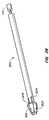
- FIG. 2is a detail view of an intra-bronchial device and delivery system according to another embodiment of the invention.
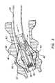
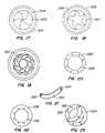
- FIG. 3is a cross-sectional view of an intra-bronchial device and deployment system according to yet another embodiment of the invention.
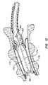
- FIG. 4is a cross-sectional view of an intra-bronchial device and deployment system according to still another embodiment of the invention.
- FIG. 5is a cross-sectional view of an intra-bronchial device and deployment system according to yet another embodiment of the invention.
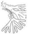
- FIG. 6shows the use of an intra-bronchial device to treat a patient.
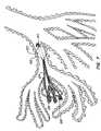
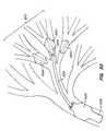
- FIG. 7shows yet another intra-bronchial device implanted in a patient's lung.
- FIG. 8shows an agent dispensing mechanism for possible use with this invention.
- FIG. 9shows a plug and delivery mechanism for use with an intra-bronchial device.
- FIG. 10is partial cross-sectional view of a plug and delivery mechanism for use with an intra-bronchial device.
- FIG. 11is a cross-sectional view of the intra-bronchial device of FIG. 10 showing the plug in place.
- FIG. 12is a cross-sectional view of another embodiment of an intra-bronchial device and delivery mechanism with a plug in place.
- FIG. 13is a cross-sectional view of yet embodiment of an intra-bronchial device and delivery mechanism.
- FIG. 14is a cross-sectional view of still embodiment of an intra-bronchial device and delivery mechanism with a tool passing through the device's plug.
- FIG. 15is a schematic view of the plug of the device of FIG. 14 in a closed position.
- FIG. 16is a schematic view of the plug of the device of FIG. 14 in an open position.
- FIG. 17shows another embodiment of an intra-bronchial device opening mechanism in a closed position.
- FIG. 18shows the embodiment of FIG. 17 in an open position.
- FIG. 19is an elevational view another embodiment of an intra-bronchial device opening mechanism.
- FIG. 20is an elevational view of a ring for use with the mechanism of FIG. 19 .
- FIG. 21is a perspective view of a blade for use with the mechanism of FIG. 19 .
- FIG. 22is an elevational view of an actuating ring for use with the mechanism of FIG. 19 .
- FIG. 23shows the mechanism of FIG. 19 , partially assembled.
- FIG. 24is a partial perspective and partial cross-sectional view of yet another embodiment of an intra-bronchial device and deployment mechanism.
- FIG. 25is a cross-sectional view of the embodiment of FIG. 25 being expanded by the deployment mechanism.
- FIG. 26is a perspective view of the intra-bronchial device of FIG. 24 and a deflation mechanism.
- FIG. 27is a cross-sectional view of the intra-bronchial device and deflation mechanism of FIG. 26 .
- FIG. 28is a perspective view of a plug for use with the intra-bronchial device of FIG. 24 .
- FIG. 29shows the plug of FIG. 28 in place within an intra-bronchial device.
- FIG. 30shows the use of a plurality of intra-bronchial devices to treat a patient's lung.
- FIG. 31is a cross-sectional view of a pressure relief system for use with the invention.
- FIG. 32shows the pressure relief system of FIG. 31 in an open position.
- the following toolsmay be used to treat COPD patients in a minimally invasive manner: Imaging and embolic devices to block blood flow through the target lung tissue; devices to help prepare the lung for devices and agents; a side wire delivery system that is advanced alongside the bronchoscope to guide and release several implants without removing the scope; a lung volume reduction implant device (Intra-Bronchial Device or IBD) that is controllably coupled to a delivery catheter that includes a working channel that runs through the center of the catheter and the implant; an inflator catheter that fits down the middle of the IBD and delivery catheter to inflate the IBD; an IBD plug element and delivery system; a deflation device to reposition or remove the IBD; a collateral flow detecting device; collateral flow blocking agents; adhesion promoting agents to maintain atelectasis; and a lung tissue compressing system.
- IBDIntra-Bronchial Device
- Perfusion of air flow in the lungscan be imaged using MRI imaging equipment while the patient breathes water saturated air or agents that are comprised primarily of water. Moving water molecules produce a strong signal, whereas static air and water will not produce a signal. This distinction is important to determine where the degraded lung hole regions reside.
- Hyper-polarized gasessuch as helium, helium-3 and xenon-129 also work extremely well in the lung to find damaged tissue and identify collateral flow.
- Computed tomographyhas also worked very well to identify damaged tissue in lungs.
- These imaging modalitiescan be used in real time before, after or during the procedure to check the patient's condition. They can also be used to guide intervention in an iterative fashion between imaging sessions or in real time. Ventilation Scans via SPECT (Xe-133) may also be used.
- Specially-designed catheterscan introduce lavage agents to the lung to wash mucus and surfactants.
- Mucus and naturally occurring surfactantstrap solids and agents to block collateral flow paths and promote adhesions within the targeted lung region or portion. Cleaning these regions to remove fluids, mucus and surfactants improves distribution of these agents and enhances adhesion of glue compositions that may be infused into the region.
- Embolic devices and agents that are used to treat peripheral vasculaturemay be utilized to embolize the pulmonary veins and arteries that normally exchange gases through the regions of the lung that are going to be or have been blocked and treated by the devices and agents of this invention.
- Exemplary embolic devicesinclude embolic polymeric implants such as Dacron fibers, metallic coils, alcohols, glutaraldehyde, gels, foams, and glue such as cyanoacrylates, PEG based glues and glutaraldehyde based glues.
- Embolizing this pulmonary vasculaturewill reduce or prevent CO 2 mixing into the heart.
- Pressure probessuch as piezo, thermal transfer flow wires or micro-electrical-mechanical system (MEMS) wave interference pressure and flow transducers may be introduced or implanted to monitor pulmonary hypertension as the blood flow paths are being blocked and to monitor results over a period of time. That way the procedure can be limited to prevent undue blood back pressure on the heart.
- the lung tissue in the regionwill still be supplied with oxygenated blood from a separate artery system that feeds pulmonary tissue.
- Implantable MEMS devicescan be used to measure pressure, temperature, strain and stress on tissues that are affected by the lung volume reduction procedure.
- MEMS transducersare passive (no electronics or batteries on board) implantable devices that can be queried using magnetic wave transmitter/receiver probes outside the body.
- the design of the current inventionresolves most of the deficiencies and issues related to the devices described above.
- the intra-bronchial deviceis placed in segments of bronchi that feed the diseased areas of the lung. As many as five to ten intra-bronchial devices could be placed in the bronchi of any one patient. The goal is to cause atelectasis in these areas and cumulatively expand the healthy portions of the lung, thereby replicating the results and benefits of LVRS, but without the morbidity and mortality.
- FIG. 1shows an intra-bronchial device 10 according to one embodiment of this invention disposed within a bronchial tube 12 of a patient's lung.
- Device 10is in contact with the inner wall of bronchial tube 12 and is preferably immobilized through a friction fit.
- device 10includes an expandable balloon 14 with a central lumen 16 through which other devices or tools (such as guidewire 18 , as shown) may be passed.
- Device 10may be delivered and deployed via a catheter 20 disposed within a working channel of a bronchoscope 22 .
- FIG. 2shows details of another embodiment of the invention.
- Device 23may be delivered and deployed via a catheter 20 disposed within a working channel of a bronchoscope 22 .
- Delivery catheter 20is connected to an inflatable balloon 25 of device 23 via a coupler 24 that may be connected and disconnected as desired.
- catheter 20has a braided shaft
- balloon 25is a folded semi-elastic balloon made from polyethylene, polyvinyl, latex, etc. (Polyethylene is particularly preferable due to the its tissue ingrowth inhibition properties.)
- Balloon 25may also be a uniform elastic balloon made from, e.g., silicone or polyurethane.
- the devicehas a ring 26 (made, e.g., from nitinol, stainless steel, polymer, Teflon, ceramic, composites, high density polyethylene, low density polyethylene, nylon, polyurethane) at its distal end marking the outlet of the balloon's central lumen.
- Catheter 20may be used to deliver the device to a target site within the patient's bronchial tube 12 and/or to inflate balloon 25 once at the target site.
- FIG. 3shows one embodiment of a balloon deployment mechanism.
- catheter 28is coupled to balloon 30 via a threaded coupler 32 cooperating with internal threads 34 within the central lumen 36 of balloon 30 .
- a fluidsuch as a hydrogel, silicone, water, saline solution, air, glue, multipart catalytic solutions, fluidized metal, suspended metal, fluoroscopic contrast medium, sodium HA
- a seal 38 at the distal end of coupler 32prevents the injection fluid from passing through the distal end of balloon lumen 36 . Instead, the fluid passes through one or more ports 40 in the wall of lumen 36 into balloon 30 to inflate the balloon.
- a one way flap 42prevents the fluid from passing back into lumen 36 once the injection fluid pressure source is removed.
- catheter 28may be rotated to disengage coupler 32 and to remove coupler 32 and seal 38 from the balloon.
- the target site for the balloonis a bronchial wall site between adjacent cartilage areas 44 and 46 , enabling the inflation of balloon 30 to distend the bronchial wall to enhance the balloon's grip on the wall.
- FIG. 4shows an alternative balloon deployment mechanism.
- a catheter 50is coupled to balloon 52 via a threaded coupler 54 cooperating with internal threads within the central lumen 56 of the balloon.
- a filler tube 58extends through catheter 50 into lumen 56 and through a port 58 formed in the lumen wall to push open a flap 60 to communicate filler tube 58 with the inside of balloon 52 .
- Filler tube 58may be used to inflate the balloon with an injection fluid, such as one of the fluids listed above.
- Filler tube 58may also be used to remove fluid from the balloon to deflate the balloon for removal or repositioning of the balloon.
- catheter 50After inflation of the balloon, catheter 50 may be rotated to disengage coupler 54 from balloon 52 .
- FIG. 5shows yet another embodiment of the invention.
- balloon 70is inflated so that it does not substantially distend the bronchial tube walls.
- Balloon 70is also longer than the balloons of the previous embodiments, extending, e.g., beyond adjacent cartilage sections of the bronchial tube wall.
- the balloon's configurationallows it to distort as the bronchial expands and contracts during the patient's breathing cycle.
- Balloon 70may be inflated using, e.g., the balloon deployment mechanisms described above with respect to FIGS. 3 and 4 .
- a port 72communicates the device's inner lumen 73 with the inside of the balloon through a flap 74 , as described above.
- FIG. 5also shows yet another mechanism for releasably coupling a catheter to the device.
- Catheter 76has a split distal end 78 with an annular engagement structure 80 configured to engage with an annular channel 82 formed on the proximal end of the device.
- a coupler sleeve 84 surrounding catheter 76may be retracted proximally to permit the split distal end 78 to expand outwardly, thereby disengaging the device, and may be advanced distally to pull distal end 78 radially inward to engage the device.
- FIG. 6shows the use of an intra-bronchial device to treat a patient.
- An expandable intra-bronchial device 100(such as one of the balloon devices described above) has been deployed at a target site in a patient's bronchial tube 102 via, e.g., a delivery catheter 104 .
- a second catheter 106has been passed through catheter 104 and the central lumen of device 100 to a treatment site 108 further down into the patient's lung.
- the distal end 110 of catheter 106may be lodged in the patient's bronchial at the treatment site.
- Catheter 106may then be used to induce atelectasis via, e.g., suction, vacuum, lavage with an anti-surfactant agent, mechanical compression, sclerosing agents (such as alcohol or other fluids or aerosols), etc.
- an anti-surfactant agente.g., suction, vacuum, lavage with an anti-surfactant agent, mechanical compression, sclerosing agents (such as alcohol or other fluids or aerosols), etc.
- FIG. 7shows the use of an intra-bronchial device to treat a patient in another manner.
- An expandable intra-bronchial device 120(such as one of the balloon devices described above) has been deployed at a target site in a patient's bronchial tube 122 .
- a plurality of wires 124are delivered through the central lumen of device 120 to place the wires' distal ends 126 within a lobe or section 128 of the patient's lung.
- the wire endsare glued or anchored to the tissue within lobe 128 .
- the proximal ends of wires 124have one-way locks 130 that may be pulled proximally through the device's 120 central lumen after anchoring of the distal ends to collapse lobe 128 inwardly. Locks 130 hold wires 124 in position, as shown.
- FIG. 8shows an agent-dispensing mechanism for possible use with this invention.
- a delivery catheter 131is mounted on a guidewire 132 via a sideport 133 .
- Agentssuch as glue or other substances may be delivered from syringe 134 via catheter 131 through an intra-bronchial device to the lung region distal to the intra-bronchial device.
- the length of catheter 131is shortened in FIG. 8 for illustration purposes. The catheter must be long enough for the syringe to be outside the patient's body and the distal end of the syringe extending into and through the intra-bronchial device.
- FIG. 9shows one embodiment of an intra-bronchial device plug 140 for deployment via catheter 142 to seal device 144 (such as one of the balloon devices described above).
- a tether 146may be used to disengage plug 140 after deployment in device 144 .
- FIGS. 10 and 11show another embodiment of an intra-bronchial device plug 150 being delivered to device 152 (such as one of the balloon devices described above) via delivery catheter 154 .
- Plug 150is releasably held to a plug pusher or catheter 156 by a tether 158 .
- Plug 150has threads 160 that engage with threads 162 in device 152 when plug 150 is rotated by catheter 156 to seal the central lumen of device 152 .
- FIG. 12shows yet another embodiment of a plug 170 for an intra-bronchial device 172 (such as the balloon device described above with respect to FIG. 5 ).
- Plug 170has a stem 174 (formed, e.g., from metal or plastic) passing through an occlusion element 176 formed from an elastomeric polymer or gel.
- Plug 170may be advanced into position by compressing it through a narrowed proximal end 182 of the central lumen 184 of device 172 through the action of a pusher or catheter (not shown) coupled to a coupling surface 180 formed on the proximal end of stem 174 .
- plug 170may be advanced distally or retracted proximally.
- the plugmay attach to the device using notches, luer locks, press fit, tapers, etc.
- FIG. 13shows yet another plug 200 for an intra-bronchial device 202 .
- plug 200forms an elastomeric seal around a central opening 204 through which tools or other devices may be inserted.
- Plug 200may be integral with device 202 so that it does not have to be delivered separately from device 202 .
- FIGS. 14-16show an intra-bronchial device 210 with an integral seal 212 having a central opening 213 formed by the cooperation of a plurality of flaps 216 .
- Seal 212may be integral with the central tube 214 of device 210 .
- Central tube 214 and seal 212may be formed from an elastic metal or polymer or rubber to allow flaps 216 to bend (as shown in FIG. 16 ) to permit devices or tools to be passed through opening 213 . Flaps 216 return to their sealing position of FIG. 15 after the tool or device (such as guide wire 215 ) has been removed.
- FIGS. 11-14show plugged intra-bronchial devices attached to their respective catheters using releasable coupling mechanisms such as those described above with respect to FIG. 5 .
- the coupling mechanismshelp hold the device in place at the target site while the plug is being inserted.
- deflate the balloon and remove the device from the patientmay also be necessary after deployment of an intra-bronchial device to deflate the balloon and remove the device from the patient.
- deflationmay be accomplished by, e.g., puncturing the balloon.
- the devicemay be coupled to a catheter as shown in FIGS. 11-14 and removed from the patient and/or deployed at a different site.
- FIGS. 17-23show alternative designs for intra-bronchial device openings formed, e.g., as actuatable iris-type shutters.
- the shutteris formed from a plurality of blades 220 rotatably mounted on a ring 222 via pins 224 inserted into holes 226 formed in the ring.
- the bladesare arranged in an overlapping arrangement as shown in FIG. 19 .
- the shutteris operated by an actuating ring 228 having slots 230 interacting with a second set of pins 232 on blades 220 .
- Rotation of ring 228 in one directionopens the shutter, and rotation of ring 228 in the other direction closes the shutter.
- FIGS. 17 and 18show a five-blade shutter design
- FIGS. 19-23show an eight-blade shutter design.
- FIGS. 24-29show another embodiment of the invention (outside of the lung, for ease of illustration).
- Intra-bronchial device 250has a central shaft 252 surrounded by an expandable member, such as balloon 254 .
- Shaft 252has an opening 256 communicating the shaft's central lumen with the interior of balloon 254 via a flexible flap valve 258 .
- Device 250may be delivered to an air passageway of a patient's lung using a delivery catheter in, e.g., a manner described above.
- An inflation catheter 260may be used to inflate balloon 254 from the unexpanded condition shown in FIG. 24 to the expanded state of FIG. 25 .
- Inflation catheter 260may be inserted into the patient through the delivery catheter (delivered together with the device 250 or after it) or independent of the delivery catheter.
- the distal tip 262 of inflation catheter 260has a pointed end to help align the inflation catheter with the intra-bronchial device's shaft.
- Inflation catheter 260has an opening 264 with seals 266 and 268 on either side. When inserted into shaft 252 , opening 264 aligns with the shaft's opening 256 when a shoulder 270 on inflation catheter 260 meets a shoulder 272 formed on the proximal end of device 250 .
- Seals 266 and 268ensure that pressurized fluid delivered to device 250 via inflation catheter 260 enters balloon 254 via openings 264 and 256 and flap valve 258 to inflate balloon 254 .
- flap valve 258closes to maintain balloon 254 in its inflated state.
- the device 250may be inflated in multiple steps, as needed.
- the inflation cathetermay protrude through the distal end of the intra-bronchial device.
- rapid exchange systemsare catheter systems that can be threaded onto a short section of wire before the user can gain control of the wire end and the catheter system.
- the wiremay be introduced into and out of a side port or it can be introduced in any combination of side, end or through lumen compartments.
- FIGS. 26 and 27show a deflation catheter 280 that may be used to deflate intra-bronchial device for removal or repositioning.
- Deflation cathetermay be advanced through the device delivery catheter or independently.
- Deflation catheterhas a plurality of fingers 282 separated by slots 283 and arranged circumferentially around the catheter's distal end. When advanced into shaft 252 , shoulders 284 formed on the distal ends of fingers 282 cam radially inward to enable the catheter 280 to be advanced into device 250 .
- a shoulder 286meets shoulder 272 of device 250 when fingers 284 have been advanced distally to the proper position with respect to opening 256 .
- FIGS. 28 and 29show a plug 300 for an intra-bronchial device, such as the device described in FIGS. 24 and 25 .
- FIG. 29shows balloon 254 in a deflated state.
- a plurality of fingers 302 separated by slots 304are disposed at the distal end of plug 300 .
- the distal end of each finger 302has an angled camming surface 306 facing distally and a steeper camming surface 308 facing proximally.
- the plughas a radially symmetric coupling handle 310 at its proximal end for attachment to a delivery and/or recapture catheter (not shown).
- plug 300When inserting plug 300 into the intra-bronchial device, distal movement of plug 300 into shaft 252 causes fingers 302 to cam radially inward until the distal end of plug 300 emerges from the distal end of shaft 252 , at which point fingers 302 move outward to lock plug 300 in place.
- a proximal shoulder(not shown) may be provided on plug 300 to prevent the plug from advancing out the distal end of the intra-bronchial device. If removal of plug 300 is desired, a proximally directed force on plug 300 will cause fingers 302 to cam inward to allow the plug to be withdrawn through shaft 252 .
- FIG. 30shows the use of the invention to compress a portion of a patient's lung.
- three intra-bronchial devices 150 a , 150 b and 150 care disposed in a portion 320 of the patient's lung.
- Devices 150 a and 150 bhave been plugged and released from their delivery systems; device 150 c is still connected to catheter 322 which communicates with the still-open lumen 324 of the device's central shaft.
- pressurized fluidis introduced into the patient's lung through sleeve 326 surrounding catheter 322 .
- An expandable (e.g., inflatable, expanding metallic frame or braid) cuff 328seals sleeve against the air passageway wall.
- Devices 150 a - cprevent the pressurized fluid from entering lung portion 320 .
- Inflation of one or more portions of the lung adjacent portion 320will cause portion 320 to collapse, venting any air in lung portion 320 to the exterior of the patient through catheter 322 .
- These devicescause effective lung tissue compression with the application of more than 10 mm Hg pressure above atmospheric pressure. By applying more pressure, the effect is made more rapid and complete: 25 mm Hg is better, 45 mm Hg is better still and more than 55 mm Hg is best.
- the sleevecan be made of typical guide catheter materials with similar construction techniques and may be covered or comprised of silicone, polyurethane, biocompatible polymers, elastic balloon materials, semi-elastic balloon materials or a mesh composite.
- the ballooncan be compliant or semi-compliant and can be made from polymers such as polyurethane or silicones.
- the cuffmay be self expanding with the use of titanium alloys and these can be made from braid. Braided funnel shaped ends work very well to seal this device.
- FIGS. 31 and 32illustrate a pressure relief system that minimizes the risk of such injuries.
- Intra-bronchial device 350has a pressure relief valve 352 that opens (as shown in FIG. 32 ) when the differential pressure between the collapsing lung portion on the distal side of device 350 and the lung portions on the proximal side of device 350 exceeds a desired amount, such as, e.g., 2 mm Hg, 10 mm Hg, 20 mm Hg or 50 mm Hg.
- a desired amountsuch as, e.g., 2 mm Hg, 10 mm Hg, 20 mm Hg or 50 mm Hg.
- the greater the differential pressurethe greater the lung volume reduction, but also the greater the risk of complications.
- a maximum lung expansionmay be targeted.
- the pressure required to open the relief valvecan be set such that the expanding lung tissue is not strained more than 150%.
- the pressure relief valvemay also reside in an intra-bronchial device plug instead of being integral with the expandable intra
- FIG. 30also shows aspects of a collateral flow detection system for use with this invention.
- this systemPrior to attempting hyperinflation of the lung to collapse the target portion of the lung, this system can be used to check for the existence of collateral flow paths from the targeted lung portion 320 back to the remaining portions of the lung and the exterior of the patient.
- Air blended with a markersuch as a detectable gas may be introduced into the lung through sleeve 326 , and the air in the target region 320 may be monitored through catheter 322 by sniffing or sampling. If the marker gas is detected, collateral flow is occurring, either due to the presence of flow paths through degraded tissue, natural airways that still need to be plugged with intra-bronchial devices, or the failure of one or more implanted intra-bronchial devices.
- Gases that may be used for collateral flow detectionare hyper-polarized gases such as helium, helium-3 and xenon-129.
- Other materialsinclude Diethyl ether, Nitrous oxide, Chloroform, Cyclopropane, Trichloroethylene, Fluroxene, Halothane, Methoxyflurane, Enflurane, Isoflurane, Desflurane, Sevoflurane or components of these. Small amounts of CO can also be tolerated and used for this purpose.
- one or more agents to block and clog the collateral flow pathsmay be introduced, e.g., through the intra-bronchial device delivery catheter so that it is installed in the isolated lung region.
- the agentwill flow through any such collateral flow path.
- This treatmentis intended to block flow of collateral pathways that are created by the degenerative disease. As such, treatments may need to be repeated periodically to block pathways that are newly formed by the disease progression. This can be easily done by coupling a delivery catheter to the intra-bronchial device and then by removing the central cap from the intra-bronchial device. This provides a direct conduit to the distal isolated lung region.
- Microparticlescan be used for blocking collateral flow in lung tissue.
- the microparticlespreferably comprise a polymeric binder or other means to make controlled geometry particles.
- Suitable polymeric binder materialsinclude poly(glycolic acid), poly-d,l-lactic acid, poly-l-lactic acid, copolymers of the foregoing, poly(aliphatic carboxylic acids), copolyoxalates, polycaprolactone, polydioxanone, poly(ortho carbonates), poly(acetals), poly(lactic acid-caprolactone), polyorthoester, poly(glycolic acid-caprolactone), polyanhydrides, polyphosphazines, albumin, casein, and waxes.
- Poly (d,l-lactic-co-glycolic acid)is commercially available from Alkermes, Inc. (Blue Ash, Ohio).
- a suitable product commercially available from Alkermes, Inc.is a 50:50 poly (d,l-lactic-co-glycolic acid) known as MEDISORB.RTM. 5050 DL. This product has a mole percent composition of 50% lactide and 50% glycolide.
- Other suitable commercially available productsare MEDISORB.RTM. 6535 DL, 7525 DL, 8515 DL and poly(d,l-lactic acid) (100 DL).
- Poly(lactide-co-glycolides)are also commercially available from Boehringer Ingelheim (Germany) under its Resomer.RTM.
- Other materialsinclude biocompatible polymers that are described in the US Pharmacopeia and include dextrans and other carbohydrate-based materials or derivatives thereof, polyethylene glycol, polyethylene oxide, polyvinyl alcohol, and starch. Additional materials include polyesters, such as polyglycolic acid, polylactic acid, poly-1,4-dioxa-2-one, polyoxaltes, polycarbonates, copolymers of polyglycolic acid and polylactic acid, polycaprolactone, poly-b-hydroxybutyrate, copolymers of epsilon-caprolactone and delta-valerolactone, copolymers of epsilon-caprolactone and DL-dilactide, and polyester hydrogels; polyvinylpyrrolidone; polyamides; gelatin; albumin; proteins; collagen; poly(orthoesters); poly(anhydrides); poly(alkyl-2-cyanoacrylates); poly(dihydropyrans); poly(acetals); poly(phos
- Block copolymersare another class of materials that are suitable for the present invention.
- Protein microspheresare another class of materials that enables the present invention.
- albumincan be cross-linked with a suitable cross-linker to generate particles for various applications including imaging.
- Other proteins suitable for enabling the present inventioninclude recombinant or naturally occurring human, animal, or vegetable proteins, including collagen, gelatin, casein, soybean protein, vegetable protein, and keratin.
- Liposomes, proliposomes, or microparticles containing fatty acids, lipids, or derivatives thereofwill also enable the invention.
- Synthetic polymeric particlescomprised of HEMA (hydroxyethlymethacrylate), AEMA (aminoethyl methacrylate), DMEMA (N,N dimethyl amino) and other acrylates, acrylamides, methacrylamides, styrene, or any polymerizable material will also work in this application.
- HEMAhydroxyethlymethacrylate
- AEMAaminoethyl methacrylate
- DMEMAN,N dimethyl amino
- Viscous solutions for the present inventionare preferably biocompatible solutions such as hydrogels or other substances including glycerol and aqueous solutions containing water soluble materials including cellulose derivatives, dextrans, starches, and any carbohydrate or carbohydrate based material or derivative thereof.
- Other aqueous solutionsmay contain synthetic polymers including povidone, PEG (polyethylene glycol or polyethylene oxide), polyvinyl alcohol, and diethyl aminoethyl (DEAE)—sephadex.
- Aqueous solutionsmay also contain proteins such as collagen and albumin.
- Other viscous solutionsmay contain non-aqueous cosolvents such as ethanol. Buffers, salts, and excipients may also be part of the formulation of thixotropic viscous solutions.
- a two-part sealantmay be applied to areas of interest.
- One novel methodinvolves applying one part of the sealant to a desired area in one lobe, and applying a second component to the other lobe.
- the two componentsmay mix and solidify at a location between the lobes, or partially in both lobes such that flow is reduced.
- Sealant components for this applicationmay include fibrin/thrombin, activated PEG/PEG-diamine, albumin/PEG, and albumin/glutaraldehyde sealants.
- the sealantis an implantable material that may contain hemostatic agents such as chitin derivatives including but not limited to carboxymethyl chitin and chitosan (1-100% deacetylated).
- the sealant componentsmay also contain additives that affect viscosity, set time, adhesion, and biocompativility.
- the albumin componentmay be formulated in weight to weight ratios of 10-50% where the remaining mass balance is aqueous solutions of salts, buffers, and additives or combinations thereof.
- the other component of the sealantis a cross-linker containing glutaraldehyde or derivatives thereof in weight to volume ratios of 1-25% where the remaining balance is an aqueous solution with or without additives, salts, or buffers or combinations thereof.
- solutionsmay be applied from dispensers that deliver a ratio of 1 unit volume of protein solution per 1 unit volume of cross-linker solution (1:1 protein:cross-linker) and may be applied in ratios up to 10 unit volumes of protein solution per unit volume of cross-linker solution.
- mixingmay occur by passing the solutions through a static mixing tip with helical or other geometrical devices that enhance the mixing efficiency. Sealants prepared from these solutions contain 5-45% protein and 0.5-14% crosslinker.
- tissue remodeling cascade processincludes tissue polymer decomposition and/or necrosis that leads to recruitment of cellular respondents that include one or more of the following: Neutrophils, white blood cells, macrophages, CD8+, MMP's, Interlukens, cytokins and protocylins. Then the tissue remodels to initiate tissue formation and thickening that culminates in the formation of tissue adhesions.
- materials that can initiate this effectare cadmium, smoke artifacts, tars, materials that irritate tissue such as alcohols, solvents, organic solvents, acids, materials that are basic and materials that are acidic. These include compounds or materials that have pH levels between 1 and 6.9 with materials closest to 1 being a preferable acid material. Additionally, compounds or materials that have pH levels between 7.5 and 14 work very well but materials closest to 14 work best.
- Our glutaraldehyde glue technologycan produce compositions that have 15 centipoise viscosity and higher levels up to and beyond 150 centipoise.
- control of depositionis very important. Ways of controlling deposition include localized dispensing of the sealant through a suitable device containing a lumen, and also through the addition of agents that increase the viscosity of one or more components of the implantable material.
- Such agentsinclude biocompatible materials with viscosities that are greater than those of water, and include glycerol, polymeric materials such as proteins, carbohydrate-based polymers and derivatives thereof, synthetic materials including polyethylene glycols (PEG), polyethylene oxides (PEO), polyvinyl pyrrolidone (PVP), polyvinyl alcohol and other components described in the “United States Pharmacopeia” and Rowe, R. C, et al., Handbook of Pharmaceutical Excipients 4 th edition 2003.
- Other materials for controlling viscosityinclude oils, lipids, and fatty acids, including oleic acid, and phosphocholines. Phase separation can be controlled with emulsifiers including poly sorbate.
- the viscosities of one or more of the componentscan be modified by adding an appropriate agent to control spreading after application. Viscosities of these components can range from 1 to 1000 centistokes (a measure of kinematic viscosity).
- Deposition and control of spreading of sealants containing two or more componentsare also affected by the gel time, or set time, of the mixed sealant.
- Sealants with short set timesare preferable to those with longer set times.
- Ideal set times for the present invention and methodrange from 1-600 seconds, and preferable from 1-60 seconds.
- Set timecan be controlled by the addition of set time modifiers, including agents that reduce or increase the set time relative to the corresponding formulation lacking the set time modifier.
- An example of an agent that decreases the set timeis carboxymethyl cellulose.
- An example of an agent that increases the set timeis glycerol.
- Glutaraldehydeundergoes reversible reactions that cause reoccurring inflammation. These properties can be improved by chemical modification of the glutaraldehyde.
- One such modificationincludes glutaraldehyde condensation reactions, as described in “Bioconjugate Techniques” by G. T. Hermanson. This condensation involves the formation of derivatives of glutaraldehyde in aqueous solutions containing acid or base. This reaction can be monitored by ultraviolet spectroscopy at or near 280 and 234 nanometers. At 280 nanometers, pure glutaraldehyde has significant absorbance, and little or no absorbance at 234 nanometers when measured as an aqueous solution at 0.5% weight to volume.
- glutaraldehydeWhen glutaraldehyde is chemically modified, it has significant absorbance at 234 nanometers. These derivatives are effective cross-linking agents when used with nucleophilic substrates such as proteins, including albumins. Furthermore, sealants prepared from glutaralde hyde derivatives are adhesive in vivo, through chemical or mechanical means, or a combination of chemical and mechanical means.
- Implantable materials for the present inventionare any agents administered into tissue, including sealants, which may be comprised of hydrogels, proteins, or other biocompatible materials, that can be implanted into compromised tissue to benefit the patient.
- hydrogelsinclude those prepared from natural sources including carbohydrate-based materials. Such materials include hyaluronans, hyaluronic acid, alginates, chitins, chitosans, and derivatives thereof.
- Proteins that enable the present inventioninclude albumins, collagens, gelatins, and other proteins that can be cross-linked or that form solutions with viscosities greater than water.
- Other implantable materialsinclude those prepared by mixing two or more components so that a viscous solution, gel, or solid is formed.
- Such implantable materialsare prepared from a protein substrate where the protein is derived from natural, synthetic, or semi-synthetic processes.
- the proteinmay also be derived from recombinant DNA technology and may be isolated from cell-culture processes, as well as from transgenic plants and animals.
- proteinsinclude albumins, collagens, and gelatins.
- Cross-linkers employed as part of the implantable material precursorsinclude aldehydes, polyaldehydes, esters, and other chemical functionality suitable for cross-linking protein(s). Examples of homobifunctional cross-linking agents are described in “Bioconjugate Techniques” by G. T. Rermanson.
- the implant componentscan be formulated at a pH in the range of 5-10 by adjusting the pH and/or by adding suitable buffers in the range of 1-500 mM.
- buffersinclude phosphate, carbonate, bicarbonate, borate, or imidazole, or mixtures thereof.
- additives or stabilizersmay be added to improve the stability of one or more of the components.
- imaging agentsmay be added to allow for detection of the material. Such agents include iodine, iodine compounds, metals such as gadolinium, radioisotopes, and other compounds for gamma scintigraphy, magnetic resonance imaging, fluoroscopy, CT, SPECT and other imaging modalities.
- the materialmay be formulated such that the mechanical properties are suitable for applications in the specific tissue to which the imp lantable material is applied. Such properties include elasticity, modulus, stiffness, brittleness, strain, cohesion, adhesion, and stress. Agents for modifying the properties include fillers, plasticizers, and adhesion modifiers.
- the implantmay induce a natural adhesive mechanism with or without the addition of chemical agents which may be added to the implant to induce a natural response. Such agents include particles in the range of 100 nm to 1 millimeter. Agents include chemical or biochemical agents (proteins or nucleic acids) that induce a natural response. Examples of such agents include bleomycin, cytokines and chemokines, and single stranded RNA molecules.
- Intra-bronchial devices according to this inventionmay be delivered through the working channel of a bronchoscope.
- the lung access system described in the U.S. patent application filed of even date herewith titled “Lung Access Device and Method,”may be used to deliver the devices to a patient's lung. This latter delivery method may be used when the diameter of a bronchoscope is too small for the device to be delivered.
- One advantage of using an intra-bronchial device with a larger, collapsed delivery diameteris that the expansion ratio from delivery diameter to deployed diameter may be made smaller than, e.g., 7.5, more preferably smaller than 6, more preferably smaller than 5, more preferably smaller than 4, and most preferably smaller than 3.
- the delivered devicesare not limited in length since they can be delivered outside the constraints of the scope channel.
- the scope path to the upper airways, where the most tissue damage normally resides,requires very small radius bends to be formed in the bronchoscope to gain access. Long implant devices that would straighten the scope if delivered through the channel can now be delivered outside the channel while the target region continues to be imaged through the scope optics.
- implant devicesthat exceed 4 mm in length can now be delivered easily using this system. Devices longer than 5 mm will work better, devices longer than 10 mm are better, devices longer than 20 mm are preferable, devices longer than 25 mm, 30 mm, and 35 mm will anchor much better.
- These devicescan be made from all biocompatible plastics, metals, ceramics, shape memory alloys and carbon based fiber or polymers.
- the catheter devicescan be lined with fluoro polymers and reinforced with metal or polymer fiber or wire braid or by using coils of similar materials.
- the wire elements that guide devicescan be made from steel or titanium alloys or other metals that do not present artifacts in MRI equipment.
- Other materials including shape memory alloyssuch as nickel- and titanium-based metals that are comprised of more than 40% titanium would perform well in that they can be made in an anisotropic way to provide different properties with bending and torque.
- one or more of the delivery and deployment cathetersmay have multiple lumens.
- a multi-lumen cathetercould be used to both inflate the intra-bronchial device and deliver glue or another substance (such as those described above) outside of, and either distal or proximal to, the intra-bronchial device. Sheaths, needles and other devices may be used to deploy such substances.
- an extra-catheter lumenis as a return path. As long as the pressure drop over the return path is less than the pressure required to inflate the intra-bronchial device, air or the inflation fluid will preferentially flow down the return path.
- the pressurecan be controlled with the delivery rate of the inflation fluid.
- This return pathcan also act as a pressure relief conduit to control the maximum inflation pressure applied to the balloon.
Landscapes
- Health & Medical Sciences (AREA)
- Life Sciences & Earth Sciences (AREA)
- Heart & Thoracic Surgery (AREA)
- General Health & Medical Sciences (AREA)
- Surgery (AREA)
- Veterinary Medicine (AREA)
- Engineering & Computer Science (AREA)
- Biomedical Technology (AREA)
- Public Health (AREA)
- Animal Behavior & Ethology (AREA)
- Anesthesiology (AREA)
- Hematology (AREA)
- Pulmonology (AREA)
- Reproductive Health (AREA)
- Vascular Medicine (AREA)
- Nuclear Medicine, Radiotherapy & Molecular Imaging (AREA)
- Biophysics (AREA)
- Medical Informatics (AREA)
- Molecular Biology (AREA)
- Child & Adolescent Psychology (AREA)
- Surgical Instruments (AREA)
Abstract
Description
Claims (17)
Priority Applications (1)
| Application Number | Priority Date | Filing Date | Title |
|---|---|---|---|
| US11/153,253US7549984B2 (en) | 2004-06-16 | 2005-06-14 | Method of compressing a portion of a lung |
Applications Claiming Priority (2)
| Application Number | Priority Date | Filing Date | Title |
|---|---|---|---|
| US58056504P | 2004-06-16 | 2004-06-16 | |
| US11/153,253US7549984B2 (en) | 2004-06-16 | 2005-06-14 | Method of compressing a portion of a lung |
Publications (2)
| Publication Number | Publication Date |
|---|---|
| US20060009748A1 US20060009748A1 (en) | 2006-01-12 |
| US7549984B2true US7549984B2 (en) | 2009-06-23 |
Family
ID=35785652
Family Applications (4)
| Application Number | Title | Priority Date | Filing Date |
|---|---|---|---|
| US11/153,855AbandonedUS20060004400A1 (en) | 2004-06-16 | 2005-06-14 | Method of treating a lung |
| US11/153,253Expired - Fee RelatedUS7549984B2 (en) | 2004-06-16 | 2005-06-14 | Method of compressing a portion of a lung |
| US11/153,233AbandonedUS20050288684A1 (en) | 2004-06-16 | 2005-06-14 | Method of reducing collateral flow in a portion of a lung |
| US11/153,235AbandonedUS20050288702A1 (en) | 2004-06-16 | 2005-06-14 | Intra-bronchial lung volume reduction system |
Family Applications Before (1)
| Application Number | Title | Priority Date | Filing Date |
|---|---|---|---|
| US11/153,855AbandonedUS20060004400A1 (en) | 2004-06-16 | 2005-06-14 | Method of treating a lung |
Family Applications After (2)
| Application Number | Title | Priority Date | Filing Date |
|---|---|---|---|
| US11/153,233AbandonedUS20050288684A1 (en) | 2004-06-16 | 2005-06-14 | Method of reducing collateral flow in a portion of a lung |
| US11/153,235AbandonedUS20050288702A1 (en) | 2004-06-16 | 2005-06-14 | Intra-bronchial lung volume reduction system |
Country Status (4)
| Country | Link |
|---|---|
| US (4) | US20060004400A1 (en) |
| EP (2) | EP3542736A1 (en) |
| JP (1) | JP2008503254A (en) |
| WO (1) | WO2006009688A2 (en) |
Cited By (48)
| Publication number | Priority date | Publication date | Assignee | Title |
|---|---|---|---|---|
| US20070225747A1 (en)* | 2006-03-08 | 2007-09-27 | Pulmonx | Methods and devices to induce controlled atelectasis and hypoxic pulmonary vasoconstriction |
| US20090012508A1 (en)* | 2004-06-24 | 2009-01-08 | Virulite Distribution Limited | Cosmetic uses of electromagnetic radiation |
| US20090012626A1 (en)* | 2006-03-13 | 2009-01-08 | Pneumrx, Inc. | Minimally invasive lung volume reduction devices, methods, and systems |
| US20090326322A1 (en)* | 2008-06-27 | 2009-12-31 | Intuitive Surgical, Inc. | Medical robotic system with image referenced camera control using partitionable orientational and translational modes |
| US20100070050A1 (en)* | 2008-09-12 | 2010-03-18 | Pneumrx, Inc. | Enhanced Efficacy Lung Volume Reduction Devices, Methods, and Systems |
| US20110178582A1 (en)* | 2008-07-14 | 2011-07-21 | Gordon Dougal | Electromagnetic Radiation And Its Therapeutic Effect |
| US20110245665A1 (en)* | 2010-03-30 | 2011-10-06 | Angiodynamics, Inc. | Bronchial catheter and method of use |
| WO2012026336A1 (en) | 2010-08-25 | 2012-03-01 | テルモ株式会社 | Therapeutic agent for pulmonary emphysema |
| US20120172909A1 (en)* | 2006-03-13 | 2012-07-05 | Pneumrx, Inc. | Lung Volume Reduction Devices, Methods, and Systems |
| US20120203271A1 (en)* | 2006-06-13 | 2012-08-09 | Intuitive Surgical Operations, Inc. | Guide tube control of minimally invasive surgical instrument |
| US8721734B2 (en) | 2009-05-18 | 2014-05-13 | Pneumrx, Inc. | Cross-sectional modification during deployment of an elongate lung volume reduction device |
| US9402633B2 (en) | 2006-03-13 | 2016-08-02 | Pneumrx, Inc. | Torque alleviating intra-airway lung volume reduction compressive implant structures |
| US9598691B2 (en) | 2008-04-29 | 2017-03-21 | Virginia Tech Intellectual Properties, Inc. | Irreversible electroporation to create tissue scaffolds |
| US9713703B2 (en) | 2013-08-09 | 2017-07-25 | Boston Scientific Scimed, Inc. | Devices and methods for treating a lung |
| US9757196B2 (en) | 2011-09-28 | 2017-09-12 | Angiodynamics, Inc. | Multiple treatment zone ablation probe |
| US9867652B2 (en) | 2008-04-29 | 2018-01-16 | Virginia Tech Intellectual Properties, Inc. | Irreversible electroporation using tissue vasculature to treat aberrant cell masses or create tissue scaffolds |
| US9895189B2 (en) | 2009-06-19 | 2018-02-20 | Angiodynamics, Inc. | Methods of sterilization and treating infection using irreversible electroporation |
| US10117707B2 (en) | 2008-04-29 | 2018-11-06 | Virginia Tech Intellectual Properties, Inc. | System and method for estimating tissue heating of a target ablation zone for electrical-energy based therapies |
| US10154874B2 (en) | 2008-04-29 | 2018-12-18 | Virginia Tech Intellectual Properties, Inc. | Immunotherapeutic methods using irreversible electroporation |
| US10238447B2 (en) | 2008-04-29 | 2019-03-26 | Virginia Tech Intellectual Properties, Inc. | System and method for ablating a tissue site by electroporation with real-time monitoring of treatment progress |
| US10245105B2 (en) | 2008-04-29 | 2019-04-02 | Virginia Tech Intellectual Properties, Inc. | Electroporation with cooling to treat tissue |
| US10272178B2 (en) | 2008-04-29 | 2019-04-30 | Virginia Tech Intellectual Properties Inc. | Methods for blood-brain barrier disruption using electrical energy |
| US10292755B2 (en) | 2009-04-09 | 2019-05-21 | Virginia Tech Intellectual Properties, Inc. | High frequency electroporation for cancer therapy |
| US10390838B1 (en) | 2014-08-20 | 2019-08-27 | Pneumrx, Inc. | Tuned strength chronic obstructive pulmonary disease treatment |
| US10470822B2 (en) | 2008-04-29 | 2019-11-12 | Virginia Tech Intellectual Properties, Inc. | System and method for estimating a treatment volume for administering electrical-energy based therapies |
| US10471254B2 (en) | 2014-05-12 | 2019-11-12 | Virginia Tech Intellectual Properties, Inc. | Selective modulation of intracellular effects of cells using pulsed electric fields |
| US10537334B2 (en) | 2013-10-25 | 2020-01-21 | Pneumrx, Inc. | Genetically-associated chronic obstructive pulmonary disease treatment |
| US10543053B2 (en) | 2016-09-30 | 2020-01-28 | Pneumrx, Inc. | Containers for medical devices |
| US10555736B2 (en) | 2016-09-30 | 2020-02-11 | Pneumrx, Inc. | Guidewire |
| US10694972B2 (en) | 2014-12-15 | 2020-06-30 | Virginia Tech Intellectual Properties, Inc. | Devices, systems, and methods for real-time monitoring of electrophysical effects during tissue treatment |
| US10702326B2 (en) | 2011-07-15 | 2020-07-07 | Virginia Tech Intellectual Properties, Inc. | Device and method for electroporation based treatment of stenosis of a tubular body part |
| US11254926B2 (en) | 2008-04-29 | 2022-02-22 | Virginia Tech Intellectual Properties, Inc. | Devices and methods for high frequency electroporation |
| US11272979B2 (en) | 2008-04-29 | 2022-03-15 | Virginia Tech Intellectual Properties, Inc. | System and method for estimating tissue heating of a target ablation zone for electrical-energy based therapies |
| US11311329B2 (en) | 2018-03-13 | 2022-04-26 | Virginia Tech Intellectual Properties, Inc. | Treatment planning for immunotherapy based treatments using non-thermal ablation techniques |
| US11382681B2 (en) | 2009-04-09 | 2022-07-12 | Virginia Tech Intellectual Properties, Inc. | Device and methods for delivery of high frequency electrical pulses for non-thermal ablation |
| US11453873B2 (en) | 2008-04-29 | 2022-09-27 | Virginia Tech Intellectual Properties, Inc. | Methods for delivery of biphasic electrical pulses for non-thermal ablation |
| US11607537B2 (en) | 2017-12-05 | 2023-03-21 | Virginia Tech Intellectual Properties, Inc. | Method for treating neurological disorders, including tumors, with electroporation |
| US11638603B2 (en) | 2009-04-09 | 2023-05-02 | Virginia Tech Intellectual Properties, Inc. | Selective modulation of intracellular effects of cells using pulsed electric fields |
| US11707629B2 (en) | 2009-05-28 | 2023-07-25 | Angiodynamics, Inc. | System and method for synchronizing energy delivery to the cardiac rhythm |
| US11723710B2 (en) | 2016-11-17 | 2023-08-15 | Angiodynamics, Inc. | Techniques for irreversible electroporation using a single-pole tine-style internal device communicating with an external surface electrode |
| US11925405B2 (en) | 2018-03-13 | 2024-03-12 | Virginia Tech Intellectual Properties, Inc. | Treatment planning system for immunotherapy enhancement via non-thermal ablation |
| US11931096B2 (en) | 2010-10-13 | 2024-03-19 | Angiodynamics, Inc. | System and method for electrically ablating tissue of a patient |
| US11950835B2 (en) | 2019-06-28 | 2024-04-09 | Virginia Tech Intellectual Properties, Inc. | Cycled pulsing to mitigate thermal damage for multi-electrode irreversible electroporation therapy |
| US12102376B2 (en) | 2012-02-08 | 2024-10-01 | Angiodynamics, Inc. | System and method for increasing a target zone for electrical ablation |
| US12114911B2 (en) | 2014-08-28 | 2024-10-15 | Angiodynamics, Inc. | System and method for ablating a tissue site by electroporation with real-time pulse monitoring |
| US12201349B2 (en) | 2009-04-03 | 2025-01-21 | Angiodynamics, Inc. | Congestive obstruction pulmonary disease (COPD) |
| US12214189B2 (en) | 2019-07-24 | 2025-02-04 | Virginia Tech Intellectual Properties, Inc. | Fourier analysis spectroscopy for monitoring tissue impedance changes and treatment outcome during electroporation-based-therapies |
| US12390262B2 (en) | 2018-03-13 | 2025-08-19 | Virginia Tech Intellectual Properties, Inc. | Treatment planning system for immunotherapy enhancement via non-thermal ablation |
Families Citing this family (132)
| Publication number | Priority date | Publication date | Assignee | Title |
|---|---|---|---|---|
| US8016823B2 (en)* | 2003-01-18 | 2011-09-13 | Tsunami Medtech, Llc | Medical instrument and method of use |
| US7892229B2 (en)* | 2003-01-18 | 2011-02-22 | Tsunami Medtech, Llc | Medical instruments and techniques for treating pulmonary disorders |
| US9433457B2 (en) | 2000-12-09 | 2016-09-06 | Tsunami Medtech, Llc | Medical instruments and techniques for thermally-mediated therapies |
| US7549987B2 (en) | 2000-12-09 | 2009-06-23 | Tsunami Medtech, Llc | Thermotherapy device |
| US8444636B2 (en) | 2001-12-07 | 2013-05-21 | Tsunami Medtech, Llc | Medical instrument and method of use |
| US7811274B2 (en)* | 2003-05-07 | 2010-10-12 | Portaero, Inc. | Method for treating chronic obstructive pulmonary disease |
| US7426929B2 (en) | 2003-05-20 | 2008-09-23 | Portaero, Inc. | Intra/extra-thoracic collateral ventilation bypass system and method |
| US7533667B2 (en) | 2003-05-29 | 2009-05-19 | Portaero, Inc. | Methods and devices to assist pulmonary decompression |
| US7252086B2 (en) | 2003-06-03 | 2007-08-07 | Cordis Corporation | Lung reduction system |
| US7377278B2 (en) | 2003-06-05 | 2008-05-27 | Portaero, Inc. | Intra-thoracic collateral ventilation bypass system and method |
| US7682332B2 (en)* | 2003-07-15 | 2010-03-23 | Portaero, Inc. | Methods to accelerate wound healing in thoracic anastomosis applications |
| US8579892B2 (en)* | 2003-10-07 | 2013-11-12 | Tsunami Medtech, Llc | Medical system and method of use |
| US7670282B2 (en) | 2004-06-14 | 2010-03-02 | Pneumrx, Inc. | Lung access device |
| US7608579B2 (en)* | 2004-06-16 | 2009-10-27 | Pneumrx, Inc. | Lung volume reduction using glue compositions |
| US7553810B2 (en) | 2004-06-16 | 2009-06-30 | Pneumrx, Inc. | Lung volume reduction using glue composition |
| US20050281739A1 (en)* | 2004-06-16 | 2005-12-22 | Glen Gong | Imaging damaged lung tissue using compositions |
| US7678767B2 (en) | 2004-06-16 | 2010-03-16 | Pneumrx, Inc. | Glue compositions for lung volume reduction |
| US20050281798A1 (en)* | 2004-06-16 | 2005-12-22 | Glen Gong | Targeting sites of damaged lung tissue using composition |
| US20050281740A1 (en)* | 2004-06-16 | 2005-12-22 | Glen Gong | Imaging damaged lung tissue |
| US20050281800A1 (en)* | 2004-06-16 | 2005-12-22 | Glen Gong | Targeting sites of damaged lung tissue |
| EP3542736A1 (en) | 2004-06-16 | 2019-09-25 | PneumRx, Inc. | Intra-bronchial lung volume reduction system |
| US7766891B2 (en)* | 2004-07-08 | 2010-08-03 | Pneumrx, Inc. | Lung device with sealing features |
| CA2570261C (en) | 2004-07-08 | 2014-06-10 | Pneumrx, Inc. | Pleural effusion treatment device, method and material |
| US20060047291A1 (en)* | 2004-08-20 | 2006-03-02 | Uptake Medical Corporation | Non-foreign occlusion of an airway and lung collapse |
| US20060130830A1 (en)* | 2004-09-07 | 2006-06-22 | Uptake Medical Corporation | Intra-bronchial implants for improved attachment |
| EP1796746B1 (en) | 2004-10-07 | 2011-05-04 | E.I. Du Pont De Nemours And Company | Polysaccharide-based polymer tissue adhesive for medical use |
| MX2007005937A (en) | 2004-11-16 | 2007-09-11 | Robert L Barry | Device and method for lung treatment. |
| US20060118126A1 (en)* | 2004-11-19 | 2006-06-08 | Don Tanaka | Methods and devices for controlling collateral ventilation |
| US7398782B2 (en) | 2004-11-19 | 2008-07-15 | Portaero, Inc. | Method for pulmonary drug delivery |
| US8220460B2 (en)* | 2004-11-19 | 2012-07-17 | Portaero, Inc. | Evacuation device and method for creating a localized pleurodesis |
| WO2006058195A2 (en) | 2004-11-23 | 2006-06-01 | Pneumrx, Inc. | Steerable device for accessing a target site and methods |
| US7824366B2 (en) | 2004-12-10 | 2010-11-02 | Portaero, Inc. | Collateral ventilation device with chest tube/evacuation features and method |
| US11883029B2 (en) | 2005-01-20 | 2024-01-30 | Pulmonx Corporation | Methods and devices for passive residual lung volume reduction and functional lung volume expansion |
| US8496006B2 (en) | 2005-01-20 | 2013-07-30 | Pulmonx Corporation | Methods and devices for passive residual lung volume reduction and functional lung volume expansion |
| US20080228137A1 (en) | 2007-03-12 | 2008-09-18 | Pulmonx | Methods and devices for passive residual lung volume reduction and functional lung volume expansion |
| US20070032785A1 (en)* | 2005-08-03 | 2007-02-08 | Jennifer Diederich | Tissue evacuation device |
| US8104474B2 (en)* | 2005-08-23 | 2012-01-31 | Portaero, Inc. | Collateral ventilation bypass system with retention features |
| US20070123927A1 (en)* | 2005-11-30 | 2007-05-31 | Farnan Robert C | Embolic device delivery system |
| US8197441B2 (en) | 2005-12-06 | 2012-06-12 | Abbott Cardiovascular Systems Inc. | Catheter mounted automatic vessel occlusion and fluid dispersion devices |
| US7406963B2 (en) | 2006-01-17 | 2008-08-05 | Portaero, Inc. | Variable resistance pulmonary ventilation bypass valve and method |
| US8181643B2 (en)* | 2006-04-28 | 2012-05-22 | Friedberg Joseph S | System and method for detection and repair of pulmonary air leaks |
| US8342182B2 (en)* | 2006-08-28 | 2013-01-01 | Pulmonx Corporation | Functional assessment and treatment catheters and methods for their use in the lung |
| WO2008036763A2 (en)* | 2006-09-20 | 2008-03-27 | Pneumrx, Inc. | Tissue adhesive compositions and methods thereof |
| US8585645B2 (en)* | 2006-11-13 | 2013-11-19 | Uptake Medical Corp. | Treatment with high temperature vapor |
| US7993323B2 (en)* | 2006-11-13 | 2011-08-09 | Uptake Medical Corp. | High pressure and high temperature vapor catheters and systems |
| WO2008066787A2 (en)* | 2006-11-27 | 2008-06-05 | E. I. Du Pont De Nemours And Company | Multi-functional polyalkylene oxides, hydrogels and tissue adhesives |
| NL1032961C2 (en)* | 2006-11-29 | 2008-06-04 | Overtoom Ltd | Balloon catheter for ureter, includes flexible check valve for covering pressure medium inlet opening in tube wall in order to keep balloon inflated |
| US20080132757A1 (en)* | 2006-12-01 | 2008-06-05 | General Electric Company | System and Method for Performing Minimally Invasive Surgery Using a Multi-Channel Catheter |
| US9037215B2 (en) | 2007-01-31 | 2015-05-19 | The Penn State Research Foundation | Methods and apparatus for 3D route planning through hollow organs |
| US20090156895A1 (en)* | 2007-01-31 | 2009-06-18 | The Penn State Research Foundation | Precise endoscopic planning and visualization |
| US8821376B2 (en) | 2007-03-12 | 2014-09-02 | David Tolkowsky | Devices and methods for performing medical procedures in tree-like luminal structures |
| US8911750B2 (en)* | 2007-04-03 | 2014-12-16 | Larry W. Tsai | Lung volume reduction therapy using crosslinked biopolymers |
| US20080281151A1 (en)* | 2007-05-11 | 2008-11-13 | Portaero, Inc. | Pulmonary pleural stabilizer |
| US7931641B2 (en) | 2007-05-11 | 2011-04-26 | Portaero, Inc. | Visceral pleura ring connector |
| US8163034B2 (en)* | 2007-05-11 | 2012-04-24 | Portaero, Inc. | Methods and devices to create a chemically and/or mechanically localized pleurodesis |
| US20080283065A1 (en)* | 2007-05-15 | 2008-11-20 | Portaero, Inc. | Methods and devices to maintain patency of a lumen in parenchymal tissue of the lung |
| US8062315B2 (en) | 2007-05-17 | 2011-11-22 | Portaero, Inc. | Variable parietal/visceral pleural coupling |
| US20080295829A1 (en)* | 2007-05-30 | 2008-12-04 | Portaero, Inc. | Bridge element for lung implant |
| EP2190373B1 (en) | 2007-08-23 | 2013-01-09 | Aegea Medical, Inc. | Uterine therapy device |
| US8241609B2 (en)* | 2007-08-24 | 2012-08-14 | E I Du Pont De Nemours And Company | Method for embolization using liquid embolic materials |
| US8322335B2 (en) | 2007-10-22 | 2012-12-04 | Uptake Medical Corp. | Determining patient-specific vapor treatment and delivery parameters |
| US8147532B2 (en)* | 2007-10-22 | 2012-04-03 | Uptake Medical Corp. | Determining patient-specific vapor treatment and delivery parameters |
| US8426492B2 (en) | 2007-11-14 | 2013-04-23 | Actamax Surgical Materials, Llc | Oxidized cationic polysaccharide-based polymer tissue adhesive for medical use |
| US20100249752A1 (en)* | 2007-12-21 | 2010-09-30 | Coloplast A/S | Pressure-controlled balloon catheter |
| WO2009105432A2 (en) | 2008-02-19 | 2009-08-27 | Portaero, Inc. | Devices and methods for delivery of a therapeutic agent through a pneumostoma |
| US8336540B2 (en)* | 2008-02-19 | 2012-12-25 | Portaero, Inc. | Pneumostoma management device and method for treatment of chronic obstructive pulmonary disease |
| US8475389B2 (en)* | 2008-02-19 | 2013-07-02 | Portaero, Inc. | Methods and devices for assessment of pneumostoma function |
| US9924992B2 (en)* | 2008-02-20 | 2018-03-27 | Tsunami Medtech, Llc | Medical system and method of use |
| US8721632B2 (en) | 2008-09-09 | 2014-05-13 | Tsunami Medtech, Llc | Methods for delivering energy into a target tissue of a body |
| US20090306544A1 (en)* | 2008-06-09 | 2009-12-10 | Ho-Kin Ng | Instillation/aspiration device |
| US8579888B2 (en) | 2008-06-17 | 2013-11-12 | Tsunami Medtech, Llc | Medical probes for the treatment of blood vessels |
| US8551136B2 (en) | 2008-07-17 | 2013-10-08 | Actamax Surgical Materials, Llc | High swell, long-lived hydrogel sealant |
| US9700365B2 (en)* | 2008-10-06 | 2017-07-11 | Santa Anna Tech Llc | Method and apparatus for the ablation of gastrointestinal tissue |
| US9561068B2 (en) | 2008-10-06 | 2017-02-07 | Virender K. Sharma | Method and apparatus for tissue ablation |
| US10064697B2 (en) | 2008-10-06 | 2018-09-04 | Santa Anna Tech Llc | Vapor based ablation system for treating various indications |
| US9561066B2 (en) | 2008-10-06 | 2017-02-07 | Virender K. Sharma | Method and apparatus for tissue ablation |
| US10695126B2 (en) | 2008-10-06 | 2020-06-30 | Santa Anna Tech Llc | Catheter with a double balloon structure to generate and apply a heated ablative zone to tissue |
| EP2349357B1 (en) | 2008-11-19 | 2012-10-03 | Actamax Surgical Materials LLC | Hydrogel tissue adhesive formed from aminated polysaccharide and aldehyde-functionalized multi-arm polyether |
| US8466327B2 (en) | 2008-11-19 | 2013-06-18 | Actamax Surgical Materials, Llc | Aldehyde-functionalized polyethers and method of making same |
| US8347881B2 (en)* | 2009-01-08 | 2013-01-08 | Portaero, Inc. | Pneumostoma management device with integrated patency sensor and method |
| US11284931B2 (en) | 2009-02-03 | 2022-03-29 | Tsunami Medtech, Llc | Medical systems and methods for ablating and absorbing tissue |
| US8518053B2 (en) | 2009-02-11 | 2013-08-27 | Portaero, Inc. | Surgical instruments for creating a pneumostoma and treating chronic obstructive pulmonary disease |
| JP2012523289A (en) | 2009-04-09 | 2012-10-04 | アクタマックス サージカル マテリアルズ リミテッド ライアビリティ カンパニー | Hydrogel tissue adhesive with reduced degradation time |
| US8580951B2 (en) | 2009-07-02 | 2013-11-12 | Actamax Surgical Materials, Llc | Aldehyde-functionalized polysaccharides |
| US8580950B2 (en) | 2009-07-02 | 2013-11-12 | Actamax Surgical Materials, Llc | Aldehyde-functionalized polysaccharides |
| US8778326B2 (en) | 2009-07-02 | 2014-07-15 | Actamax Surgical Materials, Llc | Hydrogel tissue adhesive for medical use |
| US8796242B2 (en) | 2009-07-02 | 2014-08-05 | Actamax Surgical Materials, Llc | Hydrogel tissue adhesive for medical use |
| US8900223B2 (en)* | 2009-11-06 | 2014-12-02 | Tsunami Medtech, Llc | Tissue ablation systems and methods of use |
| US9161801B2 (en)* | 2009-12-30 | 2015-10-20 | Tsunami Medtech, Llc | Medical system and method of use |
| EP2588042A4 (en) | 2010-06-29 | 2015-03-18 | Artventive Medical Group Inc | Reducing flow through a tubular structure |
| US9247942B2 (en) | 2010-06-29 | 2016-02-02 | Artventive Medical Group, Inc. | Reversible tubal contraceptive device |
| US9943353B2 (en) | 2013-03-15 | 2018-04-17 | Tsunami Medtech, Llc | Medical system and method of use |
| US9149277B2 (en) | 2010-10-18 | 2015-10-06 | Artventive Medical Group, Inc. | Expandable device delivery |
| ES2912362T3 (en) | 2010-11-09 | 2022-05-25 | Aegea Medical Inc | Method of placement and apparatus for delivering steam to the uterus |
| CA2851355C (en) | 2011-10-07 | 2020-02-18 | Aegea Medical Inc. | Integrity testing method and apparatus for delivering vapor to the uterus |
| US8859705B2 (en) | 2012-11-19 | 2014-10-14 | Actamax Surgical Materials Llc | Hydrogel tissue adhesive having decreased gelation time and decreased degradation time |
| EP3964151A3 (en) | 2013-01-17 | 2022-03-30 | Virender K. Sharma | Apparatus for tissue ablation |
| US9095344B2 (en) | 2013-02-05 | 2015-08-04 | Artventive Medical Group, Inc. | Methods and apparatuses for blood vessel occlusion |
| US8984733B2 (en) | 2013-02-05 | 2015-03-24 | Artventive Medical Group, Inc. | Bodily lumen occlusion |
| JP2016512757A (en)* | 2013-03-15 | 2016-05-09 | ケイ. アルーワリア,プラバート | Content inflation delivery system |
| US9636116B2 (en) | 2013-06-14 | 2017-05-02 | Artventive Medical Group, Inc. | Implantable luminal devices |
| US10149968B2 (en) | 2013-06-14 | 2018-12-11 | Artventive Medical Group, Inc. | Catheter-assisted tumor treatment |
| US9737306B2 (en) | 2013-06-14 | 2017-08-22 | Artventive Medical Group, Inc. | Implantable luminal devices |
| US9737308B2 (en) | 2013-06-14 | 2017-08-22 | Artventive Medical Group, Inc. | Catheter-assisted tumor treatment |
| EP3027659B1 (en) | 2013-07-29 | 2020-12-09 | Actamax Surgical Materials LLC | Low swell tissue adhesive and sealant formulations |
| JP6210794B2 (en)* | 2013-08-19 | 2017-10-11 | テルモ株式会社 | Fibrotic agent |
| AU2014240225A1 (en) | 2013-10-01 | 2015-04-16 | Uptake Medical Technology Inc. | Preferential volume reduction of diseased segments of a heterogeneous lobe |
| US9968758B2 (en) | 2014-03-21 | 2018-05-15 | Boston Scientific Scimed, Inc. | Devices and methods for treating a lung |
| US10363043B2 (en) | 2014-05-01 | 2019-07-30 | Artventive Medical Group, Inc. | Treatment of incompetent vessels |
| EP3145425B1 (en) | 2014-05-22 | 2024-10-23 | CooperSurgical, Inc. | Systems for performing endometrial ablation |
| US10179019B2 (en) | 2014-05-22 | 2019-01-15 | Aegea Medical Inc. | Integrity testing method and apparatus for delivering vapor to the uterus |
| US20150342610A1 (en)* | 2014-05-29 | 2015-12-03 | Pulmonx Corporation | Medical devices and methods for lung volume reduction |
| US10485604B2 (en) | 2014-12-02 | 2019-11-26 | Uptake Medical Technology Inc. | Vapor treatment of lung nodules and tumors |
| WO2016115193A1 (en)* | 2015-01-14 | 2016-07-21 | Shifamed Holdings, Llc | Devices and methods for lung volume reduction |
| ES2777547T3 (en)* | 2015-01-20 | 2020-08-05 | Pulmonx Corp | Bronchial Sealant Delivery Systems |
| US10531906B2 (en) | 2015-02-02 | 2020-01-14 | Uptake Medical Technology Inc. | Medical vapor generator |
| EP3416551B1 (en) | 2016-02-19 | 2022-10-12 | Aegea Medical Inc. | Apparatus for determining the integrity of a bodily cavity |
| US10813644B2 (en) | 2016-04-01 | 2020-10-27 | Artventive Medical Group, Inc. | Occlusive implant and delivery system |
| US12364537B2 (en) | 2016-05-02 | 2025-07-22 | Santa Anna Tech Llc | Catheter with a double balloon structure to generate and apply a heated ablative zone to tissue |
| US11331140B2 (en) | 2016-05-19 | 2022-05-17 | Aqua Heart, Inc. | Heated vapor ablation systems and methods for treating cardiac conditions |
| US11129673B2 (en) | 2017-05-05 | 2021-09-28 | Uptake Medical Technology Inc. | Extra-airway vapor ablation for treating airway constriction in patients with asthma and COPD |
| US11344364B2 (en) | 2017-09-07 | 2022-05-31 | Uptake Medical Technology Inc. | Screening method for a target nerve to ablate for the treatment of inflammatory lung disease |
| US11350988B2 (en) | 2017-09-11 | 2022-06-07 | Uptake Medical Technology Inc. | Bronchoscopic multimodality lung tumor treatment |
| USD845467S1 (en) | 2017-09-17 | 2019-04-09 | Uptake Medical Technology Inc. | Hand-piece for medical ablation catheter |
| US11419658B2 (en) | 2017-11-06 | 2022-08-23 | Uptake Medical Technology Inc. | Method for treating emphysema with condensable thermal vapor |
| US11490946B2 (en) | 2017-12-13 | 2022-11-08 | Uptake Medical Technology Inc. | Vapor ablation handpiece |
| EP3801324B1 (en) | 2018-06-01 | 2025-05-28 | Aqua Medical, Inc. | Vapor generation and delivery systems |
| US11653927B2 (en) | 2019-02-18 | 2023-05-23 | Uptake Medical Technology Inc. | Vapor ablation treatment of obstructive lung disease |
| US20230011945A1 (en)* | 2019-12-13 | 2023-01-12 | University Of Florida Research Foundation, Incorporated | Reversible endoluminal occlusion device for healing of colorectal anastomosis |
| CA3239706A1 (en)* | 2021-12-03 | 2023-06-08 | Tau Medical Inc. | Devices and methods for treating lung tumors |
| WO2025027561A1 (en)* | 2023-08-03 | 2025-02-06 | Covidien Lp | Removable lung isolation device for localized drug therapy |
| WO2025085919A1 (en)* | 2023-10-19 | 2025-04-24 | Tau Medical Inc. | Devices and methods for treating lung tumors |
Citations (250)
| Publication number | Priority date | Publication date | Assignee | Title |
|---|---|---|---|---|
| US3559652A (en) | 1968-08-05 | 1971-02-02 | Minnesota Mining & Mfg | Method of adhesively repairing body tissue with alkoxyalkyl 2-cyanoacrylate |
| US3834394A (en) | 1969-11-21 | 1974-09-10 | R Sessions | Occlusion device and method and apparatus for inserting the same |
| US4013080A (en) | 1974-10-03 | 1977-03-22 | Froning Edward C | Cannula connector and direction indicator means for injection system |
| US4153058A (en) | 1977-07-05 | 1979-05-08 | Nehme Alexander E | Pleural decompression catheter |
| US4233984A (en)* | 1978-06-30 | 1980-11-18 | Walling Peter T | Respiratory ventilating device |
| US4340046A (en) | 1980-12-08 | 1982-07-20 | Cox Everard F | Static tracheostomy tube |
| US4364392A (en) | 1980-12-04 | 1982-12-21 | Wisconsin Alumni Research Foundation | Detachable balloon catheter |
| US4402319A (en) | 1977-09-14 | 1983-09-06 | Kuraray Co., Ltd. | Releasable balloon catheter |
| US4479792A (en) | 1980-08-22 | 1984-10-30 | Harrison Lazarus | Peritoneal fluid treatment apparatus, package and method |
| US4532935A (en) | 1982-11-01 | 1985-08-06 | Wang Ko P | Bronchoscopic needle assembly |
| US4702260A (en) | 1985-04-16 | 1987-10-27 | Ko Pen Wang | Flexible bronchoscopic needle assembly |
| US4705517A (en) | 1985-09-03 | 1987-11-10 | Becton, Dickinson And Company | Percutaneously deliverable intravascular occlusion prosthesis |
| US4739760A (en) | 1986-06-06 | 1988-04-26 | Thomas J. Fogarty | Vein valve cutter apparatus |
| US4766906A (en) | 1981-05-06 | 1988-08-30 | Ko Pen Wang | Bronchoscopic needle assembly |
| US4769017A (en) | 1985-04-04 | 1988-09-06 | Fath John J | Self-sealing infusion manifold and catheter connector |
| US4819664A (en) | 1984-11-15 | 1989-04-11 | Stefano Nazari | Device for selective bronchial intubation and separate lung ventilation, particularly during anesthesia, intensive therapy and reanimation |
| US4828561A (en) | 1979-01-22 | 1989-05-09 | Sterling Drug Inc. | Bio compatible and blood compatible materials and methods |
| US4880015A (en) | 1988-06-03 | 1989-11-14 | Nierman David M | Biopsy forceps |
| US5056529A (en) | 1990-04-03 | 1991-10-15 | Groot William J De | Apparatus and method for performing a transbroncheal biopsy |
| US5084012A (en) | 1991-03-22 | 1992-01-28 | Kelman Charles D | Apparatus and method for irrigation and aspiration of interior regions of the human eye |
| US5108368A (en) | 1990-01-04 | 1992-04-28 | Pilot Cardiovascular System, Inc. | Steerable medical device |
| US5165420A (en) | 1990-12-21 | 1992-11-24 | Ballard Medical Products | Bronchoalveolar lavage catheter |
| US5186167A (en) | 1990-10-31 | 1993-02-16 | The United States Of America As Represented By The Department Of Health And Human Services | Catheter tip for intratracheal ventilation and intratracheal pulmonary ventilation |
| US5190546A (en) | 1983-10-14 | 1993-03-02 | Raychem Corporation | Medical devices incorporating SIM alloy elements |
| US5219895A (en) | 1991-01-29 | 1993-06-15 | Autogenesis Technologies, Inc. | Collagen-based adhesives and sealants and methods of preparation and use thereof |
| US5240011A (en) | 1991-11-27 | 1993-08-31 | Fischer Imaging Corporation | Motorized biopsy needle positioner |
| US5261889A (en) | 1992-11-24 | 1993-11-16 | Boston Scientific Corporation | Injection therapy catheter |
| US5312329A (en) | 1993-04-07 | 1994-05-17 | Valleylab Inc. | Piezo ultrasonic and electrosurgical handpiece |
| US5312331A (en) | 1993-04-15 | 1994-05-17 | Knoepfler Dennis J | Method and apparatus for talc pleurodesis |
| US5315992A (en)* | 1993-03-10 | 1994-05-31 | Dalton William J | Triple cuff endobronchial tube with selective multiple outlets served by a single airflow passage |
| US5334183A (en) | 1985-08-28 | 1994-08-02 | Valleylab, Inc. | Endoscopic electrosurgical apparatus |
| US5354287A (en) | 1991-01-16 | 1994-10-11 | Senetek Plc | Injector for delivering fluid to internal target tissue |
| US5385606A (en) | 1992-07-06 | 1995-01-31 | Kowanko; Nicholas | Adhesive composition and method |
| US5423830A (en) | 1993-07-07 | 1995-06-13 | Schneebaum; Cary W. | Polyp retrieval method and associated instrument assembly |
| US5484401A (en) | 1992-11-04 | 1996-01-16 | Denver Biomaterials, Inc. | Treatment method for pleural effusion |
| US5514536A (en) | 1993-07-16 | 1996-05-07 | Cryomedical Sciences, Inc. | Solutions for tissue preservation and bloodless surgery and methods using same |
| US5526821A (en) | 1993-06-03 | 1996-06-18 | Medical Biopsy, Inc. | Biopsy needle with sample retaining means |
| US5549904A (en) | 1993-06-03 | 1996-08-27 | Orthogene, Inc. | Biological adhesive composition and method of promoting adhesion between tissue surfaces |
| US5599294A (en) | 1992-08-12 | 1997-02-04 | Vidamed, Inc. | Microwave probe device and method |
| US5660175A (en)* | 1995-08-21 | 1997-08-26 | Dayal; Bimal | Endotracheal device |
| US5660185A (en) | 1995-04-13 | 1997-08-26 | Neovision Corporation | Image-guided biopsy apparatus with enhanced imaging and methods |
| US5697365A (en) | 1996-01-18 | 1997-12-16 | Pell; Donald M. | Endotracheal tube construction and method for intubating a patient |
| US5735264A (en) | 1989-11-21 | 1998-04-07 | Fischer Imaging Corporation | Motorized mammographic biopsy apparatus |
| US5750657A (en) | 1992-10-08 | 1998-05-12 | Bristol-Myers Squibb Company | Methods and compositions using fibrin monomer to make a fibrin sealant |
| US5846235A (en) | 1997-04-14 | 1998-12-08 | Johns Hopkins University | Endoscopic cryospray device |
| US5916212A (en) | 1998-01-23 | 1999-06-29 | Cryomedical Sciences, Inc. | Hand held cyrosurgical probe system |
| US5916210A (en) | 1990-01-26 | 1999-06-29 | Intraluminal Therapeutics, Inc. | Catheter for laser treatment of atherosclerotic plaque and other tissue abnormalities |
| US5938635A (en) | 1996-12-30 | 1999-08-17 | Kuhle; William G. | Biopsy needle with flared tip |
| US5954766A (en) | 1997-09-16 | 1999-09-21 | Zadno-Azizi; Gholam-Reza | Body fluid flow control device |
| US5954636A (en)* | 1997-07-15 | 1999-09-21 | Schwartz; Roy E. | Pediatric endotracheal tube with bronchial blocker and method for selectively blocking respiratory airflow to a pediatric patient's lung |
| US5957919A (en) | 1997-07-02 | 1999-09-28 | Laufer; Michael D. | Bleb reducer |
| US5964770A (en) | 1997-09-30 | 1999-10-12 | Litana Ltd. | High strength medical devices of shape memory alloy |
| US5972026A (en) | 1997-04-07 | 1999-10-26 | Broncus Technologies, Inc. | Bronchial stenter having diametrically adjustable electrodes |
| US5978697A (en) | 1998-01-05 | 1999-11-02 | Galil Medical Ltd. | System and method for MRI-guided cryosurgery |
| WO2000013592A1 (en) | 1998-09-08 | 2000-03-16 | Gip Medizintechnik Gmbh | Device for receiving and actuating a biopsy needle |
| US6066090A (en) | 1997-06-19 | 2000-05-23 | Yoon; Inbae | Branched endoscope system |
| US6080113A (en) | 1998-09-11 | 2000-06-27 | Imagyn Medical Technologies California, Inc. | Incisional breast biopsy device |
| US6083255A (en) | 1997-04-07 | 2000-07-04 | Broncus Technologies, Inc. | Bronchial stenter |
| US6174323B1 (en) | 1998-06-05 | 2001-01-16 | Broncus Technologies, Inc. | Method and assembly for lung volume reduction |
| US6183498B1 (en) | 1999-09-20 | 2001-02-06 | Devore Dale P. | Methods and products for sealing a fluid leak in a tissue |
| WO2001013839A1 (en) | 1999-08-24 | 2001-03-01 | Spiration, Inc. | Lung reduction device, system, and method |
| US6200333B1 (en) | 1997-04-07 | 2001-03-13 | Broncus Technologies, Inc. | Bronchial stenter |
| US6273907B1 (en) | 1997-04-07 | 2001-08-14 | Broncus Technologies, Inc. | Bronchial stenter |
| US6283988B1 (en) | 1997-04-07 | 2001-09-04 | Broncus Technologies, Inc. | Bronchial stenter having expandable electrodes |
| US6287304B1 (en) | 1999-10-15 | 2001-09-11 | Neothermia Corporation | Interstitial cauterization of tissue volumes with electrosurgically deployed electrodes |
| US6287290B1 (en)* | 1999-07-02 | 2001-09-11 | Pulmonx | Methods, systems, and kits for lung volume reduction |
| US6310166B1 (en) | 1999-08-12 | 2001-10-30 | Closure Medical Corporation | Sterilized cyanoacrylate solutions containing thickeners |
| US6312428B1 (en) | 1995-03-03 | 2001-11-06 | Neothermia Corporation | Methods and apparatus for therapeutic cauterization of predetermined volumes of biological tissue |
| US6315737B1 (en) | 1998-01-02 | 2001-11-13 | Biopsy Needle Limited Partnership | Biopsy needle for a biopsy instrument |
| US6328689B1 (en) | 2000-03-23 | 2001-12-11 | Spiration, Inc., | Lung constriction apparatus and method |
| US6328701B1 (en) | 1998-07-13 | 2001-12-11 | Medical Device Technologies, Inc. | Biopsy needle and surgical instrument |
| GB2324729B (en) | 1997-04-30 | 2002-01-02 | Bradford Hospitals Nhs Trust | Lung treatment device |
| WO2002000270A1 (en) | 2000-06-24 | 2002-01-03 | Victrex Manufacturing Limited | Bio-compatible polymeric materials |
| WO2002000275A1 (en) | 2000-06-24 | 2002-01-03 | Victrex Manufacturing Limited | Bio-compatible polymeric materials |
| WO2002002158A1 (en) | 2000-06-24 | 2002-01-10 | Victrex Manufacturing Limited | Bio-compatible polymeric materials |
| US20020007831A1 (en)* | 2000-07-19 | 2002-01-24 | Davenport Paul W. | Method for treating chronic obstructive pulmonary disorder |
| US20020026188A1 (en) | 2000-03-31 | 2002-02-28 | Balbierz Daniel J. | Tissue biopsy and treatment apparatus and method |
| US6352503B1 (en) | 1998-07-17 | 2002-03-05 | Olympus Optical Co., Ltd. | Endoscopic surgery apparatus |
| US20020042564A1 (en) | 1999-08-05 | 2002-04-11 | Cooper Joel D. | Devices for creating collateral channels in the lungs |
| US6372229B1 (en) | 1999-09-15 | 2002-04-16 | Cryolife, Inc. | Vascular coating composition |
| US6379373B1 (en) | 1998-08-14 | 2002-04-30 | Confluent Surgical, Inc. | Methods and apparatus for intraluminal deposition of hydrogels |
| US6387044B1 (en) | 1998-12-02 | 2002-05-14 | J. Morita Manufacturing Corporation | Laparoscope apparatus |
| US6390967B1 (en) | 2000-09-14 | 2002-05-21 | Xoft Microtube, Inc. | Radiation for inhibiting hyperplasia after intravascular intervention |
| US6398775B1 (en) | 1999-10-21 | 2002-06-04 | Pulmonx | Apparatus and method for isolated lung access |
| US6402701B1 (en) | 1999-03-23 | 2002-06-11 | Fna Concepts, Llc | Biopsy needle instrument |
| US6402754B1 (en) | 1999-10-20 | 2002-06-11 | Spiration, Inc. | Apparatus for expanding the thorax |
| US6411852B1 (en) | 1997-04-07 | 2002-06-25 | Broncus Technologies, Inc. | Modification of airways by application of energy |
| US6416554B1 (en) | 1999-08-24 | 2002-07-09 | Spiration, Inc. | Lung reduction apparatus and method |
| US20020091379A1 (en) | 1998-01-07 | 2002-07-11 | Danek Christopher J. | Method for treating an asthma attack |
| US20020111620A1 (en) | 2001-02-14 | 2002-08-15 | Broncus Technologies, Inc. | Devices and methods for maintaining collateral channels in tissue |
| US20020112729A1 (en) | 2001-02-21 | 2002-08-22 | Spiration, Inc. | Intra-bronchial obstructing device that controls biological interaction with the patient |
| US6443944B1 (en) | 2000-05-19 | 2002-09-03 | Rajiv Doshi | Surgical devices comprising articulated members and methods for using the same |
| US6447534B2 (en) | 1998-05-01 | 2002-09-10 | Sub-Q, Inc. | Device and method for facilitating hemostasis of a biopsy tract |
| US20020128647A1 (en) | 1999-08-05 | 2002-09-12 | Ed Roschak | Devices for applying energy to tissue |
| US20020138074A1 (en) | 1999-08-05 | 2002-09-26 | Thomas Keast | Devices for applying energy to tissue |
| US6464648B1 (en) | 1997-12-26 | 2002-10-15 | Mitaka Kohki Co., Ltd. | Biopsy device and remote control device therefor |
| US20020161399A1 (en) | 1998-11-06 | 2002-10-31 | Neomend, Inc. | Compositions, systems, and methods for creating in situ, chemically cross-linked, mechanical barriers |
| US6478730B1 (en) | 1998-09-09 | 2002-11-12 | Visionscope, Inc. | Zoom laparoscope |
| US6485436B1 (en) | 2000-08-10 | 2002-11-26 | Csaba Truckai | Pressure-assisted biopsy needle apparatus and technique |
| US20020176893A1 (en) | 2001-02-02 | 2002-11-28 | Wironen John F. | Compositions, implants, methods, and kits for closure of lumen openings, repair of ruptured tissue, and for bulking of tissue |
| US6488673B1 (en) | 1997-04-07 | 2002-12-03 | Broncus Technologies, Inc. | Method of increasing gas exchange of a lung |
| US20020183838A1 (en) | 2001-03-29 | 2002-12-05 | Liddicoat John R. | Method and apparatus for improving mitral valve function |
| US6491706B1 (en) | 2001-07-10 | 2002-12-10 | Spiration, Inc. | Constriction device including fixation structure |
| US20020185135A1 (en)* | 2001-06-11 | 2002-12-12 | David Amar | Double endobronchial catheter for one lung isolation anesthesia and surgery |
| US6494897B2 (en) | 1993-02-22 | 2002-12-17 | Heartport, Inc. | Methods and systems for performing thoracoscopic cardiac bypass and other procedures |
| US6494844B1 (en) | 2000-06-21 | 2002-12-17 | Sanarus Medical, Inc. | Device for biopsy and treatment of breast tumors |
| US6509031B1 (en) | 1999-11-15 | 2003-01-21 | Board Of Regents, Texas A&M University System | System for polymerizing collagen and collagen composites in situ for a tissue compatible wound sealant, delivery vehicle, binding agent and/or chemically modifiable matrix |
| US20030018318A1 (en) | 1999-07-29 | 2003-01-23 | Gerald Melsky | Irrigation and aspiration device |
| US6514522B2 (en) | 1998-04-08 | 2003-02-04 | Chondros, Inc. | Polymer constructs |
| US6514290B1 (en) | 2000-03-31 | 2003-02-04 | Broncus Technologies, Inc. | Lung elastic recoil restoring or tissue compressing device and method |
| US20030029452A1 (en) | 1999-10-14 | 2003-02-13 | The Trustees Of Boston University | Variable peak pressure ventilation method and system |
| US6527761B1 (en) | 2000-10-27 | 2003-03-04 | Pulmonx, Inc. | Methods and devices for obstructing and aspirating lung tissue segments |
| US20030050648A1 (en) | 2001-09-11 | 2003-03-13 | Spiration, Inc. | Removable lung reduction devices, systems, and methods |
| US20030051733A1 (en) | 2001-09-10 | 2003-03-20 | Pulmonx | Method and apparatus for endobronchial diagnosis |
| US20030055331A1 (en) | 2001-09-11 | 2003-03-20 | Pulmonx | Methods of endobronchial diagnosis using imaging |
| US6537195B2 (en) | 2001-05-07 | 2003-03-25 | Xoft, Microtube, Inc. | Combination x-ray radiation and drug delivery devices and methods for inhibiting hyperplasia |
| US6537314B2 (en) | 2000-01-31 | 2003-03-25 | Ev3 Santa Rosa, Inc. | Percutaneous mitral annuloplasty and cardiac reinforcement |
| US6540694B1 (en) | 2000-10-16 | 2003-04-01 | Sanarus Medical, Inc. | Device for biopsy tumors |
| US6540716B1 (en) | 1998-05-06 | 2003-04-01 | Bristol-Myers Squibb Company | Directional endoscopic delivery of material |
| US6549800B1 (en) | 1996-04-25 | 2003-04-15 | Johns Hopkins Unversity School Of Medicine | Methods for in vivo magnetic resonance imaging |
| US20030070676A1 (en) | 1999-08-05 | 2003-04-17 | Cooper Joel D. | Conduits having distal cage structure for maintaining collateral channels in tissue and related methods |
| US20030070682A1 (en) | 2001-10-11 | 2003-04-17 | Wilson Peter M. | Bronchial flow control devices and methods of use |
| US20030070683A1 (en) | 2000-03-04 | 2003-04-17 | Deem Mark E. | Methods and devices for use in performing pulmonary procedures |
| US6552172B2 (en) | 2001-08-30 | 2003-04-22 | Habto Biotech, Inc. | Fibrin nanoparticles and uses thereof |
| US6551302B1 (en) | 1997-09-24 | 2003-04-22 | Michael J. Rosinko | Steerable catheter with tip alignment and surface contact detector |
| US20030083671A1 (en) | 2001-10-25 | 2003-05-01 | Spiration, Inc. | Bronchial obstruction device deployment system and method |
| US6558337B2 (en) | 2001-04-17 | 2003-05-06 | Wisconsin Alumni Research Foundation | Positioner for medical devices such as biopsy needles |
| US20030100921A1 (en) | 1998-11-06 | 2003-05-29 | Neomend, Inc. | Systems, methods, and compositions for achieving closure of vascular puncture sites |
| US6577893B1 (en) | 1993-09-04 | 2003-06-10 | Motorola, Inc. | Wireless medical diagnosis and monitoring equipment |
| US20030109866A1 (en) | 1994-06-24 | 2003-06-12 | Neomend, Inc. | Vascular sealing device with microwave antenna |
| US6585639B1 (en)* | 2000-10-27 | 2003-07-01 | Pulmonx | Sheath and method for reconfiguring lung viewing scope |
| US6589161B2 (en) | 2001-10-18 | 2003-07-08 | Spiration, Inc. | Constriction device including tear resistant structures |
| US20030130657A1 (en) | 1999-08-05 | 2003-07-10 | Tom Curtis P. | Devices for applying energy to tissue |
| US20030127090A1 (en) | 2001-11-14 | 2003-07-10 | Emphasys Medical, Inc. | Active pump bronchial implant devices and methods of use thereof |
| US6592559B1 (en) | 1998-12-09 | 2003-07-15 | Cook Incorporated | Hollow, curved, superlastic medical needle |
| US6599311B1 (en) | 1998-06-05 | 2003-07-29 | Broncus Technologies, Inc. | Method and assembly for lung volume reduction |
| US20030154988A1 (en) | 2002-02-21 | 2003-08-21 | Spiration, Inc. | Intra-bronchial device that provides a medicant intra-bronchially to the patient |
| US20030158515A1 (en) | 2002-02-21 | 2003-08-21 | Spiration, Inc. | Device and method for intra-bronchial provision of a therapeutic agent |
| US6610043B1 (en) | 1999-08-23 | 2003-08-26 | Bistech, Inc. | Tissue volume reduction |
| US6622731B2 (en) | 2001-01-11 | 2003-09-23 | Rita Medical Systems, Inc. | Bone-treatment instrument and method |
| US20030181922A1 (en) | 2002-03-20 | 2003-09-25 | Spiration, Inc. | Removable anchored lung volume reduction devices and methods |
| WO2003077768A1 (en) | 2002-03-19 | 2003-09-25 | Bard Dublin Itc Limited | Biopsy device and biopsy needle module that can be inserted into the biopsy device |
| US20030181356A1 (en) | 2002-03-11 | 2003-09-25 | Edward Ingenito | Compositions and methods for treating emphysema |
| US20030191496A1 (en) | 1997-03-12 | 2003-10-09 | Neomend, Inc. | Vascular sealing device with microwave antenna |
| US6632222B1 (en) | 1993-11-08 | 2003-10-14 | Rita Medical Systems, Inc. | Tissue ablation apparatus |
| US6632239B2 (en) | 2001-10-02 | 2003-10-14 | Spiration, Inc. | Constriction device including reinforced suture holes |
| US20030195511A1 (en) | 2002-04-16 | 2003-10-16 | Spiration, Inc. | Device for and method of removing deleterious body tissue from a site within a patient |
| US20030195385A1 (en) | 2002-04-16 | 2003-10-16 | Spiration, Inc. | Removable anchored lung volume reduction devices and methods |
| US6634363B1 (en) | 1997-04-07 | 2003-10-21 | Broncus Technologies, Inc. | Methods of treating lungs having reversible obstructive pulmonary disease |
| US6638275B1 (en) | 2000-10-05 | 2003-10-28 | Medironic, Inc. | Bipolar ablation apparatus and method |
| US20030212337A1 (en) | 2002-05-09 | 2003-11-13 | Spiration, Inc. | Automated provision of information related to air evacuation from a chest cavity |
| US20030212412A1 (en) | 2002-05-09 | 2003-11-13 | Spiration, Inc. | Intra-bronchial obstructing device that permits mucus transport |
| US6653525B2 (en) | 2001-01-18 | 2003-11-25 | The Brigham And Women's Hospital, Inc. | Prosthetic device for respiratory patients |
| US6652520B2 (en) | 1999-06-17 | 2003-11-25 | Vivant Medical, Inc. | Modular biopsy and microwave ablation needle delivery apparatus adapted to in situ assembly and method of use |
| US6652516B1 (en) | 1995-08-15 | 2003-11-25 | Rita Medical Systems, Inc. | Cell necrosis apparatus |
| US20030228344A1 (en) | 2002-03-08 | 2003-12-11 | Fields Antony J. | Methods and devices for inducing collapse in lung regions fed by collateral pathways |
| US6663624B2 (en) | 1993-11-08 | 2003-12-16 | Rita Medical Systems, Inc. | RF treatment apparatus |
| US20030233099A1 (en) | 2000-10-17 | 2003-12-18 | Broncus Technologies, Inc. | Modification of airways by application of energy |
| US20040010289A1 (en) | 2000-10-17 | 2004-01-15 | Broncus Technologies, Inc. | Control system and process for application of energy to airway walls and other mediums |
| US20040010209A1 (en) | 2002-07-15 | 2004-01-15 | Spiration, Inc. | Device and method for measuring the diameter of an air passageway |
| US6679264B1 (en) | 2000-03-04 | 2004-01-20 | Emphasys Medical, Inc. | Methods and devices for use in performing pulmonary procedures |
| US6685626B2 (en) | 2001-02-02 | 2004-02-03 | Regeneration Technologies, Inc. | Compositions, devices, methods, and kits for induction of adhesions |
| US20040024356A1 (en) | 2002-07-31 | 2004-02-05 | Don Tanaka | Long term oxygen therapy system |
| US6690976B2 (en) | 2000-04-13 | 2004-02-10 | Celsion Corporation | Thermotherapy method for treatment and prevention of breast cancer and cancer in other organs |
| US20040030262A1 (en) | 2002-05-03 | 2004-02-12 | Fisher John S. | Biodegradable polymer for marking tissue and sealing tracts |
| US6695791B2 (en) | 2002-01-04 | 2004-02-24 | Spiration, Inc. | System and method for capturing body tissue samples |
| US20040040555A1 (en) | 2002-08-28 | 2004-03-04 | Don Tanaka | Collateral ventilation bypass trap system |
| US20040049187A1 (en) | 2000-10-23 | 2004-03-11 | Stuart Burnett | Self-adhesive hydratable matrix for topical therapeutic use |
| US20040052850A1 (en) | 2002-09-13 | 2004-03-18 | Kemal Schankereli | Proteinaceous hemostatic tissue sealant |
| US6709408B2 (en) | 2001-08-09 | 2004-03-23 | Biopsy Sciences, Llc | Dual action aspiration biopsy needle |
| US20040059263A1 (en) | 2002-09-24 | 2004-03-25 | Spiration, Inc. | Device and method for measuring the diameter of an air passageway |
| US20040055606A1 (en) | 2001-03-02 | 2004-03-25 | Emphasys Medical, Inc. | Bronchial flow control devices with membrane seal |
| US6712812B2 (en) | 1999-08-05 | 2004-03-30 | Broncus Technologies, Inc. | Devices for creating collateral channels |
| US20040063613A1 (en) | 1998-06-23 | 2004-04-01 | James Rolke | Methods and compositions for sealing tissue leaks |
| US6716180B2 (en) | 1999-09-14 | 2004-04-06 | Mark G. Fontenot | Over the wire breast biopsy system |
| US20040073155A1 (en) | 2000-01-14 | 2004-04-15 | Broncus Technologies, Inc. | Methods and devices for maintaining patency of surgically created channels in tissue |
| US20040073241A1 (en) | 2002-10-11 | 2004-04-15 | Spiration, Inc. | Implantable tissue constriction device and method for suppressing leakage of fluid from resectioned body tissue |
| US6723095B2 (en) | 2001-12-28 | 2004-04-20 | Hemodynamics, Inc. | Method of spinal fixation using adhesive media |
| US20040078054A1 (en) | 1998-06-05 | 2004-04-22 | Broncus Technologies, Inc. | Method for lung volume reduction |
| US20040081676A1 (en) | 2002-08-06 | 2004-04-29 | Kemal Schankereli | Biocompatible phase invertable proteinaceous compositions and methods for making and using the same |
| US6730044B2 (en) | 2000-10-13 | 2004-05-04 | Ethicon Endo-Surgery, Inc. | Firing mechanism for use in a surgical biopsy device |
| US6736822B2 (en) | 2002-02-20 | 2004-05-18 | Mcclellan Scott B. | Device and method for internal ligation of tubular structures |
| US6749606B2 (en) | 1999-08-05 | 2004-06-15 | Thomas Keast | Devices for creating collateral channels |
| US6752767B2 (en) | 2002-04-16 | 2004-06-22 | Vivant Medical, Inc. | Localization element with energized tip |
| US6752812B1 (en) | 1997-05-15 | 2004-06-22 | Regent Of The University Of Minnesota | Remote actuation of trajectory guide |
| US20040120849A1 (en) | 2002-12-23 | 2004-06-24 | Closure Medical Corporation | Sterilization of medical adhesive kits |
| US6758824B1 (en) | 2000-11-06 | 2004-07-06 | Suros Surgical Systems, Inc. | Biopsy apparatus |
| US20040133168A1 (en) | 2002-12-23 | 2004-07-08 | Salcudean Septimiu E. | Steerable needle |
| US6768425B2 (en) | 2000-12-21 | 2004-07-27 | Insulet Corporation | Medical apparatus remote control and method |
| WO2004062505A1 (en) | 2003-01-06 | 2004-07-29 | Cook Incorporated | Flexible biopsy needle |
| US6770066B1 (en) | 1992-05-11 | 2004-08-03 | Ballard Medical Products | Multi-lumen endoscopic catheter |
| US6770070B1 (en) | 2000-03-17 | 2004-08-03 | Rita Medical Systems, Inc. | Lung treatment apparatus and method |
| US20040172058A1 (en) | 1997-03-12 | 2004-09-02 | Neomend, Inc. | Universal introducer |
| US20040176801A1 (en) | 1997-03-12 | 2004-09-09 | Neomend, Inc. | Pretreatment method for enhancing tissue adhesion |
| FR2840796B1 (en) | 2002-06-13 | 2004-09-10 | Novatech Inc | BRONCHICAL SHUTTER |
| US6790185B1 (en) | 2002-12-31 | 2004-09-14 | Biopsy Sciences, Llc | Sealant plug delivery methods |
| US6789545B2 (en) | 2002-10-04 | 2004-09-14 | Sanarus Medical, Inc. | Method and system for cryoablating fibroadenomas |
| US6797001B2 (en) | 2002-03-11 | 2004-09-28 | Cardiac Dimensions, Inc. | Device, assembly and method for mitral valve repair |
| WO2004086977A1 (en) | 2003-03-29 | 2004-10-14 | C.R. Bard, Inc. | Coaxial cannula provided with a sealing element |
| US6807446B2 (en) | 2002-09-03 | 2004-10-19 | Celsion Corporation | Monopole phased array thermotherapy applicator for deep tumor therapy |
| US20040210248A1 (en) | 2003-03-12 | 2004-10-21 | Spiration, Inc. | Apparatus, method and assembly for delivery of intra-bronchial devices |
| US6808525B2 (en) | 2001-08-27 | 2004-10-26 | Gyrus Medical, Inc. | Bipolar electrosurgical hook probe for cutting and coagulating tissue |
| US20040211434A1 (en) | 2002-07-05 | 2004-10-28 | Broncus Technologies, Inc. | Extrapleural airway device and method |
| US20040225254A1 (en) | 2003-05-07 | 2004-11-11 | Don Tanaka | Localized pleurodesis chemical delivery |
| US6817973B2 (en) | 2000-03-16 | 2004-11-16 | Immersion Medical, Inc. | Apparatus for controlling force for manipulation of medical instruments |
| US20040234575A1 (en) | 2002-05-09 | 2004-11-25 | Roland Horres | Medical products comprising a haemocompatible coating, production and use thereof |
| US20040231674A1 (en) | 2003-05-20 | 2004-11-25 | Don Tanaka | Intra/extra-thoracic collateral ventilation bypass system |
| US6825091B2 (en) | 2000-01-26 | 2004-11-30 | Samsung Electronics Co., Ltd. | Semiconductor memory device and method of manufacturing same |
| US20040237966A1 (en) | 2003-05-29 | 2004-12-02 | Don Tanaka | Methods and devices to assist pulmonary decompression |
| US6827086B2 (en) | 2002-02-01 | 2004-12-07 | Spiration, Inc. | Device and method of isolating deleterious body tissue located within healthy body tissue |
| US20040244802A1 (en) | 2003-06-03 | 2004-12-09 | Don Tanaka | Lung reduction system |
| US20040244803A1 (en) | 2003-06-05 | 2004-12-09 | Don Tanaka | Intra-thoracic collateral ventilation bypass system |
| US6830756B2 (en) | 1998-11-06 | 2004-12-14 | Neomend, Inc. | Systems, methods, and compositions for achieving closure of vascular puncture sites |
| US20050004599A1 (en) | 2003-06-30 | 2005-01-06 | Mcnally-Heintzelman Karen M. | Non-light activated adhesive composite, system, and methods of use thereof |
| US6840952B2 (en) | 2000-12-07 | 2005-01-11 | Mark B. Saker | Tissue tract sealing device |
| US6840948B2 (en) | 2002-06-06 | 2005-01-11 | Ethicon-Endo Surgery, Inc. | Device for removal of tissue lesions |
| US20050013788A1 (en) | 2001-10-29 | 2005-01-20 | Martin Held | Sorptive composite materials |
| US6852108B2 (en) | 2002-05-14 | 2005-02-08 | Spiration, Inc. | Apparatus and method for resecting and removing selected body tissue from a site inside a patient |
| US20050043751A1 (en) | 2001-09-04 | 2005-02-24 | Broncus Technologies, Inc. | Methods and devices for maintaining patency of surgically created channels in a body organ |
| US20050043752A1 (en) | 2001-09-04 | 2005-02-24 | Broncus Technologies, Inc. | Methods and devices for maintaining patency of surgically created channels in a body organ |
| US6860847B2 (en) | 2001-07-10 | 2005-03-01 | Spiration, Inc. | Constriction device viewable under X ray fluoroscopy |
| US6863676B2 (en) | 1998-09-03 | 2005-03-08 | Rubicor Medical, Inc. | Excisional biopsy devices and methods |
| US20050060041A1 (en) | 2001-09-04 | 2005-03-17 | Broncus Technologies, Inc. | Methods and devices for maintaining surgically created channels in a body organ |
| US20050060042A1 (en) | 2001-09-04 | 2005-03-17 | Broncus Technologies, Inc. | Methods and devices for maintaining surgically created channels in a body organ |
| US20050061322A1 (en) | 2003-01-20 | 2005-03-24 | Pulmonx | Method and arrangement for reducing the volume of a lung |
| US6875209B2 (en) | 2001-09-27 | 2005-04-05 | Galil Medical Ltd. | Cryoplasty apparatus and method |
| US6878106B1 (en) | 1999-02-15 | 2005-04-12 | Ingo F. Herrmann | Deformable fiberscope with a displaceable supplementary device |
| US6902526B2 (en) | 2002-10-23 | 2005-06-07 | Orthopaedic Development, Llc | Visualizing ablation device and procedure |
| US6902536B2 (en) | 2002-02-28 | 2005-06-07 | Milsonix Incorporated | Ultrasonic medical treatment device for RF cauterization and related method |
| US6918881B2 (en) | 2002-05-31 | 2005-07-19 | Promex Technologies, Llc | Biopsy needle with integrated guide pin |
| US6936014B2 (en) | 2002-10-16 | 2005-08-30 | Rubicor Medical, Inc. | Devices and methods for performing procedures on a breast |
| US6942627B2 (en) | 2001-07-19 | 2005-09-13 | Ethicon Endo-Surgery, Inc. | Surgical biopsy device having a flexible cutter |
| US6962587B2 (en) | 2000-07-25 | 2005-11-08 | Rita Medical Systems, Inc. | Method for detecting and treating tumors using localized impedance measurement |
| US6964662B2 (en) | 2002-04-09 | 2005-11-15 | Pentax Corporation | Endoscopic forceps instrument |
| US6967673B2 (en) | 2001-06-26 | 2005-11-22 | Pentax Corporation | Electronic endoscope system with color-balance alteration process |
| US20050281797A1 (en) | 2004-06-16 | 2005-12-22 | Glen Gong | Lung volume reduction using glue compositions |
| US20050281802A1 (en) | 2004-06-16 | 2005-12-22 | Glen Gong | Lung volume reduction using glue composition |
| US20050281801A1 (en) | 2004-06-16 | 2005-12-22 | Glen Gong | Glue composition for lung volume reduction |
| US20050282748A1 (en) | 2004-06-16 | 2005-12-22 | Glen Gong | Glue compositions for lung volume reduction |
| US20050281798A1 (en) | 2004-06-16 | 2005-12-22 | Glen Gong | Targeting sites of damaged lung tissue using composition |
| US20050281796A1 (en) | 2004-06-16 | 2005-12-22 | Glen Gong | Targeting damaged lung tissue |
| US20050281740A1 (en) | 2004-06-16 | 2005-12-22 | Glen Gong | Imaging damaged lung tissue |
| US20050281739A1 (en) | 2004-06-16 | 2005-12-22 | Glen Gong | Imaging damaged lung tissue using compositions |
| US20050288702A1 (en) | 2004-06-16 | 2005-12-29 | Mcgurk Erin | Intra-bronchial lung volume reduction system |
| US20050288550A1 (en) | 2004-06-14 | 2005-12-29 | Pneumrx, Inc. | Lung access device |
| US20060009801A1 (en) | 2004-07-08 | 2006-01-12 | Mcgurk Erin | Pleural effusion treatment device, method and material |
| US20060025815A1 (en) | 2004-07-08 | 2006-02-02 | Mcgurk Erin | Lung device with sealing features |
| US20060167416A1 (en) | 2004-11-23 | 2006-07-27 | Mark Mathis | Steerable device for accessing a target site and methods |
| US20070221230A1 (en) | 2006-03-13 | 2007-09-27 | David Thompson | Minimally invasive lung volume reduction device and method |
| WO2008036763A2 (en) | 2006-09-20 | 2008-03-27 | Pneumrx, Inc. | Tissue adhesive compositions and methods thereof |
Family Cites Families (16)
| Publication number | Priority date | Publication date | Assignee | Title |
|---|---|---|---|---|
| US5217484A (en)* | 1991-06-07 | 1993-06-08 | Marks Michael P | Retractable-wire catheter device and method |
| US5192297A (en)* | 1991-12-31 | 1993-03-09 | Medtronic, Inc. | Apparatus and method for placement and implantation of a stent |
| US5499995C1 (en)* | 1994-05-25 | 2002-03-12 | Paul S Teirstein | Body passageway closure apparatus and method of use |
| US6409722B1 (en)* | 1998-07-07 | 2002-06-25 | Medtronic, Inc. | Apparatus and method for creating, maintaining, and controlling a virtual electrode used for the ablation of tissue |
| US5817303A (en)* | 1995-05-05 | 1998-10-06 | Protein Polymer Technologies, Inc. | Bonding together tissue with adhesive containing polyfunctional crosslinking agent and protein polymer |
| US6183492B1 (en)* | 1997-08-28 | 2001-02-06 | Charles C. Hart | Perfusion-isolation catheter apparatus and method |
| US6493589B1 (en)* | 1998-05-07 | 2002-12-10 | Medtronic, Inc. | Methods and apparatus for treatment of pulmonary conditions |
| US6368338B1 (en)* | 1999-03-05 | 2002-04-09 | Board Of Regents, The University Of Texas | Occlusion method and apparatus |
| AU2001292609A1 (en)* | 2000-09-11 | 2002-03-26 | Closure Medical Corporation | Bronchial occlusion method and apparatus |
| JP2002116182A (en)* | 2000-10-05 | 2002-04-19 | Horiba Ltd | Residual chlorine meter |
| JP4409123B2 (en)* | 2001-07-19 | 2010-02-03 | オリンパス株式会社 | Medical obturator |
| US20030018327A1 (en)* | 2001-07-20 | 2003-01-23 | Csaba Truckai | Systems and techniques for lung volume reduction |
| US7883471B2 (en)* | 2001-09-10 | 2011-02-08 | Pulmonx Corporation | Minimally invasive determination of collateral ventilation in lungs |
| US6892095B2 (en)* | 2001-12-31 | 2005-05-10 | Cardiac Pacemakers, Inc. | Method and apparatus for monitoring left ventricular work or power |
| JP2004240279A (en)* | 2003-02-07 | 2004-08-26 | Toshiba Corp | Information processing apparatus and image data transmission method |
| US7087507B2 (en)* | 2004-05-17 | 2006-08-08 | Pdf Solutions, Inc. | Implantation of deuterium in MOS and DRAM devices |
- 2005
- 2005-06-14EPEP19166403.6Apatent/EP3542736A1/ennot_activeWithdrawn
- 2005-06-14EPEP05758569.7Apatent/EP1773225B1/ennot_activeExpired - Lifetime
- 2005-06-14JPJP2007516638Apatent/JP2008503254A/enactivePending
- 2005-06-14USUS11/153,855patent/US20060004400A1/ennot_activeAbandoned
- 2005-06-14USUS11/153,253patent/US7549984B2/ennot_activeExpired - Fee Related
- 2005-06-14WOPCT/US2005/020943patent/WO2006009688A2/enactiveApplication Filing
- 2005-06-14USUS11/153,233patent/US20050288684A1/ennot_activeAbandoned
- 2005-06-14USUS11/153,235patent/US20050288702A1/ennot_activeAbandoned
Patent Citations (307)
| Publication number | Priority date | Publication date | Assignee | Title |
|---|---|---|---|---|
| US3559652A (en) | 1968-08-05 | 1971-02-02 | Minnesota Mining & Mfg | Method of adhesively repairing body tissue with alkoxyalkyl 2-cyanoacrylate |
| US3834394A (en) | 1969-11-21 | 1974-09-10 | R Sessions | Occlusion device and method and apparatus for inserting the same |
| US4013080A (en) | 1974-10-03 | 1977-03-22 | Froning Edward C | Cannula connector and direction indicator means for injection system |
| US4153058A (en) | 1977-07-05 | 1979-05-08 | Nehme Alexander E | Pleural decompression catheter |
| US4402319A (en) | 1977-09-14 | 1983-09-06 | Kuraray Co., Ltd. | Releasable balloon catheter |
| US4233984A (en)* | 1978-06-30 | 1980-11-18 | Walling Peter T | Respiratory ventilating device |
| US4828561A (en) | 1979-01-22 | 1989-05-09 | Sterling Drug Inc. | Bio compatible and blood compatible materials and methods |
| US4479792A (en) | 1980-08-22 | 1984-10-30 | Harrison Lazarus | Peritoneal fluid treatment apparatus, package and method |
| US4364392A (en) | 1980-12-04 | 1982-12-21 | Wisconsin Alumni Research Foundation | Detachable balloon catheter |
| US4340046A (en) | 1980-12-08 | 1982-07-20 | Cox Everard F | Static tracheostomy tube |
| US4766906A (en) | 1981-05-06 | 1988-08-30 | Ko Pen Wang | Bronchoscopic needle assembly |
| US4532935A (en) | 1982-11-01 | 1985-08-06 | Wang Ko P | Bronchoscopic needle assembly |
| US5190546A (en) | 1983-10-14 | 1993-03-02 | Raychem Corporation | Medical devices incorporating SIM alloy elements |
| US4819664A (en) | 1984-11-15 | 1989-04-11 | Stefano Nazari | Device for selective bronchial intubation and separate lung ventilation, particularly during anesthesia, intensive therapy and reanimation |
| US4769017A (en) | 1985-04-04 | 1988-09-06 | Fath John J | Self-sealing infusion manifold and catheter connector |
| US4702260A (en) | 1985-04-16 | 1987-10-27 | Ko Pen Wang | Flexible bronchoscopic needle assembly |
| US5334183A (en) | 1985-08-28 | 1994-08-02 | Valleylab, Inc. | Endoscopic electrosurgical apparatus |
| US4705517A (en) | 1985-09-03 | 1987-11-10 | Becton, Dickinson And Company | Percutaneously deliverable intravascular occlusion prosthesis |
| US4739760A (en) | 1986-06-06 | 1988-04-26 | Thomas J. Fogarty | Vein valve cutter apparatus |
| US4880015A (en) | 1988-06-03 | 1989-11-14 | Nierman David M | Biopsy forceps |
| US5735264A (en) | 1989-11-21 | 1998-04-07 | Fischer Imaging Corporation | Motorized mammographic biopsy apparatus |
| US5108368A (en) | 1990-01-04 | 1992-04-28 | Pilot Cardiovascular System, Inc. | Steerable medical device |
| US5916210A (en) | 1990-01-26 | 1999-06-29 | Intraluminal Therapeutics, Inc. | Catheter for laser treatment of atherosclerotic plaque and other tissue abnormalities |
| US5056529A (en) | 1990-04-03 | 1991-10-15 | Groot William J De | Apparatus and method for performing a transbroncheal biopsy |
| US5186167A (en) | 1990-10-31 | 1993-02-16 | The United States Of America As Represented By The Department Of Health And Human Services | Catheter tip for intratracheal ventilation and intratracheal pulmonary ventilation |
| US5165420A (en) | 1990-12-21 | 1992-11-24 | Ballard Medical Products | Bronchoalveolar lavage catheter |
| US5354287A (en) | 1991-01-16 | 1994-10-11 | Senetek Plc | Injector for delivering fluid to internal target tissue |
| US5219895A (en) | 1991-01-29 | 1993-06-15 | Autogenesis Technologies, Inc. | Collagen-based adhesives and sealants and methods of preparation and use thereof |
| US5084012A (en) | 1991-03-22 | 1992-01-28 | Kelman Charles D | Apparatus and method for irrigation and aspiration of interior regions of the human eye |
| US5240011A (en) | 1991-11-27 | 1993-08-31 | Fischer Imaging Corporation | Motorized biopsy needle positioner |
| US6770066B1 (en) | 1992-05-11 | 2004-08-03 | Ballard Medical Products | Multi-lumen endoscopic catheter |
| US5385606A (en) | 1992-07-06 | 1995-01-31 | Kowanko; Nicholas | Adhesive composition and method |
| US5599294A (en) | 1992-08-12 | 1997-02-04 | Vidamed, Inc. | Microwave probe device and method |
| US5750657A (en) | 1992-10-08 | 1998-05-12 | Bristol-Myers Squibb Company | Methods and compositions using fibrin monomer to make a fibrin sealant |
| US5484401A (en) | 1992-11-04 | 1996-01-16 | Denver Biomaterials, Inc. | Treatment method for pleural effusion |
| US5261889A (en) | 1992-11-24 | 1993-11-16 | Boston Scientific Corporation | Injection therapy catheter |
| US6494897B2 (en) | 1993-02-22 | 2002-12-17 | Heartport, Inc. | Methods and systems for performing thoracoscopic cardiac bypass and other procedures |
| US5315992A (en)* | 1993-03-10 | 1994-05-31 | Dalton William J | Triple cuff endobronchial tube with selective multiple outlets served by a single airflow passage |
| US5312329A (en) | 1993-04-07 | 1994-05-17 | Valleylab Inc. | Piezo ultrasonic and electrosurgical handpiece |
| US5312331A (en) | 1993-04-15 | 1994-05-17 | Knoepfler Dennis J | Method and apparatus for talc pleurodesis |
| US5549904A (en) | 1993-06-03 | 1996-08-27 | Orthogene, Inc. | Biological adhesive composition and method of promoting adhesion between tissue surfaces |
| US5526821A (en) | 1993-06-03 | 1996-06-18 | Medical Biopsy, Inc. | Biopsy needle with sample retaining means |
| US5736132A (en) | 1993-06-03 | 1998-04-07 | Orthogene, Inc. | Method of promoting adhesion between tissue surfaces |
| US5423830A (en) | 1993-07-07 | 1995-06-13 | Schneebaum; Cary W. | Polyp retrieval method and associated instrument assembly |
| US5514536A (en) | 1993-07-16 | 1996-05-07 | Cryomedical Sciences, Inc. | Solutions for tissue preservation and bloodless surgery and methods using same |
| US6577893B1 (en) | 1993-09-04 | 2003-06-10 | Motorola, Inc. | Wireless medical diagnosis and monitoring equipment |
| US6632222B1 (en) | 1993-11-08 | 2003-10-14 | Rita Medical Systems, Inc. | Tissue ablation apparatus |
| US6663624B2 (en) | 1993-11-08 | 2003-12-16 | Rita Medical Systems, Inc. | RF treatment apparatus |
| US20030109866A1 (en) | 1994-06-24 | 2003-06-12 | Neomend, Inc. | Vascular sealing device with microwave antenna |
| US6312428B1 (en) | 1995-03-03 | 2001-11-06 | Neothermia Corporation | Methods and apparatus for therapeutic cauterization of predetermined volumes of biological tissue |
| US5660185A (en) | 1995-04-13 | 1997-08-26 | Neovision Corporation | Image-guided biopsy apparatus with enhanced imaging and methods |
| US6652516B1 (en) | 1995-08-15 | 2003-11-25 | Rita Medical Systems, Inc. | Cell necrosis apparatus |
| US5660175A (en)* | 1995-08-21 | 1997-08-26 | Dayal; Bimal | Endotracheal device |
| US5697365A (en) | 1996-01-18 | 1997-12-16 | Pell; Donald M. | Endotracheal tube construction and method for intubating a patient |
| US6549800B1 (en) | 1996-04-25 | 2003-04-15 | Johns Hopkins Unversity School Of Medicine | Methods for in vivo magnetic resonance imaging |
| US5938635A (en) | 1996-12-30 | 1999-08-17 | Kuhle; William G. | Biopsy needle with flared tip |
| US20040172058A1 (en) | 1997-03-12 | 2004-09-02 | Neomend, Inc. | Universal introducer |
| US20030191496A1 (en) | 1997-03-12 | 2003-10-09 | Neomend, Inc. | Vascular sealing device with microwave antenna |
| US20040176801A1 (en) | 1997-03-12 | 2004-09-09 | Neomend, Inc. | Pretreatment method for enhancing tissue adhesion |
| US20030159700A1 (en) | 1997-04-07 | 2003-08-28 | Laufer Michael D. | Method of increasing gas exchange of a lung |
| US6299633B1 (en) | 1997-04-07 | 2001-10-09 | Broncus Technologies, Inc. | Bronchial stenter |
| US6634363B1 (en) | 1997-04-07 | 2003-10-21 | Broncus Technologies, Inc. | Methods of treating lungs having reversible obstructive pulmonary disease |
| US6200333B1 (en) | 1997-04-07 | 2001-03-13 | Broncus Technologies, Inc. | Bronchial stenter |
| US5972026A (en) | 1997-04-07 | 1999-10-26 | Broncus Technologies, Inc. | Bronchial stenter having diametrically adjustable electrodes |
| US6273907B1 (en) | 1997-04-07 | 2001-08-14 | Broncus Technologies, Inc. | Bronchial stenter |
| US6283988B1 (en) | 1997-04-07 | 2001-09-04 | Broncus Technologies, Inc. | Bronchial stenter having expandable electrodes |
| US6283989B1 (en) | 1997-04-07 | 2001-09-04 | Broncus Technolgies, Inc. | Method of treating a bronchial tube with a bronchial stenter having diametrically adjustable electrodes |
| US6411852B1 (en) | 1997-04-07 | 2002-06-25 | Broncus Technologies, Inc. | Modification of airways by application of energy |
| US6083255A (en) | 1997-04-07 | 2000-07-04 | Broncus Technologies, Inc. | Bronchial stenter |
| US6488673B1 (en) | 1997-04-07 | 2002-12-03 | Broncus Technologies, Inc. | Method of increasing gas exchange of a lung |
| US5846235A (en) | 1997-04-14 | 1998-12-08 | Johns Hopkins University | Endoscopic cryospray device |
| GB2324729B (en) | 1997-04-30 | 2002-01-02 | Bradford Hospitals Nhs Trust | Lung treatment device |
| US6752812B1 (en) | 1997-05-15 | 2004-06-22 | Regent Of The University Of Minnesota | Remote actuation of trajectory guide |
| US6066090A (en) | 1997-06-19 | 2000-05-23 | Yoon; Inbae | Branched endoscope system |
| US5957919A (en) | 1997-07-02 | 1999-09-28 | Laufer; Michael D. | Bleb reducer |
| US5954636A (en)* | 1997-07-15 | 1999-09-21 | Schwartz; Roy E. | Pediatric endotracheal tube with bronchial blocker and method for selectively blocking respiratory airflow to a pediatric patient's lung |
| US6267732B1 (en) | 1997-09-12 | 2001-07-31 | Imagyn Medical Technologies, Inc. | Incisional breast biopsy device |
| US5954766A (en) | 1997-09-16 | 1999-09-21 | Zadno-Azizi; Gholam-Reza | Body fluid flow control device |
| US6632243B1 (en) | 1997-09-16 | 2003-10-14 | Emphasys Medical Inc. | Body fluid flow control device |
| US6551302B1 (en) | 1997-09-24 | 2003-04-22 | Michael J. Rosinko | Steerable catheter with tip alignment and surface contact detector |
| US5964770A (en) | 1997-09-30 | 1999-10-12 | Litana Ltd. | High strength medical devices of shape memory alloy |
| US6464648B1 (en) | 1997-12-26 | 2002-10-15 | Mitaka Kohki Co., Ltd. | Biopsy device and remote control device therefor |
| US6315737B1 (en) | 1998-01-02 | 2001-11-13 | Biopsy Needle Limited Partnership | Biopsy needle for a biopsy instrument |
| US5978697A (en) | 1998-01-05 | 1999-11-02 | Galil Medical Ltd. | System and method for MRI-guided cryosurgery |
| US20020091379A1 (en) | 1998-01-07 | 2002-07-11 | Danek Christopher J. | Method for treating an asthma attack |
| US5916212A (en) | 1998-01-23 | 1999-06-29 | Cryomedical Sciences, Inc. | Hand held cyrosurgical probe system |
| US6514522B2 (en) | 1998-04-08 | 2003-02-04 | Chondros, Inc. | Polymer constructs |
| US6447534B2 (en) | 1998-05-01 | 2002-09-10 | Sub-Q, Inc. | Device and method for facilitating hemostasis of a biopsy tract |
| US6540716B1 (en) | 1998-05-06 | 2003-04-01 | Bristol-Myers Squibb Company | Directional endoscopic delivery of material |
| US6174323B1 (en) | 1998-06-05 | 2001-01-16 | Broncus Technologies, Inc. | Method and assembly for lung volume reduction |
| US6599311B1 (en) | 1998-06-05 | 2003-07-29 | Broncus Technologies, Inc. | Method and assembly for lung volume reduction |
| US20040078054A1 (en) | 1998-06-05 | 2004-04-22 | Broncus Technologies, Inc. | Method for lung volume reduction |
| US20040031494A1 (en) | 1998-06-10 | 2004-02-19 | Broncus Technologies, Inc. | Methods of treating asthma |
| US20040072756A1 (en) | 1998-06-23 | 2004-04-15 | James Wilkie | Primers for use with tissue sealants and adhesives and methods for using the same |
| US20040063613A1 (en) | 1998-06-23 | 2004-04-01 | James Rolke | Methods and compositions for sealing tissue leaks |
| US6328701B1 (en) | 1998-07-13 | 2001-12-11 | Medical Device Technologies, Inc. | Biopsy needle and surgical instrument |
| US6352503B1 (en) | 1998-07-17 | 2002-03-05 | Olympus Optical Co., Ltd. | Endoscopic surgery apparatus |
| US6379373B1 (en) | 1998-08-14 | 2002-04-30 | Confluent Surgical, Inc. | Methods and apparatus for intraluminal deposition of hydrogels |
| US6863676B2 (en) | 1998-09-03 | 2005-03-08 | Rubicor Medical, Inc. | Excisional biopsy devices and methods |
| WO2000013592A1 (en) | 1998-09-08 | 2000-03-16 | Gip Medizintechnik Gmbh | Device for receiving and actuating a biopsy needle |
| US6478730B1 (en) | 1998-09-09 | 2002-11-12 | Visionscope, Inc. | Zoom laparoscope |
| US6080113A (en) | 1998-09-11 | 2000-06-27 | Imagyn Medical Technologies California, Inc. | Incisional breast biopsy device |
| US20030100921A1 (en) | 1998-11-06 | 2003-05-29 | Neomend, Inc. | Systems, methods, and compositions for achieving closure of vascular puncture sites |
| US6830756B2 (en) | 1998-11-06 | 2004-12-14 | Neomend, Inc. | Systems, methods, and compositions for achieving closure of vascular puncture sites |
| US20020161399A1 (en) | 1998-11-06 | 2002-10-31 | Neomend, Inc. | Compositions, systems, and methods for creating in situ, chemically cross-linked, mechanical barriers |
| US6387044B1 (en) | 1998-12-02 | 2002-05-14 | J. Morita Manufacturing Corporation | Laparoscope apparatus |
| US6592559B1 (en) | 1998-12-09 | 2003-07-15 | Cook Incorporated | Hollow, curved, superlastic medical needle |
| US6878106B1 (en) | 1999-02-15 | 2005-04-12 | Ingo F. Herrmann | Deformable fiberscope with a displaceable supplementary device |
| US6689072B2 (en) | 1999-03-23 | 2004-02-10 | Leopold S. Kaplan | Biopsy needle instrument |
| US6402701B1 (en) | 1999-03-23 | 2002-06-11 | Fna Concepts, Llc | Biopsy needle instrument |
| US6652520B2 (en) | 1999-06-17 | 2003-11-25 | Vivant Medical, Inc. | Modular biopsy and microwave ablation needle delivery apparatus adapted to in situ assembly and method of use |
| US6287290B1 (en)* | 1999-07-02 | 2001-09-11 | Pulmonx | Methods, systems, and kits for lung volume reduction |
| US6709401B2 (en) | 1999-07-02 | 2004-03-23 | Pulmonx | Methods, systems, and kits for lung volume reduction |
| US20020062120A1 (en) | 1999-07-02 | 2002-05-23 | Pulmonx | Methods, systems, and kits for lung volume reduction |
| US20050015106A1 (en) | 1999-07-02 | 2005-01-20 | Pulmonx | Methods, systems, and kits for lung volume reduction |
| US20040158228A1 (en) | 1999-07-02 | 2004-08-12 | Pulmonx | Methods, systems, and kits for lung volume reduction |
| US6878141B1 (en)* | 1999-07-02 | 2005-04-12 | Pulmonx | Methods systems and kits for lung volume reduction |
| US20030018318A1 (en) | 1999-07-29 | 2003-01-23 | Gerald Melsky | Irrigation and aspiration device |
| US20050119614A1 (en) | 1999-07-29 | 2005-06-02 | Gerald Melsky | Irrigation and aspiration device |
| US20050049615A1 (en) | 1999-08-05 | 2005-03-03 | Broncus Technologies, Inc. | Methods for treating chronic obstructive pulmonary disease |
| US20020042565A1 (en) | 1999-08-05 | 2002-04-11 | Cooper Joel D. | Conduits for maintaining openings in tissue |
| US6692494B1 (en) | 1999-08-05 | 2004-02-17 | Broncus Technologies, Inc. | Methods and devices for creating collateral channels in the lungs |
| US6629951B2 (en) | 1999-08-05 | 2003-10-07 | Broncus Technologies, Inc. | Devices for creating collateral in the lungs |
| US20020138074A1 (en) | 1999-08-05 | 2002-09-26 | Thomas Keast | Devices for applying energy to tissue |
| US20050107783A1 (en) | 1999-08-05 | 2005-05-19 | Broncus Technologies, Inc. | Devices for applying energy to tissue |
| US20050096529A1 (en) | 1999-08-05 | 2005-05-05 | Broncus Technologies, Inc. | Methods for treating chronic obstructive pulmonary disease |
| US20050085801A1 (en) | 1999-08-05 | 2005-04-21 | Broncus Technologies, Inc. | Methods of treating chronic obstructive pulmonary disease |
| US20020128647A1 (en) | 1999-08-05 | 2002-09-12 | Ed Roschak | Devices for applying energy to tissue |
| US20020042564A1 (en) | 1999-08-05 | 2002-04-11 | Cooper Joel D. | Devices for creating collateral channels in the lungs |
| US20040073201A1 (en) | 1999-08-05 | 2004-04-15 | Broncus Technologies, Inc. | Methods for treating chronic obstructive pulmonary disease |
| US20040220556A1 (en) | 1999-08-05 | 2004-11-04 | Broncus Technologies, Inc. | Devices and methods for maintaining collateral channels in tissue |
| US20030130657A1 (en) | 1999-08-05 | 2003-07-10 | Tom Curtis P. | Devices for applying energy to tissue |
| US6712812B2 (en) | 1999-08-05 | 2004-03-30 | Broncus Technologies, Inc. | Devices for creating collateral channels |
| US20030070676A1 (en) | 1999-08-05 | 2003-04-17 | Cooper Joel D. | Conduits having distal cage structure for maintaining collateral channels in tissue and related methods |
| US6749606B2 (en) | 1999-08-05 | 2004-06-15 | Thomas Keast | Devices for creating collateral channels |
| US6310166B1 (en) | 1999-08-12 | 2001-10-30 | Closure Medical Corporation | Sterilized cyanoacrylate solutions containing thickeners |
| US20040038868A1 (en) | 1999-08-23 | 2004-02-26 | Bistech, Inc., A Delaware Corporation | Tissue volume reduction |
| US6682520B2 (en)* | 1999-08-23 | 2004-01-27 | Bistech, Inc. | Tissue volume reduction |
| US6610043B1 (en) | 1999-08-23 | 2003-08-26 | Bistech, Inc. | Tissue volume reduction |
| US6416554B1 (en) | 1999-08-24 | 2002-07-09 | Spiration, Inc. | Lung reduction apparatus and method |
| WO2001013839A1 (en) | 1999-08-24 | 2001-03-01 | Spiration, Inc. | Lung reduction device, system, and method |
| US6258100B1 (en) | 1999-08-24 | 2001-07-10 | Spiration, Inc. | Method of reducing lung size |
| US6293951B1 (en) | 1999-08-24 | 2001-09-25 | Spiration, Inc. | Lung reduction device, system, and method |
| US20020188171A1 (en) | 1999-08-24 | 2002-12-12 | Spiration, Inc. | Lung reduction apparatus and method |
| US6716180B2 (en) | 1999-09-14 | 2004-04-06 | Mark G. Fontenot | Over the wire breast biopsy system |
| US6849262B2 (en) | 1999-09-15 | 2005-02-01 | Cryolife, Inc. | Vascular coating composition |
| US6372229B1 (en) | 1999-09-15 | 2002-04-16 | Cryolife, Inc. | Vascular coating composition |
| US20020183244A1 (en) | 1999-09-15 | 2002-12-05 | Cryolife, Inc. | Vascular coating composition |
| US6183498B1 (en) | 1999-09-20 | 2001-02-06 | Devore Dale P. | Methods and products for sealing a fluid leak in a tissue |
| US20030029452A1 (en) | 1999-10-14 | 2003-02-13 | The Trustees Of Boston University | Variable peak pressure ventilation method and system |
| US6287304B1 (en) | 1999-10-15 | 2001-09-11 | Neothermia Corporation | Interstitial cauterization of tissue volumes with electrosurgically deployed electrodes |
| US6402754B1 (en) | 1999-10-20 | 2002-06-11 | Spiration, Inc. | Apparatus for expanding the thorax |
| US6569166B2 (en) | 1999-10-20 | 2003-05-27 | Spiration, Inc. | Method for expanding the thorax |
| US20020077593A1 (en) | 1999-10-21 | 2002-06-20 | Pulmonx | Apparatus and method for isolated lung access |
| US6398775B1 (en) | 1999-10-21 | 2002-06-04 | Pulmonx | Apparatus and method for isolated lung access |
| US6509031B1 (en) | 1999-11-15 | 2003-01-21 | Board Of Regents, Texas A&M University System | System for polymerizing collagen and collagen composites in situ for a tissue compatible wound sealant, delivery vehicle, binding agent and/or chemically modifiable matrix |
| US20040073155A1 (en) | 2000-01-14 | 2004-04-15 | Broncus Technologies, Inc. | Methods and devices for maintaining patency of surgically created channels in tissue |
| US6825091B2 (en) | 2000-01-26 | 2004-11-30 | Samsung Electronics Co., Ltd. | Semiconductor memory device and method of manufacturing same |
| US6537314B2 (en) | 2000-01-31 | 2003-03-25 | Ev3 Santa Rosa, Inc. | Percutaneous mitral annuloplasty and cardiac reinforcement |
| US6709456B2 (en) | 2000-01-31 | 2004-03-23 | Ev3 Santa Rosa, Inc. | Percutaneous mitral annuloplasty with hemodynamic monitoring |
| US6679264B1 (en) | 2000-03-04 | 2004-01-20 | Emphasys Medical, Inc. | Methods and devices for use in performing pulmonary procedures |
| US6904909B2 (en) | 2000-03-04 | 2005-06-14 | Emphasys Medical, Inc. | Methods and devices for use in performing pulmonary procedures |
| US20030070683A1 (en) | 2000-03-04 | 2003-04-17 | Deem Mark E. | Methods and devices for use in performing pulmonary procedures |
| US6901927B2 (en) | 2000-03-04 | 2005-06-07 | Emphasys Medical, Inc. | Methods and devices for use in performing pulmonary procedures |
| US6840243B2 (en) | 2000-03-04 | 2005-01-11 | Emphasys Medical, Inc. | Methods and devices for use in performing pulmonary procedures |
| US20030075170A1 (en) | 2000-03-04 | 2003-04-24 | Deem Mark E. | Methods and devices for use in performing pulmonary procedures |
| US6694979B2 (en) | 2000-03-04 | 2004-02-24 | Emphasys Medical, Inc. | Methods and devices for use in performing pulmonary procedures |
| US6817973B2 (en) | 2000-03-16 | 2004-11-16 | Immersion Medical, Inc. | Apparatus for controlling force for manipulation of medical instruments |
| US6770070B1 (en) | 2000-03-17 | 2004-08-03 | Rita Medical Systems, Inc. | Lung treatment apparatus and method |
| US6790172B2 (en) | 2000-03-23 | 2004-09-14 | Spiration, Inc. | Tissue resection device, system and method |
| US6328689B1 (en) | 2000-03-23 | 2001-12-11 | Spiration, Inc., | Lung constriction apparatus and method |
| US6485407B2 (en) | 2000-03-23 | 2002-11-26 | Spiration, Inc. | Tissue resection device, system and method |
| US20020026188A1 (en) | 2000-03-31 | 2002-02-28 | Balbierz Daniel J. | Tissue biopsy and treatment apparatus and method |
| US6514290B1 (en) | 2000-03-31 | 2003-02-04 | Broncus Technologies, Inc. | Lung elastic recoil restoring or tissue compressing device and method |
| US6690976B2 (en) | 2000-04-13 | 2004-02-10 | Celsion Corporation | Thermotherapy method for treatment and prevention of breast cancer and cancer in other organs |
| US6443944B1 (en) | 2000-05-19 | 2002-09-03 | Rajiv Doshi | Surgical devices comprising articulated members and methods for using the same |
| US6494844B1 (en) | 2000-06-21 | 2002-12-17 | Sanarus Medical, Inc. | Device for biopsy and treatment of breast tumors |
| US6945942B2 (en) | 2000-06-21 | 2005-09-20 | Sanarus Medical, Inc. | Device for biopsy and treatment of breast tumors |
| WO2002002158A1 (en) | 2000-06-24 | 2002-01-10 | Victrex Manufacturing Limited | Bio-compatible polymeric materials |
| WO2002000275A1 (en) | 2000-06-24 | 2002-01-03 | Victrex Manufacturing Limited | Bio-compatible polymeric materials |
| WO2002000270A1 (en) | 2000-06-24 | 2002-01-03 | Victrex Manufacturing Limited | Bio-compatible polymeric materials |
| US6568387B2 (en) | 2000-07-19 | 2003-05-27 | University Of Florida | Method for treating chronic obstructive pulmonary disorder |
| US20020007831A1 (en)* | 2000-07-19 | 2002-01-24 | Davenport Paul W. | Method for treating chronic obstructive pulmonary disorder |
| US6962587B2 (en) | 2000-07-25 | 2005-11-08 | Rita Medical Systems, Inc. | Method for detecting and treating tumors using localized impedance measurement |
| US6485436B1 (en) | 2000-08-10 | 2002-11-26 | Csaba Truckai | Pressure-assisted biopsy needle apparatus and technique |
| US6390967B1 (en) | 2000-09-14 | 2002-05-21 | Xoft Microtube, Inc. | Radiation for inhibiting hyperplasia after intravascular intervention |
| US6638275B1 (en) | 2000-10-05 | 2003-10-28 | Medironic, Inc. | Bipolar ablation apparatus and method |
| US6730044B2 (en) | 2000-10-13 | 2004-05-04 | Ethicon Endo-Surgery, Inc. | Firing mechanism for use in a surgical biopsy device |
| US6551255B2 (en) | 2000-10-16 | 2003-04-22 | Sanarus Medical, Inc. | Device for biopsy of tumors |
| US6540694B1 (en) | 2000-10-16 | 2003-04-01 | Sanarus Medical, Inc. | Device for biopsy tumors |
| US20040010289A1 (en) | 2000-10-17 | 2004-01-15 | Broncus Technologies, Inc. | Control system and process for application of energy to airway walls and other mediums |
| US20030233099A1 (en) | 2000-10-17 | 2003-12-18 | Broncus Technologies, Inc. | Modification of airways by application of energy |
| US20040049187A1 (en) | 2000-10-23 | 2004-03-11 | Stuart Burnett | Self-adhesive hydratable matrix for topical therapeutic use |
| US20040073191A1 (en) | 2000-10-27 | 2004-04-15 | Pulmonx | Methods and devices for obstructing and aspirating lung tissue segments |
| US6527761B1 (en) | 2000-10-27 | 2003-03-04 | Pulmonx, Inc. | Methods and devices for obstructing and aspirating lung tissue segments |
| US6585639B1 (en)* | 2000-10-27 | 2003-07-01 | Pulmonx | Sheath and method for reconfiguring lung viewing scope |
| US6758824B1 (en) | 2000-11-06 | 2004-07-06 | Suros Surgical Systems, Inc. | Biopsy apparatus |
| US6840952B2 (en) | 2000-12-07 | 2005-01-11 | Mark B. Saker | Tissue tract sealing device |
| US6768425B2 (en) | 2000-12-21 | 2004-07-27 | Insulet Corporation | Medical apparatus remote control and method |
| US6622731B2 (en) | 2001-01-11 | 2003-09-23 | Rita Medical Systems, Inc. | Bone-treatment instrument and method |
| US6653525B2 (en) | 2001-01-18 | 2003-11-25 | The Brigham And Women's Hospital, Inc. | Prosthetic device for respiratory patients |
| US20020176893A1 (en) | 2001-02-02 | 2002-11-28 | Wironen John F. | Compositions, implants, methods, and kits for closure of lumen openings, repair of ruptured tissue, and for bulking of tissue |
| US6685626B2 (en) | 2001-02-02 | 2004-02-03 | Regeneration Technologies, Inc. | Compositions, devices, methods, and kits for induction of adhesions |
| US20020111620A1 (en) | 2001-02-14 | 2002-08-15 | Broncus Technologies, Inc. | Devices and methods for maintaining collateral channels in tissue |
| US20020112729A1 (en) | 2001-02-21 | 2002-08-22 | Spiration, Inc. | Intra-bronchial obstructing device that controls biological interaction with the patient |
| US20040055606A1 (en) | 2001-03-02 | 2004-03-25 | Emphasys Medical, Inc. | Bronchial flow control devices with membrane seal |
| US20020183838A1 (en) | 2001-03-29 | 2002-12-05 | Liddicoat John R. | Method and apparatus for improving mitral valve function |
| US6558337B2 (en) | 2001-04-17 | 2003-05-06 | Wisconsin Alumni Research Foundation | Positioner for medical devices such as biopsy needles |
| US6537195B2 (en) | 2001-05-07 | 2003-03-25 | Xoft, Microtube, Inc. | Combination x-ray radiation and drug delivery devices and methods for inhibiting hyperplasia |
| US20020185135A1 (en)* | 2001-06-11 | 2002-12-12 | David Amar | Double endobronchial catheter for one lung isolation anesthesia and surgery |
| US6967673B2 (en) | 2001-06-26 | 2005-11-22 | Pentax Corporation | Electronic endoscope system with color-balance alteration process |
| US6491706B1 (en) | 2001-07-10 | 2002-12-10 | Spiration, Inc. | Constriction device including fixation structure |
| US6860847B2 (en) | 2001-07-10 | 2005-03-01 | Spiration, Inc. | Constriction device viewable under X ray fluoroscopy |
| US20030069488A1 (en) | 2001-07-10 | 2003-04-10 | Spiration, Inc. | Constriction device including fixation structure |
| US6942627B2 (en) | 2001-07-19 | 2005-09-13 | Ethicon Endo-Surgery, Inc. | Surgical biopsy device having a flexible cutter |
| US6908440B2 (en) | 2001-08-09 | 2005-06-21 | Biopsy Sciences, Llc | Dual action aspiration biopsy needle |
| US6709408B2 (en) | 2001-08-09 | 2004-03-23 | Biopsy Sciences, Llc | Dual action aspiration biopsy needle |
| US6808525B2 (en) | 2001-08-27 | 2004-10-26 | Gyrus Medical, Inc. | Bipolar electrosurgical hook probe for cutting and coagulating tissue |
| US6552172B2 (en) | 2001-08-30 | 2003-04-22 | Habto Biotech, Inc. | Fibrin nanoparticles and uses thereof |
| US20050060042A1 (en) | 2001-09-04 | 2005-03-17 | Broncus Technologies, Inc. | Methods and devices for maintaining surgically created channels in a body organ |
| US20050043751A1 (en) | 2001-09-04 | 2005-02-24 | Broncus Technologies, Inc. | Methods and devices for maintaining patency of surgically created channels in a body organ |
| US20050043752A1 (en) | 2001-09-04 | 2005-02-24 | Broncus Technologies, Inc. | Methods and devices for maintaining patency of surgically created channels in a body organ |
| US20050060041A1 (en) | 2001-09-04 | 2005-03-17 | Broncus Technologies, Inc. | Methods and devices for maintaining surgically created channels in a body organ |
| US20030051733A1 (en) | 2001-09-10 | 2003-03-20 | Pulmonx | Method and apparatus for endobronchial diagnosis |
| US20030055331A1 (en) | 2001-09-11 | 2003-03-20 | Pulmonx | Methods of endobronchial diagnosis using imaging |
| US20030050648A1 (en) | 2001-09-11 | 2003-03-13 | Spiration, Inc. | Removable lung reduction devices, systems, and methods |
| US6875209B2 (en) | 2001-09-27 | 2005-04-05 | Galil Medical Ltd. | Cryoplasty apparatus and method |
| US6632239B2 (en) | 2001-10-02 | 2003-10-14 | Spiration, Inc. | Constriction device including reinforced suture holes |
| US20030070682A1 (en) | 2001-10-11 | 2003-04-17 | Wilson Peter M. | Bronchial flow control devices and methods of use |
| US6589161B2 (en) | 2001-10-18 | 2003-07-08 | Spiration, Inc. | Constriction device including tear resistant structures |
| US6843767B2 (en) | 2001-10-18 | 2005-01-18 | Spiration, Inc. | Constriction device including tear resistant structures |
| US20030083671A1 (en) | 2001-10-25 | 2003-05-01 | Spiration, Inc. | Bronchial obstruction device deployment system and method |
| US6592594B2 (en) | 2001-10-25 | 2003-07-15 | Spiration, Inc. | Bronchial obstruction device deployment system and method |
| US20030183235A1 (en) | 2001-10-25 | 2003-10-02 | Spiration, Inc. | Bronchial obstruction device deployment system and method |
| US20050013788A1 (en) | 2001-10-29 | 2005-01-20 | Martin Held | Sorptive composite materials |
| US20030127090A1 (en) | 2001-11-14 | 2003-07-10 | Emphasys Medical, Inc. | Active pump bronchial implant devices and methods of use thereof |
| US6723095B2 (en) | 2001-12-28 | 2004-04-20 | Hemodynamics, Inc. | Method of spinal fixation using adhesive media |
| US6695791B2 (en) | 2002-01-04 | 2004-02-24 | Spiration, Inc. | System and method for capturing body tissue samples |
| US6827086B2 (en) | 2002-02-01 | 2004-12-07 | Spiration, Inc. | Device and method of isolating deleterious body tissue located within healthy body tissue |
| US6736822B2 (en) | 2002-02-20 | 2004-05-18 | Mcclellan Scott B. | Device and method for internal ligation of tubular structures |
| US20030154988A1 (en) | 2002-02-21 | 2003-08-21 | Spiration, Inc. | Intra-bronchial device that provides a medicant intra-bronchially to the patient |
| US20030158515A1 (en) | 2002-02-21 | 2003-08-21 | Spiration, Inc. | Device and method for intra-bronchial provision of a therapeutic agent |
| US6902536B2 (en) | 2002-02-28 | 2005-06-07 | Milsonix Incorporated | Ultrasonic medical treatment device for RF cauterization and related method |
| US20030228344A1 (en) | 2002-03-08 | 2003-12-11 | Fields Antony J. | Methods and devices for inducing collapse in lung regions fed by collateral pathways |
| US20030181356A1 (en) | 2002-03-11 | 2003-09-25 | Edward Ingenito | Compositions and methods for treating emphysema |
| US6797001B2 (en) | 2002-03-11 | 2004-09-28 | Cardiac Dimensions, Inc. | Device, assembly and method for mitral valve repair |
| WO2003077768A1 (en) | 2002-03-19 | 2003-09-25 | Bard Dublin Itc Limited | Biopsy device and biopsy needle module that can be inserted into the biopsy device |
| US20030181922A1 (en) | 2002-03-20 | 2003-09-25 | Spiration, Inc. | Removable anchored lung volume reduction devices and methods |
| US6964662B2 (en) | 2002-04-09 | 2005-11-15 | Pentax Corporation | Endoscopic forceps instrument |
| US20030195385A1 (en) | 2002-04-16 | 2003-10-16 | Spiration, Inc. | Removable anchored lung volume reduction devices and methods |
| US6752767B2 (en) | 2002-04-16 | 2004-06-22 | Vivant Medical, Inc. | Localization element with energized tip |
| US20030195511A1 (en) | 2002-04-16 | 2003-10-16 | Spiration, Inc. | Device for and method of removing deleterious body tissue from a site within a patient |
| US20040030262A1 (en) | 2002-05-03 | 2004-02-12 | Fisher John S. | Biodegradable polymer for marking tissue and sealing tracts |
| US20040234575A1 (en) | 2002-05-09 | 2004-11-25 | Roland Horres | Medical products comprising a haemocompatible coating, production and use thereof |
| US20030212337A1 (en) | 2002-05-09 | 2003-11-13 | Spiration, Inc. | Automated provision of information related to air evacuation from a chest cavity |
| US20030212412A1 (en) | 2002-05-09 | 2003-11-13 | Spiration, Inc. | Intra-bronchial obstructing device that permits mucus transport |
| US6852108B2 (en) | 2002-05-14 | 2005-02-08 | Spiration, Inc. | Apparatus and method for resecting and removing selected body tissue from a site inside a patient |
| US6918881B2 (en) | 2002-05-31 | 2005-07-19 | Promex Technologies, Llc | Biopsy needle with integrated guide pin |
| US6840948B2 (en) | 2002-06-06 | 2005-01-11 | Ethicon-Endo Surgery, Inc. | Device for removal of tissue lesions |
| FR2840796B1 (en) | 2002-06-13 | 2004-09-10 | Novatech Inc | BRONCHICAL SHUTTER |
| US20040211434A1 (en) | 2002-07-05 | 2004-10-28 | Broncus Technologies, Inc. | Extrapleural airway device and method |
| US20040010209A1 (en) | 2002-07-15 | 2004-01-15 | Spiration, Inc. | Device and method for measuring the diameter of an air passageway |
| US20040024356A1 (en) | 2002-07-31 | 2004-02-05 | Don Tanaka | Long term oxygen therapy system |
| US20040081676A1 (en) | 2002-08-06 | 2004-04-29 | Kemal Schankereli | Biocompatible phase invertable proteinaceous compositions and methods for making and using the same |
| US6886558B2 (en) | 2002-08-28 | 2005-05-03 | Cordis Corporation | Collateral ventilation bypass trap system |
| US20040040555A1 (en) | 2002-08-28 | 2004-03-04 | Don Tanaka | Collateral ventilation bypass trap system |
| US6807446B2 (en) | 2002-09-03 | 2004-10-19 | Celsion Corporation | Monopole phased array thermotherapy applicator for deep tumor therapy |
| US20040052850A1 (en) | 2002-09-13 | 2004-03-18 | Kemal Schankereli | Proteinaceous hemostatic tissue sealant |
| US20040059263A1 (en) | 2002-09-24 | 2004-03-25 | Spiration, Inc. | Device and method for measuring the diameter of an air passageway |
| US6789545B2 (en) | 2002-10-04 | 2004-09-14 | Sanarus Medical, Inc. | Method and system for cryoablating fibroadenomas |
| US20040073241A1 (en) | 2002-10-11 | 2004-04-15 | Spiration, Inc. | Implantable tissue constriction device and method for suppressing leakage of fluid from resectioned body tissue |
| US6936014B2 (en) | 2002-10-16 | 2005-08-30 | Rubicor Medical, Inc. | Devices and methods for performing procedures on a breast |
| US6902526B2 (en) | 2002-10-23 | 2005-06-07 | Orthopaedic Development, Llc | Visualizing ablation device and procedure |
| US20040120849A1 (en) | 2002-12-23 | 2004-06-24 | Closure Medical Corporation | Sterilization of medical adhesive kits |
| US20040133168A1 (en) | 2002-12-23 | 2004-07-08 | Salcudean Septimiu E. | Steerable needle |
| US6790185B1 (en) | 2002-12-31 | 2004-09-14 | Biopsy Sciences, Llc | Sealant plug delivery methods |
| WO2004062505A1 (en) | 2003-01-06 | 2004-07-29 | Cook Incorporated | Flexible biopsy needle |
| US20050061322A1 (en) | 2003-01-20 | 2005-03-24 | Pulmonx | Method and arrangement for reducing the volume of a lung |
| US20040210248A1 (en) | 2003-03-12 | 2004-10-21 | Spiration, Inc. | Apparatus, method and assembly for delivery of intra-bronchial devices |
| WO2004086977A1 (en) | 2003-03-29 | 2004-10-14 | C.R. Bard, Inc. | Coaxial cannula provided with a sealing element |
| US20040225254A1 (en) | 2003-05-07 | 2004-11-11 | Don Tanaka | Localized pleurodesis chemical delivery |
| US20040231674A1 (en) | 2003-05-20 | 2004-11-25 | Don Tanaka | Intra/extra-thoracic collateral ventilation bypass system |
| US20040237966A1 (en) | 2003-05-29 | 2004-12-02 | Don Tanaka | Methods and devices to assist pulmonary decompression |
| US20040244802A1 (en) | 2003-06-03 | 2004-12-09 | Don Tanaka | Lung reduction system |
| US20040244803A1 (en) | 2003-06-05 | 2004-12-09 | Don Tanaka | Intra-thoracic collateral ventilation bypass system |
| US20050004599A1 (en) | 2003-06-30 | 2005-01-06 | Mcnally-Heintzelman Karen M. | Non-light activated adhesive composite, system, and methods of use thereof |
| US20050288549A1 (en) | 2004-06-14 | 2005-12-29 | Pneumrx, Inc. | Guided access to lung tissues |
| US20050288550A1 (en) | 2004-06-14 | 2005-12-29 | Pneumrx, Inc. | Lung access device |
| US20050281740A1 (en) | 2004-06-16 | 2005-12-22 | Glen Gong | Imaging damaged lung tissue |
| US20050281802A1 (en) | 2004-06-16 | 2005-12-22 | Glen Gong | Lung volume reduction using glue composition |
| US20050282748A1 (en) | 2004-06-16 | 2005-12-22 | Glen Gong | Glue compositions for lung volume reduction |
| US20050281798A1 (en) | 2004-06-16 | 2005-12-22 | Glen Gong | Targeting sites of damaged lung tissue using composition |
| US20050281799A1 (en) | 2004-06-16 | 2005-12-22 | Glen Gong | Targeting damaged lung tissue using compositions |
| US20050281796A1 (en) | 2004-06-16 | 2005-12-22 | Glen Gong | Targeting damaged lung tissue |
| US20050281801A1 (en) | 2004-06-16 | 2005-12-22 | Glen Gong | Glue composition for lung volume reduction |
| US20050281739A1 (en) | 2004-06-16 | 2005-12-22 | Glen Gong | Imaging damaged lung tissue using compositions |
| US20050288702A1 (en) | 2004-06-16 | 2005-12-29 | Mcgurk Erin | Intra-bronchial lung volume reduction system |
| US20050281800A1 (en) | 2004-06-16 | 2005-12-22 | Glen Gong | Targeting sites of damaged lung tissue |
| US20050281797A1 (en) | 2004-06-16 | 2005-12-22 | Glen Gong | Lung volume reduction using glue compositions |
| US20050288684A1 (en) | 2004-06-16 | 2005-12-29 | Aronson Nathan A | Method of reducing collateral flow in a portion of a lung |
| US20060004400A1 (en) | 2004-06-16 | 2006-01-05 | Mcgurk Erin | Method of treating a lung |
| WO2006009682A2 (en) | 2004-06-16 | 2006-01-26 | Pneumrx, Inc. | Glue compositions for lung volume reduction |
| US20060009801A1 (en) | 2004-07-08 | 2006-01-12 | Mcgurk Erin | Pleural effusion treatment device, method and material |
| US20060025815A1 (en) | 2004-07-08 | 2006-02-02 | Mcgurk Erin | Lung device with sealing features |
| US20060167416A1 (en) | 2004-11-23 | 2006-07-27 | Mark Mathis | Steerable device for accessing a target site and methods |
| US20070221230A1 (en) | 2006-03-13 | 2007-09-27 | David Thompson | Minimally invasive lung volume reduction device and method |
| WO2008036763A2 (en) | 2006-09-20 | 2008-03-27 | Pneumrx, Inc. | Tissue adhesive compositions and methods thereof |
Non-Patent Citations (8)
| Title |
|---|
| Hermanson, Greg T. 1996. Bioconjugate Techniques. San Diego: Academic Press, Inc. |
| International Search Report and Written Opinion of PCT Application No. PCT/US05/20943, dated Jul. 14, 2008, 6 pages total. |
| Lam, K.N. Sin Fai el al. 1998. X-Ray Diagnosis: A Physician's Approach. Singapore: Springer. |
| Mathis, M., U.S. Appl. No. 11/286,445 entitiled "Steerable Device for Accessing a Target Site and Methods", filed Nov. 23, 2005. |
| Rowe, Raymond C., et al. 2003. Handbook of Pharmaceutical Excipients 4th Edition . London: Pharmaceutical Press. |
| Slone, Richard M. et al. 2000. Body CT: A Practical Approach. New York: McGraw-Hill. |
| Stout, George H. et al. 1989. X-Ray Structure Determination: A Practical Guide, 2nd Edition. New York: John Wiley & Sons. |
| The United States Pharmacopeia, 29th Revision. 2008. The United States Pharmacopeial Convention. |
Cited By (103)
| Publication number | Priority date | Publication date | Assignee | Title |
|---|---|---|---|---|
| US20090012508A1 (en)* | 2004-06-24 | 2009-01-08 | Virulite Distribution Limited | Cosmetic uses of electromagnetic radiation |
| US9314302B2 (en) | 2004-06-24 | 2016-04-19 | Virulite Llc | Cosmetic uses of electromagnetic radiation |
| US8136526B2 (en)* | 2006-03-08 | 2012-03-20 | Pulmonx Corporation | Methods and devices to induce controlled atelectasis and hypoxic pulmonary vasoconstriction |
| US20070225747A1 (en)* | 2006-03-08 | 2007-09-27 | Pulmonx | Methods and devices to induce controlled atelectasis and hypoxic pulmonary vasoconstriction |
| US8157837B2 (en) | 2006-03-13 | 2012-04-17 | Pneumrx, Inc. | Minimally invasive lung volume reduction device and method |
| US20090012626A1 (en)* | 2006-03-13 | 2009-01-08 | Pneumrx, Inc. | Minimally invasive lung volume reduction devices, methods, and systems |
| US10188397B2 (en) | 2006-03-13 | 2019-01-29 | Pneumrx, Inc. | Torque alleviating intra-airway lung volume reduction compressive implant structures |
| US9782558B2 (en) | 2006-03-13 | 2017-10-10 | Pneumrx, Inc. | Minimally invasive lung volume reduction devices, methods, and systems |
| US9474533B2 (en) | 2006-03-13 | 2016-10-25 | Pneumrx, Inc. | Cross-sectional modification during deployment of an elongate lung volume reduction device |
| US9402632B2 (en) | 2006-03-13 | 2016-08-02 | Pneumrx, Inc. | Lung volume reduction devices, methods, and systems |
| US10226257B2 (en) | 2006-03-13 | 2019-03-12 | Pneumrx, Inc. | Lung volume reduction devices, methods, and systems |
| US8142455B2 (en)* | 2006-03-13 | 2012-03-27 | Pneumrx, Inc. | Delivery of minimally invasive lung volume reduction devices |
| US20090076623A1 (en)* | 2006-03-13 | 2009-03-19 | Pneumrx, Inc. | Lung Volume Reduction Devices, Methods, and Systems |
| US8157823B2 (en)* | 2006-03-13 | 2012-04-17 | Pneumrx, Inc. | Lung volume reduction devices, methods, and systems |
| US20120172909A1 (en)* | 2006-03-13 | 2012-07-05 | Pneumrx, Inc. | Lung Volume Reduction Devices, Methods, and Systems |
| US8932310B2 (en) | 2006-03-13 | 2015-01-13 | Pneumrx, Inc. | Minimally invasive lung volume reduction devices, methods, and systems |
| US8282660B2 (en) | 2006-03-13 | 2012-10-09 | Pneumrx, Inc. | Minimally invasive lung volume reduction devices, methods, and systems |
| US9402971B2 (en) | 2006-03-13 | 2016-08-02 | Pneumrx, Inc. | Minimally invasive lung volume reduction devices, methods, and systems |
| US9402633B2 (en) | 2006-03-13 | 2016-08-02 | Pneumrx, Inc. | Torque alleviating intra-airway lung volume reduction compressive implant structures |
| US8668707B2 (en)* | 2006-03-13 | 2014-03-11 | Pneumrx, Inc. | Minimally invasive lung volume reduction devices, methods, and systems |
| US10188398B2 (en) | 2006-03-13 | 2019-01-29 | Pneumrx, Inc. | Cross-sectional modification during deployment of an elongate lung volume reduction device |
| US8888800B2 (en)* | 2006-03-13 | 2014-11-18 | Pneumrx, Inc. | Lung volume reduction devices, methods, and systems |
| US8740921B2 (en)* | 2006-03-13 | 2014-06-03 | Pneumrx, Inc. | Lung volume reduction devices, methods, and systems |
| US8740885B2 (en)* | 2006-06-13 | 2014-06-03 | Intuitive Surgical Operations, Inc. | Guide tube control of minimally invasive surgical instrument |
| US20120203271A1 (en)* | 2006-06-13 | 2012-08-09 | Intuitive Surgical Operations, Inc. | Guide tube control of minimally invasive surgical instrument |
| US11453873B2 (en) | 2008-04-29 | 2022-09-27 | Virginia Tech Intellectual Properties, Inc. | Methods for delivery of biphasic electrical pulses for non-thermal ablation |
| US12390268B2 (en) | 2008-04-29 | 2025-08-19 | Virginia Tech Intellectual Properties, Inc. | System and method for estimating tissue heating of a target ablation zone for electrical-energy based therapies |
| US10537379B2 (en) | 2008-04-29 | 2020-01-21 | Virginia Tech Intellectual Properties, Inc. | Irreversible electroporation using tissue vasculature to treat aberrant cell masses or create tissue scaffolds |
| US11607271B2 (en) | 2008-04-29 | 2023-03-21 | Virginia Tech Intellectual Properties, Inc. | System and method for estimating a treatment volume for administering electrical-energy based therapies |
| US10470822B2 (en) | 2008-04-29 | 2019-11-12 | Virginia Tech Intellectual Properties, Inc. | System and method for estimating a treatment volume for administering electrical-energy based therapies |
| US11737810B2 (en) | 2008-04-29 | 2023-08-29 | Virginia Tech Intellectual Properties, Inc. | Immunotherapeutic methods using electroporation |
| US11890046B2 (en) | 2008-04-29 | 2024-02-06 | Virginia Tech Intellectual Properties, Inc. | System and method for ablating a tissue site by electroporation with real-time monitoring of treatment progress |
| US11952568B2 (en) | 2008-04-29 | 2024-04-09 | Virginia Tech Intellectual Properties, Inc. | Device and methods for delivery of biphasic electrical pulses for non-thermal ablation |
| US10828085B2 (en) | 2008-04-29 | 2020-11-10 | Virginia Tech Intellectual Properties, Inc. | Immunotherapeutic methods using irreversible electroporation |
| US11974800B2 (en) | 2008-04-29 | 2024-05-07 | Virginia Tech Intellectual Properties, Inc. | Irreversible electroporation using tissue vasculature to treat aberrant cell masses or create tissue scaffolds |
| US10286108B2 (en) | 2008-04-29 | 2019-05-14 | Virginia Tech Intellectual Properties, Inc. | Irreversible electroporation to create tissue scaffolds |
| US9598691B2 (en) | 2008-04-29 | 2017-03-21 | Virginia Tech Intellectual Properties, Inc. | Irreversible electroporation to create tissue scaffolds |
| US11272979B2 (en) | 2008-04-29 | 2022-03-15 | Virginia Tech Intellectual Properties, Inc. | System and method for estimating tissue heating of a target ablation zone for electrical-energy based therapies |
| US11254926B2 (en) | 2008-04-29 | 2022-02-22 | Virginia Tech Intellectual Properties, Inc. | Devices and methods for high frequency electroporation |
| US12059197B2 (en) | 2008-04-29 | 2024-08-13 | Virginia Tech Intellectual Properties, Inc. | Blood-brain barrier disruption using reversible or irreversible electroporation |
| US9867652B2 (en) | 2008-04-29 | 2018-01-16 | Virginia Tech Intellectual Properties, Inc. | Irreversible electroporation using tissue vasculature to treat aberrant cell masses or create tissue scaffolds |
| US10959772B2 (en) | 2008-04-29 | 2021-03-30 | Virginia Tech Intellectual Properties, Inc. | Blood-brain barrier disruption using electrical energy |
| US10272178B2 (en) | 2008-04-29 | 2019-04-30 | Virginia Tech Intellectual Properties Inc. | Methods for blood-brain barrier disruption using electrical energy |
| US10828086B2 (en) | 2008-04-29 | 2020-11-10 | Virginia Tech Intellectual Properties, Inc. | Immunotherapeutic methods using irreversible electroporation |
| US10117707B2 (en) | 2008-04-29 | 2018-11-06 | Virginia Tech Intellectual Properties, Inc. | System and method for estimating tissue heating of a target ablation zone for electrical-energy based therapies |
| US10154874B2 (en) | 2008-04-29 | 2018-12-18 | Virginia Tech Intellectual Properties, Inc. | Immunotherapeutic methods using irreversible electroporation |
| US12173280B2 (en) | 2008-04-29 | 2024-12-24 | Virginia Tech Intellectual Properties, Inc. | Methods of reducing adverse effects of non-thermal ablation |
| US11655466B2 (en) | 2008-04-29 | 2023-05-23 | Virginia Tech Intellectual Properties, Inc. | Methods of reducing adverse effects of non-thermal ablation |
| US10245105B2 (en) | 2008-04-29 | 2019-04-02 | Virginia Tech Intellectual Properties, Inc. | Electroporation with cooling to treat tissue |
| US10238447B2 (en) | 2008-04-29 | 2019-03-26 | Virginia Tech Intellectual Properties, Inc. | System and method for ablating a tissue site by electroporation with real-time monitoring of treatment progress |
| US10245098B2 (en) | 2008-04-29 | 2019-04-02 | Virginia Tech Intellectual Properties, Inc. | Acute blood-brain barrier disruption using electrical energy based therapy |
| US20090326322A1 (en)* | 2008-06-27 | 2009-12-31 | Intuitive Surgical, Inc. | Medical robotic system with image referenced camera control using partitionable orientational and translational modes |
| US9955859B2 (en) | 2008-06-27 | 2018-05-01 | Intuitive Surgical Operations, Inc. | Medical robotic system with image referenced camera control using partitionable orientational and translational modes |
| US11284782B2 (en) | 2008-06-27 | 2022-03-29 | Intuitive Surgical Operations, Inc. | System and method for multi-mode imaging device control |
| US11969147B2 (en) | 2008-06-27 | 2024-04-30 | Intuitive Surgical Operations, Inc. | System and method for multi-mode imaging device control |
| US10582838B2 (en) | 2008-06-27 | 2020-03-10 | Intuitive Surgical Operations, Inc. | Medical robotic system with image referenced camera control using partitionable orientational and translational modes |
| US9179832B2 (en) | 2008-06-27 | 2015-11-10 | Intuitive Surgical Operations, Inc. | Medical robotic system with image referenced camera control using partitionable orientational and translational modes |
| US9079021B2 (en)* | 2008-07-14 | 2015-07-14 | Virulite Distribution Limited | Electromagnetic radiation and its therapeutic effect |
| US20110178582A1 (en)* | 2008-07-14 | 2011-07-21 | Gordon Dougal | Electromagnetic Radiation And Its Therapeutic Effect |
| US10285707B2 (en) | 2008-09-12 | 2019-05-14 | Pneumrx, Inc. | Enhanced efficacy lung volume reduction devices, methods, and systems |
| US10058331B2 (en) | 2008-09-12 | 2018-08-28 | Pneumrx, Inc. | Enhanced efficacy lung volume reduction devices, methods, and systems |
| US20100100196A1 (en)* | 2008-09-12 | 2010-04-22 | Pneumrx, Inc. | Elongated Lung Volume Reduction Devices, Methods, and Systems |
| US20100070050A1 (en)* | 2008-09-12 | 2010-03-18 | Pneumrx, Inc. | Enhanced Efficacy Lung Volume Reduction Devices, Methods, and Systems |
| US8632605B2 (en) | 2008-09-12 | 2014-01-21 | Pneumrx, Inc. | Elongated lung volume reduction devices, methods, and systems |
| US9192403B2 (en) | 2008-09-12 | 2015-11-24 | Pneumrx, Inc. | Elongated lung volume reduction devices, methods, and systems |
| US9173669B2 (en) | 2008-09-12 | 2015-11-03 | Pneumrx, Inc. | Enhanced efficacy lung volume reduction devices, methods, and systems |
| US12201349B2 (en) | 2009-04-03 | 2025-01-21 | Angiodynamics, Inc. | Congestive obstruction pulmonary disease (COPD) |
| US10448989B2 (en) | 2009-04-09 | 2019-10-22 | Virginia Tech Intellectual Properties, Inc. | High-frequency electroporation for cancer therapy |
| US11382681B2 (en) | 2009-04-09 | 2022-07-12 | Virginia Tech Intellectual Properties, Inc. | Device and methods for delivery of high frequency electrical pulses for non-thermal ablation |
| US11638603B2 (en) | 2009-04-09 | 2023-05-02 | Virginia Tech Intellectual Properties, Inc. | Selective modulation of intracellular effects of cells using pulsed electric fields |
| US10292755B2 (en) | 2009-04-09 | 2019-05-21 | Virginia Tech Intellectual Properties, Inc. | High frequency electroporation for cancer therapy |
| US8721734B2 (en) | 2009-05-18 | 2014-05-13 | Pneumrx, Inc. | Cross-sectional modification during deployment of an elongate lung volume reduction device |
| US11707629B2 (en) | 2009-05-28 | 2023-07-25 | Angiodynamics, Inc. | System and method for synchronizing energy delivery to the cardiac rhythm |
| US9895189B2 (en) | 2009-06-19 | 2018-02-20 | Angiodynamics, Inc. | Methods of sterilization and treating infection using irreversible electroporation |
| US20110245665A1 (en)* | 2010-03-30 | 2011-10-06 | Angiodynamics, Inc. | Bronchial catheter and method of use |
| US8425455B2 (en)* | 2010-03-30 | 2013-04-23 | Angiodynamics, Inc. | Bronchial catheter and method of use |
| US9192693B2 (en) | 2010-08-25 | 2015-11-24 | Terumo Kabushiki Kaisha | Method for treatment of emphysema |
| US9533070B2 (en) | 2010-08-25 | 2017-01-03 | Terumo Kabushiki Kaisha | Therapeutic agent for pulmonary emphysema |
| WO2012026336A1 (en) | 2010-08-25 | 2012-03-01 | テルモ株式会社 | Therapeutic agent for pulmonary emphysema |
| US11931096B2 (en) | 2010-10-13 | 2024-03-19 | Angiodynamics, Inc. | System and method for electrically ablating tissue of a patient |
| US10702326B2 (en) | 2011-07-15 | 2020-07-07 | Virginia Tech Intellectual Properties, Inc. | Device and method for electroporation based treatment of stenosis of a tubular body part |
| US12232792B2 (en) | 2011-07-15 | 2025-02-25 | Virginia Tech Intellectual Properties, Inc. | Device and method for electroporation based treatment |
| US11779395B2 (en) | 2011-09-28 | 2023-10-10 | Angiodynamics, Inc. | Multiple treatment zone ablation probe |
| US9757196B2 (en) | 2011-09-28 | 2017-09-12 | Angiodynamics, Inc. | Multiple treatment zone ablation probe |
| US12102376B2 (en) | 2012-02-08 | 2024-10-01 | Angiodynamics, Inc. | System and method for increasing a target zone for electrical ablation |
| US11957405B2 (en) | 2013-06-13 | 2024-04-16 | Angiodynamics, Inc. | Methods of sterilization and treating infection using irreversible electroporation |
| US9713703B2 (en) | 2013-08-09 | 2017-07-25 | Boston Scientific Scimed, Inc. | Devices and methods for treating a lung |
| US10537334B2 (en) | 2013-10-25 | 2020-01-21 | Pneumrx, Inc. | Genetically-associated chronic obstructive pulmonary disease treatment |
| US10471254B2 (en) | 2014-05-12 | 2019-11-12 | Virginia Tech Intellectual Properties, Inc. | Selective modulation of intracellular effects of cells using pulsed electric fields |
| US11406820B2 (en) | 2014-05-12 | 2022-08-09 | Virginia Tech Intellectual Properties, Inc. | Selective modulation of intracellular effects of cells using pulsed electric fields |
| US10390838B1 (en) | 2014-08-20 | 2019-08-27 | Pneumrx, Inc. | Tuned strength chronic obstructive pulmonary disease treatment |
| US12114911B2 (en) | 2014-08-28 | 2024-10-15 | Angiodynamics, Inc. | System and method for ablating a tissue site by electroporation with real-time pulse monitoring |
| US11903690B2 (en) | 2014-12-15 | 2024-02-20 | Virginia Tech Intellectual Properties, Inc. | Devices, systems, and methods for real-time monitoring of electrophysical effects during tissue treatment |
| US10694972B2 (en) | 2014-12-15 | 2020-06-30 | Virginia Tech Intellectual Properties, Inc. | Devices, systems, and methods for real-time monitoring of electrophysical effects during tissue treatment |
| US10543053B2 (en) | 2016-09-30 | 2020-01-28 | Pneumrx, Inc. | Containers for medical devices |
| US10555736B2 (en) | 2016-09-30 | 2020-02-11 | Pneumrx, Inc. | Guidewire |
| US11723710B2 (en) | 2016-11-17 | 2023-08-15 | Angiodynamics, Inc. | Techniques for irreversible electroporation using a single-pole tine-style internal device communicating with an external surface electrode |
| US11607537B2 (en) | 2017-12-05 | 2023-03-21 | Virginia Tech Intellectual Properties, Inc. | Method for treating neurological disorders, including tumors, with electroporation |
| US11925405B2 (en) | 2018-03-13 | 2024-03-12 | Virginia Tech Intellectual Properties, Inc. | Treatment planning system for immunotherapy enhancement via non-thermal ablation |
| US11311329B2 (en) | 2018-03-13 | 2022-04-26 | Virginia Tech Intellectual Properties, Inc. | Treatment planning for immunotherapy based treatments using non-thermal ablation techniques |
| US12390262B2 (en) | 2018-03-13 | 2025-08-19 | Virginia Tech Intellectual Properties, Inc. | Treatment planning system for immunotherapy enhancement via non-thermal ablation |
| US11950835B2 (en) | 2019-06-28 | 2024-04-09 | Virginia Tech Intellectual Properties, Inc. | Cycled pulsing to mitigate thermal damage for multi-electrode irreversible electroporation therapy |
| US12214189B2 (en) | 2019-07-24 | 2025-02-04 | Virginia Tech Intellectual Properties, Inc. | Fourier analysis spectroscopy for monitoring tissue impedance changes and treatment outcome during electroporation-based-therapies |
Also Published As
| Publication number | Publication date |
|---|---|
| US20060009748A1 (en) | 2006-01-12 |
| WO2006009688A2 (en) | 2006-01-26 |
| EP3542736A1 (en) | 2019-09-25 |
| EP1773225A2 (en) | 2007-04-18 |
| US20050288684A1 (en) | 2005-12-29 |
| EP1773225A4 (en) | 2010-05-26 |
| US20050288702A1 (en) | 2005-12-29 |
| EP1773225B1 (en) | 2019-05-01 |
| JP2008503254A (en) | 2008-02-07 |
| WO2006009688A3 (en) | 2009-04-09 |
| US20060004400A1 (en) | 2006-01-05 |
Similar Documents
| Publication | Publication Date | Title |
|---|---|---|
| US7549984B2 (en) | Method of compressing a portion of a lung | |
| US7412977B2 (en) | Methods and devices for inducing collapse in lung regions fed by collateral pathways | |
| AU2004263132B2 (en) | Bronchoscopic repair of air leaks in a lung | |
| US6941950B2 (en) | Bronchial flow control devices and methods of use | |
| US8667973B2 (en) | Bronchoscopic lung volume reduction method | |
| AU2006280120B2 (en) | Method for treating myocardial rupture | |
| JP2011527197A (en) | Device for occluding an atrial septal defect | |
| JP2008520359A (en) | Occluded stent and its use | |
| AU2002347900A1 (en) | Bronchial flow control devices and methods of use | |
| AU2015253052A1 (en) | Methods and devices for treating a hyper-inflated lung | |
| US8137302B2 (en) | Methods and systems for occluding collateral flow channels in the lung | |
| US11103678B2 (en) | Devices and methods for treating a lung |
Legal Events
| Date | Code | Title | Description |
|---|---|---|---|
| AS | Assignment | Owner name:PNEUMRX, INC., CALIFORNIA Free format text:ASSIGNMENT OF ASSIGNORS INTEREST;ASSIGNOR:MATHIS, MARK L.;REEL/FRAME:017016/0992 Effective date:20050810 | |
| STCF | Information on status: patent grant | Free format text:PATENTED CASE | |
| FPAY | Fee payment | Year of fee payment:4 | |
| FEPP | Fee payment procedure | Free format text:PAT HOLDER NO LONGER CLAIMS SMALL ENTITY STATUS, ENTITY STATUS SET TO UNDISCOUNTED (ORIGINAL EVENT CODE: STOL); ENTITY STATUS OF PATENT OWNER: LARGE ENTITY | |
| FPAY | Fee payment | Year of fee payment:8 | |
| AS | Assignment | Owner name:PNEUMRX LLC, DELAWARE Free format text:CHANGE OF NAME;ASSIGNOR:PNEUMRX, INC.;REEL/FRAME:051925/0350 Effective date:20191203 Owner name:EKOS LLC, WASHINGTON Free format text:NUNC PRO TUNC ASSIGNMENT;ASSIGNOR:PNEUMRX LLC;REEL/FRAME:051925/0675 Effective date:20191209 | |
| FEPP | Fee payment procedure | Free format text:MAINTENANCE FEE REMINDER MAILED (ORIGINAL EVENT CODE: REM.); ENTITY STATUS OF PATENT OWNER: LARGE ENTITY | |
| LAPS | Lapse for failure to pay maintenance fees | Free format text:PATENT EXPIRED FOR FAILURE TO PAY MAINTENANCE FEES (ORIGINAL EVENT CODE: EXP.); ENTITY STATUS OF PATENT OWNER: LARGE ENTITY | |
| STCH | Information on status: patent discontinuation | Free format text:PATENT EXPIRED DUE TO NONPAYMENT OF MAINTENANCE FEES UNDER 37 CFR 1.362 | |
| FP | Lapsed due to failure to pay maintenance fee | Effective date:20210623 |