US7547317B2 - Methods of performing procedures in the spine - Google Patents
Methods of performing procedures in the spineDownload PDFInfo
- Publication number
- US7547317B2 US7547317B2US11/386,451US38645106AUS7547317B2US 7547317 B2US7547317 B2US 7547317B2US 38645106 AUS38645106 AUS 38645106AUS 7547317 B2US7547317 B2US 7547317B2
- Authority
- US
- United States
- Prior art keywords
- disc
- spinal
- spine
- anterior
- axial bore
- Prior art date
- Legal status (The legal status is an assumption and is not a legal conclusion. Google has not performed a legal analysis and makes no representation as to the accuracy of the status listed.)
- Expired - Fee Related, expires
Links
Images


Classifications
- A—HUMAN NECESSITIES
- A61—MEDICAL OR VETERINARY SCIENCE; HYGIENE
- A61B—DIAGNOSIS; SURGERY; IDENTIFICATION
- A61B17/00—Surgical instruments, devices or methods
- A61B17/16—Instruments for performing osteoclasis; Drills or chisels for bones; Trepans
- A61B17/1662—Instruments for performing osteoclasis; Drills or chisels for bones; Trepans for particular parts of the body
- A61B17/1671—Instruments for performing osteoclasis; Drills or chisels for bones; Trepans for particular parts of the body for the spine
- A—HUMAN NECESSITIES
- A61—MEDICAL OR VETERINARY SCIENCE; HYGIENE
- A61B—DIAGNOSIS; SURGERY; IDENTIFICATION
- A61B17/00—Surgical instruments, devices or methods
- A61B17/16—Instruments for performing osteoclasis; Drills or chisels for bones; Trepans
- A61B17/17—Guides or aligning means for drills, mills, pins or wires
- A61B17/1739—Guides or aligning means for drills, mills, pins or wires specially adapted for particular parts of the body
- A61B17/1757—Guides or aligning means for drills, mills, pins or wires specially adapted for particular parts of the body for the spine
- A—HUMAN NECESSITIES
- A61—MEDICAL OR VETERINARY SCIENCE; HYGIENE
- A61B—DIAGNOSIS; SURGERY; IDENTIFICATION
- A61B17/00—Surgical instruments, devices or methods
- A61B17/32—Surgical cutting instruments
- A61B17/320016—Endoscopic cutting instruments, e.g. arthroscopes, resectoscopes
- A61B17/32002—Endoscopic cutting instruments, e.g. arthroscopes, resectoscopes with continuously rotating, oscillating or reciprocating cutting instruments
- A—HUMAN NECESSITIES
- A61—MEDICAL OR VETERINARY SCIENCE; HYGIENE
- A61B—DIAGNOSIS; SURGERY; IDENTIFICATION
- A61B17/00—Surgical instruments, devices or methods
- A61B17/56—Surgical instruments or methods for treatment of bones or joints; Devices specially adapted therefor
- A61B17/58—Surgical instruments or methods for treatment of bones or joints; Devices specially adapted therefor for osteosynthesis, e.g. bone plates, screws or setting implements
- A61B17/68—Internal fixation devices, including fasteners and spinal fixators, even if a part thereof projects from the skin
- A61B17/70—Spinal positioners or stabilisers, e.g. stabilisers comprising fluid filler in an implant
- A61B17/7061—Spinal positioners or stabilisers, e.g. stabilisers comprising fluid filler in an implant for stabilising vertebrae or discs by improving the condition of their tissues, e.g. using implanted medication or fluid exchange
- A—HUMAN NECESSITIES
- A61—MEDICAL OR VETERINARY SCIENCE; HYGIENE
- A61B—DIAGNOSIS; SURGERY; IDENTIFICATION
- A61B17/00—Surgical instruments, devices or methods
- A61B17/56—Surgical instruments or methods for treatment of bones or joints; Devices specially adapted therefor
- A61B17/58—Surgical instruments or methods for treatment of bones or joints; Devices specially adapted therefor for osteosynthesis, e.g. bone plates, screws or setting implements
- A61B17/68—Internal fixation devices, including fasteners and spinal fixators, even if a part thereof projects from the skin
- A61B17/70—Spinal positioners or stabilisers, e.g. stabilisers comprising fluid filler in an implant
- A61B17/7097—Stabilisers comprising fluid filler in an implant, e.g. balloon; devices for inserting or filling such implants
- A61B17/7098—Stabilisers comprising fluid filler in an implant, e.g. balloon; devices for inserting or filling such implants wherein the implant is permeable or has openings, e.g. fenestrated screw
- A—HUMAN NECESSITIES
- A61—MEDICAL OR VETERINARY SCIENCE; HYGIENE
- A61B—DIAGNOSIS; SURGERY; IDENTIFICATION
- A61B17/00—Surgical instruments, devices or methods
- A61B17/56—Surgical instruments or methods for treatment of bones or joints; Devices specially adapted therefor
- A61B17/58—Surgical instruments or methods for treatment of bones or joints; Devices specially adapted therefor for osteosynthesis, e.g. bone plates, screws or setting implements
- A61B17/88—Osteosynthesis instruments; Methods or means for implanting or extracting internal or external fixation devices
- A61B17/8802—Equipment for handling bone cement or other fluid fillers
- A61B17/8805—Equipment for handling bone cement or other fluid fillers for introducing fluid filler into bone or extracting it
- A61B17/8811—Equipment for handling bone cement or other fluid fillers for introducing fluid filler into bone or extracting it characterised by the introducer tip, i.e. the part inserted into or onto the bone
- A—HUMAN NECESSITIES
- A61—MEDICAL OR VETERINARY SCIENCE; HYGIENE
- A61B—DIAGNOSIS; SURGERY; IDENTIFICATION
- A61B17/00—Surgical instruments, devices or methods
- A61B17/56—Surgical instruments or methods for treatment of bones or joints; Devices specially adapted therefor
- A61B17/58—Surgical instruments or methods for treatment of bones or joints; Devices specially adapted therefor for osteosynthesis, e.g. bone plates, screws or setting implements
- A61B17/88—Osteosynthesis instruments; Methods or means for implanting or extracting internal or external fixation devices
- A61B17/8802—Equipment for handling bone cement or other fluid fillers
- A61B17/8805—Equipment for handling bone cement or other fluid fillers for introducing fluid filler into bone or extracting it
- A61B17/8819—Equipment for handling bone cement or other fluid fillers for introducing fluid filler into bone or extracting it characterised by the introducer proximal part, e.g. cannula handle, or by parts which are inserted inside each other, e.g. stylet and cannula
- A—HUMAN NECESSITIES
- A61—MEDICAL OR VETERINARY SCIENCE; HYGIENE
- A61B—DIAGNOSIS; SURGERY; IDENTIFICATION
- A61B17/00—Surgical instruments, devices or methods
- A61B17/56—Surgical instruments or methods for treatment of bones or joints; Devices specially adapted therefor
- A61B17/58—Surgical instruments or methods for treatment of bones or joints; Devices specially adapted therefor for osteosynthesis, e.g. bone plates, screws or setting implements
- A61B17/88—Osteosynthesis instruments; Methods or means for implanting or extracting internal or external fixation devices
- A61B17/885—Tools for expanding or compacting bones or discs or cavities therein
- A61B17/8852—Tools for expanding or compacting bones or discs or cavities therein capable of being assembled or enlarged, or changing shape, inside the bone or disc
- A61B17/8855—Tools for expanding or compacting bones or discs or cavities therein capable of being assembled or enlarged, or changing shape, inside the bone or disc inflatable, e.g. kyphoplasty balloons
- A—HUMAN NECESSITIES
- A61—MEDICAL OR VETERINARY SCIENCE; HYGIENE
- A61F—FILTERS IMPLANTABLE INTO BLOOD VESSELS; PROSTHESES; DEVICES PROVIDING PATENCY TO, OR PREVENTING COLLAPSING OF, TUBULAR STRUCTURES OF THE BODY, e.g. STENTS; ORTHOPAEDIC, NURSING OR CONTRACEPTIVE DEVICES; FOMENTATION; TREATMENT OR PROTECTION OF EYES OR EARS; BANDAGES, DRESSINGS OR ABSORBENT PADS; FIRST-AID KITS
- A61F2/00—Filters implantable into blood vessels; Prostheses, i.e. artificial substitutes or replacements for parts of the body; Appliances for connecting them with the body; Devices providing patency to, or preventing collapsing of, tubular structures of the body, e.g. stents
- A61F2/02—Prostheses implantable into the body
- A61F2/30—Joints
- A61F2/44—Joints for the spine, e.g. vertebrae, spinal discs
- A61F2/441—Joints for the spine, e.g. vertebrae, spinal discs made of inflatable pockets or chambers filled with fluid, e.g. with hydrogel
- A—HUMAN NECESSITIES
- A61—MEDICAL OR VETERINARY SCIENCE; HYGIENE
- A61F—FILTERS IMPLANTABLE INTO BLOOD VESSELS; PROSTHESES; DEVICES PROVIDING PATENCY TO, OR PREVENTING COLLAPSING OF, TUBULAR STRUCTURES OF THE BODY, e.g. STENTS; ORTHOPAEDIC, NURSING OR CONTRACEPTIVE DEVICES; FOMENTATION; TREATMENT OR PROTECTION OF EYES OR EARS; BANDAGES, DRESSINGS OR ABSORBENT PADS; FIRST-AID KITS
- A61F2/00—Filters implantable into blood vessels; Prostheses, i.e. artificial substitutes or replacements for parts of the body; Appliances for connecting them with the body; Devices providing patency to, or preventing collapsing of, tubular structures of the body, e.g. stents
- A61F2/02—Prostheses implantable into the body
- A61F2/30—Joints
- A61F2/44—Joints for the spine, e.g. vertebrae, spinal discs
- A61F2/4455—Joints for the spine, e.g. vertebrae, spinal discs for the fusion of spinal bodies, e.g. intervertebral fusion of adjacent spinal bodies, e.g. fusion cages
- A61F2/4465—Joints for the spine, e.g. vertebrae, spinal discs for the fusion of spinal bodies, e.g. intervertebral fusion of adjacent spinal bodies, e.g. fusion cages having a circular or kidney shaped cross-section substantially perpendicular to the axis of the spine
- A—HUMAN NECESSITIES
- A61—MEDICAL OR VETERINARY SCIENCE; HYGIENE
- A61F—FILTERS IMPLANTABLE INTO BLOOD VESSELS; PROSTHESES; DEVICES PROVIDING PATENCY TO, OR PREVENTING COLLAPSING OF, TUBULAR STRUCTURES OF THE BODY, e.g. STENTS; ORTHOPAEDIC, NURSING OR CONTRACEPTIVE DEVICES; FOMENTATION; TREATMENT OR PROTECTION OF EYES OR EARS; BANDAGES, DRESSINGS OR ABSORBENT PADS; FIRST-AID KITS
- A61F2/00—Filters implantable into blood vessels; Prostheses, i.e. artificial substitutes or replacements for parts of the body; Appliances for connecting them with the body; Devices providing patency to, or preventing collapsing of, tubular structures of the body, e.g. stents
- A61F2/02—Prostheses implantable into the body
- A61F2/30—Joints
- A61F2/46—Special tools for implanting artificial joints
- A61F2/4601—Special tools for implanting artificial joints for introducing bone substitute, for implanting bone graft implants or for compacting them in the bone cavity
- A—HUMAN NECESSITIES
- A61—MEDICAL OR VETERINARY SCIENCE; HYGIENE
- A61F—FILTERS IMPLANTABLE INTO BLOOD VESSELS; PROSTHESES; DEVICES PROVIDING PATENCY TO, OR PREVENTING COLLAPSING OF, TUBULAR STRUCTURES OF THE BODY, e.g. STENTS; ORTHOPAEDIC, NURSING OR CONTRACEPTIVE DEVICES; FOMENTATION; TREATMENT OR PROTECTION OF EYES OR EARS; BANDAGES, DRESSINGS OR ABSORBENT PADS; FIRST-AID KITS
- A61F2/00—Filters implantable into blood vessels; Prostheses, i.e. artificial substitutes or replacements for parts of the body; Appliances for connecting them with the body; Devices providing patency to, or preventing collapsing of, tubular structures of the body, e.g. stents
- A61F2/02—Prostheses implantable into the body
- A61F2/30—Joints
- A61F2/46—Special tools for implanting artificial joints
- A61F2/4603—Special tools for implanting artificial joints for insertion or extraction of endoprosthetic joints or of accessories thereof
- A61F2/4611—Special tools for implanting artificial joints for insertion or extraction of endoprosthetic joints or of accessories thereof of spinal prostheses
- A—HUMAN NECESSITIES
- A61—MEDICAL OR VETERINARY SCIENCE; HYGIENE
- A61B—DIAGNOSIS; SURGERY; IDENTIFICATION
- A61B1/00—Instruments for performing medical examinations of the interior of cavities or tubes of the body by visual or photographical inspection, e.g. endoscopes; Illuminating arrangements therefor
- A61B1/313—Instruments for performing medical examinations of the interior of cavities or tubes of the body by visual or photographical inspection, e.g. endoscopes; Illuminating arrangements therefor for introducing through surgical openings, e.g. laparoscopes
- A61B1/3135—Instruments for performing medical examinations of the interior of cavities or tubes of the body by visual or photographical inspection, e.g. endoscopes; Illuminating arrangements therefor for introducing through surgical openings, e.g. laparoscopes for examination of the epidural or the spinal space
- A—HUMAN NECESSITIES
- A61—MEDICAL OR VETERINARY SCIENCE; HYGIENE
- A61B—DIAGNOSIS; SURGERY; IDENTIFICATION
- A61B17/00—Surgical instruments, devices or methods
- A61B17/16—Instruments for performing osteoclasis; Drills or chisels for bones; Trepans
- A61B17/1642—Instruments for performing osteoclasis; Drills or chisels for bones; Trepans for producing a curved bore
- A—HUMAN NECESSITIES
- A61—MEDICAL OR VETERINARY SCIENCE; HYGIENE
- A61B—DIAGNOSIS; SURGERY; IDENTIFICATION
- A61B17/00—Surgical instruments, devices or methods
- A61B17/22—Implements for squeezing-off ulcers or the like on inner organs of the body; Implements for scraping-out cavities of body organs, e.g. bones; for invasive removal or destruction of calculus using mechanical vibrations; for removing obstructions in blood vessels, not otherwise provided for
- A61B17/22031—Gripping instruments, e.g. forceps, for removing or smashing calculi
- A61B17/22032—Gripping instruments, e.g. forceps, for removing or smashing calculi having inflatable gripping elements
- A—HUMAN NECESSITIES
- A61—MEDICAL OR VETERINARY SCIENCE; HYGIENE
- A61B—DIAGNOSIS; SURGERY; IDENTIFICATION
- A61B17/00—Surgical instruments, devices or methods
- A61B17/32—Surgical cutting instruments
- A61B17/3205—Excision instruments
- A61B17/3207—Atherectomy devices working by cutting or abrading; Similar devices specially adapted for non-vascular obstructions
- A61B17/320725—Atherectomy devices working by cutting or abrading; Similar devices specially adapted for non-vascular obstructions with radially expandable cutting or abrading elements
- A—HUMAN NECESSITIES
- A61—MEDICAL OR VETERINARY SCIENCE; HYGIENE
- A61B—DIAGNOSIS; SURGERY; IDENTIFICATION
- A61B17/00—Surgical instruments, devices or methods
- A61B17/56—Surgical instruments or methods for treatment of bones or joints; Devices specially adapted therefor
- A61B17/58—Surgical instruments or methods for treatment of bones or joints; Devices specially adapted therefor for osteosynthesis, e.g. bone plates, screws or setting implements
- A61B17/68—Internal fixation devices, including fasteners and spinal fixators, even if a part thereof projects from the skin
- A61B17/70—Spinal positioners or stabilisers, e.g. stabilisers comprising fluid filler in an implant
- A61B17/7055—Spinal positioners or stabilisers, e.g. stabilisers comprising fluid filler in an implant connected to sacrum, pelvis or skull
- A—HUMAN NECESSITIES
- A61—MEDICAL OR VETERINARY SCIENCE; HYGIENE
- A61B—DIAGNOSIS; SURGERY; IDENTIFICATION
- A61B17/00—Surgical instruments, devices or methods
- A61B17/56—Surgical instruments or methods for treatment of bones or joints; Devices specially adapted therefor
- A61B17/58—Surgical instruments or methods for treatment of bones or joints; Devices specially adapted therefor for osteosynthesis, e.g. bone plates, screws or setting implements
- A61B17/88—Osteosynthesis instruments; Methods or means for implanting or extracting internal or external fixation devices
- A61B17/8802—Equipment for handling bone cement or other fluid fillers
- A61B17/8805—Equipment for handling bone cement or other fluid fillers for introducing fluid filler into bone or extracting it
- A—HUMAN NECESSITIES
- A61—MEDICAL OR VETERINARY SCIENCE; HYGIENE
- A61B—DIAGNOSIS; SURGERY; IDENTIFICATION
- A61B17/00—Surgical instruments, devices or methods
- A61B17/00234—Surgical instruments, devices or methods for minimally invasive surgery
- A61B2017/00238—Type of minimally invasive operation
- A61B2017/00261—Discectomy
- A—HUMAN NECESSITIES
- A61—MEDICAL OR VETERINARY SCIENCE; HYGIENE
- A61B—DIAGNOSIS; SURGERY; IDENTIFICATION
- A61B17/00—Surgical instruments, devices or methods
- A61B17/00234—Surgical instruments, devices or methods for minimally invasive surgery
- A61B2017/00292—Surgical instruments, devices or methods for minimally invasive surgery mounted on or guided by flexible, e.g. catheter-like, means
- A61B2017/003—Steerable
- A—HUMAN NECESSITIES
- A61—MEDICAL OR VETERINARY SCIENCE; HYGIENE
- A61B—DIAGNOSIS; SURGERY; IDENTIFICATION
- A61B17/00—Surgical instruments, devices or methods
- A61B2017/00535—Surgical instruments, devices or methods pneumatically or hydraulically operated
- A61B2017/00557—Surgical instruments, devices or methods pneumatically or hydraulically operated inflatable
- A—HUMAN NECESSITIES
- A61—MEDICAL OR VETERINARY SCIENCE; HYGIENE
- A61B—DIAGNOSIS; SURGERY; IDENTIFICATION
- A61B17/00—Surgical instruments, devices or methods
- A61B2017/00681—Aspects not otherwise provided for
- A61B2017/00734—Aspects not otherwise provided for battery operated
- A—HUMAN NECESSITIES
- A61—MEDICAL OR VETERINARY SCIENCE; HYGIENE
- A61B—DIAGNOSIS; SURGERY; IDENTIFICATION
- A61B17/00—Surgical instruments, devices or methods
- A61B2017/00831—Material properties
- A61B2017/00867—Material properties shape memory effect
- A—HUMAN NECESSITIES
- A61—MEDICAL OR VETERINARY SCIENCE; HYGIENE
- A61B—DIAGNOSIS; SURGERY; IDENTIFICATION
- A61B17/00—Surgical instruments, devices or methods
- A61B17/28—Surgical forceps
- A61B17/29—Forceps for use in minimally invasive surgery
- A61B2017/2901—Details of shaft
- A61B2017/2905—Details of shaft flexible
- A—HUMAN NECESSITIES
- A61—MEDICAL OR VETERINARY SCIENCE; HYGIENE
- A61B—DIAGNOSIS; SURGERY; IDENTIFICATION
- A61B17/00—Surgical instruments, devices or methods
- A61B17/32—Surgical cutting instruments
- A61B2017/320004—Surgical cutting instruments abrasive
- A61B2017/320012—Brushes
- A—HUMAN NECESSITIES
- A61—MEDICAL OR VETERINARY SCIENCE; HYGIENE
- A61B—DIAGNOSIS; SURGERY; IDENTIFICATION
- A61B17/00—Surgical instruments, devices or methods
- A61B17/32—Surgical cutting instruments
- A61B17/3205—Excision instruments
- A61B17/3207—Atherectomy devices working by cutting or abrading; Similar devices specially adapted for non-vascular obstructions
- A61B2017/320733—Atherectomy devices working by cutting or abrading; Similar devices specially adapted for non-vascular obstructions with a flexible cutting or scraping element, e.g. with a whip-like distal filament member
- A—HUMAN NECESSITIES
- A61—MEDICAL OR VETERINARY SCIENCE; HYGIENE
- A61F—FILTERS IMPLANTABLE INTO BLOOD VESSELS; PROSTHESES; DEVICES PROVIDING PATENCY TO, OR PREVENTING COLLAPSING OF, TUBULAR STRUCTURES OF THE BODY, e.g. STENTS; ORTHOPAEDIC, NURSING OR CONTRACEPTIVE DEVICES; FOMENTATION; TREATMENT OR PROTECTION OF EYES OR EARS; BANDAGES, DRESSINGS OR ABSORBENT PADS; FIRST-AID KITS
- A61F2/00—Filters implantable into blood vessels; Prostheses, i.e. artificial substitutes or replacements for parts of the body; Appliances for connecting them with the body; Devices providing patency to, or preventing collapsing of, tubular structures of the body, e.g. stents
- A61F2/02—Prostheses implantable into the body
- A61F2/30—Joints
- A61F2/30721—Accessories
- A61F2/30723—Plugs or restrictors for sealing a cement-receiving space
- A—HUMAN NECESSITIES
- A61—MEDICAL OR VETERINARY SCIENCE; HYGIENE
- A61F—FILTERS IMPLANTABLE INTO BLOOD VESSELS; PROSTHESES; DEVICES PROVIDING PATENCY TO, OR PREVENTING COLLAPSING OF, TUBULAR STRUCTURES OF THE BODY, e.g. STENTS; ORTHOPAEDIC, NURSING OR CONTRACEPTIVE DEVICES; FOMENTATION; TREATMENT OR PROTECTION OF EYES OR EARS; BANDAGES, DRESSINGS OR ABSORBENT PADS; FIRST-AID KITS
- A61F2/00—Filters implantable into blood vessels; Prostheses, i.e. artificial substitutes or replacements for parts of the body; Appliances for connecting them with the body; Devices providing patency to, or preventing collapsing of, tubular structures of the body, e.g. stents
- A61F2/02—Prostheses implantable into the body
- A61F2/30—Joints
- A61F2/44—Joints for the spine, e.g. vertebrae, spinal discs
- A61F2/442—Intervertebral or spinal discs, e.g. resilient
- A—HUMAN NECESSITIES
- A61—MEDICAL OR VETERINARY SCIENCE; HYGIENE
- A61F—FILTERS IMPLANTABLE INTO BLOOD VESSELS; PROSTHESES; DEVICES PROVIDING PATENCY TO, OR PREVENTING COLLAPSING OF, TUBULAR STRUCTURES OF THE BODY, e.g. STENTS; ORTHOPAEDIC, NURSING OR CONTRACEPTIVE DEVICES; FOMENTATION; TREATMENT OR PROTECTION OF EYES OR EARS; BANDAGES, DRESSINGS OR ABSORBENT PADS; FIRST-AID KITS
- A61F2/00—Filters implantable into blood vessels; Prostheses, i.e. artificial substitutes or replacements for parts of the body; Appliances for connecting them with the body; Devices providing patency to, or preventing collapsing of, tubular structures of the body, e.g. stents
- A61F2/02—Prostheses implantable into the body
- A61F2/30—Joints
- A61F2/44—Joints for the spine, e.g. vertebrae, spinal discs
- A61F2/4455—Joints for the spine, e.g. vertebrae, spinal discs for the fusion of spinal bodies, e.g. intervertebral fusion of adjacent spinal bodies, e.g. fusion cages
- A—HUMAN NECESSITIES
- A61—MEDICAL OR VETERINARY SCIENCE; HYGIENE
- A61F—FILTERS IMPLANTABLE INTO BLOOD VESSELS; PROSTHESES; DEVICES PROVIDING PATENCY TO, OR PREVENTING COLLAPSING OF, TUBULAR STRUCTURES OF THE BODY, e.g. STENTS; ORTHOPAEDIC, NURSING OR CONTRACEPTIVE DEVICES; FOMENTATION; TREATMENT OR PROTECTION OF EYES OR EARS; BANDAGES, DRESSINGS OR ABSORBENT PADS; FIRST-AID KITS
- A61F2/00—Filters implantable into blood vessels; Prostheses, i.e. artificial substitutes or replacements for parts of the body; Appliances for connecting them with the body; Devices providing patency to, or preventing collapsing of, tubular structures of the body, e.g. stents
- A61F2/02—Prostheses implantable into the body
- A61F2/28—Bones
- A61F2002/2821—Bone stimulation by electromagnetic fields or electric current for enhancing ossification
- A—HUMAN NECESSITIES
- A61—MEDICAL OR VETERINARY SCIENCE; HYGIENE
- A61F—FILTERS IMPLANTABLE INTO BLOOD VESSELS; PROSTHESES; DEVICES PROVIDING PATENCY TO, OR PREVENTING COLLAPSING OF, TUBULAR STRUCTURES OF THE BODY, e.g. STENTS; ORTHOPAEDIC, NURSING OR CONTRACEPTIVE DEVICES; FOMENTATION; TREATMENT OR PROTECTION OF EYES OR EARS; BANDAGES, DRESSINGS OR ABSORBENT PADS; FIRST-AID KITS
- A61F2/00—Filters implantable into blood vessels; Prostheses, i.e. artificial substitutes or replacements for parts of the body; Appliances for connecting them with the body; Devices providing patency to, or preventing collapsing of, tubular structures of the body, e.g. stents
- A61F2/02—Prostheses implantable into the body
- A61F2/28—Bones
- A61F2002/2835—Bone graft implants for filling a bony defect or an endoprosthesis cavity, e.g. by synthetic material or biological material
- A—HUMAN NECESSITIES
- A61—MEDICAL OR VETERINARY SCIENCE; HYGIENE
- A61F—FILTERS IMPLANTABLE INTO BLOOD VESSELS; PROSTHESES; DEVICES PROVIDING PATENCY TO, OR PREVENTING COLLAPSING OF, TUBULAR STRUCTURES OF THE BODY, e.g. STENTS; ORTHOPAEDIC, NURSING OR CONTRACEPTIVE DEVICES; FOMENTATION; TREATMENT OR PROTECTION OF EYES OR EARS; BANDAGES, DRESSINGS OR ABSORBENT PADS; FIRST-AID KITS
- A61F2/00—Filters implantable into blood vessels; Prostheses, i.e. artificial substitutes or replacements for parts of the body; Appliances for connecting them with the body; Devices providing patency to, or preventing collapsing of, tubular structures of the body, e.g. stents
- A61F2/02—Prostheses implantable into the body
- A61F2/30—Joints
- A61F2002/30001—Additional features of subject-matter classified in A61F2/28, A61F2/30 and subgroups thereof
- A61F2002/30003—Material related properties of the prosthesis or of a coating on the prosthesis
- A61F2002/3006—Properties of materials and coating materials
- A61F2002/30075—Properties of materials and coating materials swellable, e.g. when wetted
- A—HUMAN NECESSITIES
- A61—MEDICAL OR VETERINARY SCIENCE; HYGIENE
- A61F—FILTERS IMPLANTABLE INTO BLOOD VESSELS; PROSTHESES; DEVICES PROVIDING PATENCY TO, OR PREVENTING COLLAPSING OF, TUBULAR STRUCTURES OF THE BODY, e.g. STENTS; ORTHOPAEDIC, NURSING OR CONTRACEPTIVE DEVICES; FOMENTATION; TREATMENT OR PROTECTION OF EYES OR EARS; BANDAGES, DRESSINGS OR ABSORBENT PADS; FIRST-AID KITS
- A61F2/00—Filters implantable into blood vessels; Prostheses, i.e. artificial substitutes or replacements for parts of the body; Appliances for connecting them with the body; Devices providing patency to, or preventing collapsing of, tubular structures of the body, e.g. stents
- A61F2/02—Prostheses implantable into the body
- A61F2/30—Joints
- A61F2002/30001—Additional features of subject-matter classified in A61F2/28, A61F2/30 and subgroups thereof
- A61F2002/30003—Material related properties of the prosthesis or of a coating on the prosthesis
- A61F2002/3006—Properties of materials and coating materials
- A61F2002/30092—Properties of materials and coating materials using shape memory or superelastic materials, e.g. nitinol
- A—HUMAN NECESSITIES
- A61—MEDICAL OR VETERINARY SCIENCE; HYGIENE
- A61F—FILTERS IMPLANTABLE INTO BLOOD VESSELS; PROSTHESES; DEVICES PROVIDING PATENCY TO, OR PREVENTING COLLAPSING OF, TUBULAR STRUCTURES OF THE BODY, e.g. STENTS; ORTHOPAEDIC, NURSING OR CONTRACEPTIVE DEVICES; FOMENTATION; TREATMENT OR PROTECTION OF EYES OR EARS; BANDAGES, DRESSINGS OR ABSORBENT PADS; FIRST-AID KITS
- A61F2/00—Filters implantable into blood vessels; Prostheses, i.e. artificial substitutes or replacements for parts of the body; Appliances for connecting them with the body; Devices providing patency to, or preventing collapsing of, tubular structures of the body, e.g. stents
- A61F2/02—Prostheses implantable into the body
- A61F2/30—Joints
- A61F2002/30001—Additional features of subject-matter classified in A61F2/28, A61F2/30 and subgroups thereof
- A61F2002/30108—Shapes
- A61F2002/30199—Three-dimensional shapes
- A61F2002/30291—Three-dimensional shapes spirally-coiled, i.e. having a 2D spiral cross-section
- A—HUMAN NECESSITIES
- A61—MEDICAL OR VETERINARY SCIENCE; HYGIENE
- A61F—FILTERS IMPLANTABLE INTO BLOOD VESSELS; PROSTHESES; DEVICES PROVIDING PATENCY TO, OR PREVENTING COLLAPSING OF, TUBULAR STRUCTURES OF THE BODY, e.g. STENTS; ORTHOPAEDIC, NURSING OR CONTRACEPTIVE DEVICES; FOMENTATION; TREATMENT OR PROTECTION OF EYES OR EARS; BANDAGES, DRESSINGS OR ABSORBENT PADS; FIRST-AID KITS
- A61F2/00—Filters implantable into blood vessels; Prostheses, i.e. artificial substitutes or replacements for parts of the body; Appliances for connecting them with the body; Devices providing patency to, or preventing collapsing of, tubular structures of the body, e.g. stents
- A61F2/02—Prostheses implantable into the body
- A61F2/30—Joints
- A61F2002/30001—Additional features of subject-matter classified in A61F2/28, A61F2/30 and subgroups thereof
- A61F2002/30316—The prosthesis having different structural features at different locations within the same prosthesis; Connections between prosthetic parts; Special structural features of bone or joint prostheses not otherwise provided for
- A61F2002/30535—Special structural features of bone or joint prostheses not otherwise provided for
- A61F2002/30537—Special structural features of bone or joint prostheses not otherwise provided for adjustable
- A61F2002/3055—Special structural features of bone or joint prostheses not otherwise provided for adjustable for adjusting length
- A—HUMAN NECESSITIES
- A61—MEDICAL OR VETERINARY SCIENCE; HYGIENE
- A61F—FILTERS IMPLANTABLE INTO BLOOD VESSELS; PROSTHESES; DEVICES PROVIDING PATENCY TO, OR PREVENTING COLLAPSING OF, TUBULAR STRUCTURES OF THE BODY, e.g. STENTS; ORTHOPAEDIC, NURSING OR CONTRACEPTIVE DEVICES; FOMENTATION; TREATMENT OR PROTECTION OF EYES OR EARS; BANDAGES, DRESSINGS OR ABSORBENT PADS; FIRST-AID KITS
- A61F2/00—Filters implantable into blood vessels; Prostheses, i.e. artificial substitutes or replacements for parts of the body; Appliances for connecting them with the body; Devices providing patency to, or preventing collapsing of, tubular structures of the body, e.g. stents
- A61F2/02—Prostheses implantable into the body
- A61F2/30—Joints
- A61F2002/30001—Additional features of subject-matter classified in A61F2/28, A61F2/30 and subgroups thereof
- A61F2002/30316—The prosthesis having different structural features at different locations within the same prosthesis; Connections between prosthetic parts; Special structural features of bone or joint prostheses not otherwise provided for
- A61F2002/30535—Special structural features of bone or joint prostheses not otherwise provided for
- A61F2002/30563—Special structural features of bone or joint prostheses not otherwise provided for having elastic means or damping means, different from springs, e.g. including an elastomeric core or shock absorbers
- A—HUMAN NECESSITIES
- A61—MEDICAL OR VETERINARY SCIENCE; HYGIENE
- A61F—FILTERS IMPLANTABLE INTO BLOOD VESSELS; PROSTHESES; DEVICES PROVIDING PATENCY TO, OR PREVENTING COLLAPSING OF, TUBULAR STRUCTURES OF THE BODY, e.g. STENTS; ORTHOPAEDIC, NURSING OR CONTRACEPTIVE DEVICES; FOMENTATION; TREATMENT OR PROTECTION OF EYES OR EARS; BANDAGES, DRESSINGS OR ABSORBENT PADS; FIRST-AID KITS
- A61F2/00—Filters implantable into blood vessels; Prostheses, i.e. artificial substitutes or replacements for parts of the body; Appliances for connecting them with the body; Devices providing patency to, or preventing collapsing of, tubular structures of the body, e.g. stents
- A61F2/02—Prostheses implantable into the body
- A61F2/30—Joints
- A61F2002/30001—Additional features of subject-matter classified in A61F2/28, A61F2/30 and subgroups thereof
- A61F2002/30316—The prosthesis having different structural features at different locations within the same prosthesis; Connections between prosthetic parts; Special structural features of bone or joint prostheses not otherwise provided for
- A61F2002/30535—Special structural features of bone or joint prostheses not otherwise provided for
- A61F2002/30565—Special structural features of bone or joint prostheses not otherwise provided for having spring elements
- A61F2002/30566—Helical springs
- A—HUMAN NECESSITIES
- A61—MEDICAL OR VETERINARY SCIENCE; HYGIENE
- A61F—FILTERS IMPLANTABLE INTO BLOOD VESSELS; PROSTHESES; DEVICES PROVIDING PATENCY TO, OR PREVENTING COLLAPSING OF, TUBULAR STRUCTURES OF THE BODY, e.g. STENTS; ORTHOPAEDIC, NURSING OR CONTRACEPTIVE DEVICES; FOMENTATION; TREATMENT OR PROTECTION OF EYES OR EARS; BANDAGES, DRESSINGS OR ABSORBENT PADS; FIRST-AID KITS
- A61F2/00—Filters implantable into blood vessels; Prostheses, i.e. artificial substitutes or replacements for parts of the body; Appliances for connecting them with the body; Devices providing patency to, or preventing collapsing of, tubular structures of the body, e.g. stents
- A61F2/02—Prostheses implantable into the body
- A61F2/30—Joints
- A61F2002/30001—Additional features of subject-matter classified in A61F2/28, A61F2/30 and subgroups thereof
- A61F2002/30316—The prosthesis having different structural features at different locations within the same prosthesis; Connections between prosthetic parts; Special structural features of bone or joint prostheses not otherwise provided for
- A61F2002/30535—Special structural features of bone or joint prostheses not otherwise provided for
- A61F2002/30593—Special structural features of bone or joint prostheses not otherwise provided for hollow
- A—HUMAN NECESSITIES
- A61—MEDICAL OR VETERINARY SCIENCE; HYGIENE
- A61F—FILTERS IMPLANTABLE INTO BLOOD VESSELS; PROSTHESES; DEVICES PROVIDING PATENCY TO, OR PREVENTING COLLAPSING OF, TUBULAR STRUCTURES OF THE BODY, e.g. STENTS; ORTHOPAEDIC, NURSING OR CONTRACEPTIVE DEVICES; FOMENTATION; TREATMENT OR PROTECTION OF EYES OR EARS; BANDAGES, DRESSINGS OR ABSORBENT PADS; FIRST-AID KITS
- A61F2/00—Filters implantable into blood vessels; Prostheses, i.e. artificial substitutes or replacements for parts of the body; Appliances for connecting them with the body; Devices providing patency to, or preventing collapsing of, tubular structures of the body, e.g. stents
- A61F2/02—Prostheses implantable into the body
- A61F2/30—Joints
- A61F2002/30001—Additional features of subject-matter classified in A61F2/28, A61F2/30 and subgroups thereof
- A61F2002/30667—Features concerning an interaction with the environment or a particular use of the prosthesis
- A61F2002/30677—Means for introducing or releasing pharmaceutical products, e.g. antibiotics, into the body
- A—HUMAN NECESSITIES
- A61—MEDICAL OR VETERINARY SCIENCE; HYGIENE
- A61F—FILTERS IMPLANTABLE INTO BLOOD VESSELS; PROSTHESES; DEVICES PROVIDING PATENCY TO, OR PREVENTING COLLAPSING OF, TUBULAR STRUCTURES OF THE BODY, e.g. STENTS; ORTHOPAEDIC, NURSING OR CONTRACEPTIVE DEVICES; FOMENTATION; TREATMENT OR PROTECTION OF EYES OR EARS; BANDAGES, DRESSINGS OR ABSORBENT PADS; FIRST-AID KITS
- A61F2/00—Filters implantable into blood vessels; Prostheses, i.e. artificial substitutes or replacements for parts of the body; Appliances for connecting them with the body; Devices providing patency to, or preventing collapsing of, tubular structures of the body, e.g. stents
- A61F2/02—Prostheses implantable into the body
- A61F2/30—Joints
- A61F2/30767—Special external or bone-contacting surface, e.g. coating for improving bone ingrowth
- A61F2/30771—Special external or bone-contacting surface, e.g. coating for improving bone ingrowth applied in original prostheses, e.g. holes or grooves
- A61F2002/30772—Apertures or holes, e.g. of circular cross section
- A61F2002/30774—Apertures or holes, e.g. of circular cross section internally-threaded
- A—HUMAN NECESSITIES
- A61—MEDICAL OR VETERINARY SCIENCE; HYGIENE
- A61F—FILTERS IMPLANTABLE INTO BLOOD VESSELS; PROSTHESES; DEVICES PROVIDING PATENCY TO, OR PREVENTING COLLAPSING OF, TUBULAR STRUCTURES OF THE BODY, e.g. STENTS; ORTHOPAEDIC, NURSING OR CONTRACEPTIVE DEVICES; FOMENTATION; TREATMENT OR PROTECTION OF EYES OR EARS; BANDAGES, DRESSINGS OR ABSORBENT PADS; FIRST-AID KITS
- A61F2/00—Filters implantable into blood vessels; Prostheses, i.e. artificial substitutes or replacements for parts of the body; Appliances for connecting them with the body; Devices providing patency to, or preventing collapsing of, tubular structures of the body, e.g. stents
- A61F2/02—Prostheses implantable into the body
- A61F2/30—Joints
- A61F2/30767—Special external or bone-contacting surface, e.g. coating for improving bone ingrowth
- A61F2/30771—Special external or bone-contacting surface, e.g. coating for improving bone ingrowth applied in original prostheses, e.g. holes or grooves
- A61F2002/30841—Sharp anchoring protrusions for impaction into the bone, e.g. sharp pins, spikes
- A—HUMAN NECESSITIES
- A61—MEDICAL OR VETERINARY SCIENCE; HYGIENE
- A61F—FILTERS IMPLANTABLE INTO BLOOD VESSELS; PROSTHESES; DEVICES PROVIDING PATENCY TO, OR PREVENTING COLLAPSING OF, TUBULAR STRUCTURES OF THE BODY, e.g. STENTS; ORTHOPAEDIC, NURSING OR CONTRACEPTIVE DEVICES; FOMENTATION; TREATMENT OR PROTECTION OF EYES OR EARS; BANDAGES, DRESSINGS OR ABSORBENT PADS; FIRST-AID KITS
- A61F2/00—Filters implantable into blood vessels; Prostheses, i.e. artificial substitutes or replacements for parts of the body; Appliances for connecting them with the body; Devices providing patency to, or preventing collapsing of, tubular structures of the body, e.g. stents
- A61F2/02—Prostheses implantable into the body
- A61F2/30—Joints
- A61F2/30767—Special external or bone-contacting surface, e.g. coating for improving bone ingrowth
- A61F2/30771—Special external or bone-contacting surface, e.g. coating for improving bone ingrowth applied in original prostheses, e.g. holes or grooves
- A61F2002/3085—Special external or bone-contacting surface, e.g. coating for improving bone ingrowth applied in original prostheses, e.g. holes or grooves with a threaded, e.g. self-tapping, bone-engaging surface, e.g. external surface
- A—HUMAN NECESSITIES
- A61—MEDICAL OR VETERINARY SCIENCE; HYGIENE
- A61F—FILTERS IMPLANTABLE INTO BLOOD VESSELS; PROSTHESES; DEVICES PROVIDING PATENCY TO, OR PREVENTING COLLAPSING OF, TUBULAR STRUCTURES OF THE BODY, e.g. STENTS; ORTHOPAEDIC, NURSING OR CONTRACEPTIVE DEVICES; FOMENTATION; TREATMENT OR PROTECTION OF EYES OR EARS; BANDAGES, DRESSINGS OR ABSORBENT PADS; FIRST-AID KITS
- A61F2/00—Filters implantable into blood vessels; Prostheses, i.e. artificial substitutes or replacements for parts of the body; Appliances for connecting them with the body; Devices providing patency to, or preventing collapsing of, tubular structures of the body, e.g. stents
- A61F2/02—Prostheses implantable into the body
- A61F2/30—Joints
- A61F2/30767—Special external or bone-contacting surface, e.g. coating for improving bone ingrowth
- A61F2/30771—Special external or bone-contacting surface, e.g. coating for improving bone ingrowth applied in original prostheses, e.g. holes or grooves
- A61F2002/30878—Special external or bone-contacting surface, e.g. coating for improving bone ingrowth applied in original prostheses, e.g. holes or grooves with non-sharp protrusions, for instance contacting the bone for anchoring, e.g. keels, pegs, pins, posts, shanks, stems, struts
- A61F2002/30879—Ribs
- A—HUMAN NECESSITIES
- A61—MEDICAL OR VETERINARY SCIENCE; HYGIENE
- A61F—FILTERS IMPLANTABLE INTO BLOOD VESSELS; PROSTHESES; DEVICES PROVIDING PATENCY TO, OR PREVENTING COLLAPSING OF, TUBULAR STRUCTURES OF THE BODY, e.g. STENTS; ORTHOPAEDIC, NURSING OR CONTRACEPTIVE DEVICES; FOMENTATION; TREATMENT OR PROTECTION OF EYES OR EARS; BANDAGES, DRESSINGS OR ABSORBENT PADS; FIRST-AID KITS
- A61F2/00—Filters implantable into blood vessels; Prostheses, i.e. artificial substitutes or replacements for parts of the body; Appliances for connecting them with the body; Devices providing patency to, or preventing collapsing of, tubular structures of the body, e.g. stents
- A61F2/02—Prostheses implantable into the body
- A61F2/30—Joints
- A61F2/30767—Special external or bone-contacting surface, e.g. coating for improving bone ingrowth
- A61F2/30771—Special external or bone-contacting surface, e.g. coating for improving bone ingrowth applied in original prostheses, e.g. holes or grooves
- A61F2002/30878—Special external or bone-contacting surface, e.g. coating for improving bone ingrowth applied in original prostheses, e.g. holes or grooves with non-sharp protrusions, for instance contacting the bone for anchoring, e.g. keels, pegs, pins, posts, shanks, stems, struts
- A61F2002/30884—Fins or wings, e.g. longitudinal wings for preventing rotation within the bone cavity
- A—HUMAN NECESSITIES
- A61—MEDICAL OR VETERINARY SCIENCE; HYGIENE
- A61F—FILTERS IMPLANTABLE INTO BLOOD VESSELS; PROSTHESES; DEVICES PROVIDING PATENCY TO, OR PREVENTING COLLAPSING OF, TUBULAR STRUCTURES OF THE BODY, e.g. STENTS; ORTHOPAEDIC, NURSING OR CONTRACEPTIVE DEVICES; FOMENTATION; TREATMENT OR PROTECTION OF EYES OR EARS; BANDAGES, DRESSINGS OR ABSORBENT PADS; FIRST-AID KITS
- A61F2/00—Filters implantable into blood vessels; Prostheses, i.e. artificial substitutes or replacements for parts of the body; Appliances for connecting them with the body; Devices providing patency to, or preventing collapsing of, tubular structures of the body, e.g. stents
- A61F2/02—Prostheses implantable into the body
- A61F2/30—Joints
- A61F2/30767—Special external or bone-contacting surface, e.g. coating for improving bone ingrowth
- A61F2/30771—Special external or bone-contacting surface, e.g. coating for improving bone ingrowth applied in original prostheses, e.g. holes or grooves
- A61F2002/30878—Special external or bone-contacting surface, e.g. coating for improving bone ingrowth applied in original prostheses, e.g. holes or grooves with non-sharp protrusions, for instance contacting the bone for anchoring, e.g. keels, pegs, pins, posts, shanks, stems, struts
- A61F2002/30891—Plurality of protrusions
- A61F2002/30892—Plurality of protrusions parallel
- A—HUMAN NECESSITIES
- A61—MEDICAL OR VETERINARY SCIENCE; HYGIENE
- A61F—FILTERS IMPLANTABLE INTO BLOOD VESSELS; PROSTHESES; DEVICES PROVIDING PATENCY TO, OR PREVENTING COLLAPSING OF, TUBULAR STRUCTURES OF THE BODY, e.g. STENTS; ORTHOPAEDIC, NURSING OR CONTRACEPTIVE DEVICES; FOMENTATION; TREATMENT OR PROTECTION OF EYES OR EARS; BANDAGES, DRESSINGS OR ABSORBENT PADS; FIRST-AID KITS
- A61F2/00—Filters implantable into blood vessels; Prostheses, i.e. artificial substitutes or replacements for parts of the body; Appliances for connecting them with the body; Devices providing patency to, or preventing collapsing of, tubular structures of the body, e.g. stents
- A61F2/02—Prostheses implantable into the body
- A61F2/30—Joints
- A61F2/30767—Special external or bone-contacting surface, e.g. coating for improving bone ingrowth
- A61F2002/30925—Special external or bone-contacting surface, e.g. coating for improving bone ingrowth etched
- A—HUMAN NECESSITIES
- A61—MEDICAL OR VETERINARY SCIENCE; HYGIENE
- A61F—FILTERS IMPLANTABLE INTO BLOOD VESSELS; PROSTHESES; DEVICES PROVIDING PATENCY TO, OR PREVENTING COLLAPSING OF, TUBULAR STRUCTURES OF THE BODY, e.g. STENTS; ORTHOPAEDIC, NURSING OR CONTRACEPTIVE DEVICES; FOMENTATION; TREATMENT OR PROTECTION OF EYES OR EARS; BANDAGES, DRESSINGS OR ABSORBENT PADS; FIRST-AID KITS
- A61F2/00—Filters implantable into blood vessels; Prostheses, i.e. artificial substitutes or replacements for parts of the body; Appliances for connecting them with the body; Devices providing patency to, or preventing collapsing of, tubular structures of the body, e.g. stents
- A61F2/02—Prostheses implantable into the body
- A61F2/30—Joints
- A61F2/3094—Designing or manufacturing processes
- A61F2002/3097—Designing or manufacturing processes using laser
- A—HUMAN NECESSITIES
- A61—MEDICAL OR VETERINARY SCIENCE; HYGIENE
- A61F—FILTERS IMPLANTABLE INTO BLOOD VESSELS; PROSTHESES; DEVICES PROVIDING PATENCY TO, OR PREVENTING COLLAPSING OF, TUBULAR STRUCTURES OF THE BODY, e.g. STENTS; ORTHOPAEDIC, NURSING OR CONTRACEPTIVE DEVICES; FOMENTATION; TREATMENT OR PROTECTION OF EYES OR EARS; BANDAGES, DRESSINGS OR ABSORBENT PADS; FIRST-AID KITS
- A61F2/00—Filters implantable into blood vessels; Prostheses, i.e. artificial substitutes or replacements for parts of the body; Appliances for connecting them with the body; Devices providing patency to, or preventing collapsing of, tubular structures of the body, e.g. stents
- A61F2/02—Prostheses implantable into the body
- A61F2/30—Joints
- A61F2/44—Joints for the spine, e.g. vertebrae, spinal discs
- A61F2002/448—Joints for the spine, e.g. vertebrae, spinal discs comprising multiple adjacent spinal implants within the same intervertebral space or within the same vertebra, e.g. comprising two adjacent spinal implants
- A—HUMAN NECESSITIES
- A61—MEDICAL OR VETERINARY SCIENCE; HYGIENE
- A61F—FILTERS IMPLANTABLE INTO BLOOD VESSELS; PROSTHESES; DEVICES PROVIDING PATENCY TO, OR PREVENTING COLLAPSING OF, TUBULAR STRUCTURES OF THE BODY, e.g. STENTS; ORTHOPAEDIC, NURSING OR CONTRACEPTIVE DEVICES; FOMENTATION; TREATMENT OR PROTECTION OF EYES OR EARS; BANDAGES, DRESSINGS OR ABSORBENT PADS; FIRST-AID KITS
- A61F2/00—Filters implantable into blood vessels; Prostheses, i.e. artificial substitutes or replacements for parts of the body; Appliances for connecting them with the body; Devices providing patency to, or preventing collapsing of, tubular structures of the body, e.g. stents
- A61F2/02—Prostheses implantable into the body
- A61F2/30—Joints
- A61F2/44—Joints for the spine, e.g. vertebrae, spinal discs
- A61F2002/4495—Joints for the spine, e.g. vertebrae, spinal discs having a fabric structure, e.g. made from wires or fibres
- A—HUMAN NECESSITIES
- A61—MEDICAL OR VETERINARY SCIENCE; HYGIENE
- A61F—FILTERS IMPLANTABLE INTO BLOOD VESSELS; PROSTHESES; DEVICES PROVIDING PATENCY TO, OR PREVENTING COLLAPSING OF, TUBULAR STRUCTURES OF THE BODY, e.g. STENTS; ORTHOPAEDIC, NURSING OR CONTRACEPTIVE DEVICES; FOMENTATION; TREATMENT OR PROTECTION OF EYES OR EARS; BANDAGES, DRESSINGS OR ABSORBENT PADS; FIRST-AID KITS
- A61F2/00—Filters implantable into blood vessels; Prostheses, i.e. artificial substitutes or replacements for parts of the body; Appliances for connecting them with the body; Devices providing patency to, or preventing collapsing of, tubular structures of the body, e.g. stents
- A61F2/02—Prostheses implantable into the body
- A61F2/30—Joints
- A61F2/46—Special tools for implanting artificial joints
- A61F2/4603—Special tools for implanting artificial joints for insertion or extraction of endoprosthetic joints or of accessories thereof
- A61F2002/4625—Special tools for implanting artificial joints for insertion or extraction of endoprosthetic joints or of accessories thereof with relative movement between parts of the instrument during use
- A61F2002/4627—Special tools for implanting artificial joints for insertion or extraction of endoprosthetic joints or of accessories thereof with relative movement between parts of the instrument during use with linear motion along or rotating motion about the instrument axis or the implantation direction, e.g. telescopic, along a guiding rod, screwing inside the instrument
- A—HUMAN NECESSITIES
- A61—MEDICAL OR VETERINARY SCIENCE; HYGIENE
- A61F—FILTERS IMPLANTABLE INTO BLOOD VESSELS; PROSTHESES; DEVICES PROVIDING PATENCY TO, OR PREVENTING COLLAPSING OF, TUBULAR STRUCTURES OF THE BODY, e.g. STENTS; ORTHOPAEDIC, NURSING OR CONTRACEPTIVE DEVICES; FOMENTATION; TREATMENT OR PROTECTION OF EYES OR EARS; BANDAGES, DRESSINGS OR ABSORBENT PADS; FIRST-AID KITS
- A61F2210/00—Particular material properties of prostheses classified in groups A61F2/00 - A61F2/26 or A61F2/82 or A61F9/00 or A61F11/00 or subgroups thereof
- A61F2210/0014—Particular material properties of prostheses classified in groups A61F2/00 - A61F2/26 or A61F2/82 or A61F9/00 or A61F11/00 or subgroups thereof using shape memory or superelastic materials, e.g. nitinol
- A—HUMAN NECESSITIES
- A61—MEDICAL OR VETERINARY SCIENCE; HYGIENE
- A61F—FILTERS IMPLANTABLE INTO BLOOD VESSELS; PROSTHESES; DEVICES PROVIDING PATENCY TO, OR PREVENTING COLLAPSING OF, TUBULAR STRUCTURES OF THE BODY, e.g. STENTS; ORTHOPAEDIC, NURSING OR CONTRACEPTIVE DEVICES; FOMENTATION; TREATMENT OR PROTECTION OF EYES OR EARS; BANDAGES, DRESSINGS OR ABSORBENT PADS; FIRST-AID KITS
- A61F2210/00—Particular material properties of prostheses classified in groups A61F2/00 - A61F2/26 or A61F2/82 or A61F9/00 or A61F11/00 or subgroups thereof
- A61F2210/0061—Particular material properties of prostheses classified in groups A61F2/00 - A61F2/26 or A61F2/82 or A61F9/00 or A61F11/00 or subgroups thereof swellable
- A—HUMAN NECESSITIES
- A61—MEDICAL OR VETERINARY SCIENCE; HYGIENE
- A61F—FILTERS IMPLANTABLE INTO BLOOD VESSELS; PROSTHESES; DEVICES PROVIDING PATENCY TO, OR PREVENTING COLLAPSING OF, TUBULAR STRUCTURES OF THE BODY, e.g. STENTS; ORTHOPAEDIC, NURSING OR CONTRACEPTIVE DEVICES; FOMENTATION; TREATMENT OR PROTECTION OF EYES OR EARS; BANDAGES, DRESSINGS OR ABSORBENT PADS; FIRST-AID KITS
- A61F2230/00—Geometry of prostheses classified in groups A61F2/00 - A61F2/26 or A61F2/82 or A61F9/00 or A61F11/00 or subgroups thereof
- A61F2230/0063—Three-dimensional shapes
- A61F2230/0091—Three-dimensional shapes helically-coiled or spirally-coiled, i.e. having a 2-D spiral cross-section
- A—HUMAN NECESSITIES
- A61—MEDICAL OR VETERINARY SCIENCE; HYGIENE
- A61F—FILTERS IMPLANTABLE INTO BLOOD VESSELS; PROSTHESES; DEVICES PROVIDING PATENCY TO, OR PREVENTING COLLAPSING OF, TUBULAR STRUCTURES OF THE BODY, e.g. STENTS; ORTHOPAEDIC, NURSING OR CONTRACEPTIVE DEVICES; FOMENTATION; TREATMENT OR PROTECTION OF EYES OR EARS; BANDAGES, DRESSINGS OR ABSORBENT PADS; FIRST-AID KITS
- A61F2310/00—Prostheses classified in A61F2/28 or A61F2/30 - A61F2/44 being constructed from or coated with a particular material
- A61F2310/00005—The prosthesis being constructed from a particular material
- A61F2310/00353—Bone cement, e.g. polymethylmethacrylate or PMMA
- A—HUMAN NECESSITIES
- A61—MEDICAL OR VETERINARY SCIENCE; HYGIENE
- A61N—ELECTROTHERAPY; MAGNETOTHERAPY; RADIATION THERAPY; ULTRASOUND THERAPY
- A61N5/00—Radiation therapy
- A61N5/10—X-ray therapy; Gamma-ray therapy; Particle-irradiation therapy
- A61N5/1001—X-ray therapy; Gamma-ray therapy; Particle-irradiation therapy using radiation sources introduced into or applied onto the body; brachytherapy
- A61N5/1027—Interstitial radiation therapy
Definitions
- the present inventionrelates generally to spinal surgery, particularly methods and apparatus for forming one or more trans-sacral axial spinal instrumentation/fusion (TASIF) axial bore through vertebral bodies in general alignment with a visualized, trans-sacral anterior or posterior axial instrumentation/fusion line (AAIFL or PAIFL) line in a minimally invasive, low trauma, manner and providing a therapy to the spine employing the axial bore.
- TASIFtrans-sacral axial spinal instrumentation/fusion
- the spinal column or backboneencloses the spinal cord and consists of 33 vertebrae superimposed upon one another in a series which provides a flexible supporting column for the trunk and head.
- the vertebrae cephalad (i.e., toward the head or superior) to the sacral vertebraeare separated by fibrocartilaginous intervertebral discs and are united by articular capsules and by ligaments.
- the uppermost seven vertebraeare referred to as the cervical vertebrae, and the next lower twelve vertebrae are referred to as the thoracic, or dorsal, vertebrae.
- the next lower succeeding five vertebrae below the thoracic vertebraeare referred to as the lumbar vertebrae and are designated L1-L5 in descending order.
- the next lower succeeding five vertebrae below the lumbar vertebraeare referred to as the sacral vertebrae and are numbered S1-S5 in descending order.
- the final four vertebrae below the sacral vertebraeare referred to as the coccygeal vertebrae.
- the five sacral vertebraefuse to form a single bone referred to as the sacrum, and the four rudimentary coccyx vertebrae fuse to form another bone called the coccyx or commonly the “tail bone”.
- the number of vertebraeis sometimes increased by an additional vertebra in one region, and sometimes one may be absent in another region.
- Typical lumbar, thoracic and cervical vertebraeconsist of a ventral or vertebral body and a dorsal or neural arch.
- the ventral bodybears two costal pits for reception of the head of a rib on each side.
- the arch which encloses the vertebral foramenis formed of two pedicles and two lamina.
- a pedicleis the bony process which projects backward or anteriorly from the body of a vertebra connecting with the lamina on each side.
- the pedicleforms the root of the vertebral arch.
- the vertebral archbears seven processes: a dorsal spinous process, two lateral transverse processes, and four articular processes (two superior and two inferior).
- a deep concavity, inferior vertebral notch, on the inferior border of the archprovides a passageway or spinal canal for the delicate spinal cord and nerves.
- the successive vertebral foraminasurround the spinal cord. Articulating processes of the vertebrae extend posteriorly of the spinal canal.
- Each spinal disccomprises a fibrous cartilage shell enclosing a central mass, the “nucleus pulposus” (or “nucleus” herein) that provides for cushioning and dampening of compressive forces to the spinal column.
- the shell enclosing the nucleuscomprises cartilaginous endplates adhered to the opposed cortical bone endplates of the cephalad and caudal vertebral bodies and the “annulus fibrosis” (or “annulus” herein) comprising an annular fibrosis layer of collagen fibers running circumferentially around the nucleus pulposus and connecting the cartilaginous endplates.
- the nucleuscontains hydrophilic (water attracting) micropolysacharides and fibrous strands.
- the nucleusis relatively inelastic, but the annulus can bulge outward slightly to accommodate loads axially applied to the spinal motion segment.
- the intervertebral discsare anterior to the spinal canal and located between the opposed end faces or endplates of a cephalad and a caudal vertebral body.
- the inferior articular processesarticulate with the superior articular processes of the next succeeding vertebra in the caudal (i.e., toward the feet or inferior) direction.
- Several ligamentssupraspinous, interspinous, anterior and posterior longitudinal, and the ligamenta flava hold the vertebrae in position yet permit a limited degree of movement.
- the assembly of two vertebral bodies, the interposed, intervertebral, spinal disc and the attached ligaments, muscles and facet jointsis referred to as a “spinal motion segment”.
- the relatively large vertebral bodies located in the anterior portion of the spine and the intervertebral discsprovide the majority of the weight bearing support of the vertebral column.
- Each vertebral bodyhas relatively strong, cortical bone layer comprising the exposed outside surface of the body, including the endplates, and weak, cancellous bone comprising the center of the vertebral body.
- a number of spinal disordersare caused by traumatic spinal injuries, disease processes, aging processes, and congenital abnormalities that cause pain, reduce the flexibility of the spine, decrease the load bearing capability of the spine, shorten the length of the spine, and/or distort the normal curvature of the spine.
- the nucleusWith aging, the nucleus becomes less fluid and more viscous and sometimes even dehydrates and contracts (sometimes referred to as “isolated disc resorption”) causing severe pain in many instances.
- the annulustends to thicken, desiccate, and become more rigid, lessening its ability to elastically deform under load and making it susceptible to fracturing or fissuring.
- the fissuremay or may not be accompanied by extrusion of nucleus material into and beyond the annulus.
- the fissureitself may be the sole morphological change, above and beyond generalized degenerative changes in the connective tissue of the disc, and disc fissures can nevertheless be painful and debilitating. Biochemicals contained within the nucleus are alleged to escape through the fissure and irritate nearby structures.
- a fissurealso may be associated with a herniation or rupture of the annulus causing the nucleus to bulge outward or extrude out through the fissure and impinge upon the spinal column or nerves (a “ruptured” or “slipped” disc).
- a herniation or rupture of the annuluscausing the nucleus to bulge outward or extrude out through the fissure and impinge upon the spinal column or nerves (a “ruptured” or “slipped” disc).
- the nucleusmay work its way partly through the annulus but is still contained within the annulus or beneath the posterior longitudinal ligament, and there are no free nucleus fragments in the spinal canal. Nevertheless, even a contained disc herniation is problematic because the outward protrusion can press on the spinal cord or on spinal nerves causing sciatica.
- Another disc problemoccurs when the disc bulges outward circumferentially in all directions and not just in one location. This occurs when over time, the disc weakens, bulges outward and takes on a “roll” shape. Mechanical stiffness of the joint is reduced and the spinal motion segment may become unstable shortening the spinal cord segment. As the disc “roll” extends beyond the normal circumference, the disc height may be compromised, and foramina with nerve roots are compressed causing pain. In addition, osteophytes may form on the outer surface of the disc roll and further encroach on the spinal canal and foramina through which nerves pass. The cephalad vertebra may eventually settle on top of the caudal vertebra. This condition is called “lumbar spondylosis”.
- spinal column displacement disordersare known in one or more spinal motion segment that are hereditary or are caused by degenerative disease processes or trauma.
- spinal displacement disordersinclude scoliosis (abnormal lateral curvature of the spine), kyphosis (abnormal forward curvature of the spine, usually in the thoracic spine), excess lordosis (abnormal backward curvature of the spine, usually in the lumbar spine), spondylolisthesis (forward displacement of one vertebra over another, usually in the lumbar or cervical spine).
- the displacement disorderis accompanied by or caused by a fracture or partial collapse of one or more vertebrae or degeneration of a disc.
- Patients who suffer from such conditionscan experience moderate to severe distortion of the thoracic skeletal structure, diminished ability to bear loads, loss of mobility, extreme and debilitating pain, and oftentimes suffer neurologic deficit in nerve function.
- Traditional, conservative methods of treatmentinclude bed rest, pain and muscle relaxant medication, physical therapy or steroid injection.
- spinal pain(assumed to be due to instability) has traditionally been treated by spinal fusion, with or without instrumentation, which causes the vertebrae above and below the disc to grow solidly together and form a single, solid piece of bone.
- the removed vertebral bonemay be just the hard cortical bone or may include soft cancellous soft bone in the interior of the vertebral bodies. Controversy exists regarding the preferred method of performing these fusions for various conditions of the spine. Sometimes, non-biological materials are used to augment and support the bone grail (fixation systems). Sometimes, the fixation is performed from the posterior route (posterior fixation), or from the anterior route (anterior fixation), or even both sides (anterior-posterior fixations or circumferential fusion).
- Various other surgical treatments that attempt to preserve the intervertebral spinal disc and to simply relieve paininclude a “nucleotomy” or “disc decompression” to remove some or most of the interior nucleus thereby decompressing and decreasing outward pressure on the annulus.
- a “nucleotomy” or “disc decompression”to remove some or most of the interior nucleus thereby decompressing and decreasing outward pressure on the annulus.
- microsurgical proceduresknown as “microlumbar discectomy” and “automated percutaneous lumbar discectomy”
- the nucleusis removed by suction through a needle laterally extended through the annulus.
- chemonucleolysisAnother method of treatment is known as “chemonucleolysis”, which is carried out by injection of the enzyme chymopapain into the nucleus through the annulus. This procedure has many complications including severe pain and spasm, which may last up to several weeks following injection. Sensitivity reactions and anaphylactic shock occur in limited but significant numbers of patients.
- Surgical complicationsinclude disc space infection, nerve root injury, hematoma formation, and instability of adjacent vertebrae.
- Percutaneous lateral procedures and instruments for performing such discectomiesare disclosed in U.S. Pat. Nos. Re.33,258, 4,573,448, 5,015,255, 5,313,962, 5,383,884, 5,702,454, 5,762,629, 5,976,146, 6,095,149, and 6,127,597 and in PCT publication WO 99/47055, for example.
- a laparascopic technique and apparatus for traversing the retroperitoneal space from an abdominal skin incision to an anterior surface of the disc annulus and performing a discoscopyis disclosed in the '962 patent, for example.
- Percutaneous surgical disc procedures and apparatus that accesses the disc in a posterolateral approach from a skin incision in the patient's backare described in the '629 and '448 patents, for example.
- the nucleusis fragmented by various mechanical cutting heads as disclosed, for example in the '258, '962, '884, and '597 patents, for example.
- thermal or laser energyis applied to desiccate the nucleus and to stiffen the annulus as described in the '149 patent, for example.
- the nucleus and portions of the cephalad and caudal vertebral bodiesare mechanically cut away to enlarge the disc space as described in the PCT '055 publication and in the '255 patent, for example.
- Irrigation fluidis introduced into the disc space or cavity and the fragments or desiccation by-products of the nucleus and any bone and annulus fragments are aspirated from the disc space or cavity.
- the irrigation and aspirationis effected through an access cannula positioned against the opening through the annulus of the herniated disc as disclosed in the '629 patent, for example, or through a lumen of the discectomy instrument, as disclosed in the '258 patent, for example.
- a measure of safety and accuracyis added to these operative procedures by the artiroscopic visualization of the annulus and other important structures which lie in the path of the instruments, such as the spinal nerve.
- a less intrusive posterior approach for treating spondylolisthesisis disclosed in U.S. Pat. No. 6,086,589, wherein a straight bore is formed through the sacrum from the exposed posterior sacral surface and in a slightly cephalad direction into the L5 vertebral body, preferably after realigning the vertebrae.
- a straight, hollow, threaded shaft with side wall holes restricted to the end portions thereof and bone growth materialare inserted into the bore.
- a discectomy of the disc between L5 and S1is preferably performed in an unexplained manner, and bone ingrowth material is also preferably inserted into the space between the cephalad and caudal vertebral bodies.
- a laterally extending spaceis prepared by removal of the disc to receive one or more disc implant, and insertion of a bone growth material, e.g. autologous bone, or a pre-formed, artificial, functional disc replacement implant.
- a bone growth materiale.g. autologous bone
- a pre-formed, artificial, functional disc replacement implante.g. autologous bone
- a number of disc shaped, functional disc replacement implants and methods of insertionhave been proposed as disclosed, for example, in U.S. Pat. Nos. 5,258,031 and 6,019,792, for example.
- Other disc shaped or vertebral body replacement implantsthat are designed to encourage bone growth and effect fusion are shown in U.S. Pat. Nos. 5,514,180 and 5,888,223, for example.
- a further type of disc implant that has been clinically employed for spinal fusioncomprises a hollow, cylindrical, titanium cage that is externally threaded and is screwed laterally into place in a lateral bore formed through the disc between two adjacent vertebrae.
- the lateral boreinvolves complete discectomy of the damaged disc and removal of portions of the cortical bone of the adjoining vertebral bodies to prepare a laterally and axially extending space to receive one or more disc implant.
- Bone grafts from cadavers or the pelvis or substances that promote bone growthare then packed into the hollow center of the cage to encourage bone growth (or ingrowth) through the cage pores to achieve fusion of the two adjacent vertebrae.
- Two such cage implants and the surgical tools employed to place themare disclosed in U.S. Pat. Nos.
- the cage implants and the associated surgical tools and approachesrequire precise drilling of a relatively large hole for each such cage laterally between two adjacent vertebral bodies and then threading a cage into each prepared hole.
- the exposed ends of the cage or side by side installed cagescan irritate nerves causing pain to emerge again.
- One technique for spinal fixationincludes the immobilization of the spine by the use of spine rods of many different configurations that run generally parallel to the spine.
- the posterior surface of the spineis isolated and bone screws are first fastened to the pedicles of the appropriate vertebrae or to the sacrum and act as anchor points for the spine rods.
- the bone screwsare generally placed two per vertebra, one at each pedicle on either side of the spinous process.
- Clamp assembliesjoin the spine rods to the screws.
- the spine rodsare generally bent to achieve the desired curvature of the spinal column. Wires may also be employed to stabilize rods to vertebrae.
- rod systemscan be effective, but require a posterior approach and implanting screws into or clamps to each vertebra over the area to be treated.
- one vertebra above and one vertebra below the area to be treatedare often used for implanting pedicle screws.
- pedicles of vertebrae above the second lumbar vertebra (L2)are very small, only small bone screws can be used which sometimes do not give the needed support to stabilize the spine.
- L2second lumbar vertebra
- These rods and screws and clamps or wiresare surgically fixed to the spine from a posterior approach, and the procedure is difficult. A large bending moment is applied to such rod assemblies, and because the rods are located outside the spinal column, they depend on the holding power of the associated components which can pull out of or away from the vertebral bone.
- radiopaque metal cages or other metal implantsalso makes it difficult to image the disc space with radiographic imaging equipment to assess the degree of fusion achieved by bone growth between the vertebral bodies separated by the cages. Laterally insertable, rigid carbon fiber and more flexible polymeric disc implants are under study as replacements for metal implants.
- the prepared cavityis substantially ovaloid and includes the removed disc and a portion of the adjoining vertebral bodies.
- the filling of the bag under pressuretends to distract, i.e., to separate, the adjoining vertebral bodies to the physiologic separation that would be provided by the undamaged disc.
- the porous bag openingis closed in a number of ways to retain the material it is filled with.
- This porous bagis distinguished from several other artificial disc designs described in the '679 patent, including an artificial disc with an elastomeric core (U.S. Pat. No. 5,071,437) or filled with hydrogel beads (U.S. Pat. No. 5,192,326).
- the discis accessed laterally through the patient's body, the annulus is perforated unless it is already rent, and a partial discectomy is performed to remove most or all of the nucleus to create a space within the annulus. Then, a mass of curable biomaterials is injected into the prepared space and the material is cured in situ. In one variation, a deflated balloon is inserted into the prepared space, and the mass of curable biomaterials is injected into the prepared space and the material is cured in situ, leaving the filled balloon and solidified biomaterial in place.
- bone growth materiale.g., bone harvested from the patient
- the disc spaceis filled with bone sutured to the screw to keep it in place between the vertebral surfaces to act as a spacer implant occupying the extracted disc between L5 and S1.
- External bridge plates or rodsare also optionally installed.
- the posterolateral or anterior lateral approachis necessitated to correct the severe spondylolisthesis displacement using the reduction tool and results in tissue injury. Because of this approach and need, the caudal bore and inserted the Judet screw can only traverse L5 and S1.
- the vertebral bodiescan thin and weaken with the development and progression of osteoporosis and certain eating disorders to the point that one or more vertebral body compression fractures occur as described in U.S. Pat. Nos. 4,969,888, 5,972,015 and 6,066,154. Vertebral compression fractures of healthy vertebral bodies can also occur due to injury. In severe cases, the vertebral body tends to collapse, shortening the vertebral body and the spine and inducing an aberrant localized spinal curvature. As noted in the '888 patent, osteoporotic vertebral body compression fractures are currently treated with bed rest, analgesics, and intravenous hydration during the first week after onset of the problem.
- the '888 patentdescribes a “balloon-assisted vertebroplasty” method of restoring the vertical height of a collapsed, compression fractured vertebral bone through a posterolateral approach from an entry point on the skin determined radiologically and is located approximately 10 cm from the midline and just inferior to a rib if present at that level.
- a guide pinis extended from the incision to the vertebral body and through the cortical bone and a predetermined distance into the cancellous bone.
- a cannulais inserted over the guide pin and its distal end is attached to the exterior cortical bone of the vertebral body.
- a drillis extended through the cannula and used to drill a hole into the cancellous bone to enlarge the cavity to be treated.
- a deflated, expandable balloonis inserted through the cannula and inflated inside the vertebral body into a disc or checker shape.
- the expansion of the ballooncompacts the cancellous bone against the inner surface of the outer cortical wall of the vertebral body thereby further enlarging the cavity and, it is asserted, filling the fractures in the cortical bone.
- the balloon expansionmay also restore the height of the vertebral body to some extent.
- the balloonis then deflated and removed, and the cavity is irrigated with saline.
- the cavityis simultaneously aspirated and filled with a flowable synthetic bone material or methyl methacrylate cement that is allowed to set to a hardened condition through the cannula. It is asserted that the compacted cortical bone or bone marrow will substantially prevent flow through the fracture.
- the '015 and '154 patentsdisclose generally the same procedure steps but employ improved, irregularly shaped, balloons that approximate the inner shape of the vertebral bodies they are inflated within in order to maximally compress cancellous bone.
- the balloonsare made of inelastic material and are kept in their defined configurations when inflated by various shape restraints. This procedure is also referred to as a “Kyphoplasty”, by Kyphon, Inc., the assignee of the '015 and '154 patents.
- a less intrusive posterior approach for treating spondylolisthesisis disclosed in U.S. Pat. No. 6,086,589, wherein a straight bore is formed through the sacrum from the exposed posterior sacral surface and in a slightly cephalad direction into the L5 vertebral body, preferably after realigning the vertebrae.
- a straight, hollow, threaded shaft with side wall holes restricted to the end portions thereof and bone growth materialare inserted into the bore.
- a discectomy of the disc between L5 and S1is preferably performed and bone ingrowth material is also preferably inserted into the space between the cephalad and caudal vertebral bodies.
- a wide variety of orthopedic implantshave also been proposed or clinically employed to stabilize broken bones or secure artificial hip, knee and finger joints.
- rods or joint supportsare placed longitudinally within longitudinal bores made in elongated bones, e.g., the femur.
- a surgical methodis disclosed in U.S. Pat. No. 5,514,137 for stabilizing a broken femur or other long bones using an elongated rod and resorbable cement. To accomplish a placement of a rod into any single bone, an end of a bone is exposed and a channel is drilled from the exposed end to the other end.
- a hollow rodis inserted, and resorbable cement is injected through the hollow rod, so as to provide fixation between the distal end of the rod and the cancellous tissue that surrounds the rod.
- a cement introducer devicecan also be used for the injection of cement.
- Drilling toolsare employed in many of the above described surgical procedures to bore straight holes into the vertebral bones.
- the boring of curved bores in other bonesis described in U.S. Pat. Nos. 4,265,231, 4,541,423, and 5,002,546, for example.
- the '231 patentdescribes an elongated drill drive shaft enclosed within a pre-curved outer sheath that is employed to drill curved suture holding open ended bores into bones so that the suture passes through both open ends of the bore.
- the '423 patentdescribes an elongated flexible drill drive shaft enclosed within a malleable outer sheath that can be manually shaped into a curve before the bore is formed.
- the '546 patentdescribes a complex curve drilling tool employing a pivotal rocker arm and curved guide for a drill bit for drilling a fixed curve path through bone. All of these approaches dictate that the curved bore that is formed follow the predetermined and fixed curvature of the outer sheath or guide. The sheath or guide is advanced through the bore as the bore is made, making it not possible for the user to adjust the curvature of the bore to track physiologic features of the bone that it traverses.
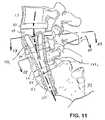
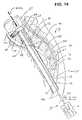
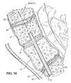
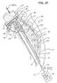
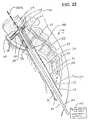
- a method of treating the spinecomprises the steps of identifying a site on the anterior surface of the sacrum, and forming a lumen from the site through the sacrum, through a disc and into at least one vertebrae.
- a procedureis thereafter performed using the lumen.
- the proceduremay be either a diagnostic or a therapeutic procedure.
- the lumenmay extend at least as far as the L4 vertebrae.
- the lumenis substantially linear, and the forming step may comprise drilling.
- the proceduremay comprise removing all or part of a nucleus, inserting a fixation device, or inserting a prosthetic nucleus.
- a bone growth facilitatormay also be introduced.
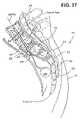
- a method of treating the spinecomprises the steps of identifying a site on the posterior side of the sacrum, and forming a nonlinear lumen from the site through the sacrum, through a disc and through at least one lumbar vertebrae. A procedure is then performed using the lumen.
- the proceduremay be either a diagnostic procedure or a therapeutic procedure.
- the lumenmay extend at least as far as the L4 vertebrae.
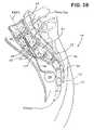
- a method of treating the spinecomprises the steps of identifying a site on the skin of a patient, within about 10 cm from the coccyx.
- An access pathwayis provided at the site and through tissue to the sacrum.
- a lumenis created through the sacrum and at least one lumbar vertebrae.
- the lumenis thereafter used to perform a procedure.
- the site on the skin of a patientis within about 5 cm from the coccyx.
- the lumenis generally at least about 5 cm in length, and, in some applications, is at least about 10 cm in length.
- the lumenmay be either linear, or curved.
- a method of treating the spine at a treatment site which is spaced apart from an access site, to minimize disruption of tissue surrounding the treatment sitecomprises the steps of identifying an access site on the spine, and advancing a device through the access site and into the spine. The device is further advanced axially through the spine for a distance across a treatment zone. The spine is treated in the treatment zone, and the distance is at least about 5 cm. In some applications, the distance is at least about 10 cm.
- the access sitemay be on the sacrum, on the thoracic spine, or elsewhere on the spine.
- the methodmay further comprise the step of introducing the device percutaneously through the skin prior to the advancing step.
- the further advancing stepmay comprise advancing the device along a linear path through the spine.
- the advancing stepmay comprise advancing the device along a non-linear path through the spine.
- the treating stepmay comprise implanting a fixation device.
- a method of treating the spinecomprises the steps of creating a minimally invasive passageway through tissue and into the spine, wherein the passageway has a longitudinal axis and a length of at least about five times its width.
- the methodadditionally comprises the step of introducing at least one device through the passageway to treat the spine, wherein an extension of the axis extends through at least two intervertebral disks.
- the passagewayhas a length of at least about ten times its width.
- the passagewaymay pass through the skin within about 5 cm of the coccyx.
- the passagewayenters the spine on the anterior side.
- the passagewaymay enter the spine on the posterior side.
- a method of performing a procedure from the inside of the spine, while minimizing the cross-sectional area of an access pathway to the procedure sitecomprises the steps of advancing a device through an access pathway in the spine to a procedure site, while the device has a first, reduced crossing profile.
- the crossing profile of at least a portion of the deviceis enlarged at the treatment site to perform the procedure.
- the advancing stepcomprises advancing the device through at least one vertebrae and at least one disk.
- the enlarging stepmay comprise advancing a portion of the device radially outwardly from a longitudinal axis of the device, to perform the procedure.
- a method of fusing the spinecomprises the steps of identifying a site on the anterior surface of the sacrum.
- a lumenis formed from the site through the sacrum, through a disk, and into at least one vertebrae, and a fusion implant is introduced through the lumen.
- the introducing stepcomprises introducing an elongate metal fusion device.
- the introducing stepcomprises introducing a cure in place media.
- a method of accessing the spine through a site on the anterior of the sacrumcomprises the steps of introducing an access device through a tissue tract from the surface of the skin to a first site on the anterior of the sacrum.
- the access deviceis advanced along the anterior surface of the sacrum to a second site.
- the sacrumis entered at the second site.
- the advancing stepcomprises advancing the distal end of the access device, both distally and laterally (cephalad), as the distal end moves along the anterior surface of the sacrum.
- the introducing stepmay comprise introducing a blunt needle trocar.
- the introducing stepmay also comprise introducing the access device through the paracoccygeal space.
- the second sitemay be anterior to S2.
- the methodmay additionally comprise a step of positioning a guide such as a tubular sheath through the tissue tract to the second site.
- a fixation devicemay be introduced along the guide or through the sheath.
- the fixation deviceis positioned across at least the S1 and L5 vertebrae.
- the fixation devicemay be positioned across at least the S1, L5 and L4 vertebrae.
- a method of positioning a linear access sheath from a paracoccygeal entrance point to the S2 vertebraecomprises the steps of introducing an access device through tissue in the paracoccygeal space. A distal end of the access device is advanced into contact with the sacrum. The distal end is swept along the anterior surface of the sacrum towards the S2 vertebrae, thereby displacing anatomical structures from the path of the access device. The distal end of the access device is then fixed with respect to the S2 vertebrae.
- the advancing stepmay comprise advancing the sheath through an incision.
- the advancing stepmay comprise advancing the sheath through a puncture.
- the fixing stepmay comprise threadably engaging the distal end of the sheath in an aperture in the S2.
- FIGS. 1-3are lateral, posterior and anterior views of the lumbar and sacral portion of the spinal column depicting the visualized PAIFL and AAIFL extending cephalad and axially from the posterior laminectomy site and the anterior target point, respectively;
- FIG. 4is a sagittal caudal view of lumbar vertebrae depicting a TASIF axial spinal implant or rod within a TASIF axial bore formed following the visualized PAIFL or AAIFL of FIGS. 1-3 ;
- FIG. 5is a sagittal caudal view of lumbar vertebrae depicting a plurality, e.g., 2, TASIF axial spinal implants or rods within a like plurality of TASIF axial bores formed in parallel with the visualized PAIFL or AAIFL of FIGS. 1-3 ;
- FIG. 6is a simplified flow chart showing the principal surgical preparation steps of percutaneously accessing a posterior or anterior target point of the sacrum and forming a percutaneous tract following the visualized PAIFL or AAIFL of FIGS. 1-3 , as well as subsequent steps of forming the TASIF bore(s) for treatment of accessed vertebral bodies and intervening discs and of implanting axial spinal implants therein;
- FIG. 7illustrates, in a partial cross-section side view, one manner of obtaining access to a posterior target point for forming a posterior TASIF axial bore through sacral and lumbar vertebrae and intervening discs axially aligned with the visualized PAIFL of FIGS. 1 and 2 ;
- FIG. 8is an enlarged partial cross section view illustrating a posterior TASIF axial bore through sacral and lumbar vertebrae and intervening discs axially aligned with the visualized PAIFL of FIGS. 1 and 2 ;
- FIG. 9illustrates, in a partial cross-section side view, one manner of obtaining access to an anterior target point for forming an anterior TASIF axial bore through sacral and lumbar vertebrae and intervening discs axially aligned with the visualized AAIFL of FIGS. 1 and 2 ;
- FIG. 10is an enlarged partial cross-section view illustrating an anterior TASIF axial bore through sacral and lumbar vertebrae and intervening discs axially aligned with the visualized AAIFL of FIGS. 1 and 2 ;
- FIG. 11depicts, in a partial cross-section side view, the formation of a plurality of curved TASIF axial bores that diverge apart from a common caudal section in the cephalad direction;
- FIGS. 12 and 13depict, in partial cross-section end views taken along lines 12 - 12 and 13 - 13 , respectively, of FIG. 7 , the divergence of the plurality of curved TASIF axial bores;
- FIG. 14illustrates, in a partial cross-section side view, one manner of performing a complete discectomy of a disc effected through a TASIF axial bore to enable insertion of bone growth materials for fusion of the vertebral bodies or a pre-formed disc implant or for performance of other therapies to the spinal column and nerves;
- FIG. 15illustrates, in a partial cross-section side view, one manner of filling a disc space formed by a complete discectomy with bone growth materials to effect fusion;
- FIG. 16illustrates, in a partial cross-section side view, one manner of strengthening the fusion of the vertebral bodies by implantation of an elongated axial spinal implant into the TASIF axial bore bridging the filled disc space;
- FIG. 17illustrates, in a partial cross-section side view, one manner of implanting a spinal disc implant following complete discectomy of a disc effected through the delivery of a porous envelope in a deflated condition through a TASIF axial bore and into the disc space;
- FIG. 18illustrates the filling of the porous envelope of FIG. 16 with a material selected from bone growth materials for fusion or other biomaterials forming an artificial spinal disc implant;
- FIG. 19illustrates, in a partial cross-section side view, an alternative planar spiral configuration of a spinal disc implant delivered through a TASIF axial bore into the disc space and the retention of the spinal disc implant by implantation of an elongated axial spinal implant into the TASIF axial bore bridging the filled disc space;
- FIG. 20is an axial view, taken along lines 20 - 20 of FIG. 19 illustrating the spiral configuration of the spinal disc implant
- FIG. 21illustrates, in a partial cross-section side view, one manner of performing a partial discectomy effected through a TASIF axial bore of at least a portion of the nucleus of a spinal disc while leaving the annulus intact;
- FIG. 22illustrates, in a partial cross-section side view, one manner of filling the annulus emptied in FIG. 21 with a disc augmentation material selected from bone growth materials and other biomaterials effected through a TASIF axial bore;
- FIG. 23is an enlarged partial cross-section view illustrating a disc augmentation of a disc involving inflating a balloon with material injected into the balloon within the empty disc space formed in FIG. 21 ;
- FIG. 24is an enlarged partial cross-section view illustrating closure of the TASIF axial bore following the partial discectomy of FIG. 21 or the disc augmentation of FIG. 22 or 23 ;
- FIG. 25illustrates, in a partial cross-section side view, one manner of performing a vertebroplasty through direct injection of bone growth material into a fractured vertebral body through at least one TASIF axial bore;
- FIG. 26is a cross-section view, taken along lines 21 - 21 of FIG. 20 , illustrating the direct injection of the bone growth material into various locations within the fractured vertebral body of FIG. 20 ;
- FIG. 27illustrates, in a partial cross-section side view, an initial step of one manner of performing a balloon-assisted vertebroplasty comprising introducing and inflating a shaped balloon within a TASIF axial bore to compress the cancellous bone and form a vertebral bone cavity;
- FIG. 28illustrates, in a partial cross-section side view, an initial step of inflating the balloon within the cancellous bone of the fractured vertebral body:
- FIG. 29illustrates, in a partial cross-section side view, the full inflation of the balloon within the cancellous bone of the fractured vertebral body:
- FIG. 30illustrates, in a partial cross-section side view, a further step of performing the balloon-assisted vertebroplasty comprising introducing bone growth material through the TASIF axial bore into the vertebral bone cavity formed by the shaped balloon or into the balloon itself;
- FIG. 31illustrates the filled cavity of the vertebral body and a plug closing the TASIF axial bore
- FIG. 32is a side view of an elongated axial spinal implant providing a distraction function
- FIG. 33is a side view of an elongated axial spinal implant providing a shock absorbing function
- FIG. 34illustrates, in a partial cross-section side view, the insertion of an axial spinal implant providing distraction or shock absorbing of two or more vertebrae across one or more intervertebral disc into a TASIF axial bore;
- FIGS. 35-37illustrate, in partial cross-section side views, extending an electrical lead from an implanted or external electrical stimulator through a TASIF axial bore to locate one or more electrical stimulation electrode of the lead within or between or outside adjoining vertebral bodies to apply electrical stimulation to encourage bone growth or to counter pain or outside adjoining vertebral bodies to counter pain;
- FIGS. 38-40illustrate, in partial cross-section side views, extending a catheter from an implanted or external drug dispenser through a TASIF axial bore to dispense a drug within or between adjoining vertebral bodies or to nearby nerves and the spinal column to encourage bone growth or to counter pain or to nearby nerves and the spinal column to counter pain;
- FIG. 41illustrates, in a partial cross-section side view, the insertion of a plurality of radioactive therapeutic seeds about the interior of a lumbar vertebral body for brachytherapy of a sarcoma;
- FIG. 42illustrates, in a partial cross-section side view, a combination of above described therapies comprising the injection of bone growth materials or bone cement through an axial bore into cancellous bone of two vertebral bodies and intervertebral disc cavities and the insertion of an elongated axial spinal implant into the axial bore to effect a fusion with reinforcement of the vertebral bodies.
- FIGS. 1-6Attention is first directed to the following description of FIGS. 1-6 is taken from the above-referenced parent provisional application No. 60/182,748.
- the acronyms TASF, AAFL, and PAFL used in the '748 applicationare changed to TASIF, AAIFL and PAIFL in this application to explicitly acknowledge that instruments can be introduced for inspection or treatments in addition to the fusion and fixation provided by axial spinal implants that may be inserted into the axial bores or pilot holes.
- FIGS. 1-3schematically illustrate the anterior and posterior TASIF surgical approaches in relation to the lumbar region of the spinal column
- FIGS. 4-5illustrate the location of the TASIF implant or pair of TASIF implants within a corresponding posterior TASIF axial bore 22 or anterior TASIF axial bore 152 or pair of TASIF axial bores 22 1 , 22 2 or 152 1 , 152 2
- Two TASIF axial bores and axial spinal implants or rodsare shown in FIG. 5 to illustrate that a plurality, that is two or more, of the same may be formed and/or employed in side by side relation in parallel alignment with the AAIFL or PAIFL or diverging from the AAIFL or PAIFL in the cephalad direction.
- TASIF surgical approaches for providing anterior and posterior trans-sacral access depicted in FIGS. 1-3 and preparing the TASIF axial bores 22 or 152 or 22 1 , 22 2 , or 152 1 , 152 2 shown in FIGS. 4 and 5are illustrated in the above-referenced '105 and '748 applications.
- FIG. 1The lower regions of the spinal column comprising the coccyx, fused sacral vertebrae S1:S5 forming the sacrum, and the lumbar vertebrae L1:L5 described above are depicted in a lateral view in FIG. 1 .
- the series of adjacent vertebrae located within the human lumbar and sacral spinehave an anterior aspect, a posterior aspect and an axial aspect, and the lumbar vertebrae are separated by intact or damaged intervertebral spinal discs labeled D1:D5 in FIG. 1 .
- FIGS. 2 and 3depict the posterior and anterior views of the sacrum and coccyx.
- the method and apparatus for forming an anterior or posterior TASIF axial boreinitially involves accessing an anterior sacral position, e.g. an anterior target point at the junction of S1 and S2 depicted in FIGS. 1 and 3 , or a posterior sacral position, e.g. a posterior laminectomy site of S2 depicted in FIGS. 1 and 2 .
- an anterior sacral positione.g. an anterior target point at the junction of S1 and S2 depicted in FIGS. 1 and 3
- a posterior sacral positione.g. a posterior laminectomy site of S2 depicted in FIGS. 1 and 2 .
- One (or more) visualized, imaginary, axial instrumentation/fusion lineextends cephalad and axially in the axial aspect through the series of adjacent vertebral bodies, L4 and L5 in this illustrated example.
- the visualized AAIFL through L4, D4, L5 and D5extends relatively straight from the anterior target point along S1 depicted in FIGS.
- the visualized PAIFLextends in the cephalad direction with more pronounced curvature from the posterior laminectomy site of S2 depicted in FIGS. 1 and 2 .
- a preoperative CT scan or magnetic resonance imaging (MRI) study of the patient's spineis conducted to visualize and map the AAIFL or PAIFL.
- FIG. 6depicts, in general terms, the surgical steps of accessing the anterior or posterior sacral positions illustrated in FIGS. 1-3 (S 100 ) forming posterior and anterior TASIF axial bores (S 200 ), optionally inspecting the discs and vertebral bodies, performing discectomy or discoscopy, disc augmentation, and vertebral bone reinforcement balloon-assisted vertebroplasty or vertebroplasty (S 300 ), and implanting posterior and anterior axial spinal implants and rods or plugs into the axial bore(s) (S 400 ) in a simplified manner.
- step S 100access to the anterior or posterior sacral position, that is the anterior target point of FIG. 3 or the posterior laminectomy site of FIG.
- step S 300is obtained, and the anterior or posterior sacral position is penetrated to provide a starting point for each axial bore that is to be created. Then, one or more axial bore is bored from each point of penetration extending in alignment with either the PAIFL or AAIFL cephalad and axially through the vertebral bodies of the series of adjacent vertebrae and any intervertebral spinal discs (S 200 ).
- the axial bore(s)can traverse one or more vertebral body cephalad to the sacral vertebral bodies S1, S2 and any intervertebral disc and can terminate at a cephalad end within a particular vertebral body or spinal disc.
- the axial boremay be visually inspected using an endoscope to determine if the procedures of step S 300 should be performed.
- step S 100 in the anterior and/or posterior TASIF proceduresmay involve drilling a pilot hole, smaller in diameter than the TASIF axial bore, in the prescribed alignment with the AAIFL and/or PAIFL in order to complete the formation of the anterior and/or posterior percutaneous tracts.
- Certain of the therapeutic procedures of steps S 300 and S 400may optionally be completed through the AAIFL/PAIFL pilot hole following step S 100 , rather than following the enlargement of the pilot hole to form the TASIF axial bore in step S 200 .
- Step S 100preferably involves creation of an anterior or posterior percutaneous pathway that enables introduction of further tools and instruments for forming an anterior or posterior percutaneous tract extending from the skin incision to the respective anterior or posterior target point of the sacral surface or, in some embodiments, the cephalad end of a pilot hole over which or through which further instruments are introduced as described in the above-referenced 222 application.
- An “anterior, presacral, percutaneous tract” 26( FIG. 1 ) extends through the “presacral space” anterior to the sacrum.
- the posterior percutaneous tract or the anterior, presacral, percutaneous tractis preferably used to bore one or more respective posterior or anterior TASIF bore in the cephalad direction through one or more lumbar vertebral bodies and intervening discs, if present.
- “Percutaneous” in this contextsimply means through the skin and to the posterior or anterior target point, as in transcutaneous or transdermal, without implying any particular procedure from other medical arts.
- the percutaneous pathwayis generally axially aligned with the AAIFL or the PAIFL extending from the respective anterior or posterior target point through at least one sacral vertebral body and one or more lumbar vertebral body in the cephalad direction as visualized by radiographic or fluoroscopic equipment.
- anterior tract 26 shown in FIG. 1 through presacral space under visualization described aboveis clinically feasible as evidenced by clinical techniques described by J. J. Trambert, MD, in “Percutaneous Interventions in the Presacral Space: CT-guided Precoccygeal Approach—Early Experience ( Radiology 1999; 213:901-904).
- Certain of the therapeutic procedures of the present inventionare conducted through relatively straight or curved anterior TASIF bores or curved posterior TASIF bores or pilot holes.
- Introduction of axial spinal implants and instruments for performing discoscopy to inspect the accessed discs, discectomies and/or disc augmentation/replacement and/or vertebroplasty, balloon-assisted vertebroplasty, fusion, alignment, drug delivery, electrical stimulation, or other therapies,is enabled by the provision of the percutaneous pathway and formation of the anterior or posterior TASIF bore(s).
- the bore forming tool setscomprise elongated drill shaft assemblies supporting distal boring tools, e.g., mechanical rotating drill bits, burrs, augurs, abraders, or the like (collectively referred to as boring heads or drill bits for convenience), that can be manipulated in use to bore a straight or curved axial bore.
- distal boring toolse.g., mechanical rotating drill bits, burrs, augurs, abraders, or the like (collectively referred to as boring heads or drill bits for convenience), that can be manipulated in use to bore a straight or curved axial bore.
- Suitable bore forming toolsare disclosed in the above-referenced provisional application No. 60/182,748 and the '105 application.
- the TASIF axial borescan be formed by other tools that mechanically puncture or penetrate vertebral bodies and intervertebral discs or otherwise form TASIF axial bores in any diameter or cross-section and that follow any alignment with the axis of the spine as visualized by the AAIFL or
- FIGS. 7 and 8illustrate step S 100 for forming the posterior percutaneous tract and the posterior TASIF axial bore 22 formed in step S 200 and extending through sacral and lumbar vertebrae and intervening discs axially aligned with the visualized PAIFL of FIGS. 1 and 2 using a boring tool of the type described in more detail in the above-referenced '105 and '748 applications.
- the same stepscan be employed to form a pilot hole of step S 100 that can be enlarged in step S 200 .
- a small diameter bore forming toole.g. 3.0 mm diameter
- the boring toolis removed, and a guidewire having a threaded distal screw-in tip is advanced through the pilot hole and screwed into to the caudal end of the pilot hole and into cephalad portion of the L4 body.
- An over-the-wire bore enlarging tool having a flexible body capable of tracking the curved guidewireis fitted over the proximal end of the guidewire and manually or mechanically rotated and advanced along it in step S 200 .
- the small pilot hole diameteris enlarged to form the anterior TASIF axial bore 22 having a diameter e.g. a 10.0 mm diameter, and the enlarging tool is then removed.
- the illustrated diameter of the posterior TASIF axial bore hole 22 relative to sizes of the vertebral bodiesis merely exemplary, and that it is contemplated that the pilot hole and bore hole diameters can range from about 1-10 mm and 3-30 mm, respectively.
- a plurality of such posterior TASIF axial bores 22 1 . . . 22 ncan be formed in side by side or diverging relation generally aligned with the PAIFL.
- the posterior surface of the sacrumis exposed in step S 100 as described in the above-referenced '222 and '748 applications.
- the area of the patient's skin surrounding the incision siteis surgically prepped, and the anus is excluded from the surgical field using adhesive drapes.
- the actual dermal entry sitemay be determined by the prone, preoperative CT scan or magnetic resonance imaging (MRI) study that maps the PAIFL.
- MRImagnetic resonance imaging
- step S 100an incision is made in the patient's skin over the posterior sacral surface of S2, and the subcutaneous tissue is separated to expose the posteriorly extending, bony ridge of the posterior sacral surface.
- a small laminectomy 14is performed through the posterior ridge of the sacrum inferior. The thecal sac and nerve roots that are exposed by the laminectomy are gently retracted, and the terminal portion of the spinal canal is exposed.
- An elongated drill shaft assembly(not shown) is axially aligned with the PAIFL at the posterior target point so that the initial penetration of the sacrum is substantially at right angles to the exposed sacral surface.
- a drill guide for receiving the drill drive shaft assembly for drilling or boring a posterior TASIF axial bore 22 from S2 along the visualized PAIFLmay optionally be attached to S2 and extended posteriorly through the exposed spinal canal and skin incision.
- the progress of the drill bitis observed using conventional imaging equipment.
- a curvatureis introduced in the cephalad segment of the posterior TASIF axial bore 22 as shown in FIG. 8 . It is necessary to maintain the plane of curvature of the distal segment aligned to the curvature of the spine.
- the drill bitadvances through the sacral vertebrae in the cephalad direction and toward the lumbar vertebral bodies while staying within the spongy, cancellous bone of each vertebral body.
- any number of vertebral bodies of the spinecan be bored through in the cephalad axial direction.
- the cephalad end of the posterior TASIF axial bore 22can terminate within a vertebral body or within a disc or disc space.
- FIGS. 9 and 10illustrate the anterior percutaneous tract formed in step S 100 and the anterior TASIF axial bore 22 formed in step S 200 and extending through sacral and lumbar vertebrae and intervening discs axially aligned with the visualized AAIFL of FIGS. 1 and 2 using a boring tool of the type described in more detail in the above-referenced '105 and '748 applications.
- the same stepscan be employed to form a pilot hole of step S 100 that can be enlarged in step S 200 as described above.
- the illustrated diameter of the anterior TASIF axial bore hole 152 relative to sizes of the vertebral bodiesis merely exemplary, and that it is contemplated that the pilot holes and bore hole diameters can range from about 1-10 mm and 3-30 mm, respectively.
- a plurality of such anterior TASIF axial bores 152 1 . . . 152 ncan be formed in side by side or diverging relation generally aligned with the AAIFL.
- the anterior TASIF axial bore(s)can be relatively straight from the anterior target point into or through at least the caudal lumbar vertebrae and intervertebral discs. But, it may be desirable or necessary to form a curved anterior TASIF axial bore(s) particularly as the bore(s) is extended in the cephalad direction to maintain the plane of curvature of the cephalad segment of the TASIF axial bore(s) aligned to the curvature of the spine. In this way, the drill bit advances through the sacral vertebrae in the cephalad direction while staying within the spongy, cancellous bone of each vertebral body.
- any number of vertebral bodies of the spinecan be bored through in the cephalad direction.
- the cephalad end of the posterior TASIF axial bore(s) 152can terminate within a vertebral body or within a disc or disc space.
- a single anterior or posterior TASIF axial boreis to be made, it preferably is axially aligned with the respective visualized AAIFL or PAIFL as shown by TASIF axial bores 22 or 152 shown in FIG. 4 .
- Plural anterior or posterior TASIF bores 22 1 . . . 22 n , or 152 1 . . . 152 n shown in FIG. 5are in parallel or diverging alignment with the visualized AAIFL and PAIFL.
- Multiple anterior or posterior TASIF axial borescan be formed all commencing at an anterior or posterior target point of FIGS. 1-3 and extending in the cephalad direction with each TASIF axial bore diverging apart from the other and away from the visualized axial AAIFL and PAIFL.
- the diverging TASIF axial boresterminate as spaced apart locations in a cephalad vertebral body or in separate cephalad vertebral bodies or spinal discs.
- FIGS. 11-13depict a group of three anterior TASIF axial bores 152 1 , 152 2 , 152 3 that are bored from a common caudal entrance bore section 152 ′ starting at the anterior target point.
- the three anterior TASIF axial bores 152 1 , 152 2 , 152 3extend in the cephalad direction generally following the curvature of the AAIFL but diverging outwardly to form a “tripod” of the diverging TASIF axial bores 152 1 , 152 2 , 152 3 .
- the divergence from the common entry bore sectioncan start in the sacral vertebra or in L5 or in L4 or in any other cephalad vertebral body that the bore extends into or through.
- the common caudal entrance bore section 152 ′ through S1, and traversing disc D5 and part of L4can be larger in diameter than the diverging TASIF axial bores 152 1 , 152 2 , 152 3 to accommodate the insertion of three elongated axial spinal implants therein or to facilitate performing a therapy as described further below. It is believed that the insertion of elongated axial spinal implants within the “tripod” of the diverging TASIF axial bores 152 1 , 152 2 , 152 3 can substantially strengthen and enhance fusion of L4, L5 and S1.
- the diverging TASIF axial bores 152 1 , 152 2 , 152 3can be extended further than shown in FIGS. 11-13 . Diverging posterior TASIF axial bores can be formed in the same manner.
- a variety of therapeutic procedurescan be performed in step S 300 or step S 400 after the curved posterior or curved or straight anterior TASIF axial bore(s) is formed employing instruments, axial spinal implants, spinal disc implants and materials.
- Certain of the therapies or therapeutic procedurescan be completed in step S 300 without an axial spinal implant also being implanted in step S 400 .
- Step S 400may also be performed in certain cases without a therapeutic procedure being performed in step S 300 .
- the therapeutic procedures that are performed using such instruments, axial spinal implants, spinal disc implants and materials of the present inventioncomprise one or more of: (1) performing a discoscopy by inserting an endoscope and inspecting the condition of the vertebral bodies and spinal discs; (2) performing a simple fusion by inserting bone growth materials into the TASIF axial bore(s) in step S 400 ; (3) performing a partial discectomy or complete discectomy of a disc accessed through a TASIF axial bore in step S 300 ; (4) performing a vertebroplasty or balloon-assisted vertebroplasty to a fractured vertebral body accessed through a TASIF axial bore in step S 300 ; (5) inserting an artificial disc implant or autologous/homologous bone or bone growth material into the disc space following a complete discectomy of a disc accessed through a TASIF axial bore in step S 400 to encourage fusion or to function as a functional disc replacement; (6) following a partial discectomy removing at least a portion of the nucleus,
- the therapeutic proceduresare illustrated in the drawings and described as follows as being performed through an anterior percutaneous tract formed using an anterior tract sheath 96 and TASIF axial bore 152 . But, it will be understood that the procedures may be performed through a posterior percutaneous tract and TASIF axial bore 22 and that certain of the procedures may be advantageously performed using parallel or diverging TASIF axial bores.
- the anterior TASIF axial bore 152is formed, as described above, through the use of anterior tract sheath 96 that inserted earlier through the presacral space 24 from a skin incision 28 to the anterior target point of the anterior surface of sacral vertebra S1 that defines the percutaneous tract 26 .
- the shaped end 98 of the anterior tract sheath 96is aligned with the anterior surface of the sacral vertebra S1 during step S 100 .
- the shaped end 98may be formed with attachment teeth or threads to fix it to the sacral bone. It will be understood that the therapeutic procedures of the present invention may be performed through the lumen of such a tract sheath 96 or simply through a defined anterior tract 26 extending through the pre-sacral space 24 to axially access the vertebrae.
- each of the following therapies to the spinal discs or the vertebral bodiescan be conducted on more than one spinal disc or vertebral body or on one or more spinal disc and one or more vertebral body traversed by at least one TASIF axial bore.
- two spinal discsmay be accessed by a single TASIF axial bore, and treated in one of the following ways, starting with the cephalad spinal disc. Then, the portion of the TASIF axial bore between the cephalad and caudal spinal disc is closed by an artificial axial spinal implant or bone growth material as appropriate.
- cephalad and caudal vertebral bodiesmay be treated by vertebroplasty or balloon-assisted vertebroplasty, and the intervertebral disc may also be treated by one of the following described therapies.
- the treatment of only a single spinal disc or vertebral bodyis described and illustrated in the drawings.
- the following therapeutic procedures of the present inventionare understood to involve accessing an anterior or posterior sacral position of a sacral vertebra in alignment with the visualized, AAIFL or PAIFL extending in said axial aspect cephalad through a series of adjacent vertebral bodies of adjacent vertebrae. Then, from the accessed anterior sacral position, at least one anterior or posterior TASIF axial bore is bored in alignment (as defined herein) with the AAIFL or PAIFL axially through at least the caudal sacral vertebra and through or into one or more cephalad vertebral bodies of the series of adjacent vertebral bodies and any interposed, intervertebral, spinal discs.
- the delivery of the therapiesis followed by the withdrawal of any tract forming tools and a simple surgical closure of the incision site.
- the therapiescan be delivered or therapeutic procedures can be performed as follows.
- the prior artprovides no convenient and relatively atraumatic way to promote fusion between adjacent vertebral bodies where the intervertebral disc has degenerated.
- One therapeutic procedure of the present inventionsimply involves filling the anterior or posterior TASIF axial bore(s) with a bone growth material which bridges the spinal disc and will effect bone growth across the spinal disc.
- the cancellous boneis typically porous with fissures and cavities, so that the bone growth material is also forced into such cancellous bone cavities and fissures.
- a “bone growth material”can be one or more of the following, or any other biocompatible material judged to have the desired physiologic response, including any natural or artificial osteoconductive, osteoinductive, osteogenic, or other fusion encouraging material.
- any natural or artificial osteoconductive, osteoinductive, osteogenic, or other fusion encouraging materialincluding any natural or artificial osteoconductive, osteoinductive, osteogenic, or other fusion encouraging material.
- morselized cortical, cancellous, or cortico-cancellous bone graftincluding autograft, allograft, or xenograft might be employed.
- any bone graft substitute or combination of bone graft substitutes, or combinations of bone graft and bone graft substitutes, or bone inducing substancescould be employed.
- Such bone graft substitutes or bone inducing substancesinclude, but not limited to, hydroxyapatite, hydroxyapatite tricalcium phosphate; bone morphogenic protein (BMP) and calcified or decalcified bone derivative and resorbable bone cements.
- the resorbable cement materialcan be a calcium derivative generally composed of hydroxyapatite, orthophosphoric acid, calcium carbonate, and calcium hydroxide formed into a semi-liquid paste with an alkaline solution of sodium hydroxide and water or a composition comprising polypropylene fumarate or a mixture of calcium phosphates.
- Other compositions that may be employedcomprise calcium salt filler, N-vinyl-2-pyrrolidone, and a peroxide or free radical initiator.
- the bone graft materialmay be mixed with a radiographic material to enable its visualization during delivery to assure proper disposition and filling of bores, cavities and spaces described herein.
- a pre-formed elongated axial spinal implantcan be inserted into at least one of the anterior or posterior TASIF axial bore(s) along with bone growth material.
- the axial spinal implantcan be a surface roughened metal rod or porous tube of the type described in the above-referenced '620 application that is configured to the particular bore curvature and size and surface treated bite into vertebral bone and to promote bone ingrowth.
- This therapyprovides a simple and relatively atraumatic approach to the vertebrae of interest where there is no need to treat or remove the intervertebral disc. Fusion may be effected in other ways described as follows using the bone growth materials and optionally using an elongated axial spinal implant.
- FIG. 14illustrates, in a partial cross-section side view, one manner of performing a complete discectomy of a spinal disc, e.g., D4, effected through a TASIF axial bore 152 to enable fusion of the vertebral body endplates of lumbar vertebrae L4 and L5 directly together or to provide a disc space for receipt of a pre-formed artificial disc implant that mimics the function of a patent spinal disc.
- a spinal disce.g., D4
- the complete discectomy procedureinvolves more or less complete excision of the intervertebral disc D4 including the nucleus, the annulus, the cartilaginous endplates adhered to the opposed cortical bone endplates of the cephalad and caudal vertebral bodies, and, optionally, the vertebral periosteum, cortical endplate bone and cancellous bone to a desired depth and shape.
- Distractionis applied to the lumbar vertebrae L4 and L5 by suitably supporting the patient's body to maintain the disc space for fusion and/or implantation of an artificial disc implant.
- the excised materialsare withdrawn from the disc space and the fusion materials or artificial disc implant is introduced to the disc space through the TASIF axial bore 152 .
- the TASIF axial bore 152either terminates within the spinal disc to be removed or extends into a vertebral body cephalad to that spinal disc in the event that an axial spinal implant or bone growth material is to be inserted into the TASIF axial bore bridging the excised disc space.
- FIG. 14depicts the TASIF axial bore 152 in solid lines terminating in the disc D4 and in broken lines extending into vertebral body L4.
- a discectomy tool 130is inserted through the axially aligned anterior tract 26 defined by the lumen of the anterior tract sheath 96 .
- the discectomy tool 130is formed like a flexible atherectomy catheter for fragmenting and removing obstructions in blood vessels using a cutting head 134 to fragment the disc material and to scrape away cortical and cancellous bone and aspiration with saline flushing to remove the fragments from the disc space.
- the cutting head 134is mounted into a deflectable or steerable distal end section 132 of discectomy tool shaft 136 extending through the TASIF axial bore 152 and anterior tract 26 from an externally disposed energy source and deflection control 140 .
- the distal end sectionmay be angularly deflected using a deflection mechanism, e.g., a pull wire within a pull wire lumen of the elongated, flexible, discectomy tool shaft 136 and a proximal pull wire control of the proximal guiding and cutting mechanism 140 coupled thereto.
- the cutting head 134may be pulled back and forth laterally and/or swept in a 360° arc about the axial bore 152 to traverse and excise selected symptomatic portions of or the entire spinal disc D4 and to cut away layers of bone from the endplates of vertebral bodies L4 and L5 by manipulation of the proximal end portion of the discectomy tool shaft 136 extending from the skin incision 28 .
- the complete discectomy cutting head 134 and tool shaft 136are shown schematically and not necessarily to scale to one another or to the TASIF axial bore 152 .
- the cutting head 134may comprise any of the known types, e.g. mechanical cutters including rotating wire brushes, cutting blades, scissors, screws, bits, extendable/retractable rotating wires, or the like, or energy application heads, e.g., resistive heating, laser energy or ultrasonic energy heads, or fluid jets or the like.
- the bore expanding cutting tools disclosed in the above-referenced '369 applicationcould also be modified to reach the disc annulus and excise it.
- Various forms of cutting headsare disclosed in the above-referenced '146, '962 and '629 patents and in further U.S. Pat. Nos. 6,095,149, 6,127,597, 5,383,884, and RE 33,258, for example.
- the tool shafts of the prior art discectomy toolsare designed for the lateral access into the spinal disc and are not disclosed as being introduced axially and then into the space between the vertebral body endplates.
- the cutting head 134 in this exampleis a mechanical screw thread that can be selectively covered in whole or part by a sheath (not shown) for exposing the end or a lateral portion of the cutting screw thread in the manner of the augur disclosed in the above-referenced '884 patent for example.
- the cutting head 134is attached to a drive shaft extending through a drive shaft lumen of the tool shaft 136 to a drive motor for rotating the drive shaft and cutting head 134 and a deflection control for operating the pull wire or the like for deflecting the distal section 132 , both included in energy source and deflection control 140 .
- an aspiration lumenis included within the discectomy tool body 136 with a distal opening adjacent to the cutting head 134 and terminating proximally at a side suction port 138 adapted to be coupled to a source of suction to aspirate the fragments of the disc from disc space 154 .
- a saline flush lumen and supplycan also be incorporated within to flush blood and excised fragments for aspiration.
- the operation and movement of the cutting head about the spinal disc D4is preferably observed employing fluoroscopy or other radiographic visualization techniques.
- An endoscopic visualization or the disc space 154could also be employed using a separate or incorporated deflectable tip endoscope for illumination and observation of the site.
- the resulting disc space 154can either be substantially disc-shaped with more or less planar opposed sides having a height in the range of about 8 mm to about 14 mm, a lateral width of from about 26 mm to about 32 mm, and an anterior-posterior width of from about 22 mm to about 30 mm.
- the disc space 154can be selectively enlarged into a convex disc shape extending caudally into the cancellous bone of caudal vertebral body L5 and/or in the cephalad direction into the cancellous bone of cephalad vertebral body L4.
- This disc space shapingforms a pocket that helps to confine a spinal disc implant inserted into the prepared disc space 154 or bone growth materials dispensed into the prepared disc space 154 .
- a fusion of vertebral bodies D4 and D5 or the implantation of an artificial spinal disc into the disc space 154 through the anterior TASIF axial bore 152 and percutaneous tract 26may be undertaken after the disc space is cleared of debris.
- the TASIF axial borecan be filled with an axial spinal implant that provides internal stabilization, alignment, and reinforcement of the spinal motion segment.
- This therapeutic procedure of the present inventioncan be advantageously conducted without any injury to any ligaments, muscles and facet joints of the spinal motion segment.
- fusion of the prepared endplates of L4 and L5can be effected by pumping a bone growth material slurry into the disc space 154 through the TASIF axial bore 152 and percutaneous tract 26 as shown in FIG. 15 .
- a catheter 50 having a straight or flexible tip section as showncan be employed that is extended through the axially aligned anterior tract 26 and the TASIF axial bore 152 to dispense the bone growth material 60 in the disc space 154 .
- a plungercan also be employed that is extended through the axially aligned anterior tract 26 and the TASIF axial bore 152 to pack the dispensed material from the TASIF axial bore 152 into the disc space 154 .
- the caudal end opening or the full length of the TASIF axial bore 152is plugged with an artificial plug or bone growth material, and the sheath 96 is withdrawn.
- the incision 28is closed and the patient rests for a time until bone growth takes place.
- This procedure illustrated in FIG. 15may suffer from the inability to adequately fill the disc space 154 or to confine the dispensed bone growth material within the disc space during the procedure and may require prolonged bed rest recuperation to avoid later settling and ejection of the bone growth material.
- a pre-formed, rod shaped, artificial axial spinal implant 70can be inserted into the TASIF axial bore 152 to fill all or part of it in order to help maintain the distraction of the vertebral bodies as shown in FIG. 16 .
- the pre-formed axial spinal implant 70can extend into the cephalad vertebral body L4 if the TASIF axial bore 154 extends into it as shown in FIG. 16 and may include the distraction and shock absorbing characteristics of the particular axial spinal implants described below. In this case, it may be desirable to form parallel or diverging TASIF axial bores and implant an axial spinal implant in each bore.
- the pre-formed, rod shaped, artificial axial spinal implant 70may employ a fixation mechanism and/or be fixed in place using one of the above described bone growth materials.
- fusion of the vertebral bodiescan be effected according to a further aspect of the present invention using spinal disc implants that are dispensed into the disc space through the TASIF axial bore 152 and percutaneous tract 26 and maintained there in a variety of ways.
- One approach, shown in FIG. 17is to dispense a porous, deflated, shaped bag or balloon or sack or other envelope 80 of a type described in the above-referenced '679 patent into the disc space 154 formed in FIG. 14 and to then fill and inflate the envelope 80 with a bone growth material 60 of one of the types described above as shown in FIG. 18 , and to close the opening into the envelope 80 .
- the porous fabrichas pores that are small enough to confine the bone growth material within the envelope while allowing passage of fluids and bone growth therethrough.
- the envelope 80could be formed of a tightly woven, high molecular weight, high tenacity, flexible polymeric fabric e.g., high molecular weight polyethylene, polyester, polyolefine, polyethylene terepthalate, polytetrafluoroethylene, polysulfone, nylons, or any other high molecular weight, and other high tenacity materials including carbon fiber yarns, ceramic fibers, metallic fibers, etc.
- the envelope 80can be inserted into the prepared disc space 154 in a variety of ways, one of which is shown in FIG. 17 .
- the envelope 80is folded against a blunt tip flexible push wire 52 which extends through the lumen of a fill tube 54 that extends into the balloon opening.
- the folded envelope 80 , push wire 52 and fill tube 54are in turn inserted into the lumen of a tubular catheter or sheath 56 that is advanced through the percutaneous tract 26 and TASIF axial bore 152 .
- the envelope 80is advanced into the prepared, distracted disc space 154 and spread out by pushing and torqueing the push wire 52 .
- Air or liquid inflation of the shaped envelope 80can also be used to spread the envelope out in the disc space instead of the push wire 52 .
- the push wire 52is withdrawn, air is evacuated from the envelope interior trough the lumen of the fill tube 54 , and bone growth material 60 is then injected through the lumen of the fill tube 54 as shown in FIG. 18 .
- the filled envelope 80conforms with the prepared disc space 154 , and the dispensed bone growth material 60 is confined therein to maintain the spacing between the vertebral body end plates when the envelope opening is closed.
- the envelope openingcan be closed by drawing a purse-string suture sewn around the opening tight and tying it through the TASIF axial bore 152 and percutaneous tract 26 or in other manners described in the above-referenced '679 and '736 patents and in U.S. Pat. No. 5,702,454.
- the bone growth material confined within the envelope 80maintains the distraction spacing between the prepared vertebral body endplates.
- One manner of maintaining the filled envelope 80 in placewould be to advance an elongated axial spinal implant 70 in the caudal direction into the opening of the filled envelope 80 to abut a section of the envelope fabric against the cephalad vertebral endplate, thereby both sealing the envelope opening and locking it into place.
- Another way to maintain the filled envelope 80 in placewould be to form the disc space 154 with concave, prepared vertebral body endplates during the complete discectomy and to form the envelope 80 with convex sides that fit within the concave surfaces when the envelope 80 is filled with bone growth material.
- a further manner of keeping the filled envelope 80 in placeis to form it to expand upon filling with bone growth material into a toroidal or tire shape providing a hole aligned with the TASIF axial bore 152 that an elongated axial spinal implant 70 can be fitted through.
- the envelopecan be formed as an elongated tube 82 that is shaped to assume a toroidal member shape or a spiral member shape having a number of spiral turns in a common plane upon filling and inflation with bone growth material as shown in FIGS. 19 and 20 .
- spiral spinal disc implantsspiral members
- the spiral member cross-section of the spiral membercan be circular or rectangular or any other suitable shape, and the spiral height is selected to fit within the disc space 154 and extend between the distracted vertebral body endplates.
- a rectangular cross section strip of nickel-titanium alloy (Nitinol) having a spiral shape memorycan be straightened, advanced through the lumen of an introducer tube lumen, and released o assume the planar spiral shape within the disc space 154 .
- each of these spinal disc implantscan be pinned in the disc space 154 by bone growth material or a pre-formed, rod shaped, axial spinal implant 70 extending through the TASIF axial bore 152 and the central hole of the toroidal or tire shaped or spirally arranged spinal disc implant tube 82 as shown in FIGS. 19 and 20 .
- This therapeutic procedure of the present inventioncan be advantageously conducted without any injury to any ligaments, muscles and facet joints of the spinal motion segment.
- various forms of spinal disc implantsthat mimic the function and characteristics of a natural disc without promoting fusion of the cephalad and caudal vertebral bodies can be inserted into the disc space through the TASIF axial bore 152 and percutaneous tract 26 and maintained there in a variety of ways.
- One approachis to dispense a deflated, shaped bag or balloon or sack or other envelope of a type described in the above-referenced '326 and '454 patents and in U.S. Pat. Nos. 5,888,220 and 5,562,736 into the disc space 154 formed in FIG. 14 .
- the envelope 80is filled and inflated with a curable biomaterial that does not necessarily encourage bone growth and preferably is resilient.
- the '326, '220 and '736 patentsdescribe performing a discectomy through lateral approaches to remove the nucleus while retaining as much of the annulus as possible, given the lateral penetration through it.
- the annulusis removed in the complete discectomy described above, and the deflated envelope 80 is inserted and inflated through the TASIF axial bore 152 and percutaneous tract 26 as shown in FIG. 17 and described above.
- the envelope 80conforms with the prepared disc space, and the dispensed biomaterial is confined therein to maintain the spacing between the vertebral body end plates when the envelope opening is closed as described above and depicted in FIGS. 18-20 .
- the dispensed biomaterial confined within the envelopemaintains the distraction spacing between the prepared vertebral body endplates but does not necessarily encourage fusion.
- the preparation of the disc space 154 , the form of the spinal disc implant, and the manner of inserting, filling, closing and locking the spinal disc implant in placecan be the same as described in the above section related to spinal fusion with respect to FIGS. 17-20 .
- the biomaterial filling the envelope 80is a material that is capable of being introduced to the site of a joint by minimally invasive means, and be hydrated or cured in place to provide desired physical-chemical properties as described, for example, in the above-referenced '326, '454, '220 and '736 patents.
- a hydrogelcan be injected into the envelope 80 in a liquid or dry particulate form or in microspheres or beads in the manner shown in FIG. 18 .
- a preferred hydrogelis formulated as a mixture of hydrogel polyacrylonitrile or any hydrophilic acrylate derivative with a unique multiblock copolymer structure or any other hydrogel material having the ability to imbibe and expel fluids while maintaining its structure under various stresses.
- the hydrogelcan be formulated as a mixture of polyvinyl alcohol and water.
- the hydrogel core formed within the envelope 80will swell as it absorbs fluids through the porous fabric wall of the envelope 80 much like a native nucleus.
- the hydrogel corehas a time constant of swelling which is highly similar to that of the natural nucleus and will thus experience a 5-30% and preferably a 15-20% volume change depending on load over the course of 2-8 (preferably 4-8) hours. When fully hydrated, the hydrogel core will have a water content of between 25-65%.
- the hydrogel material of the preferred embodimentis manufactured under the trade name Hypan® by Hymedix International, Inc.
- any of the hydrogels and solvents identified in the above-referenced '326 patentmay be employed to fill the envelope 80
- the preferred woven construction of the envelopecreates a plurality of small openings large enough to allow bodily fluids to interact with the hydrogel core, but small enough to prevent the hydrogel from escaping.
- the openingsPreferably, have an average diameter of about 10 micrometers, although other dimensions are acceptable.
- the flexible materialallows expansion and contraction of the hydrogel core in a controlled fashion as it imbibes and expels fluids.
- the hydrogel coreacts as a cushion against various loads placed upon it.
- the hydrogel coreimbibes surrounding fluids and expands as the load is decreased (e.g. when the patient reclines).
- the preferred envelope fabricis substantially inelastic and has a burst strength which is greater than the swelling pressure of the hydrogel core when fully hydrated to prevent rending and loss of the hydrogel core.
- Suitable envelope materialsinclude
- This therapeutic procedure of the present inventionprovides a shock absorbing functional disc replacement of the nucleus of the spinal disc and can be advantageously conducted without any injury to any ligaments, muscles and facet joints of the spinal motion segment.
- the anterior or posterior TASIF axial bore 152 or 22is formed in the manner described above and terminates within the nucleus of the disc to be treated or optionally extends into the cephalad vertebral body to facilitate fusion of the vertebral bodies. Then, instruments can be introduced through the TASIF axial bore and percutaneous tract into the nucleus to fragment or desiccate all or part of the nucleus, including any projecting into the annulus or from a fissure in the annulus, and create a void or disc cavity within the annulus and the cartilaginous endplates.
- any fissures or other damage or weakening of the annuluscan be treated from within the created void in a manner described in the above-referenced '149 patent, for example.
- the entry into the nucleus and the TASIF axial borecan then be closed with a simple elongated axial spinal implant or a shorter plug formed of a bone growth material or another bio-compatible material or bone cement.
- a disc augmentationcan be performed before closure as described further below.
- FIG. 21illustrates, in a partial cross-section side view, one manner of performing a partial discectomy of a spinal disc to remove at least a portion of the nucleus NP effected through a TASIF axial bore 152 while leaving the annulus AF intact.
- a discectomy tool 110is inserted through the axially aligned anterior tract 26 defined by the lumen of the anterior tract sheath 96 and the TASIF axial bore 152 .
- the discectomy tool 110is formed like a flexible atherectomy catheter for fragmenting and removing obstructions in blood vessels using a cutting head 114 to fragment or desiccate the disc material and aspiration with saline flushing to remove the fragments or byproducts from the void or cavity created in the annulus.
- the cutting head 114is mounted into a deflectable or steerable distal end section 112 of discectomy tool shaft 116 extending through the TASIF axial bore 152 and anterior tract 26 from an externally disposed energy source and deflection control 140 .
- the cutting head 114may comprise any of the known types, e.g. mechanical cutters including rotating wire brushes, cutting blades, scissors, screws, bits, extendable/retractable rotating wires, or fluid jets or the like.
- the cutting head 114may be an energy emitting head, e.g., a resistive heating, electrocautery, laser energy, or ultrasonic energy head that desiccates, that is, heats up and shrinks or vaporizes the nucleus.
- the cutting head 114can comprise one of various forms of cutting heads disclosed in the above-referenced patents '258 and '146, '149, '258, '597, '962, '629 and '884 patents, for example.
- the tool shafts of the prior art discectomy toolsare designed for the lateral access into the spinal disc and are not disclosed as being introduced axially and then into the space between the vertebral body endplates and do not have the capability of being introduced in this manner.
- the cutting head 114can comprise a cutting wire that is projected from a side opening in the distal end section 112 as a loop that is rotated to slice sections of the nucleus into fragments that are aspirated through a lumen of the discectomy tool shaft 116 .
- the distal end section 112may be angularly deflected using a deflection mechanism, e.g., a pull wire within a pull wire lumen of the elongated, flexible, discectomy tool shaft 116 and a proximal pull wire control of the proximal guiding and cutting mechanism 120 coupled thereto.
- the cutting head 114may be pulled back and forth laterally and/or swept in a 360° arc about the axial bore 152 to traverse and excise selected symptomatic portions of the spinal disc D4 including the internally disposed nucleus NP and to cut away an portion of it that is extruded through a fissure in the annulus AF by manipulation of the proximal end portion of the discectomy tool shaft 116 extending from the skin incision 28 .
- the complete discectomy cutting head 114 and tool shaft 116are shown schematically and not necessarily to scale to one another or to the TASIF axial bore 152 .
- the retractable/expandable cutting wire of exemplary cutting head 114can be extended out of and retracted back into the cutting head 114 .
- the distal section 112is attached to a drive shaft extending through a drive shaft lumen of the tool shaft 116 to a drive motor included in energy source and deflection control 120 for rotating the drive shaft and cutting head 114 .
- a deflection control for operating the pull wire or the like for deflecting the distal section 112is also included in energy source and deflection control 120 .
- an aspiration lumenis included within the discectomy tool body 116 with a distal opening adjacent to the cutting head 114 and terminating proximally at a side suction port 118 adapted to be coupled to a source of suction to aspirate the fragments of the nucleus from the cavity formed inside the annulus of spinal disc D4.
- a saline flush lumen and supplycan also be incorporated within to flush excised fragments for aspiration.
- the operation and movement of the cutting head 114 about the interior of spinal disc D4is preferably observed employing fluoroscopy or other radiographic visualization techniques.
- An endoscopic visualization or discoscopy of the cavity formed within the annulus AFcould also be employed using a separate or incorporated deflectable tip endoscope for illumination and observation of the site. Weakened or damaged sections or fissures in the annulus AF can be visually detected in this way.
- This therapeutic procedure of the present inventionallows the partial discectomy to be advantageously conducted without any injury to any ligaments, muscles and facet joints of the spinal motion segment.
- annulus AFcan itself be used as an envelope to contain the delivered disc augmentation materials comprising spinal disc implant(s), bone growth material or other biomaterials.
- meansare provided to contain the disc augmentation materials within the desired space, e.g., by delivering the disc augmentation materials into an additional envelope within the cavity as described above with reference to FIGS. 17-20 .
- the TASIF axial bore 152may be extended into the cephalad vertebral body and axial spinal implants and/or bone growth material dispensed within the cavity and the TASIF axial bore 152 as described above with reference to FIGS. 19-20 . Or a portion of the caudal and cephalad cartilaginous endplates and vertebral body endplates can be removed in the partial discectomy to expose vertebral bone to promote fusion with the bone growth materials dispensed into the cavity.
- FIG. 22depicts a disc augmentation tool 160 having a proximal source of augmentation materials 162 coupled to an augmentation tool shaft 164 for delivering augmentation materials to distal exit port 166 in a deflectable distal section 168 of the augmentation tool shaft 164 .
- the augmentation procedureis illustrated in FIG. 22 in the context of employing the anterior TASIF axial bore 152 or pilot hole 150 , but FIG. 22 is intended to convey how the same procedure would be employed using augmentation tool 160 to augment discs accessed via the posterior TASIF axial bore or pilot hole.
- the disc augmentation procedureemploys augmentation material 170 , e.g., a solid particulate material or a biomaterial or a bone growth material of the types described above introduced in a fluid form to solidify in situ within the annulus AF to provide support as a spinal disc implant between the opposed vertebral bodies.
- the disc augmentation materials 170can be pumped or otherwise delivered directly into each disc cavity through the distal exit port 166 located within the cavity. Then, the caudal opening into the spinal disc is closed with an artificial plug 72 inserted into the TASIF axial bore 152 and fixed in place as shown in FIG. 24 .
- the plug 72is threaded into or otherwise attached to vertebral bone with sufficient holding force to resist the pressure on the disc augmentation material 170 when an axial load is placed on the spinal column to thereby prevent its expulsion into the TASIF axial bore.
- the plug 72can be of any suitable length and could extend all the way to the anterior target point. As illustrated in FIG. 24 , the remaining caudal length of the TASIF axial bore is preferably filled with one or more axial spinal implant 70 and 74 or with bone growth material.
- the disc augmentation materialscan be pumped or otherwise delivered directly into the TASIF axial bore 152 as the augmentation tool is withdrawn to fill the disc cavity and the TASIF axial bore, particularly if the therapy comprises fusion of the cephalad and caudal vertebral bodies. Then, the plug 72 may be inserted in more caudally in the vertebral body L5. Moreover, the TASIF axial bore 152 may extend in the cephalad direction in vertebral body L4 as shown in the broken lines of FIG. 21 , and the elongated TASIF axial bore can be filled with bone growth material and/or an axial spinal implant bridging the augmented spinal disc D4 in the manner shown in FIG. 19 , for example.
- FIG. 23illustrates the implantation and filling of an envelope 80 within the disc cavity.
- the envelope 80may be implanted, filled with a variety of bone growth or biomaterials and closed in the same manner as described above in reference to FIGS. 17-20 .
- the envelope 80is confined and maintained in place by the annulus AF, and the closure of the TASIF axial bore 152 can be effected in the manner described above to prevent caudal expulsion of the filled envelope 80 .
- the envelope 80 or other axial spinal implant fitted into the annulus AFmay be shaped as described above as a toroidal or planar spiral form as described above with reference to FIGS. 19 and 20 .
- the vertebroplasty procedureinvolves forcing vertebral body repairing or reinforcing material comprising bone growth material or bone cement for repairing or reinforcing the vertebral body into the cancellous bone of the fractured vertebral body.
- vertebroplastyhas been done through a lateral exposure and penetration of the cortical bone of a side surface of the vertebral body.
- the lateral approachpresents a number of problems that are eliminated by the present invention.
- One approach to performing a vertebroplasty in accordance with the present inventionis to simply bore one or more small diameter TASIF axial bore into the fractured cancellous bone, introduce a catheter into the TASIF axial bore, and to pump in the bone growth material or bone cement so that it penetrates and fills the fissures in the cancellous bone. Then, the caudal end of the TASIF axial bore is closed where it passes through the harder cortical bone forming the caudal vertebral endplate, e.g., by use of a threaded plug as described above. This procedure may be repeated at several different angles.
- FIGS. 25 and 26illustrate steps of performing a vertebroplasty through direct injection of a bone growth material of one of the types described above into a fractured vertebral body L4 through at least one TASIF axial bore 152 , although several, small diameter diverging TASIF axial bores could be advantageously employed in performing this therapy. It will be assumed that the vertebral body L4 may also be somewhat collapsed.
- a preferred vertebroplasty procedure of the present inventionis performed via a posterior or anterior TASIF axial bore(s) using an elongated catheter 90 having a deflectable distal end portion 92 and exit port 94 that is introduced through the lumen of an elongated introducer 100 .
- the distal end portion 92straightens when confined within the lumen of the stiffer catheter 90 as shown in FIG. 25 and is curved upon release as shown in FIG. 26 .
- the distal end portion 92 and exit port 94can be aimed somewhat laterally toward the cortical bone and then advanced in any direction away from the TASIF axial bore 152 by retracting introducer 100 and/or advancing catheter 90 .
- the tapered end of the catheter 90enables its advancement into the soft, fractured cancellous bone where the bone growth material is pumped in under pressure. The process is repeated several times to distribute the bone growth material. Bone growth material is also dispensed as the catheter and sheath are withdrawn. Then, the caudal end of the TASIF axial bore where it passes through the harder cortical bone forming the caudal vertebral endplate is closed, e.g., by use of a threaded plug as described above.
- the vertebroplasty procedure of the present inventioncan be advantageously conducted in conjunction with a discectomy, fusion of a spinal motion segment or disc augmentation employing the same TASIF axial bore and optionally including implantation of an elongated axial spinal implant into the TASIF axial bore to maintain alignment, strengthen and stabilize the spinal motion segment.
- This therapeutic procedure of the present inventioncan be advantageously conducted without any injury to any ligaments, muscles and facet joints of the spinal motion segment.
- the balloon-assisted vertebroplasty procedureinvolves forming an enlarged vertebral body cavity within a fractured and often collapsed vertebral body by introducing and inflating an elastic balloon or an inelastic, shaped balloon under pressure sufficient to compress the cancellous bone toward the cortical bone and to restore the shape of a compressed vertebral body. Then, the balloon is deflated, and either withdrawn so that the vertebral body cavity can be filled with vertebral body reinforcing material for reinforcing the vertebral body or left in place and filled with vertebral body reinforcing material.
- the balloon and bone growth materialhave been introduced through a lateral exposure and penetration of the cortical bone of a side surface of the vertebral body. As noted above, the lateral approach presents a number of problems that are eliminated by the present invention.
- FIGS. 27-31illustrate one manner of performing a balloon-assisted vertebroplasty of a fractured vertebral body L5 in accordance with this aspect of the present invention.
- a balloon catheter 180 bearing a deflated, inelastic, symmetric or asymmetric, shaped balloon 182is introduced either within the lumen of an introducer 190 or over a guidewire (not shown) or by itself through the lumen of anterior tract sheath 96 and the TASIF axial bore 152 into the vertebral body D5 in FIG. 27 .
- the cavity forming balloon 182can either be formed over a distal end segment of the balloon catheter body to expand outward therefrom as shown in FIGS. 27 and 28 or extends distally from the distal end of the balloon catheter body.
- the balloon catheter 180is rotated to orient the asymmetric balloon 182 in the proper relation to the vertebral body employing markers on the proximal balloon catheter body.
- the deflated balloonis deployed through the length of the axial bore 152 within the vertebral body in the desired orientation employing markers on the proximal balloon catheter body.
- the introducer 190is retracted and the balloon 182 is inflated with a radiographic fluid enabling fluoroscopic visualization to compress the cancellous bone toward the cortical bone, thereby forming a vertebral bone cavity 184 shaped by the balloon shape and the resistance to deformation.
- the balloon 182is deflated and is withdrawn so that the cavity 184 can be filled with bone growth material by itself as shown in FIG. 30 .
- a dispensing catheter 192is inserted through introducer 190 or directly through the axial bore 152 , and either an empty balloon 182 or the vertebral bone cavity 184 is filled with a bone growth or bone cement.
- the cavity forming balloon 182can be filled with a biomaterial that cures and solidifies in situ.
- bone growth material or bone cementcan be confined within a further porous balloon or envelope that is closed and left in place in the cavity 184 .
- the caudal end of the TASIF axial bore 152is closed where the bore 152 traverses passes through the harder cortical bone forming the caudal vertebral endplate, e.g., by use of a threaded plug 72 as described above.
- This balloon-assisted vertebroplasty proceduremay be repeated using two or more TASIF axial bores and balloon catheters introduced into different areas of the vertebral body.
- a balloon or envelopeis filled with bone growth material and left in the cavity 184 , it is preferably porous to allow fluid transfer and bone growth through the pores. Consequently, a separate, shaped balloon than the balloon 182 is inserted into the cavity and is filled in the manner described above with respect to disc augmentation FIGS. 17 and 18 , detached from a material fill catheter, and the balloon or envelope opening is closed.
- the balloon-assisted vertebroplasty procedure of the present inventioncan be advantageously conducted in conjunction with a discectomy, fusion of a spinal motion segment or disc augmentation employing the same TASIF axial bore and optionally including implantation of an elongated axial spinal implant into the TASIF axial bore to maintain alignment, strengthen and stabilize the spinal motion segment.
- This therapeutic procedure of the present inventioncan be advantageously conducted without any injury to any ligaments, muscles and facet joints of the spinal motion segment.
- FIGS. 32-33Further embodiments of posterior and anterior axial spinal implants or rods 200 and 230 of the present invention adapted to be fitted into the posterior and anterior TASIF axial bores 22 and 152 are depicted in FIGS. 32-33 which address the problems of distracting adjacent vertebrae to maintain normal separation and shock absorption of the anterior spine respectively. Distraction of the adjacent vertebrae may relieve pressure on nerves and relieve pain and immobility. The shock absorption of stresses placed on the anterior spine also relieves pain and restores more normal anterior spine loading function.
- the implantation of these TASIF axial spinal implants 200 and 230 in a TASIF axial bore 152is depicted in FIG. 34 . It is necessary to extend the TASIF axial bore through a spinal disc, and a disc augmentation with a toroidal or spiral artificial disc implant as described above may be performed prior to implantation of these axial spinal implants or rods 200 and 230 .
- the TASIF axial spinal implant 200 illustrated in FIGS. 32 and 34comprises a threaded rod 202 extending between a caudal bore engaging body 204 and a cephalad axial bore engaging body 206 .
- the center portion of the threaded rod 202may be straight as shown for insertion into an anterior TASIF axial bore 152 or curved for insertion of implant 200 into a posterior TASIF axial bore 22 .
- the caudal bore engaging body 204 and the threaded rodare rotated with respect to one another to cause the caudal bore engaging body 204 and a cephalad axial bore engaging body 206 to separate from one another as shown by arrow 216 . If the caudal bore engaging body 204 grips the TASIF axial bore traversing the caudal vertebrae sufficiently, then the caudal and cephalad vertebrae will be distracted as the caudal bore engaging body 204 and cephalad axial bore engaging body 206 to separate from one another.
- the threaded rod 204has a male threaded caudal rod end 208 that is clockwise threaded to engage the female threaded bore of the caudal bore engaging body 204 and a male threaded cephalad rod end 210 that is counter-clockwise threaded to engage the female threaded bore of the cephalad bore engaging body 206 .
- the caudal bore engaging body 204is formed with exterior engaging threads or flanges 212 adapted to bite into the caudal vertebrae by rotating it as the TASIF axial spinal implant 200 is seated into the TASIF axial bore.
- the threaded rod 204can then be rotated by engagement of its caudal end and rotation with a distraction tool 220 to thereby increase the distance between the caudal bore engaging body 204 from the cephalad axial bore engaging body 206 .
- cephalad end of the threaded rod 202could be fixed to the cephalad axial bore engaging body 206 , although it would then also be rotated with rotation of the threaded rod 202 .
- the TASIF axial spinal implant 230 of FIG. 33possesses a shock absorbing function, two forms of which are illustrated, namely a hydraulic or a spring based, shock absorbing function.
- a plunger rod 232extends between a caudal bore engaging body 234 and a cephalad axial bore engaging body 236 .
- the center portion of the plunger rod 232may be straight as shown for insertion into an anterior TASIF axial bore 152 or curved for insertion of implant 200 into a posterior TASIF axial bore 22 .
- the caudal bore engaging body 234is formed with exterior engaging threads or flanges 242 adapted to bite into the caudal vertebrae by rotating it as the TASIF axial spinal implant 230 is seated into the TASIF axial bore.
- the caudal bore engaging body 234is shown and described below incorporating a spring-based shock absorbing function and the cephalad axial bore engaging body 236 is shown and described below incorporating a hydraulic fluid based shock absorbing function. It will be understood that one or the other shock absorbing function could be incorporated in a given TASIF axial spinal implant or rod.
- the caudal bore engaging body 234is formed with an interior cavity 248 retaining a spring 240 and the caudal end of the plunger rod 232 .
- a fluid barrier gasketis also employed surrounding the caudal end of the plunger rod 232 to inhibit ingress of body fluids into the interior cavity 248 .
- the cephalad bore engaging body 236is formed with an interior cavity 238 retaining a shock absorbing fluid and the cephalad end of the plunger rod 232 .
- a fluid barrier gasketis also employed surrounding the cephalad end of the plunger rod 232 to inhibit ingress of body fluids into the interior cavity 238 .
- the caudal bore engaging body 234 and the rodis rotated with respect to one another using the insertion tool 250 to cause the threads 242 of the caudal bore engaging body 234 to screw into the TASIF axial bore wall. If the caudal bore engaging body 234 grips the TASIF axial bore sufficiently, then the caudal and cephalad vertebrae will be distracted by the length of the plunger rod 232 maintained by the spring force in a resting state.
- the distance between the cephalad axial bore engaging body 236 and the caudal bore engaging body 234will shorten and lengthen in the direction of arrow 246 as force or load is exerted between the lumbar vertebrae bodies by spinal movement due to patient activity against spring 240 and then abates, restoring spring length.
- Other forms of shock absorbing mechanismscan be employed to retain and allow the plunger rod to work against spring forces and/or hydraulic forces.
- the caudal and cephalad bore engaging bodies of the axial spinal implants or rods 200 and 230can be affixed using bone cement or one of the affixation mechanisms disclosed in the above-referenced provisional '748 application and '620 patent application.
- Bone cement material placed in the TASIF axial bore with the TASIF axial spinal implantcures therein such that fixation occurs between the elongated axial spinal implant and adjacent vertebral bone of sufficient strength to withstand physiological loads until fixation occurs by osteogenic growth between the bone and the caudal and cephalad ends of the TASIF axial spinal implant.
- This therapeutic procedure of the present inventioncan be advantageously conducted without any injury to any ligaments, muscles and facet joints of the spinal motion segment.
- FIGS. 35-37illustrate embodiments of a further electrical stimulation therapy that can be delivered in relation to a spinal structure of interest for electrical stimulation, e.g., to a vertebral body or spinal disc or to adjoining nerves, tissues, and the spinal column through one or more TASIF axial bore.
- the stimulation electrodescan take any known form including unipolar, bipolar or multi-polar electrodes located on an electrical medical lead extended through the TASIF axial bore(s) to position the electrode(s) within one or more vertebral body or spinal disc or extending outward from a vertebral body or spinal disc into operative relation to the spinal cord or a nerve.
- the electrode(s)can constitute an electrically conductive portion(s) or all of an electrically conductive spinal disc implant or elongated axial spinal implant implanted in various locations as described above.
- the lead(s)may be connected to a permanently implanted electrical stimulator or may extend through the closed skin incision or a transdermal electrical connector for connection with an external electrical stimulator for temporary electrical stimulation.
- the permanent implanted electrical stimulator (IES)may be battery powered, e.g., the Itrel 3TM neurostimulation system or powered transdermally receiving power from an external power transmitter, e.g., the MattrixTM neurostimulation system, both available from Medtronic, Inc., Minneapolis, Minn.
- a bone growth stimulatorof the type described in the above-referenced '502 patent or of the type available from EBI Medical Systems, Inc., Parsippany, N.J., can be employed. Certain of these variations are illustrated, for example, in FIGS. 35-37 .
- an IES 300is coupled through a lead 302 extending through the anterior TASIF axial bore 152 to two active electrodes 304 and 306 , for example.
- the IES 300provides continuous, negative polarity, electrical stimulation of the type described in the above-referenced '502 patent to the vertebral bodies L4 and L5 to promote bone growth. Or, the IES 300 provides pulsed electrical stimulation to nearby nerves that suppresses pain.
- the patientmay be provided with a limited function programmer for commanding the IES 300 to deliver a drug bolus when symptoms are felt.
- the electrodes 304 and 306are preferably cylindrical and have the same diameter as the diameter of the lead 302 .
- the axial bore diametermay be just large enough to receive the lead 302 , so that the lead may be cemented into the TASIF axial bore 152 .
- the conductive housing of the IES 300provides a return, positive, electrode.
- the electrodes 304 and 306may take any shape and be replaced by a single elongated electrode.
- the IES 300is coupled through first and second leads 308 and 310 that extend to a first electrode 312 and a second electrode 314 located within diverging TASIF axial bores 152 1 and 152 2 , respectively, to locate the electrodes 312 and 314 nearer to the spinal column or a vertebral nerve branch.
- the IES 300provides pulsed electrical stimulation to nearby nerves that suppresses pain. The stimulation may be applied between the electrodes 312 and 314 , or between the commonly connected electrodes 312 and 314 and housing of IES 300 .
- the IES 300is coupled through lead 320 to the electrically conductive, elongated axial spinal implant 70 .
- the spinal discs D4 and D3may be fused by way of a complete or partial discectomy and augmented as described above.
- the IES 300provides continuous, negative polarity, electrical stimulation of the type described in the above-referenced '502 patent to the vertebral bodies L4 and L5 to promote bone growth.
- the IES 300provides pulsed electrical stimulation to nearby nerves that suppresses pain.
- a curved TASIF axial bore cephaladcephalad to any selected vertebral body and then through the side of a vertebral body to locate one or more of the stimulation electrodes outside the vertebral body and into closer relation with the spinal column or other nerves.
- a pathmay be cleared into the thecal sac employing a flexible endoscope and cutting tool. Then, the lead may be advanced along that path to locate the electrode(s) within the thecal sac or the epidural space.
- This therapeutic procedure of the present inventioncan be advantageously conducted without any injury to any ligaments, muscles and facet joints of the spinal motion segment.
- FIGS. 38-40illustrate embodiments of a further drug delivery therapy that can be delivered through one or more TASIF axial bore to a spinal structure of interest to be treated, e.g., a vertebral body or spinal disc or to adjoining nerves, tissues, and the spinal column.
- the drugcan take any of the known therapy forms, e.g., drugs for analgesic pain killing or for treatment of a disease or disease process or to promote bone growth.
- the delivery catheterscan take any of the known forms, including infusion micro-catheters, infusion guidewires, and drug delivery catheters of the type sold by Medtronic, Inc., Minneapolis, Minn., together with a battery powered implantable drug dispenser, as the SynchroMedTM drug delivery system.
- the drug delivery catheterhas one or more drug delivery port along its length and is extended through a TASIF axial bore(s) to position the delivery port within one or more vertebral body or spinal disc or extending outward from a vertebral body or spinal disc into operative relation to the spinal cord or a nerve.
- the drug deliverycan be through pores of a portion or all of an electrically conductive spinal disc implant or elongated axial spinal implant implanted in various locations as described above.
- the drug delivery catheter(s)may be connected to a permanently implanted drug dispenser or may extend through the closed skin incision or a transdermal electrical connector for connection with an external drug dispenser for temporary drug delivery.
- the permanent implanted drug dispensermay be battery powered or may be powered transdermally receiving power from an external power transmitter. Certain of these variations are illustrated, for example, in FIGS. 38-40 .
- an IDP 330is coupled to a drug delivery catheter 352 extending through the anterior TASIF axial bore 152 to a drug delivery port section of the catheter or a porous axial spinal implant 354 located within the vertebral body L5.
- the housing of the IDP 330may contain a fill port oriented toward the patient's skin so that the internal drug reservoir can be percutaneously refilled in a manner well known in the art.
- the IDP 330provides continuous or periodic delivery of doses of the drug.
- the patientmay be provided with a limited function programmer for commanding the IDP 330 to deliver a drug bolus when symptoms are felt.
- the drug delivery section or porous axial spinal implant 354is preferable cylindrical, and the axial bore diameter may be just large enough to receive the drug delivery catheter or porous axial spinal implant 354 , so that it may be cemented into the TASIF axial bore 152 at least at the caudal end thereof by a cement plug 370 bridging the disc D5 and vertebral body S1 in this case. More than one such TASIF axial bore 152 may be formed and more than one drug delivery catheter 352 may be provided to distribute the delivered drug more widely in a single vertebral body or in multiple vertebral bodies.
- the TASIF axial bore 152diverges from the anterior sacral starting point in a posterior direction to a cephalad opening in vertebral body L5, and a drug delivery catheter 332 extends from the IDP 330 through the TASIF axial bore 154 to locate at least one drug delivery port 334 adjacent to the thecal sac.
- a distal portion of the drug delivery catheter 332can be extended from the cephalad opening to locate the distal port or an array of such ports 334 alongside a nerve or portion of the spinal cord of interest.
- the TASIF axial bore 152may be extended through any number of vertebral bodies and through the cortical bone of the side wall of any of the vertebral bodies to locate the drug delivery port(s) 334 in proximity to the nerve or portion of the spinal cord of interest. More than one such TASIF axial bore 152 can be formed and more than one drug delivery catheter 332 inserted therethrough and coupled with the IDP 330 to deliver drug therapy over a wider area.
- a pathmay be cut into the thecal sac or more cephalad into the epidural space of the spinal column employing a flexible endoscope and cutting tool. Then, the drug delivery catheter distal portion may be advanced along that path to locate the drug delivery port(s) within the thecal sac or the epidural space.
- the TASIF axial bore 152terminates within the spinal disc D4 or a disc space created there by a discectomy, and a drug delivery catheter 340 extends from the IDP 330 through the TASIF axial bore 154 to locate a distal portion 342 and at least one drug delivery port 344 within the spinal disc or disc space.
- the drugcan be delivered into the spinal disc or disc space.
- the distal portion of the drug delivery catheter 332can be extended through the annulus, if present, and toward any nerve or interest or the spinal column to deliver drug alongside a nerve or portion of the spinal cord of interest.
- the TASIF axial bore 152may be extended through any number of vertebral bodies and spinal discs to locate the drug delivery port(s) 344 in proximity to a spinal disc or disc space or a nerve or portion of the spinal cord of interest. More than one such TASIF axial bore 152 can be formed and more than one drug delivery catheter 340 inserted therethrough and coupled with the IDP 330 to deliver drug therapy to a number of spinal discs, disc spaces or nerves or portions of the spinal cord of interest
- a curved TASIF axial bore cephaladcephalad to any selected intervertebral spinal disc and then through the annulus to locate the drug delivery port(s) outside the spinal disc and into closer relation with the spinal column or other nerves.
- a pathmay be cleared into the thecal sac or more cephalad into the epidural space of the spinal column employing a flexible endoscope and cutting tool. Then, the drug delivery catheter distal portion may be advanced along that path to locate the drug delivery port(s) within the thecal sac or the epidural space.
- This therapeutic procedure of the present inventioncan be advantageously conducted without any injury to any ligaments, muscles and facet joints of the spinal motion segment.
- FIG. 41illustrates, in a partial cross-section side view, the insertion of a plurality of radioactive therapeutic seeds about the interior of a lumbar vertebral body for brachytherapy to treat metastatic disease in the spine or adenopathy in the retroperitoneum.
- Radioactive seed therapyis a well known and well accepted medical procedure for the treatment of various oncological and other medical conditions.
- Seed therapyalso known as brachytherapy, typically involves the implantation of fifty to one hundred tiny capsules (seeds) into or around a treatment site.
- the capsulescontain a radioactive isotope which irradiates the treatment site at close range without adversely affecting other parts of the body.
- Brachytherapyhas been used successfully in the treatment of various types of cancers such as prostate cancer. It has also been used to prevent the growth or re-growth of tissues in the treatment of various occlusive diseases such as arteriosclerosis and arthrosclerosis subsequent to balloon angioplasty.
- Radioactive therapeutic seedsare carefully designed to possess several important qualities. First, they must be relatively small, approximately 0.025 inch in diameter and approximately 0.16 inch long so that they may be implanted using minimally invasive instruments and techniques. Second, the radioactive isotope must be enclosed in a bio-compatible protective package since the seeds are typically not removed and will remain in the body for many years. Third, each seed preferably includes a radiopaque (e.g. high Z material) marker so that it can be located at the treatment site with the aid of fluoroscopy. Fourth, the protective package and the radiopaque marker preferably do not cast “shadows” in the irradiation pattern of the isotope. Fifth, the isotope should be evenly distributed within the protective package so as to avoid any “hot spots” of radiation. The state of the art of such radioactive therapeutic seeds is disclosed in U.S. Pat. No. 6,080,099 and patents referenced therein.
- a plurality n of small diameter, anterior TASIF axial bores 152 1 , 152 2 , . . . 152 n that diverge apart from a common axial bore 152are bored from the anterior sacral position accessed via the anterior tract sheath 96 into vertebral body L5 and a further plurality N of radioactive seeds 196 are deposited in the bores.
- Each radioactive seed 196is deposited into an axial bore through a small bore catheter or needle 198 , optionally using a wire stylet to push the seed from the needle lumen. More than one radioactive seed separated apart by a separator can be inserted into each axial bore.
- the radioactive seedscan be distributed within a vertebral bore in an optimal pattern that cannot be accomplished as atraumatically by a lateral laparascopic approach and penetration of the vertebral body.
- FIG. 42illustrates, in a partial cross-section side view, a combination of certain of the above described therapies particularly for effecting spinal fusion, and alignment, and reinforcement.
- the anterior TASIF axial bore 152is formed as described above via the tract sheath 96 extending through vertebral bodies S1, L5 and into L4. Partial discectomies are performed of the intervertebral discs D5 and D4 to form respective disc cavities.
- a bolus of bone growth material or bone cement 60is injected into the disc cavities of intervertebral discs D4 and D5 through the axial bore. Vertebroplasty procedures of the type shown in FIG.
- a bone growth material or bone cement mass 94into the gaps and fissures of cancellous bone of the two vertebral bodies L4 and L5, which may or may not be fractured or collapsed.
- An elongated axial spinal implant 70is inserted into the axial bore 152 and through the injected bone growth material or bone cement 94 and 60 to effect a fusion with reinforcement of the vertebral bodies L4 and L5.
- the elongated axial spinal implant 70is preferably a 4.0 mm by 60.0 mm carbon fiber rod having a spiral screw thread or other affixation mechanism extending along its length.
- This therapymay advantageously be performed using three such elongated axial spinal implants implanted in this way into three diverging axial bores to maximize the strength of the fusion.
- any spinal disc implants or axial spinal implants or other implanted medical devicesis effected employing conventional imaging techniques including open MRI, fluoroscopy or ultrasound through the patient's body or using endoscopic techniques through an axial bore.
- a method of treating the spinecomprises the steps of identifying a site on the anterior surface of the sacrum, and forming a lumen from the site through the sacrum, through a disc and into at least one vertebrae.
- the sitemay be on the anterior side of the S2 or S1 vertebrae to accomplish, for example, anterior lumbar sacral fixations.
- a procedureis thereafter performed using the lumen.
- the proceduremay be any of the diagnostic or therapeutic procedures identified above.
- the proceduremay comprise removing all or part of a nucleus, inserting a fixation device, or inserting a prosthetic nucleus.
- a bone growth facilitatormay also be introduced.
- the lumenmay extend at least as far as the L5 or L4 vertebrae, in lumbar sacral fixations, and further cephalad to or through any of the L3, L2, L1 or beyond.
- the lumenis substantially linear, and the forming step may comprise drilling.
- the lumenmay alternatively be formed into at least one disk, such as to enable a simple percutaneous discectomy.
- a methodwhich comprises the steps of identifying a site on the posterior side of the sacrum, such as on the posterior side of S2, and forming a nonlinear lumen from the site through S2 and S1, through at least the disc at D5 and through at least one lumbar vertebrae (e.g. L5).
- a procedureis then performed using the lumen.
- the proceduremay be either a diagnostic procedure or a therapeutic procedure as discussed above.
- the lumenmay extend at least as far as the L4 vertebrae, and optionally through any or each of L4, L3, L2, L1 or beyond.
- a methodwhich comprises the steps of identifying a site on the skin of a patient, within about 10 cm from the coccyx.
- An access pathwayis provided at the site and through tissue to the sacrum.
- the access pathwaymay include a minimally invasive puncture and/or a small surgical incision, or an open surgical cut down, although minimally invasive access is generally preferred.
- a lumenis created through the sacrum and at least one lumbar vertebrae. The lumen is thereafter used to perform a procedure.
- the site on the skin of a patientis within about 5 cm, or within about 2.5 cm from the coccyx.
- the lumenis generally at least about 5 cm in length, and, in some applications, is at least about 10 cm in length.
- the lumenmay be either linear, or curved.
- the lumenwill extend through S1 and L5, and may also extend through S2.
- the lumenmay extend at least as far as each of L4, L3, L2, L1 or beyond.
- a method of treating the spine at a treatment site which is spaced apart from an access site, to minimize disruption of tissue surrounding the treatment sitecomprises the steps of identifying an access site on the spine, and advancing a device through the access site and into the spine. The device is further advanced axially through the spine either inferiorly or superiorly for a distance across a treatment zone.
- the spineis treated at least in or adjacent the treatment zone, and the distance is at least about 3 cm or at least about 5 cm. In some applications, the distance is at least about 7 cm or 10 cm or 15 cm or greater.
- the access sitemay be on the sacrum, on the lumbar, thoracic or cervical portions of the spine.
- the treatment zonemay include or extend beyond the second or third or fourth or fifth vertebrae in either the inferior or superior direction from the vertebrae which includes the access site.
- the methodmay further comprise the step of introducing the device percutaneously through the skin prior to the advancing step, such as through a tissue pathway having a cross section no greater than about 2 cm or no greater than about 1.5 cm or 1.0 cm or less. In certain applications, the tissue pathway is within the range of from about 0.35 cm to about 0.5 cm in cross section.
- the further advancing stepmay comprise advancing the device along a linear path through the spine, typically in an anterior approach.
- the advancing stepmay comprise advancing the device along a non-linear path through the spine, such as in a posterior approach to an access point on S2, or in any approach (anterior, posterior or lateral) to the spine in any of the lumbar, thoracic or cervical portions of the spine.
- the treating stepmay comprise implanting one or more fixation devices or any of the therapeutic or diagnostic procedures discussed elsewhere herein.
- a method of treating the spinecomprises the steps of creating a minimally invasive passageway through tissue and into the spine, wherein the passageway has a longitudinal axis and a length of at least about five times its average width.
- the methodadditionally comprises the step of introducing at least one device through the passageway to treat the spine, wherein an extension of the axis extends through at least two intervertebral disks.
- the passagewayhas a length of at least about ten times its average width.
- the passagewaymay pass through the skin within about 10 cm, and in some procedures within about 5 cm of the coccyx.
- the passagewayenters the spine on the anterior side.
- the passagewaymay enter the spine on the posterior side.
- a method of performing a procedure from the inside of the spine, while minimizing the cross-sectional area of an access pathway to the procedure sitecomprises the steps of advancing a device through an access pathway in the spine to a procedure site, while the device has a first, reduced crossing profile.
- the pathwaymay have a length within the spine of at least about 3 cm or 5 cm, or as much as 10 cm or 15 cm or greater, depending upon the desired access site and treatment zone. In general, the length of the pathway is sufficient to displace the procedure injury due to the access from the diseased or injured hard (i.e. bone) or soft tissue at the treatment site.
- the crossing profile of at least a portion of the deviceis enlarged at the treatment site to perform the procedure.
- the advancing stepcomprises advancing the device through at least one vertebrae and at least one disk.
- the enlarging stepmay comprise advancing or inclining at least a portion of the device radially outwardly from a longitudinal axis of the device, to perform the procedure.
- the proceduremay include any of those identified elsewhere herein, including removing a portion of or all of the nucleus and/or implanting material into the spine.
- a method of fusing the spinecomprises the steps of identifying a site on the anterior surface of the sacrum.
- a lumenis formed from the site through the sacrum, through a disk, and into at least one vertebrae, and optionally through at least a second or third or fourth vertebrae.
- a fusion implantis introduced through the lumen.
- the introducing stepcomprises introducing an elongate metal fusion device.
- the introducing stepcomprises introducing a bone growth stimulating or facilitating material or a cure in place media.
- a method of accessing the spine through a site on the anterior of the sacrumcomprises the steps of introducing an access device through a tissue tract from the surface of the skin to a first site on the anterior of the sacrum.
- the access deviceis advanced cephalad along the anterior surface of the sacrum to a second site.
- the sacrumis entered at the second site.
- the first sitemay be inferior to the S2, such as on one of the coccygeal vertebrae.
- the advancing stepcomprises advancing the distal end of the access device, both distally and laterally, as the distal end moves along the concavely curved anterior surface of the spine, such as the coccygeal vertebrae or sacrum.
- Thisallows creation of a linear access pathway from the access point on the skin to the S2 or S1, without damaging internal structures such as the bowel which are pushed out of the way.
- the introducing stepmay comprise introducing a blunt needle trocar to allow the device to sweep along the spine while minimizing trauma to the spine or adjacent tissue.
- the introducing stepmay comprise introducing the access device through the paracoccygeal space.
- the second sitemay be on or cephalad to S2.
- the methodmay additionally comprise a step of positioning a guide such as a wire or a tubular sheath through the tissue tract to the second site.
- a fixation devicemay be introduced along the wire or through the sheath.
- the fixation deviceis positioned across at least the S1 and L5 vertebrae.
- the fixation devicemay be positioned across at least the S1, L5 and L4 vertebrae, and optionally into the L3 vertebrae.
- a method of positioning an access guide such as a sheath from a paracoccygeal entrance point to the S2 vertebraecomprises the steps of introducing an access device through tissue in the paracoccygeal space. A distal end of the access device is advanced into contact with the sacrum. The distal end is swept along the curved anterior surface of the sacrum towards the S2 vertebrae, thereby displacing anatomical structures such as the bowel from the path of the access device. The distal end of the access device is then fixed with respect to the S2 vertebrae.
- the access devicemay be substantially linear or curved
- the advancing stepmay comprise advancing the sheath through an incision.
- the advancing stepmay comprise advancing the sheath through a puncture.
- the fixing stepmay comprise threadably engaging the distal end of the sheath in an aperture in the S2, or driving a penetrating distal anchor into the bone at the fixation site.
Landscapes
- Health & Medical Sciences (AREA)
- Orthopedic Medicine & Surgery (AREA)
- Life Sciences & Earth Sciences (AREA)
- Surgery (AREA)
- Engineering & Computer Science (AREA)
- Biomedical Technology (AREA)
- General Health & Medical Sciences (AREA)
- Heart & Thoracic Surgery (AREA)
- Animal Behavior & Ethology (AREA)
- Public Health (AREA)
- Veterinary Medicine (AREA)
- Neurology (AREA)
- Medical Informatics (AREA)
- Molecular Biology (AREA)
- Nuclear Medicine, Radiotherapy & Molecular Imaging (AREA)
- Transplantation (AREA)
- Oral & Maxillofacial Surgery (AREA)
- Cardiology (AREA)
- Vascular Medicine (AREA)
- Physical Education & Sports Medicine (AREA)
- Dentistry (AREA)
- Chemical & Material Sciences (AREA)
- Dispersion Chemistry (AREA)
- Prostheses (AREA)
- Surgical Instruments (AREA)
Abstract
Description
Claims (13)
Priority Applications (2)
| Application Number | Priority Date | Filing Date | Title |
|---|---|---|---|
| US11/386,451US7547317B2 (en) | 2000-02-16 | 2006-03-21 | Methods of performing procedures in the spine |
| US12/473,791US20090240293A1 (en) | 2000-02-16 | 2009-05-28 | Methods and apparatus for performing procedures in the spine |
Applications Claiming Priority (4)
| Application Number | Priority Date | Filing Date | Title |
|---|---|---|---|
| US18274800P | 2000-02-16 | 2000-02-16 | |
| US09/782,583US6558390B2 (en) | 2000-02-16 | 2001-02-13 | Methods and apparatus for performing therapeutic procedures in the spine |
| US09/848,556US7014633B2 (en) | 2000-02-16 | 2001-05-03 | Methods of performing procedures in the spine |
| US11/386,451US7547317B2 (en) | 2000-02-16 | 2006-03-21 | Methods of performing procedures in the spine |
Related Parent Applications (1)
| Application Number | Title | Priority Date | Filing Date |
|---|---|---|---|
| US09/848,556ContinuationUS7014633B2 (en) | 2000-02-16 | 2001-05-03 | Methods of performing procedures in the spine |
Related Child Applications (1)
| Application Number | Title | Priority Date | Filing Date |
|---|---|---|---|
| US12/473,791ContinuationUS20090240293A1 (en) | 2000-02-16 | 2009-05-28 | Methods and apparatus for performing procedures in the spine |
Publications (2)
| Publication Number | Publication Date |
|---|---|
| US20070010717A1 US20070010717A1 (en) | 2007-01-11 |
| US7547317B2true US7547317B2 (en) | 2009-06-16 |
Family
ID=46149963
Family Applications (3)
| Application Number | Title | Priority Date | Filing Date |
|---|---|---|---|
| US09/848,556Expired - LifetimeUS7014633B2 (en) | 2000-02-16 | 2001-05-03 | Methods of performing procedures in the spine |
| US11/386,451Expired - Fee RelatedUS7547317B2 (en) | 2000-02-16 | 2006-03-21 | Methods of performing procedures in the spine |
| US12/473,791AbandonedUS20090240293A1 (en) | 2000-02-16 | 2009-05-28 | Methods and apparatus for performing procedures in the spine |
Family Applications Before (1)
| Application Number | Title | Priority Date | Filing Date |
|---|---|---|---|
| US09/848,556Expired - LifetimeUS7014633B2 (en) | 2000-02-16 | 2001-05-03 | Methods of performing procedures in the spine |
Family Applications After (1)
| Application Number | Title | Priority Date | Filing Date |
|---|---|---|---|
| US12/473,791AbandonedUS20090240293A1 (en) | 2000-02-16 | 2009-05-28 | Methods and apparatus for performing procedures in the spine |
Country Status (1)
| Country | Link |
|---|---|
| US (3) | US7014633B2 (en) |
Cited By (48)
| Publication number | Priority date | Publication date | Assignee | Title |
|---|---|---|---|---|
| US20050137607A1 (en)* | 2003-10-23 | 2005-06-23 | Assell Robert L. | Bone dilator system |
| US20080109003A1 (en)* | 2006-11-08 | 2008-05-08 | Peckham Steven M | Methods of employing calcium phosphate cement compositions and osteoinductive proteins to effect vertebrae interbody fusion absent an interbody device |
| US7641657B2 (en) | 2003-06-10 | 2010-01-05 | Trans1, Inc. | Method and apparatus for providing posterior or anterior trans-sacral access to spinal vertebrae |
| US7727263B2 (en) | 2000-02-16 | 2010-06-01 | Trans1, Inc. | Articulating spinal implant |
| US7794463B2 (en) | 2000-02-16 | 2010-09-14 | Trans1 Inc. | Methods and apparatus for performing therapeutic procedures in the spine |
| US7931689B2 (en) | 2000-02-28 | 2011-04-26 | Spineology Inc. | Method and apparatus for treating a vertebral body |
| US20110184349A1 (en)* | 2010-01-27 | 2011-07-28 | Warsaw Orthopedic, Inc. | Drug dispensing balloon for treating disc disease or pain |
| US8292928B2 (en) | 2000-02-16 | 2012-10-23 | Trans1 Inc. | Method and apparatus for spinal distraction and fusion |
| US20130030530A1 (en)* | 2011-06-28 | 2013-01-31 | Spinal Elements, Inc. | Apparatus for promoting movement of nutrients to intervertebral space and method of use |
| US8394129B2 (en) | 2011-03-10 | 2013-03-12 | Interventional Spine, Inc. | Method and apparatus for minimally invasive insertion of intervertebral implants |
| US8518087B2 (en) | 2011-03-10 | 2013-08-27 | Interventional Spine, Inc. | Method and apparatus for minimally invasive insertion of intervertebral implants |
| US20140309646A1 (en)* | 2010-11-12 | 2014-10-16 | Smith & Nephew, Inc. | Inflatable, steerable balloon for elevation of tissue within a body |
| US8954165B2 (en) | 2012-01-25 | 2015-02-10 | Nevro Corporation | Lead anchors and associated systems and methods |
| US9220554B2 (en) | 2010-02-18 | 2015-12-29 | Globus Medical, Inc. | Methods and apparatus for treating vertebral fractures |
| US9265935B2 (en) | 2013-06-28 | 2016-02-23 | Nevro Corporation | Neurological stimulation lead anchors and associated systems and methods |
| US9277928B2 (en) | 2013-03-11 | 2016-03-08 | Interventional Spine, Inc. | Method and apparatus for minimally invasive insertion of intervertebral implants |
| US9522070B2 (en) | 2013-03-07 | 2016-12-20 | Interventional Spine, Inc. | Intervertebral implant |
| US9814598B2 (en) | 2013-03-14 | 2017-11-14 | Quandary Medical, Llc | Spinal implants and implantation system |
| US9839530B2 (en) | 2007-06-26 | 2017-12-12 | DePuy Synthes Products, Inc. | Highly lordosed fusion cage |
| US9883951B2 (en) | 2012-08-30 | 2018-02-06 | Interventional Spine, Inc. | Artificial disc |
| US9895236B2 (en) | 2010-06-24 | 2018-02-20 | DePuy Synthes Products, Inc. | Enhanced cage insertion assembly |
| US9913727B2 (en) | 2015-07-02 | 2018-03-13 | Medos International Sarl | Expandable implant |
| US9931223B2 (en) | 2008-04-05 | 2018-04-03 | DePuy Synthes Products, Inc. | Expandable intervertebral implant |
| US9993353B2 (en) | 2013-03-14 | 2018-06-12 | DePuy Synthes Products, Inc. | Method and apparatus for minimally invasive insertion of intervertebral implants |
| US9993349B2 (en) | 2002-06-27 | 2018-06-12 | DePuy Synthes Products, Inc. | Intervertebral disc |
| US10058433B2 (en) | 2012-07-26 | 2018-08-28 | DePuy Synthes Products, Inc. | Expandable implant |
| US10390963B2 (en) | 2006-12-07 | 2019-08-27 | DePuy Synthes Products, Inc. | Intervertebral implant |
| US10398563B2 (en) | 2017-05-08 | 2019-09-03 | Medos International Sarl | Expandable cage |
| US10433977B2 (en) | 2008-01-17 | 2019-10-08 | DePuy Synthes Products, Inc. | Expandable intervertebral implant and associated method of manufacturing the same |
| US10500062B2 (en) | 2009-12-10 | 2019-12-10 | DePuy Synthes Products, Inc. | Bellows-like expandable interbody fusion cage |
| US10537436B2 (en) | 2016-11-01 | 2020-01-21 | DePuy Synthes Products, Inc. | Curved expandable cage |
| US10548741B2 (en) | 2010-06-29 | 2020-02-04 | DePuy Synthes Products, Inc. | Distractible intervertebral implant |
| US10888433B2 (en) | 2016-12-14 | 2021-01-12 | DePuy Synthes Products, Inc. | Intervertebral implant inserter and related methods |
| US10940016B2 (en) | 2017-07-05 | 2021-03-09 | Medos International Sarl | Expandable intervertebral fusion cage |
| US11344424B2 (en) | 2017-06-14 | 2022-05-31 | Medos International Sarl | Expandable intervertebral implant and related methods |
| US11426286B2 (en) | 2020-03-06 | 2022-08-30 | Eit Emerging Implant Technologies Gmbh | Expandable intervertebral implant |
| US11426290B2 (en) | 2015-03-06 | 2022-08-30 | DePuy Synthes Products, Inc. | Expandable intervertebral implant, system, kit and method |
| US11446156B2 (en) | 2018-10-25 | 2022-09-20 | Medos International Sarl | Expandable intervertebral implant, inserter instrument, and related methods |
| US11452607B2 (en) | 2010-10-11 | 2022-09-27 | DePuy Synthes Products, Inc. | Expandable interspinous process spacer implant |
| US11510788B2 (en) | 2016-06-28 | 2022-11-29 | Eit Emerging Implant Technologies Gmbh | Expandable, angularly adjustable intervertebral cages |
| US11596522B2 (en) | 2016-06-28 | 2023-03-07 | Eit Emerging Implant Technologies Gmbh | Expandable and angularly adjustable intervertebral cages with articulating joint |
| US11612491B2 (en) | 2009-03-30 | 2023-03-28 | DePuy Synthes Products, Inc. | Zero profile spinal fusion cage |
| US11752009B2 (en) | 2021-04-06 | 2023-09-12 | Medos International Sarl | Expandable intervertebral fusion cage |
| US11850160B2 (en) | 2021-03-26 | 2023-12-26 | Medos International Sarl | Expandable lordotic intervertebral fusion cage |
| US11911287B2 (en) | 2010-06-24 | 2024-02-27 | DePuy Synthes Products, Inc. | Lateral spondylolisthesis reduction cage |
| USRE49973E1 (en) | 2013-02-28 | 2024-05-21 | DePuy Synthes Products, Inc. | Expandable intervertebral implant, system, kit and method |
| US12090064B2 (en) | 2022-03-01 | 2024-09-17 | Medos International Sarl | Stabilization members for expandable intervertebral implants, and related systems and methods |
| US12440346B2 (en) | 2023-03-31 | 2025-10-14 | DePuy Synthes Products, Inc. | Expandable intervertebral implant |
Families Citing this family (297)
| Publication number | Priority date | Publication date | Assignee | Title |
|---|---|---|---|---|
| US6602248B1 (en) | 1995-06-07 | 2003-08-05 | Arthro Care Corp. | Methods for repairing damaged intervertebral discs |
| US20050131269A1 (en)* | 1995-06-07 | 2005-06-16 | Talmadge Karen D. | System and method for delivering a therapeutic agent for bone disease |
| US20050131268A1 (en)* | 1995-06-07 | 2005-06-16 | Talmadge Karen D. | System and method for delivering a therapeutic agent for bone disease |
| US20050004634A1 (en)* | 1995-06-07 | 2005-01-06 | Arthrocare Corporation | Methods for electrosurgical treatment of spinal tissue |
| WO2003024506A2 (en)* | 2001-09-14 | 2003-03-27 | Arthrocare Corporation | Methods and apparatus for treating intervertebral discs |
| US20050131267A1 (en)* | 1995-06-07 | 2005-06-16 | Talmadge Karen D. | System and method for delivering a therapeutic agent for bone disease |
| US7357798B2 (en) | 1996-07-16 | 2008-04-15 | Arthrocare Corporation | Systems and methods for electrosurgical prevention of disc herniations |
| US6726684B1 (en) | 1996-07-16 | 2004-04-27 | Arthrocare Corporation | Methods for electrosurgical spine surgery |
| US6620155B2 (en) | 1996-07-16 | 2003-09-16 | Arthrocare Corp. | System and methods for electrosurgical tissue contraction within the spine |
| US7104986B2 (en) | 1996-07-16 | 2006-09-12 | Arthrocare Corporation | Intervertebral disc replacement method |
| EP0873145A2 (en) | 1996-11-15 | 1998-10-28 | Advanced Bio Surfaces, Inc. | Biomaterial system for in situ tissue repair |
| US7717958B2 (en)* | 2000-02-16 | 2010-05-18 | Trans1, Inc. | Prosthetic nucleus apparatus |
| US7014633B2 (en)* | 2000-02-16 | 2006-03-21 | Trans1, Inc. | Methods of performing procedures in the spine |
| US6899716B2 (en)* | 2000-02-16 | 2005-05-31 | Trans1, Inc. | Method and apparatus for spinal augmentation |
| US20030191474A1 (en)* | 2000-02-16 | 2003-10-09 | Cragg Andrew H. | Apparatus for performing a discectomy through a trans-sacral axial bore within the vertebrae of the spine |
| US7938836B2 (en) | 2003-10-23 | 2011-05-10 | Trans1, Inc. | Driver assembly for simultaneous axial delivery of spinal implants |
| US6740090B1 (en)* | 2000-02-16 | 2004-05-25 | Trans1 Inc. | Methods and apparatus for forming shaped axial bores through spinal vertebrae |
| US6558386B1 (en)* | 2000-02-16 | 2003-05-06 | Trans1 Inc. | Axial spinal implant and method and apparatus for implanting an axial spinal implant within the vertebrae of the spine |
| US6402750B1 (en)* | 2000-04-04 | 2002-06-11 | Spinlabs, Llc | Devices and methods for the treatment of spinal disorders |
| US6805695B2 (en) | 2000-04-04 | 2004-10-19 | Spinalabs, Llc | Devices and methods for annular repair of intervertebral discs |
| US20030158545A1 (en)* | 2000-09-28 | 2003-08-21 | Arthrocare Corporation | Methods and apparatus for treating back pain |
| US8083768B2 (en)* | 2000-12-14 | 2011-12-27 | Ensure Medical, Inc. | Vascular plug having composite construction |
| US6623509B2 (en) | 2000-12-14 | 2003-09-23 | Core Medical, Inc. | Apparatus and methods for sealing vascular punctures |
| US6846319B2 (en)* | 2000-12-14 | 2005-01-25 | Core Medical, Inc. | Devices for sealing openings through tissue and apparatus and methods for delivering them |
| US6890343B2 (en)* | 2000-12-14 | 2005-05-10 | Ensure Medical, Inc. | Plug with detachable guidewire element and methods for use |
| US6896692B2 (en) | 2000-12-14 | 2005-05-24 | Ensure Medical, Inc. | Plug with collet and apparatus and method for delivering such plugs |
| GB0108044D0 (en)* | 2001-03-30 | 2001-05-23 | British Telecomm | Application synchronisation |
| US20040083002A1 (en)* | 2001-04-06 | 2004-04-29 | Belef William Martin | Methods for treating spinal discs |
| US6746451B2 (en)* | 2001-06-01 | 2004-06-08 | Lance M. Middleton | Tissue cavitation device and method |
| US6736815B2 (en)* | 2001-09-06 | 2004-05-18 | Core Medical, Inc. | Apparatus and methods for treating spinal discs |
| US7488320B2 (en)* | 2001-11-01 | 2009-02-10 | Renova Orthopedics, Llc | Orthopaedic implant fixation using an in-situ formed anchor |
| US6723095B2 (en)* | 2001-12-28 | 2004-04-20 | Hemodynamics, Inc. | Method of spinal fixation using adhesive media |
| US6761723B2 (en)* | 2002-01-14 | 2004-07-13 | Dynamic Spine, Inc. | Apparatus and method for performing spinal surgery |
| US7011684B2 (en)* | 2002-01-17 | 2006-03-14 | Concept Matrix, Llc | Intervertebral disk prosthesis |
| JP4070493B2 (en)* | 2002-04-03 | 2008-04-02 | 株式会社東芝 | X-ray diagnostic apparatus and medical image analysis apparatus |
| DE10224153A1 (en)* | 2002-05-27 | 2003-12-11 | Celon Ag Medical Instruments | therapy device |
| WO2004011037A2 (en)* | 2002-07-31 | 2004-02-05 | Tyco Heathcare Group, Lp | Tool member cover and cover deployment device |
| US7901407B2 (en) | 2002-08-02 | 2011-03-08 | Boston Scientific Scimed, Inc. | Media delivery device for bone structures |
| DE60324448D1 (en) | 2002-09-10 | 2008-12-11 | Cianna Medical Inc | brachytherapy |
| WO2004024005A1 (en)* | 2002-09-11 | 2004-03-25 | Nuvasive, Inc. | Systems and methods for removing body tissue |
| US20040054414A1 (en) | 2002-09-18 | 2004-03-18 | Trieu Hai H. | Collagen-based materials and methods for augmenting intervertebral discs |
| US8361067B2 (en) | 2002-09-30 | 2013-01-29 | Relievant Medsystems, Inc. | Methods of therapeutically heating a vertebral body to treat back pain |
| US7320686B2 (en)* | 2002-10-09 | 2008-01-22 | Depuy Acromed, Inc. | Device for distracting vertebrae and delivering a flowable material into a disc space |
| US20040088021A1 (en)* | 2002-10-30 | 2004-05-06 | Tracy Cameron | System and method for treatment of sexual dysfunction |
| JP2006515765A (en) | 2002-11-15 | 2006-06-08 | エスディージーアイ・ホールディングス・インコーポレーテッド | Collagen-based materials and methods for treating synovial joints |
| US7776042B2 (en)* | 2002-12-03 | 2010-08-17 | Trans1 Inc. | Methods and apparatus for provision of therapy to adjacent motion segments |
| US20040186471A1 (en)* | 2002-12-07 | 2004-09-23 | Sdgi Holdings, Inc. | Method and apparatus for intervertebral disc expansion |
| US20040127893A1 (en)* | 2002-12-13 | 2004-07-01 | Arthrocare Corporation | Methods for visualizing and treating intervertebral discs |
| WO2004058334A1 (en)* | 2002-12-23 | 2004-07-15 | Medtronic, Inc. | Implantable drug delivery systems and methods |
| GB0301085D0 (en)* | 2003-01-17 | 2003-02-19 | Krishna Manoj | Articulating spinal disc prosthesis |
| DE10303573B4 (en)* | 2003-01-30 | 2011-02-24 | Robert Bosch Gmbh | Method, computer program, storage medium and control and / or regulating device for operating an internal combustion engine, and internal combustion engine, in particular for a motor vehicle |
| AU2004212942A1 (en) | 2003-02-14 | 2004-09-02 | Depuy Spine, Inc. | In-situ formed intervertebral fusion device |
| TWI315010B (en)* | 2003-03-31 | 2009-09-21 | Sharp Corporatio | Liquid crystal display device and method of manufacturing the same |
| US7794456B2 (en)* | 2003-05-13 | 2010-09-14 | Arthrocare Corporation | Systems and methods for electrosurgical intervertebral disc replacement |
| WO2005009299A1 (en)* | 2003-07-25 | 2005-02-03 | Impliant Ltd. | Elastomeric spinal disc nucleus replacement |
| EP1651150B1 (en)* | 2003-08-07 | 2021-03-24 | Dynamic Spine, Inc. | Intervertebral prosthetic device and associated devices and methods for implanting the intervertebral prosthetic device |
| US7955355B2 (en) | 2003-09-24 | 2011-06-07 | Stryker Spine | Methods and devices for improving percutaneous access in minimally invasive surgeries |
| US8002798B2 (en) | 2003-09-24 | 2011-08-23 | Stryker Spine | System and method for spinal implant placement |
| US8708993B1 (en)* | 2003-10-15 | 2014-04-29 | Physician Technologies, Inc. | Infusion catheter procedure and system |
| US7361183B2 (en) | 2003-10-17 | 2008-04-22 | Ensure Medical, Inc. | Locator and delivery device and method of use |
| US8852229B2 (en)* | 2003-10-17 | 2014-10-07 | Cordis Corporation | Locator and closure device and method of use |
| US7708733B2 (en) | 2003-10-20 | 2010-05-04 | Arthrocare Corporation | Electrosurgical method and apparatus for removing tissue within a bone body |
| US20050165487A1 (en)* | 2004-01-28 | 2005-07-28 | Muhanna Nabil L. | Artificial intervertebral disc |
| US7959634B2 (en)* | 2004-03-29 | 2011-06-14 | Soteira Inc. | Orthopedic surgery access devices |
| US20050234493A1 (en)* | 2004-03-31 | 2005-10-20 | Carr John P | Methods and devices for cavity creation in mammalian bone tissue |
| US7578834B2 (en)* | 2004-05-03 | 2009-08-25 | Abdou M S | Devices and methods for the preservation of spinal prosthesis function |
| US7500972B2 (en)* | 2004-05-07 | 2009-03-10 | Ethicon Endo-Surgery, Inc. | Device for alternately holding, or effecting relative longitudinal movement, of members of a medical instrument |
| US8142462B2 (en) | 2004-05-28 | 2012-03-27 | Cavitech, Llc | Instruments and methods for reducing and stabilizing bone fractures |
| US7641690B2 (en)* | 2004-08-23 | 2010-01-05 | Abdou M Samy | Bone fixation and fusion device |
| WO2006041963A2 (en)* | 2004-10-05 | 2006-04-20 | Abdou M S | Devices and methods for inter-vertebral orthopedic device placement |
| US20110190772A1 (en) | 2004-10-15 | 2011-08-04 | Vahid Saadat | Powered tissue modification devices and methods |
| US9247952B2 (en) | 2004-10-15 | 2016-02-02 | Amendia, Inc. | Devices and methods for tissue access |
| US20080161809A1 (en)* | 2006-10-03 | 2008-07-03 | Baxano, Inc. | Articulating Tissue Cutting Device |
| US8048080B2 (en) | 2004-10-15 | 2011-11-01 | Baxano, Inc. | Flexible tissue rasp |
| US9101386B2 (en) | 2004-10-15 | 2015-08-11 | Amendia, Inc. | Devices and methods for treating tissue |
| US7887538B2 (en)* | 2005-10-15 | 2011-02-15 | Baxano, Inc. | Methods and apparatus for tissue modification |
| US7857813B2 (en) | 2006-08-29 | 2010-12-28 | Baxano, Inc. | Tissue access guidewire system and method |
| US8613745B2 (en)* | 2004-10-15 | 2013-12-24 | Baxano Surgical, Inc. | Methods, systems and devices for carpal tunnel release |
| US7738969B2 (en)* | 2004-10-15 | 2010-06-15 | Baxano, Inc. | Devices and methods for selective surgical removal of tissue |
| US7959577B2 (en)* | 2007-09-06 | 2011-06-14 | Baxano, Inc. | Method, system, and apparatus for neural localization |
| US20100331883A1 (en) | 2004-10-15 | 2010-12-30 | Schmitz Gregory P | Access and tissue modification systems and methods |
| US7578819B2 (en)* | 2005-05-16 | 2009-08-25 | Baxano, Inc. | Spinal access and neural localization |
| US20090018507A1 (en)* | 2007-07-09 | 2009-01-15 | Baxano, Inc. | Spinal access system and method |
| US20090171381A1 (en)* | 2007-12-28 | 2009-07-02 | Schmitz Gregory P | Devices, methods and systems for neural localization |
| US7938830B2 (en)* | 2004-10-15 | 2011-05-10 | Baxano, Inc. | Powered tissue modification devices and methods |
| US8062300B2 (en)* | 2006-05-04 | 2011-11-22 | Baxano, Inc. | Tissue removal with at least partially flexible devices |
| US20070213734A1 (en)* | 2006-03-13 | 2007-09-13 | Bleich Jeffery L | Tissue modification barrier devices and methods |
| US8257356B2 (en)* | 2004-10-15 | 2012-09-04 | Baxano, Inc. | Guidewire exchange systems to treat spinal stenosis |
| US20060122458A1 (en)* | 2004-10-15 | 2006-06-08 | Baxano, Inc. | Devices and methods for tissue access |
| JP5243034B2 (en) | 2004-10-15 | 2013-07-24 | バクサノ,インク. | Tissue removal device |
| US8221397B2 (en) | 2004-10-15 | 2012-07-17 | Baxano, Inc. | Devices and methods for tissue modification |
| US8430881B2 (en)* | 2004-10-15 | 2013-04-30 | Baxano, Inc. | Mechanical tissue modification devices and methods |
| US7963915B2 (en)* | 2004-10-15 | 2011-06-21 | Baxano, Inc. | Devices and methods for tissue access |
| US20060085076A1 (en)* | 2004-10-15 | 2006-04-20 | Manoj Krishna | Posterior spinal arthroplasty-development of a new posteriorly inserted artificial disc and an artificial facet joint |
| US20060085073A1 (en)* | 2004-10-18 | 2006-04-20 | Kamshad Raiszadeh | Medical device systems for the spine |
| US20060265074A1 (en)* | 2004-10-21 | 2006-11-23 | Manoj Krishna | Posterior spinal arthroplasty-development of a new posteriorly inserted artificial disc, a new anteriorly inserted artifical disc and an artificial facet joint |
| US7662082B2 (en) | 2004-11-05 | 2010-02-16 | Theragenics Corporation | Expandable brachytherapy device |
| WO2006058221A2 (en) | 2004-11-24 | 2006-06-01 | Abdou Samy M | Devices and methods for inter-vertebral orthopedic device placement |
| AR055833A1 (en)* | 2005-01-07 | 2007-09-12 | Celonova Biosciences Inc | IMPLANTABLE THREE DIMENSIONAL BEAR SUPPORT |
| US8066702B2 (en) | 2005-01-11 | 2011-11-29 | Rittman Iii William J | Combination electrical stimulating and infusion medical device and method |
| US20060155343A1 (en)* | 2005-01-11 | 2006-07-13 | Vilims Bradley D | Combination electrical stimulating and infusion medical device and method |
| US7386350B2 (en)* | 2005-01-11 | 2008-06-10 | Vilims Bradley D | Combination electrical stimulating and infusion medical device |
| US20080009927A1 (en)* | 2005-01-11 | 2008-01-10 | Vilims Bradley D | Combination Electrical Stimulating and Infusion Medical Device and Method |
| US7945331B2 (en)* | 2005-01-11 | 2011-05-17 | Bradley D. Vilims | Combination electrical stimulating and infusion medical device and method |
| US20060184192A1 (en)* | 2005-02-11 | 2006-08-17 | Markworth Aaron D | Systems and methods for providing cavities in interior body regions |
| US20060253198A1 (en)* | 2005-05-03 | 2006-11-09 | Disc Dynamics, Inc. | Multi-lumen mold for intervertebral prosthesis and method of using same |
| US20060253199A1 (en)* | 2005-05-03 | 2006-11-09 | Disc Dynamics, Inc. | Lordosis creating nucleus replacement method and apparatus |
| US8926654B2 (en) | 2005-05-04 | 2015-01-06 | Cordis Corporation | Locator and closure device and method of use |
| US8088144B2 (en)* | 2005-05-04 | 2012-01-03 | Ensure Medical, Inc. | Locator and closure device and method of use |
| JP4907908B2 (en)* | 2005-06-29 | 2012-04-04 | ルネサスエレクトロニクス株式会社 | Driving circuit and display device |
| US20070010845A1 (en)* | 2005-07-08 | 2007-01-11 | Gorman Gong | Directionally controlled expandable device and methods for use |
| US20070149990A1 (en)* | 2005-07-11 | 2007-06-28 | Palmer Erika I | Apparatus and methods of tissue removal within a spine |
| US20070060935A1 (en)* | 2005-07-11 | 2007-03-15 | Schwardt Jeffrey D | Apparatus and methods of tissue removal within a spine |
| US7611537B2 (en)* | 2005-08-01 | 2009-11-03 | Warsaw Orthopedic, Inc. | System, device, and method for percutaneous interbody device and nucleus removal system |
| WO2007021772A2 (en)* | 2005-08-09 | 2007-02-22 | Trans1, Inc. | Exchange system for axial spinal procedures |
| AU2006279558B2 (en) | 2005-08-16 | 2012-05-17 | Izi Medical Products, Llc | Spinal tissue distraction devices |
| US8366773B2 (en) | 2005-08-16 | 2013-02-05 | Benvenue Medical, Inc. | Apparatus and method for treating bone |
| WO2008103781A2 (en) | 2007-02-21 | 2008-08-28 | Benvenue Medical, Inc. | Devices for treating the spine |
| US20070043118A1 (en)* | 2005-08-16 | 2007-02-22 | Discogen, Llc | Method of treating a subject suffering from degenerative disc disease using a nitric oxide synthase inhibitor |
| US20070067034A1 (en)* | 2005-08-31 | 2007-03-22 | Chirico Paul E | Implantable devices and methods for treating micro-architecture deterioration of bone tissue |
| US8998923B2 (en)* | 2005-08-31 | 2015-04-07 | Spinealign Medical, Inc. | Threaded bone filling material plunger |
| WO2007041648A2 (en)* | 2005-10-03 | 2007-04-12 | Abdou Samy M | Devices and methods for inter-vertebral orthopedic device placement |
| US8870920B2 (en)* | 2005-10-07 | 2014-10-28 | M. Samy Abdou | Devices and methods for inter-vertebral orthopedic device placement |
| US8366712B2 (en) | 2005-10-15 | 2013-02-05 | Baxano, Inc. | Multiple pathways for spinal nerve root decompression from a single access point |
| US8092456B2 (en)* | 2005-10-15 | 2012-01-10 | Baxano, Inc. | Multiple pathways for spinal nerve root decompression from a single access point |
| US20080091227A1 (en)* | 2006-08-25 | 2008-04-17 | Baxano, Inc. | Surgical probe and method of making |
| US8062298B2 (en)* | 2005-10-15 | 2011-11-22 | Baxano, Inc. | Flexible tissue removal devices and methods |
| US20070118131A1 (en)* | 2005-10-17 | 2007-05-24 | Gooch Hubert L | Anchor for Augmentation of Screw Purchase and Improvement of Screw Safety in Pedicle Screw Fixation and Bone Fracture Fixation Systems |
| US20080221623A1 (en)* | 2005-10-17 | 2008-09-11 | Gooch Hubert L | Systems and Methods for the Medical Treatment of Structural Tissue |
| US20080221624A1 (en)* | 2005-10-17 | 2008-09-11 | Gooch Hubert L | Systems and Methods for the Medical Treatment of Structural Tissue |
| WO2007053823A2 (en)* | 2005-10-31 | 2007-05-10 | Biolucent, Inc. | Brachytherapy apparatus and methods of using same |
| US7862496B2 (en) | 2005-11-10 | 2011-01-04 | Cianna Medical, Inc. | Brachytherapy apparatus and methods for using them |
| WO2007059397A1 (en)* | 2005-11-10 | 2007-05-24 | Biolucent, Inc. | Helical brachytherapy apparatus and methods of using same |
| EP1956991A1 (en)* | 2005-11-15 | 2008-08-20 | Aoi Medical, Inc. | Inflatable device for restoring anatomy of fractured bone |
| WO2007057908A2 (en)* | 2005-11-21 | 2007-05-24 | Nicast Ltd. | Spinal nucleus prosthesis device |
| US20070168041A1 (en)* | 2006-01-17 | 2007-07-19 | Sudhakar Kadiyala | Method and instruments for intervertebral disc augmentation through a pedicular approach |
| US20070173824A1 (en)* | 2006-01-19 | 2007-07-26 | Rosen Charles D | Method of percutaneous paracoccygeal pre-sacral stabilization of a failed artificial disc replacement |
| US8273005B2 (en)* | 2006-02-02 | 2012-09-25 | Samy Abdou | Treatment of pain, neurological dysfunction and neoplasms using radiation delivery catheters |
| EP1981422B1 (en)* | 2006-02-06 | 2018-10-24 | Stryker European Holdings I, LLC | Rod contouring apparatus for percutaneous pedicle screw extension |
| US20070213718A1 (en)* | 2006-02-14 | 2007-09-13 | Sdgi Holdings, Inc. | Treatment of the vertebral column |
| US7520888B2 (en)* | 2006-02-14 | 2009-04-21 | Warsaw Orthopedic, Inc. | Treatment of the vertebral column |
| US20070213717A1 (en)* | 2006-02-14 | 2007-09-13 | Sdgi Holdings, Inc. | Biological fusion in the vertebral column |
| US20070227547A1 (en)* | 2006-02-14 | 2007-10-04 | Sdgi Holdings, Inc. | Treatment of the vertebral column |
| US8016859B2 (en)* | 2006-02-17 | 2011-09-13 | Medtronic, Inc. | Dynamic treatment system and method of use |
| US7918889B2 (en)* | 2006-02-27 | 2011-04-05 | Warsaw Orthopedic, Inc. | Expandable spinal prosthetic devices and associated methods |
| US7879034B2 (en) | 2006-03-02 | 2011-02-01 | Arthrocare Corporation | Internally located return electrode electrosurgical apparatus, system and method |
| US20070233245A1 (en)* | 2006-03-31 | 2007-10-04 | Sdgi Holdings, Inc. | Methods and instruments for delivering intervertebral devices |
| US20070255286A1 (en)* | 2006-04-27 | 2007-11-01 | Sdgi Holdings, Inc. | Devices, apparatus, and methods for improved disc augmentation |
| US20070255406A1 (en)* | 2006-04-27 | 2007-11-01 | Sdgi Holdings, Inc. | Devices, apparatus, and methods for bilateral approach to disc augmentation |
| US8133279B2 (en) | 2006-04-27 | 2012-03-13 | Warsaw Orthopedic, Inc. | Methods for treating an annulus defect of an intervertebral disc |
| US20070276491A1 (en)* | 2006-05-24 | 2007-11-29 | Disc Dynamics, Inc. | Mold assembly for intervertebral prosthesis |
| US8092536B2 (en)* | 2006-05-24 | 2012-01-10 | Disc Dynamics, Inc. | Retention structure for in situ formation of an intervertebral prosthesis |
| EP2029230A2 (en) | 2006-06-02 | 2009-03-04 | Cianna Medical, Inc. | Expandable brachytherapy apparatus |
| US20070287933A1 (en)* | 2006-06-08 | 2007-12-13 | Chris Phan | Tissue debulking device and method of using the same |
| US20070288009A1 (en)* | 2006-06-08 | 2007-12-13 | Steven Brown | Dynamic spinal stabilization device |
| US8858600B2 (en)* | 2006-06-08 | 2014-10-14 | Spinadyne, Inc. | Dynamic spinal stabilization device |
| US7905906B2 (en)* | 2006-06-08 | 2011-03-15 | Disc Motion Technologies, Inc. | System and method for lumbar arthroplasty |
| US8118779B2 (en)* | 2006-06-30 | 2012-02-21 | Warsaw Orthopedic, Inc. | Collagen delivery device |
| US8399619B2 (en)* | 2006-06-30 | 2013-03-19 | Warsaw Orthopedic, Inc. | Injectable collagen material |
| US20080009877A1 (en)* | 2006-07-07 | 2008-01-10 | Meera Sankaran | Medical device with expansion mechanism |
| US8303630B2 (en)* | 2006-07-27 | 2012-11-06 | Samy Abdou | Devices and methods for the minimally invasive treatment of spinal stenosis |
| US20080082104A1 (en)* | 2006-07-27 | 2008-04-03 | Lanx, Llc | Methods and apparatuses for facilitating percutaneous fusion |
| US20090005821A1 (en)* | 2007-06-29 | 2009-01-01 | Spineworks Medical, Inc. | Methods and devices for stabilizing bone compatible for use with bone screws |
| US20080294204A1 (en)* | 2007-03-07 | 2008-11-27 | Spineworks Medical, Inc. | Systems, methods, and devices for soft tissue attachment to bone |
| ATE514457T1 (en) | 2006-10-08 | 2011-07-15 | Cianna Medical Inc | EXPANDABLE BRACHYTHERAPY DEVICE |
| US8057458B2 (en)* | 2006-10-30 | 2011-11-15 | Warsaw Orthopedic, Inc. | Method for treating facet pain |
| US20080114364A1 (en)* | 2006-11-15 | 2008-05-15 | Aoi Medical, Inc. | Tissue cavitation device and method |
| EP2241274B1 (en)* | 2006-12-07 | 2012-02-01 | Baxano, Inc. | Tissue removal devices |
| WO2008076330A1 (en)* | 2006-12-15 | 2008-06-26 | Soteira, Inc. | Drills and methods for vertebrostenting |
| US20080177389A1 (en)* | 2006-12-21 | 2008-07-24 | Rob Gene Parrish | Intervertebral disc spacer |
| US8974496B2 (en)* | 2007-08-30 | 2015-03-10 | Jeffrey Chun Wang | Interspinous implant, tools and methods of implanting |
| US20080167655A1 (en)* | 2007-01-05 | 2008-07-10 | Jeffrey Chun Wang | Interspinous implant, tools and methods of implanting |
| EP2124778B1 (en) | 2007-02-21 | 2019-09-25 | Benvenue Medical, Inc. | Devices for treating the spine |
| WO2008109566A1 (en)* | 2007-03-02 | 2008-09-12 | Spinealign Medical, Inc. | Fracture fixation system and method |
| US20080269754A1 (en)* | 2007-03-06 | 2008-10-30 | Orthobond, Inc. | Preparation Tools and Methods of Using the Same |
| WO2008109870A1 (en)* | 2007-03-07 | 2008-09-12 | Spinealign Medical, Inc. | Transdiscal interbody fusion device and method |
| US8864801B2 (en)* | 2007-04-30 | 2014-10-21 | Warsaw Orthopedic, Inc. | Method of deformity correction in a spine using injectable materials |
| WO2008137192A1 (en)* | 2007-05-08 | 2008-11-13 | Spinealign Medical, Inc. | Systems, devices and methods for stabilizing bone |
| US20090276048A1 (en)* | 2007-05-08 | 2009-11-05 | Chirico Paul E | Devices and method for bilateral support of a compression-fractured vertebral body |
| US20090131952A1 (en)* | 2007-05-21 | 2009-05-21 | Brian Schumacher | Delivery system and method for inflatable devices |
| WO2008157513A1 (en)* | 2007-06-15 | 2008-12-24 | Baxano, Inc. | Devices and methods for measuring the space around a nerve root |
| US8377130B2 (en) | 2010-04-30 | 2013-02-19 | Mark R. Moore | Spondylolisthesis correction apparatus and method |
| US7744649B2 (en)* | 2007-06-25 | 2010-06-29 | Moore Mark R | Spondylolisthesis correction apparatus and method |
| US8216313B2 (en) | 2007-06-25 | 2012-07-10 | Moore Mark R | Spondylolisthesis correction apparatus and method |
| US20090005816A1 (en)* | 2007-06-26 | 2009-01-01 | Denardo Andrew J | Spinal rod, insertion device, and method of using |
| US20090030399A1 (en)* | 2007-07-23 | 2009-01-29 | Kamshad Raiszadeh | Drug Delivery Device and Method |
| US7867263B2 (en)* | 2007-08-07 | 2011-01-11 | Transcorp, Inc. | Implantable bone plate system and related method for spinal repair |
| US8709054B2 (en) | 2007-08-07 | 2014-04-29 | Transcorp, Inc. | Implantable vertebral frame systems and related methods for spinal repair |
| WO2009021144A2 (en)* | 2007-08-07 | 2009-02-12 | Transcorp, Inc. | Device for variably adjusting intervertebral distraction and lordosis |
| US8808380B2 (en)* | 2007-08-27 | 2014-08-19 | William Casey Fox | Method and apparatus for an osteotomy fixation or arthrodesis cage |
| WO2009032071A2 (en)* | 2007-08-29 | 2009-03-12 | Spinemedica Corporation | Orthopaedic cement mixtures with low weight percent polyvinyl alcohol (pva) solution |
| WO2009036367A1 (en)* | 2007-09-13 | 2009-03-19 | Transcorp, Inc. | Transcorporeal spinal decompression and repair system and related method |
| WO2009036360A1 (en)* | 2007-09-13 | 2009-03-19 | Transcorp, Inc. | Device and method for tissue retraction in spinal surgery |
| US8430882B2 (en)* | 2007-09-13 | 2013-04-30 | Transcorp, Inc. | Transcorporeal spinal decompression and repair systems and related methods |
| US20090088604A1 (en)* | 2007-09-28 | 2009-04-02 | David Lowry | Vertebrally-mounted tissue retractor and method for use in spinal surgery |
| US20090099660A1 (en)* | 2007-10-10 | 2009-04-16 | Warsaw Orthopedic, Inc. | Instrumentation to Facilitate Access into the Intervertebral Disc Space and Introduction of Materials Therein |
| JP5004771B2 (en)* | 2007-11-22 | 2012-08-22 | 株式会社リコー | Image forming apparatus |
| EP2227181A1 (en) | 2007-11-27 | 2010-09-15 | Transcorp, Inc. | Methods and systems for repairing an intervertebral disc using a transcorporal approach |
| US8192436B2 (en)* | 2007-12-07 | 2012-06-05 | Baxano, Inc. | Tissue modification devices |
| US8517907B2 (en) | 2007-12-16 | 2013-08-27 | Cianna Medical, Inc. | Expandable brachytherapy apparatus and methods for using them |
| US7763268B2 (en)* | 2008-01-18 | 2010-07-27 | The Research Foundation Of State University Of New York | Load bearing hydrogel implants |
| US8118845B2 (en)* | 2008-02-01 | 2012-02-21 | William M White | Apparatus and procedure for anterior cervical microdiskectomy |
| US20090209803A1 (en)* | 2008-02-14 | 2009-08-20 | Lovoi Paul A | Adjuvant brachytherapy apparatus and method for use with kyphoplasty |
| US20090216260A1 (en)* | 2008-02-20 | 2009-08-27 | Souza Alison M | Interlocking handle |
| US20090222096A1 (en)* | 2008-02-28 | 2009-09-03 | Warsaw Orthopedic, Inc. | Multi-compartment expandable devices and methods for intervertebral disc expansion and augmentation |
| US8202299B2 (en)* | 2008-03-19 | 2012-06-19 | Collabcom II, LLC | Interspinous implant, tools and methods of implanting |
| FR2929502B1 (en)* | 2008-04-04 | 2011-04-08 | Clariance | NUCLEIC IMPLANT. |
| US20090270892A1 (en)* | 2008-04-25 | 2009-10-29 | Greg Arcenio | Steerable medical device for tissue disruption |
| US20090270893A1 (en)* | 2008-04-25 | 2009-10-29 | Greg Arcenio | Medical device for tissue disruption with serrated expandable portion |
| US20090270862A1 (en)* | 2008-04-25 | 2009-10-29 | Greg Arcenio | Medical device with one-way rotary drive mechanism |
| EP2285312A4 (en) | 2008-05-01 | 2014-03-12 | Columna Pty Ltd | Systems methods and apparatuses for formation and insertion of tissue prostheses |
| US20090292323A1 (en)* | 2008-05-20 | 2009-11-26 | Chirico Paul E | Systems, devices and methods for posterior lumbar interbody fusion |
| US20090299378A1 (en)* | 2008-06-02 | 2009-12-03 | Knopp Peter G | Controlled deployment handles for bone stabilization devices |
| WO2009155319A1 (en) | 2008-06-17 | 2009-12-23 | Soteira, Inc. | Devices and methods for fracture reduction |
| US8398641B2 (en) | 2008-07-01 | 2013-03-19 | Baxano, Inc. | Tissue modification devices and methods |
| US8409206B2 (en) | 2008-07-01 | 2013-04-02 | Baxano, Inc. | Tissue modification devices and methods |
| US9314253B2 (en) | 2008-07-01 | 2016-04-19 | Amendia, Inc. | Tissue modification devices and methods |
| AU2009271047B2 (en)* | 2008-07-14 | 2014-04-17 | Baxano Surgical, Inc. | Tissue modification devices |
| US20100016906A1 (en)* | 2008-07-21 | 2010-01-21 | Abdou M Samy | Device and method to access the anterior column of the spine |
| US20100030216A1 (en)* | 2008-07-30 | 2010-02-04 | Arcenio Gregory B | Discectomy tool having counter-rotating nucleus disruptors |
| US8636635B2 (en) | 2008-08-18 | 2014-01-28 | Cianna Medical, Inc. | Brachytherapy apparatus, systems, and methods for using them |
| US8808294B2 (en) | 2008-09-09 | 2014-08-19 | William Casey Fox | Method and apparatus for a multiple transition temperature implant |
| US10028753B2 (en) | 2008-09-26 | 2018-07-24 | Relievant Medsystems, Inc. | Spine treatment kits |
| CA2737374C (en) | 2008-09-26 | 2017-03-28 | Relievant Medsystems, Inc. | Systems and methods for navigating an instrument through bone |
| WO2010078488A2 (en)* | 2008-12-31 | 2010-07-08 | Spinealign Medical, Inc. | Self-expanding bone stabilization devices |
| WO2010094032A2 (en) | 2009-02-16 | 2010-08-19 | Aoi Medical Inc. | Trauma nail accumulator |
| EP2405823A4 (en) | 2009-03-13 | 2012-07-04 | Baxano Inc | Flexible neural localization devices and methods |
| US8535327B2 (en) | 2009-03-17 | 2013-09-17 | Benvenue Medical, Inc. | Delivery apparatus for use with implantable medical devices |
| WO2010111246A1 (en) | 2009-03-23 | 2010-09-30 | Soteira, Inc. | Devices and methods for vertebrostenting |
| US20110040331A1 (en)* | 2009-05-20 | 2011-02-17 | Jose Fernandez | Posterior stabilizer |
| US20100298832A1 (en)* | 2009-05-20 | 2010-11-25 | Osseon Therapeutics, Inc. | Steerable curvable vertebroplasty drill |
| US8394102B2 (en)* | 2009-06-25 | 2013-03-12 | Baxano, Inc. | Surgical tools for treatment of spinal stenosis |
| RU2012112460A (en)* | 2009-09-01 | 2013-10-10 | Зинтес Гмбх | BONE CEMENT CONTAINING BONE MARROW |
| US20110082424A1 (en)* | 2009-10-02 | 2011-04-07 | Barnhouse Michael P | Method and apparatus for bone graft insertion |
| US8795335B1 (en) | 2009-11-06 | 2014-08-05 | Samy Abdou | Spinal fixation devices and methods of use |
| US20110112373A1 (en)* | 2009-11-10 | 2011-05-12 | Trans1 Inc. | Soft tissue access apparatus and methods for spinal surgery |
| FR2952518B1 (en)* | 2009-11-13 | 2011-10-28 | Univ Paris 6 Pierre Et Marie Curie | DEVICE FOR MEASURING THE ACTIVITY OF THE SPINAL CORD OF A VERTEBRA |
| US8764806B2 (en) | 2009-12-07 | 2014-07-01 | Samy Abdou | Devices and methods for minimally invasive spinal stabilization and instrumentation |
| WO2011075745A2 (en)* | 2009-12-18 | 2011-06-23 | Palmaz Scientific, Inc. | Interosteal and intramedullary implants and method of implanting same |
| BR112012021729B1 (en) | 2010-03-05 | 2019-01-02 | Synthes Gmbh | bone cement system to strengthen a bone |
| US8814775B2 (en)* | 2010-03-18 | 2014-08-26 | Cianna Medical, Inc. | Expandable brachytherapy apparatus and methods for using them |
| US8425569B2 (en) | 2010-05-19 | 2013-04-23 | Transcorp, Inc. | Implantable vertebral frame systems and related methods for spinal repair |
| US8979838B2 (en) | 2010-05-24 | 2015-03-17 | Arthrocare Corporation | Symmetric switching electrode method and related system |
| RU2012157129A (en) | 2010-06-08 | 2014-07-20 | Смит Энд Нефью, Инк. | IMPLANT AND METHODS OF ITS MANUFACTURE |
| EP2590579B1 (en) | 2010-07-07 | 2019-08-28 | Carevature Medical Ltd. | Surgical device for tissue removal |
| US9883919B2 (en) | 2010-07-21 | 2018-02-06 | Cianna Medical, Inc. | Brachytherapy apparatus, systems, and methods for using them |
| US9220535B2 (en)* | 2010-10-26 | 2015-12-29 | Christian Röbling | Process for introducing a stabilizing element into a vertebral column |
| US9067063B2 (en) | 2010-11-03 | 2015-06-30 | Cianna Medical, Inc. | Expandable brachytherapy apparatus and methods for using them |
| CA2833543A1 (en) | 2011-05-05 | 2012-11-08 | Zyga Technology, Inc. | Sacroiliac fusion system |
| US8814873B2 (en) | 2011-06-24 | 2014-08-26 | Benvenue Medical, Inc. | Devices and methods for treating bone tissue |
| US8845728B1 (en) | 2011-09-23 | 2014-09-30 | Samy Abdou | Spinal fixation devices and methods of use |
| AU2012362524B2 (en) | 2011-12-30 | 2018-12-13 | Relievant Medsystems, Inc. | Systems and methods for treating back pain |
| US20130226240A1 (en) | 2012-02-22 | 2013-08-29 | Samy Abdou | Spinous process fixation devices and methods of use |
| US9198767B2 (en) | 2012-08-28 | 2015-12-01 | Samy Abdou | Devices and methods for spinal stabilization and instrumentation |
| US11419613B2 (en) | 2012-09-11 | 2022-08-23 | Carevature Medical Ltd. | Tissue removal device |
| US10588691B2 (en) | 2012-09-12 | 2020-03-17 | Relievant Medsystems, Inc. | Radiofrequency ablation of tissue within a vertebral body |
| US9320617B2 (en) | 2012-10-22 | 2016-04-26 | Cogent Spine, LLC | Devices and methods for spinal stabilization and instrumentation |
| WO2014071161A1 (en)* | 2012-11-05 | 2014-05-08 | Relievant Medsystems, Inc. | System and methods for creating curved paths through bone and modulating nerves within the bone |
| US20140213999A1 (en)* | 2013-01-28 | 2014-07-31 | Olympus Biotech Corporation | Biological Chamber Interbody Spinal Fusion |
| US9486330B2 (en)* | 2013-03-01 | 2016-11-08 | Bones And Spine Surgery Inc. | Minimally invasive method and surgical tools for trans-PSOAS approach |
| US9827020B2 (en) | 2013-03-14 | 2017-11-28 | Stryker European Holdings I, Llc | Percutaneous spinal cross link system and method |
| CA2846149C (en) | 2013-03-14 | 2018-03-20 | Stryker Spine | Systems and methods for percutaneous spinal fusion |
| US10085783B2 (en) | 2013-03-14 | 2018-10-02 | Izi Medical Products, Llc | Devices and methods for treating bone tissue |
| US9724151B2 (en) | 2013-08-08 | 2017-08-08 | Relievant Medsystems, Inc. | Modulating nerves within bone using bone fasteners |
| EP3057517B1 (en) | 2013-10-15 | 2020-04-08 | Stryker Corporation | Device for creating a void space in a living tissue, the device including a handle with a control knob that can be set regardless of the orientation of the handle |
| US10159579B1 (en) | 2013-12-06 | 2018-12-25 | Stryker European Holdings I, Llc | Tubular instruments for percutaneous posterior spinal fusion systems and methods |
| US9744050B1 (en) | 2013-12-06 | 2017-08-29 | Stryker European Holdings I, Llc | Compression and distraction system for percutaneous posterior spinal fusion |
| US9408716B1 (en) | 2013-12-06 | 2016-08-09 | Stryker European Holdings I, Llc | Percutaneous posterior spinal fusion implant construction and method |
| US9861375B2 (en) | 2014-01-09 | 2018-01-09 | Zyga Technology, Inc. | Undercutting system for use in conjunction with sacroiliac fusion |
| US9730707B2 (en) | 2014-08-20 | 2017-08-15 | Kyphon SÀRL | Surgical instrument with graduated markings correlating to angulation |
| US9101408B1 (en)* | 2014-08-20 | 2015-08-11 | ZynFusion, LLC | Minimally invasive spinal fusion system and method |
| JP6145892B2 (en)* | 2014-09-22 | 2017-06-14 | 富士フイルム株式会社 | Medical image processing apparatus, method and program |
| CA3008161C (en) | 2014-12-09 | 2023-09-26 | John A. Heflin | Spine alignment system |
| US10524819B2 (en)* | 2015-09-04 | 2020-01-07 | Jeffrey Scott Smith | Posterior to lateral interbody fusion approach with associated instrumentation and implants |
| US11602368B2 (en) | 2015-09-04 | 2023-03-14 | Jeffrey Scott Smith | Posterior to lateral interbody fusion approach with associated instrumentation and implants |
| US10857003B1 (en) | 2015-10-14 | 2020-12-08 | Samy Abdou | Devices and methods for vertebral stabilization |
| US10368881B2 (en)* | 2016-06-03 | 2019-08-06 | Quandary Medical, Llc | Method and apparatus for minimally invasive posterolateral spinal fusion |
| US10973648B1 (en) | 2016-10-25 | 2021-04-13 | Samy Abdou | Devices and methods for vertebral bone realignment |
| US10744000B1 (en) | 2016-10-25 | 2020-08-18 | Samy Abdou | Devices and methods for vertebral bone realignment |
| JP2019534130A (en) | 2016-10-27 | 2019-11-28 | ディーファイン,インコーポレイティド | Articulated osteotome with cement delivery channel |
| CA3041114A1 (en) | 2016-11-28 | 2018-05-31 | Dfine, Inc. | Tumor ablation devices and related methods |
| US10470781B2 (en) | 2016-12-09 | 2019-11-12 | Dfine, Inc. | Medical devices for treating hard tissues and related methods |
| US10660656B2 (en) | 2017-01-06 | 2020-05-26 | Dfine, Inc. | Osteotome with a distal portion for simultaneous advancement and articulation |
| US10716627B2 (en) | 2017-05-03 | 2020-07-21 | Covidien Lp | Method and system for planning a surgical instrument path |
| US10888432B2 (en) | 2017-07-27 | 2021-01-12 | Quandary Medical Llc | Lordotic rod-washer implant for lumbo-sacral fusion |
| EP3662444B1 (en)* | 2017-07-31 | 2022-06-29 | Shanghai United Imaging Healthcare Co., Ltd. | Systems and methods for automatic vertebrae segmentation and identification in medical images |
| CN107874735B (en)* | 2017-10-25 | 2018-10-19 | 珠海康弘发展有限公司 | A kind of percutaneous spinal endoscopes in big channel suitable for fusion device under mirror |
| EP3773235B1 (en)* | 2018-03-29 | 2023-07-19 | Trice Medical, Inc. | Fully integrated endoscope with biopsy capabilities |
| US11179248B2 (en) | 2018-10-02 | 2021-11-23 | Samy Abdou | Devices and methods for spinal implantation |
| US11937864B2 (en) | 2018-11-08 | 2024-03-26 | Dfine, Inc. | Ablation systems with parameter-based modulation and related devices and methods |
| US11369752B2 (en)* | 2019-04-22 | 2022-06-28 | Warsaw Orthopedic, Inc. | Delivery of therapeutic material via sub-ligamentous space |
| US11849986B2 (en) | 2019-04-24 | 2023-12-26 | Stryker Corporation | Systems and methods for off-axis augmentation of a vertebral body |
| AU2020346827A1 (en) | 2019-09-12 | 2022-03-31 | Relievant Medsystems, Inc. | Systems and methods for tissue modulation |
| US12082876B1 (en) | 2020-09-28 | 2024-09-10 | Relievant Medsystems, Inc. | Introducer drill |
| EP4268150A4 (en) | 2020-12-22 | 2024-12-18 | Relievant Medsystems, Inc. | PREDICTION OF CANDIDATES FOR SPINAL NEUROMODULATION |
| US12433668B1 (en) | 2021-11-08 | 2025-10-07 | Relievant Medsystems, Inc. | Impedance stoppage mitigation during radiofrequency tissue ablation procedures |
| US11766338B1 (en)* | 2023-02-06 | 2023-09-26 | Robert E. Simonson | Method and apparatus for placement of a reduced vertebral body replacement device during a surgical operation on the cervical portion of the spine including into a transcorporeal void |
Citations (240)
| Publication number | Priority date | Publication date | Assignee | Title |
|---|---|---|---|---|
| US1630239A (en) | 1924-05-09 | 1927-05-24 | Roy S Binkley | Antrum burr |
| US2336338A (en) | 1932-04-20 | 1943-12-07 | John A Zublin | Apparatus for drilling bores deviating from existing well bores |
| US3367326A (en)* | 1965-06-15 | 1968-02-06 | Calvin H. Frazier | Intra spinal fixation rod |
| US3554192A (en) | 1967-07-24 | 1971-01-12 | Orthopedic Equipment Co | Medullary space drill |
| US3892232A (en) | 1973-09-24 | 1975-07-01 | Alonzo J Neufeld | Method and apparatus for performing percutaneous bone surgery |
| US4135506A (en) | 1976-03-16 | 1979-01-23 | Ulrich Max B | Method of and device for pinning a fractured vertebra |
| US4170990A (en) | 1977-01-28 | 1979-10-16 | Fried. Krupp Gesellschaft Mit Beschrankter Haftung | Method for implanting and subsequently removing mechanical connecting elements from living tissue |
| US4175555A (en) | 1977-02-24 | 1979-11-27 | Interfix Limited | Bone screw |
| US4265231A (en) | 1979-04-30 | 1981-05-05 | Scheller Jr Arnold D | Curved drill attachment for bone drilling uses |
| US4309777A (en) | 1980-11-13 | 1982-01-12 | Patil Arun A | Artificial intervertebral disc |
| US4401112A (en) | 1980-09-15 | 1983-08-30 | Rezaian Seyed M | Spinal fixator |
| US4453539A (en) | 1982-03-01 | 1984-06-12 | The University Of Toledo | Expandable intramedullary nail for the fixation of bone fractures |
| US4541423A (en) | 1983-01-17 | 1985-09-17 | Barber Forest C | Drilling a curved hole |
| US4545374A (en) | 1982-09-03 | 1985-10-08 | Jacobson Robert E | Method and instruments for performing a percutaneous lumbar diskectomy |
| US4553273A (en) | 1983-11-23 | 1985-11-19 | Henry Ford Hospital | Vertebral body prosthesis and spine stabilizing method |
| US4554914A (en) | 1983-10-04 | 1985-11-26 | Kapp John P | Prosthetic vertebral body |
| US4573448A (en) | 1983-10-05 | 1986-03-04 | Pilling Co. | Method for decompressing herniated intervertebral discs |
| US4636217A (en) | 1985-04-23 | 1987-01-13 | Regents Of The University Of Minnesota | Anterior spinal implant |
| US4640271A (en) | 1985-11-07 | 1987-02-03 | Zimmer, Inc. | Bone screw |
| US4650466A (en) | 1985-11-01 | 1987-03-17 | Angiobrade Partners | Angioplasty device |
| US4657550A (en) | 1984-12-21 | 1987-04-14 | Daher Youssef H | Buttressing device usable in a vertebral prosthesis |
| US4756649A (en) | 1986-01-29 | 1988-07-12 | Heinrich Heule | Counterbore for the bilateral attachment of plane surfaces to bolt holes |
| US4844088A (en) | 1987-12-11 | 1989-07-04 | Parviz Kambin | Surgical cutting device with reciprocating cutting member |
| US4858601A (en) | 1988-05-27 | 1989-08-22 | Glisson Richard R | Adjustable compression bone screw |
| US4862891A (en) | 1988-03-14 | 1989-09-05 | Canyon Medical Products | Device for sequential percutaneous dilation |
| USRE33258E (en) | 1984-07-23 | 1990-07-10 | Surgical Dynamics Inc. | Irrigating, cutting and aspirating system for percutaneous surgery |
| USRE33348E (en) | 1985-11-07 | 1990-09-25 | Zimmer, Inc. | Bone screw |
| US4966604A (en) | 1989-01-23 | 1990-10-30 | Interventional Technologies Inc. | Expandable atherectomy cutter with flexibly bowed blades |
| US4969888A (en) | 1989-02-09 | 1990-11-13 | Arie Scholten | Surgical protocol for fixation of osteoporotic bone using inflatable device |
| US5002546A (en) | 1987-04-13 | 1991-03-26 | Romano Jack W | Curved bore drilling apparatus |
| US5015255A (en) | 1989-05-10 | 1991-05-14 | Spine-Tech, Inc. | Spinal stabilization method |
| US5019079A (en) | 1989-11-20 | 1991-05-28 | Zimmer, Inc. | Bone screw |
| US5030201A (en) | 1989-11-24 | 1991-07-09 | Aubrey Palestrant | Expandable atherectomy catheter device |
| US5059193A (en) | 1989-11-20 | 1991-10-22 | Spine-Tech, Inc. | Expandable spinal implant and surgical method |
| US5062850A (en) | 1990-01-16 | 1991-11-05 | University Of Florida | Axially-fixed vertebral body prosthesis and method of fixation |
| US5071437A (en) | 1989-02-15 | 1991-12-10 | Acromed Corporation | Artificial disc |
| US5147404A (en) | 1987-12-07 | 1992-09-15 | Downey Ernest L | Vertebra prosthesis |
| US5171279A (en) | 1992-03-17 | 1992-12-15 | Danek Medical | Method for subcutaneous suprafascial pedicular internal fixation |
| US5190546A (en) | 1983-10-14 | 1993-03-02 | Raychem Corporation | Medical devices incorporating SIM alloy elements |
| US5192326A (en) | 1990-12-21 | 1993-03-09 | Pfizer Hospital Products Group, Inc. | Hydrogel bead intervertebral disc nucleus |
| US5195968A (en) | 1990-02-02 | 1993-03-23 | Ingemar Lundquist | Catheter steering mechanism |
| US5236460A (en) | 1990-02-12 | 1993-08-17 | Midas Rex Pneumatic Tools, Inc. | Vertebral body prosthesis |
| US5242444A (en) | 1991-11-04 | 1993-09-07 | University Of Florida | Lumbosacral fixation and fusion method and device |
| US5242461A (en) | 1991-07-22 | 1993-09-07 | Dow Corning Wright | Variable diameter rotating recanalization catheter and surgical method |
| US5242443A (en) | 1991-08-15 | 1993-09-07 | Smith & Nephew Dyonics, Inc. | Percutaneous fixation of vertebrae |
| US5246458A (en) | 1992-10-07 | 1993-09-21 | Graham Donald V | Artificial disk |
| US5258031A (en) | 1992-01-06 | 1993-11-02 | Danek Medical | Intervertebral disk arthroplasty |
| US5269785A (en) | 1990-06-28 | 1993-12-14 | Bonutti Peter M | Apparatus and method for tissue removal |
| US5285795A (en) | 1991-09-12 | 1994-02-15 | Surgical Dynamics, Inc. | Percutaneous discectomy system having a bendable discectomy probe and a steerable cannula |
| US5290289A (en) | 1990-05-22 | 1994-03-01 | Sanders Albert E | Nitinol spinal instrumentation and method for surgically treating scoliosis |
| US5313962A (en) | 1991-10-18 | 1994-05-24 | Obenchain Theodore G | Method of performing laparoscopic lumbar discectomy |
| US5336223A (en) | 1993-02-04 | 1994-08-09 | Rogers Charles L | Telescoping spinal fixator |
| US5360448A (en) | 1991-10-07 | 1994-11-01 | Thramann Jeffrey J | Porous-coated bone screw for securing prosthesis |
| US5366457A (en) | 1991-12-13 | 1994-11-22 | David A. McGuire | Method and apparatus for preparing a bone and tendon graft |
| US5383884A (en) | 1992-12-04 | 1995-01-24 | American Biomed, Inc. | Spinal disc surgical instrument |
| US5395317A (en) | 1991-10-30 | 1995-03-07 | Smith & Nephew Dyonics, Inc. | Unilateral biportal percutaneous surgical procedure |
| US5395188A (en) | 1993-12-23 | 1995-03-07 | Roy E. Bowling | Guide for angled and curved drilling |
| US5396880A (en) | 1992-04-08 | 1995-03-14 | Danek Medical, Inc. | Endoscope for direct visualization of the spine and epidural space |
| US5403276A (en) | 1993-02-16 | 1995-04-04 | Danek Medical, Inc. | Apparatus for minimally invasive tissue removal |
| US5415661A (en) | 1993-03-24 | 1995-05-16 | University Of Miami | Implantable spinal assist device |
| US5433739A (en) | 1993-11-02 | 1995-07-18 | Sluijter; Menno E. | Method and apparatus for heating an intervertebral disc for relief of back pain |
| WO1995022285A1 (en) | 1994-02-18 | 1995-08-24 | Smith & Nephew Richards, Inc. | Apparatus for treating herniated discs |
| US5445140A (en) | 1993-06-07 | 1995-08-29 | United States Surgical Corporation | Endoscopic surgical device |
| US5480440A (en) | 1991-08-15 | 1996-01-02 | Smith & Nephew Richards, Inc. | Open surgical technique for vertebral fixation with subcutaneous fixators positioned between the skin and the lumbar fascia of a patient |
| US5484437A (en) | 1988-06-13 | 1996-01-16 | Michelson; Gary K. | Apparatus and method of inserting spinal implants |
| US5489308A (en) | 1989-07-06 | 1996-02-06 | Spine-Tech, Inc. | Spinal implant |
| US5514137A (en) | 1993-12-06 | 1996-05-07 | Coutts; Richard D. | Fixation of orthopedic devices |
| US5514180A (en) | 1994-01-14 | 1996-05-07 | Heggeness; Michael H. | Prosthetic intervertebral devices |
| US5520688A (en) | 1994-07-20 | 1996-05-28 | Lin; Chih-I | Vertebral auxiliary fixation device |
| US5534031A (en) | 1992-01-28 | 1996-07-09 | Asahi Kogaku Kogyo Kabushiki Kaisha | Prosthesis for spanning a space formed upon removal of an intervertebral disk |
| EP0611116B1 (en) | 1993-02-11 | 1996-07-17 | SMITH & NEPHEW RICHARDS, INC. | Spinal column retaining apparatus |
| US5549679A (en) | 1994-05-20 | 1996-08-27 | Kuslich; Stephen D. | Expandable fabric implant for stabilizing the spinal motion segment |
| US5554163A (en) | 1995-04-27 | 1996-09-10 | Shturman Cardiology Systems, Inc. | Atherectomy device |
| US5558674A (en) | 1993-12-17 | 1996-09-24 | Smith & Nephew Richards, Inc. | Devices and methods for posterior spinal fixation |
| US5562736A (en) | 1994-10-17 | 1996-10-08 | Raymedica, Inc. | Method for surgical implantation of a prosthetic spinal disc nucleus |
| US5571190A (en) | 1993-08-20 | 1996-11-05 | Heinrich Ulrich | Implant for the replacement of vertebrae and/or stabilization and fixing of the spinal column |
| US5571192A (en) | 1994-07-02 | 1996-11-05 | Heinrich Ulrich | Prosthetic vertebral implant |
| US5584887A (en) | 1991-08-15 | 1996-12-17 | Smith & Nephew Richards, Inc. | Percutaneous screw adapter |
| US5591170A (en) | 1994-10-14 | 1997-01-07 | Genesis Orthopedics | Intramedullary bone cutting saw |
| US5630816A (en) | 1995-05-01 | 1997-05-20 | Kambin; Parviz | Double barrel spinal fixation system and method |
| US5653708A (en) | 1992-12-28 | 1997-08-05 | Advanced Spine Fixation Systems, Inc. | Cervical spine rod fixation system |
| US5665122A (en) | 1995-01-31 | 1997-09-09 | Kambin; Parviz | Expandable intervertebral cage and surgical method |
| US5669909A (en) | 1995-03-27 | 1997-09-23 | Danek Medical, Inc. | Interbody fusion device and method for restoration of normal spinal anatomy |
| WO1997040878A1 (en) | 1996-05-02 | 1997-11-06 | Smith & Nephew, Inc. | Oval cannula assembly |
| US5700291A (en) | 1993-02-10 | 1997-12-23 | Spine-Tech, Inc. | Laparoscopic spinal stabilization method |
| US5702454A (en) | 1993-04-21 | 1997-12-30 | Sulzer Orthopadie Ag | Process for implanting an invertebral prosthesis |
| US5702453A (en) | 1994-12-09 | 1997-12-30 | Sofamor Danek Group | Adjustable vertebral body replacement |
| US5702455A (en) | 1996-07-03 | 1997-12-30 | Saggar; Rahul | Expandable prosthesis for spinal fusion |
| US5713904A (en) | 1997-02-12 | 1998-02-03 | Third Millennium Engineering, Llc | Selectively expandable sacral fixation screw-sleeve device |
| US5733284A (en) | 1993-08-27 | 1998-03-31 | Paulette Fairant | Device for anchoring spinal instrumentation on a vertebra |
| US5735899A (en) | 1994-12-08 | 1998-04-07 | Vanderbilt University | Low profile intraosseous anterior spinal fusion system and method |
| US5741261A (en) | 1996-06-25 | 1998-04-21 | Sdgi Holdings, Inc. | Minimally invasive spinal surgical methods and instruments |
| US5741253A (en) | 1988-06-13 | 1998-04-21 | Michelson; Gary Karlin | Method for inserting spinal implants |
| US5743912A (en) | 1995-08-23 | 1998-04-28 | Biomat | Upper femoral epiphysis osteosynthesis implant |
| US5779704A (en) | 1996-03-19 | 1998-07-14 | Kim; Andrew C. | Bi-directional universal dynamic compression device |
| US5785709A (en) | 1993-05-10 | 1998-07-28 | Hospital For Joint Diseases Orthopaedic Institute | Apparatus and method for performing a surgical procedure on bone lesions |
| US5792044A (en) | 1996-03-22 | 1998-08-11 | Danek Medical, Inc. | Devices and methods for percutaneous surgery |
| WO1998038918A1 (en) | 1997-03-07 | 1998-09-11 | Mordechay Beyar | Systems for percutaneous bone and spinal stabilization, fixation and repair |
| US5807318A (en) | 1994-07-28 | 1998-09-15 | Heartport, Inc. | Method of perfusing the coronary vasculature |
| US5827328A (en) | 1996-11-22 | 1998-10-27 | Buttermann; Glenn R. | Intervertebral prosthetic device |
| US5827285A (en) | 1996-12-12 | 1998-10-27 | Bramlet; Dale G. | Multipiece interfragmentary fixation assembly |
| US5888223A (en) | 1995-12-08 | 1999-03-30 | Bray, Jr.; Robert S. | Anterior stabilization device |
| US5888220A (en) | 1994-05-06 | 1999-03-30 | Advanced Bio Surfaces, Inc. | Articulating joint repair |
| US5906616A (en) | 1994-09-15 | 1999-05-25 | Surgical Dynamics, Inc. | Conically shaped anterior fusion cage and method of implantation |
| US5916267A (en) | 1997-04-07 | 1999-06-29 | Arthit Sitiso | Anterior spinal implant system for vertebral body prosthesis |
| US5921971A (en) | 1996-09-13 | 1999-07-13 | Boston Scientific Corporation | Single operator exchange biliary catheter |
| US5928239A (en) | 1998-03-16 | 1999-07-27 | University Of Washington | Percutaneous surgical cavitation device and method |
| US5961329A (en) | 1997-07-02 | 1999-10-05 | Stucki-Mccormick; Suzanne U. | Combination distraction dental implant and method of use |
| US5964761A (en) | 1997-07-15 | 1999-10-12 | Kambin; Parviz | Method and instruments for percutaneous arthroscopic disc removal, bone biopsy and fixation of vertebrae |
| US5968062A (en) | 1996-04-12 | 1999-10-19 | Surgical Dynamics, Inc. | Surgical cutting device removeably connected to a rotarty drive element |
| US5972015A (en) | 1997-08-15 | 1999-10-26 | Kyphon Inc. | Expandable, asymetric structures for deployment in interior body regions |
| US5976187A (en) | 1997-01-21 | 1999-11-02 | Spinal Innovations, L.L.C. | Fusion implant |
| US5976146A (en) | 1997-07-11 | 1999-11-02 | Olympus Optical Co., Ltd. | Surgical operation system and method of securing working space for surgical operation in body |
| US5980504A (en) | 1996-08-13 | 1999-11-09 | Oratec Interventions, Inc. | Method for manipulating tissue of an intervertebral disc |
| US5989290A (en) | 1995-05-24 | 1999-11-23 | Biedermann; Lutz | Height-adjustable artificial vertebral body |
| US5989256A (en) | 1999-01-19 | 1999-11-23 | Spineology, Inc. | Bone fixation cable ferrule |
| US6001101A (en) | 1994-07-05 | 1999-12-14 | Depuy France | Screw device with threaded head for permitting the coaptation of two bone fragments |
| US6010495A (en) | 1995-03-17 | 2000-01-04 | Tilton, Jr.; Eugene B. | Instrumentation for endoscopic surgical insertion and application of liquid, gel and like material |
| US6010502A (en) | 1995-12-19 | 2000-01-04 | Spine-Tech, Inc. | Method and apparatus for conjoining bone bodies |
| US6019792A (en) | 1998-04-23 | 2000-02-01 | Cauthen Research Group, Inc. | Articulating spinal implant |
| US6022362A (en) | 1998-09-03 | 2000-02-08 | Rubicor Medical, Inc. | Excisional biopsy devices and methods |
| EP0980677A1 (en) | 1998-08-20 | 2000-02-23 | Endius Incorporated | Cannula for receiving surgical instruments |
| US6030162A (en) | 1998-12-18 | 2000-02-29 | Acumed, Inc. | Axial tension screw |
| US6036696A (en) | 1997-12-19 | 2000-03-14 | Stryker Technologies Corporation | Guide-pin placement device and method of use |
| US6053916A (en) | 1999-02-17 | 2000-04-25 | Moore; Michael R. | Sacroiliac implant |
| US6056749A (en) | 1999-03-15 | 2000-05-02 | Spineology, Inc. | Method and device for fixing and correcting spondylolisthesis anteriorly |
| US6066154A (en) | 1994-01-26 | 2000-05-23 | Kyphon Inc. | Inflatable device for use in surgical protocol relating to fixation of bone |
| US6066152A (en) | 1998-12-16 | 2000-05-23 | Micro Therapeutics, Inc. | Rotatable attachment mechanism for attaching a medical obstruction treatment device sub-assembly to a drive motor unit |
| US6080099A (en) | 1998-08-12 | 2000-06-27 | Syntheon, Llc | Radioactive therapeutic seeds |
| US6086589A (en)* | 1999-02-02 | 2000-07-11 | Spineology, Inc. | Method and device for fixing spondylolisthesis posteriorly |
| US6093205A (en) | 1997-06-25 | 2000-07-25 | Bridport-Gundry Plc C/O Pearsalls Implants | Surgical implant |
| US6093207A (en) | 1994-03-18 | 2000-07-25 | Pisharodi; Madhavan | Middle expanded, removable intervertebral disk stabilizer disk |
| USRE36857E (en) | 1988-03-10 | 2000-09-05 | Scimed Life Systems, Inc. | Interlocking peel-away dilation catheter |
| US6120502A (en) | 1988-06-13 | 2000-09-19 | Michelson; Gary Karlin | Apparatus and method for the delivery of electrical current for interbody spinal arthrodesis |
| US6123705A (en) | 1988-06-13 | 2000-09-26 | Sdgi Holdings, Inc. | Interbody spinal fusion implants |
| WO2000067650A1 (en) | 1999-05-07 | 2000-11-16 | University Of Virginia Patent Foundation | Method and system for fusing a spinal region |
| US6152871A (en) | 1996-03-22 | 2000-11-28 | Sdgi Holdings, Inc. | Apparatus for percutaneous surgery |
| US6159212A (en) | 1999-08-02 | 2000-12-12 | Schoedinger, Iii; George R. | Surgical tool and method to reduce vertebral displacement |
| US6162170A (en) | 1996-03-22 | 2000-12-19 | Sdgi Holdings, Inc. | Devices and methods for percutaneous surgery |
| US6175758B1 (en) | 1997-07-15 | 2001-01-16 | Parviz Kambin | Method for percutaneous arthroscopic disc removal, bone biopsy and fixation of the vertebrae |
| US6206826B1 (en) | 1997-12-18 | 2001-03-27 | Sdgi Holdings, Inc. | Devices and methods for percutaneous surgery |
| US6210412B1 (en) | 1988-06-13 | 2001-04-03 | Gary Karlin Michelson | Method for inserting frusto-conical interbody spinal fusion implants |
| WO2001028468A1 (en) | 1999-10-20 | 2001-04-26 | Cauthen Research Group, Inc. | Spinal implant insertion instrument for spinal interbody prostheses |
| US6241734B1 (en) | 1998-08-14 | 2001-06-05 | Kyphon, Inc. | Systems and methods for placing materials into bone |
| US6264656B1 (en) | 1988-06-13 | 2001-07-24 | Gary Karlin Michelson | Threaded spinal implant |
| WO2001060268A1 (en) | 2000-02-15 | 2001-08-23 | Spineology Group, Llc | Expandable reamer |
| US6280191B1 (en) | 1999-09-03 | 2001-08-28 | Christopher B. Gordon | Distractor suitable for permanent implantation into bone |
| US6287313B1 (en) | 1999-11-23 | 2001-09-11 | Sdgi Holdings, Inc. | Screw delivery system and method |
| US6306143B1 (en) | 1999-01-19 | 2001-10-23 | Nobel Biocere Ab | Distraction osteogenesis fixture |
| US6306140B1 (en) | 2001-01-17 | 2001-10-23 | Synthes (Usa) | Bone screw |
| US6315795B1 (en) | 1996-09-06 | 2001-11-13 | Osteotech, Inc. | Fusion implant device and method of use |
| US6319254B1 (en) | 1999-04-22 | 2001-11-20 | Newdeal | Compression osteosynthesis screw, and an ancillaty device for use therewith |
| WO2002009801A1 (en) | 2000-08-01 | 2002-02-07 | Endius Incorporated | Method and apparatus for securing vertebrae |
| US6348055B1 (en) | 1999-03-24 | 2002-02-19 | Parallax Medical, Inc. | Non-compliant system for delivery of implant material |
| WO2002013732A2 (en) | 2000-08-11 | 2002-02-21 | Orthopedic Designs, Inc. | Apparatus and method for fusing opposing spinal vertebrae |
| US20020022888A1 (en) | 1997-10-17 | 2002-02-21 | Hassan Serhan | Spinal disc |
| US20020032444A1 (en) | 1999-12-09 | 2002-03-14 | Mische Hans A. | Methods and devices for treatment of bone fractures |
| US20020038123A1 (en) | 2000-09-20 | 2002-03-28 | Visotsky Jeffrey L. | Osteotomy implant |
| US6371990B1 (en) | 1999-10-08 | 2002-04-16 | Bret A. Ferree | Annulus fibrosis augmentation methods and apparatus |
| US6371968B1 (en) | 1996-05-09 | 2002-04-16 | Olympus Optical Co., Ltd. | Cavity retaining tool for bone surgery, a cavity retaining tool for general surgery, an endoscopic surgery system involving the use of a cavity retaining tool, and a procedure for surgery |
| US6379334B1 (en) | 1997-02-10 | 2002-04-30 | Essex Technology, Inc. | Rotate advance catheterization system |
| WO2002034120A2 (en) | 2000-10-27 | 2002-05-02 | Blackstone Medical, Inc. | Facet fixation devices |
| US6383190B1 (en) | 1998-04-01 | 2002-05-07 | Parallax Medical, Inc. | High pressure applicator |
| US6387130B1 (en) | 1999-04-16 | 2002-05-14 | Nuvasive, Inc. | Segmented linked intervertebral implant systems |
| US6395007B1 (en) | 1999-03-16 | 2002-05-28 | American Osteomedix, Inc. | Apparatus and method for fixation of osteoporotic bone |
| US6395034B1 (en) | 1999-11-24 | 2002-05-28 | Loubert Suddaby | Intervertebral disc prosthesis |
| US20020068939A1 (en) | 1998-10-26 | 2002-06-06 | Expanding Orthopedics Inc. | Expandable orthopedic device |
| US6402750B1 (en) | 2000-04-04 | 2002-06-11 | Spinlabs, Llc | Devices and methods for the treatment of spinal disorders |
| US20020072801A1 (en) | 2000-02-04 | 2002-06-13 | Michelson Gary K. | Expandable threaded arcuate interbody spinal fusion implant with lordotic configuration during insertion |
| US20020077700A1 (en) | 2000-06-12 | 2002-06-20 | Ortho Development Corporation | Intervertebral spacer |
| US20020077632A1 (en) | 2000-05-01 | 2002-06-20 | Tsou Paul M. | Method and apparatus for endoscopic spinal surgery |
| US20020077702A1 (en) | 2000-12-19 | 2002-06-20 | Cortek, Inc. | Dynamic implanted intervertebral spacer |
| US6409766B1 (en) | 1998-07-30 | 2002-06-25 | Expanding Concepts, Llc | Collapsible and expandable interbody fusion device |
| US20020082598A1 (en) | 2000-06-23 | 2002-06-27 | Teitelbaum George P. | Percutaneous vertebral fusion system |
| US20020087163A1 (en) | 2001-01-02 | 2002-07-04 | Dixon Robert A. | Vertebral distraction stabilizer |
| US6416515B1 (en) | 1996-10-24 | 2002-07-09 | Spinal Concepts, Inc. | Spinal fixation system |
| US6419677B2 (en) | 1997-01-02 | 2002-07-16 | St. Francis Medical Technologies, Inc. | Spine distraction implant and method |
| US6419678B1 (en) | 2000-11-28 | 2002-07-16 | Wilson T. Asfora | Curved drill guide system |
| US6423095B1 (en) | 1995-10-16 | 2002-07-23 | Sdgi Holdings, Inc. | Intervertebral spacers |
| WO2002058599A2 (en) | 2000-10-27 | 2002-08-01 | Sdgi Holdings, Inc. | Annulus repair systems and methods |
| US20020107573A1 (en) | 1999-03-07 | 2002-08-08 | Discure Ltd. | Method and apparatus for computerized surgery |
| US20020110439A1 (en) | 2001-02-15 | 2002-08-15 | Arnold Craven | Screw |
| US6436143B1 (en) | 1999-02-22 | 2002-08-20 | Anthony C. Ross | Method and apparatus for treating intervertebral disks |
| US6436102B1 (en) | 2001-07-16 | 2002-08-20 | Third Millennium Engineering, Llc | Method of distracting vertebral bones |
| US6436140B1 (en) | 1998-08-28 | 2002-08-20 | Sofamor S.N.C. | Expandable interbody fusion cage and method for insertion |
| US6436098B1 (en) | 1993-06-10 | 2002-08-20 | Sofamor Danek Holdings, Inc. | Method for inserting spinal implants and for securing a guard to the spine |
| US6440138B1 (en) | 1998-04-06 | 2002-08-27 | Kyphon Inc. | Structures and methods for creating cavities in interior body regions |
| US6447547B1 (en) | 1988-06-28 | 2002-09-10 | Sofamor Danek Group, Inc. | Artificial spinal fusion implants |
| US6447518B1 (en) | 1995-07-18 | 2002-09-10 | William R. Krause | Flexible shaft components |
| US6447514B1 (en) | 2000-03-07 | 2002-09-10 | Zimmer | Polymer filled hip fracture fixation device |
| WO2002071921A2 (en) | 2001-03-08 | 2002-09-19 | Spine Wave, Inc. | Tissue distraction device |
| US20020147485A1 (en) | 2000-11-15 | 2002-10-10 | George Mamo | Minimally invasive apparatus for implanting a sacral stimulation lead |
| US6464713B2 (en) | 1990-06-28 | 2002-10-15 | Peter M. Bonutti | Body tissue fastening |
| US6485518B1 (en) | 1999-12-10 | 2002-11-26 | Nuvasive | Facet screw and bone allograft intervertebral support and fusion system |
| US20020188292A1 (en) | 2001-06-06 | 2002-12-12 | Hugh Sharkey | Intervertebral disc device employing flexible probe |
| US6500173B2 (en) | 1992-01-07 | 2002-12-31 | Ronald A. Underwood | Methods for electrosurgical spine surgery |
| US20030028193A1 (en) | 2001-07-05 | 2003-02-06 | Weil Lowell Scott | Self-tapping screw for small-bone surgery |
| US6517541B1 (en) | 1998-12-23 | 2003-02-11 | Nenad Sesic | Axial intramedullary screw for the osteosynthesis of long bones |
| US20030045881A1 (en) | 1998-12-30 | 2003-03-06 | Depuy France | Self-compressing osteosynthesis screw for surgery of small bones |
| US6533791B1 (en) | 1997-11-13 | 2003-03-18 | Sulzer Orthopedics Ltd. | Instrument system for the operative correction of vertebral displacements |
| US6540747B1 (en) | 1999-04-16 | 2003-04-01 | Nuvasive, Inc. | System for securing joints together |
| US6607530B1 (en) | 1999-05-10 | 2003-08-19 | Highgate Orthopedics, Inc. | Systems and methods for spinal fixation |
| US6610091B1 (en) | 1999-10-22 | 2003-08-26 | Archus Orthopedics Inc. | Facet arthroplasty devices and methods |
| US20030176926A1 (en) | 2000-11-13 | 2003-09-18 | Boehm Frank H. | Device and method for lumbar interbody fusion |
| US20030181982A1 (en) | 2002-03-04 | 2003-09-25 | Spineology, Inc. | No-profile, lumbo-sacral fixation device and method |
| US20030212400A1 (en) | 2002-03-12 | 2003-11-13 | Aesculap Ag & Co. Kg | Methods for treating spinal stenosis by pedicle distraction |
| US6692495B1 (en) | 1999-10-14 | 2004-02-17 | Fred Zacouto | Vertebral fixator and articulation |
| US20040138752A1 (en) | 2002-02-02 | 2004-07-15 | Michelson Gary K. | Spinal fusion implant having deployable bone engaging projections |
| US6764489B2 (en) | 2001-03-27 | 2004-07-20 | Novasive, Inc | Hinged anterior thoracic/lumbar plate |
| US20040141827A1 (en) | 2003-01-07 | 2004-07-22 | Altenloh, Brinck & Co., Gmbh & Co. Kg | Self-tapping screw for composite materials |
| US20040193155A1 (en) | 2003-03-27 | 2004-09-30 | Hand Innovations, Inc. | Fracture fixation plate with particular plate hole and fastener engagement and methods of using the same |
| US6805697B1 (en) | 1999-05-07 | 2004-10-19 | University Of Virginia Patent Foundation | Method and system for fusing a spinal region |
| US20040220577A1 (en) | 2000-02-16 | 2004-11-04 | Cragg Andrew H. | Methods and apparatus for forming shaped axial bores through spinal vertebrae |
| US20040230195A1 (en) | 2003-05-14 | 2004-11-18 | Inion Ltd. | Soft-tissue screw |
| US20050038438A1 (en) | 2003-08-11 | 2005-02-17 | Depuy Acromed, Inc. | Distraction screw |
| US6875215B2 (en) | 2002-02-15 | 2005-04-05 | John Stanley Taras | Distraction pin for fracture fixation |
| US20050101961A1 (en) | 2003-11-12 | 2005-05-12 | Huebner Randall J. | Bone screws |
| US20050107791A1 (en) | 2003-11-14 | 2005-05-19 | Manderson Easton L. | Intramedullary locked compression screw for stabiliziation and union of complex ankle and subtalar deformities |
| US20050113929A1 (en) | 2000-02-16 | 2005-05-26 | Cragg Andrew H. | Spinal mobility preservation apparatus |
| US6991653B2 (en) | 2002-03-21 | 2006-01-31 | Sdgi Holdings, Inc. | Vertebral body and disc space replacement devices |
| US20060058800A1 (en) | 2002-12-03 | 2006-03-16 | Trans1 Inc. | Methods and apparatus for provision of therapy to adjacent motion segments |
| US20060079898A1 (en) | 2003-10-23 | 2006-04-13 | Trans1 Inc. | Spinal motion preservation assemblies |
| US7033394B2 (en) | 1999-05-05 | 2006-04-25 | Sdgi Holdings, Inc. | Interbody spinal fusion implants with end cap for locking vertebral body penetrating members |
| US20060155297A1 (en) | 2003-10-23 | 2006-07-13 | Ainsworth Stephen D | Driver assembly for simultaneous axial delivery of spinal implants |
| US7087056B2 (en) | 2001-10-03 | 2006-08-08 | Vaughan Medical Technologies, Inc. | Vertebral stabilization assembly and method |
| US20060206209A1 (en) | 2003-10-23 | 2006-09-14 | Cragg Andrew H | Prosthetic nucleus apparatus and methods |
| US20060229622A1 (en) | 1993-01-21 | 2006-10-12 | Huebner Randall J | System for fusing joints |
| US20070010819A1 (en) | 2003-08-30 | 2007-01-11 | Grampian Health Board | Bone fixing device and method for distracting a fracture and jig and method for insertion of a bone fixing device |
| US20070055260A1 (en) | 2003-06-10 | 2007-03-08 | Cragg Andrew H | Method and apparatus for providing posterior or anterior trans-sacral access to spinal vertebrae |
| US20070066977A1 (en) | 2004-10-22 | 2007-03-22 | Assell Robert L | Exchange system for axial spinal procedures |
| US20070112351A1 (en) | 2005-11-04 | 2007-05-17 | Trans1 Inc. | Application of therapy aligned to an internal target path |
| US20070118132A1 (en) | 2002-07-19 | 2007-05-24 | Triage Medical, Inc. | Method and apparatus for spinal fixation |
| US20070168036A1 (en) | 2003-10-23 | 2007-07-19 | Trans1 Inc. | Spinal motion preservation assemblies |
| US20070260270A1 (en) | 2000-02-16 | 2007-11-08 | Trans1 Inc. | Cutter for preparing intervertebral disc space |
| US20070265652A1 (en) | 2000-02-16 | 2007-11-15 | Trans1 Inc. | Specialized cutter blades for preparing intervertebral disc space |
| US7309338B2 (en) | 2000-02-16 | 2007-12-18 | Trans1 Inc. | Methods and apparatus for performing therapeutic procedures in the spine |
| US7329259B2 (en) | 2000-02-16 | 2008-02-12 | Transl Inc. | Articulating spinal implant |
| US20080065080A1 (en) | 2003-10-23 | 2008-03-13 | Trans1 Inc. | Mobility kits for enabling axial access and procedures in the spine |
| US20080262555A1 (en) | 2007-01-05 | 2008-10-23 | Trans1 Inc. | Percutaneous delivery of facet screws using depth control indicator |
| US20080262502A1 (en) | 2006-10-24 | 2008-10-23 | Trans1, Inc. | Multi-membrane prosthetic nucleus |
Family Cites Families (5)
| Publication number | Priority date | Publication date | Assignee | Title |
|---|---|---|---|---|
| US20030191474A1 (en)* | 2000-02-16 | 2003-10-09 | Cragg Andrew H. | Apparatus for performing a discectomy through a trans-sacral axial bore within the vertebrae of the spine |
| DK1578315T3 (en)* | 2000-02-16 | 2008-10-06 | Trans1 Inc | Device for vertebral column distribution and fusion |
| US6575979B1 (en)* | 2000-02-16 | 2003-06-10 | Axiamed, Inc. | Method and apparatus for providing posterior or anterior trans-sacral access to spinal vertebrae |
| US6558386B1 (en)* | 2000-02-16 | 2003-05-06 | Trans1 Inc. | Axial spinal implant and method and apparatus for implanting an axial spinal implant within the vertebrae of the spine |
| US7014633B2 (en)* | 2000-02-16 | 2006-03-21 | Trans1, Inc. | Methods of performing procedures in the spine |
- 2001
- 2001-05-03USUS09/848,556patent/US7014633B2/ennot_activeExpired - Lifetime
- 2006
- 2006-03-21USUS11/386,451patent/US7547317B2/ennot_activeExpired - Fee Related
- 2009
- 2009-05-28USUS12/473,791patent/US20090240293A1/ennot_activeAbandoned
Patent Citations (286)
| Publication number | Priority date | Publication date | Assignee | Title |
|---|---|---|---|---|
| US1630239A (en) | 1924-05-09 | 1927-05-24 | Roy S Binkley | Antrum burr |
| US2336338A (en) | 1932-04-20 | 1943-12-07 | John A Zublin | Apparatus for drilling bores deviating from existing well bores |
| US3367326A (en)* | 1965-06-15 | 1968-02-06 | Calvin H. Frazier | Intra spinal fixation rod |
| US3554192A (en) | 1967-07-24 | 1971-01-12 | Orthopedic Equipment Co | Medullary space drill |
| US3892232A (en) | 1973-09-24 | 1975-07-01 | Alonzo J Neufeld | Method and apparatus for performing percutaneous bone surgery |
| US4135506A (en) | 1976-03-16 | 1979-01-23 | Ulrich Max B | Method of and device for pinning a fractured vertebra |
| US4170990A (en) | 1977-01-28 | 1979-10-16 | Fried. Krupp Gesellschaft Mit Beschrankter Haftung | Method for implanting and subsequently removing mechanical connecting elements from living tissue |
| US4175555A (en) | 1977-02-24 | 1979-11-27 | Interfix Limited | Bone screw |
| US4265231A (en) | 1979-04-30 | 1981-05-05 | Scheller Jr Arnold D | Curved drill attachment for bone drilling uses |
| US4401112A (en) | 1980-09-15 | 1983-08-30 | Rezaian Seyed M | Spinal fixator |
| US4309777A (en) | 1980-11-13 | 1982-01-12 | Patil Arun A | Artificial intervertebral disc |
| US4453539A (en) | 1982-03-01 | 1984-06-12 | The University Of Toledo | Expandable intramedullary nail for the fixation of bone fractures |
| US4545374A (en) | 1982-09-03 | 1985-10-08 | Jacobson Robert E | Method and instruments for performing a percutaneous lumbar diskectomy |
| US4541423A (en) | 1983-01-17 | 1985-09-17 | Barber Forest C | Drilling a curved hole |
| US4554914A (en) | 1983-10-04 | 1985-11-26 | Kapp John P | Prosthetic vertebral body |
| US4573448A (en) | 1983-10-05 | 1986-03-04 | Pilling Co. | Method for decompressing herniated intervertebral discs |
| US5190546A (en) | 1983-10-14 | 1993-03-02 | Raychem Corporation | Medical devices incorporating SIM alloy elements |
| US4553273A (en) | 1983-11-23 | 1985-11-19 | Henry Ford Hospital | Vertebral body prosthesis and spine stabilizing method |
| USRE33258E (en) | 1984-07-23 | 1990-07-10 | Surgical Dynamics Inc. | Irrigating, cutting and aspirating system for percutaneous surgery |
| US4657550A (en) | 1984-12-21 | 1987-04-14 | Daher Youssef H | Buttressing device usable in a vertebral prosthesis |
| US4636217A (en) | 1985-04-23 | 1987-01-13 | Regents Of The University Of Minnesota | Anterior spinal implant |
| US4650466A (en) | 1985-11-01 | 1987-03-17 | Angiobrade Partners | Angioplasty device |
| US4640271A (en) | 1985-11-07 | 1987-02-03 | Zimmer, Inc. | Bone screw |
| USRE33348E (en) | 1985-11-07 | 1990-09-25 | Zimmer, Inc. | Bone screw |
| US4756649A (en) | 1986-01-29 | 1988-07-12 | Heinrich Heule | Counterbore for the bilateral attachment of plane surfaces to bolt holes |
| US5002546A (en) | 1987-04-13 | 1991-03-26 | Romano Jack W | Curved bore drilling apparatus |
| US5147404A (en) | 1987-12-07 | 1992-09-15 | Downey Ernest L | Vertebra prosthesis |
| US4844088A (en) | 1987-12-11 | 1989-07-04 | Parviz Kambin | Surgical cutting device with reciprocating cutting member |
| USRE36857E (en) | 1988-03-10 | 2000-09-05 | Scimed Life Systems, Inc. | Interlocking peel-away dilation catheter |
| US4862891A (en) | 1988-03-14 | 1989-09-05 | Canyon Medical Products | Device for sequential percutaneous dilation |
| US4858601A (en) | 1988-05-27 | 1989-08-22 | Glisson Richard R | Adjustable compression bone screw |
| US5505732A (en) | 1988-06-13 | 1996-04-09 | Michelson; Gary K. | Apparatus and method of inserting spinal implants |
| US6264656B1 (en) | 1988-06-13 | 2001-07-24 | Gary Karlin Michelson | Threaded spinal implant |
| US5484437A (en) | 1988-06-13 | 1996-01-16 | Michelson; Gary K. | Apparatus and method of inserting spinal implants |
| US20030065394A1 (en) | 1988-06-13 | 2003-04-03 | Karlin Technology, Inc. | Spinal fusion implant having a curved end |
| US5741253A (en) | 1988-06-13 | 1998-04-21 | Michelson; Gary Karlin | Method for inserting spinal implants |
| US6210412B1 (en) | 1988-06-13 | 2001-04-03 | Gary Karlin Michelson | Method for inserting frusto-conical interbody spinal fusion implants |
| US6123705A (en) | 1988-06-13 | 2000-09-26 | Sdgi Holdings, Inc. | Interbody spinal fusion implants |
| US6120502A (en) | 1988-06-13 | 2000-09-19 | Michelson; Gary Karlin | Apparatus and method for the delivery of electrical current for interbody spinal arthrodesis |
| US6447547B1 (en) | 1988-06-28 | 2002-09-10 | Sofamor Danek Group, Inc. | Artificial spinal fusion implants |
| US4966604A (en) | 1989-01-23 | 1990-10-30 | Interventional Technologies Inc. | Expandable atherectomy cutter with flexibly bowed blades |
| US4969888A (en) | 1989-02-09 | 1990-11-13 | Arie Scholten | Surgical protocol for fixation of osteoporotic bone using inflatable device |
| US5071437A (en) | 1989-02-15 | 1991-12-10 | Acromed Corporation | Artificial disc |
| US5015255A (en) | 1989-05-10 | 1991-05-14 | Spine-Tech, Inc. | Spinal stabilization method |
| US5489308A (en) | 1989-07-06 | 1996-02-06 | Spine-Tech, Inc. | Spinal implant |
| US5059193A (en) | 1989-11-20 | 1991-10-22 | Spine-Tech, Inc. | Expandable spinal implant and surgical method |
| US5019079A (en) | 1989-11-20 | 1991-05-28 | Zimmer, Inc. | Bone screw |
| US5030201A (en) | 1989-11-24 | 1991-07-09 | Aubrey Palestrant | Expandable atherectomy catheter device |
| US5062850A (en) | 1990-01-16 | 1991-11-05 | University Of Florida | Axially-fixed vertebral body prosthesis and method of fixation |
| US5195968A (en) | 1990-02-02 | 1993-03-23 | Ingemar Lundquist | Catheter steering mechanism |
| US5236460A (en) | 1990-02-12 | 1993-08-17 | Midas Rex Pneumatic Tools, Inc. | Vertebral body prosthesis |
| US5290289A (en) | 1990-05-22 | 1994-03-01 | Sanders Albert E | Nitinol spinal instrumentation and method for surgically treating scoliosis |
| US6464713B2 (en) | 1990-06-28 | 2002-10-15 | Peter M. Bonutti | Body tissue fastening |
| US5269785A (en) | 1990-06-28 | 1993-12-14 | Bonutti Peter M | Apparatus and method for tissue removal |
| US5192326A (en) | 1990-12-21 | 1993-03-09 | Pfizer Hospital Products Group, Inc. | Hydrogel bead intervertebral disc nucleus |
| US5242461A (en) | 1991-07-22 | 1993-09-07 | Dow Corning Wright | Variable diameter rotating recanalization catheter and surgical method |
| US5545228A (en) | 1991-08-15 | 1996-08-13 | Smith & Nephew Richards Inc. | Offset bone bolt |
| US5242443A (en) | 1991-08-15 | 1993-09-07 | Smith & Nephew Dyonics, Inc. | Percutaneous fixation of vertebrae |
| US5584887A (en) | 1991-08-15 | 1996-12-17 | Smith & Nephew Richards, Inc. | Percutaneous screw adapter |
| US5480440A (en) | 1991-08-15 | 1996-01-02 | Smith & Nephew Richards, Inc. | Open surgical technique for vertebral fixation with subcutaneous fixators positioned between the skin and the lumbar fascia of a patient |
| US5285795A (en) | 1991-09-12 | 1994-02-15 | Surgical Dynamics, Inc. | Percutaneous discectomy system having a bendable discectomy probe and a steerable cannula |
| US5360448A (en) | 1991-10-07 | 1994-11-01 | Thramann Jeffrey J | Porous-coated bone screw for securing prosthesis |
| US5313962A (en) | 1991-10-18 | 1994-05-24 | Obenchain Theodore G | Method of performing laparoscopic lumbar discectomy |
| US5762629A (en) | 1991-10-30 | 1998-06-09 | Smith & Nephew, Inc. | Oval cannula assembly and method of use |
| US5395317A (en) | 1991-10-30 | 1995-03-07 | Smith & Nephew Dyonics, Inc. | Unilateral biportal percutaneous surgical procedure |
| US5242444A (en) | 1991-11-04 | 1993-09-07 | University Of Florida | Lumbosacral fixation and fusion method and device |
| US5366457A (en) | 1991-12-13 | 1994-11-22 | David A. McGuire | Method and apparatus for preparing a bone and tendon graft |
| US5258031A (en) | 1992-01-06 | 1993-11-02 | Danek Medical | Intervertebral disk arthroplasty |
| US6500173B2 (en) | 1992-01-07 | 2002-12-31 | Ronald A. Underwood | Methods for electrosurgical spine surgery |
| US5534031A (en) | 1992-01-28 | 1996-07-09 | Asahi Kogaku Kogyo Kabushiki Kaisha | Prosthesis for spanning a space formed upon removal of an intervertebral disk |
| US5728097A (en) | 1992-03-17 | 1998-03-17 | Sdgi Holding, Inc. | Method for subcutaneous suprafascial internal fixation |
| US5171279A (en) | 1992-03-17 | 1992-12-15 | Danek Medical | Method for subcutaneous suprafascial pedicular internal fixation |
| US6033406A (en) | 1992-03-17 | 2000-03-07 | Sdgi Holdings, Inc. | Method for subcutaneous suprafascial pedicular internal fixation |
| US5496322A (en) | 1992-03-17 | 1996-03-05 | Danek Medical Inc. | Method for subcutaneous suprafascial pedicular internal fixation |
| US5357983A (en) | 1992-03-17 | 1994-10-25 | Danek Medical, Inc. | Method for subcutaneous suprafascial pedicular internal fixation |
| US5569248A (en) | 1992-03-17 | 1996-10-29 | Danek Medical, Inc. | Apparatus for subcutaneous suprafascial pedicular internal fixation |
| US5396880A (en) | 1992-04-08 | 1995-03-14 | Danek Medical, Inc. | Endoscope for direct visualization of the spine and epidural space |
| US5246458A (en) | 1992-10-07 | 1993-09-21 | Graham Donald V | Artificial disk |
| US5383884A (en) | 1992-12-04 | 1995-01-24 | American Biomed, Inc. | Spinal disc surgical instrument |
| US5653708A (en) | 1992-12-28 | 1997-08-05 | Advanced Spine Fixation Systems, Inc. | Cervical spine rod fixation system |
| US20060229622A1 (en) | 1993-01-21 | 2006-10-12 | Huebner Randall J | System for fusing joints |
| US5336223A (en) | 1993-02-04 | 1994-08-09 | Rogers Charles L | Telescoping spinal fixator |
| US5700291A (en) | 1993-02-10 | 1997-12-23 | Spine-Tech, Inc. | Laparoscopic spinal stabilization method |
| EP0611116B1 (en) | 1993-02-11 | 1996-07-17 | SMITH & NEPHEW RICHARDS, INC. | Spinal column retaining apparatus |
| US5403276A (en) | 1993-02-16 | 1995-04-04 | Danek Medical, Inc. | Apparatus for minimally invasive tissue removal |
| US5415661A (en) | 1993-03-24 | 1995-05-16 | University Of Miami | Implantable spinal assist device |
| US5702454A (en) | 1993-04-21 | 1997-12-30 | Sulzer Orthopadie Ag | Process for implanting an invertebral prosthesis |
| US5785709A (en) | 1993-05-10 | 1998-07-28 | Hospital For Joint Diseases Orthopaedic Institute | Apparatus and method for performing a surgical procedure on bone lesions |
| US5445140A (en) | 1993-06-07 | 1995-08-29 | United States Surgical Corporation | Endoscopic surgical device |
| US6436098B1 (en) | 1993-06-10 | 2002-08-20 | Sofamor Danek Holdings, Inc. | Method for inserting spinal implants and for securing a guard to the spine |
| US5571190A (en) | 1993-08-20 | 1996-11-05 | Heinrich Ulrich | Implant for the replacement of vertebrae and/or stabilization and fixing of the spinal column |
| US5733284A (en) | 1993-08-27 | 1998-03-31 | Paulette Fairant | Device for anchoring spinal instrumentation on a vertebra |
| US5433739A (en) | 1993-11-02 | 1995-07-18 | Sluijter; Menno E. | Method and apparatus for heating an intervertebral disc for relief of back pain |
| US5514137A (en) | 1993-12-06 | 1996-05-07 | Coutts; Richard D. | Fixation of orthopedic devices |
| US5558674A (en) | 1993-12-17 | 1996-09-24 | Smith & Nephew Richards, Inc. | Devices and methods for posterior spinal fixation |
| US5395188A (en) | 1993-12-23 | 1995-03-07 | Roy E. Bowling | Guide for angled and curved drilling |
| US5514180A (en) | 1994-01-14 | 1996-05-07 | Heggeness; Michael H. | Prosthetic intervertebral devices |
| US6066154A (en) | 1994-01-26 | 2000-05-23 | Kyphon Inc. | Inflatable device for use in surgical protocol relating to fixation of bone |
| WO1995022285A1 (en) | 1994-02-18 | 1995-08-24 | Smith & Nephew Richards, Inc. | Apparatus for treating herniated discs |
| US6093207A (en) | 1994-03-18 | 2000-07-25 | Pisharodi; Madhavan | Middle expanded, removable intervertebral disk stabilizer disk |
| US5888220A (en) | 1994-05-06 | 1999-03-30 | Advanced Bio Surfaces, Inc. | Articulating joint repair |
| US5549679A (en) | 1994-05-20 | 1996-08-27 | Kuslich; Stephen D. | Expandable fabric implant for stabilizing the spinal motion segment |
| US5571189A (en) | 1994-05-20 | 1996-11-05 | Kuslich; Stephen D. | Expandable fabric implant for stabilizing the spinal motion segment |
| US5571192A (en) | 1994-07-02 | 1996-11-05 | Heinrich Ulrich | Prosthetic vertebral implant |
| US6001101A (en) | 1994-07-05 | 1999-12-14 | Depuy France | Screw device with threaded head for permitting the coaptation of two bone fragments |
| US5520688A (en) | 1994-07-20 | 1996-05-28 | Lin; Chih-I | Vertebral auxiliary fixation device |
| US5807318A (en) | 1994-07-28 | 1998-09-15 | Heartport, Inc. | Method of perfusing the coronary vasculature |
| US5906616A (en) | 1994-09-15 | 1999-05-25 | Surgical Dynamics, Inc. | Conically shaped anterior fusion cage and method of implantation |
| US5591170A (en) | 1994-10-14 | 1997-01-07 | Genesis Orthopedics | Intramedullary bone cutting saw |
| US5562736A (en) | 1994-10-17 | 1996-10-08 | Raymedica, Inc. | Method for surgical implantation of a prosthetic spinal disc nucleus |
| US5735899A (en) | 1994-12-08 | 1998-04-07 | Vanderbilt University | Low profile intraosseous anterior spinal fusion system and method |
| US5702453A (en) | 1994-12-09 | 1997-12-30 | Sofamor Danek Group | Adjustable vertebral body replacement |
| US5665122A (en) | 1995-01-31 | 1997-09-09 | Kambin; Parviz | Expandable intervertebral cage and surgical method |
| US6010495A (en) | 1995-03-17 | 2000-01-04 | Tilton, Jr.; Eugene B. | Instrumentation for endoscopic surgical insertion and application of liquid, gel and like material |
| US5669909A (en) | 1995-03-27 | 1997-09-23 | Danek Medical, Inc. | Interbody fusion device and method for restoration of normal spinal anatomy |
| US5554163A (en) | 1995-04-27 | 1996-09-10 | Shturman Cardiology Systems, Inc. | Atherectomy device |
| US5630816A (en) | 1995-05-01 | 1997-05-20 | Kambin; Parviz | Double barrel spinal fixation system and method |
| US5989290A (en) | 1995-05-24 | 1999-11-23 | Biedermann; Lutz | Height-adjustable artificial vertebral body |
| US6447518B1 (en) | 1995-07-18 | 2002-09-10 | William R. Krause | Flexible shaft components |
| US5743912A (en) | 1995-08-23 | 1998-04-28 | Biomat | Upper femoral epiphysis osteosynthesis implant |
| US6423095B1 (en) | 1995-10-16 | 2002-07-23 | Sdgi Holdings, Inc. | Intervertebral spacers |
| US5888223A (en) | 1995-12-08 | 1999-03-30 | Bray, Jr.; Robert S. | Anterior stabilization device |
| US6010502A (en) | 1995-12-19 | 2000-01-04 | Spine-Tech, Inc. | Method and apparatus for conjoining bone bodies |
| US5779704A (en) | 1996-03-19 | 1998-07-14 | Kim; Andrew C. | Bi-directional universal dynamic compression device |
| US6152871A (en) | 1996-03-22 | 2000-11-28 | Sdgi Holdings, Inc. | Apparatus for percutaneous surgery |
| US6162170A (en) | 1996-03-22 | 2000-12-19 | Sdgi Holdings, Inc. | Devices and methods for percutaneous surgery |
| US5902231A (en) | 1996-03-22 | 1999-05-11 | Sdgi Holdings, Inc. | Devices and methods for percutaneous surgery |
| US6217509B1 (en) | 1996-03-22 | 2001-04-17 | Sdgi Holdings, Inc. | Devices and methods for percutaneous surgery |
| US5954635A (en) | 1996-03-22 | 1999-09-21 | Sdgi Holdings Inc. | Devices and methods for percutaneous surgery |
| US5792044A (en) | 1996-03-22 | 1998-08-11 | Danek Medical, Inc. | Devices and methods for percutaneous surgery |
| US6007487A (en) | 1996-03-22 | 1999-12-28 | Sdgi Holdings, Inc. | Tissue retractor for use through a cannula |
| US6206822B1 (en) | 1996-03-22 | 2001-03-27 | Sdgi Holdings, Inc. | Devices and methods for percutaneous surgery |
| US6176823B1 (en) | 1996-03-22 | 2001-01-23 | Sdgi Holdings, Inc. | Fixture for supporting a viewing element within a cannula |
| US5968062A (en) | 1996-04-12 | 1999-10-19 | Surgical Dynamics, Inc. | Surgical cutting device removeably connected to a rotarty drive element |
| WO1997040878A1 (en) | 1996-05-02 | 1997-11-06 | Smith & Nephew, Inc. | Oval cannula assembly |
| US6371968B1 (en) | 1996-05-09 | 2002-04-16 | Olympus Optical Co., Ltd. | Cavity retaining tool for bone surgery, a cavity retaining tool for general surgery, an endoscopic surgery system involving the use of a cavity retaining tool, and a procedure for surgery |
| US5891147A (en) | 1996-06-25 | 1999-04-06 | Sdgi Holdings, Inc. | Minimally invasive spinal surgical methods & instruments |
| US5885292A (en) | 1996-06-25 | 1999-03-23 | Sdgi Holdings, Inc. | Minimally invasive spinal surgical methods and instruments |
| US5741261A (en) | 1996-06-25 | 1998-04-21 | Sdgi Holdings, Inc. | Minimally invasive spinal surgical methods and instruments |
| US5702455A (en) | 1996-07-03 | 1997-12-30 | Saggar; Rahul | Expandable prosthesis for spinal fusion |
| US6095149A (en) | 1996-08-13 | 2000-08-01 | Oratec Interventions, Inc. | Method for treating intervertebral disc degeneration |
| US5980504A (en) | 1996-08-13 | 1999-11-09 | Oratec Interventions, Inc. | Method for manipulating tissue of an intervertebral disc |
| US6315795B1 (en) | 1996-09-06 | 2001-11-13 | Osteotech, Inc. | Fusion implant device and method of use |
| US5921971A (en) | 1996-09-13 | 1999-07-13 | Boston Scientific Corporation | Single operator exchange biliary catheter |
| US6416515B1 (en) | 1996-10-24 | 2002-07-09 | Spinal Concepts, Inc. | Spinal fixation system |
| US5827328A (en) | 1996-11-22 | 1998-10-27 | Buttermann; Glenn R. | Intervertebral prosthetic device |
| US5827285A (en) | 1996-12-12 | 1998-10-27 | Bramlet; Dale G. | Multipiece interfragmentary fixation assembly |
| US6419677B2 (en) | 1997-01-02 | 2002-07-16 | St. Francis Medical Technologies, Inc. | Spine distraction implant and method |
| US5976187A (en) | 1997-01-21 | 1999-11-02 | Spinal Innovations, L.L.C. | Fusion implant |
| US6379334B1 (en) | 1997-02-10 | 2002-04-30 | Essex Technology, Inc. | Rotate advance catheterization system |
| US5713904A (en) | 1997-02-12 | 1998-02-03 | Third Millennium Engineering, Llc | Selectively expandable sacral fixation screw-sleeve device |
| US6127597A (en) | 1997-03-07 | 2000-10-03 | Discotech N.V. | Systems for percutaneous bone and spinal stabilization, fixation and repair |
| WO1998038918A1 (en) | 1997-03-07 | 1998-09-11 | Mordechay Beyar | Systems for percutaneous bone and spinal stabilization, fixation and repair |
| US5916267A (en) | 1997-04-07 | 1999-06-29 | Arthit Sitiso | Anterior spinal implant system for vertebral body prosthesis |
| US6093205A (en) | 1997-06-25 | 2000-07-25 | Bridport-Gundry Plc C/O Pearsalls Implants | Surgical implant |
| US5961329A (en) | 1997-07-02 | 1999-10-05 | Stucki-Mccormick; Suzanne U. | Combination distraction dental implant and method of use |
| US5976146A (en) | 1997-07-11 | 1999-11-02 | Olympus Optical Co., Ltd. | Surgical operation system and method of securing working space for surgical operation in body |
| US6175758B1 (en) | 1997-07-15 | 2001-01-16 | Parviz Kambin | Method for percutaneous arthroscopic disc removal, bone biopsy and fixation of the vertebrae |
| US5964761A (en) | 1997-07-15 | 1999-10-12 | Kambin; Parviz | Method and instruments for percutaneous arthroscopic disc removal, bone biopsy and fixation of vertebrae |
| US5972015A (en) | 1997-08-15 | 1999-10-26 | Kyphon Inc. | Expandable, asymetric structures for deployment in interior body regions |
| US20020022888A1 (en) | 1997-10-17 | 2002-02-21 | Hassan Serhan | Spinal disc |
| US6533791B1 (en) | 1997-11-13 | 2003-03-18 | Sulzer Orthopedics Ltd. | Instrument system for the operative correction of vertebral displacements |
| US6206826B1 (en) | 1997-12-18 | 2001-03-27 | Sdgi Holdings, Inc. | Devices and methods for percutaneous surgery |
| US6036696A (en) | 1997-12-19 | 2000-03-14 | Stryker Technologies Corporation | Guide-pin placement device and method of use |
| US5928239A (en) | 1998-03-16 | 1999-07-27 | University Of Washington | Percutaneous surgical cavitation device and method |
| WO1999047055A1 (en) | 1998-03-16 | 1999-09-23 | University Of Washington | Percutaneous surgical cavitation device and method |
| US6383190B1 (en) | 1998-04-01 | 2002-05-07 | Parallax Medical, Inc. | High pressure applicator |
| US6440138B1 (en) | 1998-04-06 | 2002-08-27 | Kyphon Inc. | Structures and methods for creating cavities in interior body regions |
| US6019792A (en) | 1998-04-23 | 2000-02-01 | Cauthen Research Group, Inc. | Articulating spinal implant |
| US6409766B1 (en) | 1998-07-30 | 2002-06-25 | Expanding Concepts, Llc | Collapsible and expandable interbody fusion device |
| US6080099A (en) | 1998-08-12 | 2000-06-27 | Syntheon, Llc | Radioactive therapeutic seeds |
| US6241734B1 (en) | 1998-08-14 | 2001-06-05 | Kyphon, Inc. | Systems and methods for placing materials into bone |
| EP0980677A1 (en) | 1998-08-20 | 2000-02-23 | Endius Incorporated | Cannula for receiving surgical instruments |
| US6187000B1 (en) | 1998-08-20 | 2001-02-13 | Endius Incorporated | Cannula for receiving surgical instruments |
| US6436140B1 (en) | 1998-08-28 | 2002-08-20 | Sofamor S.N.C. | Expandable interbody fusion cage and method for insertion |
| US6022362A (en) | 1998-09-03 | 2000-02-08 | Rubicor Medical, Inc. | Excisional biopsy devices and methods |
| US20020068939A1 (en) | 1998-10-26 | 2002-06-06 | Expanding Orthopedics Inc. | Expandable orthopedic device |
| US6066152A (en) | 1998-12-16 | 2000-05-23 | Micro Therapeutics, Inc. | Rotatable attachment mechanism for attaching a medical obstruction treatment device sub-assembly to a drive motor unit |
| US6030162A (en) | 1998-12-18 | 2000-02-29 | Acumed, Inc. | Axial tension screw |
| US6517541B1 (en) | 1998-12-23 | 2003-02-11 | Nenad Sesic | Axial intramedullary screw for the osteosynthesis of long bones |
| US20030045881A1 (en) | 1998-12-30 | 2003-03-06 | Depuy France | Self-compressing osteosynthesis screw for surgery of small bones |
| US6652535B2 (en) | 1999-01-19 | 2003-11-25 | Nobel Biocare Ab | Distraction osteogenesis fixture |
| US5989256A (en) | 1999-01-19 | 1999-11-23 | Spineology, Inc. | Bone fixation cable ferrule |
| US20020052608A1 (en) | 1999-01-19 | 2002-05-02 | Bjarne Kvarnstrom | Distraction osteogenesis fixture |
| US6306143B1 (en) | 1999-01-19 | 2001-10-23 | Nobel Biocere Ab | Distraction osteogenesis fixture |
| US6086589A (en)* | 1999-02-02 | 2000-07-11 | Spineology, Inc. | Method and device for fixing spondylolisthesis posteriorly |
| US6053916A (en) | 1999-02-17 | 2000-04-25 | Moore; Michael R. | Sacroiliac implant |
| EP1029519A2 (en) | 1999-02-17 | 2000-08-23 | Michael R Moore | Sacro-iliac implant |
| US6436143B1 (en) | 1999-02-22 | 2002-08-20 | Anthony C. Ross | Method and apparatus for treating intervertebral disks |
| US20020107573A1 (en) | 1999-03-07 | 2002-08-08 | Discure Ltd. | Method and apparatus for computerized surgery |
| US6056749A (en) | 1999-03-15 | 2000-05-02 | Spineology, Inc. | Method and device for fixing and correcting spondylolisthesis anteriorly |
| US6395007B1 (en) | 1999-03-16 | 2002-05-28 | American Osteomedix, Inc. | Apparatus and method for fixation of osteoporotic bone |
| US6348055B1 (en) | 1999-03-24 | 2002-02-19 | Parallax Medical, Inc. | Non-compliant system for delivery of implant material |
| US6387130B1 (en) | 1999-04-16 | 2002-05-14 | Nuvasive, Inc. | Segmented linked intervertebral implant systems |
| US6540747B1 (en) | 1999-04-16 | 2003-04-01 | Nuvasive, Inc. | System for securing joints together |
| US6319254B1 (en) | 1999-04-22 | 2001-11-20 | Newdeal | Compression osteosynthesis screw, and an ancillaty device for use therewith |
| US7033394B2 (en) | 1999-05-05 | 2006-04-25 | Sdgi Holdings, Inc. | Interbody spinal fusion implants with end cap for locking vertebral body penetrating members |
| US20060206208A1 (en) | 1999-05-05 | 2006-09-14 | Sdgi Holdings, Inc. | Push-in interbody spinal fusion implant with multi-lock for locking opposed screws and method for use thereof |
| US6805697B1 (en) | 1999-05-07 | 2004-10-19 | University Of Virginia Patent Foundation | Method and system for fusing a spinal region |
| WO2000067650A1 (en) | 1999-05-07 | 2000-11-16 | University Of Virginia Patent Foundation | Method and system for fusing a spinal region |
| US6607530B1 (en) | 1999-05-10 | 2003-08-19 | Highgate Orthopedics, Inc. | Systems and methods for spinal fixation |
| US6159212A (en) | 1999-08-02 | 2000-12-12 | Schoedinger, Iii; George R. | Surgical tool and method to reduce vertebral displacement |
| US6280191B1 (en) | 1999-09-03 | 2001-08-28 | Christopher B. Gordon | Distractor suitable for permanent implantation into bone |
| US6371990B1 (en) | 1999-10-08 | 2002-04-16 | Bret A. Ferree | Annulus fibrosis augmentation methods and apparatus |
| US6692495B1 (en) | 1999-10-14 | 2004-02-17 | Fred Zacouto | Vertebral fixator and articulation |
| WO2001028468A1 (en) | 1999-10-20 | 2001-04-26 | Cauthen Research Group, Inc. | Spinal implant insertion instrument for spinal interbody prostheses |
| US6610091B1 (en) | 1999-10-22 | 2003-08-26 | Archus Orthopedics Inc. | Facet arthroplasty devices and methods |
| US6562046B2 (en) | 1999-11-23 | 2003-05-13 | Sdgi Holdings, Inc. | Screw delivery system and method |
| US6287313B1 (en) | 1999-11-23 | 2001-09-11 | Sdgi Holdings, Inc. | Screw delivery system and method |
| US6395034B1 (en) | 1999-11-24 | 2002-05-28 | Loubert Suddaby | Intervertebral disc prosthesis |
| US20020032444A1 (en) | 1999-12-09 | 2002-03-14 | Mische Hans A. | Methods and devices for treatment of bone fractures |
| US6485518B1 (en) | 1999-12-10 | 2002-11-26 | Nuvasive | Facet screw and bone allograft intervertebral support and fusion system |
| US20020072801A1 (en) | 2000-02-04 | 2002-06-13 | Michelson Gary K. | Expandable threaded arcuate interbody spinal fusion implant with lordotic configuration during insertion |
| WO2001060268A1 (en) | 2000-02-15 | 2001-08-23 | Spineology Group, Llc | Expandable reamer |
| US6383188B2 (en) | 2000-02-15 | 2002-05-07 | The Spineology Group Llc | Expandable reamer |
| US20050113929A1 (en) | 2000-02-16 | 2005-05-26 | Cragg Andrew H. | Spinal mobility preservation apparatus |
| US20080091199A1 (en) | 2000-02-16 | 2008-04-17 | Trans1 Inc. | Methods and apparatus for performing therapeutic procedures in the spine |
| US20050149191A1 (en) | 2000-02-16 | 2005-07-07 | Cragg Andrew H. | Spinal mobility preservation apparatus having an expandable membrane |
| US7309338B2 (en) | 2000-02-16 | 2007-12-18 | Trans1 Inc. | Methods and apparatus for performing therapeutic procedures in the spine |
| US20050113928A1 (en) | 2000-02-16 | 2005-05-26 | Cragg Andrew H. | Dual anchor prosthetic nucleus apparatus |
| US20070265652A1 (en) | 2000-02-16 | 2007-11-15 | Trans1 Inc. | Specialized cutter blades for preparing intervertebral disc space |
| US20050113919A1 (en) | 2000-02-16 | 2005-05-26 | Cragg Andrew H. | Prosthetic nucleus apparatus |
| US20070260270A1 (en) | 2000-02-16 | 2007-11-08 | Trans1 Inc. | Cutter for preparing intervertebral disc space |
| US20040220577A1 (en) | 2000-02-16 | 2004-11-04 | Cragg Andrew H. | Methods and apparatus for forming shaped axial bores through spinal vertebrae |
| US20080065076A1 (en) | 2000-02-16 | 2008-03-13 | Cragg Andrew H | Spinal mobility preservation apparatus |
| US7329259B2 (en) | 2000-02-16 | 2008-02-12 | Transl Inc. | Articulating spinal implant |
| US20080188895A1 (en) | 2000-02-16 | 2008-08-07 | Cragg Andrew H | Spinal mobility preservation apparatus |
| US6447514B1 (en) | 2000-03-07 | 2002-09-10 | Zimmer | Polymer filled hip fracture fixation device |
| US6402750B1 (en) | 2000-04-04 | 2002-06-11 | Spinlabs, Llc | Devices and methods for the treatment of spinal disorders |
| US20020077632A1 (en) | 2000-05-01 | 2002-06-20 | Tsou Paul M. | Method and apparatus for endoscopic spinal surgery |
| US20020077700A1 (en) | 2000-06-12 | 2002-06-20 | Ortho Development Corporation | Intervertebral spacer |
| US20020082598A1 (en) | 2000-06-23 | 2002-06-27 | Teitelbaum George P. | Percutaneous vertebral fusion system |
| WO2002009801A1 (en) | 2000-08-01 | 2002-02-07 | Endius Incorporated | Method and apparatus for securing vertebrae |
| WO2002013732A2 (en) | 2000-08-11 | 2002-02-21 | Orthopedic Designs, Inc. | Apparatus and method for fusing opposing spinal vertebrae |
| US6447546B1 (en) | 2000-08-11 | 2002-09-10 | Dale G. Bramlet | Apparatus and method for fusing opposing spinal vertebrae |
| US20020038123A1 (en) | 2000-09-20 | 2002-03-28 | Visotsky Jeffrey L. | Osteotomy implant |
| WO2002034120A2 (en) | 2000-10-27 | 2002-05-02 | Blackstone Medical, Inc. | Facet fixation devices |
| WO2002058599A2 (en) | 2000-10-27 | 2002-08-01 | Sdgi Holdings, Inc. | Annulus repair systems and methods |
| US20030176926A1 (en) | 2000-11-13 | 2003-09-18 | Boehm Frank H. | Device and method for lumbar interbody fusion |
| US20020147485A1 (en) | 2000-11-15 | 2002-10-10 | George Mamo | Minimally invasive apparatus for implanting a sacral stimulation lead |
| US6419678B1 (en) | 2000-11-28 | 2002-07-16 | Wilson T. Asfora | Curved drill guide system |
| US20020077702A1 (en) | 2000-12-19 | 2002-06-20 | Cortek, Inc. | Dynamic implanted intervertebral spacer |
| US20020087163A1 (en) | 2001-01-02 | 2002-07-04 | Dixon Robert A. | Vertebral distraction stabilizer |
| US6306140B1 (en) | 2001-01-17 | 2001-10-23 | Synthes (Usa) | Bone screw |
| US20020110439A1 (en) | 2001-02-15 | 2002-08-15 | Arnold Craven | Screw |
| US20040151559A1 (en) | 2001-02-15 | 2004-08-05 | Arnold Craven | Screw |
| WO2002071921A2 (en) | 2001-03-08 | 2002-09-19 | Spine Wave, Inc. | Tissue distraction device |
| US6764489B2 (en) | 2001-03-27 | 2004-07-20 | Novasive, Inc | Hinged anterior thoracic/lumbar plate |
| US20020188292A1 (en) | 2001-06-06 | 2002-12-12 | Hugh Sharkey | Intervertebral disc device employing flexible probe |
| US7037309B2 (en) | 2001-07-05 | 2006-05-02 | Depuy (Ireland) Limted | Self-tapping screw for small-bone surgery |
| US20030028193A1 (en) | 2001-07-05 | 2003-02-06 | Weil Lowell Scott | Self-tapping screw for small-bone surgery |
| US6436102B1 (en) | 2001-07-16 | 2002-08-20 | Third Millennium Engineering, Llc | Method of distracting vertebral bones |
| US7087056B2 (en) | 2001-10-03 | 2006-08-08 | Vaughan Medical Technologies, Inc. | Vertebral stabilization assembly and method |
| US20040138752A1 (en) | 2002-02-02 | 2004-07-15 | Michelson Gary K. | Spinal fusion implant having deployable bone engaging projections |
| US6875215B2 (en) | 2002-02-15 | 2005-04-05 | John Stanley Taras | Distraction pin for fracture fixation |
| US20030181982A1 (en) | 2002-03-04 | 2003-09-25 | Spineology, Inc. | No-profile, lumbo-sacral fixation device and method |
| US20030212400A1 (en) | 2002-03-12 | 2003-11-13 | Aesculap Ag & Co. Kg | Methods for treating spinal stenosis by pedicle distraction |
| US6991653B2 (en) | 2002-03-21 | 2006-01-31 | Sdgi Holdings, Inc. | Vertebral body and disc space replacement devices |
| US20070118132A1 (en) | 2002-07-19 | 2007-05-24 | Triage Medical, Inc. | Method and apparatus for spinal fixation |
| US20060058800A1 (en) | 2002-12-03 | 2006-03-16 | Trans1 Inc. | Methods and apparatus for provision of therapy to adjacent motion segments |
| US20040141827A1 (en) | 2003-01-07 | 2004-07-22 | Altenloh, Brinck & Co., Gmbh & Co. Kg | Self-tapping screw for composite materials |
| US20040193155A1 (en) | 2003-03-27 | 2004-09-30 | Hand Innovations, Inc. | Fracture fixation plate with particular plate hole and fastener engagement and methods of using the same |
| US20040230195A1 (en) | 2003-05-14 | 2004-11-18 | Inion Ltd. | Soft-tissue screw |
| US20070055260A1 (en) | 2003-06-10 | 2007-03-08 | Cragg Andrew H | Method and apparatus for providing posterior or anterior trans-sacral access to spinal vertebrae |
| US20050038438A1 (en) | 2003-08-11 | 2005-02-17 | Depuy Acromed, Inc. | Distraction screw |
| US20070010819A1 (en) | 2003-08-30 | 2007-01-11 | Grampian Health Board | Bone fixing device and method for distracting a fracture and jig and method for insertion of a bone fixing device |
| US20080065093A1 (en) | 2003-10-23 | 2008-03-13 | Trans1 Inc. | Guide pin introducer for guiding instrumentation through soft tissue to a point on the spine |
| US20070167951A1 (en) | 2003-10-23 | 2007-07-19 | Trans1 Inc. | Methods and tools for delivery of spinal motion preservation assemblies |
| US20080154275A1 (en) | 2003-10-23 | 2008-06-26 | Trans1 Inc. | Methods of use of an exchange system for soft tissue access pathway |
| US20070168036A1 (en) | 2003-10-23 | 2007-07-19 | Trans1 Inc. | Spinal motion preservation assemblies |
| US20060206209A1 (en) | 2003-10-23 | 2006-09-14 | Cragg Andrew H | Prosthetic nucleus apparatus and methods |
| US20080004707A1 (en) | 2003-10-23 | 2008-01-03 | Cragg Andrew H | Prosthetic nucleus apparatus and method |
| US20060155297A1 (en) | 2003-10-23 | 2006-07-13 | Ainsworth Stephen D | Driver assembly for simultaneous axial delivery of spinal implants |
| US20080065080A1 (en) | 2003-10-23 | 2008-03-13 | Trans1 Inc. | Mobility kits for enabling axial access and procedures in the spine |
| US20080097452A1 (en) | 2003-10-23 | 2008-04-24 | Trans1 Inc. | Method and apparatus for spinal distraction |
| US20080065094A1 (en) | 2003-10-23 | 2008-03-13 | Trans1 Inc. | Spinal fusion kit for guiding instrumentation through soft tissue to a point on the spine |
| US20080071278A1 (en) | 2003-10-23 | 2008-03-20 | Trans1 Inc. | Disc preparation kits for enabling axial access and procedures in the spine |
| US20060079898A1 (en) | 2003-10-23 | 2006-04-13 | Trans1 Inc. | Spinal motion preservation assemblies |
| US20080065092A1 (en) | 2003-10-23 | 2008-03-13 | Trans1 Inc. | Access assembly for guiding instrumentation through soft tissue to a point on the spine |
| US20080071282A1 (en) | 2003-10-23 | 2008-03-20 | Trans1 Inc. | Access kits for enabling axial access and procedures in the spine |
| US20050101961A1 (en) | 2003-11-12 | 2005-05-12 | Huebner Randall J. | Bone screws |
| US20050107791A1 (en) | 2003-11-14 | 2005-05-19 | Manderson Easton L. | Intramedullary locked compression screw for stabiliziation and union of complex ankle and subtalar deformities |
| US20070066977A1 (en) | 2004-10-22 | 2007-03-22 | Assell Robert L | Exchange system for axial spinal procedures |
| US20070112351A1 (en) | 2005-11-04 | 2007-05-17 | Trans1 Inc. | Application of therapy aligned to an internal target path |
| US20080262502A1 (en) | 2006-10-24 | 2008-10-23 | Trans1, Inc. | Multi-membrane prosthetic nucleus |
| US20080262555A1 (en) | 2007-01-05 | 2008-10-23 | Trans1 Inc. | Percutaneous delivery of facet screws using depth control indicator |
Non-Patent Citations (33)
| Title |
|---|
| B. Jeanneret, et al., "Posterior Stabilization in L5-S1 Isthmic Spondylolisthesis with Paralaminar Screw Fixation: Anatomical and Clinical Results," Journal of Spinal Disorders, vol. 9, No. 3, pp. 223-233 (1996) Lippincott-Raven Publishers, Philadelphia. |
| Curtis A. Dickman, M.D., et al., "Transpedicular screw-rod fixation of the lumbar spine: operative technique and outcome in 104 cases," J. Neurosurg, Dec. 1992, vol. 77, pp. 860-870. |
| Friedrich W. Rathke and Karl F. Schlegel, Surgery of the Spine, Atlas of Orthopaedic Operations, vol. 1, 1979, pp. 222-224. |
| Hallett H. Mathews, M.D., "Minimally Invasive Fusion Techniques, Percutaneous Interbody Fusions," Orthopedic Clinics of North America, Oct. 1998, vol. 29, No. 4. |
| Hallett H. Mathews, M.D., et al., "Perspectives on Modern Orthopaedics, Minimally Invasive Techniques for the Treatment of Intervertebral Disk Herniation," Journal of the American Academy of Orthopaedic Surgeons, Mar./Apr. 2002, vol. 10, No. 2. |
| J. Dove, Frcs, "The Hartshill System for the Front of the Lumbosacral Spine," Lumbosacral and Spinopelvic Fusion, Chapter 42 (pp. 539-543) Lippincott-Raven Publishers (1996). |
| J.J. Trambert, M.D., "Percutaneous Interventions in the Presacral Space: CT-guided Precoccygeal Approach-Early Experience," (Radiology 1999; 213:901-904). |
| J.W. Olgilvie, M.D., et al., "Overview of Fixation to the Sacrum & Pelvis in Spinal Surgery," Lumbosacral and Spinopelvic Fusion, Chapter 17 (pp. 191-198) Lippincott-Raven Publishers (1996). |
| Jason A. Smith, MD, et al., "Clinical Outcome of Trans-Sacral Interbody Fusion After Partial Reduction for High-Grade L5-S1 Spondylolisthesis," Spine, 2001, vol. 26, No. 20, pp. 2227-2234. |
| John L. Emmett, M.D., M.S. (Urology), David M. Witten, M.D., M.S. (Radiology)-vol. 1, Third Edition-Clinical Urography-An Atlas and Textbook of Roentgenologic Diagnosis-1971-Phneumography (Retroperitoneal Gas [Air] Insufflation; Perirenal Insufflation; Presacral Insufflation). |
| M.R. Zindrick, M.D., et al., "Clinical Anatomy of the Lumbosacral Junction and Pelvix," Lumbosacral and Spinopelvic Fusion, Chapter 2 (pp. 13-25) Lippincott-Raven Publishers (1996). |
| Michael MacMillan, et al., Biomechanical Analysis of a New Anterior Spine Implant for Post-Corpectomy Instability, Journal of Spinal Disorders, 1995, vol. 8, No. 1, pp. 56-61. |
| Michael MacMillan, MD, et al., "Percutaneous Lumbosacral Fixation and Fusion," Percutaneous Spine Techniques, Jan. 1996, vol. 7, No. 1, pp. 99-106. |
| P. Kambin, M.D., et al., "Arthroscopic fusion of the Lumbosacral Spine," Lumbosacral and Spinopelvic Fusion, Chapter 44 (pp. 565-577) Lippincott-Raven Publishers (1996). |
| Parviz Kambin, M.D., "Percutaneous Spine Techniques, Diagnostic and Therapeutic Spinal Arthroscopy," Neurosurgery Clinics of North America, Jan. 1996, vol. 7, No. 1. |
| Parviz Kambin, M.D., et al., "Arthroscopic Discectomy of the Lumbar Spine," Clinical Orthopaedics and Related Research, Apr. 1997, No. 337. |
| Parviz Kambin, M.D., et al., "Arthroscopic Microdiscectomy: An Alternative to Open Disc Surgery," The Mount Sinai Journal of Medicine, Sep. 2000, vol. 67, No. 4. |
| R. Johnsson, et al., "Posterolateral Lumbar Fusion Using Facet Joint Fixation With Biodegradable Rods: A Pilot Study," Eur Spine J., (1997) 6:144-148. |
| R.P. Louis, M.D., "Anatomy, Physiology, and Biomechanics of the Lumbopelvic Junction," Lumbosacral and Spinopelvic Fusion, Chapter 1 (pp. 1-11) Lippincott-Raven Publishers (1996). |
| R.P. Louis, M.D., "Lumbopelvic Fusion," Lumbosacral and Spinopelvic Fusion, Chapter 38, (pp. 479-492) Lippincott-Raven Publishers (1996). |
| Richard M. Slone, MD, et al., "Spinal Fixation, Part 1, Basic Hardware, and Fixation Techniques for the Cervical Spine," RadioGraphics, 1993, vol. 13, No. 2, pp. 341-356. |
| Richard M. Slone, MD, et al., "Spinal Fixation, Part 2, Fixation Techniques and Hardware for the Thoracic and Lumbosacral Spine," RadioGraphics, 1993, vol. 13, No. 3, pp. 521-543. |
| S.A. Caruso, M.E., et al., "Instrumented Fusions of the Lumbosacral Spine: A Technical Overview," Lumbosacral and Spinopelvic Fusion, Chapter 18 (pp. 199-210) Lippincott-Raven Publishers (1996). |
| U.S. Appl. No. 11/762,646, filed Jun. 13, 2007, Cragg. |
| U.S. Appl. No. 11/762,669, filed Jun. 13, 2007, Cragg. |
| U.S. Appl. No. 11/940,208, filed Nov. 14, 2007, Assell, et al. |
| U.S. Appl. No. 11/940,252, filed Nov. 14, 2007, Assell, et al. |
| U.S. Appl. No. 11/940,265, filed Nov. 14, 2007, Assell, et al. |
| U.S. Appl. No. 11/942,470, filed Nov. 19, 2007, Assell, et al. |
| U.S. Appl. No. 11/942,488, filed Nov. 19, 2007, Assell, et al. |
| U.S. Appl. No. 11/942,547, filed Nov. 19, 2007, Assell, et al. |
| U.S. Appl. No. 11/953,724, filed Dec. 10, 2007, Assell, et al. |
| U.S. Appl. No. 12/046,335, filed Mar. 11, 2008, Assell, et al. |
Cited By (134)
| Publication number | Priority date | Publication date | Assignee | Title |
|---|---|---|---|---|
| US8034055B2 (en) | 1999-12-13 | 2011-10-11 | Trans1 Inc. | Method and apparatus for providing access to a presacral space |
| US8317867B2 (en) | 2000-02-16 | 2012-11-27 | Trans1 Inc. | Methods and apparatus for performing therapeutic procedures in the spine |
| US8292928B2 (en) | 2000-02-16 | 2012-10-23 | Trans1 Inc. | Method and apparatus for spinal distraction and fusion |
| US7727263B2 (en) | 2000-02-16 | 2010-06-01 | Trans1, Inc. | Articulating spinal implant |
| US8105365B2 (en) | 2000-02-16 | 2012-01-31 | Trans1 Inc. | Methods and apparatus for performing therapeutic procedures in the spine |
| US7744599B2 (en) | 2000-02-16 | 2010-06-29 | Trans1 Inc. | Articulating spinal implant |
| US7794463B2 (en) | 2000-02-16 | 2010-09-14 | Trans1 Inc. | Methods and apparatus for performing therapeutic procedures in the spine |
| US8709087B2 (en) | 2000-02-16 | 2014-04-29 | Baxano Surgical, Inc. | Methods and apparatus for performing therapeutic procedures in the spine |
| US7931689B2 (en) | 2000-02-28 | 2011-04-26 | Spineology Inc. | Method and apparatus for treating a vertebral body |
| US9993349B2 (en) | 2002-06-27 | 2018-06-12 | DePuy Synthes Products, Inc. | Intervertebral disc |
| US7641657B2 (en) | 2003-06-10 | 2010-01-05 | Trans1, Inc. | Method and apparatus for providing posterior or anterior trans-sacral access to spinal vertebrae |
| US7799032B2 (en) | 2003-10-23 | 2010-09-21 | Trans1 Inc. | Guide pin introducer for guiding instrumentation through soft tissue to a point on the spine |
| US7914535B2 (en) | 2003-10-23 | 2011-03-29 | Trans1 Inc. | Method and apparatus for manipulating material in the spine |
| US7799033B2 (en) | 2003-10-23 | 2010-09-21 | Trans1 Inc. | Access kits for enabling axial access and procedures in the spine |
| US7763025B2 (en) | 2003-10-23 | 2010-07-27 | Trans1 Inc. | Spinal fusion kit for guiding instrumentation through soft tissue to a point on the spine |
| US8052613B2 (en) | 2003-10-23 | 2011-11-08 | Trans1 Inc. | Spinal nucleus extraction tool |
| US7740633B2 (en) | 2003-10-23 | 2010-06-22 | Trans1 Inc. | Guide pin for guiding instrumentation along a soft tissue tract to a point on the spine |
| US8308777B2 (en) | 2003-10-23 | 2012-11-13 | Trans1 Inc. | Method and apparatus for removable spinal implant extending between at least two adjacent vertebral bodies |
| US20080065094A1 (en)* | 2003-10-23 | 2008-03-13 | Trans1 Inc. | Spinal fusion kit for guiding instrumentation through soft tissue to a point on the spine |
| US20050137607A1 (en)* | 2003-10-23 | 2005-06-23 | Assell Robert L. | Bone dilator system |
| US20080109003A1 (en)* | 2006-11-08 | 2008-05-08 | Peckham Steven M | Methods of employing calcium phosphate cement compositions and osteoinductive proteins to effect vertebrae interbody fusion absent an interbody device |
| US8388626B2 (en)* | 2006-11-08 | 2013-03-05 | Warsaw Orthopedic, Inc. | Methods of employing calcium phosphate cement compositions and osteoinductive proteins to effect vertebrae interbody fusion absent an interbody device |
| US11432942B2 (en) | 2006-12-07 | 2022-09-06 | DePuy Synthes Products, Inc. | Intervertebral implant |
| US10583015B2 (en) | 2006-12-07 | 2020-03-10 | DePuy Synthes Products, Inc. | Intervertebral implant |
| US10390963B2 (en) | 2006-12-07 | 2019-08-27 | DePuy Synthes Products, Inc. | Intervertebral implant |
| US11497618B2 (en) | 2006-12-07 | 2022-11-15 | DePuy Synthes Products, Inc. | Intervertebral implant |
| US10398566B2 (en) | 2006-12-07 | 2019-09-03 | DePuy Synthes Products, Inc. | Intervertebral implant |
| US11660206B2 (en) | 2006-12-07 | 2023-05-30 | DePuy Synthes Products, Inc. | Intervertebral implant |
| US11712345B2 (en) | 2006-12-07 | 2023-08-01 | DePuy Synthes Products, Inc. | Intervertebral implant |
| US11273050B2 (en) | 2006-12-07 | 2022-03-15 | DePuy Synthes Products, Inc. | Intervertebral implant |
| US11642229B2 (en) | 2006-12-07 | 2023-05-09 | DePuy Synthes Products, Inc. | Intervertebral implant |
| US10973652B2 (en) | 2007-06-26 | 2021-04-13 | DePuy Synthes Products, Inc. | Highly lordosed fusion cage |
| US9839530B2 (en) | 2007-06-26 | 2017-12-12 | DePuy Synthes Products, Inc. | Highly lordosed fusion cage |
| US11622868B2 (en) | 2007-06-26 | 2023-04-11 | DePuy Synthes Products, Inc. | Highly lordosed fusion cage |
| US11737881B2 (en) | 2008-01-17 | 2023-08-29 | DePuy Synthes Products, Inc. | Expandable intervertebral implant and associated method of manufacturing the same |
| US10449058B2 (en) | 2008-01-17 | 2019-10-22 | DePuy Synthes Products, Inc. | Expandable intervertebral implant and associated method of manufacturing the same |
| US10433977B2 (en) | 2008-01-17 | 2019-10-08 | DePuy Synthes Products, Inc. | Expandable intervertebral implant and associated method of manufacturing the same |
| US12023255B2 (en) | 2008-04-05 | 2024-07-02 | DePuy Synthes Products, Inc. | Expandable inter vertebral implant |
| US11712341B2 (en) | 2008-04-05 | 2023-08-01 | DePuy Synthes Products, Inc. | Expandable intervertebral implant |
| US11617655B2 (en) | 2008-04-05 | 2023-04-04 | DePuy Synthes Products, Inc. | Expandable intervertebral implant |
| US11707359B2 (en) | 2008-04-05 | 2023-07-25 | DePuy Synthes Products, Inc. | Expandable intervertebral implant |
| US11602438B2 (en) | 2008-04-05 | 2023-03-14 | DePuy Synthes Products, Inc. | Expandable intervertebral implant |
| US10449056B2 (en) | 2008-04-05 | 2019-10-22 | DePuy Synthes Products, Inc. | Expandable intervertebral implant |
| US12011361B2 (en) | 2008-04-05 | 2024-06-18 | DePuy Synthes Products, Inc. | Expandable intervertebral implant |
| US11712342B2 (en) | 2008-04-05 | 2023-08-01 | DePuy Synthes Products, Inc. | Expandable intervertebral implant |
| US11701234B2 (en) | 2008-04-05 | 2023-07-18 | DePuy Synthes Products, Inc. | Expandable intervertebral implant |
| US9993350B2 (en) | 2008-04-05 | 2018-06-12 | DePuy Synthes Products, Inc. | Expandable intervertebral implant |
| US9931223B2 (en) | 2008-04-05 | 2018-04-03 | DePuy Synthes Products, Inc. | Expandable intervertebral implant |
| US11612491B2 (en) | 2009-03-30 | 2023-03-28 | DePuy Synthes Products, Inc. | Zero profile spinal fusion cage |
| US12097124B2 (en) | 2009-03-30 | 2024-09-24 | DePuy Synthes Products, Inc. | Zero profile spinal fusion cage |
| US11607321B2 (en) | 2009-12-10 | 2023-03-21 | DePuy Synthes Products, Inc. | Bellows-like expandable interbody fusion cage |
| US10500062B2 (en) | 2009-12-10 | 2019-12-10 | DePuy Synthes Products, Inc. | Bellows-like expandable interbody fusion cage |
| US8864711B2 (en) | 2010-01-27 | 2014-10-21 | Warsaw Orthopedic, Inc. | Drug dispensing balloon for treating disc disease or pain |
| US20110184349A1 (en)* | 2010-01-27 | 2011-07-28 | Warsaw Orthopedic, Inc. | Drug dispensing balloon for treating disc disease or pain |
| US9220554B2 (en) | 2010-02-18 | 2015-12-29 | Globus Medical, Inc. | Methods and apparatus for treating vertebral fractures |
| US11872139B2 (en) | 2010-06-24 | 2024-01-16 | DePuy Synthes Products, Inc. | Enhanced cage insertion assembly |
| US11911287B2 (en) | 2010-06-24 | 2024-02-27 | DePuy Synthes Products, Inc. | Lateral spondylolisthesis reduction cage |
| US9895236B2 (en) | 2010-06-24 | 2018-02-20 | DePuy Synthes Products, Inc. | Enhanced cage insertion assembly |
| US10966840B2 (en) | 2010-06-24 | 2021-04-06 | DePuy Synthes Products, Inc. | Enhanced cage insertion assembly |
| US12318304B2 (en) | 2010-06-24 | 2025-06-03 | DePuy Synthes Products, Inc. | Lateral spondylolisthesis reduction cage |
| US11654033B2 (en) | 2010-06-29 | 2023-05-23 | DePuy Synthes Products, Inc. | Distractible intervertebral implant |
| US10548741B2 (en) | 2010-06-29 | 2020-02-04 | DePuy Synthes Products, Inc. | Distractible intervertebral implant |
| US11452607B2 (en) | 2010-10-11 | 2022-09-27 | DePuy Synthes Products, Inc. | Expandable interspinous process spacer implant |
| US9439705B2 (en)* | 2010-11-12 | 2016-09-13 | Smith & Nephew, Inc. | Inflatable, steerable balloon for elevation of tissue within a body |
| US20140309646A1 (en)* | 2010-11-12 | 2014-10-16 | Smith & Nephew, Inc. | Inflatable, steerable balloon for elevation of tissue within a body |
| US8518087B2 (en) | 2011-03-10 | 2013-08-27 | Interventional Spine, Inc. | Method and apparatus for minimally invasive insertion of intervertebral implants |
| US8597333B2 (en) | 2011-03-10 | 2013-12-03 | Interventional Spine, Inc. | Method and apparatus for minimally invasive insertion of intervertebral implants |
| US10729462B2 (en) | 2011-03-10 | 2020-08-04 | DePuy Synthes Products, Inc. | Method and apparatus for minimally invasive insertion of intervertebral implants |
| US10736661B2 (en) | 2011-03-10 | 2020-08-11 | DePuy Synthes Products, Inc. | Method and apparatus for minimally invasive insertion of intervertebral implants |
| US10743914B2 (en) | 2011-03-10 | 2020-08-18 | DePuy Snythes Products, Inc. | Method and apparatus for minimally invasive insertion of intervertebral implants |
| US10743915B2 (en) | 2011-03-10 | 2020-08-18 | DePuy Synthes Products, Inc. | Method and apparatus for minimally invasive insertion of intervertebral implants |
| US10743913B2 (en) | 2011-03-10 | 2020-08-18 | DePuy Synthes Products, Inc. | Method and apparatus for minimally invasive insertion of intervertebral implants |
| US10744004B2 (en) | 2011-03-10 | 2020-08-18 | DePuy Synthes Products, Inc. | Method and apparatus for minimally invasive insertion of intervertebral implants |
| US11547443B2 (en) | 2011-03-10 | 2023-01-10 | DePuy Synthes Products, Inc. | Method and apparatus for minimally invasive insertion of intervertebral implants |
| US10182842B2 (en) | 2011-03-10 | 2019-01-22 | DePuy Synthes Products, Inc. | Method and apparatus for minimally invasive insertion of intervertebral implants |
| US10111759B2 (en) | 2011-03-10 | 2018-10-30 | DePuy Synthes Products, Inc. | Method and apparatus for minimally invasive insertion of intervertebral implants |
| US8394129B2 (en) | 2011-03-10 | 2013-03-12 | Interventional Spine, Inc. | Method and apparatus for minimally invasive insertion of intervertebral implants |
| US11484418B2 (en) | 2011-03-10 | 2022-11-01 | DePuy Synthes Products, Inc. | Method and apparatus for minimally invasive insertion of intervertebral implants |
| US11484420B2 (en) | 2011-03-10 | 2022-11-01 | DePuy Synthes Products, Inc. | Method and apparatus for minimally invasive insertion of intervertebral implants |
| US11484419B2 (en) | 2011-03-10 | 2022-11-01 | DePuy Synthes Products, Inc. | Method and apparatus for minimally invasive insertion of intervertebral implants |
| US9492194B2 (en) | 2011-03-10 | 2016-11-15 | Interventional Spine, Inc. | Method and apparatus for minimally invasive insertion of intervertebral implants |
| US9486149B2 (en) | 2011-03-10 | 2016-11-08 | Interventional Spine, Inc. | Method and apparatus for minimally invasive insertion of intervertebral implants |
| US11547442B2 (en) | 2011-03-10 | 2023-01-10 | DePuy Synthes Products, Inc. | Method and apparatus for minimally invasive insertion of intervertebral implants |
| US8852242B2 (en) | 2011-03-10 | 2014-10-07 | Interventional Spine, Inc. | Method and apparatus for minimally invasive insertion of intervertebral implants |
| US8852243B2 (en) | 2011-03-10 | 2014-10-07 | Interventional Spine, Inc. | Method and apparatus for minimally invasive insertion of intervertebral implants |
| US9089438B2 (en)* | 2011-06-28 | 2015-07-28 | Spinal Elements, Inc. | Apparatus for promoting movement of nutrients to intervertebral space and method of use |
| US9468473B2 (en)* | 2011-06-28 | 2016-10-18 | Spinal Elements, Inc. | Apparatus for promoting movement of nutrients to intervertebral space and method of use |
| US20130030530A1 (en)* | 2011-06-28 | 2013-01-31 | Spinal Elements, Inc. | Apparatus for promoting movement of nutrients to intervertebral space and method of use |
| US8954165B2 (en) | 2012-01-25 | 2015-02-10 | Nevro Corporation | Lead anchors and associated systems and methods |
| US10058433B2 (en) | 2012-07-26 | 2018-08-28 | DePuy Synthes Products, Inc. | Expandable implant |
| US9883951B2 (en) | 2012-08-30 | 2018-02-06 | Interventional Spine, Inc. | Artificial disc |
| USRE49973E1 (en) | 2013-02-28 | 2024-05-21 | DePuy Synthes Products, Inc. | Expandable intervertebral implant, system, kit and method |
| US10413422B2 (en) | 2013-03-07 | 2019-09-17 | DePuy Synthes Products, Inc. | Intervertebral implant |
| US9522070B2 (en) | 2013-03-07 | 2016-12-20 | Interventional Spine, Inc. | Intervertebral implant |
| US11497619B2 (en) | 2013-03-07 | 2022-11-15 | DePuy Synthes Products, Inc. | Intervertebral implant |
| US11850164B2 (en) | 2013-03-07 | 2023-12-26 | DePuy Synthes Products, Inc. | Intervertebral implant |
| US9277928B2 (en) | 2013-03-11 | 2016-03-08 | Interventional Spine, Inc. | Method and apparatus for minimally invasive insertion of intervertebral implants |
| US10918495B2 (en) | 2013-03-11 | 2021-02-16 | DePuy Synthes Products, Inc. | Method and apparatus for minimally invasive insertion of intervertebral implants |
| US9855058B2 (en) | 2013-03-11 | 2018-01-02 | DePuy Synthes Products, Inc. | Method and apparatus for minimally invasive insertion of intervertebral implants |
| US12004960B2 (en) | 2013-03-11 | 2024-06-11 | DePuy Synthes Products, Inc. | Method and apparatus for minimally invasive insertion of intervertebral implants |
| US11759329B2 (en) | 2013-03-11 | 2023-09-19 | DePuy Synthes Products, Inc. | Method and apparatus for minimally invasive insertion of intervertebral implants |
| US10813772B2 (en) | 2013-03-11 | 2020-10-27 | DePuy Synthes Products, Inc. | Method and apparatus for minimally invasive insertion of intervertebral implants |
| US10898342B2 (en) | 2013-03-11 | 2021-01-26 | DePuy Synthes Products, Inc. | Method and apparatus for minimally invasive insertion of intervertebral implants |
| US10898341B2 (en) | 2013-03-11 | 2021-01-26 | DePuy Synthes Products, Inc. | Method and apparatus for minimally invasive insertion of intervertebral implants |
| US9814598B2 (en) | 2013-03-14 | 2017-11-14 | Quandary Medical, Llc | Spinal implants and implantation system |
| US9993353B2 (en) | 2013-03-14 | 2018-06-12 | DePuy Synthes Products, Inc. | Method and apparatus for minimally invasive insertion of intervertebral implants |
| US12285342B2 (en) | 2013-03-14 | 2025-04-29 | DePuy Synthes Products, Inc. | Method and apparatus for minimally invasive insertion of intervertebral implants |
| US11590002B2 (en) | 2013-03-14 | 2023-02-28 | DePuy Synthes Products, Inc. | Method and apparatus for minimally invasive insertion of intervertebral implants |
| US9913728B2 (en) | 2013-03-14 | 2018-03-13 | Quandary Medical, Llc | Spinal implants and implantation system |
| US10537443B2 (en) | 2013-03-14 | 2020-01-21 | DePuy Synthes Products, Inc. | Method and apparatus for minimally invasive insertion of intervertebral implants |
| US9687649B2 (en) | 2013-06-28 | 2017-06-27 | Nevro Corp. | Neurological stimulation lead anchors and associated systems and methods |
| US9265935B2 (en) | 2013-06-28 | 2016-02-23 | Nevro Corporation | Neurological stimulation lead anchors and associated systems and methods |
| US11426290B2 (en) | 2015-03-06 | 2022-08-30 | DePuy Synthes Products, Inc. | Expandable intervertebral implant, system, kit and method |
| US9913727B2 (en) | 2015-07-02 | 2018-03-13 | Medos International Sarl | Expandable implant |
| US11596522B2 (en) | 2016-06-28 | 2023-03-07 | Eit Emerging Implant Technologies Gmbh | Expandable and angularly adjustable intervertebral cages with articulating joint |
| US12433757B2 (en) | 2016-06-28 | 2025-10-07 | Eit Emerging Implant Technologies Gmbh | Expandable, angularly adjustable and articulating intervertebral cages |
| US11596523B2 (en) | 2016-06-28 | 2023-03-07 | Eit Emerging Implant Technologies Gmbh | Expandable and angularly adjustable articulating intervertebral cages |
| US11510788B2 (en) | 2016-06-28 | 2022-11-29 | Eit Emerging Implant Technologies Gmbh | Expandable, angularly adjustable intervertebral cages |
| US12390343B2 (en) | 2016-06-28 | 2025-08-19 | Eit Emerging Implant Technologies Gmbh | Expandable, angularly adjustable intervertebral cages |
| US10537436B2 (en) | 2016-11-01 | 2020-01-21 | DePuy Synthes Products, Inc. | Curved expandable cage |
| US10888433B2 (en) | 2016-12-14 | 2021-01-12 | DePuy Synthes Products, Inc. | Intervertebral implant inserter and related methods |
| US10398563B2 (en) | 2017-05-08 | 2019-09-03 | Medos International Sarl | Expandable cage |
| US12427031B2 (en) | 2017-05-08 | 2025-09-30 | Medos International Sarl | Expandable cage |
| US11446155B2 (en) | 2017-05-08 | 2022-09-20 | Medos International Sarl | Expandable cage |
| US11344424B2 (en) | 2017-06-14 | 2022-05-31 | Medos International Sarl | Expandable intervertebral implant and related methods |
| US10940016B2 (en) | 2017-07-05 | 2021-03-09 | Medos International Sarl | Expandable intervertebral fusion cage |
| US11446156B2 (en) | 2018-10-25 | 2022-09-20 | Medos International Sarl | Expandable intervertebral implant, inserter instrument, and related methods |
| US11426286B2 (en) | 2020-03-06 | 2022-08-30 | Eit Emerging Implant Technologies Gmbh | Expandable intervertebral implant |
| US11806245B2 (en) | 2020-03-06 | 2023-11-07 | Eit Emerging Implant Technologies Gmbh | Expandable intervertebral implant |
| US11850160B2 (en) | 2021-03-26 | 2023-12-26 | Medos International Sarl | Expandable lordotic intervertebral fusion cage |
| US12023258B2 (en) | 2021-04-06 | 2024-07-02 | Medos International Sarl | Expandable intervertebral fusion cage |
| US11752009B2 (en) | 2021-04-06 | 2023-09-12 | Medos International Sarl | Expandable intervertebral fusion cage |
| US12090064B2 (en) | 2022-03-01 | 2024-09-17 | Medos International Sarl | Stabilization members for expandable intervertebral implants, and related systems and methods |
| US12440346B2 (en) | 2023-03-31 | 2025-10-14 | DePuy Synthes Products, Inc. | Expandable intervertebral implant |
Also Published As
| Publication number | Publication date |
|---|---|
| US20070010717A1 (en) | 2007-01-11 |
| US20090240293A1 (en) | 2009-09-24 |
| US7014633B2 (en) | 2006-03-21 |
| US20020016583A1 (en) | 2002-02-07 |
Similar Documents
| Publication | Publication Date | Title |
|---|---|---|
| US7547317B2 (en) | Methods of performing procedures in the spine | |
| US8709087B2 (en) | Methods and apparatus for performing therapeutic procedures in the spine | |
| US7744599B2 (en) | Articulating spinal implant | |
| US6899716B2 (en) | Method and apparatus for spinal augmentation | |
| US7608077B2 (en) | Method and apparatus for spinal distraction and fusion | |
| EP1265541B1 (en) | Axial spinal implant | |
| US20030191474A1 (en) | Apparatus for performing a discectomy through a trans-sacral axial bore within the vertebrae of the spine |
Legal Events
| Date | Code | Title | Description |
|---|---|---|---|
| STCF | Information on status: patent grant | Free format text:PATENTED CASE | |
| CC | Certificate of correction | ||
| CC | Certificate of correction | ||
| FPAY | Fee payment | Year of fee payment:4 | |
| AS | Assignment | Owner name:BAXANO SURGICAL, INC., NORTH CAROLINA Free format text:CHANGE OF NAME;ASSIGNOR:TRANS1 INC.;REEL/FRAME:031624/0304 Effective date:20130531 | |
| AS | Assignment | Owner name:QUANDARY MEDICAL, LLC, COLORADO Free format text:ASSIGNMENT OF ASSIGNORS INTEREST;ASSIGNOR:BAXANO SURGICAL, INC;REEL/FRAME:034954/0526 Effective date:20150120 | |
| AS | Assignment | Owner name:FORDHAM CAPITAL PARTNERS, LLC, ILLINOIS Free format text:SECURITY INTEREST;ASSIGNOR:QUANDRY MEDICAL LLC;REEL/FRAME:035207/0812 Effective date:20150216 | |
| REMI | Maintenance fee reminder mailed | ||
| FPAY | Fee payment | Year of fee payment:8 | |
| SULP | Surcharge for late payment | Year of fee payment:7 | |
| AS | Assignment | Owner name:MIS IP HOLDINGS LLC, COLORADO Free format text:ASSIGNMENT OF ASSIGNORS INTEREST;ASSIGNOR:QUANDARY MEDICAL LLC;REEL/FRAME:044343/0094 Effective date:20170817 | |
| FEPP | Fee payment procedure | Free format text:MAINTENANCE FEE REMINDER MAILED (ORIGINAL EVENT CODE: REM.); ENTITY STATUS OF PATENT OWNER: SMALL ENTITY | |
| LAPS | Lapse for failure to pay maintenance fees | Free format text:PATENT EXPIRED FOR FAILURE TO PAY MAINTENANCE FEES (ORIGINAL EVENT CODE: EXP.); ENTITY STATUS OF PATENT OWNER: SMALL ENTITY | |
| STCH | Information on status: patent discontinuation | Free format text:PATENT EXPIRED DUE TO NONPAYMENT OF MAINTENANCE FEES UNDER 37 CFR 1.362 | |
| FP | Lapsed due to failure to pay maintenance fee | Effective date:20210616 |