US7538859B2 - Methods and systems for monitoring and obtaining information of at least one portion of a sample using conformal laser therapy procedures, and providing electromagnetic radiation thereto - Google Patents
Methods and systems for monitoring and obtaining information of at least one portion of a sample using conformal laser therapy procedures, and providing electromagnetic radiation theretoDownload PDFInfo
- Publication number
- US7538859B2 US7538859B2US11/670,058US67005807AUS7538859B2US 7538859 B2US7538859 B2US 7538859B2US 67005807 AUS67005807 AUS 67005807AUS 7538859 B2US7538859 B2US 7538859B2
- Authority
- US
- United States
- Prior art keywords
- electro
- magnetic radiation
- laser
- process according
- sample
- Prior art date
- Legal status (The legal status is an assumption and is not a legal conclusion. Google has not performed a legal analysis and makes no representation as to the accuracy of the status listed.)
- Expired - Fee Related, expires
Links
Images


Classifications
- A—HUMAN NECESSITIES
- A61—MEDICAL OR VETERINARY SCIENCE; HYGIENE
- A61B—DIAGNOSIS; SURGERY; IDENTIFICATION
- A61B5/00—Measuring for diagnostic purposes; Identification of persons
- A61B5/0059—Measuring for diagnostic purposes; Identification of persons using light, e.g. diagnosis by transillumination, diascopy, fluorescence
- A61B5/0082—Measuring for diagnostic purposes; Identification of persons using light, e.g. diagnosis by transillumination, diascopy, fluorescence adapted for particular medical purposes
- A61B5/0084—Measuring for diagnostic purposes; Identification of persons using light, e.g. diagnosis by transillumination, diascopy, fluorescence adapted for particular medical purposes for introduction into the body, e.g. by catheters
- A—HUMAN NECESSITIES
- A61—MEDICAL OR VETERINARY SCIENCE; HYGIENE
- A61B—DIAGNOSIS; SURGERY; IDENTIFICATION
- A61B5/00—Measuring for diagnostic purposes; Identification of persons
- A61B5/0059—Measuring for diagnostic purposes; Identification of persons using light, e.g. diagnosis by transillumination, diascopy, fluorescence
- A61B5/0062—Arrangements for scanning
- A61B5/0066—Optical coherence imaging
- A—HUMAN NECESSITIES
- A61—MEDICAL OR VETERINARY SCIENCE; HYGIENE
- A61B—DIAGNOSIS; SURGERY; IDENTIFICATION
- A61B18/00—Surgical instruments, devices or methods for transferring non-mechanical forms of energy to or from the body
- A61B18/18—Surgical instruments, devices or methods for transferring non-mechanical forms of energy to or from the body by applying electromagnetic radiation, e.g. microwaves
- A61B18/20—Surgical instruments, devices or methods for transferring non-mechanical forms of energy to or from the body by applying electromagnetic radiation, e.g. microwaves using laser
- A61B18/22—Surgical instruments, devices or methods for transferring non-mechanical forms of energy to or from the body by applying electromagnetic radiation, e.g. microwaves using laser the beam being directed along or through a flexible conduit, e.g. an optical fibre; Couplings or hand-pieces therefor
- A61B18/24—Surgical instruments, devices or methods for transferring non-mechanical forms of energy to or from the body by applying electromagnetic radiation, e.g. microwaves using laser the beam being directed along or through a flexible conduit, e.g. an optical fibre; Couplings or hand-pieces therefor with a catheter
- A—HUMAN NECESSITIES
- A61—MEDICAL OR VETERINARY SCIENCE; HYGIENE
- A61B—DIAGNOSIS; SURGERY; IDENTIFICATION
- A61B5/00—Measuring for diagnostic purposes; Identification of persons
- A61B5/68—Arrangements of detecting, measuring or recording means, e.g. sensors, in relation to patient
- A61B5/6846—Arrangements of detecting, measuring or recording means, e.g. sensors, in relation to patient specially adapted to be brought in contact with an internal body part, i.e. invasive
- A61B5/6847—Arrangements of detecting, measuring or recording means, e.g. sensors, in relation to patient specially adapted to be brought in contact with an internal body part, i.e. invasive mounted on an invasive device
- A61B5/6852—Catheters
- A—HUMAN NECESSITIES
- A61—MEDICAL OR VETERINARY SCIENCE; HYGIENE
- A61B—DIAGNOSIS; SURGERY; IDENTIFICATION
- A61B17/00—Surgical instruments, devices or methods
- A61B2017/00017—Electrical control of surgical instruments
- A61B2017/00022—Sensing or detecting at the treatment site
- A61B2017/00057—Light
- A—HUMAN NECESSITIES
- A61—MEDICAL OR VETERINARY SCIENCE; HYGIENE
- A61B—DIAGNOSIS; SURGERY; IDENTIFICATION
- A61B17/00—Surgical instruments, devices or methods
- A61B17/22—Implements for squeezing-off ulcers or the like on inner organs of the body; Implements for scraping-out cavities of body organs, e.g. bones; for invasive removal or destruction of calculus using mechanical vibrations; for removing obstructions in blood vessels, not otherwise provided for
- A61B2017/22051—Implements for squeezing-off ulcers or the like on inner organs of the body; Implements for scraping-out cavities of body organs, e.g. bones; for invasive removal or destruction of calculus using mechanical vibrations; for removing obstructions in blood vessels, not otherwise provided for with an inflatable part, e.g. balloon, for positioning, blocking, or immobilisation
- A—HUMAN NECESSITIES
- A61—MEDICAL OR VETERINARY SCIENCE; HYGIENE
- A61B—DIAGNOSIS; SURGERY; IDENTIFICATION
- A61B18/00—Surgical instruments, devices or methods for transferring non-mechanical forms of energy to or from the body
- A61B18/18—Surgical instruments, devices or methods for transferring non-mechanical forms of energy to or from the body by applying electromagnetic radiation, e.g. microwaves
- A61B18/20—Surgical instruments, devices or methods for transferring non-mechanical forms of energy to or from the body by applying electromagnetic radiation, e.g. microwaves using laser
- A61B2018/2065—Multiwave; Wavelength mixing, e.g. using four or more wavelengths
- A61B2018/207—Multiwave; Wavelength mixing, e.g. using four or more wavelengths mixing two wavelengths
- A—HUMAN NECESSITIES
- A61—MEDICAL OR VETERINARY SCIENCE; HYGIENE
- A61B—DIAGNOSIS; SURGERY; IDENTIFICATION
- A61B90/00—Instruments, implements or accessories specially adapted for surgery or diagnosis and not covered by any of the groups A61B1/00 - A61B50/00, e.g. for luxation treatment or for protecting wound edges
- A61B90/30—Devices for illuminating a surgical field, the devices having an interrelation with other surgical devices or with a surgical procedure
- A61B2090/306—Devices for illuminating a surgical field, the devices having an interrelation with other surgical devices or with a surgical procedure using optical fibres
- A—HUMAN NECESSITIES
- A61—MEDICAL OR VETERINARY SCIENCE; HYGIENE
- A61B—DIAGNOSIS; SURGERY; IDENTIFICATION
- A61B90/00—Instruments, implements or accessories specially adapted for surgery or diagnosis and not covered by any of the groups A61B1/00 - A61B50/00, e.g. for luxation treatment or for protecting wound edges
- A61B90/36—Image-producing devices or illumination devices not otherwise provided for
- A61B90/361—Image-producing devices, e.g. surgical cameras
- A61B2090/3614—Image-producing devices, e.g. surgical cameras using optical fibre
Definitions
- the present inventionrelates to systems and methods for monitoring at least one portion of a sample using conformal laser therapy procedures, providing electromagnetic radiation thereto and obtaining information associated with at least one characteristic of the sample.
- a use of lasers for ablating or thermally destroying diseased tissueis known and at time preferred, primarily due to the potential for precise local effect with minimal collateral damage.
- laser therapyhas been less than perfect for use in certain clinical applications, such as the treatment of early epithelial cancers and their precursors.
- One of the problems with laser therapy for these applicationshas been the inability to accurately control and guide the treatment depth, resulting in either disease recurrence due to incomplete therapy or complications associated with overly aggressive treatment.
- Epithelial canceralso presents challenges for therapy. Since they are superficial, access to epithelial lesions can frequently be obtained through the use of minimally invasive catheters or endoscope. The therapeutic challenge, however, is in comprehensively killing, resecting or ablating the entire lesion without damage to underlying or adjacent tissues. This is particularly challenging since the depth of disease and even the thickness of normal epithelial layers can vary substantially. Additionally, epithelial tissues are highly compliant and therapeutic instrumentation can result in significant compression. As a result, therapies designed to affect tissue to a fixed depth risk either under-treatment resulting in recurrence, or over-treatment that can lead to significant complications.
- BEBarrett's esophagus
- the current consensus(as described in publications 2 and 3 identified below) holds that comprehensive destruction of BE in a controlled fashion, along with anti-reflux treatment, results in squamous regrowth and that continued reflux control prevents the return of BE.
- the challengeis in achieving comprehensive removal of the pathologic mucosa, while preserving the underlying tissues of the esophageal wall. Treatment that is incomplete can result in a squamous overgrowth that masks underlying pathology. Overly aggressive therapy can result in stricture or perforation of the esophageal wall.
- Information relating to screening and therapy of BEis provided below.
- Brush cytologyas described in publications 4 and 5 identified below
- biological markerssuch as the deletion and/or mutation of the 17p (p53) gene
- High magnification video endoscopyas described in publication 8 identified below
- fluorescence spectroscopyas described in publications 9 identified below
- light-scattering spectroscopyas described in publications 10 identified below
- High-resolution endoscopic ultrasound and chromoendoscopycan both be applied to a wide field, but have suffered from low sensitivity and specificity.
- OCToptical coherence tomography
- Certain accurate OCT diagnostic criteriahave been developed for specialized intestinal metaplasia, dysplasia and adenocarcinoma, as described in International Patent Application PCT/US2004/029148, filed Sep. 8, 2004, U.S. patent application Ser. No. 10/501,276, filed Jul. 9, 2004, and publications 15-17 identified below.
- advances in OCT technologyhave occurred demonstrating that the acquisition of an OCT signal in the wavelength domain (as opposed to the time domain) can provide orders of magnitude improvement in imaging speed while maintaining excellent image quality, as described in publications 18-20 identified below.
- OFDIoptical frequency domain imaging
- U.S. patent application Ser. No. 11/266,779filed Nov. 2, 2005 and publication 21 identified below.
- OFDI methods, techniques and systemshigh-resolution ranging can be conducted in a tissue by detecting spectrally-resolved interference between the tissue sample and a reference while the source wavelength is tuned.
- OFDI methods, techniques and systemsmay be capable of capturing (e.g., 10 ⁇ m) 3 voxels at rates of approximately 40 million per second and the imaging speeds may likely be more than double in the near future, as provided in publication 23 identified below.
- phase-sensitive OFDI methods, techniques and systemshas been used for imaging flow, as provided in publication 24 identified below.
- Certain endoluminal approacheshave been evaluated for the treatment of SIM (with and without dysplasia), including photodynamic therapy (PDT) (as provided in reference 25 identified below), laser (532 nm and 1064 nm) (as provided in reference 26 identified below), multipolar electrocoagulation (as provided in reference 27 identified below), argon plasma coagulation (as provided in reference 28 identified below), endoscopic mucosal resection (as provided in reference 29 identified below), radiofrequency ablation (as provided in reference 30 identified below) and cryoablation (as provided in reference 31 identified below) using liquid nitrogen.
- PDTphotodynamic therapy
- laser532 nm and 1064 nm
- multipolar electrocoagulationas provided in reference 27 identified below
- argon plasma coagulationas provided in reference 28 identified below
- endoscopic mucosal resectionas provided in reference 29 identified below
- radiofrequency ablationas provided in reference 30 identified below
- cryoablationas provided in reference 31 identified below
- PDTmay result in a stricture rate of 30% for single treatments and 50% for more than one treatment (as provided in reference 32 identified below).
- An exemplary reason for failureis not entirely clear but possible contributing causes include the operator-dependent nature of many of these hand-held, hand-aimed devices, the large surface area that requires treatment and the inherent preference for a physician-determined visual end point for the treatment (as provided in references 3 and 30 identified below).
- a high variabilitymay exist in the thickness of mucosal layers within and between patients and have directly observed significant compression of the soft tissues of the esophagus.
- the prior therapeutic approachesdo not account for the variability of layer thickness or compressibility of the esophageal wall.
- exemplary embodiments of methods and systemscan be provided for monitoring at least one portion of a sample using conformal laser therapy procedures, providing electromagnetic radiation thereto and obtaining information associated with at least one characteristic of the sample.
- a method and systemare provided for obtaining information associated with at least one portion of a sample. For example, a temperature change can be caused in the portion of the sample.
- At least one first electro-magnetic radiationcan be forwarded to a section near or in the portion of the sample.
- a deformation of the sectioncan be identified at a plurality of depths as a function of (i) a phase of at least one second electro-magnetic radiation provided from the section, and/or (ii) a rate of change of the phase and/or an amplitude of the second electro-magnetic radiation.
- the interferometric signalcan be measured as a function of a wavelength of the second electro-magnetic radiation.
- the first electro-magnetic radiationcan have a wavelength that varies over time.
- the temperature changemay be caused using a laser arrangement.
- a bordercan be defined between the at least one changed portion and an unchanged portion of the sample as a function of the information associated with the deformation.
- the samplecan be a biological structure, and the changed portion may be denatured, damaged and/or destroyed.
- an interferometric signal associated with the second electro-magnetic radiationcan be generated, and the amplitude of the second electro-magnetic radiation can be determined using the interferometric signal.
- the interferometric signalcan be measured as a function of a wavelength of the second electro-magnetic radiation.
- a method and systemare provided for controlling a temperature distribution in a sample.
- an electro-magnetic radiationcan be provided to the section in the sample at a particular wavelength.
- the temperature distributioncan be controlled by modifying the particular wavelength of the electro-magnetic radiation when the electro-magnetic radiation is provided to the section.
- the modification to the particular wavelengthcan change a distribution of damage in at least one portion of the sample.
- the temperature distributioncan be further controlled by modifying a power of the electro-magnetic radiation.
- the particular wavelengthmay be modified to be in a range of approximately (i) about 1.35 ⁇ m to 1.5 ⁇ m and/or (ii) about 1.7 ⁇ m to 2.2 ⁇ m.
- the temperature distributioncan be substantially due to an absorption of the electro-magnetic radiation by water.
- the electro-magnetic radiationcan be provided by a thulium laser amplifier arrangement and/or an erbium laser amplifier arrangement.
- a rate at which the particular wavelength is modifiedcan be greater that about 10 nm per second.
- the particular wavelengthmay be modified in a non-random manner.
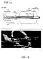
- FIG. 1Ais a schematic diagram of an OFDI balloon catheter in accordance with an exemplary embodiment of the present invention
- FIG. 1Bis a photograph of the OFDI balloon catheter shown in FIG. 1A ;
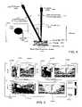
- FIG. 2Ais an exemplary image of a perspective view of a swine esophagus obtained using the OFDI balloon catheter in accordance with an exemplary embodiment of the present invention
- FIG. 2Bis an exemplary image of a top view of the swine esophagus of FIG. 2A ;
- FIG. 2Cis an exemplary image of a side view of a wall of the swine esophagus of FIG. 2A ;
- FIG. 3is an exemplary OFDI image acquired in a human subject using a BE technique in accordance with an exemplary embodiment of the present invention
- FIG. 4is a schematic diagram of exemplary arrangement and usage thereof for treating and monitoring tissue in accordance with an exemplary embodiment of the present invention
- FIG. 5is a set of multiple exemplary m-mode OFDI phase images obtained using the exemplary arrangement of FIG. 4 together with a corresponding histology;
- FIGS. 6A-6Dare exemplary images associated with the OFDI data acquired for a translating sample in accordance with the exemplary embodiment of the present invention.
- FIG. 7Ais an exemplary pre-laser treatment OFDI image obtained using the exemplary embodiment of the present invention.
- FIG. 7Bis an exemplary pre-laser treatment birefringence image obtained using the exemplary embodiment of the present invention.
- FIG. 7Cis an exemplary post-laser treatment OFDI image obtained using the exemplary embodiment of the present invention.
- FIG. 7Dis an exemplary post-laser treatment birefringence image obtained using the exemplary embodiment of the present invention.
- FIG. 8is an image of an exemplary vascular map extrated from a comprehensive dataset obtained from porcine esophagus in-vivo which can be obtained using the exemplary embodiment of the present invention
- FIG. 9is an exemplary in-vivo Doppler flow image of a porcine esophagus obtained using the exemplary embodiment of the present invention.
- FIG. 10is an plot of water absorption coefficient and corresponding penetration depth as a function of wavelength obtained using the exemplary embodiment of the present invention.
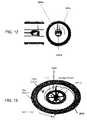
- FIG. 11is a schematic diagram of a two beam catheter probe in accordance with another exemplary embodiment of the present invention.
- FIG. 12is schematic side and front illustrations of a three beam catheter probe in accordance with yet another exemplary embodiment of the present invention.
- FIG. 13is a perspective view of a watch-spring multichannel optical rotary junction in accordance with an exemplary embodiment of the present invention.
- FIG. 14is a conceptual rendering of an image which can provide feedback to a user obtained using an exemplary embodiment of the present invention.
- FIG. 15is a block diagram of a sample arm of an OFDI system incorporating an optical switch in accordance with a further exemplary embodiment of the present invention.
- FIG. 16is a block diagram of the sample arm of the OFDI system incorporating an optical splitter in accordance with a still further exemplary embodiment of the present invention.
- FIG. 17is a block diagram of the sample arm of the OFDI system incorporating a single wavelength-division multiplexer in accordance with yet further exemplary embodiment of the present invention.
- FIG. 18is a block diagram of the sample arm of the OFDI system incorporating a cladding mode coupler and a dual-clad fiber in accordance with still another exemplary embodiment of the present invention.
- FIG. 19is a block diagram of a three-port rotary coupler and catheter in accordance with an exemplary embodiment of the present invention.
- FIG. 20is a block diagram of a single-fiber rotary coupler with subsequent demultiplexing of the therapy light and capable of splitting of imaging light in accordance with another exemplary embodiment of the present invention
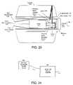
- FIG. 21is a schematic diagram and usage of a two-beam in-line catheter probe in accordance with an exemplary embodiment of the present invention.
- FIG. 22are front and side illustrations of a three-beam catheter probe and balloon catheter in accordance with an exemplary embodiment of the present invention.
- FIG. 23is a side view of a micro-motor-based arrangement capable of generating a slowly rotatable therapy beam and fast scanning imaging beam in accordance with an exemplary embodiment of the present invention
- FIG. 24is a block diagram of a therapy source incorporating a low power tunable source followed by a broadband booster amplifier in accordance with an exemplary embodiment of the present invention
- FIG. 25is a block diagram of a therapy source incorporating multiple laser diodes (LDs) at difference wavelengths and polarizations in accordance with another exemplary embodiment of the present invention.
- LDslaser diodes
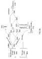
- FIG. 26is an illustration of a wavelength-tunable therapy source incorporating a laser diode bar and results generated thereby in accordance with an exemplary embodiment of the present invention
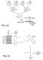
- FIG. 27is a side view of another exemplary embodiment of a system which includes a galvanometric scanner which can allow the OFDI beam to be repetitively scanned across the surface of the tissue, and usage thereof;
- FIG. 28is a schematic diagram of a further exemplary embodiment of the OFDI system according to the present invention which can be used to detect both the imaging and monitoring signals via acousto-optic frequency shifters;
- FIG. 29Ais a flow diagram of an exemplary embodiment of a method for obtaining information associated with at least one portion of a sample according to the present invention.
- FIG. 29Bis a flow diagram of another exemplary embodiment of the method for controlling a temperature distribution in the sample according to the present invention.
- FIG. 29Cis a flow diagram of yet another exemplary embodiment of the method for applying a laser radiation to at least one portion of a biological structure according to the present invention.
- An exemplary embodiment of the system and method according to the present invention for controlling and localizing therapycan be based on a thermal excitation delivered by a conventional, spatially scanned laser beam.
- the laser energy absorbed by tissuecan be substantially or entirely converted to a temperature rise, as described in publication 33 identified below.
- temperatures in excess of 60-70° C.generally can lead to irreversible protein denaturation and cell death irrespective of duration, as described in publication 34 identified below.
- the energyis absorbed, it can be subject to a spatial redistribution by a thermal diffusion.
- Eq. 1indicates that laser parameters P d , t, and wavelength can be used to control the depth of thermal injury and to minimize collateral damage to underlying tissues.
- P d , t, and wavelengthcan be used to control the depth of thermal injury and to minimize collateral damage to underlying tissues.
- the absorption spectrum of biological tissues near 1.45 ⁇ mmay be dominated by water, and can therefore be roughly constant across a range of tissues.
- absorption lengthscan be selected that range from over 2 mm to 300 ⁇ m. This exemplary range is well matched to the depths characteristic of epithelial lesions.
- the monitoring signalarose only after the zone of thermal injury has transitioned across a boundary of the specific tissue types. None could determine the depth of thermal injury or the spatial relationship of the damaged tissue to adjacent viable tissue. Certain degree of spatial resolution has been achieved by monitoring the portion of laser light that is not absorbed by the tissue. By inserting an optical fiber through a needle, this light can be collected from different perspectives surrounding the heated volume and temperature-dependent scattering changes can be measured (as described in publication 43 identified below). A more direct approach, high-resolution in situ imaging, has also been demonstrated for visualizing scattering changes and the physical removal of tissue resulting from ablative laser irradiation (as described in publication 44 identified below).
- Exemplary embodiments of monitoring systems, methods and techniques according to the present inventionmay utilize information regarding well-known tissue responses to a thermal injury.
- These exemplary responsescan include, but not limited to, microscopic deformation (as described in publication 33 identified below) and changes in scattering (as described in publications 36, 38 and 45 identified below), birefringence (as described in publication 46 identified below), and blood flow (as described in publication 47 identified below) that can result from laser heating and that can be observed over a range of temperatures beginning as low as 45° C.
- microscopic deformationas described in publication 33 identified below
- changes in scatteringas described in publications 36, 38 and 45 identified below
- birefringenceas described in publication 46 identified below
- blood flowas described in publication 47 identified below
- One exemplary aspect of an exemplary embodiment of the method and technique according to the present inventionis that these thermal responses can be detected with high spatial resolution and presented in a cross-sectional image format along with the microscopic tissue structure.
- a system, arrangement and methodcan be provided that are capable of screening and delivering precisely guided laser therapy. Since the characteristic length-scales preferably usable for comprehensive screening and comprehensive therapy are likely distinct, it is possible to separately perform these objectives.
- the screeninge.g., possibly performed as a first step
- This exemplary procedurecan be used to identify regions for subsequent therapy. After the performance of the screening procedure, the endoscopic probe can be directed back to the specified regions, and therapy may be performed under real-time guidance so that all disease is treated and collateral damage is minimized.
- This exemplary resultcan improve the management of patients with Barrett's esophagus by, e.g., increasing the effectiveness of therapy while decreasing the risk of complications.
- the exemplary embodiments of the system, techniques and methods according to the present inventioncan be applicable to any application of laser treatment including but not limited to, for example, applications in dermatology.
- Some relevant epithelial cancers and precancerous lesions addressed by the exemplary embodiments of the present inventioncan include, but not limited to, the larynx, cervix and ovaries, bladder, oral cavity and lung.
- the exemplary embodiments of the present inventioncan be applicable to the areas of monitoring photodynamic therapy, radiofrequency ablation, and cryotherapy to provide control over depth and spatial extent of therapy.
- an exemplary embodiment of an OFDI cathetermay be provided in accordance with the present invention that can be centered within the esophageal lumen using a balloon sheath shown in FIGS. 1A and 1B .
- the exemplary cathetermay include of a probe scanner 2000 which can rotate, and may pull back an inner optical core 2010 .
- the inner core 2010can be enclosed within a transparent sheath 2020 .
- the balloon 2040which, when inflated, may center the imaging optics.
- the imaging beam 2030can be focused onto the esophageal surface 2050 . This imaging beam 2030 may be scanned to achieve comprehensive imaging.
- the balloon 2040can have an inflated diameter of 1.8 cm, and may allow for a longitudinal imaging over a length of 4.5 cm without repositioning.
- the optical core 2010 of the cathetercan include an optical fiber, a spacer for expansion of the optical beam, a gradient index lens for focusing, and a right-angle prism for directing the beam perpendicularly to the longitudinal axis of the catheter.
- a miniature cylindrical lenswas fabricated in-house and placed on the second surface of the prism. This lens compensated for astigmatism induced by the plastic sheaths and resulted in a diffraction-limited beam (30 ⁇ m diameter) on the tissue surface.
- the exemplary cathetermay be rotated at rate of about 4 revolutions per second, allowing the acquisition of 2500 axial scans per circular cross-section.
- This exemplary OFDI systemcan record an encoder signal to precisely track the rotation and pull-back of the catheter. This information is used in reconstructing the 3-dimensional data set.
- FIGS. 2A-2CThe esophageal imaging techniques can be performed in two ⁇ 50 kg swine. Although the complete 20 GB data set may likely not be represented in discrete figures, the information content is shown by FIGS. 2A-2C .
- an image 2100provides a 3D rendering of the entire imaged esophagus.
- an image 2110illustrates a single transverse cross section of the imaged esophagus.
- image 2120shows a zoomed cross sectional image of at least one portion of the esophagus.
- a sampling with a resolution of 10 ⁇ m ⁇ 20 ⁇ m ⁇ 30 ⁇ m (r, ⁇ ,z)can yield a comprehensive microscopic data set that can be displayed volumetrically a the image 2100 of FIG. 2A for mapping and orientation, or in high-resolution cross-sectional images in which the entire esophageal wall can be visualized as the image 2110 in FIG. 2B .
- An expanded view of the image 2120 of FIG. 2Cdepicts the architectural structure of the mucosal layers.
- FIG. 3An exemplary single rotational image 2150 is shown in FIG. 3 . Hallmark features of SIM (disorganized epithelial architecture with irregular surface; presence of large epithelial glands) of a patient are shown therein. This patient had a prior diagnosis of BE, and imaging was performed prior to PDT.
- SIMstructured epithelial architecture with irregular surface; presence of large epithelial glands
- tissue proteins and collagencan denature, giving rise to microscopic deformation (described in publications 33 identified below), increased in scattering (described in publications 36, 38 and 45 identified below), reduced birefringence (described in publication 46 identified below), and reduced blood flow (described in publication 47 identified below).
- the description belowprovides the methods for monitoring these changes using exemplary OFDI in accordance with the exemplary embodiments of the present invention.
- porcine esophagus samples and duodenum samples(as a proxy for SIM) were mounted with a microscope cover glass on the epithelial surface so that the approximate pressure and thermal conductivity of the balloon catheter could be simulated.
- FIG. 4An exemplary embodiment of an apparatus for collecting OFDI signals during a laser irradiation and use thereof according to the present invention is shown in FIG. 4 .
- the treatment lightis delivered through a collimator 2200 .
- the imaging lightis delivered through a second collimator 2220 .
- the treatment beam 2210 and imaging beam 2230overlap when reaching the tissue 2270 which is covered with a think glass cover slip 2260 and resting on a backing 2280 .
- the tissueis translated by a motorized translation stage 2290 .
- the imaging beamis focused by a lens 2250 .
- the top-down image depicting beam overlap 2250is provided.
- the OFDI sampling beamcan be focused at the tissue surface to, e.g., a 1/e 2 intensity diameter of 23 ⁇ m and aligned so that it overlapped with the laser spot as shown in FIG. 4 .
- the samplesmay be held at a fixed location and/or translated using a motorized stage.
- a red horizontal line 2330 a , 2330 b , 2330 c , at the top of each phase-shift imagedenotes the interval over which the laser was on.
- a superficial region of positive phase-shift overlying a lower region of negative shifthas been observed.
- the depth at which the phase transitioned from positive to negativebecame progressively deeper and the magnitude of the overlying phase-shift decreased. No measurable phase-shift was detected after the laser was switched off.
- a protein denaturationgives rise to local microscopic structural changes and a nidus of local deformation that is detected as a phase-shift in the interferometric signal.
- the zone of active denaturationpropagates in depth with overlying tissues becoming completely denatured. The depth at which the direction of the phase-shift reverses identifies the focal center of active denaturation.
- NBTCnitro-blue tetrazolium chloride
- the depth-derivative of the phase-shifthas been determined for each A-line and defined the depth of injury as the point of maximum negative value of the derivative.
- the depths determined in this wayare provided in FIG. 5 as vertical lines adjacent to each M-mode image and show a good correspondence with histomorphometry.
- the computer-controlled translation stage 2290can be controlled to repetitively toggle the sample velocity from 1.8 mm/s to 0.9 mm/s.
- FIG. 6AAn OFDI intensity image 2400 acquired with no laser irradiation is shown in FIG. 6A .
- the 1450 nm laser powerwas about 400 mW.
- the translation of the samples during the exposureresulted in a line of laser damage across the surface of the sample. Since the thermal energy deposition can be proportional to the exposure time (see Eq. 1), the depth of laser damage can vary along the line according to the inverse of the translation velocity. Histology sections, obtained from regions of fast and slow translation and with an orientation perpendicular to the line, indicated laser injury depths of 0.41 mm and 0.69 mm respectively.
- phase-shift data corresponding to the image 2410 of FIG. 6Bis illustrated as the image 2420 in FIG. 6C .
- the depth of the damage determined by the phase-shift datacan be 0.40 mm and 0.67 mm in the regions of fast and slow velocity, respectively.
- Speckleis a phenomenon that is commonly observed when imaging with coherent illumination and manifests as a grainy pattern of high- and low-intensity that does not appear to correlate with the macroscopic structure.
- speckleIn tissue, speckle generally arises from the interference between photons that have traversed different paths during propagation within the sample. If the scatterers within the tissue are moving, even on a microscopic scale, the speckle pattern is likely seen to rapidly fluctuate. The measurements of the time-evolution of the speckle pattern can therefore provide insight into microscopic motion within the sample.
- This exemplary techniquehas been provided for investigating biomechanical properties (as described in publication 50 identified below), and thermal excitation (as described in publication 51 identified below), in biological tissues. The extension of these concepts to the depth-resolved monitoring of laser tissue interactions with OFDI has been reviewed.
- the image 2430 of FIG. 6Dis the speckle decorrelation image corresponding to the images 2410 and 2420 of FIGS. 6B and 6C , respectively.
- the depth of the peak decorrelation 2431 rateblack band, denoted by arrows in FIG. 6D ) can be observed to vary in correspondence with the translational rate of the sample and the depth of laser damage indicated in histology. The consistency of this finding across samples of esophagus and duodenum confirm that the depth of peak decorrelation rate is a quantifiable metric for determining the depth of laser injury.
- PS-OCTPolarization-sensitive OCT
- the apparatus of FIG. 4can be modified to include a galvanometric scanner so that the OFDI beam may be repetitively scanned across the surface of the tissue while the sample was held stationary and the 1450 nm laser spot remained fixed on center, as shown in FIG. 27 .
- the treatment lightcan be delivered through a first collimator 2500 providing a treatment beam 2510 incident on the tissue 2550 that is covered by a cover slip 2540 and against a backing 2560 .
- the imaging lightmay be provided by a second collimator 2570 producing an imaging beam 2580 that is directed by a galvo-mirror 2520 through a lens 2530 .
- This arrangement/systemcan be an exemplary embodiment of a therapeutic monitoring system applicable to applications in dermatology. OFDI images or video of esophageal and duodenal tissues were acquired during laser irradiation.
- FIGS. 7A-7Dshow images of the representative data.
- the layered esophageal structurecan be observed in the intensity image 2450 (see FIG. 7A ) and characteristic birefringence banding can be observed in the corresponding polarization image 2460 (see FIG. 7B ).
- the epithelial scattering intensitymay be increased dramatically within the 1.1 mm laser spot 2470 (see FIG. 7C ), and the birefringence banding in the corresponding polarization image 2480 (see FIG. 7D ) can be lost.
- a zone of decreased birefringencecan be observed that may begin near the surface and propagated downward. These observations are generally consistent with a downward propagating zone of denatured tissue. Measurements of the percent-loss of birefringence is a quantitative metric for monitoring laser thermal damage.
- Thermally induced changes to the microscopic structure of tissuecan alter optical scattering. Since the signal in OFDI arises from scattering and small changes can be detected over a large dynamic range, we investigated the use of scattering measurements for monitoring thermally induced changes in tissue. Scattering changes observed in image 2460 of FIG. 7B may be representative of the preliminary observations in both duodenum and esophagus samples. In certain cases, it was determined that the significant scattering changes within the epithelium and relatively smaller changes in the underlying tissues of the muscularis mucosa and muscularis basement. For example, two potential quantitative metrics for laser damage that could be obtained from scattering measurements: changes in the depth-resolved scattering intensity and changes in the depth-integrated scatting intensity.
- FIG. 8An image 2490 of FIG. 8 , acquired during our recent swine studies, graphically illustrates the porcine esophageal vascularity. This exemplary image 2490 was generated by unwrapping the tubular image data to display the epithelial surface as if the esophagus was longitudinally opened and pinned flat. The intensity data has been integrated over depth into the tissue. Although this type of large scale visualization is a convenient way to map the vessels, it is possible to use a more sensitive and quantitative method/technique/system for measuring blood flow.
- Doppler OCT(as described in publications 55 and 56 identified below) has been demonstrated for visualizing and quantifying blood flow in tissue and has been investigated as an arrangement for assessing flow following laser therapy (as described in publication 57 identified below).
- the Doppler measurements with OFDI(as described in publication 24 identified below) have been described, and the possibility of simultaneously measuring structure and flow in vivo has been reviewed.
- a cross-sectional view of an exemplary image 2590 of FIG. 9has been acquired in the esophagus of a living swine and displays intensity as a grayscale and Doppler as a superimposed color.
- the coordinates (r, ⁇ ) of this datahave been mapped to Cartesian coordinates (vertical, horizontal) for simplicity of display. This result was representative of our observations at multiple locations in two swine. Additionally, in time-sequences of Doppler images, we clearly observed pulsatile flow.
- phase-shift and speckle decorrelationwhich are only applicable during laser irradiation, may be more sensitive and provide greater spatial resolution.
- the changes in birefringence, scattering and floware persistent and could be applied for follow-up imaging after laser treatment.
- thermal injurycan be controlled through the use of a raster-scanned, spatially-collimated beam.
- a flat-top beam with a diameter of 1-3 mm with well-defined edgesmay allow spatial control while also permitting therapy of large epithelial areas through raster scanning.
- Exemplary laser control parametersare described herein below in the context of Eq. 1.
- the temperature distribution of Eq. 1generally applies only in the limit of weak scattering.
- ⁇ awould likely be an optimal parameter for control over depth of laser injury.
- ⁇ ais characteristic of the sample rather than an externally controllable parameter
- absorption lengthsIn the vicinity of the water absorption band near 1.45 ⁇ m, absorption lengths (see graph 2595 of FIG. 10 ) range from 0.3 mm to over 2 mm within a narrow spectral range (1375 nm to 1430 nm). These lengths correspond well to the characteristic length scales appropriate for the treatment of epithelial disease.
- a tunable laser, operable in the vicinity of the 1450 nm water absorption bandcan be used to control therapy through wavelength tuning.
- the absorption coefficientdoes more than control the exponential depth decay of the temperature distribution; e.g., it also can control the amplitude. Since the amplitude term is also dependent upon power density and exposure duration, these variables can be used to normalize the amplitude while allowing the absorption coefficient to change.
- the procedure performance timemay be estimated by 2At/( ⁇ rv), where At is the treatment area, r is the laser spot radius, and v is the laser spot scan velocity. For an esophageal treatment length of 60 mm and an esophageal diameter of 20 mm.
- a combined systemcan be provided which may allow for a controlled laser excitation.
- the exemplary systemcan be used endoscopically for conformal laser therapy capable of comprehensively treating epithelial lesions while minimizing collateral damage to adjacent tissues.
- a systemcan be provided for performing conformal laser therapy of epithelial disease through a combination of monitoring and control. Since laser beams can easily be shaped and spatially scanned and since margins in the transverse plane (along the surface of the esophagus) are less critical, the primary challenge for achieving accurate control of laser therapy is in limiting and adjusting the depth of laser damage. Based on the modeling and analysis described above, it is possible to utilize laser wavelength and power and scanning speed to vary the depth of laser damage over a clinically significant range while not significantly altering the transverse extent of injury.
- the laser wavelengths between approximately 1375 nm and 1430 nmcan provide absorption lengths ranging from over 2 mm to less than 0.3 mm.
- Semiconductor laserscan operate in this spectral range. Since such lasers can be compact and environmentally stable, these laser can be effectively used in clinical applications. Materials suitable for this specific wavelength range, however, may not be standard.
- a less expensive alternative for the early testing phase of exemplary embodiments of the methods according to the presentcan be provided by a solid-state laser material, tetravalent chromium-doped YAG (Cr4+:YAG). For example, a tunability with this material over the spectral range of 1340 nm-1570 nm can be implemented (as described in publications 58 and 59 identified below).
- the OFDI sampling beammay be focused at the sample to a diameter of ⁇ 25 ⁇ m.
- the axial location of the focuscan be determined using a standard z-scan technique, and may be registered within the OFDI cross-sectional image.
- the subsequent axial positioning of the samples within the OFDI image windowmay ensure a constant focus location for all samples.
- Datamay be collected with the two beams fixed with respect to each other and while the sample is translated perpendicular to the laser beam axis.
- the offset between the OFDI beam and the center of the laser spotis not critical for monitoring.
- OFDI datamay be collected for various offsets (as depicted in FIG. 4 ) to determine the offset that yields the greatest indicated depth of thermal injury.
- This offsetcan then be used in all subsequent studies and may be registered as follows.
- a small, low-power, short-duration epithelial burnmay be induced on the surface of the sample while the sample is held fixed (non-translating).
- the increase in epithelial scatteringcan be readily observed in OFDI and is spatially localized as defined by the laser beam profile.
- the OFDI beamcan be relayed to the focusing lens by a pair of galvanometers that provide two-dimensional scanning.
- the galvanometersmay be used to generate an en face OFDI image of the sample and the epithelial burn may appear as a circle of increased scattering.
- the galvanometerscan then be positioned and fixed so that the OFDI beam is positioned with the desired offset (as schematically shown in FIG. 4 ).
- Laser wavelengthmay be varied from about 1375 nm to 1405 nm in 2 nm steps with laser spot size and scanning speed held constant.
- the laser powermay be adjusted so that the product P d ⁇ a in Eq. 1 can be maintained as constant. This should yield lines of constant width and with damage depth ranging from approximately 0.25 to 1.5 mm.
- One exemplary embodiment according to the present invention for affecting therapeutic depthmay include scaling the scan velocity.
- the therapy beam scan speedcan be varied from 1 mm/s to 5 mm/s. Slower scan speeds allow time for heat to conduct to deeper areas of the tissue, producing deeper therapy.
- the spatial relationship between the OFDI sampling beam and the laser spotcan be controlled.
- One exemplary embodiment of the present inventioncan include an endoscopic probe for comprehensive, volumetric imaging and simultaneous laser therapy, as shown in FIG. 11 .
- two beam relay optics 2640 a and 2640 bmay be used, one of which conveys imaging light 2640 b and the other therapy light 2640 a .
- These relay opticsare placed within a housing 2630 that is enclosed within a first transparent sheath 2600 .
- a balloon centration mechanism (as described above) 2620may be used to maintain a constant distance between the optical probe 2630 and the tissue surface 2610 .
- Laser light and the OFDI beammay be delivered through separate optical fibers 2641 a and 2641 b .
- Each fibermay have its own relay optics to produce independently controllable spot sizes.
- a further exemplary embodiment of the present inventioncan include these relay optics designed to produce overlapping spots.
- the optical fibers and distal opticsmay be housed in a wound-wire drive shaft and placed inside a balloon-centering probe identical to the balloon shea
- Longitudinal scanscan be activated using a computer controlled translation stage attached to the proximal end of the drive shaft.
- This exemplary arrangementmay be the same as the arrangement which can be used for the pull-back esophageal imaging of our preliminary studies.
- a manual rotation of the drive shaftmay be possible, as is automated rotation using an exemplary rotary coupler 2900 shown in FIG. 13 .
- an endoscopic systemmay screen for disease over large fields-of-view, accurate monitoring of laser-tissue interaction, and precisely control laser therapy.
- One of the applications of such exemplary embodimentmay be the identification and treatment of epithelial cancers and their precursors.
- the systemcan incorporate procedures and software modules than can directly link screening, monitoring, and control.
- the systemmay be used to generate a high-resolution, 3-dimensional map of the entire distal esophagus to facilitate therapeutic planning. Thereafter, the use may be presented with a ‘live’ cross-sectional image comprising three sections, as illustrated in FIG. 14 .
- a right section 2700 of the imagemay be the tissue immediately ahead of the therapeutic laser
- the center 2730 of the imagemay be the location of the laser with a marker 2740 designating the zone of therapy
- the left section 2710 of the imagemay be the tissue that has already been treated. Since the three beams may be continuously scanning, the tissue may appear to move from right to left as the image updates.
- the usermay operate a control servo to start/stop the treatment and increase or decrease the depth of therapy.
- the usermay be able to steer and conform the region of laser therapy to the desired target.
- FIG. 12An exemplary embodiment of the endoscopic probe for imaging, monitoring and laser therapy through a centering balloon according to the present invention is shown in FIG. 12 .
- This exemplary probecan rotate to scan the esophagus circumferentially and may be longitudinally translated at a slower rate to define segments for therapy.
- This probemay include, e.g., three or more optical channels: a first channel 2800 c for imaging the tissue prior to laser irradiation, a second channel 2800 b for treatment, and a third channel 2800 a for monitoring.
- Each optical fibermay be separately imaged transversely onto the esophageal wall through the balloon.
- the alignment of the resulting output beamsmay be such that, upon rotation in the clockwise direction, the imaging beam precedes the treatment beam sufficiently so that non-treated tissue may be sampled.
- the monitoring beammay be aligned to fall within the laser spot.
- the opticsmay be bonded together with epoxy, and the alignment may be fixed.
- FIG. 13An exemplary rotational coupler according to the present invention which can connect the three-channel catheter to the OFDI system is shown in FIG. 13 , and can be referred to “watch-spring” rotary junction (since it can rely on two concentric spools).
- watch-springrotary junction
- optical fiberis wound from the outer spool 2910 onto the inner spool 2900 .
- the fibercan unwind from the inner spool.
- Ribbon optical fibermay be used and two parallel plates 2920 with a gap matched to the ribbon width can ensure that the windings remain flat and do not bind.
- the platesmay be sufficiently large so that up to, e.g., 100 rotations may be possible prior to requiring counter rotation.
- a further exemplary embodiment of the system and arrangement according to the present inventioncan utilize, e.g., a high-speed imaging system.
- the exemplary embodiment of the digital acquisition and processing systemcan be based on VME-bus hardware for acquiring, processing and storing the OFDI signals in real-time.
- the exemplary components of such exemplary system and arrangementmay comprise a VME chassis containing high-speed digitizers residing on a single-board computer and fiber-optic links to a RAID storage array.
- This exemplary system and arrangementcan be controlled via a host processor (e.g., a personal computer).
- the analog OFDI signalsmay be digitized using wideband receivers (e.g., 12 bit, 210 MS/s) with integrated field-programmable-gate-array (FPGA) processors. Processing power, resident on the acquisition board, may be importance since the raw data rate may be 800 MB/s for the two polarization channels of the OFDI system.
- the FPGA processorcan be configured or programmed to transform each polarization channel from the frequency-domain to a 1024-element array representing reflectivity versus depth (one A-line). This data can be passed to the single-board computer for subsequent processing and for combining the two channels prior to transferring the final data to a RAID array of hard drives.
- the final data storage ratemay be, e.g., 400 MB/s. By striping the data across multiple hard drives, this data rate can be continuously sustained.
- Software on a processing arrangement in accordance with an exemplary embodiment of the present inventioncan permit a user control over the exemplary system, and may enable a display of the images at a down-sampled rate in real-time.
- the exemplary systemcan be used in two exemplary modes: a burst mode at full data rate, and continuous mode at half data rate.
- the exemplary endoscopic system and arrangementcan include the components and software described above, and additional procedures (e.g., software) can be provided to program both the FPGA processor and single-board computer to facilitate the computations of phase-shift, birefringence, speckle, and Doppler signals in real-time.
- the combined computational capacity of the Vertex 4 Pro FPGA and quad G4 single-board computersmay be ample for displaying the monitoring signal in real-time.
- the spot size while maintaining a constant scan velocitycan be doubled by using a 4-fold increase in the laser power in order to maintain a constant temperature distribution in the tissue. Doubling the scan velocity at a constant spot size should use a doubled laser power.
- One exemplary embodiment of a laser arrangement in accordance with the present inventioncan utilize a single-emitter semiconductor laser diode. Previous devices have provided more than 3 W of laser power over this spectral range using a simple external cavity design including a diffraction grating for wavelength control. The laser power and wavelength may be controlled via the host processing arrangement of the OFDI system based on an analog signal from a potentiometer.
- the potentiometermay be a hand-held dial that the user (e.g., an endoscopist) may use to increase or decrease the depth of laser damage.
- the exemplary embodiment of the system and method according to the present inventioncan provide a user interface to the operator that includes a cross-sectional image of the tissue.
- the imagemay be continuously updated and may include views of treated and upcoming, untreated tissue as well as a designation for the zone of laser treatment as determined by the monitoring procedures.
- the user interfacemay be programmed on the host processing arrangement, and can use computational results from the FPGA processor and single-board computer. Images and laser parameters may be archived to the RAID array.
- the imaging system/arrangement 100can be connected to a three-fiber probe using an optical switch 115 as shown in a block diagram of FIG. 15 .
- the exemplary probesuch as that described above with reference to FIG. 12 , can include two imaging fibers and one therapy fiber.
- the switch 115can alternately couple imaging light to one of the two imaging fibers 120 a , 120 b which can be used to acquire re-therapy images and, e.g., during-therapy imaging.
- a therapeutic light source 105may connect directly to the therapy fiber 125 c .
- the fiberscan be connected to the catheter 130 , which can be, e.g., the exemplary catheter shown FIG. 12 .
- a signal from the imaging system 100can control the state of the optical switch 115 .
- the exemplary imaging system/arrangement 200can be coupled to an exemplary three-port catheter such as one shown in FIG. 12 via an optical splitter 215 that can couple light to both of two imaging fibers 220 a , 220 b .
- This exemplary imaging systemcan separate the image signal from each using path-length encoding techniques.
- an optical delay 235may be placed in one fiber 220 b or multiple fibers.
- the therapeutic light source 205can be coupled directly or indirectly to the therapy bomb 225 c of the catheter.
- lightmay be combined with the therapy source 805 using a single wavelength-division multiplexer 810 .
- the combined lightmay be coupled to a single fiber rotary coupler, and then to an exemplary single fiber catheter such as the catheter shown in FIG. 21 .
- lightmay be combined with the therapy light 905 using a cladding mode coupler that couples the imaging system 900 light from the single mode fiber 901 to the single mode core of a dual-clad fiber 911 and the therapy light from a multimode fiber 906 to the cladding mode of a dual-clad fiber 911 .
- FIG. 19shows exemplary connections between a system 400 with three output fibers 405 a , 405 b , 405 c , such as one shown schematically in, e.g., FIGS. 15 and 16 , and a three-port catheter 415 , such as one shown in FIG. 12 via a multi-channel rotary coupler 410 , such as one shown in FIG. 13 .
- FIG. 20shows a schematic diagram of an exemplary system 300 according to the present invention in which a single fiber 305 containing both the imaging light and therapy light may be coupled to a single-channel rotary coupler 310 .
- the lightcan be divided by a wavelength-division multiplexer (WDM) 330 that separates the imaging light onto a first fiber 332 and the therapy light onto a second fiber 331 .
- the imaging lightmay further be separated using an optical splitter 335 that greats two imaging ports 336 a and 336 b .
- the fibers 31 , 336 a , 336 bcan be connected to a three-port catheter 325 design such as that shown in FIG. 12 .
- the catheter section 320may be flexible allowing endoscopic insertion and the section containing a WDM 330 and a splitter 335 can be enclosed within a rigid tube 315 to protect these components.
- FIG. 21shows a side view of an exemplary embodiment of a distal optics arrangement according to the present invention that may create a single imaging beam 1125 and a separate therapy beam 1120 from a single-mode fiber 1101 .
- the light from the fiber containing both imaging and therapy lightcan first be focused by a first GRIN lens 1100 .
- the lightis then passed into a wavelength-division multiplexing prism 1105 that can direct the therapy light wavelengths upward to create the therapy beam 1120 , and transmits the imaging light wavelengths to a second GRIN lens 1110 , which can alternately focus the imaging light and directs it to a final prism 1115 that directs the imaging beam 1125 upward.
- the angle of the prisms 1105 and 1115may be such that the beams are made to overlap at the appropriate distance from the device.
- FIG. 22shows side and front views of an exemplary embodiment of a three-port catheter in accordance with the present invention.
- the exemplary cathetercan include three fibers 1005 that connect to three sets of focusing optics 1035 contained in a V-groove 1020 inside a housing 1040 .
- the focusing opticscan provide beam focusing.
- Micro-prisms 1025can redirect the optical beam upward through a cylindrical lens 1030 that corrects for astigmatism induced by the transparent sheath 1000 .
- a balloon 1010 centration mechanismmay be used to maintain centering of the optics 1035 within the luminal tissue 1015 .
- the monitoring beam 1050 c , therapy beam 1050 b , and pre-imaging beam 1050 acan be seen.
- the housing 1040can be adapted to rotate by a multichannel rotary coupler such as one shown in FIG. 13 .
- FIG. 23shows a side view of an exemplary embodiment of a catheter in accordance with the present invention which can utilize a miniature motor 1260 to achieve rotation of the imaging beam.
- the motor 1260can be enclosed within a transparent sheath 1235 .
- the rotation of the motor shaftcan rotate a prism 1220 .
- the imaging lightcan be coupled to the distal optics via a fiber 1210 , where the light can be focused by focusing optics 1215 , and reflected onto the prism 1220 by a reflector 1225 .
- the rotation of the prism 1220sweeps the imaging beam circumferentially.
- the motor electrical connections 1205can be achieved through the same lumen as the fiber 1210 .
- the therapy lightis coupled to the distal optics on fiber 1200 .
- This therapy lightmay be focused using focusing optics 1250 , and directed sideways by prism 1245 at a fixed rotational angle relative to the inner sheath 1235 .
- the imaging beamthus sweeps through the fixed therapy spot.
- the translation of the therapy spotis achieved by rotation of the entire inner sheath 1235 within the outer sheath 1240 .
- This exemplary rotationcan be achieved through the use of a multi-channel rotary coupler such as a coupler shown in FIG. 13 .
- the cathetercan use a balloon 1255 for centration of an optical core 1230 .
- FIG. 24shows a block diagram of an exemplary embodiment of a laser therapy source according to the present invention with wavelength tunability utilizing a low power wavelength tunable source 600 , followed by a broadband booster amplifier 605 to increase the optical power.
- FIG. 25shows a block and functional diagram of an exemplary embodiment of a laser therapy source incorporating multiple laser diodes 500 a , 500 b , 500 c , 500 d at difference wavelengths and polarizations, and the exemplary procedure to implement such arrangement.
- the lightcan be combined by the polarization multiplexers 505 a , 505 b and wavelength division multiplexers 510 to a single mode fiber 515 .
- the lightcan be coupled to a multimode fiber 520 .
- a fast mode scrambler 525can be used to scramble the transverse mode pattern output from the multi-mode fiber at a fast rate.
- Other source arrangements which can output light on a single mode fibercan use a similar design to couple light to a multimode fiber.
- FIG. 26shows an exemplary embodiment of a therapy light source and use thereof according to the present invention.
- a laser diode bar 700can be used with multiple wavelengths 701 a - g .
- Each waveguidecan be coupled to a free-space laser cavity through a lens apparatus 705 and a grating 710 and a partially reflecting end mirror 715 . Because of the wavelength dispersion of the grating, the laser formed by each waveguide lases at a different wavelength. Thus, by adjusting the drive current to each of the waveguides 701 a - g , the laser output 720 power and spectral shape can be adjusted.
- a single OFDI systemcan be modified to facilitate a detection of both the imaging and monitoring signals through the use of acousto-optic frequency shifters as shown in FIG. 28 .
- a wavelength swept laser source 3000can be separated by a first splitter 3020 to produce a sample arm path and reference arm path.
- the sample arm pathis further separated by a second splitter 3030 , with a first output of this splitter being directed to a first frequency shifter 3061 and a second output being directed to a second frequency shifter 3060 .
- Each of the frequency shifterscan be driven with a separate frequency.
- the light from the first frequency shifter 3061can be coupled through an optical circulator 3071 to the imaging fiber 3072 of a three-fiber rotary coupler 3110 like that shown in FIG. 13 .
- the light from the second frequency shifter 3060may be coupled through a circulator 3070 to a monitoring fiber 3073 of the same rotary coupler.
- a separate therapy laser 3010can be coupled to the third therapy fiber.
- the returned light on the imaging fiber 3072 and monitoring fiber 3073may be recombined at an optical combiner 2080 , and mixed with the reference arm light at a second combiner 3090 with the output directed to a set of detectors 3100 . Due to the frequency shifters, the interference signal due to the imaging light and the interference signal due to the monitoring light are encoded at separate carrier frequencies and can be separated through conventional frequency domain techniques.
- FIG. 29Ashows a flow diagram of an exemplary embodiment of a method for obtaining information associated with at least one portion of a sample according to the present invention.
- a temperature changecan be caused in the portion of the sample in step 3100 .
- At least one first electro-magnetic radiationcan be forwarded to a section near or in the portion of the sample in step 3110 .
- a deformation of the sectioncan be identified at a plurality of depths as a function of (i) a phase of at least one second electro-magnetic radiation provided from the section, and/or (ii) a rate of change of the phase and/or an amplitude of the second electro-magnetic radiation in step 3120 .
- FIG. 29Bshows a flow diagram of another exemplary embodiment of the method for controlling a temperature distribution in the sample according to the present invention.
- an electro-magnetic radiationcan be provided to the section in the sample at a particular wavelength in step 3130 .
- the temperature distributioncan be controlled by modifying the particular wavelength of the electro-magnetic radiation when the electro-magnetic radiation can be provided to the section in step 3140 .
- FIG. 29Cillustrates a flow diagram of yet another exemplary embodiment of the method for applying a laser radiation to at least one portion of a biological structure according to the present invention.
- a beam of the laser radiationcan be provided to the portion in step 3150 , whereas a cross-sectional area of the beam is at most about 1/10 th of an entire area of the at least one portion.
- the beamcan be applied to the portion (I) based on a predetermined pattern, (II) while modulating a wavelength of the laser radiation, and/or (III) while monitoring a depth of the application of the laser radiation.
Landscapes
- Health & Medical Sciences (AREA)
- Life Sciences & Earth Sciences (AREA)
- Physics & Mathematics (AREA)
- Surgery (AREA)
- Animal Behavior & Ethology (AREA)
- Public Health (AREA)
- General Health & Medical Sciences (AREA)
- Veterinary Medicine (AREA)
- Engineering & Computer Science (AREA)
- Biomedical Technology (AREA)
- Heart & Thoracic Surgery (AREA)
- Medical Informatics (AREA)
- Molecular Biology (AREA)
- Pathology (AREA)
- Biophysics (AREA)
- Nuclear Medicine, Radiotherapy & Molecular Imaging (AREA)
- Radiology & Medical Imaging (AREA)
- Optics & Photonics (AREA)
- Electromagnetism (AREA)
- Otolaryngology (AREA)
- Laser Surgery Devices (AREA)
- Investigating Or Analysing Materials By Optical Means (AREA)
- Radiation-Therapy Devices (AREA)
- Endoscopes (AREA)
Abstract
Description
where ρ is the tissue density, c the heat capacity, and r the radial distance from the center of a Gaussian laser beam of 1/e2 radius, W. Although this approximation neglects scattering of the laser light as it propagates into the tissue, models that explicitly include scattering (as described in publication 36 identified below) indicate less than 10% deviation from Eq. 1 under the stated conditions.
| Summary of Monitoring |
| Cause | Effect | Measurement |
| Thermal denaturing of | Focal deformation | Phase &Speckle |
| cellular proteins and collagen | Loss of birefringence | Polarization |
| Δ Scattering | Intensity | |
| Thermal coagulation of | Loss of blood flow | Doppler flow & |
| vessels | Vascular Map | |
- 1. Devesa S S, Blot W J and Fraumeni J F, Jr. Changing patterns in the incidence of esophageal and gastric carcinoma in the United States. Cancer 1998; 83:2049-2053.
- 2. Barr H, Stone N and Rembacken B. Endoscopic therapy for Barrett's esophagus. Gut 2005; 54:875-884.
- 3. Johnston M H. Technology insight: ablative techniques for Barrett's esophagus—current and emerging trends. Nature Clinical Practice Gastroenterology & Hepatology 2005; 2:323-330.
- 4. Falk G W, Chittajallu R, Goldblum J R, Biscotti C V, Geisinger K R, Petras R E, Birgisson S, Rice T W and Richter J E. Surveillance of patients with Barrett's esophagus for dysplasia and cancer with balloon cytology. Gastroenterology 1997; 112:1787-1797.
- 5. Spechler S J. Barrett's esophagus: should we brush off this ballooning problem? Gastroenterology 1997; 112:2138-2142.
- 6. Kubba A K, Poole N A and Watson A. Role of p53 assessment in management of Barrett's esophagus. Dig Dis Sci 1999; 44:659-667.
- 7. Reid B J. p53 and neoplastic progression in Barrett's esophagus. Am J Gastroenterol 2001; 96:1321-1323.
- 8. Sharma P, Weston A P, Topalovski M, Cheman R, Bhattacharyya A, Sampliner R E. Magnification chromoendoscopy for the detection of intestinal metaplasia and dysplasia in Barrett's oesophagus, GUT 2003; 52: 24-27.
- 9. Kuipers E J and Haringsma J. Diagnostic and therapeutic endoscopy. Journal of Surgical Oncology 2005; 92:203-209.
- 10. Georgakoudi I, Jacobson B C, Van Dam J, Backman V, Wallace M B, Muller M G, Zhang Q, Badizadegan K, Sun D, Thomas G A, Perelman L T and Feld M S. Fluorescence, reflectance, and light-scattering spectroscopy for evaluating dysplasia in patients with Barrett's esophagus. Gastroenterology 2001; 120:1620-1629.
- 11. Adrain A L, Ter H C, Cassidy M J, Schiano T D, Liu J B and Miller L S. High-resolution endoluminal zoography is a sensitive modality for the identification of Barrett's metaplasia. Gastrointest Endosc 1997; 46:147-151.
- 12. Canto M I. Vital staining and Barrett's esophagus. Gastrointest Endosc 1999; 49:S12-16.
- 13. Huang D, Swanson E A, Lin C P, Schuman J S, Stinson W G, Chang W, Hee M R, Flotte T, Gregory K, Puliafito C A and Fujimoto J G. Optical coherence tomography. Science 1991; 254:1178-1181.
- 14. Tearney G J, Brezinski M E, Bouma B E, Boppart S A, Pitvis C, Southern J F and Fujimoto J G. In vivo endoscopic optical biopsy with optical coherence tomography. Science 1997; 276:2037-2039.
- 15. Evans J A, Poneros J M, Bouma B E, Bressner J, Halpern E F, Shishkov M, Lauwers G Y, Mino-Kenudson M, Nishioka N S and Tearney G J. Optical Coherence Tomography to Identify Intramucosal Carcinoma and High Grade Dysplasia in Barrett's Esophagus. Clinical Gastroenterology and Hepatology 2005; 4:38-43.
- 16. Poneros J M, Brand S, Bouma B E, Tearney G J, Compton C C and Nishioka N S. Diagnosis of Specialized Intestinal Metaplasia by Optical Coherence Tomography. Gastroenterology 2001; 120:7-12.
- 17. Brand S, Poneros J M, Bouma B E, Tearney G J, Compton C C and Nishioka N S. Optical Coherent Tomography in the Gastrointestinal Tract.
Endoscopy 2000; 32:796-803. - 18. de Boer J F, Cense B, Park B H, Pierce M C, Tearney G J and Bouma B E. Improved signal-to-noise ratio in spectral-domain compared with time-domain optical coherence tomography. Optics Letters 2003; 28:2067-2069.
- 19. Choma M A, Sarunic M V, Changhuei Y and Izatt J A. Sensitivity advantage of swept source and Fourier domain optical coherence tomography. Optics Express 2003; 11:2183-2189.
- 20. Leitgeb R, Hitzenberger C K and Fercher A F. Performance of Fourier domain vs. time domain optical coherence tomography. Optics Express 2003; 11:889-894.
- 21. Yun S H, Tearney G J, de Boer J F, Iftimia N and Bouma B E. High-speed optical frequency-domain imaging. Optics Express 2003; 11:2953-2963.
- 22. Yun S H, Boudoux C, Tearney G J and Bouma B E. High-speed wavelength-swept semiconductor laser with a polygon-scanner-based wavelength filter. Optics Letters 2003; 28:1981-1983.
- 23. Oh W Y, Yun S H, Tearney G J and Bouma B E. 115 kHz tuning repetition rate ultrahigh-speed wavelength-swept semiconductor laser. Optics Letters 2005; 30:3159-3161.
- 24. Vakoc B J, Yun S H, de Boer J F, Tearney G J and Bouma B E. Phase-resolved optical frequency domain imaging. Optics Express 2005; 13:5483-5493.
- 25. Brown S B, Brown E A and Walker I. The present and future role of photodynamic therapy in cancer treatment. Lancet Oncol 2004; 5:497-508.
- 26. van den Boogert J, van Hillegersberg R, Siersema P D, de Bruin R W and Tilanus H W. Endoscopic ablation therapy for Barrett's esophagus with high-grade dysplasia: a review. Am J Gastroenterol 1999; 94:1153-1160.
- 27. Sampliner R E, Fennerty B and Garewal H S. Reversal of Barrett's esophagus with acid suppression and multipolar electrocoagulation: preliminary results. Gastrointest Endosc 1996; 44:532-535.
- 28. Sampliner R E. Endoscopic ablative therapy for Barrett's esophagus: current status. Gastrointest Endosc 2004; 59:66-69.
- 29. Soetikno R M, Gotoda T, Nakanishi Y and Soehendra N. Endoscopic mucosal resection. Gastrointest Endosc 2003; 57:567-579.
- 30. Ganz R A, Utley D S, Stern R A, Jackson J, Batts K P and Termin P. Complete ablation of esophageal epithelium with a balloon-based bipolar electrode: a phased evaluation in the porcine and in the human esophagus. Gastrointest Endosc 2004; 60:1002-1010.
- 31. Mark H. Johnston, Brooks D. Cash, Cathy A. Dykes, Halisha S. Mays and Lavonne R. Johnston Cryoablation of Dysplasia in Barrett's Esophagus (BE) and Early Stage Esophageal Cancer. Gastrointest Endosc 2006; 63: AB223.
- 32. Overholt B F, Panjehpour M and Haydek J M. Photodynamic therapy for Barrett's esophagus: follow-up in 100 patients. Gastrointest Endosc 1999; 49:1-7.
- 33. Vogel A and Venugopalan V. Mechanisms of pulsed laser ablation of biological tissues. Chemical Reviews 2003; 103:2079-2079.
- 34. McKenzie A L. Physics of thermal processes in laser-tissue interactions. Physics in Medicine & Biology 1990; 35:1175-1209.
- 35. Anderson R R and Parrish J A. Selective photothermolysis: precise microsurgery by selective absorption of pulsed radiation. Science 1983; 220:524-527.
- 36. Jacques S L. Role of tissue optics and pulse duration on tissue effects during high-power laser irradiation. Applied Optics 1993; 32:2447-2454.
- 37. Nahen K and Vogel A. Investigations on acoustic on-line monitoring of IR laser ablation of burned skin. Lasers in Surgery & Medicine 1999; 25:69-78.
- 38. Jerath M R, Kaisig D, Rylander H G, 3rd and Welch A J. Calibrated real-time control of lesion size based on reflectance images. Applied Optics 1993; 32:1200-1209.
- 39. Jerath M R, Gardner C M, Rylander H G, 3rd and Welch A J. Dynamic optical property changes: implications for reflectance feedback control of photocoagulation. Journal of Photochemistry & Photobiology B—Biology 1992; 16:113-126.
- 40. Deckelbaum L I. Coronary laser angioplasty. Lasers in Surgery & Medicine 1994; 14:101-110.
- 41. Kim B M, Feit M D, Rubenchik A M, Mammini B M and Da Silva L B. Optical feedback signal for ultra short laser pulse ablation of tissue. Applied Surface Science 1998; 127-129:857-862.
- 42. Brinkmann R, Hansen C, Mohrenstecher D, Scheu M and Birngruber R. Analysis of cavitation dynamics during pulsed laser tissue ablation by optical on-line monitoring. Selected Topics in Quantum Electronics, IEEE Journal of 1996; 2:826.
- 43. Whelan W M, Davidson S R H, Chin L C L and Vitkin I A. A Novel Strategy For Monitoring Laser Thermal Therapy Based on Changes in Optothermal Properties of Heated Tissues. International Journal of Thermophysics 2005; 26:233-241.
- 44. Boppart S A, Herrmann J, Pitris C, Stamper D L, Brezinski M E and Fujimoto J G. High-resolution optical coherence tomography-guided laser ablation of surgical tissue. Journal of Surgical Research 1999; 82:275-284.
- 45. Thomsen S L, Jacques S L and Flock S T. Microscopic correlates of macroscopic optical property changes during thermal coagulation of myocardium. Proceedings of the SPIE 1990; 1202:2-11.
- 46. Maitland D J and Walsh J T, Jr. Quantitative measurements of linear birefringence during heating of native collagen. Lasers Surg Med 1997; 20:310-318.
- 47. Kimel S, Svaasand L O, Hammer-Wilson M, Schell M J, Milner T E, Nelson J S and Berns M W. Differential vascular response to laser photothermolysis. Journal of Investigative Dermatology 1994; 103:693-700.
- 48. Khan M H, Sink R K, Manstein D, Eimerl D and Anderson R R. Intradermally focused infrared laser pulses: Thermal effects at defined tissue depths. Lasers in Surgery and Medicine 2005; 36:270-280.
- 49. Neumann R A, Knobler R M, Pieczkowski F and Gebhart W. Enzyme histochemical analysis of cell viability after argon laser-induced coagulation necrosis of the skin. Journal of the American Academy of Dermatology 1991; 25:991-998.
- 50. Nadkarni S, Helg T, Bouma B E, Chan R C, Minsky M S, Chau A H, Motz J, Houser S L and Tearney G J. Characterization of atherosclerotic plaques by laser speckle analysis. Circulation 2005; 112:885-892.
- 51. Zimnyakov D A, Agafonov D N, Sviridov A P, Omel'chenko A I, Kuznetsova L V and Bagratashvili V N. Speckle-contrast monitoring of tissue thermal modification. Appl Opt 2002; 41:5989-5996.
- 52. Pierce M C, Sheridan R L, Park B H, Cense B and de Boer J F. Collagen denaturation can be quantified in burned human skin using polarization-sensitive optical coherence tomography. Burns 2004; 30:511-517.
- 53. de Boer J F, Milner T E, van Gemert M J C and Nelson J S. Two-dimensional birefringence imaging in biological tissue using polarization sensitive optical coherence tomography. Optics Letters 1997; 22:934-936.
- 54. Morelli J G, Tan O T, Garden J, Margolis R, Seki Y, Boll J, Carney J M, Anderson R R, Furumoto H and Parrish J A. Tunable dye laser (577 nm) treatment of port wine stains. Lasers Surg Med 1986; 6:94-99.
- 55. Chen Z P, Milner T E, Dave D and Nelson J S. Optical Doppler tomographic imaging of fluid flow velocity in highly scattering media. Optics Letters 1997; 22:64-66.
- 56. Izatt J A, Kulkami M D, Yazdanfar S, Barton J K and Welch A J. In vivo bidirectional color Doppler flow imaging of picoliter blood volumes using optical coherence tomography. Optics Letters 1997; 22:1439.
- 57. Barton J K, Welch A J and Izatt J A. Investigating pulsed dye laser-blood vessel interaction with color Doppler optical coherence tomography. Optics Express 1998; 3:251-256.
- 58. French P M W, Rizvi N H, Taylor J R and Shestakov A V. Continuous-wave mode-locked Cr4+:YAG laser. Optics Letters 1993; 18:39-41.
- 59. Sennaroglu A, Pollock C R and Nathel H. Efficient continuous-wave chromium-doped YAG laser. Journal of the Optical Society of America B 1995; 12:930-937.
- 60. Bouma B, Gouveia-Neto A, Izatt J A, Russell J, Sierra R, Keller U and Fujimoto J G. Hybrid mode locking of a flash-lamp-pumped Ti:Al2O3 laser. Optics Letters 1994; 19:1858-1860.
- 61. Bouma B, Tearney G J, Boppart S A, Hee M R, Brezinski M E and Fujimoto J G. High-resolution optical coherence tomographic imaging using a mode-locked Ti:Al2O3 laser source. Optics Letters 1995; 20:1486-1488.
- 62. Bouma B E and Fujimoto J G. Compact Kerr-lens mode-locked resonators. Optics Letters 1996; 21:134-136.
- 63. Bouma B E, Nelson L E, Tearney G J, Jones D J, Brezinski M E and Fujimoto J G. Optical coherence tomographic imaging of human tissue at 1.55 μm and 1.81 μm using Er and Tm-doped fiber sources. Journal of Biomedical Optics 1998; 3:76-79.
- 64. Bouma B E, Ramaswamy-Paye M and Fujimoto J G. Compact resonator designs for mode-locked solid-state lasers. Applied Physics B (Lasers and Optics) 1997; 65:213-220.
- 65. Bouma B E, Tearney G J, Bilinsky I P, Golubovic B and Fujimoto J G. Self-phase-modulated Kerr-lens mode-locked Cr:forsterite laser source for optical coherence tomography. Optics Letters 1996; 21:1839-1841.
Claims (25)
Priority Applications (1)
| Application Number | Priority Date | Filing Date | Title |
|---|---|---|---|
| US11/670,058US7538859B2 (en) | 2006-02-01 | 2007-02-01 | Methods and systems for monitoring and obtaining information of at least one portion of a sample using conformal laser therapy procedures, and providing electromagnetic radiation thereto |
Applications Claiming Priority (3)
| Application Number | Priority Date | Filing Date | Title |
|---|---|---|---|
| US76462206P | 2006-02-01 | 2006-02-01 | |
| US81086806P | 2006-06-01 | 2006-06-01 | |
| US11/670,058US7538859B2 (en) | 2006-02-01 | 2007-02-01 | Methods and systems for monitoring and obtaining information of at least one portion of a sample using conformal laser therapy procedures, and providing electromagnetic radiation thereto |
Publications (2)
| Publication Number | Publication Date |
|---|---|
| US20070177152A1 US20070177152A1 (en) | 2007-08-02 |
| US7538859B2true US7538859B2 (en) | 2009-05-26 |
Family
ID=38834173
Family Applications (3)
| Application Number | Title | Priority Date | Filing Date |
|---|---|---|---|
| US11/670,027Active2031-07-28US9186066B2 (en) | 2006-02-01 | 2007-02-01 | Apparatus for applying a plurality of electro-magnetic radiations to a sample |
| US11/670,058Expired - Fee RelatedUS7538859B2 (en) | 2006-02-01 | 2007-02-01 | Methods and systems for monitoring and obtaining information of at least one portion of a sample using conformal laser therapy procedures, and providing electromagnetic radiation thereto |
| US13/901,921ActiveUS9186067B2 (en) | 2006-02-01 | 2013-05-24 | Apparatus for applying a plurality of electro-magnetic radiations to a sample |
Family Applications Before (1)
| Application Number | Title | Priority Date | Filing Date |
|---|---|---|---|
| US11/670,027Active2031-07-28US9186066B2 (en) | 2006-02-01 | 2007-02-01 | Apparatus for applying a plurality of electro-magnetic radiations to a sample |
Family Applications After (1)
| Application Number | Title | Priority Date | Filing Date |
|---|---|---|---|
| US13/901,921ActiveUS9186067B2 (en) | 2006-02-01 | 2013-05-24 | Apparatus for applying a plurality of electro-magnetic radiations to a sample |
Country Status (4)
| Country | Link |
|---|---|
| US (3) | US9186066B2 (en) |
| EP (3) | EP2659852A3 (en) |
| JP (1) | JP5680829B2 (en) |
| WO (1) | WO2007149603A2 (en) |
Cited By (114)
| Publication number | Priority date | Publication date | Assignee | Title |
|---|---|---|---|---|
| US20070191682A1 (en)* | 2006-02-15 | 2007-08-16 | Jannick Rolland | Optical probes for imaging narrow vessels or lumens |
| US20080304074A1 (en)* | 2007-06-08 | 2008-12-11 | Brennan Iii James F | Optical catheter configurations combining raman spectroscopy with optical fiber-based low coherence reflectometry |
| US20090116032A1 (en)* | 2007-07-18 | 2009-05-07 | Jason Michael Zara | Optical Coherence Tomography / Acoustic Radiation Force Imaging Probe |
| US20100085619A1 (en)* | 2004-07-27 | 2010-04-08 | Patrick Mills | Amplified bimorph scanning mirror, optical system and method of scanning |
| US20100113906A1 (en)* | 2008-11-06 | 2010-05-06 | Prescient Medical, Inc. | Hybrid basket catheters |
| US20100204560A1 (en)* | 2008-11-11 | 2010-08-12 | Amr Salahieh | Low profile electrode assembly |
| US7794407B2 (en) | 2006-10-23 | 2010-09-14 | Bard Access Systems, Inc. | Method of locating the tip of a central venous catheter |
| US20100274270A1 (en)* | 2009-04-28 | 2010-10-28 | Patel Himanshu N | Guidewire support catheter |
| US20100331776A1 (en)* | 2009-06-24 | 2010-12-30 | Amr Salahieh | Steerable Medical Delivery Devices and Methods of Use |
| US20120265061A1 (en)* | 2011-04-13 | 2012-10-18 | St. Jude Medical, Inc. | High speed elastographic property mapping of lumens utilizing micropalpation delivered from an oct-equipped catheter tip |
| US8361097B2 (en) | 2008-04-23 | 2013-01-29 | Avinger, Inc. | Catheter system and method for boring through blocked vascular passages |
| US8388541B2 (en) | 2007-11-26 | 2013-03-05 | C. R. Bard, Inc. | Integrated system for intravascular placement of a catheter |
| US8388546B2 (en) | 2006-10-23 | 2013-03-05 | Bard Access Systems, Inc. | Method of locating the tip of a central venous catheter |
| US8437833B2 (en) | 2008-10-07 | 2013-05-07 | Bard Access Systems, Inc. | Percutaneous magnetic gastrostomy |
| US8478382B2 (en) | 2008-02-11 | 2013-07-02 | C. R. Bard, Inc. | Systems and methods for positioning a catheter |
| US8548571B2 (en) | 2009-12-08 | 2013-10-01 | Avinger, Inc. | Devices and methods for predicting and preventing restenosis |
| US8644913B2 (en) | 2011-03-28 | 2014-02-04 | Avinger, Inc. | Occlusion-crossing devices, imaging, and atherectomy devices |
| USD699359S1 (en) | 2011-08-09 | 2014-02-11 | C. R. Bard, Inc. | Ultrasound probe head |
| US8708953B2 (en) | 2009-06-24 | 2014-04-29 | Shifamed Holdings, Llc | Steerable medical delivery devices and methods of use |
| US8781555B2 (en) | 2007-11-26 | 2014-07-15 | C. R. Bard, Inc. | System for placement of a catheter including a signal-generating stylet |
| US8784336B2 (en) | 2005-08-24 | 2014-07-22 | C. R. Bard, Inc. | Stylet apparatuses and methods of manufacture |
| US8805466B2 (en) | 2008-11-11 | 2014-08-12 | Shifamed Holdings, Llc | Low profile electrode assembly |
| US8801693B2 (en) | 2010-10-29 | 2014-08-12 | C. R. Bard, Inc. | Bioimpedance-assisted placement of a medical device |
| US8840601B2 (en) | 2010-03-24 | 2014-09-23 | Shifamed Holdings, Llc | Intravascular tissue disruption |
| US8849382B2 (en) | 2007-11-26 | 2014-09-30 | C. R. Bard, Inc. | Apparatus and display methods relating to intravascular placement of a catheter |
| US8873034B2 (en)* | 2012-12-19 | 2014-10-28 | China Medical University | Fiber-type image capturing method and apparatus thereof |
| US20140350534A1 (en)* | 2013-02-20 | 2014-11-27 | Sloan-Kettering Institute For Cancer Research | Raman based ablation/resection systems and methods |
| WO2014186674A3 (en)* | 2013-05-17 | 2015-01-08 | Ninepoint Medical, Inc. | Method of displaying optical coherence tomography images of a lumen |
| US8961550B2 (en) | 2012-04-17 | 2015-02-24 | Indian Wells Medical, Inc. | Steerable endoluminal punch |
| USD724745S1 (en) | 2011-08-09 | 2015-03-17 | C. R. Bard, Inc. | Cap for an ultrasound probe |
| US9125578B2 (en) | 2009-06-12 | 2015-09-08 | Bard Access Systems, Inc. | Apparatus and method for catheter navigation and tip location |
| US9125562B2 (en) | 2009-07-01 | 2015-09-08 | Avinger, Inc. | Catheter-based off-axis optical coherence tomography imaging system |
| WO2015135058A1 (en)* | 2014-03-14 | 2015-09-17 | Synaptive Medical (Barbados) Inc. | Methods and systems for intraoperatively confirming location of tissue structures |
| US9211107B2 (en) | 2011-11-07 | 2015-12-15 | C. R. Bard, Inc. | Ruggedized ultrasound hydrogel insert |
| US9333031B2 (en) | 2013-04-08 | 2016-05-10 | Apama Medical, Inc. | Visualization inside an expandable medical device |
| US9339206B2 (en) | 2009-06-12 | 2016-05-17 | Bard Access Systems, Inc. | Adaptor for endovascular electrocardiography |
| US9345398B2 (en) | 2012-05-14 | 2016-05-24 | Avinger, Inc. | Atherectomy catheter drive assemblies |
| US9345406B2 (en) | 2011-11-11 | 2016-05-24 | Avinger, Inc. | Occlusion-crossing devices, atherectomy devices, and imaging |
| US9345510B2 (en) | 2010-07-01 | 2016-05-24 | Avinger, Inc. | Atherectomy catheters with longitudinally displaceable drive shafts |
| US9445734B2 (en) | 2009-06-12 | 2016-09-20 | Bard Access Systems, Inc. | Devices and methods for endovascular electrography |
| US9456766B2 (en) | 2007-11-26 | 2016-10-04 | C. R. Bard, Inc. | Apparatus for use with needle insertion guidance system |
| US20160308090A1 (en)* | 2013-07-09 | 2016-10-20 | The Silanna Group Pty Ltd | Optical tuning of light emitting semiconductor junctions |
| US9492097B2 (en) | 2007-11-26 | 2016-11-15 | C. R. Bard, Inc. | Needle length determination and calibration for insertion guidance system |
| US9498247B2 (en) | 2014-02-06 | 2016-11-22 | Avinger, Inc. | Atherectomy catheters and occlusion crossing devices |
| US9498600B2 (en) | 2009-07-01 | 2016-11-22 | Avinger, Inc. | Atherectomy catheter with laterally-displaceable tip |
| US9521961B2 (en) | 2007-11-26 | 2016-12-20 | C. R. Bard, Inc. | Systems and methods for guiding a medical instrument |
| US9532724B2 (en) | 2009-06-12 | 2017-01-03 | Bard Access Systems, Inc. | Apparatus and method for catheter navigation using endovascular energy mapping |
| US9554716B2 (en) | 2007-11-26 | 2017-01-31 | C. R. Bard, Inc. | Insertion guidance system for needles and medical components |
| US9557156B2 (en) | 2012-05-14 | 2017-01-31 | Avinger, Inc. | Optical coherence tomography with graded index fiber for biological imaging |
| US9592075B2 (en) | 2014-02-06 | 2017-03-14 | Avinger, Inc. | Atherectomy catheters devices having multi-channel bushings |
| US9636031B2 (en) | 2007-11-26 | 2017-05-02 | C.R. Bard, Inc. | Stylets for use with apparatus for intravascular placement of a catheter |
| US9649048B2 (en) | 2007-11-26 | 2017-05-16 | C. R. Bard, Inc. | Systems and methods for breaching a sterile field for intravascular placement of a catheter |
| US9655677B2 (en) | 2010-05-12 | 2017-05-23 | Shifamed Holdings, Llc | Ablation catheters including a balloon and electrodes |
| US9677869B2 (en) | 2012-12-05 | 2017-06-13 | Perimeter Medical Imaging, Inc. | System and method for generating a wide-field OCT image of a portion of a sample |
| US9788790B2 (en) | 2009-05-28 | 2017-10-17 | Avinger, Inc. | Optical coherence tomography for biological imaging |
| US9795442B2 (en) | 2008-11-11 | 2017-10-24 | Shifamed Holdings, Llc | Ablation catheters |
| US20170343791A1 (en)* | 2016-05-30 | 2017-11-30 | Eric Swanson | Few-mode fiber endoscope |
| US9839372B2 (en) | 2014-02-06 | 2017-12-12 | C. R. Bard, Inc. | Systems and methods for guidance and placement of an intravascular device |
| US9854979B2 (en) | 2013-03-15 | 2018-01-02 | Avinger, Inc. | Chronic total occlusion crossing devices with imaging |
| CN107726053A (en)* | 2016-08-12 | 2018-02-23 | 通用电气公司 | Probe system and detection method |
| US9901714B2 (en) | 2008-08-22 | 2018-02-27 | C. R. Bard, Inc. | Catheter assembly including ECG sensor and magnetic assemblies |
| US9949754B2 (en) | 2011-03-28 | 2018-04-24 | Avinger, Inc. | Occlusion-crossing devices |
| US10046139B2 (en) | 2010-08-20 | 2018-08-14 | C. R. Bard, Inc. | Reconfirmation of ECG-assisted catheter tip placement |
| US10098694B2 (en) | 2013-04-08 | 2018-10-16 | Apama Medical, Inc. | Tissue ablation and monitoring thereof |
| US10105456B2 (en) | 2012-12-19 | 2018-10-23 | Sloan-Kettering Institute For Cancer Research | Multimodal particles, methods and uses thereof |
| US10130386B2 (en) | 2013-07-08 | 2018-11-20 | Avinger, Inc. | Identification of elastic lamina to guide interventional therapy |
| US10321825B2 (en)* | 2015-06-01 | 2019-06-18 | Elbit Systems Land And C4I Ltd | System and method for determining audio characteristics from within a body |
| US10322194B2 (en) | 2012-08-31 | 2019-06-18 | Sloan-Kettering Institute For Cancer Research | Particles, methods and uses thereof |
| US10335173B2 (en) | 2012-09-06 | 2019-07-02 | Avinger, Inc. | Re-entry stylet for catheter |
| US10349890B2 (en) | 2015-06-26 | 2019-07-16 | C. R. Bard, Inc. | Connector interface for ECG-based catheter positioning system |
| US10349824B2 (en) | 2013-04-08 | 2019-07-16 | Apama Medical, Inc. | Tissue mapping and visualization systems |
| US10357277B2 (en) | 2014-07-08 | 2019-07-23 | Avinger, Inc. | High speed chronic total occlusion crossing devices |
| US10363062B2 (en) | 2011-10-17 | 2019-07-30 | Avinger, Inc. | Atherectomy catheters and non-contact actuation mechanism for catheters |
| US10420537B2 (en) | 2015-03-27 | 2019-09-24 | Shifamed Holdings, Llc | Steerable medical devices, systems, and methods of use |
| US10426548B2 (en)* | 2006-02-01 | 2019-10-01 | The General Hosppital Corporation | Methods and systems for providing electromagnetic radiation to at least one portion of a sample using conformal laser therapy procedures |
| US10449330B2 (en) | 2007-11-26 | 2019-10-22 | C. R. Bard, Inc. | Magnetic element-equipped needle assemblies |
| US10527587B2 (en)* | 2017-12-05 | 2020-01-07 | Hohai University | Distributed sensing fiber acoustic emission apparatus and method for monitoring hydraulic engineering safety behavior |
| US10524691B2 (en) | 2007-11-26 | 2020-01-07 | C. R. Bard, Inc. | Needle assembly including an aligned magnetic element |
| US10548478B2 (en) | 2010-07-01 | 2020-02-04 | Avinger, Inc. | Balloon atherectomy catheters with imaging |
| US10568520B2 (en) | 2015-07-13 | 2020-02-25 | Avinger, Inc. | Micro-molded anamorphic reflector lens for image guided therapeutic/diagnostic catheters |
| US10577573B2 (en) | 2017-07-18 | 2020-03-03 | Perimeter Medical Imaging, Inc. | Sample container for stabilizing and aligning excised biological tissue samples for ex vivo analysis |
| US10639008B2 (en) | 2009-10-08 | 2020-05-05 | C. R. Bard, Inc. | Support and cover structures for an ultrasound probe head |
| US10688202B2 (en) | 2014-07-28 | 2020-06-23 | Memorial Sloan-Kettering Cancer Center | Metal(loid) chalcogen nanoparticles as universal binders for medical isotopes |
| US10736693B2 (en) | 2015-11-16 | 2020-08-11 | Apama Medical, Inc. | Energy delivery devices |
| US10751509B2 (en) | 2007-11-26 | 2020-08-25 | C. R. Bard, Inc. | Iconic representations for guidance of an indwelling medical device |
| US10820885B2 (en) | 2012-06-15 | 2020-11-03 | C. R. Bard, Inc. | Apparatus and methods for detection of a removable cap on an ultrasound probe |
| US10888227B2 (en) | 2013-02-20 | 2021-01-12 | Memorial Sloan Kettering Cancer Center | Raman-triggered ablation/resection systems and methods |
| US10912947B2 (en) | 2014-03-04 | 2021-02-09 | Memorial Sloan Kettering Cancer Center | Systems and methods for treatment of disease via application of mechanical force by controlled rotation of nanoparticles inside cells |
| US10919089B2 (en) | 2015-07-01 | 2021-02-16 | Memorial Sloan Kettering Cancer Center | Anisotropic particles, methods and uses thereof |
| US10933221B2 (en) | 2015-11-09 | 2021-03-02 | Kalila Medical, Inc. | Steering assemblies for medical devices, and methods of use |
| US10932670B2 (en) | 2013-03-15 | 2021-03-02 | Avinger, Inc. | Optical pressure sensor assembly |
| US10973584B2 (en) | 2015-01-19 | 2021-04-13 | Bard Access Systems, Inc. | Device and method for vascular access |
| US10992079B2 (en) | 2018-10-16 | 2021-04-27 | Bard Access Systems, Inc. | Safety-equipped connection systems and methods thereof for establishing electrical connections |
| US11000207B2 (en) | 2016-01-29 | 2021-05-11 | C. R. Bard, Inc. | Multiple coil system for tracking a medical device |
| US11045075B2 (en) | 2018-12-10 | 2021-06-29 | Covidien Lp | System and method for generating a three-dimensional model of a surgical site |
| US11052226B2 (en) | 2015-04-24 | 2021-07-06 | Kalila Medical, Inc. | Steerable medical devices, systems, and methods of use |
| US11096717B2 (en) | 2013-03-15 | 2021-08-24 | Avinger, Inc. | Tissue collection device for catheter |
| US11103213B2 (en) | 2009-10-08 | 2021-08-31 | C. R. Bard, Inc. | Spacers for use with an ultrasound probe |
| US20210270674A1 (en)* | 2020-05-21 | 2021-09-02 | University Of Electronic Science And Technology Of China | Device for measuring surface temperature of turbine blade based on rotatable prism |
| US11172184B2 (en) | 2018-12-13 | 2021-11-09 | Covidien Lp | Systems and methods for imaging a patient |
| US11224459B2 (en) | 2016-06-30 | 2022-01-18 | Avinger, Inc. | Atherectomy catheter with shapeable distal tip |
| US11234751B2 (en) | 2009-07-31 | 2022-02-01 | Case Western Reserve University | Characterizing ablation lesions using optical coherence tomography (OCT) |
| US11278248B2 (en) | 2016-01-25 | 2022-03-22 | Avinger, Inc. | OCT imaging catheter with lag correction |
| US11284916B2 (en) | 2012-09-06 | 2022-03-29 | Avinger, Inc. | Atherectomy catheters and occlusion crossing devices |
| US11344327B2 (en) | 2016-06-03 | 2022-05-31 | Avinger, Inc. | Catheter device with detachable distal end |
| US11382653B2 (en) | 2010-07-01 | 2022-07-12 | Avinger, Inc. | Atherectomy catheter |
| US11399863B2 (en) | 2016-04-01 | 2022-08-02 | Avinger, Inc. | Atherectomy catheter with serrated cutter |
| US11406412B2 (en) | 2012-05-14 | 2022-08-09 | Avinger, Inc. | Atherectomy catheters with imaging |
| US11617493B2 (en) | 2018-12-13 | 2023-04-04 | Covidien Lp | Thoracic imaging, distance measuring, surgical awareness, and notification system and method |
| US11793400B2 (en) | 2019-10-18 | 2023-10-24 | Avinger, Inc. | Occlusion-crossing devices |
| US11801113B2 (en) | 2018-12-13 | 2023-10-31 | Covidien Lp | Thoracic imaging, distance measuring, and notification system and method |
| US12167867B2 (en) | 2018-04-19 | 2024-12-17 | Avinger, Inc. | Occlusion-crossing devices |
| US12290302B2 (en) | 2011-09-30 | 2025-05-06 | Covidien Lp | Energy delivery device and methods of use |
| US12420069B2 (en) | 2018-12-19 | 2025-09-23 | Covidien Lp | Internal carotid artery thrombectomy devices and methods |
Families Citing this family (66)
| Publication number | Priority date | Publication date | Assignee | Title |
|---|---|---|---|---|
| US8498695B2 (en) | 2006-12-22 | 2013-07-30 | Novadaq Technologies Inc. | Imaging system with a single color image sensor for simultaneous fluorescence and color video endoscopy |
| WO2009009802A1 (en) | 2007-07-12 | 2009-01-15 | Volcano Corporation | Oct-ivus catheter for concurrent luminal imaging |
| WO2009137704A1 (en)* | 2008-05-07 | 2009-11-12 | Volcano Corporation | Optical imaging catheter for aberration balancing |
| US8059274B2 (en)* | 2007-12-07 | 2011-11-15 | The Spectranetics Corporation | Low-loss polarized light diversion |
| KR101517264B1 (en) | 2008-03-18 | 2015-05-04 | 노바다크 테크놀러지즈 인코포레이티드 | Imaging system for combined full-color reflectance and near-infrared imaging |
| US20100087741A1 (en)* | 2008-10-02 | 2010-04-08 | Novadaq Technologies Inc. | Method for identifying malignancies in barrett's esophagus using white light endoscopy |
| JP2010127902A (en)* | 2008-12-01 | 2010-06-10 | Fujifilm Corp | Optical three-dimensional structure image device and optical signal processing method therefor |
| JP2012515576A (en)* | 2009-01-20 | 2012-07-12 | ザ ジェネラル ホスピタル コーポレイション | Endoscopic biopsy device, system, and method |
| JP5819823B2 (en)* | 2009-07-14 | 2015-11-24 | ザ ジェネラル ホスピタル コーポレイション | Device for measuring the flow and pressure inside a blood vessel and method of operating the device |
| CN102770087A (en) | 2009-09-14 | 2012-11-07 | 纪念斯隆-凯特林癌症中心 | Device, system and method for providing laser guidance and focusing for cutting, resection and ablation of tissue in minimally invasive surgery |
| US8478384B2 (en) | 2010-01-19 | 2013-07-02 | Lightlab Imaging, Inc. | Intravascular optical coherence tomography system with pressure monitoring interface and accessories |
| EP2676123B1 (en)* | 2011-02-18 | 2024-07-17 | The General Hospital Corporation | Method for measuring mechanical properties of biological tissue via a laser speckle interferometric measurement |
| KR101223283B1 (en)* | 2011-03-08 | 2013-01-21 | 경북대학교 산학협력단 | Diagnostic display and operating intergrated optical tomography otoscope |
| CN103765091B (en) | 2011-03-08 | 2017-09-26 | 诺瓦达克技术公司 | Full Spectrum LED Illuminators |
| AU2012312066C1 (en) | 2011-09-22 | 2016-06-16 | 460Medical, Inc. | Systems and methods for visualizing ablated tissue |
| WO2013047261A1 (en)* | 2011-09-27 | 2013-04-04 | テルモ株式会社 | Abrasion device |
| US20140249517A1 (en)* | 2011-10-17 | 2014-09-04 | Ajoy I. SINGH | Method and apparatus for eliminating atherosclerosis from a region of the arterial tree |
| WO2013177527A1 (en) | 2012-05-25 | 2013-11-28 | Acist Medical Systems, Inc. | Fluid flow measurement systems and methods |
| CN102970548B (en)* | 2012-11-27 | 2015-01-21 | 西安交通大学 | Image depth sensing device |
| CN105073186B (en) | 2013-01-31 | 2018-07-10 | 迪格玛医疗有限公司 | Methods and systems for reducing neural activity in an organ of a subject |
| US9655521B2 (en)* | 2013-01-31 | 2017-05-23 | Physical Sciences, Inc. | Combined reflectance confocal microscopy-optical coherence tomography system for imaging of biological tissue |
| EP3498211B1 (en) | 2013-08-09 | 2024-12-25 | The General Hospital Corporation | Apparatus for treating dermal melasma |
| JP6737705B2 (en) | 2013-11-14 | 2020-08-12 | ザ・ジョージ・ワシントン・ユニバーシティThe George Washingtonuniversity | Method of operating system for determining depth of injury site and system for generating images of heart tissue |
| US9291500B2 (en)* | 2014-01-29 | 2016-03-22 | Raytheon Company | Configurable combination spectrometer and polarizer |
| US10835313B2 (en)* | 2014-01-30 | 2020-11-17 | Medlumics S.L. | Radiofrequency ablation catheter with optical tissue evaluation |
| WO2015117241A1 (en) | 2014-02-05 | 2015-08-13 | British Columbia Cancer Agency Branch | Systems for optical imaging of biological tissues |
| US10542961B2 (en) | 2015-06-15 | 2020-01-28 | The Research Foundation For The State University Of New York | System and method for infrasonic cardiac monitoring |
| US10779904B2 (en) | 2015-07-19 | 2020-09-22 | 460Medical, Inc. | Systems and methods for lesion formation and assessment |
| US10194981B2 (en) | 2015-07-29 | 2019-02-05 | Medlumics S.L. | Radiofrequency ablation catheter with optical tissue evaluation |
| US11147503B2 (en)* | 2015-09-30 | 2021-10-19 | The General Hospital Corporation | Systems and methods for an actively controlled optical imaging device |
| AU2016351730B2 (en) | 2015-11-13 | 2019-07-11 | Novadaq Technologies Inc. | Systems and methods for illumination and imaging of a target |
| WO2017127929A1 (en) | 2016-01-26 | 2017-08-03 | Novadaq Technologies Inc. | Configurable platform |
| CN105769112A (en)* | 2016-03-30 | 2016-07-20 | 中国科学院上海光学精密机械研究所 | Optical probe for endoscopic imaging |
| USD916294S1 (en) | 2016-04-28 | 2021-04-13 | Stryker European Operations Limited | Illumination and imaging device |
| EP3469420A4 (en) | 2016-06-14 | 2020-02-12 | Novadaq Technologies ULC | METHODS AND SYSTEMS FOR ADAPTIVE IMAGING FOR THE AMPLIFICATION OF A WEAK LIGHT SIGNAL IN A MEDICAL VISUALIZATION |
| JP7048606B2 (en)* | 2016-08-14 | 2022-04-05 | ジグマ メディカル リミテッド | Devices and methods for nerve ablation in the walls of the gastrointestinal tract |
| US10575904B1 (en) | 2016-08-14 | 2020-03-03 | Digma Medical Ltd. | Apparatus and method for selective submucosal ablation |
| US11007080B2 (en)* | 2017-01-30 | 2021-05-18 | Alcon Inc. | System and method for cutting a flap using polarization sensitive optical coherence tomography |
| EP3580609B1 (en) | 2017-02-10 | 2023-05-24 | Stryker European Operations Limited | Open-field handheld fluorescence imaging systems and methods |
| WO2018204674A1 (en) | 2017-05-04 | 2018-11-08 | Massachusetts Institute Of Technology | Scanning optical imaging device |
| US20190192879A1 (en)* | 2017-12-22 | 2019-06-27 | Inikoa Medical, Inc. | Disinfecting Methods and Apparatus |
| JP7348916B2 (en) | 2018-05-23 | 2023-09-21 | アシスト・メディカル・システムズ,インコーポレイテッド | Flow measurement using image data |
| US20190380589A1 (en)* | 2018-06-18 | 2019-12-19 | Medlumics S.L. | Catheter with merged optical tissue evaluation and laser ablation |
| CN111166470B (en)* | 2019-10-15 | 2021-06-18 | 东北大学 | Depth-distinguishable target detection and ablation system |
| EP4087511A4 (en) | 2020-01-08 | 2024-02-14 | 460Medical, Inc. | Systems and methods for optical interrogation of ablation lesions |
| JP7451676B2 (en)* | 2020-02-20 | 2024-03-18 | 東京エレクトロン株式会社 | Substrate liquid processing method and substrate liquid processing apparatus |
| US11633534B2 (en) | 2020-08-18 | 2023-04-25 | Acist Medical Systems, Inc. | Angiogram injections using electrocardiographic synchronization |
| US11992372B2 (en) | 2020-10-02 | 2024-05-28 | Cilag Gmbh International | Cooperative surgical displays |
| US12064293B2 (en) | 2020-10-02 | 2024-08-20 | Cilag Gmbh International | Field programmable surgical visualization system |
| US11830602B2 (en) | 2020-10-02 | 2023-11-28 | Cilag Gmbh International | Surgical hub having variable interconnectivity capabilities |
| US12213801B2 (en) | 2020-10-02 | 2025-02-04 | Cilag Gmbh International | Surgical visualization and particle trend analysis system |
| US11963683B2 (en) | 2020-10-02 | 2024-04-23 | Cilag Gmbh International | Method for operating tiered operation modes in a surgical system |
| US11911030B2 (en) | 2020-10-02 | 2024-02-27 | Cilag Gmbh International | Communication capability of a surgical device with component |
| US11877897B2 (en) | 2020-10-02 | 2024-01-23 | Cilag Gmbh International | Situational awareness of instruments location and individualization of users to control displays |
| US11883022B2 (en) | 2020-10-02 | 2024-01-30 | Cilag Gmbh International | Shared situational awareness of the device actuator activity to prioritize certain aspects of displayed information |
| US11748924B2 (en) | 2020-10-02 | 2023-09-05 | Cilag Gmbh International | Tiered system display control based on capacity and user operation |
| US11877792B2 (en) | 2020-10-02 | 2024-01-23 | Cilag Gmbh International | Smart energy combo control options |
| US11510743B2 (en) | 2020-10-02 | 2022-11-29 | Cilag Gmbh International | Communication control for a surgeon controlled secondary display and primary display |
| US12016566B2 (en) | 2020-10-02 | 2024-06-25 | Cilag Gmbh International | Surgical instrument with adaptive function controls |
| US11672534B2 (en) | 2020-10-02 | 2023-06-13 | Cilag Gmbh International | Communication capability of a smart stapler |
| US11883052B2 (en) | 2020-10-02 | 2024-01-30 | Cilag Gmbh International | End effector updates |
| US20240260834A1 (en)* | 2021-05-21 | 2024-08-08 | The General Hospital Corporation | Methods and systems for conversion of one data type to another |
| EP4388354A4 (en) | 2021-08-18 | 2025-08-20 | Lyte Ai Inc | INTEGRATED ARRAYS FOR COHERENT OPTICAL DETECTION |
| CN118829402A (en)* | 2021-12-30 | 2024-10-22 | 奥普思瑞斯股份有限公司 | Fiber optic medical treatment device for treating the urethra of a subject |
| WO2024182253A1 (en)* | 2023-02-27 | 2024-09-06 | Lyte Ai, Inc. | Transceiver array with wavelength multiplexing |
| WO2025043247A1 (en)* | 2023-08-24 | 2025-02-27 | Laser Thermal Analysis, Inc. | Thermo-optical plane source measurements |
Citations (248)
| Publication number | Priority date | Publication date | Assignee | Title |
|---|---|---|---|---|
| US2339754A (en) | 1941-03-04 | 1944-01-25 | Westinghouse Electric & Mfg Co | Supervisory apparatus |
| US3601480A (en) | 1968-07-10 | 1971-08-24 | Physics Int Co | Optical tunnel high-speed camera system |
| GB1257778A (en) | 1967-12-07 | 1971-12-22 | ||
| US3856000A (en) | 1972-06-19 | 1974-12-24 | Machido Seisakusho Kk | Endoscope |
| US3941121A (en) | 1974-12-20 | 1976-03-02 | The University Of Cincinnati | Focusing fiber-optic needle endoscope |
| US3973219A (en) | 1975-04-24 | 1976-08-03 | Cornell Research Foundation, Inc. | Very rapidly tuned cw dye laser |
| US3983507A (en) | 1975-01-06 | 1976-09-28 | Research Corporation | Tunable laser systems and method |
| US4030827A (en) | 1973-12-03 | 1977-06-21 | Institut National De La Sante Et De La Recherche Medicale (Inserm) | Apparatus for the non-destructive examination of heterogeneous samples |
| US4141362A (en) | 1977-05-23 | 1979-02-27 | Richard Wolf Gmbh | Laser endoscope |
| GB2030313A (en) | 1978-06-29 | 1980-04-02 | Wolf Gmbh Richard | Endoscopes |
| US4295738A (en) | 1979-08-30 | 1981-10-20 | United Technologies Corporation | Fiber optic strain sensor |
| US4300816A (en) | 1979-08-30 | 1981-11-17 | United Technologies Corporation | Wide band multicore optical fiber |
| US4303300A (en) | 1979-02-07 | 1981-12-01 | Thomson-Csf | Rotary-joint device providing for an optical waveguide transmission |
| US4428643A (en) | 1981-04-08 | 1984-01-31 | Xerox Corporation | Optical scanning system with wavelength shift correction |
| EP0110201A2 (en) | 1982-11-25 | 1984-06-13 | Központi Elelmiszeripari Kutato Intezet | Apparatus for providing radiation of controlled spectral composition |
| US4479499A (en) | 1982-01-29 | 1984-10-30 | Alfano Robert R | Method and apparatus for detecting the presence of caries in teeth using visible light |
| US4533247A (en) | 1981-09-03 | 1985-08-06 | International Standard Electric Corporation | Optical transmission system |
| US4585349A (en) | 1983-09-12 | 1986-04-29 | Battelle Memorial Institute | Method of and apparatus for determining the position of a device relative to a reference |
| US4601036A (en) | 1982-09-30 | 1986-07-15 | Honeywell Inc. | Rapidly tunable laser |
| US4607622A (en) | 1985-04-11 | 1986-08-26 | Charles D. Fritch | Fiber optic ocular endoscope |
| US4631498A (en) | 1985-04-26 | 1986-12-23 | Hewlett-Packard Company | CW Laser wavemeter/frequency locking technique |
| US4770492A (en) | 1986-10-28 | 1988-09-13 | Spectran Corporation | Pressure or strain sensitive optical fiber |
| US4868834A (en) | 1988-09-14 | 1989-09-19 | The United States Of America As Represented By The Secretary Of The Army | System for rapidly tuning a low pressure pulsed laser |
| US4892406A (en) | 1988-01-11 | 1990-01-09 | United Technologies Corporation | Method of and arrangement for measuring vibrations |
| US4925302A (en) | 1988-04-13 | 1990-05-15 | Hewlett-Packard Company | Frequency locking device |
| US4928005A (en) | 1988-01-25 | 1990-05-22 | Thomson-Csf | Multiple-point temperature sensor using optic fibers |
| US4965599A (en) | 1989-11-13 | 1990-10-23 | Eastman Kodak Company | Scanning apparatus for halftone image screen writing |
| US4965441A (en) | 1988-01-27 | 1990-10-23 | Commissariat A L'energie Atomique | Method for the scanning confocal light-optical microscopic and indepth examination of an extended field and devices for implementing said method |
| US4993834A (en) | 1988-10-03 | 1991-02-19 | Fried. Krupp Gmbh | Spectrometer for the simultaneous measurement of intensity in various spectral regions |
| US5039193A (en) | 1990-04-03 | 1991-08-13 | Focal Technologies Incorporated | Fibre optic single mode rotary joint |
| US5040889A (en) | 1986-05-30 | 1991-08-20 | Pacific Scientific Company | Spectrometer with combined visible and ultraviolet sample illumination |
| US5045936A (en) | 1988-07-25 | 1991-09-03 | Keymed (Medical And Industrial Equipment) Limited | Laser scanning imaging apparatus and method of ranging |
| US5046501A (en) | 1989-01-18 | 1991-09-10 | Wayne State University | Atherosclerotic identification |
| GB2209221B (en) | 1987-09-01 | 1991-10-23 | Litton Systems Inc | Hydrophone demodulator circuit and method |
| US5065331A (en) | 1981-05-18 | 1991-11-12 | Vachon Reginald I | Apparatus and method for determining the stress and strain in pipes, pressure vessels, structural members and other deformable bodies |
| US5120953A (en) | 1988-07-13 | 1992-06-09 | Harris Martin R | Scanning confocal microscope including a single fibre for transmitting light to and receiving light from an object |
| US5127730A (en) | 1990-08-10 | 1992-07-07 | Regents Of The University Of Minnesota | Multi-color laser scanning confocal imaging system |
| WO1992019930A1 (en) | 1991-04-29 | 1992-11-12 | Massachusetts Institute Of Technology | Method and apparatus for optical imaging and measurement |
| WO1993003672A1 (en) | 1991-08-20 | 1993-03-04 | Redd Douglas C B | Optical histochemical analysis, in vivo detection and real-time guidance for ablation of abnormal tissues using a raman spectroscopic detection system |
| US5197470A (en) | 1990-07-16 | 1993-03-30 | Eastman Kodak Company | Near infrared diagnostic method and instrument |
| US5202745A (en) | 1990-11-07 | 1993-04-13 | Hewlett-Packard Company | Polarization independent optical coherence-domain reflectometry |
| US5228001A (en) | 1991-01-23 | 1993-07-13 | Syracuse University | Optical random access memory |
| US5248876A (en) | 1992-04-21 | 1993-09-28 | International Business Machines Corporation | Tandem linear scanning confocal imaging system with focal volumes at different heights |
| US5262644A (en) | 1990-06-29 | 1993-11-16 | Southwest Research Institute | Remote spectroscopy for raman and brillouin scattering |
| EP0251062B1 (en) | 1986-06-20 | 1993-12-22 | Fujitsu Limited | Dual balanced optical signal receiver |
| US5291885A (en) | 1990-11-27 | 1994-03-08 | Kowa Company Ltd. | Apparatus for measuring blood flow |
| US5293873A (en) | 1991-08-29 | 1994-03-15 | Siemens Aktiengesellschaft | Measuring arrangement for tissue-optical examination of a subject with visible, NIR or IR light |
| US5293872A (en) | 1991-04-03 | 1994-03-15 | Alfano Robert R | Method for distinguishing between calcified atherosclerotic tissue and fibrous atherosclerotic tissue or normal cardiovascular tissue using Raman spectroscopy |
| US5304810A (en) | 1990-07-18 | 1994-04-19 | Medical Research Council | Confocal scanning optical microscope |
| US5305759A (en) | 1990-09-26 | 1994-04-26 | Olympus Optical Co., Ltd. | Examined body interior information observing apparatus by using photo-pulses controlling gains for depths |
| US5317389A (en) | 1989-06-12 | 1994-05-31 | California Institute Of Technology | Method and apparatus for white-light dispersed-fringe interferometric measurement of corneal topography |
| US5353790A (en) | 1992-01-17 | 1994-10-11 | Board Of Regents, The University Of Texas System | Method and apparatus for optical measurement of bilirubin in tissue |
| US5383467A (en) | 1992-11-18 | 1995-01-24 | Spectrascience, Inc. | Guidewire catheter and apparatus for diagnostic imaging |
| US5411016A (en) | 1994-02-22 | 1995-05-02 | Scimed Life Systems, Inc. | Intravascular balloon catheter for use in combination with an angioscope |
| US5419323A (en) | 1988-12-21 | 1995-05-30 | Massachusetts Institute Of Technology | Method for laser induced fluorescence of tissue |
| US5439000A (en) | 1992-11-18 | 1995-08-08 | Spectrascience, Inc. | Method of diagnosing tissue with guidewire |
| US5441053A (en) | 1991-05-03 | 1995-08-15 | University Of Kentucky Research Foundation | Apparatus and method for multiple wavelength of tissue |
| US5450203A (en) | 1993-12-22 | 1995-09-12 | Electroglas, Inc. | Method and apparatus for determining an objects position, topography and for imaging |
| US5454807A (en) | 1993-05-14 | 1995-10-03 | Boston Scientific Corporation | Medical treatment of deeply seated tissue using optical radiation |
| US5459325A (en) | 1994-07-19 | 1995-10-17 | Molecular Dynamics, Inc. | High-speed fluorescence scanner |
| US5465147A (en) | 1991-04-29 | 1995-11-07 | Massachusetts Institute Of Technology | Method and apparatus for acquiring images using a ccd detector array and no transverse scanner |
| US5486701A (en) | 1992-06-16 | 1996-01-23 | Prometrix Corporation | Method and apparatus for measuring reflectance in two wavelength bands to enable determination of thin film thickness |
| US5491524A (en) | 1994-10-05 | 1996-02-13 | Carl Zeiss, Inc. | Optical coherence tomography corneal mapping apparatus |
| US5491552A (en) | 1993-03-29 | 1996-02-13 | Bruker Medizintechnik | Optical interferometer employing mutually coherent light source and an array detector for imaging in strongly scattered media |
| US5526338A (en) | 1995-03-10 | 1996-06-11 | Yeda Research & Development Co. Ltd. | Method and apparatus for storage and retrieval with multilayer optical disks |
| WO1996028212A1 (en) | 1995-03-09 | 1996-09-19 | Innotech Usa, Inc. | Laser surgical device and method of its use |
| US5565986A (en) | 1994-03-30 | 1996-10-15 | Kn+E,Uml U+Ee Ttel; Alexander | Stationary optical spectroscopic imaging in turbid objects by special light focusing and signal detection of light with various optical wavelengths |
| US5583342A (en) | 1993-06-03 | 1996-12-10 | Hamamatsu Photonics K.K. | Laser scanning optical system and laser scanning optical apparatus |
| US5590660A (en) | 1994-03-28 | 1997-01-07 | Xillix Technologies Corp. | Apparatus and method for imaging diseased tissue using integrated autofluorescence |
| US5600486A (en) | 1995-01-30 | 1997-02-04 | Lockheed Missiles And Space Company, Inc. | Color separation microlens |
| US5621830A (en) | 1995-06-07 | 1997-04-15 | Smith & Nephew Dyonics Inc. | Rotatable fiber optic joint |
| US5623336A (en) | 1993-04-30 | 1997-04-22 | Raab; Michael | Method and apparatus for analyzing optical fibers by inducing Brillouin spectroscopy |
| WO1997032182A1 (en) | 1996-02-27 | 1997-09-04 | Massachusetts Institute Of Technology | Method and apparatus for performing optical measurements using a fiber optic imaging guidewire, catheter or endoscope |
| US5698397A (en) | 1995-06-07 | 1997-12-16 | Sri International | Up-converting reporters for biological and other assays using laser excitation techniques |
| US5697373A (en) | 1995-03-14 | 1997-12-16 | Board Of Regents, The University Of Texas System | Optical method and apparatus for the diagnosis of cervical precancers using raman and fluorescence spectroscopies |
| WO1998001074A1 (en) | 1996-07-08 | 1998-01-15 | Boston Scientific Corporation | Diagnosing and performing interventional procedures on tissue in vivo |
| US5710630A (en) | 1994-05-05 | 1998-01-20 | Boehringer Mannheim Gmbh | Method and apparatus for determining glucose concentration in a biological sample |
| US5716324A (en) | 1992-08-25 | 1998-02-10 | Fuji Photo Film Co., Ltd. | Endoscope with surface and deep portion imaging systems |
| US5719399A (en) | 1995-12-18 | 1998-02-17 | The Research Foundation Of City College Of New York | Imaging and characterization of tissue based upon the preservation of polarized light transmitted therethrough |
| US5735276A (en) | 1995-03-21 | 1998-04-07 | Lemelson; Jerome | Method and apparatus for scanning and evaluating matter |
| WO1998014132A1 (en) | 1996-10-01 | 1998-04-09 | Leica Lasertechnik Gmbh | Confocal surface-measuring device |
| US5740808A (en) | 1996-10-28 | 1998-04-21 | Ep Technologies, Inc | Systems and methods for guilding diagnostic or therapeutic devices in interior tissue regions |
| US5748598A (en) | 1995-12-22 | 1998-05-05 | Massachusetts Institute Of Technology | Apparatus and methods for reading multilayer storage media using short coherence length sources |
| US5748318A (en)* | 1996-01-23 | 1998-05-05 | Brown University Research Foundation | Optical stress generator and detector |
| EP0590268B1 (en) | 1985-03-22 | 1998-07-01 | Massachusetts Institute Of Technology | Fiber Optic Probe System for Spectrally Diagnosing Tissue |
| US5784352A (en) | 1995-07-21 | 1998-07-21 | Massachusetts Institute Of Technology | Apparatus and method for accessing data on multilayered optical media |
| US5785651A (en) | 1995-06-07 | 1998-07-28 | Keravision, Inc. | Distance measuring confocal microscope |
| US5795295A (en) | 1996-06-25 | 1998-08-18 | Carl Zeiss, Inc. | OCT-assisted surgical microscope with multi-coordinate manipulator |
| US5801826A (en) | 1997-02-18 | 1998-09-01 | Williams Family Trust B | Spectrometric device and method for recognizing atomic and molecular signatures |
| US5803082A (en) | 1993-11-09 | 1998-09-08 | Staplevision Inc. | Omnispectramammography |
| WO1998038907A1 (en) | 1997-03-06 | 1998-09-11 | Massachusetts Institute Of Technology | Instrument for optically scanning of living tissue |
| US5807261A (en) | 1992-09-14 | 1998-09-15 | Sextant Medical Corporation | Noninvasive system for characterizing tissue in vivo |
| US5817144A (en) | 1994-10-25 | 1998-10-06 | Latis, Inc. | Method for contemporaneous application OF laser energy and localized pharmacologic therapy |
| WO1998046123A1 (en) | 1997-04-11 | 1998-10-22 | Imperial College Of Science, Technology And Medicine | Anatomical probe |
| WO1998035203A3 (en) | 1997-02-07 | 1998-10-29 | Massachusetts Inst Technology | Method and apparatus for performing optical measurements using a rapidly frequency-tuned laser |
| US5840023A (en) | 1996-01-31 | 1998-11-24 | Oraevsky; Alexander A. | Optoacoustic imaging for medical diagnosis |
| US5840075A (en) | 1996-08-23 | 1998-11-24 | Eclipse Surgical Technologies, Inc. | Dual laser device for transmyocardial revascularization procedures |
| US5843052A (en) | 1996-10-04 | 1998-12-01 | Benja-Athon; Anuthep | Irrigation kit for application of fluids and chemicals for cleansing and sterilizing wounds |
| US5843000A (en) | 1996-05-07 | 1998-12-01 | The General Hospital Corporation | Optical biopsy forceps and method of diagnosing tissue |
| US5842995A (en) | 1996-06-28 | 1998-12-01 | Board Of Regents, The Univerisity Of Texas System | Spectroscopic probe for in vivo measurement of raman signals |
| US5847827A (en) | 1995-06-23 | 1998-12-08 | Carl Zeiss Jena Gmbh | Coherence biometry and coherence tomography with dynamic coherent |
| WO1998048838B1 (en) | 1998-04-29 | 1998-12-30 | Compounds | |
| US5862273A (en) | 1996-02-23 | 1999-01-19 | Kaiser Optical Systems, Inc. | Fiber optic probe with integral optical filtering |
| US5865754A (en) | 1995-08-24 | 1999-02-02 | Purdue Research Foundation Office Of Technology Transfer | Fluorescence imaging system and method |
| US5867268A (en) | 1995-03-01 | 1999-02-02 | Optical Coherence Technologies, Inc. | Optical fiber interferometer with PZT scanning of interferometer arm optical length |
| WO1999005487A1 (en) | 1997-07-25 | 1999-02-04 | Physical Optics Corporation | Accurate tissue injury assessment using hybrid neural network analysis |
| US5871449A (en) | 1996-12-27 | 1999-02-16 | Brown; David Lloyd | Device and method for locating inflamed plaque in an artery |
| US5872879A (en) | 1996-11-25 | 1999-02-16 | Boston Scientific Corporation | Rotatable connecting optical fibers |
| DE19542955C2 (en) | 1995-11-17 | 1999-02-18 | Schwind Gmbh & Co Kg Herbert | endoscope |
| US5877856A (en) | 1996-05-14 | 1999-03-02 | Carl Zeiss Jena Gmbh | Methods and arrangement for increasing contrast in optical coherence tomography by means of scanning an object with a dual beam |
| US5887009A (en) | 1997-05-22 | 1999-03-23 | Optical Biopsy Technologies, Inc. | Confocal optical scanning system employing a fiber laser |
| US5892583A (en) | 1997-08-21 | 1999-04-06 | Li; Ming-Chiang | High speed inspection of a sample using superbroad radiation coherent interferometer |
| US5920373A (en) | 1997-09-24 | 1999-07-06 | Heidelberg Engineering Optische Messysteme Gmbh | Method and apparatus for determining optical characteristics of a cornea |
| US5920390A (en) | 1997-06-26 | 1999-07-06 | University Of North Carolina | Fiberoptic interferometer and associated method for analyzing tissue |
| US5921926A (en) | 1997-07-28 | 1999-07-13 | University Of Central Florida | Three dimensional optical imaging colposcopy |
| WO1999044089A1 (en) | 1998-02-26 | 1999-09-02 | The General Hospital Corporation | Confocal microscopy with multi-spectral encoding |
| US5951482A (en) | 1997-10-03 | 1999-09-14 | Intraluminal Therapeutics, Inc. | Assemblies and methods for advancing a guide wire through body tissue |
| US5968064A (en) | 1997-02-28 | 1999-10-19 | Lumend, Inc. | Catheter system for treating a vascular occlusion |
| US5983125A (en) | 1993-12-13 | 1999-11-09 | The Research Foundation Of City College Of New York | Method and apparatus for in vivo examination of subcutaneous tissues inside an organ of a body using optical spectroscopy |
| WO1999057507A1 (en) | 1998-05-01 | 1999-11-11 | Board Of Regents, The University Of Texas System | Method and apparatus for subsurface imaging |
| US5987346A (en) | 1993-02-26 | 1999-11-16 | Benaron; David A. | Device and method for classification of tissue |
| US5991697A (en) | 1996-12-31 | 1999-11-23 | The Regents Of The University Of California | Method and apparatus for optical Doppler tomographic imaging of fluid flow velocity in highly scattering media |
| US5994690A (en) | 1997-03-17 | 1999-11-30 | Kulkarni; Manish D. | Image enhancement in optical coherence tomography using deconvolution |
| US6002480A (en) | 1997-06-02 | 1999-12-14 | Izatt; Joseph A. | Depth-resolved spectroscopic optical coherence tomography |
| US6006128A (en) | 1997-06-02 | 1999-12-21 | Izatt; Joseph A. | Doppler flow imaging using optical coherence tomography |
| US6004314A (en) | 1994-08-18 | 1999-12-21 | Carl Zeiss, Inc. | Optical coherence tomography assisted surgical apparatus |
| US6010449A (en) | 1997-02-28 | 2000-01-04 | Lumend, Inc. | Intravascular catheter system for treating a vascular occlusion |
| US6014214A (en) | 1997-08-21 | 2000-01-11 | Li; Ming-Chiang | High speed inspection of a sample using coherence processing of scattered superbroad radiation |
| US6033721A (en) | 1994-10-26 | 2000-03-07 | Revise, Inc. | Image-based three-axis positioner for laser direct write microchemical reaction |
| US6044288A (en) | 1996-11-08 | 2000-03-28 | Imaging Diagnostics Systems, Inc. | Apparatus and method for determining the perimeter of the surface of an object being scanned |
| US6048742A (en) | 1998-02-26 | 2000-04-11 | The United States Of America As Represented By The Secretary Of The Air Force | Process for measuring the thickness and composition of thin semiconductor films deposited on semiconductor wafers |
| US6053613A (en) | 1998-05-15 | 2000-04-25 | Carl Zeiss, Inc. | Optical coherence tomography with new interferometer |
| US6069698A (en) | 1997-08-28 | 2000-05-30 | Olympus Optical Co., Ltd. | Optical imaging apparatus which radiates a low coherence light beam onto a test object, receives optical information from light scattered by the object, and constructs therefrom a cross-sectional image of the object |
| US6091984A (en) | 1997-10-10 | 2000-07-18 | Massachusetts Institute Of Technology | Measuring tissue morphology |
| US6091496A (en) | 1997-01-28 | 2000-07-18 | Zetetic Institute | Multiple layer, multiple track optical disk access by confocal interference microscopy using wavenumber domain reflectometry and background amplitude reduction and compensation |
| US6111645A (en) | 1991-04-29 | 2000-08-29 | Massachusetts Institute Of Technology | Grating based phase control optical delay line |
| US6117128A (en) | 1997-04-30 | 2000-09-12 | Kenton W. Gregory | Energy delivery catheter and method for the use thereof |
| US6120516A (en) | 1997-02-28 | 2000-09-19 | Lumend, Inc. | Method for treating vascular occlusion |
| WO2000058766A1 (en) | 1999-03-29 | 2000-10-05 | Scimed Life Systems, Inc. | Single mode optical fiber coupling systems |
| US6134033A (en) | 1998-02-26 | 2000-10-17 | Tyco Submarine Systems Ltd. | Method and apparatus for improving spectral efficiency in wavelength division multiplexed transmission systems |
| US6134010A (en) | 1997-11-07 | 2000-10-17 | Lucid, Inc. | Imaging system using polarization effects to enhance image quality |
| US6151522A (en) | 1998-03-16 | 2000-11-21 | The Research Foundation Of Cuny | Method and system for examining biological materials using low power CW excitation raman spectroscopy |
| US6161031A (en) | 1990-08-10 | 2000-12-12 | Board Of Regents Of The University Of Washington | Optical imaging methods |
| US6159445A (en) | 1994-07-20 | 2000-12-12 | Nycomed Imaging As | Light imaging contrast agents |
| US6166373A (en) | 1998-07-21 | 2000-12-26 | The Institute For Technology Development | Focal plane scanner with reciprocating spatial window |
| US6175669B1 (en) | 1998-03-30 | 2001-01-16 | The Regents Of The Universtiy Of California | Optical coherence domain reflectometry guidewire |
| US6174291B1 (en) | 1998-03-09 | 2001-01-16 | Spectrascience, Inc. | Optical biopsy system and methods for tissue diagnosis |
| US6185271B1 (en) | 1999-02-16 | 2001-02-06 | Richard Estyn Kinsinger | Helical computed tomography with feedback scan control |
| WO2001008579A1 (en) | 1999-07-30 | 2001-02-08 | Ceramoptec Industries, Inc. | Dual wavelength medical diode laser system |
| US6191862B1 (en) | 1999-01-20 | 2001-02-20 | Lightlab Imaging, Llc | Methods and apparatus for high speed longitudinal scanning in imaging systems |
| US6193676B1 (en) | 1997-10-03 | 2001-02-27 | Intraluminal Therapeutics, Inc. | Guide wire assembly |
| US6198956B1 (en) | 1999-09-30 | 2001-03-06 | Oti Ophthalmic Technologies Inc. | High speed sector scanning apparatus having digital electronic control |
| US6201989B1 (en) | 1997-03-13 | 2001-03-13 | Biomax Technologies Inc. | Methods and apparatus for detecting the rejection of transplanted tissue |
| US6208887B1 (en) | 1999-06-24 | 2001-03-27 | Richard H. Clarke | Catheter-delivered low resolution Raman scattering analyzing system for detecting lesions |
| US6208415B1 (en) | 1997-06-12 | 2001-03-27 | The Regents Of The University Of California | Birefringence imaging in biological tissue using polarization sensitive optical coherent tomography |
| WO2001038820A1 (en) | 1999-11-24 | 2001-05-31 | Haag-Streit Ag | Method and device for measuring the optical properties of at least two regions located at a distance from one another in a transparent and/or diffuse object |
| WO2001042735A1 (en) | 1999-12-09 | 2001-06-14 | Oti Ophthalmic Technologies Inc. | Optical mapping apparatus with adjustable depth resolution |
| US6249349B1 (en) | 1996-09-27 | 2001-06-19 | Vincent Lauer | Microscope generating a three-dimensional representation of an object |
| US6264610B1 (en) | 1999-05-05 | 2001-07-24 | The University Of Connecticut | Combined ultrasound and near infrared diffused light imaging system |
| US6272376B1 (en) | 1999-01-22 | 2001-08-07 | Cedars-Sinai Medical Center | Time-resolved, laser-induced fluorescence for the characterization of organic material |
| US6274871B1 (en) | 1998-10-22 | 2001-08-14 | Vysis, Inc. | Method and system for performing infrared study on a biological sample |
| US6308092B1 (en) | 1999-10-13 | 2001-10-23 | C. R. Bard Inc. | Optical fiber tissue localization device |
| US6324419B1 (en) | 1998-10-27 | 2001-11-27 | Nejat Guzelsu | Apparatus and method for non-invasive measurement of stretch |
| US20010047137A1 (en) | 1998-10-08 | 2001-11-29 | University Of Kentucky Research Foundation, Kentucky Corporation | Methods and apparatus for in vivo identification and characterization of vulnerable atherosclerotic plaques |
| US20020016533A1 (en) | 2000-05-03 | 2002-02-07 | Marchitto Kevin S. | Optical imaging of subsurface anatomical structures and biomolecules |
| US6353693B1 (en) | 1999-05-31 | 2002-03-05 | Sanyo Electric Co., Ltd. | Optical communication device and slip ring unit for an electronic component-mounting apparatus |
| US6359692B1 (en) | 1999-07-09 | 2002-03-19 | Zygo Corporation | Method and system for profiling objects having multiple reflective surfaces using wavelength-tuning phase-shifting interferometry |
| US6377349B1 (en) | 1998-03-30 | 2002-04-23 | Carl Zeiss Jena Gmbh | Arrangement for spectral interferometric optical tomography and surface profile measurement |
| US6384915B1 (en) | 1998-03-30 | 2002-05-07 | The Regents Of The University Of California | Catheter guided by optical coherence domain reflectometry |
| WO2002036015A1 (en) | 2000-10-30 | 2002-05-10 | The General Hospital Corporation | Optical methods and systems for tissue analysis |
| US6393312B1 (en) | 1999-10-13 | 2002-05-21 | C. R. Bard, Inc. | Connector for coupling an optical fiber tissue localization device to a light source |
| US6394964B1 (en) | 1998-03-09 | 2002-05-28 | Spectrascience, Inc. | Optical forceps system and method of diagnosing and treating tissue |
| US20020064341A1 (en) | 2000-11-27 | 2002-05-30 | Fauver Mark E. | Micro-fabricated optical waveguide for use in scanning fiber displays and scanned fiber image acquisition |
| US20020076152A1 (en) | 2000-12-14 | 2002-06-20 | Hughes Richard P. | Optical fiber termination |
| US20020085209A1 (en) | 2000-11-14 | 2002-07-04 | Mittleman Daniel M. | Interferometric imaging system and method |
| WO2002054027A1 (en) | 2000-12-28 | 2002-07-11 | Coretek, Inc. | Wavelength monitoring system |
| US20020093662A1 (en) | 2000-09-05 | 2002-07-18 | Chen Gang Paul | System and method for fabricating components of precise optical path length |
| US6445944B1 (en) | 1999-02-01 | 2002-09-03 | Scimed Life Systems | Medical scanning system and related method of scanning |
| US20020122246A1 (en) | 1998-02-26 | 2002-09-05 | Tearney Guillermo J. | Confocal microscopy with multi-spectral encoding and system and apparatus for spectroscopically encoded confocal microscopy |
| US6463313B1 (en) | 1997-07-09 | 2002-10-08 | Thomas R. Winston | Systems for guiding a medical instrument through a body |
| US6469846B2 (en) | 2000-06-29 | 2002-10-22 | Riken | Grism |
| US20020161357A1 (en) | 2000-12-28 | 2002-10-31 | Anderson R. Rox | Method and apparatus for EMR treatment |
| US20020163622A1 (en) | 2001-04-06 | 2002-11-07 | Paul Magnin | Apparatus and method for selective data collection and signal to noise ratio enhancement using optical coherence tomography |
| US20020172485A1 (en) | 2001-04-02 | 2002-11-21 | Keaton Gregory L. | Optical wavelength filtering apparatus with depressed-index claddings |
| US6485482B1 (en) | 1999-07-30 | 2002-11-26 | Scimed Life Systems, Inc. | Rotational and translational drive coupling for catheter assembly |
| US6485413B1 (en) | 1991-04-29 | 2002-11-26 | The General Hospital Corporation | Methods and apparatus for forward-directed optical scanning instruments |
| US20020188204A1 (en) | 2001-06-07 | 2002-12-12 | Mcnamara Edward I. | Fiber optic endoscopic gastrointestinal probe |
| US20020198457A1 (en) | 2001-04-30 | 2002-12-26 | Tearney Guillermo J. | Method and apparatus for improving image clarity and sensitivity in optical coherence tomography using dynamic feedback to control focal properties and coherence gating |
| US20020196446A1 (en) | 2001-01-22 | 2002-12-26 | Roth Jonathan E. | Method and apparatus for polarization-sensitive optical coherence tomography |
| US6501551B1 (en) | 1991-04-29 | 2002-12-31 | Massachusetts Institute Of Technology | Fiber optic imaging endoscope interferometer with at least one faraday rotator |
| US20030026735A1 (en) | 2001-06-22 | 2003-02-06 | Nolte David D. | Bio-optical compact disk system |
| WO2002038040A3 (en) | 2000-11-10 | 2003-02-27 | Gen Hospital Corp | Spectrally encoded miniature endoscopic imaging probe |
| US6549801B1 (en) | 1998-06-11 | 2003-04-15 | The Regents Of The University Of California | Phase-resolved optical coherence tomography and optical doppler tomography for imaging fluid flow in tissue with fast scanning speed and high velocity sensitivity |
| US6556853B1 (en) | 1995-12-12 | 2003-04-29 | Applied Spectral Imaging Ltd. | Spectral bio-imaging of the eye |
| US6556305B1 (en) | 2000-02-17 | 2003-04-29 | Veeco Instruments, Inc. | Pulsed source scanning interferometer |
| US6558324B1 (en) | 2000-11-22 | 2003-05-06 | Siemens Medical Solutions, Inc., Usa | System and method for strain image display |
| US6564089B2 (en) | 1999-02-04 | 2003-05-13 | University Hospital Of Cleveland | Optical imaging device |
| US6564087B1 (en) | 1991-04-29 | 2003-05-13 | Massachusetts Institute Of Technology | Fiber optic needle probes for optical coherence tomography imaging |
| WO2003052478A1 (en) | 2001-12-18 | 2003-06-26 | Advanced Cardiovascular Systems Inc. | Rotatable ferrules and interfaces for use with an optical guidewire |
| US20030135101A1 (en) | 2001-12-21 | 2003-07-17 | Advanced Cardiovascular Systems, Inc. | System and methods for imaging within a body lumen |
| US6615071B1 (en) | 1995-09-20 | 2003-09-02 | Board Of Regents, The University Of Texas System | Method and apparatus for detecting vulnerable atherosclerotic plaque |
| US20030164952A1 (en) | 2000-08-25 | 2003-09-04 | Nikolaj Deichmann | Method and apparatus for three-dimensional optical scanning of interior surfaces |
| US20030171691A1 (en) | 1999-06-25 | 2003-09-11 | Casscells S. Ward | Method and apparatus for detecting vulnerable atherosclerotic plaque |
| US6622732B2 (en) | 1998-07-15 | 2003-09-23 | Corazon Technologies, Inc. | Methods and devices for reducing the mineral content of vascular calcified lesions |
| US20030199769A1 (en) | 2002-04-08 | 2003-10-23 | Adrian Podoleanu | Apparatus for high resolution imaging of moving organs |
| US20030216719A1 (en) | 2001-12-12 | 2003-11-20 | Len Debenedictis | Method and apparatus for treating skin using patterns of optical energy |
| WO2003062802A3 (en) | 2002-01-24 | 2003-12-04 | Gen Hospital Corp | Apparatus and method for rangings and noise reduction of low coherence interferometry lci and optical coherence tomography (oct) signals by parallel detection of spectral bands |
| US20030236443A1 (en) | 2002-04-19 | 2003-12-25 | Cespedes Eduardo Ignacio | Methods and apparatus for the identification and stabilization of vulnerable plaque |
| US6680780B1 (en) | 1999-12-23 | 2004-01-20 | Agere Systems, Inc. | Interferometric probe stabilization relative to subject movement |
| WO2003020119A3 (en) | 2001-09-04 | 2004-01-29 | Bioluminate Inc | Multisensor probe for tissue identification |
| US6687007B1 (en) | 2000-12-14 | 2004-02-03 | Kestrel Corporation | Common path interferometer for spectral image generation |
| US6687010B1 (en) | 1999-09-09 | 2004-02-03 | Olympus Corporation | Rapid depth scanning optical imaging device |
| US6687036B2 (en) | 2000-11-03 | 2004-02-03 | Nuonics, Inc. | Multiplexed optical scanner technology |
| US20040086245A1 (en) | 2002-03-19 | 2004-05-06 | Farroni Julia A. | Optical fiber |
| US6741355B2 (en) | 2000-11-20 | 2004-05-25 | Robert Bosch Gmbh | Short coherence fiber probe interferometric measuring device |
| US20040100631A1 (en) | 2002-11-27 | 2004-05-27 | Mark Bashkansky | Method and apparatus for reducing speckle in optical coherence tomography images |
| US20040100681A1 (en) | 2000-08-11 | 2004-05-27 | Anders Bjarklev | Optical wavelength converter |
| US20040133191A1 (en) | 2002-12-27 | 2004-07-08 | Masayuki Momiuchi | Laser device for medical treatment system |
| WO2004034869A3 (en) | 2002-10-18 | 2004-07-08 | Arieh Sher | Atherectomy system with imaging guidewire |
| US20040150829A1 (en) | 2001-04-17 | 2004-08-05 | Peter Koch | Interferometric arrangement for determining the transit time of light in a sample |
| US20040166593A1 (en) | 2001-06-22 | 2004-08-26 | Nolte David D. | Adaptive interferometric multi-analyte high-speed biosensor |
| US6790175B1 (en) | 1999-10-28 | 2004-09-14 | Pentax Corporation | Endoscope system |
| US20040212808A1 (en) | 2002-09-25 | 2004-10-28 | Olympus Optical Co., Ltd. | Optical probe system |
| WO2004105598A1 (en) | 2003-05-22 | 2004-12-09 | Boston Scientific Limited | Systems and methods for dynamic optical imaging |
| WO2004066824A3 (en) | 2003-01-24 | 2004-12-29 | Gen Hospital Corp | System and method for identifying tissue using low-coherence interferometry |
| US6839496B1 (en) | 1999-06-28 | 2005-01-04 | University College Of London | Optical fibre probe for photoacoustic material analysis |
| WO2005000115A1 (en) | 2003-06-23 | 2005-01-06 | Infraredx, Inc. | Intraluminal spectroscope with wall-contacting probe |
| US20050018201A1 (en) | 2002-01-24 | 2005-01-27 | De Boer Johannes F | Apparatus and method for ranging and noise reduction of low coherence interferometry lci and optical coherence tomography oct signals by parallel detection of spectral bands |
| WO2004088361A3 (en) | 2003-03-31 | 2005-04-07 | Gen Hospital Corp | Speckle reduction in optical coherence tomography by path length encoded angular compounding |
| US20050075547A1 (en) | 2003-06-04 | 2005-04-07 | Feiling Wang | Coherence-gated optical glucose monitor |
| US20050083534A1 (en) | 2003-08-28 | 2005-04-21 | Riza Nabeel A. | Agile high sensitivity optical sensor |
| EP1426799A3 (en) | 2002-11-29 | 2005-05-18 | Matsushita Electric Industrial Co., Ltd. | Optical demultiplexer, optical multi-/demultiplexer, and optical device |
| US6903820B2 (en) | 2003-06-04 | 2005-06-07 | Tomophase Corporation | Measurements of substances using two different propagation modes of light through a common optical path |
| WO2005054780A1 (en) | 2003-11-28 | 2005-06-16 | The General Hospital Corporation | Method and apparatus for three-dimensional spectrally encoded imaging |
| WO2005082225A1 (en) | 2004-02-27 | 2005-09-09 | Optiscan Pty Ltd | Optical element |
| DE10351319B4 (en) | 2003-10-31 | 2005-10-20 | Med Laserzentrum Luebeck Gmbh | Interferometer for optical coherence tomography |
| US6980299B1 (en) | 2001-10-16 | 2005-12-27 | General Hospital Corporation | Systems and methods for imaging a sample |
| US7006231B2 (en) | 2001-10-18 | 2006-02-28 | Scimed Life Systems, Inc. | Diffraction grating based interferometric systems and methods |
| US20060103850A1 (en) | 2004-11-12 | 2006-05-18 | Alphonse Gerard A | Single trace multi-channel low coherence interferometric sensor |
| DE4309056B4 (en) | 1993-03-20 | 2006-05-24 | Häusler, Gerd, Prof. Dr. | Method and device for determining the distance and scattering intensity of scattering points |
| US20060155193A1 (en) | 2000-01-27 | 2006-07-13 | National Research Center Of Canada | Visible-near infrared spectroscopy in burn injury assessment |
| US20060244973A1 (en) | 2003-10-27 | 2006-11-02 | Seok-Hyun Yun | Method and apparatus for performing optical imaging using frequency-domain interferometry |
| WO2006130797A3 (en) | 2005-05-31 | 2007-01-18 | Gen Hospital Corp | Spectral encoding heterodyne interferometry techniques for imaging |
| WO2007038787A1 (en) | 2005-09-29 | 2007-04-05 | General Hospital Corporation | Method and apparatus for optical imaging via spectral encoding |
| US7248907B2 (en)* | 2004-10-23 | 2007-07-24 | Hogan Josh N | Correlation of concurrent non-invasively acquired signals |
| EP0933096B1 (en) | 1998-01-29 | 2008-01-02 | International Business Machines Corporation | Laser for dermal ablation |
| JP4135550B2 (en) | 2003-04-18 | 2008-08-20 | 日立電線株式会社 | Semiconductor light emitting device |
| JP4135551B2 (en) | 2002-05-07 | 2008-08-20 | 松下電工株式会社 | Position sensor |
Family Cites Families (461)
| Publication number | Priority date | Publication date | Assignee | Title |
|---|---|---|---|---|
| US76217A (en)* | 1868-03-31 | Geobge w | ||
| US174339A (en)* | 1876-02-29 | Improvement in stockings | ||
| US238040A (en)* | 1881-02-22 | Seed-sower | ||
| US46837A (en)* | 1865-03-14 | Improvement in the manufacture of felted fabrics | ||
| US16533A (en)* | 1857-02-03 | Shirt-bosom stud | ||
| US86017A (en)* | 1869-01-19 | Improvement in base-burning stoves | ||
| US1071A (en)* | 1839-01-21 | Mode of constructing liee-boats | ||
| US86251A (en)* | 1869-01-26 | robinson | ||
| US126048A (en)* | 1872-04-23 | Improvement in printing-telegraphs | ||
| US100837A (en)* | 1870-03-15 | Improved railway-car coupling | ||
| US254061A (en)* | 1882-02-21 | Telegraphic receivi ng-instrum ent | ||
| US254027A (en)* | 1882-02-21 | Back roll and support therefor for looms | ||
| US85540A (en)* | 1869-01-05 | Improvement in coffee-pots | ||
| US1580211A (en)* | 1921-12-15 | 1926-04-13 | John A Mcgrew | Method and means for controlling the operation of vehicle propulsion units |
| US1710438A (en)* | 1926-08-24 | 1929-04-23 | Andrew J Sweeney | Apparatus for refrigerating by absorption |
| US1900208A (en)* | 1930-10-18 | 1933-03-07 | Sharpsville Boiler Works Co | Coupling |
| GB462330A (en)* | 1935-09-07 | 1937-03-08 | Marconi Wireless Telegraph Co | Improvements in or relating to television picture reproducing apparatus |
| US2440973A (en)* | 1944-10-25 | 1948-05-04 | Podolan George | Vernier caliper radii attachment |
| US3020119A (en)* | 1958-04-17 | 1962-02-06 | Marchal Maurice | Apparatus for recording light spot traces on film |
| US3028114A (en)* | 1959-09-21 | 1962-04-03 | Kloeckner Werke Ag | Arrangement for coiling metal strip material |
| US3090753A (en)* | 1960-08-02 | 1963-05-21 | Exxon Research Engineering Co | Ester oil compositions containing acid anhydride |
| US3120137A (en)* | 1961-01-03 | 1964-02-04 | Ingersoll Rand Canada | Apparatus for forming varying shaped bores in hollow members |
| US3872407A (en) | 1972-09-01 | 1975-03-18 | Us Navy | Rapidly tunable laser |
| JPS584481Y2 (en) | 1973-06-23 | 1983-01-26 | オリンパス光学工業株式会社 | Naishikiyoushiyahenkankogakkei |
| US4077949A (en)* | 1973-12-28 | 1978-03-07 | Sloan-Kettering Institute For Cancer Research | Polypeptide hormones of the thymus |
| US4030831A (en) | 1976-03-22 | 1977-06-21 | The United States Of America As Represented By The Secretary Of The Navy | Phase detector for optical figure sensing |
| US4072200A (en)* | 1976-05-12 | 1978-02-07 | Morris Fred J | Surveying of subterranean magnetic bodies from an adjacent off-vertical borehole |
| US4135550A (en)* | 1977-03-11 | 1979-01-23 | Trelleborg Rubber Company, Inc. | Pinch valve control circuit |
| US4135551A (en)* | 1977-05-31 | 1979-01-23 | Fmc Corporation | Modified dry-break coupler |
| US4224929A (en) | 1977-11-08 | 1980-09-30 | Olympus Optical Co., Ltd. | Endoscope with expansible cuff member and operation section |
| GB2047894B (en) | 1978-03-09 | 1982-11-03 | Nat Res Dev | Speckle interferometric measurement of small oscillatory movements |
| US4263843A (en)* | 1979-07-30 | 1981-04-28 | Aluminum Company Of America | Method and apparatus for controlled removal of excess slurry from organic foam |
| US4309056A (en)* | 1979-11-26 | 1982-01-05 | Long Alvin L | Air deflector support structure |
| US5302025A (en) | 1982-08-06 | 1994-04-12 | Kleinerman Marcos Y | Optical systems for sensing temperature and other physical parameters |
| US4639999A (en) | 1984-11-02 | 1987-02-03 | Xerox Corporation | High resolution, high efficiency I.R. LED printing array fabrication method |
| US4763977A (en) | 1985-01-09 | 1988-08-16 | Canadian Patents And Development Limited-Societe | Optical fiber coupler with tunable coupling ratio and method of making |
| US4641650A (en)* | 1985-03-11 | 1987-02-10 | Mcm Laboratories, Inc. | Probe-and-fire lasers |
| US4682594A (en)* | 1985-03-11 | 1987-07-28 | Mcm Laboratories, Inc. | Probe-and-fire lasers |
| US5318024A (en) | 1985-03-22 | 1994-06-07 | Massachusetts Institute Of Technology | Laser endoscope for spectroscopic imaging |
| US5104392A (en)* | 1985-03-22 | 1992-04-14 | Massachusetts Institute Of Technology | Laser spectro-optic imaging for diagnosis and treatment of diseased tissue |
| DE3610165A1 (en) | 1985-03-27 | 1986-10-02 | Olympus Optical Co., Ltd., Tokio/Tokyo | OPTICAL SCAN MICROSCOPE |
| US4650327A (en) | 1985-10-28 | 1987-03-17 | Oximetrix, Inc. | Optical catheter calibrating assembly |
| JPS62188001U (en) | 1986-05-20 | 1987-11-30 | ||
| JPH0824665B2 (en)* | 1986-11-28 | 1996-03-13 | オリンパス光学工業株式会社 | Endoscope device |
| US4744656A (en) | 1986-12-08 | 1988-05-17 | Spectramed, Inc. | Disposable calibration boot for optical-type cardiovascular catheter |
| JPS63158363A (en) | 1986-12-22 | 1988-07-01 | Daikin Mfg Co Ltd | Seal device for air rotary joint |
| US4751706A (en) | 1986-12-31 | 1988-06-14 | The United States Of America As Represented By The Secretary Of The Army | Laser for providing rapid sequence of different wavelengths |
| US4834111A (en) | 1987-01-12 | 1989-05-30 | The Trustees Of Columbia University In The City Of New York | Heterodyne interferometer |
| US5202931A (en) | 1987-10-06 | 1993-04-13 | Cell Analysis Systems, Inc. | Methods and apparatus for the quantitation of nuclear protein |
| JPH01167898A (en)* | 1987-12-04 | 1989-07-03 | Internatl Business Mach Corp <Ibm> | Voice recognition equipment |
| US4909631A (en)* | 1987-12-18 | 1990-03-20 | Tan Raul Y | Method for film thickness and refractive index determination |
| US4890901A (en)* | 1987-12-22 | 1990-01-02 | Hughes Aircraft Company | Color corrector for embedded prisms |
| US4998972A (en) | 1988-04-28 | 1991-03-12 | Thomas J. Fogarty | Real time angioscopy imaging system |
| US5730731A (en)* | 1988-04-28 | 1998-03-24 | Thomas J. Fogarty | Pressure-based irrigation accumulator |
| US4905169A (en) | 1988-06-02 | 1990-02-27 | The United States Of America As Represented By The United States Department Of Energy | Method and apparatus for simultaneously measuring a plurality of spectral wavelengths present in electromagnetic radiation |
| US5242437A (en) | 1988-06-10 | 1993-09-07 | Trimedyne Laser Systems, Inc. | Medical device applying localized high intensity light and heat, particularly for destruction of the endometrium |
| US5214538A (en) | 1988-07-25 | 1993-05-25 | Keymed (Medical And Industrial Equipment) Limited | Optical apparatus |
| US4940328A (en) | 1988-11-04 | 1990-07-10 | Georgia Tech Research Corporation | Optical sensing apparatus and method |
| US4966589A (en) | 1988-11-14 | 1990-10-30 | Hemedix International, Inc. | Intravenous catheter placement device |
| US5085496A (en) | 1989-03-31 | 1992-02-04 | Sharp Kabushiki Kaisha | Optical element and optical pickup device comprising it |
| JPH03126449A (en)* | 1989-10-09 | 1991-05-29 | Olympus Optical Co Ltd | Laser equipment for medical treatment |
| US5133035A (en) | 1989-11-14 | 1992-07-21 | Hicks John W | Multifiber endoscope with multiple scanning modes to produce an image free of fixed pattern noise |
| US4984888A (en) | 1989-12-13 | 1991-01-15 | Imo Industries, Inc. | Two-dimensional spectrometer |
| KR930003307B1 (en) | 1989-12-14 | 1993-04-24 | 주식회사 금성사 | Stereoscopic projector |
| US5251009A (en) | 1990-01-22 | 1993-10-05 | Ciba-Geigy Corporation | Interferometric measuring arrangement for refractive index measurements in capillary tubes |
| DD293205B5 (en) | 1990-03-05 | 1995-06-29 | Zeiss Carl Jena Gmbh | Optical fiber guide for a medical observation device |
| US5071417A (en)* | 1990-06-15 | 1991-12-10 | Rare Earth Medical Lasers, Inc. | Laser fusion of biological materials |
| US5312396A (en) | 1990-09-06 | 1994-05-17 | Massachusetts Institute Of Technology | Pulsed laser system for the surgical removal of tissue |
| US5241364A (en) | 1990-10-19 | 1993-08-31 | Fuji Photo Film Co., Ltd. | Confocal scanning type of phase contrast microscope and scanning microscope |
| US5250186A (en) | 1990-10-23 | 1993-10-05 | Cetus Corporation | HPLC light scattering detector for biopolymers |
| US5275594A (en) | 1990-11-09 | 1994-01-04 | C. R. Bard, Inc. | Angioplasty system having means for identification of atherosclerotic plaque |
| US5784162A (en) | 1993-08-18 | 1998-07-21 | Applied Spectral Imaging Ltd. | Spectral bio-imaging methods for biological research, medical diagnostics and therapy |
| US5170450A (en)* | 1991-04-03 | 1992-12-08 | The Charles Stark Draper Laboratory, Inc. | Dual-core fiber launching coupler |
| US5281811A (en) | 1991-06-17 | 1994-01-25 | Litton Systems, Inc. | Digital wavelength division multiplex optical transducer having an improved decoder |
| US5208651A (en) | 1991-07-16 | 1993-05-04 | The Regents Of The University Of California | Apparatus and method for measuring fluorescence intensities at a plurality of wavelengths and lifetimes |
| US5177488A (en) | 1991-10-08 | 1993-01-05 | Hughes Aircraft Company | Programmable fiber optic delay line, and radar target simulation system incorporating the same |
| JPH0751282Y2 (en) | 1991-10-30 | 1995-11-22 | マックス株式会社 | Pneumatic driven nailer |
| ATE150573T1 (en) | 1991-12-30 | 1997-04-15 | Philips Electronics Nv | OPTICAL DEVICE AND DEVICE PROVIDED WITH SUCH AN OPTICAL DEVICE FOR SCANNING AN INFORMATION PLANE |
| US5212667A (en) | 1992-02-03 | 1993-05-18 | General Electric Company | Light imaging in a scattering medium, using ultrasonic probing and speckle image differencing |
| US5217456A (en) | 1992-02-24 | 1993-06-08 | Pdt Cardiovascular, Inc. | Device and method for intra-vascular optical radial imaging |
| US5283795A (en) | 1992-04-21 | 1994-02-01 | Hughes Aircraft Company | Diffraction grating driven linear frequency chirped laser |
| JPH05329219A (en)* | 1992-05-30 | 1993-12-14 | Nidek Co Ltd | Laser treatment device |
| US5411025A (en) | 1992-06-30 | 1995-05-02 | Cordis Webster, Inc. | Cardiovascular catheter with laterally stable basket-shaped electrode array |
| US5348003A (en) | 1992-09-03 | 1994-09-20 | Sirraya, Inc. | Method and apparatus for chemical analysis |
| EP0587514A1 (en) | 1992-09-11 | 1994-03-16 | Welch Allyn, Inc. | Processor module for video inspection probe |
| DE69327147T2 (en) | 1992-09-21 | 2000-06-15 | Institut National De La Sante Et De La Recherche Medicale I.N.S.E.R.M., Paris | Probe and method for accurately determining the speed or flow of a liquid |
| US5785663A (en) | 1992-12-21 | 1998-07-28 | Artann Corporation | Method and device for mechanical imaging of prostate |
| US5400771A (en) | 1993-01-21 | 1995-03-28 | Pirak; Leon | Endotracheal intubation assembly and related method |
| JPH06222242A (en) | 1993-01-27 | 1994-08-12 | Shin Etsu Chem Co Ltd | Optical fiber coupler and manufacturing method thereof |
| US5414509A (en) | 1993-03-08 | 1995-05-09 | Associated Universities, Inc. | Optical pressure/density measuring means |
| JP3112595B2 (en) | 1993-03-17 | 2000-11-27 | 安藤電気株式会社 | Optical fiber strain position measuring device using optical frequency shifter |
| FI93781C (en) | 1993-03-18 | 1995-05-26 | Wallac Oy | Biospecific multiparametric method of determination |
| US5485079A (en) | 1993-03-29 | 1996-01-16 | Matsushita Electric Industrial Co., Ltd. | Magneto-optical element and optical magnetic field sensor |
| SE501932C2 (en) | 1993-04-30 | 1995-06-26 | Ericsson Telefon Ab L M | Apparatus and method for dispersion compensation in a fiber optic transmission system |
| US5424827A (en) | 1993-04-30 | 1995-06-13 | Litton Systems, Inc. | Optical system and method for eliminating overlap of diffraction spectra |
| JP3234353B2 (en) | 1993-06-15 | 2001-12-04 | 富士写真フイルム株式会社 | Tomographic information reader |
| US5840031A (en) | 1993-07-01 | 1998-11-24 | Boston Scientific Corporation | Catheters for imaging, sensing electrical potentials and ablating tissue |
| JPH0751282A (en)* | 1993-08-09 | 1995-02-28 | Olympus Optical Co Ltd | Laser medical treatment device |
| US5995645A (en) | 1993-08-18 | 1999-11-30 | Applied Spectral Imaging Ltd. | Method of cancer cell detection |
| JPH07100218A (en)* | 1993-10-06 | 1995-04-18 | Matsushita Electric Ind Co Ltd | Photochemical treatment diagnostic device |
| ZA948393B (en)* | 1993-11-01 | 1995-06-26 | Polartechnics Ltd | Method and apparatus for tissue type recognition |
| WO1996002184A1 (en) | 1994-07-14 | 1996-02-01 | Washington Research Foundation | Method and apparatus for detecting barrett's metaplasia of the esophagus |
| WO1996004839A1 (en) | 1994-08-08 | 1996-02-22 | Computed Anatomy, Incorporated | Processing of keratoscopic images using local spatial phase |
| JPH08136345A (en) | 1994-11-10 | 1996-05-31 | Anritsu Corp | Double monochromator |
| JPH08160129A (en) | 1994-12-05 | 1996-06-21 | Uniden Corp | Speed detector |
| US5566267A (en) | 1994-12-15 | 1996-10-15 | Ceram Optec Industries Inc. | Flat surfaced optical fibers and diode laser medical delivery devices |
| US5776174A (en)* | 1995-01-30 | 1998-07-07 | Illumenex Corporation | Stabilization of vascular lesions by ultraviolet radiation |
| US5648848A (en) | 1995-02-01 | 1997-07-15 | Nikon Precision, Inc. | Beam delivery apparatus and method for interferometry using rotatable polarization chucks |
| DE19506484C2 (en) | 1995-02-24 | 1999-09-16 | Stiftung Fuer Lasertechnologie | Method and device for selective non-invasive laser myography (LMG) |
| US5868731A (en) | 1996-03-04 | 1999-02-09 | Innotech Usa, Inc. | Laser surgical device and method of its use |
| DE19681304T1 (en) | 1995-03-24 | 1998-04-16 | Optiscan Pty Ltd | Confocal imaging system with optical fiber and variable close confocal control |
| JP3758709B2 (en)* | 1995-05-12 | 2006-03-22 | 株式会社ニデック | Laser treatment device |
| US5565983A (en) | 1995-05-26 | 1996-10-15 | The Perkin-Elmer Corporation | Optical spectrometer for detecting spectra in separate ranges |
| JP3654309B2 (en) | 1995-06-28 | 2005-06-02 | 株式会社日立メディコ | Acicular ultrasonic probe |
| US5829439A (en) | 1995-06-28 | 1998-11-03 | Hitachi Medical Corporation | Needle-like ultrasonic probe for ultrasonic diagnosis apparatus, method of producing same, and ultrasonic diagnosis apparatus using same |
| US6104945A (en) | 1995-08-01 | 2000-08-15 | Medispectra, Inc. | Spectral volume microprobe arrays |
| US6016197A (en) | 1995-08-25 | 2000-01-18 | Ceramoptec Industries Inc. | Compact, all-optical spectrum analyzer for chemical and biological fiber optic sensors |
| FR2738343B1 (en) | 1995-08-30 | 1997-10-24 | Cohen Sabban Joseph | OPTICAL MICROSTRATIGRAPHY DEVICE |
| WO1997010748A1 (en) | 1995-09-20 | 1997-03-27 | Texas Heart Institute | Detecting thermal discrepancies in vessel walls |
| US6763261B2 (en) | 1995-09-20 | 2004-07-13 | Board Of Regents, The University Of Texas System | Method and apparatus for detecting vulnerable atherosclerotic plaque |
| US5742419A (en) | 1995-11-07 | 1998-04-21 | The Board Of Trustees Of The Leland Stanford Junior Universtiy | Miniature scanning confocal microscope |
| JP3699761B2 (en) | 1995-12-26 | 2005-09-28 | オリンパス株式会社 | Epifluorescence microscope |
| US5642194A (en) | 1996-02-05 | 1997-06-24 | The Regents Of The University Of California | White light velocity interferometer |
| US6020963A (en) | 1996-06-04 | 2000-02-01 | Northeastern University | Optical quadrature Interferometer |
| US6245026B1 (en) | 1996-07-29 | 2001-06-12 | Farallon Medsystems, Inc. | Thermography catheter |
| US6396941B1 (en) | 1996-08-23 | 2002-05-28 | Bacus Research Laboratories, Inc. | Method and apparatus for internet, intranet, and local viewing of virtual microscope slides |
| US6544193B2 (en) | 1996-09-04 | 2003-04-08 | Marcio Marc Abreu | Noninvasive measurement of chemical substances |
| JPH1090603A (en) | 1996-09-18 | 1998-04-10 | Olympus Optical Co Ltd | Endscopic optical system |
| US5801831A (en) | 1996-09-20 | 1998-09-01 | Institute For Space And Terrestrial Science | Fabry-Perot spectrometer for detecting a spatially varying spectral signature of an extended source |
| RU2108122C1 (en) | 1996-09-24 | 1998-04-10 | Владимир Павлович Жаров | Method and device for physiotherapeutic irradiation with light |
| US5752518A (en) | 1996-10-28 | 1998-05-19 | Ep Technologies, Inc. | Systems and methods for visualizing interior regions of the body |
| US5904651A (en) | 1996-10-28 | 1999-05-18 | Ep Technologies, Inc. | Systems and methods for visualizing tissue during diagnostic or therapeutic procedures |
| US6517532B1 (en) | 1997-05-15 | 2003-02-11 | Palomar Medical Technologies, Inc. | Light energy delivery head |
| US6437867B2 (en) | 1996-12-04 | 2002-08-20 | The Research Foundation Of The City University Of New York | Performing selected optical measurements with optical coherence domain reflectometry |
| US6249630B1 (en) | 1996-12-13 | 2001-06-19 | Imra America, Inc. | Apparatus and method for delivery of dispersion-compensated ultrashort optical pulses with high peak power |
| US5906759A (en) | 1996-12-26 | 1999-05-25 | Medinol Ltd. | Stent forming apparatus with stent deforming blades |
| WO1998029768A1 (en) | 1996-12-31 | 1998-07-09 | Corning Incorporated | Optical couplers with multilayer fibers |
| JP3213250B2 (en) | 1997-01-29 | 2001-10-02 | 株式会社生体光情報研究所 | Optical measurement device |
| US5836877A (en) | 1997-02-24 | 1998-11-17 | Lucid Inc | System for facilitating pathological examination of a lesion in tissue |
| US6078047A (en) | 1997-03-14 | 2000-06-20 | Lucent Technologies Inc. | Method and apparatus for terahertz tomographic imaging |
| JPH10267631A (en) | 1997-03-26 | 1998-10-09 | Kowa Co | Optical measuring device |
| JPH10267830A (en) | 1997-03-26 | 1998-10-09 | Kowa Co | Optical measuring device |
| DE69820867T2 (en) | 1997-04-29 | 2004-11-18 | Amersham Health As | LIGHT IMAGING CONTRAST AGENTS |
| JP2001526650A (en) | 1997-04-29 | 2001-12-18 | ニユコメド・イメージング・アクシエセルカペト | Optical contrast agent |
| EP0989822A4 (en) | 1997-06-23 | 2004-07-28 | Focus Surgery Inc | Methods and devices for providing acoustic hemostasis |
| US6013072A (en)* | 1997-07-09 | 2000-01-11 | Intraluminal Therapeutics, Inc. | Systems and methods for steering a catheter through body tissue |
| US6297018B1 (en) | 1998-04-17 | 2001-10-02 | Ljl Biosystems, Inc. | Methods and apparatus for detecting nucleic acid polymorphisms |
| US5955737A (en) | 1997-10-27 | 1999-09-21 | Systems & Processes Engineering Corporation | Chemometric analysis for extraction of individual fluorescence spectrum and lifetimes from a target mixture |
| US6052186A (en) | 1997-11-05 | 2000-04-18 | Excel Precision, Inc. | Dual laser system for extended heterodyne interferometry |
| US6037579A (en) | 1997-11-13 | 2000-03-14 | Biophotonics Information Laboratories, Ltd. | Optical interferometer employing multiple detectors to detect spatially distorted wavefront in imaging of scattering media |
| US6107048A (en) | 1997-11-20 | 2000-08-22 | Medical College Of Georgia Research Institute, Inc. | Method of detecting and grading dysplasia in epithelial tissue |
| US6011885A (en)* | 1997-12-13 | 2000-01-04 | Lightchip, Inc. | Integrated bi-directional gradient refractive index wavelength division multiplexer |
| GB2349730B (en) | 1998-01-28 | 2003-04-09 | Ht Medical Systems Inc | Interface device and method for interfacing instruments to medical procedure simulation system |
| RU2148378C1 (en) | 1998-03-06 | 2000-05-10 | Геликонов Валентин Михайлович | Device for performing optic coherent tomography, optic fiber scanning device and method for diagnosing biological tissue in vivo |
| JPH11276499A (en)* | 1998-03-30 | 1999-10-12 | Terumo Corp | Laser irradiator |
| US5975699A (en) | 1998-04-29 | 1999-11-02 | Carl Zeiss, Inc. | Method and apparatus for simultaneously measuring the length and refractive error of an eye |
| US6996549B2 (en) | 1998-05-01 | 2006-02-07 | Health Discovery Corporation | Computer-aided image analysis |
| JPH11326826A (en) | 1998-05-13 | 1999-11-26 | Sony Corp | Illuminating method and illuminator |
| FR2778838A1 (en) | 1998-05-19 | 1999-11-26 | Koninkl Philips Electronics Nv | METHOD FOR DETECTING VARIATIONS IN ELASTICITY AND ECHOGRAPHIC APPARATUS FOR CARRYING OUT THIS METHOD |
| CA2332833A1 (en)* | 1998-05-19 | 1999-11-25 | Spectrx, Inc. | Apparatus and method for determining tissue characteristics |
| US5995223A (en) | 1998-06-01 | 1999-11-30 | Power; Joan Fleurette | Apparatus for rapid phase imaging interferometry and method therefor |
| JPH11352409A (en) | 1998-06-05 | 1999-12-24 | Olympus Optical Co Ltd | Fluorescence detector |
| JP2000046729A (en) | 1998-07-31 | 2000-02-18 | Takahisa Mitsui | Apparatus and method for high-speed measurement of optical topographic image by using wavelength dispersion |
| US8024027B2 (en) | 1998-09-03 | 2011-09-20 | Hyperspectral Imaging, Inc. | Infrared endoscopic balloon probes |
| US6741884B1 (en) | 1998-09-03 | 2004-05-25 | Hypermed, Inc. | Infrared endoscopic balloon probes |
| IL141864A0 (en) | 1998-09-11 | 2002-03-10 | Spectrx Inc | Multi-modal optical tissue diagnostic system |
| JP2000131222A (en) | 1998-10-22 | 2000-05-12 | Olympus Optical Co Ltd | Optical tomographic imaging system |
| JP2000121961A (en) | 1998-10-13 | 2000-04-28 | Olympus Optical Co Ltd | Confocal optical scanning probe system |
| JP2000126116A (en) | 1998-10-28 | 2000-05-09 | Olympus Optical Co Ltd | Photo-diagnosis system |
| US6524249B2 (en) | 1998-11-11 | 2003-02-25 | Spentech, Inc. | Doppler ultrasound method and apparatus for monitoring blood flow and detecting emboli |
| US6516014B1 (en) | 1998-11-13 | 2003-02-04 | The Research And Development Institute, Inc. | Programmable frequency reference for laser frequency stabilization, and arbitrary optical clock generator, using persistent spectral hole burning |
| DE69932485T2 (en) | 1998-11-20 | 2007-01-11 | Fuji Photo Film Co. Ltd., Minamiashigara | Blood vessel imaging system |
| US5975697A (en) | 1998-11-25 | 1999-11-02 | Oti Ophthalmic Technologies, Inc. | Optical mapping apparatus with adjustable depth resolution |
| US6352502B1 (en) | 1998-12-03 | 2002-03-05 | Lightouch Medical, Inc. | Methods for obtaining enhanced spectroscopic information from living tissue, noninvasive assessment of skin condition and detection of skin abnormalities |
| RU2149464C1 (en) | 1999-01-19 | 2000-05-20 | Таганрогский государственный радиотехнический университет | Dynamic memory unit for storage of radio signals |
| DE19908883A1 (en) | 1999-03-02 | 2000-09-07 | Rainer Heintzmann | Process for increasing the resolution of optical imaging |
| US20070048818A1 (en) | 1999-03-12 | 2007-03-01 | Human Genome Sciences, Inc. | Human secreted proteins |
| US6577391B1 (en)* | 1999-03-25 | 2003-06-10 | Spectrx, Inc. | Apparatus and method for determining tissue characteristics |
| US6859275B2 (en) | 1999-04-09 | 2005-02-22 | Plain Sight Systems, Inc. | System and method for encoded spatio-spectral information processing |
| US6611833B1 (en) | 1999-06-23 | 2003-08-26 | Tissueinformatics, Inc. | Methods for profiling and classifying tissue using a database that includes indices representative of a tissue population |
| JP2001004447A (en) | 1999-06-23 | 2001-01-12 | Yokogawa Electric Corp | Spectroscope |
| US6993170B2 (en) | 1999-06-23 | 2006-01-31 | Icoria, Inc. | Method for quantitative analysis of blood vessel structure |
| AU6093400A (en) | 1999-07-13 | 2001-01-30 | Chromavision Medical Systems, Inc. | Automated detection of objects in a biological sample |
| US6445939B1 (en) | 1999-08-09 | 2002-09-03 | Lightlab Imaging, Llc | Ultra-small optical probes, imaging optics, and methods for using same |
| JP2001046321A (en) | 1999-08-09 | 2001-02-20 | Asahi Optical Co Ltd | Endoscope device |
| US6725073B1 (en) | 1999-08-17 | 2004-04-20 | Board Of Regents, The University Of Texas System | Methods for noninvasive analyte sensing |
| JP3869589B2 (en) | 1999-09-02 | 2007-01-17 | ペンタックス株式会社 | Fiber bundle and endoscope apparatus |
| JP4464519B2 (en) | 2000-03-21 | 2010-05-19 | オリンパス株式会社 | Optical imaging device |
| JP2001174744A (en) | 1999-10-06 | 2001-06-29 | Olympus Optical Co Ltd | Optical scanning probe device |
| JP4363719B2 (en) | 1999-10-08 | 2009-11-11 | オリンパス株式会社 | Ultrasound-guided puncture system device |
| WO2001027679A1 (en) | 1999-10-15 | 2001-04-19 | Cellavision Ab | Microscope and method for manufacturing a composite image with a high resolution |
| US6538817B1 (en) | 1999-10-25 | 2003-03-25 | Aculight Corporation | Method and apparatus for optical coherence tomography with a multispectral laser source |
| IL132687A0 (en) | 1999-11-01 | 2001-03-19 | Keren Mechkarim Ichilov Pnimit | System and method for evaluating body fluid samples |
| CA2392228A1 (en) | 1999-11-19 | 2001-05-25 | Ming Xiao | Compact spectrofluorometer |
| US7236637B2 (en) | 1999-11-24 | 2007-06-26 | Ge Medical Systems Information Technologies, Inc. | Method and apparatus for transmission and display of a compressed digitized image |
| JP2001174404A (en) | 1999-12-15 | 2001-06-29 | Takahisa Mitsui | Apparatus and method for measuring optical tomographic image |
| US6738144B1 (en) | 1999-12-17 | 2004-05-18 | University Of Central Florida | Non-invasive method and low-coherence apparatus system analysis and process control |
| US6445485B1 (en) | 2000-01-21 | 2002-09-03 | At&T Corp. | Micro-machine polarization-state controller |
| JP3660185B2 (en) | 2000-02-07 | 2005-06-15 | 独立行政法人科学技術振興機構 | Tomographic image forming method and apparatus therefor |
| US6475210B1 (en) | 2000-02-11 | 2002-11-05 | Medventure Technology Corp | Light treatment of vulnerable atherosclerosis plaque |
| US6618143B2 (en) | 2000-02-18 | 2003-09-09 | Idexx Laboratories, Inc. | High numerical aperture flow cytometer and method of using same |
| US6372379B1 (en)* | 2000-02-25 | 2002-04-16 | Abbas M. Samii | Microporous membrane battery separator for silver zinc batteries |
| US6751490B2 (en) | 2000-03-01 | 2004-06-15 | The Board Of Regents Of The University Of Texas System | Continuous optoacoustic monitoring of hemoglobin concentration and hematocrit |
| AU2001251114A1 (en) | 2000-03-28 | 2001-10-08 | Board Of Regents, The University Of Texas System | Enhancing contrast in biological imaging |
| US6687013B2 (en) | 2000-03-28 | 2004-02-03 | Hitachi, Ltd. | Laser interferometer displacement measuring system, exposure apparatus, and electron beam lithography apparatus |
| US6749623B1 (en)* | 2000-03-31 | 2004-06-15 | Richard A Hsi | Method and apparatus for catheter phototherapy with dose sensing |
| JP4723156B2 (en)* | 2000-03-31 | 2011-07-13 | アンジオ ダイナミクス インコーポレイテッド | Tissue biopsy and treatment equipment |
| US6567585B2 (en) | 2000-04-04 | 2003-05-20 | Optiscan Pty Ltd | Z sharpening for fibre confocal microscopes |
| US6692430B2 (en) | 2000-04-10 | 2004-02-17 | C2Cure Inc. | Intra vascular imaging apparatus |
| AU2001259188A1 (en) | 2000-04-27 | 2001-11-07 | Iridex Corporation | Method and apparatus for real-time detection, control and recording of sub-clinical therapeutic laser lesions during ocular laser photocoagulation |
| US6711283B1 (en) | 2000-05-03 | 2004-03-23 | Aperio Technologies, Inc. | Fully automatic rapid microscope slide scanner |
| US6441959B1 (en) | 2000-05-19 | 2002-08-27 | Avanex Corporation | Method and system for testing a tunable chromatic dispersion, dispersion slope, and polarization mode dispersion compensator utilizing a virtually imaged phased array |
| US6301048B1 (en) | 2000-05-19 | 2001-10-09 | Avanex Corporation | Tunable chromatic dispersion and dispersion slope compensator utilizing a virtually imaged phased array |
| US6560259B1 (en) | 2000-05-31 | 2003-05-06 | Applied Optoelectronics, Inc. | Spatially coherent surface-emitting, grating coupled quantum cascade laser with unstable resonance cavity |
| US6975898B2 (en) | 2000-06-19 | 2005-12-13 | University Of Washington | Medical imaging, diagnosis, and therapy using a scanning single optical fiber system |
| JP2002035005A (en)* | 2000-07-21 | 2002-02-05 | Olympus Optical Co Ltd | Therapeutic device |
| US6757467B1 (en) | 2000-07-25 | 2004-06-29 | Optical Air Data Systems, Lp | Optical fiber system |
| US6441356B1 (en) | 2000-07-28 | 2002-08-27 | Optical Biopsy Technologies | Fiber-coupled, high-speed, angled-dual-axis optical coherence scanning microscopes |
| US6882432B2 (en) | 2000-08-08 | 2005-04-19 | Zygo Corporation | Frequency transform phase shifting interferometry |
| DE10042840A1 (en) | 2000-08-30 | 2002-03-14 | Leica Microsystems | Device and method for exciting fluorescence microscope markers in multiphoton scanning microscopy |
| JP2002095663A (en) | 2000-09-26 | 2002-04-02 | Fuji Photo Film Co Ltd | Method of acquiring optical tomographic image of sentinel lymph node and its device |
| JP2002113017A (en) | 2000-10-05 | 2002-04-16 | Fuji Photo Film Co Ltd | Laser treatment device |
| AU1210502A (en) | 2000-10-31 | 2002-05-15 | Forskningsct Riso | Optical amplification in coherent optical frequency modulated continuous wave reflectometry |
| JP3842101B2 (en) | 2000-10-31 | 2006-11-08 | 富士写真フイルム株式会社 | Endoscope device |
| JP2002148185A (en) | 2000-11-08 | 2002-05-22 | Fuji Photo Film Co Ltd | Oct apparatus |
| AU2002216035A1 (en) | 2000-11-13 | 2002-05-21 | Gnothis Holding Sa | Detection of nucleic acid polymorphisms |
| US7027633B2 (en) | 2000-11-30 | 2006-04-11 | Foran David J | Collaborative diagnostic systems |
| JP4786027B2 (en) | 2000-12-08 | 2011-10-05 | オリンパス株式会社 | Optical system and optical apparatus |
| WO2002054046A1 (en) | 2000-12-28 | 2002-07-11 | Dmitri Olegovich Lapotko | Method and device for photothermal examination of microinhomogeneities |
| EP1221581A1 (en) | 2001-01-04 | 2002-07-10 | Universität Stuttgart | Interferometer |
| JP2002205434A (en) | 2001-01-10 | 2002-07-23 | Seiko Epson Corp | Image output device and printing system |
| CA2433797A1 (en) | 2001-01-11 | 2002-07-18 | The Johns Hopkins University | Assessment of tooth structure using laser based ultrasonics |
| US7177491B2 (en) | 2001-01-12 | 2007-02-13 | Board Of Regents The University Of Texas System | Fiber-based optical low coherence tomography |
| JP3628615B2 (en) | 2001-01-16 | 2005-03-16 | 独立行政法人科学技術振興機構 | Heterodyne beat image synchronous measurement device |
| US6697652B2 (en) | 2001-01-19 | 2004-02-24 | Massachusetts Institute Of Technology | Fluorescence, reflectance and light scattering spectroscopy for measuring tissue |
| US7973936B2 (en) | 2001-01-30 | 2011-07-05 | Board Of Trustees Of Michigan State University | Control system and apparatus for use with ultra-fast laser |
| US20020140942A1 (en) | 2001-02-17 | 2002-10-03 | Fee Michale Sean | Acousto-optic monitoring and imaging in a depth sensitive manner |
| GB0104378D0 (en) | 2001-02-22 | 2001-04-11 | Expro North Sea Ltd | Improved tubing coupling |
| US6654127B2 (en) | 2001-03-01 | 2003-11-25 | Carl Zeiss Ophthalmic Systems, Inc. | Optical delay line |
| US6721094B1 (en) | 2001-03-05 | 2004-04-13 | Sandia Corporation | Long working distance interference microscope |
| US7244232B2 (en) | 2001-03-07 | 2007-07-17 | Biomed Solutions, Llc | Process for identifying cancerous and/or metastatic cells of a living organism |
| IL142773A (en) | 2001-03-08 | 2007-10-31 | Xtellus Inc | Fiber optical attenuator |
| JP2002263055A (en) | 2001-03-12 | 2002-09-17 | Olympus Optical Co Ltd | Tip hood for endoscope |
| US8046057B2 (en) | 2001-04-11 | 2011-10-25 | Clarke Dana S | Tissue structure identification in advance of instrument |
| US7139598B2 (en) | 2002-04-04 | 2006-11-21 | Veralight, Inc. | Determination of a measure of a glycation end-product or disease state using tissue fluorescence |
| US20020158211A1 (en) | 2001-04-16 | 2002-10-31 | Dakota Technologies, Inc. | Multi-dimensional fluorescence apparatus and method for rapid and highly sensitive quantitative analysis of mixtures |
| US7616986B2 (en) | 2001-05-07 | 2009-11-10 | University Of Washington | Optical fiber scanner for performing multimodal optical imaging |
| US6701181B2 (en) | 2001-05-31 | 2004-03-02 | Infraredx, Inc. | Multi-path optical catheter |
| US6615062B2 (en) | 2001-05-31 | 2003-09-02 | Infraredx, Inc. | Referencing optical catheters |
| DE60219627T2 (en) | 2001-06-04 | 2008-02-07 | The General Hospital Corp., Boston | IDENTIFICATION AND THERAPY OF SENSITIVE PLAQUE WITH PHOTODYNAMIC COMPOUNDS |
| EP1191321B1 (en) | 2001-06-07 | 2002-12-11 | Agilent Technologies, Inc. (a Delaware corporation) | Determination of properties of an optical device |
| DE10129651B4 (en) | 2001-06-15 | 2010-07-08 | Carl Zeiss Jena Gmbh | Method for compensation of the dispersion in signals of short-coherence and / or OCT interferometers |
| US6702744B2 (en) | 2001-06-20 | 2004-03-09 | Advanced Cardiovascular Systems, Inc. | Agents that stimulate therapeutic angiogenesis and techniques and devices that enable their delivery |
| US6723090B2 (en) | 2001-07-02 | 2004-04-20 | Palomar Medical Technologies, Inc. | Fiber laser device for medical/cosmetic procedures |
| US6847454B2 (en)* | 2001-07-16 | 2005-01-25 | Scimed Life Systems, Inc. | Systems and methods for processing signals from an interferometer by an ultrasound console |
| US6795199B2 (en) | 2001-07-18 | 2004-09-21 | Avraham Suhami | Method and apparatus for dispersion compensated reflected time-of-flight tomography |
| DE10137530A1 (en) | 2001-08-01 | 2003-02-13 | Presens Prec Sensing Gmbh | Arrangement and method for multiple fluorescence measurement |
| WO2003011764A2 (en) | 2001-08-03 | 2003-02-13 | Volker Westphal | Real-time imaging system and method |
| AU2002337666A1 (en) | 2001-08-03 | 2003-02-17 | Joseph A. Izatt | Aspects of basic oct engine technologies for high speed optical coherence tomography and light source and other improvements in oct |
| US20030030816A1 (en) | 2001-08-11 | 2003-02-13 | Eom Tae Bong | Nonlinearity error correcting method and phase angle measuring method for displacement measurement in two-freqency laser interferometer and displacement measurement system using the same |
| US6900899B2 (en) | 2001-08-20 | 2005-05-31 | Agilent Technologies, Inc. | Interferometers with coated polarizing beam splitters that are rotated to optimize extinction ratios |
| EP1293925A1 (en) | 2001-09-18 | 2003-03-19 | Agfa-Gevaert | Radiographic scoring method |
| US6961123B1 (en) | 2001-09-28 | 2005-11-01 | The Texas A&M University System | Method and apparatus for obtaining information from polarization-sensitive optical coherence tomography |
| JP2003102672A (en) | 2001-10-01 | 2003-04-08 | Japan Science & Technology Corp | Method and apparatus for automatically detecting and treating or collecting a target site such as a lesion |
| DE10150934A1 (en) | 2001-10-09 | 2003-04-10 | Zeiss Carl Jena Gmbh | Depth resolved measurement and imaging of biological samples using laser scanning microscopy, whereby heterodyne detection and optical modulation is used to allow imaging of deep sample regions |
| US7822470B2 (en) | 2001-10-11 | 2010-10-26 | Osypka Medical Gmbh | Method for determining the left-ventricular ejection time TLVE of a heart of a subject |
| US6658278B2 (en) | 2001-10-17 | 2003-12-02 | Terumo Cardiovascular Systems Corporation | Steerable infrared imaging catheter having steering fins |
| US6749344B2 (en) | 2001-10-24 | 2004-06-15 | Scimed Life Systems, Inc. | Connection apparatus for optical coherence tomography catheters |
| US6661513B1 (en) | 2001-11-21 | 2003-12-09 | Roygbiv, Llc | Refractive-diffractive spectrometer |
| EP1463441A4 (en) | 2001-12-11 | 2009-01-21 | C2Cure Inc | Apparatus, method and system for intravascular photographic imaging |
| US20040246583A1 (en) | 2001-12-14 | 2004-12-09 | Emmerich Mueller | Retro-reflecting device in particular for tunable lasers |
| US7365858B2 (en) | 2001-12-18 | 2008-04-29 | Massachusetts Institute Of Technology | Systems and methods for phase measurements |
| US6975891B2 (en) | 2001-12-21 | 2005-12-13 | Nir Diagnostics Inc. | Raman spectroscopic system with integrating cavity |
| EP1324051A1 (en) | 2001-12-26 | 2003-07-02 | Kevin R. Forrester | Motion measuring device |
| US20080154090A1 (en) | 2005-01-04 | 2008-06-26 | Dune Medical Devices Ltd. | Endoscopic System for In-Vivo Procedures |
| EP1468245B1 (en) | 2002-01-11 | 2011-03-30 | The General Hospital Corporation | Apparatus for OCT imaging with axial line focus for improved resolution and depth of field |
| US7072045B2 (en) | 2002-01-16 | 2006-07-04 | The Regents Of The University Of California | High resolution optical coherence tomography with an improved depth range using an axicon lens |
| EP1475606A4 (en) | 2002-02-14 | 2007-04-04 | Imalux Corp | Method for studying an object and an optical interferometer for carrying out said method |
| US20030165263A1 (en) | 2002-02-19 | 2003-09-04 | Hamer Michael J. | Histological assessment |
| US7006232B2 (en) | 2002-04-05 | 2006-02-28 | Case Western Reserve University | Phase-referenced doppler optical coherence tomography |
| US6704590B2 (en) | 2002-04-05 | 2004-03-09 | Cardiac Pacemakers, Inc. | Doppler guiding catheter using sensed blood turbulence levels |
| US7016048B2 (en) | 2002-04-09 | 2006-03-21 | The Regents Of The University Of California | Phase-resolved functional optical coherence tomography: simultaneous imaging of the stokes vectors, structure, blood flow velocity, standard deviation and birefringence in biological samples |
| US7503904B2 (en) | 2002-04-25 | 2009-03-17 | Cardiac Pacemakers, Inc. | Dual balloon telescoping guiding catheter |
| JP3834789B2 (en) | 2002-05-17 | 2006-10-18 | 独立行政法人科学技術振興機構 | Autonomous ultra-short optical pulse compression, phase compensation, waveform shaping device |
| US6895137B2 (en)* | 2002-06-07 | 2005-05-17 | Infraredx, Inc. | Multi-channel optical coupler for spinning catheter |
| RU2242710C2 (en) | 2002-06-07 | 2004-12-20 | Геликонов Григорий Валентинович | Method and device for building object image and device for delivering low coherence optical radiation |
| US7272252B2 (en) | 2002-06-12 | 2007-09-18 | Clarient, Inc. | Automated system for combining bright field and fluorescent microscopy |
| WO2003105678A2 (en) | 2002-06-12 | 2003-12-24 | Advanced Research And Technology Institute, Inc. | Method and apparatus for improving both lateral and axial resolution in ophthalmoscopy |
| RU2213421C1 (en) | 2002-06-21 | 2003-09-27 | Южно-Российский государственный университет экономики и сервиса | Dynamic radio-signal memory device |
| JP4045140B2 (en) | 2002-06-21 | 2008-02-13 | 国立大学法人 筑波大学 | Polarization-sensitive optical spectral interference coherence tomography apparatus and method for measuring polarization information inside a sample using the apparatus |
| US20040039252A1 (en) | 2002-06-27 | 2004-02-26 | Koch Kenneth Elmon | Self-navigating endotracheal tube |
| JP3621693B2 (en) | 2002-07-01 | 2005-02-16 | フジノン株式会社 | Interferometer device |
| WO2004006751A2 (en) | 2002-07-12 | 2004-01-22 | Volker Westphal | Method and device for quantitative image correction for optical coherence tomography |
| JP3950378B2 (en) | 2002-07-19 | 2007-08-01 | 新日本製鐵株式会社 | Synchronous machine |
| JP4258015B2 (en) | 2002-07-31 | 2009-04-30 | 毅 椎名 | Ultrasonic diagnostic system, strain distribution display method, and elastic modulus distribution display method |
| US6904199B2 (en)* | 2002-08-14 | 2005-06-07 | Infraredx, Inc. | Optical catheter with double-clad fiber |
| JP4373651B2 (en) | 2002-09-03 | 2009-11-25 | Hoya株式会社 | Diagnostic light irradiation device |
| JP2004113780A (en) | 2002-09-06 | 2004-04-15 | Pentax Corp | Endoscope and optical tomography endoscope device |
| US20040110206A1 (en) | 2002-09-26 | 2004-06-10 | Bio Techplex Corporation | Waveform modulated light emitting diode (LED) light source for use in a method of and apparatus for screening to identify drug candidates |
| US6842254B2 (en) | 2002-10-16 | 2005-01-11 | Fiso Technologies Inc. | System and method for measuring an optical path difference in a sensing interferometer |
| US20040092829A1 (en) | 2002-11-07 | 2004-05-13 | Simon Furnish | Spectroscope with modified field-of-view |
| JP4246986B2 (en) | 2002-11-18 | 2009-04-02 | 株式会社町田製作所 | Vibration object observation system and vocal cord observation processing apparatus |
| US20040111032A1 (en)* | 2002-12-04 | 2004-06-10 | Jeff Korn | Optical coupler for rotating catheter |
| DE10260256B9 (en) | 2002-12-20 | 2007-03-01 | Carl Zeiss | Interferometer system and measuring / machining tool |
| GB0229734D0 (en) | 2002-12-23 | 2003-01-29 | Qinetiq Ltd | Grading oestrogen and progesterone receptors expression |
| US7123363B2 (en) | 2003-01-03 | 2006-10-17 | Rose-Hulman Institute Of Technology | Speckle pattern analysis method and system |
| US7075658B2 (en) | 2003-01-24 | 2006-07-11 | Duke University | Method for optical coherence tomography imaging with molecular contrast |
| US8054468B2 (en) | 2003-01-24 | 2011-11-08 | The General Hospital Corporation | Apparatus and method for ranging and noise reduction of low coherence interferometry LCI and optical coherence tomography OCT signals by parallel detection of spectral bands |
| US6943892B2 (en) | 2003-01-29 | 2005-09-13 | Sarnoff Corporation | Instrument having a multi-mode optical element and method |
| US7474407B2 (en) | 2003-02-20 | 2009-01-06 | Applied Science Innovations | Optical coherence tomography with 3d coherence scanning |
| JP4338412B2 (en) | 2003-02-24 | 2009-10-07 | Hoya株式会社 | Confocal probe and confocal microscope |
| US7297154B2 (en)* | 2003-02-24 | 2007-11-20 | Maxwell Sensors Inc. | Optical apparatus for detecting and treating vulnerable plaque |
| US7271918B2 (en) | 2003-03-06 | 2007-09-18 | Zygo Corporation | Profiling complex surface structures using scanning interferometry |
| US7110109B2 (en) | 2003-04-18 | 2006-09-19 | Ahura Corporation | Raman spectroscopy system and method and specimen holder therefor |
| JP2004317437A (en) | 2003-04-18 | 2004-11-11 | Olympus Corp | Optical imaging apparatus |
| WO2004098396A2 (en) | 2003-05-01 | 2004-11-18 | The Cleveland Clinic Foundation | Method and apparatus for measuring a retinal sublayer characteristic |
| JP4571625B2 (en) | 2003-05-05 | 2010-10-27 | ディーフォーディー テクノロジーズ エルエルシー | Imaging by optical tomography |
| CN100522043C (en) | 2003-05-12 | 2009-08-05 | 富士能株式会社 | Air bag type endoscope |
| SE527164C2 (en)* | 2003-05-14 | 2006-01-10 | Spectracure Ab | Interactive therapy/diagnosis system for tumor, has operation mode selector to optically direct non-ionizing electromagnetic therapeutic and/or diagnostic radiation to tumor site, through radiation conductor |
| WO2004111929A2 (en) | 2003-05-28 | 2004-12-23 | Duke University | Improved system for fourier domain optical coherence tomography |
| US7362500B2 (en) | 2003-05-29 | 2008-04-22 | The Regents Of The University Of Michigan | Double-clad fiber scanning microscope |
| WO2004111661A2 (en) | 2003-05-30 | 2004-12-23 | Duke University | System and method for low coherence broadband quadrature interferometry |
| KR101386971B1 (en) | 2003-06-06 | 2014-04-18 | 더 제너럴 하스피탈 코포레이션 | Process and apparatus for a wavelength tunning source |
| US7458683B2 (en) | 2003-06-16 | 2008-12-02 | Amo Manufacturing Usa, Llc | Methods and devices for registering optical measurement datasets of an optical system |
| US7170913B2 (en) | 2003-06-19 | 2007-01-30 | Multiwave Photonics, Sa | Laser source with configurable output beam characteristics |
| JP4535697B2 (en)* | 2003-07-23 | 2010-09-01 | オリンパス株式会社 | Endoscope device for light scattering observation of biological tissue |
| JP4677208B2 (en) | 2003-07-29 | 2011-04-27 | オリンパス株式会社 | Confocal microscope |
| US20050038322A1 (en) | 2003-08-11 | 2005-02-17 | Scimed Life Systems | Imaging endoscope |
| US7307734B2 (en) | 2003-08-14 | 2007-12-11 | University Of Central Florida | Interferometric sensor for characterizing materials |
| US7539530B2 (en) | 2003-08-22 | 2009-05-26 | Infraredx, Inc. | Method and system for spectral examination of vascular walls through blood during cardiac motion |
| JP4590171B2 (en) | 2003-08-29 | 2010-12-01 | オリンパス株式会社 | Capsule type medical device and medical device equipped with the capsule type medical device |
| JP2005077964A (en) | 2003-09-03 | 2005-03-24 | Fujitsu Ltd | Spectroscope apparatus |
| US20050059894A1 (en) | 2003-09-16 | 2005-03-17 | Haishan Zeng | Automated endoscopy device, diagnostic method, and uses |
| US20050057680A1 (en) | 2003-09-16 | 2005-03-17 | Agan Martin J. | Method and apparatus for controlling integration time in imagers |
| US7935055B2 (en) | 2003-09-19 | 2011-05-03 | Siemens Medical Solutions Usa, Inc. | System and method of measuring disease severity of a patient before, during and after treatment |
| US6949072B2 (en) | 2003-09-22 | 2005-09-27 | Infraredx, Inc. | Devices for vulnerable plaque detection |
| US8172747B2 (en) | 2003-09-25 | 2012-05-08 | Hansen Medical, Inc. | Balloon visualization for traversing a tissue wall |
| JP3796550B2 (en) | 2003-09-26 | 2006-07-12 | 日本電信電話株式会社 | Optical interference tomography device |
| WO2005031320A1 (en) | 2003-09-26 | 2005-04-07 | The Kitasato Gakuen Foundation | Variable-wavelength light generator and light interference tomograph |
| US7142835B2 (en) | 2003-09-29 | 2006-11-28 | Silicon Laboratories, Inc. | Apparatus and method for digital image correction in a receiver |
| US7292792B2 (en) | 2003-09-30 | 2007-11-06 | Lucent Technologies Inc. | High speed modulation of optical subcarriers |
| DE10349230A1 (en) | 2003-10-23 | 2005-07-07 | Carl Zeiss Meditec Ag | Apparatus for interferometric eye length measurement with increased sensitivity |
| US7130320B2 (en) | 2003-11-13 | 2006-10-31 | Mitutoyo Corporation | External cavity laser with rotary tuning element |
| US7359062B2 (en)* | 2003-12-09 | 2008-04-15 | The Regents Of The University Of California | High speed spectral domain functional optical coherence tomography and optical doppler tomography for in vivo blood flow dynamics and tissue structure |
| DE10358735B4 (en) | 2003-12-15 | 2011-04-21 | Siemens Ag | Catheter device comprising a catheter, in particular an intravascular catheter |
| US7145661B2 (en) | 2003-12-31 | 2006-12-05 | Carl Zeiss Meditec, Inc. | Efficient optical coherence tomography (OCT) system and method for rapid imaging in three dimensions |
| JP4414771B2 (en) | 2004-01-08 | 2010-02-10 | オリンパス株式会社 | Confocal microspectroscope |
| US20050171437A1 (en)* | 2004-01-14 | 2005-08-04 | Neptec Optical Solutions, Inc. | Optical switching system for catheter-based analysis and treatment |
| US20050159731A1 (en)* | 2004-01-16 | 2005-07-21 | Lee Don W. | Intravascular catheter |
| RU2255426C1 (en) | 2004-02-19 | 2005-06-27 | Южно-Российский государственный университет экономики и сервиса | Radio-signal dynamic memory device having series binary fiber- optic system |
| JP4462959B2 (en) | 2004-02-25 | 2010-05-12 | 富士通株式会社 | Microscope image photographing system and method |
| US20050254059A1 (en) | 2004-05-14 | 2005-11-17 | Alphonse Gerard A | Low coherence interferometric system for optical metrology |
| US7242480B2 (en) | 2004-05-14 | 2007-07-10 | Medeikon Corporation | Low coherence interferometry for detecting and characterizing plaques |
| US7190464B2 (en) | 2004-05-14 | 2007-03-13 | Medeikon Corporation | Low coherence interferometry for detecting and characterizing plaques |
| KR101239250B1 (en) | 2004-05-29 | 2013-03-05 | 더 제너럴 하스피탈 코포레이션 | Process, system and software arrangement for a chromatic dispersion compensation using reflective layers in optical coherence tomography (oct) imaging |
| AU2005270037B2 (en) | 2004-07-02 | 2012-02-09 | The General Hospital Corporation | Endoscopic imaging probe comprising dual clad fibre |
| DE102004035269A1 (en) | 2004-07-21 | 2006-02-16 | Rowiak Gmbh | Laryngoscope with OCT |
| EP1782020B1 (en) | 2004-08-06 | 2012-10-03 | The General Hospital Corporation | Process, system and software arrangement for determining at least one location in a sample using an optical coherence tomography |
| WO2006020605A2 (en) | 2004-08-10 | 2006-02-23 | The Regents Of The University Of California | Device and method for the delivery and/or elimination of compounds in tissue |
| US7365859B2 (en) | 2004-09-10 | 2008-04-29 | The General Hospital Corporation | System and method for optical coherence imaging |
| KR101257100B1 (en) | 2004-09-29 | 2013-04-22 | 더 제너럴 하스피탈 코포레이션 | System and Method for Optical Coherence Imaging |
| US7113625B2 (en) | 2004-10-01 | 2006-09-26 | U.S. Pathology Labs, Inc. | System and method for image analysis of slides |
| SE0402435L (en) | 2004-10-08 | 2006-04-09 | Trajan Badju | Process and system for generating three-dimensional images |
| JP2008517281A (en) | 2004-10-22 | 2008-05-22 | ファーミスカン・オーストラリア・ピーティーワイ・リミテッド | Analysis method and apparatus |
| WO2006050320A2 (en) | 2004-10-29 | 2006-05-11 | The General Hospital Corporation | Polarization-sensitive optical coherence tomography |
| WO2006050453A1 (en) | 2004-11-02 | 2006-05-11 | The General Hospital Corporation | Fiber-optic rotational device, optical system and method for imaging a sample |
| US8617152B2 (en) | 2004-11-15 | 2013-12-31 | Medtronic Ablation Frontiers Llc | Ablation system with feedback |
| DE102005045071A1 (en) | 2005-09-21 | 2007-04-12 | Siemens Ag | Catheter device with a position sensor system for the treatment of a partial and / or complete vascular occlusion under image monitoring |
| GB0425419D0 (en) | 2004-11-18 | 2004-12-22 | Sira Ltd | Interference apparatus and method and probe |
| WO2006058187A2 (en) | 2004-11-23 | 2006-06-01 | Robert Eric Betzig | Optical lattice microscopy |
| GB0426609D0 (en) | 2004-12-03 | 2005-01-05 | Ic Innovations Ltd | Analysis |
| JP2006162366A (en) | 2004-12-06 | 2006-06-22 | Fujinon Corp | Optical tomographic imaging system |
| US7450242B2 (en) | 2004-12-10 | 2008-11-11 | Fujifilm Corporation | Optical tomography apparatus |
| US7336366B2 (en) | 2005-01-20 | 2008-02-26 | Duke University | Methods and systems for reducing complex conjugate ambiguity in interferometric data |
| US8315282B2 (en) | 2005-01-20 | 2012-11-20 | Massachusetts Institute Of Technology | Fourier domain mode locking: method and apparatus for control and improved performance |
| US7330270B2 (en) | 2005-01-21 | 2008-02-12 | Carl Zeiss Meditec, Inc. | Method to suppress artifacts in frequency-domain optical coherence tomography |
| US7342659B2 (en) | 2005-01-21 | 2008-03-11 | Carl Zeiss Meditec, Inc. | Cross-dispersed spectrometer in a spectral domain optical coherence tomography system |
| HU227859B1 (en) | 2005-01-27 | 2012-05-02 | E Szilveszter Vizi | Real-time 3d nonlinear microscope measuring system and its application |
| US7267494B2 (en) | 2005-02-01 | 2007-09-11 | Finisar Corporation | Fiber stub for cladding mode coupling reduction |
| US7860555B2 (en) | 2005-02-02 | 2010-12-28 | Voyage Medical, Inc. | Tissue visualization and manipulation system |
| US7664300B2 (en) | 2005-02-03 | 2010-02-16 | Sti Medical Systems, Llc | Uterine cervical cancer computer-aided-diagnosis (CAD) |
| DE102005007574B3 (en) | 2005-02-18 | 2006-08-31 | Siemens Ag | catheter device |
| WO2006090320A1 (en) | 2005-02-23 | 2006-08-31 | Lyncee Tec S.A. | Wave front sensing method and apparatus |
| JP4628820B2 (en) | 2005-02-25 | 2011-02-09 | サンテック株式会社 | Wavelength scanning fiber laser light source |
| US7530948B2 (en) | 2005-02-28 | 2009-05-12 | University Of Washington | Tethered capsule endoscope for Barrett's Esophagus screening |
| DE102005010790A1 (en) | 2005-03-09 | 2006-09-14 | Basf Ag | Photovoltaic cell with a photovoltaically active semiconductor material contained therein |
| US20060224053A1 (en) | 2005-03-30 | 2006-10-05 | Skyline Biomedical, Inc. | Apparatus and method for non-invasive and minimally-invasive sensing of venous oxygen saturation and pH levels |
| KR20080013919A (en) | 2005-04-22 | 2008-02-13 | 더 제너럴 하스피탈 코포레이션 | Apparatus, Systems and Methods for Providing Spectral Domain Polarization Sensitive Optical Coherence Tomography |
| US9599611B2 (en) | 2005-04-25 | 2017-03-21 | Trustees Of Boston University | Structured substrates for optical surface profiling |
| US9468500B2 (en)* | 2005-04-26 | 2016-10-18 | Tea Time Partners, L.P. | Image-guided laser catheter |
| JP2008541096A (en) | 2005-05-13 | 2008-11-20 | ザ ジェネラル ホスピタル コーポレイション | Apparatus, system, and method capable of performing spectral domain optical coherence reflectometry for sensitive detection of chemical and biological samples |
| EP2453241B1 (en) | 2005-05-23 | 2017-01-11 | Harald F. Hess | Optical microscopy with phototransformable optical labels |
| US9060689B2 (en) | 2005-06-01 | 2015-06-23 | The General Hospital Corporation | Apparatus, method and system for performing phase-resolved optical frequency domain imaging |
| WO2006131859A2 (en) | 2005-06-07 | 2006-12-14 | Philips Intellectual Property & Standards Gmbh | Laser optical feedback tomography sensor and method |
| US7391520B2 (en) | 2005-07-01 | 2008-06-24 | Carl Zeiss Meditec, Inc. | Fourier domain optical coherence tomography employing a swept multi-wavelength laser and a multi-channel receiver |
| US20080218696A1 (en) | 2005-07-01 | 2008-09-11 | Jose Mir | Non-Invasive Monitoring System |
| DE102005034443A1 (en) | 2005-07-22 | 2007-02-22 | Carl Zeiss Jena Gmbh | Sample e.g. cell particle, luminescence microscopy method, involves prevailing one of sample regions for image of sample, so that image has local resolution which is enhanced in relation to excitation radiation distribution |
| US7292347B2 (en) | 2005-08-01 | 2007-11-06 | Mitutoyo Corporation | Dual laser high precision interferometer |
| JP4376837B2 (en) | 2005-08-05 | 2009-12-02 | サンテック株式会社 | Wavelength scanning laser light source |
| EP2267404B1 (en) | 2005-08-09 | 2016-10-05 | The General Hospital Corporation | Apparatus and method for performing polarization-based quadrature demodulation in optical coherence tomography |
| US7668342B2 (en) | 2005-09-09 | 2010-02-23 | Carl Zeiss Meditec, Inc. | Method of bioimage data processing for revealing more meaningful anatomic features of diseased tissues |
| WO2007030835A2 (en) | 2005-09-10 | 2007-03-15 | Baer Stephen C | High resolution microscopy using an optically switchable fluorophore |
| WO2007035553A2 (en) | 2005-09-15 | 2007-03-29 | The Regents Of The University Of California | Methods and compositions for detecting neoplastic cells |
| JP4708937B2 (en) | 2005-09-15 | 2011-06-22 | Hoya株式会社 | OCT observation instrument, fixing instrument, and OCT system |
| KR100743591B1 (en) | 2005-09-23 | 2007-07-27 | 한국과학기술원 | Confocal self-interference microscope with side lobe removed |
| US7450241B2 (en)* | 2005-09-30 | 2008-11-11 | Infraredx, Inc. | Detecting vulnerable plaque |
| US7400410B2 (en) | 2005-10-05 | 2008-07-15 | Carl Zeiss Meditec, Inc. | Optical coherence tomography for eye-length measurement |
| US7545504B2 (en) | 2005-10-07 | 2009-06-09 | Biotigen, Inc. | Imaging systems using unpolarized light and related methods and controllers |
| US7636168B2 (en) | 2005-10-11 | 2009-12-22 | Zygo Corporation | Interferometry method and system including spectral decomposition |
| CA2967964A1 (en) | 2005-10-11 | 2007-04-19 | Duke University | Systems and method for endoscopic angle-resolved low coherence interferometry |
| US7408649B2 (en) | 2005-10-26 | 2008-08-05 | Kla-Tencor Technologies Corporation | Method and apparatus for optically analyzing a surface |
| US20070223006A1 (en) | 2006-01-19 | 2007-09-27 | The General Hospital Corporation | Systems and methods for performing rapid fluorescence lifetime, excitation and emission spectral measurements |
| DK1973466T3 (en) | 2006-01-19 | 2021-02-01 | Massachusetts Gen Hospital | BALLOON IMAGING CATHETER |
| US8145018B2 (en) | 2006-01-19 | 2012-03-27 | The General Hospital Corporation | Apparatus for obtaining information for a structure using spectrally-encoded endoscopy techniques and methods for producing one or more optical arrangements |
| GB0601183D0 (en) | 2006-01-20 | 2006-03-01 | Perkinelmer Ltd | Improvements in and relating to imaging |
| US7787129B2 (en) | 2006-01-31 | 2010-08-31 | The Board Of Trustees Of The University Of Illinois | Method and apparatus for measurement of optical properties in tissue |
| US9777053B2 (en) | 2006-02-08 | 2017-10-03 | The General Hospital Corporation | Methods, arrangements and systems for obtaining information associated with an anatomical sample using optical microscopy |
| US8184367B2 (en) | 2006-02-15 | 2012-05-22 | University Of Central Florida Research Foundation | Dynamically focused optical instrument |
| DE102006008990B4 (en) | 2006-02-23 | 2008-05-21 | Atmos Medizintechnik Gmbh & Co. Kg | Method and arrangement for generating a signal corresponding to the opening state of the vocal folds of the larynx |
| TWI414543B (en) | 2006-02-24 | 2013-11-11 | Toray Industries | Fiber reinforced thermoplastic resin molded body, molding material, and process for manufacturing the same |
| JP2007271761A (en) | 2006-03-30 | 2007-10-18 | Fujitsu Ltd | Spectroscopic device and wavelength dispersion control device |
| JP5135324B2 (en) | 2006-04-05 | 2013-02-06 | ザ ジェネラル ホスピタル コーポレイション | Method, arrangement and system for polarization sensitive optical frequency domain imaging of samples |
| US20070253901A1 (en) | 2006-04-27 | 2007-11-01 | David Deng | Atherosclerosis genes and related reagents and methods of use thereof |
| WO2007127395A2 (en) | 2006-04-28 | 2007-11-08 | Bioptigen, Inc. | Methods, systems and computer program products for optical coherence tomography (oct) using automatic dispersion compensation |
| US7782464B2 (en) | 2006-05-12 | 2010-08-24 | The General Hospital Corporation | Processes, arrangements and systems for providing a fiber layer thickness map based on optical coherence tomography images |
| US7460248B2 (en) | 2006-05-15 | 2008-12-02 | Carestream Health, Inc. | Tissue imaging system |
| EP1859727A1 (en) | 2006-05-26 | 2007-11-28 | Stichting voor de Technische Wetenschappen | optical triggering system for stroboscopy and a stroboscopic system |
| US7599074B2 (en) | 2006-06-19 | 2009-10-06 | The Board Of Trustees Of The Leland Stanford Junior University | Grating angle magnification enhanced angular sensor and scanner |
| US20070291277A1 (en) | 2006-06-20 | 2007-12-20 | Everett Matthew J | Spectral domain optical coherence tomography system |
| EP2057582A4 (en) | 2006-08-28 | 2011-08-31 | Thermo Electron Scient Instr | Spectroscopic Microscopy with Image Controlled Analysis |
| US8838213B2 (en) | 2006-10-19 | 2014-09-16 | The General Hospital Corporation | Apparatus and method for obtaining and providing imaging information associated with at least one portion of a sample, and effecting such portion(s) |
| US8554035B2 (en) | 2006-10-26 | 2013-10-08 | Cornell Research Foundation, Inc. | Production of optical pulses at a desired wavelength using soliton self-frequency shift in higher-order-mode fiber |
| WO2008053474A2 (en) | 2006-10-30 | 2008-05-08 | Elfi-Tech Ltd. | System and method for in vivo measurement of biological parameters |
| DE102006054556A1 (en) | 2006-11-20 | 2008-05-21 | Zimmer Medizinsysteme Gmbh | Apparatus and method for non-invasive, optical detection of chemical and physical blood values and body constituents |
| US20080204762A1 (en) | 2007-01-17 | 2008-08-28 | Duke University | Methods, systems, and computer program products for removing undesired artifacts in fourier domain optical coherence tomography (FDOCT) systems using integrating buckets |
| WO2008089406A2 (en) | 2007-01-19 | 2008-07-24 | The General Hospital Corporation | Apparatus and method for simultaneous inspection at different depths based on the principle of frequency domain optical coherence tomography |
| US20080226029A1 (en) | 2007-03-12 | 2008-09-18 | Weir Michael P | Medical device including scanned beam unit for imaging and therapy |
| JP5227525B2 (en) | 2007-03-23 | 2013-07-03 | 株式会社日立製作所 | Biological light measurement device |
| EP2133423B1 (en) | 2007-03-26 | 2013-01-09 | National University Corporation Tokyo University of Marine Science And Technology | Germ cell marker using fish vasa gene |
| JP5683946B2 (en) | 2007-04-10 | 2015-03-11 | ユニヴァーシティー オブ サザン カリフォルニア | Method and system for blood flow measurement using Doppler optical coherence tomography |
| WO2008137637A2 (en) | 2007-05-04 | 2008-11-13 | The General Hospital Corporation | Methods, arrangements and systems for obtaining information associated with a sample using brillouin microscopy |
| US7799558B1 (en) | 2007-05-22 | 2010-09-21 | Dultz Shane C | Ligand binding assays on microarrays in closed multiwell plates |
| US8166967B2 (en) | 2007-08-15 | 2012-05-01 | Chunyuan Qiu | Systems and methods for intubation |
| WO2009033064A2 (en) | 2007-09-05 | 2009-03-12 | The General Hospital Corporation | Systems, methods and computer-accessible medium for providing spectral-domain optical coherence phase microscopy for cell and deep tissue imaging |
| WO2009049296A2 (en) | 2007-10-12 | 2009-04-16 | The General Hospital Corporation | Systems and processes for optical imaging of luminal anatomic structures |
| US9332942B2 (en) | 2008-01-28 | 2016-05-10 | The General Hospital Corporation | Systems, processes and computer-accessible medium for providing hybrid flourescence and optical coherence tomography imaging |
| JP5192247B2 (en) | 2008-01-29 | 2013-05-08 | 並木精密宝石株式会社 | OCT probe |
| US7898656B2 (en) | 2008-04-30 | 2011-03-01 | The General Hospital Corporation | Apparatus and method for cross axis parallel spectroscopy |
| US8184298B2 (en) | 2008-05-21 | 2012-05-22 | The Board Of Trustees Of The University Of Illinois | Spatial light interference microscopy and fourier transform light scattering for cell and tissue characterization |
| CN102046071B (en) | 2008-06-02 | 2013-11-06 | 光学实验室成像公司 | Quantitative methods for obtaining tissue characteristics from optical coherence tomography images |
| JP5324839B2 (en) | 2008-06-19 | 2013-10-23 | 株式会社トプコン | Optical image measuring device |
| JP5546112B2 (en) | 2008-07-07 | 2014-07-09 | キヤノン株式会社 | Ophthalmic imaging apparatus and ophthalmic imaging method |
| US8133127B1 (en) | 2008-07-21 | 2012-03-13 | Synder Terrance W | Sports training device and methods of use |
| JP5371315B2 (en) | 2008-07-30 | 2013-12-18 | キヤノン株式会社 | Optical coherence tomography method and optical coherence tomography apparatus |
| US20110160681A1 (en) | 2008-12-04 | 2011-06-30 | Searete Llc, A Limited Liability Corporation Of The State Of Delaware | Systems, devices, and methods including catheters having light removable coatings based on a sensed condition |
| CN101744601B (en) | 2008-12-05 | 2013-04-24 | 德昌电机(深圳)有限公司 | Capsule type imaging device and internal image capturing system |
| US8864669B2 (en) | 2008-12-29 | 2014-10-21 | Perseus-Biomed Inc. | Method and system for tissue imaging and analysis |
| US8457715B2 (en) | 2009-04-08 | 2013-06-04 | Covidien Lp | System and method for determining placement of a tracheal tube |
| US9089331B2 (en) | 2009-07-31 | 2015-07-28 | Case Western Reserve University | Characterizing ablation lesions using optical coherence tomography (OCT) |
| US20120228523A1 (en) | 2009-11-09 | 2012-09-13 | Tata Institute Of Fundamental Research | Biological laser plasma x-ray point source |
| KR20130004326A (en) | 2010-03-05 | 2013-01-09 | 더 제너럴 하스피탈 코포레이션 | Systems, methods and computer-accessible medium which provide microscopic images of at least one anatomical structure at a particular resolution |
- 2007
- 2007-02-01JPJP2008553511Apatent/JP5680829B2/enactiveActive
- 2007-02-01WOPCT/US2007/061481patent/WO2007149603A2/enactiveSearch and Examination
- 2007-02-01EPEP13178939.8Apatent/EP2659852A3/ennot_activeWithdrawn
- 2007-02-01EPEP07845211Apatent/EP1986545A2/ennot_activeWithdrawn
- 2007-02-01EPEP13178937.2Apatent/EP2659851A3/ennot_activeWithdrawn
- 2007-02-01USUS11/670,027patent/US9186066B2/enactiveActive
- 2007-02-01USUS11/670,058patent/US7538859B2/ennot_activeExpired - Fee Related
- 2013
- 2013-05-24USUS13/901,921patent/US9186067B2/enactiveActive
Patent Citations (270)
| Publication number | Priority date | Publication date | Assignee | Title |
|---|---|---|---|---|
| US2339754A (en) | 1941-03-04 | 1944-01-25 | Westinghouse Electric & Mfg Co | Supervisory apparatus |
| GB1257778A (en) | 1967-12-07 | 1971-12-22 | ||
| US3601480A (en) | 1968-07-10 | 1971-08-24 | Physics Int Co | Optical tunnel high-speed camera system |
| US3856000A (en) | 1972-06-19 | 1974-12-24 | Machido Seisakusho Kk | Endoscope |
| US4030827A (en) | 1973-12-03 | 1977-06-21 | Institut National De La Sante Et De La Recherche Medicale (Inserm) | Apparatus for the non-destructive examination of heterogeneous samples |
| US3941121A (en) | 1974-12-20 | 1976-03-02 | The University Of Cincinnati | Focusing fiber-optic needle endoscope |
| US3983507A (en) | 1975-01-06 | 1976-09-28 | Research Corporation | Tunable laser systems and method |
| US3973219A (en) | 1975-04-24 | 1976-08-03 | Cornell Research Foundation, Inc. | Very rapidly tuned cw dye laser |
| US4141362A (en) | 1977-05-23 | 1979-02-27 | Richard Wolf Gmbh | Laser endoscope |
| GB2030313A (en) | 1978-06-29 | 1980-04-02 | Wolf Gmbh Richard | Endoscopes |
| US4303300A (en) | 1979-02-07 | 1981-12-01 | Thomson-Csf | Rotary-joint device providing for an optical waveguide transmission |
| US4295738A (en) | 1979-08-30 | 1981-10-20 | United Technologies Corporation | Fiber optic strain sensor |
| US4300816A (en) | 1979-08-30 | 1981-11-17 | United Technologies Corporation | Wide band multicore optical fiber |
| US4428643A (en) | 1981-04-08 | 1984-01-31 | Xerox Corporation | Optical scanning system with wavelength shift correction |
| US5065331A (en) | 1981-05-18 | 1991-11-12 | Vachon Reginald I | Apparatus and method for determining the stress and strain in pipes, pressure vessels, structural members and other deformable bodies |
| US4533247A (en) | 1981-09-03 | 1985-08-06 | International Standard Electric Corporation | Optical transmission system |
| US4479499A (en) | 1982-01-29 | 1984-10-30 | Alfano Robert R | Method and apparatus for detecting the presence of caries in teeth using visible light |
| US4601036A (en) | 1982-09-30 | 1986-07-15 | Honeywell Inc. | Rapidly tunable laser |
| EP0110201A2 (en) | 1982-11-25 | 1984-06-13 | Központi Elelmiszeripari Kutato Intezet | Apparatus for providing radiation of controlled spectral composition |
| US4585349A (en) | 1983-09-12 | 1986-04-29 | Battelle Memorial Institute | Method of and apparatus for determining the position of a device relative to a reference |
| EP0590268B1 (en) | 1985-03-22 | 1998-07-01 | Massachusetts Institute Of Technology | Fiber Optic Probe System for Spectrally Diagnosing Tissue |
| US4607622A (en) | 1985-04-11 | 1986-08-26 | Charles D. Fritch | Fiber optic ocular endoscope |
| US4631498A (en) | 1985-04-26 | 1986-12-23 | Hewlett-Packard Company | CW Laser wavemeter/frequency locking technique |
| US5040889A (en) | 1986-05-30 | 1991-08-20 | Pacific Scientific Company | Spectrometer with combined visible and ultraviolet sample illumination |
| EP0251062B1 (en) | 1986-06-20 | 1993-12-22 | Fujitsu Limited | Dual balanced optical signal receiver |
| US4770492A (en) | 1986-10-28 | 1988-09-13 | Spectran Corporation | Pressure or strain sensitive optical fiber |
| GB2209221B (en) | 1987-09-01 | 1991-10-23 | Litton Systems Inc | Hydrophone demodulator circuit and method |
| US4892406A (en) | 1988-01-11 | 1990-01-09 | United Technologies Corporation | Method of and arrangement for measuring vibrations |
| US4928005A (en) | 1988-01-25 | 1990-05-22 | Thomson-Csf | Multiple-point temperature sensor using optic fibers |
| US4965441A (en) | 1988-01-27 | 1990-10-23 | Commissariat A L'energie Atomique | Method for the scanning confocal light-optical microscopic and indepth examination of an extended field and devices for implementing said method |
| US4925302A (en) | 1988-04-13 | 1990-05-15 | Hewlett-Packard Company | Frequency locking device |
| US5120953A (en) | 1988-07-13 | 1992-06-09 | Harris Martin R | Scanning confocal microscope including a single fibre for transmitting light to and receiving light from an object |
| US5045936A (en) | 1988-07-25 | 1991-09-03 | Keymed (Medical And Industrial Equipment) Limited | Laser scanning imaging apparatus and method of ranging |
| US4868834A (en) | 1988-09-14 | 1989-09-19 | The United States Of America As Represented By The Secretary Of The Army | System for rapidly tuning a low pressure pulsed laser |
| US4993834A (en) | 1988-10-03 | 1991-02-19 | Fried. Krupp Gmbh | Spectrometer for the simultaneous measurement of intensity in various spectral regions |
| US5562100A (en) | 1988-12-21 | 1996-10-08 | Massachusetts Institute Of Technology | Method for laser induced fluorescence of tissue |
| US5419323A (en) | 1988-12-21 | 1995-05-30 | Massachusetts Institute Of Technology | Method for laser induced fluorescence of tissue |
| US5046501A (en) | 1989-01-18 | 1991-09-10 | Wayne State University | Atherosclerotic identification |
| US5317389A (en) | 1989-06-12 | 1994-05-31 | California Institute Of Technology | Method and apparatus for white-light dispersed-fringe interferometric measurement of corneal topography |
| US4965599A (en) | 1989-11-13 | 1990-10-23 | Eastman Kodak Company | Scanning apparatus for halftone image screen writing |
| US5039193A (en) | 1990-04-03 | 1991-08-13 | Focal Technologies Incorporated | Fibre optic single mode rotary joint |
| US5262644A (en) | 1990-06-29 | 1993-11-16 | Southwest Research Institute | Remote spectroscopy for raman and brillouin scattering |
| US5197470A (en) | 1990-07-16 | 1993-03-30 | Eastman Kodak Company | Near infrared diagnostic method and instrument |
| US5304810A (en) | 1990-07-18 | 1994-04-19 | Medical Research Council | Confocal scanning optical microscope |
| US6161031A (en) | 1990-08-10 | 2000-12-12 | Board Of Regents Of The University Of Washington | Optical imaging methods |
| US5127730A (en) | 1990-08-10 | 1992-07-07 | Regents Of The University Of Minnesota | Multi-color laser scanning confocal imaging system |
| US5305759A (en) | 1990-09-26 | 1994-04-26 | Olympus Optical Co., Ltd. | Examined body interior information observing apparatus by using photo-pulses controlling gains for depths |
| US5202745A (en) | 1990-11-07 | 1993-04-13 | Hewlett-Packard Company | Polarization independent optical coherence-domain reflectometry |
| US5291885A (en) | 1990-11-27 | 1994-03-08 | Kowa Company Ltd. | Apparatus for measuring blood flow |
| US5228001A (en) | 1991-01-23 | 1993-07-13 | Syracuse University | Optical random access memory |
| US5293872A (en) | 1991-04-03 | 1994-03-15 | Alfano Robert R | Method for distinguishing between calcified atherosclerotic tissue and fibrous atherosclerotic tissue or normal cardiovascular tissue using Raman spectroscopy |
| US5956355A (en) | 1991-04-29 | 1999-09-21 | Massachusetts Institute Of Technology | Method and apparatus for performing optical measurements using a rapidly frequency-tuned laser |
| US5459570A (en) | 1991-04-29 | 1995-10-17 | Massachusetts Institute Of Technology | Method and apparatus for performing optical measurements |
| US6485413B1 (en) | 1991-04-29 | 2002-11-26 | The General Hospital Corporation | Methods and apparatus for forward-directed optical scanning instruments |
| US6421164B2 (en) | 1991-04-29 | 2002-07-16 | Massachusetts Institute Of Technology | Interferometeric imaging with a grating based phase control optical delay line |
| US6282011B1 (en) | 1991-04-29 | 2001-08-28 | Massachusetts Institute Of Technology | Grating based phase control optical delay line |
| US6564087B1 (en) | 1991-04-29 | 2003-05-13 | Massachusetts Institute Of Technology | Fiber optic needle probes for optical coherence tomography imaging |
| WO1992019930A1 (en) | 1991-04-29 | 1992-11-12 | Massachusetts Institute Of Technology | Method and apparatus for optical imaging and measurement |
| US5321501A (en) | 1991-04-29 | 1994-06-14 | Massachusetts Institute Of Technology | Method and apparatus for optical imaging with means for controlling the longitudinal range of the sample |
| US6501551B1 (en) | 1991-04-29 | 2002-12-31 | Massachusetts Institute Of Technology | Fiber optic imaging endoscope interferometer with at least one faraday rotator |
| US6160826A (en) | 1991-04-29 | 2000-12-12 | Massachusetts Institute Of Technology | Method and apparatus for performing optical frequency domain reflectometry |
| US6111645A (en) | 1991-04-29 | 2000-08-29 | Massachusetts Institute Of Technology | Grating based phase control optical delay line |
| US5465147A (en) | 1991-04-29 | 1995-11-07 | Massachusetts Institute Of Technology | Method and apparatus for acquiring images using a ccd detector array and no transverse scanner |
| US6134003A (en) | 1991-04-29 | 2000-10-17 | Massachusetts Institute Of Technology | Method and apparatus for performing optical measurements using a fiber optic imaging guidewire, catheter or endoscope |
| US5441053A (en) | 1991-05-03 | 1995-08-15 | University Of Kentucky Research Foundation | Apparatus and method for multiple wavelength of tissue |
| WO1993003672A1 (en) | 1991-08-20 | 1993-03-04 | Redd Douglas C B | Optical histochemical analysis, in vivo detection and real-time guidance for ablation of abnormal tissues using a raman spectroscopic detection system |
| US5293873A (en) | 1991-08-29 | 1994-03-15 | Siemens Aktiengesellschaft | Measuring arrangement for tissue-optical examination of a subject with visible, NIR or IR light |
| US5353790A (en) | 1992-01-17 | 1994-10-11 | Board Of Regents, The University Of Texas System | Method and apparatus for optical measurement of bilirubin in tissue |
| US5248876A (en) | 1992-04-21 | 1993-09-28 | International Business Machines Corporation | Tandem linear scanning confocal imaging system with focal volumes at different heights |
| US5486701A (en) | 1992-06-16 | 1996-01-23 | Prometrix Corporation | Method and apparatus for measuring reflectance in two wavelength bands to enable determination of thin film thickness |
| US5716324A (en) | 1992-08-25 | 1998-02-10 | Fuji Photo Film Co., Ltd. | Endoscope with surface and deep portion imaging systems |
| US5807261A (en) | 1992-09-14 | 1998-09-15 | Sextant Medical Corporation | Noninvasive system for characterizing tissue in vivo |
| US5439000A (en) | 1992-11-18 | 1995-08-08 | Spectrascience, Inc. | Method of diagnosing tissue with guidewire |
| US5601087A (en) | 1992-11-18 | 1997-02-11 | Spectrascience, Inc. | System for diagnosing tissue with guidewire |
| US5383467A (en) | 1992-11-18 | 1995-01-24 | Spectrascience, Inc. | Guidewire catheter and apparatus for diagnostic imaging |
| US5987346A (en) | 1993-02-26 | 1999-11-16 | Benaron; David A. | Device and method for classification of tissue |
| DE4309056B4 (en) | 1993-03-20 | 2006-05-24 | Häusler, Gerd, Prof. Dr. | Method and device for determining the distance and scattering intensity of scattering points |
| US5491552A (en) | 1993-03-29 | 1996-02-13 | Bruker Medizintechnik | Optical interferometer employing mutually coherent light source and an array detector for imaging in strongly scattered media |
| US5623336A (en) | 1993-04-30 | 1997-04-22 | Raab; Michael | Method and apparatus for analyzing optical fibers by inducing Brillouin spectroscopy |
| US5454807A (en) | 1993-05-14 | 1995-10-03 | Boston Scientific Corporation | Medical treatment of deeply seated tissue using optical radiation |
| US5583342A (en) | 1993-06-03 | 1996-12-10 | Hamamatsu Photonics K.K. | Laser scanning optical system and laser scanning optical apparatus |
| US5803082A (en) | 1993-11-09 | 1998-09-08 | Staplevision Inc. | Omnispectramammography |
| US5983125A (en) | 1993-12-13 | 1999-11-09 | The Research Foundation Of City College Of New York | Method and apparatus for in vivo examination of subcutaneous tissues inside an organ of a body using optical spectroscopy |
| US5450203A (en) | 1993-12-22 | 1995-09-12 | Electroglas, Inc. | Method and apparatus for determining an objects position, topography and for imaging |
| US5411016A (en) | 1994-02-22 | 1995-05-02 | Scimed Life Systems, Inc. | Intravascular balloon catheter for use in combination with an angioscope |
| US5590660A (en) | 1994-03-28 | 1997-01-07 | Xillix Technologies Corp. | Apparatus and method for imaging diseased tissue using integrated autofluorescence |
| US5565986A (en) | 1994-03-30 | 1996-10-15 | Kn+E,Uml U+Ee Ttel; Alexander | Stationary optical spectroscopic imaging in turbid objects by special light focusing and signal detection of light with various optical wavelengths |
| US5710630A (en) | 1994-05-05 | 1998-01-20 | Boehringer Mannheim Gmbh | Method and apparatus for determining glucose concentration in a biological sample |
| WO1995033971A1 (en) | 1994-06-02 | 1995-12-14 | Massachusetts Institute Of Technology | Method and apparatus for acquiring images |
| US5459325A (en) | 1994-07-19 | 1995-10-17 | Molecular Dynamics, Inc. | High-speed fluorescence scanner |
| US6159445A (en) | 1994-07-20 | 2000-12-12 | Nycomed Imaging As | Light imaging contrast agents |
| US6004314A (en) | 1994-08-18 | 1999-12-21 | Carl Zeiss, Inc. | Optical coherence tomography assisted surgical apparatus |
| US5491524A (en) | 1994-10-05 | 1996-02-13 | Carl Zeiss, Inc. | Optical coherence tomography corneal mapping apparatus |
| US5817144A (en) | 1994-10-25 | 1998-10-06 | Latis, Inc. | Method for contemporaneous application OF laser energy and localized pharmacologic therapy |
| US6033721A (en) | 1994-10-26 | 2000-03-07 | Revise, Inc. | Image-based three-axis positioner for laser direct write microchemical reaction |
| US5600486A (en) | 1995-01-30 | 1997-02-04 | Lockheed Missiles And Space Company, Inc. | Color separation microlens |
| US5867268A (en) | 1995-03-01 | 1999-02-02 | Optical Coherence Technologies, Inc. | Optical fiber interferometer with PZT scanning of interferometer arm optical length |
| WO1996028212A1 (en) | 1995-03-09 | 1996-09-19 | Innotech Usa, Inc. | Laser surgical device and method of its use |
| US5526338A (en) | 1995-03-10 | 1996-06-11 | Yeda Research & Development Co. Ltd. | Method and apparatus for storage and retrieval with multilayer optical disks |
| US5697373A (en) | 1995-03-14 | 1997-12-16 | Board Of Regents, The University Of Texas System | Optical method and apparatus for the diagnosis of cervical precancers using raman and fluorescence spectroscopies |
| US5735276A (en) | 1995-03-21 | 1998-04-07 | Lemelson; Jerome | Method and apparatus for scanning and evaluating matter |
| US5698397A (en) | 1995-06-07 | 1997-12-16 | Sri International | Up-converting reporters for biological and other assays using laser excitation techniques |
| US5621830A (en) | 1995-06-07 | 1997-04-15 | Smith & Nephew Dyonics Inc. | Rotatable fiber optic joint |
| US5785651A (en) | 1995-06-07 | 1998-07-28 | Keravision, Inc. | Distance measuring confocal microscope |
| US5847827A (en) | 1995-06-23 | 1998-12-08 | Carl Zeiss Jena Gmbh | Coherence biometry and coherence tomography with dynamic coherent |
| US5784352A (en) | 1995-07-21 | 1998-07-21 | Massachusetts Institute Of Technology | Apparatus and method for accessing data on multilayered optical media |
| US5865754A (en) | 1995-08-24 | 1999-02-02 | Purdue Research Foundation Office Of Technology Transfer | Fluorescence imaging system and method |
| US6615071B1 (en) | 1995-09-20 | 2003-09-02 | Board Of Regents, The University Of Texas System | Method and apparatus for detecting vulnerable atherosclerotic plaque |
| DE19542955C2 (en) | 1995-11-17 | 1999-02-18 | Schwind Gmbh & Co Kg Herbert | endoscope |
| US6556853B1 (en) | 1995-12-12 | 2003-04-29 | Applied Spectral Imaging Ltd. | Spectral bio-imaging of the eye |
| US5719399A (en) | 1995-12-18 | 1998-02-17 | The Research Foundation Of City College Of New York | Imaging and characterization of tissue based upon the preservation of polarized light transmitted therethrough |
| US5748598A (en) | 1995-12-22 | 1998-05-05 | Massachusetts Institute Of Technology | Apparatus and methods for reading multilayer storage media using short coherence length sources |
| US5748318A (en)* | 1996-01-23 | 1998-05-05 | Brown University Research Foundation | Optical stress generator and detector |
| US5840023A (en) | 1996-01-31 | 1998-11-24 | Oraevsky; Alexander A. | Optoacoustic imaging for medical diagnosis |
| US5862273A (en) | 1996-02-23 | 1999-01-19 | Kaiser Optical Systems, Inc. | Fiber optic probe with integral optical filtering |
| WO1997032182A1 (en) | 1996-02-27 | 1997-09-04 | Massachusetts Institute Of Technology | Method and apparatus for performing optical measurements using a fiber optic imaging guidewire, catheter or endoscope |
| US5843000A (en) | 1996-05-07 | 1998-12-01 | The General Hospital Corporation | Optical biopsy forceps and method of diagnosing tissue |
| US5877856A (en) | 1996-05-14 | 1999-03-02 | Carl Zeiss Jena Gmbh | Methods and arrangement for increasing contrast in optical coherence tomography by means of scanning an object with a dual beam |
| US5795295A (en) | 1996-06-25 | 1998-08-18 | Carl Zeiss, Inc. | OCT-assisted surgical microscope with multi-coordinate manipulator |
| US5842995A (en) | 1996-06-28 | 1998-12-01 | Board Of Regents, The Univerisity Of Texas System | Spectroscopic probe for in vivo measurement of raman signals |
| WO1998001074A1 (en) | 1996-07-08 | 1998-01-15 | Boston Scientific Corporation | Diagnosing and performing interventional procedures on tissue in vivo |
| US5840075A (en) | 1996-08-23 | 1998-11-24 | Eclipse Surgical Technologies, Inc. | Dual laser device for transmyocardial revascularization procedures |
| US6249349B1 (en) | 1996-09-27 | 2001-06-19 | Vincent Lauer | Microscope generating a three-dimensional representation of an object |
| US6263234B1 (en) | 1996-10-01 | 2001-07-17 | Leica Microsystems Heidelberg Gmbh | Confocal surface-measuring device |
| WO1998014132A1 (en) | 1996-10-01 | 1998-04-09 | Leica Lasertechnik Gmbh | Confocal surface-measuring device |
| US5843052A (en) | 1996-10-04 | 1998-12-01 | Benja-Athon; Anuthep | Irrigation kit for application of fluids and chemicals for cleansing and sterilizing wounds |
| US5740808A (en) | 1996-10-28 | 1998-04-21 | Ep Technologies, Inc | Systems and methods for guilding diagnostic or therapeutic devices in interior tissue regions |
| US6044288A (en) | 1996-11-08 | 2000-03-28 | Imaging Diagnostics Systems, Inc. | Apparatus and method for determining the perimeter of the surface of an object being scanned |
| US5949929A (en) | 1996-11-25 | 1999-09-07 | Boston Scientific Corporation | Rotatably connecting optical fibers |
| US5872879A (en) | 1996-11-25 | 1999-02-16 | Boston Scientific Corporation | Rotatable connecting optical fibers |
| US5871449A (en) | 1996-12-27 | 1999-02-16 | Brown; David Lloyd | Device and method for locating inflamed plaque in an artery |
| US5991697A (en) | 1996-12-31 | 1999-11-23 | The Regents Of The University Of California | Method and apparatus for optical Doppler tomographic imaging of fluid flow velocity in highly scattering media |
| US6091496A (en) | 1997-01-28 | 2000-07-18 | Zetetic Institute | Multiple layer, multiple track optical disk access by confocal interference microscopy using wavenumber domain reflectometry and background amplitude reduction and compensation |
| WO1998035203A3 (en) | 1997-02-07 | 1998-10-29 | Massachusetts Inst Technology | Method and apparatus for performing optical measurements using a rapidly frequency-tuned laser |
| US5801826A (en) | 1997-02-18 | 1998-09-01 | Williams Family Trust B | Spectrometric device and method for recognizing atomic and molecular signatures |
| US6010449A (en) | 1997-02-28 | 2000-01-04 | Lumend, Inc. | Intravascular catheter system for treating a vascular occlusion |
| US5968064A (en) | 1997-02-28 | 1999-10-19 | Lumend, Inc. | Catheter system for treating a vascular occlusion |
| US6120516A (en) | 1997-02-28 | 2000-09-19 | Lumend, Inc. | Method for treating vascular occlusion |
| WO1998038907A1 (en) | 1997-03-06 | 1998-09-11 | Massachusetts Institute Of Technology | Instrument for optically scanning of living tissue |
| US6201989B1 (en) | 1997-03-13 | 2001-03-13 | Biomax Technologies Inc. | Methods and apparatus for detecting the rejection of transplanted tissue |
| US5994690A (en) | 1997-03-17 | 1999-11-30 | Kulkarni; Manish D. | Image enhancement in optical coherence tomography using deconvolution |
| WO1998046123A1 (en) | 1997-04-11 | 1998-10-22 | Imperial College Of Science, Technology And Medicine | Anatomical probe |
| US6117128A (en) | 1997-04-30 | 2000-09-12 | Kenton W. Gregory | Energy delivery catheter and method for the use thereof |
| US5887009A (en) | 1997-05-22 | 1999-03-23 | Optical Biopsy Technologies, Inc. | Confocal optical scanning system employing a fiber laser |
| US6002480A (en) | 1997-06-02 | 1999-12-14 | Izatt; Joseph A. | Depth-resolved spectroscopic optical coherence tomography |
| US6006128A (en) | 1997-06-02 | 1999-12-21 | Izatt; Joseph A. | Doppler flow imaging using optical coherence tomography |
| US20030023153A1 (en) | 1997-06-02 | 2003-01-30 | Joseph A. Izatt | Doppler flow imaging using optical coherence tomography |
| US6208415B1 (en) | 1997-06-12 | 2001-03-27 | The Regents Of The University Of California | Birefringence imaging in biological tissue using polarization sensitive optical coherent tomography |
| US5920390A (en) | 1997-06-26 | 1999-07-06 | University Of North Carolina | Fiberoptic interferometer and associated method for analyzing tissue |
| US6463313B1 (en) | 1997-07-09 | 2002-10-08 | Thomas R. Winston | Systems for guiding a medical instrument through a body |
| WO1999005487A1 (en) | 1997-07-25 | 1999-02-04 | Physical Optics Corporation | Accurate tissue injury assessment using hybrid neural network analysis |
| US5921926A (en) | 1997-07-28 | 1999-07-13 | University Of Central Florida | Three dimensional optical imaging colposcopy |
| US6141577A (en) | 1997-07-28 | 2000-10-31 | University Of Central Florida | Three dimensional optical imaging colposcopy |
| US5892583A (en) | 1997-08-21 | 1999-04-06 | Li; Ming-Chiang | High speed inspection of a sample using superbroad radiation coherent interferometer |
| US6014214A (en) | 1997-08-21 | 2000-01-11 | Li; Ming-Chiang | High speed inspection of a sample using coherence processing of scattered superbroad radiation |
| US6069698A (en) | 1997-08-28 | 2000-05-30 | Olympus Optical Co., Ltd. | Optical imaging apparatus which radiates a low coherence light beam onto a test object, receives optical information from light scattered by the object, and constructs therefrom a cross-sectional image of the object |
| US5920373A (en) | 1997-09-24 | 1999-07-06 | Heidelberg Engineering Optische Messysteme Gmbh | Method and apparatus for determining optical characteristics of a cornea |
| US6193676B1 (en) | 1997-10-03 | 2001-02-27 | Intraluminal Therapeutics, Inc. | Guide wire assembly |
| US5951482A (en) | 1997-10-03 | 1999-09-14 | Intraluminal Therapeutics, Inc. | Assemblies and methods for advancing a guide wire through body tissue |
| US6091984A (en) | 1997-10-10 | 2000-07-18 | Massachusetts Institute Of Technology | Measuring tissue morphology |
| US6134010A (en) | 1997-11-07 | 2000-10-17 | Lucid, Inc. | Imaging system using polarization effects to enhance image quality |
| EP0933096B1 (en) | 1998-01-29 | 2008-01-02 | International Business Machines Corporation | Laser for dermal ablation |
| US20020122246A1 (en) | 1998-02-26 | 2002-09-05 | Tearney Guillermo J. | Confocal microscopy with multi-spectral encoding and system and apparatus for spectroscopically encoded confocal microscopy |
| US6048742A (en) | 1998-02-26 | 2000-04-11 | The United States Of America As Represented By The Secretary Of The Air Force | Process for measuring the thickness and composition of thin semiconductor films deposited on semiconductor wafers |
| WO1999044089A1 (en) | 1998-02-26 | 1999-09-02 | The General Hospital Corporation | Confocal microscopy with multi-spectral encoding |
| US6341036B1 (en) | 1998-02-26 | 2002-01-22 | The General Hospital Corporation | Confocal microscopy with multi-spectral encoding |
| US6134033A (en) | 1998-02-26 | 2000-10-17 | Tyco Submarine Systems Ltd. | Method and apparatus for improving spectral efficiency in wavelength division multiplexed transmission systems |
| US6174291B1 (en) | 1998-03-09 | 2001-01-16 | Spectrascience, Inc. | Optical biopsy system and methods for tissue diagnosis |
| US6394964B1 (en) | 1998-03-09 | 2002-05-28 | Spectrascience, Inc. | Optical forceps system and method of diagnosing and treating tissue |
| US6151522A (en) | 1998-03-16 | 2000-11-21 | The Research Foundation Of Cuny | Method and system for examining biological materials using low power CW excitation raman spectroscopy |
| US6384915B1 (en) | 1998-03-30 | 2002-05-07 | The Regents Of The University Of California | Catheter guided by optical coherence domain reflectometry |
| US6377349B1 (en) | 1998-03-30 | 2002-04-23 | Carl Zeiss Jena Gmbh | Arrangement for spectral interferometric optical tomography and surface profile measurement |
| US6175669B1 (en) | 1998-03-30 | 2001-01-16 | The Regents Of The Universtiy Of California | Optical coherence domain reflectometry guidewire |
| WO1998048838B1 (en) | 1998-04-29 | 1998-12-30 | Compounds | |
| WO1999057507A1 (en) | 1998-05-01 | 1999-11-11 | Board Of Regents, The University Of Texas System | Method and apparatus for subsurface imaging |
| US6053613A (en) | 1998-05-15 | 2000-04-25 | Carl Zeiss, Inc. | Optical coherence tomography with new interferometer |
| US6549801B1 (en) | 1998-06-11 | 2003-04-15 | The Regents Of The University Of California | Phase-resolved optical coherence tomography and optical doppler tomography for imaging fluid flow in tissue with fast scanning speed and high velocity sensitivity |
| US6622732B2 (en) | 1998-07-15 | 2003-09-23 | Corazon Technologies, Inc. | Methods and devices for reducing the mineral content of vascular calcified lesions |
| US6166373A (en) | 1998-07-21 | 2000-12-26 | The Institute For Technology Development | Focal plane scanner with reciprocating spatial window |
| US20010047137A1 (en) | 1998-10-08 | 2001-11-29 | University Of Kentucky Research Foundation, Kentucky Corporation | Methods and apparatus for in vivo identification and characterization of vulnerable atherosclerotic plaques |
| US6816743B2 (en) | 1998-10-08 | 2004-11-09 | University Of Kentucky Research Foundation | Methods and apparatus for in vivo identification and characterization of vulnerable atherosclerotic plaques |
| US6274871B1 (en) | 1998-10-22 | 2001-08-14 | Vysis, Inc. | Method and system for performing infrared study on a biological sample |
| US6324419B1 (en) | 1998-10-27 | 2001-11-27 | Nejat Guzelsu | Apparatus and method for non-invasive measurement of stretch |
| US6191862B1 (en) | 1999-01-20 | 2001-02-20 | Lightlab Imaging, Llc | Methods and apparatus for high speed longitudinal scanning in imaging systems |
| US6272376B1 (en) | 1999-01-22 | 2001-08-07 | Cedars-Sinai Medical Center | Time-resolved, laser-induced fluorescence for the characterization of organic material |
| US6445944B1 (en) | 1999-02-01 | 2002-09-03 | Scimed Life Systems | Medical scanning system and related method of scanning |
| US6564089B2 (en) | 1999-02-04 | 2003-05-13 | University Hospital Of Cleveland | Optical imaging device |
| US6185271B1 (en) | 1999-02-16 | 2001-02-06 | Richard Estyn Kinsinger | Helical computed tomography with feedback scan control |
| WO2000058766A1 (en) | 1999-03-29 | 2000-10-05 | Scimed Life Systems, Inc. | Single mode optical fiber coupling systems |
| US6264610B1 (en) | 1999-05-05 | 2001-07-24 | The University Of Connecticut | Combined ultrasound and near infrared diffused light imaging system |
| US6353693B1 (en) | 1999-05-31 | 2002-03-05 | Sanyo Electric Co., Ltd. | Optical communication device and slip ring unit for an electronic component-mounting apparatus |
| US6208887B1 (en) | 1999-06-24 | 2001-03-27 | Richard H. Clarke | Catheter-delivered low resolution Raman scattering analyzing system for detecting lesions |
| US20030171691A1 (en) | 1999-06-25 | 2003-09-11 | Casscells S. Ward | Method and apparatus for detecting vulnerable atherosclerotic plaque |
| US6839496B1 (en) | 1999-06-28 | 2005-01-04 | University College Of London | Optical fibre probe for photoacoustic material analysis |
| US6359692B1 (en) | 1999-07-09 | 2002-03-19 | Zygo Corporation | Method and system for profiling objects having multiple reflective surfaces using wavelength-tuning phase-shifting interferometry |
| WO2001008579A1 (en) | 1999-07-30 | 2001-02-08 | Ceramoptec Industries, Inc. | Dual wavelength medical diode laser system |
| US6485482B1 (en) | 1999-07-30 | 2002-11-26 | Scimed Life Systems, Inc. | Rotational and translational drive coupling for catheter assembly |
| US6687010B1 (en) | 1999-09-09 | 2004-02-03 | Olympus Corporation | Rapid depth scanning optical imaging device |
| US6198956B1 (en) | 1999-09-30 | 2001-03-06 | Oti Ophthalmic Technologies Inc. | High speed sector scanning apparatus having digital electronic control |
| US6393312B1 (en) | 1999-10-13 | 2002-05-21 | C. R. Bard, Inc. | Connector for coupling an optical fiber tissue localization device to a light source |
| US6308092B1 (en) | 1999-10-13 | 2001-10-23 | C. R. Bard Inc. | Optical fiber tissue localization device |
| US6790175B1 (en) | 1999-10-28 | 2004-09-14 | Pentax Corporation | Endoscope system |
| WO2001038820A1 (en) | 1999-11-24 | 2001-05-31 | Haag-Streit Ag | Method and device for measuring the optical properties of at least two regions located at a distance from one another in a transparent and/or diffuse object |
| US6806963B1 (en) | 1999-11-24 | 2004-10-19 | Haag-Streit Ag | Method and device for measuring the optical properties of at least two regions located at a distance from one another in a transparent and/or diffuse object |
| WO2001042735A1 (en) | 1999-12-09 | 2001-06-14 | Oti Ophthalmic Technologies Inc. | Optical mapping apparatus with adjustable depth resolution |
| US6680780B1 (en) | 1999-12-23 | 2004-01-20 | Agere Systems, Inc. | Interferometric probe stabilization relative to subject movement |
| US20060155193A1 (en) | 2000-01-27 | 2006-07-13 | National Research Center Of Canada | Visible-near infrared spectroscopy in burn injury assessment |
| US6556305B1 (en) | 2000-02-17 | 2003-04-29 | Veeco Instruments, Inc. | Pulsed source scanning interferometer |
| US20020016533A1 (en) | 2000-05-03 | 2002-02-07 | Marchitto Kevin S. | Optical imaging of subsurface anatomical structures and biomolecules |
| US6469846B2 (en) | 2000-06-29 | 2002-10-22 | Riken | Grism |
| US20040100681A1 (en) | 2000-08-11 | 2004-05-27 | Anders Bjarklev | Optical wavelength converter |
| US20030164952A1 (en) | 2000-08-25 | 2003-09-04 | Nikolaj Deichmann | Method and apparatus for three-dimensional optical scanning of interior surfaces |
| US6459487B1 (en) | 2000-09-05 | 2002-10-01 | Gang Paul Chen | System and method for fabricating components of precise optical path length |
| US20020093662A1 (en) | 2000-09-05 | 2002-07-18 | Chen Gang Paul | System and method for fabricating components of precise optical path length |
| US7231243B2 (en) | 2000-10-30 | 2007-06-12 | The General Hospital Corporation | Optical methods for tissue analysis |
| WO2002036015A1 (en) | 2000-10-30 | 2002-05-10 | The General Hospital Corporation | Optical methods and systems for tissue analysis |
| US6687036B2 (en) | 2000-11-03 | 2004-02-03 | Nuonics, Inc. | Multiplexed optical scanner technology |
| WO2002038040A3 (en) | 2000-11-10 | 2003-02-27 | Gen Hospital Corp | Spectrally encoded miniature endoscopic imaging probe |
| US20020085209A1 (en) | 2000-11-14 | 2002-07-04 | Mittleman Daniel M. | Interferometric imaging system and method |
| US6741355B2 (en) | 2000-11-20 | 2004-05-25 | Robert Bosch Gmbh | Short coherence fiber probe interferometric measuring device |
| US6558324B1 (en) | 2000-11-22 | 2003-05-06 | Siemens Medical Solutions, Inc., Usa | System and method for strain image display |
| US20020064341A1 (en) | 2000-11-27 | 2002-05-30 | Fauver Mark E. | Micro-fabricated optical waveguide for use in scanning fiber displays and scanned fiber image acquisition |
| US6501878B2 (en) | 2000-12-14 | 2002-12-31 | Nortel Networks Limited | Optical fiber termination |
| US20020076152A1 (en) | 2000-12-14 | 2002-06-20 | Hughes Richard P. | Optical fiber termination |
| US6687007B1 (en) | 2000-12-14 | 2004-02-03 | Kestrel Corporation | Common path interferometer for spectral image generation |
| US20020161357A1 (en) | 2000-12-28 | 2002-10-31 | Anderson R. Rox | Method and apparatus for EMR treatment |
| WO2002054027A1 (en) | 2000-12-28 | 2002-07-11 | Coretek, Inc. | Wavelength monitoring system |
| US20020196446A1 (en) | 2001-01-22 | 2002-12-26 | Roth Jonathan E. | Method and apparatus for polarization-sensitive optical coherence tomography |
| US20020172485A1 (en) | 2001-04-02 | 2002-11-21 | Keaton Gregory L. | Optical wavelength filtering apparatus with depressed-index claddings |
| US20020163622A1 (en) | 2001-04-06 | 2002-11-07 | Paul Magnin | Apparatus and method for selective data collection and signal to noise ratio enhancement using optical coherence tomography |
| US6552796B2 (en) | 2001-04-06 | 2003-04-22 | Lightlab Imaging, Llc | Apparatus and method for selective data collection and signal to noise ratio enhancement using optical coherence tomography |
| US20040150829A1 (en) | 2001-04-17 | 2004-08-05 | Peter Koch | Interferometric arrangement for determining the transit time of light in a sample |
| US20020198457A1 (en) | 2001-04-30 | 2002-12-26 | Tearney Guillermo J. | Method and apparatus for improving image clarity and sensitivity in optical coherence tomography using dynamic feedback to control focal properties and coherence gating |
| US20020188204A1 (en) | 2001-06-07 | 2002-12-12 | Mcnamara Edward I. | Fiber optic endoscopic gastrointestinal probe |
| US6685885B2 (en) | 2001-06-22 | 2004-02-03 | Purdue Research Foundation | Bio-optical compact dist system |
| US20030026735A1 (en) | 2001-06-22 | 2003-02-06 | Nolte David D. | Bio-optical compact disk system |
| US20040166593A1 (en) | 2001-06-22 | 2004-08-26 | Nolte David D. | Adaptive interferometric multi-analyte high-speed biosensor |
| WO2003020119A3 (en) | 2001-09-04 | 2004-01-29 | Bioluminate Inc | Multisensor probe for tissue identification |
| US6980299B1 (en) | 2001-10-16 | 2005-12-27 | General Hospital Corporation | Systems and methods for imaging a sample |
| US7006231B2 (en) | 2001-10-18 | 2006-02-28 | Scimed Life Systems, Inc. | Diffraction grating based interferometric systems and methods |
| US20030216719A1 (en) | 2001-12-12 | 2003-11-20 | Len Debenedictis | Method and apparatus for treating skin using patterns of optical energy |
| WO2003052478A1 (en) | 2001-12-18 | 2003-06-26 | Advanced Cardiovascular Systems Inc. | Rotatable ferrules and interfaces for use with an optical guidewire |
| US20030135101A1 (en) | 2001-12-21 | 2003-07-17 | Advanced Cardiovascular Systems, Inc. | System and methods for imaging within a body lumen |
| WO2003062802A3 (en) | 2002-01-24 | 2003-12-04 | Gen Hospital Corp | Apparatus and method for rangings and noise reduction of low coherence interferometry lci and optical coherence tomography (oct) signals by parallel detection of spectral bands |
| US20050018201A1 (en) | 2002-01-24 | 2005-01-27 | De Boer Johannes F | Apparatus and method for ranging and noise reduction of low coherence interferometry lci and optical coherence tomography oct signals by parallel detection of spectral bands |
| US20040086245A1 (en) | 2002-03-19 | 2004-05-06 | Farroni Julia A. | Optical fiber |
| US20030199769A1 (en) | 2002-04-08 | 2003-10-23 | Adrian Podoleanu | Apparatus for high resolution imaging of moving organs |
| US20030236443A1 (en) | 2002-04-19 | 2003-12-25 | Cespedes Eduardo Ignacio | Methods and apparatus for the identification and stabilization of vulnerable plaque |
| JP4135551B2 (en) | 2002-05-07 | 2008-08-20 | 松下電工株式会社 | Position sensor |
| US20040212808A1 (en) | 2002-09-25 | 2004-10-28 | Olympus Optical Co., Ltd. | Optical probe system |
| WO2004034869A3 (en) | 2002-10-18 | 2004-07-08 | Arieh Sher | Atherectomy system with imaging guidewire |
| US20040100631A1 (en) | 2002-11-27 | 2004-05-27 | Mark Bashkansky | Method and apparatus for reducing speckle in optical coherence tomography images |
| EP1426799A3 (en) | 2002-11-29 | 2005-05-18 | Matsushita Electric Industrial Co., Ltd. | Optical demultiplexer, optical multi-/demultiplexer, and optical device |
| US20040133191A1 (en) | 2002-12-27 | 2004-07-08 | Masayuki Momiuchi | Laser device for medical treatment system |
| WO2004066824A3 (en) | 2003-01-24 | 2004-12-29 | Gen Hospital Corp | System and method for identifying tissue using low-coherence interferometry |
| WO2004088361A3 (en) | 2003-03-31 | 2005-04-07 | Gen Hospital Corp | Speckle reduction in optical coherence tomography by path length encoded angular compounding |
| JP4135550B2 (en) | 2003-04-18 | 2008-08-20 | 日立電線株式会社 | Semiconductor light emitting device |
| WO2004105598A1 (en) | 2003-05-22 | 2004-12-09 | Boston Scientific Limited | Systems and methods for dynamic optical imaging |
| US20050075547A1 (en) | 2003-06-04 | 2005-04-07 | Feiling Wang | Coherence-gated optical glucose monitor |
| US6903820B2 (en) | 2003-06-04 | 2005-06-07 | Tomophase Corporation | Measurements of substances using two different propagation modes of light through a common optical path |
| WO2005000115A1 (en) | 2003-06-23 | 2005-01-06 | Infraredx, Inc. | Intraluminal spectroscope with wall-contacting probe |
| US20050083534A1 (en) | 2003-08-28 | 2005-04-21 | Riza Nabeel A. | Agile high sensitivity optical sensor |
| US20060244973A1 (en) | 2003-10-27 | 2006-11-02 | Seok-Hyun Yun | Method and apparatus for performing optical imaging using frequency-domain interferometry |
| DE10351319B4 (en) | 2003-10-31 | 2005-10-20 | Med Laserzentrum Luebeck Gmbh | Interferometer for optical coherence tomography |
| WO2005054780A1 (en) | 2003-11-28 | 2005-06-16 | The General Hospital Corporation | Method and apparatus for three-dimensional spectrally encoded imaging |
| WO2005082225A1 (en) | 2004-02-27 | 2005-09-09 | Optiscan Pty Ltd | Optical element |
| US7248907B2 (en)* | 2004-10-23 | 2007-07-24 | Hogan Josh N | Correlation of concurrent non-invasively acquired signals |
| US20060103850A1 (en) | 2004-11-12 | 2006-05-18 | Alphonse Gerard A | Single trace multi-channel low coherence interferometric sensor |
| WO2006130797A3 (en) | 2005-05-31 | 2007-01-18 | Gen Hospital Corp | Spectral encoding heterodyne interferometry techniques for imaging |
| WO2007038787A1 (en) | 2005-09-29 | 2007-04-05 | General Hospital Corporation | Method and apparatus for optical imaging via spectral encoding |
Non-Patent Citations (773)
| Title |
|---|
| A. Ymeti et al., "Integration of microfluidics with a four-channel integrated optical Young interferometer immunosensor", Biosensors and Bioelectronics, Elsevier Science Publishers, 2005, pp. 1417-1421. ** |
| Abbas, G.L., V.W.S. Chan et al., "Local-Oscillator Excess-Noise Suppression for Homodyne and Heterodyne-Detection," Optics Letters, vol. 8, pp. 419-421, Aug. 1983 issue. |
| Acioli, L. H., M. Ulman, et al., (1991). "Femtosecond Temporal Encoding in Barium-Titanate." Optics Letters 16(24):1984-1986. |
| Adrain, Alyn L. et al. (1997) "High-Resolution Endoluminal Sonography is a Sensitive Modality for the Identification of Barrett's Meaplasia" Gastrointestinal Endoscopy vol. 46, No. 2, pp. 147-151. |
| Agrawal, G.P., "Population Pulsations and Nondegenerate 4-Wave Mixing in Semiconductor-Laser and Amplifiers," Journal Of The Optical Society Of America B-Optical Physics, vol. 5, pp. 147-159, Jan. 1998. |
| Aigouy, L., A. Lahrech, et al., (1999). "Polarization effects in apertureless scanning near-field optical microscopy: an experimental study." Optics Letters 24(4): 187-189. |
| Akiba, M., K. P. Chan, et al. (2003). "Full-field optical coherence tomography by two-dimensional heterodyne detection with a pair of CCD cameras." Optics Letters 28(10): 816-818. |
| Akiba, Masahiro et al. "En-face optical coherence imaging for three-dimensional microscopy", SPIE, 2002, pp. 8-15. |
| Akkin, T., D. P. Dave, et al. (2003). "Imaging tissue response to electrical and photothermal stimulation with nanometer sensitivity." Lasers in Surgery and Medicine 33(4): 219-225. |
| Akkin, T., D. P. Dave, et al. (2003). "Surface analysis using phase sensitive optical low coherence reflectometry." Lasers in Surgery and Medicine: 4-4. |
| Akkin, T., D.P. Dave, et al. (2004). "Detection of neural activity using phase-sensitive optical low-coherence reflectometry." Optics Express 12(11):2377-2386. |
| Akkin, T., T. E. Milner, et al. (2002). "Phase-sensitive measurement of birefringence change as an indication of neural functionality and diseases." Lasers in Surgery and Medicine: 6-6. |
| Anderson, R. Rox et al. (1983) "Selective Photothermolysis Precise Microsurgery by Selective Absorption of Pulsed Radiation" Science vol. 220. No. 4596, pp. 524-527. |
| Andreas Zumbusch et al. "Three-dimensional vibrational imaging by coherent anti-Stokes Raman scattering", Physical Review Letters 1999, 82 (20): 4142-4145. |
| Andretzky, P. et al., "Optical Coherence Tomography by Spectral Radar: Improvement of Signal-to-Noise Ratio," The International Society for Optical Engineering, USA vol. 3915, 2000. |
| Andretzky, P., Lindner, M.W., Herrmann, J.M., Schultz, A., Konzog, M., Kiesewetter, F., Haeusler, G. (1999). "Optical coherence tomography by ‘spectral radar’: Dynamic range estimation and in vivo measurement of skin." Proceedings of SPIE—The International Society for Optical Engineering 3567: pp. 78-87. |
| Antcliff, R. J., M. R. Stanford, et al. (2000). "Comparison between optical coherence tomography and fundus fluorescein angiography for the detection of cystoid macular edema in patients with uveitis." Ophthalmology 107(3): 593-9. |
| Antcliff, R. J., T. J. ffytche, et al. (2000). "Optical coherence tomography of melanocytoma." American Journal of Ophthalmology 130(6):845-7. |
| Anvari, B., B. S. Tanenbaum, et al. (1995). "A Theoretical-Study of the Thermal Response of Skin to Cryogen Spray Cooling and Pulsed-Laser Irradiation—Implications for Treatment of Port-Wine Stain Birthmarks." Physics in Medical and Biology 40(9): 1451-1465. |
| Anvari, B., T. E. Milner, et al. (1995). "Selective Cooling of Biological Tissues—Application for Thermal Mediated Therapeutic Procedures." Physics in Medical and Biology 40(2): 241-252. |
| Arend, O., M. Ruffer, et al. (2000). "Macular circulation in patients with diabetes mellitus with and without arterial hypertension." British Journal of Ophthalmology 84(12): 1392-1396. |
| Arimoto, H. and Y. Ohtsuka (1997). "Measurements of the complex degree of spectral coherence by use of a wave-front-folded interferometer." Optics Letters 22(13):958-960. |
| Azzolini, C., F. Patelli, et al. (2001). "Correlation between optical coherence tomography data and biomicroscopic interpretation of idiopathic macular hole." American Journal of Ophthalmology 132(3): 348-55. |
| Baba, T., K. Ohno-Matsui, et al. (2002). "Optical coherence tomography of choroidal neovascularization in high myopia." Acta Ophthalmoloqica Scandinavica 80(1): 82-7. |
| Bail, M. A, H., Gerd; Herrmann, Juergen M.; Lindner, Michael W.; Ringler, R. (1996). "Optical coherence tomography with the "spectral radar": fast optical analysis in volume scatters by short-coherence interferometry." Proc. SPIE, 2925: p. 298-303. |
| Ballif, J. et al., "Rapid and Scalable Scans at 21 m/s in optical Low-Coherence Reflectometry," Optics Letters, vol. 22, pp. 757-759, Jun. 1997. |
| Baney, D. M. and W. V. Sorin (1993). "Extended-Range Optical Low-Coherence Reflectometry Using a Recirculating Delay Technique." Ieee Photonics Technology Letters 5(9): 1109-1112. |
| Baney, D. M., B. Szafraniec, et al. (2002). "Coherent Optical spectrum analyzer." Ieee Photonics Technology Letters 14(3): 355-357. |
| Barakat, R. (1981). "Bilinear Constraints between Elements of the 4by4 Mueller-Jones Transfer-Matrix of Polarization Theory." Optics Communications 38(3):159-161. |
| Barakat, R. (1993). "Analytic Proofs of the Arago-Fresnel Laws for the Interference of Polarized-Light." Journal of the Optical Society of America a-Optics image Science and Vision 10(1): 180-185. |
| Barakat, Richard, "Statistics of the Stokes Parameters," J. Opt. Soc. Am. B., vol. 4, No. 7, Jul. 1987, pp. 1256-1263. |
| Barbastathis, G. and D. J. Brady (1999). "Multidimensional tomographic imaging using volume holography." Proceedings of the leee 87(12):2098-2120. |
| Bardal, S., A. Kamal, et al. (1992). "Photoinduced Birefringence in Optical Fibers—a Comparative-Study of Low-Birefringence and High-Birefringence Fibers." Optics Letters 17(6): 411-413. |
| Barfuss H. et al., "Modified Optical Frequency-Domain Reflectometry with High Spatial-Resolution for Components of Integrated Optic Systems," Journal Of Lightwave Technology, vol. 7, pp. 3-10, Jan. 1989. |
| Barr, H et al. (2005) "Endoscopic Theorapy for Barrett's Oesophaugs" Gut vol. 54:875-884. |
| Barry Cense et al., "Spectral-domain polarization-sensitive optical coherence tomography at 850nm", Coherence Domain Optical Methods and Optical Coherence Tomography in Biomedicine IX, 2005, pp. 159-162. ** |
| Barsky, S. H., S. Rosen, et al. (1980). "Nature and Evolution of Port Wine Stains—Computer-Assisted Study." Journal of Investigative Dermatology 74(3): 154-157. |
| Barton, J. K., A. J. Welch, et al. (1998). "Investigating pulsed dye laser-blood vessel interaction with color Doppler optical coherence tomography." Optics Express 3. |
| Barton, J. K., J. A. Izatt, et al. (1999). "Three-dimensional reconstruction of blood vessels from in vivo color Doppler optical coherence tomography images." Dermatology 198(4): 355-361. |
| Barton, J.K., A. Rollins et al. (2001). "Photothermal coagulation of blood vessels: a comparison of high-speed optical coherence tomography and numerical modelling." Physics in Medicine and Biology 46. |
| Bashkansky, et al., "Signal Processing for Improving Field Cross-Correlation Function in Optical Coherence Tomography," Optics & Photonics News, vol. 9, pp. 8137-8138, May 1998. |
| Bashkansky, M. and J. Reintjes (2000). "Statistics and reduction of speckle in optical coherence tomography." Optics Letters 25(8): 545-547. |
| Bashkansky, M., M. D. Duncan, et al. (1997). "Subsurface defect detection in ceramics by high-speed high-resolution optical coherent tomography." Optics Letters 22 (1): 61-63. |
| Baumgartner, A., C. K. Hitzenberger, et al. (1998). "Signal and resolution enhancements in dual beam optical coherence tomography of the human eye." Journal of Biomedical Optics 3(1): 45-54. |
| Baumgartner, A., C. K. Hitzenberger, et al. (2000). "Resolution-improved dual-beam and standard optical coherence tomography; a comparison." Graefes Archive for Clinical and Experimental Ophthalmology 238(5): 385-392. |
| Baumgartner, A., S. Dichtl, et al. (2000). "Polarization-sensitive optical coherence tomography of dental structures." Caries Research 34(1): 59-69. |
| Beaud, P. et al., "Optical Reflectometry with Micrometer Resolution for the Investigation of Integrated Optical-Devices," Leee Journal of Quantum Electronics, vol. 25, pp. 755-759, Apr. 1989. |
| Beaurepaire, E., L. Moreaux, et al. (1999). "Combined scanning optical coherence and two-photon-excited fluorescene microscopy." Optics Letters 24(14): 969-971. |
| Beaurepaire, E., P. Gleyzes, et at. (1998). Optical coherence microscopy for the in-depth study of biological structures: System based on a parallel detection scheme, Proceedings of SPIE—The International Society for Optical Engineering. |
| Bechara, F. G., T. Gambichler, et al. (2004). "Histomorpholgic correlation with routine histology and optical coherence tomography." Skin Research and Technology 10 (3): 169-173. |
| Bechmann, M., M. J. Theil, et al. (2000). "Central corneal thickness determined with optical coherence tomography in various types of glaucoma. [see comments]." British Journal of Ophthalmology 84(11):1233-7. |
| Bek, T. and M. Kandi (2000). "Quantitative anomaloscopy and optical coherence tomography scanning in central serous chorioretinopathy." Acta Ophthalmologica Scandinavica 78(6):632-7. |
| Benoit, A. M., K. Naoun, et al. (2001). "Linear dichroism of the retinal nerve fiber layer expressed with Mueller matrices." Applied Optics 40(4): 565-569. |
| Berovic, N. "Observation of Brillion scatering from single muscle fibers", European Biophysics Journal, 1989, vol. 17, pp. 69-74. |
| Bickel, S. William et al., "Stokes Vectors, Mueller Matrices, and Polarized Scattered Light," Am. J. Phys., vol. 53, No. 5, May 1985 pp. 468-478. |
| Bicout, D., C. Brosseau, et al. (1994). "Depolarization of Multiply Scattered Waves by Spherical Diffusers—Influence of the Size Parameter" Physical Review E 49(2): 1767-1770. |
| Blanchot,L., M. Lebec, et al. (1997). Low-coherence in depth microscopy for biological tissues imaging: Design of a real time control system. Proceedings of SPIE—The International Society for Optical Engineering. |
| Blumenthal, E. Z. and R. N. Weinreb (2001). "Assessment of the retinal nerve fiber layer in clinical trials of glaucoma neuroprotection. [Review] [36 refs]." Survey of Ophthalmology 45 (Suppl 3): S305-12; discussion S332-4. |
| Blumenthal, E. Z., J. M. Willaims, et al. (2000). "Reproducibility of nerve fiber layer thickness measuremnets by use of optical coherence tomography." Ophthalmology 107(12): 2278-82. |
| Boas et al., "Diffusing temporal light correlation for burn diagnosis", SPIE 1999, 2979:468-477. |
| Boogert, Jolanda Van Den et al. (1999) "Endoscopic Ablation Therapy for Barrett's Esophagus with High- Grade Dysplasia: A Review" The American Journal of Gastroenterology vol. 94, No. 5, pp. 1153-1160. |
| Boppart, S. A., B. E. Bouma, et al. (1996). "Imaging developing neural morphology using optical coherence tomography." Journal of Neuroscience Methods 70. |
| Boppart, S. A., B. E. Bouma, et al. (1997). "Foward-imaging instruments for optical coherence tomogaphy." Optical Letters 22. |
| Boppart, S. A., B. E. Bouma, et al. (1998). "Intraoperative assement of microsurgery with three-dimensional optical coherence tomography." Radiology 208: 81-86. |
| Boppart, S. A., B. E. Herrmann, et al. (1999). "High-resolution optical coherence tomogrpahy-guided laser ablation of surgical tissue." Journal of Surgical Research 82(2): 275-84. |
| Bouma, B et al. (1994) "Hybrid Mode Locking of a Flash-Lamp-Pumped Ti: Al2O3 Laser" Optics Letters vol. 19, No. 22, pp. 1858-1860. |
| Bouma, B et al. (1995) "High Resolution Optical Coherence Tomography Imaging Using a Mode-Locked Ti:Al2O3 Laser Source" Optics Letters vol. 20, No. 13. pp. 1486-1488. |
| Bouma, B. E. and G. J. Tearney (2002). "Clinical imaging with optical coherence tomography." Academic Radiology 9(8): 942-953. |
| Bouma, B. E. and J. G. Fujimoto (1996). "Compact Kerr-lens mode-locked resonators." Optics Letters 21. 134-136. |
| Bouma, B. E., G. J. Tearney, et al. (1996). "Self-phase-modulated Kerr-lens mode-locked Cr:forsterite laser source for optical coherence tomography." Optics Letters 21(22): 1839. |
| Bouma, B. E., G. J. Tearney, et al. (2000). "High-resolution imaging of the human esophagus and stomach in vivo using optical coherence tomography." Gastrointestinal Endoscopy 51(4): 467-474. |
| Bouma, B. E., G. J. Tearney, et al. (2003). "Evalution of intracoronary stenting by intravascular optical coherence tomography." Heart 89(3): 317-320. |
| Bouma, B. E., L. E. Nelson, et al. (1998). "Optical coherence tomographic imaging of human tissue at 1.55 mu m and 1.81 mu m using Er and Tm-doped fiber sources." Journal of Biomedical Optics 3. 76-79. |
| Bouma, B. E., M. Ramaswamy-Paye, et al. (1997)."Compact resonator designs for mode-locked solid-state lasers." Applied Physics B (Lasers and Optics) B65. 213-220. |
| Bouma, Brett et al., "Power-Efficient Nonreciprocal Interferometer and Linear-Scanning Fiber-Optic Catheter for Optical Coherence Tomography," Optics Letters, vol. 24, pp. 531-533, Apr. 1999. |
| Bourquin, S., P. Seitz, et al. (2001). "Optical coherence topography based on a two-dimensional smart detector array." Optics Letters 26(8): 512-514. |
| Bourquin, S., V. Monterosso, et al. (2000). "Video-rate optical low-coherence reflectometry based on a linear smart detector array." Optics Letters 25(2): 102-104. |
| Bouzid, A., M. A. G. Abushagur, et al. (1995). "Fiber-optic four-detector polarimeter." Optics Communications 118(3-4): 329-334. |
| Bowd, C., L. M. Zangwill, et al. (2001). "Detecting early glaucoma by assessment of retinal nerve fiber layer thickness and visual function." Investigative Ophthalmology & Visual Science 42(9): 1993-2003. |
| Bowd, C., L. M. Zangwill, et al. (2002). "Imaging of the optic disc and retinal nerve fiber layer: the effect of age, optic disc area, refractive error, and gender." Journal of the Optical Society of America, A, Optics, Image Science, & Vision 19(1): 197-207. |
| Bowd, C., R. N. Weinreb, et al. (2000). "The retinal nerve fiber layer thickness in ocular hypertensive, normal, and glaucomatous eyes with optical coherence tomography." Archives of Ophtalmology 118(1): 22-6. |
| Brand, S., J. M. Poneros, et al. (2000). "Optical coherence tomography in the gastrointestinal tract." Endoscopy 32(10): 796-803. |
| Bréhonnet, F. Le Roy et al., "Optical Media and Target Characterization by Mueller Matrix Decomposition," J. Phys. D: Appl. Phys. 29, 1996, pp. 34-38. |
| Brezinski, M. E. and J. G. Fujimoto (1999). "Optical coherence tomography: high-resolution imaging in nontransparent tissue." IEEE Journal of Selected Topics in Quantum Electronics 5(4): 1185-1192. |
| Brezinski, M. E. G. J. Tearney, et al. (1997). "Assessing atherosclerotic plaque morphology: Comparison of optical coherence tomography and high frequency intravascular ultrasound." Heart 77(5): 397-403. |
| Brezinski, M. E., G. J. Tearney, et al. (1996). "Imaging of coronary artey microstructure (in vitro) with optical coherence tomography." American Journal of Cardiology 77 (1): 92-93. |
| Brezinski, M. E., G. J. Tearney, et al. (1996). "Optical coherence tomography for optical biopsy—Properties and demonstration of vascular pathology." Circulation 93(6): 1206-1213. |
| Brink, H. B. K. and G. J. Vanblokland (1998). "Birefringence of the Human Foveal Area Assessed Invivo with Mueller-Matrix Ellipsometry." Journal of the Optical Society of America a-Optics Image Science and Vision 5(1): 49-57. |
| Brinkman, Ralf et al. (1996) "Analysis of Cavitation Dynamics During Pulsed Laser Tissue Ablation by Optical On-Line Monitoring" IEEE Journal of Selected Topics in Quantum Electronics vol. 2, No. 4, pp. 826-835. |
| Brinkmeyer, E, et al., "Efficient Algorithm for Non-Equidistant Interpolation of Sampled Data," Electronics Letters, vol. 28, p. 693, Mar. 1992. |
| Brinkmeyer, E. et al., "High-Resolution OCDR in Dispersive Wave-Guides," Electronics Letters, vol. 26, pp. 413-414, Mar. 1990. |
| Brosseau, C. and D. Bicout (1994). "Entropy Production in Multiple-Scattering of Light by a Spatially Random Medium." Physical Review E 50(6): 4997-5005. |
| Brown, Stanley B. et al. (2004) "The Present and Future Role of Photodynamic Therapy in Cancer Treatment" The Lancet Oncology vol. 5, pp. 497-508. |
| Burgoyne, C. F., D. E. Mercante, et al. (2002). "Change detection in regional and volumetric disc parameters using longitudinal confocal scanning laser tomography." Ophthalmology 109(3): 455-66. |
| C.J. Stewart et al., "A comparison of two laser-based methods for determination of burn scar perfusion: Laser Doppler versus laser speckle imaging", Elsevier Ltd., 2005, Vol. 31, pp. 744-752. |
| Cameron, Brent D. et al., "Measurement and Calculation of the Two-Dimensional Backscattering Mueller Matrix of a Turbid Medium," Optics Letters, vol. 23, No. 7, Apr. 1, 1998, pp. 485-487. |
| Candido, R. and T. J. Allen (2002). "Haemodynamics in microvascular complications in type 1 diabetes." Diabetes-Metabolism Research and Reviews 18(4): 286-304. |
| Canto, Marcia Irene et al (1999) "Vital Staining and Barrett's Esophagus" Gastrointestinal Endoscopy vol. 49, No. 3, pp. 12-16. |
| Cense, B., N. Nassif, et al. (2004). "Ultrahigh-Resolution High-Speed Retinal Imaging Using Spectral-Domain Optical Coherence Tomography." Optics Express 12(11): 2435-2447. |
| Cense, B., T. C. Chen, et al. (2004). "Thickness and birefringence of healthy retinal nerve fiber layer tissue measured with polarization-sensitive optical coherence tomography." Investigative Ophthalmology & Visual Science 45(8): 2606-2612. |
| Cense, Barry et al., "In Vivo Birefringence and Thickness Measurements of the Human Retinal Nerve Fiber Layer Using Polarization-Sensitive Optical Coherence Tomography," Journal of Biomedical Optics, vol. 9, No. 1, Jan./Feb. 2004, pp. 121-125. |
| Cense, Barry et al., "In Vivo Depth-Resolved Birefringence Measurements of the Human Retinal Nerve Fiber Layer by Polarization-Sensitive Optical Coherence Tomography," Optics Letters, vol. 27, No. 18, Sep. 15, 2002, pp. 1610-1612. |
| Chance, B., J. S. Leigh, et al. (1988). "Comparison of Time-Resolved and Time-Unresolved Measurements of Deoxyhemoglobin in Brain." Proceedings of the National Academy of Sciences of the United States of America 85(14): 4971-4975. |
| Chang, E. P., D. A. Keedy, et al. (1974). "Ultrastructures of Rabbit Corneal Stroma—Mapping of Optical and Morphological Anisotropies." Biochimica Et Biophysica Acta 343(3): 615-626. |
| Chartier, T., A. Hideur, et al. (2001). "Measurement of the elliptical birefringence of single-mode optical fibers." Applied Optics 40(30): 5343-5353. |
| Chauhan, B. C., J. W. Blanchard, et al. (2000). "Technique for Detecting Serial Topographic Changes in the Optic Disc and Peripapillary Retina Using Scanning Laser Tomograph." Invest Ophthalmol Vis Sci 41: 775-782. |
| Chen, Z. P., T. E. Milner, et al. (1997). "Noninvasive imaging of in vivo blood flow velocity using optical Doppler tomography." Optics Letters 22(14): 1119-1121. |
| Chen, Z. P., T. E. Milner, et al. (1997). "Optical Doppler tomographic imaging of fluid flow velocity in highly scattering media." Optics Letters 22(1): 64-66. |
| Chen, Z. P., Y. H. Zhao, et al. (1999). "Optical Doppler tomography." Ieee Journal of Selected Topics in Quantum Electronics 5(4): 1134-1142. |
| Cheong, W. F., S. A. Prahl, et al. (1990). "A Review of the Optical-Properties of Biological Tissues." Ieee Journal of Quantum Electronics 26(12): 2166-2185. |
| Chernikov, S. V., Y. Zhu, et al. (1997). "Supercontinuum self-Q-switched ytterbium fiber laser." Optics Letters 22(5): 298-300. |
| Chinn, S.R. et al., "Optical Coherence Tomography Using a Frequency-Tunable Optical Sources," Optics Letters, vol. 22, pp. 340-342, Mar. 1997. |
| Cho, S. H., B. E. Bouma, et al. (1999). "Low-repetition-rate high-peak-power Kerr-lens mode-locked Ti:AI/sub 2/0/sub 3/ laser with a multiple-pass cavity." Optics Letters 24(6): 417-419. |
| Choma, M. A., C. H. Yang, et al. (2003). "Instantaneous quadrature low-coherence interferometry with 3×3 fiber-optic couplers." Optics Letters 28(22): 2162-2164. |
| Choma, M. A., M. V. Sarunic, et al. (2003). "Sensitivity advantage of swept source and Fourier domain optical coherence tomography." Optics Express 11(18): 2183-2189. |
| Choplin, N. T. and D. C. Lundy (2001). "The sensitivity and specificity of scanning laser polarimetry in the detection of glaucoma in a clinical setting." Ophthalmology 108 (5): 899-904. |
| Christens Barry, W. A., W. J. Green, et al. (1996). "Spatial mapping of polarized light transmission in the central rabbit cornea." Experimental Eye Research 62(6): 651-662. |
| Chvapil, M., D. P. Speer, et al. (1984). "Identification of the depth of burn injury by collagen stainability." Plastic & Reconstructive Surgery 73(3): 438-41. |
| Cioffi, G. A. (2001). "Three common assumptions about ocular blood flow and glaucoma." Survey of Ophthalmology 45: S325-S331. |
| Clark et al., "Tracking Speckle Patterns with Optical Correlation", SPIE 1992, 1772:77-87. |
| Coleman, A. L. (1999). "Glaucoma." Lancet 354(9192): 1803-10. |
| Collaborative Normal-Tension Glaucoma Study Group (1998). "Comparison of Glaucomatous Progression Between Untreated Patients With Normal Tension Glaucoma and Patients with Therapeutically Reduced Intraocular Pressures." Am J Ophthalmol 126: 487-97. |
| Collaborative Normal-Tension Glaucoma Study Group (1998). "The effectiveness of intraocular pressure reduction in the treatment of normal-tension glaucoma." Am J Ophthalmol 126: 498-505. |
| Collaborative Normal-Tension Glaucoma Study Group (2001). "Natural History of Normal-Tension Glaucoma." Ophthalmology 108: 247-253. |
| Colston, B. W., M. J. Everett, et al. (1998). "Imaging of hard- and soft-tissue structure in the oral cavity by optical coherence tomography." Applied Optics 37(16): 3582-3585.. |
| Colston, B. W., U. S. Sathyam, et al. (1998). "Dental OCT." Optics Express 3(6): 230-238. |
| Congdon, N. G., D. S. Friedman, et al. (2003). "Important causes of visual impairment in the world today." Jama-Journal of the American Medical Association 290(15): 2057-2060. |
| Cregan, R. F., B. J. Mangan, et al. (1999). "Single-mode photonic band gap guidance of light in air." Science 285(5433): 1537-1539. |
| D. Fu et al., "Non-invasive quantitative reconstruction of tissue elasticity using an iterative forward approach", Phys. Med. Biol. 2000 (45): 1495-1509. ** |
| D. Huang et al., "Optical Coherence Tomography," Science, vol. 254, pp. 1178-1181, Nov. 1991. |
| D. Yelin et al., "Three-dimensional imaging using spectral encoding heterodyne interferometry", Optics letters, Jul. 15, 2005, vol. 14, pp. 1794-1796. |
| DalMolin, M., A. Galtarossa, et al. (1997). "Experimental investigation of linear polarization in high-birefringence single-mode fibers." Applied Optics 36(12): 2526-2528. |
| Danielson, B. L. and C. D. Whittenberg (1987). "Guided-Wave Reflectometry with Micromter Resolution." Applied Optics 26(14): 2836-2842. |
| Danielson, B.L. et al., "Absolute Optical Ranging Using Low Coherence Interferometry," Applied Optics, vol. 30, p. 2975, Jul. 1991. |
| Dave, D. P. and T. E. Milner (2000). "Doppler-angle measurement in highly scattering media." Optics Letters 25(20): 1523-1525. |
| Davé, Digant P. et al., "Polarization-Maintaining Fiber-Based Optical Low-Coherence Reflectometer for Characterization and Ranging of Birefringence," Optics Letters, vol. 28, No. 19, Oct. 1, 2003, pp. 1775-1777. |
| David J. Briers, "Speckle fluctuations and biomedical optics: implications and applications", Optical Engineering, 1993, 32(2):277-283.Clark et al., "Tracking Speckle Patterns with Optical Correlation", SPIE, 1992, 1772:77-87. |
| de Boer, J. F., C. E. Saxer, et al. (2001). "Stable carrier generation and phase-resolved digital data processing in optical coherence tomography." Applied Optics 40(31): 5787-5790. |
| de Boer, J. F., T. E. Milner, et al. (1998). Two dimensional birefringence imaging in biological tissues using phase and polarization sensitive optical coherence tomography. Trends in Optics and Photonics (TOPS): Advances in Optical Imaging and Photon Migration, Orlando, USA, Optical Society of America, Washington, DC 1998. |
| De Boer, Johannes F. et al., "Determination of the Depth-Resolved Stokes Parameters of Light Backscattered from Turbid Media by use of Polarization-Sensitive Optical Coherence Tomography," Optics Letters, vol. 24, No. 5, Mar. 1, 1999, pp. 300-302. |
| De Boer, Johannes F. et al., "Imaging Thermally Damaged Tissued by Polarization Sensitive Optical Coherence Tomography," Optics Express, vol. 3, No. 6, Sep. 14, 1998, pp. 212-218. |
| De Boer, Johannes F. et al., "Improved Signal-to-Noise Ratio in Spectral-Domain Compared with Time-Domain Optical Coherence Tomography," Optics Letters, vol. 28, No. 21, Nov. 1, 2003, pp. 2067-2069. |
| De Boer, Johannes F. et al., "Polarization Effects in Optical Coherence Tomography of Various Viological Tissues," IEEE Journal of Selected Topics in Quantum Electronics, vol. 5, No. 4, Jul./Aug. 1999, pp. 1200-1204. |
| De Boer, Johannes F. et al., "Review of Polarization Sensitive Optical Coherence Tomography and Stokes Vector Determination," Journal of Biomedical Optics, Jul. 2002, vol. 7, No.3, pp. 359-371. |
| De Boer, Johannes F. et al., "Review of Polarization Sensitive Optical Coherence Tomography and Stokes Vector Determination," Journal of Biomedical Optics, vol. 7, No. 3, Jul. 2002, pp. 359-371. |
| De Boer, Johannes F. et al., "Two-Dimensional Birefringence Imaging in Biological Tissue by Polarization-Sensitive Optical Coherence Tomography," Optics Letters, vol. 22, No. 12, Jun. 15, 1997, pp. 934-936. |
| Deckelbaum, Lawrence I. (1994) "Coronary Laser Angioplaty" Lasers in Surgery and Medicine vol. 14, pp. 101-110. |
| Degroot, P. and L. Deck (1993). "3-Dimensional Imaging by Sub-Nyquist Sampling of White-Light Interferograms." Optics Letters 18(17): 1462-1464. |
| Denk, W., J. H. Strickler, et al. (1990). "2-Photon Laser Scanning Fluorescenence Microscopy." Science 248(4951): 73-76. |
| Descour, M. R., A. H. O. Karkkainen, et al. (2002). "Toward the development of miniaturized Imaging systems for detection of pre-cancer." Ieee Journal of Quantum Electronics 38(2): 122-130. |
| Desjardins A.E., et al., "Speckle reduction in OCT using massively-parallel detection and frequency-domain ranging", Optics Express, May 15, 2006, vol. 14, No. 11, pp. 4736-4745. |
| Dettwiller, L. (1997). "Polarization state interference: A general investigation." Pure and Applied Optics 6(1): 41-53. |
| Devesa, Susan S. et al. (1998) "Changing Patterns in the Incidence of Esophegeal and Gastric Carcinoma in the United States." American Cancer Society vol. 83, No. 10 pp. 2049-2053. |
| DiCarlo, C. D., W. P. Roach, et al. (1999). "Comparison of optical coherence tomography imaging of cataracts with histopathology." Journal of Biomedical Optics 4. |
| Ding, Z., Y. Zhao, et al. (2002). "Real-time phase-resolved optical coherence tomography and optical Doppler tomography." Optics Express 10(5): 236-245. |
| Dobrin, P. B. (1996). "Effect of histologic preparation on the cross-sectional area of arterial rings." Journal of Surgical Research 61(2): 413-5. |
| Donohue, D. J., B. J. Stoyanov, et al. (1995). "Numerical Modeling of the Corneas Lamellar Sturcture and Birefringence Properties." Journal of the Optical Society of America a-Optics Image Science and Vision 12(7): 1425-1438. |
| Doornbos, R. M. P., R. Lang, et al. (1999). "The determination of in vivo human tissue optical properties and absolute chromophore concentrations using spatially resolved steady-state diffuse reflectance spectroscopy." Physics in Medicine and Biology 44(4): 967-981. |
| Dorrer, C. et al., "Spectral Resolution and Sampling Issues in Fourier-Transform Spectral Interferometry," Journal of the Optical of America B-Optical Physics, vol. 17, pp. 1795-1802, Oct. 2000. |
| Drexler, W., A. Baumgartner, et al. (1997). "Biometric investigation of changes in the anterior eye segment during accommodation." Vision Research 37(19): 2789-2800. |
| Drexler, W., A. Baumgartner, et al. (1997). "Submicrometer precision biometry of the anterior segment of the human eye." Investigative Ophthalmology & Visual Science 38(7): 1304-1313. |
| Drexler, W., A. Baumgartner, et al. (1998). "Dual beam optical coherence tomography: signal identification for ophthalmologic diagnosis." Journal of Biomedical Optics 3 (1): 55-65. |
| Drexler, W., C. K. Hitzenberger, et al. (1995). "Measurement of the Thickness of Fundus Layers by Partial Coherence Tomography." Optical Engineering 34(3): 701-710. |
| Drexler, W., C. K. Hitzenberger, et al. (1996). "(Sub)micrometer precision biometry of the human eye by optical coherence tomography and topography." Investigative Ophthalmology & Visual Science 37(3): 4374-4374. |
| Drexler, W., C. K. Hitzenberger, et al. (1998). "Investigation of dispersion effects in occur media by multiple wavelength partial coherence interferometry." Experimental Eye Research 66(1): 25-33. |
| Drexler, W., D. Stamper, et al. (2001). "Correlation of collagen organization with polarization sensitive imaging of in vitro cartilage: implications for osteoarthritis." Journal of Rheumatology 28(6): 1311-8. |
| Drexler, W., H. Sattmann, et al. (2003). "Enhanced visualization of macular pathology with the use of ultrahigh-resolution optical coherence tomography." Archives of Ophthalmology 121(5): 695-706. |
| Drexler, W., O. Findl, et al. (1997). "Clinical feasibility of dual beam optical coherence topography and tomography for ophthalmologic diagnosis." Investigative Ophthalmology & Visual Science 38(4): 1038-1038. |
| Drexler, W., O. Findl, et al. (1998). "Partial coherence interferometry: A novel approach to biometry in cataract surgery." American Journal of Ophthalmology 126(4): 524-534. |
| Drexler, W., U. Morgner, et al. (2001). "Ultrahigh-resolution ophthalmic optical coherence tomography. [eratum appears in Nat Med 2001 May;7(5):636.]." Nature Medicine 7(4): 502-7. |
| Drexler, W., U. Morgner, et al. (2001). "Ultrahigh-resolution ophthalmic optical coherence tomography." Nature Medicine 7(4): 502-507. |
| Droog, E. J., W. Steenbergen, et al. (2001). "Measurement of depth of burns by laser Doppler perfusion imaging." Burns 27(6): 561-8. |
| Dubois Arnaud et al., "Ultrahigh-resolution OCT using white-light interference microscopy", Proceedings of SPIE, 2003, vol. 4956, pp. 14-21. |
| Dubois, A., K. Grieve, et al. (2004). "Ultrahigh-resolution full-field optical coherence tomography." Applied Optics 43(14): 2874-2883. |
| Dubois, A., L. Vabre, et al. (2002). "High-resolution full-field optical coherence tomography with a Linnik microscope." Applied Optics 41(4): 805-812. |
| Ducros, M. G., J. D. Marsack, et al. (2001). "Primate retina imaging with polarization-sensitive optical coherence tomography." Journal of the Optical Society of America a-Optics Image Science and Vision 18(12): 2945-2956. |
| Ducros, M., M. Laubscher, et al. (2002). "Parallel optical coherence tomography in scattering samples using a two-dimensional smart-pixel detector array." Optics Communications 202(1-3): 29-35. |
| Ducros, Mathieu G. et al., "Polarization Sensitive Optical Coherence Tomography of the Rabbit Eye," IEEE Journal of Selected Topics in Quantum Electronics, vol. 5, No. 4, Jul./Aug. 1999, pp. 1159-1167. |
| Dudley, J.M. et al., "Cross-Correlation Frequency Resolved Optical Gating Analysis of Broadband Continuum Generation in Photonic Crystal Fiber: Simulations and Experiments," Optics Express, vol. 10, p. 1215, Oct. 2002. |
| Duncan, A., J. H. Meek, et al. (1995). "Optical Pathlength Measurements on Adult Head, Calf and Foreman and the Head of the Newborn-Infant Using Phase-Resolved Optical Spectroscopy." Physics in Medicine and Biology 40(2): 295-304. |
| Eickhoff, W. et al., "Optical Frequency-Domain Reflectometry in Single-Mode Fiber," Applied Physics Letters, vol. 39, pp. 693-695, 1981. |
| Eigensee, A., G. Haeusler, et al. (1996). "New method of short-coherence interferometry in human skin (in vivo) and in solid volume scatters." Proceedings of SPIE—The International Society for Optical Engineering 2925: 169-178. |
| Eisenbeiss, W., J. Marotz, et al. (1999). "Reflection-optical multispectral imaging method for objective determination of burn depth." Burns 25(8): 697-704. |
| Elbaum, M., M. King, et al. (1972). "Wavelength-Diversity Technique for Reduction of Speckle Size." Journal of the Optical Society of America 62(5): 732-&. |
| Elliott, K. H. "The use of commercial CCD cameras as linear detectors in the physics undergraduate teaching laboratory", European Journal of Physics 19, 1998, pp. 107-117.******** |
| Erdelyi et al. "Generation of diffraction-free beams for applications in optical microlithography", J. Vac. Sci. Technol. B 15 (12), Mar/Apr 1997, pp. 287-292. |
| Ervin, J. C., H. G. Lemij, et al. (2002). "Clinician change detection viewing longitudinal stereophotographs compared to confocal scanning laser tomography in the LSU Experimental Glaucoma (LEG) Study." Ophthalmology 109(3): 467-81. |
| Essenpreis, M., C. E. Elwell, et al. (1993). "Spectral Dependence of Temporal Point Spread Functions in Human Tissues." Applied Optics 32(4): 418-425. |
| Eun, H. C. (1995). "Evalution of skin blood flow by laser Doppler flowmetry. [Review] [151 refs]." Clinics in Dermatology 13(4): 337-47. |
| European Patent Office Search report for Application No. 01991092.6-2305 dated Jan. 12, 2006. |
| Evans John A. et al. (2006) "Optical Coherence Tomography to Identity Intramucosal Carcinoma and High-Grade Dysplasia in Barrett's Esophagus" Clinical Gastroenterology and Hepatology vol. 4, pp. 38-43. |
| Evans, J. A., J. M. Poneros, et al. (2004). "Application of a histopathologic scoring system to optical coherence tomography (OCT) images to identify high-grade dysplasia in Barrett's esophagus." Gastroenterology 126(4): A51-A51. |
| Everett, M.J. et al., "Birefringence Characterization of Biological Tissue by Use of Optical Coherence Tomography," Optics Letters, vol. 23, No. 3, Feb. 1, 1998, pp. 228-230. |
| Facchini et al., "An endoscopic system for DSPI", Optik, 1993, 95(1):27-30. |
| Falk, Gary W. et al. (1997) "Surveillance of Patients with Barrett's Esophagus for Dysplasia and Cancer with Ballon Cytology" Gastrorenterology vol. 112, pages 1787-1797. |
| Feldchtein, F. I., G. V. Gelikonov, et al. (1998). "Endoscopic applications of optical coherence tomography." Optics Express 3(6): 257-270. |
| Feldchtein, F. I., G. V. Gelikonov, et al. (1998). "In vivo OCT imaging of hard and soft tissue of the oral cavity." Optics Express 3(6): 239-250. |
| Fercher, A. F., C. Hitzenberger, et al. (1991). "Measurement of Intraocular Optical Distances Using Partially Coherent Laser-Light." Journal of Modern Optics 38(7): 1327-1333. |
| Fercher, A. F., C. Hitzenberger, et al. (1993). "In-Vivo Optical Coherence Tomography." American Journal of Ophthalmology 116(1): 113-115. |
| Fercher, A. F., C. Hitzenberger, et al. (1994). In-vivo dual-beam optical coherence tomography Proceedings of SPIE—The International Society for Optical Engineering. |
| Fercher, A. F., C. Hitzenberger, et al. (1995). "Measurement of Intraocular Distances by Backscattering Spectral Interferometry." Optics Communications 117(1-2): 43-48. |
| Fercher, A. F., C. Hitzenberger, et al. (1996). Ocular partial coherence interferometry. Proceedings of SPIE—The International Society for Optical Engineering. |
| Fercher, A. F., C. Hitzenberger, et al. (2000). "A thermal light source technique for optical coherence tomography." Optics Communications 185(1-3): 57-64. |
| Fercher, A. F., C. Hitzenberger, et al. (2001). "Numerical dispersion compensation for Partial Coherence Interferometry and Optical Coherence Tomography." Optics Express 9(12): 610-615. |
| Fercher, A. F., C. K. Hitzenberger, et al. (2002). "Dispersion compensation for optical coherence tomography depth-scan signals by a numerical technique." Optics Communications 204(1-6): 67-74. |
| Fercher, A. F., H. C. Li, et al. (1993). "Slit Lamp Laser-Doppler Interferometer." Lasers in Surgery and Medicine 13(4): 447-452. |
| Fercher, A. F., K. Mengedoht, et at. (1988). "Eye-Length Measurement by Interferometry with Partially Coherent-Light." Optics Letters 13(3): 186-188. |
| Fercher, A. F., W. Drexler, et al. (1994). Measurement of optical distances by optical spectrum modulation. Proceedings of SPIE—The international Society for Optical Engineering. |
| Fercher, A. F., W. Drexler, et al. (1997). "Optical ocular tomography." Neuro-Ophthalmology 18(2): 39-49. |
| Fercher, A. F., W. Drexler, et al. (2003). "Optical coherence tomography —principles and applications." Reports on Progress in Physics 66(2): 239-303. |
| Fercher, Adolf"Optical Coherence Tomography," Journal of Biomedical Optics, vol. 1, pp. 157-173, Apr. 1996. |
| Fernádez, Cabrera Delia et al. "Automated detection of retinal layer structures on optical coherence tomography images", Optics Express vol. 13, No. 25, Oct. 4, 2005, pp. 10200-10216. |
| Ferreira, L.A. et al., "Polarization-Insensitive Fiberoptic White-Light Interferometry," Optics Communications, vol. 114, pp. 386-392, Feb. 1995. |
| Ferro, P., Haelterman, et al. (1991). "All-Optical Polarization Switch with Long Low-Birefringence Fiber." Electronics Letters 27916): 1407-1408. |
| Fetterman, M. R., D. Goswami, et al. (1998). "Ultrafast pulse shaping: amplification and characterization." Optics Express 3(10): 366-375. |
| Findl, O., W. Drexler, et al. (2001). "Improved prediction of intraocular lens power using partial coherence interferometry." Journal of Cataract and Refractive Surgery 27 (6): 861-867. |
| Fork, R. L., C. H. B. Cruz, et al. (1987). "Compression of Optical Pulses to 6 Femtoseconds by using Cubic Phase Compensation." Optics Letters 12(7): 483-485. |
| Foschini, G. J. and C. D. Poole (1991). "Statistical-Theory of Polarization Dispersion in Single-Mode Fibers." Journal of Lightwave Technology 9(11): 1439-1456. |
| Francia, C., F. Bruyere, et al. (1998). "PMD second-order effects on pulse propagation in single-mode optical fibers." Ieee Photonics Technology Letters 10(12): 1739-1741. |
| French, P.M.W. et al. (1993) "Continuous-wave Mode-Locked Cr4+: YAG Laser" Optics Letters vol. 18, No. 1, pp. 39-41. |
| Fried, D., R. E. Glena, et al. (1995). "Nature of Light-Scattering in Dental Enamel and Dentin at Visible and near-infrared Wavelengths." Applied Optics 34(7): 1278-1285. |
| Fried, Daniel et al., "Imaging Caries Lesions and Lesion Progression with Polarization Sensitive Optical Coherence Tomography," Journal of Biomedical Optics, vol. 7, No. 4, Oct. 2002, pp. 618-627. |
| Froehly, J. et al. (2003) "Multiplexed 3D Imaging Using Wavelength Encoded Spectral Interferometry: A Proof of Principle" Optics Communications vol. 222, pp. 127-136. |
| Fuji, Yohji, "High-Isolation Polarization-Independent Optical Circulator", Journal of Lightwave Technology, vol. 9, pp. 1239-1243, Oct. 1991. |
| Fujimoto et al., "High Resolution in Vivo Intra-Arterial Imaging with Optical Coherehence Tomography," Official Journal of the British Cardiac Society, vol. 82, pp. 128-133 Heart, 1999. |
| Fujimoto, J. G., M. E. Brezinski, et al. (1995). "Optical Biopsy and Imaging Using Optical Coherence Tomography." Nature Medicine 1(9): 970-972. |
| Fukasawa, A. and H. lijima (2002)."Optical coherence tomography of choridal osteoma." American Journal of Ophthalmology 133(3): 419-21. |
| Fymat, A. L. (1981). "High-Resolution Interferometric Spectrophotopolarimetry." Optical Engineering 20(1): 25-30. |
| G. J. Tearney et al., "Atherosclerotic plaque characterization by spatial and temporal speckle pattern analysis", CLEO 2001, vol. 56, pp. 307-307. |
| Galtarossa, A., L. Palmieri, et al. (2000). "Measurements of beat length and perturbation length in long single-mode fibers." Optics Letters 25(6): 384-386. |
| Galtarossa, A., L. Palmieri, et al. (2000). "Statistical characterization of fiber random birefringence." Optics Letters 25(18): 1322-1324. |
| Gandjbakhche, A. H., P. Mills, et al. (1994). "Light-Scattering Technique for the Study of Orientation and Deformation of Red-Blood-Cells in a Concentrated Suspension." Applied Optics 33(6): 1070-1078. |
| Ganz, Robert A. et al. (2004) "Complete Ablation of Esophageal Epithelium with a Balloon-based Bipolar Electrode: A Phased Evaluation in the Porcine and in the Human Esophagus" Gastrointestinal Endoscopy vol. 60, No. 6, pp. 1002-1010. |
| Garcia, N. and M. Nieto-Vesperinas (2002). "Left-handed materials do not make perfect lens." Physical Review Letters 88(20). |
| Gelikonov, V. M., G. V. Gelikonov, et al. (1995). "Coherent Optical Tomography of Microscopic Inhomogeneities in Biological Tissues." Jetp Letters 61(2): 158-162. |
| Georgakoudi, Irene et al. (2001) "Fluorescene, Reflectance, and Light-Scattering Spectroscopy for Evaluating Dysplasia in Patients with Barrett's Esophagus" Gastroenterology vol. 120, pp. 1620-1629. |
| George, N. and A. Jain (1973). "Speckle Reduction Using Multiple Tones of Illumination." Applied Optics 12(6): 1202-1212. |
| Gibson, G. N., R. Klank, et al. (1996). "Electro-optically cavity-dumped ultrashort-pulse Ti:sapphire oscillator." Optics Letters 21(14): 1055. |
| Gil, J. J. (2000). "Characteristic properties of Mueller matrices." Journal of the Optical Society of America a-Optics Image Science and Vision 17(2): 328-334. |
| Gil, J. J. and E. Bernabue (1987). "Obtainment of the Polarizing and Retardation Parameters of a Nondepolarizing Optical-System from the Polar Decomposition of Its Mueller Matrix." Optik 76(2): 67-71. |
| Gladkova, N. D., G. A. Petrova, et al. (2000). "In vivo optical coherence tomography imaging of human skin: norm and pathology." Skin Research and Technology 6 (1): 6-16. |
| Glaessl, A., A. G. Schreyer, et al. (2001). "Laser surgical plannig with magnetic resonance imaging-based 3-dimensional reconstructions for intralesional Nd: YAG laser therapy of a venous malformation of the neck." Archives of Dermatology 137(10): 1331-1335. |
| Glance, B., "Polarization Independent Coherent Optical Reciever," Journal of Lightwave Technology, vol. LT-5, p. 274, Feb. 1987. |
| Gloesmann, M., B. Hermann, et al. (2003). "Histologic correlation of pig retina radial stratification with ultrahigh-resolution optical coherence tomography." Investigative Ophthalmology & Visual Science 44(4): 16976-1703. |
| Glombitza, U., "Coherent Frequency-Domain Reflectometry for Characterization of Single-Mode Integrated-Optical Wave Guides," Journal of Lightwave Technology, vol. 11, pp. 1377-1384, Aug. 1993. |
| Goldberg, L. and D. Mehuys (1994). "High-Power Superluminescent Diode Source." Electronics Letters 30(20): 1682-1684. |
| Goldsmith, J. A., Y. Li, et al. (2005). "Anterior chamber width measurement by high speed optical coherence tomography." Ophthalmology 112(2): 238-244. |
| Goldstein, L. E., J. A. Muffat, et al. (2003). "Cytosolic beta-amyloid deposition and supranuclear cataracts in lenses from people with Alzheimer's disease." Lancet 361(9365): 1258-1265. |
| Golubovic, B. et al., "Optical Frequency-Domain Reflectometry Using Rapid Wavelength Tuning of a Cr4+Forsterite Laser," Optics Letters, vol. 11, pp. 1704-1706, Nov. 1997. |
| Golubovic, B., B. E. Bouma, et al. (1996). "Thin crystal, room-temperature Cr/sup 4 +/:forsetefite laser using near-infrared pumping." Optics Letters 21(24): 1993-1995. |
| Gonzalez, S. and Z. Tannous (2002). "Real-time, in vivo confocal reflectance microscopy of basal cell carcinoma." Journal of the American Academy of Dermatology 47(6): 869-874. |
| Gordon, M. O. and M. A. Kass (1999). " The Ocular Hypertension Treatment Study: design and baseline description of the participants." Archives of Ophthalmology 117(5): 573-83. |
| Götzinger, Erich et al., "Measurement and Imaging of Birefringent Properties of the Human Cornea with Phase-Resolved, Polarization-Sensitive Optical Coherence Tomography," Journal of Biomedical Optics, vol. 9, No. 1, Jan./Feb. 2004, pp. 94-102. |
| Grayson, T. P., J. R. Torgerson, et al. (1994). "Observation of a Nonlocal Pancharatnam Phase-Shift in Process of Induced Coherence without Induced Emission." Physical Review A 49(1): 626-628. |
| Greaney, M. J., D. C. Hoffman, et al. (2002). "Comparison of optic nerve imaging methods to distinguish normal eyes from those with glaucoma." Investigative Ophthalmology & Visual Science 43(1): 140-5. |
| Greenfield, D. S., H. Bagga, et al. (20030. "Macular thickness changes in glaucomatous optic neuropathy detected using optical coherence tomography." Archives of Ophthalmology 121(1): 41-46. |
| Greenfield, D. S., R. W. Knighton, et al. (200). "Effect of corneal polarization axis on assessment of retinal nerve fiber layer thickness by scanning laser polarimetry." American Journal of Ophthalmology 129(6): 715-722. |
| Griffin, R. A., D. D. Sampson, et al. (1995). "Coherence Coding for Photonic Code-Division Multiple-Access Networks." Journal of Lightwave Technology 13(9): 1826-1837. |
| Groner, Warren et al., "Orthogonal Polarization Spectral Imaging: A New Method for Study of the Microcirculation," Nature Medicine Inc., vol. 5 No. 10, Oct. 1999, pp. 1209-1213. |
| Guedes, V., J. S. Schuman, et al. (2003). "Optical Coherence tomography measurement of macular and nerve fiber layer thickness in normal and glaucomatous human eyes." Ophthalmology 110(1): 177-189. |
| Guegniaud, P. Y., H. Carsin, et al. (2000). "Current advances in the initial management of major thermal burns. [Review] [76 refs]." Intensive Care Medicine 26(7): 848-56. |
| Guido, S. and R. T. Tranquillo (1993). "A Methodology for the Systematic and Quantitative Study of Cell Contact Guidance in Oriented Collagen Gels—Correlation of Fibroblast Orientation and Gel Birefringence." Journal of Cell Science 105: 317-331. |
| Guo, Bujin et al., "Laser-based mid-infrared reflectance imaging of biological tissues", Optics Express, Jan. 12, 2004, vol. 12, No. 1, pp. 208-219. |
| Guo, Shuguang et al., "Depth-Resolved Birefringence and Differential Optical Axis Orientation Measurements with Finer-based Polarization-Sensitive Optical Coherence Tomography," Optics Letters, vol. 29, No. 17, Sep. 1, 2004, pp. 2025-2027. |
| Gurses-Ozden, R., H. Ishikawa, et al. (1999). "Increasing sampling density improves reproducibility of optical coherence tomography measurements." Journal of Glaucoma 8(4): 238-41. |
| Guzzi, R. (1998). "Scattering Theory from Homogenous and Coated Spheres." 1-11. |
| Haberland, U. B., Vladimir, Schmitt, Hans J. (1996). "Optical coherent tomography of scattering media using electrically tunable near-infrared semiconductor laser." Applied Optics Draft. |
| Haberland, U. H. P. et al., "Chirp Optical Coherence Tomography of Layered Scattering Media," Journal of Biomedical Optics, vol. 3, pp. 259-266, Jul. 1998. |
| Haberland, U. W., Walter; Blazek, Vladimir, Schmitt, Hans J. (1995). "Investigation of highly scattering media using near-infrared continous wave tunable semiconductor laser." Proc. SPIE, 2389: 503-512. |
| Hajime Tanaka et al., "New Method of Superheterodyne Light Beating Spectroscopy for Brillouin-Scattering Using Frequency-Tunable Lasers", Physical Review Letters 1995, 74 (9): 1609-1612. |
| Hale, G. M. and M. R. Querry (1973). "Optical-Constants of Water in 200-Nm to 200-Mum Wavelength Region." Applied Optics 12(3): 555-563. |
| Hammer, D. X., R. D. Ferguson, et al. (2002). "Image stabilization for scanning laser ophthalmoscopy." Optics Express 10(26): 1542. |
| Hammer, Daniel X. et al., "Spectrally Resolved White-Light Interferometry for Measurement of Ocular Dispersion," Journal of the Optical Society of America A-Optics Image Science and Vision, vol. 16, pp. 2092-2102, Sep. 1999. |
| Hara, T., Y. Ooi, et al. (1989). "Tranfer Characteristics of the Microchannel Spatial Light-Modulator." Applied Optics 28(22): 4781-4786. |
| Hariri, Lida P. et al. "Endoscopic Optical Coherence Tomography and Laser-Induced Fluorescence Spectroscopy in a Murine Colon Cancer Model", Laser in Surgery and Medicine, vol. 38, 2006, pp. 305-313. |
| Harland, C. C., S. G. Kale, et al. (2000). "Differentiation of common benign pigmented skin lesions from melanoma by high-resolution ultrasound." British Journal of Dermatology 143(2): 281-289. |
| Hartl, I., X. D. Li, et al. (2001). "Ultrahigh-resolution optical coherence tomography using continum generation in an air-silica microstructure optical fiber." Optics Letters 26(9): 608-610. |
| Harvey, K. C. et al., "External-Cavity Diode-Laser Using a Grazing-Incidence Diffraction Grating," Optics Letters, vol. 16, pp. 910-912, Jun. 1991. |
| Hassenstein, A., A. A. Bialasiewicz, et al. (2000). "Optical coherence tomography in uveitis patients." American Journal of Ophthalmologv 130(5): 669-70. |
| Hattenhauer, M. G., D. H. Johnson, et al. (1998). "The probability of blindness from open-angle glaucoma. [see comments]." Ophthalmology 105(11): 2099-104. |
| Hausler, G., J. M. Herrmann, et al. (1996). "Observation of light propagation in volume scatters with 10(11)-fold slow motion." Optics Letters 21(14): 1087-1089. |
| Hausler, Gerd et al., "‘Coherence Radar’ and ‘Spectral Radar’ New Tools for Dermatological Diagnosis," Journal of Biomedical Optics, vol., 3, pp. 21-31, Jan. 1998. |
| Hazebroek, H. F. and A. A. Holscher (1973). "Interferometric Ellipsometry." Journal of Physics E-Scientific Instruments 6(9): 822-826. |
| Hazebroek, H. F. and W. M. Visser (1983). "Automated Laser Interferometric Ellipsometry and Precision Reflectometry." Journal of Physics E-Scientific Instruments 16(7): 654-661. |
| He, Z. Y., N. Mukohzaka, et al. (1997). "Selective image extraction by synthesis of the coherence function using two-dimensional optical lock-in amplifier with microchannel spatial light modulator." Ieee Photonics Technology Letters 9(4): 514-516. |
| Hee, M. R., C. A. Puliafito, et al. (1995). "Quantitative assessment of macular edema with optical coherence tomography." Archives of Ophthalmology 113(8): 1019-29. |
| Hee, M. R., C. A. Puliafito, et al. (1998). "Topography of diabetic macular edema with optical coherence tomogaphy." Ophthalmology 105(2): 360-70. |
| Hee, M. R., J. A. Izatt, et al. (1993). "Femtosecond Transillumination Optical Coherence Tomography." Optics Letters 18(12): 950-952. |
| Hee, M. R., J. A. Izatt, et al. (1995). "Optical coherence tomography of the human retina." Archives of Ophthalmology 113(3): 325-32. |
| Hee, Michael R. et al., "Polarization-Senitive Low-Coherence Reflectometer for Birefringence Characterization and Ranging," J. Opt. Soc. Am. B., vol. 9, No. 6, Jun. 1992, pp. 903-908. |
| Hee, Michael R. et al., "Polarization-Sensitive Low-Coherence Reflectometer for Birefringence Characterization and Ranging," Journal of the Optical Society of America B (Optical Physics), vol. 9, pp. 903-908, Jun. 1992. |
| Hellmuth, T. and M. Welle (1998). "Simultaneous measurement of dispersion, spectrum, and distance with a fourier transform spectrometer." Journal of Biomedical Optics 3(1): 7-11. |
| Hemenger, R. P. (1989). "Birefringence of a medium of tenuous parallel cylinders." Applied Optics 28(18): 4030-4034. |
| Henry, M. (1981). "Fresnel-Arago Laws for Interference in Polarized-Light—Demonstration Experiment." American Journal of Physics 49(7): 690-691. |
| Herz, P. R., Y. Chen, et al. (2004). "Micromotor endoscope catheter for in vivo, ultrahigh-resolution optical coherence tomography." Optics Letters 29(19): 2261-2263. |
| Hirakawa, H., H. Iijima, et al. (1999). "Optical coherence tomography of cystoid macular edema associated with retinitis pigmentosa." American Journal of Ophthalmology 128(2): 185-91. |
| Hitzenberger, C. K., A. Baumgartner, et al. (1994). "Interferometric Measurement of Corneal Thickness with Micrometer Precision." American Journal of Ophthalmology 118(4): 468-476. |
| Hitzenberger, C. K., A. Baumgartner, et al. (1998). "Dispersion induced multiple signal peak splitting in partial coherence interferometry." Optics Communications 154 (4): 179-185. |
| Hitzenberger, C. K., A. Baumgartner, et al. (1999). "Dispersion effects in partial coherence interferometry: Implications for intraocular ranging." Journal of Biomedical Optics 4(1): 144-151. |
| Hitzenberger, C. K., and A. F. Fercher (1999). "Differential phase contrast in optical coherence tomography." Optics Letters 24(9): 622-624. |
| Hitzenberger, C. K., M. Danner, et al. (1999). "Measurement of the spatial coherence of superluminescent diodes." Journal of Modern Optics 46(12): 1763-1774. |
| Hitzenberger, C. K., M. Sticker, et al. (2001). "Differential phase measurements in low-coherence interferometry without 2 pi ambiguity." Optics Letters 26(23): 1864-1866. |
| Hitzenberger, Christopher K. et al., "Measurement and Imaging of Birefringence and Optic Axis Orientation by Phase Resolved Polarization Sensitive Optical Coherence Tomography," Optics Express, vol. 9, No. 13, Dec. 17, 2001, pp. 780-790. |
| Ho, W. Y. et al. (2005) "115 KHz Tuning Repetition Rate Ultrahigh-Speed Wavelength-Swept Semiconductor Laser" Optics Letters Col. 30, No. 23, pp. 3159-3161. |
| Hoeling, B. M., A. D. Fernandez, et al. (2000). "An optical coherence microscope for 3-dimensional imaging in developmental biology." Optics Express 6(7): 136-146. |
| Hoerauf, H., C. Scholz, et al. (2002). "Transscleral optical coherence tomography: a new imaging method for the anterior segment of the eye." Archives of Ophthalmology 120(6): 816-9. |
| Hoffmann, K., M. Happe, et al. (1998). "Optical coherence tomography (OCT) in dermatology." Journal of Investigative Dermatology 110(4): 583-583. |
| Hoh, S. T., D. S. Greenfield, et al. (2000). "Optical coherence tomography and scanning laser polarimetry in normal, ocular hypertensive, and glaucomatous eyes." American Journal of Ophthalmology 129(2): 129-35. |
| Hohenleutner, U., M. Hilbert, et al. (1995). "Epidermal Damage and Limited Coagulation Depth with the Flashlamp-Pumped Pulsed Dye-Laser—a Histochemical-Study." Journal of Investigative Dematology 104(5): 798-802. |
| Holland, A. J. A., H. C. O. Martin, et al. (2002). "Laser Doppler imaging prediction of burn wound outcome in children." Burns 28(1): 11-17. |
| Hotate Kazuo et al., "Optical Coherence Domain Reflectometry by Synthesis of Coherence Function," Journal of Lightwave Technology, vol. 11, pp. 1701-1710, Oct. 1993. |
| Hotate, K. and T. Okugawa (1994). "Optical Information-Processing by Synthesis of the Coherence Function." Journal of Lightwave Technology 12(7): 1247-1255. |
| Hourdakis, C. J. and A. Perris. (1995). "A Monte-Carlo Estimation of Tissue Optical-Properties for Use in Laser Dosimetry." Physics in Medicine and Biology 40(3): 351-364. |
| Hrabovsky, M., "Theory of speckle dispacement and decorrelation: application in mechanics", SPIE, 1998, 3479:345-354. |
| Hu, Z., F. Li, et al. (2000). "Wavelength-tunable narrow-linewidth semiconductor fiber-ring laser." IEEE Photonics Technology Letters 12(8): 977-979. |
| Huang, F., W. Yang, et al. (2001). "Quadrature spectral interferometric detection and pulse shaping." Optics Letters 26(6): 382-384. |
| Huang, X. R. And R. W. Knighton (2002). "Linear birefringence of the retinal nerve fiber layer measured in vitro with a multispectral imaging micropolarimeter." Journal of Biomedical Optics 7(2): 199-204. |
| Huang, Xiang-Run et al., "Variation of Peripapillary Retinal Nerve Fiber Layer Birefringence in Normal Human Subjects," Investigative Opthalmology & Visual Science, vol. 45, No. 9, Sep. 2004, pp. 3073-3080. |
| Huber, R., M. Wojitkowski, et al. (2005). "Amplified frequency swept lasers for frequency domain reflectometry and OCT imaging: design and scaling principles." Optics Express 13(9): 3513-3528. |
| Hunter, D. G., J. C. Sandruck, et al. (1999). "Mathematical modeling of retinal birefringence scanning." Journal of the Optical Society of America a-Optics image Science and Vision 16(9): 2103-2111. |
| Hurwitz, H. H. and R. C. Jones (1941). "A new calculus for the treatment of optical systems II. Proof of three general equivalence theorems." Journal of the Optical Society of America 31(7): 493-499. |
| Huttner, B., B. Gisin, et al. (1999). "Distributed PMD measurement with a polarization-OTDR in optical fibers." Journal of Lightwave Technology 17(10): 1843-1848. |
| Huttner, B., C. De Barros, et al. (1999). "Polarization-induced pulse spreading in birefringent optical fibers with zero differential group delay." Optics Letters 24(6): 370-372. |
| Huttner, B., J. Reecht, et al. (1998). "Local birefringence measurements in single-mode fibers with coherent optical frequency-domain reflectometry." Ieee Photonics Technology Letters 10(10): 1458-1460. |
| Hyde, S. C. W., N. P. Barry, et al. (1995). "Depth-Resolved Holographic Imaging through Scattering Media by Photorefraction." Optics Letters 20(11): 1331-1333. |
| Hyde, S. C. W., N. P. Barry, et al. (1995). "Sub-100-Mu-M Depth-Resolved Holographic Imaging through Scattering Media in the near-Infrared." Optics Letters 20(22): 2330-2332. |
| Iftimia, N. V., B. E. Bouma, et al. (2004). "Adaptive ranging for optical coherence tomography." Optics Express 12(17): 4025-4034. |
| Iida, T., N. Hagimura, et al. (2000). "Evaluation of central serous chorioretinopathy with optical coherence tomography." American Journal of Ophthalmology 129(1): 16-20. |
| Imai, M., H. Iijima, et al. (2001). "Optical coherence tomography of tractional macular elevations in eyes with proliferative diabetic retinopathy. [republished in Am J Ophthalmol. 2001 Sep;132(3):458-61 ; 11530091.]." American Journal of Ophthalmology 132(1): 81-4. |
| Indebetouw, G. and P. Klysubun (2000). "Imaging through scattering media with depth resolution by use of low-coherence gating in spatiotemporal digital holography." Optics Letters 25(4): 212-214. |
| Inoue, Kyo et al., "Nearly Degenerate 4-Wave-Mixing in a Traveling- Wave Semiconductor-Laser Amplifier" Applied Physics Letters, vol. 51, pp. 1051-1053, 1987. |
| International Wriiten Opinion for International Patent application No. PCT/US2006/016677 filed Apr. 28, 2006. |
| International Written Opinion for International Patent application No. PCT/US2006/018865 filed May 5, 2006. |
| Ip, M. S., B. J. Baker, et al. (2002). "Anatomical outcomes of surgery for idiopathic macular hole as determined by optical coherence tomography." Archives of Ophthalmology 120(1): 29-35. |
| Ishikawa, Hiroshi et al. "Macular Segmentation with optical coherence tomography", Investigative Ophthalmology & Visual Science, vol. 46, No. 6, Jun. 2005, pp. 2012-2017. |
| Ismail, R., V. Tanner et al. (2002). " Optical coherence tomography imaging of severe commotio retinae and associated macular hole." British Journal of Ophthalmology 86(4): 473-4. |
| Ivanov, A.P. et al, "Interferometric Study of the Spatial Structure of a Light-Scattering Medium," Journal of Applied Spectroscopy, vol. 28, pp. 518-525, 1978. |
| Ivanov, A.P. et al., "New Method for High-Range Resolution Measurements of Light Scattering in Optically Dense Inhomogeneous Media," Optics Letters, vol. 1, pp. 226-228, Dec. 1977. |
| Izatt, J. A., M. D. Kulkami, et al. (1996). "Optical coherence tomography and microscopy in gastrointestinal tissues." IEEE Journal of Selected Topics in Quantum Electronics 2(4): 1017. |
| Izatt, J. A., M. D. Kulkami, et al. (1997). "In vivo bidirectional color Doppler flow imaging of picoliter blood volumes using optical coherence tomography." Optics Letters 22(18): 1439-1441. |
| Izatt, J. A., M. R. Hee, et al. (1994). "Micrometer-scale resolution imaging of the anterior eye in vivo with optical coherence tomography." Archives of Ophthalmology 112 (12): 1584-9. |
| Izatt, J. A., M. R. Hee, et al. (1994). "Optical Coherence Microscopy in Scattering Media." Optics Letters 19(8): 590-592. |
| Jacques, S. L., J. R. Roman, et al. (2000). "Imaging superficial tissues with polarized light." Lasers in Surgery and Medicine 26(2): 119-129. |
| Jacques, S. L., J. S. Nelson, et al. (1993). "Pulsed Photothermal Radiometry of Port-Wine-Stain Lesions." Applied Optics 32(13): 2439-2446. |
| Jacques, Steven L. (1993) "Role of Tissue Optics and Pulse Duration on Tissue Effects During High-Power Laser Irradiation" Applied Optics vol. 32, No. 13, pp. 2447-2454. |
| Jang, I. K., B. D. MacNeill, et al. (2002). "In-vivo characterization of coronary plaques in patients with ST elevation acute myocardial infarction using optical coherence tomography (OCT)." Circulation 106(19): 698-698 3440 Suppl. S,. |
| Jang, I. K., B. E. Bouma, et al. (2002). "Visualization of coronary atherosclerotic plaques in patients using optical coherence tomography: Comparison with intravascular ultrasound." Journal of the American College of Cardiology 39(4): 604-609. |
| Jang, I. K., G. J. Tearney, et al. (2000). "Comparison of optical coherence tomography and intravascular ultrasound for detection of coronary plaques with large lipid-core in living patients." Circulation 102(18): 509-509. |
| Jeng, J. C., A. Bridgeman, et al. (2003). "Laser Doppler imaging determines need for excision and grafting in advance of clinical judgment: a prospective blinded trial." Burns 29(7): 665-670. |
| Jerath, Maya R. et al. (1992) "Dynamic Optical Property Changes: Implications for Reflectance Feedback Control of Photocoagulation" Journal of Photochemical,. Photobiology. B: Biol vol. 16, pp. 113-126. |
| Jerath, Maya R. et al. (1993) "Calibrated Real-Time Control of Lesion Size Based on Reflectance Images" Applied Optics vol. 32, No. 7, pp. 1200-1209. |
| Jesser, C. A., S. A. Boppart, et al. (1999). "High resolution imaging of transitional cell carcinoma with optical coherence tomography: feasibility for the evaluation of bladder pathology." British Journal of Radiology 72: 1170-1176. |
| Jiao, Shuliang et al., "Contrast Mechanisms in Polarization-Sensitive Mueller-Matrix Optical Coherence Tomography and Application in Burn Imaging," Applied Optics, vol. 42, No. 25, Sep. 1, 2003, pp. 5191-5197. |
| Jiao, Shuliang et al., "Depth-Resolved Two-Dimensional Stokes Vectors of Backscattered Light and Mueller Matrices of Biological Tissue Measured with Optical Coherence Tomography," Applied Optics, vol. 39, Dec. 1, 2000, pp. 6318-6324. |
| Jiao, Shuliang et al., "Jones-Matrix Imaging of Biological Tissues with Quadruple-Channel Optical Coherence Tomography" Journal of Biomedical Optics, vol. 7, No. 3, Jul. 2002, pp. 350-358. |
| Jiao, Shuliang et al., "Optical-Fiber-Based Mueller Optical Coherence Tomography," Optics Letters, vol. 28, No. 14, Jul. 15, 2003, pp. 1206-1208. |
| Jiao, Shuliang et al., "Two-Dimensional Depth-Resolved Mueller Matrix of Biological Tissue Measured with Double-Beam Polarization-Sensitive Optical Coherence Tomography," Optics Letters, vol. 27, No. 2, Jan. 15, 2002, pp. 101-103. |
| John M. Poneros, "Diagnosis of Barrett's esophagus using optical coherence tomography Gastrointestinal Endoscopy clinics of North America", 14 (2004) pp. 573-588. ** |
| Johnson, C. A., J. L. Keltner, et al. (2002). "Baseline visual field characteristics in the ocular hypertension treatment study." Ophthalmology 109(3): 432-7. |
| Johnston, Mark H. (2005) "Technology Insight: Ablative Techniques for Barrett's Esophagus-Current and Emerging Trends" www.Nature.com/clinicalpractice/gasthep. |
| Jones, R. C. (1941). "A new calculus for the treatment of optical systems I. Description and discussion of the calculus." Journal of the Optical Society of America 31 (7): 488-493. |
| Jones, R. C. (1941). "A new calculus for the treatment of optical systems III. The Sohncke theory of optical activity." Journal of the Optical Society of America 31 (7): 500-503. |
| Jones, R. C. (1942). "A new calculus for the treatment of optical systems. IV." Journal of the Optical Society of America 32(8): 486-493. |
| Jones, R. C. (1947). "A New Calculus for theTreatment of Optical Systems . 6. Experimental Determination of the Matrix." Journal of the Optical Society of America 37(2): 110-112. |
| Jones, R. C. (1947). "A New Calculus for theTreatment of Optical Systems .5. A More General Formulation, and Description of Another Calculus." Journal of the Optical Society of America 37(2): 107-110. |
| Jones, R. C. (1948). "A New Calculus for theTreatment of Optical Systems .7. Properties of the N-Matrices." Journal of the Optical Society of America 38(8): 671-685. |
| Jones, R. C. (1956). "A New Calculus for theTreatment of Optical Systems .8. Electromagnetic Theory." Journal of the Optical Society of America 46(2): 126-131. |
| Joo, Chulmin et al., Spectral-domain optical coherence phase microscopy for quantutative phase-contrast imaging, Optics Letters, Aug. 15, 2005, vol. 30, No. 16, pp. 2131-2133. |
| Jopson, R. M., L. E. Nelson, et al. (1999). "Measurement of second-order polarization-mode dispersion vectors in optical fibers." Ieee Photonics Technology Letters 11 (9): 1153-1155. |
| Jost, B. M., A. V. Sergienko, et al. (1998). "Spatial correlations of spontaneously down-converted photon pairs detected with a single-photon-sensitive CCD camera." Optics Express 3(2): 81-88. |
| K.J. Koski et al., "Brillouin imaging" Applied Physics Letters 87, 2005. |
| Kaplan, B., E. Compain, et al. (2000). "Phase-modulated Mueller ellipsometry characterization of scattering by latex sphere suspensions." Applied Optics 39 (4): 629-636. |
| Kass, M. A., D. K. Heuer, et al. (2002). "The Ocular Hypertension Treatment Study: a randomized trial determines that topical ocular hypotensive medication delays or prevents the onset of primary open-angle glaucoma." Archives of Ophthalmology 120(6): 701-13; discussion 829-30. |
| Kasuga, Y., J. Arai, et al. (2000). "Optical coherence tomography to confirm early closure of macular holes." American Journal of Ophthalmology 130(5): 675-6. |
| Katrin Kneipp et al., "Single molecule detection using surface-enhanced Raman scattering (SERS)", Physical Review Letters 1997, 78 (9): 1667-1670. |
| Kaufman, T., S. N. Lusthaus, et al. (1990). "Deep Partial Skin Thickness Burns—a Reproducible Animal-Model to Study Burn Wound-Healing." Burns 16(1): 13-16. |
| Kazovsky, L. G. et al., "Heterodyne Detection Through Rain, Snow, and Turbin Media: Effective Receiver Size at Optical Through Millimeter Wavelengths," Applied Optics, vol. 22, pp. 706-710, Mar. 1983. |
| Kemp, N. J., Park, et al. (2005). "High-sensitivity determination of birefringence in turbid media with enhanced polarization-sensitive optical coherence tomography." Journal of the Optical Society of America a-Optics Image Science and Vision 22(3): 552-560. |
| Kerrigan-Baumrind, L. A., H. A. Quigley, et al. (2000). "Number of ganglion cells in glaucoma eyes compared with threshold visual field tests in the same persons." Investigative Ophthalmology & Visual Science 41 (3): 741-8. |
| Kersey, A. D. et al., "Adaptive Polarization Diversity Receiver Configuration for Coherent Optical Fiber Communications," Electronics Letters, vol. 25, pp. 275-277, Feb. 1989. |
| Kesen, M. R., G. L. Spaeth, et al. (2002). "The Heidelberg Retina Tomograph vs clinical impression in the diagnosis of glaucoma." American Journal of Ophthalmology 133(5): 613-6. |
| Khan, Misban Huzaira et al. (2005) "Intradermally Focused Infrared Laser Pulses: Thermal Effects at Defined Tissue Depths" Lasers in Surgery and Medicine vol. 36, pp. 270-280. |
| Kienle, A. and R. Hibst (1995). "A New Optimal Wavelength for Treatment of Port-Wine Stains." Physics in Medicine and Biology 40(10): 1559-1576. |
| Kienle, A., L. Lilge, et al. (1996). "Spatially resolved absolute diffuse reflectance measurements for noninvasive determination of the optical scattering and absorption coefficients of biological tissue." Applied Optics 35(13): 2304-2314. |
| Kim, B. Y. and S. S. Choi (1981). "Analysis and Measurement of Birefringence in Single-Mode Fibers Using the Backscattering Method." Optics Letters 6(11): 578-580. |
| Kim, B.M. et al. (1998) "Optical Feedback Signal for Ultrashort Laser Pulse Ablation of Tissue" Applied Surface Science vol. 127-129, pp. 857-862. |
| Kimel, S., L. O. Svaasand, et al. (1994). "Differential Vascular-Response to Laser Photothermolysis." Journal of Investigative Dermatology 103(5): 693-700. |
| Kirkpatrick J. Sean et al. "Optical Assessment of Tissue Mechanical Properties", Proceedings of the SPIE-The International Society for Optical Engineering SPIE - vol. 4001, 2000, pp. 92-101. |
| Kloppenberg, F. W. H., G. Beerthuizen, et al. (2001). "Perfusion of burn wounds assessed by Laser Doppler Imaging is related to burn depth and healing time." Burns 27(4): 359-363. |
| Knighton, R. W. and X. R. Huang (2002). "Analytical methods for scanning laser polarimetry." Optics Express 10(21): 1179-1189. |
| Knighton, R. W. and X. R. Huang, et al. (2002). "Analytical methods for scanning laser polarimetry for retinal nerve fiber layer assessment." Investigative Ophthalmology & Visual Science 43(2): 383-392. |
| Knuettel, A. and M. Boehlau-Godau (2000). "Spatially confined and temporally resolved refractive index and scattering evaluation in human skin performed with optical coherence tomography." Journal of Biomedical Optics 5(1): 83-92. |
| Knuettel, A. and M. Schmitt (1993). "Stationary Depth-Profiling Reflectometer Based on Low-Coherence Interferometry." Optics Communications 102(3-4): 193-198. |
| Knuettel, A. and M. Schmitt, et al. (1994). "Low-Coherence Reflectometry for Stationary Lateral and Depth Profiling with Acoustooptic Deflectors and Ccd Camera." Optics Letters 19(4): 302-304. |
| Knuettel, A. R. S., Joseph M.: Shay, M.; Knutson, Jay R. (1994). "Stationary low-coherence light imaging and spectroscopy using a CCD camera." Proc. SPIE, vol. 2135: pp. 239-250. |
| Ko T et al., "Ultrahigh resolution in vivo versus ex vivo OCT imaging and tissue preservation", Conference on Lasers and electro-optics, 2001, pp. 252-253. ** |
| Kobayashi, M., H. Hanafusa, et al. (1991). "Polarization-Independent Interferometric Optical-Time-Domain Reflectometer." Journal of Lightwave Technology 9(5): 623-628. |
| Kohlhaas, Andreas et al., "High-Resolution OCDR for Testing Integrated-Optical Waveguides: Dispersion-Corrupted Experimental Data Corrected by a Numerial Algorithm," Journal of Lightwave Technology, vol. 9, pp. 1493-1502, Nov. 1991. |
| Kolios, M. C., M. D. Sherar, et al. (1995). "Large Blood-Vessel Cooling in Heated Tissues—a Numerical Study." Physics in Medicine and Biology 40(4): 477-494. |
| Koozekanai, D., K. Boyer, et al. (2001). "Retinal thickness measurements from optical coherence tomography using a Markov boundary model." Ieee Transactions on Medical Imaging 20(9): 900-916. |
| Kop, R. H. J. and R. Sprik (1995). "Phase-sensitive interferometry with ultrashort optical pulses." Review of Scientific Instruments 66(12): 5459-5463. |
| Kramer, R. Z., J. Bella, et al. (1999). "Sequence dependent conformational variations of collagen triple-helical structure." Nature Structural Biology 6(5): 454-7. |
| Kubba A.K. et al. (1999) "Role of p53 Assessment in Management of Barrett's Esophagus" Digestive Disease and Sciences vol. 44, No. 4. pp. 659-667. |
| Kuipers E.J et al. (2005) "Diagnostic and Therapeutic Endoscopy" Journal of Surgical Oncology vol. 92, pp. 203-209. |
| Kulkarni, et al., "Image Enhancement in Optical Coherence Tomography Using Deconvolution," Electronics Letters, vol. 33, pp. 1365-1367, Jul. 1997. |
| Kulkarni, M. D., T. G. van Leeuwen, et al. (1998). "Velocity-estimation accuracy and frame-rate limitations in color Doppler optical coherence tomography." Optics Letters 23(13): 1057-1059. |
| Kursnov, R.V. et al., "Complementary Use of Cross-Polarization and Standard OCT for Differential Diagnosis of Pathological Tissues," Optics Express, vol. 10, No. 15, Jul. 29, 2002, pp. 707-713. |
| Kwon, Y. H., C. S. Kim, et al. (2001). "Rate of visual field loss and long-term visual outcome in primary open-angle glaucoma." American Journal of Ophthalmology 132(1): 47-56. |
| Kwong, K. F., D. Yankelevich, et al. (1993). "400-Hz Mechanical Scanning Optical Delay-Line." Optics Letters 18(7): 558-560. |
| Landers, J., I. Goldberg, et al. (2002). "Analysis of risk factors that may be associated with progeression from ocular hypertension to primary open angle glaucoma." Clin Experiment Ophthalmogy 30(4): 242-7. |
| Larkin, Kieran G., "Efficient Nonlinear Algorithm for Envelope Detection in White Light Interferometry," Journal of the optical Society of America A-Optics Image Science and Vision, vol. 13, pp. 832-843, Apr. 1996. |
| Laszlo, A. Venetianer (1998). Heat resistance in mammalian cells: Lessons and challenges. Stress of Life. 851: 169-178. |
| Lauer, V. "New approach to optical diffraction tomography yielding a vector equation of diffraction tomography and a novel tomographic micrographic microscope", Journal of Microscopy vol. 205, Issue 2, 2002, pp. 165-176. ******** |
| Laufer, J., R. Simpson, et al. (1998). "Effect of temperature on the optical properties of ex vivo human dermis and subdermis." Physics in Medicine and Biology 43(9): 2479-2489. |
| Lazlo, A. Venetianer (1998). "Heat resistance in mammalian cells: lessons and challenges. [Review] [52 refs]." Annals of the New York Academy of Sciences 851: 169-78. |
| Lederer, D. E., J. S. Schuman, et al. (2003). "Analysis of macular volume in normal and glaucomatous eyes using optical coherence tomography." American Journal of Ophthalmology 135(6): 838-843. |
| Lee, P. P., Z. W. Feldman, et al. (2003). "Longitudinal prevalence of major eye diseases." Archives of Ophthalmology 121(9): 1303-1310. |
| Lees, S. et al., "Studies of Compact Hard Tissue and Collagen by Means of Brillouin Light Scattering", Connective Tissue Research, 1990, vol. 24, pp. 187-205. |
| Lehrer, M. S., T. T. Sun, et al. (1998). "Strategies of epithelial repair: modulation of stem cell and transit amplifying cell proliferation." Journal of Cell Science 111(Pt 19): 286-75. |
| Leibowitz, H. M., D. E. Krueger, et al. (1980). "The Framingham Eye Study monograph: An ophthalmological and epidemiological study of cataract, glaucoma, diabetic retinopathy, macular degeneration, and visual acuity in a general population of 2631 adults, 1973-1975." Survey of Ophthalmology 24(Suppl): 335-610. |
| Leitgeb, R. A., C. K. Hitzenberger, et al. (2003). "Phase-shifting algorithm to achieve high-speed long-depth-range probing by frequency-domain optical coherence tomography." Optics Letters 28(22): 2201-2203. |
| Leitgeb, R. A., L. Schmetter, et al. (2003). "Real-time assessment of retinal blood flow with ultrafast acquisition by color Doppler Fourier domain optical coherence tomography." Optics Express 11(23): 3116-3121. |
| Leitgeb, R. A., L. Schmetter, et al. (2004). "Real-time measurement of in vitro flow by Fourier-domain color Doppler optical coherence tomography." Optics Letters 29 (2): 171-173. |
| Leitgeb, R. A., W. Drexler, et al. (2004). "Ultrahigh resolution Fourier domain optical coherence tomography." Optics Express 12(10): 2156-2165. |
| Leitgeb, R. et al., "Spectral measurement of Absorption by Spectroscopic Frequency-Domain Optical Coherence Tomography," Optics Letters, vol. 25, pp. 820-822, Jun. 2000. |
| Leitgeb, R., C. K. Hitzenberger, et al. (2003). "Performance of fourier domain vs. time domain optical coherence tomography." Optics Express 11(8): 889-894. |
| Leitgeb, R., L. F. Schmetterer, et al. (2002). "Flow velocity measurements by frequency domain short coherence interferometry." Proc. SPIE 4619: 16-21. |
| LeRoyBrehonnet, F. and B. LeJeune (1997). "Utilization of Mueller matrix formalism to obtain optical targets depolarization and polarization properties." Progress in Quantum Electronics 21(2): 109-151. |
| Leske, M. C., A. Heijl, et al. (1999). "Early Manifest Glaucoma Trial. Design and Baseline Data." Ophthalmology 106(11): 2144-2153. |
| Leske, M. C., A. M. Connell, et al. (1995). "Risk factors for open-angle glaucoma. The Barbados Eye Study. [see comments]." Archives of Ophthalmology 113(7): 918-24. |
| Leske, M. C., A. M. Connell, et al. (2001). "Incidence of open-angle glaucoma: the Barbados Eye Studies. The Barbados Eye Studies Group. [see Comments]." Archives of Ophthalmolgy 119(1): 89-95. |
| Lewis, S. E., J. R. DeBoer, et al. (2005). "Sensitive, selective, and analytical improvements to a porous silicon gas sensor." Sensors and Actuators B: Chemical 110(1): 54-65. |
| Lexer, F. et al., "Wavelength-Tuning Interferometry of Intraocular Distances," Applied Optics, vol. 36, pp. 6548-6553, Sep. 1997. |
| Lexer, F., C. K. Hitzenberger, et al. (1999). "Dynamic coherent focus OCT with depth- independent transversal resolution." Journal of Modern Optics 46(3): 541-553. |
| Li, X., C. Chudoba, et al. (2000). "Imaging needle for optical coherence tomography." Optics Letters 25: 1520-1522. |
| Li, X., T. H. Ko, et al. (2001). "Intraluminal fiber-optic Doppler imaging catheter for structural and functional optical coherence tomography." Optics Letters 26; 1906-1908. |
| Liddington, M. I. and P. G. Shakespeare (1996). "Timing of the thermographic assessment of burns." Burns 22(1): 26-8. |
| Lindmo, T., D. J. Smithies, et al. (1998). "Accuracy and noise in optical Doppler tomography studied by Monte Carlo simulation." Physics in Medicine and Biology 43(10): 3045-3064. |
| Lisauskas B. Jennifer et al., "Investigation of Plaque Biomechanics from Intravascular Ultrasound Images using Finite Element Modeling", Proceedings of the 19th International Conference-IEEE Oct. 30 - Nov. 2, 1997, pp. 887-888. |
| Liu, J., X. Chen, et al. (1999). "New thermal wave aspects on burn evalution of skin subjected to instantaneous heating." IEEE Transactions on Biomedical Engineering 46(4): 420-8. |
| Loree et al., "Mechanical Properties of Model Atherosclerotic Lesion Lipid Pools", Arteriosclerosis and Thrombosis, 1994, 14(2):230-234. |
| Lu, Shih-Yau et al., "Homogeneous and Inhomogeneous Jones Matrices," J. Opt. Soc. Am. A., vol. 11, No. 2, Feb. 1994, pp. 766-773. |
| Luke, D. G., R. McBride, et al. (1995). "Polarization mode dispersion minimization in fiber-woound piezoelectric cylinders." Optics Letters 20(24): 2550-2552. |
| M. Gualini Muddassir et al., "Recent Advancements of Optical Interferometry Applied to Medicine", IEEE Transactions on Medical Imaging, vol. 23, No.2, Feb. 2004, pp. 205-212. |
| M. Wussling et al., "Laser diffraction and speckling studies in skeletal and heart muscle", Biomed, Biochim, Acta, 1986, 45(1/2):S 23- S 27. |
| MacNeill, B. D., I. K. Jang, et al. (2004). "Focal and multi-focal plaque distributions in patients with macrophage acute and stable presentations of coronary artery disease." Journal of the American College of Cardiology 44(5): 972-979. |
| Mahgerefteh, D. and C. R. Menyuk (1999). "Effect of first-order PMD compensation on the statistics of pulse broadening in a fiber with randomly varying birefringence." Ieee Photonics Technology Letters 11 (3): 340-342. |
| Maitland, D. J. and J. T. Walsh, Jr. (1997). "Quantitative measurements of linear birefringence during heating of native collagen." Lasers in Surgery & Medicine 20 (3): 310-8. |
| Majaron, B., S. M. Srinivas, et al. (2000). "Deep coagulation of dermal collagen with repetitive Er : YAG laser irradiation." Lasers in Surgery and Medicine 26(2): 215-222. |
| Mansuripur, M. (1991). "Effects of High-Numerical-Aperture Focusing on the State of Polarization in Optical and Magnetooptic Data-Storage Systems." Applied Optics 30(22): 3154-3162. |
| Marc Nikles et al., "Brillouin gain spectrum characterization in single-mode optical fibera", Journal of Lightwave Technology 1997, 15 (10): 1842-1851. |
| Marshall, G. W., S. J. Marshall, et al. (1997). "The dentin substrate: structure and properties related to bonding." Journal of Dentistry 25(6): 441-458. |
| Martin, P. (1997). "Wound healing—Aiming for perfect skin regeneration." Science 276 (5309): 75-81. |
| Martinez, O. E. (1987). "3000 Times Grating Compressor with Positive Group-Velocity Dispersion—Application to Fiber Compensation in 1.3-1.6 Mu-M Region." Ieee Journal of Quantum Electronics 23(1): 59-64. |
| Martinez, O. E., J. P. Gordon, et al. (9184). "Negative Group-Velocity Dispersion Using Refraction." Journal of the Optical Society of America a-Optics Image Science and Vision 1 (10): 1003-1006. |
| Matcher, Stephen J. et al., "The Collagen Structure of Bovine Intervertebral Disc Studied Using Polarization-Sensitive Optical Coherence Tomography," Physics in Medicine and Biology, 2004, pp. 1295-1306. |
| Maurice L. Roch et al. "Noninvasive Vascular Elastography: Theoretical Framework", IEEE Transactions on Medical Imaging, vol. 23, No. 2, Feb. 2004, pp. 164-180. |
| McKenzie, A. L. (1990) "Physics of Thermal Processes in Laser-Tissue Interaction" Phys. Med. Biol vol. 35, No. 9, pp. 1175-1209. |
| McKinney, J. D., M. A. Webster, et al. (2000). "Characterization and imaging in optically scattering media by use of laser speckle and a variable-coherence source." Optics Letters 25(1): 4-6. |
| Miglior, S., M. Casula, et al. (2001). "Clinical abiltiy of Heidelberg retinal tomograph examination to detect glaucomatous visual field changes." Ophthalmology 108 (9): 1621-7. |
| Milner, T. E., D. J. Smithies, et al. (1996). "Depth determination of chromophores in human skin by pulsed photothermal radiometry." Applied Optics 35(19): 3379-3385. |
| Milner, T. E., D. M. Goodman, et al. (1995). "Depth Profiling of Laser-Heated Chromophores in Biological Tissues by Pulsed Photothermal Radiometry." Journal of the Optical Society of America a-Optics Image Science and Vision 12 (7): 1479-1488. |
| Milner, T. E., D. M. Goodman, et al. (1996). "Imaging laser heated subsurface chromophores in biological materials: Determination of lateral physical dimensions." Physics in Medicine and Biology 41(1): 31-44. |
| Mishchenko, M. I. and J. W. Hovenier (1995). "Depolarization of Light Backscattered by Randomly Oriented Nonspherical Particles." Optics Letters 20(12): 1356-&. |
| Mistlberger, A., J. M. Liebmann, et al. (1999). "Heidelberg retina tomography and optical coherence tomography in normal, ocular-hypertensive, and glaucomatous eyes." Ophthalmolgy 106(10): 2027-32. |
| Mitsui, T. (1999). "High-speed detection of ballistic photons propagating through suspensions using spectral interferometry." Japanese Journal of Applied Physics Part 1-Regular Papers Short Notes & Review Papers 38(5A): 2978-2982. |
| Mitsui, Takahisa, "Dynamic Range of Optical Reflectometry with Spectral Interferometry," Japanese Journal of Applied Physics Part 1-Regular Papers Short Notes & Review Papers, vol. 38, pp. 6133-6137, 1999. |
| Molteno, A. C., N. J. Bosma, et al. (1999). "Otago glaucoma surgery outcome study: long-term results of trabeculectomy--1976 to 1995." Ophthalmolgy 106(9): 1742-50. |
| Moreau, Julien et al., "Full-Field Birefringence Imaging by Thermal-Light Polarization-Sensitive Optical Coherence Tomography. I. Theory," Applied Optics, vol. 42, No. 19, Jul. 1, 2003, pp. 3800-3810. |
| Moreau, Julien et al., "Full-Field Birefringence Imaging by Thermal-Light Polarization-Sensitive Optical Coherence Tomography. II. Instrument and Results," Applied Optics, vol. 42, No. 19, Jul. 1, 2003, pp. 3811-3818. |
| Morelli, J.G., et al (1986) "Tunable Dye Laser (577nm) Treatment of Port Wine Stains" Lasers in Surgey and Medicine vol. 6, pp. 94-99. |
| Morgan, Stephen P. et al., "Surface-Reflection Elimination in Polarization Imaging of Superficial Tissue," Optics Letters, vol. 28, No. 2, Jan. 15, 2003, pp. 114-116. |
| Morgner, U., F. X. Kartner, et al. (1999). "Sub-two-cycle pulses from a Kerr-lens mode-locked Ti : sapphire laser (vol. 24, p. 411, 1999)." Optics Letters 24(13): 920-920. |
| Morgner, U., W. Drexler, et al. (2000). "Spectroscopic optical coherence tomography." Optics Letters 25(2): 111-113. |
| Mourant, J. R., A. H. Hielscher, et al. (1998). "Evidence of intrinsic differences in the light scattering properties of tumorigenic and nontumorigenic cells." Cancer Cytopathology 84(6): 366-374. |
| Muller, M., J. Squier, et al. (1998). "Dispersion pre-compensation of 15 femtosecond optical pulses for high-numerical-aperture objectives." Journal of Microscopy-Oxford 191: 141-150. |
| Muscat, S., N. Mckay, et al. (2002). "Repeatability and reproducibility of corneal thickness measurements by optical coherence tomography." Investigative Ophthalmology & Visual Science 43(6): 1791-5. |
| Musch, D. C., P. R. Lichter, et al. (1999). "The Collaborative Initial Glaucoma Treatment Study. Study Design, Methods, and Baseline Characteristics of Enrolled Patients." Ophthalmology 106: 653-662. |
| N. V. Salunke et al., "Biomechanics of Atherosclerotic Plaque" Critical Reviews™ in Biomedical Engineering 1997, 25(3):243-285. |
| Nadkarni, Seemantini K. et al (2005) "Charaterization of Atherosclerotic Plaques by Laser Speckle Imaging" Circulation vol. 112, pp. 885-892. |
| Nadkarni, Seemantini K., et al., "Measurement of fibrous cap thickness in atherosclerotic plaques by spatiotemporal analysis of laser speckle images", Journal of Biomedical Optics, vol. 11 Marsh/Apr. 2006, pp. 0210061-1 -0210061-8. |
| Naganuma, Kazunori et al., "Group-Delay Measurement Using the Fourier-Transform of an Interferometric Cross-Correlation Generated by White Light," Optics Letters, vol. 15, pp. 393-395, Apr. 1990. |
| Nahen, Kester et al. (1999) "Investigations on Acosustic On-Line Monitoring of IR Laser Ablation oF burned Skin" Lasers in Surgery and Medicine vol. 25, pp. 69-78. |
| Nassif, N.A. et al., "In Vivo High-Resolution Video-Rate Spectral-Domain Optical Coherence Tomography of the Human Retina and Optic Nerve," Optics Express, vol. 12, No. 3, Feb. 9, 2004, pp. 367-376. |
| Nassif, Nader et al., "In Vivo Human Retinal Imaging by Ultrahigh-Speed Domain Optical Coherence Tomography," Optics Letters, vol. 29, No. 5, Mar. 1, 2004, pp. 480-482. |
| Neerken, S., Lucassen, G. W., Bisschop, M.A., Lenderink, E., Nuijs, T.A.M. (2004). "Characterization of age-related effects in human skin: A comparative study that applies confocal laser scanning microscopy and optical coherence tomography." Journal of Biomedical Optics 9(2): 274-281. |
| Nelson, J. S., K. M. Kelly, et al. (2001). "Imaging blood flow in human port-wine stain in situ and in real time using optical Doppler tomography." Archives of Dermatology 137(6): 741-774. |
| Neumann, R.A. et al. (1991) "Enzyme Histochemical Analysis of Cell Viability After Argon Laser-Induced Coagulation Necrosis of the Skin" Journal of the American Academy of Dermatology vol. 25, No. 6, pp. 991-998. |
| Newson, T. P., F. Farahi, et al. (1988). "Combined Interferometric and Polarimetric Fiber Optic Temperature Sensor with a Short Coherence Length Source." Optics Communications 68(3): 161-165. |
| Nicusor V. Iftimia et al., "A Portable, Low Coherence Interferometry Based Instruments, for Fine Needle Aspiration Biopsy Guidance," Accepted to Review of Scientific Instruments, 2005. |
| Notice of Allowance dated Oct. 3, 2007 for U.S. Appl. No. 11/225,840. |
| November, L. J. (1993). "Recovery of the Matrix Operators in the Similarity and Congruency Transformations—Applications in Polarimetry." Journal of the Optical Society of America a-Optics Image Science and Vision 10(4): 719-739. |
| Office Action dated Aug. 10, 2007 for U.S. Appl. No. 10/997,789. |
| Office Action dated Aug. 24, 2006 for U.S. Appl. No. 10/137,749. |
| Office Action dated DEc. 18, 2006 for U.S. Appl. No. 10/501,276. |
| Office Action dated Dec. 18, 2007 for U.S. Appl. No. 11/288,994. |
| Office Action dated Dec. 21, 2007 for U.S. Appl. No. 11/264,655. |
| Office Action dated Dec. 6, 2006 for U.S. Appl. No. 10/997,789. |
| Office Action dated Feb. 2, 2007 for U.S. Appl. No. 11/174,425. |
| Office Action dated Feb. 4, 2008 for U.S. Appl. No. 10/861,179. |
| Office Action dated Jan. 10, 2008 for U.S. Appl. No. 11/410,937. |
| Office Action dated Jan. 10, 2008 for U.S. Appl. No. 11/435,228. |
| Office Action dated Jan. 11, 2008 for U.S. Appl. No. 11/445,990. |
| Office Action dated Jan. 3, 2008 for U.S. Appl. No. 10/997,789. |
| Office Action dated Mar. 28, 2007 for U.S. Appl. No. 11/241,907. |
| Office Action dated May 23, 2007 for U.S. Appl. No. 10/406,751. |
| Office Action dated May 23, 2007 for U.S. Appl. No. 10/551,735. |
| Office Action dated Nov. 13, 2006 for U.S. Appl. No. 10/501,268. |
| Office Action dated Nov. 20, 2006 for U.S. Appl. No. 09/709,162. |
| Office Action dated Oct. 11, 2007 for U.S. Appl. No. 11/534,095. |
| Office Action dated Oct. 30, 2007 for U.S. Appl. No. 11/670,069. |
| Office Action dated Oct. 9, 2007 for U.S. Appl. No. 09/709,162. |
| Oh, Jung-Taek et al., "Polarization-Sensitive Optical Coherence Tomography for Photoelasticity Testing of Glass/Epoxy Composites," Optics Express, vol. 11, No. 14, Jul. 14, 2003, pp. 1669-1676. |
| Oh, W. Y., S. H. Yun, et al. (2005). "Wide tuning range wavelength-swept laser with two semiconductor optical amplifiers." Ieee Photonics Technology Letters 17(3): 678-680. |
| Oka, K. and T. Kato (1999). "Spectroscopic polarimetry with a channeled spectrum." Optics Letters 24(21): 1475-1477. |
| Okoshi, Takanori, "Polarization-State Control Schemes for Heterodyne or Homodyne Optical Fiber Communications," Journal of Lightwave Technology, vol. LT-3, pp. 1232-1237, Dec. 1995. |
| Okugawa, T. and K. Rotate (1996). "Real-time optical image processing by synthesis of the coherence function using real-time holography." Ieee Photonics Technology Letters 8(2): 257-259. |
| Oscar Eduardo Martinez, "3000 Times Grating Compress or with Positive Group Velocity Dispersion," IEEE, vol. QE-23, pp. 59-64, Jan. 1987. |
| Oshima, M., R. Torii, et al. (2001). "Finite element simulation of blood flow in the cerebral artery." Computer Methods in Applied Mechanics and Engineering 191 (6-7): 661-671. |
| Overholt, Bergein F. et al. (1999) "Photodynamic Therapy for Barrett's Esophagus: Follow-Up in 100 Patients" Gastrointestinal Endoscopy vol. 49, No. 1, pp. 1-7. |
| P.F. Escobar et al., "Diagnostic efficacy of optical coherence tomography in the management of preinvasive and invasive cancer of uterine cervix and vulva", Int. Journal of Gynecological Cancer 2004, 14, pp. 470-474. ** |
| Pan, Y. T., H. K. Xie, et al. (2001). "Endoscopic optical coherence tomography based on a microelectromechanical mirror." Optics Letters 26(24): 1966-1968. |
| Parisi, V., G. Manni, et al. (2001). "Correlation between optical coherence tomography, pattern electroretinogram, and visual evoked potentials in open-angle glaucoma patients." Ophthalmology 108(5): 905-12. |
| Park, B. H., M. C. Pierce, et al. (2005). "Real-time fiber-based multi-functional spectral-domain optical coherence tomography at 1.3 mu m." Optics Express 13(11): 3931-3944. |
| Park, B. Hyle et al., "Comment on Optical-Fiber-Based Mueller Optical Coherence Tomography," Optics Letters, vol. 29, No. 24, Dec. 15, 2004, pp. 2873-2874. |
| Park, B. Hyle et al., "In Vivo Burn Depth Determination by High-Speed Fiber-Based Polarization Sensitive Optical Coherence Tomography," Journal of Biomedical Optics, vol. 6 No. 4, Oct. 2001, pp. 474-479. |
| Park, B. Hyle et al., "Jones Matrix Analysis for a Polarization-Sensitive Optical Coherence Tomography System Using Fiber-Optic Components," Optics Letters, vol. 29, No. 21, Nov. 1, 2004, pp. 2512-2514. |
| Park, B. Hyle et al., "Real-Time Multi-Functional Optical Coherence Tomography," Optics Express, vol. 11, No. 7, Apr. 7, 2003, pp. 782-793. |
| Park, D. H., J. W. Hwang, et al. (1998). "Use of laser Doppler flowmetry for estimation of the depth of burns." Plastic and Reconstructive Surgery 101(6): 1516-1523. |
| Parker K. J. et al., "Techniques for Elastic Imaging: A Review", IEEE Engineering in Medicine and Biology, Nov./Dec. 1996, pp. 52-59. |
| Passy, R. et al., "Experimental and Theoretical Investigations of Coherent OFDR with Semiconductor-Laser Sources," Journal of Lightwave Technology, vol. 12, pp. 1622-1630, Sep. 1994. |
| Paul M. Ripley et al., "A comparison of Artificial Intelligence techniques for spectral classification in the diagnosis of human pathologies based upon optical biopsy", Journal of Optical Society of America, 2000, pp. 217-219. ** |
| PCT International Preliminary Report on Patentability for Application No. PCT/US2005/043951 dated Jun. 7, 2007. |
| PCT International Preliminary Report on Patentabilty for International Application No. PCT/US2004/038404 dated Jun. 2, 2006. |
| PCT International Search Report and Written Opinion for Application No. PCT/US2006/031905 dated May 3, 2007. |
| PCT International Search Report and Written Opinion for Application No. PCT/US2007/060319 dated Jun. 6, 2007. |
| PCT International Search Report and Written Opinion for Application No. PCT/US2007/060481 dated May 23, 2007. |
| PCT International Search Report and Written Opinion for Application No. PCT/US2007/060657 dated Aug. 13, 2007. |
| PCT International Search Report and Written Opinion for Application No. PCT/US2007/060670 dated Sep. 21, 2007. |
| PCT International Search Report and Written Opinion for Application No. PCT/US2007/060717 dated May 24, 2007. |
| PCT International Search Report and Written Opinion for Application No. PCT/US2007/061463 dated Jan. 23, 2008. |
| PCT International Search Report and Written Opinion for Application No. PCT/US2007/061481 dated Mar. 17, 2008. |
| PCT International Search Report and Written Opinion for Application No. PCT/US2007/061815 dated Aug. 2, 2007. |
| PCT International Search Report and Written Opinion for Application No. PCT/US2007/062465 dated Aug. 8, 2007. |
| PCT International Search Report and Written Opinion for Application No. PCT/US2007/066017 dated Aug. 30, 2007. |
| PCT International Search Report and Written Opinion for Application No. PCT/US2007/078254 dated Mar. 28, 2008. |
| PCT International Search Report for Application No. PCT/US2004/023585 filed Jul. 23, 2004. |
| PCT International Search Report for Application No. PCT/US2006/016677 filed Apr. 28, 2006. |
| PCT International Search Report for Application No. PCT/US2006/018865 filed May 5, 2006. |
| PCT International Search Report for Application No. PCT/US2007/060787 dated Mar.18, 2008. |
| PCT International Search Report for Application No. PCT/US2007/068233 dated Feb. 21, 2008. |
| Pendry, J. B., A. J. Holden, et al. (1999). "Magnetism from conductors and enhanced nonlinear phenomena." Ieee Transactions on Microwave Theory and Techniques 47(11): 2075-2084. |
| Penninckx, D. and V. Morenas (1999). "Jones matrix of polarization mode dipersion." Optics Letters 24(13): 875-877. |
| Pfefer, Jorje at al. (2006) "Performance of the Aer-O-Scope, A Pneumatic, Self Propelling, Self Navigating Colonoscope in Animal Experiments" Gastrointestinal Endoscopy vol. 63, No. 5, pp. AB223. |
| Phytila John W. et al., "Determining nuclear morphology using an improved angle-resolved low coherence interferometry system", Optics Express, Dec. 15, 2003, vol. 11, No. 25, pp. 3473-3484. |
| Pierce, M. C., M. Shishkov, et al. (2005). "Effects of sample arm motion in endoscopic polarization-sensitive optical coherence tomography." Optics Express 13(15): 5739-5749. |
| Pierce, Mark C. et al., "Advances in Opitcal Coherence Tomography Imaging for Dermatology," The Society for Investigating Dermatology, Inc. 2004, pp. 458-463. |
| Pierce, Mark C. et al., "Birefringence Measurements in Human Skin Using Polarization-Sensitive Optical Coherence Tomography," Journal of Biomedical Optics, vol. 9, No. 2, Mar./Apr. 2004, pp. 287-291. |
| Pierce, Mark C. et al., "Collagen Denaturation can be Quantified in Burned Human Skin Using Polarization-Sensitive Optical Coherence Tomography," Elsevier, Burns, 2004, pp. 511-517. |
| Pierce, Mark C. et al., "Simultaneous Intensity, Birefringence, and Flow Measurements with High-Speed Fiber-Based Optical Coherence Tomography," Optics Letters, vol. 27, No. 17, Sep. 1, 2002, pp. 1534-1536. |
| Pircher, M., E. Gotzinger, et al. (2003). "Measurement and imaging of water concentration in human cornea with differential absorption optical coherence tomography." Optics Express 11(18): 2190-2197. |
| Pircher, M., E. Gotzinger, et al. (2003). "Speckle reduction in optical coherence tomography by frequency compounding." Journal of Biomedical Optics 8(3): 565-569. |
| Pircher, Michael et al., "Imaging Of Polarization Properties of Human Retina in Vivo with Phase Resolved Transversal PS-OCT," Optics Express, vol. 12, No. 24, Nov. 29, 2004 pp. 5940-5951. |
| Pircher, Michael et al., "Three Dimensional Polarization Sensitive OCT of Human Skin In Vivo," 2004, Optical Society of America. |
| Pircher, Michael et al., "Transversal Phase Resolved Polarization Sensitive Optical Coherence Tomography," Physics in Medicine & Biology, 2004, pp. 1257-1263. |
| Podbielska, H. "Interferometric Methods and Biomedical Research", SPIE, 1999, 2732:134-141. |
| Podoleanu, A. G. and D. A. Jackson (1999). "Noise analysis of a combined optical coherence tomography and a confocal scanning ophthalmoscope." Applied Optics 38(10): 2116-2127. |
| Podoleanu, A. G., G. M. Dobre, et al. (1998). "En-face coherence imaging using galvanometer scanner modulation." Optics Letters 23(3): 147-149. |
| Podoleanu, A. G., J. A. Rogers, et al. (2000). "Three dimensional OCT images from retina and skin." Optics Express 7(9): 292-298. |
| Podoleanu, A. G., M. Seeger, et al. (1998). "Transversal and longitudinal images from the retina of the living eye using low coherence reflectometry." Journal of Biomedical Optics 3(1): 12-20. |
| Podoleanu, Adrian G., "Unbalanced Versus Balanced Operation in an Optical Coherence Tomography System," Applied Optics, vol. 39, pp. 173-182, Jan. 2000. |
| Poneros, John M. et al. (2001) "Diagnosis of Specialized Intestinal Metaplasia by Optical Coherence Tomography" Gastroenterology vol. 120, pp. 7-12. |
| Poole, C. D. (1988). "Statistical Treatment of Polarization Dispersion in Single-Mode Fiber." Optics Letters 13(8): 687-689. |
| Povazay, B., K. Bizheva, et al. (2002). "Submicrometer axial resolution optical coherence tomography." Optics Letters 27(20): 1800-1802. |
| Price, J. H. V. et al., "Tunable, Femtosecond Pulse Source Operating in the Range 1.06-1.33 mu m Based on an Yb3+-doped Holey Fiber Amplifier," Journal of the Optical Society of America B-Optical Physics, vol. 19, pp. 1286-1294, Jun. 2002. |
| Pyhtila John W. et al., "Rapid, depth-resolved light scattering measurements using Fourier domain, angle-resolved low coherence interferometry", Optics Society of America, 2004. |
| Qi, B., A. P. Himmer, et al. (2004). "Dynamic focus control in high-speed optical coherence tomography based on a microelectromechanical mirror." Optics Communications 232(1-6): 123-128. |
| Radhakrishnan, S., A. M. Rollins, et al. (2001). "Real-time optical coherence tomography of the anterior segment at 1310 nm." Archives of Ophthalmology 119(8): 1179-1185. |
| Ramasamy Manoharan et al., "Biochemical analysis and mapping of atherosclerotic human artery using FT-IR microspectroscopy", Atherosclerosis, May 1993, 181-1930. |
| Reid, Brian J. (2001) "p53 and Neoplastic Progression in Barrett's Esophagus" The American Journal of Gastroenterology vol. 96, No. 5, pp. 1321-1323. |
| Ren, Hongwu et al., "Phase-Resolved Functional Optical Coherence Tomography: Simultaneous Imaging of In Situ Tissue Structure, Blood Flow Velocity, Standard Deviation, Birefringence, and Stokes Vectors in Human Skin," Optics Letters, vol. 27, No. 19, Oct. 1, 2002, pp. 1702-1704. |
| Richards-Kortum et al., "Spectral diagnosis of atherosclerosis using an optical fiber laser catheter", American Heart Journal, 1989, 118(2):381-391. |
| Rogers, A. J. (1981). "Polarization-Optical Time Domain Reflectometry—a Technique for the Measurement of Field Distributions." Applied Optics 20(6): 1060-1074. |
| Rollins, A. M. and J. A. Izatt (1999). "Optimal interferometer designs for optical coherence tomography." Optics Letters 24(21): 1484-1486. |
| Rollins, A. M., R. Ung-arunyawee, et al. (1999). "Real-time in vivo imaging of human gastrointestinal ultrastructure by use of endoscopic optical coherence tomography with a novel efficient interferometer design." Optics Letters 24(19): 1358-1360. |
| Rollins, A. M., S. Yazdanfar, et al. (2000). "Imaging of human retinal hemodynamics using color Doppler optical coherence tomography." Investigative Ophthalmology & Visual Science 41(4): S548-S548. |
| Rollins, A. M., S. Yazdanfar, et al. (2002). "Real-time in vivo colors Doppler optical coherence tomography." Journal of Biomedical Optics 7(1): 123-129. |
| Rollins, et al., "In Vivo Video Rate Optical Coherence Tomography," Optics Express, vol. 3, pp. 219-229, Sep. 1998. |
| Roth, Jonathan E. et al., "Simplified Method for Polarization-Sensitive Optical Coherence Tomography," Optics Letters, vol. 26, No. 14, Jul. 15, 2001, pp. 1069-1071. |
| Ruth, B. "blood flow determination by the laser speckle method", Int J Microcirc: Clin Exp, 1990, 9:21-45. |
| S. B. Adams Jr. et al., "The use of polarization sensitive optical coherence tomography and elastography to assess connective tiisue", Optical Soc. of American Washington 2002, p. 3.** |
| Sadhwani, Ajay et al., "Determination of Telfon thickness with laser speckle I. Potential for burn depth diagnosis", Optical Society of America, 1996, vol. 35, No. 28, pp. 5727-5735. |
| Sampliner, Richard E. (2004) "Endoscopic Ablative Therapy for Barrett's Esophagus: Current Status" Gastrointestinal Endoscopy vol. 59, No. 1, pp. 66-69. |
| Sampliner, Richard E. et al. (1996) "Reversal of Barrett's Esophagus with Acid Suppression and Multipolar Electrocoagulation: Preliminary Results" Gastrointestinal Endoscopy vol. 44, No. 5, pp. 532-535. |
| Sandoz, P. (1997). "Wavelet transform as a processing tool in white-light interferometry." Optics Letters 22(14): 1065-1067. |
| Sankaran, V., J. T. Walsh, et al. (2000). "Polarized light propagation through tissue phanto, ehms containing densely packed scatters." Optics Letters 25(4): 239-241. |
| Sankaran, V., M. J. Everett, et al. (1999). "Comparison of polarized-light propagation in biological tissue and phantoms." Optics Letters 24(15): 1044-1046. |
| Sarunic, M. V., M. A. Choma, et al. (2005). "Instantaneous complex conjugate resolved spectral domain and swept-source OCT using 3×3 fiber couplers." Optics Express 13(3): 957-967. |
| Sathyam, U. S., B. W. Colston, et al. (1999). "Evaluation of optical coherence quantitation of analytes in turbid media by use of two wavelengths." Applied Optics 38(10): 2097-2104. |
| Saxer, et al., "High Speed Fiber-Based Polarization-Sensitive Optical Coherence Tomography of in Vivo Human Skin," Optical Society of America, vol. 25, pp. 1355-1357, Sep. 2000. |
| Schmitt M. Joseph et al. "OCT elastography: imaging microscopic deformation and strain of tissue", Optics Express, vol. 3, No. 6, Sep. 14, 1998, pp. 199-211. |
| Schmitt, J. M. (1997). "Array detection for speckle reduction in optical coherence microscopy." Physics in Medicine and Biology 42(7): 1427-1439. |
| Schmitt, J. M. (1999). "Optical coherence tomography (OCT): A review." Ieee Journal of Selected Topics in Quantum Electronics 5(4): 1205-1215. |
| Schmitt, J. M. and A. Knuttel (1997). "Model of optical coherence tomography of heterogeneous tissue." Journal of the Optical Society of America a-Optics Image Science and Vision 14(6): 1231-1242. |
| Schmitt, J. M. et al, "Measurement of Optical-Properties of Biological Tissues By Low-Coherence Reflectometry," Applied Optics, vol. 32, pp. 6032-6042, Oct. 1993. |
| Schmitt, J. M., M. J. Yadlowsky, et al. (1995). "Subsurface Imaging of Living Skin with Optical Coherence Microscopy." Dermatology 191(2): 93-98. |
| Schmitt, J. M., S. H. Xiang, et aI. (1998). "Speckle in optical coherence tomography." Journal of Biomedical Optics 4(1): 95-105. |
| Schmitt, J. M., S. H. Xiang, et al. (1998). "Differential absorption imaging with optical coherence tomography." Journal of the Optical Society of America a-Optics Image Science and Vision 15(9): 2288-2296. |
| Schmitt, J. M., S. L. Lee, et al. (1997). "An optical coherence microscope with enhanced resolving power in thick tissue." Optics Communications 142(4-6): 203-207. |
| Schmitt, J.M. et al., "Cross-Polarized Backscatter in Optical Coherence Tomography of Biological Tissue," Optics Letters, vol. 23, No. 13, Jul. 1, 1998, pp. 1060-1062. |
| Schoenenberger, Klaus et al., "Mapping of Birefringence and Thermal Damage in Tissue by use of Polarization-Sensitive Optical Coherence Tomography," Applied Optics, vol. 37, No. 25, Sep. 1, 1998, pp. 6026-6036. |
| Sean J. Kirkpatrick et al., "Laser speckle microstrain measurements in vascular tissue", SPIE, 1999, 3598:121-129. |
| Sean J. Kirkpatrick et al., "Micromechanical behavior of cortical bone as inferred from laser speckle data", Journal of Biomedical Materials Research, 1998, 39(3):373-379. |
| Sennaroglu, Alphan at al. (1995) "Efficient Continuous-Wave Chromium-Doped YAG Laser" Journal of Optical Society of America vol. 12, No. 5, pp. 930-937. |
| Sepchler, Stuart Jon. (1997) "Barrett's Esophagus: Should We Brush off this Balloning Problems?" Gastroenterology vol. 112, pp. 2138-2152. |
| Shapo et al., "Intravascular strain imaging: Experiments on an Inhomogeneous Phantom", IEEE Ultrasonics Symposium 1996, 2:1177-1180. |
| Shapo et al., "Ultrasonic displacement and strain imaging of coronary arteries with a catheter array", IEEE Ultrasonics Symposium 1995, 2:1511-1514. |
| Sharma, P. et al. (2003) "Magnification Chromoendoscopy for the Detection of Intestinal Metaplasia and Dysplasia in Barrett's Oesophagus" Gut vol. 52, pp. 24-27. |
| Shi, H., I. Nitta, et al. (1999). "Demonstration of phase correlation in multiwavelength mode-locked semiconductor diode laser." Optics Letters 24(4): 238-240. |
| Shi, H., J. Finlay, et al. (1997). "Multiwavelength 10-GHz picosecond pulse generation from a single-stripe semiconductor diode laser." Ieee Photonics Technology Letters 9(11): 1439-1441. |
| Shribak, Michael et al., "Techniques for Fast and Sensitive Measurements of Two-Dimensional Birefringence Distributions," Applied Optics, vol. 42, No. 16, Jun. 1, 2003, pp. 3009-3017. |
| Siavash Yazdanfar et al., "In Vivo imaging in Blood flow in human retinal vessels using color Doppler optical coherence tomography", SPIE, 1999 vol. 3598, pp. 177-184. |
| Silberberg, Y. et al., "Passive-Mode Locking of a Semiconductor Diode-Laser," Optics Letters, vol. 9, pp. 507-509, Nov. 1984. |
| Simon, R. (1982). "The Connection between Mueller and Jones Matrices of Polarization Optics." Optics Communications 42(5): 293-297. |
| Sir Randall, John et al., "Brillouin scattering in systems of biological significance", Phil. Trans. R. Soc. Lond. A 293, 1979, pp. 341-348. |
| Smith, L. Montgomery et al., "Absolute Displacement Measurement Using Modulation of the Spectrum of White-Light in a Michelson Interfereometer," Applied Optics, vol. 28, pp. 3339-3342, Aug. 1989. |
| Smithies, D. J., T. Lindmo, et al. (1998). "Signal attenuation and localization in optical coherence tomography studied by Monte Carlo simulation." Physics in Medicine and Biology 43(10): 3025-3044. |
| Soetikno, Roy M. et al. (2003) "Endoscopic Mucosal resection" Gastrointestinal Endoscopy vol. 57, No. 4, pp. 567-579. |
| Somervell, A.R.D. et al., "Direct Measurement of Fringe Amplitude and Phase Using a Heterodyne Interferometer Operating in Broadband Light," Elsevier, Optics Communications, Oct. 2003. |
| Sonnenschein, C. M. et al., "Signal-To-Noise Relationships for Coaxial Systems that Heterodyne Backscatter from Atmosphere," Applied Optics, vol. 10, pp. 1600-1604, Jul. 1971. |
| Sorin, W. V. and D. F. Gray (1992). "Simultaneous Thickness and Group Index Measurement Using Optical Low-Coherence Reflectometry." Ieee Photonics Technology Letters 4(1): 105-107. |
| Sorin, W. V. et al., "A Simple Intensity Noise-Reduction Technique for Optical Low-Coherence Reflectometry," IEEE Photonics Technology Letters, vol. 4, 1404-1406, Dec. 1992. |
| Sorin, W. V. et al., "Measurement of Rayleigh Backscattering at 1.55 mu m with 32 mu m Spatial Resolution," IEEE Photonics Technology Letters, vol. 4, pp. 374-376, Apr. 1992. |
| Srinivas, Shyam M. et al., "Determination of Burn Depth by Polarization-Sensitive Optical Coherence Tomography," Journal of Biomedical Optics, vol. 9, No. 1, Jan./Feb. 2004, pp. 207-212. |
| Statement under Article 19 and Reply to PCT Written Opinion for PCT International Application No. PCT/US2005/043951 dated Jun. 6, 2006. |
| Sticker, M., C. K. Hitzenberger, et al. (2001). "Quantitative differential phase measurement and imaging in transparent and turbid media by optical coherence tomography." Optics Letters 26(8): 518-520. |
| Sticker, M., M. Pircher, et al. (2002). "En face imaging of single cell layers by differential phase-contrast optical coherence microscopy." Optics Letters 27(13): 1126-1128. |
| Stifter, D. et al., "Polarization-Sensitive Optical CoherenceTomography for Material Characterisation and Strain-Field Mapping," Applied Physics A 76, Materials Science & Processing, Jan. 2003, pp. 947-951. |
| Stoller, P., B. M. Kim, et al. (2002). "Polarization-dependent optical second-harmonic imaging of a rat-tail tendon." Journal of Biomedical Optics 7(2): 205-214. |
| Strasswimmer, John et al., "Polarization-Sensitive Optical Coherence Tomography of Invasive Basal Cell Carcinoma," Journal of Biomedical Optics, vol. 9, No. 2, Mar./Apr. 2004, pp. 292-298. |
| Sun, C. S. (2003). "Multiplexing of fiber-optics acoustic sensors in a Michelson interferometer configuration." Optics Letters 28(12): 1001-1003. |
| Swanson, E. A. et al., "High-Speed Optical Coherence Domain Reflectometry," Optics Letters, vol. 17, pp. 151-153, Jan. 1992. |
| Swanson, E. A., J. A. Izatt, et al. (1993). "In-Vivo Retinal Imaging by Optical Coherence Tomography." Optics Letters 18(21): 1864-1866. |
| T. Yoshimura et al., "Statistical properties of dynamic speckles", J. Opt. Soc. Am A. 1986, 3(7): 1032-1054. |
| Takada, K. et al., "High-Resolution OFDR with Incorporated Fiberoptic Frequency Encoder," IEEE Photonics Technology Letters, vol. 4, pp. 1069-1072, Sep. 1992. |
| Takada, K., A. Himeno, et al. (1991). "Phase-Noise and Shot-Noise Limited Operations of Low Coherence Optical-Time Domain Reflectometry." Applied Physics Letters 59(20): 2483-2485. |
| Takada, Kazumasa et al., "Narrow-Band light Source with Acoustooptic Tunable Filter for Optical Low-Coherence Reflectometry," IEEE Photonics Technology Letters, vol. 8, pp. 658-660, May, 1996. |
| Takada, Kazumasa et al., "New Measurement System for Fault Location in Optical Wave-Guide Devices Based on an Interometric-Technique," Applied Optics, vol. 26, pp. 1603-1606, May 1987. |
| Takagi, Yasunari, "Application of a microscope to Brillouin scattering spectroscopy", Review of Scientific Instruments, No. 12, Dec. 1992, pp. 5552-5555. |
| Takenaka, H. (1973). "Unified Formalism for Polarization Optics by Using Group-Theory I (Theory)." Japanese Journal of Applied Physics 12(2): 226-231. |
| Tang C. L. et al., "Transient effects in wavelength-modulated dye lasers", Applied Physics Letters, vol. 26, No. 9, May 1, 1975, pp. 534-537. |
| Tang C. L. et al., "Wide-band electro-optical tuning of semiconductor laser", Applied Physics Letters, vol. 30, No. 2, Jan. 15, 1977, pp. 113-116. |
| Tan-no, N., T. Ichimaura, et al. (1994). "Optical Multimode Frequency-Domain Reflectometer." Optics Letters 19(8): 587-589. |
| Tanno, N., T. Ichimaura, et al. (1994). "Optical Multimode Frequency-Domain Reflectometer." Optics Letters 19(8): 587-589. |
| Targowski, P., M. Wojtkowski, et al. (2004). "Complex spectral OCT in human eye imaging in vivo." Optics Communications 229(1-6): 79-84. |
| Tateda, Mitsuhiro et al., "Interferometric Method for Chromatic Dispersion Measurement in a Single-Mode Optical Fiber," IEEE Journal Of Quantum Electronics, vol. 17, pp. 404-407, Mar. 1981. |
| Tearney et al., "High-Speed Phase—and Group Delay Scanning with a Grating Based Phase Control Delay Line," Optics Letters, vol. 22, pp. 1811-1813, Dec. 1997. |
| Tearney et al., "Spectrally encoded miniature endoscopy" Optical Society of America; Optical Letters vol. 27, No. 6, Mar.15, 2002; pp. 412-414. |
| Tearney, et al., "In Vivo Endoscopic Optical Biospy with Optical Coherence Tomography," Science, vol. 276, Jun. 1997. |
| Tearney, G. J., B. E. Bouma, et al. (1996). "Rapid acquisition of in vivo biological images by use of optical coherence tomography." Optics Letters 21(17): 1408-1410. |
| Tearney, G. J., B. E. Bouma, et al. (1997). "In vivo endoscopic optical biopsy with optical coherence tomography." Science 276(5321): 2037-2039. |
| Tearney, G. J., H. Yabushita, et al. (2003). "Quantification of macrophage content in atherosclerotic plaques by optical coherence tomography." Circulation 107(1): 113-119. |
| Tearney, G. J., I. K. Jang, et al. (2000). "Porcine coronary imaging in vivo by optical coherence tomography." Acta Cardiologica 55(4): 233-237. |
| Tearney, G. J., M. E. Brezinski, et al. (1995). "Determination of the refractive index of highly scattering human tissue by optical coherence tomography." Optics Letters 20(21): 2258-2260. |
| Tearney, G. J., M. E. Brezinski, et al. (1996). "Catheter-based optical imaging of a human coronary artery." Circulation 94(11): 3013-3013. |
| Tearney, G. J., M. E. Brezinski, et al. (1997). "In vivo endoscopic optical biopsy with optical coherence tomography." Science 276(5321): 2037-9. |
| Tearney, G. J., M. E. Brezinski, et al. (1997). "Optical biopsy in human gastrointestinal tissue using optical coherence tomography." American Journal of Gastroenterology 92(10): 1800-1804. |
| Tearney, G. J., R. H. Webb, et al. (1998). "Spectrally encoded confocal microscopy." Optics Letters 23(15): 1152-1154. |
| Tearney, G. J., S. A. Boppart, et al. (1996). "Scanning single-mode fiber optic catheter- endoscope for optical coherence tomography (vol. 21, p. 543, 1996)." Optics Letters 21(12): 912-912. |
| Telle M. John, et al., "New method for electro-optical tuning of tunable lasers", Applied Physics Letters, vol. 24, No. 2, Jan.15, 1974, pp. 85-87. |
| Telle M. John, et al., "Very rapid tuning of cw dye laser", Applied Physics Letters, vol. 26, No. 10, May 15, 1975, pp. 572-574. |
| Thompson et al., "Diffusive media characterization with laser speckle", Applied Optics, 1997, 36(16):3726-3734. |
| Thompson et al., "Imaging in scattering media by use of laser speckle", Opt. Soc. Am. A., 1997, 14(9):2269-2277. |
| Thomsen, Sharon et al. (1990) "Microscopic Correlates of Macroscopic Optical Property Changes During Thermal Coalulation of Myocardium" SPIE vol. 1202, pp. 2-11. |
| Todorovi{hacek over (c)}, Milo{hacek over (s)} et al., "Determination of Local Polarization Properties of Biological Samples in the Presence of Diattenuation by use of Mueller Optical Coherence Tomography," Optics Letters, vol. 29, No. 20, Oct. 15, 2004, pp. 2402-2404. |
| Toide, M et al., "Two-Dimensional Coherent Detection Imaging in Multiple Scattering Media Based the Direrctional Resolution Capability of the Optical Heterodyne Method," Applied Physics B (Photophyscis and Laser Chemistry), vol. B52, pp. 391-394, 1991. |
| Tower, T. T. and R. T. Tranquillo (2001). "Alignment maps of tissues: I. Microscopic elliptical polarimetry." Biophysical Journal 81(5): 2954-2963. |
| Tower, T. T. and R. T. Tranquillo (2001). "Alignment maps of tissues: II. Fast harmonic analysis for imaging." Biophysical Journal 81(5): 2964-2971. |
| Tripathi, Renu et al., "Spectral Shaping for Non-Gaussian Source Spectra in Optical Coherence Tomography," Optics Letters, vol. 27, No. 6, Mar. 15, 2002, pp. 406-408. |
| Troy, T. L. and S. N. Thennadil (2001). "Optical properties of human skin in the near infrared wavelength range of 1000 to 2200nm." Journal of Biomedical Optics 6 (2): 167-176. |
| Trutna, W. R. et al., "Continuously Tuned External-Cavity Semiconductor-Laser," Journal of Lightwave Technology, vol. 11, pp. 1279-1286, Aug. 1993. |
| Tsuyoshi Sonehara et al. "Forced Brillouin Spectroscopy Using Frequency-Tunable Continuous-Wave Lasers", Physical Review Letters 1995, 75 (23): 4234-4237. |
| Tuchin, Valery V., "Coherent Optical Techniques for the Analysis of Tissue Structure and Dynamics," Journal of Biomedical Optics, 1999, 4(1): 106-124. |
| Uttam, Deepak et al., "Precision Time Domain Reflectometry in Optical Fiber Systems Using a Frequency Modulated Continous Wave Ranging Technique," Journal of Lightwave Technology, vol. 3, pp. 971-977, Oct. 1985. |
| Vabre, L., A. Dubois, et al. (2002). "Thermal-light full-field optical coherence tomography." Optics Letters 27(7): 530-532. |
| Vakhtin, A. B., D. J. Kane, et al. (2003). "Common-path interferometer for frequency-domain optical coherence tomography." Applied Optics 42(34): 6953-6958. |
| Vakhtin, A. B., K. A. Peterson, et al. (2003). "Differential spectral interferometry: an imaging technique for biomedical applications." Optics Letters 28(15): 1332-1334. |
| Vakoc, B. J., S. H. Yun, et al. (2005). "Phase-resolved optical frequency domain imaging." Optics Express 13(14): 5483-5493. |
| van Leeuwen, T. G., M. D. Kulkarni, et al. (1999). "High-flow-velocity and shear-rate imaging by use of color Doppler optical coherence tomography." Optics Letters 24(22): 1584-1586. |
| Vansteenkiste, N., P. Vignolo, et al. (1993). "Optical Reversibility Theorems for Polarization—Application to Remote-Control of Polarization." Journal of the Optical Society of America a-Optics Image Science and Vision 10(10): 2240-2245. |
| Vargas, O., E. K. Chan, et al. (1999). "Use of an agent to reduce scattering in skin." Lasers in Surgery and Medicine 24(2): 133-141. |
| Victor S. Y. Lin et al., "A Porous Silicon-Based Optical Interferometric Biosensor," Science Magazine, vol. 278, pp. 840-843, Oct. 31, 1997. |
| Vogel, Alfred et al. (2003) "Mechanisms of Pulsed Laser Ablation of Biological Tissues" American Chemical Society vol. 103, pp. 577-644. |
| Von Der Weid, J. P. et al., "On the Characterization of Optical Fiber Network Components with Optical Frequency Domain Reflectometry," Journal of Lightwave Technology, vol. 15, pp. 1131-1141, Jul. 1997. |
| W. Drexler et al., "In Vivo Ultrahigh-Resolution Optical Coherence Tomography," Optics Letters vol. 24, pp. 1221-1223, Sep. 1999. |
| Wang, R. K. (1999). "Resolution improved optical coherence-gated tomography for imaging through biological tissues." Journal of Modern Optics 46(13): 1905-1912. |
| Wang, X. J., T. E. Milner, et al. (1995). "Characterization of Fluid-Flow Velocity by Optical Doppler Tomography." Optics Letters 20(11): 1337-1339. |
| Wang, X. J., T. E. Milner, et al. (1997). "Measurement of fluid-flow-velocity profile in turbid media by the use of optical Doppler tomography." Applied Optics 36(1): 144-149. |
| Wang, Xiao-Jun et al., "Characterization of Dentin and Enamel by Use of Optical Coherence Tomography," Applied Optics, vol. 38, No. 10, Apr. 1, 1999, pp. 2092-2096. |
| Wang, Y. M., J. S. Nelson, et al. (2003). "Optimal wavelength for ultrahigh-resolution optical coherence tomography." Optics Express 11(12): 1411-1417. |
| Wang, Y. M., Y. H. Zhao, et al. (2003). "Ultrahigh-resolution optical coherence tomography by broadband continuum generation from a photonic crystal fiber." Optics Letters 28(3): 182-184. |
| Watkins, L. R., S. M. Tan, et al. (1999). "Determination of interferometer phase distributions by use of wavelets." Optics Letters 24(13): 905-907. |
| Webb RH et al. "Confocal Scanning Laser Ophthalmoscope", Applied Optics 1987, 26 (8): 1492-1499. |
| Wentworth, R. H. (1989). "Theoretical Noise Performance of Coherence-Multiplexed Interferometric Sensors." Journal of Lightwave Technology 7(6): 941-956. |
| Westphal, V., A. M. Rollins, et al. (2002). "Correction of geometric and refractive image distortions in optical coherenece tomography applying Fermat's principle." Optics Express 10(9): 397-404. |
| Westphal, V., S. Yazdanfar, et al. (2002). "Real-time, high velocity-resolution color Doppler optical coherence tomography." Optics Letters 27(1): 34-36. |
| Wetzel, J. (2001). "Optical coherence tomography in dermatology: a review." Skin Research and Technology 7(1): 1-9. |
| Whelan, W. M. et al. (2005) "A novel Strategy for Monitoring Laser Thermal Therapy Based on Changes in Optothermal Properties of Heated Tissues" International Journal of Thermophysics vol. 26., No. 1, pp. 233-241. |
| White, Brian R. et al., "In Vivo Dynamic Human Retinal Blood Flow Imaging Using Ultra-High-Speed Spectral Domain Optical Doppler Tomography," Optics Express, vol. 11, No. 25, Dec. 15, 2003, pp. 3490-3497. |
| Williams, P. A. (1999). "Rotating-wave-plate Stokes polarimeter for differential group delay measurements of polarization-mode dispersion." Applied Optics 38(31): 6508-6515. |
| Wojtkowski, M., A. Kowalczyk, et al. (2002). "Full range complex spectral optical coherence tomography technique in eye imaging." Optics Letters 27(16): 1415-1417. |
| Wojtkowski, M., R. Leitgeb, et al. (2002). "Fourier domain OCT imaging of the human eye in vivo." Proc. SPIE 4619: 230-236. |
| Wojtkowski, M., R. Leitgeb, et al. (2002). "In vivo human retinal imaging by Fourier domain optical coherence tomography." Journal of Biomedical Optics 7(3): 457-463. |
| Wojtkowski, M., T. Bajraszewski, et al. (2003). "Real-time in vivo imaging by high-speed spectral optical coherence tomography." Optics Letters 28(19): 1745-1747. |
| Wojtkowski, M., V. J. Srinivasan, et al. (2004). "Ultrahigh-resolution, high-speed, Fourier domain optical coherence tomography and methods for dispersion compensation." Optics Express 12(11): 2404-2422. |
| Wolfgang Drexler et al., "Ultrahigh-resolution optical coherence tomography", Journal of Biomedical Optics Spie USA, 2004, pp. 47-74. ** |
| Wong, B. J. F., H. Zhao, et al. (2004). "Imaging the internal structure of the rat cochlea using optical coherence tomography at 0.827 mu m and 1.3 mu m." Otolaryngology-Head and Neck Surgery 130(3): 334-338. |
| Wong, Brian J.F. et al., "Optical Coherence Tomography of the Rat Cochlea," Journal of Biomedical Optics, vol. 5, No. 4, Oct. 2000, pp. 367-370. |
| Wysocki, P.F. et al., "Broad-Spectrum, Wavelength-Swept, Erbrium-Doped Fiber Laser at 1.55-Mu-M," Optics Letters, vol. 15, pp. 879-881, Aug. 1990. |
| Yamanari M. et al., "Polarization sensitive Fourier domain optical coherence tomography with continuous polarization modulation", Proc. of SPIE, vol. 6079, 2006. |
| Yang, C. H., A. Wax, et al. (2001). "Interferometric phase-dispersion microscopy." Optics Letters 25(20): 1526-1528. |
| Yang, C. H., A. Wax, et al. (2001). "Phase-dispersion optical tomography." Optics Letters 26(10): 686-688. |
| Yang, C., A. Wax, et al. (2001). "Phase-dispersion optical tomography." Optics Letters 26(10): 686-688. |
| Yang, C., A. Wax, et al. (2001). "Phase-referenced interferometer with subwavelength and subhertz sensitivity applied to the study of cell membrane dynamics." Optics Letters 26(16): 1271-1273. |
| Yang, V. X. D., B. Qi, et al. (2003). "In vivo feasibility of endoscopic catheter-based Doppler optical coherence tomography." Gastroenterologv 124(4): A49-A50. |
| Yang, V. X. D., M. L. Gordon, et al. (2002). "Improved phase-resolved optical Doppler tomography using the Kasai velocity estimator and histogram segmentation." Optics Communications 208(4-6): 209-214. |
| Yang, V. X. D., M. L. Gordon, et al. (2003). "High speed, wide velocity dynamic range Doppler optical coherence tomography (Part I): System design, signal processing, and performance." Optics Express 11(7): 794-809. |
| Yang, V. X. D., M. L. Gordon, et al. (2003). "High speed, wide velocity dynamic range Doppler optical coherence tomography (Part II): Imaging in vivo cardiac dynamics of Xenopus laevis." Optics Express 11(14): 1650-1658. |
| Yang, V. X. D., M. L. Gordon, et al. (2003). "High speed, wide velocity dynamic range Doppler optical coherence tomography (Part III): in vivo endoscopic imaging of blood flow in the rat and human gastrointestinal tracts." Optics Express 11(19): 2416-2424. |
| Yang, Ying et al., "Observations of Birefringence in Tissues from Optic-Fibre-Based Optical Coherence Tomography," Measurement Science and Technology, Nov. 2002, pp. 41-46. |
| Yao, G. and L. H. V. Wang (2000). "Theoretical and experimental studies of ultrasound-modulated optical tomography in biological tissue." Applied Optics 39(4): 659-664. |
| Yao, Gang et al., "Propagation of Polarized Light in Turbid Media : Simulated Animation Sequences," Optics Express, vol. 7, No. 5, Aug. 28, 2000, pp. 198-203. |
| Yao, Gang et al., "Two-Dimensional Depth-Resolved Mueller Matrix Characterization of Biological Tissue by Optical Coherence Tomography," Optics Letters, Apr. 15, 1999, vol. 24, No. 8, pp. 537-539. |
| Yasuno, Y. et al., "Birefringence Imaging of Human Skin by Polarization-Sensitive Spectral Interferometric Optical Coherence Tomography," Optics Letters, vol. 27, No. 20, Oct. 15, 2002 pp. 1803-1805. |
| Yasuno, Yoshiaki et al., "Polarization-Sensitive Complex Fourier Domain Optical Coherence Tomography for Jones Matrix Imaging of Biological Samples," Applied Physics Letters, vol. 85, No. 15, Oct. 11, 2004, pp. 3023-3025. |
| Yazdanfar, S. and J. A. Izatt (2002). "Self-reference Doppler optical coherence tomography." Optics Letters 27(23): 2085-2087. |
| Yazdanfar, S., A. M. Rollins, et al. (2000). "Imaging and velocimetry of the human retinal circulation with color Doppler optical coherence tomography." Optics Letters 25(19): 1448-1450. |
| Yazdanfar, S., A. M. Rollins, et al. (2000). "Noninvasive imaging and velocimetry of human retinal blood flow using color Doppler optical coherence tomography." Investigative Ophthalmology & Visual Science 41(4): S548-S548. |
| Yazdanfar, S., A. M. Rollins, et al. (2003). "In vivo imaging of human retinal flow dynamics by color Doppler optical coherence tomography." Archives of Ophthalmology 121(2): 235-239. |
| Yazdanfar, S., C. H. Yang, et al. (2005). "Frequency estimation precision in Doppler optical coherence tomography using the Cramer-Rao lower bound." Optics Express 13(2): 410-416. |
| Yazdanfar, S., M. D. Kulkarni, et al. (1997). "High resolution imaging of in vivo cardiac dynamics using color Doppler optical coherence tomography." Optics Express 1 (13) : 424-431. |
| Yelin et al., "Double-clad Fiber for Endoscopy" Optical Society of America; Optical Letters vol. 29, No. 20, Oct. 16, 2005; pp. 2408-2410. |
| Youngquist, Robert C. et al., "Optical Coherence-Domain Reflectometry—A New Optical Evaluation Technique," Optics Letters, vol. 12, pp. 158-160, Mar. 1987. |
| Yu, P. et al. "Imaging of tumor necrose using full-frame optical coherence imaging", Prcoeedings of SPIE vol. 4956, 2003, pp. 34-41. ******** |
| Yun, S. H. et al., "Interrogation of Fiber Grating Sensor Arrays with a Wavelength-Swept Fiber Laser," Optics Letters, vol. 23, pp.843-845, Jun. 1998. |
| Yun, S. H. et al., "Wavelength-Swept Fiber Laser with Frequency Shifted Feedback and Resonantly Swept Intra-Cavity Acoustooptic Tunable Filter," IEEE Journal of Selected Topics in Quantum Electronics, vol. 13, pp. 1087-1096, Aug. 1997. |
| Yun, S. H., C. Boudoux, et al. (2003). "High-speed wavelength-swept semiconductor laser with a polygon-scanner-based wavelength filter." Optics Letters 28(20): 1981-1983. |
| Yun, S. H., C. Boudoux, et al. (2004). "Extended-cavity semiconductor wavelength- swept laser for biomedical imaging." Ieee Photonics Technology Letters 16(1): 293-295. |
| Yun, S. H., G. J. Tearney, et al. (2004). "Motion artifacts in optical coherence tomography with frequency-domain ranging." Optics Express 12(13): 2977-2998. |
| Yun, S. H., G. J. Tearney, et al. (2004). "Pulsed-source and swept-source spectral- domain optical coherence tomography with reduced motion artifacts." Optics Express 12(23): 5614-5624. |
| Yun, S. H., G. J. Tearney, et al. (2004). "Removing the depth-degeneracy in optical frequency domain imaging with frequency shifting." Optics Express 12(20): 4822-4828. |
| Yun, S.H. et al., "High-Speed Optical Frequency-Domain Imaging," Optics Express, vol. 11, No. 22, Nov. 3, 2003, pp. 2953-2963. |
| Yun, S.H. et al., "High-Speed Spectral-Domain Optical Coherence Tomography at 1.3 μm Wavelength," Optics Express, vol. 11, No. 26, Dec. 29, 2003, pp. 3598-3604. |
| Yung et al., "Phase-Domain Processing of Optical Coherence Tomography Imges," Journal of Biomedical Optics, vol. 4, pp. 125-136, Jan. 1999. |
| Yung, K. M., "Phase-Domain Processing of Optical Coherence Tomography Images," Journal of Biomedical Optics, vol. 4, pp. 125-136, Jan. 1999. |
| Zhang Jun et al., "Full range polarization-sensitive Fourier domain optical coherence tomography", Optical Express, Nov. 29, 2004, vol. 12, No. 24, pp. 6033-6039. |
| Zhang, J., J. S. Nelson, et al. (2005). "Removal of a mirror image and enhancement of the signal-to-noise ratio in Fourier-domain optical coherence tomography using an electro-optic phase modulator." Optics Letters 30(2): 147-149. |
| Zhang, Jun et al., "Determination of Birefringence and Absolute Optic Axis Orientating Using Polarization-Sensitive Optical Coherence Tomography with PM Fibers," Optics Express, vol. 11, No. 24, Dec. 1, 2003, pp. 3262-3270. |
| Zhang, Y., M. Sato, et al. (2001). "Numerical investigations of optimal synthesis of several low coherence sources for resolution improvement." Optics Communications 192(3-6): 183-192. |
| Zhang, Y., M. Sato, et al. (2001). "Resolution improvement in optical coherence tomography by optimal synthesis of light-emitting diodes." Optics Letters 26(4): 205-207. |
| Zhao, Y. et al. "Three-dimensional reconstruction of in vivo blood vessel in human skin using phase-resolved optical Doppler tomography", IEEE Journal of Selected Topics in Quantum Electronics 7.6 (2001): 931-935. ******** |
| Zhao, Y. H., Z. Chen, et al. (2000). "Doppler standard deviation imaging for clinical monitoring of in vivo human skin blood flow." Optics Letters 25(18): 1358-1360. |
| Zhao, Y. H., Z. Chen, et al. (2000). "Phase-resolved optical coherence tomography and optical Doppler tomography for imaging blood flow in human skin with fast scanning speed and high velocity sensitivity." Optics Letters 25(2): 114-116. |
| Zhao, Y., Z. Chen, et al. (2002). "Real-time phase-resolved functional optical coherence tomography by use of optical Hilbert transformation." Optics Letters 27(2): 98-100. |
| Zhou, D., P. R. Prucnal, et al. (1998). "A widely tunable narrow linewidth semiconductor fiber ring laser." IEEE Photonics Technology Letters 10(6): 781-783. |
| Zhou, Xiao-Qun et al., "Extended-Range FMCW Reflectometry Using an optical Loop with a Frequency Shifter," IEEE Photonics Technology Letters, vol. 8, pp. 248-250, Feb. 1996. |
| Zimnyakov et al., "A study of statistical properties of partially developed speckle fields as applied to the diagnosis of structural changes in human skin", Optics and Spectroscopy, 1994, 76(5): 747-753. |
| Zimnyakov et al., "Spatial speckle correlometry in applications to tissue structure monitoring", Applied Optics 1997, 36(22): 5594-5607. |
| Zimnyakov et al., "Speckle patterns polarization analysis as an approach to turbid tissue structure monitoring", SPIE 1999, 2981:172-180. |
| Zimnyakov, Dmitry A. et al (2002) "Speckle-Contrast Monitoring of Tissue Thermal Modification" Applied Optics vol. 41, No. 28, pp. 5989-5996. |
| Zorabedian, Paul et al., "Tuning Fidelity of Acoustooptically Controlled External Cavity Semiconductor-Lasers," Journal of Lightwave Technology, vol. 13, pp. 62-66, Jan. 1995. |
| Zuluaga, A. F. and R. Richards-Kortum (1999). "Spatially resolved spectral interferometry for determination of subsurface structure." Optics Letters 24(8): 519-521. |
| Zvyagin, A. V., J. B. FitzGerald, et al. (2000). "Real-time detection technique for Doppler optical coherence tomography." Optics Letters 25(22): 1645-1647. |
Cited By (227)
| Publication number | Priority date | Publication date | Assignee | Title |
|---|---|---|---|---|
| US8184351B2 (en) | 2004-07-27 | 2012-05-22 | The George Washington University | Amplified bimorph scanning mirror, optical system and method of scanning |
| US20100085619A1 (en)* | 2004-07-27 | 2010-04-08 | Patrick Mills | Amplified bimorph scanning mirror, optical system and method of scanning |
| US10004875B2 (en) | 2005-08-24 | 2018-06-26 | C. R. Bard, Inc. | Stylet apparatuses and methods of manufacture |
| US8784336B2 (en) | 2005-08-24 | 2014-07-22 | C. R. Bard, Inc. | Stylet apparatuses and methods of manufacture |
| US11207496B2 (en) | 2005-08-24 | 2021-12-28 | C. R. Bard, Inc. | Stylet apparatuses and methods of manufacture |
| US10426548B2 (en)* | 2006-02-01 | 2019-10-01 | The General Hosppital Corporation | Methods and systems for providing electromagnetic radiation to at least one portion of a sample using conformal laser therapy procedures |
| US20070191682A1 (en)* | 2006-02-15 | 2007-08-16 | Jannick Rolland | Optical probes for imaging narrow vessels or lumens |
| US9345422B2 (en) | 2006-10-23 | 2016-05-24 | Bard Acess Systems, Inc. | Method of locating the tip of a central venous catheter |
| US8512256B2 (en) | 2006-10-23 | 2013-08-20 | Bard Access Systems, Inc. | Method of locating the tip of a central venous catheter |
| US9833169B2 (en) | 2006-10-23 | 2017-12-05 | Bard Access Systems, Inc. | Method of locating the tip of a central venous catheter |
| US8858455B2 (en) | 2006-10-23 | 2014-10-14 | Bard Access Systems, Inc. | Method of locating the tip of a central venous catheter |
| US7794407B2 (en) | 2006-10-23 | 2010-09-14 | Bard Access Systems, Inc. | Method of locating the tip of a central venous catheter |
| US9265443B2 (en) | 2006-10-23 | 2016-02-23 | Bard Access Systems, Inc. | Method of locating the tip of a central venous catheter |
| US8774907B2 (en) | 2006-10-23 | 2014-07-08 | Bard Access Systems, Inc. | Method of locating the tip of a central venous catheter |
| US8388546B2 (en) | 2006-10-23 | 2013-03-05 | Bard Access Systems, Inc. | Method of locating the tip of a central venous catheter |
| US7952719B2 (en)* | 2007-06-08 | 2011-05-31 | Prescient Medical, Inc. | Optical catheter configurations combining raman spectroscopy with optical fiber-based low coherence reflectometry |
| US20080304074A1 (en)* | 2007-06-08 | 2008-12-11 | Brennan Iii James F | Optical catheter configurations combining raman spectroscopy with optical fiber-based low coherence reflectometry |
| US7999945B2 (en)* | 2007-07-18 | 2011-08-16 | The George Washington University | Optical coherence tomography / acoustic radiation force imaging probe |
| US20090116032A1 (en)* | 2007-07-18 | 2009-05-07 | Jason Michael Zara | Optical Coherence Tomography / Acoustic Radiation Force Imaging Probe |
| US10105121B2 (en) | 2007-11-26 | 2018-10-23 | C. R. Bard, Inc. | System for placement of a catheter including a signal-generating stylet |
| US11134915B2 (en) | 2007-11-26 | 2021-10-05 | C. R. Bard, Inc. | System for placement of a catheter including a signal-generating stylet |
| US11779240B2 (en) | 2007-11-26 | 2023-10-10 | C. R. Bard, Inc. | Systems and methods for breaching a sterile field for intravascular placement of a catheter |
| US9492097B2 (en) | 2007-11-26 | 2016-11-15 | C. R. Bard, Inc. | Needle length determination and calibration for insertion guidance system |
| US11707205B2 (en) | 2007-11-26 | 2023-07-25 | C. R. Bard, Inc. | Integrated system for intravascular placement of a catheter |
| US10342575B2 (en) | 2007-11-26 | 2019-07-09 | C. R. Bard, Inc. | Apparatus for use with needle insertion guidance system |
| US11529070B2 (en) | 2007-11-26 | 2022-12-20 | C. R. Bard, Inc. | System and methods for guiding a medical instrument |
| US8388541B2 (en) | 2007-11-26 | 2013-03-05 | C. R. Bard, Inc. | Integrated system for intravascular placement of a catheter |
| US8781555B2 (en) | 2007-11-26 | 2014-07-15 | C. R. Bard, Inc. | System for placement of a catheter including a signal-generating stylet |
| US9999371B2 (en) | 2007-11-26 | 2018-06-19 | C. R. Bard, Inc. | Integrated system for intravascular placement of a catheter |
| US9456766B2 (en) | 2007-11-26 | 2016-10-04 | C. R. Bard, Inc. | Apparatus for use with needle insertion guidance system |
| US9521961B2 (en) | 2007-11-26 | 2016-12-20 | C. R. Bard, Inc. | Systems and methods for guiding a medical instrument |
| US9526440B2 (en) | 2007-11-26 | 2016-12-27 | C.R. Bard, Inc. | System for placement of a catheter including a signal-generating stylet |
| US8849382B2 (en) | 2007-11-26 | 2014-09-30 | C. R. Bard, Inc. | Apparatus and display methods relating to intravascular placement of a catheter |
| US9549685B2 (en) | 2007-11-26 | 2017-01-24 | C. R. Bard, Inc. | Apparatus and display methods relating to intravascular placement of a catheter |
| US9681823B2 (en) | 2007-11-26 | 2017-06-20 | C. R. Bard, Inc. | Integrated system for intravascular placement of a catheter |
| US11123099B2 (en) | 2007-11-26 | 2021-09-21 | C. R. Bard, Inc. | Apparatus for use with needle insertion guidance system |
| US10165962B2 (en) | 2007-11-26 | 2019-01-01 | C. R. Bard, Inc. | Integrated systems for intravascular placement of a catheter |
| US10231753B2 (en) | 2007-11-26 | 2019-03-19 | C. R. Bard, Inc. | Insertion guidance system for needles and medical components |
| US10966630B2 (en) | 2007-11-26 | 2021-04-06 | C. R. Bard, Inc. | Integrated system for intravascular placement of a catheter |
| US10238418B2 (en) | 2007-11-26 | 2019-03-26 | C. R. Bard, Inc. | Apparatus for use with needle insertion guidance system |
| US9554716B2 (en) | 2007-11-26 | 2017-01-31 | C. R. Bard, Inc. | Insertion guidance system for needles and medical components |
| US10849695B2 (en) | 2007-11-26 | 2020-12-01 | C. R. Bard, Inc. | Systems and methods for breaching a sterile field for intravascular placement of a catheter |
| US10751509B2 (en) | 2007-11-26 | 2020-08-25 | C. R. Bard, Inc. | Iconic representations for guidance of an indwelling medical device |
| US10602958B2 (en) | 2007-11-26 | 2020-03-31 | C. R. Bard, Inc. | Systems and methods for guiding a medical instrument |
| US10524691B2 (en) | 2007-11-26 | 2020-01-07 | C. R. Bard, Inc. | Needle assembly including an aligned magnetic element |
| US10449330B2 (en) | 2007-11-26 | 2019-10-22 | C. R. Bard, Inc. | Magnetic element-equipped needle assemblies |
| US9636031B2 (en) | 2007-11-26 | 2017-05-02 | C.R. Bard, Inc. | Stylets for use with apparatus for intravascular placement of a catheter |
| US9649048B2 (en) | 2007-11-26 | 2017-05-16 | C. R. Bard, Inc. | Systems and methods for breaching a sterile field for intravascular placement of a catheter |
| US8478382B2 (en) | 2008-02-11 | 2013-07-02 | C. R. Bard, Inc. | Systems and methods for positioning a catheter |
| US8971994B2 (en) | 2008-02-11 | 2015-03-03 | C. R. Bard, Inc. | Systems and methods for positioning a catheter |
| US10869685B2 (en) | 2008-04-23 | 2020-12-22 | Avinger, Inc. | Catheter system and method for boring through blocked vascular passages |
| US9572492B2 (en) | 2008-04-23 | 2017-02-21 | Avinger, Inc. | Occlusion-crossing devices, imaging, and atherectomy devices |
| US9918734B2 (en) | 2008-04-23 | 2018-03-20 | Avinger, Inc. | Catheter system and method for boring through blocked vascular passages |
| US8361097B2 (en) | 2008-04-23 | 2013-01-29 | Avinger, Inc. | Catheter system and method for boring through blocked vascular passages |
| US11027101B2 (en) | 2008-08-22 | 2021-06-08 | C. R. Bard, Inc. | Catheter assembly including ECG sensor and magnetic assemblies |
| US9901714B2 (en) | 2008-08-22 | 2018-02-27 | C. R. Bard, Inc. | Catheter assembly including ECG sensor and magnetic assemblies |
| US8437833B2 (en) | 2008-10-07 | 2013-05-07 | Bard Access Systems, Inc. | Percutaneous magnetic gastrostomy |
| US9907513B2 (en) | 2008-10-07 | 2018-03-06 | Bard Access Systems, Inc. | Percutaneous magnetic gastrostomy |
| US20100113906A1 (en)* | 2008-11-06 | 2010-05-06 | Prescient Medical, Inc. | Hybrid basket catheters |
| US8805466B2 (en) | 2008-11-11 | 2014-08-12 | Shifamed Holdings, Llc | Low profile electrode assembly |
| US9795442B2 (en) | 2008-11-11 | 2017-10-24 | Shifamed Holdings, Llc | Ablation catheters |
| US9717557B2 (en) | 2008-11-11 | 2017-08-01 | Apama Medical, Inc. | Cardiac ablation catheters and methods of use thereof |
| US8295902B2 (en) | 2008-11-11 | 2012-10-23 | Shifamed Holdings, Llc | Low profile electrode assembly |
| US10251700B2 (en) | 2008-11-11 | 2019-04-09 | Shifamed Holdings, Llc | Ablation catheters |
| US11744639B2 (en) | 2008-11-11 | 2023-09-05 | Shifamed Holdings Llc | Ablation catheters |
| US20100204560A1 (en)* | 2008-11-11 | 2010-08-12 | Amr Salahieh | Low profile electrode assembly |
| US9610006B2 (en) | 2008-11-11 | 2017-04-04 | Shifamed Holdings, Llc | Minimally invasive visualization systems |
| US11998311B2 (en) | 2009-04-28 | 2024-06-04 | Avinger, Inc. | Guidewire positioning catheter |
| US11076773B2 (en) | 2009-04-28 | 2021-08-03 | Avinger, Inc. | Guidewire positioning catheter |
| US20100274270A1 (en)* | 2009-04-28 | 2010-10-28 | Patel Himanshu N | Guidewire support catheter |
| US9642646B2 (en) | 2009-04-28 | 2017-05-09 | Avinger, Inc. | Guidewire positioning catheter |
| US8696695B2 (en) | 2009-04-28 | 2014-04-15 | Avinger, Inc. | Guidewire positioning catheter |
| US11284839B2 (en) | 2009-05-28 | 2022-03-29 | Avinger, Inc. | Optical coherence tomography for biological imaging |
| US10342491B2 (en) | 2009-05-28 | 2019-07-09 | Avinger, Inc. | Optical coherence tomography for biological imaging |
| US12178613B2 (en) | 2009-05-28 | 2024-12-31 | Avinger, Inc. | Optical coherence tomography for biological imaging |
| US9788790B2 (en) | 2009-05-28 | 2017-10-17 | Avinger, Inc. | Optical coherence tomography for biological imaging |
| US11839493B2 (en) | 2009-05-28 | 2023-12-12 | Avinger, Inc. | Optical coherence tomography for biological imaging |
| US9445734B2 (en) | 2009-06-12 | 2016-09-20 | Bard Access Systems, Inc. | Devices and methods for endovascular electrography |
| US10271762B2 (en) | 2009-06-12 | 2019-04-30 | Bard Access Systems, Inc. | Apparatus and method for catheter navigation using endovascular energy mapping |
| US11419517B2 (en) | 2009-06-12 | 2022-08-23 | Bard Access Systems, Inc. | Apparatus and method for catheter navigation using endovascular energy mapping |
| US9339206B2 (en) | 2009-06-12 | 2016-05-17 | Bard Access Systems, Inc. | Adaptor for endovascular electrocardiography |
| US10912488B2 (en) | 2009-06-12 | 2021-02-09 | Bard Access Systems, Inc. | Apparatus and method for catheter navigation and tip location |
| US10231643B2 (en) | 2009-06-12 | 2019-03-19 | Bard Access Systems, Inc. | Apparatus and method for catheter navigation and tip location |
| US9532724B2 (en) | 2009-06-12 | 2017-01-03 | Bard Access Systems, Inc. | Apparatus and method for catheter navigation using endovascular energy mapping |
| US9125578B2 (en) | 2009-06-12 | 2015-09-08 | Bard Access Systems, Inc. | Apparatus and method for catheter navigation and tip location |
| US8708953B2 (en) | 2009-06-24 | 2014-04-29 | Shifamed Holdings, Llc | Steerable medical delivery devices and methods of use |
| US8920369B2 (en) | 2009-06-24 | 2014-12-30 | Shifamed Holdings, Llc | Steerable delivery sheaths |
| US9586025B2 (en) | 2009-06-24 | 2017-03-07 | Shifamed Holdings, Llc | Steerable delivery sheaths |
| US20100331776A1 (en)* | 2009-06-24 | 2010-12-30 | Amr Salahieh | Steerable Medical Delivery Devices and Methods of Use |
| US10188832B2 (en) | 2009-06-24 | 2019-01-29 | Shifamed Holdings, Llc | Steerable delivery sheaths |
| US8323241B2 (en) | 2009-06-24 | 2012-12-04 | Shifamed Holdings, Llc | Steerable medical delivery devices and methods of use |
| US12053260B2 (en) | 2009-07-01 | 2024-08-06 | Avinger, Inc. | Catheter-based off-axis optical coherence tomography imaging system |
| US10729326B2 (en) | 2009-07-01 | 2020-08-04 | Avinger, Inc. | Catheter-based off-axis optical coherence tomography imaging system |
| US9498600B2 (en) | 2009-07-01 | 2016-11-22 | Avinger, Inc. | Atherectomy catheter with laterally-displaceable tip |
| US10052125B2 (en) | 2009-07-01 | 2018-08-21 | Avinger, Inc. | Atherectomy catheter with laterally-displaceable tip |
| US11717314B2 (en) | 2009-07-01 | 2023-08-08 | Avinger, Inc. | Atherectomy catheter with laterally-displaceable tip |
| US9125562B2 (en) | 2009-07-01 | 2015-09-08 | Avinger, Inc. | Catheter-based off-axis optical coherence tomography imaging system |
| US12089868B2 (en) | 2009-07-01 | 2024-09-17 | Avinger, Inc. | Methods of using atherectomy catheter with deflectable distal tip |
| US11234751B2 (en) | 2009-07-31 | 2022-02-01 | Case Western Reserve University | Characterizing ablation lesions using optical coherence tomography (OCT) |
| US12133671B2 (en) | 2009-07-31 | 2024-11-05 | Case Western Reserve University | Characterizing ablation lesions using optical coherence tomography (OCT) |
| US11103213B2 (en) | 2009-10-08 | 2021-08-31 | C. R. Bard, Inc. | Spacers for use with an ultrasound probe |
| US10639008B2 (en) | 2009-10-08 | 2020-05-05 | C. R. Bard, Inc. | Support and cover structures for an ultrasound probe head |
| US11998386B2 (en) | 2009-10-08 | 2024-06-04 | C. R. Bard, Inc. | Support and cover structures for an ultrasound probe head |
| US8548571B2 (en) | 2009-12-08 | 2013-10-01 | Avinger, Inc. | Devices and methods for predicting and preventing restenosis |
| US8840601B2 (en) | 2010-03-24 | 2014-09-23 | Shifamed Holdings, Llc | Intravascular tissue disruption |
| US9655677B2 (en) | 2010-05-12 | 2017-05-23 | Shifamed Holdings, Llc | Ablation catheters including a balloon and electrodes |
| US10349974B2 (en) | 2010-07-01 | 2019-07-16 | Avinger, Inc. | Atherectomy catheters with longitudinally displaceable drive shafts |
| US10548478B2 (en) | 2010-07-01 | 2020-02-04 | Avinger, Inc. | Balloon atherectomy catheters with imaging |
| US9345510B2 (en) | 2010-07-01 | 2016-05-24 | Avinger, Inc. | Atherectomy catheters with longitudinally displaceable drive shafts |
| US11382653B2 (en) | 2010-07-01 | 2022-07-12 | Avinger, Inc. | Atherectomy catheter |
| US10046139B2 (en) | 2010-08-20 | 2018-08-14 | C. R. Bard, Inc. | Reconfirmation of ECG-assisted catheter tip placement |
| US8801693B2 (en) | 2010-10-29 | 2014-08-12 | C. R. Bard, Inc. | Bioimpedance-assisted placement of a medical device |
| US9415188B2 (en) | 2010-10-29 | 2016-08-16 | C. R. Bard, Inc. | Bioimpedance-assisted placement of a medical device |
| US11903677B2 (en) | 2011-03-28 | 2024-02-20 | Avinger, Inc. | Occlusion-crossing devices, imaging, and atherectomy devices |
| US11134849B2 (en) | 2011-03-28 | 2021-10-05 | Avinger, Inc. | Occlusion-crossing devices, imaging, and atherectomy devices |
| US10952763B2 (en) | 2011-03-28 | 2021-03-23 | Avinger, Inc. | Occlusion-crossing devices |
| US8644913B2 (en) | 2011-03-28 | 2014-02-04 | Avinger, Inc. | Occlusion-crossing devices, imaging, and atherectomy devices |
| US9949754B2 (en) | 2011-03-28 | 2018-04-24 | Avinger, Inc. | Occlusion-crossing devices |
| US12137931B2 (en) | 2011-03-28 | 2024-11-12 | Avinger, Inc. | Occlusion-crossing devices |
| US12257029B2 (en) | 2011-03-28 | 2025-03-25 | Avinger, Inc. | Occlusion-crossing devices, imaging, and atherectomy devices |
| US9138148B2 (en)* | 2011-04-13 | 2015-09-22 | St. Jude Medical, Inc. | High speed elastographic property mapping of lumens utilizing micropalpation delivered from an OCT-equipped catheter tip |
| US20120265061A1 (en)* | 2011-04-13 | 2012-10-18 | St. Jude Medical, Inc. | High speed elastographic property mapping of lumens utilizing micropalpation delivered from an oct-equipped catheter tip |
| USD724745S1 (en) | 2011-08-09 | 2015-03-17 | C. R. Bard, Inc. | Cap for an ultrasound probe |
| USD754357S1 (en) | 2011-08-09 | 2016-04-19 | C. R. Bard, Inc. | Ultrasound probe head |
| USD699359S1 (en) | 2011-08-09 | 2014-02-11 | C. R. Bard, Inc. | Ultrasound probe head |
| US12290302B2 (en) | 2011-09-30 | 2025-05-06 | Covidien Lp | Energy delivery device and methods of use |
| US10363062B2 (en) | 2011-10-17 | 2019-07-30 | Avinger, Inc. | Atherectomy catheters and non-contact actuation mechanism for catheters |
| US9211107B2 (en) | 2011-11-07 | 2015-12-15 | C. R. Bard, Inc. | Ruggedized ultrasound hydrogel insert |
| US12257003B2 (en) | 2011-11-11 | 2025-03-25 | Avinger, Inc. | Occlusion-crossing devices, atherectomy devices, and imaging |
| US11135019B2 (en) | 2011-11-11 | 2021-10-05 | Avinger, Inc. | Occlusion-crossing devices, atherectomy devices, and imaging |
| US9345406B2 (en) | 2011-11-11 | 2016-05-24 | Avinger, Inc. | Occlusion-crossing devices, atherectomy devices, and imaging |
| US9707007B2 (en) | 2012-04-17 | 2017-07-18 | Indian Wells Medical, Inc. | Steerable endoluminal punch |
| US8961550B2 (en) | 2012-04-17 | 2015-02-24 | Indian Wells Medical, Inc. | Steerable endoluminal punch |
| US11406412B2 (en) | 2012-05-14 | 2022-08-09 | Avinger, Inc. | Atherectomy catheters with imaging |
| US12171407B2 (en) | 2012-05-14 | 2024-12-24 | Avinger, Inc. | Atherectomy catheter drive assemblies |
| US9557156B2 (en) | 2012-05-14 | 2017-01-31 | Avinger, Inc. | Optical coherence tomography with graded index fiber for biological imaging |
| US9345398B2 (en) | 2012-05-14 | 2016-05-24 | Avinger, Inc. | Atherectomy catheter drive assemblies |
| US10952615B2 (en) | 2012-05-14 | 2021-03-23 | Avinger, Inc. | Optical coherence tomography with graded index fiber for biological imaging |
| US11647905B2 (en) | 2012-05-14 | 2023-05-16 | Avinger, Inc. | Optical coherence tomography with graded index fiber for biological imaging |
| US11206975B2 (en) | 2012-05-14 | 2021-12-28 | Avinger, Inc. | Atherectomy catheter drive assemblies |
| US10244934B2 (en) | 2012-05-14 | 2019-04-02 | Avinger, Inc. | Atherectomy catheter drive assemblies |
| US10820885B2 (en) | 2012-06-15 | 2020-11-03 | C. R. Bard, Inc. | Apparatus and methods for detection of a removable cap on an ultrasound probe |
| US10322194B2 (en) | 2012-08-31 | 2019-06-18 | Sloan-Kettering Institute For Cancer Research | Particles, methods and uses thereof |
| US11284916B2 (en) | 2012-09-06 | 2022-03-29 | Avinger, Inc. | Atherectomy catheters and occlusion crossing devices |
| US10335173B2 (en) | 2012-09-06 | 2019-07-02 | Avinger, Inc. | Re-entry stylet for catheter |
| US9677869B2 (en) | 2012-12-05 | 2017-06-13 | Perimeter Medical Imaging, Inc. | System and method for generating a wide-field OCT image of a portion of a sample |
| US10359271B2 (en) | 2012-12-05 | 2019-07-23 | Perimeter Medical Imaging, Inc. | System and method for tissue differentiation in imaging |
| US10105456B2 (en) | 2012-12-19 | 2018-10-23 | Sloan-Kettering Institute For Cancer Research | Multimodal particles, methods and uses thereof |
| US8873034B2 (en)* | 2012-12-19 | 2014-10-28 | China Medical University | Fiber-type image capturing method and apparatus thereof |
| US20140350534A1 (en)* | 2013-02-20 | 2014-11-27 | Sloan-Kettering Institute For Cancer Research | Raman based ablation/resection systems and methods |
| US10888227B2 (en) | 2013-02-20 | 2021-01-12 | Memorial Sloan Kettering Cancer Center | Raman-triggered ablation/resection systems and methods |
| US11890076B2 (en) | 2013-03-15 | 2024-02-06 | Avinger, Inc. | Chronic total occlusion crossing devices with imaging |
| US10722121B2 (en) | 2013-03-15 | 2020-07-28 | Avinger, Inc. | Chronic total occlusion crossing devices with imaging |
| US10932670B2 (en) | 2013-03-15 | 2021-03-02 | Avinger, Inc. | Optical pressure sensor assembly |
| US11096717B2 (en) | 2013-03-15 | 2021-08-24 | Avinger, Inc. | Tissue collection device for catheter |
| US11980386B2 (en) | 2013-03-15 | 2024-05-14 | Avinger, Inc. | Tissue collection device for catheter |
| US11723538B2 (en) | 2013-03-15 | 2023-08-15 | Avinger, Inc. | Optical pressure sensor assembly |
| US9854979B2 (en) | 2013-03-15 | 2018-01-02 | Avinger, Inc. | Chronic total occlusion crossing devices with imaging |
| US10098694B2 (en) | 2013-04-08 | 2018-10-16 | Apama Medical, Inc. | Tissue ablation and monitoring thereof |
| US10349824B2 (en) | 2013-04-08 | 2019-07-16 | Apama Medical, Inc. | Tissue mapping and visualization systems |
| US11439298B2 (en) | 2013-04-08 | 2022-09-13 | Boston Scientific Scimed, Inc. | Surface mapping and visualizing ablation system |
| US11684415B2 (en) | 2013-04-08 | 2023-06-27 | Boston Scientific Scimed, Inc. | Tissue ablation and monitoring thereof |
| US9333031B2 (en) | 2013-04-08 | 2016-05-10 | Apama Medical, Inc. | Visualization inside an expandable medical device |
| US10092189B2 (en) | 2013-05-17 | 2018-10-09 | Ninepoint Medical, Inc. | Determining angular orientation for imaging |
| WO2014186674A3 (en)* | 2013-05-17 | 2015-01-08 | Ninepoint Medical, Inc. | Method of displaying optical coherence tomography images of a lumen |
| US10806484B2 (en) | 2013-07-08 | 2020-10-20 | Avinger, Inc. | Identification of elastic lamina to guide interventional therapy |
| US11944342B2 (en) | 2013-07-08 | 2024-04-02 | Avinger, Inc. | Identification of elastic lamina to guide interventional therapy |
| US10130386B2 (en) | 2013-07-08 | 2018-11-20 | Avinger, Inc. | Identification of elastic lamina to guide interventional therapy |
| US9614122B2 (en)* | 2013-07-09 | 2017-04-04 | The Silanna Group Pty Ltd | Optical tuning of light emitting semiconductor junctions |
| US20160308090A1 (en)* | 2013-07-09 | 2016-10-20 | The Silanna Group Pty Ltd | Optical tuning of light emitting semiconductor junctions |
| US10568655B2 (en) | 2014-02-06 | 2020-02-25 | Avinger, Inc. | Atherectomy catheters devices having multi-channel bushings |
| US10470795B2 (en) | 2014-02-06 | 2019-11-12 | Avinger, Inc. | Atherectomy catheters and occlusion crossing devices |
| US9592075B2 (en) | 2014-02-06 | 2017-03-14 | Avinger, Inc. | Atherectomy catheters devices having multi-channel bushings |
| US9839372B2 (en) | 2014-02-06 | 2017-12-12 | C. R. Bard, Inc. | Systems and methods for guidance and placement of an intravascular device |
| US10863920B2 (en) | 2014-02-06 | 2020-12-15 | C. R. Bard, Inc. | Systems and methods for guidance and placement of an intravascular device |
| US9498247B2 (en) | 2014-02-06 | 2016-11-22 | Avinger, Inc. | Atherectomy catheters and occlusion crossing devices |
| US10912947B2 (en) | 2014-03-04 | 2021-02-09 | Memorial Sloan Kettering Cancer Center | Systems and methods for treatment of disease via application of mechanical force by controlled rotation of nanoparticles inside cells |
| WO2015135058A1 (en)* | 2014-03-14 | 2015-09-17 | Synaptive Medical (Barbados) Inc. | Methods and systems for intraoperatively confirming location of tissue structures |
| US11931061B2 (en) | 2014-07-08 | 2024-03-19 | Avinger, Inc. | High speed chronic total occlusion crossing devices |
| US11147583B2 (en) | 2014-07-08 | 2021-10-19 | Avinger, Inc. | High speed chronic total occlusion crossing devices |
| US10357277B2 (en) | 2014-07-08 | 2019-07-23 | Avinger, Inc. | High speed chronic total occlusion crossing devices |
| US10688202B2 (en) | 2014-07-28 | 2020-06-23 | Memorial Sloan-Kettering Cancer Center | Metal(loid) chalcogen nanoparticles as universal binders for medical isotopes |
| US10973584B2 (en) | 2015-01-19 | 2021-04-13 | Bard Access Systems, Inc. | Device and method for vascular access |
| US10420537B2 (en) | 2015-03-27 | 2019-09-24 | Shifamed Holdings, Llc | Steerable medical devices, systems, and methods of use |
| US11052226B2 (en) | 2015-04-24 | 2021-07-06 | Kalila Medical, Inc. | Steerable medical devices, systems, and methods of use |
| US11523737B2 (en)* | 2015-06-01 | 2022-12-13 | Elbit Systems Land And C4I Ltd | System and method for determining audio characteristics from within a body |
| US10321825B2 (en)* | 2015-06-01 | 2019-06-18 | Elbit Systems Land And C4I Ltd | System and method for determining audio characteristics from within a body |
| US10349890B2 (en) | 2015-06-26 | 2019-07-16 | C. R. Bard, Inc. | Connector interface for ECG-based catheter positioning system |
| US11026630B2 (en) | 2015-06-26 | 2021-06-08 | C. R. Bard, Inc. | Connector interface for ECG-based catheter positioning system |
| US10919089B2 (en) | 2015-07-01 | 2021-02-16 | Memorial Sloan Kettering Cancer Center | Anisotropic particles, methods and uses thereof |
| US11627881B2 (en) | 2015-07-13 | 2023-04-18 | Avinger, Inc. | Micro-molded anamorphic reflector lens for image guided therapeutic/diagnostic catheters |
| US11033190B2 (en) | 2015-07-13 | 2021-06-15 | Avinger, Inc. | Micro-molded anamorphic reflector lens for image guided therapeutic/diagnostic catheters |
| US10568520B2 (en) | 2015-07-13 | 2020-02-25 | Avinger, Inc. | Micro-molded anamorphic reflector lens for image guided therapeutic/diagnostic catheters |
| US11974830B2 (en) | 2015-07-13 | 2024-05-07 | Avinger, Inc. | Micro-molded anamorphic reflector lens for image guided therapeutic/diagnostic catheters |
| US10933221B2 (en) | 2015-11-09 | 2021-03-02 | Kalila Medical, Inc. | Steering assemblies for medical devices, and methods of use |
| US10736693B2 (en) | 2015-11-16 | 2020-08-11 | Apama Medical, Inc. | Energy delivery devices |
| US11278248B2 (en) | 2016-01-25 | 2022-03-22 | Avinger, Inc. | OCT imaging catheter with lag correction |
| US11000207B2 (en) | 2016-01-29 | 2021-05-11 | C. R. Bard, Inc. | Multiple coil system for tracking a medical device |
| US11957376B2 (en) | 2016-04-01 | 2024-04-16 | Avinger, Inc. | Atherectomy catheter with serrated cutter |
| US11399863B2 (en) | 2016-04-01 | 2022-08-02 | Avinger, Inc. | Atherectomy catheter with serrated cutter |
| US10969571B2 (en)* | 2016-05-30 | 2021-04-06 | Eric Swanson | Few-mode fiber endoscope |
| US20170343791A1 (en)* | 2016-05-30 | 2017-11-30 | Eric Swanson | Few-mode fiber endoscope |
| US11774743B2 (en) | 2016-05-30 | 2023-10-03 | Eric Swanson | Few-mode optical fiber measurement instrument |
| US12279789B2 (en) | 2016-06-03 | 2025-04-22 | Avinger, Inc. | Catheter device with detachable distal end |
| US11344327B2 (en) | 2016-06-03 | 2022-05-31 | Avinger, Inc. | Catheter device with detachable distal end |
| US12161360B2 (en) | 2016-06-30 | 2024-12-10 | Avinger, Inc. | Atherectomy catheter with shaped distal tip |
| US11224459B2 (en) | 2016-06-30 | 2022-01-18 | Avinger, Inc. | Atherectomy catheter with shapeable distal tip |
| CN107726053A (en)* | 2016-08-12 | 2018-02-23 | 通用电气公司 | Probe system and detection method |
| CN107726053B (en)* | 2016-08-12 | 2020-10-13 | 通用电气公司 | Probe system and detection method |
| US10577573B2 (en) | 2017-07-18 | 2020-03-03 | Perimeter Medical Imaging, Inc. | Sample container for stabilizing and aligning excised biological tissue samples for ex vivo analysis |
| US10894939B2 (en) | 2017-07-18 | 2021-01-19 | Perimeter Medical Imaging, Inc. | Sample container for stabilizing and aligning excised biological tissue samples for ex vivo analysis |
| US10527587B2 (en)* | 2017-12-05 | 2020-01-07 | Hohai University | Distributed sensing fiber acoustic emission apparatus and method for monitoring hydraulic engineering safety behavior |
| US12167867B2 (en) | 2018-04-19 | 2024-12-17 | Avinger, Inc. | Occlusion-crossing devices |
| US11621518B2 (en) | 2018-10-16 | 2023-04-04 | Bard Access Systems, Inc. | Safety-equipped connection systems and methods thereof for establishing electrical connections |
| US10992079B2 (en) | 2018-10-16 | 2021-04-27 | Bard Access Systems, Inc. | Safety-equipped connection systems and methods thereof for establishing electrical connections |
| US11045075B2 (en) | 2018-12-10 | 2021-06-29 | Covidien Lp | System and method for generating a three-dimensional model of a surgical site |
| US11793402B2 (en) | 2018-12-10 | 2023-10-24 | Covidien Lp | System and method for generating a three-dimensional model of a surgical site |
| US12201483B2 (en) | 2018-12-13 | 2025-01-21 | Covidien Lp | Thoracic imaging, distance measuring, and notification system and method |
| US11617493B2 (en) | 2018-12-13 | 2023-04-04 | Covidien Lp | Thoracic imaging, distance measuring, surgical awareness, and notification system and method |
| US11801113B2 (en) | 2018-12-13 | 2023-10-31 | Covidien Lp | Thoracic imaging, distance measuring, and notification system and method |
| US11730562B2 (en) | 2018-12-13 | 2023-08-22 | Covidien Lp | Systems and methods for imaging a patient |
| US11172184B2 (en) | 2018-12-13 | 2021-11-09 | Covidien Lp | Systems and methods for imaging a patient |
| US12318064B2 (en) | 2018-12-13 | 2025-06-03 | Covidien Lp | Thoracic imaging, distance measuring, surgical awareness, and notification system and method |
| US12420069B2 (en) | 2018-12-19 | 2025-09-23 | Covidien Lp | Internal carotid artery thrombectomy devices and methods |
| US11793400B2 (en) | 2019-10-18 | 2023-10-24 | Avinger, Inc. | Occlusion-crossing devices |
| US11680851B2 (en)* | 2020-05-21 | 2023-06-20 | University Of Electronic Science And Technology Of China | Device for measuring surface temperature of turbine blade based on rotatable prism |
| US20210270674A1 (en)* | 2020-05-21 | 2021-09-02 | University Of Electronic Science And Technology Of China | Device for measuring surface temperature of turbine blade based on rotatable prism |
Also Published As
| Publication number | Publication date |
|---|---|
| US9186066B2 (en) | 2015-11-17 |
| JP2009536718A (en) | 2009-10-15 |
| WO2007149603A2 (en) | 2007-12-27 |
| US20070179487A1 (en) | 2007-08-02 |
| US9186067B2 (en) | 2015-11-17 |
| EP2659851A3 (en) | 2014-01-15 |
| EP2659852A2 (en) | 2013-11-06 |
| EP1986545A2 (en) | 2008-11-05 |
| JP5680829B2 (en) | 2015-03-04 |
| WO2007149603A3 (en) | 2008-05-08 |
| EP2659851A2 (en) | 2013-11-06 |
| US20070177152A1 (en) | 2007-08-02 |
| US20130338510A1 (en) | 2013-12-19 |
| EP2659852A3 (en) | 2014-01-15 |
Similar Documents
| Publication | Publication Date | Title |
|---|---|---|
| US7538859B2 (en) | Methods and systems for monitoring and obtaining information of at least one portion of a sample using conformal laser therapy procedures, and providing electromagnetic radiation thereto | |
| US7418169B2 (en) | Apparatus for controlling at least one of at least two sections of at least one fiber | |
| US10426548B2 (en) | Methods and systems for providing electromagnetic radiation to at least one portion of a sample using conformal laser therapy procedures | |
| CN101400313B (en) | Means for controlling at least one of at least two portions of at least one optical fiber | |
| US10568687B2 (en) | Integrated intraoperative diagnosis and thermal therapy system | |
| US8838213B2 (en) | Apparatus and method for obtaining and providing imaging information associated with at least one portion of a sample, and effecting such portion(s) | |
| US7952718B2 (en) | High resolution optical coherence tomography based imaging for intraluminal and interstitial use implemented with a reduced form factor | |
| EP1983920B1 (en) | Methods and systems for monitoring and obtaining information of at least one portion of a sample using conformal laser therapy procedures, and providing electromagnetic radiation thereto |
Legal Events
| Date | Code | Title | Description |
|---|---|---|---|
| AS | Assignment | Owner name:GENERAL HOSPITAL CORPORATION, THE, MASSACHUSETTS Free format text:ASSIGNMENT OF ASSIGNORS INTEREST;ASSIGNORS:TEARNEY, GUILLERMO J.;SHISHKOV, MILEN;BOUMA, BRETT EUGENE;AND OTHERS;REEL/FRAME:018849/0781 Effective date:20070126 | |
| STCF | Information on status: patent grant | Free format text:PATENTED CASE | |
| AS | Assignment | Owner name:US GOVERNMENT - SECRETARY FOR THE ARMY, MARYLAND Free format text:CONFIRMATORY LICENSE;ASSIGNOR:THE GENERAL HOSPITAL CORPORATION;REEL/FRAME:023048/0297 Effective date:20081106 | |
| FPAY | Fee payment | Year of fee payment:4 | |
| FPAY | Fee payment | Year of fee payment:8 | |
| FEPP | Fee payment procedure | Free format text:MAINTENANCE FEE REMINDER MAILED (ORIGINAL EVENT CODE: REM.); ENTITY STATUS OF PATENT OWNER: LARGE ENTITY | |
| LAPS | Lapse for failure to pay maintenance fees | Free format text:PATENT EXPIRED FOR FAILURE TO PAY MAINTENANCE FEES (ORIGINAL EVENT CODE: EXP.); ENTITY STATUS OF PATENT OWNER: LARGE ENTITY | |
| STCH | Information on status: patent discontinuation | Free format text:PATENT EXPIRED DUE TO NONPAYMENT OF MAINTENANCE FEES UNDER 37 CFR 1.362 | |
| FP | Lapsed due to failure to pay maintenance fee | Effective date:20210526 |