US7527601B2 - Cervimeter - Google Patents
CervimeterDownload PDFInfo
- Publication number
- US7527601B2 US7527601B2US11/321,061US32106105AUS7527601B2US 7527601 B2US7527601 B2US 7527601B2US 32106105 AUS32106105 AUS 32106105AUS 7527601 B2US7527601 B2US 7527601B2
- Authority
- US
- United States
- Prior art keywords
- elongate body
- movable elements
- coupled
- medical device
- array
- Prior art date
- Legal status (The legal status is an assumption and is not a legal conclusion. Google has not performed a legal analysis and makes no representation as to the accuracy of the status listed.)
- Expired - Fee Related, expires
Links
- 230000010339dilationEffects0.000claimsabstractdescription46
- 238000005259measurementMethods0.000claimsabstractdescription24
- 238000004891communicationMethods0.000claimsabstractdescription20
- 239000012530fluidSubstances0.000claimsabstractdescription12
- 210000003679cervix uteriAnatomy0.000description10
- 208000037805labourDiseases0.000description6
- 238000012544monitoring processMethods0.000description6
- 230000000007visual effectEffects0.000description5
- 238000013459approachMethods0.000description2
- 210000003754fetusAnatomy0.000description2
- 239000000463materialSubstances0.000description2
- 238000000034methodMethods0.000description2
- 230000035935pregnancyEffects0.000description2
- 206010003118Arrested labourDiseases0.000description1
- 230000003416augmentationEffects0.000description1
- 238000009530blood pressure measurementMethods0.000description1
- 239000011248coating agentSubstances0.000description1
- 238000000576coating methodMethods0.000description1
- 238000011161developmentMethods0.000description1
- 238000012976endoscopic surgical procedureMethods0.000description1
- 239000000835fiberSubstances0.000description1
- 238000005286illuminationMethods0.000description1
- 230000036512infertilityEffects0.000description1
- 238000003780insertionMethods0.000description1
- 230000037431insertionEffects0.000description1
- 238000012986modificationMethods0.000description1
- 230000004048modificationEffects0.000description1
- 239000008177pharmaceutical agentSubstances0.000description1
- 230000001681protective effectEffects0.000description1
- 238000011160researchMethods0.000description1
- 230000001960triggered effectEffects0.000description1
- 210000001215vaginaAnatomy0.000description1
Images


Classifications
- A—HUMAN NECESSITIES
- A61—MEDICAL OR VETERINARY SCIENCE; HYGIENE
- A61B—DIAGNOSIS; SURGERY; IDENTIFICATION
- A61B5/00—Measuring for diagnostic purposes; Identification of persons
- A61B5/103—Measuring devices for testing the shape, pattern, colour, size or movement of the body or parts thereof, for diagnostic purposes
- A61B5/107—Measuring physical dimensions, e.g. size of the entire body or parts thereof
- A61B5/1076—Measuring physical dimensions, e.g. size of the entire body or parts thereof for measuring dimensions inside body cavities, e.g. using catheters
- A—HUMAN NECESSITIES
- A61—MEDICAL OR VETERINARY SCIENCE; HYGIENE
- A61B—DIAGNOSIS; SURGERY; IDENTIFICATION
- A61B5/00—Measuring for diagnostic purposes; Identification of persons
- A61B5/43—Detecting, measuring or recording for evaluating the reproductive systems
- A61B5/4306—Detecting, measuring or recording for evaluating the reproductive systems for evaluating the female reproductive systems, e.g. gynaecological evaluations
- A61B5/4343—Pregnancy and labour monitoring, e.g. for labour onset detection
- A61B5/435—Assessing cervix alteration or dilation
- A—HUMAN NECESSITIES
- A61—MEDICAL OR VETERINARY SCIENCE; HYGIENE
- A61B—DIAGNOSIS; SURGERY; IDENTIFICATION
- A61B5/00—Measuring for diagnostic purposes; Identification of persons
- A61B5/68—Arrangements of detecting, measuring or recording means, e.g. sensors, in relation to patient
- A61B5/6846—Arrangements of detecting, measuring or recording means, e.g. sensors, in relation to patient specially adapted to be brought in contact with an internal body part, i.e. invasive
- A61B5/6885—Monitoring or controlling sensor contact pressure
- A—HUMAN NECESSITIES
- A61—MEDICAL OR VETERINARY SCIENCE; HYGIENE
- A61B—DIAGNOSIS; SURGERY; IDENTIFICATION
- A61B2562/00—Details of sensors; Constructional details of sensor housings or probes; Accessories for sensors
- A61B2562/02—Details of sensors specially adapted for in-vivo measurements
- A61B2562/0247—Pressure sensors
- A—HUMAN NECESSITIES
- A61—MEDICAL OR VETERINARY SCIENCE; HYGIENE
- A61B—DIAGNOSIS; SURGERY; IDENTIFICATION
- A61B2562/00—Details of sensors; Constructional details of sensor housings or probes; Accessories for sensors
- A61B2562/04—Arrangements of multiple sensors of the same type
- A61B2562/043—Arrangements of multiple sensors of the same type in a linear array
- A—HUMAN NECESSITIES
- A61—MEDICAL OR VETERINARY SCIENCE; HYGIENE
- A61B—DIAGNOSIS; SURGERY; IDENTIFICATION
- A61B2562/00—Details of sensors; Constructional details of sensor housings or probes; Accessories for sensors
- A61B2562/24—Hygienic packaging for medical sensors; Maintaining apparatus for sensor hygiene
- A61B2562/247—Hygienic covers, i.e. for covering the sensor or apparatus during use
Definitions
- the present inventionrelates to obstetric devices and more particularly, to a method and apparatus for measuring cervical dilation during pregnancy.
- the cervixtypically undergoes numerous physical changes which provide increased safety and ease with which the fetus can be delivered. Particularly, the cervical canal tissue softens and increases in pliability, and subsequently, the diameter of the cervical canal begins to increase. Eventually, the dilation of the cervix is completed, allowing for the unobstructed passage of the fetus.
- Cervical diameteris monitored throughout labor and is instrumental in diagnosing such conditions as dysfunctional or arrested labor, to determine whether labor augmentation or a cesarean section should be performed, as well as to establish whether or when various pharmaceutical agents should be administered.
- Physical examination of the cervical diameteris generally performed by inserting two fingers into the vagina and up to the cervix. Upon reaching the cervix, the fingers are spread apart to determine the approximate dilated diameter. While an obstetrician may be fairly experienced in performing a manual cervical diameter measurement, the accuracy of such a measurement can be highly subjective and can further vary depending on the particular experience, judgment, and even finger size of the attending physician. Considering the importance of the cervical dilation measurement in assessing labor progression, it is crucial to provide dilation information that is precise as well as reproducible among different healthcare providers or physicians.
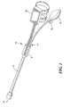
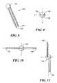
- FIG. 8is a perspective illustration of an embodiment of a cervical dilation sensor in accordance with the present invention.
- the medical device 10 of the present inventionmay include additional features providing safety, ease of use, and the like.
- the medical device 10may include a protective sheath 42 encasing at least a portion of the distal end 16 of the elongate body 12 .
- the sheath 42may include one or more layers of various materials to provide a water-tight seal around the medical device, as well as adding to patient comfort by having additional padding and/or a lubricious coating to ease positioning of the device.
- a distal pad 44may be coupled to the elongate body 12 at or near the distal end 16 , where the distal pad 44 may be contoured or shaped to conform to the curvature of the head of a baby.
- a precise dilation measurementmay be performed during the various stages of labor.
- the medical device 10in a deflated state, may be positioned such that the distal end 16 of the elongate body 12 is in proximity to the dilated region of the cervix 54 .
- Proper positioningcan be aided by feedback provided by the distal pressure sensor 46 when contacting the head of the baby, as well as monitoring the visual feedback from the camera 45 .
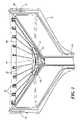
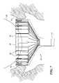
- the array of movable elements 24may be extended to contact the tissue of the cervix 54 , for example, by actuating the inflation source 22 to inflate the expandable element 38 .
- the inflation source 22may continue to inflate the expandable element 38 until the movable elements 24 of the medical device 10 come into contact with the dilated cervix 54 . Such contact can be indicated and monitored through information provided by the pressure sensors 32 coupled to the movable elements 24 .
- the control element 20which is in communication with the sensors, may include an algorithm or computational ability to determine if the pressure sensor feedback indicates a substantially uniform circular state. That is to say, that the pressure measurements from each of the pressure sensors 32 disposed about the movable elements 24 are approximately the same.
- the inflation source 22may be deactivated, or, alternatively, the exhaust valve 52 may be triggered to prevent additional fluid from entering the expandable element 38 .
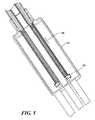
- a cervical dilation censor 100is provided to aid in the manual, two-finger approach of measuring cervical dilation.
- the cervical dilation sensor 100may include a first rod 102 , a second rod 104 , and a sensor housing 106 .
- the first and second rods 102 , 104may be rotatably and pivotably coupled to the sensor housing 106 , as to freely move about the housing in at least two planes of motion.
- the sensor housing 106may include one or more sensors coupled to the first and second rods 102 , 104 as to measure the movement of the two rods.
- the cervical dilation sensor 100may also include a control monitor (not shown) in communication with the one or more sensors in the sensor housing 106 for displaying and monitoring information provided by the sensors.
Landscapes
- Health & Medical Sciences (AREA)
- Life Sciences & Earth Sciences (AREA)
- Surgery (AREA)
- Biomedical Technology (AREA)
- General Health & Medical Sciences (AREA)
- Biophysics (AREA)
- Pathology (AREA)
- Engineering & Computer Science (AREA)
- Animal Behavior & Ethology (AREA)
- Heart & Thoracic Surgery (AREA)
- Medical Informatics (AREA)
- Molecular Biology (AREA)
- Physics & Mathematics (AREA)
- Veterinary Medicine (AREA)
- Public Health (AREA)
- Gynecology & Obstetrics (AREA)
- Dentistry (AREA)
- Pregnancy & Childbirth (AREA)
- Oral & Maxillofacial Surgery (AREA)
- Reproductive Health (AREA)
- Measurement Of The Respiration, Hearing Ability, Form, And Blood Characteristics Of Living Organisms (AREA)
- Surgical Instruments (AREA)
- Measuring Pulse, Heart Rate, Blood Pressure Or Blood Flow (AREA)
- Measuring And Recording Apparatus For Diagnosis (AREA)
- Measuring Fluid Pressure (AREA)
- Endoscopes (AREA)
Abstract
Description
Claims (1)
Priority Applications (14)
| Application Number | Priority Date | Filing Date | Title |
|---|---|---|---|
| US11/321,061US7527601B2 (en) | 2005-12-29 | 2005-12-29 | Cervimeter |
| US11/401,749US7811239B2 (en) | 2005-12-29 | 2006-04-11 | Cervical dilation measurement apparatus |
| US11/544,261US20070156068A1 (en) | 2005-12-29 | 2006-10-06 | Cervimetry control apparatus |
| EP06827882AEP1968445A2 (en) | 2005-12-29 | 2006-11-20 | Cervimeter |
| CA2635383ACA2635383C (en) | 2005-12-29 | 2006-11-20 | Cervical dilation measurement apparatus |
| EP06849898AEP1976432A2 (en) | 2005-12-29 | 2006-11-20 | Cervimetry control apparatus |
| PCT/US2006/044960WO2007078448A2 (en) | 2005-12-29 | 2006-11-20 | Cervimeter |
| CA2635712ACA2635712C (en) | 2005-12-29 | 2006-11-20 | Cervimetry control apparatus |
| PCT/US2006/045003WO2007089319A2 (en) | 2005-12-29 | 2006-11-20 | Cervimetry control apparatus |
| CA2635561ACA2635561C (en) | 2005-12-29 | 2006-11-20 | Cervimeter |
| PCT/US2006/044961WO2007078449A1 (en) | 2005-12-29 | 2006-11-20 | Cervical dilation measurement apparatus |
| EP06844448.8AEP1968446B1 (en) | 2005-12-29 | 2006-11-20 | Cervical dilation measurement apparatus |
| US12/015,291US7654970B2 (en) | 2005-12-29 | 2008-01-16 | Cervical dilation measurement apparatus |
| US12/061,287US7749176B2 (en) | 2005-12-29 | 2008-04-02 | Cervical dilation measurement apparatus |
Applications Claiming Priority (1)
| Application Number | Priority Date | Filing Date | Title |
|---|---|---|---|
| US11/321,061US7527601B2 (en) | 2005-12-29 | 2005-12-29 | Cervimeter |
Related Child Applications (1)
| Application Number | Title | Priority Date | Filing Date |
|---|---|---|---|
| US11/401,749Continuation-In-PartUS7811239B2 (en) | 2005-12-29 | 2006-04-11 | Cervical dilation measurement apparatus |
Publications (2)
| Publication Number | Publication Date |
|---|---|
| US20070156067A1 US20070156067A1 (en) | 2007-07-05 |
| US7527601B2true US7527601B2 (en) | 2009-05-05 |
Family
ID=37966478
Family Applications (2)
| Application Number | Title | Priority Date | Filing Date |
|---|---|---|---|
| US11/321,061Expired - Fee RelatedUS7527601B2 (en) | 2005-12-29 | 2005-12-29 | Cervimeter |
| US11/544,261AbandonedUS20070156068A1 (en) | 2005-12-29 | 2006-10-06 | Cervimetry control apparatus |
Family Applications After (1)
| Application Number | Title | Priority Date | Filing Date |
|---|---|---|---|
| US11/544,261AbandonedUS20070156068A1 (en) | 2005-12-29 | 2006-10-06 | Cervimetry control apparatus |
Country Status (4)
| Country | Link |
|---|---|
| US (2) | US7527601B2 (en) |
| EP (2) | EP1968446B1 (en) |
| CA (2) | CA2635561C (en) |
| WO (2) | WO2007078449A1 (en) |
Cited By (14)
| Publication number | Priority date | Publication date | Assignee | Title |
|---|---|---|---|---|
| US20100152767A1 (en)* | 2006-11-20 | 2010-06-17 | Septrx, Inc. | Mechanical Tissue Device and Method |
| US20100185123A1 (en)* | 2007-06-26 | 2010-07-22 | Diniz Zanetti Miriam R | Perineal elasticity meter |
| US9357905B2 (en) | 2012-06-01 | 2016-06-07 | Robert Molnar | Airway device, airway assist device and the method of using same |
| US9415179B2 (en) | 2012-06-01 | 2016-08-16 | Wm & Dg, Inc. | Medical device, and the methods of using same |
| US20160317794A1 (en)* | 2013-02-08 | 2016-11-03 | Aminex, Inc. | Surgical device with integrated visualization and cauterization |
| US9918618B2 (en) | 2014-08-08 | 2018-03-20 | Wm & Dg, Inc. | Medical devices and methods of placement |
| US10653307B2 (en) | 2018-10-10 | 2020-05-19 | Wm & Dg, Inc. | Medical devices for airway management and methods of placement |
| US10722110B2 (en) | 2014-08-08 | 2020-07-28 | Wm & Dg, Inc. | Medical devices and methods of placement |
| US11039926B2 (en) | 2016-03-25 | 2021-06-22 | Spiration, Inc. | Valve planning tool |
| US11051682B2 (en) | 2017-08-31 | 2021-07-06 | Wm & Dg, Inc. | Medical devices with camera and methods of placement |
| US11147442B2 (en) | 2014-08-08 | 2021-10-19 | Wm & Dg, Inc. | Medical devices and methods of placement |
| US11202561B2 (en) | 2014-08-08 | 2021-12-21 | Wm & Dg, Inc. | Medical devices and methods of placement |
| US11497394B2 (en) | 2020-10-12 | 2022-11-15 | Wm & Dg, Inc. | Laryngoscope and intubation methods |
| US12257387B2 (en) | 2018-10-10 | 2025-03-25 | Wm & Dg, Inc. | Medical devices for airway management and methods of placement |
Families Citing this family (11)
| Publication number | Priority date | Publication date | Assignee | Title |
|---|---|---|---|---|
| KR100822820B1 (en)* | 2007-11-13 | 2008-04-18 | 이신형 | Sphincter movement device and its control method |
| US8251925B2 (en)* | 2007-12-31 | 2012-08-28 | Personics Holdings Inc. | Device and method for radial pressure determination |
| CA2734512C (en)* | 2008-08-18 | 2020-06-30 | Glenveigh Medical, Llc | Cervical dilation meter |
| US8728013B2 (en)* | 2009-03-18 | 2014-05-20 | Contipi Medical Ltd. | Device and method for fitting a pessary |
| US20110190580A1 (en)* | 2009-09-28 | 2011-08-04 | Bennett James D | Analysis engine within a network supporting intravaginal monitoring |
| US8460217B2 (en) | 2009-09-29 | 2013-06-11 | Khashayar Shakiba | Electronic pelvic organ prolapse quantification system |
| CA2777526C (en)* | 2009-10-13 | 2018-11-13 | Materna Medical, Inc. | Methods and apparatus for preventing vaginal lacerations during childbirth |
| US10207090B2 (en)* | 2012-03-28 | 2019-02-19 | Board Of Regents Of The University Of Texas System | Advanced cervical ripening system |
| US10716672B2 (en)* | 2015-04-07 | 2020-07-21 | St. Jude Medical, Cardiology Division, Inc. | System and method for intraprocedural assessment of geometry and compliance of valve annulus for trans-catheter valve implantation |
| EP3319534B1 (en) | 2015-07-10 | 2025-05-28 | Materna Medical, Inc. | Systems for the treatment and prevention of female pelvic dysfunction |
| US20200022727A1 (en)* | 2017-02-01 | 2020-01-23 | Partus Llc | Systems, devices, and methods for treating and monitoring a pregnant patient having a prematurely open cervix |
Citations (79)
| Publication number | Priority date | Publication date | Assignee | Title |
|---|---|---|---|---|
| US2642672A (en) | 1950-10-09 | 1953-06-23 | Ohio Brass Co | Hole gauge |
| US2924220A (en) | 1959-02-20 | 1960-02-09 | Micsky Lajos I Von | Cervicometer |
| US3273559A (en) | 1963-08-28 | 1966-09-20 | Conductron Corp | Method and apparatus for monitoring the approach of birth |
| US3312222A (en) | 1963-09-03 | 1967-04-04 | Allen And Hanburys Surgical In | Obstetrical cervical dilator with rotary, expansible arms |
| US3606879A (en) | 1968-06-17 | 1971-09-21 | Electro Medical Systems Inc | Monitoring the physiological phenomena of childbirth with ultrasound |
| US3626949A (en) | 1969-01-23 | 1971-12-14 | Wallace B Shute | Cervical dilator |
| US3768459A (en) | 1971-06-28 | 1973-10-30 | Utah Res & Dev Co Inc | Cervical dilation measuring device |
| US4141345A (en) | 1976-01-08 | 1979-02-27 | National Research Development Corporation | Cervical dilation measurement instruments |
| US4207902A (en) | 1978-11-08 | 1980-06-17 | Yury Krementsov | Instrument and method of measuring dilatation of cervix uteri |
| US4245656A (en) | 1979-02-14 | 1981-01-20 | Farr Larry D | Obstetric gloves |
| US4362167A (en) | 1981-02-06 | 1982-12-07 | Nicolai Donald R | Diagnostic measuring instrument |
| GB2137499A (en) | 1983-04-07 | 1984-10-10 | Univ Paris | Probe with variable geometry for measuring the radial strains in a sphincter of a living organism |
| US4611603A (en) | 1985-02-28 | 1986-09-16 | Kelso Jimmie J | Calibrated examining glove |
| US4682609A (en) | 1985-11-25 | 1987-07-28 | Natan Parsons | Cervical-dilation meter |
| US4719925A (en) | 1985-11-25 | 1988-01-19 | Natan Parsons | Cervical-dilation meter |
| US4805628A (en) | 1982-12-06 | 1989-02-21 | Indianapolis Center For Advanced Research, Inc. | Ultrasound contrast media for medically implantable and insertable devices |
| US4986980A (en) | 1984-11-01 | 1991-01-22 | Nycomed As | Water-soluble, carrier-bound paramagnetic metal containing diagnostic agents |
| US5143505A (en) | 1991-02-26 | 1992-09-01 | Rutgers University | Actuator system for providing force feedback to a dextrous master glove |
| DE4137751A1 (en) | 1991-11-16 | 1993-05-19 | Luis Frontela Carreras | Instrument for hymen measuring and examining - consists of inflatable head piece insertable into vagina through opening |
| US5222485A (en) | 1990-09-17 | 1993-06-29 | Ravinder Jerath | Ultrasound labor monitoring method and apparatus |
| US5275169A (en) | 1992-01-15 | 1994-01-04 | Innovation Associates | Apparatus and method for determining physiologic characteristics of body lumens |
| US5301680A (en) | 1992-12-09 | 1994-04-12 | Hygeia Biomedical Research Inc. | Apparatus and method for the diagnosis of labor |
| US5354162A (en) | 1991-02-26 | 1994-10-11 | Rutgers University | Actuator system for providing force feedback to portable master support |
| US5373852A (en) | 1993-06-25 | 1994-12-20 | The Regents Of The University Of California | Monitoring uterine contractions by radiotelemetric transmission |
| US5373846A (en) | 1992-10-06 | 1994-12-20 | Molecular Biosystems, Inc. | Method of ultrasonic imaging of body cavities |
| US5405356A (en) | 1993-06-30 | 1995-04-11 | Jcs Biomedical, Inc. | Child-birth assisting system |
| US5406961A (en) | 1993-10-29 | 1995-04-18 | Artal; Raul | Monitoring device and method for detection of premature labor |
| US5438996A (en) | 1994-10-12 | 1995-08-08 | Triton Technology, Inc. | Ambulatory, ultrasonic transit time, real-time, cervical effacement and dilatation monitor with disposable probes |
| US5450857A (en) | 1994-05-19 | 1995-09-19 | Board Of Regents, The University Of Texas System | Method for the diagnosis of cervical changes |
| EP0752233A1 (en) | 1995-07-07 | 1997-01-08 | Dani Sherman | Method of monitoring cervical dilatation during labor and ultrasound transducer particularly useful in such method |
| US5658295A (en) | 1996-06-03 | 1997-08-19 | Krementsov; Yury | Instrument for measuring dilatation of cervix uteri |
| WO1997042871A1 (en) | 1996-05-10 | 1997-11-20 | Cardiovascular Concepts, Inc. | Lesion diameter measurement catheter and method |
| WO1998009565A1 (en) | 1996-09-05 | 1998-03-12 | Ob Innovations, Inc. | Method and apparatus for monitoring cervical diameter |
| US5807376A (en) | 1994-06-24 | 1998-09-15 | United States Surgical Corporation | Apparatus and method for performing surgical tasks during laparoscopic procedures |
| US5807281A (en) | 1996-10-01 | 1998-09-15 | Welch; Robert A. | Cervical ring to detect labor |
| US5829438A (en) | 1994-10-12 | 1998-11-03 | Gibbs; David L. | System and method for the infusing of tocolytic drugs in response to the onset of premature labor detected by ultrasonic monitoring of the dilatation and/or effacement of the cervix os |
| US5851188A (en) | 1994-10-12 | 1998-12-22 | Bullard; Kelli M. | Device for holding medical instrumentation sensors at and upon the cervix os of a human female, particularly for holding the ultrasonic transducers of an ultrasonic transit time, real-time, cervical effacement and dilatation monitor |
| US5867831A (en) | 1997-04-28 | 1999-02-09 | Husain; Abbas M. | Examination glove with palpable markings |
| US5876357A (en) | 1997-11-20 | 1999-03-02 | Labor Control System (L.C.S.) Ltd. | Uterine cervix dilation, effacement, and consistency monitoring system |
| US5935061A (en) | 1997-01-03 | 1999-08-10 | Biosense, Inc. | Obstetrical instrument system and method |
| US6066104A (en) | 1996-03-05 | 2000-05-23 | Dao; Leland H. | Device for cervical and pelvic measurement in medical obstetrics |
| US6110200A (en)* | 1995-06-07 | 2000-08-29 | St. Jude Medical, Inc. | Adjustable sizing apparatus |
| US6123923A (en) | 1997-12-18 | 2000-09-26 | Imarx Pharmaceutical Corp. | Optoacoustic contrast agents and methods for their use |
| US6200279B1 (en) | 1997-05-05 | 2001-03-13 | Ultra-Guide Ltd. | Method and apparatus monitoring the progress of labor |
| US6231834B1 (en) | 1995-06-07 | 2001-05-15 | Imarx Pharmaceutical Corp. | Methods for ultrasound imaging involving the use of a contrast agent and multiple images and processing of same |
| US6261247B1 (en) | 1998-12-31 | 2001-07-17 | Ball Semiconductor, Inc. | Position sensing system |
| US6270458B1 (en) | 1999-03-05 | 2001-08-07 | Barnev Inc. | Cervix dilation and labor progression monitor |
| US20010039388A1 (en) | 2000-03-10 | 2001-11-08 | Korotko Joseph R. | Apparatus for measuring the length and width of blood vessels and other body lumens |
| US20010040550A1 (en) | 1998-03-12 | 2001-11-15 | Scott Vance | Multiple pressure sensors per finger of glove for virtual full typing |
| US6363271B1 (en) | 2000-11-01 | 2002-03-26 | Daniel K. Berry | Amniotic fluid alerting device |
| US6383137B1 (en) | 2000-11-01 | 2002-05-07 | Daniel K. Berry | Labor alerting device |
| US6419646B1 (en) | 2000-04-10 | 2002-07-16 | Cervilenz | Devices and methods for cervix measurement |
| US6423000B1 (en) | 2000-11-01 | 2002-07-23 | Daniel K. Berry | Labor monitoring device |
| US6423016B1 (en) | 2000-06-08 | 2002-07-23 | Lms Medical Systems Ltd. | System and method for evaluating labor progress during childbirth |
| US6450977B1 (en) | 2000-04-10 | 2002-09-17 | Cervilenz | Devices and methods for cervix measurement |
| US6524259B2 (en) | 2001-06-08 | 2003-02-25 | Cervilenz, Inc. | Devices and methods for cervix measurement |
| US6526669B2 (en) | 2000-04-26 | 2003-03-04 | Agency Of Industrial Science And Technology Ministry Of International Trade And Industry | Apparatus for acquiring human finger manipulation data |
| US6567990B1 (en) | 2002-02-26 | 2003-05-27 | Richard James Spitznagle | Electromyographic examination glove |
| US6569108B2 (en) | 2001-03-28 | 2003-05-27 | Profile, Llc | Real time mechanical imaging of the prostate |
| US20030114779A1 (en) | 1997-05-05 | 2003-06-19 | Yoav Paltieli | Method and apparatus for monitoring the progress of labor |
| US6592315B2 (en) | 2000-05-08 | 2003-07-15 | William Joseph Osborne, Jr. | Self-feeding apparatus with hover mode |
| US20030229267A1 (en) | 2000-12-15 | 2003-12-11 | Amir Belson | Obstetrical imaging system and integrated fetal vacuum extraction system |
| WO2004006767A2 (en) | 2002-07-15 | 2004-01-22 | Spiration, Inc. | Device and method for measuring the diameter of an air passageway |
| US20040068203A1 (en) | 2002-10-03 | 2004-04-08 | Scimed Life Systems, Inc. | Sensing pressure |
| WO2004062526A2 (en) | 2003-01-16 | 2004-07-29 | Galil Medical Ltd. | Device, system, and method for detecting, localizing, and characterizing plaque-induced stenosis of a blood vessel |
| US20040210136A1 (en) | 2003-04-21 | 2004-10-21 | Tomy Varghese | Method and apparatus for imaging the cervix and uterine wall |
| US20040225235A1 (en) | 2003-05-07 | 2004-11-11 | Miki Ben-Cnaan | Cervical dilation monitor |
| US20040236193A1 (en) | 2001-06-05 | 2004-11-25 | Yehuda Sharf | Birth monitoring system |
| US20050038340A1 (en) | 1998-09-18 | 2005-02-17 | University Of Washington | Use of contrast agents to increase the effectiveness of high intensity focused ultrasound therapy |
| US20050049509A1 (en) | 2003-08-28 | 2005-03-03 | Mansour Hebah Noshy | Cervix monitoring system and related devices and methods |
| US20050055043A1 (en) | 2002-12-12 | 2005-03-10 | Os Technology, Llc. | Cervical canal dilator |
| US6866643B2 (en) | 1992-07-06 | 2005-03-15 | Immersion Corporation | Determination of finger position |
| US20050113852A1 (en)* | 2003-11-20 | 2005-05-26 | Vascular Control Systems, Inc. | Uterine artery occlusion device with cervical receptacle |
| WO2005070061A2 (en) | 1999-11-16 | 2005-08-04 | Barrx Medical, Inc. | Methods and systems for determining physiologic characteristics for treatment of the esophagus |
| WO2005084745A1 (en) | 2004-03-08 | 2005-09-15 | Medilator | Catheter and method for dilating a body passageway |
| US20060064038A1 (en)* | 2003-02-12 | 2006-03-23 | Nihon University | Device for measuring elastic characteristics of organism tissue |
| US7042438B2 (en) | 2003-09-06 | 2006-05-09 | Mcrae Michael William | Hand manipulated data apparatus for computers and video games |
| US20060129070A1 (en) | 2004-11-05 | 2006-06-15 | Pearl Michael L | Fingertip tracker |
| US7150108B2 (en) | 2004-08-05 | 2006-12-19 | Babb Pamela E | Obstetric calibration guide |
Family Cites Families (5)
| Publication number | Priority date | Publication date | Assignee | Title |
|---|---|---|---|---|
| US5345927A (en)* | 1990-03-02 | 1994-09-13 | Bonutti Peter M | Arthroscopic retractors |
| US5383137A (en)* | 1992-12-03 | 1995-01-17 | Motorola, Inc. | Emulation system and method for development of a low power data processor |
| CA2287971C (en)* | 1997-04-29 | 2007-04-24 | Raymond F. Lippitt | Annularly expanding and retracting gripping and releasing mechanism |
| US6524249B2 (en)* | 1998-11-11 | 2003-02-25 | Spentech, Inc. | Doppler ultrasound method and apparatus for monitoring blood flow and detecting emboli |
| DE19900795C2 (en)* | 1999-01-12 | 2003-01-30 | Fraunhofer Ges Forschung | forceps |
- 2005
- 2005-12-29USUS11/321,061patent/US7527601B2/ennot_activeExpired - Fee Related
- 2006
- 2006-10-06USUS11/544,261patent/US20070156068A1/ennot_activeAbandoned
- 2006-11-20CACA2635561Apatent/CA2635561C/ennot_activeExpired - Fee Related
- 2006-11-20EPEP06844448.8Apatent/EP1968446B1/ennot_activeNot-in-force
- 2006-11-20EPEP06827882Apatent/EP1968445A2/ennot_activeWithdrawn
- 2006-11-20WOPCT/US2006/044961patent/WO2007078449A1/enactiveApplication Filing
- 2006-11-20CACA2635383Apatent/CA2635383C/ennot_activeExpired - Fee Related
- 2006-11-20WOPCT/US2006/044960patent/WO2007078448A2/enactiveApplication Filing
Patent Citations (85)
| Publication number | Priority date | Publication date | Assignee | Title |
|---|---|---|---|---|
| US2642672A (en) | 1950-10-09 | 1953-06-23 | Ohio Brass Co | Hole gauge |
| US2924220A (en) | 1959-02-20 | 1960-02-09 | Micsky Lajos I Von | Cervicometer |
| US3273559A (en) | 1963-08-28 | 1966-09-20 | Conductron Corp | Method and apparatus for monitoring the approach of birth |
| US3312222A (en) | 1963-09-03 | 1967-04-04 | Allen And Hanburys Surgical In | Obstetrical cervical dilator with rotary, expansible arms |
| US3606879A (en) | 1968-06-17 | 1971-09-21 | Electro Medical Systems Inc | Monitoring the physiological phenomena of childbirth with ultrasound |
| US3626949A (en) | 1969-01-23 | 1971-12-14 | Wallace B Shute | Cervical dilator |
| US3768459A (en) | 1971-06-28 | 1973-10-30 | Utah Res & Dev Co Inc | Cervical dilation measuring device |
| US4141345A (en) | 1976-01-08 | 1979-02-27 | National Research Development Corporation | Cervical dilation measurement instruments |
| US4207902A (en) | 1978-11-08 | 1980-06-17 | Yury Krementsov | Instrument and method of measuring dilatation of cervix uteri |
| US4245656A (en) | 1979-02-14 | 1981-01-20 | Farr Larry D | Obstetric gloves |
| US4362167A (en) | 1981-02-06 | 1982-12-07 | Nicolai Donald R | Diagnostic measuring instrument |
| US4805628A (en) | 1982-12-06 | 1989-02-21 | Indianapolis Center For Advanced Research, Inc. | Ultrasound contrast media for medically implantable and insertable devices |
| GB2137499A (en) | 1983-04-07 | 1984-10-10 | Univ Paris | Probe with variable geometry for measuring the radial strains in a sphincter of a living organism |
| US4986980A (en) | 1984-11-01 | 1991-01-22 | Nycomed As | Water-soluble, carrier-bound paramagnetic metal containing diagnostic agents |
| US4611603A (en) | 1985-02-28 | 1986-09-16 | Kelso Jimmie J | Calibrated examining glove |
| US4682609A (en) | 1985-11-25 | 1987-07-28 | Natan Parsons | Cervical-dilation meter |
| US4719925A (en) | 1985-11-25 | 1988-01-19 | Natan Parsons | Cervical-dilation meter |
| US5222485A (en) | 1990-09-17 | 1993-06-29 | Ravinder Jerath | Ultrasound labor monitoring method and apparatus |
| US5143505A (en) | 1991-02-26 | 1992-09-01 | Rutgers University | Actuator system for providing force feedback to a dextrous master glove |
| US5354162A (en) | 1991-02-26 | 1994-10-11 | Rutgers University | Actuator system for providing force feedback to portable master support |
| DE4137751A1 (en) | 1991-11-16 | 1993-05-19 | Luis Frontela Carreras | Instrument for hymen measuring and examining - consists of inflatable head piece insertable into vagina through opening |
| US5275169A (en) | 1992-01-15 | 1994-01-04 | Innovation Associates | Apparatus and method for determining physiologic characteristics of body lumens |
| US6866643B2 (en) | 1992-07-06 | 2005-03-15 | Immersion Corporation | Determination of finger position |
| US5373846A (en) | 1992-10-06 | 1994-12-20 | Molecular Biosystems, Inc. | Method of ultrasonic imaging of body cavities |
| US5301680A (en) | 1992-12-09 | 1994-04-12 | Hygeia Biomedical Research Inc. | Apparatus and method for the diagnosis of labor |
| US5373852A (en) | 1993-06-25 | 1994-12-20 | The Regents Of The University Of California | Monitoring uterine contractions by radiotelemetric transmission |
| US5405356A (en) | 1993-06-30 | 1995-04-11 | Jcs Biomedical, Inc. | Child-birth assisting system |
| US5406961A (en) | 1993-10-29 | 1995-04-18 | Artal; Raul | Monitoring device and method for detection of premature labor |
| US5450857A (en) | 1994-05-19 | 1995-09-19 | Board Of Regents, The University Of Texas System | Method for the diagnosis of cervical changes |
| US5807376A (en) | 1994-06-24 | 1998-09-15 | United States Surgical Corporation | Apparatus and method for performing surgical tasks during laparoscopic procedures |
| US5438996A (en) | 1994-10-12 | 1995-08-08 | Triton Technology, Inc. | Ambulatory, ultrasonic transit time, real-time, cervical effacement and dilatation monitor with disposable probes |
| US5829438A (en) | 1994-10-12 | 1998-11-03 | Gibbs; David L. | System and method for the infusing of tocolytic drugs in response to the onset of premature labor detected by ultrasonic monitoring of the dilatation and/or effacement of the cervix os |
| US5851188A (en) | 1994-10-12 | 1998-12-22 | Bullard; Kelli M. | Device for holding medical instrumentation sensors at and upon the cervix os of a human female, particularly for holding the ultrasonic transducers of an ultrasonic transit time, real-time, cervical effacement and dilatation monitor |
| US6110200A (en)* | 1995-06-07 | 2000-08-29 | St. Jude Medical, Inc. | Adjustable sizing apparatus |
| US6231834B1 (en) | 1995-06-07 | 2001-05-15 | Imarx Pharmaceutical Corp. | Methods for ultrasound imaging involving the use of a contrast agent and multiple images and processing of same |
| US5713371A (en) | 1995-07-07 | 1998-02-03 | Sherman; Dani | Method of monitoring cervical dilatation during labor, and ultrasound transducer particularly useful in such method |
| EP0752233A1 (en) | 1995-07-07 | 1997-01-08 | Dani Sherman | Method of monitoring cervical dilatation during labor and ultrasound transducer particularly useful in such method |
| US6066104A (en) | 1996-03-05 | 2000-05-23 | Dao; Leland H. | Device for cervical and pelvic measurement in medical obstetrics |
| WO1997042871A1 (en) | 1996-05-10 | 1997-11-20 | Cardiovascular Concepts, Inc. | Lesion diameter measurement catheter and method |
| US5658295A (en) | 1996-06-03 | 1997-08-19 | Krementsov; Yury | Instrument for measuring dilatation of cervix uteri |
| US6039701A (en) | 1996-09-05 | 2000-03-21 | Ob Inovations, Inc. | Method and apparatus for monitoring cervical diameter |
| WO1998009565A1 (en) | 1996-09-05 | 1998-03-12 | Ob Innovations, Inc. | Method and apparatus for monitoring cervical diameter |
| US5807281A (en) | 1996-10-01 | 1998-09-15 | Welch; Robert A. | Cervical ring to detect labor |
| US5935061A (en) | 1997-01-03 | 1999-08-10 | Biosense, Inc. | Obstetrical instrument system and method |
| US5867831A (en) | 1997-04-28 | 1999-02-09 | Husain; Abbas M. | Examination glove with palpable markings |
| US6200279B1 (en) | 1997-05-05 | 2001-03-13 | Ultra-Guide Ltd. | Method and apparatus monitoring the progress of labor |
| US6669653B2 (en) | 1997-05-05 | 2003-12-30 | Trig Medical Ltd. | Method and apparatus for monitoring the progress of labor |
| US20030114779A1 (en) | 1997-05-05 | 2003-06-19 | Yoav Paltieli | Method and apparatus for monitoring the progress of labor |
| US5876357A (en) | 1997-11-20 | 1999-03-02 | Labor Control System (L.C.S.) Ltd. | Uterine cervix dilation, effacement, and consistency monitoring system |
| US6123923A (en) | 1997-12-18 | 2000-09-26 | Imarx Pharmaceutical Corp. | Optoacoustic contrast agents and methods for their use |
| US20010040550A1 (en) | 1998-03-12 | 2001-11-15 | Scott Vance | Multiple pressure sensors per finger of glove for virtual full typing |
| US20050038340A1 (en) | 1998-09-18 | 2005-02-17 | University Of Washington | Use of contrast agents to increase the effectiveness of high intensity focused ultrasound therapy |
| US6261247B1 (en) | 1998-12-31 | 2001-07-17 | Ball Semiconductor, Inc. | Position sensing system |
| US6270458B1 (en) | 1999-03-05 | 2001-08-07 | Barnev Inc. | Cervix dilation and labor progression monitor |
| WO2005070061A2 (en) | 1999-11-16 | 2005-08-04 | Barrx Medical, Inc. | Methods and systems for determining physiologic characteristics for treatment of the esophagus |
| US20010039388A1 (en) | 2000-03-10 | 2001-11-08 | Korotko Joseph R. | Apparatus for measuring the length and width of blood vessels and other body lumens |
| US6450977B1 (en) | 2000-04-10 | 2002-09-17 | Cervilenz | Devices and methods for cervix measurement |
| US6419646B1 (en) | 2000-04-10 | 2002-07-16 | Cervilenz | Devices and methods for cervix measurement |
| US6526669B2 (en) | 2000-04-26 | 2003-03-04 | Agency Of Industrial Science And Technology Ministry Of International Trade And Industry | Apparatus for acquiring human finger manipulation data |
| US6592315B2 (en) | 2000-05-08 | 2003-07-15 | William Joseph Osborne, Jr. | Self-feeding apparatus with hover mode |
| US6423016B1 (en) | 2000-06-08 | 2002-07-23 | Lms Medical Systems Ltd. | System and method for evaluating labor progress during childbirth |
| US6423000B1 (en) | 2000-11-01 | 2002-07-23 | Daniel K. Berry | Labor monitoring device |
| US6363271B1 (en) | 2000-11-01 | 2002-03-26 | Daniel K. Berry | Amniotic fluid alerting device |
| US6383137B1 (en) | 2000-11-01 | 2002-05-07 | Daniel K. Berry | Labor alerting device |
| US20030229267A1 (en) | 2000-12-15 | 2003-12-11 | Amir Belson | Obstetrical imaging system and integrated fetal vacuum extraction system |
| US6569108B2 (en) | 2001-03-28 | 2003-05-27 | Profile, Llc | Real time mechanical imaging of the prostate |
| US20040236193A1 (en) | 2001-06-05 | 2004-11-25 | Yehuda Sharf | Birth monitoring system |
| US6524259B2 (en) | 2001-06-08 | 2003-02-25 | Cervilenz, Inc. | Devices and methods for cervix measurement |
| US6802817B2 (en) | 2001-06-08 | 2004-10-12 | Cervilenz, Inc. | Devices and methods for cervix measurement |
| US20050027215A1 (en) | 2001-06-08 | 2005-02-03 | Baxter-Jones Rosalyn P. | Devices and methods for cervix measurement |
| US6567990B1 (en) | 2002-02-26 | 2003-05-27 | Richard James Spitznagle | Electromyographic examination glove |
| WO2004006767A2 (en) | 2002-07-15 | 2004-01-22 | Spiration, Inc. | Device and method for measuring the diameter of an air passageway |
| US20040068203A1 (en) | 2002-10-03 | 2004-04-08 | Scimed Life Systems, Inc. | Sensing pressure |
| US20050055043A1 (en) | 2002-12-12 | 2005-03-10 | Os Technology, Llc. | Cervical canal dilator |
| WO2004062526A2 (en) | 2003-01-16 | 2004-07-29 | Galil Medical Ltd. | Device, system, and method for detecting, localizing, and characterizing plaque-induced stenosis of a blood vessel |
| US20060064038A1 (en)* | 2003-02-12 | 2006-03-23 | Nihon University | Device for measuring elastic characteristics of organism tissue |
| US20040210136A1 (en) | 2003-04-21 | 2004-10-21 | Tomy Varghese | Method and apparatus for imaging the cervix and uterine wall |
| US20040225235A1 (en) | 2003-05-07 | 2004-11-11 | Miki Ben-Cnaan | Cervical dilation monitor |
| US20050049509A1 (en) | 2003-08-28 | 2005-03-03 | Mansour Hebah Noshy | Cervix monitoring system and related devices and methods |
| WO2005020814A1 (en) | 2003-08-28 | 2005-03-10 | Hebah Noshy Mansour | Cervix monitoring system and related devices and methods |
| US7042438B2 (en) | 2003-09-06 | 2006-05-09 | Mcrae Michael William | Hand manipulated data apparatus for computers and video games |
| US20050113852A1 (en)* | 2003-11-20 | 2005-05-26 | Vascular Control Systems, Inc. | Uterine artery occlusion device with cervical receptacle |
| WO2005084745A1 (en) | 2004-03-08 | 2005-09-15 | Medilator | Catheter and method for dilating a body passageway |
| US7150108B2 (en) | 2004-08-05 | 2006-12-19 | Babb Pamela E | Obstetric calibration guide |
| US20060129070A1 (en) | 2004-11-05 | 2006-06-15 | Pearl Michael L | Fingertip tracker |
Cited By (22)
| Publication number | Priority date | Publication date | Assignee | Title |
|---|---|---|---|---|
| US20100152767A1 (en)* | 2006-11-20 | 2010-06-17 | Septrx, Inc. | Mechanical Tissue Device and Method |
| US20100185123A1 (en)* | 2007-06-26 | 2010-07-22 | Diniz Zanetti Miriam R | Perineal elasticity meter |
| US8512261B2 (en)* | 2007-06-26 | 2013-08-20 | Miriam R. Diniz Zanetti | Perineal elasticity meter |
| US10279136B2 (en) | 2012-06-01 | 2019-05-07 | Wm & Dg Inc. | Method of opening an airway of a patient by a medical professional in a medical procedure using an airway device |
| US9357905B2 (en) | 2012-06-01 | 2016-06-07 | Robert Molnar | Airway device, airway assist device and the method of using same |
| US9415179B2 (en) | 2012-06-01 | 2016-08-16 | Wm & Dg, Inc. | Medical device, and the methods of using same |
| US10342944B2 (en) | 2012-06-01 | 2019-07-09 | Wm & Dg, Inc. | Airway device with camera |
| US11154696B2 (en) | 2013-02-08 | 2021-10-26 | Arrinex, Inc. | Surgical device with integrated visualization and cauterization |
| US10201687B2 (en)* | 2013-02-08 | 2019-02-12 | Arrinex, Inc. | Surgical device with integrated visualization and cauterization |
| US20160317794A1 (en)* | 2013-02-08 | 2016-11-03 | Aminex, Inc. | Surgical device with integrated visualization and cauterization |
| US11147442B2 (en) | 2014-08-08 | 2021-10-19 | Wm & Dg, Inc. | Medical devices and methods of placement |
| US10722110B2 (en) | 2014-08-08 | 2020-07-28 | Wm & Dg, Inc. | Medical devices and methods of placement |
| US9918618B2 (en) | 2014-08-08 | 2018-03-20 | Wm & Dg, Inc. | Medical devices and methods of placement |
| US11202561B2 (en) | 2014-08-08 | 2021-12-21 | Wm & Dg, Inc. | Medical devices and methods of placement |
| US11633093B2 (en) | 2014-08-08 | 2023-04-25 | Wm & Dg, Inc. | Medical devices and methods of placement |
| US11039926B2 (en) | 2016-03-25 | 2021-06-22 | Spiration, Inc. | Valve planning tool |
| US11826255B2 (en) | 2016-03-25 | 2023-11-28 | Gyrus Acmi, Inc. | Valve planning tool |
| US11051682B2 (en) | 2017-08-31 | 2021-07-06 | Wm & Dg, Inc. | Medical devices with camera and methods of placement |
| US10653307B2 (en) | 2018-10-10 | 2020-05-19 | Wm & Dg, Inc. | Medical devices for airway management and methods of placement |
| US11628036B2 (en) | 2018-10-10 | 2023-04-18 | Wm & Dg, Inc. | Medical devices for airway management and methods of placement |
| US12257387B2 (en) | 2018-10-10 | 2025-03-25 | Wm & Dg, Inc. | Medical devices for airway management and methods of placement |
| US11497394B2 (en) | 2020-10-12 | 2022-11-15 | Wm & Dg, Inc. | Laryngoscope and intubation methods |
Also Published As
| Publication number | Publication date |
|---|---|
| CA2635383C (en) | 2011-09-20 |
| EP1968445A2 (en) | 2008-09-17 |
| CA2635561C (en) | 2012-01-24 |
| WO2007078448A3 (en) | 2007-10-04 |
| US20070156067A1 (en) | 2007-07-05 |
| EP1968446A1 (en) | 2008-09-17 |
| US20070156068A1 (en) | 2007-07-05 |
| CA2635383A1 (en) | 2007-07-12 |
| EP1968446B1 (en) | 2014-07-30 |
| WO2007078449A1 (en) | 2007-07-12 |
| CA2635561A1 (en) | 2007-07-12 |
| WO2007078448A2 (en) | 2007-07-12 |
| WO2007078449B1 (en) | 2007-08-23 |
Similar Documents
| Publication | Publication Date | Title |
|---|---|---|
| CA2635561C (en) | Cervimeter | |
| US7654970B2 (en) | Cervical dilation measurement apparatus | |
| US7713216B2 (en) | Method for cervical dilation and/or measurement | |
| US7947001B1 (en) | Methods and devices for measuring structural and elastic properties of a hollow organ | |
| US8348864B2 (en) | Uterine cavity length measurement | |
| US20170209057A1 (en) | Systems, devices and methods for assessment of body cavity pressures | |
| JP2001523507A (en) | Cervical dilatation, retraction and consistency monitoring system | |
| US6966881B2 (en) | Cervical dilation monitor | |
| CN210612678U (en) | Uterine balloon stent with movable sheath | |
| CA2635712C (en) | Cervimetry control apparatus | |
| EP4103044B1 (en) | Device for detecting prostate features | |
| CN114795156A (en) | A measuring device for in vivo | |
| US20070179410A1 (en) | Cervigage | |
| US20190343449A1 (en) | Method and system for monitoring cervix dilation to a desired dimension | |
| CN120323926A (en) | An automatic pelvic organ prolapse measurement and evaluation system | |
| IL171571A (en) | Cervical dilation monitor |
Legal Events
| Date | Code | Title | Description |
|---|---|---|---|
| AS | Assignment | Owner name:INTRAPARTUM VENTURES, LLC, FLORIDA Free format text:ASSIGNMENT OF ASSIGNORS INTEREST;ASSIGNORS:DUBEY, DHARMESH;BAIRD, TIM;REEL/FRAME:017632/0986 Effective date:20051216 | |
| STCF | Information on status: patent grant | Free format text:PATENTED CASE | |
| AS | Assignment | Owner name:INTRAPARTUM, LLC, FLORIDA Free format text:CHANGE OF NAME;ASSIGNOR:INTRAPARTUM VENTURES, LLC;REEL/FRAME:023115/0262 Effective date:20060519 Owner name:INTRAPARTUM, LLC,FLORIDA Free format text:CHANGE OF NAME;ASSIGNOR:INTRAPARTUM VENTURES, LLC;REEL/FRAME:023115/0262 Effective date:20060519 | |
| FPAY | Fee payment | Year of fee payment:4 | |
| REMI | Maintenance fee reminder mailed | ||
| FPAY | Fee payment | Year of fee payment:8 | |
| SULP | Surcharge for late payment | Year of fee payment:7 | |
| FEPP | Fee payment procedure | Free format text:MAINTENANCE FEE REMINDER MAILED (ORIGINAL EVENT CODE: REM.); ENTITY STATUS OF PATENT OWNER: SMALL ENTITY | |
| LAPS | Lapse for failure to pay maintenance fees | Free format text:PATENT EXPIRED FOR FAILURE TO PAY MAINTENANCE FEES (ORIGINAL EVENT CODE: EXP.); ENTITY STATUS OF PATENT OWNER: SMALL ENTITY | |
| STCH | Information on status: patent discontinuation | Free format text:PATENT EXPIRED DUE TO NONPAYMENT OF MAINTENANCE FEES UNDER 37 CFR 1.362 | |
| FP | Lapsed due to failure to pay maintenance fee | Effective date:20210505 |