US7526342B2 - Apparatus for endoscopic cardiac mapping and lead placement - Google Patents
Apparatus for endoscopic cardiac mapping and lead placementDownload PDFInfo
- Publication number
- US7526342B2 US7526342B2US10/697,906US69790603AUS7526342B2US 7526342 B2US7526342 B2US 7526342B2US 69790603 AUS69790603 AUS 69790603AUS 7526342 B2US7526342 B2US 7526342B2
- Authority
- US
- United States
- Prior art keywords
- heart
- suction
- cardiac lead
- channel
- cardiac
- Prior art date
- Legal status (The legal status is an assumption and is not a legal conclusion. Google has not performed a legal analysis and makes no representation as to the accuracy of the status listed.)
- Expired - Fee Related, expires
Links
- 230000000747cardiac effectEffects0.000titleclaimsabstractdescription115
- 238000013507mappingMethods0.000titleabstractdescription6
- 210000002216heartAnatomy0.000claimsabstractdescription68
- 238000001356surgical procedureMethods0.000claimsdescription12
- 230000013011matingEffects0.000claimsdescription2
- 238000010009beatingMethods0.000abstractdescription21
- 238000000034methodMethods0.000abstractdescription10
- 238000004873anchoringMethods0.000abstractdescription7
- 230000000004hemodynamic effectEffects0.000abstractdescription6
- 238000004458analytical methodMethods0.000abstractdescription3
- 230000002708enhancing effectEffects0.000abstract2
- 210000004165myocardiumAnatomy0.000description34
- 238000003780insertionMethods0.000description24
- 230000037431insertionEffects0.000description24
- 210000003516pericardiumAnatomy0.000description12
- 210000001519tissueAnatomy0.000description10
- 230000001862defibrillatory effectEffects0.000description8
- 238000012800visualizationMethods0.000description8
- 230000001225therapeutic effectEffects0.000description7
- 230000000007visual effectEffects0.000description6
- 239000004020conductorSubstances0.000description5
- 210000000038chestAnatomy0.000description4
- 210000005240left ventricleAnatomy0.000description3
- 210000004224pleuraAnatomy0.000description3
- 210000003281pleural cavityAnatomy0.000description3
- 208000032843HemorrhageDiseases0.000description2
- 238000009125cardiac resynchronization therapyMethods0.000description2
- 210000004351coronary vesselAnatomy0.000description2
- 239000012530fluidSubstances0.000description2
- 230000002262irrigationEffects0.000description2
- 238000003973irrigationMethods0.000description2
- 230000028161membrane depolarizationEffects0.000description2
- 208000005907mitral valve insufficiencyDiseases0.000description2
- 208000010125myocardial infarctionDiseases0.000description2
- 210000003462veinAnatomy0.000description2
- 241001631457CannulaSpecies0.000description1
- 208000006017Cardiac TamponadeDiseases0.000description1
- FAPWRFPIFSIZLT-UHFFFAOYSA-MSodium chlorideChemical compound[Na+].[Cl-]FAPWRFPIFSIZLT-UHFFFAOYSA-M0.000description1
- 239000004809TeflonSubstances0.000description1
- 229920006362Teflon®Polymers0.000description1
- 210000001015abdomenAnatomy0.000description1
- 230000004913activationEffects0.000description1
- 230000001154acute effectEffects0.000description1
- 238000013459approachMethods0.000description1
- 230000000740bleeding effectEffects0.000description1
- 238000007675cardiac surgeryMethods0.000description1
- 238000013131cardiovascular procedureMethods0.000description1
- 238000006243chemical reactionMethods0.000description1
- 210000003109clavicleAnatomy0.000description1
- 238000004891communicationMethods0.000description1
- 230000000295complement effectEffects0.000description1
- 238000010168coupling processMethods0.000description1
- 238000005859coupling reactionMethods0.000description1
- 230000000994depressogenic effectEffects0.000description1
- 238000013461designMethods0.000description1
- 230000000916dilatatory effectEffects0.000description1
- 210000005003heart tissueAnatomy0.000description1
- 208000014674injuryDiseases0.000description1
- 239000011810insulating materialSubstances0.000description1
- 230000003601intercostal effectEffects0.000description1
- 210000000876intercostal muscleAnatomy0.000description1
- 210000004072lungAnatomy0.000description1
- 239000000463materialSubstances0.000description1
- VYQNWZOUAUKGHI-UHFFFAOYSA-NmonobenzoneChemical compoundC1=CC(O)=CC=C1OCC1=CC=CC=C1VYQNWZOUAUKGHI-UHFFFAOYSA-N0.000description1
- 230000000149penetrating effectEffects0.000description1
- 230000035515penetrationEffects0.000description1
- 229920000515polycarbonatePolymers0.000description1
- 239000004417polycarbonateSubstances0.000description1
- 229920000642polymerPolymers0.000description1
- 230000000717retained effectEffects0.000description1
- 239000011780sodium chlorideSubstances0.000description1
- 230000006641stabilisationEffects0.000description1
- 238000011105stabilizationMethods0.000description1
- 238000007920subcutaneous administrationMethods0.000description1
- 238000012360testing methodMethods0.000description1
- 238000002560therapeutic procedureMethods0.000description1
- 230000008733traumaEffects0.000description1
- 238000011282treatmentMethods0.000description1
Images


Classifications
- A—HUMAN NECESSITIES
- A61—MEDICAL OR VETERINARY SCIENCE; HYGIENE
- A61B—DIAGNOSIS; SURGERY; IDENTIFICATION
- A61B17/00—Surgical instruments, devices or methods
- A61B17/34—Trocars; Puncturing needles
- A61B17/3417—Details of tips or shafts, e.g. grooves, expandable, bendable; Multiple coaxial sliding cannulas, e.g. for dilating
- A61B17/3421—Cannulas
- A—HUMAN NECESSITIES
- A61—MEDICAL OR VETERINARY SCIENCE; HYGIENE
- A61B—DIAGNOSIS; SURGERY; IDENTIFICATION
- A61B1/00—Instruments for performing medical examinations of the interior of cavities or tubes of the body by visual or photographical inspection, e.g. endoscopes; Illuminating arrangements therefor
- A61B1/00064—Constructional details of the endoscope body
- A61B1/00071—Insertion part of the endoscope body
- A61B1/0008—Insertion part of the endoscope body characterised by distal tip features
- A61B1/00094—Suction openings
- A—HUMAN NECESSITIES
- A61—MEDICAL OR VETERINARY SCIENCE; HYGIENE
- A61B—DIAGNOSIS; SURGERY; IDENTIFICATION
- A61B17/00—Surgical instruments, devices or methods
- A61B17/00008—Vein tendon strippers
- A—HUMAN NECESSITIES
- A61—MEDICAL OR VETERINARY SCIENCE; HYGIENE
- A61B—DIAGNOSIS; SURGERY; IDENTIFICATION
- A61B17/00—Surgical instruments, devices or methods
- A61B17/34—Trocars; Puncturing needles
- A61B17/3403—Needle locating or guiding means
- A—HUMAN NECESSITIES
- A61—MEDICAL OR VETERINARY SCIENCE; HYGIENE
- A61B—DIAGNOSIS; SURGERY; IDENTIFICATION
- A61B17/00—Surgical instruments, devices or methods
- A61B17/34—Trocars; Puncturing needles
- A61B17/3417—Details of tips or shafts, e.g. grooves, expandable, bendable; Multiple coaxial sliding cannulas, e.g. for dilating
- A—HUMAN NECESSITIES
- A61—MEDICAL OR VETERINARY SCIENCE; HYGIENE
- A61B—DIAGNOSIS; SURGERY; IDENTIFICATION
- A61B90/00—Instruments, implements or accessories specially adapted for surgery or diagnosis and not covered by any of the groups A61B1/00 - A61B50/00, e.g. for luxation treatment or for protecting wound edges
- A61B90/10—Instruments, implements or accessories specially adapted for surgery or diagnosis and not covered by any of the groups A61B1/00 - A61B50/00, e.g. for luxation treatment or for protecting wound edges for stereotaxic surgery, e.g. frame-based stereotaxis
- A61B90/11—Instruments, implements or accessories specially adapted for surgery or diagnosis and not covered by any of the groups A61B1/00 - A61B50/00, e.g. for luxation treatment or for protecting wound edges for stereotaxic surgery, e.g. frame-based stereotaxis with guides for needles or instruments, e.g. arcuate slides or ball joints
- A—HUMAN NECESSITIES
- A61—MEDICAL OR VETERINARY SCIENCE; HYGIENE
- A61B—DIAGNOSIS; SURGERY; IDENTIFICATION
- A61B17/00—Surgical instruments, devices or methods
- A61B17/04—Surgical instruments, devices or methods for suturing wounds; Holders or packages for needles or suture materials
- A61B17/0487—Suture clamps, clips or locks, e.g. for replacing suture knots; Instruments for applying or removing suture clamps, clips or locks
- A—HUMAN NECESSITIES
- A61—MEDICAL OR VETERINARY SCIENCE; HYGIENE
- A61B—DIAGNOSIS; SURGERY; IDENTIFICATION
- A61B17/00—Surgical instruments, devices or methods
- A61B17/34—Trocars; Puncturing needles
- A61B17/3468—Trocars; Puncturing needles for implanting or removing devices, e.g. prostheses, implants, seeds, wires
- A—HUMAN NECESSITIES
- A61—MEDICAL OR VETERINARY SCIENCE; HYGIENE
- A61B—DIAGNOSIS; SURGERY; IDENTIFICATION
- A61B17/00—Surgical instruments, devices or methods
- A61B17/34—Trocars; Puncturing needles
- A61B17/3478—Endoscopic needles, e.g. for infusion
- A—HUMAN NECESSITIES
- A61—MEDICAL OR VETERINARY SCIENCE; HYGIENE
- A61B—DIAGNOSIS; SURGERY; IDENTIFICATION
- A61B18/00—Surgical instruments, devices or methods for transferring non-mechanical forms of energy to or from the body
- A61B18/04—Surgical instruments, devices or methods for transferring non-mechanical forms of energy to or from the body by heating
- A61B18/12—Surgical instruments, devices or methods for transferring non-mechanical forms of energy to or from the body by heating by passing a current through the tissue to be heated, e.g. high-frequency current
- A61B18/14—Probes or electrodes therefor
- A61B18/1482—Probes or electrodes therefor having a long rigid shaft for accessing the inner body transcutaneously in minimal invasive surgery, e.g. laparoscopy
- A—HUMAN NECESSITIES
- A61—MEDICAL OR VETERINARY SCIENCE; HYGIENE
- A61B—DIAGNOSIS; SURGERY; IDENTIFICATION
- A61B17/00—Surgical instruments, devices or methods
- A61B17/00234—Surgical instruments, devices or methods for minimally invasive surgery
- A61B2017/00238—Type of minimally invasive operation
- A61B2017/00243—Type of minimally invasive operation cardiac
- A61B2017/00247—Making holes in the wall of the heart, e.g. laser Myocardial revascularization
- A—HUMAN NECESSITIES
- A61—MEDICAL OR VETERINARY SCIENCE; HYGIENE
- A61B—DIAGNOSIS; SURGERY; IDENTIFICATION
- A61B17/00—Surgical instruments, devices or methods
- A61B17/04—Surgical instruments, devices or methods for suturing wounds; Holders or packages for needles or suture materials
- A61B17/06—Needles ; Sutures; Needle-suture combinations; Holders or packages for needles or suture materials
- A61B17/06066—Needles, e.g. needle tip configurations
- A61B2017/061—Needles, e.g. needle tip configurations hollow or tubular
- A—HUMAN NECESSITIES
- A61—MEDICAL OR VETERINARY SCIENCE; HYGIENE
- A61B—DIAGNOSIS; SURGERY; IDENTIFICATION
- A61B17/00—Surgical instruments, devices or methods
- A61B17/22—Implements for squeezing-off ulcers or the like on inner organs of the body; Implements for scraping-out cavities of body organs, e.g. bones; for invasive removal or destruction of calculus using mechanical vibrations; for removing obstructions in blood vessels, not otherwise provided for
- A61B2017/22072—Implements for squeezing-off ulcers or the like on inner organs of the body; Implements for scraping-out cavities of body organs, e.g. bones; for invasive removal or destruction of calculus using mechanical vibrations; for removing obstructions in blood vessels, not otherwise provided for with an instrument channel, e.g. for replacing one instrument by the other
- A61B2017/22074—Implements for squeezing-off ulcers or the like on inner organs of the body; Implements for scraping-out cavities of body organs, e.g. bones; for invasive removal or destruction of calculus using mechanical vibrations; for removing obstructions in blood vessels, not otherwise provided for with an instrument channel, e.g. for replacing one instrument by the other the instrument being only slidable in a channel, e.g. advancing optical fibre through a channel
- A61B2017/22077—Implements for squeezing-off ulcers or the like on inner organs of the body; Implements for scraping-out cavities of body organs, e.g. bones; for invasive removal or destruction of calculus using mechanical vibrations; for removing obstructions in blood vessels, not otherwise provided for with an instrument channel, e.g. for replacing one instrument by the other the instrument being only slidable in a channel, e.g. advancing optical fibre through a channel with a part piercing the tissue
- A—HUMAN NECESSITIES
- A61—MEDICAL OR VETERINARY SCIENCE; HYGIENE
- A61B—DIAGNOSIS; SURGERY; IDENTIFICATION
- A61B17/00—Surgical instruments, devices or methods
- A61B17/30—Surgical pincettes, i.e. surgical tweezers without pivotal connections
- A61B2017/306—Surgical pincettes, i.e. surgical tweezers without pivotal connections holding by means of suction
- A61B2017/308—Surgical pincettes, i.e. surgical tweezers without pivotal connections holding by means of suction with suction cups
- A—HUMAN NECESSITIES
- A61—MEDICAL OR VETERINARY SCIENCE; HYGIENE
- A61B—DIAGNOSIS; SURGERY; IDENTIFICATION
- A61B17/00—Surgical instruments, devices or methods
- A61B17/32—Surgical cutting instruments
- A61B2017/320044—Blunt dissectors
- A—HUMAN NECESSITIES
- A61—MEDICAL OR VETERINARY SCIENCE; HYGIENE
- A61B—DIAGNOSIS; SURGERY; IDENTIFICATION
- A61B17/00—Surgical instruments, devices or methods
- A61B17/34—Trocars; Puncturing needles
- A61B17/3417—Details of tips or shafts, e.g. grooves, expandable, bendable; Multiple coaxial sliding cannulas, e.g. for dilating
- A61B17/3421—Cannulas
- A61B2017/3445—Cannulas used as instrument channel for multiple instruments
- A—HUMAN NECESSITIES
- A61—MEDICAL OR VETERINARY SCIENCE; HYGIENE
- A61B—DIAGNOSIS; SURGERY; IDENTIFICATION
- A61B17/00—Surgical instruments, devices or methods
- A61B17/34—Trocars; Puncturing needles
- A61B2017/348—Means for supporting the trocar against the body or retaining the trocar inside the body
- A61B2017/3482—Means for supporting the trocar against the body or retaining the trocar inside the body inside
- A61B2017/3484—Anchoring means, e.g. spreading-out umbrella-like structure
- A61B2017/3488—Fixation to inner organ or inner body tissue
- A—HUMAN NECESSITIES
- A61—MEDICAL OR VETERINARY SCIENCE; HYGIENE
- A61B—DIAGNOSIS; SURGERY; IDENTIFICATION
- A61B18/00—Surgical instruments, devices or methods for transferring non-mechanical forms of energy to or from the body
- A61B2018/00053—Mechanical features of the instrument of device
- A61B2018/00273—Anchoring means for temporary attachment of a device to tissue
- A61B2018/00291—Anchoring means for temporary attachment of a device to tissue using suction
- A—HUMAN NECESSITIES
- A61—MEDICAL OR VETERINARY SCIENCE; HYGIENE
- A61B—DIAGNOSIS; SURGERY; IDENTIFICATION
- A61B18/00—Surgical instruments, devices or methods for transferring non-mechanical forms of energy to or from the body
- A61B2018/00315—Surgical instruments, devices or methods for transferring non-mechanical forms of energy to or from the body for treatment of particular body parts
- A61B2018/00345—Vascular system
- A61B2018/00351—Heart
- A61B2018/00392—Transmyocardial revascularisation
- A—HUMAN NECESSITIES
- A61—MEDICAL OR VETERINARY SCIENCE; HYGIENE
- A61B—DIAGNOSIS; SURGERY; IDENTIFICATION
- A61B18/00—Surgical instruments, devices or methods for transferring non-mechanical forms of energy to or from the body
- A61B2018/00982—Surgical instruments, devices or methods for transferring non-mechanical forms of energy to or from the body combined with or comprising means for visual or photographic inspections inside the body, e.g. endoscopes
- A—HUMAN NECESSITIES
- A61—MEDICAL OR VETERINARY SCIENCE; HYGIENE
- A61B—DIAGNOSIS; SURGERY; IDENTIFICATION
- A61B90/00—Instruments, implements or accessories specially adapted for surgery or diagnosis and not covered by any of the groups A61B1/00 - A61B50/00, e.g. for luxation treatment or for protecting wound edges
- A61B90/03—Automatic limiting or abutting means, e.g. for safety
- A61B2090/033—Abutting means, stops, e.g. abutting on tissue or skin
- A61B2090/036—Abutting means, stops, e.g. abutting on tissue or skin abutting on tissue or skin
- A—HUMAN NECESSITIES
- A61—MEDICAL OR VETERINARY SCIENCE; HYGIENE
- A61B—DIAGNOSIS; SURGERY; IDENTIFICATION
- A61B90/00—Instruments, implements or accessories specially adapted for surgery or diagnosis and not covered by any of the groups A61B1/00 - A61B50/00, e.g. for luxation treatment or for protecting wound edges
- A61B90/06—Measuring instruments not otherwise provided for
- A61B2090/062—Measuring instruments not otherwise provided for penetration depth
- A—HUMAN NECESSITIES
- A61—MEDICAL OR VETERINARY SCIENCE; HYGIENE
- A61B—DIAGNOSIS; SURGERY; IDENTIFICATION
- A61B90/00—Instruments, implements or accessories specially adapted for surgery or diagnosis and not covered by any of the groups A61B1/00 - A61B50/00, e.g. for luxation treatment or for protecting wound edges
- A61B90/39—Markers, e.g. radio-opaque or breast lesions markers
Definitions
- This inventionrelates to endoscopic cardiovascular surgical procedures and instruments, and more particularly to apparatus including a vacuum-assisted cannula and surgical instruments operable therewith, and to surgical procedures utilizing such apparatus.
- Electrode placements by such techniquesare not site specific but are only generally oriented within the region of the left ventricle of the heart. More specific electrode placement within the posterior lateral aspect of the heart between the mid-portion of the ventricle and the base of the heart would be desirable, for example, for implementing cardiac resynchronization therapy (CRT) on patients that may require accurate electrode placement.
- CRTcardiac resynchronization therapy
- a specialized instrumentis advanced through an operating channel of an endoscopic cannula to place elements in controlled manner into the wall of a beating heart.
- a needleis used to form an incision for placement, sufficient control must be provided to ensure that the needle does not puncture a cardiac vein or coronary artery and cause hemorrhage within the pericardial space, with subsequent cardiac tamponade.
- Movement of the beating heartfurther complicates electrode placement because of erratic movement of the heart as sites for electrode placements are analyzed and placement of pacing electrodes on the surface of a beating heart must be carefully performed to avoid puncture of a cardiac vein or coronary artery with concomitant complications.
- a substantially rigid cannulaincludes separate elongated lumens extending between distal and proximal ends of the cannula to provide an instrument channel and one or more separate vacuum channels at the distal end of the cannula.
- the instrument channelis sized to accommodate various surgical instruments including a device to anchor cardiac leads utilizing a hollow needle for penetrating the myocardium.
- the needleis configured for shallow penetration to avoid puncturing into a chamber of the heart with associated complications.
- the needleis sized to accommodate a guide channel housing epicardial pacing or defibrillating leads.
- the cannula with separate lumens or channels therethroughmay be incorporated into or disposed within an instrument channel of an endoscopic cannula that houses an endoscope aligned with a distal transparent tip.
- This assemblage of surgical instrumentsmay be conveniently positioned through tissue disposed between a subxiphoid incision and a surgical site on the epicardium of a beating heart, or positioned through tissue disposed between a thoracotomy incision and a surgical site on the epicardium of a beating heart.
- a laterally expandable sheathmay be employed to form a working cavity in tissue to facilitate the placement of the vacuum channel and instrument channel at the surgical site on the epicardium, as described in the aforecited related applications.
- a guide tubecarries a suction tube slidably therein and supports a lead-placing channel thereon which includes rotatable or slidable half sections that house a cardiac pacing or defibrillating lead.
- the lead-placing channelcan be configured to enclose a cardiac lead and to release the lead along a longitudinal slot therein that results from reconfiguring the channel by sliding or rotating the half sections after placement of a distal end of the cardiac lead into the myocardium.
- the suction tubeterminates at its distal end in a suction pod that carries unipolar or bipolar electrode contacts on its distal face for providing temporary suction attachment of the assembly and electrode contact at a selected surgical location on the epicardial surface of a beating heart.
- the suction podis maneuvered along the epicardial surface of the left (or right) ventricle for sensing electrical signals that can be analyzed with respect to various parameters.
- a cardiac electrodeis manipulated within the placement channel to anchor the distal end of the cardiac lead in the myocardium while the placement channel is temporarily suction-anchored to the heart via the suction pod.
- an U-shaped bodycarries a needle and a guide channel.
- the guide channelcan be configured to enclose a cardiac lead and to allow placement of a distal end of the cardiac lead into the myocardium. Additionally, the guide channel can be withdrawn slightly to provide endoscopic visualization of the placement of a distal end of the cardiac lead into the myocardium.
- a suction port at the distal end of the U-shaped bodyprovides temporary suction attachment of the assembly at a selected surgical location on the myocardium of a beating heart while a cardiac lead is manipulated within the guide channel to anchor the distal end of the cardiac lead to the myocardium.
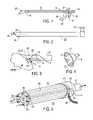
- FIG. 1is a side view of a vacuum-assisted insertion cannula in accordance with one embodiment of the present invention
- FIG. 2is a side view of an endoscopic cannula for use with the insertion cannula of FIG. 1 ;
- FIG. 3is a partial side view of the assembled cannulas of FIGS. 1 and 2 in a surgical procedure
- FIG. 4is a perspective partial view of a suction cup with associated sensing and pacing electrodes positioned therein for contacting the surface of the heart;
- FIG. 5is a perspective view of another embodiment of an insertion cannula in accordance with the present invention.
- FIGS. 6 a and 6 bcomprise a flow chart illustrating a surgical procedure in accordance with the present invention
- FIG. 7is a plan view of cardiac lead with screw-in electrode at the distal tip and with attached connector at the proximal end;
- FIG. 8is a partial plan view of an insertion cannula in one configuration incorporating an open channel for placement of a cardiac lead;
- FIG. 9is a partial plan view of the insertion cannula of FIG. 8 in a complementary configuration incorporating a closed channel;
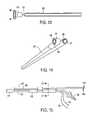
- FIG. 10is a plan view of a releasable guide for a cardiac lead according to another embodiment of the present invention.
- FIG. 11is a partial plan view of the distal end of the releasable guide in the embodiment of FIG. 10 ;
- FIG. 12is a partial plan view of the proximal end of the releasable guide in the embodiment of FIG. 10 ;
- FIG. 13is a top view of the distal end of the releasable guide in the embodiment of FIG. 10 ;
- FIG. 14is a perspective view of the distal end of the releasable guide according to the embodiment illustrated in FIG. 10 ;
- FIG. 15is a partial plan view of a releasable guide in accordance with the embodiment illustrated in FIG. 10 ;
- FIG. 16is a partial plan view of the releasable guide of FIG. 10 assembled within an endoscopic cannula;
- FIG. 17is a sectional view of the releasable guide of FIG. 15 ;
- FIG. 18is a partial plan view of one embodiment of the proximal end of the guide channel of the releasable guide of FIG. 15 ;
- FIG. 19is an end view of the proximal end of the guide channel of FIG. 15 .
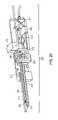
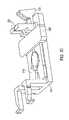
- FIG. 20is a perspective cut away view of a cardiac lead delivery device in accordance with one embodiment of the present invention.
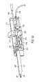
- FIG. 21is a partial cut away side view of the cardiac lead delivery device of FIGS. 20 in accordance with one embodiment of the present invention.
- FIGS. 22 a, b, c and dare, respectively, top, side, end and bottom views of an U-shaped body in accordance with one embodiment of the present invention.
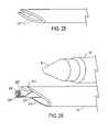
- FIGS. 23 a, b, c , and dare, respectively, top, perspective, side and end views of a needle in accordance with one embodiment of the present invention.
- FIG. 24is a perspective view of a guide channel in accordance with one embodiment of the present invention.
- FIG. 25is a partial plan view of another embodiment of the suction port in accordance with one embodiment of the present invention.
- FIG. 26is a partial side view of the cardiac lead delivery device of FIGS. 20 with a guide channel encasing a cardiac lead advanced in accordance with one embodiment of the present invention
- FIGS. 27A and 27Bare, respectively, partial plan and perspective views of the distal end of the releasable guide in accordance with one embodiment of the present invention.
- FIG. 28is a perspective view of an open clamp according to one embodiment of the present invention.
- FIG. 29is a perspective view of the clamp of FIG. 28 disposed in another operational configuration according to one embodiment of the present invention.
- FIG. 30is a perspective view of the clamp of FIG. 28 disposed in another operational configuration according to one embodiment of the present invention.
- FIG. 31is a perspective view of a cardiac lead delivery device with a needle advanced along a U-shaped body in accordance with one embodiment of the present invention.
- FIG. 32is a perspective view of a cardiac lead delivery device with a needle and guide channel advanced along a U-shaped body in accordance with one embodiment of the present invention
- FIG. 33is a cut-away perspective view of the cardiac lead delivery device of FIG. 32 with a guide channel slightly withdrawn in accordance with one embodiment of the present invention
- FIG. 34is a cut-away perspective view of the cardiac lead delivery device of FIG. 32 with the clamp unclamped and a needle withdrawn from a heart incision in accordance with one embodiment of the present invention
- FIG. 35is a cut-away perspective view of the cardiac lead delivery device of FIG. 32 with the guide channel completely withdrawn in accordance with one embodiment of the present invention.
- FIG. 36is a flow chart illustrating a surgical procedure for implanting a cardiac lead in accordance with one embodiment of the present invention.
- FIG. 1there is shown one embodiment of a suction assisted insertion cannula 10 according to the present invention including a closed channel 9 and a superior channel 11 attached to the closed channel.
- the closed channel 9includes a suitable hose connection 13 and a three-way vacuum control valve 15 including an irrigation port 16 at the proximal end.
- a three-way valve 15 on the cannula 9allows suction in the pod 17 to be turned on or off, and allows irrigation fluid such as saline to be injected through the suction pod 17 at the distal end while suction is turned off.
- the suction pod 17includes a flexible, resilient suction cup with a porous distal face 19 or suction ports that serves as a vacuum port.
- the distal surface of the suction cupincludes one or more surface electrodes 8 , 12 as shown in FIG. 4 , for contacting a surface of the heart.
- the surface electrodes 8 , 12 carried by the suction cup 17can be positioned against the epicardium to facilitate electrical contact during temporary vacuum-assisted fixation as a result of the reduced air pressure of vacuum supplied to the suction pod 17 .
- the distal end of the superior channel 11 that is attached to the closed channel 9may thus be held in accurate fixation in alignment with a selected surgical site on the epicardium relative to the suction fixation location of the suction pod 17 on the epicardium.
- Electrical conductors 22 , 24connect to the surface electrodes 8 , 12 and traverse the length of the suction channel 9 to facilitate connection thereto of diagnostic equipment that analyzes electrical signals sensed by the surface electrodes 8 , 12 held in contact with the epicardium.
- the superior channel 11is sized to accommodate slidable movement therein of a cardiac lead 21 in a configuration as shown, for example, in FIG. 7 .
- a cardiac lead 21exhibits lateral flexibility and torsional and axial rigidity over its length between the proximal end and the helical or corkscrew anchor electrode 25 at the distal end to facilitate screwing the helical anchor 25 into myocardium by rotating the proximal end of the cardiac lead 21 .
- the superior channel 11may be about 2-2.5 mm in diameter with an internal bore of sufficient size to accommodate a cardiac lead 21 of diameter up to approximately 2 mm in diameter.
- the suction pod 17includes a flexible, resilient suction cup 19 , as shown in FIG. 4 , that may be mounted in alignment with the closed channel 9 which serves as the vacuum channel, or may be mounted in skewed orientation thereto for convenient positioning of the surface electrodes 8 , 12 about the epicardium.
- Each of the surface electrodes 8 , 12is connected to a conductor 22 , 24 that extends along the vacuum channel 9 to a proximal location at which a diagnostic instrument of conventional design such as a cardiac pace/sense analyzer (PSA), for example, may be connected.
- a diagnostic instrument of conventional designsuch as a cardiac pace/sense analyzer (PSA), for example
- Such diagnostic instrumentsenses the electrical signals on the surface electrodes 8 , 12 operating in bipolar or unipolar mode at various locations on the epicardium to analyze various parameters such as maximum depolarization interval or maximum ventricle-to-ventricle timing for identifying a site of maximum therapeutic benefit from applied pacing signals.
- pacing signalscan be supplied to the surface electrodes via conductors 22 , 24 and specific hemodynamic parameters such as degree of mitral valve regurgitation, fractional ejection volume, cardiac output, and the like, can be analyzed to identify the specific site for maximum therapeutic value derived from pacing signals applied thereto.
- a cardiac lead implanted in the heart at a site determined by the procedure described aboveis extended out through a small initial incision in the patient, and the proximal end may then be tunneled subcutaneously from the initial incision to an incision in the patient's upper chest where a pacemaker or defibrillator will be located for connection to the cardiac electrode 21 .
- the superior channel 11is longitudinally slotted for placing a cardiac lead that may incorporate a large diameter connector 26 , as illustrated in FIG. 7 .
- a split sheathcan be positioned around the cardiac lead 21 to facilitate advancement and rotation of the cardiac lead within the closed superior channel 11 .
- the slotted superior channel 11is opened by rotating mating element 18 in the superior channel 11 , as illustrated in FIGS. 8 and 9 , to allow release of the cardiac lead 21 from the superior channel 11 .
- the structure according to this embodiment of the inventionis disposed to slide within the instrument channel 28 in an endoscopic cannula 27 , as shown in FIG. 2 .
- This cannulaincludes an endoscope 29 therein that extends from a tapered transparent tip 31 attached to the distal end, to a viewing port 33 at the proximal end that can be adapted to accommodate a video camera.
- the structure as illustrated in FIG. 1may be positioned within the instrument channel in the cannula 27 of FIG. 2 to position the suction pod 17 and a distal end 25 of a cardiac lead 21 in alignment with a surgical target on the heart, as illustrated in FIG. 3 .
- the suction pod 17is temporarily affixed to the epicardium in response to suction applied to the porous face 19 of the suction pod 17 under control of a suction valve 15 , with the surface electrodes 8 , 12 carried on the distal face of the suction cup disposed in contact with epicardium at a test site.
- the cardiac lead 21may then be advanced and rotated from the proximal end to anchor the distal end 25 into the myocardium at an accurately positioned surgical site, all within the visual field of the endoscope 29 through the transparent tip 31 .
- the various channels in the endoscopic cannula 27 and the insertion cannula 10have specific orientations with respect to each other in order to provide stabilization on the epicardial surface and allow visual control of the electrode attachment process.
- the instrument channelis positioned below the endoscopic channel and this allows the cannula 27 and the transparent tapered tip 31 on the endoscope 29 to retract the pericardium 93 away from the epicardial surface of the heart at the operative site. This creates a space 95 for contacting the heart below the pericardium, as illustrated in FIG. 3 .
- the suction pod 17is visualized through the endoscope 29 and transparent tip 31 , as the suction pod 17 is placed on the epicardial surface of the heart.
- the suctionis activated to attach the pod 17 to the heart with the surface electrodes 8 , 12 in contact with the epicardium.
- the configuration of the superior channel 11 of the insertion cannula 10 on top of the suction channel 9allows the superior channel 11 and the suction pod 17 to be visible upon exiting from the instrument channel of the cannula 27 , and to maintain visualization of the cardiac lead 21 within the visual field of the endoscope along the path of travel from the insertion cannula 10 to contact with the epicardium.
- the configuration of the suction pod 17 with the distal surface of the suction cup oriented substantially normally to the insertion cannula 10facilitates delivery of a cardiac electrode substantially perpendicular to the epicardial surface.
- a cardiac electrodeenter the myocardium in an orientation that is generally perpendicular to the epicardial surface for secure anchoring in the myocardium.
- the insertion cannula 10is advanced through the endoscopic cannula 27 and approaches the epicardial surface of the heart at a tangential angle.
- the insertion cannula 10may be configured to facilitate deforming the epicardial surface in order to achieve perpendicular entry of the distal end 25 of a cardiac lead 21 into the myocardium, as illustrated in FIG. 3 .
- the suction pod 17 of the insertion cannula 10temporarily attaches to the epicardial surface upon application of vacuum under control of the valve 15 . Downward pressure can be exerted on the epicardial surface via the substantially rigid insertion cannula 10 .
- the pliable myocardiumthus deforms to create a surface ledge 100 distal to the suction pod 17 oriented perpendicular to the axis of the superior instrument channel 11 of the insertion cannula 10 , as illustrated in FIG. 3 .
- the distal end electrode 25enters the myocardium generally perpendicularly to the epicardial surface as thus deformed for desirable lead placement.
- the insertion cannula 10is sized to fit in slidable orientation within the instrument channel of about 5-7 mm diameter in the endoscopic cannula 27 .
- the outer dimensions of the suction pod 17are flexible and resilient for confinement in less than 5-7 mm diameter.
- the suction cup of the suction pod 17may be skewed laterally relative to the suction channel 81 , as illustrated in FIG. 11 .
- the suction channel 9 , 81is laterally displaced from the superior channel 11 , 85 to avoid obstructing the forward movement of the cardiac lead 21 past the suction pod 17 , 91 .
- FIG. 5there is shown a perspective view of another embodiment of an insertion cannula 35 similar to insertion cannula 10 described above, including an elongated body 36 having a central bore 37 , and including one or more eccentric channels 39 that serve as suction conduits.
- the central bore 37may be sized to slidably support surgical instruments 41 therein such as a cardiac lead 21 disposed within a sheath, or the like.
- the suction pod 17attaches to the epicardial surface while suction is applied to facilitate surface electrodes 38 , 42 contacting the heart at the desired site under direct endoscopic visualization for precise cardiac mapping in response to signals sensed by the surface electrodes 38 , 42 operating in bipolar or unipolar configuration.
- the suction channels 39 in the cannula 35 of FIG. 5may form a suction attachment surface at the distal end of the cannula 35 , or may be disposed in fluid communication with a suitable suction pod with a porous distal face and with a central opening in alignment with the central bore 37 .
- the suction-attaching distal faceprovides an opposite reaction force against a tool that exerts a pushing force such as a screw-in tip 25 of a cardiac lead 21 , or other device deployed through the central bore 37 of the cannula 35 .
- proximal ends of the eccentric channels 39are connected via a manifold or fluid-coupling collar 43 to a vacuum line 45 , and conductors 46 , 50 connected to the surface electrodes 38 , 42 extend through the cannula 35 to the proximal end thereof to facilitate connection thereto of conventional diagnostic instrumentation.
- a single channel 39may communicate with an annular recess or groove disposed concentrically about the central bore 37 within the distal end to serve as a suction-assisted attachment surface.
- a cardiac lead 21 slidably disposed within the central bore 37may be extended beyond the distal end of the cannula 35 , within the visual field of an endoscope.
- the distal end 25 of the cardiac lead 21can be oriented in alignment with a target site on the epicardium prior to supplying suction thereto to temporarily affix the cannula 35 in such position with surface electrodes 38 , 42 in contact with the epicardium.
- a cannula 35 formed of transparent bioinert material such as polycarbonate polymerfacilitates visual alignment of the cannula 35 and the surface electrodes 38 , 42 with a target site, without requiring initial extension of a cardiac lead 21 forward of the distal end within the visual field of an endoscope.
- the central lumen or bore 37may serve as a suction lumen with multiple surface electrodes 38 , 42 disposed about the central bore 37 .
- the surgical procedure for epicardially mapping the beating heart of a patientproceeds from forming 51 an initial incision at a subxiphoid location on the patient.
- the incisionis extended 52 through the midline fibrous layer (linea alba).
- the tissue disposed between the location of subxiphoid incision and the heartis bluntly dissected 53 , for example, using a blunt-tip dissector disposed within a split-sheath cannula of the type described in the aforecited Related Applications.
- the channel thus formed in dissected tissuemay optionally be expanded 55 by dilating tissue surrounding the channel, for example, using a balloon dilator or the split-sheath cannula referenced above, in order to form a working cavity through the dissected and dilated tissue, although this may be unnecessary.
- An endoscopic cannulafor example, as illustrated in FIG. 2 including an endoscope and a lumen for receiving surgical instruments therein is inserted 57 into the working cavity through the subxiphoid incision toward the heart to provide a field of vision around a target site on the heart, and to provide convenient access via the lumen for surgical instruments of types associated with surgical procedures on the heart.
- One such instrumentis a pericardial entry instrument, as described in the aforecited Related Applications, which generally grasps the pericardium in a side-bite manner to form an elevated ridge of tissue through which a hole can be safely formed without contacting the epicardial surface. Once the pericardium is penetrated 58 , other instruments can be inserted through the hole and into the working space 58 .
- One such instrumentis an insertion cannula, for example, as illustrated in FIG. 1 , that includes a suction channel and a superior channel and is slidably supported 59 within the instrument lumen of the endoscopic cannula.
- the suction channel of such instrumentextends through the length thereof from a proximal end to a suction pod at the distal end that can be extended into contact 61 with the beating heart of the patient at a selected target site.
- the suction podcan be carefully positioned on the epicardium under visualization through the endoscope, and the suction can be applied to establish temporary attachment of the insertion cannula to the epicardium and to establish contact of surface electrodes with the epicardium.
- the electrical signals sensed on the surface electrodesmay be analyzed 62 for various timing characteristics such as maximum depolarization interval or maximum (left) ventricle to (right) ventricle conduction timing, or the like.
- pacing signalsmay be applied to the epicardium via surface electrodes 38 , 42 positioned at various sites about the heart in order to analyze 63 the heart's responses relative to specific hemodynamic parameters such as degree of mitral valve regurgitation, fractional ejection volume, cardiac output, and the like.
- a cardiac leadis installed 65 at the selected site by advancing and rotating the distal end electrode into the myocardium for good physical anchoring and electrical conduction.
- the insertion cannulais then reconfigured 66 to open a longitudinal slot in the superior channel in order to release the anchored cardiac lead so that the insertion cannula can be removed 67 from the site through the instrument channel of the endoscopic cannula, leaving the cardiac electrode anchored in the myocardium at the selected site.
- One or more cardiac leadsmay be installed in this manner, after which the endoscopic cannula is also removed 69 from the working cavity.
- a pacing unitis then implanted 70 in the patient's chest near the clavicle, or the abdomen near the subxiphoid incision, and is connected to the one or more installed cardiac leads to deliver requisite pacing signals.
- the initial subxiphoid entry incisionis then sutured closed 71 to conclude the surgical procedure.
- the endoscopic cannula and pericardial entry instrumentmay also be applied from a thoracotomy incision to gain access to the heart.
- a 2 cm incisionis performed in an intercostal space in either the left or the right chest. Ideally, the incision is made between the midclavicular line and the posterior axillary line.
- the incisionis extended through the intercostal muscles and the pleura, until the pleural cavity is entered.
- the endoscopic cannulais then inserted into the pleural cavity and advanced to the desired area of entry on the contour of the heart, visualized within the pleural cavity.
- the pericardial entry instrument and procedure as described in the aforecited Related Applicationsare used to grasp the pleura, as a concentric tubular blade cuts a hole in the pleura to expose the pericardium underneath. The pericardium is then grasped by the pericardial entry instrument, and the tubular blade is used to cut a hole in the pericardium, allowing access to the heart.
- the transparent tapered tip 31 of the endoscopic cannula 29aids in pleural and pericardial entry by retracting lung and pleural tissue that may impede visualization of the pericardial entry site.
- the endoscopic cannula 29may be moved around to visualize anterior and posterior epicardial surfaces as target sites for sensing surface electrical signals or for applying pacing signals in the manner as previously described herein.
- FIG. 10there is shown an assembly of suction tube 81 slidably disposed within a guide tube 83 to which is mounted a lower, slotted segment 85 of a guide channel.
- An upper, slotted segment 87 of the guide channelis slidably or rotatably received within the lower slotted segment 85 and a cardiac pacing or defibrillating lead 89 is housed within the guide channel that is configured in the one orientation of the upper and lower segments forming closed guide channel.
- Another configuration of the upper and lower segments of the guide channelforms an open channel or slot, as shown in FIG. 13 later described herein, for convenient release of the cardiac lead 89 .
- the suction tubeincludes a suction pod 91 at the distal end thereof and a suction-line connection fitting 94 at the proximal end for convenient hose or tubing attachment to a source of vacuum.
- the connection fitting 94may include a suction control valve 88 for adjusting the suction attachments of the suction pod to the epicardium of a patient's heart.
- Surface electrodes 96 , 98 disposed on the tissue-contacting surface of the suction pod 91are connected via conductors 90 , 92 that extend beyond the proximal end of the assembly for attachment to diagnostic or therapeutic equipment.
- the cardiac pacing or defibrillating lead 89is slidably and rotatably housed within the guide channel 85 , 87 in the closed configuration, and includes a helical or screw-in electrode 97 attached to the distal end of the cardiac lead 89 , as illustrated in FIG. 11 .
- the cardiac lead 89exhibits high torsional and compressional rigidity and high lateral flexibility so that the electrode 97 may be accurately manipulated into screw-like attachment to the myocardium at the selected site via manual manipulation of the proximal end 99 of the cardiac lead 89 .
- Such cardiac lead 89may include braided multiple strands of wire coated with a layer of insulating material such as Teflon, or the like.
- the accuracy of placement of the screw-in electrode 97 in the myocardium of a patient's beating heartis significantly enhanced by temporary suction attachment of the suction pod 91 to the pericardium or exposed myocardium.
- the suction pod 91 including a flexible, resilient suction cup with one or more surface electrodes 96 , 98may be disposed in lateral or skewed orientation relative to the elongated axis of the suction tube 81 . This facilitates the temporary suction attachment of the surface electrodes 96 , 98 during analysis of sensed signals or hemodynamic properties of the heart.
- the electrode 97 at the distal end of the cardiac lead 89is slidably guided within the guide channel 85 , 87 (which is disposed in skewed orientation relative to the suction pod 91 and vacuum tube 81 ) and is rotated to anchor the electrode 97 into the myocardium.
- the guide channel that houses the cardiac lead 89may be re-configured into the alternate configuration including an open slot along the length of the guide channel, as illustrated in FIG. 13 , from which the cardiac lead 89 may be easily extracted or released.
- This open slot configurationmay be achieved by sliding the upper segment 87 proximally along the lower segment 85 , as illustrated in FIG. 12 , or by rotating the upper segment 87 within the lower segment 85 , as illustrated in FIG. 14 . In this way, a longitudinal slot or groove is opened along the entire length of the guide channel that is wide enough to extract the cardiac lead 89 therethrough. This is particularly important for anchoring a cardiac lead 89 of about 2 mm diameter that includes a proximal connector 99 which is too large to pass through a guide channel 85 , 87 of reasonable interior dimension.
- a suction cup with surface electrodes 96 , 98 disposed in suction pod 91is oriented in skewed substantially perpendicular orientation relative to the elongated axis of the guide channel that is formed by the upper and lower segments 87 , 85 .
- a non-round guide tube 83that is attached to the lower segment 85 of the guide channel and that slidably supports therein the suction tube 81 of corresponding non-round cross section.
- the guide channel formed by segments 85 , 87is retained in substantially parallel axial alignment with the suction tube 81 as the suction pod 91 and the distal end of the guide channel are relatively slidably positioned near and against the epicardium of a patient's heart.
- the assembly of guide tube 83 and suction tube 81 and guide channel 85 , 87may all be disposed within the instrument channel of an endoscopic cannula 101 having a distal end disposed to facilitate endoscopic viewing of the suction pod 91 and the distal end of the guide channel 85 , 87 .
- the upper and lower segment 85 , 87 of the guide channelmay include stepped flanges 103 , 105 at the proximal ends thereof, as illustrated in FIGS.
- the upper 87 segmentcan be rotated in the lower segment 85 from the closed configuration in order to align the respective elongated slots sufficiently to release a cardiac lead 89 from within the guide channel.
- FIG. 20there is shown another embodiment of a cardiac lead delivery device 210 according to the present invention.
- the cardiac lead delivery device 210includes a housing 212 .
- An U-shaped elongated body 211is attached to the distal end of the housing 212 . Referring to FIGS.
- the U-shaped elongated body 211can be hollow and includes a suitable hose connection 213 for connection to a vacuum source at the proximal end, and the distal end of the U-shaped body 211 may be angled relative to the elongated axis of the body 211 , as shown the angled distal end of the U-shaped body 211 includes a U-shaped suction that is confined within boundary walls disposed substantially in a plane that is skewed at an acute angle relative to an elongated axis of the cardiac lead delivery device 210 .
- An upward orientation of the U-shaped body 211is preferred for better visualization of a cardiac lead that is disposed within the U-shape during placement.
- the suction port 217may comprise two separate channels as illustrated in FIG. 25 that are positioned on opposite sides of the distal end of the elongated body 211 .
- the suction port 217 at the distal end of the U-shaped body 211can be positioned against the epicardium to facilitate temporary fixation thereto resulting from reduced air pressure of vacuum supplied to the hose 213 .
- the distal end of the U-shaped bodymay thus be held in accurate temporary fixation in alignment with a selected surgical site on the epicardium relative to the suction fixation location of the suction port 217 on the epicardium.
- the angled suction port 217may also be used to apply gentle pressure on the epicardium to stop bleeding at small puncture sites in the epicardium.
- the U-shaped body 211is sized to accommodate slidable movement therein of a hollow needle 221 that is connected a bulkhead 214 located inside the housing 212 .
- the needle 221may exhibit lateral flexibility over its length at the proximal end to the sharpened distal end 225 .
- the needle 221may be about 2-3 mm in diameter with an internal bore of sufficient size to accommodate a lead and guide channel of diameter up to approximately 2 mm in diameter.
- the cardiac lead delivery device 210is sized to fit in slidable orientation within the instrument channel 28 of about 5-10 mm diameter in the endoscopic cannula 27 , as illustrated in FIG. 2 .
- FIG. 21there is shown an assembly of suction port 217 of the cardiac lead delivery device 210 in which a needle 221 is slidably disposed within the U-shaped body 211 .
- the guide channel 287is slidably and rotatably received within the needle 221 and a cardiac pacing or defibrillating lead 289 is housed within the guide channel 287 .
- the guide channel 287is coupled to an actuation arm 215 , as illustrated in FIG. 20 , that is slidable along the housing 212 .
- the cardiac pacing or defibrillating lead 289is slidably and rotatably housed within the guide channel 287 in the closed configuration, and includes a helical or screw-in electrode 297 attached to the distal end of the cardiac lead 289 , as illustrated in FIGS. 27A and 27B .
- the suction port 217facilitates the temporary suction attachment while the electrode 297 at the distal end of the cardiac lead 289 that is slidably guided within the guide channel 287 (which is disposed in substantially fixed axial orientation relative to the suction port 217 ) is being anchored into myocardium.
- a sled 216is located proximally of the bulkhead 214 and is slidable within the housing 212 .
- the sled 216is temporarily referenced against the bulkhead 214 by a pair of resilient detents 218 .
- Clamp 219 with arms 223is pivotally mounted on sled 216 for activation between opened and clamped configurations by a slide 220 .
- the clamp 219is open when the slide 220 is positioned near the distal end of the sled 216 as illustrated in FIG. 28 .
- the cardiac lead 289is placed between the two clamp arms 223 and within the guide channel 287 that is positioned within the needle 221 .
- the cardiac lead 289is loosely clamped in place for easier maneuverability.
- the slide 220is positioned against the clamp arms 223 and at the proximal end of the sled 216 , the clamp 219 is fully engaged and the cardiac lead 289 is firmly clamped within the clamp arms 223 , as illustrated in FIG. 30 .
- the suction hose 213is disposed above the slide 220 and located within the U-shaped body 211 .
- a wedge 222holds the suction 213 out of the way of the cardiac lead 289 .
- the suction hose 213exits the housing 212 distal the bulkhead 214 .
- the actuation arm 215is moved distally forward to abut the bulkhead 214 which in turns moves distally forward advancing the needle 221 that is attached to the bulkhead 214 .
- the actuation arm 215moves further distally, causing the sled 216 and the guide channel 287 to move forward which in turn causes the cardiac lead 289 to slide along the needle 221 into the heart.
- moving the actuation arm 215distally causes the sled 216 to bump against the detents 218 creating a friction stop.
- the guide channel 287may be angled distally, as illustrated in FIG. 24 , to move heart tissue away from the incision caused by the needle 221 .
- the actuation arm 215is pulled proximally to abut against the clamp arms 223 , as illustrated in FIG. 33 .
- These movements of the actuation arm 215results in the guide channel 287 being withdrawn slightly through the U-shaped body 211 to provide better endoscopic visualization of the placement of the distal end of a cardiac lead in a patient's heart.
- the cardiac electrode 297is rotated and anchored by hand into the correct position.
- the slide 220is then moved distally to unclamp the cardiac electrode 297 from the clamp 219 .
- the electrode 296remains anchored in the patient's heart as the actuation arm 215 , coupled with the guide channel 287 , is completely withdrawn from the housing 212 at the same time that the bulkhead 214 is moved proximally within the house to remove the needle 221 from the heart incision, as shown in FIGS. 34 and 35 .
- the placement of the suction port 217 at the distal end of the lead placement assemblyfacilitates establishing temporary vacuum-assisted attachment of the suction port 217 to the epicardium (or to myocardium that is exposed via the entry under the pericardium) which can then be depressed or otherwise distorted by manual application of axial or lateral force at the proximal end of the instrument in order to position the electrode 297 at the proper location and angle for anchoring in the myocardium of the patient's beating heart.
- the initial surgical proceduresare performed in a manner as previously described in the aforecited related applications from the initial incision 251 through to the insertion of the endoscopic cannula 257 .
- the releasable guide assemblyincluding U-shaped body 211 , needle 221 and guide channel 287 , is slid through the endoscopic cannula 309 toward the heart.
- the suction port 217is advanced into contact with the myocardium through the penetrated pericardium and suction is established to temporarily anchor 310 the suction port 217 at a desired surgical site.
- a cardiac lead 289 with a screw-in electrode 297 on the distal end of the cardiac leadis positioned at or near the distal end of the guide channel 287 in the closed configuration as the guide channel is advanced 312 toward the desired surgical site adjacent the temporary anchor site of the suction channel 211 on the myocardium.
- the guide channelis withdrawn slightly to provide endoscopic visualization of the cardiac lead in the heart incision.
- the proximal end of the cardiac lead 289may now be manually manipulated to screw in the electrode 297 at the distal end into the myocardium to thereby anchor 313 the cardiac lead 289 in the myocardium.
- the guide channel 287may now be completely withdrawn from the patient's body. Thereafter, the assembly of U-shaped body 211 and needle 221 may be retracted from the instrument channel of the cannula 27 , and the endoscopic cannula 27 may be removed 316 from within the working cavity, with the cardiac lead 289 in position therein.
- a subcutaneous tractis formed from the subxiphoid incision to the location of the pacing or defibrillation generator, usually placed in the patient's upper chest, and the cardiac lead is then connected to the generator 317 .
- the subxiphoid (or other) incisionis sutured closed to complete the surgical procedure.
- the surgical procedures described above including steps 309 - 315may be performed multiple times in order to anchor multiple cardiac leads in the myocardium prior to removing 316 the endoscopic cannula and suturing 318 the initial incision closed.
- the surgical apparatus and methods of the present inventionpromote careful placement of surface electrodes on the epicardial surface for electrocardial mapping of a beating heart.
- the present inventionpromotes careful placement of a needle or electrode or other surgical instrument on the surface of a beating heart by temporarily affixing the distal end of a guiding cannula at a selected position on the heart in response to suction applied to a suction port in a structure that supports the surface electrodes.
- the guiding cannulacan be positioned through a working cavity formed in tissue between the heart and a subxiphoid or other entry incision to minimize trauma and greatly facilitate surgical treatment of a beating heart.
- Such treatments and proceduresinclude initial sensing of electrical signals or delivery of pacing signals at selected sites on the epicardium for analyzing optimum sites at which cardiac electrodes are anchored for supplying electrical pacing signals with maximum therapeutic benefit, and thereafter placing pacing or defibrillating leads into the myocardium at the optimum sites.
Landscapes
- Health & Medical Sciences (AREA)
- Life Sciences & Earth Sciences (AREA)
- Surgery (AREA)
- Animal Behavior & Ethology (AREA)
- Public Health (AREA)
- Engineering & Computer Science (AREA)
- Biomedical Technology (AREA)
- Heart & Thoracic Surgery (AREA)
- Medical Informatics (AREA)
- Molecular Biology (AREA)
- Veterinary Medicine (AREA)
- General Health & Medical Sciences (AREA)
- Nuclear Medicine, Radiotherapy & Molecular Imaging (AREA)
- Pathology (AREA)
- Rheumatology (AREA)
- Physics & Mathematics (AREA)
- Biophysics (AREA)
- Optics & Photonics (AREA)
- Radiology & Medical Imaging (AREA)
- Oral & Maxillofacial Surgery (AREA)
- Surgical Instruments (AREA)
- Electrotherapy Devices (AREA)
Abstract
Description
Claims (2)
Priority Applications (4)
| Application Number | Priority Date | Filing Date | Title |
|---|---|---|---|
| US10/697,906US7526342B2 (en) | 1999-08-10 | 2003-10-29 | Apparatus for endoscopic cardiac mapping and lead placement |
| EP04795673AEP1689486A4 (en) | 2003-10-29 | 2004-10-19 | Apparatus and method for endoscopic cardiac mapping and lead placement |
| PCT/US2004/034538WO2005044079A2 (en) | 2002-06-17 | 2004-10-19 | Apparatus and method for endoscopic cardiac mapping and lead placement |
| JP2006538086AJP2007509702A (en) | 2003-10-29 | 2004-10-19 | Apparatus and method for endoscopic cardiac mapping and lead placement |
Applications Claiming Priority (6)
| Application Number | Priority Date | Filing Date | Title |
|---|---|---|---|
| US14813099P | 1999-08-10 | 1999-08-10 | |
| US15073799P | 1999-08-25 | 1999-08-25 | |
| US63572100A | 2000-08-09 | 2000-08-09 | |
| US10/140,309US20030187460A1 (en) | 1999-08-10 | 2002-05-06 | Methods and apparatus for endoscopic cardiac surgery |
| US10/174,454US20030187461A1 (en) | 1999-08-10 | 2002-06-17 | Releasable guide and method for endoscopic cardiac lead placement |
| US10/697,906US7526342B2 (en) | 1999-08-10 | 2003-10-29 | Apparatus for endoscopic cardiac mapping and lead placement |
Related Parent Applications (1)
| Application Number | Title | Priority Date | Filing Date |
|---|---|---|---|
| US10/174,454Continuation-In-PartUS20030187461A1 (en) | 1999-08-10 | 2002-06-17 | Releasable guide and method for endoscopic cardiac lead placement |
Publications (2)
| Publication Number | Publication Date |
|---|---|
| US20040153098A1 US20040153098A1 (en) | 2004-08-05 |
| US7526342B2true US7526342B2 (en) | 2009-04-28 |
Family
ID=35789083
Family Applications (1)
| Application Number | Title | Priority Date | Filing Date |
|---|---|---|---|
| US10/697,906Expired - Fee RelatedUS7526342B2 (en) | 1999-08-10 | 2003-10-29 | Apparatus for endoscopic cardiac mapping and lead placement |
Country Status (2)
| Country | Link |
|---|---|
| US (1) | US7526342B2 (en) |
| WO (1) | WO2005044079A2 (en) |
Cited By (122)
| Publication number | Priority date | Publication date | Assignee | Title |
|---|---|---|---|---|
| US20060253181A1 (en)* | 2005-05-05 | 2006-11-09 | Alfred E. Mann Foundation For Scientific Research | Lead insertion tool |
| US20090076521A1 (en)* | 2007-09-18 | 2009-03-19 | Morten Hansen | Apparatus and method for inserting implants into the body |
| US20090182402A1 (en)* | 2008-01-10 | 2009-07-16 | Arkady Glukhovsky | Methods and apparatus for implanting electronic implants within the body |
| US20100036465A1 (en)* | 2008-08-07 | 2010-02-11 | Arkady Glukhovsky | Insertion tools and methods for an electrical stimulation implant |
| US8012143B1 (en) | 2006-12-12 | 2011-09-06 | Pacesetter, Inc. | Intrapericardial delivery tools and methods |
| US8346339B2 (en) | 2011-04-22 | 2013-01-01 | Topera, Inc. | Basket style cardiac mapping catheter having a flexible electrode assembly for detection of cardiac rhythm disorders |
| US8961551B2 (en) | 2006-12-22 | 2015-02-24 | The Spectranetics Corporation | Retractable separating systems and methods |
| US9028520B2 (en) | 2006-12-22 | 2015-05-12 | The Spectranetics Corporation | Tissue separating systems and methods |
| US20150343201A1 (en)* | 2010-09-28 | 2015-12-03 | The Board Of Trustees Of The Leland Stanford Junior University | Device and method for positioning an electrode in tissue |
| US9283040B2 (en) | 2013-03-13 | 2016-03-15 | The Spectranetics Corporation | Device and method of ablative cutting with helical tip |
| US9291663B2 (en) | 2013-03-13 | 2016-03-22 | The Spectranetics Corporation | Alarm for lead insulation abnormality |
| US9370655B1 (en) | 2013-03-07 | 2016-06-21 | Subhajit Datta | Lead and conduit placement device and method |
| US9402531B2 (en) | 2012-07-05 | 2016-08-02 | Pavilion Medical Innovations, Llc | Endoscopic cannulas and methods of using the same |
| US9413896B2 (en) | 2012-09-14 | 2016-08-09 | The Spectranetics Corporation | Tissue slitting methods and systems |
| USD765243S1 (en) | 2015-02-20 | 2016-08-30 | The Spectranetics Corporation | Medical device handle |
| US9456872B2 (en) | 2013-03-13 | 2016-10-04 | The Spectranetics Corporation | Laser ablation catheter |
| USD770616S1 (en) | 2015-02-20 | 2016-11-01 | The Spectranetics Corporation | Medical device handle |
| US9511219B1 (en)* | 2014-03-24 | 2016-12-06 | Subhajit Datta | Dual vacuum device for medical fixture placement including for thoracoscopic left ventricular lead placement |
| US9526909B2 (en) | 2014-08-28 | 2016-12-27 | Cardiac Pacemakers, Inc. | Medical device with triggered blanking period |
| US9592391B2 (en) | 2014-01-10 | 2017-03-14 | Cardiac Pacemakers, Inc. | Systems and methods for detecting cardiac arrhythmias |
| US9603618B2 (en) | 2013-03-15 | 2017-03-28 | The Spectranetics Corporation | Medical device for removing an implanted object |
| US9669230B2 (en) | 2015-02-06 | 2017-06-06 | Cardiac Pacemakers, Inc. | Systems and methods for treating cardiac arrhythmias |
| US9668765B2 (en) | 2013-03-15 | 2017-06-06 | The Spectranetics Corporation | Retractable blade for lead removal device |
| US9853743B2 (en) | 2015-08-20 | 2017-12-26 | Cardiac Pacemakers, Inc. | Systems and methods for communication between medical devices |
| US9872981B2 (en) | 2010-09-28 | 2018-01-23 | Biotrace Medical, Inc. | Device and method for positioning an electrode in a body cavity |
| US9883885B2 (en) | 2013-03-13 | 2018-02-06 | The Spectranetics Corporation | System and method of ablative cutting and pulsed vacuum aspiration |
| US9925366B2 (en) | 2013-03-15 | 2018-03-27 | The Spectranetics Corporation | Surgical instrument for removing an implanted object |
| US9956414B2 (en) | 2015-08-27 | 2018-05-01 | Cardiac Pacemakers, Inc. | Temporal configuration of a motion sensor in an implantable medical device |
| US9968787B2 (en) | 2015-08-27 | 2018-05-15 | Cardiac Pacemakers, Inc. | Spatial configuration of a motion sensor in an implantable medical device |
| US9980743B2 (en) | 2013-03-15 | 2018-05-29 | The Spectranetics Corporation | Medical device for removing an implanted object using laser cut hypotubes |
| US10029107B1 (en) | 2017-01-26 | 2018-07-24 | Cardiac Pacemakers, Inc. | Leadless device with overmolded components |
| US10050700B2 (en) | 2015-03-18 | 2018-08-14 | Cardiac Pacemakers, Inc. | Communications in a medical device system with temporal optimization |
| US10046167B2 (en) | 2015-02-09 | 2018-08-14 | Cardiac Pacemakers, Inc. | Implantable medical device with radiopaque ID tag |
| US10065041B2 (en) | 2015-10-08 | 2018-09-04 | Cardiac Pacemakers, Inc. | Devices and methods for adjusting pacing rates in an implantable medical device |
| US10092760B2 (en) | 2015-09-11 | 2018-10-09 | Cardiac Pacemakers, Inc. | Arrhythmia detection and confirmation |
| US10137305B2 (en) | 2015-08-28 | 2018-11-27 | Cardiac Pacemakers, Inc. | Systems and methods for behaviorally responsive signal detection and therapy delivery |
| US10136913B2 (en) | 2013-03-15 | 2018-11-27 | The Spectranetics Corporation | Multiple configuration surgical cutting device |
| US10159842B2 (en) | 2015-08-28 | 2018-12-25 | Cardiac Pacemakers, Inc. | System and method for detecting tamponade |
| US10183170B2 (en) | 2015-12-17 | 2019-01-22 | Cardiac Pacemakers, Inc. | Conducted communication in a medical device system |
| US10213610B2 (en) | 2015-03-18 | 2019-02-26 | Cardiac Pacemakers, Inc. | Communications in a medical device system with link quality assessment |
| US10220213B2 (en) | 2015-02-06 | 2019-03-05 | Cardiac Pacemakers, Inc. | Systems and methods for safe delivery of electrical stimulation therapy |
| US10226631B2 (en) | 2015-08-28 | 2019-03-12 | Cardiac Pacemakers, Inc. | Systems and methods for infarct detection |
| US10232170B2 (en) | 2014-05-09 | 2019-03-19 | Biotrace Medical, Inc. | Device and method for positioning an electrode in a body cavity |
| US10328272B2 (en) | 2016-05-10 | 2019-06-25 | Cardiac Pacemakers, Inc. | Retrievability for implantable medical devices |
| US10350423B2 (en) | 2016-02-04 | 2019-07-16 | Cardiac Pacemakers, Inc. | Delivery system with force sensor for leadless cardiac device |
| US10357159B2 (en) | 2015-08-20 | 2019-07-23 | Cardiac Pacemakers, Inc | Systems and methods for communication between medical devices |
| US10383691B2 (en) | 2013-03-13 | 2019-08-20 | The Spectranetics Corporation | Last catheter with helical internal lumen |
| US10391319B2 (en) | 2016-08-19 | 2019-08-27 | Cardiac Pacemakers, Inc. | Trans septal implantable medical device |
| US10405924B2 (en) | 2014-05-30 | 2019-09-10 | The Spectranetics Corporation | System and method of ablative cutting and vacuum aspiration through primary orifice and auxiliary side port |
| US10413733B2 (en) | 2016-10-27 | 2019-09-17 | Cardiac Pacemakers, Inc. | Implantable medical device with gyroscope |
| US10426962B2 (en) | 2016-07-07 | 2019-10-01 | Cardiac Pacemakers, Inc. | Leadless pacemaker using pressure measurements for pacing capture verification |
| US10434317B2 (en) | 2016-10-31 | 2019-10-08 | Cardiac Pacemakers, Inc. | Systems and methods for activity level pacing |
| US10434314B2 (en) | 2016-10-27 | 2019-10-08 | Cardiac Pacemakers, Inc. | Use of a separate device in managing the pace pulse energy of a cardiac pacemaker |
| US10448999B2 (en) | 2013-03-15 | 2019-10-22 | The Spectranetics Corporation | Surgical instrument for removing an implanted object |
| US10463305B2 (en) | 2016-10-27 | 2019-11-05 | Cardiac Pacemakers, Inc. | Multi-device cardiac resynchronization therapy with timing enhancements |
| US10512784B2 (en) | 2016-06-27 | 2019-12-24 | Cardiac Pacemakers, Inc. | Cardiac therapy system using subcutaneously sensed P-waves for resynchronization pacing management |
| US10525179B2 (en) | 2016-03-31 | 2020-01-07 | Heartware, Inc. | Crenellated inflow cannula |
| US10561330B2 (en) | 2016-10-27 | 2020-02-18 | Cardiac Pacemakers, Inc. | Implantable medical device having a sense channel with performance adjustment |
| US10583303B2 (en) | 2016-01-19 | 2020-03-10 | Cardiac Pacemakers, Inc. | Devices and methods for wirelessly recharging a rechargeable battery of an implantable medical device |
| US10583301B2 (en) | 2016-11-08 | 2020-03-10 | Cardiac Pacemakers, Inc. | Implantable medical device for atrial deployment |
| US10617874B2 (en) | 2016-10-31 | 2020-04-14 | Cardiac Pacemakers, Inc. | Systems and methods for activity level pacing |
| US10632313B2 (en) | 2016-11-09 | 2020-04-28 | Cardiac Pacemakers, Inc. | Systems, devices, and methods for setting cardiac pacing pulse parameters for a cardiac pacing device |
| US10639486B2 (en) | 2016-11-21 | 2020-05-05 | Cardiac Pacemakers, Inc. | Implantable medical device with recharge coil |
| US10668294B2 (en) | 2016-05-10 | 2020-06-02 | Cardiac Pacemakers, Inc. | Leadless cardiac pacemaker configured for over the wire delivery |
| US10688304B2 (en) | 2016-07-20 | 2020-06-23 | Cardiac Pacemakers, Inc. | Method and system for utilizing an atrial contraction timing fiducial in a leadless cardiac pacemaker system |
| US10722720B2 (en) | 2014-01-10 | 2020-07-28 | Cardiac Pacemakers, Inc. | Methods and systems for improved communication between medical devices |
| US10737102B2 (en) | 2017-01-26 | 2020-08-11 | Cardiac Pacemakers, Inc. | Leadless implantable device with detachable fixation |
| US10758724B2 (en) | 2016-10-27 | 2020-09-01 | Cardiac Pacemakers, Inc. | Implantable medical device delivery system with integrated sensor |
| US10758737B2 (en) | 2016-09-21 | 2020-09-01 | Cardiac Pacemakers, Inc. | Using sensor data from an intracardially implanted medical device to influence operation of an extracardially implantable cardioverter |
| US10765871B2 (en) | 2016-10-27 | 2020-09-08 | Cardiac Pacemakers, Inc. | Implantable medical device with pressure sensor |
| US10780278B2 (en) | 2016-08-24 | 2020-09-22 | Cardiac Pacemakers, Inc. | Integrated multi-device cardiac resynchronization therapy using P-wave to pace timing |
| US10821288B2 (en) | 2017-04-03 | 2020-11-03 | Cardiac Pacemakers, Inc. | Cardiac pacemaker with pacing pulse energy adjustment based on sensed heart rate |
| US10835753B2 (en) | 2017-01-26 | 2020-11-17 | Cardiac Pacemakers, Inc. | Intra-body device communication with redundant message transmission |
| US10835279B2 (en) | 2013-03-14 | 2020-11-17 | Spectranetics Llc | Distal end supported tissue slitting apparatus |
| US10842532B2 (en) | 2013-03-15 | 2020-11-24 | Spectranetics Llc | Medical device for removing an implanted object |
| US10870008B2 (en) | 2016-08-24 | 2020-12-22 | Cardiac Pacemakers, Inc. | Cardiac resynchronization using fusion promotion for timing management |
| US10874861B2 (en) | 2018-01-04 | 2020-12-29 | Cardiac Pacemakers, Inc. | Dual chamber pacing without beat-to-beat communication |
| US10881869B2 (en) | 2016-11-21 | 2021-01-05 | Cardiac Pacemakers, Inc. | Wireless re-charge of an implantable medical device |
| US10881863B2 (en) | 2016-11-21 | 2021-01-05 | Cardiac Pacemakers, Inc. | Leadless cardiac pacemaker with multimode communication |
| US10894163B2 (en) | 2016-11-21 | 2021-01-19 | Cardiac Pacemakers, Inc. | LCP based predictive timing for cardiac resynchronization |
| US10905889B2 (en) | 2016-09-21 | 2021-02-02 | Cardiac Pacemakers, Inc. | Leadless stimulation device with a housing that houses internal components of the leadless stimulation device and functions as the battery case and a terminal of an internal battery |
| US10905872B2 (en) | 2017-04-03 | 2021-02-02 | Cardiac Pacemakers, Inc. | Implantable medical device with a movable electrode biased toward an extended position |
| US10905886B2 (en) | 2015-12-28 | 2021-02-02 | Cardiac Pacemakers, Inc. | Implantable medical device for deployment across the atrioventricular septum |
| US10918875B2 (en) | 2017-08-18 | 2021-02-16 | Cardiac Pacemakers, Inc. | Implantable medical device with a flux concentrator and a receiving coil disposed about the flux concentrator |
| US10980481B2 (en) | 2018-07-31 | 2021-04-20 | Calyan Technologies, Inc. | Subcutaneous device for monitoring and/or providing therapies |
| US10987060B1 (en) | 2020-09-14 | 2021-04-27 | Calyan Technologies, Inc. | Clip design for a subcutaneous device |
| US10994145B2 (en) | 2016-09-21 | 2021-05-04 | Cardiac Pacemakers, Inc. | Implantable cardiac monitor |
| US11040208B1 (en) | 2019-12-17 | 2021-06-22 | Biosense Webster (Israel) Ltd. | Distributed cardiac pacing system |
| US11052258B2 (en) | 2017-12-01 | 2021-07-06 | Cardiac Pacemakers, Inc. | Methods and systems for detecting atrial contraction timing fiducials within a search window from a ventricularly implanted leadless cardiac pacemaker |
| US11058880B2 (en) | 2018-03-23 | 2021-07-13 | Medtronic, Inc. | VFA cardiac therapy for tachycardia |
| US11065459B2 (en) | 2017-08-18 | 2021-07-20 | Cardiac Pacemakers, Inc. | Implantable medical device with pressure sensor |
| US11071870B2 (en) | 2017-12-01 | 2021-07-27 | Cardiac Pacemakers, Inc. | Methods and systems for detecting atrial contraction timing fiducials and determining a cardiac interval from a ventricularly implanted leadless cardiac pacemaker |
| US11116988B2 (en) | 2016-03-31 | 2021-09-14 | Cardiac Pacemakers, Inc. | Implantable medical device with rechargeable battery |
| US11147979B2 (en) | 2016-11-21 | 2021-10-19 | Cardiac Pacemakers, Inc. | Implantable medical device with a magnetically permeable housing and an inductive coil disposed about the housing |
| US11179571B2 (en) | 2018-07-31 | 2021-11-23 | Manicka Institute Llc | Subcutaneous device for monitoring and/or providing therapies |
| US11185703B2 (en) | 2017-11-07 | 2021-11-30 | Cardiac Pacemakers, Inc. | Leadless cardiac pacemaker for bundle of his pacing |
| US11207532B2 (en) | 2017-01-04 | 2021-12-28 | Cardiac Pacemakers, Inc. | Dynamic sensing updates using postural input in a multiple device cardiac rhythm management system |
| US11207527B2 (en) | 2016-07-06 | 2021-12-28 | Cardiac Pacemakers, Inc. | Method and system for determining an atrial contraction timing fiducial in a leadless cardiac pacemaker system |
| US11213676B2 (en) | 2019-04-01 | 2022-01-04 | Medtronic, Inc. | Delivery systems for VfA cardiac therapy |
| US11235161B2 (en) | 2018-09-26 | 2022-02-01 | Medtronic, Inc. | Capture in ventricle-from-atrium cardiac therapy |
| US11235163B2 (en) | 2017-09-20 | 2022-02-01 | Cardiac Pacemakers, Inc. | Implantable medical device with multiple modes of operation |
| US11235159B2 (en) | 2018-03-23 | 2022-02-01 | Medtronic, Inc. | VFA cardiac resynchronization therapy |
| US11260216B2 (en) | 2017-12-01 | 2022-03-01 | Cardiac Pacemakers, Inc. | Methods and systems for detecting atrial contraction timing fiducials during ventricular filling from a ventricularly implanted leadless cardiac pacemaker |
| US11285326B2 (en) | 2015-03-04 | 2022-03-29 | Cardiac Pacemakers, Inc. | Systems and methods for treating cardiac arrhythmias |
| US11305127B2 (en) | 2019-08-26 | 2022-04-19 | Medtronic Inc. | VfA delivery and implant region detection |
| US11400296B2 (en) | 2018-03-23 | 2022-08-02 | Medtronic, Inc. | AV synchronous VfA cardiac therapy |
| US11433233B2 (en) | 2020-11-25 | 2022-09-06 | Calyan Technologies, Inc. | Electrode contact for a subcutaneous device |
| US11478650B2 (en) | 2018-07-31 | 2022-10-25 | Calyan Technologies, Inc. | Subcutaneous device |
| US11529523B2 (en) | 2018-01-04 | 2022-12-20 | Cardiac Pacemakers, Inc. | Handheld bridge device for providing a communication bridge between an implanted medical device and a smartphone |
| US11660444B2 (en) | 2018-07-31 | 2023-05-30 | Manicka Institute Llc | Resilient body component contact for a subcutaneous device |
| US11679265B2 (en) | 2019-02-14 | 2023-06-20 | Medtronic, Inc. | Lead-in-lead systems and methods for cardiac therapy |
| US11697025B2 (en) | 2019-03-29 | 2023-07-11 | Medtronic, Inc. | Cardiac conduction system capture |
| US11712188B2 (en) | 2019-05-07 | 2023-08-01 | Medtronic, Inc. | Posterior left bundle branch engagement |
| US11717674B2 (en) | 2018-07-31 | 2023-08-08 | Manicka Institute Llc | Subcutaneous device for use with remote device |
| US11813463B2 (en) | 2017-12-01 | 2023-11-14 | Cardiac Pacemakers, Inc. | Leadless cardiac pacemaker with reversionary behavior |
| US11813466B2 (en) | 2020-01-27 | 2023-11-14 | Medtronic, Inc. | Atrioventricular nodal stimulation |
| US11813464B2 (en) | 2020-07-31 | 2023-11-14 | Medtronic, Inc. | Cardiac conduction system evaluation |
| US11911168B2 (en) | 2020-04-03 | 2024-02-27 | Medtronic, Inc. | Cardiac conduction system therapy benefit determination |
| US11951313B2 (en) | 2018-11-17 | 2024-04-09 | Medtronic, Inc. | VFA delivery systems and methods |
| US11950839B2 (en) | 2020-07-30 | 2024-04-09 | Sd Cardiothoracic Innovations, Llc | Multiple vacuum device for medical fixture placement |
| US12053203B2 (en) | 2014-03-03 | 2024-08-06 | Spectranetics, Llc | Multiple configuration surgical cutting device |
| US12296177B2 (en) | 2018-12-21 | 2025-05-13 | Medtronic, Inc. | Delivery systems and methods for left ventricular pacing |
Families Citing this family (58)
| Publication number | Priority date | Publication date | Assignee | Title |
|---|---|---|---|---|
| US7384423B1 (en) | 1995-07-13 | 2008-06-10 | Origin Medsystems, Inc. | Tissue dissection method |
| WO1998007375A1 (en)* | 1996-08-22 | 1998-02-26 | The Trustees Of Columbia University | Endovascular flexible stapling device |
| US7326178B1 (en) | 1998-06-22 | 2008-02-05 | Origin Medsystems, Inc. | Vessel retraction device and method |
| US6976957B1 (en) | 1998-06-22 | 2005-12-20 | Origin Medsystems, Inc. | Cannula-based surgical instrument and method |
| US6830546B1 (en) | 1998-06-22 | 2004-12-14 | Origin Medsystems, Inc. | Device and method for remote vessel ligation |
| EP0979635A2 (en) | 1998-08-12 | 2000-02-16 | Origin Medsystems, Inc. | Tissue dissector apparatus |
| US6558313B1 (en) | 2000-11-17 | 2003-05-06 | Embro Corporation | Vein harvesting system and method |
| US7165552B2 (en) | 2003-03-27 | 2007-01-23 | Cierra, Inc. | Methods and apparatus for treatment of patent foramen ovale |
| AU2004226374B2 (en) | 2003-03-27 | 2009-11-12 | Terumo Kabushiki Kaisha | Methods and apparatus for treatment of patent foramen ovale |
| US8021362B2 (en) | 2003-03-27 | 2011-09-20 | Terumo Kabushiki Kaisha | Methods and apparatus for closing a layered tissue defect |
| US6939348B2 (en) | 2003-03-27 | 2005-09-06 | Cierra, Inc. | Energy based devices and methods for treatment of patent foramen ovale |
| US7972330B2 (en) | 2003-03-27 | 2011-07-05 | Terumo Kabushiki Kaisha | Methods and apparatus for closing a layered tissue defect |
| US7293562B2 (en) | 2003-03-27 | 2007-11-13 | Cierra, Inc. | Energy based devices and methods for treatment of anatomic tissue defects |
| US7186251B2 (en) | 2003-03-27 | 2007-03-06 | Cierra, Inc. | Energy based devices and methods for treatment of patent foramen ovale |
| US7311701B2 (en) | 2003-06-10 | 2007-12-25 | Cierra, Inc. | Methods and apparatus for non-invasively treating atrial fibrillation using high intensity focused ultrasound |
| US7840270B2 (en) | 2003-07-23 | 2010-11-23 | Synapse Biomedical, Inc. | System and method for conditioning a diaphragm of a patient |
| US7367975B2 (en) | 2004-06-21 | 2008-05-06 | Cierra, Inc. | Energy based devices and methods for treatment of anatomic tissue defects |
| JP3965170B2 (en)* | 2004-07-27 | 2007-08-29 | オリンパス株式会社 | Endoscope |
| EP1693011A1 (en)* | 2005-02-22 | 2006-08-23 | Depuy International Limited | An instrument for implanting a sensor |
| US7585302B2 (en)* | 2005-02-22 | 2009-09-08 | Depuy International Ltd. | Instrument for implanting a sensor |
| US8109274B2 (en) | 2005-04-11 | 2012-02-07 | Terumo Kabushiki Kaisha | Methods and electrode apparatus to achieve a closure of a layered tissue defect |
| JP5007967B2 (en)* | 2005-07-05 | 2012-08-22 | 学校法人日本大学 | Cannula |
| US9050005B2 (en) | 2005-08-25 | 2015-06-09 | Synapse Biomedical, Inc. | Method and apparatus for transgastric neurostimulation |
| CA2631915A1 (en)* | 2005-12-02 | 2007-06-07 | Synapse Biomedical, Inc. | Transvisceral neurostimulation mapping device and method |
| US9770230B2 (en) | 2006-06-01 | 2017-09-26 | Maquet Cardiovascular Llc | Endoscopic vessel harvesting system components |
| NZ599528A (en)* | 2006-06-30 | 2013-11-29 | Cvdevices Llc | Percutaneous intravascular access to cardiac tissue |
| US9993639B2 (en) | 2006-10-31 | 2018-06-12 | Medtronic, Inc. | Implantable medical elongated member including a tissue receiving fixation cavity |
| US9079016B2 (en) | 2007-02-05 | 2015-07-14 | Synapse Biomedical, Inc. | Removable intramuscular electrode |
| US9820671B2 (en) | 2007-05-17 | 2017-11-21 | Synapse Biomedical, Inc. | Devices and methods for assessing motor point electromyogram as a biomarker |
| US20090076463A1 (en)* | 2007-09-17 | 2009-03-19 | Jurg Attinger | Trocar Cannula |
| US8428726B2 (en) | 2007-10-30 | 2013-04-23 | Synapse Biomedical, Inc. | Device and method of neuromodulation to effect a functionally restorative adaption of the neuromuscular system |
| US8478412B2 (en) | 2007-10-30 | 2013-07-02 | Synapse Biomedical, Inc. | Method of improving sleep disordered breathing |
| WO2010096920A1 (en)* | 2009-02-24 | 2010-09-02 | George Klein | Anchoring catheter sheath |
| JP5415925B2 (en)* | 2009-03-02 | 2014-02-12 | オリンパス株式会社 | Endoscope |
| US8747297B2 (en)* | 2009-03-02 | 2014-06-10 | Olympus Corporation | Endoscopic heart surgery method |
| US9050129B2 (en)* | 2009-04-24 | 2015-06-09 | Medtronic, Inc. | Auto-closure apical access positioner device and method |
| EP2481377A4 (en)* | 2009-09-22 | 2017-12-20 | Olympus Corporation | Space-securing device |
| US8308723B2 (en) | 2009-10-09 | 2012-11-13 | Coaptus Medical Corporation | Tissue-penetrating guidewires with shaped tips, and associated systems and methods |
| US9789029B2 (en)* | 2011-07-17 | 2017-10-17 | Nutriseal Limited Partnership | Nasogastric tube |
| US9839584B2 (en)* | 2011-07-17 | 2017-12-12 | Nutriseal Limited Partnership | Nasogastric tube |
| US10350145B2 (en)* | 2011-07-17 | 2019-07-16 | Envizion Medical Ltd. | Nasogastric tube |
| US9827169B2 (en) | 2011-07-17 | 2017-11-28 | Nutriseal L.P. | Nasogastric tube with camera |
| JP6318088B2 (en) | 2011-07-26 | 2018-04-25 | アンフォラ メディカル, インコーポレイテッド | Apparatus and method for modulating pelvic nerve tissue |
| WO2014164641A1 (en) | 2013-03-11 | 2014-10-09 | Mayo Foundation For Medical Education And Research | Pericardial modification systems and methods for heart failure treatment |
| CN104546074A (en)* | 2013-10-24 | 2015-04-29 | 心诺普医疗技术(北京)有限公司 | Pericardium puncture needle component |
| JP2017516620A (en) | 2014-05-23 | 2017-06-22 | アンフォラ メディカル, インコーポレイテッド | Methods and devices for treatment of pelvic conditions |
| ES2726988T3 (en)* | 2014-08-14 | 2019-10-11 | Envizion Medical Ltd | Nasogastric tube |
| EP3253357B1 (en) | 2015-02-02 | 2019-09-11 | Envizion Medical Ltd. | Enteral feeding pump |
| US10098695B2 (en) | 2016-03-10 | 2018-10-16 | Mayo Foundation For Medical Education And Research | Pericardial modification devices and methods |
| CN107469231B (en)* | 2016-06-07 | 2023-08-18 | 四川大学华西医院 | An electric stimulator acting through the natural orifice of the human body |
| US10828146B2 (en)* | 2016-08-04 | 2020-11-10 | Stryker Corporation | Instrumentation for soft tissue reconstruction |
| US10471251B1 (en) | 2018-07-31 | 2019-11-12 | Manicka Institute Llc | Subcutaneous device for monitoring and/or providing therapies |
| CN112546427B (en)* | 2018-07-31 | 2023-11-03 | 卡利安科技有限公司 | subcutaneous device |
| US11471683B2 (en) | 2019-01-29 | 2022-10-18 | Synapse Biomedical, Inc. | Systems and methods for treating sleep apnea using neuromodulation |
| US11272956B2 (en) | 2019-03-17 | 2022-03-15 | Ripple, LLC | Systems and methods for implanting electrode leads |
| WO2021080302A2 (en)* | 2019-10-23 | 2021-04-29 | 고려대학교 산학협력단 | Chest tube insertion device using endoscopic guide |
| CN114615942B (en)* | 2019-11-05 | 2025-10-03 | 波士顿科学国际有限公司 | Tissue collection spiral device |
| US11826075B2 (en)* | 2020-04-07 | 2023-11-28 | Boston Scientific Medical Device Limited | Elongated medical assembly |
Citations (184)
| Publication number | Priority date | Publication date | Assignee | Title |
|---|---|---|---|---|
| US207932A (en) | 1878-09-10 | Improvement in surgical dilators | ||
| US702789A (en) | 1902-03-20 | 1902-06-17 | Charles Gordon Gibson | Dilator. |
| US1727495A (en) | 1929-09-10 | Beinhold h | ||
| US1867624A (en) | 1930-04-01 | 1932-07-19 | Memorial Hospital For The Trea | Device for obtaining biopsy specimens |
| US2011169A (en) | 1932-04-13 | 1935-08-13 | Wappler Frederick Charles | Forcipated surgical electrode |
| US2028635A (en) | 1933-09-11 | 1936-01-21 | Wappler Frederick Charles | Forcipated surgical instrument |
| US2201749A (en) | 1939-02-15 | 1940-05-21 | Vandegrift Middleton | Expanding vein tube |
| US2316297A (en) | 1943-01-15 | 1943-04-13 | Beverly A Southerland | Surgical instrument |
| US2868206A (en) | 1956-07-25 | 1959-01-13 | Frederick G Stoesser | Intra luminal vein stripper |
| US2944552A (en) | 1958-12-29 | 1960-07-12 | Richard B Wilk | Surgical instrument |
| FR1370580A (en) | 1962-12-04 | 1964-08-28 | Centre Nat Rech Scient | Contact endoscope |
| US3185155A (en) | 1963-03-13 | 1965-05-25 | Slaten | Vein stripper |
| US3338916A (en) | 1964-02-05 | 1967-08-29 | Wander Ag Dr A | Benzimidazolone derviatives |
| US3763806A (en) | 1972-10-16 | 1973-10-09 | C & W Sewing Machine | Separately retractable paired needles |
| US3856016A (en) | 1972-11-03 | 1974-12-24 | H Davis | Method for mechanically applying an occlusion clip to an anatomical tubular structure |
| US3870048A (en) | 1973-07-30 | 1975-03-11 | In Bae Yoon | Device for sterilizing the human female or male by ligation |
| US3877491A (en) | 1970-03-19 | 1975-04-15 | E Ramussen As | Insulated pipe systems |
| US3882854A (en) | 1973-08-23 | 1975-05-13 | Research Corp | Surgical clip and applicator |
| US3920024A (en)* | 1973-04-16 | 1975-11-18 | Vitatron Medical Bv | Threshold tracking system and method for stimulating a physiological system |
| US3934115A (en) | 1973-09-25 | 1976-01-20 | Peterson Gerald H | Method and apparatus for electric singe cutting |
| SU510235A1 (en) | 1973-01-16 | 1976-04-15 | Ростовский государственный медицинский институт | Veno extractor |
| USRE29088E (en) | 1972-10-10 | 1976-12-28 | Surgical cutting instrument having electrically heated cutting edge | |
| US4022191A (en) | 1976-06-04 | 1977-05-10 | Khosrow Jamshidi | Biopsy needle guard and guide |
| US4141365A (en) | 1977-02-24 | 1979-02-27 | The Johns Hopkins University | Epidural lead electrode and insertion needle |
| US4142528A (en) | 1977-01-28 | 1979-03-06 | Whelan Jr Joseph G | Surgical tubular member |
| US4181123A (en) | 1977-12-28 | 1980-01-01 | The University Of Virginia Alumni Patents Foundation | Apparatus for cardiac surgery and treatment of cardiovascular disease |
| US4235246A (en)* | 1979-02-05 | 1980-11-25 | Arco Medical Products Company | Epicardial heart lead and assembly and method for optimal fixation of same for cardiac pacing |
| US4270549A (en) | 1979-04-30 | 1981-06-02 | Mieczyslaw Mirowski | Method for implanting cardiac electrodes |
| US4271839A (en) | 1979-07-25 | 1981-06-09 | Thomas J. Fogarty | Dilation catheter method and apparatus |
| US4291707A (en) | 1979-04-30 | 1981-09-29 | Mieczyslaw Mirowski | Implantable cardiac defibrillating electrode |
| US4318410A (en) | 1980-08-07 | 1982-03-09 | Thomas J. Fogarty | Double lumen dilatation catheter |
| GB2082459A (en) | 1980-08-26 | 1982-03-10 | Atrium Medical Corp | Apparatus for vein removal |
| US4319562A (en) | 1977-12-28 | 1982-03-16 | The University Of Virginia Alumni Patents Foundation | Method and apparatus for permanent epicardial pacing or drainage of pericardial fluid and pericardial biopsy |
| EP0095727A1 (en) | 1982-06-01 | 1983-12-07 | Purdue Research Foundation | Method and apparatus for inserting a defibrillator electrode and defibrillator electrode |
| US4479497A (en) | 1982-11-12 | 1984-10-30 | Thomas J. Fogarty | Double lumen dilatation catheter |
| US4493711A (en) | 1982-06-25 | 1985-01-15 | Thomas J. Fogarty | Tubular extrusion catheter |
| US4526175A (en) | 1983-02-22 | 1985-07-02 | Thomas J. Fogarty | Double lumen dilatation catheter |
| US4572548A (en) | 1983-10-04 | 1986-02-25 | O'donnell & Associates, Inc. | Pipelock |
| US4630609A (en) | 1981-05-14 | 1986-12-23 | Thomas J. Fogarty | Dilatation catheter method and apparatus |
| US4662371A (en) | 1983-01-26 | 1987-05-05 | Whipple Terry L | Surgical instrument |
| SU1371689A1 (en) | 1986-03-20 | 1988-02-07 | 1-Й Московский Медицинский Институт Им.И.М.Сеченова | Method of treatment of varicosis |
| GB2195540A (en) | 1986-09-19 | 1988-04-13 | Anthony John Milne | Vein stripper |
| US4765341A (en) | 1981-06-22 | 1988-08-23 | Mieczyslaw Mirowski | Cardiac electrode with attachment fin |
| US4779611A (en) | 1987-02-24 | 1988-10-25 | Grooters Ronald K | Disposable surgical scope guide |
| US4784133A (en) | 1987-01-28 | 1988-11-15 | Mackin Robert A | Working well balloon angioscope and method |
| US4863440A (en) | 1985-12-23 | 1989-09-05 | Thomas J. Fogarty | Pressurized manual advancement dilatation catheter |
| US4921483A (en) | 1985-12-19 | 1990-05-01 | Leocor, Inc. | Angioplasty catheter |
| US4957477A (en) | 1986-05-22 | 1990-09-18 | Astra Tech Ab | Heart assist jacket and method of using it |
| US4961738A (en) | 1987-01-28 | 1990-10-09 | Mackin Robert A | Angioplasty catheter with illumination and visualization within angioplasty balloon |
| US4991578A (en) | 1989-04-04 | 1991-02-12 | Siemens-Pacesetter, Inc. | Method and system for implanting self-anchoring epicardial defibrillation electrodes |
| DE3942589A1 (en) | 1989-12-22 | 1991-07-04 | Olympus Optical Europ | VENAL VALVE CUTTING KNIFE |
| US5033477A (en) | 1987-11-13 | 1991-07-23 | Thomas J. Fogarty | Method and apparatus for providing intrapericardial access and inserting intrapericardial electrodes |
| US5071428A (en) | 1989-09-08 | 1991-12-10 | Ventritex, Inc. | Method and apparatus for providing intrapericardial access and inserting intrapericardial electrodes |
| US5129394A (en) | 1991-01-07 | 1992-07-14 | Medtronic, Inc. | Method and apparatus for controlling heart rate in proportion to left ventricular pressure |
| US5131905A (en) | 1990-07-16 | 1992-07-21 | Grooters Ronald K | External cardiac assist device |
| US5135501A (en) | 1990-12-06 | 1992-08-04 | Ethicon, Inc. | Material for through the needle catheter |
| US5143082A (en) | 1991-04-03 | 1992-09-01 | Ethicon, Inc. | Surgical device for enclosing an internal organ |
| US5150706A (en) | 1991-08-15 | 1992-09-29 | Cox James L | Cooling net for cardiac or transplant surgery |
| US5163949A (en) | 1990-03-02 | 1992-11-17 | Bonutti Peter M | Fluid operated retractors |
| US5183465A (en) | 1990-12-28 | 1993-02-02 | Dimitrios Xanthakos | Apparatus for supporting and moving needles and trocars for penetrating the abdomen |
| US5183464A (en) | 1991-05-17 | 1993-02-02 | Interventional Thermodynamics, Inc. | Radially expandable dilator |
| US5215521A (en) | 1991-11-26 | 1993-06-01 | Cochran James C | Laparoscopy organ retrieval apparatus and procedure |
| US5246014A (en) | 1991-11-08 | 1993-09-21 | Medtronic, Inc. | Implantable lead system |
| US5256132A (en) | 1992-08-17 | 1993-10-26 | Snyders Robert V | Cardiac assist envelope for endoscopic application |
| US5271380A (en) | 1990-11-06 | 1993-12-21 | Siegfried Riek | Penetration instrument |
| US5282811A (en) | 1992-04-16 | 1994-02-01 | Cook Pacemaker Corporation | Two part surgical ligating clip, applicator and method of use |
| US5313962A (en)* | 1991-10-18 | 1994-05-24 | Obenchain Theodore G | Method of performing laparoscopic lumbar discectomy |
| US5318589A (en) | 1992-04-15 | 1994-06-07 | Microsurge, Inc. | Surgical instrument for endoscopic surgery |
| US5330502A (en) | 1992-10-09 | 1994-07-19 | Ethicon, Inc. | Rotational endoscopic mechanism with jointed drive mechanism |
| US5331975A (en) | 1990-03-02 | 1994-07-26 | Bonutti Peter M | Fluid operated retractors |
| US5334150A (en) | 1992-11-17 | 1994-08-02 | Kaali Steven G | Visually directed trocar for laparoscopic surgical procedures and method of using same |
| US5336252A (en)* | 1992-06-22 | 1994-08-09 | Cohen Donald M | System and method for implanting cardiac electrical leads |
| US5339801A (en) | 1992-03-12 | 1994-08-23 | Uresil Corporation | Surgical retractor and surgical method |
| US5373840A (en) | 1992-10-02 | 1994-12-20 | Knighton; David R. | Endoscope and method for vein removal |
| US5385156A (en) | 1993-08-27 | 1995-01-31 | Rose Health Care Systems | Diagnostic and treatment method for cardiac rupture and apparatus for performing the same |
| US5391156A (en) | 1992-06-30 | 1995-02-21 | Ethicon, Inc. | Flexible encoscopic surgical port |
| US5397304A (en) | 1992-04-10 | 1995-03-14 | Medtronic Cardiorhythm | Shapable handle for steerable electrode catheter |
| US5433198A (en) | 1993-03-11 | 1995-07-18 | Desai; Jawahar M. | Apparatus and method for cardiac ablation |
| US5437680A (en) | 1987-05-14 | 1995-08-01 | Yoon; Inbae | Suturing method, apparatus and system for use in endoscopic procedures |
| US5453094A (en) | 1993-09-17 | 1995-09-26 | Minnesota Mining And Manufacturing Company | Kit assembly for use during a laparoscopic surgical procedure |
| US5464447A (en) | 1994-01-28 | 1995-11-07 | Sony Corporation | Implantable defibrillator electrodes |
| WO1996000038A1 (en) | 1994-06-23 | 1996-01-04 | Cormedics Corp. | Intrapericardial space drug delivery apparatus and method |
| US5482925A (en) | 1994-03-17 | 1996-01-09 | Comedicus Incorporated | Complexes of nitric oxide with cardiovascular amines as dual acting cardiovascular agents |
| US5489256A (en) | 1992-09-01 | 1996-02-06 | Adair; Edwin L. | Sterilizable endoscope with separable disposable tube assembly |
| US5490819A (en) | 1991-08-05 | 1996-02-13 | United States Surgical Corporation | Articulating endoscopic surgical apparatus |
| US5496345A (en) | 1992-06-02 | 1996-03-05 | General Surgical Innovations, Inc. | Expansible tunneling apparatus for creating an anatomic working space |
| US5514153A (en) | 1990-03-02 | 1996-05-07 | General Surgical Innovations, Inc. | Method of dissecting tissue layers |
| US5540711A (en) | 1992-06-02 | 1996-07-30 | General Surgical Innovations, Inc. | Apparatus and method for developing an anatomic space for laparoscopic procedures with laparoscopic visualization |
| WO1996032882A1 (en) | 1995-04-20 | 1996-10-24 | Heartport, Inc. | Method and apparatus for thoracoscopic intracardiac procedures |
| US5569183A (en) | 1994-06-01 | 1996-10-29 | Archimedes Surgical, Inc. | Method for performing surgery around a viewing space in the interior of the body |
| US5569291A (en) | 1995-02-01 | 1996-10-29 | Ethicon Endo-Surgery, Inc. | Surgical penetration and dissection instrument |
| US5569292A (en) | 1995-02-01 | 1996-10-29 | Ethicon Endo-Surgery, Inc. | Surgical penetration instrument with transparent blades and tip cover |
| US5571161A (en)* | 1995-04-12 | 1996-11-05 | Starksen; Niel F. | Apparatus and method for implanting electrical leads in the heart |
| US5591192A (en) | 1995-02-01 | 1997-01-07 | Ethicon Endo-Surgery, Inc. | Surgical penetration instrument including an imaging element |
| US5601589A (en) | 1994-06-29 | 1997-02-11 | General Surgical Innovations, Inc. | Extraluminal balloon dissection apparatus and method |
| US5601576A (en) | 1994-08-10 | 1997-02-11 | Heartport Inc. | Surgical knot pusher and method of use |
| US5607441A (en) | 1995-03-24 | 1997-03-04 | Ethicon Endo-Surgery, Inc. | Surgical dissector |
| US5613937A (en) | 1993-02-22 | 1997-03-25 | Heartport, Inc. | Method of retracting heart tissue in closed-chest heart surgery using endo-scopic retraction |
| US5613947A (en) | 1994-07-01 | 1997-03-25 | Origin Medsystems, Inc. | Everting cannula apparatus and method |
| US5630813A (en) | 1994-12-08 | 1997-05-20 | Kieturakis; Maciej J. | Electro-cauterizing dissector and method for facilitating breast implant procedure |
| US5650447A (en) | 1992-08-24 | 1997-07-22 | The United States Of America As Represented By The Secretary Of The Department Of Health And Human Services | Nitric oxide-releasing polymers to treat restenosis and related disorders |
| WO1997026831A1 (en) | 1996-01-24 | 1997-07-31 | Origin Medsystems, Inc. | Tissue separation cannula with dissection probe and method |
| US5653726A (en) | 1994-11-03 | 1997-08-05 | Archimedes Surgical, Inc. | Retrograde dissector and method for facilitating a TRAM flap |
| US5653722A (en) | 1995-01-03 | 1997-08-05 | Kieturakis; Maciej J. | Anterograde/retrograde spiral dissector and method of use in vein grafting |
| EP0791330A2 (en) | 1996-02-20 | 1997-08-27 | Cardiothoracic Systems, Inc. | Surgical instruments and procedures for stabilizing the beating heart during coronary artery bypass graft surgery |
| US5667472A (en) | 1994-03-18 | 1997-09-16 | Clarus Medical Systems, Inc. | Surgical instrument and method for use with a viewing system |
| US5667520A (en) | 1990-03-02 | 1997-09-16 | General Surgical Innovations, Inc. | Method of performing balloon dissection |
| US5669927A (en) | 1994-11-10 | 1997-09-23 | Richard Wolf Gmbh | Instrument for morcellating |
| US5685820A (en) | 1990-11-06 | 1997-11-11 | Partomed Medizintechnik Gmbh | Instrument for the penetration of body tissue |
| US5695504A (en) | 1995-02-24 | 1997-12-09 | Heartport, Inc. | Devices and methods for performing a vascular anastomosis |
| EP0642764B1 (en) | 1993-09-13 | 1997-12-10 | United States Surgical Corporation | Optical Trocar |
| US5700275A (en) | 1996-04-25 | 1997-12-23 | United States Surgical Corporation | Articulating endoscopic surgical instrument |
| US5702343A (en) | 1996-10-02 | 1997-12-30 | Acorn Medical, Inc. | Cardiac reinforcement device |
| US5702417A (en) | 1995-05-22 | 1997-12-30 | General Surgical Innovations, Inc. | Balloon loaded dissecting instruments |
| US5713950A (en) | 1993-11-01 | 1998-02-03 | Cox; James L. | Method of replacing heart valves using flexible tubes |
| US5716392A (en) | 1996-01-05 | 1998-02-10 | Medtronic, Inc. | Minimally invasive medical electrical lead |
| US5722977A (en) | 1996-01-24 | 1998-03-03 | Danek Medical, Inc. | Method and means for anterior lumbar exact cut with quadrilateral osteotome and precision guide/spacer |
| US5725492A (en) | 1996-03-04 | 1998-03-10 | Cormedics Corp | Extracorporeal circulation apparatus and method |
| US5728148A (en) | 1995-11-08 | 1998-03-17 | Pacesetter Ab | Stylet unit for implanting a medical electrode cable |
| US5730756A (en) | 1992-06-02 | 1998-03-24 | General Surgical Innovations, Inc. | Method for developing an anatomic space for laparoscopic procedures with laparoscopic visualization |
| US5738628A (en) | 1995-03-24 | 1998-04-14 | Ethicon Endo-Surgery, Inc. | Surgical dissector and method for its use |
| US5755765A (en) | 1997-01-24 | 1998-05-26 | Cardiac Pacemakers, Inc. | Pacing lead having detachable positioning member |
| US5755764A (en) | 1996-09-10 | 1998-05-26 | Sulzer Intermedics Inc. | Implantable cardiac stimulation catheter |
| WO1998024378A1 (en) | 1996-12-05 | 1998-06-11 | Comedicus Incorporated | Apparatus and method for accessing the pericardial space |
| WO1998024488A2 (en) | 1996-12-06 | 1998-06-11 | Comedicus Incorporated | Treatment method via the pericardial space |
| US5772680A (en) | 1992-06-02 | 1998-06-30 | General Surgical Innovations, Inc. | Apparatus and method for developing an anatomic space for laparoscopic procedures with laparoscopic visualization |
| US5797946A (en) | 1995-07-13 | 1998-08-25 | Origin Medsystems, Inc. | Method for arterial harvest and anastomosis for coronary bypass grafting |
| US5800449A (en) | 1997-03-11 | 1998-09-01 | Ethicon Endo-Surgery, Inc. | Knife shield for surgical instruments |
| US5810878A (en) | 1997-02-12 | 1998-09-22 | Sdgi Holdings, Inc. | Rod introducer forceps |
| US5827216A (en) | 1995-06-07 | 1998-10-27 | Cormedics Corp. | Method and apparatus for accessing the pericardial space |
| WO1999013936A1 (en) | 1997-09-19 | 1999-03-25 | Comedicus Incorporated | Direct pericardial access device and method |
| WO1999013785A1 (en) | 1997-09-19 | 1999-03-25 | Comedicus Incorporated | Direct pericardial access device with deflecting mechanism and method |
| US5897586A (en) | 1997-08-15 | 1999-04-27 | Regents Of The University Of Minnesota | Implantable defibrillator lead |
| US5900433A (en) | 1995-06-23 | 1999-05-04 | Cormedics Corp. | Vascular treatment method and apparatus |
| US5902331A (en) | 1998-03-10 | 1999-05-11 | Medtronic, Inc. | Arrangement for implanting an endocardial cardiac lead |
| US5904711A (en)* | 1996-02-08 | 1999-05-18 | Heartport, Inc. | Expandable thoracoscopic defibrillation catheter system and method |
| US5957835A (en) | 1997-05-16 | 1999-09-28 | Guidant Corporation | Apparatus and method for cardiac stabilization and arterial occlusion |
| US5972020A (en)* | 1997-02-14 | 1999-10-26 | Cardiothoracic Systems, Inc. | Surgical instrument for cardiac valve repair on the beating heart |
| US5972010A (en) | 1998-05-14 | 1999-10-26 | Taheri; Syde A. | Vein harvesting system |
| US5980548A (en) | 1997-10-29 | 1999-11-09 | Kensey Nash Corporation | Transmyocardial revascularization system |
| US6007546A (en) | 1998-10-26 | 1999-12-28 | Boston Scientific Ltd. | Injection snare |
| US6007522A (en) | 1996-09-13 | 1999-12-28 | Boston Scientific Corporation | Single operator exchange biliary catheter |
| US6010531A (en) | 1993-02-22 | 2000-01-04 | Heartport, Inc. | Less-invasive devices and methods for cardiac valve surgery |
| US6030406A (en) | 1998-10-05 | 2000-02-29 | Origin Medsystems, Inc. | Method and apparatus for tissue dissection |
| US6036714A (en) | 1995-07-13 | 2000-03-14 | Origin Medsystems, Inc. | Tissue separation method |
| US6039748A (en) | 1997-08-05 | 2000-03-21 | Femrx, Inc. | Disposable laparoscopic morcellator |
| US6048337A (en) | 1992-01-07 | 2000-04-11 | Principal Ab | Transdermal perfusion of fluids |
| US6068621A (en) | 1998-11-20 | 2000-05-30 | Embol X, Inc. | Articulating cannula |
| US6080174A (en) | 1993-03-05 | 2000-06-27 | Innerdyne, Inc. | Trocar system having expandable port |
| US6085754A (en) | 1998-07-13 | 2000-07-11 | Acorn Cardiovascular, Inc. | Cardiac disease treatment method |
| WO2000040159A1 (en)* | 1998-12-31 | 2000-07-13 | Yeung Teresa T | Tissue fastening devices and delivery means |
| US6095968A (en) | 1998-04-10 | 2000-08-01 | Cardio Technologies, Inc. | Reinforcement device |
| US6096064A (en)* | 1997-09-19 | 2000-08-01 | Intermedics Inc. | Four chamber pacer for dilated cardiomyopthy |
| US6102046A (en) | 1995-11-22 | 2000-08-15 | Arthrocare Corporation | Systems and methods for electrosurgical tissue revascularization |
| US6132456A (en)* | 1998-03-10 | 2000-10-17 | Medtronic, Inc. | Arrangement for implanting an endocardial cardiac lead |
| US6165183A (en) | 1998-07-15 | 2000-12-26 | St. Jude Medical, Inc. | Mitral and tricuspid valve repair |
| US6237605B1 (en) | 1996-10-22 | 2001-05-29 | Epicor, Inc. | Methods of epicardial ablation |
| US6267763B1 (en) | 1999-03-31 | 2001-07-31 | Surgical Dynamics, Inc. | Method and apparatus for spinal implant insertion |
| US6287250B1 (en) | 1999-09-21 | 2001-09-11 | Origin Medsystems, Inc. | Method and apparatus for cardiac lifting during beating heart surgery using pericardial clips |
| US6315778B1 (en) | 1999-09-10 | 2001-11-13 | C. R. Bard, Inc. | Apparatus for creating a continuous annular lesion |
| US6322536B1 (en) | 1998-03-06 | 2001-11-27 | Cornell Research Foundation, Inc. | Minimally invasive gene therapy delivery and method |
| US20010047170A1 (en) | 1999-02-04 | 2001-11-29 | Branco Antonio Carlos | Kit for endovascular venous surgery |
| US6346074B1 (en) | 1993-02-22 | 2002-02-12 | Heartport, Inc. | Devices for less invasive intracardiac interventions |
| US20020035361A1 (en) | 1999-06-25 | 2002-03-21 | Houser Russell A. | Apparatus and methods for treating tissue |
| US20020052602A1 (en) | 1998-05-20 | 2002-05-02 | Wang Paul J. | Device and method for forming a lesion |
| US20020058925A1 (en) | 1999-09-16 | 2002-05-16 | Kaplan Aaron V. | Methods and apparatus for pericardial access |
| US6428556B1 (en) | 1999-08-25 | 2002-08-06 | Origin Medsystems, Inc. | Longitudinal dilator and method |
| US20020111637A1 (en) | 1999-05-20 | 2002-08-15 | Kaplan Aaron V. | Methods and apparatus for transpericardial left atrial appendage closure |
| US6463332B1 (en) | 1999-09-17 | 2002-10-08 | Core Medical, Inc. | Method and system for pericardial enhancement |
| US6461333B1 (en) | 1997-10-30 | 2002-10-08 | Laboratoire Aquettant | Safety syringe for medical use |
| US6478028B1 (en) | 1999-01-21 | 2002-11-12 | Coroneo, Inc. | Surgical apparatus and method for performing transabdominal cardiac surgery |
| US20020173622A1 (en) | 2001-03-14 | 2002-11-21 | Wettstein Daniel Albert | Tsg101-GAGp6 interaction and use thereof |
| US20020177207A1 (en) | 2001-03-14 | 2002-11-28 | Myriad Genetics, Incorporated | Tsg101-interacting proteins and use thereof |
| US6569082B1 (en) | 1999-08-10 | 2003-05-27 | Origin Medsystems, Inc. | Apparatus and methods for cardiac restraint |
| US6612978B2 (en) | 1999-12-22 | 2003-09-02 | Paracor Surgical, Inc. | Expandable cardiac harness for treating congestive heart failure |
| US20030187461A1 (en) | 1999-08-10 | 2003-10-02 | Chin Albert K. | Releasable guide and method for endoscopic cardiac lead placement |
| US20030212446A1 (en) | 2002-05-10 | 2003-11-13 | Kaplan Aaron V. | Methods and apparatus for lead placement on a surface of the heart |
| US6689048B2 (en) | 2000-01-14 | 2004-02-10 | Acorn Cardiovascular, Inc. | Delivery of cardiac constraint jacket |
| US6697677B2 (en) | 2000-12-28 | 2004-02-24 | Medtronic, Inc. | System and method for placing a medical electrical lead |
| US6706052B1 (en) | 1999-08-10 | 2004-03-16 | Origin Medsystems, Inc. | Longitudinal dilator and method |
| US6835193B2 (en) | 2001-07-10 | 2004-12-28 | Myocardial Therapeutics, Inc. | Methods for controlled depth injections into interior body cavities |
| US6851722B2 (en) | 2001-09-28 | 2005-02-08 | Hiwin Technologies Corp. | Coupled circulation tube for ball screw unit |
| US6889091B2 (en) | 2002-03-06 | 2005-05-03 | Medtronic, Inc. | Method and apparatus for placing a coronary sinus/cardiac vein pacing lead using a multi-purpose side lumen |
| US20080045946A1 (en) | 1997-10-15 | 2008-02-21 | Matthias Vaska | Devices and methods for ablating cardiac tissue |
Family Cites Families (2)
| Publication number | Priority date | Publication date | Assignee | Title |
|---|---|---|---|---|
| US3336916A (en)* | 1963-10-30 | 1967-08-22 | Richard F Edlich | Electrocautery process |
| US6030365A (en)* | 1998-06-10 | 2000-02-29 | Laufer; Michael D. | Minimally invasive sterile surgical access device and method |
- 2003
- 2003-10-29USUS10/697,906patent/US7526342B2/ennot_activeExpired - Fee Related
- 2004
- 2004-10-19WOPCT/US2004/034538patent/WO2005044079A2/enactiveApplication Filing
Patent Citations (207)
| Publication number | Priority date | Publication date | Assignee | Title |
|---|---|---|---|---|
| US207932A (en) | 1878-09-10 | Improvement in surgical dilators | ||
| US1727495A (en) | 1929-09-10 | Beinhold h | ||
| US702789A (en) | 1902-03-20 | 1902-06-17 | Charles Gordon Gibson | Dilator. |
| US1867624A (en) | 1930-04-01 | 1932-07-19 | Memorial Hospital For The Trea | Device for obtaining biopsy specimens |
| US2011169A (en) | 1932-04-13 | 1935-08-13 | Wappler Frederick Charles | Forcipated surgical electrode |
| US2028635A (en) | 1933-09-11 | 1936-01-21 | Wappler Frederick Charles | Forcipated surgical instrument |
| US2201749A (en) | 1939-02-15 | 1940-05-21 | Vandegrift Middleton | Expanding vein tube |
| US2316297A (en) | 1943-01-15 | 1943-04-13 | Beverly A Southerland | Surgical instrument |
| US2868206A (en) | 1956-07-25 | 1959-01-13 | Frederick G Stoesser | Intra luminal vein stripper |
| US2944552A (en) | 1958-12-29 | 1960-07-12 | Richard B Wilk | Surgical instrument |
| FR1370580A (en) | 1962-12-04 | 1964-08-28 | Centre Nat Rech Scient | Contact endoscope |
| US3357433A (en) | 1962-12-04 | 1967-12-12 | Centre Nat Rech Scient | Endoscope for illumination and observation of contacting distal regions |
| US3185155A (en) | 1963-03-13 | 1965-05-25 | Slaten | Vein stripper |
| US3338916A (en) | 1964-02-05 | 1967-08-29 | Wander Ag Dr A | Benzimidazolone derviatives |
| US3877491A (en) | 1970-03-19 | 1975-04-15 | E Ramussen As | Insulated pipe systems |
| USRE29088E (en) | 1972-10-10 | 1976-12-28 | Surgical cutting instrument having electrically heated cutting edge | |
| US3763806A (en) | 1972-10-16 | 1973-10-09 | C & W Sewing Machine | Separately retractable paired needles |
| US3856016A (en) | 1972-11-03 | 1974-12-24 | H Davis | Method for mechanically applying an occlusion clip to an anatomical tubular structure |
| SU510235A1 (en) | 1973-01-16 | 1976-04-15 | Ростовский государственный медицинский институт | Veno extractor |
| US3920024A (en)* | 1973-04-16 | 1975-11-18 | Vitatron Medical Bv | Threshold tracking system and method for stimulating a physiological system |
| US3870048A (en) | 1973-07-30 | 1975-03-11 | In Bae Yoon | Device for sterilizing the human female or male by ligation |
| US3882854A (en) | 1973-08-23 | 1975-05-13 | Research Corp | Surgical clip and applicator |
| US3934115A (en) | 1973-09-25 | 1976-01-20 | Peterson Gerald H | Method and apparatus for electric singe cutting |
| US4022191A (en) | 1976-06-04 | 1977-05-10 | Khosrow Jamshidi | Biopsy needle guard and guide |
| US4142528A (en) | 1977-01-28 | 1979-03-06 | Whelan Jr Joseph G | Surgical tubular member |
| US4141365A (en) | 1977-02-24 | 1979-02-27 | The Johns Hopkins University | Epidural lead electrode and insertion needle |
| US4181123A (en) | 1977-12-28 | 1980-01-01 | The University Of Virginia Alumni Patents Foundation | Apparatus for cardiac surgery and treatment of cardiovascular disease |
| US4319562A (en) | 1977-12-28 | 1982-03-16 | The University Of Virginia Alumni Patents Foundation | Method and apparatus for permanent epicardial pacing or drainage of pericardial fluid and pericardial biopsy |
| US4235246A (en)* | 1979-02-05 | 1980-11-25 | Arco Medical Products Company | Epicardial heart lead and assembly and method for optimal fixation of same for cardiac pacing |
| US4270549A (en) | 1979-04-30 | 1981-06-02 | Mieczyslaw Mirowski | Method for implanting cardiac electrodes |
| US4291707A (en) | 1979-04-30 | 1981-09-29 | Mieczyslaw Mirowski | Implantable cardiac defibrillating electrode |
| US4271839A (en) | 1979-07-25 | 1981-06-09 | Thomas J. Fogarty | Dilation catheter method and apparatus |
| US4318410A (en) | 1980-08-07 | 1982-03-09 | Thomas J. Fogarty | Double lumen dilatation catheter |
| GB2082459A (en) | 1980-08-26 | 1982-03-10 | Atrium Medical Corp | Apparatus for vein removal |
| US4630609A (en) | 1981-05-14 | 1986-12-23 | Thomas J. Fogarty | Dilatation catheter method and apparatus |
| US4765341A (en) | 1981-06-22 | 1988-08-23 | Mieczyslaw Mirowski | Cardiac electrode with attachment fin |
| EP0095727A1 (en) | 1982-06-01 | 1983-12-07 | Purdue Research Foundation | Method and apparatus for inserting a defibrillator electrode and defibrillator electrode |
| US4493711A (en) | 1982-06-25 | 1985-01-15 | Thomas J. Fogarty | Tubular extrusion catheter |
| US4479497A (en) | 1982-11-12 | 1984-10-30 | Thomas J. Fogarty | Double lumen dilatation catheter |
| US4662371A (en) | 1983-01-26 | 1987-05-05 | Whipple Terry L | Surgical instrument |
| US4526175A (en) | 1983-02-22 | 1985-07-02 | Thomas J. Fogarty | Double lumen dilatation catheter |
| US4572548A (en) | 1983-10-04 | 1986-02-25 | O'donnell & Associates, Inc. | Pipelock |
| US4921483A (en) | 1985-12-19 | 1990-05-01 | Leocor, Inc. | Angioplasty catheter |
| US4863440A (en) | 1985-12-23 | 1989-09-05 | Thomas J. Fogarty | Pressurized manual advancement dilatation catheter |
| SU1371689A1 (en) | 1986-03-20 | 1988-02-07 | 1-Й Московский Медицинский Институт Им.И.М.Сеченова | Method of treatment of varicosis |
| US4957477A (en) | 1986-05-22 | 1990-09-18 | Astra Tech Ab | Heart assist jacket and method of using it |
| GB2195540A (en) | 1986-09-19 | 1988-04-13 | Anthony John Milne | Vein stripper |
| US4784133A (en) | 1987-01-28 | 1988-11-15 | Mackin Robert A | Working well balloon angioscope and method |
| US4961738A (en) | 1987-01-28 | 1990-10-09 | Mackin Robert A | Angioplasty catheter with illumination and visualization within angioplasty balloon |
| US4779611A (en) | 1987-02-24 | 1988-10-25 | Grooters Ronald K | Disposable surgical scope guide |
| US5437680A (en) | 1987-05-14 | 1995-08-01 | Yoon; Inbae | Suturing method, apparatus and system for use in endoscopic procedures |
| US5033477A (en) | 1987-11-13 | 1991-07-23 | Thomas J. Fogarty | Method and apparatus for providing intrapericardial access and inserting intrapericardial electrodes |
| US4991578A (en) | 1989-04-04 | 1991-02-12 | Siemens-Pacesetter, Inc. | Method and system for implanting self-anchoring epicardial defibrillation electrodes |
| US5071428A (en) | 1989-09-08 | 1991-12-10 | Ventritex, Inc. | Method and apparatus for providing intrapericardial access and inserting intrapericardial electrodes |
| DE3942589A1 (en) | 1989-12-22 | 1991-07-04 | Olympus Optical Europ | VENAL VALVE CUTTING KNIFE |
| US5667520A (en) | 1990-03-02 | 1997-09-16 | General Surgical Innovations, Inc. | Method of performing balloon dissection |
| US5707390A (en) | 1990-03-02 | 1998-01-13 | General Surgical Innovations, Inc. | Arthroscopic retractors |
| US5163949A (en) | 1990-03-02 | 1992-11-17 | Bonutti Peter M | Fluid operated retractors |
| US5860997A (en) | 1990-03-02 | 1999-01-19 | General Surgical Innovations, Inc. | Method of dissecting tissue layers |
| US5514153A (en) | 1990-03-02 | 1996-05-07 | General Surgical Innovations, Inc. | Method of dissecting tissue layers |
| US5331975A (en) | 1990-03-02 | 1994-07-26 | Bonutti Peter M | Fluid operated retractors |
| US5131905A (en) | 1990-07-16 | 1992-07-21 | Grooters Ronald K | External cardiac assist device |
| US5685820A (en) | 1990-11-06 | 1997-11-11 | Partomed Medizintechnik Gmbh | Instrument for the penetration of body tissue |
| US5271380A (en) | 1990-11-06 | 1993-12-21 | Siegfried Riek | Penetration instrument |
| US5135501A (en) | 1990-12-06 | 1992-08-04 | Ethicon, Inc. | Material for through the needle catheter |
| US5183465A (en) | 1990-12-28 | 1993-02-02 | Dimitrios Xanthakos | Apparatus for supporting and moving needles and trocars for penetrating the abdomen |
| US5129394A (en) | 1991-01-07 | 1992-07-14 | Medtronic, Inc. | Method and apparatus for controlling heart rate in proportion to left ventricular pressure |
| US5143082A (en) | 1991-04-03 | 1992-09-01 | Ethicon, Inc. | Surgical device for enclosing an internal organ |
| US5183464A (en) | 1991-05-17 | 1993-02-02 | Interventional Thermodynamics, Inc. | Radially expandable dilator |
| US5490819A (en) | 1991-08-05 | 1996-02-13 | United States Surgical Corporation | Articulating endoscopic surgical apparatus |
| US5150706A (en) | 1991-08-15 | 1992-09-29 | Cox James L | Cooling net for cardiac or transplant surgery |
| US5313962A (en)* | 1991-10-18 | 1994-05-24 | Obenchain Theodore G | Method of performing laparoscopic lumbar discectomy |
| US5246014A (en) | 1991-11-08 | 1993-09-21 | Medtronic, Inc. | Implantable lead system |
| US5215521A (en) | 1991-11-26 | 1993-06-01 | Cochran James C | Laparoscopy organ retrieval apparatus and procedure |
| US6048337A (en) | 1992-01-07 | 2000-04-11 | Principal Ab | Transdermal perfusion of fluids |
| US5339801A (en) | 1992-03-12 | 1994-08-23 | Uresil Corporation | Surgical retractor and surgical method |
| US5397304A (en) | 1992-04-10 | 1995-03-14 | Medtronic Cardiorhythm | Shapable handle for steerable electrode catheter |
| US5318589A (en) | 1992-04-15 | 1994-06-07 | Microsurge, Inc. | Surgical instrument for endoscopic surgery |
| US5282811A (en) | 1992-04-16 | 1994-02-01 | Cook Pacemaker Corporation | Two part surgical ligating clip, applicator and method of use |
| US5540711A (en) | 1992-06-02 | 1996-07-30 | General Surgical Innovations, Inc. | Apparatus and method for developing an anatomic space for laparoscopic procedures with laparoscopic visualization |
| US5730756A (en) | 1992-06-02 | 1998-03-24 | General Surgical Innovations, Inc. | Method for developing an anatomic space for laparoscopic procedures with laparoscopic visualization |
| US5772680A (en) | 1992-06-02 | 1998-06-30 | General Surgical Innovations, Inc. | Apparatus and method for developing an anatomic space for laparoscopic procedures with laparoscopic visualization |
| US5496345A (en) | 1992-06-02 | 1996-03-05 | General Surgical Innovations, Inc. | Expansible tunneling apparatus for creating an anatomic working space |
| US5336252A (en)* | 1992-06-22 | 1994-08-09 | Cohen Donald M | System and method for implanting cardiac electrical leads |
| US5391156A (en) | 1992-06-30 | 1995-02-21 | Ethicon, Inc. | Flexible encoscopic surgical port |
| US5256132A (en) | 1992-08-17 | 1993-10-26 | Snyders Robert V | Cardiac assist envelope for endoscopic application |
| US5650447A (en) | 1992-08-24 | 1997-07-22 | The United States Of America As Represented By The Secretary Of The Department Of Health And Human Services | Nitric oxide-releasing polymers to treat restenosis and related disorders |
| US5489256A (en) | 1992-09-01 | 1996-02-06 | Adair; Edwin L. | Sterilizable endoscope with separable disposable tube assembly |
| US5373840A (en) | 1992-10-02 | 1994-12-20 | Knighton; David R. | Endoscope and method for vein removal |
| US5330502A (en) | 1992-10-09 | 1994-07-19 | Ethicon, Inc. | Rotational endoscopic mechanism with jointed drive mechanism |
| US5551947A (en) | 1992-11-17 | 1996-09-03 | Worldwide Optical Trocar Licensing Corporation | Visually directed trocar for laparoscopic surgical procedures and method of using same |
| US5376076A (en) | 1992-11-17 | 1994-12-27 | Kaali; Steven G. | Visually directed trocar for laparoscopic surgical procedures and method of using same |
| US5334150A (en) | 1992-11-17 | 1994-08-02 | Kaali Steven G | Visually directed trocar for laparoscopic surgical procedures and method of using same |
| US6346074B1 (en) | 1993-02-22 | 2002-02-12 | Heartport, Inc. | Devices for less invasive intracardiac interventions |
| US6010531A (en) | 1993-02-22 | 2000-01-04 | Heartport, Inc. | Less-invasive devices and methods for cardiac valve surgery |
| US5613937A (en) | 1993-02-22 | 1997-03-25 | Heartport, Inc. | Method of retracting heart tissue in closed-chest heart surgery using endo-scopic retraction |
| US6080174A (en) | 1993-03-05 | 2000-06-27 | Innerdyne, Inc. | Trocar system having expandable port |
| US5433198A (en) | 1993-03-11 | 1995-07-18 | Desai; Jawahar M. | Apparatus and method for cardiac ablation |
| US5385156A (en) | 1993-08-27 | 1995-01-31 | Rose Health Care Systems | Diagnostic and treatment method for cardiac rupture and apparatus for performing the same |
| EP0642764B1 (en) | 1993-09-13 | 1997-12-10 | United States Surgical Corporation | Optical Trocar |
| US5453094A (en) | 1993-09-17 | 1995-09-26 | Minnesota Mining And Manufacturing Company | Kit assembly for use during a laparoscopic surgical procedure |
| US5713950A (en) | 1993-11-01 | 1998-02-03 | Cox; James L. | Method of replacing heart valves using flexible tubes |
| US5618287A (en) | 1994-01-28 | 1997-04-08 | Thomas J. Fogarty | Methods of surgically implanting a defibrillator electrode within a patient |
| US5464447A (en) | 1994-01-28 | 1995-11-07 | Sony Corporation | Implantable defibrillator electrodes |
| US5690648A (en) | 1994-01-28 | 1997-11-25 | Thomas J. Fogarty | Methods and apparatus for rolling a defibrillator electrode |
| US5482925A (en) | 1994-03-17 | 1996-01-09 | Comedicus Incorporated | Complexes of nitric oxide with cardiovascular amines as dual acting cardiovascular agents |
| US5667472A (en) | 1994-03-18 | 1997-09-16 | Clarus Medical Systems, Inc. | Surgical instrument and method for use with a viewing system |
| US5762604A (en) | 1994-06-01 | 1998-06-09 | Archimedes Surgical, Inc. | Surgical instrument permitting endoscopic viewing and dissecting |
| US5569183A (en) | 1994-06-01 | 1996-10-29 | Archimedes Surgical, Inc. | Method for performing surgery around a viewing space in the interior of the body |
| US5634895A (en) | 1994-06-23 | 1997-06-03 | Cormedics Corp. | Apparatus and method for transpericardial delivery of fluid |
| WO1996000038A1 (en) | 1994-06-23 | 1996-01-04 | Cormedics Corp. | Intrapericardial space drug delivery apparatus and method |
| US5681278A (en) | 1994-06-23 | 1997-10-28 | Cormedics Corp. | Coronary vasculature treatment method |
| US5601589A (en) | 1994-06-29 | 1997-02-11 | General Surgical Innovations, Inc. | Extraluminal balloon dissection apparatus and method |
| US5613947A (en) | 1994-07-01 | 1997-03-25 | Origin Medsystems, Inc. | Everting cannula apparatus and method |
| US5601576A (en) | 1994-08-10 | 1997-02-11 | Heartport Inc. | Surgical knot pusher and method of use |
| US5653726A (en) | 1994-11-03 | 1997-08-05 | Archimedes Surgical, Inc. | Retrograde dissector and method for facilitating a TRAM flap |
| US5669927A (en) | 1994-11-10 | 1997-09-23 | Richard Wolf Gmbh | Instrument for morcellating |
| US5630813A (en) | 1994-12-08 | 1997-05-20 | Kieturakis; Maciej J. | Electro-cauterizing dissector and method for facilitating breast implant procedure |
| US5653722A (en) | 1995-01-03 | 1997-08-05 | Kieturakis; Maciej J. | Anterograde/retrograde spiral dissector and method of use in vein grafting |
| US5569292A (en) | 1995-02-01 | 1996-10-29 | Ethicon Endo-Surgery, Inc. | Surgical penetration instrument with transparent blades and tip cover |
| US5569291A (en) | 1995-02-01 | 1996-10-29 | Ethicon Endo-Surgery, Inc. | Surgical penetration and dissection instrument |
| US5591192A (en) | 1995-02-01 | 1997-01-07 | Ethicon Endo-Surgery, Inc. | Surgical penetration instrument including an imaging element |
| US5695504A (en) | 1995-02-24 | 1997-12-09 | Heartport, Inc. | Devices and methods for performing a vascular anastomosis |
| US5607441A (en) | 1995-03-24 | 1997-03-04 | Ethicon Endo-Surgery, Inc. | Surgical dissector |
| US5738628A (en) | 1995-03-24 | 1998-04-14 | Ethicon Endo-Surgery, Inc. | Surgical dissector and method for its use |
| US5571161A (en)* | 1995-04-12 | 1996-11-05 | Starksen; Niel F. | Apparatus and method for implanting electrical leads in the heart |
| WO1996032882A1 (en) | 1995-04-20 | 1996-10-24 | Heartport, Inc. | Method and apparatus for thoracoscopic intracardiac procedures |
| US5702417A (en) | 1995-05-22 | 1997-12-30 | General Surgical Innovations, Inc. | Balloon loaded dissecting instruments |
| US5827216A (en) | 1995-06-07 | 1998-10-27 | Cormedics Corp. | Method and apparatus for accessing the pericardial space |
| US6162195A (en) | 1995-06-07 | 2000-12-19 | Cormedics Corp. | Method and apparatus for accessing the pericardial space |
| US5900433A (en) | 1995-06-23 | 1999-05-04 | Cormedics Corp. | Vascular treatment method and apparatus |
| US6036714A (en) | 1995-07-13 | 2000-03-14 | Origin Medsystems, Inc. | Tissue separation method |
| US5797946A (en) | 1995-07-13 | 1998-08-25 | Origin Medsystems, Inc. | Method for arterial harvest and anastomosis for coronary bypass grafting |
| US5728148A (en) | 1995-11-08 | 1998-03-17 | Pacesetter Ab | Stylet unit for implanting a medical electrode cable |
| US6102046A (en) | 1995-11-22 | 2000-08-15 | Arthrocare Corporation | Systems and methods for electrosurgical tissue revascularization |
| US5716392A (en) | 1996-01-05 | 1998-02-10 | Medtronic, Inc. | Minimally invasive medical electrical lead |
| WO1997026831A1 (en) | 1996-01-24 | 1997-07-31 | Origin Medsystems, Inc. | Tissue separation cannula with dissection probe and method |
| US5722977A (en) | 1996-01-24 | 1998-03-03 | Danek Medical, Inc. | Method and means for anterior lumbar exact cut with quadrilateral osteotome and precision guide/spacer |
| US5904711A (en)* | 1996-02-08 | 1999-05-18 | Heartport, Inc. | Expandable thoracoscopic defibrillation catheter system and method |
| EP0791330A2 (en) | 1996-02-20 | 1997-08-27 | Cardiothoracic Systems, Inc. | Surgical instruments and procedures for stabilizing the beating heart during coronary artery bypass graft surgery |
| US5725492A (en) | 1996-03-04 | 1998-03-10 | Cormedics Corp | Extracorporeal circulation apparatus and method |
| US5957880A (en) | 1996-03-04 | 1999-09-28 | Cormedics Corp. | Extracorporeal circulation apparatus and method |
| US5700275A (en) | 1996-04-25 | 1997-12-23 | United States Surgical Corporation | Articulating endoscopic surgical instrument |
| US5755764A (en) | 1996-09-10 | 1998-05-26 | Sulzer Intermedics Inc. | Implantable cardiac stimulation catheter |
| US6007522A (en) | 1996-09-13 | 1999-12-28 | Boston Scientific Corporation | Single operator exchange biliary catheter |
| US6077218A (en) | 1996-10-02 | 2000-06-20 | Acorn Cardiovascular, Inc. | Cardiac reinforcement device |
| US6126590A (en) | 1996-10-02 | 2000-10-03 | Acorn Cardiovascular, Inc. | Cardiac reinforcement device |
| US5702343A (en) | 1996-10-02 | 1997-12-30 | Acorn Medical, Inc. | Cardiac reinforcement device |
| US6237605B1 (en) | 1996-10-22 | 2001-05-29 | Epicor, Inc. | Methods of epicardial ablation |
| US5931810A (en) | 1996-12-05 | 1999-08-03 | Comedicus Incorporated | Method for accessing the pericardial space |
| US6156009A (en) | 1996-12-05 | 2000-12-05 | Comedicus Incorporated | Apparatus for accessing the pericardial space |
| WO1998024378A1 (en) | 1996-12-05 | 1998-06-11 | Comedicus Incorporated | Apparatus and method for accessing the pericardial space |
| US6206004B1 (en) | 1996-12-06 | 2001-03-27 | Comedicus Incorporated | Treatment method via the pericardial space |
| WO1998024488A3 (en) | 1996-12-06 | 1998-10-22 | Comedicus Inc | Treatment method via the pericardial space |
| WO1998024488A2 (en) | 1996-12-06 | 1998-06-11 | Comedicus Incorporated | Treatment method via the pericardial space |
| US5755765A (en) | 1997-01-24 | 1998-05-26 | Cardiac Pacemakers, Inc. | Pacing lead having detachable positioning member |
| US5810878A (en) | 1997-02-12 | 1998-09-22 | Sdgi Holdings, Inc. | Rod introducer forceps |
| US5972020A (en)* | 1997-02-14 | 1999-10-26 | Cardiothoracic Systems, Inc. | Surgical instrument for cardiac valve repair on the beating heart |
| US5800449A (en) | 1997-03-11 | 1998-09-01 | Ethicon Endo-Surgery, Inc. | Knife shield for surgical instruments |
| US5957835A (en) | 1997-05-16 | 1999-09-28 | Guidant Corporation | Apparatus and method for cardiac stabilization and arterial occlusion |
| US6039748A (en) | 1997-08-05 | 2000-03-21 | Femrx, Inc. | Disposable laparoscopic morcellator |
| US5897586A (en) | 1997-08-15 | 1999-04-27 | Regents Of The University Of Minnesota | Implantable defibrillator lead |
| US6096064A (en)* | 1997-09-19 | 2000-08-01 | Intermedics Inc. | Four chamber pacer for dilated cardiomyopthy |
| WO1999013936A1 (en) | 1997-09-19 | 1999-03-25 | Comedicus Incorporated | Direct pericardial access device and method |
| US5972013A (en) | 1997-09-19 | 1999-10-26 | Comedicus Incorporated | Direct pericardial access device with deflecting mechanism and method |
| WO1999013785A1 (en) | 1997-09-19 | 1999-03-25 | Comedicus Incorporated | Direct pericardial access device with deflecting mechanism and method |
| US20080045946A1 (en) | 1997-10-15 | 2008-02-21 | Matthias Vaska | Devices and methods for ablating cardiac tissue |
| US5980548A (en) | 1997-10-29 | 1999-11-09 | Kensey Nash Corporation | Transmyocardial revascularization system |
| US6461333B1 (en) | 1997-10-30 | 2002-10-08 | Laboratoire Aquettant | Safety syringe for medical use |
| US6322536B1 (en) | 1998-03-06 | 2001-11-27 | Cornell Research Foundation, Inc. | Minimally invasive gene therapy delivery and method |
| US6132456A (en)* | 1998-03-10 | 2000-10-17 | Medtronic, Inc. | Arrangement for implanting an endocardial cardiac lead |
| US5902331A (en) | 1998-03-10 | 1999-05-11 | Medtronic, Inc. | Arrangement for implanting an endocardial cardiac lead |
| US6095968A (en) | 1998-04-10 | 2000-08-01 | Cardio Technologies, Inc. | Reinforcement device |
| US5972010A (en) | 1998-05-14 | 1999-10-26 | Taheri; Syde A. | Vein harvesting system |
| US20020052602A1 (en) | 1998-05-20 | 2002-05-02 | Wang Paul J. | Device and method for forming a lesion |
| US6085754A (en) | 1998-07-13 | 2000-07-11 | Acorn Cardiovascular, Inc. | Cardiac disease treatment method |
| US6165183A (en) | 1998-07-15 | 2000-12-26 | St. Jude Medical, Inc. | Mitral and tricuspid valve repair |
| US6030406A (en) | 1998-10-05 | 2000-02-29 | Origin Medsystems, Inc. | Method and apparatus for tissue dissection |
| US6007546A (en) | 1998-10-26 | 1999-12-28 | Boston Scientific Ltd. | Injection snare |
| US6068621A (en) | 1998-11-20 | 2000-05-30 | Embol X, Inc. | Articulating cannula |
| US6530933B1 (en) | 1998-12-31 | 2003-03-11 | Teresa T. Yeung | Methods and devices for fastening bulging or herniated intervertebral discs |
| WO2000040159A1 (en)* | 1998-12-31 | 2000-07-13 | Yeung Teresa T | Tissue fastening devices and delivery means |
| US6478028B1 (en) | 1999-01-21 | 2002-11-12 | Coroneo, Inc. | Surgical apparatus and method for performing transabdominal cardiac surgery |
| US20010047170A1 (en) | 1999-02-04 | 2001-11-29 | Branco Antonio Carlos | Kit for endovascular venous surgery |
| US6267763B1 (en) | 1999-03-31 | 2001-07-31 | Surgical Dynamics, Inc. | Method and apparatus for spinal implant insertion |
| US6488689B1 (en) | 1999-05-20 | 2002-12-03 | Aaron V. Kaplan | Methods and apparatus for transpericardial left atrial appendage closure |
| US20020111637A1 (en) | 1999-05-20 | 2002-08-15 | Kaplan Aaron V. | Methods and apparatus for transpericardial left atrial appendage closure |
| US20020035361A1 (en) | 1999-06-25 | 2002-03-21 | Houser Russell A. | Apparatus and methods for treating tissue |
| US6569082B1 (en) | 1999-08-10 | 2003-05-27 | Origin Medsystems, Inc. | Apparatus and methods for cardiac restraint |
| US6706052B1 (en) | 1999-08-10 | 2004-03-16 | Origin Medsystems, Inc. | Longitudinal dilator and method |
| US20030187461A1 (en) | 1999-08-10 | 2003-10-02 | Chin Albert K. | Releasable guide and method for endoscopic cardiac lead placement |
| US6428556B1 (en) | 1999-08-25 | 2002-08-06 | Origin Medsystems, Inc. | Longitudinal dilator and method |
| US6607547B1 (en) | 1999-08-25 | 2003-08-19 | Origin Medsystems, Inc. | Longitudinal dilator and method |
| US6315778B1 (en) | 1999-09-10 | 2001-11-13 | C. R. Bard, Inc. | Apparatus for creating a continuous annular lesion |
| US20020058925A1 (en) | 1999-09-16 | 2002-05-16 | Kaplan Aaron V. | Methods and apparatus for pericardial access |
| US6463332B1 (en) | 1999-09-17 | 2002-10-08 | Core Medical, Inc. | Method and system for pericardial enhancement |
| US6287250B1 (en) | 1999-09-21 | 2001-09-11 | Origin Medsystems, Inc. | Method and apparatus for cardiac lifting during beating heart surgery using pericardial clips |
| US6612978B2 (en) | 1999-12-22 | 2003-09-02 | Paracor Surgical, Inc. | Expandable cardiac harness for treating congestive heart failure |
| US6702732B1 (en) | 1999-12-22 | 2004-03-09 | Paracor Surgical, Inc. | Expandable cardiac harness for treating congestive heart failure |
| US6689048B2 (en) | 2000-01-14 | 2004-02-10 | Acorn Cardiovascular, Inc. | Delivery of cardiac constraint jacket |
| US6697677B2 (en) | 2000-12-28 | 2004-02-24 | Medtronic, Inc. | System and method for placing a medical electrical lead |
| US20020177207A1 (en) | 2001-03-14 | 2002-11-28 | Myriad Genetics, Incorporated | Tsg101-interacting proteins and use thereof |
| US20020173622A1 (en) | 2001-03-14 | 2002-11-21 | Wettstein Daniel Albert | Tsg101-GAGp6 interaction and use thereof |
| US6835193B2 (en) | 2001-07-10 | 2004-12-28 | Myocardial Therapeutics, Inc. | Methods for controlled depth injections into interior body cavities |
| US6851722B2 (en) | 2001-09-28 | 2005-02-08 | Hiwin Technologies Corp. | Coupled circulation tube for ball screw unit |
| US6889091B2 (en) | 2002-03-06 | 2005-05-03 | Medtronic, Inc. | Method and apparatus for placing a coronary sinus/cardiac vein pacing lead using a multi-purpose side lumen |
| US20030212446A1 (en) | 2002-05-10 | 2003-11-13 | Kaplan Aaron V. | Methods and apparatus for lead placement on a surface of the heart |
Non-Patent Citations (39)
| Title |
|---|
| "Incision Decision", Atrium Medical Corporation advertisement, appearing in J. Thorac. Cardiovasc. Surg., 83(4), 1982. |
| "Saphenous Vein Grafts Are No. 1, Period," Atrium Medical Corporation advertisement, appearing in J. Thorac. Cardiovas. Surg., 82(6), 1981. |
| Bartoccioni, S., et al., Laparoscopic Harvesting of Right Gastroepiploic Artery for Coronary Artery Bypass Graft Performed Without Stemotomy [online], [retrieved on Oct. 5, 1999] Retrieved from the internet <URL:http://www.ctsnet.org/doc/2628. |
| Benetti, Federico, et al., "Video Assisted Coronary Bypass Surgery", J Card Surgery, 1995, pp. 620-625. |
| Bernhard, Victor M. et al., "Cardiovascular Endoscopy: Historical Perspectives", Endovascular Surgery, 1989 W.B. Saunders Company, pp. 13-30. |
| Carpentier, A., "Technique d'implantation de pace-maker par une voie d'abord abdominale sous-xyphoidienne," La Presse Medicale, Masson et Cie, Editeurs, Paris, vol. 76, No. 2, Jan. 13, 1968, 2 pp. |
| Comedicus Gets Approval to Sell Product in European Union, Mar. 1, 1999, Swenson NHB Investor Relations, 4 pages. |
| Delaria, G.A. et al., "Leg Wound Complications Associated With Coronary Revascularization", J. Thorac, Cardiovasc. Surgery, 81:403-407, 1981. |
| Dimitri, W.R., et al., "A Quick and Atraumatic Method of Autologous Vein Harvesting Using the Subcutaneous Extraluminal Dissector", J. Cardiovasc. Surg., 28:103-11, 1987. |
| European Supplementary Search Report, EP04795673, Jan. 4, 2008, 6 pages. |
| Fogarty, M.D., Thomas J., et al., "Selected Applications of Ballon Dissection", pp. 45-52. |
| Fontenelle, Larry, J., "Subxiphoid Pericardial Window", Thoracic and Cardiovascular Surgery, The American Association for Thoracic Surgery, Jul. 1971, vol. 62, No. 1, pp. 95-97. |
| Grandjean, Jan G., et al., "Coronary Reoperation via Small Laparotomy Using Right Gastroepiploic Artery Without CPB", Society of Thoracic Surgeons, 1996, pp. |
| Hauer, G., et al. "Endoscopic Subfascial Discussion of Perforating Vein", Surg. Endos. 2:5-12, 1988. |
| International Search Report and Written Opinion, PCT/US04/00760, Jul. 6, 2005. |
| International Search Report and Written Opinion, PCT/US04/00859, Jun. 20, 2005. |
| Kaminski, Diane, "Firm Aims to Bypass Heart-piercing Treatments", Medical Industry Today, Medical Data International, Sep. 23, 1998. |
| Kirklin, John W., et al., "Cardiac Surgery: Morphology, Diagnostic Criteria, Natural History, Techniques, Results, and Indications", vol. 2, Second Edition, 1993, Chapter 52, p. 1695. |
| L. Watkins, Jr., M.D. et al., "Implantation of the Automatic Defibrillator: The Subxiphoid Approach," The Annals of Thoracic Surgery, vol. 34, No. 5, Nov. 1982, pp. 515-520. |
| Levin, Bradley H., "The Subxiphoid Pericardial Window", Surgery, Gynecology & Obstetrics, Dec. 1982, vol. 155, pp. 804-806. |
| M. Zenati, M.D. et al., "Left Heart Pacing Lead Implantation Using Subxiphoid Videopericardioscopy," J. Cardiovasc Electrophysiol, vol. 14, Sep. 2003, pp. 949-963. |
| Meldrum-Hanna, W. et al., "Long Saphenous Vein Harvesting," J. Surg., 56: 923-924, 1986. |
| Moazami, N., M.D. et al., "Minimally Invasive Greater Saphenous Vein Harvesting For Coronary Artery Bypass Surgery", Mar. 1997, pp. 94-98. |
| Myers, E. L. et al., "Tsg101, and Inactive Homologue of Ubiquitin Ligase E2, Interacts Specifically With Human Immunodeficiency Virus Type 2 Gag Polyprotein and Results in Increased Levels of Ubiquinated Gag," J. Virol, Nov. 2002, vol. 76, No. 22. |
| P.J. de Feyter et al., "Permanent Cardiac Pacing with Sutureless Myocardial Electrodes: Experience in First One Hundred Patients," PACE, vol. 3, No. 2, Mar. 1980, pp. 144-149. |
| PCT International Search Report and Written Opinion, PCT/US04/00760, Sep. 27, 2006, 7 pages. |
| PCT International Search Report and Written Opinion, PCT/US04/22137, Jul. 9, 2007, 8 pages. |
| PCT International Search Report and Written Opinion; PCT/US04/34538, Nov. 3, 2005, 10 pages. |
| Prager, Richard L., et al., "The Subxiphoid Approach to Pericardial Disease", The Annals of Thoracic Surgery, vol. 34, No. 1, Jul. 1982. |
| R. Broadman et al., "ICD Implantation via Thoracoscopy, "Mailslot" Thoracotomy and Subxiphoid Incision," The Annals of Thoracic Surgery , vol. 57, No. 2, Feb. 1994, pp. 475-476. |
| Rashid, A., et al., "Subcutaneous Technique for Saphenous Vein Harvest", Ann. Thorac. Surg., 37(2):169-170, 1984. |
| S. Stewart, M.D., "Placement of the Sutureless Epicardial Pacemaker Lead by the Subxiphoid Approach," The Annals of Thoracic Surgery, vol. 18, No. 3, Sep. 1974, pp. 308-313. |
| Sabiston, David C., Jr., et al., "Atlas of Cardiothoracic Surgery", W.B. Saunders Company, 1995, pp. 235-237. |
| Santos, Gil H., et al., "The Subxiphoid Approach in the Treatment of Pericardial Effusion", Albert Einstein College of Medicine, Sep. 21, 1976, pp. 467-470. |
| Simonsen, Michael, Ph.D., "Researchers Undaunted by Setbacks in the Angiogeneis Sector", American Health Consultants, vol. 5, No. 5, May 1999. |
| Spodick, David H., "Directly Applied Cardiac Therapy: Experts Explore Potential Benefits", Internal Medicine World Report, 1998. |
| Spodick, David H., "IPTD: Intrapericardial Therapeutics and Diagnostics: The PerDUCER Permits Direct Access to the Heart", Cath-Lab Digest, Sep. 1999, vol. 7, No. 9. |
| The 4th International Symposium on Intrapericardial Therapeutics and Diagnostics, Mar. 6, 1999, New Orleans, Louisiana. |
| Wheatley, D.J., M.D., ed., "Surgery of Coronary Artery Disease", C.V. Mosby Company, pp. 348-349, pp. 374-375. |
Cited By (198)
| Publication number | Priority date | Publication date | Assignee | Title |
|---|---|---|---|---|
| US20060253181A1 (en)* | 2005-05-05 | 2006-11-09 | Alfred E. Mann Foundation For Scientific Research | Lead insertion tool |
| US8012143B1 (en) | 2006-12-12 | 2011-09-06 | Pacesetter, Inc. | Intrapericardial delivery tools and methods |
| US9808275B2 (en) | 2006-12-22 | 2017-11-07 | The Spectranetics Corporation | Retractable separating systems and methods |
| US10537354B2 (en) | 2006-12-22 | 2020-01-21 | The Spectranetics Corporation | Retractable separating systems and methods |
| US9289226B2 (en) | 2006-12-22 | 2016-03-22 | The Spectranetics Corporation | Retractable separating systems and methods |
| US9028520B2 (en) | 2006-12-22 | 2015-05-12 | The Spectranetics Corporation | Tissue separating systems and methods |
| US10869687B2 (en) | 2006-12-22 | 2020-12-22 | Spectranetics Llc | Tissue separating systems and methods |
| US8961551B2 (en) | 2006-12-22 | 2015-02-24 | The Spectranetics Corporation | Retractable separating systems and methods |
| US9801650B2 (en) | 2006-12-22 | 2017-10-31 | The Spectranetics Corporation | Tissue separating systems and methods |
| US20090076521A1 (en)* | 2007-09-18 | 2009-03-19 | Morten Hansen | Apparatus and method for inserting implants into the body |
| US8057486B2 (en)* | 2007-09-18 | 2011-11-15 | Bioness Inc. | Apparatus and method for inserting implants into the body |
| US7962206B2 (en) | 2008-01-10 | 2011-06-14 | Arkady Glukhovsky | Methods for implanting electronic implants within the body |
| US8082035B2 (en) | 2008-01-10 | 2011-12-20 | Bioness Inc. | Methods and apparatus for implanting electronic implants within the body |
| US8364277B2 (en) | 2008-01-10 | 2013-01-29 | Bioness Inc. | Methods and apparatus for implanting electronic implants within the body |
| US20090182402A1 (en)* | 2008-01-10 | 2009-07-16 | Arkady Glukhovsky | Methods and apparatus for implanting electronic implants within the body |
| US20090182403A1 (en)* | 2008-01-10 | 2009-07-16 | Arkady Glukhovsky | Methods and apparatus for implanting electronic implants within the body |
| US20090182401A1 (en)* | 2008-01-10 | 2009-07-16 | Arkady Glukhovsky | Methods and apparatus for implanting electronic implants within the body |
| US8494650B2 (en) | 2008-08-07 | 2013-07-23 | Bioness, Inc. | Insertion tools and methods for an electrical stimulation implant |
| US20100036465A1 (en)* | 2008-08-07 | 2010-02-11 | Arkady Glukhovsky | Insertion tools and methods for an electrical stimulation implant |
| US10124162B2 (en) | 2010-09-28 | 2018-11-13 | The Board Of Trustees Of The Leland Stanford Junior University | Device and method for positioning an electrode in tissue |
| US9872981B2 (en) | 2010-09-28 | 2018-01-23 | Biotrace Medical, Inc. | Device and method for positioning an electrode in a body cavity |
| US20150343201A1 (en)* | 2010-09-28 | 2015-12-03 | The Board Of Trustees Of The Leland Stanford Junior University | Device and method for positioning an electrode in tissue |
| US12102821B2 (en) | 2010-09-28 | 2024-10-01 | The Board Of Trustees Of The Leland Stanford Junior University | Device and method for positioning an electrode in tissue |
| US9844663B2 (en)* | 2010-09-28 | 2017-12-19 | The Board Of Trustees Of The Leland Stanford Junior University | Device and method for positioning an electrode in tissue |
| US9855421B2 (en) | 2010-09-28 | 2018-01-02 | The Board Of Trustees Of The Leland Stanford Junior University | Device and method for positioning an electrode in tissue |
| US8364235B2 (en) | 2011-04-22 | 2013-01-29 | Topera, Inc. | Basket style cardiac mapping catheter having an atraumatic basket tip for detection of cardiac rhythm disorders |
| US8812074B2 (en) | 2011-04-22 | 2014-08-19 | Topera, Inc. | Methods for detection of cardiac rhythm disorders using basket style cardiac mapping catheter |
| US8346339B2 (en) | 2011-04-22 | 2013-01-01 | Topera, Inc. | Basket style cardiac mapping catheter having a flexible electrode assembly for detection of cardiac rhythm disorders |
| US10244960B2 (en) | 2011-04-22 | 2019-04-02 | Topera, Inc. | Basket style cardiac mapping catheter having spline bends for detection of cardiac rhythm disorders |
| US8364234B2 (en) | 2011-04-22 | 2013-01-29 | Topera, Inc. | Basket style cardiac mapping catheter having spline bends for detection of cardiac rhythm disorders |
| US9895072B2 (en) | 2011-04-22 | 2018-02-20 | Topera, Inc. | Basket style cardiac mapping catheter having an atraumatic, metallic two-part distal tip for detection of cardiac rhythm disorders |
| US9504399B2 (en) | 2011-04-22 | 2016-11-29 | Topera, Inc. | Basket style cardiac mapping catheter having a flexible electrode assembly for sensing monophasic action potentials |
| US8364236B2 (en) | 2011-04-22 | 2013-01-29 | Topera, Inc. | Flexible electrode assembly for insertion into body lumen or organ |
| US12336825B2 (en) | 2011-04-22 | 2025-06-24 | Topera Inc. | Basket style cardiac mapping catheter having a flexible electrode assembly for detection of cardiac rhythm disorders |
| US9560982B2 (en) | 2011-04-22 | 2017-02-07 | Topera, Inc. | Methods for detection of cardiac rhythm disorders using basket style cardiac mapping catheter |
| US8504133B2 (en) | 2011-04-22 | 2013-08-06 | Topera, Inc. | Basket style cardiac mapping catheter having a flexible electrode assembly and an atraumatic tip for detection of cardiac rhythm disorders |
| US8644902B2 (en) | 2011-04-22 | 2014-02-04 | Topera, Inc. | Methods for detection of cardiac rhythm disorders using basket style cardiac mapping catheter |
| US9877643B2 (en) | 2012-07-05 | 2018-01-30 | Pavillion Medical Innovations, LLC | Endoscopic cannulas and methods of using the same |
| US9402531B2 (en) | 2012-07-05 | 2016-08-02 | Pavilion Medical Innovations, Llc | Endoscopic cannulas and methods of using the same |
| US10368900B2 (en) | 2012-09-14 | 2019-08-06 | The Spectranetics Corporation | Tissue slitting methods and systems |
| US11596435B2 (en) | 2012-09-14 | 2023-03-07 | Specrtranetics Llc | Tissue slitting methods and systems |
| US9413896B2 (en) | 2012-09-14 | 2016-08-09 | The Spectranetics Corporation | Tissue slitting methods and systems |
| US9724122B2 (en) | 2012-09-14 | 2017-08-08 | The Spectranetics Corporation | Expandable lead jacket |
| US9763692B2 (en) | 2012-09-14 | 2017-09-19 | The Spectranetics Corporation | Tissue slitting methods and systems |
| US9949753B2 (en) | 2012-09-14 | 2018-04-24 | The Spectranetics Corporation | Tissue slitting methods and systems |
| US10531891B2 (en) | 2012-09-14 | 2020-01-14 | The Spectranetics Corporation | Tissue slitting methods and systems |
| US9623236B1 (en) | 2013-03-07 | 2017-04-18 | Subhajit Datta | Lead and conduit placement device and method |
| US9370655B1 (en) | 2013-03-07 | 2016-06-21 | Subhajit Datta | Lead and conduit placement device and method |
| US9656062B1 (en) | 2013-03-07 | 2017-05-23 | Subhajit Datta | Lead and conduit placement device and method |
| US9456872B2 (en) | 2013-03-13 | 2016-10-04 | The Spectranetics Corporation | Laser ablation catheter |
| US10485613B2 (en) | 2013-03-13 | 2019-11-26 | The Spectranetics Corporation | Device and method of ablative cutting with helical tip |
| US9883885B2 (en) | 2013-03-13 | 2018-02-06 | The Spectranetics Corporation | System and method of ablative cutting and pulsed vacuum aspiration |
| US9291663B2 (en) | 2013-03-13 | 2016-03-22 | The Spectranetics Corporation | Alarm for lead insulation abnormality |
| US10383691B2 (en) | 2013-03-13 | 2019-08-20 | The Spectranetics Corporation | Last catheter with helical internal lumen |
| US10265520B2 (en) | 2013-03-13 | 2019-04-23 | The Spetranetics Corporation | Alarm for lead insulation abnormality |
| US9925371B2 (en) | 2013-03-13 | 2018-03-27 | The Spectranetics Corporation | Alarm for lead insulation abnormality |
| US9937005B2 (en) | 2013-03-13 | 2018-04-10 | The Spectranetics Corporation | Device and method of ablative cutting with helical tip |
| US9283040B2 (en) | 2013-03-13 | 2016-03-15 | The Spectranetics Corporation | Device and method of ablative cutting with helical tip |
| US10799293B2 (en) | 2013-03-13 | 2020-10-13 | The Spectranetics Corporation | Laser ablation catheter |
| US10835279B2 (en) | 2013-03-14 | 2020-11-17 | Spectranetics Llc | Distal end supported tissue slitting apparatus |
| US11925380B2 (en) | 2013-03-14 | 2024-03-12 | Spectranetics Llc | Distal end supported tissue slitting apparatus |
| US9956399B2 (en) | 2013-03-15 | 2018-05-01 | The Spectranetics Corporation | Medical device for removing an implanted object |
| US10314615B2 (en) | 2013-03-15 | 2019-06-11 | The Spectranetics Corporation | Medical device for removing an implanted object |
| US9980743B2 (en) | 2013-03-15 | 2018-05-29 | The Spectranetics Corporation | Medical device for removing an implanted object using laser cut hypotubes |
| US10842532B2 (en) | 2013-03-15 | 2020-11-24 | Spectranetics Llc | Medical device for removing an implanted object |
| US10849603B2 (en) | 2013-03-15 | 2020-12-01 | Spectranetics Llc | Surgical instrument for removing an implanted object |
| US10052129B2 (en) | 2013-03-15 | 2018-08-21 | The Spectranetics Corporation | Medical device for removing an implanted object |
| US9918737B2 (en) | 2013-03-15 | 2018-03-20 | The Spectranetics Corporation | Medical device for removing an implanted object |
| US10448999B2 (en) | 2013-03-15 | 2019-10-22 | The Spectranetics Corporation | Surgical instrument for removing an implanted object |
| US11160579B2 (en) | 2013-03-15 | 2021-11-02 | Spectranetics Llc | Multiple configuration surgical cutting device |
| US9925366B2 (en) | 2013-03-15 | 2018-03-27 | The Spectranetics Corporation | Surgical instrument for removing an implanted object |
| US10136913B2 (en) | 2013-03-15 | 2018-11-27 | The Spectranetics Corporation | Multiple configuration surgical cutting device |
| US11925334B2 (en) | 2013-03-15 | 2024-03-12 | Spectranetics Llc | Surgical instrument for removing an implanted object |
| US10524817B2 (en) | 2013-03-15 | 2020-01-07 | The Spectranetics Corporation | Surgical instrument including an inwardly deflecting cutting tip for removing an implanted object |
| US9668765B2 (en) | 2013-03-15 | 2017-06-06 | The Spectranetics Corporation | Retractable blade for lead removal device |
| US10219819B2 (en) | 2013-03-15 | 2019-03-05 | The Spectranetics Corporation | Retractable blade for lead removal device |
| US9603618B2 (en) | 2013-03-15 | 2017-03-28 | The Spectranetics Corporation | Medical device for removing an implanted object |
| US9592391B2 (en) | 2014-01-10 | 2017-03-14 | Cardiac Pacemakers, Inc. | Systems and methods for detecting cardiac arrhythmias |
| US10722720B2 (en) | 2014-01-10 | 2020-07-28 | Cardiac Pacemakers, Inc. | Methods and systems for improved communication between medical devices |
| US12053203B2 (en) | 2014-03-03 | 2024-08-06 | Spectranetics, Llc | Multiple configuration surgical cutting device |
| US9511219B1 (en)* | 2014-03-24 | 2016-12-06 | Subhajit Datta | Dual vacuum device for medical fixture placement including for thoracoscopic left ventricular lead placement |
| US10232170B2 (en) | 2014-05-09 | 2019-03-19 | Biotrace Medical, Inc. | Device and method for positioning an electrode in a body cavity |
| US12076555B2 (en) | 2014-05-09 | 2024-09-03 | Merit Medical Systems, Inc. | Device and method for positioning an electrode in a body cavity |
| US10953223B2 (en) | 2014-05-09 | 2021-03-23 | Biotrace Medical, Inc. | Device and method for positioning an electrode in a body cavity |
| US10405924B2 (en) | 2014-05-30 | 2019-09-10 | The Spectranetics Corporation | System and method of ablative cutting and vacuum aspiration through primary orifice and auxiliary side port |
| US9526909B2 (en) | 2014-08-28 | 2016-12-27 | Cardiac Pacemakers, Inc. | Medical device with triggered blanking period |
| US10220213B2 (en) | 2015-02-06 | 2019-03-05 | Cardiac Pacemakers, Inc. | Systems and methods for safe delivery of electrical stimulation therapy |
| US10238882B2 (en) | 2015-02-06 | 2019-03-26 | Cardiac Pacemakers | Systems and methods for treating cardiac arrhythmias |
| US11020595B2 (en) | 2015-02-06 | 2021-06-01 | Cardiac Pacemakers, Inc. | Systems and methods for treating cardiac arrhythmias |
| US11224751B2 (en) | 2015-02-06 | 2022-01-18 | Cardiac Pacemakers, Inc. | Systems and methods for safe delivery of electrical stimulation therapy |
| US9669230B2 (en) | 2015-02-06 | 2017-06-06 | Cardiac Pacemakers, Inc. | Systems and methods for treating cardiac arrhythmias |
| US11020600B2 (en) | 2015-02-09 | 2021-06-01 | Cardiac Pacemakers, Inc. | Implantable medical device with radiopaque ID tag |
| US10046167B2 (en) | 2015-02-09 | 2018-08-14 | Cardiac Pacemakers, Inc. | Implantable medical device with radiopaque ID tag |
| USD770616S1 (en) | 2015-02-20 | 2016-11-01 | The Spectranetics Corporation | Medical device handle |
| USD854682S1 (en) | 2015-02-20 | 2019-07-23 | The Spectranetics Corporation | Medical device handle |
| USD806245S1 (en) | 2015-02-20 | 2017-12-26 | The Spectranetics Corporation | Medical device handle |
| USD765243S1 (en) | 2015-02-20 | 2016-08-30 | The Spectranetics Corporation | Medical device handle |
| USD819204S1 (en) | 2015-02-20 | 2018-05-29 | The Spectranetics Corporation | Medical device handle |
| US11285326B2 (en) | 2015-03-04 | 2022-03-29 | Cardiac Pacemakers, Inc. | Systems and methods for treating cardiac arrhythmias |
| US10213610B2 (en) | 2015-03-18 | 2019-02-26 | Cardiac Pacemakers, Inc. | Communications in a medical device system with link quality assessment |
| US10050700B2 (en) | 2015-03-18 | 2018-08-14 | Cardiac Pacemakers, Inc. | Communications in a medical device system with temporal optimization |
| US11476927B2 (en) | 2015-03-18 | 2022-10-18 | Cardiac Pacemakers, Inc. | Communications in a medical device system with temporal optimization |
| US10946202B2 (en) | 2015-03-18 | 2021-03-16 | Cardiac Pacemakers, Inc. | Communications in a medical device system with link quality assessment |
| US10357159B2 (en) | 2015-08-20 | 2019-07-23 | Cardiac Pacemakers, Inc | Systems and methods for communication between medical devices |
| US9853743B2 (en) | 2015-08-20 | 2017-12-26 | Cardiac Pacemakers, Inc. | Systems and methods for communication between medical devices |
| US9956414B2 (en) | 2015-08-27 | 2018-05-01 | Cardiac Pacemakers, Inc. | Temporal configuration of a motion sensor in an implantable medical device |
| US10709892B2 (en) | 2015-08-27 | 2020-07-14 | Cardiac Pacemakers, Inc. | Temporal configuration of a motion sensor in an implantable medical device |
| US9968787B2 (en) | 2015-08-27 | 2018-05-15 | Cardiac Pacemakers, Inc. | Spatial configuration of a motion sensor in an implantable medical device |
| US10226631B2 (en) | 2015-08-28 | 2019-03-12 | Cardiac Pacemakers, Inc. | Systems and methods for infarct detection |
| US10589101B2 (en) | 2015-08-28 | 2020-03-17 | Cardiac Pacemakers, Inc. | System and method for detecting tamponade |
| US10137305B2 (en) | 2015-08-28 | 2018-11-27 | Cardiac Pacemakers, Inc. | Systems and methods for behaviorally responsive signal detection and therapy delivery |
| US10159842B2 (en) | 2015-08-28 | 2018-12-25 | Cardiac Pacemakers, Inc. | System and method for detecting tamponade |
| US10092760B2 (en) | 2015-09-11 | 2018-10-09 | Cardiac Pacemakers, Inc. | Arrhythmia detection and confirmation |
| US10065041B2 (en) | 2015-10-08 | 2018-09-04 | Cardiac Pacemakers, Inc. | Devices and methods for adjusting pacing rates in an implantable medical device |
| US10933245B2 (en) | 2015-12-17 | 2021-03-02 | Cardiac Pacemakers, Inc. | Conducted communication in a medical device system |
| US10183170B2 (en) | 2015-12-17 | 2019-01-22 | Cardiac Pacemakers, Inc. | Conducted communication in a medical device system |
| US10905886B2 (en) | 2015-12-28 | 2021-02-02 | Cardiac Pacemakers, Inc. | Implantable medical device for deployment across the atrioventricular septum |
| US10583303B2 (en) | 2016-01-19 | 2020-03-10 | Cardiac Pacemakers, Inc. | Devices and methods for wirelessly recharging a rechargeable battery of an implantable medical device |
| US10350423B2 (en) | 2016-02-04 | 2019-07-16 | Cardiac Pacemakers, Inc. | Delivery system with force sensor for leadless cardiac device |
| US10525179B2 (en) | 2016-03-31 | 2020-01-07 | Heartware, Inc. | Crenellated inflow cannula |
| US11116988B2 (en) | 2016-03-31 | 2021-09-14 | Cardiac Pacemakers, Inc. | Implantable medical device with rechargeable battery |
| US10328272B2 (en) | 2016-05-10 | 2019-06-25 | Cardiac Pacemakers, Inc. | Retrievability for implantable medical devices |
| US10668294B2 (en) | 2016-05-10 | 2020-06-02 | Cardiac Pacemakers, Inc. | Leadless cardiac pacemaker configured for over the wire delivery |
| US11497921B2 (en) | 2016-06-27 | 2022-11-15 | Cardiac Pacemakers, Inc. | Cardiac therapy system using subcutaneously sensed p-waves for resynchronization pacing management |
| US10512784B2 (en) | 2016-06-27 | 2019-12-24 | Cardiac Pacemakers, Inc. | Cardiac therapy system using subcutaneously sensed P-waves for resynchronization pacing management |
| US11207527B2 (en) | 2016-07-06 | 2021-12-28 | Cardiac Pacemakers, Inc. | Method and system for determining an atrial contraction timing fiducial in a leadless cardiac pacemaker system |
| US10426962B2 (en) | 2016-07-07 | 2019-10-01 | Cardiac Pacemakers, Inc. | Leadless pacemaker using pressure measurements for pacing capture verification |
| US10688304B2 (en) | 2016-07-20 | 2020-06-23 | Cardiac Pacemakers, Inc. | Method and system for utilizing an atrial contraction timing fiducial in a leadless cardiac pacemaker system |
| US10391319B2 (en) | 2016-08-19 | 2019-08-27 | Cardiac Pacemakers, Inc. | Trans septal implantable medical device |
| US10870008B2 (en) | 2016-08-24 | 2020-12-22 | Cardiac Pacemakers, Inc. | Cardiac resynchronization using fusion promotion for timing management |
| US11464982B2 (en) | 2016-08-24 | 2022-10-11 | Cardiac Pacemakers, Inc. | Integrated multi-device cardiac resynchronization therapy using p-wave to pace timing |
| US10780278B2 (en) | 2016-08-24 | 2020-09-22 | Cardiac Pacemakers, Inc. | Integrated multi-device cardiac resynchronization therapy using P-wave to pace timing |
| US10758737B2 (en) | 2016-09-21 | 2020-09-01 | Cardiac Pacemakers, Inc. | Using sensor data from an intracardially implanted medical device to influence operation of an extracardially implantable cardioverter |
| US10994145B2 (en) | 2016-09-21 | 2021-05-04 | Cardiac Pacemakers, Inc. | Implantable cardiac monitor |
| US10905889B2 (en) | 2016-09-21 | 2021-02-02 | Cardiac Pacemakers, Inc. | Leadless stimulation device with a housing that houses internal components of the leadless stimulation device and functions as the battery case and a terminal of an internal battery |
| US10758724B2 (en) | 2016-10-27 | 2020-09-01 | Cardiac Pacemakers, Inc. | Implantable medical device delivery system with integrated sensor |
| US10561330B2 (en) | 2016-10-27 | 2020-02-18 | Cardiac Pacemakers, Inc. | Implantable medical device having a sense channel with performance adjustment |
| US10413733B2 (en) | 2016-10-27 | 2019-09-17 | Cardiac Pacemakers, Inc. | Implantable medical device with gyroscope |
| US10434314B2 (en) | 2016-10-27 | 2019-10-08 | Cardiac Pacemakers, Inc. | Use of a separate device in managing the pace pulse energy of a cardiac pacemaker |
| US10765871B2 (en) | 2016-10-27 | 2020-09-08 | Cardiac Pacemakers, Inc. | Implantable medical device with pressure sensor |
| US11305125B2 (en) | 2016-10-27 | 2022-04-19 | Cardiac Pacemakers, Inc. | Implantable medical device with gyroscope |
| US10463305B2 (en) | 2016-10-27 | 2019-11-05 | Cardiac Pacemakers, Inc. | Multi-device cardiac resynchronization therapy with timing enhancements |
| US10434317B2 (en) | 2016-10-31 | 2019-10-08 | Cardiac Pacemakers, Inc. | Systems and methods for activity level pacing |
| US10617874B2 (en) | 2016-10-31 | 2020-04-14 | Cardiac Pacemakers, Inc. | Systems and methods for activity level pacing |
| US10583301B2 (en) | 2016-11-08 | 2020-03-10 | Cardiac Pacemakers, Inc. | Implantable medical device for atrial deployment |
| US10632313B2 (en) | 2016-11-09 | 2020-04-28 | Cardiac Pacemakers, Inc. | Systems, devices, and methods for setting cardiac pacing pulse parameters for a cardiac pacing device |
| US10639486B2 (en) | 2016-11-21 | 2020-05-05 | Cardiac Pacemakers, Inc. | Implantable medical device with recharge coil |
| US10881869B2 (en) | 2016-11-21 | 2021-01-05 | Cardiac Pacemakers, Inc. | Wireless re-charge of an implantable medical device |
| US10881863B2 (en) | 2016-11-21 | 2021-01-05 | Cardiac Pacemakers, Inc. | Leadless cardiac pacemaker with multimode communication |
| US11147979B2 (en) | 2016-11-21 | 2021-10-19 | Cardiac Pacemakers, Inc. | Implantable medical device with a magnetically permeable housing and an inductive coil disposed about the housing |
| US10894163B2 (en) | 2016-11-21 | 2021-01-19 | Cardiac Pacemakers, Inc. | LCP based predictive timing for cardiac resynchronization |
| US11207532B2 (en) | 2017-01-04 | 2021-12-28 | Cardiac Pacemakers, Inc. | Dynamic sensing updates using postural input in a multiple device cardiac rhythm management system |
| US11590353B2 (en) | 2017-01-26 | 2023-02-28 | Cardiac Pacemakers, Inc. | Intra-body device communication with redundant message transmission |
| US10737102B2 (en) | 2017-01-26 | 2020-08-11 | Cardiac Pacemakers, Inc. | Leadless implantable device with detachable fixation |
| US10029107B1 (en) | 2017-01-26 | 2018-07-24 | Cardiac Pacemakers, Inc. | Leadless device with overmolded components |
| US10835753B2 (en) | 2017-01-26 | 2020-11-17 | Cardiac Pacemakers, Inc. | Intra-body device communication with redundant message transmission |
| US10821288B2 (en) | 2017-04-03 | 2020-11-03 | Cardiac Pacemakers, Inc. | Cardiac pacemaker with pacing pulse energy adjustment based on sensed heart rate |
| US10905872B2 (en) | 2017-04-03 | 2021-02-02 | Cardiac Pacemakers, Inc. | Implantable medical device with a movable electrode biased toward an extended position |
| US10918875B2 (en) | 2017-08-18 | 2021-02-16 | Cardiac Pacemakers, Inc. | Implantable medical device with a flux concentrator and a receiving coil disposed about the flux concentrator |
| US12151116B2 (en) | 2017-08-18 | 2024-11-26 | Cardiac Pacemakers, Inc. | Implantable medical device with pressure sensor |
| US11065459B2 (en) | 2017-08-18 | 2021-07-20 | Cardiac Pacemakers, Inc. | Implantable medical device with pressure sensor |
| US11235163B2 (en) | 2017-09-20 | 2022-02-01 | Cardiac Pacemakers, Inc. | Implantable medical device with multiple modes of operation |
| US11185703B2 (en) | 2017-11-07 | 2021-11-30 | Cardiac Pacemakers, Inc. | Leadless cardiac pacemaker for bundle of his pacing |
| US11071870B2 (en) | 2017-12-01 | 2021-07-27 | Cardiac Pacemakers, Inc. | Methods and systems for detecting atrial contraction timing fiducials and determining a cardiac interval from a ventricularly implanted leadless cardiac pacemaker |
| US11260216B2 (en) | 2017-12-01 | 2022-03-01 | Cardiac Pacemakers, Inc. | Methods and systems for detecting atrial contraction timing fiducials during ventricular filling from a ventricularly implanted leadless cardiac pacemaker |
| US11813463B2 (en) | 2017-12-01 | 2023-11-14 | Cardiac Pacemakers, Inc. | Leadless cardiac pacemaker with reversionary behavior |
| US11052258B2 (en) | 2017-12-01 | 2021-07-06 | Cardiac Pacemakers, Inc. | Methods and systems for detecting atrial contraction timing fiducials within a search window from a ventricularly implanted leadless cardiac pacemaker |
| US11529523B2 (en) | 2018-01-04 | 2022-12-20 | Cardiac Pacemakers, Inc. | Handheld bridge device for providing a communication bridge between an implanted medical device and a smartphone |
| US10874861B2 (en) | 2018-01-04 | 2020-12-29 | Cardiac Pacemakers, Inc. | Dual chamber pacing without beat-to-beat communication |
| US11400296B2 (en) | 2018-03-23 | 2022-08-02 | Medtronic, Inc. | AV synchronous VfA cardiac therapy |
| US11235159B2 (en) | 2018-03-23 | 2022-02-01 | Medtronic, Inc. | VFA cardiac resynchronization therapy |
| US11819699B2 (en) | 2018-03-23 | 2023-11-21 | Medtronic, Inc. | VfA cardiac resynchronization therapy |
| US11058880B2 (en) | 2018-03-23 | 2021-07-13 | Medtronic, Inc. | VFA cardiac therapy for tachycardia |
| US10980481B2 (en) | 2018-07-31 | 2021-04-20 | Calyan Technologies, Inc. | Subcutaneous device for monitoring and/or providing therapies |
| US11179571B2 (en) | 2018-07-31 | 2021-11-23 | Manicka Institute Llc | Subcutaneous device for monitoring and/or providing therapies |
| US11717674B2 (en) | 2018-07-31 | 2023-08-08 | Manicka Institute Llc | Subcutaneous device for use with remote device |
| US11660444B2 (en) | 2018-07-31 | 2023-05-30 | Manicka Institute Llc | Resilient body component contact for a subcutaneous device |
| US11478650B2 (en) | 2018-07-31 | 2022-10-25 | Calyan Technologies, Inc. | Subcutaneous device |
| US12011295B2 (en) | 2018-07-31 | 2024-06-18 | Calyan Technologies, Inc. | Subcutaneous device for monitoring and/or providing therapies |
| US12186550B2 (en) | 2018-07-31 | 2025-01-07 | Manicka Institute Llc | Resilient body component contact for a subcutaneous device |
| US12179029B2 (en) | 2018-07-31 | 2024-12-31 | Calyan Technologies, Inc. | Subcutaneous device |
| US12172021B2 (en) | 2018-09-26 | 2024-12-24 | Medtronic, Inc. | Capture in ventricle-from-atrium cardiac therapy |
| US11235161B2 (en) | 2018-09-26 | 2022-02-01 | Medtronic, Inc. | Capture in ventricle-from-atrium cardiac therapy |
| US11951313B2 (en) | 2018-11-17 | 2024-04-09 | Medtronic, Inc. | VFA delivery systems and methods |
| US12296177B2 (en) | 2018-12-21 | 2025-05-13 | Medtronic, Inc. | Delivery systems and methods for left ventricular pacing |
| US11679265B2 (en) | 2019-02-14 | 2023-06-20 | Medtronic, Inc. | Lead-in-lead systems and methods for cardiac therapy |
| US11697025B2 (en) | 2019-03-29 | 2023-07-11 | Medtronic, Inc. | Cardiac conduction system capture |
| US11213676B2 (en) | 2019-04-01 | 2022-01-04 | Medtronic, Inc. | Delivery systems for VfA cardiac therapy |
| US11712188B2 (en) | 2019-05-07 | 2023-08-01 | Medtronic, Inc. | Posterior left bundle branch engagement |
| US11305127B2 (en) | 2019-08-26 | 2022-04-19 | Medtronic Inc. | VfA delivery and implant region detection |
| US11040208B1 (en) | 2019-12-17 | 2021-06-22 | Biosense Webster (Israel) Ltd. | Distributed cardiac pacing system |
| US11813466B2 (en) | 2020-01-27 | 2023-11-14 | Medtronic, Inc. | Atrioventricular nodal stimulation |
| US11911168B2 (en) | 2020-04-03 | 2024-02-27 | Medtronic, Inc. | Cardiac conduction system therapy benefit determination |
| US11950839B2 (en) | 2020-07-30 | 2024-04-09 | Sd Cardiothoracic Innovations, Llc | Multiple vacuum device for medical fixture placement |
| US11813464B2 (en) | 2020-07-31 | 2023-11-14 | Medtronic, Inc. | Cardiac conduction system evaluation |
| US12178614B2 (en) | 2020-09-14 | 2024-12-31 | Calyan Technologies, Inc. | Clip design for a subcutaneous device |
| US10987060B1 (en) | 2020-09-14 | 2021-04-27 | Calyan Technologies, Inc. | Clip design for a subcutaneous device |
| US11433233B2 (en) | 2020-11-25 | 2022-09-06 | Calyan Technologies, Inc. | Electrode contact for a subcutaneous device |
Also Published As
| Publication number | Publication date |
|---|---|
| WO2005044079A3 (en) | 2006-02-02 |
| WO2005044079A2 (en) | 2005-05-19 |
| US20040153098A1 (en) | 2004-08-05 |
Similar Documents
| Publication | Publication Date | Title |
|---|---|---|
| US7526342B2 (en) | Apparatus for endoscopic cardiac mapping and lead placement | |
| US20030187461A1 (en) | Releasable guide and method for endoscopic cardiac lead placement | |
| EP1558331B1 (en) | Apparatus for accessing and stabilizing an area of the heart | |
| US6837848B2 (en) | Methods and apparatus for accessing and stabilizing an area of the heart | |
| US20030187460A1 (en) | Methods and apparatus for endoscopic cardiac surgery | |
| US7398781B1 (en) | Method for subxiphoid endoscopic access | |
| US6890295B2 (en) | Anatomical space access tools and methods | |
| JP4722441B2 (en) | Kit for penetration into the septum and placement of the transseptal device, especially placement of the stimulation probe in the left heart chamber | |
| US20050261673A1 (en) | Methods and apparatus for accessing and stabilizing an area of the heart | |
| JPH07255853A (en) | Electrophysiologic positioning catheter | |
| WO2004066828A2 (en) | Apparatus for endoscopic surgical procedures | |
| US20080051864A1 (en) | Epicardial lead | |
| EP1689486A2 (en) | Apparatus and method for endoscopic cardiac mapping and lead placement | |
| US9656062B1 (en) | Lead and conduit placement device and method | |
| US11950839B2 (en) | Multiple vacuum device for medical fixture placement | |
| US20230381502A1 (en) | Catheter and leads for cardiac conduction system | |
| EP3471819B1 (en) | Lead and conduit placement device and method | |
| EP3471607B1 (en) | Dual vacuum device for medical fixture placement including thoracoscopic left ventricular lead placement | |
| CN115252078B (en) | Pericardium puncture needle |
Legal Events
| Date | Code | Title | Description |
|---|---|---|---|
| AS | Assignment | Owner name:ORIGIN MEDSYSTEMS, INC., CALIFORNIA Free format text:ASSIGNMENT OF ASSIGNORS INTEREST;ASSIGNORS:CHIN, ALBERT K.;DAVID, JOHN W.;WESTLUND, RANDY;REEL/FRAME:015166/0989;SIGNING DATES FROM 20040304 TO 20040326 | |
| AS | Assignment | Owner name:MAQUET CARDIOVASCULAR LLC, CALIFORNIA Free format text:ASSIGNMENT OF ASSIGNORS INTEREST;ASSIGNORS:BOSTON SCIENTIFIC LIMITED;BOSTON SCIENTIFIC SCIMED, INC.;CORVITA CORPORATION;AND OTHERS;REEL/FRAME:020462/0322 Effective date:20080102 Owner name:MAQUET CARDIOVASCULAR LLC,CALIFORNIA Free format text:ASSIGNMENT OF ASSIGNORS INTEREST;ASSIGNORS:BOSTON SCIENTIFIC LIMITED;BOSTON SCIENTIFIC SCIMED, INC.;CORVITA CORPORATION;AND OTHERS;REEL/FRAME:020462/0322 Effective date:20080102 | |
| AS | Assignment | Owner name:ORIGIN MEDSYSTEMS, LLC, DELAWARE Free format text:CHANGE OF NAME;ASSIGNOR:ORIGIN MEDSYSTEMS, INC.;REEL/FRAME:022338/0814 Effective date:20080103 Owner name:MAQUET CARDIOVASCULAR LLC, CALIFORNIA Free format text:ASSIGNMENT OF ASSIGNORS INTEREST;ASSIGNOR:ORIGIN MEDSYSTEMS, LLC;REEL/FRAME:022338/0588 Effective date:20090226 | |
| CC | Certificate of correction | ||
| REMI | Maintenance fee reminder mailed | ||
| LAPS | Lapse for failure to pay maintenance fees | ||
| STCH | Information on status: patent discontinuation | Free format text:PATENT EXPIRED DUE TO NONPAYMENT OF MAINTENANCE FEES UNDER 37 CFR 1.362 | |
| FP | Lapsed due to failure to pay maintenance fee | Effective date:20130428 |