US7520860B2 - Detection of coronary artery disease using an electronic stethoscope - Google Patents
Detection of coronary artery disease using an electronic stethoscopeDownload PDFInfo
- Publication number
- US7520860B2 US7520860B2US11/402,654US40265406AUS7520860B2US 7520860 B2US7520860 B2US 7520860B2US 40265406 AUS40265406 AUS 40265406AUS 7520860 B2US7520860 B2US 7520860B2
- Authority
- US
- United States
- Prior art keywords
- data
- patient
- coronary artery
- fft
- bell curve
- Prior art date
- Legal status (The legal status is an assumption and is not a legal conclusion. Google has not performed a legal analysis and makes no representation as to the accuracy of the status listed.)
- Expired - Fee Related, expires
Links
- 208000029078coronary artery diseaseDiseases0.000titleclaimsabstractdescription31
- 238000001514detection methodMethods0.000titleabstractdescription3
- 238000000034methodMethods0.000claimsabstractdescription67
- 208000031481Pathologic ConstrictionDiseases0.000claimsabstractdescription60
- 208000037804stenosisDiseases0.000claimsabstractdescription60
- 230000036262stenosisEffects0.000claimsabstractdescription60
- 210000001367arteryAnatomy0.000claimsabstractdescription27
- 210000004351coronary vesselAnatomy0.000claimsabstractdescription25
- 238000003745diagnosisMethods0.000claimsabstractdescription21
- 238000012545processingMethods0.000claimsabstractdescription16
- 230000003601intercostal effectEffects0.000claimsabstractdescription10
- 230000015654memoryEffects0.000claimsdescription5
- 239000013078crystalSubstances0.000claimsdescription2
- 238000012546transferMethods0.000abstractdescription5
- 230000004083survival effectEffects0.000abstract1
- 238000002399angioplastyMethods0.000description18
- 210000000038chestAnatomy0.000description10
- 230000008569processEffects0.000description10
- 238000012360testing methodMethods0.000description10
- 206010011089Coronary artery stenosisDiseases0.000description8
- 238000010586diagramMethods0.000description8
- 201000000057Coronary StenosisDiseases0.000description7
- 238000004458analytical methodMethods0.000description7
- 201000010099diseaseDiseases0.000description6
- 208000037265diseases, disorders, signs and symptomsDiseases0.000description6
- 208000024891symptomDiseases0.000description5
- 230000008901benefitEffects0.000description4
- 230000017531blood circulationEffects0.000description4
- 230000000747cardiac effectEffects0.000description4
- 238000002583angiographyMethods0.000description3
- 230000036541healthEffects0.000description3
- 208000037803restenosisDiseases0.000description3
- 238000004891communicationMethods0.000description2
- 238000002608intravascular ultrasoundMethods0.000description2
- 238000012216screeningMethods0.000description2
- 230000035945sensitivityEffects0.000description2
- 230000003595spectral effectEffects0.000description2
- 238000001228spectrumMethods0.000description2
- 238000009662stress testingMethods0.000description2
- 206010003225Arteriospasm coronaryDiseases0.000description1
- 206010003658Atrial FibrillationDiseases0.000description1
- 208000009433Moyamoya DiseaseDiseases0.000description1
- 208000037656Respiratory SoundsDiseases0.000description1
- 208000027418Wounds and injuryDiseases0.000description1
- 238000003491arrayMethods0.000description1
- 230000009286beneficial effectEffects0.000description1
- 239000008280bloodSubstances0.000description1
- 210000004369bloodAnatomy0.000description1
- 230000008859changeEffects0.000description1
- 230000002596correlated effectEffects0.000description1
- 230000006378damageEffects0.000description1
- 238000013500data storageMethods0.000description1
- 238000012631diagnostic techniqueMethods0.000description1
- 230000003205diastolic effectEffects0.000description1
- 230000008034disappearanceEffects0.000description1
- 238000005516engineering processMethods0.000description1
- 230000006870functionEffects0.000description1
- 210000003709heart valveAnatomy0.000description1
- 230000000004hemodynamic effectEffects0.000description1
- 208000014674injuryDiseases0.000description1
- 238000013152interventional procedureMethods0.000description1
- 238000004519manufacturing processMethods0.000description1
- 238000012986modificationMethods0.000description1
- 230000004048modificationEffects0.000description1
- 238000012544monitoring processMethods0.000description1
- 230000035935pregnancyEffects0.000description1
- 238000003672processing methodMethods0.000description1
- 238000011084recoveryMethods0.000description1
- 238000005070samplingMethods0.000description1
- 238000000926separation methodMethods0.000description1
- 238000007619statistical methodMethods0.000description1
- 238000011477surgical interventionMethods0.000description1
- 238000001356surgical procedureMethods0.000description1
- 238000012800visualizationMethods0.000description1
Images


Classifications
- A—HUMAN NECESSITIES
- A61—MEDICAL OR VETERINARY SCIENCE; HYGIENE
- A61B—DIAGNOSIS; SURGERY; IDENTIFICATION
- A61B7/00—Instruments for auscultation
- A61B7/02—Stethoscopes
- A61B7/04—Electric stethoscopes
Definitions
- the inventionrelates to medical diagnostic devices and, more particularly, electronic stethoscopes.
- Coronary artery diseaseaffects the lives of millions of people, and may affect the health of a patient without warning. Detection of coronary artery stenosis involves patient history, physical examination, stress testing and possibly a coronary angiogram. Beyond history and physical examination, the diagnostic technique is associated with significant cost and risk. Although the stress test is the most frequently ordered test to detect possible coronary artery disease, sensitivity and specificity of the stress test vary greatly from 40 percent to 90 percent, depending upon whether there is single or multi-vessel disease.
- a stethoscopeDuring routine physical examination in a clinic office, physicians and other medical providers use a stethoscope. It is inexpensive, easily portable, relatively comfortable, and safe. Advancements in digital technology have led to the production of electronic stethoscopes that can amplify sound, record patient data and transmit data to a computer for further processing. The transmitted data can be used to plot a phonocardiogram, improve patient records and even perform automated heart sound diagnosis.
- an acoustic sensing deviceis employed to transmit the raw sound data from the patient to electronics within the stethoscope.
- the raw data from these sensorscontains a plethora of acoustic information emanating from the thorax that includes not only heart valve and lung sounds but also acoustic information within the stethoscope sampling frequency and signal to noise ratio.
- the data from the stethoscope sensoris filtered so that it sounds like a mechanical stethoscope.
- the inventionis directed to an electronic stethoscope system that detects coronary artery disease in patients.
- the systemuses an electronic stethoscope to record acoustic data from the chest.
- a processing techniqueis applied to the data in order to produce Fast Fourier Transform (FFT) data of magnitude versus frequency.
- FFTFast Fourier Transform
- the systemproduces an output to indicate that the patient is likely to have 50 to 99 percent blockage, or stenosis, in the coronary artery.
- a blockage exceeding 50 percentmanifests in a bell curve in the range of 50-80 Hz within the FFT data.
- the systemautomatically determines that the patient is likely have 50 to 99 percent blockage, or stenosis, in the coronary artery when a bell curve is identified on the graph between 50 and 80 Hz with a peak magnitude of greater than 2.5 units. If no bell curve is present, the patient may have stenosis of less than 50 percent in the coronary artery.
- a quick and non-invasive technique described hereinwould greatly increase the number of diseased patients that could be identified before their health deteriorates.
- Implementing the described processing methods into an electronic stethoscopemay allow early warning for patients without disease symptoms. Positively identified patients could then participate in more invasive techniques to specifically identify the extent of the disease. This technique may also assist in preventing normal patients from having unnecessary expensive and dangerous techniques, i.e. angiography, performed.
- the inventionprovides method for detecting coronary artery disease using a stethoscope comprising placing an acoustic sensor over the fourth left intercostal space of a patient's chest, recording acoustic data from the acoustic sensor, applying one or more filters to the acoustic data and calculating a Fast Fourier Transform (FFT) of the data to produce FFT data, and automatically analyzing the FFT data to identify a bell curve within a predefined frequency range indicative of coronary artery disease.
- FFTFast Fourier Transform
- the inventionprovides an electronic stethoscope comprising an acoustic sensor, a memory that stores acoustic data from the acoustic sensor and instructions for processing the acoustic data, and a processor that processes the acoustic data to identify a bell curve in FFT data between a predefined frequency range indicative of coronary artery disease.
- the inventionmay be especially applicable to detecting left anterior descending coronary artery stenosis, the invention alternatively may be applied to blockages in other arteries or the phenomenon of restinosis after a stent has been implanted to open the artery.
- the inventionmay provide one or more advantages.
- the use of an electronic stethoscope to diagnose coronary artery diseasemay allow any patient to be screened due to the widespread use of electronic stethoscopes and the little amount of time this technique would require.
- a physicianmay decide to perform this technique with only the equipment on his person and a few minutes of time.
- the physicianmay decide to have the system automatically produce a diagnosis based upon the processed data, or the physician may choose to print the data from a local computer to analyze the FFT graphed data himself.
- This technique to identify coronary artery diseasemay also benefit patients who would normally be subjected to inaccurate stress testing or invasive angiography. Some patients with less than 50 percent artery occlusion, who would have been subjected to one of these tests, would be cleared of any addition testing. This may save patients from possible pain, injury, procedural costs, and unnecessary time. Strained health care facilities may also save personnel time and procedural costs.
- the patientmay modify the parameters of the processing technique to accommodate a variety of patients.
- An external modulemay be used to communicate with the electronic stethoscope to modify settings or upload updated processing tools.
- the external modulemay also be used to download data from the electronic stethoscope, transfer data to a general purpose computer, and recharge the battery of the electronic stethoscope.
- FIG. 1is a schematic diagram illustrating an electronic stethoscope system placed on the chest of a patient to diagnose coronary artery disease.
- FIG. 2is functional block diagram illustrating the method to record and process acoustic data from the stethoscope.
- FIG. 3is functional block diagram illustrating the method to diagnose coronary artery stenosis from the processed acoustic data.
- FIG. 4shows data charts from two separate normal patient data sets after processing is completed.
- FIG. 5shows data charts from a patient with 90-99% artery stenosis.
- Graph Ashows pre-angiogram data
- graph Bshows post-angioplasty data.
- FIG. 6shows data charts from a patient with 50-75% artery stenosis.
- Graph Ashows pre-angiogram data
- graph Bshows post-angioplasty data.
- FIG. 7shows data charts from a patient with 75-90% and 100% artery stenosis in different artery sections.
- Graph Ashows pre-angiogram data
- graph Bshows post-angioplasty data.
- FIG. 8shows data charts from a patient with 50-75% and 100% artery stenosis in different artery sections.
- Graph Ashows pre-angiogram data
- graph Bshows post-angioplasty data.
- FIG. 1is a schematic diagram illustrating an electronic stethoscope system 10 to diagnose coronary artery disease.
- system 10includes an electronic stethoscope 14 applied to the chest of patient 12 .
- Electronic stethoscope 14may communicate with interface module 16 , which may be internal or external to stethoscope 14 , in order to transfer data from stethoscope 14 to a general purpose computer (either wirelessly or via conventional wired communication) or to modify diagnosis parameters of stethoscope 14 .
- electronic stethoscope 14includes an acoustic sensor, earphones, and an electronic processing element.
- the acoustic sensoris located at the distal, or patient, end of stethoscope 14 . This sensor turns sound waves created within patient 12 into electrical signals that may be used by the stethoscope.
- Various types of sensors that may be usedinclude microphones or piezoelectric crystals, or any sensor capable of detecting audible or inaudible vibrations associated with hemodynamics or arterial elasticity in the coronary arteries.
- a plurality of acoustic sensorsmay be implemented to acquire patient data.
- the acoustic datamay be filtered or amplified before reaching the earphones located at the proximal, or physician, end of electronic stethoscope 14 .
- the physicianmay be able to modify the sound by adjusting the volume or certain frequencies.
- the acoustic sensor of stethoscope 14may be placed on the chest of patient 12 .
- the acoustic sensormay be placed over the fourth left intercostal space.
- blockages within an artery lumenmay cause the blood flow profile to change from laminar flow to turbulent flow as the blood passes faster over these obstructions.
- This turbulent flowproduces sounds capable of being detected by the sensor within stethoscope 14 .
- the sounds most applicable to coronary artery stenosisare audible from the chest.
- other locationsmay be more appropriate when attempting to record acoustic data from this or other arteries.
- the electronic processing element of electronic stethoscope 14controls the function of the stethoscope.
- Acoustic data covering systole and diastoleis acquired from the acoustic sensor, at which time the analog signal may be amplified or modified for listening by the physician.
- the signalis sent in real time to the earphones of the stethoscope for patient diagnosis purposes. While the signal is sent to the earphones, it may also be recorded within the processing element. Acoustic data may be stored in the memory for off-line analysis.
- the acoustic data recorded by stethoscope 14may be downloaded by interface module 16 .
- Communication between stethoscope 14 and interface module 16may be accomplished through a Universal Serial Bus (USB) connection, IEEE 1394 connection, wireless telemetry connection, or some other transfer connection.
- Interface module 16may then transfer the data to a general purpose computer, i.e. a desktop, notebook, or hand-held computer.
- the datamay then be processed using computational software, i.e. MATLAB, to generate data for diagnosis. This data may be viewed on a screen, transferred to another computing device, or printed on paper. The physician may then make a diagnosis of the patient.
- acoustic datamay be processed within the processing element.
- stethoscope 14may automatically process the acoustic data and make a recommended diagnosis of the patient.
- stethoscope 14may present the diagnosis information through an LED indicator, LCD screen, or audio message to the physician.
- the physicianmay be able to quickly analyze the acoustic data without the use of multiple devices.
- the physicianmay have the option of manually analyzing the data as well.
- acoustic datamay be automatically processed by sending acoustic data from stethoscope 14 to interface module 16 .
- Data processing and analyzingmay occur at this module or a host computer to conserve battery life or improve processing speed.
- the recommended diagnosismay then be delivered to the physician through a user interface on module 16 or the host computer.
- Some embodiments of electronic stethoscope 14may be able to monitor a variety of patient information and suggest a coronary artery analysis based upon certain indicators. In this case, stethoscope 14 may be able to assist the physician in detecting artery blockages or in-stent restenosis in patients with limited signs or symptoms.
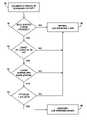
- FIG. 2is a functional block diagram illustrating the method to record and process acoustic data from stethoscope 14 .
- the techniquebegins when the physician turns on electronic stethoscope 14 . Once initiated, the acoustic sensor of stethoscope 14 is placed over the fourth left intercostal space of the chest ( 18 ). Once positioned, stethoscope 14 begins recording acoustic sounds produced over systole and diastole within the chest ( 20 ). Stethoscope 14 begins processing the signal by applying an analog to digital converter to produce digital data representative of an acoustic signal ( 22 ). Next, stethoscope 14 downsamples the digital data by one half to create a manageable data set ( 24 ).
- Stethoscope 14also applies a first filter (“Filter 1”) to the data in order to remove non-stenosis frequencies from the data ( 26 ), after which the stethoscope applies a low-pass filter (“Filter 2”) to the data to eliminate frequencies above 100 Hz ( 28 ). Then, stethoscope 14 calculates a Fast Fourier Transform (FFT) of the data ( 30 ), in which the resultant FFT data may analyzed and/or graphically plotted with frequency on the x-axis and magnitude on the y-axis ( 32 ). Fourier analysis is based on the concept that signals can be approximated by a sum of sinusoids, each at a different frequency.
- FFTFast Fourier Transform
- Stethoscope 14may then calculates a maximum and minimum bar from the FFT plot ( 34 ) and produces diagnostic data for the coronary artery ( 36 ).
- this functional block diagramshows the process stethoscope 14 would use to automatically process acoustic data for the diagnosis of coronary artery disease.
- this processmay be performed automatically by interface module 16 or manually by a clinician or technician with the aid of a personal computer.
- FIG. 3is a functional block diagram illustrating an exemplary method in which stethoscope 14 automatically diagnoses coronary artery stenosis from the processed acoustic data.
- the diagnostic data containing the coronary artery data ( 36 )needs to be analyzed for indications of stenosis. This may be done manually by a clinician or automatically by stethoscope 14 , as illustrated by this example.
- stethoscope 14determines whether a bell shaped curve exists within a particular spectral region of the FFT data ( 38 ). In particular, the stethoscope 14 determines whether the onset of the curve (i.e., the lower frequency where the upslope of the curve rises) occurs substantially at or closely after 50 Hz ( 40 ).
- stethoscopedetermines whether the downslope of the bell shaped curve ends substantially at or before 80 Hz, i.e., that the bell shaped curve is bounded by this spectral region ( 42 ). If so, stethoscope 14 determines whether the FFT peak of the bell curve bounded by this region exceeds than 2.5 units ( 44 ). If all four of these criteria are met, stethoscope 14 (or interface module 16 ) outputs an indicator, e.g., a message, that the Left Anterior Descending (LAD) portion of the coronary artery is likely to have more 50 to 99 percent stenosis preventing blood flow and that coronary disease may be present. ( 46 ). If one or more of the presented criteria is not satisfied, then the stethoscope outputs an indicator, e.g., message, that the patient is likely to have less than 50 percent of the LAD portion of the coronary artery blocked ( 48 ).
- LADLeft Anterior Descending
- this functional block diagramshows the process stethoscope 14 would use to automatically diagnose coronary artery disease from processed acoustic data.
- this processmay be performed automatically by interface module 16 or manually by a clinician or technician with the aid of a personal computer.
- the FFT datamay be graphed or plotted for the clinician to observe the data as a whole, and the identified bell curve may be highlighted or colored differently from the other portions of the graph. For example, a bell shaped group of data would be identifiable between 50 and 80 Hz on the graph in the case of a patient with greater than 50 percent artery stenosis.
- the criteria described in FIG. 3may be determined in different order. For example, after the bell shaped curve is identified in block 38 , the next criteria of the curve identified may be a FFT peak of greater than 2.5 units as shown in block 44 . While each criterion is assessed to determine which indicator to output relative to the likely presence of LAD stenosis, the order in which they are assessed is not as critical.
- certain criteriamay indicate a particular percentage of stenosis and enable the diagnosis to separate a patient into more than two diseased states.
- a more sensitive diagnosis indicating more percentage levels of stenosismay allow for certain patients to wait before undergoing treatment while patients diagnosed with a greater percentage of stenosis would be urged to have immediate surgical intervention.
- LAD coronary artery stenosismay be detected through a slightly different method. Calculating the sum of the energy under the bell curve described above may allow the stethoscope to present an interval of probability of 50 to 99 percent stenosis. Calculating the energy from the data may enable more accurate diagnoses. Further, the summed energy information may allow the stethoscope to provide increased separation of the degree of stenosis. For example, the patient may be identified as likely having 25 to 50 percent, 50 to 75 percent, or 75 to 99 percent stenosis of the LAD or alternate portion of the coronary artery. This information may be beneficial to both the patient and physician.
- the specific numerical indicators for detecting stenosis in the LADmay vary depending on the condition of the patient.
- the FFT peak in block 44may be greater than 2.5 units to indicate stenosis over 50 percent patients over 60 years of age, whereas the FFT peak in block 44 may be greater than 3 units to indicate stenosis over 50 percent in patients younger than 60 years of age.
- Factorssuch as age, gender, height, weight or history may be used in determining the exact criteria numbers. In general, the exact coefficients used in these criteria may be modified to best match the patient.
- This techniquemay be particularly useful for initial screening of patients for LAD stenosis indicating coronary artery disease during a routine exam with a primary care physician. Patients failing to meet every criterion would not need further tests until at some time they show stenosis greater than 50 percent. Patients diagnosed with LAD stenosis greater than 50 percent may be directed toward further testing, such as an angiogram or angioplasty.
- processorsmay include processors that are realized by microprocessors, Application-Specific Integrated Circuits (ASIC), Field-Programmable Gate Arrays (FPGA), or other equivalent integrated logic circuitry.
- the processormay also utilize several different types of storage methods to hold computer-readable instructions for the device operation and data storage. These memory and storage media types may include a type of hard disk, random access memory (RAM), or flash memory, e.g. CompactFlash or SmartMedia. Each storage option may be chosen depending on the embodiment of the invention.
- FIGS. 4 through 8illustrate some of the results of the disclosed technique for diagnosing coronary artery stenosis when performed on patients that were already scheduled for a routine coronary angiogram due to typical symptoms or a positive stress test. Patients were excluded from this study if they had prior coronary artery bypass or cardiac transplant surgery, persistent atrial fibrillation, or any type of cardiac device including pacemakers and prosthetic valves.
- Coronary artery stenosiswas classified as 25-50 percent, 50-75 percent, 75-90 percent, and 90-99 percent occlusion pre-intervention at the referral center.
- Data from 55 random patientswas collected (age range 29-84, mean 57; 30 female, 25 male, Body Mass Index range 21-50, mean 30). Seventeen patients had normal angiograms, 6 had stenosis in coronary arteries other than the LAD, 11 had 25-50 percent stenosis, 5 had 50-75 percent stenosis, 5 had 75-90 percent stenosis, and 7 had 90-99 percent stenosis.
- the 4 lics thorax soundswere sampled as a series of voltages representing the sound amplitude of the acoustic signature.
- a filterwas applied to the data to remove frequency information not related to stenosis in the LAD.
- a lowpass filterwas then applied to the signal to eliminate noise and narrow the bandwidth to less than 100 Hz.
- the techniquesincluded an analysis of acoustic information within the 20-100 Hz range of both the systolic and diastolic segments of the cardiac cycle. The frequency range and use of the entire cardiac cycle was used.
- FFTFast Fourier Transform
- the graphs of FIGS. 4-8compares data from real patients with and without coronary artery disease as determined through an angiogram, the current standard in conventional diagnosis. In the case of patients receiving intervention to rectify their disease, data was acquired before the angiogram and after the angioplasty. The same stethoscope was used for each diagnosis of a single patient.
- FIG. 4shows data charts generated for two separate normal patient data sets after processing is completed.
- the FFT graphical resultshows normal coronary arteries between 0 and 100 Hz.
- the 50 to 80 Hz frequency bands associated with suspected LAD stenosisare highlighted with a rectangular box.
- the coefficient magnitudedecays with increasing frequency. In this example, there is no bell-shaped curve, so peak magnitude is irrelevant and the data fails to meet the criteria set forth in FIG. 3 .
- FIG. 5shows data charts from a patient confirmed to have 90 to 99 percent artery stenosis in the proximal LAD.
- Graph Ashows pre-angiogram data
- graph Bshows post-angioplasty data.
- the bell-shaped curve that may be associated with LAD stenosiswas detected as shown.
- the bell-shaped curve upslopebegins at 50 Hz and the downslope ends near 65 Hz.
- the maximum coefficient magnitudeis greater than 2.5 units and is found at 55 Hz, the peak of the bell-shaped curve.
- Graph B of the patent taken post angioplastyis similar to a graph of a normal patient.
- FIG. 6shows data charts from a patient confirmed to have 50 to 75 percent stenosis in the first diagonal branch of the LAD.
- Graph Ashows pre-angiogram data
- graph Bshows post-angioplasty data.
- LAD stenosiswas identified with the bell-shaped curve in the middle of the 50 to 80 Hz frequency range, and then confirmed via an angiogram. In this case, the maximum coefficient peak was greater than 2.5 units at approximately 65 Hz and decays to 80 Hz.
- a single drug-eluting stentwas placed during angioplasty of this patient. Again, the techniques correctly identified a patient with a diseased coronary artery.
- FIG. 7shows data charts from a patient with 75 to 90 percent stenosis in the LAD and 100 percent artery stenosis in the right coronary artery.
- Graph Ashows pre-angiogram data
- graph Bshows post-angioplasty data.
- the upslope of the bell-shaped curvebegins at 50 Hz.
- the maximum coefficient magnitudeis greater than 2.5 units at 55 Hz.
- the bell-shaped curveends at 60 Hz. This technique correctly diagnosed a diseased coronary artery in this patient.
- Graph Bshows the absence of the bell-shaped curve after the angioplasty of the LAD was performed, and stenosis similar to that of a normal patient.
- FIG. 8shows data charts from a patient with 50 to 75 percent stenosis in the LAD and 100 percent artery stenosis in the right coronary artery.
- Graph Ashows pre-angiogram data
- graph Bshows post-angioplasty data.
- the upslope of the bell-shaped curvebegins at 50 Hz, peaks above 2.5 units at 65 Hz and downslopes to end before 80 Hz.
- graph Bshows the disappearance of the bell-shaped curve due to the opening and stenting of the LAD.
- the datawas statistically analyzed by first grouping the data according to whether a patient had any stenosis in the LAD. If a patient had stenosis in other coronary arteries but not the LAD, they were considered normal. The 25-50 percent stenosed patients were included in normal because the angiogram indicated ‘no significant focal stenosis.” There were a total of 34 normal patients. If the LAD was stenosed more than 50 percent, it was called stenosed LAD. There were a total of 17 stenosed LAD patients.
- Table 1shows how the patients were assessed in the angiogram and how well the technique performed:
- this toolmay be useful in screening patients with suspected coronary artery disease, atypical symptoms, family history or multiple risk factors.
- the methodis applied to raw acoustic information, the resulting analysis can be correlated to stenosis in the left anterior descending coronary artery.
- data from several stethoscope diagnosesmay be compared as the condition of a patient develops. This technique may be particularly useful in monitoring the recovery of a patient after angioplasty or placement of a stent.
- the blockage in an arterymay return in a process called restenosis.
- Using a stethoscope to diagnose restenosismay allow for more frequent tests and the avoidance of multiple angiograms.
Landscapes
- Physics & Mathematics (AREA)
- Acoustics & Sound (AREA)
- Health & Medical Sciences (AREA)
- Life Sciences & Earth Sciences (AREA)
- Engineering & Computer Science (AREA)
- Biomedical Technology (AREA)
- Heart & Thoracic Surgery (AREA)
- Medical Informatics (AREA)
- Molecular Biology (AREA)
- Surgery (AREA)
- Animal Behavior & Ethology (AREA)
- General Health & Medical Sciences (AREA)
- Public Health (AREA)
- Veterinary Medicine (AREA)
- Measuring Pulse, Heart Rate, Blood Pressure Or Blood Flow (AREA)
Abstract
Description
| TABLE 1 | ||||
| Normal | Stenosed LAD | Total | ||
| Normal | 17 | 0 | 17 | ||
| 6 | 0 | 6 | |||
| 25–50% | 9 | 2 | 11 | ||
| 32 | 2 | 34 | |||
| 50-75% | 0 | 5 | 5 | ||
| 75-90% | 3 | 2 | 5 | ||
| 90-99% | 3 | 4 | 7 | ||
| 6 | 11 | 17 | |||
Claims (24)
Priority Applications (2)
| Application Number | Priority Date | Filing Date | Title |
|---|---|---|---|
| US11/402,654US7520860B2 (en) | 2005-04-13 | 2006-04-12 | Detection of coronary artery disease using an electronic stethoscope |
| US12/406,849US10039520B2 (en) | 2005-04-13 | 2009-03-18 | Detection of coronary artery disease using an electronic stethoscope |
Applications Claiming Priority (2)
| Application Number | Priority Date | Filing Date | Title |
|---|---|---|---|
| US67090505P | 2005-04-13 | 2005-04-13 | |
| US11/402,654US7520860B2 (en) | 2005-04-13 | 2006-04-12 | Detection of coronary artery disease using an electronic stethoscope |
Related Child Applications (1)
| Application Number | Title | Priority Date | Filing Date |
|---|---|---|---|
| US12/406,849ContinuationUS10039520B2 (en) | 2005-04-13 | 2009-03-18 | Detection of coronary artery disease using an electronic stethoscope |
Publications (2)
| Publication Number | Publication Date |
|---|---|
| US20060245597A1 US20060245597A1 (en) | 2006-11-02 |
| US7520860B2true US7520860B2 (en) | 2009-04-21 |
Family
ID=37234447
Family Applications (2)
| Application Number | Title | Priority Date | Filing Date |
|---|---|---|---|
| US11/402,654Expired - Fee RelatedUS7520860B2 (en) | 2005-04-13 | 2006-04-12 | Detection of coronary artery disease using an electronic stethoscope |
| US12/406,849Active2028-01-21US10039520B2 (en) | 2005-04-13 | 2009-03-18 | Detection of coronary artery disease using an electronic stethoscope |
Family Applications After (1)
| Application Number | Title | Priority Date | Filing Date |
|---|---|---|---|
| US12/406,849Active2028-01-21US10039520B2 (en) | 2005-04-13 | 2009-03-18 | Detection of coronary artery disease using an electronic stethoscope |
Country Status (1)
| Country | Link |
|---|---|
| US (2) | US7520860B2 (en) |
Cited By (18)
| Publication number | Priority date | Publication date | Assignee | Title |
|---|---|---|---|---|
| US20090177107A1 (en)* | 2005-04-13 | 2009-07-09 | Marie A. Guion-Johnson | Detection of coronary artery disease using an electronic stethoscope |
| US20110137210A1 (en)* | 2009-12-08 | 2011-06-09 | Johnson Marie A | Systems and methods for detecting cardiovascular disease |
| US20110196243A1 (en)* | 2010-02-05 | 2011-08-11 | Riheng Wu | Non-contact detection of physiological data using stochastic resonance |
| US8301232B2 (en) | 2010-06-08 | 2012-10-30 | Alivecor, Inc. | Wireless, ultrasonic personal health monitoring system |
| US8509882B2 (en) | 2010-06-08 | 2013-08-13 | Alivecor, Inc. | Heart monitoring system usable with a smartphone or computer |
| US8700137B2 (en) | 2012-08-30 | 2014-04-15 | Alivecor, Inc. | Cardiac performance monitoring system for use with mobile communications devices |
| US20150080748A1 (en)* | 2012-04-13 | 2015-03-19 | Laila Hübbert | Method and System for Predicting Cardiovascular Events |
| US20150250445A1 (en)* | 2012-09-07 | 2015-09-10 | The Regents Of The University Of California | Multisensor wireless abdominal monitoring apparatus, systems, and methods |
| US9220430B2 (en) | 2013-01-07 | 2015-12-29 | Alivecor, Inc. | Methods and systems for electrode placement |
| US9226726B1 (en) | 2014-11-25 | 2016-01-05 | John L Semmlow | Method and system for detection of cardiac sounds |
| US9247911B2 (en) | 2013-07-10 | 2016-02-02 | Alivecor, Inc. | Devices and methods for real-time denoising of electrocardiograms |
| US9254095B2 (en) | 2012-11-08 | 2016-02-09 | Alivecor | Electrocardiogram signal detection |
| US9254092B2 (en) | 2013-03-15 | 2016-02-09 | Alivecor, Inc. | Systems and methods for processing and analyzing medical data |
| US9351654B2 (en) | 2010-06-08 | 2016-05-31 | Alivecor, Inc. | Two electrode apparatus and methods for twelve lead ECG |
| US9420956B2 (en) | 2013-12-12 | 2016-08-23 | Alivecor, Inc. | Methods and systems for arrhythmia tracking and scoring |
| US9839363B2 (en) | 2015-05-13 | 2017-12-12 | Alivecor, Inc. | Discordance monitoring |
| US11284827B2 (en) | 2017-10-21 | 2022-03-29 | Ausculsciences, Inc. | Medical decision support system |
| US11375976B2 (en) | 2017-11-03 | 2022-07-05 | John Jun Cai | Wireless stethoscope for transmitting, recording, storing and diagnostic capabilities including an earpiece |
Families Citing this family (18)
| Publication number | Priority date | Publication date | Assignee | Title |
|---|---|---|---|---|
| US8920343B2 (en) | 2006-03-23 | 2014-12-30 | Michael Edward Sabatino | Apparatus for acquiring and processing of physiological auditory signals |
| EP2462871A1 (en)* | 2010-12-13 | 2012-06-13 | Acarix A/S | System, stethoscope and method for indicating risk of coronary artery disease |
| US9636025B2 (en) | 2012-08-15 | 2017-05-02 | Nanyang Technological University | Systems and methods for pedal revascularization assessment |
| US11206990B2 (en) | 2013-01-23 | 2021-12-28 | Pedra Technology Pte Ltd | Deep tissue flowmetry using diffuse speckle contrast analysis |
| CA2921206A1 (en)* | 2013-08-14 | 2015-02-19 | Nanyang Technological University | Systems and methods for revascularization assessment |
| EP2945084A1 (en)* | 2014-05-12 | 2015-11-18 | Electrosalus Biyomedikal Sanayi ve Ticaret Anonim Sirketi | Auscultation data acquisition, communication and evaluation system incorporating mobile facilities |
| RU2644546C1 (en)* | 2016-10-31 | 2018-02-12 | Общество с ограниченной ответственностью "Научно-технический центр "Интеллектуальные телеуправляемые робототехнические системы" (ООО "НТЦ "Интелрос") | Electronic medical stethoscope |
| JP7393947B2 (en) | 2017-05-22 | 2023-12-07 | ジェネテシス エルエルシー | Mechanical identification of anomalies in bioelectromagnetic fields |
| US11134877B2 (en) | 2017-08-09 | 2021-10-05 | Genetesis, Inc. | Biomagnetic detection |
| US12262997B2 (en) | 2017-08-09 | 2025-04-01 | Genetesis, Inc. | Biomagnetic detection |
| TWI721395B (en)* | 2018-04-21 | 2021-03-11 | 美商腎通生醫科技公司 | Arteriovenous fistula stenosis detection system and method thereof and sensing device |
| US20220240793A1 (en)* | 2018-04-21 | 2022-08-04 | Above Care Inc. | Detection system, detection method and sensing device for detecting stenosis of carotid artery |
| JP7394133B2 (en)* | 2018-11-20 | 2023-12-07 | ジェネテシス インク. | System and method for diagnosing cardiac ischemia and coronary artery disease |
| US10602940B1 (en) | 2018-11-20 | 2020-03-31 | Genetesis, Inc. | Systems, devices, software, and methods for diagnosis of cardiac ischemia and coronary artery disease |
| RU2700471C9 (en)* | 2018-12-26 | 2020-04-30 | Альберт Сергеевич Канышев | Method of non-invasive acoustic spectral screening of heart vessels |
| WO2020150512A1 (en)* | 2019-01-16 | 2020-07-23 | General Electric Company | Vascular assessment using acoustic sensing |
| GB202403149D0 (en) | 2024-03-05 | 2024-04-17 | Univ London Queen Mary | Arterial blockage detector |
| CN120078440A (en)* | 2025-03-13 | 2025-06-03 | 苏州仲如悦科技有限责任公司 | A detection system for arteriovenous vascular access stenosis based on auscultatory acoustic signals |
Citations (10)
| Publication number | Priority date | Publication date | Assignee | Title |
|---|---|---|---|---|
| US4905706A (en) | 1988-04-20 | 1990-03-06 | Nippon Colin Co., Ltd. | Method an apparatus for detection of heart disease |
| US5010889A (en)* | 1988-02-04 | 1991-04-30 | Bloodline Technology | Intelligent stethoscope |
| US5036857A (en) | 1989-10-26 | 1991-08-06 | Rutgers, The State University Of New Jersey | Noninvasive diagnostic system for coronary artery disease |
| US5109863A (en) | 1989-10-26 | 1992-05-05 | Rutgers, The State University Of New Jersey | Noninvasive diagnostic system for coronary artery disease |
| US6048319A (en) | 1998-10-01 | 2000-04-11 | Integrated Medical Systems, Inc. | Non-invasive acoustic screening device for coronary stenosis |
| US6053872A (en) | 1996-12-18 | 2000-04-25 | Aurora Holdings, Llc | Cardiac sonospectrographic analyzer |
| US6135966A (en) | 1998-05-01 | 2000-10-24 | Ko; Gary Kam-Yuen | Method and apparatus for non-invasive diagnosis of cardiovascular and related disorders |
| US6193668B1 (en) | 1997-11-10 | 2001-02-27 | Medacoustics, Inc. | Acoustic sensor array for non-invasive detection of coronary artery disease |
| US6328698B1 (en) | 1998-08-18 | 2001-12-11 | Hiroshi Matsumoto | Diagnostic system and method for coronary artery disease and others |
| US20030229289A1 (en)* | 2002-03-18 | 2003-12-11 | Mohler Sailor Hampton | Method and system for generating a likelihood of cardiovascular disease, analyzing cardiovascular sound signals remotely from the location of cardiovascular sound signal acquisition, and determining time and phase information from cardiovascular sound signals |
Family Cites Families (176)
| Publication number | Priority date | Publication date | Assignee | Title |
|---|---|---|---|---|
| US3830223A (en) | 1972-09-18 | 1974-08-20 | Technicon Instr | Methodology and apparatus for non-invasive biophysical diagnosis |
| US3951140A (en)* | 1974-11-13 | 1976-04-20 | Indianapolis Center For Advanced Research | Ultrasonic therapy apparatus and method |
| US4373532A (en)* | 1980-07-07 | 1983-02-15 | Palo Alto Medical Research Foundation | Ultrasonic marker for physiologic diagnosis and method of using same |
| US4759374A (en) | 1985-05-06 | 1988-07-26 | American Telephone And Telegraph Company And At&T Bell Laboratories | Non-invasive blood flow measurements utilizing cardiac cycle synchronization |
| US4719923A (en)* | 1985-05-06 | 1988-01-19 | American Telephone And Telegraph Company, At&T Bell Laboratories | Non-invasive blood flow measurements utilizing autoregressive analysis with averaged reflection coefficients |
| DE3744605C1 (en) | 1987-12-31 | 1989-04-27 | Jochen Dipl-Ing Heimann | Sensor |
| US5218969A (en)* | 1988-02-04 | 1993-06-15 | Blood Line Technology, Inc. | Intelligent stethoscope |
| US4947859A (en) | 1989-01-25 | 1990-08-14 | Cherne Medical, Inc. | Bio-acoustic signal sensing device |
| EP0520015A1 (en) | 1990-03-16 | 1992-12-30 | Seismed Instruments, Inc. | Myocardial ischemia detection system |
| US5337752A (en) | 1992-05-21 | 1994-08-16 | Mcg International, Inc. | System for simultaneously producing and synchronizing spectral patterns of heart sounds and an ECG signal |
| US5365937A (en) | 1992-09-09 | 1994-11-22 | Mcg International, Inc. | Disposable sensing device with contaneous conformance |
| DK63193D0 (en) | 1993-06-02 | 1993-06-02 | Bang & Olufsen Tech As | HEART SIGNAL MEASUREMENT APPARATUS |
| AU7715494A (en)* | 1993-08-30 | 1995-03-22 | Mcg International, Inc. | Disposable acoustic pad sensors |
| CA2198844C (en) | 1994-08-30 | 2007-08-07 | Knud Erik Baekgaard | An electronic stethoscope |
| US8280682B2 (en) | 2000-12-15 | 2012-10-02 | Tvipr, Llc | Device for monitoring movement of shipped goods |
| US5722419A (en)* | 1994-11-30 | 1998-03-03 | Semmlow; John L. | System for determining the viability of tissue |
| US5617869A (en)* | 1995-06-16 | 1997-04-08 | The United States Of America As Represented By The Secretary Of The Navy | Device and method for locating flow blockage in a three-dimensional object |
| US5957866A (en) | 1995-07-03 | 1999-09-28 | University Technology Corporation | Apparatus and methods for analyzing body sounds |
| US5687738A (en) | 1995-07-03 | 1997-11-18 | The Regents Of The University Of Colorado | Apparatus and methods for analyzing heart sounds |
| US5825895A (en) | 1995-07-21 | 1998-10-20 | Stethtech Corporation | Electronic stethoscope |
| US6002777A (en) | 1995-07-21 | 1999-12-14 | Stethtech Corporation | Electronic stethoscope |
| US5638823A (en)* | 1995-08-28 | 1997-06-17 | Rutgers University | System and method for noninvasive detection of arterial stenosis |
| NO304870B1 (en)* | 1995-11-16 | 1999-02-22 | Meditron As | Electronic Stethoscope listening head |
| US7811090B2 (en) | 1996-05-08 | 2010-10-12 | Gaumard Scientific Company, Inc. | Interactive education system for teaching patient care |
| US5913826A (en)* | 1996-06-12 | 1999-06-22 | K-One Technologies | Wideband external pulse cardiac monitor |
| AU8677098A (en) | 1997-07-31 | 1999-02-22 | Case Western Reserve University | A system and method for non-invasive electrocardiographic imaging |
| CA2300843A1 (en) | 1997-08-19 | 1999-02-25 | Philipp Lang | Measurement of capillary related interstitial fluid using ultrasound methods and devices |
| US6005915A (en) | 1997-11-07 | 1999-12-21 | Advanced Micro Devices, Inc. | Apparatus and method for measuring the roughness of a target material surface based upon the scattering of incident X-ray photons |
| US6278890B1 (en) | 1998-11-09 | 2001-08-21 | Medacoustics, Inc. | Non-invasive turbulent blood flow imaging system |
| US6261237B1 (en) | 1998-08-20 | 2001-07-17 | Medacoustics, Inc. | Thin film piezoelectric polymer sensor |
| US7346174B1 (en)* | 1998-10-05 | 2008-03-18 | Clive Smith | Medical device with communication, measurement and data functions |
| US6371924B1 (en)* | 1998-11-09 | 2002-04-16 | Medacoustics, Inc. | Acoustic window identification |
| CA2359816C (en)* | 1999-01-06 | 2010-08-03 | Genenews Inc. | Method for the detection of gene transcripts in blood and uses thereof |
| US7037268B1 (en)* | 1999-03-01 | 2006-05-02 | Medacoustics, Inc. | Low profile acoustic sensor arry and sensors with pleated transmission lines and related methods |
| AU2004201785B2 (en) | 1999-03-01 | 2007-10-25 | Harris Corporation | Low Profile Acoustic sensor array and sensors with pleated transmission lines and related methods |
| US6572560B1 (en) | 1999-09-29 | 2003-06-03 | Zargis Medical Corp. | Multi-modal cardiac diagnostic decision support system and method |
| US6878117B1 (en)* | 1999-09-29 | 2005-04-12 | Zargis Medical Corp. | Handheld sensor for acoustic data acquisition |
| US7246069B1 (en) | 1999-10-15 | 2007-07-17 | Ue Systems, Inc. | Method and apparatus for online health monitoring |
| US6527729B1 (en) | 1999-11-10 | 2003-03-04 | Pacesetter, Inc. | Method for monitoring patient using acoustic sensor |
| US6477406B1 (en) | 1999-11-10 | 2002-11-05 | Pacesetter, Inc. | Extravascular hemodynamic acoustic sensor |
| EP1248556A2 (en) | 2000-01-07 | 2002-10-16 | Rice Creek Medical, L.L.C. | Non-invasive method and apparatus for monitoring intracranial pressure |
| AU2001243273A1 (en)* | 2000-02-23 | 2001-09-03 | The Johns-Hopkins University | System and method for diagnosing pathologic heart conditions |
| US20040260193A1 (en) | 2000-05-12 | 2004-12-23 | Lasala Anthony F. | Cardiac impulse detector |
| US6533736B1 (en)* | 2000-05-30 | 2003-03-18 | Mark Moore | Wireless medical stethoscope |
| US6994675B2 (en)* | 2000-07-19 | 2006-02-07 | Sharrock Nigel E | Non-invasive measurement of suprasystolic signals |
| US6733450B1 (en)* | 2000-07-27 | 2004-05-11 | Texas Systems, Board Of Regents | Therapeutic methods and apparatus for use of sonication to enhance perfusion of tissue |
| US6544189B2 (en)* | 2000-09-25 | 2003-04-08 | Zargis Medical Corp. | Handheld sensor for acoustic data acquisition |
| US6709399B1 (en)* | 2000-10-20 | 2004-03-23 | Cardiotran Lcc | Method and system for the detection of heart disease |
| US20070258894A1 (en) | 2000-11-08 | 2007-11-08 | Melker Richard J | System and Method for Real-Time Diagnosis, Treatment, and Therapeutic Drug Monitoring |
| US6947789B2 (en) | 2000-11-13 | 2005-09-20 | Innovise Medical, Inc. | Method for detecting, sizing and locating old myocardial infarct |
| US20060079773A1 (en)* | 2000-11-28 | 2006-04-13 | Allez Physionix Limited | Systems and methods for making non-invasive physiological assessments by detecting induced acoustic emissions |
| AU2002239360A1 (en)* | 2000-11-28 | 2002-06-11 | Allez Physionix Limited | Systems and methods for making non-invasive physiological assessments |
| US20020133069A1 (en) | 2000-12-18 | 2002-09-19 | Roberts Lauri E. | Electrode placement device for taking electrocardiograms and method of use |
| US7527597B2 (en)* | 2001-01-16 | 2009-05-05 | Biomedical Acoustic Research Corporation | Acoustic detection of vascular conditions |
| US6780159B2 (en) | 2001-01-16 | 2004-08-24 | Biomedical Acoustic Research Corporation | Acoustic detection of vascular conditions |
| ATE308922T1 (en)* | 2001-03-02 | 2005-11-15 | Palti Yoram Prof | DEVICE FOR DETECTING ARTERIAL STENOSIS |
| US20020156385A1 (en) | 2001-04-24 | 2002-10-24 | Genquan Feng | Differentiation of CAD vs NCI with different patterns of empi indexes |
| EP1389957A1 (en) | 2001-05-28 | 2004-02-25 | Health Devices Pte Ltd. | Heart diagnosis system |
| US7054679B2 (en) | 2001-10-31 | 2006-05-30 | Robert Hirsh | Non-invasive method and device to monitor cardiac parameters |
| KR20030065228A (en)* | 2002-01-31 | 2003-08-06 | 김종수 | System for outputs Ch'ongjin acoustic signal |
| JP3875581B2 (en) | 2002-03-18 | 2007-01-31 | 独立行政法人科学技術振興機構 | Ultrasound diagnostic system |
| DE10219367A1 (en) | 2002-04-30 | 2003-11-13 | Jan Manolas | Device for determining and evaluating parameters of the diastolic function of the left ventricle |
| US7113825B2 (en) | 2002-05-03 | 2006-09-26 | Cardiac Pacemakers, Inc. | Method and apparatus for detecting acoustic oscillations in cardiac rhythm |
| USD477405S1 (en) | 2002-06-21 | 2003-07-15 | George Sommerfeld | Electronic stethoscope chestpiece |
| WO2004040000A2 (en)* | 2002-09-09 | 2004-05-13 | Nura, Inc | G protein coupled receptors and uses thereof |
| US7458939B2 (en) | 2002-10-09 | 2008-12-02 | Bang & Olufsen Medicom A/S | Procedure for extracting information from a heart sound signal |
| US20040073094A1 (en)* | 2002-10-15 | 2004-04-15 | Baker Donald A. | Fetal monitoring systems with ambulatory patient units and telemetric links for improved uses |
| US7020581B2 (en)* | 2002-10-18 | 2006-03-28 | Medacoustics Research & Technology | Medical hearing aid analysis system |
| US7819814B2 (en)* | 2002-10-21 | 2010-10-26 | Noam Gavriely | Acoustic assessment of the heart |
| US20040092846A1 (en)* | 2002-10-31 | 2004-05-13 | Watrous Raymond L. | Graphical user interface and voice-guided protocol for an auscultatory diagnostic decision support system |
| US7289634B2 (en) | 2002-11-18 | 2007-10-30 | Zargis Medical Corporation | Electronic stethoscope measurement system and method |
| US7260429B2 (en)* | 2002-12-02 | 2007-08-21 | Cardiac Pacemakers, Inc. | Method and apparatus for phonocardiographic image acquisition and presentation |
| US7123962B2 (en)* | 2002-12-02 | 2006-10-17 | Cardiac Pacemakers, Inc. | Phonocardiographic image-based atrioventricular delay optimization |
| JP3683886B2 (en) | 2002-12-27 | 2005-08-17 | 株式会社ワイディ | Blood volume analysis and display method using Myo Cardial Blood volume map |
| US7001338B2 (en)* | 2003-03-26 | 2006-02-21 | The Johns Hopkins University | System and method for diagnosing pathologic heart conditions |
| JP2006524546A (en) | 2003-04-23 | 2006-11-02 | ヘムチャンドラ・シャートゥクド | Apparatus and method for non-invasive diagnosis of coronary artery disease |
| US6974415B2 (en) | 2003-05-22 | 2005-12-13 | Magnetus Llc | Electromagnetic-acoustic imaging |
| US7096060B2 (en) | 2003-06-27 | 2006-08-22 | Innovise Medical, Inc. | Method and system for detection of heart sounds |
| US7351207B2 (en)* | 2003-07-18 | 2008-04-01 | The Board Of Trustees Of The University Of Illinois | Extraction of one or more discrete heart sounds from heart sound information |
| IL164030A0 (en)* | 2003-09-12 | 2005-12-18 | Revital Pery Shechter | Photoacoustic analyzer of a region of interest in a human body |
| US7300405B2 (en) | 2003-10-22 | 2007-11-27 | 3M Innovative Properties Company | Analysis of auscultatory sounds using single value decomposition |
| EP1682004B1 (en)* | 2003-10-23 | 2012-06-20 | Koninklijke Philips Electronics N.V. | Ultrasound imaging apparatus |
| US7142919B2 (en)* | 2003-10-24 | 2006-11-28 | Medtronic, Inc. | Reconfigurable, fault tolerant multiple-electrode cardiac lead systems |
| US7300407B2 (en) | 2003-11-10 | 2007-11-27 | Zargis Medical Corporation | Handheld auscultatory scanner with synchronized display of heart sounds |
| US7028861B2 (en)* | 2003-12-16 | 2006-04-18 | Joseph S. Kanfer | Electronically keyed dispensing systems and related methods of installation and use |
| US7477936B2 (en)* | 2003-12-19 | 2009-01-13 | Aalborg Universitet | System and a method for analyzing ECG curvature |
| EP1543770A1 (en) | 2003-12-19 | 2005-06-22 | Aalborg Universitet | A system and a method for analysing an ECG signal |
| US20050137464A1 (en) | 2003-12-23 | 2005-06-23 | Bomba Frank C. | Wireless sensor and sensor initialization device and method |
| EP1722686A4 (en) | 2004-02-10 | 2009-07-22 | Cardiovascular Resonances Llc | Methods, systems, and computer program products for analyzing cardiovascular sounds using eigen functions |
| US20050222515A1 (en) | 2004-02-23 | 2005-10-06 | Biosignetics Corporation | Cardiovascular sound signature: method, process and format |
| US7981058B2 (en) | 2004-03-12 | 2011-07-19 | The Trustees Of Dartmouth College | Intelligent wearable monitor systems and methods |
| US20050261598A1 (en)* | 2004-04-07 | 2005-11-24 | Triage Wireless, Inc. | Patch sensor system for measuring vital signs |
| US7162294B2 (en)* | 2004-04-15 | 2007-01-09 | Ge Medical Systems Information Technologies, Inc. | System and method for correlating sleep apnea and sudden cardiac death |
| USD527819S1 (en) | 2004-04-19 | 2006-09-05 | Bang & Olufsen Medicom A/S | Forepart to a stethoscope |
| US7765001B2 (en)* | 2005-08-31 | 2010-07-27 | Ebr Systems, Inc. | Methods and systems for heart failure prevention and treatments using ultrasound and leadless implantable devices |
| US7462153B2 (en) | 2004-07-23 | 2008-12-09 | Sonomedica, Inc. | Method and system for modeling cardiovascular disease using a probability regession model |
| US20060025690A1 (en)* | 2004-07-27 | 2006-02-02 | Guigne Jacques Y | Acoustic body examination |
| EP1784130A4 (en)* | 2004-08-31 | 2014-11-19 | Univ Washington | ULTRASONIC TECHNIQUE FOR EVALUATING VIBRATIONS OF WALLS IN STENOSED BLOOD VESSELS |
| US20090030324A1 (en)* | 2004-10-19 | 2009-01-29 | Makoto Kato | Ultrasonic diagnostic apparatus and method for controlling the same |
| US7174203B2 (en)* | 2004-11-18 | 2007-02-06 | Inovise Medical, Inc. | Method and system relating to monitoring and characterizing heart condition |
| US7044922B1 (en)* | 2004-12-29 | 2006-05-16 | Leon Michael Dondysh | Non-invasive diagnostic apparatus and method comprising a cerebral stethoscope for detecting cerebrovascular disease |
| US20060161064A1 (en) | 2005-01-18 | 2006-07-20 | Zargis Medical Corp. | Computer-assisted detection of systolic murmurs associated with hypertrophic cardiomyopathy |
| US20070055151A1 (en) | 2005-01-20 | 2007-03-08 | Shertukde Hemchandra M | Apparatus and methods for acoustic diagnosis |
| US20060167529A1 (en)* | 2005-01-26 | 2006-07-27 | Schecter Stuart O | Method and algorithm for defining the pathologic state from a plurality of intrinsically and extrinsically derived signals |
| US20060241527A1 (en) | 2005-03-03 | 2006-10-26 | Robert Muratore | System and method for inducing controlled cardiac damage |
| US7520860B2 (en) | 2005-04-13 | 2009-04-21 | Marie G. Johnson | Detection of coronary artery disease using an electronic stethoscope |
| DE102005053109A1 (en) | 2005-11-04 | 2007-05-10 | Koehler, Ullrich, Prof. Dr. | Body noise detection |
| EP1954195A1 (en) | 2005-11-17 | 2008-08-13 | Koninklijke Philips Electronics N.V. | Vascular flow sensor with acoustic coupling detector |
| IL185609A0 (en) | 2007-08-30 | 2008-01-06 | Dan Furman | Multi function senssor |
| WO2007091244A1 (en)* | 2006-02-07 | 2007-08-16 | Impulse Dynamics Nv | Assessing cardiac activity |
| US8920343B2 (en) | 2006-03-23 | 2014-12-30 | Michael Edward Sabatino | Apparatus for acquiring and processing of physiological auditory signals |
| US7806833B2 (en) | 2006-04-27 | 2010-10-05 | Hd Medical Group Limited | Systems and methods for analysis and display of heart sounds |
| US9540694B2 (en) | 2006-05-19 | 2017-01-10 | The Johns Hopkins University | HEYL as a therapeutic target and a diagnostic marker for neoplasia and uses therefor |
| WO2008000255A1 (en)* | 2006-06-26 | 2008-01-03 | Coloplast A/S | Method for segmenting a cardiovascular signal |
| WO2008000254A1 (en)* | 2006-06-26 | 2008-01-03 | Coloplast A/S | Multi parametric classification of cardiovascular sounds |
| US20080013747A1 (en) | 2006-06-30 | 2008-01-17 | Bao Tran | Digital stethoscope and monitoring instrument |
| US7877142B2 (en)* | 2006-07-05 | 2011-01-25 | Micardia Corporation | Methods and systems for cardiac remodeling via resynchronization |
| US20080058607A1 (en)* | 2006-08-08 | 2008-03-06 | Zargis Medical Corp | Categorizing automatically generated physiological data based on industry guidelines |
| US20080039733A1 (en)* | 2006-08-08 | 2008-02-14 | Kamil Unver | Systems and methods for calibration of heart sounds |
| US8364249B2 (en)* | 2006-08-11 | 2013-01-29 | 3M Innovative Properties Company | Automatic generation of heart sounds and murmurs using a lumped-parameter recirculating pressure-flow model for the left heart |
| US8001472B2 (en) | 2006-09-21 | 2011-08-16 | Apple Inc. | Systems and methods for providing audio and visual cues via a portable electronic device |
| WO2008036911A2 (en) | 2006-09-22 | 2008-03-27 | University Of Medicine And Dentistry Of New Jersey | System and method for acoustic detection of coronary artery disease |
| US20080091090A1 (en) | 2006-10-12 | 2008-04-17 | Kenneth Shane Guillory | Self-contained surface physiological monitor with adhesive attachment |
| US7840259B2 (en)* | 2006-11-30 | 2010-11-23 | General Electric Company | Method and system for electrocardiogram evaluation |
| US8260406B2 (en)* | 2006-11-30 | 2012-09-04 | Aalborg Universitet | System and method for analyzing complex curvature of ECG curves |
| EP1930045A1 (en)* | 2006-12-08 | 2008-06-11 | BIOTRONIK CRM Patent AG | Implantable medical system with acoustic sensor to measure mitral blood flow |
| US20090093687A1 (en)* | 2007-03-08 | 2009-04-09 | Telfort Valery G | Systems and methods for determining a physiological condition using an acoustic monitor |
| US20080255433A1 (en) | 2007-04-11 | 2008-10-16 | The Board Of Regents Of The University Of Texas Syatem | Optoacoustic monitoring of multiple parameters |
| US9173638B2 (en) | 2007-06-04 | 2015-11-03 | Biosense Webster, Inc. | Cardiac mechanical assessment using ultrasound |
| CN101677810B (en) | 2007-06-04 | 2012-04-04 | 松下电器产业株式会社 | Ultrasonic diagnostic device |
| CN101675296A (en)* | 2007-07-27 | 2010-03-17 | 夏普株式会社 | Lighting device and liquid crystal display device |
| WO2009070302A1 (en) | 2007-11-30 | 2009-06-04 | The Johns Hopkins University | Prostate specific membrane antigen (psma) targeted nanoparticles for therapy of prostate cancer |
| US8280484B2 (en) | 2007-12-18 | 2012-10-02 | The Invention Science Fund I, Llc | System, devices, and methods for detecting occlusions in a biological subject |
| CN101951830B (en) | 2007-12-20 | 2012-11-28 | 阿克瑞克公司 | An adhesive patch for monitoring acoustic signals |
| EP2268311A4 (en)* | 2008-04-04 | 2014-08-27 | Immunolight Llc | Non-invasive systems and methods for in-situ photobiomodulation |
| US20090292309A1 (en) | 2008-05-20 | 2009-11-26 | Michael Maschke | System and workflow for diagnosing and treating septum defects |
| WO2010009747A1 (en) | 2008-07-25 | 2010-01-28 | Helmholtz Zentrum München Deutsches Forschungszentrum Für Gesundheit Und Umwelt (Gmbh) | Quantitative multi-spectral opto-acoustic tomography (msot) of tissue biomarkers |
| US20100030095A1 (en)* | 2008-07-30 | 2010-02-04 | Sustineo Biotechnology | Pulse Acoustic Analysis System for the Diagnostic of Cardiovascular Disease |
| US8235912B2 (en) | 2009-03-18 | 2012-08-07 | Acarix A/S | Segmenting a cardiac acoustic signal |
| TWI511756B (en)* | 2009-04-21 | 2015-12-11 | 伊穆諾萊特公司 | Pharmaceutical composition or kit for modifying a target structure which mediates or is associated with a biological activity |
| US20100312118A1 (en) | 2009-06-03 | 2010-12-09 | Horzewski Michael J | Systems and Methods for Perfusion Enhanced Diagnostic Imaging |
| US9154866B2 (en) | 2009-06-10 | 2015-10-06 | Apple Inc. | Fiber-based electronic device structures |
| US8346354B2 (en)* | 2009-07-28 | 2013-01-01 | The Invention Science Fund I, Llc | Determining a neuromodulation treatment regimen in response to contactlessly acquired information |
| EP3381937A3 (en) | 2009-08-13 | 2018-10-31 | The Johns Hopkins University | Methods of modulating immune function |
| WO2011039329A1 (en) | 2009-09-30 | 2011-04-07 | Acarix A/S | An adhesive patch for monitoring acoustic signals |
| US20110105928A1 (en)* | 2009-11-05 | 2011-05-05 | Newcardio, Inc. | ECG Reconstruction For Atrial Activity Monitoring And Detection |
| US20110137210A1 (en)* | 2009-12-08 | 2011-06-09 | Johnson Marie A | Systems and methods for detecting cardiovascular disease |
| US8154175B2 (en)* | 2009-12-16 | 2012-04-10 | Hamilton Sundstrand Corporation | Sensing device |
| US8509882B2 (en) | 2010-06-08 | 2013-08-13 | Alivecor, Inc. | Heart monitoring system usable with a smartphone or computer |
| WO2012019109A1 (en)* | 2010-08-05 | 2012-02-09 | Abbott Point Of Care Inc. | Oscillating immunoassay method and device |
| US8157742B2 (en)* | 2010-08-12 | 2012-04-17 | Heartflow, Inc. | Method and system for patient-specific modeling of blood flow |
| US8824762B2 (en)* | 2010-10-22 | 2014-09-02 | The Johns Hopkins University | Method and system for processing ultrasound data |
| EP2462871A1 (en) | 2010-12-13 | 2012-06-13 | Acarix A/S | System, stethoscope and method for indicating risk of coronary artery disease |
| US9256933B2 (en) | 2011-02-08 | 2016-02-09 | Region Nordjylland, Aalborg Sygehus | System for determining flow properties of a blood vessel |
| US20120209131A1 (en) | 2011-02-11 | 2012-08-16 | AventuSoft, LLC | Method and System of a Cardio-acoustic Classification system for Screening, Diagnosis and Monitoring of Cardiovascular Conditions |
| US20130211208A1 (en) | 2011-03-08 | 2013-08-15 | Vijay K. Varadan | Smart materials, dry textile sensors, and electronics integration in clothing, bed sheets, and pillow cases for neurological, cardiac and/or pulmonary monitoring |
| US9693759B2 (en) | 2011-11-16 | 2017-07-04 | Coloplast A/S | Operating device with a control handle and a flexible element connected to the control handle |
| US20130137960A1 (en)* | 2011-11-30 | 2013-05-30 | Nellcor Puritan Bennett Llc | Methods and systems for photoacoustic monitoring using indicator dilution |
| US8886294B2 (en)* | 2011-11-30 | 2014-11-11 | Covidien Lp | Methods and systems for photoacoustic monitoring using indicator dilution |
| HK1205025A1 (en) | 2012-05-11 | 2015-12-11 | 加利福尼亚大学董事会 | Portable device to initiate and monitor treatment of stroke victims in the field |
| US9492138B2 (en) | 2012-10-15 | 2016-11-15 | Rijuven Corp | Mobile front-end system for comprehensive cardiac diagnosis |
| JP6678118B2 (en) | 2014-05-15 | 2020-04-08 | ザ リージェンツ オブ ザ ユニバーシティ オブ カリフォルニア | Multi-sensor physiological monitoring system |
| US10478120B2 (en) | 2014-05-17 | 2019-11-19 | The Johns Hopkins University | MRI-guided intraarterial catheter-based method for predicting territory of local blood brain barrier opening |
| US10733266B2 (en) | 2014-05-30 | 2020-08-04 | Apple Inc. | Systems and methods of providing patient apps |
| US9610016B2 (en) | 2014-08-27 | 2017-04-04 | Vladimir Shusterman | Wireless health monitoring in the setting of X-ray, magnetic resonance imaging and other sources of electromagnetic interference |
| US9918646B2 (en) | 2014-09-02 | 2018-03-20 | Apple Inc. | Sensor fusion approach to energy expenditure estimation |
| WO2016040879A1 (en) | 2014-09-12 | 2016-03-17 | The Board Of Trustees Of The Leland Stanford Junior University | Physical examination method and apparatus |
| WO2016070128A1 (en) | 2014-10-31 | 2016-05-06 | Irhythm Technologies, Inc. | Wireless physiological monitoring device and systems |
| US9396369B1 (en) | 2015-02-03 | 2016-07-19 | Apple Inc. | Electronic tag transmissions corresponding to physical disturbance of tag |
| US10335043B2 (en) | 2015-04-06 | 2019-07-02 | Thomas Jefferson University | Implantable vital sign sensor |
| EP3280315B1 (en) | 2015-04-06 | 2020-11-18 | Thomas Jefferson University | Implantable vital sign sensor |
| US11000195B2 (en) | 2015-04-06 | 2021-05-11 | Thomas Jefferson University | Implantable vital sign sensor |
| US9699546B2 (en) | 2015-09-16 | 2017-07-04 | Apple Inc. | Earbuds with biometric sensing |
| WO2017058655A1 (en) | 2015-09-29 | 2017-04-06 | Apple Inc. | Pressure measurement designs |
| US10028677B2 (en) | 2015-10-12 | 2018-07-24 | Corsens Medical Ltd. | Quantitatively differentiating cardiac from non-cardiac related chest pain and other cardiac diagnostics |
| US11096653B2 (en) | 2016-02-12 | 2021-08-24 | Newton Howard | Networked electronic stethoscope |
- 2006
- 2006-04-12USUS11/402,654patent/US7520860B2/ennot_activeExpired - Fee Related
- 2009
- 2009-03-18USUS12/406,849patent/US10039520B2/enactiveActive
Patent Citations (11)
| Publication number | Priority date | Publication date | Assignee | Title |
|---|---|---|---|---|
| US5010889A (en)* | 1988-02-04 | 1991-04-30 | Bloodline Technology | Intelligent stethoscope |
| US4905706A (en) | 1988-04-20 | 1990-03-06 | Nippon Colin Co., Ltd. | Method an apparatus for detection of heart disease |
| US5036857A (en) | 1989-10-26 | 1991-08-06 | Rutgers, The State University Of New Jersey | Noninvasive diagnostic system for coronary artery disease |
| US5109863A (en) | 1989-10-26 | 1992-05-05 | Rutgers, The State University Of New Jersey | Noninvasive diagnostic system for coronary artery disease |
| US6053872A (en) | 1996-12-18 | 2000-04-25 | Aurora Holdings, Llc | Cardiac sonospectrographic analyzer |
| US6193668B1 (en) | 1997-11-10 | 2001-02-27 | Medacoustics, Inc. | Acoustic sensor array for non-invasive detection of coronary artery disease |
| US6135966A (en) | 1998-05-01 | 2000-10-24 | Ko; Gary Kam-Yuen | Method and apparatus for non-invasive diagnosis of cardiovascular and related disorders |
| US6328698B1 (en) | 1998-08-18 | 2001-12-11 | Hiroshi Matsumoto | Diagnostic system and method for coronary artery disease and others |
| US6048319A (en) | 1998-10-01 | 2000-04-11 | Integrated Medical Systems, Inc. | Non-invasive acoustic screening device for coronary stenosis |
| US6478746B2 (en) | 1998-11-09 | 2002-11-12 | Medacoustics, Inc. | Acoustic sensor array for non-invasive detection of coronary artery disease |
| US20030229289A1 (en)* | 2002-03-18 | 2003-12-11 | Mohler Sailor Hampton | Method and system for generating a likelihood of cardiovascular disease, analyzing cardiovascular sound signals remotely from the location of cardiovascular sound signal acquisition, and determining time and phase information from cardiovascular sound signals |
Non-Patent Citations (17)
| Title |
|---|
| H. Sherman et al., "Computer-Assisted Diagnosis in Noninvasive Evaluation of Coronary Artery Disease," Journal of the American College of Cardiology, vol. 3, No. 2, Part 1, pp. 465-466, Feb. 1984. |
| J. Manolas, "Noninvasive Detection of Coronary Artery Disease by Assessing Diastolic Abnormalities During Low Isometric Exercise," Clinical Cardiology, vol. 16, No. 3, pp. 205-212, Mar. 1993. |
| J. Semmlow et al., "Coronary Artery-Disease-Correlates Between Diastolic Auditory Characteristics and Coronary Artery Stenoses," IEEE Transactions on Biomedical Engineering, vol. 30, No. 2, pp. 136-139, Feb. 1983. |
| J.S. Karliner, "Noninvasive Evaluation of the Patient with Suspected Coronary Artery Disease," Current Problems in Cardiology, vol. 3, No. 4, pp. 1-66, Jul. 1978. |
| J.Z. Wang et al., "Modeling Sound Generation in Stenosed Coronary Arteries," IEEE Transactions on Biomedical Engeering, vol. 37, No. 11, pp. 1087-1094, Nov. 1990. |
| M. Akay et al., "Acoustical Detection of Coronary Occlusions Using Neural Networks," Journal of Biomedical Engineering, vol. 15, No. 6, pp. 469-473, Nov. 1993. |
| M. Akay et al., "Application of Adaptive Filters to Noninvasive Acoustical Detection of Coronary Occlusions Before and After Angioplasty," IEEE Transactions on Biomedical Engineering, vol. 39, No. 2, pp. 176-184, Feb. 1992. |
| M. Akay et al., "Application of Adaptive FTF/FAEST Zero Tracking Filters to Noninvasive Characterization of the Sound Pattern Caused by Coronary Artery Stenosis Before and After Angioplasty," Annals of Biomedical Engineering, vol. 21, No. 1, pp. 9-17, Jan./Feb. 1993. |
| M. Akay et al., "Application of the ARMA Method to Acoustic Detection of Coronary Artery Disease," Medical & Biological Engineering & Computing, vol. 29, No. 4, pp. 365-372, Jul. 1991. |
| M. Akay et al., "Noninvasive Characterization of the Sound Pattern Caused by Coronary Stenosis Using FTF/FAEST Zero Tracking Filters: Normal/Abnormal Study," Annals of Biomedical Engineering, vol. 21, No. 2, pp. 175-182, Mar./Apr. 1993. |
| M. Akay et al., "Noninvasive Detection of Coronary Stenoses Before and After Angioplasty Using Eigenvector Methods," IEEE Transactions on Biomedical Engineering, vol. 37, No. 11, pp. 1095-1104, Nov. 1990. |
| M. Akay, "Noninvasive Diagnosis of Coronary Artery Disease Using a Neural Network Algorithm," Biological Cybernetics, vol. 67, No. 4, pp. 361-337, 1992. |
| O. Tateishi, "Clinical Significance of the Acoustic Detection of Coronary Artery Stenosis," J. Cardiol, vol. 38, No. 5, pp. 255-262, Nov. 2001. |
| P.F. Cohn et al., "Diastolic Heart Sounds and Filling Waves in Coronary Artery Disease," Circulation, vol. XLIV, pp. 196-202, Jul.-Dec. 1971. |
| T.O. Cheng, "Murmurs in Coronary-Artery Disease," New England Journal of Medicine, vol. 283, No. 16, pp. 1054, Oct. 15, 1970. |
| V. Padmanabhan et al, "Dynamical Analysis of Diastolic Heart Sounds Associated with Coronary Artery Disease," Annals of Biomedical Engineering, vol. 22, No. 3, pp. 264-271, 1994. |
| Y. Akay et al., "Noninvasive Acoustical Detection of Coronary Artery Disease: A Comparative Study of Signal Processing Methods," IEEE Transactions on Biomedical Engineering, vol. 40, No. 6, pp. 571-578, 1993. |
Cited By (30)
| Publication number | Priority date | Publication date | Assignee | Title |
|---|---|---|---|---|
| US20090177107A1 (en)* | 2005-04-13 | 2009-07-09 | Marie A. Guion-Johnson | Detection of coronary artery disease using an electronic stethoscope |
| US10039520B2 (en) | 2005-04-13 | 2018-08-07 | Aum Cardiovascular, Inc | Detection of coronary artery disease using an electronic stethoscope |
| US20110137210A1 (en)* | 2009-12-08 | 2011-06-09 | Johnson Marie A | Systems and methods for detecting cardiovascular disease |
| US20110196243A1 (en)* | 2010-02-05 | 2011-08-11 | Riheng Wu | Non-contact detection of physiological data using stochastic resonance |
| US9833158B2 (en) | 2010-06-08 | 2017-12-05 | Alivecor, Inc. | Two electrode apparatus and methods for twelve lead ECG |
| US9351654B2 (en) | 2010-06-08 | 2016-05-31 | Alivecor, Inc. | Two electrode apparatus and methods for twelve lead ECG |
| US9026202B2 (en) | 2010-06-08 | 2015-05-05 | Alivecor, Inc. | Cardiac performance monitoring system for use with mobile communications devices |
| US9649042B2 (en) | 2010-06-08 | 2017-05-16 | Alivecor, Inc. | Heart monitoring system usable with a smartphone or computer |
| US8509882B2 (en) | 2010-06-08 | 2013-08-13 | Alivecor, Inc. | Heart monitoring system usable with a smartphone or computer |
| US11382554B2 (en) | 2010-06-08 | 2022-07-12 | Alivecor, Inc. | Heart monitoring system usable with a smartphone or computer |
| US8301232B2 (en) | 2010-06-08 | 2012-10-30 | Alivecor, Inc. | Wireless, ultrasonic personal health monitoring system |
| US20150080748A1 (en)* | 2012-04-13 | 2015-03-19 | Laila Hübbert | Method and System for Predicting Cardiovascular Events |
| US8700137B2 (en) | 2012-08-30 | 2014-04-15 | Alivecor, Inc. | Cardiac performance monitoring system for use with mobile communications devices |
| US20150250445A1 (en)* | 2012-09-07 | 2015-09-10 | The Regents Of The University Of California | Multisensor wireless abdominal monitoring apparatus, systems, and methods |
| US10478084B2 (en) | 2012-11-08 | 2019-11-19 | Alivecor, Inc. | Electrocardiogram signal detection |
| US9254095B2 (en) | 2012-11-08 | 2016-02-09 | Alivecor | Electrocardiogram signal detection |
| US9220430B2 (en) | 2013-01-07 | 2015-12-29 | Alivecor, Inc. | Methods and systems for electrode placement |
| US9579062B2 (en) | 2013-01-07 | 2017-02-28 | Alivecor, Inc. | Methods and systems for electrode placement |
| US9254092B2 (en) | 2013-03-15 | 2016-02-09 | Alivecor, Inc. | Systems and methods for processing and analyzing medical data |
| US9247911B2 (en) | 2013-07-10 | 2016-02-02 | Alivecor, Inc. | Devices and methods for real-time denoising of electrocardiograms |
| US9681814B2 (en) | 2013-07-10 | 2017-06-20 | Alivecor, Inc. | Devices and methods for real-time denoising of electrocardiograms |
| US9572499B2 (en) | 2013-12-12 | 2017-02-21 | Alivecor, Inc. | Methods and systems for arrhythmia tracking and scoring |
| US9420956B2 (en) | 2013-12-12 | 2016-08-23 | Alivecor, Inc. | Methods and systems for arrhythmia tracking and scoring |
| US10159415B2 (en) | 2013-12-12 | 2018-12-25 | Alivecor, Inc. | Methods and systems for arrhythmia tracking and scoring |
| US9320489B1 (en) | 2014-11-25 | 2016-04-26 | John Leonard Semmlow | Apparatus for detection of cardiac acoustic signals |
| US9226726B1 (en) | 2014-11-25 | 2016-01-05 | John L Semmlow | Method and system for detection of cardiac sounds |
| US9839363B2 (en) | 2015-05-13 | 2017-12-12 | Alivecor, Inc. | Discordance monitoring |
| US10537250B2 (en) | 2015-05-13 | 2020-01-21 | Alivecor, Inc. | Discordance monitoring |
| US11284827B2 (en) | 2017-10-21 | 2022-03-29 | Ausculsciences, Inc. | Medical decision support system |
| US11375976B2 (en) | 2017-11-03 | 2022-07-05 | John Jun Cai | Wireless stethoscope for transmitting, recording, storing and diagnostic capabilities including an earpiece |
Also Published As
| Publication number | Publication date |
|---|---|
| US10039520B2 (en) | 2018-08-07 |
| US20060245597A1 (en) | 2006-11-02 |
| US20090177107A1 (en) | 2009-07-09 |
Similar Documents
| Publication | Publication Date | Title |
|---|---|---|
| US7520860B2 (en) | Detection of coronary artery disease using an electronic stethoscope | |
| Thiyagaraja et al. | A novel heart-mobile interface for detection and classification of heart sounds | |
| TW480168B (en) | Multi-modal cardiac diagnostic decision support system | |
| US6953436B2 (en) | Multi-modal cardiac diagnostic decision support system and method | |
| US6050950A (en) | Passive/non-invasive systemic and pulmonary blood pressure measurement | |
| US6171263B1 (en) | Foetal circulatory impedance monitor | |
| US8992435B2 (en) | System and method for classifying a heart sound | |
| US20140243616A1 (en) | Systems and methods for detecting cardiovascular disease | |
| US20040260188A1 (en) | Automated auscultation system | |
| US11412944B2 (en) | Non-invasive method for measuring sound frequencies created by vortices in a carotid artery | |
| US20060161064A1 (en) | Computer-assisted detection of systolic murmurs associated with hypertrophic cardiomyopathy | |
| US20230414192A1 (en) | Heart valve abnormality detection device, non-transitory recording medium, and detection method | |
| Sang et al. | Detection of normal and paradoxical splitting in second heart sound (S2) using a wearable accelerometer contact microphone | |
| Oktivasari et al. | A real-time heart rate signal detection using an electronic stethoscope with labview | |
| Monika et al. | Embedded Stethoscope for Real Time Diagnosis of Cardiovascular Diseases | |
| Kara et al. | Low-cost compact ECG with graphic LCD and phonocardiogram system design | |
| JP2020146140A (en) | Heart abnormality detection method and detection device | |
| Schmidt et al. | Noise and the detection of coronary artery disease with an electronic stethoscope | |
| Oktivasari et al. | Active filter analysis on designing electronic stethoscope | |
| CN106137245A (en) | A kind of auscultation method with reference to multiple cardiographic detector signal analysis | |
| US7479113B2 (en) | Method and device for automatically determining heart valve damage | |
| US20240065665A1 (en) | Auscultatory sound analysis system | |
| EP3284401A1 (en) | Method, device and computer progam product for examination of intra-uterus condition of the foetus | |
| Arathy et al. | PC based heart sound monitoring system | |
| Gagana et al. | A Survey of Cost-Efficient Technological Developments in the Field of Cardiovascular Diagnostics and Auscultation |
Legal Events
| Date | Code | Title | Description |
|---|---|---|---|
| AS | Assignment | Owner name:REGENTS OF THE UNIVERSITY OF MINNESOTA, MINNESOTA Free format text:ASSIGNMENT OF ASSIGNORS INTEREST;ASSIGNORS:GUION-JOHNSON, MARIE A.;MADHUSOODANAN, KOZHUVATTASSERIL P.;REEL/FRAME:018029/0225;SIGNING DATES FROM 20060530 TO 20060531 | |
| AS | Assignment | Owner name:GUION-JOHNSON, MARIE A., MINNESOTA Free format text:ASSIGNMENT OF ASSIGNORS INTEREST;ASSIGNOR:REGENTS OF THE UNIVERSITY OF MINNESOTA;REEL/FRAME:022221/0342 Effective date:20090205 | |
| STCF | Information on status: patent grant | Free format text:PATENTED CASE | |
| CC | Certificate of correction | ||
| CC | Certificate of correction | ||
| FEPP | Fee payment procedure | Free format text:PAYOR NUMBER ASSIGNED (ORIGINAL EVENT CODE: ASPN); ENTITY STATUS OF PATENT OWNER: SMALL ENTITY | |
| AS | Assignment | Owner name:AUM CARDIOVASCULAR, INC., MINNESOTA Free format text:ASSIGNMENT OF ASSIGNORS INTEREST;ASSIGNOR:GUION-JOHNSON, MARIE A.;REEL/FRAME:027530/0009 Effective date:20120112 | |
| FPAY | Fee payment | Year of fee payment:4 | |
| FPAY | Fee payment | Year of fee payment:8 | |
| AS | Assignment | Owner name:HUBBARD CAPITAL VENTURES, LLC, MINNESOTA Free format text:SECURITY INTEREST;ASSIGNOR:AUM CARDIOVASCULAR, INC.;REEL/FRAME:042790/0086 Effective date:20170622 | |
| FEPP | Fee payment procedure | Free format text:MAINTENANCE FEE REMINDER MAILED (ORIGINAL EVENT CODE: REM.); ENTITY STATUS OF PATENT OWNER: SMALL ENTITY | |
| AS | Assignment | Owner name:JOHNSON, MARIE A, DR, MINNESOTA Free format text:ASSIGNMENT OF ASSIGNORS INTEREST;ASSIGNOR:AUM CARDIOVASCULAR, INC.;REEL/FRAME:054995/0436 Effective date:20201104 | |
| LAPS | Lapse for failure to pay maintenance fees | Free format text:PATENT EXPIRED FOR FAILURE TO PAY MAINTENANCE FEES (ORIGINAL EVENT CODE: EXP.); ENTITY STATUS OF PATENT OWNER: SMALL ENTITY | |
| STCH | Information on status: patent discontinuation | Free format text:PATENT EXPIRED DUE TO NONPAYMENT OF MAINTENANCE FEES UNDER 37 CFR 1.362 | |
| FP | Lapsed due to failure to pay maintenance fee | Effective date:20210421 |