US7488930B2 - Multi-channel low coherence interferometer - Google Patents
Multi-channel low coherence interferometerDownload PDFInfo
- Publication number
- US7488930B2 US7488930B2US11/445,514US44551406AUS7488930B2US 7488930 B2US7488930 B2US 7488930B2US 44551406 AUS44551406 AUS 44551406AUS 7488930 B2US7488930 B2US 7488930B2
- Authority
- US
- United States
- Prior art keywords
- light
- sample
- sensing
- light portion
- reflected
- Prior art date
- Legal status (The legal status is an assumption and is not a legal conclusion. Google has not performed a legal analysis and makes no representation as to the accuracy of the status listed.)
- Expired - Fee Related, expires
Links
- 239000000523sampleSubstances0.000claimsabstractdescription232
- 230000003287optical effectEffects0.000claimsabstractdescription69
- 238000000034methodMethods0.000claimsdescription41
- 238000004891communicationMethods0.000claimsdescription9
- 238000012545processingMethods0.000claimsdescription9
- 239000012472biological sampleSubstances0.000claims2
- 239000000463materialSubstances0.000abstractdescription12
- 239000000203mixtureSubstances0.000abstractdescription3
- 230000003993interactionEffects0.000abstractdescription2
- 239000000835fiberSubstances0.000description60
- 210000001519tissueAnatomy0.000description26
- 230000010287polarizationEffects0.000description23
- 239000013307optical fiberSubstances0.000description19
- 230000008901benefitEffects0.000description13
- 210000001367arteryAnatomy0.000description12
- 230000003247decreasing effectEffects0.000description11
- 230000001965increasing effectEffects0.000description11
- 238000005305interferometryMethods0.000description8
- 238000012014optical coherence tomographyMethods0.000description8
- 238000012546transferMethods0.000description8
- 230000006870functionEffects0.000description7
- 238000003384imaging methodMethods0.000description7
- 238000003780insertionMethods0.000description7
- 230000037431insertionEffects0.000description7
- 210000000056organAnatomy0.000description7
- 230000008859changeEffects0.000description6
- 238000001514detection methodMethods0.000description6
- 230000005684electric fieldEffects0.000description6
- 230000008569processEffects0.000description6
- 238000010586diagramMethods0.000description5
- 238000005286illuminationMethods0.000description5
- 230000003902lesionEffects0.000description5
- 238000002281optical coherence-domain reflectometryMethods0.000description5
- 230000017531blood circulationEffects0.000description4
- 238000003745diagnosisMethods0.000description4
- 150000002632lipidsChemical class0.000description4
- 238000005259measurementMethods0.000description4
- 238000012360testing methodMethods0.000description4
- 238000013459approachMethods0.000description3
- 238000001574biopsyMethods0.000description3
- 230000001934delayEffects0.000description3
- 238000013461designMethods0.000description3
- 230000002526effect on cardiovascular systemEffects0.000description3
- 238000007689inspectionMethods0.000description3
- 238000004519manufacturing processMethods0.000description3
- 210000004165myocardiumAnatomy0.000description3
- 238000012216screeningMethods0.000description3
- 238000000926separation methodMethods0.000description3
- 239000000126substanceSubstances0.000description3
- HVYWMOMLDIMFJA-DPAQBDIFSA-NcholesterolChemical compoundC1C=C2C[C@@H](O)CC[C@]2(C)[C@@H]2[C@@H]1[C@@H]1CC[C@H]([C@H](C)CCCC(C)C)[C@@]1(C)CC2HVYWMOMLDIMFJA-DPAQBDIFSA-N0.000description2
- 210000001072colonAnatomy0.000description2
- 208000029078coronary artery diseaseDiseases0.000description2
- 201000010099diseaseDiseases0.000description2
- 208000037265diseases, disorders, signs and symptomsDiseases0.000description2
- 230000003511endothelial effectEffects0.000description2
- 210000003238esophagusAnatomy0.000description2
- 238000011156evaluationMethods0.000description2
- 238000007387excisional biopsyMethods0.000description2
- 230000007246mechanismEffects0.000description2
- 230000028161membrane depolarizationEffects0.000description2
- 208000010125myocardial infarctionDiseases0.000description2
- 230000000704physical effectEffects0.000description2
- 230000035945sensitivityEffects0.000description2
- 210000003491skinAnatomy0.000description2
- 238000010408sweepingMethods0.000description2
- 230000009466transformationEffects0.000description2
- 238000004804windingMethods0.000description2
- 206010002388Angina unstableDiseases0.000description1
- 208000037260Atherosclerotic PlaqueDiseases0.000description1
- 238000012935AveragingMethods0.000description1
- 206010008479Chest PainDiseases0.000description1
- 206010053567CoagulopathiesDiseases0.000description1
- 206010011091Coronary artery thrombosisDiseases0.000description1
- WQZGKKKJIJFFOK-GASJEMHNSA-NGlucoseNatural productsOC[C@H]1OC(O)[C@H](O)[C@@H](O)[C@@H]1OWQZGKKKJIJFFOK-GASJEMHNSA-N0.000description1
- 206010028980NeoplasmDiseases0.000description1
- 208000007536ThrombosisDiseases0.000description1
- 208000007814Unstable AnginaDiseases0.000description1
- 230000003213activating effectEffects0.000description1
- 206010000891acute myocardial infarctionDiseases0.000description1
- 238000004458analytical methodMethods0.000description1
- 238000005452bendingMethods0.000description1
- 230000015572biosynthetic processEffects0.000description1
- 239000008280bloodSubstances0.000description1
- 210000004369bloodAnatomy0.000description1
- 201000011510cancerDiseases0.000description1
- 210000004027cellAnatomy0.000description1
- 239000004568cementSubstances0.000description1
- 238000012512characterization methodMethods0.000description1
- 238000006243chemical reactionMethods0.000description1
- 235000012000cholesterolNutrition0.000description1
- 230000035602clottingEffects0.000description1
- 239000011248coating agentSubstances0.000description1
- 238000000576coating methodMethods0.000description1
- 238000002591computed tomographyMethods0.000description1
- 238000004624confocal microscopyMethods0.000description1
- 239000000470constituentSubstances0.000description1
- 208000002528coronary thrombosisDiseases0.000description1
- 210000004351coronary vesselAnatomy0.000description1
- 238000012937correctionMethods0.000description1
- 239000013078crystalSubstances0.000description1
- 238000013480data collectionMethods0.000description1
- 230000001419dependent effectEffects0.000description1
- 210000004207dermisAnatomy0.000description1
- 238000011161developmentMethods0.000description1
- 238000012631diagnostic techniqueMethods0.000description1
- 239000006185dispersionSubstances0.000description1
- 239000003814drugSubstances0.000description1
- 230000009977dual effectEffects0.000description1
- 238000013399early diagnosisMethods0.000description1
- 210000003038endotheliumAnatomy0.000description1
- 238000005516engineering processMethods0.000description1
- 239000012530fluidSubstances0.000description1
- 239000008103glucoseSubstances0.000description1
- 230000003118histopathologic effectEffects0.000description1
- 230000001976improved effectEffects0.000description1
- 230000006872improvementEffects0.000description1
- 230000001939inductive effectEffects0.000description1
- 210000004969inflammatory cellAnatomy0.000description1
- 230000010354integrationEffects0.000description1
- 201000004332intermediate coronary syndromeDiseases0.000description1
- 238000011835investigationMethods0.000description1
- 230000002262irrigationEffects0.000description1
- 238000003973irrigationMethods0.000description1
- 239000004816latexSubstances0.000description1
- 229920000126latexPolymers0.000description1
- 238000002595magnetic resonance imagingMethods0.000description1
- 230000007257malfunctionEffects0.000description1
- 238000012986modificationMethods0.000description1
- 230000004048modificationEffects0.000description1
- 230000001575pathological effectEffects0.000description1
- 230000007170pathologyEffects0.000description1
- 230000035515penetrationEffects0.000description1
- 238000013404process transferMethods0.000description1
- 230000001902propagating effectEffects0.000description1
- 230000004044responseEffects0.000description1
- 210000002460smooth muscleAnatomy0.000description1
- 210000000329smooth muscle myocyteAnatomy0.000description1
- 239000007787solidSubstances0.000description1
- 238000001228spectrumMethods0.000description1
- 229910001220stainless steelInorganic materials0.000description1
- 239000010935stainless steelSubstances0.000description1
- 210000002784stomachAnatomy0.000description1
- 238000006467substitution reactionMethods0.000description1
- 238000001356surgical procedureMethods0.000description1
- 230000002885thrombogenetic effectEffects0.000description1
- 230000000451tissue damageEffects0.000description1
- 231100000827tissue damageToxicity0.000description1
- 238000002604ultrasonographyMethods0.000description1
- 210000005166vasculatureAnatomy0.000description1
- 210000003462veinAnatomy0.000description1
- 210000001835visceraAnatomy0.000description1
- 230000000007visual effectEffects0.000description1
Images


Classifications
- G—PHYSICS
- G01—MEASURING; TESTING
- G01B—MEASURING LENGTH, THICKNESS OR SIMILAR LINEAR DIMENSIONS; MEASURING ANGLES; MEASURING AREAS; MEASURING IRREGULARITIES OF SURFACES OR CONTOURS
- G01B9/00—Measuring instruments characterised by the use of optical techniques
- G01B9/02—Interferometers
- G01B9/02049—Interferometers characterised by particular mechanical design details
- G01B9/0205—Interferometers characterised by particular mechanical design details of probe head
- G—PHYSICS
- G01—MEASURING; TESTING
- G01B—MEASURING LENGTH, THICKNESS OR SIMILAR LINEAR DIMENSIONS; MEASURING ANGLES; MEASURING AREAS; MEASURING IRREGULARITIES OF SURFACES OR CONTOURS
- G01B9/00—Measuring instruments characterised by the use of optical techniques
- G01B9/02—Interferometers
- G01B9/02015—Interferometers characterised by the beam path configuration
- G01B9/02027—Two or more interferometric channels or interferometers
- G01B9/02028—Two or more reference or object arms in one interferometer
- G—PHYSICS
- G01—MEASURING; TESTING
- G01B—MEASURING LENGTH, THICKNESS OR SIMILAR LINEAR DIMENSIONS; MEASURING ANGLES; MEASURING AREAS; MEASURING IRREGULARITIES OF SURFACES OR CONTOURS
- G01B9/00—Measuring instruments characterised by the use of optical techniques
- G01B9/02—Interferometers
- G01B9/0209—Low-coherence interferometers
- G—PHYSICS
- G01—MEASURING; TESTING
- G01B—MEASURING LENGTH, THICKNESS OR SIMILAR LINEAR DIMENSIONS; MEASURING ANGLES; MEASURING AREAS; MEASURING IRREGULARITIES OF SURFACES OR CONTOURS
- G01B2290/00—Aspects of interferometers not specifically covered by any group under G01B9/02
- G01B2290/70—Using polarization in the interferometer
Definitions
- the present inventionrelates generally to the field of interferometry, and, more specifically, to an interferometric system employing an optical switch configuration defining a plurality of probe arms.
- Biomedical imaging technologyfor example, magnetic resonance imaging, X-ray computed tomography, ultrasound, and confocal microscopy could be used to inspect and characterize a variety of tissues and organs.
- biomedical diagnosticsare not adequate. This is particularly true where high resolution, about 1 micron, imaging is required. Resolution at this level often requires biopsy and histopathologic examination. While such examinations are among the most powerful medical diagnostic techniques, they are invasive and can be time consuming and costly.
- conventional excisional biopsyis not possible. Coronary artery disease, a leading cause of morbidity and mortality, is one important example of a disease where conventional diagnostic excisional biopsy cannot be performed. There are many other examples where biopsy cannot be performed or conventional imaging techniques lack the sensitivity and resolution for definitive diagnosis.
- a borescopeis an optical device such as a prism or optical fiber that can be used to inspect inaccessible spaces.
- An endoscopeis an instrument for visualizing the interior of a hollow organ like a colon or esophagus. The observed part of the internal surface is illuminated by an illumination channel and the optical observation system allows investigation of the internal space surface. During inspection it is often advantageous and important to investigate lateral surface in the space.
- Endoscopes and borescopeshave included a means of articulating the tip of the scope so that it bends in several directions to look around a cavity.
- a rigid endoscopecontains a mount, an optical system for observation, and a light guide.
- the mount and the light guideare placed in a tube housing.
- the optical axes of the observation and illumination system for lateral directionare deflected at an angle with respect to the lens optical axis with the help of a prism.
- Fiber optic inspection devicescontain a lens in a mount and illumination lamps installed in a housing at a lateral wall of the housing where a window is provided. Lateral observation is performed by way of a reflection prism situated opposite the window. For panoramic observation of the walls in a space the entire housing needs to be rotated. In some instruments the illumination source must also be rotated complicating the design and operation of such a device.
- OCToptical coherence tomography
- Optical coherence domain reflectometryis an optical technique that uses a scanning Michelson interferometer in conjunction with a broadband illuminating source and cross-correlation detection.
- OCDR and OCTuse optical data collected by a single mode optical fiber to determine the morphology, physical properties and location of various types of interspersed materials or biological tissue.
- a probe used in conjunction with either OCDR and OCTincludes an optical fiber having a head at its distal tip.
- a window in the tubeallows light to pass to and from the head at the tip of the optical fiber.
- the probeis then inserted into the tissue or organ to be examined. Light emitted by the head of the optical fiber is reflected from the adjacent body of tissue or organ. The head then collects the reflected light, also known as “back-scattered” light.
- a Michelson interferometerin conjunction with this apparatus the morphology, properties, and location of the various materials, tissue, or organ elements that caused the back-scattered light are determined and an image generated to provide a real-time visual display of the device, body of tissue, or organ being examined.
- a gradient refractive index lens or a mirrored corner cubeon the head of the optical fiber has been used to obtain lateral scans.
- Either a gradient refractive index (GRIN) lens or a mirrored corner cubedeflect the emitted light at an angle transverse to the axial centerline of the optical fiber, and thus provide for lateral viewing.
- GRINgradient refractive index
- these apparatusadd bulk to the head of the optical fiber.
- the diameter of an optical fiber typically used in conjunction with OCDR and OCTis on the order of about 90 microns, while the diameter of the smallest GRIN lens is about 150 microns and that of the smallest mirrored corner cube is about 125 microns.
- the reference light pathis further configured to direct the reference light portion at a reflecting device and to receive the reference light portion reflected from the reflecting device.
- the systemfurther includes means for increasing and decreasing the effective light path length of the sensing light path and means for increasing and decreasing the effective light path length of the reference light path.
- the means for increasing and decreasing the effective light path length of the sensing light path and the means for increasing and decreasing the effective light path length of the reference light pathare configured such that when one of the means is increasing the other of the means is decreasing.
- a system for determining a characteristic of a sample in accordance with another embodiment of the inventionincludes a splitter receptive to a light from a light source to produce a sensing light portion and a reference light portion.
- a sensing light pathis provided and includes a light path configured to communicate the sensing light portion from the splitter and the sensing light portion reflected from the sample, a plurality of probe light paths configured to direct the sensing light portion at the sample and to receive the sensing light portion reflected from the sample, and a plurality of switches arranged in a multi-level configuration.
- the optical switchesare selectable to define communication between the light path and a selected at least one of the probe light paths.
- a system for determining a characteristic of a sample in accordance with yet another embodiment of the inventionincludes a splitter receptive to a light from a light source to produce a sensing light portion and a reference light portion.
- a sensing light pathis provided and includes a light path configured to communicate the sensing light portion from the splitter and the sensing light portion reflected from the sample, a plurality of probe light paths configured to direct the sensing light portion at the sample and to receive the sensing light portion reflected from the sample, and an optical switch selectable to define communication between the light path and a selected at least one of the probe light paths.
- a reference light pathis also provided and is configured to communicate the reference light portion from the splitter.
- the reference light pathis further configured to direct the reference light portion at a reflecting device and to receive said reference light portion reflected from the reflecting device.
- the systemfurther includes means for generating an interference condition between the reference light portion reflected from the reflecting device and the sensing light portion reflected from the sample.
- the systemstill further includes a detector receptive to the reference light portion reflected from the reflecting device and the sensing light portion reflected from the sample. The detector generating a signal indicative of an interference of the reference light portion reflected from the reflecting device and the sensing light portion reflected from the sample.
- the systemincludes a first circulator disposed in the reference light path to transfer the reference light portion from the splitter to the reflecting device, and to transfer the reference light portion reflected from the reflecting device to the detector.
- the systemalso includes a second circulator disposed in the sensing light path to transfer the sensing light portion from the splitter to the optical switch, and to transfer the sensing light portion reflected from the sample to the detector.
- the systemfurther includes a coupler receptive to the reference light portion reflected from the reflecting device and transferred by the first circulator.
- the coupleris also receptive to the sensing light portion reflected from the sample and transferred by the second circulator.
- the couplerpresents the reference light portion reflected from the reflecting device and the sensing light portion reflected from the sample to the detector.
- the systemalso includes processing means configured to determine a characteristic of the sample from the signal indicative of the interference of the reference light portion reflected from the reflecting device and the sensing light portion reflected from the sample.
- a system for determining a characteristic of a sample in accordance with another embodiment of the inventionincludes a splitter receptive to a light from a light source to produce a sensing light portion and a reference light portion.
- a sensing light pathis provided and includes a light path configured to communicate the sensing light portion from the splitter and the sensing light portion reflected from the sample, a plurality of probe light paths configured to direct the sensing light portion at the sample and to receive the sensing light portion reflected from the sample, and an optical switch selectable to define communication between the light path and a selected at least one of the probe light paths.
- a reference light pathis also provided and is configured to communicate the reference light portion from the splitter, said reference light path further configured to direct the reference light portion at a reflecting device and to receive the reference light portion reflected from the reflecting device.
- the systemfurther includes means for generating an interference condition in a frequency domain between the sensing light portion reflected from the sample and the reference light portion reflected from the reflecting device.
- the systemstill further includes a detector receptive to the sensing light portion reflected from the sample and the reference light portion reflected from the reflecting device. The detector generating a signal indicative of an interference of the sensing light portion reflected from the sample and the reference light portion reflected from the reflecting device.
- the systemalso includes processing means configured to determine a characteristic of the sample from the signal indicative of the interference of the sensing light portion reflected from the sample and the reference light portion reflected from the reflecting device.
- a system for determining a characteristic of a sample in accordance with still another embodiment of the inventionincludes a splitter receptive to a light from a light source to produce a sensing light portion and a reference light portion.
- the splitterhas an asymmetrical ratio such that the sensing light portion is greater than the reference light portion.
- a sensing light pathis provided and includes a light path configured to communicate the sensing light portion from the splitter and the sensing light portion reflected from the sample, a plurality of probe light paths configured to direct the sensing light portion at the sample and to receive the sensing light portion reflected from the sample, and an optical switch selectable to define communication between the light path and a selected at least one of the probe light paths.
- a system for determining a characteristic of a sample in accordance with an embodiment of the inventionincludes a splitter receptive to a light from a light source to produce a sensing light portion and a reference light portion.
- a sensing light pathis provided and includes a light path configured to communicate the sensing light portion from the splitter and the sensing light portion reflected from the sample, a plurality of probe light paths configured to direct the sensing light portion at the sample and to receive the sensing light portion reflected from the sample, and an optical switch selectable to define communication between the light path and a selected at least one of the probe light paths.
- a reference light pathis also provided and is configured to communicate the reference light portion from the splitter.
- the reference light pathis further configured to direct the reference light portion at a reflecting device and to receive the reference light portion reflected from the reflecting device.
- the systemfurther includes a variable optical attenuator disposed in the reference light path to attenuate the reference light portion.
- the systemstill further includes means for generating an interference condition between the sensing light portion reflected from the sample and the reference light portion reflected from the reflecting device.
- the systemincludes a detector receptive to the sensing light portion reflected from the sample and the reference light portion reflected from the reflecting device. The detector generating a signal indicative of an interference of the sensing light portion reflected from the sample and the reference light portion reflected from the reflecting device.
- the systemalso includes processing means configured to determine a characteristic of the sample from the signal indicative of the interference of the sensing light portion reflected from the sample and the reference light portion reflected from said reflecting device.
- a method for determining a characteristic of a sample in accordance with an embodiment of the inventionincludes asymmetrically splitting a light from a light source into a sensing light portion and a reference light portion, such that the sensing light portion is greater than the reference light portion.
- the methodfurther includes selecting a probe light path from a plurality of probe light paths.
- the probe light pathsform a portion of a sensing light path.
- the methodstill further includes directing the sensing light portion by means of the sensing light path, including the selected at least one of the probe light paths, at the sample.
- the methodincludes receiving the sensing light portion reflected from the sample by means of the sensing light path, including the selected at least one of said probe light paths.
- the methodalso includes directing the reference light portion by means of a reference light path at a reflecting device.
- the methodincludes receiving the reference light portion reflected from the reflecting device by means of the reference light path.
- the methodfurther includes generating an interference condition between the sensing light portion reflected from the sample and the reference light portion reflected from said reflecting device.
- the methodstill further includes detecting the sensing light portion reflected from the sample and the reference light portion reflected from the reflecting device, to generate a signal indicative of an interference of the sensing light portion reflected from the sample and the reference light portion reflected from the reflecting device.
- the methodalso includes determining a characteristic of the sample from the signal indicative of the interference of the sensing light portion reflected from the sample and the reference light portion reflected from the reflecting device.
- a method for determining a characteristic of a sample in accordance with another embodiment of the inventionincludes splitting a light from a light source into a sensing light portion and a reference light portion.
- the methodfurther includes selecting a probe light path from a plurality of probe light paths.
- the probe light pathsform a portion of a sensing light path.
- the methodstill further includes directing the sensing light portion by means of the sensing light path, including the selected at least one of the probe light paths, at the sample.
- the methodincludes receiving the sensing light portion reflected from the sample by means of the sensing light path, including the selected at least one of the probe light paths.
- the methodalso includes directing the reference light portion by means of a reference light path at a reflecting device.
- the methodincludes receiving the reference light portion reflected from the reflecting device by means of the reference light path.
- the methodfurther includes generating an interference condition in a frequency domain between the sensing light portion reflected from the sample and the reference light portion reflected from the reflecting device.
- the methodstill further includes detecting the sensing light portion reflected from the sample and the reference light portion reflected from the reflecting device, to generate a signal indicative of an interference of the sensing light portion reflected from the sample and the reference light portion reflected from the reflecting device.
- the methodalso includes determining a characteristic of the sample from the signal indicative of the interference of the sensing light portion reflected from the sample and the reference light portion reflected from the reflecting device.
- FIG. 1is a schematic diagram of an all fiber Michelson low coherence interferometer in accordance with the prior art
- FIG. 2is a plot of depth dependence of the sensing signal of the interferometer of FIG. 1 for a skin tissue sample
- FIG. 3is a schematic diagram of a Michelson based interferometer with multiple adjustable delay sensing arms that are joined together by a coupler, in accordance with the prior art
- FIG. 4is a plot of an interference signal trace showing the relation among the various delays elements used to resolve the various interference signals and other components of the interferometer of FIG. 3 ;
- FIG. 5is a schematic diagram of a Michelson based interferometer with multiple sensing arms that are that are selected by an optical switch, in accordance with an embodiment of present invention
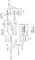
- FIG. 7is a schematic diagram of a Michelson based interferometer with multiple sensing arms that are that are selected by an optical switch, in accordance with another embodiment of the present invention.
- FIG. 8Ais a diagrammatic side view a portion of the interferometer of FIG. 5 utilizing a catheter configuration
- FIG. 8Bis a cross section view of the catheter configuration of FIG. 8A disposed in an artery.
- Embodiments of the present inventioninclude devices that have sensing and reference arms, at least one of which has variable delay.
- the sensing armincluding an optical switch for connecting to two or more probe arms.
- the distal ends of the probe armscollect source light backscattered from a sample.
- the backscattered light collected by the distal end of a probe armis combined with reference light and a low coherence interferometric (LCI) signal is produced by a sweep of a variable delay of the device.
- the interference signal produced by the interaction of reference and sensing light at a detectormeasures backscattered light.
- the devices of the present inventionmay be used to characterize a material based on the interference between reference light and sensing light backscattered (or reflected) from the sample.
- the low coherence interferometric signalscan provide information about the morphology, physical nature, composition, and properties of the sample.
- the devicemay be used to discriminate between finished surfaces and corroded surfaces, healthy and diseased tissue, and can sample the material or tissue in two or more areas. Versions of the invention include devices that are interferometers and or autocorrelators.
- a reference armis described herein as comprised of optical fiber, but may be comprised of any structure, such as, waveguides, free space structures, or combinations thereof.
- a reflectoris disposed at the distal end of the reference arm.
- a delay compensatoris an interferometer structure that can include waveguides, optical fibers and reflectors that compensate for the separation of the reference and backscattered light from a sample that is outside the coherence length of the source.
- a sample armis also described herein as comprised of optical fiber, but may be comprised of any structure, such as, waveguides, optical fibers, free space structures, or combinations thereof that propagate low coherence source light to a sample and collect backscattered source light from the sample.
- Each probe armdefines a portion of the sample arm where the fiber carries sensing light to the sample and collect backscattered sensing light from the sample.
- a processorcan provide an output, for example but not limited to a digital, current, voltage or combination of these, which is proportional to the interference measured and includes phase, amplitude, or a combinations these.
- the low-coherence Michelson interferometeris shown as an all-fiber low-coherence Michelson interferometer.
- This interferometerconsists of a broadband light source 110 , such as a superluminescent diode (SLD); an optical isolator 106 coupled to a fiber 114 , a fiber splitter 118 (having a ratio of 50:50), an optical detector 122 , and two arms 126 and 134 .
- a broadband light source 110such as a superluminescent diode (SLD)
- an optical isolator 106coupled to a fiber 114
- a fiber splitter 118having a ratio of 50:50
- an optical detector 122and two arms 126 and 134 .
- Arm 126is referred to as the reference arm, which has an adjustable length (L) by way of a piezoelectric (PZT) stretcher 142 with a mirror 130 at its end and carries the reference light E r .
- Arm 134is referred to as the sensing arm, which allows light to penetrate to a distance (z) in a medium, object, or sample 138 , and captures the reflected or back-scattered light E s from the sample 138 .
- the Variable length L of the reference armcorresponds a time delay ⁇ undergone by E r . Different path lengths for light interacting with the sample may be probed by the interferometric sensor through adjustment of the physical length, optical length, or optical delay of the reference arm of the interferometer.
- Interference between the reference arm and sample arm lightoccurs when the optical path length between the two is within the coherence length of the source light. Either moving the mirror 130 at the end of the reference arm or stretching the fiber can provide this length or delay.
- This stretchingis achieved by the PZT stretcher 142 comprising winding part of the fiber around a PZT drum to which a voltage is applied.
- the PZT stretcher 142has the advantage of using no mechanical moving parts, and therefore can be relatively fast. By applying a voltage ramp to the PZT stretcher 142 , the length is changed or scanned over a given range to provide path length matching with the light from within the sample under study.
- the maximum depth z max for penetration of light in the sampleis of the order of 1-2 millimeters (mm), and a scan can be used.
- a 40-meter length of fiber wound around a PZT drumcan provide an optical path length, nL of 5 mm, where n is the effective refractive index of the fiber, with the application of a peak voltage of about 540 volts in a 5 millisecond ramp to provide a scanning rate of about 1000 mm/s (millimeter per second).
- mechanical scanning with the moving mirror 130is used, to provide a scanning rate of about 40 mm/s.
- a similar but shorter delay length L 1 or time delay ⁇ 1is applied to the sensing arm, by a modulator 146 , in much the same fashion.
- I dI r +I s +i s ( z ) (1)
- i s (z)is the signal resulting from the interference of the reference and sensing light (* represents the complex conjugate of the field).
- i s ⁇ ( z )S ⁇ ( z ) ⁇ I r ⁇ I s ⁇ exp ⁇ [ - ( ⁇ ⁇ ⁇ L o - n s ⁇ z L c ) 2 ] ⁇ cos ⁇ ( 2 ⁇ ⁇ ⁇ ⁇ ⁇ vt ⁇ o ⁇ ⁇ ⁇ ( z ) ) ( 2 )
- FWHMfull-width-half-maxim
- This is a sinusoidal signal of frequency fv/ ⁇ o , called Doppler frequency, with an envelope given by the exponential function.
- An ordinary amplitude demodulatorregardless of the value of the Doppler frequency easily detects this peak.
- Thiscan be computed by a processor 150 (comprising a receiver and a computer), which is connected to the detector 122 . As the reference path length is changed, this function gives a profile of the scattering signal, and it is illustrated in FIG.
- the detection sensitivity of the current represented by Equation 4is limited by fluctuation noise.
- the minimum detectable signalis reached when the power it contains is equal to the noise power, i.e., when the signal-to-noise ratio (SNR) is equal to unity.
- Vthe degree of polarization of the light source
- Reducing the electrical bandwidth Bcan reduce all the noise components.
- the receiver noiseis the lowest, followed by the RIN and the shot noise.
- the RINis proportional to the square of the DC current
- the aboveimplies that the source power should be limited to about 16 ⁇ W, the sensing fiber tip reflection (4% in the absence of an anti-reflection coating) would be 0.16 ⁇ W, while the actual signal power, which depends on S(z) would be less than 4 ⁇ 10 ⁇ 4 ⁇ W.
- a high-power sourcesuch as 10 milliwatts
- a highly unbalanced (asymmetrical) splitting ratiosuch as 1:500
- Equation 4In a typical LCI or OCT (optical coherence tomography) system, only the magnitude is measured, and Equation 4 essentially gives the signal. More sophisticated systems can be designed to measure both the magnitude and the phase of the signal. Such systems are described in detail in Patent Applications invented by Gerard A. Alphonse and assigned to Medeikon Corporation; U.S. patent application Ser. No. 10/845,853, titled LOW COHERENCE INTERFEROMETRY UTILIZING MAGNITUDE, and U.S. patent application Ser. No. 10/845,849, titled LOW COHERENCE INTERFEROMETRY UTILIZING PHASE, the contents of each are incorporated by reference in the present application in their entirety. For the purpose of discussion, we will limit the rest of this disclosure to amplitude measurements. This does not exclude phase measurements when applicable.
- the collection of multiple information either in a single trace or by rapidly switching several probe arms into a single interferometerwill reduce the time for data collection and interpretation. For example, in probing the circumference of an artery, collecting all the LCI data in a single trace will enable rapid identification of a radial position with vulnerable plaque as compared with healthy ones, and enable selection of specific probing regions for further study or evaluation. The identification can be done by comparing the various components of a trace (addition/subtraction) to quickly determine difference, common features, and provide diagnosis.
- FIG. 3a single trace multi-channel low coherence interferometer is generally shown, such as described in a Patent Application invented by Gerard A. Alphonse et.al. and assigned to Medeikon Corporation, U.S. patent application Ser. No. 04/038,082, titled SINGLE TRACE MULTI-CHANNEL LOW COHERENCE INTERFEROMETRIC SENSOR, the contents of which is incorporated by reference in the present application in its entirety.
- the sensing armis split into several paths by means of a splitter, each path or probe having a slightly different optical path length than the others, the difference being equal or slightly longer than the optical depth of interest in the sample under test (about 2 mm for measurements inside the human artery).
- Scanning the variable path length of the reference armcauses the reference path length to sequentially match the length of each individual probe. This results in a single long signal trace, which contains a sequence of all the LCI signals obtained from the individual probes.
- the total sweep distance or delay in the reference armis equal to the sum of the individual delays of the various sensing arms. Since an LCI signal is obtained only when the reference path length is equal to the individual sensing arm path lengths, then the individual LCIs will appear in a single trace, being separated by their respective path length differences.
- This interferometerhas two or more sensing arms each having an adjustable delay 416 and 420 , and a reference arm also having an adjustable delay 412 .
- the single sensing arm of the standard interferometer discussed above with reference to FIG. 1is replaced by a multiplicity of sensing arms joined together by means of a 1:N coupler 426 .
- This coupler 426splits its input light equally among the N branches.
- a splitter 430divides light between the reference and sample sections.
- the sensing arms and reference armsare coupled to a low coherence light sources 404 and utilize an optional isolator 408 .
- the reference and sample arms of the sensorare configured, by a change in their length, a delay time, position of moveable mirror 434 , or a change in the index of refraction of the guide so that the delay for each of the sample arms and reference arm is within the coherence length of the source light.
- Backscattered sample light from the two or more sensing arms and the reference arm lightare coupled together and interference measured by detector 442 .
- a signal proportional to the interference measured for each of the sample arms during a sweep of the delay 412are displayed, compared to normal material, or digitized using processor 450 .
- the different branchesare shown to probe the same general area of a sample 438 .
- the operation of the systemcan be analyzed in the same manner as the ordinary interferometer described above with reference to FIG. 1 , with the input electric field to the detector being the sum of the electric fields returning from the reference arm 422 and all the sensing arms 410 , 414 , and 418 .
- the analysis of the interferometeris shown for two sensing arms. In the case of two sensing arms 410 and 414 , the total electric field E i at the detector 442 is the sum of the reference field E r and those of the two sensing arms E s1 and E s2 .
- the interferometerhas ability to independently retrieve these two components, in other words to completely or partially prevent them from overlapping in the LCI trace. This is dependent upon the choice of the total scanning distance L, of the reference arm 422 , the individual sensing arm distances L 1 and L 2 , and the gating property of the functions G( ⁇ 1 ) and G( ⁇ 2 ) shown below:
- FIG. 4illustrates how two LCI profiles from separate sample arms can be resolved with a single trace of the reference arm 422 . This can be achieved, for example, by allowing the instrument shown in FIG. 3 to scan the reference arm 422 length L at speed ⁇ over a time duration t, with the sample arms configured so that L 2 >L 1 +z max and L>L 2 +z max . In this configuration independent observe or measure the back-scattered LCI profiles I s1 (z) and I s2 (z) can be made.
- FIG. 4shows the appropriate relationship among L 1 , L 2 , z max , and L to achieve complete resolution of profiles I s1 (z) and I s2 (z).
- the single trace multi-channel low coherence interferometer of FIG. 3while well suited for particular applications, suffers from a power penalty caused by the 1 ⁇ N splitting of the sensing light in a round trip through the fiber.
- the total scan lengthis 18 mm for the six LCI signals, and this requires an air delay line controlled by a linear motor, which is best operated at low speed, of the order of 40 mm/s (millimeter per second).
- FIG. 5a switching multiple-probe interferometer in accordance with an embodiment of the invention is generally shown.
- This switching multiple-probe interferometerseeks to maximize both the optical signal to noise ratio and speed.
- the capability of having an arbitrary number of sensing probes without sacrificing speed to enable real-time imagingis advantageous in many applications, such as with biological tissues. Further, fabrication constraints are minimized and assembly is simplified with this multi-probe interferometer as compared to the above-discussed prior art.
- the FRM 524is a Faraday Rotator with a mirror at its end.
- the Faraday Rotatoruses a birefringent crystal adjusted to rotate the plane of polarization by 45 degrees. Upon reflection from the mirror, the light is rotated by another 45 degrees upon propagation through the Faraday Rotator. As a result, the light undergoes a total rotation of 90 degrees. In other words, whatever was in the S state goes into the P state upon reflection, and vice-versa. Therefore, after a round trip through the SM fiber of the reference arm the state of polarization of the reflected light is the same as that at the input of the fiber. In the interferometer, this process is used to maintain the polarization state of the reference light.
- the two polarization controllers 536which can be electrically (and/or manually) adjusted, are set such that equal amounts of total light (reference and sensing) exist at the two outputs, designated P 1 & S 1 and P 2 & S 2 .
- Probe arms 520are designed and fabricated for the application of interest.
- the probescan be a set of single-mode fibers designed to aim their light toward an arterial wall and configured to fit inside a catheter.
- the interferometerfunctions as discussed above, i.e., as if the selected arm were the only sample arm. Interference between the reference arm and sample arm light occurs when the optical path length between the two is within the coherence length of the source light.
- a device that stretches the fibercan provide this length or delay. This stretching can be achieved by winding part of the fiber around a piezoelectric (PZT) drum to which a voltage is applied.
- PZTpiezoelectric
- the PZT stretcherhas the advantage of using no mechanical moving parts, and therefore can be relatively fast.
- the lengthcan be changed or scanned over a given range to provide path length matching with the light from within the sample under study. Scanning changes the relative path length between the reference arm and the sensing arm and acquire the LCI signal. It is sufficient to scan only over a length corresponding to the desired probing depth in the sample.
- the probing depthis about 2 mm, including about 0.5 mm for propagation through blood.
- the Doppler frequency of the LCI signal with a faster scanincreases with the scan rate. This does not add any speed burden on the DAQ because the raw LCI signal can be envelope-detected. Envelope detection prior to analog-to-digital conversion yields the correct baseband signal regardless of Doppler frequency.
- the use of the optical switch 522has a number of advantageous features over the splitter 430 described in the prior art of FIG. 4 .
- One advantageis that the optical switch 522 has a much lower insertion loss than the splitter 430 .
- the insertion loss of a commercial 1 ⁇ 6 switchis only about 2.4 dB as compared to 15.6 dB for a commercial splitter with the same number of ports.
- Another advantageis that the optical switch 522 is more power-efficient. Aside from the small insertion loss, the switching process transfers all the input power to the selected output port, and therefore it provides an SNR improvement of 20 log(1/N) over the prior art of FIG. 4 .
- Each output fiber in that levelis connected to the input of a set of the other six switches 522 b - g constituting the second level, and the process can be extrapolated in the same manner to additional levels.
- the selection of a particular output fiberis done by simultaneously applying a code to the switch 522 a at the first level and to the appropriate switch among 522 b - g at the second level. Since all the switches are independent and have the same switching time (e.g., 500 microseconds), then they can be selected at the same time by random access, therefore giving the same access time to any particular probe in the N ⁇ N system.
- the only penaltyis an increase of loss by the amount of insertion loss of the selected switch in the second level.
- the total insertion losswould be 4.8 dB.
- the loss in the prior art system described with reference to FIG. 4would be 10 log(1/N 2 ) or about 31 dB, the access time would be six times longer, and access to the individual probes is constrained to be serial not be randomly accessible.
- An additional advantage of the present invention over the prior art system described with reference to FIG. 4is the relaxed tolerance on the probe fiber lengths. Whereas each fiber in the prior art system described with reference to FIG. 4 must have lengths that are different from one another by a precise amount, the only length requirement in the switch approach is that the fibers in any probe arm 520 have the same nominal total length (within manufacturing tolerance, which can be of the order of less than 1 millimeter). The relaxed length requirement lends itself well to mass fabrication.
- a switching multiple-probe interferometerin accordance with as alternate embodiment of the present invention is generally shown.
- This interferometeremploys a broadband light source 702 , such as a high-power superluminescent diode (SLD).
- the SLD 702is connected to an input port of a three-port optical circulator 728 .
- An optical circulatoris a device that transfers light from an input port to a second port and transfers the light reflected from the second port to a third port. In this manner, it protects the source from undesired reflection and makes all the reflected light available for systems operation.
- An output port of the optical circulator 728is connected to a fiber splitter 704 .
- the fiber splitter 704is used to direct a large fraction of the light source power to a multiple probe system 706 for delivery of the maximum amount of available light to a test sample 708 .
- a 5:95 splitting ratiois also illustrated, as such is presently commercially available, but other high splitting ratios may also be employed as previously discussed.
- the fiber splitter 704results in a reference arm 712 and a sensing arm 714 .
- the multiple probe system 706is located at the distal end in of the sensing arm 714 .
- an electrically controlled variable optical attenuator (VOA) 716is introduced into the reference arm 712 .
- VOAvariable optical attenuator
- the VOAassists in reducing the reference light beyond levels obtained by the asymmetric splitter.
- Automatic electrical control of the VOA 716is provided by a digitally controlled voltage source (DCV) 718 .
- DCVdigitally controlled voltage source
- Optical switch 722is a MEM-based device in which the sensing fiber at the input port can be randomly or sequentially connected to any of the N output fibers (probe arms) by the application of a digital code under computer control.
- the 1 ⁇ 2 coupler 726receives the combined reflected reference and sensing lights from the optical circulator 728 in order to enable interference at a detector, which is comprised of a pair of photodetectors 732 in communication with the two outputs (P and S) of the coupler 757 .
- a detectorwhich is comprised of a pair of photodetectors 732 in communication with the two outputs (P and S) of the coupler 757 .
- An advantage of the dual photodetectorsis that they add the signals and subtract the common-mode noise, such as the RIN discussed above. It is desired to reduce the power incident on each photodetectors, by driving VOA to give rise to a desired value of attenuation, below 2 to 5 ⁇ W, then RIN does not dominate the noise in the detector circuitry.
- Polarization controllers 736 and polarization splitter 738are introduced at the outputs of the coupler 757 .
- the output of the detectors 732is combined by an analog subtractor 733 and applied to a digital to analog converter (DAQ) 734 , which is associated with a processor, that digitally processes it for further use. Alternatively, it can be envelope-detected to produce a signal similar the trace shown in FIG. 2 , and then processed by the DAQ 734 .
- DAQdigital to analog converter
- Probe arms 720are scanned by changing the relative path length between the reference arm and the sensing arm, and acquiring the LCI signal.
- Two PZT fiber stretchers 740 and 742one in the reference arm, the other in the sensing arm are used for scanning and are driving in opposite directions, as discussed in the previous embodiment.
- the PZTsare biased and driven in push-pull, i.e., with opposite voltages so that one undergoes increased stretching while the other undergoes decreased stretching. This amounts to doubling the scan rate if the desired path length difference is less than the maximum change. Alternatively, it also doubles the effective scan range for the same total scan time if a range larger than that of a single scanner is desired.
- FIGS. 5 and 7are operated as Optical Time Domain Interferometry (OTDI), because of their operation in the time domain, i.e. the direct output of the detectors is a signal in the time domain, which is calibrated in terms of distance from the value of the scanning rate.
- OTDIOptical Time Domain Interferometry
- Another approach to interferometryis Optical Frequency Domain Interferometry (OFDI).
- OFDIOptical Frequency Domain Interferometry
- the OTDI systems described abovecan be configured to operate as OFDI systems, by: utilizing a light source which is a scanning laser or a swept laser, i.e., a laser that can be externally tuned at a given rate, in place of light source 502 ( FIG. 5 ), 702 ( FIG.
- a swept lasermay be a broadband source such as an SLD in a cavity containing a grating, which can be rotated.
- a tunable laseri.e., a narrow band source whose output wavelength within the broadband spectrum is determined by the position of the grating.
- tunable lasersare available commercially or by custom design with available sweep range larger than the FWHM bandwidth of the SLD.
- a swept laseris formed from a 1,310 nm SLD, producing laser light with linewidth of about 0.1 nm, sweeping a range of 80 nm (from 1270 nm to 1350 nm) in a 1 millisecond timeframe. This is equivalent to a chirped laser with the wavelength is changing at a rate 80 nm per millisecond.
- the total LCIis obtained by integration over the scan time, and according to theory, it is a function of frequency which, when converted to the time domain by Fourier transformation, reproduces the time-domain LCI signal with a resolution corresponding to the sweeping width. If the sweep width is 80 nm, then the resolution of the OFDI system is 9.4 microns.
- the OFDI systemshave several advantages over the OTDI systems.
- the OFDI systemshave a depth range given by the coherence length of the laser elements of the tunable laser, which, being longer than the required depth for cardiovascular diagnostics, makes it unnecessary to use a scanner in the reference or sensing arm.
- a modulatorcould be used for fine adjustments of the optical path lengths.
- the chirping range of the tunable laser sourcedetermines the resolution.
- the tuning rangecan be made two to three times the FWHM bandwidth of the SLD, hence improving the LCI resolution by the same factor (resolution below 10 microns are readily possible).
- the optical power per individual laseris at least the same as the full SLD power under the same drive condition (a laser is much more efficient than an SLD), so the number of photons per unit bandwidth is larger than for the OTDI by at least two orders of magnitude, thus giving it a significant advantage in signal-to-noise ratio (SNR).
- SNRsignal-to-noise ratio
- the instantaneous bandwidth of the sourceis narrow, being a laser. Therefore, the SNR is limited only by shot noise, whereas the noise in the OTDI is limited by the RIN of the broadband source, which dominates the response above about 4 microwatts of optical power at the detector.
- the OFDI systemcan operate at a much higher source power level than the OTDI system.
- the various probes of the interferometer sensorfor example probes 520 , FIG. 5 or 720 , FIG. 7 are brought together and placed inside a housing 970 .
- the device having guide wire 966 and optical head 946is essentially a wire-guided catheter, which can be inserted in an artery 954 using conventional medical procedures and the radial light 972 , 976 into and backscattered from the sample and others not shown, from optical head 946 of the device used to detect a vulnerable plaque 962 covering a lipid 958 pocket in the artery 954 .
- This plaqueconsists of a layer of calcified material and a lipid pool between it and the arterial wall. The widths of the plaque and lipid regions are measures of their respective thicknesses.
- the number of fibers that can be placed inside the housing 970can vary and along with the dimensions of the waveguides, and will vary depending on the inside diameter of the guide wire or conduit used as a guide.
- this deviceis used to probe the walls of an artery, which may contain vulnerable plaques.
- the ends of the probes (fibers)are configured such that the output lights of the fibers are directed toward the arterial walls, e.g., pointing to different radial directions.
- All six LCI profiles(in this example one for each of the fibers) are individually obtained either sequentially or by random access. The scan can be digitized and the various profiles can be separated and stored in the computer memory for processing.
- Field-of-view from the borescope or endoscope timemay range from 10 to about 90 degrees and can be chosen based on the distance from the distal end of the borescope to the subject.
- a borescopecan have has a very large depth of field which can be from infinity down to a centimeter or less.
- Borescopes and endoscopes of the present inventionmay be rigid or flexible and can use but are not limited to fiber optic illumination to carry light from an external light source through a flexible light guide, then through the borescope, to the distal end.
- This prophetic exampleillustrates how a version of the present invention can be used for detecting plaque within the coronary or other patient vasculature.
- the apparatus and techniquescould also be applied to characterizing lesions in other body lumens, which are associated with various disease conditions.
- the methods and apparatuscan be implemented within the body lumen to identify diseased tissue or monitor the course of treatment for a particular condition.
- the apparatusis able to interrogate the body lumen over a relatively long distance to characterize the tissue in an efficient fashion by providing interferometric information on the tissue from multiple probes in a single sweep of a variable delay of the apparatus.
- Coronary artery disease resulting from the build-up of atherosclerotic plaque in the coronary arteriesis a leading cause of death.
- the build-up of plaquecauses a narrowing of the artery, commonly referred to as a lesion, which reduces blood flow to the myocardium (heart muscle tissue).
- Myocardial infarctioncan occur when an arterial lesion abruptly closes the vessel, causing complete cessation of blood flow to portions of the myocardium. Even if abrupt closure does not occur, blood flow may decrease resulting in chronically insufficient blood flow, which can cause significant tissue damage over time.
- the ruptured plaquereleases highly thrombogenic constituent materials, which are capable of activating the clotting cascade and inducing rapid and substantial coronary thrombosis.
- Such plaqueis referred to as unstable or vulnerable, and the resulting thrombus formation can cause unstable angina chest pain, acute myocardial infarction (heart attack), sudden coronary death, and stroke.
- the sensor of the present inventionmay be used to determine the location, chemical and physical properties, and nature of the lesion in an artery. This information can be used to facilitate determining whether the plaque is stable or unstable, and may be used to treat and monitor the treatment.
Landscapes
- Physics & Mathematics (AREA)
- General Physics & Mathematics (AREA)
- Investigating Or Analysing Materials By Optical Means (AREA)
Abstract
Description
Id=Ir+Is+is(z) (1)
where Ir=ErEr* is the dc current caused by the reference light, Is=EsEs* is the dc current caused by the total light in the sensing arm, and is(z) is the signal resulting from the interference of the reference and sensing light (* represents the complex conjugate of the field). For a broadband light source with a Gaussian profile, with the reference path length changing as a ramp in the form ΔL=ΔL0+vt−nsz, then is(z) is given by:
is the so-called coherence length of the light source having a full-width-half-maximum (FWHM) bandwidth of Δλ, where φ(z) is a phase variable, and where S(z) is a coefficient representing the amount of scattered light collected from distance z by the sensing arm. This is a sinusoidal signal of frequency f=v/λo, called Doppler frequency, with an envelope given by the exponential function. This function has a peak given by:
Is(z)peak=S(z)√{square root over (IrIs)} (4)
which occurs when ΔLo−nsz=0 and φ(z)=0. An ordinary amplitude demodulator regardless of the value of the Doppler frequency easily detects this peak. This can be computed by a processor150 (comprising a receiver and a computer), which is connected to the
σi2=σr2+Rσs2+Rσe2 (5)
where R is the input resistance of the receiver. Then the SNR is given by
Claims (9)
Priority Applications (2)
| Application Number | Priority Date | Filing Date | Title |
|---|---|---|---|
| US11/445,514US7488930B2 (en) | 2006-06-02 | 2006-06-02 | Multi-channel low coherence interferometer |
| PCT/US2007/012146WO2007142814A2 (en) | 2006-06-02 | 2007-05-22 | Multi-channel low coherence interferometer |
Applications Claiming Priority (1)
| Application Number | Priority Date | Filing Date | Title |
|---|---|---|---|
| US11/445,514US7488930B2 (en) | 2006-06-02 | 2006-06-02 | Multi-channel low coherence interferometer |
Publications (2)
| Publication Number | Publication Date |
|---|---|
| US20070278389A1 US20070278389A1 (en) | 2007-12-06 |
| US7488930B2true US7488930B2 (en) | 2009-02-10 |
Family
ID=38789008
Family Applications (1)
| Application Number | Title | Priority Date | Filing Date |
|---|---|---|---|
| US11/445,514Expired - Fee RelatedUS7488930B2 (en) | 2006-06-02 | 2006-06-02 | Multi-channel low coherence interferometer |
Country Status (2)
| Country | Link |
|---|---|
| US (1) | US7488930B2 (en) |
| WO (1) | WO2007142814A2 (en) |
Cited By (29)
| Publication number | Priority date | Publication date | Assignee | Title |
|---|---|---|---|---|
| US20090131921A1 (en)* | 2007-09-06 | 2009-05-21 | Lensx Lasers, Inc. | Precise Targeting of Surgical Photodisruption |
| US20110194743A1 (en)* | 2010-02-05 | 2011-08-11 | Ferenc Raksi | Gradient Search Integrated with Local Imaging in Laser Surgical Systems |
| WO2012012355A1 (en)* | 2010-07-19 | 2012-01-26 | Lumetrics, Inc. | Fiber-based interferometric device for measuring axial dimensions of a human eye |
| WO2012103557A3 (en)* | 2011-01-28 | 2012-10-04 | The Regents Of The University Of Colorado, A Body Corporate | Spectral phase analysis for precision ranging |
| US8398238B1 (en) | 2011-08-26 | 2013-03-19 | Alcon Lensx, Inc. | Imaging-based guidance system for ophthalmic docking using a location-orientation analysis |
| US8398236B2 (en) | 2010-06-14 | 2013-03-19 | Alcon Lensx, Inc. | Image-guided docking for ophthalmic surgical systems |
| US8414564B2 (en) | 2010-02-18 | 2013-04-09 | Alcon Lensx, Inc. | Optical coherence tomographic system for ophthalmic surgery |
| US8459794B2 (en) | 2011-05-02 | 2013-06-11 | Alcon Lensx, Inc. | Image-processor-controlled misalignment-reduction for ophthalmic systems |
| US20150011850A1 (en)* | 2012-07-16 | 2015-01-08 | Timothy Ruchti | Multiplexed pathlength resolved noninvasive analyzer apparatus with dynamic optical paths and method of use thereof |
| US20150018644A1 (en)* | 2012-07-16 | 2015-01-15 | Sandeep Gulati | Multiplexed pathlength resolved noninvasive analyzer apparatus with non-uniform detector array and method of use thereof |
| US9023016B2 (en) | 2011-12-19 | 2015-05-05 | Alcon Lensx, Inc. | Image processor for intra-surgical optical coherence tomographic imaging of laser cataract procedures |
| US9066784B2 (en) | 2011-12-19 | 2015-06-30 | Alcon Lensx, Inc. | Intra-surgical optical coherence tomographic imaging of cataract procedures |
| US9351671B2 (en) | 2012-07-16 | 2016-05-31 | Timothy Ruchti | Multiplexed pathlength resolved noninvasive analyzer apparatus and method of use thereof |
| US9351672B2 (en) | 2012-07-16 | 2016-05-31 | Timothy Ruchti | Multiplexed pathlength resolved noninvasive analyzer apparatus with stacked filters and method of use thereof |
| US20160170081A1 (en)* | 2013-08-30 | 2016-06-16 | Halliburton Energy Services, Inc. | Distributed acoustic sensing system with variable spatial resolution |
| US20160242682A1 (en)* | 2012-07-16 | 2016-08-25 | Sandeep Gulati | Noninvasive analyzer apparatus and method of use thereof for separating distributed probing photons emerging from a sample |
| US9442065B2 (en) | 2014-09-29 | 2016-09-13 | Zyomed Corp. | Systems and methods for synthesis of zyotons for use in collision computing for noninvasive blood glucose and other measurements |
| US9492322B2 (en) | 2009-11-16 | 2016-11-15 | Alcon Lensx, Inc. | Imaging surgical target tissue by nonlinear scanning |
| US9532708B2 (en) | 2010-09-17 | 2017-01-03 | Alcon Lensx, Inc. | Electronically controlled fixation light for ophthalmic imaging systems |
| US9554738B1 (en) | 2016-03-30 | 2017-01-31 | Zyomed Corp. | Spectroscopic tomography systems and methods for noninvasive detection and measurement of analytes using collision computing |
| US9599613B2 (en) | 2011-07-20 | 2017-03-21 | University Of Washington Through Its Center For Commercialization | Photonic blood typing |
| US9622913B2 (en) | 2011-05-18 | 2017-04-18 | Alcon Lensx, Inc. | Imaging-controlled laser surgical system |
| US9766126B2 (en) | 2013-07-12 | 2017-09-19 | Zyomed Corp. | Dynamic radially controlled light input to a noninvasive analyzer apparatus and method of use thereof |
| US10031138B2 (en) | 2012-01-20 | 2018-07-24 | University Of Washington Through Its Center For Commercialization | Hierarchical films having ultra low fouling and high recognition element loading properties |
| US10337316B2 (en) | 2013-08-30 | 2019-07-02 | Halliburton Energy Services, Inc. | Distributed acoustic sensing system with variable spatial resolution |
| US11243346B2 (en)* | 2013-06-23 | 2022-02-08 | Eric Swanson | Interferometric optical fiber measurement system with multicore optical fiber |
| US11397075B2 (en) | 2013-06-23 | 2022-07-26 | Eric Swanson | Photonic integrated receiver |
| US20230092947A1 (en)* | 2021-09-20 | 2023-03-23 | Zygo Corporation | Optical contact metrology |
| US12085387B1 (en) | 2023-09-23 | 2024-09-10 | Hamamatsu Photonics K.K. | Optical coherence tomography system for subsurface inspection |
Families Citing this family (76)
| Publication number | Priority date | Publication date | Assignee | Title |
|---|---|---|---|---|
| US7486405B2 (en)* | 2006-05-01 | 2009-02-03 | Hogan Josh N | Optimized reference level generation |
| US9867530B2 (en) | 2006-08-14 | 2018-01-16 | Volcano Corporation | Telescopic side port catheter device with imaging system and method for accessing side branch occlusions |
| EP2178442B1 (en) | 2007-07-12 | 2017-09-06 | Volcano Corporation | Catheter for in vivo imaging |
| US9596993B2 (en) | 2007-07-12 | 2017-03-21 | Volcano Corporation | Automatic calibration systems and methods of use |
| WO2009009802A1 (en) | 2007-07-12 | 2009-01-15 | Volcano Corporation | Oct-ivus catheter for concurrent luminal imaging |
| US20100238452A1 (en)* | 2008-12-15 | 2010-09-23 | Frederique Vanholsbeeck | Dual Fiber Stretchers for Dispersion Compensation |
| US8892191B2 (en)* | 2009-03-08 | 2014-11-18 | Oprobe, Llc | Methods of determining motion and distance during medical and veterinary procedures |
| EP2470886A4 (en)* | 2009-08-26 | 2016-11-02 | Tomophase Inc | Optical tissue imaging based on optical frequency domain imaging |
| US20110282331A1 (en)* | 2010-05-13 | 2011-11-17 | Oprobe, Llc | Optical coherence tomography with multiple imaging instruments |
| US8822875B2 (en)* | 2010-09-25 | 2014-09-02 | Queen's University At Kingston | Methods and systems for coherent imaging and feedback control for modification of materials |
| US11141063B2 (en) | 2010-12-23 | 2021-10-12 | Philips Image Guided Therapy Corporation | Integrated system architectures and methods of use |
| US11040140B2 (en) | 2010-12-31 | 2021-06-22 | Philips Image Guided Therapy Corporation | Deep vein thrombosis therapeutic methods |
| US9204800B2 (en) | 2011-03-07 | 2015-12-08 | St. Jude Medical, Inc. | Low cost high efficiency signal interrogation for multi-channel optical coherence tomography |
| US8868356B2 (en)* | 2011-03-07 | 2014-10-21 | St. Jude Medical, Inc. | Multi-channel optical coherence tomography for imaging and temperature and force sensing |
| ES2415555B2 (en)* | 2011-05-20 | 2014-07-09 | Medlumics, S.L. | SWEEP DEVICE FOR LOW COHERENCE INTERFEROMETRY. |
| CN103959043B (en)* | 2011-05-31 | 2016-11-02 | 光学实验室成像公司 | Multimodal imaging systems, devices and methods |
| US9360630B2 (en) | 2011-08-31 | 2016-06-07 | Volcano Corporation | Optical-electrical rotary joint and methods of use |
| WO2013033413A1 (en)* | 2011-09-02 | 2013-03-07 | Volcano Corporation | Reconfigurable interferometer |
| AT511935B1 (en)* | 2011-09-12 | 2015-09-15 | Ima Integrated Microsystems Austria Gmbh | METHOD AND DEVICE FOR SPATIAL MEASUREMENT OF TISSUE STRUCTURES |
| US8781266B2 (en)* | 2011-12-23 | 2014-07-15 | General Electric Company | Distributed, multiplexed fiber optic current transducer using optical power division |
| US9360660B2 (en)* | 2012-05-24 | 2016-06-07 | Northwestern University | Methods and apparatus for laser scanning structured illumination microscopy and tomography |
| DE102012211549B3 (en)* | 2012-07-03 | 2013-07-04 | Polytec Gmbh | Apparatus and method for interferometric measurement of an object |
| US20160249836A1 (en)* | 2012-07-16 | 2016-09-01 | Sandeep Gulati | Sample optical pathlength control using a noninvasive analyzer apparatus and method of use thereof |
| EP2690395A1 (en)* | 2012-07-24 | 2014-01-29 | Hexagon Technology Center GmbH | Interferometric distance measuring assembly and method |
| US9292918B2 (en) | 2012-10-05 | 2016-03-22 | Volcano Corporation | Methods and systems for transforming luminal images |
| US9367965B2 (en) | 2012-10-05 | 2016-06-14 | Volcano Corporation | Systems and methods for generating images of tissue |
| US10568586B2 (en) | 2012-10-05 | 2020-02-25 | Volcano Corporation | Systems for indicating parameters in an imaging data set and methods of use |
| CA2887421A1 (en) | 2012-10-05 | 2014-04-10 | David Welford | Systems and methods for amplifying light |
| US9858668B2 (en) | 2012-10-05 | 2018-01-02 | Volcano Corporation | Guidewire artifact removal in images |
| US10070827B2 (en) | 2012-10-05 | 2018-09-11 | Volcano Corporation | Automatic image playback |
| US20140100454A1 (en) | 2012-10-05 | 2014-04-10 | Volcano Corporation | Methods and systems for establishing parameters for three-dimensional imaging |
| US9324141B2 (en) | 2012-10-05 | 2016-04-26 | Volcano Corporation | Removal of A-scan streaking artifact |
| US11272845B2 (en) | 2012-10-05 | 2022-03-15 | Philips Image Guided Therapy Corporation | System and method for instant and automatic border detection |
| US9286673B2 (en) | 2012-10-05 | 2016-03-15 | Volcano Corporation | Systems for correcting distortions in a medical image and methods of use thereof |
| US9307926B2 (en) | 2012-10-05 | 2016-04-12 | Volcano Corporation | Automatic stent detection |
| US9840734B2 (en) | 2012-10-22 | 2017-12-12 | Raindance Technologies, Inc. | Methods for analyzing DNA |
| EP2931132B1 (en) | 2012-12-13 | 2023-07-05 | Philips Image Guided Therapy Corporation | System for targeted cannulation |
| WO2014113188A2 (en) | 2012-12-20 | 2014-07-24 | Jeremy Stigall | Locating intravascular images |
| EP2934310A4 (en) | 2012-12-20 | 2016-10-12 | Nathaniel J Kemp | Optical coherence tomography system that is reconfigurable between different imaging modes |
| EP2934311B1 (en) | 2012-12-20 | 2020-04-15 | Volcano Corporation | Smooth transition catheters |
| US10942022B2 (en) | 2012-12-20 | 2021-03-09 | Philips Image Guided Therapy Corporation | Manual calibration of imaging system |
| US10939826B2 (en) | 2012-12-20 | 2021-03-09 | Philips Image Guided Therapy Corporation | Aspirating and removing biological material |
| US11406498B2 (en) | 2012-12-20 | 2022-08-09 | Philips Image Guided Therapy Corporation | Implant delivery system and implants |
| US10058284B2 (en) | 2012-12-21 | 2018-08-28 | Volcano Corporation | Simultaneous imaging, monitoring, and therapy |
| US10413317B2 (en) | 2012-12-21 | 2019-09-17 | Volcano Corporation | System and method for catheter steering and operation |
| EP2936241B1 (en) | 2012-12-21 | 2020-10-21 | Nathaniel J. Kemp | Power-efficient optical buffering using a polarisation-maintaining active optical switch |
| JP2016501625A (en) | 2012-12-21 | 2016-01-21 | ジェローム マイ, | Ultrasound imaging with variable line density |
| US10332228B2 (en) | 2012-12-21 | 2019-06-25 | Volcano Corporation | System and method for graphical processing of medical data |
| CA2895769A1 (en) | 2012-12-21 | 2014-06-26 | Douglas Meyer | Rotational ultrasound imaging catheter with extended catheter body telescope |
| US9486143B2 (en) | 2012-12-21 | 2016-11-08 | Volcano Corporation | Intravascular forward imaging device |
| EP2934323A4 (en) | 2012-12-21 | 2016-08-17 | Andrew Hancock | SYSTEM AND METHOD FOR MULTIPLE PROCESSING OF IMAGE SIGNALS |
| US9612105B2 (en) | 2012-12-21 | 2017-04-04 | Volcano Corporation | Polarization sensitive optical coherence tomography system |
| JP2016507892A (en) | 2012-12-21 | 2016-03-10 | デイビッド ウェルフォード, | System and method for narrowing the wavelength emission of light |
| US10226597B2 (en) | 2013-03-07 | 2019-03-12 | Volcano Corporation | Guidewire with centering mechanism |
| WO2014138555A1 (en) | 2013-03-07 | 2014-09-12 | Bernhard Sturm | Multimodal segmentation in intravascular images |
| US20140276923A1 (en) | 2013-03-12 | 2014-09-18 | Volcano Corporation | Vibrating catheter and methods of use |
| EP2967391A4 (en) | 2013-03-12 | 2016-11-02 | Donna Collins | SYSTEMS AND METHODS FOR DIAGNOSING CORONARY MICROVASCULAR DISEASE |
| US9301687B2 (en) | 2013-03-13 | 2016-04-05 | Volcano Corporation | System and method for OCT depth calibration |
| US11026591B2 (en) | 2013-03-13 | 2021-06-08 | Philips Image Guided Therapy Corporation | Intravascular pressure sensor calibration |
| WO2014159819A1 (en) | 2013-03-13 | 2014-10-02 | Jinhyoung Park | System and methods for producing an image from a rotational intravascular ultrasound device |
| US12343198B2 (en) | 2013-03-14 | 2025-07-01 | Philips Image Guided Therapy Corporation | Delivery catheter having imaging capabilities |
| US10292677B2 (en) | 2013-03-14 | 2019-05-21 | Volcano Corporation | Endoluminal filter having enhanced echogenic properties |
| US10219887B2 (en) | 2013-03-14 | 2019-03-05 | Volcano Corporation | Filters with echogenic characteristics |
| US20160030151A1 (en) | 2013-03-14 | 2016-02-04 | Volcano Corporation | Filters with echogenic characteristics |
| CN103558582B (en)* | 2013-10-23 | 2015-11-04 | 合肥工业大学 | A method for measuring head space attitude guided by ultrasonic positioning |
| DE102016205370B4 (en)* | 2016-03-31 | 2022-08-18 | Optomedical Technologies Gmbh | OCT system |
| WO2018055606A1 (en)* | 2016-09-26 | 2018-03-29 | Ixa Amc Office / Academic Medical Center | Single-chip optical coherence tomography device |
| US11366244B2 (en) | 2017-02-23 | 2022-06-21 | Halliburton Energy Services, Inc. | Distributed acoustic sensing system with a polarization control device for improving signal-to-noise ratio |
| US10982947B2 (en)* | 2017-06-12 | 2021-04-20 | Sightline Innovation Inc. | System and method of surface inspection of an object using mulitplexed optical coherence tomography |
| KR101990251B1 (en)* | 2018-10-15 | 2019-06-17 | 경북대학교 산학협력단 | Apparatus for optical coherence tomography and method for image generate using thereof |
| US11175126B2 (en)* | 2019-04-08 | 2021-11-16 | Canon U.S.A., Inc. | Automated polarization control |
| CN112945108B (en)* | 2021-01-26 | 2022-11-18 | 中国计量科学研究院 | A precision displacement measurement method and device based on electro-optical modulation sidebands |
| JP7751788B2 (en)* | 2021-09-15 | 2025-10-09 | オムロン株式会社 | Optical Interferometric Distance Sensor |
| JP7739883B2 (en)* | 2021-09-15 | 2025-09-17 | オムロン株式会社 | Optical Interferometric Distance Sensor |
| JP7723897B2 (en)* | 2021-09-15 | 2025-08-15 | オムロン株式会社 | Optical Interferometric Distance Sensor |
| CN119924824B (en)* | 2025-04-07 | 2025-07-25 | 中国人民解放军国防科技大学 | Sleep monitoring device and method based on optical fiber Michelson interferometer |
Citations (114)
| Publication number | Priority date | Publication date | Assignee | Title |
|---|---|---|---|---|
| EP0317121A2 (en) | 1987-11-17 | 1989-05-24 | Kurashiki Boseki Kabushiki Kaisha | Spectroscopic method and apparatus for measuring sugar concentrations |
| WO1992019930A1 (en) | 1991-04-29 | 1992-11-12 | Massachusetts Institute Of Technology | Method and apparatus for optical imaging and measurement |
| US5173747A (en) | 1990-09-20 | 1992-12-22 | Battelle Memorial Institute | Integrated optical directional-coupling refractometer apparatus |
| US5202745A (en) | 1990-11-07 | 1993-04-13 | Hewlett-Packard Company | Polarization independent optical coherence-domain reflectometry |
| US5341205A (en) | 1991-01-15 | 1994-08-23 | The United States Of America As Represented By The Secretary Of The Navy | Method for characterization of optical waveguide devices using partial coherence interferometry |
| US5383467A (en) | 1992-11-18 | 1995-01-24 | Spectrascience, Inc. | Guidewire catheter and apparatus for diagnostic imaging |
| US5398681A (en) | 1992-12-10 | 1995-03-21 | Sunshine Medical Instruments, Inc. | Pocket-type instrument for non-invasive measurement of blood glucose concentration |
| US5434791A (en) | 1993-06-29 | 1995-07-18 | Electronic Data Systems Corporation | Product structure management |
| US5465147A (en) | 1991-04-29 | 1995-11-07 | Massachusetts Institute Of Technology | Method and apparatus for acquiring images using a ccd detector array and no transverse scanner |
| US5491552A (en) | 1993-03-29 | 1996-02-13 | Bruker Medizintechnik | Optical interferometer employing mutually coherent light source and an array detector for imaging in strongly scattered media |
| US5501226A (en) | 1994-10-19 | 1996-03-26 | Carl Zeiss, Inc. | Short coherence length, doppler velocimetry system |
| US5507288A (en) | 1994-05-05 | 1996-04-16 | Boehringer Mannheim Gmbh | Analytical system for monitoring a substance to be analyzed in patient-blood |
| US5565986A (en) | 1994-03-30 | 1996-10-15 | Kn+E,Uml U+Ee Ttel; Alexander | Stationary optical spectroscopic imaging in turbid objects by special light focusing and signal detection of light with various optical wavelengths |
| US5582171A (en) | 1994-07-08 | 1996-12-10 | Insight Medical Systems, Inc. | Apparatus for doppler interferometric imaging and imaging guidewire |
| US5710630A (en) | 1994-05-05 | 1998-01-20 | Boehringer Mannheim Gmbh | Method and apparatus for determining glucose concentration in a biological sample |
| US5726801A (en) | 1994-12-21 | 1998-03-10 | E-Tek Dynamics, Inc. | Reduced optical isolator module for a miniaturized laser diode assembly |
| EP0831312A1 (en) | 1995-03-01 | 1998-03-25 | Nizhny Novgorod Center for High Technology Incubation ( NCHTI) | Optical fibre interferometer and optical fibre piezo-electric modulator |
| US5830145A (en) | 1996-09-20 | 1998-11-03 | Cardiovascular Imaging Systems, Inc. | Enhanced accuracy of three-dimensional intraluminal ultrasound (ILUS) image reconstruction |
| US5835215A (en) | 1996-05-16 | 1998-11-10 | Fuji Photo Film Co., Ltd. | Glucose concentration measuring method and apparatus with short coherence source and heterodyne interferometer |
| US5836877A (en) | 1997-02-24 | 1998-11-17 | Lucid Inc | System for facilitating pathological examination of a lesion in tissue |
| US5847827A (en) | 1995-06-23 | 1998-12-08 | Carl Zeiss Jena Gmbh | Coherence biometry and coherence tomography with dynamic coherent |
| US5877856A (en) | 1996-05-14 | 1999-03-02 | Carl Zeiss Jena Gmbh | Methods and arrangement for increasing contrast in optical coherence tomography by means of scanning an object with a dual beam |
| US5883717A (en) | 1996-06-04 | 1999-03-16 | Northeastern University | Optical quadrature interferometry utilizing polarization to obtain in-phase and quadrature information |
| US5892583A (en) | 1997-08-21 | 1999-04-06 | Li; Ming-Chiang | High speed inspection of a sample using superbroad radiation coherent interferometer |
| US5905572A (en) | 1997-08-21 | 1999-05-18 | Li; Ming-Chiang | Sample inspection using interference and/or correlation of scattered superbroad radiation |
| WO1999032897A2 (en) | 1997-12-23 | 1999-07-01 | Magnetic Diagnostics, Inc. | Nmr apparatus and method for non-invasive in vivo testing of a patient's body fluid glucose levels |
| US5920390A (en) | 1997-06-26 | 1999-07-06 | University Of North Carolina | Fiberoptic interferometer and associated method for analyzing tissue |
| US5921926A (en) | 1997-07-28 | 1999-07-13 | University Of Central Florida | Three dimensional optical imaging colposcopy |
| US5943133A (en) | 1996-12-04 | 1999-08-24 | The Research Foundation Of City College Of New York | System and method for performing selected optical measurements on a sample using a diffraction grating |
| US5956355A (en) | 1991-04-29 | 1999-09-21 | Massachusetts Institute Of Technology | Method and apparatus for performing optical measurements using a rapidly frequency-tuned laser |
| US5962852A (en) | 1996-01-26 | 1999-10-05 | Roche Diagnostics Gmbh | Process and device for determining an analyte contained in a scattering matrix |
| WO1999057507A1 (en) | 1998-05-01 | 1999-11-11 | Board Of Regents, The University Of Texas System | Method and apparatus for subsurface imaging |
| US5991697A (en) | 1996-12-31 | 1999-11-23 | The Regents Of The University Of California | Method and apparatus for optical Doppler tomographic imaging of fluid flow velocity in highly scattering media |
| US5994690A (en) | 1997-03-17 | 1999-11-30 | Kulkarni; Manish D. | Image enhancement in optical coherence tomography using deconvolution |
| US6002480A (en) | 1997-06-02 | 1999-12-14 | Izatt; Joseph A. | Depth-resolved spectroscopic optical coherence tomography |
| US6006128A (en) | 1997-06-02 | 1999-12-21 | Izatt; Joseph A. | Doppler flow imaging using optical coherence tomography |
| US6014214A (en) | 1997-08-21 | 2000-01-11 | Li; Ming-Chiang | High speed inspection of a sample using coherence processing of scattered superbroad radiation |
| US6015969A (en) | 1996-09-16 | 2000-01-18 | The Regents Of The University Of California | Multiple-wavelength spectroscopic quantitation of light-absorbing species in scattering media |
| US6020963A (en) | 1996-06-04 | 2000-02-01 | Northeastern University | Optical quadrature Interferometer |
| US6037579A (en) | 1997-11-13 | 2000-03-14 | Biophotonics Information Laboratories, Ltd. | Optical interferometer employing multiple detectors to detect spatially distorted wavefront in imaging of scattering media |
| US6053613A (en) | 1998-05-15 | 2000-04-25 | Carl Zeiss, Inc. | Optical coherence tomography with new interferometer |
| US6057920A (en) | 1998-03-30 | 2000-05-02 | Carl Zeiss Jena Gmbh | Optical coherence tomography with dynamic coherent focus |
| US6069698A (en) | 1997-08-28 | 2000-05-30 | Olympus Optical Co., Ltd. | Optical imaging apparatus which radiates a low coherence light beam onto a test object, receives optical information from light scattered by the object, and constructs therefrom a cross-sectional image of the object |
| US6111645A (en) | 1991-04-29 | 2000-08-29 | Massachusetts Institute Of Technology | Grating based phase control optical delay line |
| US6124930A (en) | 1997-07-21 | 2000-09-26 | Carl Zeiss Jena Gmbh | Method and arrangement for transverse optical coherence tomography |
| US6134003A (en) | 1991-04-29 | 2000-10-17 | Massachusetts Institute Of Technology | Method and apparatus for performing optical measurements using a fiber optic imaging guidewire, catheter or endoscope |
| US6144449A (en) | 1996-01-26 | 2000-11-07 | Boehringer Mannheim Gmbh | Low coherence interferometric device |
| US6152875A (en) | 1997-12-25 | 2000-11-28 | Fuji Photo Film Co., Ltd. | Glucose concentration measuring method and apparatus |
| US6175669B1 (en)* | 1998-03-30 | 2001-01-16 | The Regents Of The Universtiy Of California | Optical coherence domain reflectometry guidewire |
| US6191862B1 (en) | 1999-01-20 | 2001-02-20 | Lightlab Imaging, Llc | Methods and apparatus for high speed longitudinal scanning in imaging systems |
| US6198540B1 (en) | 1997-03-26 | 2001-03-06 | Kowa Company, Ltd. | Optical coherence tomography have plural reference beams of differing modulations |
| US6201608B1 (en) | 1998-03-13 | 2001-03-13 | Optical Biopsy Technologies, Inc. | Method and apparatus for measuring optical reflectivity and imaging through a scattering medium |
| US6208415B1 (en) | 1997-06-12 | 2001-03-27 | The Regents Of The University Of California | Birefringence imaging in biological tissue using polarization sensitive optical coherent tomography |
| US6219055B1 (en) | 1995-12-20 | 2001-04-17 | Solidworks Corporation | Computer based forming tool |
| US6226089B1 (en) | 1998-07-24 | 2001-05-01 | Fuji Photo Film Co., Ltd | Method of and system for measuring glucose concentration |
| US6268921B1 (en) | 1998-09-10 | 2001-07-31 | Csem Centre Suisse D'electronique Et De Microtechnique Sa | Interferometric device for recording the depth optical reflection and/or transmission characteristics of an object |
| US6288784B1 (en) | 1998-05-15 | 2001-09-11 | Laser Diagnostics Technologies Inc. | Method and apparatus for recording three-dimensional distribution of light backscattering potential in transparent and semi-transparent structures |
| US6304373B1 (en) | 1998-03-09 | 2001-10-16 | Lucid, Inc. | Imaging system using multi-mode laser illumination to enhance image quality |
| US20010047137A1 (en) | 1998-10-08 | 2001-11-29 | University Of Kentucky Research Foundation, Kentucky Corporation | Methods and apparatus for in vivo identification and characterization of vulnerable atherosclerotic plaques |
| US6351325B1 (en) | 2000-07-28 | 2002-02-26 | Optical Biopsy Technologies, Inc. | Fiber-coupled, angled-dual-axis confocal scanning microscopes for imaging in a scattering medium |
| US6370422B1 (en) | 1998-03-19 | 2002-04-09 | Board Of Regents, The University Of Texas System | Fiber-optic confocal imaging apparatus and methods of use |
| US6377349B1 (en) | 1998-03-30 | 2002-04-23 | Carl Zeiss Jena Gmbh | Arrangement for spectral interferometric optical tomography and surface profile measurement |
| US6381025B1 (en) | 1999-08-19 | 2002-04-30 | Texas Tech University | Interferometric detection system and method |
| US6381490B1 (en) | 1999-08-18 | 2002-04-30 | Scimed Life Systems, Inc. | Optical scanning and imaging system and method |
| US6381015B1 (en) | 1997-05-26 | 2002-04-30 | Hitachi, Ltd. | Inspection apparatus using optical interferometer |
| US6384915B1 (en) | 1998-03-30 | 2002-05-07 | The Regents Of The University Of California | Catheter guided by optical coherence domain reflectometry |
| US6390978B1 (en) | 1999-10-01 | 2002-05-21 | Karl Storz Gmbh & Co. Kg | Imaging method for determining a physical or chemical condition of tissue in human or animal bodies, and system for carrying out the method |
| US6407872B1 (en) | 2001-02-16 | 2002-06-18 | Carl Zeiss, Inc. | Optical path length scanner using moving prisms |
| US6419360B1 (en) | 2000-07-01 | 2002-07-16 | Carl-Zeiss-Stiftung | Scanner |
| US6423956B1 (en) | 2000-07-28 | 2002-07-23 | Optical Biopsy Technologies | Fiber-coupled, high-speed, integrated, angled-dual-axis confocal scanning microscopes employing vertical cross-section scanning |
| US6430455B1 (en) | 1999-01-25 | 2002-08-06 | General Electric Company | Managing how current files of a product are at the time of release |
| US6437867B2 (en) | 1996-12-04 | 2002-08-20 | The Research Foundation Of The City University Of New York | Performing selected optical measurements with optical coherence domain reflectometry |
| US6441356B1 (en) | 2000-07-28 | 2002-08-27 | Optical Biopsy Technologies | Fiber-coupled, high-speed, angled-dual-axis optical coherence scanning microscopes |
| US6445939B1 (en) | 1999-08-09 | 2002-09-03 | Lightlab Imaging, Llc | Ultra-small optical probes, imaging optics, and methods for using same |
| US6445944B1 (en) | 1999-02-01 | 2002-09-03 | Scimed Life Systems | Medical scanning system and related method of scanning |
| US6456769B1 (en) | 1999-09-02 | 2002-09-24 | Asahi Kogaku Kogyo Kabushiki Kaisha | Fiber bundle and endoscope apparatus |
| US6466713B2 (en) | 2000-08-18 | 2002-10-15 | The Regents Of The University Of California | Optical fiber head for providing lateral viewing |
| WO2002080767A1 (en) | 2001-04-03 | 2002-10-17 | Sensys Medical, Inc. | A multi-tier method of developing localized calibration models for non-invasive blood analyte prediction |
| US6469489B1 (en) | 1999-07-02 | 2002-10-22 | Csem Centre Suisse D'electronique Et De Microtechnique Sa | Adaptive array sensor and electrical circuit therefore |
| US6477403B1 (en) | 1999-08-09 | 2002-11-05 | Asahi Kogaku Kogyo Kabushiki Kaisha | Endoscope system |
| US6485413B1 (en) | 1991-04-29 | 2002-11-26 | The General Hospital Corporation | Methods and apparatus for forward-directed optical scanning instruments |
| US6496267B1 (en) | 1999-04-05 | 2002-12-17 | Olympus Optical Co., Ltd. | Scanning microscope |
| US6498948B1 (en) | 1999-08-25 | 2002-12-24 | Pentax Corporation | Endoscope system |
| US6501551B1 (en) | 1991-04-29 | 2002-12-31 | Massachusetts Institute Of Technology | Fiber optic imaging endoscope interferometer with at least one faraday rotator |
| US20030002046A1 (en)* | 2001-05-29 | 2003-01-02 | Myers Michael H. | Compound asymmetric interferometric wavelength converter |
| US6507747B1 (en) | 1998-12-02 | 2003-01-14 | Board Of Regents, The University Of Texas System | Method and apparatus for concomitant structural and biochemical characterization of tissue |
| US20030023152A1 (en) | 2001-04-11 | 2003-01-30 | Abbink Russell E. | System for non-invasive measurement of glucose in humans |
| US20030023170A1 (en) | 2001-04-11 | 2003-01-30 | Craig Gardner | Optically similar reference samples and related methods for multivariate calibration models used in optical spectroscopy |
| US20030028100A1 (en) | 2001-05-01 | 2003-02-06 | Tearney Guillermo J. | Method and apparatus for determination of atherosclerotic plaque type by measurement of tissue optical properties |
| WO2003010510A2 (en) | 2001-07-25 | 2003-02-06 | Argose, Inc. | Adjunct quantitative system and method for non-invasive measurement of in vivo analytes |
| US6519076B2 (en) | 1996-10-30 | 2003-02-11 | Photogen, Inc. | Methods and apparatus for optical imaging |
| US6522913B2 (en) | 1996-10-28 | 2003-02-18 | Ep Technologies, Inc. | Systems and methods for visualizing tissue during diagnostic or therapeutic procedures |
| US6527708B1 (en) | 1999-07-02 | 2003-03-04 | Pentax Corporation | Endoscope system |
| US20030055307A1 (en) | 2001-06-04 | 2003-03-20 | David Elmaleh | Devices for detection and therapy of atheromatous plaque |
| US6538817B1 (en) | 1999-10-25 | 2003-03-25 | Aculight Corporation | Method and apparatus for optical coherence tomography with a multispectral laser source |
| US6546272B1 (en) | 1999-06-24 | 2003-04-08 | Mackinnon Nicholas B. | Apparatus for in vivo imaging of the respiratory tract and other internal organs |
| US6549801B1 (en) | 1998-06-11 | 2003-04-15 | The Regents Of The University Of California | Phase-resolved optical coherence tomography and optical doppler tomography for imaging fluid flow in tissue with fast scanning speed and high velocity sensitivity |
| US6552796B2 (en) | 2001-04-06 | 2003-04-22 | Lightlab Imaging, Llc | Apparatus and method for selective data collection and signal to noise ratio enhancement using optical coherence tomography |
| US20030076508A1 (en) | 2001-09-20 | 2003-04-24 | Cornsweet Tom N. | Non-invasive blood glucose monitoring by interferometry |
| US6564087B1 (en) | 1991-04-29 | 2003-05-13 | Massachusetts Institute Of Technology | Fiber optic needle probes for optical coherence tomography imaging |
| US6564089B2 (en) | 1999-02-04 | 2003-05-13 | University Hospital Of Cleveland | Optical imaging device |
| US6570659B2 (en) | 2001-03-16 | 2003-05-27 | Lightlab Imaging, Llc | Broadband light source system and method and light source combiner |
| US6571117B1 (en) | 2000-08-11 | 2003-05-27 | Ralf Marbach | Capillary sweet spot imaging for improving the tracking accuracy and SNR of noninvasive blood analysis methods |
| US6577394B1 (en) | 1997-11-07 | 2003-06-10 | Lucid, Inc. | Imaging system using polarization effects to enhance image quality |
| US20030112444A1 (en) | 2001-12-18 | 2003-06-19 | Massachusetts Institute Of Technology | System and method for measuring optical distance |
| US20030137669A1 (en) | 2001-08-03 | 2003-07-24 | Rollins Andrew M. | Aspects of basic OCT engine technologies for high speed optical coherence tomography and light source and other improvements in optical coherence tomography |
| US6615071B1 (en) | 1995-09-20 | 2003-09-02 | Board Of Regents, The University Of Texas System | Method and apparatus for detecting vulnerable atherosclerotic plaque |
| US20030171691A1 (en) | 1999-06-25 | 2003-09-11 | Casscells S. Ward | Method and apparatus for detecting vulnerable atherosclerotic plaque |
| WO2003088817A2 (en) | 2002-04-19 | 2003-10-30 | Kenneth Michlitsch J | Methods and apparatus for the identification and stabilization of vulnerable plaque |
| US6701181B2 (en) | 2001-05-31 | 2004-03-02 | Infraredx, Inc. | Multi-path optical catheter |
| US20040218189A1 (en) | 1998-09-11 | 2004-11-04 | Izatt Joseph A. | Interferometers for optical coherence domain reflectometry and optical coherence tomography using nonreciprocal optical elements |
| US20050004453A1 (en) | 2003-01-24 | 2005-01-06 | Tearney Guillermo J. | System and method for identifying tissue using low-coherence interferometry |
| US20050254058A1 (en) | 2004-05-14 | 2005-11-17 | Alphonse Gerard A | Low coherence interferometry utilizing magnitude |
| EP1071370B1 (en) | 1998-04-15 | 2006-01-04 | Intraluminal Therapeutics Inc. | Guide wire assembly |
- 2006
- 2006-06-02USUS11/445,514patent/US7488930B2/ennot_activeExpired - Fee Related
- 2007
- 2007-05-22WOPCT/US2007/012146patent/WO2007142814A2/enactiveApplication Filing
Patent Citations (135)
| Publication number | Priority date | Publication date | Assignee | Title |
|---|---|---|---|---|
| US4883953A (en) | 1987-11-17 | 1989-11-28 | Kurashiki Boseki Kabushiki Kaisha | Spectroscopic method and apparatus for measuring sugar concentrations |
| EP0317121A2 (en) | 1987-11-17 | 1989-05-24 | Kurashiki Boseki Kabushiki Kaisha | Spectroscopic method and apparatus for measuring sugar concentrations |
| US5173747A (en) | 1990-09-20 | 1992-12-22 | Battelle Memorial Institute | Integrated optical directional-coupling refractometer apparatus |
| US5202745A (en) | 1990-11-07 | 1993-04-13 | Hewlett-Packard Company | Polarization independent optical coherence-domain reflectometry |
| US5341205A (en) | 1991-01-15 | 1994-08-23 | The United States Of America As Represented By The Secretary Of The Navy | Method for characterization of optical waveguide devices using partial coherence interferometry |
| US5459570A (en) | 1991-04-29 | 1995-10-17 | Massachusetts Institute Of Technology | Method and apparatus for performing optical measurements |
| US6564087B1 (en) | 1991-04-29 | 2003-05-13 | Massachusetts Institute Of Technology | Fiber optic needle probes for optical coherence tomography imaging |
| US6282011B1 (en) | 1991-04-29 | 2001-08-28 | Massachusetts Institute Of Technology | Grating based phase control optical delay line |
| WO1992019930A1 (en) | 1991-04-29 | 1992-11-12 | Massachusetts Institute Of Technology | Method and apparatus for optical imaging and measurement |
| US6485413B1 (en) | 1991-04-29 | 2002-11-26 | The General Hospital Corporation | Methods and apparatus for forward-directed optical scanning instruments |
| US6421164B2 (en) | 1991-04-29 | 2002-07-16 | Massachusetts Institute Of Technology | Interferometeric imaging with a grating based phase control optical delay line |
| US5465147A (en) | 1991-04-29 | 1995-11-07 | Massachusetts Institute Of Technology | Method and apparatus for acquiring images using a ccd detector array and no transverse scanner |
| US6501551B1 (en) | 1991-04-29 | 2002-12-31 | Massachusetts Institute Of Technology | Fiber optic imaging endoscope interferometer with at least one faraday rotator |
| US5956355A (en) | 1991-04-29 | 1999-09-21 | Massachusetts Institute Of Technology | Method and apparatus for performing optical measurements using a rapidly frequency-tuned laser |
| US5321501A (en) | 1991-04-29 | 1994-06-14 | Massachusetts Institute Of Technology | Method and apparatus for optical imaging with means for controlling the longitudinal range of the sample |
| US6111645A (en) | 1991-04-29 | 2000-08-29 | Massachusetts Institute Of Technology | Grating based phase control optical delay line |
| US6160826A (en) | 1991-04-29 | 2000-12-12 | Massachusetts Institute Of Technology | Method and apparatus for performing optical frequency domain reflectometry |
| US6134003A (en) | 1991-04-29 | 2000-10-17 | Massachusetts Institute Of Technology | Method and apparatus for performing optical measurements using a fiber optic imaging guidewire, catheter or endoscope |
| US5383467A (en) | 1992-11-18 | 1995-01-24 | Spectrascience, Inc. | Guidewire catheter and apparatus for diagnostic imaging |
| US5398681A (en) | 1992-12-10 | 1995-03-21 | Sunshine Medical Instruments, Inc. | Pocket-type instrument for non-invasive measurement of blood glucose concentration |
| US5491552A (en) | 1993-03-29 | 1996-02-13 | Bruker Medizintechnik | Optical interferometer employing mutually coherent light source and an array detector for imaging in strongly scattered media |
| US5434791A (en) | 1993-06-29 | 1995-07-18 | Electronic Data Systems Corporation | Product structure management |
| US5565986A (en) | 1994-03-30 | 1996-10-15 | Kn+E,Uml U+Ee Ttel; Alexander | Stationary optical spectroscopic imaging in turbid objects by special light focusing and signal detection of light with various optical wavelengths |
| US5507288B1 (en) | 1994-05-05 | 1997-07-08 | Boehringer Mannheim Gmbh | Analytical system for monitoring a substance to be analyzed in patient-blood |
| US5710630A (en) | 1994-05-05 | 1998-01-20 | Boehringer Mannheim Gmbh | Method and apparatus for determining glucose concentration in a biological sample |
| US5507288A (en) | 1994-05-05 | 1996-04-16 | Boehringer Mannheim Gmbh | Analytical system for monitoring a substance to be analyzed in patient-blood |
| US5582171A (en) | 1994-07-08 | 1996-12-10 | Insight Medical Systems, Inc. | Apparatus for doppler interferometric imaging and imaging guidewire |
| US5549114A (en) | 1994-10-19 | 1996-08-27 | Carl Zeiss, Inc. | Short coherence length, doppler velocimetry system |
| US5501226A (en) | 1994-10-19 | 1996-03-26 | Carl Zeiss, Inc. | Short coherence length, doppler velocimetry system |
| US5726801A (en) | 1994-12-21 | 1998-03-10 | E-Tek Dynamics, Inc. | Reduced optical isolator module for a miniaturized laser diode assembly |
| US5867268A (en) | 1995-03-01 | 1999-02-02 | Optical Coherence Technologies, Inc. | Optical fiber interferometer with PZT scanning of interferometer arm optical length |
| EP0831312A1 (en) | 1995-03-01 | 1998-03-25 | Nizhny Novgorod Center for High Technology Incubation ( NCHTI) | Optical fibre interferometer and optical fibre piezo-electric modulator |
| US5835642A (en) | 1995-03-01 | 1998-11-10 | Optical Coherence Technologies, Inc. | Optical fiber interferometer and piezoelectric modulator |
| EP0831312B1 (en) | 1995-03-01 | 2001-10-17 | Optical Coherence Technologies, Inc. | Optical fibre interferometer and optical fibre piezo-electric modulator |
| US5847827A (en) | 1995-06-23 | 1998-12-08 | Carl Zeiss Jena Gmbh | Coherence biometry and coherence tomography with dynamic coherent |
| US6615071B1 (en) | 1995-09-20 | 2003-09-02 | Board Of Regents, The University Of Texas System | Method and apparatus for detecting vulnerable atherosclerotic plaque |
| US6219055B1 (en) | 1995-12-20 | 2001-04-17 | Solidworks Corporation | Computer based forming tool |
| US5962852A (en) | 1996-01-26 | 1999-10-05 | Roche Diagnostics Gmbh | Process and device for determining an analyte contained in a scattering matrix |
| US6330063B1 (en) | 1996-01-26 | 2001-12-11 | Boehringer Mannheim Gmbh | Low coherence interferometer apparatus |
| US6144449A (en) | 1996-01-26 | 2000-11-07 | Boehringer Mannheim Gmbh | Low coherence interferometric device |
| US5877856A (en) | 1996-05-14 | 1999-03-02 | Carl Zeiss Jena Gmbh | Methods and arrangement for increasing contrast in optical coherence tomography by means of scanning an object with a dual beam |
| US5835215A (en) | 1996-05-16 | 1998-11-10 | Fuji Photo Film Co., Ltd. | Glucose concentration measuring method and apparatus with short coherence source and heterodyne interferometer |
| US5883717A (en) | 1996-06-04 | 1999-03-16 | Northeastern University | Optical quadrature interferometry utilizing polarization to obtain in-phase and quadrature information |
| US6020963A (en) | 1996-06-04 | 2000-02-01 | Northeastern University | Optical quadrature Interferometer |
| US6015969A (en) | 1996-09-16 | 2000-01-18 | The Regents Of The University Of California | Multiple-wavelength spectroscopic quantitation of light-absorbing species in scattering media |
| US5830145A (en) | 1996-09-20 | 1998-11-03 | Cardiovascular Imaging Systems, Inc. | Enhanced accuracy of three-dimensional intraluminal ultrasound (ILUS) image reconstruction |
| US6522913B2 (en) | 1996-10-28 | 2003-02-18 | Ep Technologies, Inc. | Systems and methods for visualizing tissue during diagnostic or therapeutic procedures |
| US6519076B2 (en) | 1996-10-30 | 2003-02-11 | Photogen, Inc. | Methods and apparatus for optical imaging |
| US6525862B2 (en) | 1996-10-30 | 2003-02-25 | Photogen, Inc. | Methods and apparatus for optical imaging |
| US5943133A (en) | 1996-12-04 | 1999-08-24 | The Research Foundation Of City College Of New York | System and method for performing selected optical measurements on a sample using a diffraction grating |
| US6437867B2 (en) | 1996-12-04 | 2002-08-20 | The Research Foundation Of The City University Of New York | Performing selected optical measurements with optical coherence domain reflectometry |
| US5991697A (en) | 1996-12-31 | 1999-11-23 | The Regents Of The University Of California | Method and apparatus for optical Doppler tomographic imaging of fluid flow velocity in highly scattering media |
| US5836877A (en) | 1997-02-24 | 1998-11-17 | Lucid Inc | System for facilitating pathological examination of a lesion in tissue |
| US5994690A (en) | 1997-03-17 | 1999-11-30 | Kulkarni; Manish D. | Image enhancement in optical coherence tomography using deconvolution |
| US6198540B1 (en) | 1997-03-26 | 2001-03-06 | Kowa Company, Ltd. | Optical coherence tomography have plural reference beams of differing modulations |
| US6381015B1 (en) | 1997-05-26 | 2002-04-30 | Hitachi, Ltd. | Inspection apparatus using optical interferometer |
| US6006128A (en) | 1997-06-02 | 1999-12-21 | Izatt; Joseph A. | Doppler flow imaging using optical coherence tomography |
| US6002480A (en) | 1997-06-02 | 1999-12-14 | Izatt; Joseph A. | Depth-resolved spectroscopic optical coherence tomography |
| US6208415B1 (en) | 1997-06-12 | 2001-03-27 | The Regents Of The University Of California | Birefringence imaging in biological tissue using polarization sensitive optical coherent tomography |
| US5920390A (en) | 1997-06-26 | 1999-07-06 | University Of North Carolina | Fiberoptic interferometer and associated method for analyzing tissue |
| US6124930A (en) | 1997-07-21 | 2000-09-26 | Carl Zeiss Jena Gmbh | Method and arrangement for transverse optical coherence tomography |
| US6141577A (en) | 1997-07-28 | 2000-10-31 | University Of Central Florida | Three dimensional optical imaging colposcopy |
| US5921926A (en) | 1997-07-28 | 1999-07-13 | University Of Central Florida | Three dimensional optical imaging colposcopy |
| US6072765A (en) | 1997-07-28 | 2000-06-06 | University Of Central Florida | Optical disk readout method using optical coherence tomography and spectral interferometry |
| US5905572A (en) | 1997-08-21 | 1999-05-18 | Li; Ming-Chiang | Sample inspection using interference and/or correlation of scattered superbroad radiation |
| US5892583A (en) | 1997-08-21 | 1999-04-06 | Li; Ming-Chiang | High speed inspection of a sample using superbroad radiation coherent interferometer |
| US6014214A (en) | 1997-08-21 | 2000-01-11 | Li; Ming-Chiang | High speed inspection of a sample using coherence processing of scattered superbroad radiation |
| US6069698A (en) | 1997-08-28 | 2000-05-30 | Olympus Optical Co., Ltd. | Optical imaging apparatus which radiates a low coherence light beam onto a test object, receives optical information from light scattered by the object, and constructs therefrom a cross-sectional image of the object |
| US6577394B1 (en) | 1997-11-07 | 2003-06-10 | Lucid, Inc. | Imaging system using polarization effects to enhance image quality |
| US6037579A (en) | 1997-11-13 | 2000-03-14 | Biophotonics Information Laboratories, Ltd. | Optical interferometer employing multiple detectors to detect spatially distorted wavefront in imaging of scattering media |
| WO1999032897A2 (en) | 1997-12-23 | 1999-07-01 | Magnetic Diagnostics, Inc. | Nmr apparatus and method for non-invasive in vivo testing of a patient's body fluid glucose levels |
| US6152875A (en) | 1997-12-25 | 2000-11-28 | Fuji Photo Film Co., Ltd. | Glucose concentration measuring method and apparatus |
| US6304373B1 (en) | 1998-03-09 | 2001-10-16 | Lucid, Inc. | Imaging system using multi-mode laser illumination to enhance image quality |
| US6252666B1 (en) | 1998-03-13 | 2001-06-26 | Optreal Biopsy Technologies, Inc. | Method and apparatus for performing optical coherence-domain reflectometry and imaging through a scattering medium employing a power-efficient interferometer |
| US6233055B1 (en) | 1998-03-13 | 2001-05-15 | Optical Biopsy Technologies, Inc. | Method and apparatus for performing optical coherence-domain reflectometry and imaging through a scattering medium |
| US6201608B1 (en) | 1998-03-13 | 2001-03-13 | Optical Biopsy Technologies, Inc. | Method and apparatus for measuring optical reflectivity and imaging through a scattering medium |
| US6307633B1 (en) | 1998-03-13 | 2001-10-23 | Optical Biopsy Technologies, Inc. | Method and apparatus for performing scanning optical coherence confocal microscopy through a scattering medium |
| US6370422B1 (en) | 1998-03-19 | 2002-04-09 | Board Of Regents, The University Of Texas System | Fiber-optic confocal imaging apparatus and methods of use |
| US6385358B1 (en) | 1998-03-30 | 2002-05-07 | The Regents Of The University Of California | Birefringence insensitive optical coherence domain reflectometry system |
| US6377349B1 (en) | 1998-03-30 | 2002-04-23 | Carl Zeiss Jena Gmbh | Arrangement for spectral interferometric optical tomography and surface profile measurement |
| US6175669B1 (en)* | 1998-03-30 | 2001-01-16 | The Regents Of The Universtiy Of California | Optical coherence domain reflectometry guidewire |
| US6384915B1 (en) | 1998-03-30 | 2002-05-07 | The Regents Of The University Of California | Catheter guided by optical coherence domain reflectometry |
| US6057920A (en) | 1998-03-30 | 2000-05-02 | Carl Zeiss Jena Gmbh | Optical coherence tomography with dynamic coherent focus |
| EP1071370B1 (en) | 1998-04-15 | 2006-01-04 | Intraluminal Therapeutics Inc. | Guide wire assembly |
| WO1999057507A1 (en) | 1998-05-01 | 1999-11-11 | Board Of Regents, The University Of Texas System | Method and apparatus for subsurface imaging |
| US6053613A (en) | 1998-05-15 | 2000-04-25 | Carl Zeiss, Inc. | Optical coherence tomography with new interferometer |
| US6288784B1 (en) | 1998-05-15 | 2001-09-11 | Laser Diagnostics Technologies Inc. | Method and apparatus for recording three-dimensional distribution of light backscattering potential in transparent and semi-transparent structures |
| US6307634B2 (en) | 1998-05-15 | 2001-10-23 | Laser Diagnostic Technologies, Inc. | Method and apparatus for recording three-dimensional distribution of light backscattering potential in transparent and semi-transparent structures |
| US6549801B1 (en) | 1998-06-11 | 2003-04-15 | The Regents Of The University Of California | Phase-resolved optical coherence tomography and optical doppler tomography for imaging fluid flow in tissue with fast scanning speed and high velocity sensitivity |
| US6226089B1 (en) | 1998-07-24 | 2001-05-01 | Fuji Photo Film Co., Ltd | Method of and system for measuring glucose concentration |
| US6268921B1 (en) | 1998-09-10 | 2001-07-31 | Csem Centre Suisse D'electronique Et De Microtechnique Sa | Interferometric device for recording the depth optical reflection and/or transmission characteristics of an object |
| US20040218189A1 (en) | 1998-09-11 | 2004-11-04 | Izatt Joseph A. | Interferometers for optical coherence domain reflectometry and optical coherence tomography using nonreciprocal optical elements |
| US20010047137A1 (en) | 1998-10-08 | 2001-11-29 | University Of Kentucky Research Foundation, Kentucky Corporation | Methods and apparatus for in vivo identification and characterization of vulnerable atherosclerotic plaques |
| US6507747B1 (en) | 1998-12-02 | 2003-01-14 | Board Of Regents, The University Of Texas System | Method and apparatus for concomitant structural and biochemical characterization of tissue |
| US6191862B1 (en) | 1999-01-20 | 2001-02-20 | Lightlab Imaging, Llc | Methods and apparatus for high speed longitudinal scanning in imaging systems |
| US6430455B1 (en) | 1999-01-25 | 2002-08-06 | General Electric Company | Managing how current files of a product are at the time of release |
| US6445944B1 (en) | 1999-02-01 | 2002-09-03 | Scimed Life Systems | Medical scanning system and related method of scanning |
| US6564089B2 (en) | 1999-02-04 | 2003-05-13 | University Hospital Of Cleveland | Optical imaging device |
| US6496267B1 (en) | 1999-04-05 | 2002-12-17 | Olympus Optical Co., Ltd. | Scanning microscope |
| US6546272B1 (en) | 1999-06-24 | 2003-04-08 | Mackinnon Nicholas B. | Apparatus for in vivo imaging of the respiratory tract and other internal organs |
| US20030171691A1 (en) | 1999-06-25 | 2003-09-11 | Casscells S. Ward | Method and apparatus for detecting vulnerable atherosclerotic plaque |
| US6469489B1 (en) | 1999-07-02 | 2002-10-22 | Csem Centre Suisse D'electronique Et De Microtechnique Sa | Adaptive array sensor and electrical circuit therefore |
| US6527708B1 (en) | 1999-07-02 | 2003-03-04 | Pentax Corporation | Endoscope system |
| US6477403B1 (en) | 1999-08-09 | 2002-11-05 | Asahi Kogaku Kogyo Kabushiki Kaisha | Endoscope system |
| US6445939B1 (en) | 1999-08-09 | 2002-09-03 | Lightlab Imaging, Llc | Ultra-small optical probes, imaging optics, and methods for using same |
| US6381490B1 (en) | 1999-08-18 | 2002-04-30 | Scimed Life Systems, Inc. | Optical scanning and imaging system and method |
| US6381025B1 (en) | 1999-08-19 | 2002-04-30 | Texas Tech University | Interferometric detection system and method |
| US6498948B1 (en) | 1999-08-25 | 2002-12-24 | Pentax Corporation | Endoscope system |
| US6456769B1 (en) | 1999-09-02 | 2002-09-24 | Asahi Kogaku Kogyo Kabushiki Kaisha | Fiber bundle and endoscope apparatus |
| US6390978B1 (en) | 1999-10-01 | 2002-05-21 | Karl Storz Gmbh & Co. Kg | Imaging method for determining a physical or chemical condition of tissue in human or animal bodies, and system for carrying out the method |
| US6538817B1 (en) | 1999-10-25 | 2003-03-25 | Aculight Corporation | Method and apparatus for optical coherence tomography with a multispectral laser source |
| US6419360B1 (en) | 2000-07-01 | 2002-07-16 | Carl-Zeiss-Stiftung | Scanner |
| US6423956B1 (en) | 2000-07-28 | 2002-07-23 | Optical Biopsy Technologies | Fiber-coupled, high-speed, integrated, angled-dual-axis confocal scanning microscopes employing vertical cross-section scanning |
| US6441356B1 (en) | 2000-07-28 | 2002-08-27 | Optical Biopsy Technologies | Fiber-coupled, high-speed, angled-dual-axis optical coherence scanning microscopes |
| US6351325B1 (en) | 2000-07-28 | 2002-02-26 | Optical Biopsy Technologies, Inc. | Fiber-coupled, angled-dual-axis confocal scanning microscopes for imaging in a scattering medium |
| US6571117B1 (en) | 2000-08-11 | 2003-05-27 | Ralf Marbach | Capillary sweet spot imaging for improving the tracking accuracy and SNR of noninvasive blood analysis methods |
| US6466713B2 (en) | 2000-08-18 | 2002-10-15 | The Regents Of The University Of California | Optical fiber head for providing lateral viewing |
| US6407872B1 (en) | 2001-02-16 | 2002-06-18 | Carl Zeiss, Inc. | Optical path length scanner using moving prisms |
| US6570659B2 (en) | 2001-03-16 | 2003-05-27 | Lightlab Imaging, Llc | Broadband light source system and method and light source combiner |
| WO2002080767A1 (en) | 2001-04-03 | 2002-10-17 | Sensys Medical, Inc. | A multi-tier method of developing localized calibration models for non-invasive blood analyte prediction |
| US6552796B2 (en) | 2001-04-06 | 2003-04-22 | Lightlab Imaging, Llc | Apparatus and method for selective data collection and signal to noise ratio enhancement using optical coherence tomography |
| US20030023170A1 (en) | 2001-04-11 | 2003-01-30 | Craig Gardner | Optically similar reference samples and related methods for multivariate calibration models used in optical spectroscopy |
| US20030023152A1 (en) | 2001-04-11 | 2003-01-30 | Abbink Russell E. | System for non-invasive measurement of glucose in humans |
| US20030028100A1 (en) | 2001-05-01 | 2003-02-06 | Tearney Guillermo J. | Method and apparatus for determination of atherosclerotic plaque type by measurement of tissue optical properties |
| US20030002046A1 (en)* | 2001-05-29 | 2003-01-02 | Myers Michael H. | Compound asymmetric interferometric wavelength converter |
| US6701181B2 (en) | 2001-05-31 | 2004-03-02 | Infraredx, Inc. | Multi-path optical catheter |
| US20030055307A1 (en) | 2001-06-04 | 2003-03-20 | David Elmaleh | Devices for detection and therapy of atheromatous plaque |
| WO2003010510A2 (en) | 2001-07-25 | 2003-02-06 | Argose, Inc. | Adjunct quantitative system and method for non-invasive measurement of in vivo analytes |
| US20030137669A1 (en) | 2001-08-03 | 2003-07-24 | Rollins Andrew M. | Aspects of basic OCT engine technologies for high speed optical coherence tomography and light source and other improvements in optical coherence tomography |
| US20030076508A1 (en) | 2001-09-20 | 2003-04-24 | Cornsweet Tom N. | Non-invasive blood glucose monitoring by interferometry |
| US20030112444A1 (en) | 2001-12-18 | 2003-06-19 | Massachusetts Institute Of Technology | System and method for measuring optical distance |
| WO2003088817A2 (en) | 2002-04-19 | 2003-10-30 | Kenneth Michlitsch J | Methods and apparatus for the identification and stabilization of vulnerable plaque |
| US20050004453A1 (en) | 2003-01-24 | 2005-01-06 | Tearney Guillermo J. | System and method for identifying tissue using low-coherence interferometry |
| US20050254058A1 (en) | 2004-05-14 | 2005-11-17 | Alphonse Gerard A | Low coherence interferometry utilizing magnitude |
| WO2005114094A1 (en) | 2004-05-14 | 2005-12-01 | Medeikon Corporation | Method and system of low coherence interferometry for analyzing biological samples |
Non-Patent Citations (55)
| Title |
|---|
| A F. Fercher, K. Mengedoht, and W. Werner, "Eye-Length Measurement By Interferometry with Partially Coherent Light," Optic Letters, Optical Society of America, vol. 13 No. 3, Mar. 1988, pp. 186-188. |
| A. Dogariu and G. Popescu, "High-Resolution spatial and Spectral Characterization of Optical Fields," Optics & Photonics News, Dec. 2002, p. 21. |
| A. Dunn and R. Richards-Kortum, Three-dimensional Computation of Light Scattering from Cells; IEEE Journal of Selected Topics in Quantum Electronics; vol. 2; No. 4; Dec. 1996. |
| A. Smith, D. Yang, H. Delcher,. J. Eppstein, D. Williams, and S. Wilkes, Fluorescein kinetics in Interstitial Fluid Harvested from Diabetic Skin During Fluorescein Angiography: Implications for Glucose Monitoring; Diabetes Technology & Therapeutics vol. 1(1); 1999; pp. 21-27. |
| Akira Ishimaru, "Diffusion of Light in Turbid Material," Applied Optics, vol. 28, No. 12, Jun. 15, 1989, pp. 2210-2215. |
| Andrew M. Rollins and Joseph A. Izatt, "Optimal Interferometer Designs for Optical Coherence Tomography," Optics Lett. vol. 24, No. 21, Nov. 1, 1999, pp. 1484-1486. |
| Aristide Dogariu and Gabriel Popsecu, "Measuring the Phase of Spatially Coherent Polychromatic Fields," School of Optics, University of Central Florida, vol. 89, No. 24, Dec. 9, 2002, pp. 1-2. |
| Aryan Vink, "Atherosclerotic Plaques: How Vulnerable is the Definition of the Vulnerable Plaque"? Journal of Interventional Cardiology, vol. 16, No. 2, 2003, pp. 115-122. |
| B.L. Danielson and C.D. Whittenberg, "Guided-wave Reflectometry with Micrometer Resolution," Applied Optics, vol. 26, No. 14, Jul. 15, 1987. |
| C. Lentner, ed. Geigy Scientific Tables, Ciba-Geigy, Basle, Switzerland, 1984, pp. 69. |
| D.J. Durian, Influence of the Boundary Reflection and Refraction on Diffusive Photon Transport; Phys. Rev. E; vol. 50; 1994; pp. 442-444. |
| F. A. Duck, Physical Properties of Tissue; Academic; Longon; 1990. |
| G. Popescu, and A. Dogariu, Optical Path-Length Spectroscopy of Wave Propagation in Random Media; Optic Letters; vol. 24(7); Apr. 1, 1999; pp. 442-444. |
| G. Popescu, C. Mjufat, A. Dogariu, Evidence of Scattering Anisotropy Effects on Boundary Conditions of the Diffusion Equation; Phys. Rev. E; Eol. 61; 2000; pp. 4523-4529. |
| Gerard A. Alphonse, "Design of High-Power Superluminescent Diodes with Low Spectral Modulation," Proceedings of SPIE, vol. 4648, pp. 125-138 (2002). |
| H.C. Casey, Jr. and M.B. Panish, Heterostructure Lasers Parts A and B, Academic Press, New York, 1978. |
| Harry Delcher, "Continuous Measurement of Glucose in Interstitial Fluid for Extended Time Periods," SpectRx Inc., pp. 1-8. |
| Integrated Optic Error Detecting Circuit Using Ti-Linbo3 Interferometric Light Modulators by Hiroshi Haga, Masato Ohta, Masayuki Izutsu and Tadasi Sueta Faculty of Engineering Science, Osaka University Toyonaka, Osaka 560, Japan IOOOC-ECOC '85. |
| International Search Report, PCT/US2004/042643, May 20, 2005. |
| International Search Report, PCT/US2007/012146, Feb. 19, 2008. |
| J. M. Schmidt, S.H. Xiang, K.M.Yung, "Speckle in Optical Coherence Tomography" J. of Biomedical Optics, vol. 4, No. 1, Jan. 1999, pp. 95-105. |
| J. M. Schmitt, G. Kumar, Optical Scattering Properties of Soft Tissue: A Discrete Particle Model, Applied Optics, vol. 37, No. 13, May 1, 1998, pp. 2788. |
| J. T. Bruulsema, et al, "Correlation Between Blood Glucose Concentration in Diabetics and Noninvasively Measured Tissue Optical Scattering Coefficient," Optics Letters, vol. 22, No. 3, Feb. 1, 1997, pp. 190. |
| J.J. Duderstadt, and L. J. Hamilton. Nuclear Reactor Analysis: Wiley: New York: 1976: pp. 133-138. |
| J.X. Zhu, D.J. Pine, and D.A. Weitz, Internal Reflection of Diffusive Light in Random Media; Physical Review A; vol. 44; 1991; pp. 3948-3959. |
| John S. Maier et al., "In Vivo Study of Human Tissues with a Portable Near-Infrared Tissue Spectrometer," http://lfd.uiuc.edu/spie95/jm/jmspie95.html, pp. 1-8. |
| Joseph M. Schmitt, "Optical Coherence Tomography (OCT): A Review," IEEE J. Select Topics in Quant. Elect. vol. 5., No. 4, Jul./Aug. 1999, pp. 1205-1215. |
| Kazumasa Takada, Itaru Yokohama, Kazumori Chida, and Juichi Noda, "New Measurement System for Fault Location in Optical Waveguide Devices Based on an Interferometric Technique," Applied Optics, vol. 26, No. 9., May 1, 1987, pp. 1603-1606. |
| Kirill V. Larin, Massoud Motamedi, Taras V. Ashitkov, and Rinat O. Esenaliev, "Specificity of Noninvasive Blood Glucose Sensing Using Optical Coherence Tomography Technique: A Pilot Study," Physics in Medicine and Biology, Phys. Med. Biol. 48 (2003) pp. 1371-1390. |
| Kirill V. Larin, Moshen S. Eledrisi, Massoud Motemedi, and Rinat O. Esenaliev, "Noninvasive Blood Glucose Monitoring with Optical Coherence Tomography-A Pilot Study in the Human Subjects," Diabetes Care, vol. 25, No. 12, Dec. 2002, pp. 2263-2267. |
| Low Coherence Interferometric Fibre Multiplex Sensor Systems Using an Integrated Optical Configuration, by A.J. Rogers, M.J. Plissi, Department of Electronic and Electrical Engineering, King's College London, UK Spie vol. 2510 XP-002112588. |
| M. Kohl, M. Cope, M. Essenpries, D. Bocker, "Influence of Glucose Concentration on Light Scattering in Tissue-Simulating Phantoms," Optics Letters, vol. 19, No. 2, Dec. 15, 1994, pp. 2170. |
| M.J. C. van Gemert, S.L. Jacques, H.J.C.M. Sterenbord, and W.M. Star, Skin Optics; IEEE Transactions on Biomedical Engineering; vol. 36(12); 1989; 99. 1146-1154. |
| M.S. Patterson, B. Chance, and B.C. Wilson, Time Resolved Reflectance and Transmittance for the Non-invasive Measurement of Tissue Optical Properties; Applied Optics; vol. 28(12); 1989; pp. 2331-2336. |
| Morteza Naghavi MD, "From Vulnerable Plaque to Vulnerable Patient," Circulation, Oct. 7, 2003, pp. 1664-1672. |
| Optical Coherence tomography-principles and applications, by A.F. Fercher, W. Drexler, C.K. Hitzenberger and T. Lasser, Institute of Physics Publishing, Reports on Progress in Physics, Jan. 20, 2003, pp. 239-303. |
| PCT International Search Report PCT/US2005/015372 Apr. 5, 2005. |
| PCT International Search Report PCT/US2005/015373 Apr. 5, 2005. |
| PCT International Search Report PCT/US2005/015558 Apr. 5, 2005. |
| PCT International Search Report/US2006/002244. |
| R.J. McNichols, G.L. Cote, Optical Glucose Sensing in Biological Fluids: An Overview, Journal of Biomedical Optics, vol. 5, 2000, pp. 5. |
| R.W. Waynant, V.M. Chenault, Overview of Non-Invasive Fluid Glucose Measurement Using Optical Techniques to Maintain Glucose Control in Diabetes Mellitus, LEOS Newsletter, vol. 12, No. 2, Apr. 1998. |
| Recent progress in fibre optic low-coherence interferometry by Yun-Jiang Rao and David A. Jackson Applied Optics Group, Physics Dept., University of Kent at Canterbury, Kent CT2 7NR, UK Meas.Sci. Technol. 7 (1996) pp. 981-999. |
| Renu Virami, "Lessons from Sudden Coronary Death," Arterioscler Thromb Biol., May 2000, pp. 1262-1275. |
| Renu Virami, M.D., "Pathology of the Thin-Cap Fibroatheroma: A Type of Vulnerable Plaque," Journal of Interventional Cardiology, vol. 16, No. 3, 2003, pp. 267-272. |
| Rinat O. Esenaliev, Kirill V. Larin and Irina V. Larina, "Noninvasive Monitoring of Glucose Concentration with Optical Coherence Tomography," Optics Lett. vol. 28, No. 13, Jul. 1, 2001, pp. 992-994. |
| Robert C. Youngquist, Sally Carr and D.E.N. Davies, Optical Coherence-Domain Reflectometry: A New Optical Evaluation Technique, Optics Letters, vol. 12, No. 3, Mar. 1987. |
| S.L. Jacques, Skin Optics, Oregon Medical Laser Center News, Jan. 1998. |
| Sid Bennett and Steven R. Emge, "Fiber Optic Rate Gyro for Land Navigation and Platform Stabilization," Sensors Exp 1994, Cleveland, Ohio, Sep. 20, 1994. |
| V. Cherassky, Model Complexity Control and Statistical Learning Theory; Natural Computing; Kluwer; vol. 1; No.; 2002; pp. 1-19. |
| V. Cherassky; and F. Mulier, Learning from Data; John Wiley & Sons; 1998; pp. 17-21; 38-41; 54; 122. |
| V. N. Vapnik, Statistical Learning Theory; John Wiley & Sons, 1998, pp. 19-51. |
| Valery V. Tuchin, "Light Propagation in Tissues with Controlled Optical Properties," J. Biomedical Optics 2(04), 1997, pp. 401-417. |
| W.F. Cheong, S.A. Prahl, and A.J. Welsh, A Review of the Optical Properties of Biological Tissues; IEEE Journal of Quantum Electronics; vol. 26; 1990; pp. 2166-2185. |
| W.H. Press et al, Numerical Recipes in C: the Art of Scientific Computing; 2nd Edition; Cambridge University Press; 1992; pp. 394-455. |
Cited By (52)
| Publication number | Priority date | Publication date | Assignee | Title |
|---|---|---|---|---|
| US9408749B2 (en) | 2007-09-06 | 2016-08-09 | Alcon Lensx, Inc. | Precise targeting of surgical photodisruption |
| US9044303B2 (en) | 2007-09-06 | 2015-06-02 | Alcon Lensx, Inc. | Precise targeting of surgical photodisruption |
| US20090131921A1 (en)* | 2007-09-06 | 2009-05-21 | Lensx Lasers, Inc. | Precise Targeting of Surgical Photodisruption |
| US8764737B2 (en) | 2007-09-06 | 2014-07-01 | Alcon Lensx, Inc. | Precise targeting of surgical photodisruption |
| US9492322B2 (en) | 2009-11-16 | 2016-11-15 | Alcon Lensx, Inc. | Imaging surgical target tissue by nonlinear scanning |
| US20110194743A1 (en)* | 2010-02-05 | 2011-08-11 | Ferenc Raksi | Gradient Search Integrated with Local Imaging in Laser Surgical Systems |
| US8265364B2 (en) | 2010-02-05 | 2012-09-11 | Alcon Lensx, Inc. | Gradient search integrated with local imaging in laser surgical systems |
| US8414564B2 (en) | 2010-02-18 | 2013-04-09 | Alcon Lensx, Inc. | Optical coherence tomographic system for ophthalmic surgery |
| US8398236B2 (en) | 2010-06-14 | 2013-03-19 | Alcon Lensx, Inc. | Image-guided docking for ophthalmic surgical systems |
| US8279446B2 (en) | 2010-07-19 | 2012-10-02 | Lumetrics, Inc. | Fiber-based interferometric device for measuring axial dimensions of a human eye |
| WO2012012355A1 (en)* | 2010-07-19 | 2012-01-26 | Lumetrics, Inc. | Fiber-based interferometric device for measuring axial dimensions of a human eye |
| US9532708B2 (en) | 2010-09-17 | 2017-01-03 | Alcon Lensx, Inc. | Electronically controlled fixation light for ophthalmic imaging systems |
| WO2012103557A3 (en)* | 2011-01-28 | 2012-10-04 | The Regents Of The University Of Colorado, A Body Corporate | Spectral phase analysis for precision ranging |
| US9025160B2 (en) | 2011-01-28 | 2015-05-05 | The Regents Of The University Of Colorado, A Body Corporate | Spectral phase analysis for precision ranging |
| US8459794B2 (en) | 2011-05-02 | 2013-06-11 | Alcon Lensx, Inc. | Image-processor-controlled misalignment-reduction for ophthalmic systems |
| US9622913B2 (en) | 2011-05-18 | 2017-04-18 | Alcon Lensx, Inc. | Imaging-controlled laser surgical system |
| US10073102B2 (en) | 2011-07-20 | 2018-09-11 | University Of Washington Through Its Center For Commercialization | Photonic blood typing |
| US11105820B2 (en) | 2011-07-20 | 2021-08-31 | University Of Washington Through Its Center For Commercialization | Photonic pathogen detection |
| US9599613B2 (en) | 2011-07-20 | 2017-03-21 | University Of Washington Through Its Center For Commercialization | Photonic blood typing |
| US10794921B2 (en) | 2011-07-20 | 2020-10-06 | University Of Washington | Photonic blood typing |
| US8398238B1 (en) | 2011-08-26 | 2013-03-19 | Alcon Lensx, Inc. | Imaging-based guidance system for ophthalmic docking using a location-orientation analysis |
| US9066784B2 (en) | 2011-12-19 | 2015-06-30 | Alcon Lensx, Inc. | Intra-surgical optical coherence tomographic imaging of cataract procedures |
| US9023016B2 (en) | 2011-12-19 | 2015-05-05 | Alcon Lensx, Inc. | Image processor for intra-surgical optical coherence tomographic imaging of laser cataract procedures |
| US9456927B2 (en) | 2011-12-19 | 2016-10-04 | Alcon Lensx, Inc. | Image processor for intra-surgical optical coherence tomographic imaging of laser cataract procedures |
| US9456926B2 (en) | 2011-12-19 | 2016-10-04 | Alcon Lensx, Inc. | Intra-surgical optical coherence tomographic imaging of cataract procedures |
| US10031138B2 (en) | 2012-01-20 | 2018-07-24 | University Of Washington Through Its Center For Commercialization | Hierarchical films having ultra low fouling and high recognition element loading properties |
| US9351671B2 (en) | 2012-07-16 | 2016-05-31 | Timothy Ruchti | Multiplexed pathlength resolved noninvasive analyzer apparatus and method of use thereof |
| US9375170B2 (en) | 2012-07-16 | 2016-06-28 | Timothy Ruchti | Multiplexed pathlength resolved noninvasive analyzer apparatus with stacked filters and method of use thereof |
| US20160242682A1 (en)* | 2012-07-16 | 2016-08-25 | Sandeep Gulati | Noninvasive analyzer apparatus and method of use thereof for separating distributed probing photons emerging from a sample |
| US20150018644A1 (en)* | 2012-07-16 | 2015-01-15 | Sandeep Gulati | Multiplexed pathlength resolved noninvasive analyzer apparatus with non-uniform detector array and method of use thereof |
| US20150011850A1 (en)* | 2012-07-16 | 2015-01-08 | Timothy Ruchti | Multiplexed pathlength resolved noninvasive analyzer apparatus with dynamic optical paths and method of use thereof |
| US9351672B2 (en) | 2012-07-16 | 2016-05-31 | Timothy Ruchti | Multiplexed pathlength resolved noninvasive analyzer apparatus with stacked filters and method of use thereof |
| US9585604B2 (en)* | 2012-07-16 | 2017-03-07 | Zyomed Corp. | Multiplexed pathlength resolved noninvasive analyzer apparatus with dynamic optical paths and method of use thereof |
| US11579356B2 (en) | 2013-06-23 | 2023-02-14 | Eric Swanson | Integrated optical system with wavelength tuning and spatial switching |
| US11243346B2 (en)* | 2013-06-23 | 2022-02-08 | Eric Swanson | Interferometric optical fiber measurement system with multicore optical fiber |
| US11397075B2 (en) | 2013-06-23 | 2022-07-26 | Eric Swanson | Photonic integrated receiver |
| US9766126B2 (en) | 2013-07-12 | 2017-09-19 | Zyomed Corp. | Dynamic radially controlled light input to a noninvasive analyzer apparatus and method of use thereof |
| US10337316B2 (en) | 2013-08-30 | 2019-07-02 | Halliburton Energy Services, Inc. | Distributed acoustic sensing system with variable spatial resolution |
| US9869795B2 (en)* | 2013-08-30 | 2018-01-16 | Halliburton Energy Services, Inc. | Distributed acoustic sensing system with variable spatial resolution |
| US20160170081A1 (en)* | 2013-08-30 | 2016-06-16 | Halliburton Energy Services, Inc. | Distributed acoustic sensing system with variable spatial resolution |
| US9442065B2 (en) | 2014-09-29 | 2016-09-13 | Zyomed Corp. | Systems and methods for synthesis of zyotons for use in collision computing for noninvasive blood glucose and other measurements |
| US9459203B2 (en) | 2014-09-29 | 2016-10-04 | Zyomed, Corp. | Systems and methods for generating and using projector curve sets for universal calibration for noninvasive blood glucose and other measurements |
| US9459202B2 (en) | 2014-09-29 | 2016-10-04 | Zyomed Corp. | Systems and methods for collision computing for detection and noninvasive measurement of blood glucose and other substances and events |
| US9448165B2 (en) | 2014-09-29 | 2016-09-20 | Zyomed Corp. | Systems and methods for control of illumination or radiation collection for blood glucose and other analyte detection and measurement using collision computing |
| US9610018B2 (en) | 2014-09-29 | 2017-04-04 | Zyomed Corp. | Systems and methods for measurement of heart rate and other heart-related characteristics from photoplethysmographic (PPG) signals using collision computing |
| US9448164B2 (en) | 2014-09-29 | 2016-09-20 | Zyomed Corp. | Systems and methods for noninvasive blood glucose and other analyte detection and measurement using collision computing |
| US9453794B2 (en) | 2014-09-29 | 2016-09-27 | Zyomed Corp. | Systems and methods for blood glucose and other analyte detection and measurement using collision computing |
| US9459201B2 (en) | 2014-09-29 | 2016-10-04 | Zyomed Corp. | Systems and methods for noninvasive blood glucose and other analyte detection and measurement using collision computing |
| US9554738B1 (en) | 2016-03-30 | 2017-01-31 | Zyomed Corp. | Spectroscopic tomography systems and methods for noninvasive detection and measurement of analytes using collision computing |
| US20230092947A1 (en)* | 2021-09-20 | 2023-03-23 | Zygo Corporation | Optical contact metrology |
| US12305980B2 (en)* | 2021-09-20 | 2025-05-20 | Zygo Corporation | Optical contact metrology |
| US12085387B1 (en) | 2023-09-23 | 2024-09-10 | Hamamatsu Photonics K.K. | Optical coherence tomography system for subsurface inspection |
Also Published As
| Publication number | Publication date |
|---|---|
| WO2007142814A3 (en) | 2008-07-03 |
| US20070278389A1 (en) | 2007-12-06 |
| WO2007142814A2 (en) | 2007-12-13 |
Similar Documents
| Publication | Publication Date | Title |
|---|---|---|
| US7488930B2 (en) | Multi-channel low coherence interferometer | |
| US7852485B2 (en) | Single trace multi-channel low coherence interferometric sensor | |
| WO1999049779A1 (en) | Catheter guided by optical coherence domain reflectometry | |
| US6501551B1 (en) | Fiber optic imaging endoscope interferometer with at least one faraday rotator | |
| EP0883793B1 (en) | Method and apparatus for performing optical measurements using a fiber optic imaging guidewire, catheter or endoscope | |
| US8035819B2 (en) | Arterial probe for OCT | |
| US7307734B2 (en) | Interferometric sensor for characterizing materials | |
| US6831781B2 (en) | Confocal microscopy with multi-spectral encoding and system and apparatus for spectroscopically encoded confocal microscopy | |
| RU2148378C1 (en) | Device for performing optic coherent tomography, optic fiber scanning device and method for diagnosing biological tissue in vivo | |
| EP1804661B1 (en) | Integrated disease diagnosis and treatment system | |
| US6451009B1 (en) | OCDR guided laser ablation device | |
| WO1999049780A1 (en) | Optical coherence domain reflectometry guidewire | |
| US7857758B2 (en) | Optical probe and optical tomography system | |
| US20150116726A1 (en) | Arrangements, devices, endoscopes, catheters and methods for performing optical imaging by simultaneously illuminating and detecting multiple points on a sample | |
| US20120136259A1 (en) | Polarization-sensitive spectral interferometry | |
| WO1997032182A9 (en) | Method and apparatus for performing optical measurements using a fiber optic imaging guidewire, catheter or endoscope | |
| WO2006054975A1 (en) | Single trace multi-channel low coherence interferometric sensor | |
| JP5105558B2 (en) | Optical probe and optical tomographic imaging apparatus | |
| Rollins | Real time endoscopic and functional imaging of biological ultrastructure using optical coherence tomography | |
| Bouma et al. | Optical coherence tomography for upper gastrointestinal tract diagnosis |
Legal Events
| Date | Code | Title | Description |
|---|---|---|---|
| AS | Assignment | Owner name:MEDEIKON CORPORATION, PENNSYLVANIA Free format text:ASSIGNMENT OF ASSIGNORS INTEREST;ASSIGNORS:AJGAONKAR, MAHESH;ALPHONSE, GERARD A.;REEL/FRAME:017954/0368 Effective date:20060525 | |
| AS | Assignment | Owner name:CARDIOVASCULAR SOLUTIONS, INC.,NEW JERSEY Free format text:CHANGE OF NAME;ASSIGNOR:MEDEIKON CORPORATION;REEL/FRAME:023870/0852 Effective date:20080410 Owner name:CARDIOVASCULAR SOLUTIONS, INC., NEW JERSEY Free format text:CHANGE OF NAME;ASSIGNOR:MEDEIKON CORPORATION;REEL/FRAME:023870/0852 Effective date:20080410 | |
| AS | Assignment | Owner name:CANTOR COLBURN LLP,CONNECTICUT Free format text:ATTORNEYS CHARGING LIEN;ASSIGNOR:CARDLOVASCULAR SOLUTIONS, INC. FKA MEDELKON CORP.;REEL/FRAME:024170/0350 Effective date:20031117 Owner name:CANTOR COLBURN LLP,CONNECTICUT Free format text:ATTORNEYS CHARGING LIEN;ASSIGNOR:CARDLOVASCULAR SOLUTIONS, INC. FKA MEDEIKON CORP.;REEL/FRAME:024170/0350 Effective date:20031117 | |
| AS | Assignment | Owner name:VZN CAPITAL, LLC, VIRGINIA Free format text:ASSIGNMENT OF ASSIGNORS INTEREST;ASSIGNOR:CARDIOVASCULAR SOLUTIONS, INC.;REEL/FRAME:024906/0907 Effective date:20100830 | |
| AS | Assignment | Owner name:CARDIOLUMEN, INC., VIRGINIA Free format text:ASSIGNMENT OF ASSIGNORS INTEREST;ASSIGNOR:VZN CAPITAL, LLC;REEL/FRAME:027341/0019 Effective date:20110712 | |
| REMI | Maintenance fee reminder mailed | ||
| LAPS | Lapse for failure to pay maintenance fees | ||
| STCH | Information on status: patent discontinuation | Free format text:PATENT EXPIRED DUE TO NONPAYMENT OF MAINTENANCE FEES UNDER 37 CFR 1.362 | |
| FP | Lapsed due to failure to pay maintenance fee | Effective date:20130210 |