US7474915B2 - Separating mixed signals containing a distorted signal - Google Patents
Separating mixed signals containing a distorted signalDownload PDFInfo
- Publication number
- US7474915B2 US7474915B2US11/189,677US18967705AUS7474915B2US 7474915 B2US7474915 B2US 7474915B2US 18967705 AUS18967705 AUS 18967705AUS 7474915 B2US7474915 B2US 7474915B2
- Authority
- US
- United States
- Prior art keywords
- electrocardiogram signal
- frame
- polynomial
- thoracic
- signal
- Prior art date
- Legal status (The legal status is an assumption and is not a legal conclusion. Google has not performed a legal analysis and makes no representation as to the accuracy of the status listed.)
- Expired - Fee Related, expires
Links
- 238000000034methodMethods0.000claimsabstractdescription44
- 230000003187abdominal effectEffects0.000claimsabstractdescription35
- 230000001605fetal effectEffects0.000claimsabstractdescription26
- 210000001015abdomenAnatomy0.000claimsabstractdescription7
- 210000000038chestAnatomy0.000claimsabstractdescription4
- 210000000115thoracic cavityAnatomy0.000claimsdescription31
- 239000011159matrix materialSubstances0.000claimsdescription22
- 238000013507mappingMethods0.000claimsdescription10
- 230000001131transforming effectEffects0.000claimsdescription6
- 238000000638solvent extractionMethods0.000claims4
- 230000008774maternal effectEffects0.000abstractdescription21
- 230000003044adaptive effectEffects0.000description10
- 230000009466transformationEffects0.000description8
- 238000000605extractionMethods0.000description7
- 210000002458fetal heartAnatomy0.000description5
- 238000001914filtrationMethods0.000description5
- 238000012545processingMethods0.000description5
- 238000000926separation methodMethods0.000description5
- 239000002131composite materialSubstances0.000description4
- 210000003754fetusAnatomy0.000description4
- 238000012880independent component analysisMethods0.000description4
- 238000012544monitoring processMethods0.000description3
- 238000010420art techniqueMethods0.000description2
- 238000001514detection methodMethods0.000description2
- 239000000284extractSubstances0.000description2
- 230000008569processEffects0.000description2
- 230000002861ventricularEffects0.000description2
- 238000007792additionMethods0.000description1
- 230000004075alterationEffects0.000description1
- 238000013459approachMethods0.000description1
- 230000008901benefitEffects0.000description1
- 230000000747cardiac effectEffects0.000description1
- 238000000354decomposition reactionMethods0.000description1
- 230000007547defectEffects0.000description1
- 239000003814drugSubstances0.000description1
- 238000013399early diagnosisMethods0.000description1
- 230000000694effectsEffects0.000description1
- 230000003862health statusEffects0.000description1
- 230000002452interceptive effectEffects0.000description1
- 239000000463materialSubstances0.000description1
- 239000000203mixtureSubstances0.000description1
- 238000012986modificationMethods0.000description1
- 230000004048modificationEffects0.000description1
- 210000003205muscleAnatomy0.000description1
- 230000000737periodic effectEffects0.000description1
- 238000011160researchMethods0.000description1
- 230000003595spectral effectEffects0.000description1
- 238000001228spectrumMethods0.000description1
- 230000001629suppressionEffects0.000description1
Images


Classifications
- A—HUMAN NECESSITIES
- A61—MEDICAL OR VETERINARY SCIENCE; HYGIENE
- A61B—DIAGNOSIS; SURGERY; IDENTIFICATION
- A61B5/00—Measuring for diagnostic purposes; Identification of persons
- A61B5/24—Detecting, measuring or recording bioelectric or biomagnetic signals of the body or parts thereof
- A61B5/316—Modalities, i.e. specific diagnostic methods
- A61B5/318—Heart-related electrical modalities, e.g. electrocardiography [ECG]
- A61B5/344—Foetal cardiography
- A—HUMAN NECESSITIES
- A61—MEDICAL OR VETERINARY SCIENCE; HYGIENE
- A61B—DIAGNOSIS; SURGERY; IDENTIFICATION
- A61B5/00—Measuring for diagnostic purposes; Identification of persons
- A61B5/43—Detecting, measuring or recording for evaluating the reproductive systems
- A61B5/4306—Detecting, measuring or recording for evaluating the reproductive systems for evaluating the female reproductive systems, e.g. gynaecological evaluations
- A61B5/4343—Pregnancy and labour monitoring, e.g. for labour onset detection
- A61B5/4362—Assessing foetal parameters
- A—HUMAN NECESSITIES
- A61—MEDICAL OR VETERINARY SCIENCE; HYGIENE
- A61B—DIAGNOSIS; SURGERY; IDENTIFICATION
- A61B5/00—Measuring for diagnostic purposes; Identification of persons
- A61B5/72—Signal processing specially adapted for physiological signals or for diagnostic purposes
- A61B5/7235—Details of waveform analysis
- A61B5/7253—Details of waveform analysis characterised by using transforms
- A61B5/726—Details of waveform analysis characterised by using transforms using Wavelet transforms
Definitions
- This inventionrelates to a method of separating a signal from a mixture of two signals where a distorted version of one of the signal is available. It is particularly useful for the estimation of a fetal electrocardiogram (FECG) using signals recorded at the surface of the body.
- the FECG signalreflects the electrical activity of the fetal heart. It contains information on the health status of the fetus and therefore, an early diagnosis of any cardiac defects before delivery increases the effectiveness of the appropriate treatment.
- There are several technical problems associated with the extraction of FECG from signals recorded at the abdominal surfacewhich result in a poor signal quality.
- the main sources of possible noiseinclude the maternal ECG, the maternal electromyogram EMG, (50 or 60) Hz power line interference, baseline wandering and random electronic noise.
- this inventionprovides a method of suppressing the maternal electrocardiogram (MECG) to extract the FECG.
- MECGmaternal electrocardiogram
- U.S. Pat. No 5,372,139describes a method for suppressing a maternal electrocardiogram signal from a fetal electrocardiogram signal obtained with invasive and non-invasive techniques using an almost pure maternal electrocardiogram signal as a trigger.
- the MECG suppressionis achieved by using a substantially pure MECG trace as a trigger signal to calculate an MECG complex template which is subtracted from the composite signal using the MECG trace to align the MECG complex template with each MECG complex (16) in the composite signal.
- U.S. Pat. No 6,751,498describes an apparatus and method for non-invasive, passive fetal heart monitoring.
- An apparatus and method for fetal heart and maternal heart and uterine monitoringis described which acquires waveforms indicative of the mother's heart beat from sensors located at or near the mother's chest, and waveforms indicative of the combined maternal and fetal heart beats from abdominal sensors located on the mother's abdomen, lower back, or both.
- the signals from the abdominal sensorsare divided into a plurality of channels.
- An adaptive signal processing filter (ASPF) algorithm or other suitable algorithmis then used to cancel the estimated maternal waveform from each channel derived from the abdominal sensors.
- the systemselects from the resulting waveforms at least one waveform to serve as the reference fetal waveform.
- the reference waveformis then processed against the other abdominal waveforms preferably using the ASPF algorithm again to form an enhanced fetal signal that is a representation of the fetus's electrocardiogram.
- APFadaptive signal processing filter
- U.S. Pat. No 5,123,420describes a method and apparatus for processing heart rate traces in a fetal monitor.
- a fetal monitoris disclosed which is capable of recording the heart rate trace, preferably the beat-to-beat heart rate trace, of a fetus and a second heart rate trace of the mother or of a second fetus.
- Coincidence between the heart rate tracesis detected by means of a direct or indirect comparison of the two traces and comparison of the difference with a predefined or adaptive limit and a warning signal is generated if coincidence is detected.
- the inventionprovides an improved method of estimating a desired signal from a first signal and a second signal, where the first signal is a mixed signal considered to comprise the desired signal combined with a distorted version of the second signal, in particular where the desired signal is a fetal electrocardiogram signal, the first signal is an abdominal electrocardiogram signal and the second signal is a thoracic electrocardiogram signal.
- the abdominal electrocardiogram signalis considered to comprise the fetal electrocardiogram combined with a distorted version of a maternal electrocardiogram signal and the thoracic electrocardiogram signal is assumed to accurately represent the maternal electrocardiogram signal.
- the nonlinear distortion which the maternal electrocardiogram signal undergoesis modelled using polynomial networks.
- An advantage of this approachis that the method is non iterative.
- FIG. 1illustrates schematically an outline of the method of the present invention
- FIG. 2is a graph illustrating a maternal electrocardiogram signal plotted against an abdominal maternal electrocardiogram signal
- FIG. 3is a flow chart illustrating the method of the present invention:
- FIG. 4is a flow chart illustrating the transforming method step in more detail
- FIG. 5is a graph illustrating a transformed maternal electrocardiogram signal plotted against an abdominal maternal electrocardiogram signal.
- FIG. 6is a graph comparing the plots shown in FIG. 4 and FIG. 2 .
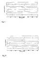
- FIGS. 7 , 8 and 9illustrate results of applying the method of the invention to various pair of signals
- FIG. 10illustrates the result of applying a prior art technique to the signals of FIG. 7 .
- w(n)represents an abdominal ECG signal and x(n)represents a thoracic ECG signal which is assumed to represent accurately the maternal ECG.
- FIG. 2shows a 360-sample record of normalized w(n) plotted against the corresponding normalized record of x(n).
- the nonlinearity of the mappingcan easily be seen by the amount of deviation from the unity-slope line.
- the thoracic signal x(n)is predominantly maternal (MECG), and it is assumed that the fetal component FECG in it is negligible. It should be noted that a proper placement of the thoracic and abdomen electrodes would result in a clean estimate of the FECG such that ⁇ (n) ⁇ s(n). If the abdomen electrode is not placed low enough on the mothers abdominal area then the resulting noisy fetal ECG estimate can be cleaned by post filtering using wavelet denoising and/or nonlinear filtering such as median filtering.
- the transformation Tis generated using polynomial networks which are described in W. M. Campbell, K. T. Assaleh, and C. C. Broun, “Speaker recognition with polynomial classifiers”, IEEE Transactions on Speech and Audio Processing, vol. 10, no. 4, pp 205-212, 2002; K. T. Assaleh and W. M. Campbell, “Speaker Identification Using a Polynomial-based Classifier”, Proceedings of the fourth international Symposium on Signal processing and its Applications ISSPA '99, Brisbane, Australia, August 1999; and W. M. Campbell and Khaled Assaleh, “Low-Complexity Small-Vocabulary Speech Recognition for Portable Devices”, Proceedings of the fourth International Symposium on Signal Processing and its Applications ISSPA '99, Brisbane, Australia, August 1999.
- a nonlinear mapping between x(n)and w(n)is determined by minimizing a mean-squared error criterion. This minimization occurs when the dominant component of w(n) (i.e. the MECG) is aligned with x(n).
- the method for generating the transformation Tis frame-based.
- the signals x(n)and w(n)are partitioned into contiguous frames of N-samples long.
- x i ( m )x ( iN+m )
- w i ( m )w ( iN+m ), where 0 ⁇ m ⁇ N ⁇ - 1
- a time derivative vector sequenceis generated from the frame x,(m)and its J time derivatives. Such that (in matrix notation):
- the time derivative vector sequence X iis then used to generate a Polynomial matrix P i (comprising a vector sequence p(x i (m))) which is generated by polynomial expansion.
- x i (m)[x i (m) ⁇ dot over (x) ⁇ i (m)] and a second order polynomial
- the polynomial basis termsare monomials of the form
- mapping of the input sequence x i (m) onto the desired output sequence w i (m)is achieved by using mean-squared error as the objective criterion such that:
- c iarg ⁇ ⁇ min c i ⁇ ⁇ P i ⁇ c i - w i ⁇ 2
- the vector c imaps the input sequence to the output sequence such that the MECG component in w i (m) is optimally aligned with x i (m).
- the FECG component ⁇ i[ ⁇ i (0) ⁇ i (1) . . . ⁇ i (N ⁇ 1]I the frame w i (m) can be extracted by subtracting the estimated MECG component from it as follows.
- ⁇ tw t ⁇ P c t
- FIGS. 3 and 4are flow charts illustrating the method of the present invention using the principles described above.
- the abdominal electrocardiogram signal wis measured; at step 32 the thoracic electrocardiogram signal x is measured.
- the thoracic electrocardiogram signalis transformed to provided a distorted version of the thoracic electrocardiogram signal ⁇ circumflex over (x) ⁇ such that the difference between the distorted version ⁇ circumflex over (x) ⁇ and the abdominal electrocardiogram signal w is minimised.
- the distorted version of the thoracic electrocardiogram signal ⁇ circumflex over (x) ⁇is subtracted from the abdominal electrocardiogram signal w to provide an estimate of the fetal electrocardiogram.
- each framecomprises 360 samples of the respective electrocardiogram signal
- a matrix X iis then generated from a frame of the thoracic electrocardiogram signal x i .
- 3 time derivativesare used, thus the matrix X i has is 360 ⁇ 4 values.
- Polynomial expansionis than applied to the matrix X i at step 47 to create a polynomial matrix P i .
- P ihas the dimensions 360 ⁇ 35.
- Vector c iis then multiplied by the polynomial matrix P i at step 49 to produce the required distorted version of the thoracic electrocardiogram signal ⁇ circumflex over (x) ⁇ .
- FIGS. 5shows a graph of w(n) against the separated MECG component ⁇ circumflex over (x) ⁇ (n). The larger values of both signals are perfectly aligned do it is possible to subtract the MECG component from w(n).
- FIG. 6show the graph of FIG. 5 superimposed upon the graph of FIG. 2 to illustrate alignment before and after the transformation. It will be appreciated that that the MECG component in w(n) is aligned while the FECG component is almost unchanged.
- FIGS. 7 , 8 and 9show how the proposed technique extracts the FECG component from w(n) for three representative frames.
- FIG. 7 . ashows one frame of w(n)containing non-overlapping 3 FECG beats and two MECG beats.
- FIG. 7 . bshows the output x(n)of the nonlinear mapping process as described above.
- FIG. 7 . cdepicts the extracted FECG signal which clearly shows the separated 3 beats while completely eliminating the presence of the MECG in w(n).
- FIG. 8 . a, bshows another frame of w(n) containing a partial overlap between one of the FECG beats with one MECG beat, and the corresponding frame of ⁇ circumflex over (x) ⁇ (n).
- FIG. 7 . cdepicts the extracted FECG signal which clearly shows the separated 3 beats while completely eliminating the presence of the MECG in w(n).
- FIG. 9 . ashows a third frame of w(n) containing full overlap between the first FECG and the MECG beats. This case represents the extreme case where the FECG is completely dominated by the MECG component to the point that the FECG beat is no longer visually distinguishable.
- FIG. 9 . cshows that the algorithm was still successfully capable of extracting the FECG signal.
- ICAIndependent component analysis
- the method of the inventionis capable of extracting FECG using two leads only. However, this is not possible with the ICA method described by Zaroso and Nandi.
- FIG. 10 . ashows one frame of w(n) containing non-overlapping three FECG beats and two MECG beats (same as in FIG. 7 . a ).
- FIGS. 10 b , and 10 cshow the output of the ICA separated MECG and FECG respectively using two channels only. Clearly, the generated outputs are still composite, and a strong component of the MECG is still apparent in the extracted FECG.
Landscapes
- Health & Medical Sciences (AREA)
- Life Sciences & Earth Sciences (AREA)
- Biomedical Technology (AREA)
- Heart & Thoracic Surgery (AREA)
- Cardiology (AREA)
- Physics & Mathematics (AREA)
- Veterinary Medicine (AREA)
- Biophysics (AREA)
- Pathology (AREA)
- Engineering & Computer Science (AREA)
- Public Health (AREA)
- General Health & Medical Sciences (AREA)
- Medical Informatics (AREA)
- Molecular Biology (AREA)
- Surgery (AREA)
- Animal Behavior & Ethology (AREA)
- Gynecology & Obstetrics (AREA)
- Pediatric Medicine (AREA)
- Pregnancy & Childbirth (AREA)
- Reproductive Health (AREA)
- Measurement And Recording Of Electrical Phenomena And Electrical Characteristics Of The Living Body (AREA)
Abstract
Description
w(n)={circumflex over (x)}(n)+ŝ(n)
ŝ(n)=s(n)+η(n)
{circumflex over (x)}(n) is distorted due to the fact that the signal is measured far away from its source (the mother's heart), and consequently it encounters some nonlinear transformation as it travels to the abdominal area ie:
{circumflex over (x)}(n)=T(x(n))
xi(m)=x(iN+m)
wi(m)=w(iN+m), where 0≦m≦N−-1
p(xi(m)=└1xi(m){dot over (x)}i(m)xi(m){dot over (x)}i(m)2xi(m){dot over (x)}i(m)2┘
PitPici=Pitwi
ci=(PitPi)−1Pitwi
T(xi(n))={circumflex over (x)}≈Pici
ŝt=wt−Pct
Claims (12)
Priority Applications (1)
| Application Number | Priority Date | Filing Date | Title |
|---|---|---|---|
| US11/189,677US7474915B2 (en) | 2005-07-26 | 2005-07-26 | Separating mixed signals containing a distorted signal |
Applications Claiming Priority (1)
| Application Number | Priority Date | Filing Date | Title |
|---|---|---|---|
| US11/189,677US7474915B2 (en) | 2005-07-26 | 2005-07-26 | Separating mixed signals containing a distorted signal |
Publications (2)
| Publication Number | Publication Date |
|---|---|
| US20070027396A1 US20070027396A1 (en) | 2007-02-01 |
| US7474915B2true US7474915B2 (en) | 2009-01-06 |
Family
ID=37695284
Family Applications (1)
| Application Number | Title | Priority Date | Filing Date |
|---|---|---|---|
| US11/189,677Expired - Fee RelatedUS7474915B2 (en) | 2005-07-26 | 2005-07-26 | Separating mixed signals containing a distorted signal |
Country Status (1)
| Country | Link |
|---|---|
| US (1) | US7474915B2 (en) |
Cited By (5)
| Publication number | Priority date | Publication date | Assignee | Title |
|---|---|---|---|---|
| US20080146953A1 (en)* | 2005-01-31 | 2008-06-19 | Yoshitaka Kimura | Electrocardiogram Signal-Processing Method and Electrocardiogram Signal-Processing Device |
| US20100305481A1 (en)* | 2007-05-09 | 2010-12-02 | Koninklijke Philips Electronics N.V. | Method of automatically monitoring the movement activity of a fetus |
| US9131915B2 (en) | 2011-07-06 | 2015-09-15 | University Of New Brunswick | Method and apparatus for noise cancellation |
| US9392952B1 (en)* | 2015-03-10 | 2016-07-19 | Nuvo Group Ltd. | Systems, apparatus and methods for sensing fetal activity |
| US20170172426A1 (en)* | 2015-03-16 | 2017-06-22 | Nuvo Group Ltd. | Continuous non-invasive monitoring of a pregnant human subject |
Families Citing this family (6)
| Publication number | Priority date | Publication date | Assignee | Title |
|---|---|---|---|---|
| US7742812B2 (en)* | 2006-03-29 | 2010-06-22 | Medtronic, Inc. | Method and apparatus for detecting arrhythmias in a medical device |
| US7869863B2 (en)* | 2008-01-10 | 2011-01-11 | The Johns Hopkins University | Apparatus and method for non-invasive, passive fetal heart monitoring |
| EP2440139B1 (en) | 2009-06-09 | 2016-05-18 | Koninklijke Philips N.V. | Method and apparatus for recognizing moving anatomical structures using ultrasound |
| WO2018109669A1 (en)* | 2016-12-13 | 2018-06-21 | King Abdullah University Of Science And Technology | System and method for non-invasive extraction of fetal electrocardiogram signals |
| JP6649982B2 (en)* | 2018-05-10 | 2020-02-19 | アトムメディカル株式会社 | Fetal ECG signal processing method and fetal ECG signal processing device |
| US11246538B2 (en)* | 2019-03-20 | 2022-02-15 | Zoll Medical Corporation | Single channel and dual channel noise detection systems and techniques |
Citations (6)
| Publication number | Priority date | Publication date | Assignee | Title |
|---|---|---|---|---|
| US4211237A (en)* | 1977-04-14 | 1980-07-08 | Biotronik Mess- Und Therapiegerate Gmbh & Co. | Method and apparatus for identifying recurring signal patterns |
| US5042499A (en) | 1988-09-30 | 1991-08-27 | Frank Thomas H | Noninvasive electrocardiographic method of real time signal processing for obtaining and displaying instantaneous fetal heart rate and fetal heart rate beat-to-beat variability |
| US5123420A (en) | 1991-03-25 | 1992-06-23 | Hewlett-Packard Company | Method and apparatus for processing heart rate traces in a fetal monitor |
| US5372139A (en)* | 1991-06-24 | 1994-12-13 | Paul Benjamin Crilly | Method for suppressing a maternal electrocardiogram signal from a fetal electrocardiogram signal obtained with invasive and non-invasive techniques using an almost pure maternal electrocardiogram signal as a trigger |
| US6751498B1 (en)* | 1999-03-15 | 2004-06-15 | The Johns Hopkins University | Apparatus and method for non-invasive, passive fetal heart monitoring |
| US20070066908A1 (en)* | 2003-10-31 | 2007-03-22 | Graupe Menachem H | Separation of one or more fetal heart component signals from heart signal information obtained from a pregnant female |
- 2005
- 2005-07-26USUS11/189,677patent/US7474915B2/ennot_activeExpired - Fee Related
Patent Citations (6)
| Publication number | Priority date | Publication date | Assignee | Title |
|---|---|---|---|---|
| US4211237A (en)* | 1977-04-14 | 1980-07-08 | Biotronik Mess- Und Therapiegerate Gmbh & Co. | Method and apparatus for identifying recurring signal patterns |
| US5042499A (en) | 1988-09-30 | 1991-08-27 | Frank Thomas H | Noninvasive electrocardiographic method of real time signal processing for obtaining and displaying instantaneous fetal heart rate and fetal heart rate beat-to-beat variability |
| US5123420A (en) | 1991-03-25 | 1992-06-23 | Hewlett-Packard Company | Method and apparatus for processing heart rate traces in a fetal monitor |
| US5372139A (en)* | 1991-06-24 | 1994-12-13 | Paul Benjamin Crilly | Method for suppressing a maternal electrocardiogram signal from a fetal electrocardiogram signal obtained with invasive and non-invasive techniques using an almost pure maternal electrocardiogram signal as a trigger |
| US6751498B1 (en)* | 1999-03-15 | 2004-06-15 | The Johns Hopkins University | Apparatus and method for non-invasive, passive fetal heart monitoring |
| US20070066908A1 (en)* | 2003-10-31 | 2007-03-22 | Graupe Menachem H | Separation of one or more fetal heart component signals from heart signal information obtained from a pregnant female |
Cited By (8)
| Publication number | Priority date | Publication date | Assignee | Title |
|---|---|---|---|---|
| US20080146953A1 (en)* | 2005-01-31 | 2008-06-19 | Yoshitaka Kimura | Electrocardiogram Signal-Processing Method and Electrocardiogram Signal-Processing Device |
| US8175692B2 (en)* | 2005-01-31 | 2012-05-08 | Tohoku University | Electrocardiogram signal-processing method and electrocardiogram signal-processing device |
| US20100305481A1 (en)* | 2007-05-09 | 2010-12-02 | Koninklijke Philips Electronics N.V. | Method of automatically monitoring the movement activity of a fetus |
| US9173613B2 (en)* | 2007-05-09 | 2015-11-03 | Koninklijke Philips N.V. | Method of autonomously monitoring the movement activity of a fetus |
| US9131915B2 (en) | 2011-07-06 | 2015-09-15 | University Of New Brunswick | Method and apparatus for noise cancellation |
| US9392952B1 (en)* | 2015-03-10 | 2016-07-19 | Nuvo Group Ltd. | Systems, apparatus and methods for sensing fetal activity |
| US20170172426A1 (en)* | 2015-03-16 | 2017-06-22 | Nuvo Group Ltd. | Continuous non-invasive monitoring of a pregnant human subject |
| US10039459B2 (en)* | 2015-03-16 | 2018-08-07 | Nuvo Group Ltd. | Continuous non-invasive monitoring of a pregnant human subject |
Also Published As
| Publication number | Publication date |
|---|---|
| US20070027396A1 (en) | 2007-02-01 |
Similar Documents
| Publication | Publication Date | Title |
|---|---|---|
| Assaleh et al. | A novel technique for the extraction of fetal ECG using polynomial networks | |
| Choudhary et al. | Automatic detection of aortic valve opening using seismocardiography in healthy individuals | |
| He et al. | Application of independent component analysis in removing artefacts from the electrocardiogram | |
| Jagannath et al. | Issues and research on foetal electrocardiogram signal elicitation | |
| Azzerboni et al. | A new approach based on wavelet-ICA algorithms for fetal electrocardiogram extraction. | |
| Barhatte et al. | Noise analysis of ECG signal using fast ICA | |
| Fotiadou et al. | Enhancement of low-quality fetal electrocardiogram based on time-sequenced adaptive filtering | |
| US20240366155A1 (en) | Systems and methods for maternal uterine activity detection | |
| US7474915B2 (en) | Separating mixed signals containing a distorted signal | |
| Devuyst et al. | Cancelling ECG artifacts in EEG using a modified independent component analysis approach | |
| John et al. | Extraction of foetal ECG from abdominal ECG by nonlinear transformation and estimations | |
| CN115486856A (en) | Surface electrophysiological signal processing method and system for eliminating signal phase shift | |
| Sanyal et al. | Application of framelet transform in filtering baseline drift from ECG signals | |
| Zia et al. | Noise detection in heart sound recordings | |
| Kotas | Combined application of independent component analysis and projective filtering to fetal ECG extraction | |
| Gupta et al. | A novel approach to fetal ECG extraction and enhancement using blind source separation (BSS-ICA) and adaptive fetal ECG enhancer (AFE) | |
| Castells et al. | Atrial activity extraction from atrial fibrillation episodes based on maximum likelihood source separation | |
| Zubair et al. | Electromyography noise suppression in electrocardiogram signal using modified garrote threshold shrinkage function | |
| Krishnan et al. | Motion artifact reduction in photopleythysmography using magnitude-based frequency domain independent component analysis | |
| Ayat et al. | Extracting fetal ECG from a single maternal abdominal record | |
| Llinares et al. | Application of constrained independent component analysis algorithms in electrocardiogram arrhythmias | |
| Algunaidi et al. | Fetal heart rate monitoring based on adaptive noise cancellation and maternal QRS removal window | |
| Yerande et al. | A hybrid technique for non-invasive fetal ecg extraction and heart rate estimation from the mother’s abdomen signal | |
| Alcaraz et al. | Adaptive singular value QRST cancellation for the analysis of short single lead atrial fibrillation electrocardiograms | |
| Assaleh et al. | Noniterative method for two-lead fetal ECG extraction |
Legal Events
| Date | Code | Title | Description |
|---|---|---|---|
| AS | Assignment | Owner name:AMERICAN UNIVERSITY OF SHARJAH, UNITED ARAB EMIRAT Free format text:ASSIGNMENT OF ASSIGNORS INTEREST;ASSIGNORS:ASSALEH, KHALED;AL-NASHASH, HASAN;REEL/FRAME:016823/0025 Effective date:20050721 Owner name:ARAB SCIENCE AND TECHNOLOGY FOUNDATION, UNITED ARA Free format text:ASSIGNMENT OF ASSIGNORS INTEREST;ASSIGNORS:ASSALEH, KHALED;AL-NASHASH, HASAN;REEL/FRAME:016823/0025 Effective date:20050721 | |
| STCF | Information on status: patent grant | Free format text:PATENTED CASE | |
| FPAY | Fee payment | Year of fee payment:4 | |
| SULP | Surcharge for late payment | ||
| REMI | Maintenance fee reminder mailed | ||
| FPAY | Fee payment | Year of fee payment:8 | |
| SULP | Surcharge for late payment | Year of fee payment:7 | |
| FEPP | Fee payment procedure | Free format text:MAINTENANCE FEE REMINDER MAILED (ORIGINAL EVENT CODE: REM.); ENTITY STATUS OF PATENT OWNER: SMALL ENTITY | |
| LAPS | Lapse for failure to pay maintenance fees | Free format text:PATENT EXPIRED FOR FAILURE TO PAY MAINTENANCE FEES (ORIGINAL EVENT CODE: EXP.); ENTITY STATUS OF PATENT OWNER: SMALL ENTITY | |
| STCH | Information on status: patent discontinuation | Free format text:PATENT EXPIRED DUE TO NONPAYMENT OF MAINTENANCE FEES UNDER 37 CFR 1.362 | |
| FP | Lapsed due to failure to pay maintenance fee | Effective date:20210106 |