US7470241B2 - Controlled high efficiency lesion formation using high intensity ultrasound - Google Patents
Controlled high efficiency lesion formation using high intensity ultrasoundDownload PDFInfo
- Publication number
- US7470241B2 US7470241B2US10/633,726US63372603AUS7470241B2US 7470241 B2US7470241 B2US 7470241B2US 63372603 AUS63372603 AUS 63372603AUS 7470241 B2US7470241 B2US 7470241B2
- Authority
- US
- United States
- Prior art keywords
- ultrasound
- tissue
- transducer
- treatment
- high intensity
- Prior art date
- Legal status (The legal status is an assumption and is not a legal conclusion. Google has not performed a legal analysis and makes no representation as to the accuracy of the status listed.)
- Expired - Lifetime
Links
- 238000002604ultrasonographyMethods0.000titleclaimsabstractdescription111
- 230000003902lesionEffects0.000titledescription29
- 230000015572biosynthetic processEffects0.000titledescription4
- 238000011282treatmentMethods0.000claimsabstractdescription92
- 206010028980NeoplasmDiseases0.000claimsabstractdescription64
- 238000000034methodMethods0.000claimsabstractdescription45
- 230000036770blood supplyEffects0.000claimsabstractdescription9
- 201000010260leiomyomaDiseases0.000claimsdescription59
- 206010046798Uterine leiomyomaDiseases0.000claimsdescription42
- 208000010579uterine corpus leiomyomaDiseases0.000claimsdescription17
- 201000007954uterine fibroidDiseases0.000claimsdescription17
- 238000010438heat treatmentMethods0.000claimsdescription15
- 230000017074necrotic cell deathEffects0.000claimsdescription9
- 230000002956necrotizing effectEffects0.000claims2
- 238000003384imaging methodMethods0.000abstractdescription32
- 238000002560therapeutic procedureMethods0.000abstractdescription22
- 239000007788liquidSubstances0.000abstractdescription10
- 230000001338necrotic effectEffects0.000abstractdescription10
- 239000013536elastomeric materialSubstances0.000abstractdescription3
- 210000001519tissueAnatomy0.000description109
- XLYOFNOQVPJJNP-UHFFFAOYSA-NwaterSubstancesOXLYOFNOQVPJJNP-UHFFFAOYSA-N0.000description27
- 230000008878couplingEffects0.000description14
- 238000010168coupling processMethods0.000description14
- 238000005859coupling reactionMethods0.000description14
- 239000007789gasSubstances0.000description14
- 210000004291uterusAnatomy0.000description14
- 230000001225therapeutic effectEffects0.000description13
- 238000012285ultrasound imagingMethods0.000description12
- 210000000056organAnatomy0.000description11
- 238000010586diagramMethods0.000description10
- 239000012530fluidSubstances0.000description10
- 238000013459approachMethods0.000description8
- 230000006378damageEffects0.000description8
- 238000009210therapy by ultrasoundMethods0.000description8
- 206010028851NecrosisDiseases0.000description7
- FAPWRFPIFSIZLT-UHFFFAOYSA-MSodium chlorideChemical compound[Na+].[Cl-]FAPWRFPIFSIZLT-UHFFFAOYSA-M0.000description7
- 239000011780sodium chlorideSubstances0.000description7
- 230000000740bleeding effectEffects0.000description6
- 230000017531blood circulationEffects0.000description6
- 230000000694effectsEffects0.000description6
- 230000008569processEffects0.000description6
- 238000001356surgical procedureMethods0.000description6
- 210000000683abdominal cavityAnatomy0.000description5
- 238000010521absorption reactionMethods0.000description5
- 210000003484anatomyAnatomy0.000description5
- 201000010099diseaseDiseases0.000description5
- 208000037265diseases, disorders, signs and symptomsDiseases0.000description5
- 230000006870functionEffects0.000description5
- 239000000499gelSubstances0.000description5
- 230000000149penetrating effectEffects0.000description5
- 238000009557abdominal ultrasonographyMethods0.000description4
- 210000003815abdominal wallAnatomy0.000description4
- 210000004204blood vesselAnatomy0.000description4
- 230000023597hemostasisEffects0.000description4
- 230000007246mechanismEffects0.000description4
- 239000000523sampleSubstances0.000description4
- 238000001228spectrumMethods0.000description4
- 238000002679ablationMethods0.000description3
- 210000004369bloodAnatomy0.000description3
- 239000008280bloodSubstances0.000description3
- 230000008859changeEffects0.000description3
- 238000013461designMethods0.000description3
- 239000011159matrix materialSubstances0.000description3
- 239000012528membraneSubstances0.000description3
- 210000000664rectumAnatomy0.000description3
- 210000000685uterine arteryAnatomy0.000description3
- 230000008901benefitEffects0.000description2
- 230000005540biological transmissionEffects0.000description2
- 239000000919ceramicSubstances0.000description2
- 238000006243chemical reactionMethods0.000description2
- 239000002826coolantSubstances0.000description2
- 238000001514detection methodMethods0.000description2
- 239000002961echo contrast mediaSubstances0.000description2
- 238000002592echocardiographyMethods0.000description2
- 238000009558endoscopic ultrasoundMethods0.000description2
- 208000014674injuryDiseases0.000description2
- 238000002357laparoscopic surgeryMethods0.000description2
- 210000004185liverAnatomy0.000description2
- 230000003287optical effectEffects0.000description2
- 210000004789organ systemAnatomy0.000description2
- 239000002245particleSubstances0.000description2
- 230000009024positive feedback mechanismEffects0.000description2
- 238000012545processingMethods0.000description2
- 210000002307prostateAnatomy0.000description2
- 238000011084recoveryMethods0.000description2
- 239000000243solutionSubstances0.000description2
- 230000003595spectral effectEffects0.000description2
- 239000008223sterile waterSubstances0.000description2
- 230000008685targetingEffects0.000description2
- 230000003685thermal hair damageEffects0.000description2
- 230000008733traumaEffects0.000description2
- 210000001215vaginaAnatomy0.000description2
- 101100182881Caenorhabditis elegans madd-3 geneProteins0.000description1
- 206010069729Collateral circulationDiseases0.000description1
- 206010060932Postoperative adhesionDiseases0.000description1
- 206010054094Tumour necrosisDiseases0.000description1
- 210000001015abdomenAnatomy0.000description1
- 230000003187abdominal effectEffects0.000description1
- 230000002411adverseEffects0.000description1
- XAGFODPZIPBFFR-UHFFFAOYSA-NaluminiumChemical group[Al]XAGFODPZIPBFFR-UHFFFAOYSA-N0.000description1
- 229910052782aluminiumInorganic materials0.000description1
- 230000010108arterial embolizationEffects0.000description1
- 210000001367arteryAnatomy0.000description1
- QVGXLLKOCUKJST-UHFFFAOYSA-Natomic oxygenChemical compound[O]QVGXLLKOCUKJST-UHFFFAOYSA-N0.000description1
- 230000002238attenuated effectEffects0.000description1
- 230000004888barrier functionEffects0.000description1
- 210000000481breastAnatomy0.000description1
- 210000003679cervix uteriAnatomy0.000description1
- 230000004087circulationEffects0.000description1
- 238000004891communicationMethods0.000description1
- 230000001143conditioned effectEffects0.000description1
- 239000002872contrast mediaSubstances0.000description1
- 229940039231contrast mediaDrugs0.000description1
- 238000001816coolingMethods0.000description1
- 239000012809cooling fluidSubstances0.000description1
- 238000000315cryotherapyMethods0.000description1
- 238000009792diffusion processMethods0.000description1
- 230000003292diminished effectEffects0.000description1
- 229940079593drugDrugs0.000description1
- 239000003814drugSubstances0.000description1
- 239000013013elastic materialSubstances0.000description1
- 238000013161embolization procedureMethods0.000description1
- 238000001839endoscopyMethods0.000description1
- 210000003238esophagusAnatomy0.000description1
- 210000001105femoral arteryAnatomy0.000description1
- 238000005111flow chemistry techniqueMethods0.000description1
- 235000003642hungerNutrition0.000description1
- 238000009802hysterectomyMethods0.000description1
- 230000000977initiatory effectEffects0.000description1
- 238000002347injectionMethods0.000description1
- 239000007924injectionSubstances0.000description1
- 238000003780insertionMethods0.000description1
- 230000037431insertionEffects0.000description1
- 230000009545invasionEffects0.000description1
- 230000002262irrigationEffects0.000description1
- 238000003973irrigationMethods0.000description1
- 210000003734kidneyAnatomy0.000description1
- 230000004807localizationEffects0.000description1
- 230000007774longtermEffects0.000description1
- 239000000463materialSubstances0.000description1
- 238000000691measurement methodMethods0.000description1
- 239000000203mixtureSubstances0.000description1
- 238000012986modificationMethods0.000description1
- 230000004048modificationEffects0.000description1
- 238000001208nuclear magnetic resonance pulse sequenceMethods0.000description1
- 235000015097nutrientsNutrition0.000description1
- 238000012634optical imagingMethods0.000description1
- 230000002611ovarianEffects0.000description1
- 239000001301oxygenSubstances0.000description1
- 229910052760oxygenInorganic materials0.000description1
- 210000000496pancreasAnatomy0.000description1
- 230000001575pathological effectEffects0.000description1
- 239000004033plasticSubstances0.000description1
- 230000035935pregnancyEffects0.000description1
- 230000002035prolonged effectEffects0.000description1
- 230000008439repair processEffects0.000description1
- 238000007789sealingMethods0.000description1
- 210000000952spleenAnatomy0.000description1
- 230000037351starvationEffects0.000description1
- 230000004936stimulating effectEffects0.000description1
- 239000000126substanceSubstances0.000description1
- 230000004083survival effectEffects0.000description1
- 208000024891symptomDiseases0.000description1
- 238000012546transferMethods0.000description1
- 238000011269treatment regimenMethods0.000description1
- 230000000007visual effectEffects0.000description1
- 238000012800visualizationMethods0.000description1
- 239000002699waste materialSubstances0.000description1
Images


Classifications
- A—HUMAN NECESSITIES
- A61—MEDICAL OR VETERINARY SCIENCE; HYGIENE
- A61N—ELECTROTHERAPY; MAGNETOTHERAPY; RADIATION THERAPY; ULTRASOUND THERAPY
- A61N7/00—Ultrasound therapy
- A—HUMAN NECESSITIES
- A61—MEDICAL OR VETERINARY SCIENCE; HYGIENE
- A61B—DIAGNOSIS; SURGERY; IDENTIFICATION
- A61B8/00—Diagnosis using ultrasonic, sonic or infrasonic waves
- A61B8/12—Diagnosis using ultrasonic, sonic or infrasonic waves in body cavities or body tracts, e.g. by using catheters
- A—HUMAN NECESSITIES
- A61—MEDICAL OR VETERINARY SCIENCE; HYGIENE
- A61B—DIAGNOSIS; SURGERY; IDENTIFICATION
- A61B8/00—Diagnosis using ultrasonic, sonic or infrasonic waves
- A61B8/42—Details of probe positioning or probe attachment to the patient
- A61B8/4272—Details of probe positioning or probe attachment to the patient involving the acoustic interface between the transducer and the tissue
- A61B8/4281—Details of probe positioning or probe attachment to the patient involving the acoustic interface between the transducer and the tissue characterised by sound-transmitting media or devices for coupling the transducer to the tissue
- A—HUMAN NECESSITIES
- A61—MEDICAL OR VETERINARY SCIENCE; HYGIENE
- A61B—DIAGNOSIS; SURGERY; IDENTIFICATION
- A61B8/00—Diagnosis using ultrasonic, sonic or infrasonic waves
- A61B8/44—Constructional features of the ultrasonic, sonic or infrasonic diagnostic device
- A61B8/4444—Constructional features of the ultrasonic, sonic or infrasonic diagnostic device related to the probe
- A61B8/445—Details of catheter construction
- A—HUMAN NECESSITIES
- A61—MEDICAL OR VETERINARY SCIENCE; HYGIENE
- A61B—DIAGNOSIS; SURGERY; IDENTIFICATION
- A61B8/00—Diagnosis using ultrasonic, sonic or infrasonic waves
- A61B8/54—Control of the diagnostic device
- A61B8/546—Control of the diagnostic device involving monitoring or regulation of device temperature
- A—HUMAN NECESSITIES
- A61—MEDICAL OR VETERINARY SCIENCE; HYGIENE
- A61N—ELECTROTHERAPY; MAGNETOTHERAPY; RADIATION THERAPY; ULTRASOUND THERAPY
- A61N7/00—Ultrasound therapy
- A61N7/02—Localised ultrasound hyperthermia
- A—HUMAN NECESSITIES
- A61—MEDICAL OR VETERINARY SCIENCE; HYGIENE
- A61B—DIAGNOSIS; SURGERY; IDENTIFICATION
- A61B17/00—Surgical instruments, devices or methods
- A61B17/42—Gynaecological or obstetrical instruments or methods
- A61B2017/4216—Operations on uterus, e.g. endometrium
- A—HUMAN NECESSITIES
- A61—MEDICAL OR VETERINARY SCIENCE; HYGIENE
- A61B—DIAGNOSIS; SURGERY; IDENTIFICATION
- A61B90/00—Instruments, implements or accessories specially adapted for surgery or diagnosis and not covered by any of the groups A61B1/00 - A61B50/00, e.g. for luxation treatment or for protecting wound edges
- A61B90/36—Image-producing devices or illumination devices not otherwise provided for
- A61B90/37—Surgical systems with images on a monitor during operation
- A61B2090/378—Surgical systems with images on a monitor during operation using ultrasound
- A—HUMAN NECESSITIES
- A61—MEDICAL OR VETERINARY SCIENCE; HYGIENE
- A61N—ELECTROTHERAPY; MAGNETOTHERAPY; RADIATION THERAPY; ULTRASOUND THERAPY
- A61N7/00—Ultrasound therapy
- A61N2007/0052—Ultrasound therapy using the same transducer for therapy and imaging
- A—HUMAN NECESSITIES
- A61—MEDICAL OR VETERINARY SCIENCE; HYGIENE
- A61N—ELECTROTHERAPY; MAGNETOTHERAPY; RADIATION THERAPY; ULTRASOUND THERAPY
- A61N7/00—Ultrasound therapy
- A61N7/02—Localised ultrasound hyperthermia
- A61N7/022—Localised ultrasound hyperthermia intracavitary
Definitions
- This inventionrelates generally to methods and apparatus for efficiently heating biological tissues with high intensity ultrasound for therapeutic purposes, and in particular, to endoscopic devices for applying ultrasound energy to uterine fibroids and other pathologic tissues that are inside body or organ cavities, to destroy the tumor or the diseased tissue.
- Fibroidsare benign tumors in women's uteri. There are different types of fibroids, including submucosal, which are inside the uterine cavity; intramural, which are in the uterine wall; and subserosal, which are outside the uterus. Fibroids may cause excessive bleeding and pain.
- surgeryis the predominate treatment. Every year in the U.S., there are more than 200,000 cases of fibroid-caused hysterectomies. To preserve the uterus, the patient may choose myomectomy, which removes the fibroids only. There are more than 80,000 abdominal myomectomies each year in the U.S. These surgical procedures cause significant trauma to the patients and result in significant costs. Consequently, patients need several days of hospital stay and suffer from the prolonged recovery.
- MISMinimally invasive surgical
- Many MIS apparatushave been developed to make the procedure less difficult.
- Several prior art devicesare described in U.S. Pat. No. 5,304,124; U.S. Pat. No. 5,662,680; and U.S. Pat. No. 5,709,679.
- alternative treatmentsinclude using different energy forms, such as laser, radio frequency (RF), and cryo-therapy, to thermally ablate or necrose the fibroid tissue. Most of these techniques require the insertion of needles or other types of devices into the body of the fibroid.
- Uterine arterial embolizationhas been investigated as an alternative treatment for uterine fibroids.
- UAEUterine arterial embolization
- a catheteris inserted into the patient's femoral artery. The catheter is then advanced until its tip reaches the uterine artery. Many small particles are then injected into the uterine artery to block the blood flow. Both left and right uterine arteries are treated. Blood vessels supplying uterine fibroids are typically larger than the vessels in the normal uterine tissue. With properly sized particles, the blood vessels feeding the uterine fibroids are embolized, but not those in the normal uterine tissue. The fibroids then starve and die due to lack of a blood supply.
- the uterussurvives, however, on the blood supplied from the ovarian artery and other collateral circulation.
- the embolization proceduremay cause severe pain in the first few days after the treatment.
- Other disadvantages of UAEmay include long X-ray radiation exposure during the procedure and other long-term potential adverse effects. The procedure is not recommended if the patient seeks a future pregnancy.
- Ultrasoundis a term that refers to acoustic waves having a frequency above the high limit of the human audible range (i.e., above 20 KHz). Ultrasound waves have the capability of penetrating into the human body. Based on this property, ultrasound in the frequency range of 2-20 MHz has been widely used to image internal human organs for diagnostic purposes. Ultrasound imaging has also been suggested as a tool for guidance during a resectoscopic surgery (U.S. Pat. No. 5,957,849).
- ultrasound energyWhen ultrasound energy is absorbed by tissue, it becomes thermal energy, raising the temperature of the tissue. To avoid thermal damage to tissue, the power level in diagnostic ultrasound imaging is kept very low.
- the typical ultrasound intensity (power per unit area) used in imagingis less than 0.1 watt per square centimeter.
- High intensity focused ultrasoundwhich can have an intensity above 1000 watts per square centimeter, can raise the tissue temperature at the region of the spatial focus to above 60-80 degrees Celsius in a few seconds and can cause tissue necrosis almost instantaneously.
- Pat. Nos. 5,471,988, 5,492,126, 5,666,954, 5,697,897, and 5,873,828, endoscopic ultrasound devices with both imaging and therapeutic capabilitiesare disclosed. These devices all have an elongated tube or shaft, so that they can be inserted in organ cavities (e.g., into the rectum) or into the abdominal cavity through a puncture hole in the abdominal wall to bring the ultrasound imaging and treatment sources closer to the disease sites. Some of them have flexible ends, which can be bent to fit the anatomy of a specific patient.
- the therapeutic ultrasound beamis focused inside tissue to a small spot of a few millimeters in size. At the focus, tissue temperature rapidly exceeds a level sufficient to cause tissue necrosis, thus achieving the desired therapeutic effect. Outside of the focus, ultrasound energy is less concentrated, tissue temperature rise remains below the necrosis level during the typically short exposure times employed.
- the ultrasound focusis deflected mechanically or electronically to scan, or incrementally expose, the target tissue volume.
- One disadvantage of the current high intensity ultrasound therapyis its inefficiency when treating large tumors or heating a large volume of tissue.
- the ultrasound treatmentmust typically pause 40-60 seconds between two subsequent pulses to allow the intermediate tissue between the focus and the ultrasound transducer to cool sufficiently to avoid thermally damaging the tissue.
- the volume of tissue necrosis for each treatment pulseis very small ( ⁇ 0.05 cm 3 ).
- to treat a volume of tissue within a 3 cm diameter sphereit will take more than 4 hours, too long to be practical in most clinical situations.
- Many symptomatic uterine fibroidsare larger than 2-3 cm in diameter, and multiple fibroids are also common. To be acceptable for clinicians and patients, the ultrasound treatment time must be significantly reduced.
- the diameter of the treatment transduceris approximately equal to the maximum depth, where the f-number (transducer diameter divided by its focal length) of the transducer is about one (f/1).
- the transducer surface areamust also be sufficiently large to generate high ultrasound power.
- endoscopic devicesfor example, in U.S. Pat. Nos. 5,471,988 and 5,873,828, there is a large orifice in the center of the therapy transducer for positioning an imaging transducer. This orifice reduces the area of the treatment transducer and increases its effective f-number.
- the size of the treatment transducermust be increased to maintain its effectiveness, so that the overall dimensions of the device are increased.
- the maximum acceptable diameter of an ultrasound deviceis about 10 mm. It is seen that it is very difficult to meet this requirement with the large two-transducer configuration.
- the deviceshould preferably cause minimal or no trauma to the patient body so that the patient requires minimum or no recovery time; it should be easy to use; and, the treatment should be quickly administered.
- the deviceshould preferably not cause blood loss during the treatment procedure; it should not mechanically damage the treated organ (e.g. uterus) to avoid the need for complicated organ repair (such as suturing or extensive cauterization); and, it should not increase the risk of post-operative adhesions and other complications.
- the deviceshould be capable of carrying out the following functions:
- the present inventionis directed to a method and apparatus for efficiently treating uterine fibroids and other diseases with high intensity ultrasound, where the apparatus is small enough to fit in the limited space in a patient organ cavity or a limited puncture size on an abdominal wall.
- an ultrasonic system for destroying undesired tissue at an internal site within a body of a patientincludes a probe that is sized to be inserted within a body of a patient.
- An ultrasonic transduceris mounted proximate a distal end of the probe and is adapted to couple to a power supply used to selectively energize the ultrasonic transducer so that it produces a focused beam of high intensity ultrasonic energy.
- An ultrasound transmissive interfaceis coupled to the distal end of the probe and is disposed and adapted to conform to a surface of the undesired tissue. The interface provides a liquid layer that more efficiently transmits the high intensity ultrasonic energy produced by the ultrasonic transducer into the undesired tissue. The high intensity ultrasonic energy increases a temperature of the undesired tissue sufficiently to cause the tissue to necrose.
- the ultrasound transmissive interfacecomprises an elastomeric cavity that is adapted to contain a liquid.
- the elastomeric cavityis disposed between the ultrasonic transducer and the surface of the undesired tissue so that the high intensity ultrasonic energy passes through the liquid within the elastomeric cavity and into the undesired tissue.
- the elastomeric cavityis formed at least in part from a semi-permeable membrane, so that the liquid from within the elastomeric cavity weeps onto a surface of undesired tissue to increase the efficiency with which the high intensity ultrasonic energy is coupled into the undesired tissue.
- the ultrasound transmissive interfacecomprises a cap made of an elastomeric material, which is disposed to surround the ultrasonic transducer.
- the capis adapted to seal against the undesired tissue and to contain a liquid that increases an efficiency with which the high intensity ultrasonic energy is coupled into the undesired tissue.
- the cappreferably includes a rim having a double lip seal formed around a perimeter. A passage in the cap is adapted to couple the double lip seal to a vacuum line so that the rim of the cap is held against a surface of the undesirable tissue, sealing the liquid inside of the cap.
- Another aspect of the present inventionis directed to a method for administering an ultrasonic therapy to destroy at least a portion of an undesired tissue mass.
- the methodincludes the steps of providing an ultrasonic transducer that emits a focused high energy ultrasonic energy when energized, and positioning the ultrasonic transducer proximate the undesired tissue mass.
- the ultrasonic transduceris directed toward a desired focal point within the undesired tissue mass.
- the ultrasonic transduceris energized so that it emits the focused high energy ultrasonic energy at the desired focal point, causing necrosis of a portion of the undesired tissue mass disposed at the desired focal point.
- At least one of an f-number, an intensity, a time, and a direction of the high intensity ultrasonic energy emitted into the undesired tissue massis controlled to achieve a desired shape and size of a necrotic zone of undesired tissue, destroyed as a result of being heated by the high intensity ultrasonic energy.
- the necrotic zonesubstantially blocks the high intensity ultrasonic energy from penetrating beyond the necrotic zone.
- the desired shape and size of the necrotic zoneare preferably selected and formed so as to cause substantially of the undesired tissue mass to ultimately be destroyed.
- the step of controllingpreferably includes the step of repositioning the ultrasonic transducer to direct the high intensity ultrasonic energy at a different portion of the undesired tissue mass, to achieve the desired shape and size of the necrotic zone.
- the desired shape and size of the necrotic zoneare selected so that formation of the necrotic zone substantially deprives the undesired tissue mass of a blood supply, causing the ultimate destruction of the undesired tissue mass.
- the desired shape and size of the necrotic zoneare selected to control bleeding at a treatment site.
- FIG. 1is a block diagram of the positive feedback mechanism of the improved tissue heating process
- FIG. 2A-2Dillustrate different thermal lesion shapes
- FIG. 3Ais a cross-sectional view of a portion of a patient's body, illustrating application of an endoscopic device in accord with the present invention, which can both acquire ultrasound images and generate high intensity therapeutic ultrasound at its distal end;
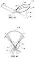
- FIGS. 3B and 3Care side elevational views of a portion of the device shown in FIG. 3A , illustrating an imaging field and a treatment field of the device;
- FIGS. 4A and 4Bare schematic diagrams of different treatment beam forming techniques used to control the lesion geometry and illustrating spatial lesion formation;
- FIGS. 5A and 5Bare schematic diagrams of different treatment beam forming techniques used to control the lesion geometry and illustrating spatial-temporal lesion formation
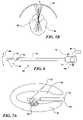
- FIG. 6is a schematic diagram of the trans-cervical ultrasound device with an articulated end
- FIGS. 7A and 7Bare schematic diagrams showing transcervical hemostasis treatment performed in combination with resectoscopic removal of submucosal fibroids
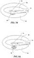
- FIGS. 8A and 8Bare schematic diagrams respectively showing the trans-cervical and the trans-abdominal ultrasound device treating intramural fibroids from inside and from outside of the uterus;
- FIGS. 9A and 9Bare schematic diagrams respectively showing laparoscopic occlusion treatment of subserosal fibroids, and a wedge of necrosed tissue produced thereby;
- FIG. 10is a block diagram of the trans-cervical ultrasound device connected with its control unit, display, and fluid management unit;
- FIG. 11is a side elevational view of the ultrasound device and a cross-sectional view of a liquid-filled vacuum cap that provides coupling between the fibroid tumor and the ultrasound transducer;
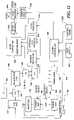
- FIG. 12is a system block diagram of the control electronics in the control unit.
- FIG. 13is an isometric view of a portion of an ultrasound device that includes a structure to maintain a gap between the tissue being treated and an ultrasound transducer array, to convey a coolant liquid.
- the therapy deliveredmay be thermal ablation, where a temperature rise is established to a level at which tissues are no longer viable; mechanical ablation, where cavitation is employed as the primary ablative means; or may achieve hemostasis wherein bleeding or blood flow in intact organs is arrested.
- Such applications of the present inventionmay be accomplished in open, invasive surgery, by way of established minimally invasive techniques (for example, by way of body entry through one or more small incisions or punctures), or in some cases, noninvasively, through the skin surface or through the linings of body cavities such as the rectum, vagina, or esophagus.
- Ablative treatment with the present inventionmay be applied to a wide range of benign or cancerous lesions of the liver, kidney, pancreas, spleen, prostate, breast, bowel, rectum or similar organ systems, wherein the device described herein may be placed in close proximity to the disease location.
- acoustic hemostasis treatmentmay be employed to deprive a disease lesion of its blood supply or used to facilitate surgical procedures by arresting bleeding or blood flow.
- tumorssuch as uterine fibroids
- uterine fibroidslocate superficially inside or outside the organ.
- surgeonscan easily reach the surfaces of those tumors with an intra-cervical or intra-abdominal instrument.
- intra-cervical or intra-abdominal instrumentFor an ultrasound transducer at the tip of the intra-cavity instrument touching the tumor directly, there will be little or no intermediate tissue that needs to be spared and cooled, so that pauses in the treatment for this purpose may become unnecessary.
- the pre-focal heatingis considered to be a negative effect and needs to be minimized.
- this pre-focal heatingcan provide significant enhancement to the efficiency of tissue heating when the ultrasound transducer can be disposed in close contact with the tumor surface.
- a positive feedback mechanism of tissue heating(illustrated in FIG. 1 ) is preferably used to improve the efficiency of the treatment provided by the present invention.
- the positive feedback indicated by a block 2 of FIG. 1enhances acoustic absorption.
- the acoustic energyis converted to heat, as noted in a block 4 , resulting in a greater temperature rise in the tissue, as indicated in a block 6 .
- Tissue acoustic absorptionincreases significantly when its temperature rises above 50° C.
- a small f-number, high intensity ultrasound transducer 10running in continuous-wave (CW) mode, raises the temperature in tissue 12 at its focus to 70-90° C. in less than two seconds and forms a small lesion 14 .
- This isolated thermal lesionserves two purposes. First, it is the initial seed to start the positive feedback heating process; and, secondly, its high acoustic absorption blocks ultrasound energy from penetrating beyond the focal depth to cause undesirable damage to normal tissue.
- a wedge-shaped lesion of tissue necrosiswas generated with this mechanism by running the ultrasound power continuously, while keeping the transducer position fixed.
- the volume of the thermal lesionwas about 4.5 cm 3 , and the treatment time was approximately two minutes.
- the average treatment ratewas about 2.25 cm 3 /min, which was 45 times faster than provided by a conventional pulse-pause treatment strategy.
- a circular transducer 18 with a relatively large f-number ( ⁇ 2)is used to treat the tissue over a relatively short time.
- a circular transducer with a small f-number ( ⁇ 1)is used to treat the tissue for a relatively long time.
- a cylindrical or truncated circular transduceris used to treat the tissue over a relatively long time.
- 2Dcan be generated by forming a row of tightly spaced lesion columns 22 , 24 , and 26 .
- Each columnis formed from a fixed transducer position in a short time. The transducer may then be quickly shifted laterally to generate the next adjacent column, moving from position “A” to “B” to “C” as shown in FIG. 2D .
- Thermal diffusion in the tissuefuses the columns together to form rectangular lesion plane 21 . It is also possible to create a large lesion in the tissue without damaging the organ surface.
- One approachis to cool the tissue surface with circulating water or saline.
- the other approachis to use an attenuation measurement technique described below, to monitor lesion progress (growth) and control power, accordingly.
- FIGS. 3A and 3 BThe basic concept and configuration of a high intensity ultrasound device 29 in accord with the present invention are shown in FIGS. 3A and 3 B.
- the devicehas a thin, elongate shaft 28 that can be inserted through the cervix into the uterine cavity, or, as shown in FIG. 3A , through a laparoscopic opening 34 in the abdominal wall and into the abdominal cavity.
- a distal end 30 of the shaftcontains a concave-shaped ultrasound transducer array 36 ( FIG. 3B ) and may be formed into different curves to fit different anatomies of individual patients.
- the distal end that is thus formedcan be permanently fixed or articulated by turning a control knob 32 on a handle 33 of the device.
- Transducer array 36 in FIG. 3Bis operable for both ultrasound imaging and treatment.
- the transducer arrayTo form an ultrasound image, the transducer array generates ultrasound pulses and receives echoes from the imaged anatomy in a cross-sectional area 40 .
- the two-dimensional (2D) ultrasound imagedisplays the cross-sectional view of the anatomy.
- the imagecan be updated rapidly in real-time with a frame rate of, for example, 10-30 frames per second. Physicians can then view this real-time image to locate the tumor or other tissue that needs to be treated or spared from treatment.
- the transducer arrayWhen the treatment area is identified in the image, the transducer array is employed to generate high intensity ultrasound focused in a treatment area 38 . After the tissue in the treatment area has been necrosed, the distal end of the ultrasound device is moved to a new location to sequentially treat another part of the tumor tissue.
- the imaging and the treatmentare interleaved in time so that the treatment process and the progress of the treatment may be monitored.
- Doppler flow imagingmay be utilized to assist targeting and to monitor treatment effects and to determine the endpoint of the therapy. Imaging blood flow is particularly useful when a blood flow occlusion strategy is being utilized, since the cessation of blood flow can be directly monitored. Doppler imaging facilitates localization of the vascularity typically surrounding uterine fibroid tumors or other tumor masses.
- Imaging and therapymay be one-, two-, or three-dimensional in various combinations; scan geometries may be fixed or selectable; and imaging and therapy may proceed either simultaneously or sequentially in time.
- a preferred embodiment of the ultrasound intra-cavity device discussed hereinhas the capability to carryout 2D real-time imaging and the capability to produce tissue necrosis in a substantially 2D slice (thickness of this slice is nominally less than one centimeter).
- Including the lesion-control techniques discussed abovethere are many ways to control treatment geometry with this device. Different spatial beam patterns can be generated from by the ultrasound transducer array included on the device to form a specific lesion shape, or potentially, to reduce treatment time. Multiple sequential exposures of different spatial beam patterns can also be used to control the treatment dosage at different locations to form lesion shapes that cannot be generated by fixed beam patterns.
- a trans-cervical ultrasound device 68is adapted to treat submucosal fibroids.
- the deviceis inserted into a patient's uterine cavity through the vagina and the cervical canal.
- the uterine cavityis distended with sterile water or saline under 50-80 mm Hg pressure delivered through internal channels inside a shaft 70 of the device and connected to couplings 78 and 76 .
- the waterprovides working space for manipulation of the device, and the water thus infused also serves as a transducer coupling and cooling medium.
- the fibroidis visualized by ultrasound imaging using trans-cervical ultrasound device 68 .
- the physicianselects the appropriate treatment geometry and turns the therapeutic ultrasound power on to necrose a slice volume of the tumor tissue in front of the transducer.
- the entire tumoris then treated typically piece by piece.
- the transducer(not separately shown) at a distal end 72 of the device does not have to directly contact the tumor surface—the water in the uterus is a good acoustic coupling and transmission medium.
- the physicianremoves the device and drains the water from the patient's uterus. The procedure is finished without any surgical invasion to the tissue.
- the physiciancan treat the whole tumor directly with the ultrasound device, as shown in FIG. 7A , or treat only a remaining tumor base 96 , as shown in FIG. 7B , after a portion of the tumor is removed by using a resectoscope.
- FIG. 7Athe transducer in distal end 72 is placed adjacent to tumor 94 inside a water-filled uterine cavity 90 .
- the ultrasound deviceworks not only as an ablation tool, but also as a hemostasis tool to seal off the open, bleeding vessels around and inside the exposed tumor base.
- FIGS. 8A and 8BA similar technique may be used to treat intramural fibroids as illustrated in FIGS. 8A and 8B . If a tumor 93 is closer to the inside of the uterus ( FIG. 8A ), a trans-cervical ultrasound device is the choice for the treatment. Otherwise, a trans-abdominal device may be used ( FIG. 8B ).
- Some intramural fibroids 96are imbedded inside normal uterine tissue, e.g., in a uterine wall 92 . The physician may want only to necrose the tumor but not the uterine wall that covers the tumor. In this case, the physician can use the lesion geometry control techniques described above to heat only the tumor inside the uterine wall without thermally damaging the surrounding tissue.
- Subserosal fibroidsare disposed substantially outside of the uterus. When these are symptomatic, they may be larger than submucosal and intramural fibroids. However, the trans-abdominal ultrasound device according to the present invention can also be used to treat them. If the physician uses the same treatment technique as described above to thermally necrose the entire tumor, it will take longer time, because they are relatively large.
- An alternative approachis shown in FIGS. 9A and 9B , where only the tumor base is treated by a series of sectors, or pie-shaped applications 100 , 102 ( FIG. 9B ) that are circumferentially disposed around the base of a tumor 98 . After the entire tumor base is heated sector by sector, the tumor tissue in the base shrinks.
- tissue shrinkageoccludes blood vessels in the base and achieves effective tumor starvation as oxygen and nutrient supplies are interrupted. Without a blood supply, the tumor will die. The necrosed tumor will then shrink in volume, so that the pressure symptoms experienced by the patient due to the growth of the tumor will be relieved.
- a system 104 that supports operation of trans-cervical ultrasound device 68is shown in FIG. 10 .
- the systemconsists includes one or more ultrasound applicators 126 , an optional optical hysteroscope (not separately shown), which is inside the applicator, and its associated camera 112 , a treatment control unit 110 , a TV monitor 122 , and a fluid management system that includes a fluid management system pump 120 , tubing 116 , and a waste collection container 114 .
- the hysteroscope, camera, monitor, and fluid management systemare typically available in a well-equipped gynecology operating room.
- the optional hysteroscopemay be useful for visually locating the tumor.
- Control unit 110provides electronic signals and power to the ultrasound transducer for both imaging and therapy.
- the ultrasonic image and the optical image from the camera attached to the hysteroscopeare combined in the control unit and are preferably displayed on the monitor in a “picture-in-a-picture” format 124 . Alternatively, either one of the images may be displayed alone.
- Fluid management system pump 120controls the saline or water pressure and the flow rate into the uterus.
- Different configurations of the trans-cervical ultrasound device shown in FIG. 6have specific advantages. They all have two irrigation channels for fluid in and out, one electrical cable to connect to the control unit, and one utility channel for the hysteroscope. The difference is in their tip configuration.
- the distal end of the applicatorcan bend to different angles 80 about a pivot 74 , to accommodate different approaches to the treatment zone.
- a knob 77 at the device handlecontrols the tip articulation, providing an adjustable head angle over a range of up to 90 degrees.
- the distal end of the devicemay be fixed, and several applicators of different fixed tip angles can be provided for different treatments.
- the ultrasound transducer in the end of the trans-cervical applicatormay have a limited usable lifetime.
- the tip of the devicemay be a reposable (disposable, with a limited number of times of reuse). A used tip can thus be removed, and a new tip attached.
- the reposable portionmay include shaft 70 , so that the connection port will be in the handle, which stays outside the patient and is not immersed in fluid.
- Trans-abdominal ultrasound device 29 shown in FIGS. 3A and 3Bhas a long shaft 28 that can be inserted into the patient's abdominal cavity through laparoscopic surgery cannula 34 , which is disposed in a puncture hole on the abdominal wall. Under visual guidance of a laparoscope, distal end 30 of the device is brought in close contact with the uterine fibroid. As in the trans-cervical device, ultrasound array transducer 36 is preferably mounted at distal end 30 of the device for imaging and therapy. Guided by the ultrasound image, the physician uses the device to necrose the fibroid tissue. The distal end of the device is preferably articulated at a flexible shaft segment 31 , as shown in FIG.
- This flexible shaft segmentpermits treatment zone 38 to point in different directions to accommodate different tumor positions.
- the ultrasound transducermay be disposed in a cover case balloon 41 or other cover at the tip of the device ( FIG. 3C ).
- Cover case balloon 41is elastomeric and conforms to an outer surface of a tumor, providing more efficient acoustical coupling between the transducer and the treatment area; the curvature of the tumor contour will, in general, be different from the curvature of the ultrasound transducer.
- the patient's abdomenis inflated with CO 2 gas to create a large working space.
- a gas gap between the transducer and the tumorwould block the ultrasound transmission. Instead of penetrating into the tumor, the ultrasound beam would be reflected back to the transducer. The therapeutic effect would thus be diminished and the transducer might be damaged by the reflected ultrasound energy.
- water-filled cover case balloon 41( FIG. 3C ) is fabricated of thin elastic material and is placed between the transducer and the fibroid to ensure effective coupling of the ultrasonic energy into the tumor mass. Under a small manual pressure, the balloon is conformed to both the transducer surface and the fibroid surface. If the transducer is inside the balloon, only the fibroid surface needs to be wetted with sterile saline to keep a good coupling to the balloon surface.
- cover case balloon 41may be fabricated of a semi-permeable membrane material that enables liquid to weep from inside the balloon. The “weeping” of the fluid from the balloon thus can keep the fibroid surface wet during the treatment. When the internal pressure is higher than the pressure in the abdominal cavity, the sterile saline or water inside the semi-permeable balloon readily weeps through the semi-permeable membrane to create a fluid interface-layer that maintains continuous effective coupling.
- a vacuum cap 138made of soft rubber, plastic, or other elastomeric material, may also be applied at the distal end of the device to provide the acoustic coupling as shown in cross section at FIG. 11 .
- the capsurrounds ultrasound transducer array 36 and is open at its front, opposite the array. The front opening of the cap is large enough to permit the ultrasound beam to pass without obstruction.
- a rim 131Around the open end of the cap, a rim 131 has a double lip 130 . The double lip is soft and elastomeric and can conform to the shape of a tumor surface 136 .
- a vacuum port 134is provided in fluid communication with the double lip, and a vacuum source coupled to this port provides a negative pressure within the double lip that holds the cap tightly on the tumor.
- Sterile wateris then provided through a port 132 that communicates with an interior of the cap to provide the acoustic coupling between the transducer and the tumor.
- the capworks as a wall to block gas from getting into the cap. In case there are any minor leaks, the leaking gas and water are removed immediately at the double lip.
- the present inventionpreferably uses the ultrasound imaging capability to detect the existence of gas.
- a gas gapexists, it causes a strong reflection detected when ultrasound imaging.
- the reflectionmay also bounce back and forth between the transducer and the gas gap, resulting in a reverberation (multiple reflections).
- the strong reflection or reverberationappear(s) as very bright echoes in a large portion of the image.
- the medical practitionermay adjust the position or the coupling of the ultrasound device to eliminate the trapped gas.
- an automatic gas detection techniquemay be used to avoid the reflection damage.
- the systemmay detect its existence during the imaging process.
- the systemmay automatically turn off the high intensity ultrasound output to the area where there are gas gaps. This automatic power shut down process is accomplished almost instantaneously, so that thermal damage to the transducer array is avoided.
- the ultrasound transducerDuring therapy application, the ultrasound transducer generate heat internally. This heat can possibly cause damage or reduce the service life of the transducer array. Moreover, if the transducer array touches the tumor tissue directly, the high temperature of the transducer array can prematurely, or inadvertently, necrose the tissue surface. The high acoustic absorption of the necrosed tissue at the surface would also prevent the ultrasound beam from penetrating deep into the tumor, so that the deep tumor tissue might not be properly treated. It is therefore very important to keep the temperature of the transducer array and at the tissue interface relatively low during the treatment.
- a plurality of techniquescan be employed to cool the transducer array.
- the simplest approachis to immerse the transducer in water, maintain a gap between the transducer surface and the tumor, and then ensure that the water flows through the gap during the treatment.
- Two water channels preferably disposed inside the device casing to circulate the cooling fluidmay optionally be used for this purpose.
- the ultrasound transducer arrayis disposed in one of the channels.
- both the transducer and the tumormay be immersed in water.
- the uterine cavityis conveniently filled with water. In certain trans-abdominal situations, it may be possible to fill a portion of the abdominal cavity with water.
- a water damsealed at its periphery to the organ surface, creating a water pool in which the applicator may be positioned.
- a thin-wire fence 162 or frame attached to distal end 72maintains a gap between transducer array 36 and the first interface of patient tissue (e.g., the tumor's outside surface).
- patient tissuee.g., the tumor's outside surface.
- a water jet from a port 160introduces water, or saline, into the gap. Circulation of conditioned water through one or more such ports may be used to control water temperature, pressure, chemical composition, gas content, or volume.
- the transducer arraymay be cooled by using a thermal-conductive, acoustic-matching layer (e.g., aluminum) bonded to the piezoelectric ceramic of the ultrasound transducer array.
- This thermal-conductive layerremoves the heat from the transducer ceramic. The heat is removed by water flowing in attached lines or by heat sinks that are connected to the thermal-conductive layer.
- one ultrasound transducer arrayis used for both imaging and therapy.
- a concave transducer arrayprovides a good compromise to simplify the design for both functions. Natural focusing of the concave geometry simplifies the ultrasound beam forming, where there is no (or less) phase delay needed, and cross-talk among array elements is less of a problem. Because of the minimum phase delay required, larger element pitch size can be used. Large pitch size reduces the number of elements in the array and the number of electronic signal channels required. It also helps to reduce the cost of the transducer and the cost of the control unit.
- Treatment area 38is geometrically inside imaging area 40 of the array (see FIG. 3B ). The entire treatment area is under the ultrasound imaging monitoring—there is no blind spot in the treatment area.
- FIG. 12is a simplified block diagram of the electronic control system according to the invention.
- the specific applicator device connected to the control systemis recognized electronically by a system controller 206 , which reads applicator data from a memory device, an ID tag 172 . Such data include specific functional and calibration information.
- a switch matrix 176connects a concave transducer array 170 to the therapeutic circuitry or to the imaging circuitry.
- an imaging transmitter 186generates pulse sequences to drive the ultrasound transducer array through a transmit-receive switching matrix 190 .
- the imaging receiveramplifies and processes the echo signals captured by the transducer array.
- switch matrix 176connects the transducer array to the therapeutic transmitter chain to form and steer a high intensity ultrasound beam within the tissue being treated.
- the transducer arraymay be periodically switch back to the imaging circuitry to form frames of ultrasound images during the treatment.
- System controller 206provides overall control and synchronization of the multiplicity of functions executed by the system including an operator interface control panel 208 , a foot switch 200 that is used for initiating and arresting therapy, and a timing logic 194 , employed for establishing appropriate phasing of the therapeutic phased array transmit chain.
- This chaincomprises a primary oscillator 182 , a phase locked loop 184 , a multi-channel power amplifier 180 and matching networks 178 .
- timing logic 194provides data to the imaging chain that includes the receive amplifiers and time-gain compensation circuits 188 , a quadrature detection circuit 196 , an analog-to-digital conversion circuit 192 , an Intensity (B) mode processing circuit 198 , an attenuation processing circuit 204 , a Doppler flow processing circuit 212 , and a scan conversion circuit 202 .
- Images of the target tissueare converted to a format compatible with standardized operating room video display in image merging circuits 210 and mixed with other video sources (e.g., hysteroscopic optical imaging), and user interface graphics, and processed in graphic overlay 216 , which is included in a video processor module 214 , for display.
- Thermally necrosed tissuehas a much higher acoustic attenuation (>1.0 dB/cm/MHz) than the untreated tissue (0.4-0.7 dB/cm/MHz). This property may be used to monitor or visualize the treatment area.
- One technique to measure the tissue attenuation changeis to measure the frequency spectral change in the echo signal. High frequency components in the frequency band are attenuated more than the low frequency components. By subtracting the spectrum before the treatment from the spectrum after the treatment, the attenuation change can be measured. If the subtracted spectrum is near zero, it indicates that the tissue where the echo is acquired has not been treated. If the result of spectrum subtraction has a significant slope, it means the tissue attenuation has changed, indicating that this area has been necrosed.
- elasticity imagingmay be employed to assess tissue state before, during, or after ultrasonic treatment.
- Elasticity imagingthe principles of which are well known in the art, provides a visualization of physical and mechanical tissue properties. Necrosed tissues are stiffer and demonstrate elasticity changes. Treatment endpoints may be manually or automatically controlled (under operator control) by use of elasticity imaging parameters.
- the patientmay be given an injection of ultrasound contrast agent, which is a solution of encapsulated air-containing micro-bubbles that are sufficiently small to circulate safely in the blood and blood vessels.
- ultrasound contrast agentis a solution of encapsulated air-containing micro-bubbles that are sufficiently small to circulate safely in the blood and blood vessels.
- cavitationmay be utilized as a mechanism for speeding effective treatment.
- Ultrasound with high acoustic pressure and lower frequencyincreases the likelihood of stimulating the onset of cavitation.
- the presence of contrast media or bubblesalso encourages cavitation. Cavitation can aggressively disrupt tissue and increase energy transfer for an enhanced heating effect.
Landscapes
- Health & Medical Sciences (AREA)
- Life Sciences & Earth Sciences (AREA)
- Animal Behavior & Ethology (AREA)
- Veterinary Medicine (AREA)
- Nuclear Medicine, Radiotherapy & Molecular Imaging (AREA)
- Radiology & Medical Imaging (AREA)
- Engineering & Computer Science (AREA)
- Biomedical Technology (AREA)
- Public Health (AREA)
- General Health & Medical Sciences (AREA)
- Physics & Mathematics (AREA)
- Pathology (AREA)
- Surgery (AREA)
- Molecular Biology (AREA)
- Medical Informatics (AREA)
- Heart & Thoracic Surgery (AREA)
- Biophysics (AREA)
- Acoustics & Sound (AREA)
- Surgical Instruments (AREA)
- Ultra Sonic Daignosis Equipment (AREA)
Abstract
Description
- (1) Ultrasonically increase the tissue temperature in the uterine fibroid to cause tumor necrosis. Shrinkage of the necrosed tissue will reduce the blood supply to the tumor. This occlusion effect will further reduce the chance of survival for the tumor.
- (2) Significantly reduce the ultrasound treatment time and thereby improve physician and patient acceptance. A positive feedback heating process can be provided to efficiently and rapidly raise the temperature in a large volume of tissue.
- (3) Combine the ultrasound imaging and therapy transducer in one to enable the dimensions of the apparatus to be more compact so that the device can be inserted into patient's uterine cavity or permit practical laparoscopic use (e.g., be inserted trans-abdominally).
- (4) Include a treatment transducer that does not have an orifice in its center, so that the tumor tissue can be treated thoroughly.
- (5) Provide ultrasound imaging capability for treatment guidance. The imaging capability should provide real-time assessment of the anatomy before, during, and after the treatment. Doppler imaging can be advantageously employed to aid targeting and the assessment of treatment.
- (6) Use ultrasound to detect and differentiate the tissue property changes before and after the treatment to make an assessment of the treatment result possible.
- (7) Create an acoustic absorption barrier inside the treated tissue to prevent the tissue beyond the desired treatment zone from being thermally damaged.
- (8) Provide a feedback control mechanism to turn the treatment transducer element off when the transducer is not properly coupled to the tissue to prevent the device from being damaged by reflected ultrasound power.
- (9) Provide an effective cooling mechanism to prevent the device from being thermally damaged.
- (10) Use an ultrasound contrast agent (micro-bubbles) to enhance the treatment effect.
- (11) Provide effective means to acoustically couple an ultrasound source to targeted tissue structures.
- (12) Use elasticity imaging to assess the state of tissues prior, during, and after ultrasonic treatment.
- (13) Employ cavitation as a therapeutic means to necrose selected tissues.
Claims (7)
Priority Applications (4)
| Application Number | Priority Date | Filing Date | Title |
|---|---|---|---|
| US10/633,726US7470241B2 (en) | 1999-11-26 | 2003-08-04 | Controlled high efficiency lesion formation using high intensity ultrasound |
| US12/247,969US8622937B2 (en) | 1999-11-26 | 2008-10-08 | Controlled high efficiency lesion formation using high intensity ultrasound |
| US12/951,850US20110066085A1 (en) | 1999-11-26 | 2010-11-22 | Formation of ultrasound based heating regions adjacent blood vessels |
| US14/148,637US20140121568A1 (en) | 1999-11-26 | 2014-01-06 | Controlled high efficiency lesion formation using high intensity ultrasound |
Applications Claiming Priority (3)
| Application Number | Priority Date | Filing Date | Title |
|---|---|---|---|
| US16770799P | 1999-11-26 | 1999-11-26 | |
| US09/721,526US6626855B1 (en) | 1999-11-26 | 2000-11-22 | Controlled high efficiency lesion formation using high intensity ultrasound |
| US10/633,726US7470241B2 (en) | 1999-11-26 | 2003-08-04 | Controlled high efficiency lesion formation using high intensity ultrasound |
Related Parent Applications (1)
| Application Number | Title | Priority Date | Filing Date |
|---|---|---|---|
| US09/721,526ContinuationUS6626855B1 (en) | 1999-11-26 | 2000-11-22 | Controlled high efficiency lesion formation using high intensity ultrasound |
Related Child Applications (1)
| Application Number | Title | Priority Date | Filing Date |
|---|---|---|---|
| US12/247,969ContinuationUS8622937B2 (en) | 1999-11-26 | 2008-10-08 | Controlled high efficiency lesion formation using high intensity ultrasound |
Publications (2)
| Publication Number | Publication Date |
|---|---|
| US20040030268A1 US20040030268A1 (en) | 2004-02-12 |
| US7470241B2true US7470241B2 (en) | 2008-12-30 |
Family
ID=28456738
Family Applications (5)
| Application Number | Title | Priority Date | Filing Date |
|---|---|---|---|
| US09/721,526Expired - Fee RelatedUS6626855B1 (en) | 1999-11-26 | 2000-11-22 | Controlled high efficiency lesion formation using high intensity ultrasound |
| US10/633,726Expired - LifetimeUS7470241B2 (en) | 1999-11-26 | 2003-08-04 | Controlled high efficiency lesion formation using high intensity ultrasound |
| US12/247,969Expired - Fee RelatedUS8622937B2 (en) | 1999-11-26 | 2008-10-08 | Controlled high efficiency lesion formation using high intensity ultrasound |
| US12/951,850AbandonedUS20110066085A1 (en) | 1999-11-26 | 2010-11-22 | Formation of ultrasound based heating regions adjacent blood vessels |
| US14/148,637AbandonedUS20140121568A1 (en) | 1999-11-26 | 2014-01-06 | Controlled high efficiency lesion formation using high intensity ultrasound |
Family Applications Before (1)
| Application Number | Title | Priority Date | Filing Date |
|---|---|---|---|
| US09/721,526Expired - Fee RelatedUS6626855B1 (en) | 1999-11-26 | 2000-11-22 | Controlled high efficiency lesion formation using high intensity ultrasound |
Family Applications After (3)
| Application Number | Title | Priority Date | Filing Date |
|---|---|---|---|
| US12/247,969Expired - Fee RelatedUS8622937B2 (en) | 1999-11-26 | 2008-10-08 | Controlled high efficiency lesion formation using high intensity ultrasound |
| US12/951,850AbandonedUS20110066085A1 (en) | 1999-11-26 | 2010-11-22 | Formation of ultrasound based heating regions adjacent blood vessels |
| US14/148,637AbandonedUS20140121568A1 (en) | 1999-11-26 | 2014-01-06 | Controlled high efficiency lesion formation using high intensity ultrasound |
Country Status (1)
| Country | Link |
|---|---|
| US (5) | US6626855B1 (en) |
Cited By (52)
| Publication number | Priority date | Publication date | Assignee | Title |
|---|---|---|---|---|
| US20060100514A1 (en)* | 2002-07-08 | 2006-05-11 | Prorhythm, Inc. | Cardiac ablation using microbubbles |
| US20060229597A1 (en)* | 2005-04-07 | 2006-10-12 | Mcintyre Jon T | Ultrasound medical device and related methods of use |
| US20070219602A1 (en)* | 2006-03-14 | 2007-09-20 | Isaac Ostrovsky | Device for thermal treatment of tissue and for temperature measurement of tissue providing feedback |
| US20070232913A1 (en)* | 2006-01-13 | 2007-10-04 | Mirabilis Medica Inc. | Methods and apparatus for the treatment of menometrorrhagia, endometrial pathology, and cervical neoplasia using high intensity focused ultrasound energy |
| US20080228075A1 (en)* | 2005-08-30 | 2008-09-18 | Koninklijke Philips Electronics N.V. | Combination Imaging and Therapy Transducer With Therapy Transducer Amplifier |
| US20090036773A1 (en)* | 2007-07-31 | 2009-02-05 | Mirabilis Medica Inc. | Methods and apparatus for engagement and coupling of an intracavitory imaging and high intensity focused ultrasound probe |
| US20090088636A1 (en)* | 2006-01-13 | 2009-04-02 | Mirabilis Medica, Inc. | Apparatus for delivering high intensity focused ultrasound energy to a treatment site internal to a patient's body |
| US20090118729A1 (en)* | 2007-11-07 | 2009-05-07 | Mirabilis Medica Inc. | Hemostatic spark erosion tissue tunnel generator with integral treatment providing variable volumetric necrotization of tissue |
| US20090326372A1 (en)* | 2008-06-30 | 2009-12-31 | Darlington Gregory | Compound Imaging with HIFU Transducer and Use of Pseudo 3D Imaging |
| US20100036291A1 (en)* | 2008-08-06 | 2010-02-11 | Mirabilis Medica Inc. | Optimization and feedback control of hifu power deposition through the frequency analysis of backscattered hifu signals |
| US20100106019A1 (en)* | 2008-10-24 | 2010-04-29 | Mirabilis Medica, Inc. | Method and apparatus for feedback control of hifu treatments |
| US20100228126A1 (en)* | 2009-03-06 | 2010-09-09 | Mirabilis Medica Inc. | Ultrasound treatment and imaging applicator |
| US20100286522A1 (en)* | 2004-08-31 | 2010-11-11 | University Of Washington | Ultrasonic technique for assessing wall vibrations in stenosed blood vessels |
| US20110066085A1 (en)* | 1999-11-26 | 2011-03-17 | Kona Medical, Inc. | Formation of ultrasound based heating regions adjacent blood vessels |
| WO2011123862A3 (en)* | 2010-04-02 | 2012-03-08 | Mirabilis Medica, Inc. | Office-based system for treating uterine fibroids or other tissues with hifu |
| US8137274B2 (en) | 1999-10-25 | 2012-03-20 | Kona Medical, Inc. | Methods to deliver high intensity focused ultrasound to target regions proximate blood vessels |
| US8167805B2 (en) | 2005-10-20 | 2012-05-01 | Kona Medical, Inc. | Systems and methods for ultrasound applicator station keeping |
| US8197409B2 (en) | 1999-09-17 | 2012-06-12 | University Of Washington | Ultrasound guided high intensity focused ultrasound treatment of nerves |
| US8206299B2 (en) | 2003-12-16 | 2012-06-26 | University Of Washington | Image guided high intensity focused ultrasound treatment of nerves |
| WO2012125172A1 (en) | 2011-03-15 | 2012-09-20 | Kona Medical, Inc. | Energetic modulation of nerves |
| US8295912B2 (en) | 2009-10-12 | 2012-10-23 | Kona Medical, Inc. | Method and system to inhibit a function of a nerve traveling with an artery |
| US8337434B2 (en) | 1999-09-17 | 2012-12-25 | University Of Washington | Methods for using high intensity focused ultrasound and associated systems and devices |
| US8374674B2 (en) | 2009-10-12 | 2013-02-12 | Kona Medical, Inc. | Nerve treatment system |
| US20130053691A1 (en)* | 2010-04-09 | 2013-02-28 | Kenichi Kawabata | Ultrasound diagnostic and treatment device |
| US8414494B2 (en) | 2005-09-16 | 2013-04-09 | University Of Washington | Thin-profile therapeutic ultrasound applicators |
| US8439907B2 (en) | 2007-11-07 | 2013-05-14 | Mirabilis Medica Inc. | Hemostatic tissue tunnel generator for inserting treatment apparatus into tissue of a patient |
| US8469904B2 (en) | 2009-10-12 | 2013-06-25 | Kona Medical, Inc. | Energetic modulation of nerves |
| US20130204167A1 (en)* | 2010-10-18 | 2013-08-08 | CardioSonic Ltd. | Ultrasound transceiver and cooling thereof |
| US8512262B2 (en) | 2009-10-12 | 2013-08-20 | Kona Medical, Inc. | Energetic modulation of nerves |
| US8517962B2 (en) | 2009-10-12 | 2013-08-27 | Kona Medical, Inc. | Energetic modulation of nerves |
| US8845559B2 (en) | 2008-10-03 | 2014-09-30 | Mirabilis Medica Inc. | Method and apparatus for treating tissues with HIFU |
| US8845629B2 (en) | 2002-04-08 | 2014-09-30 | Medtronic Ardian Luxembourg S.A.R.L. | Ultrasound apparatuses for thermally-induced renal neuromodulation |
| US8986211B2 (en) | 2009-10-12 | 2015-03-24 | Kona Medical, Inc. | Energetic modulation of nerves |
| US8986231B2 (en) | 2009-10-12 | 2015-03-24 | Kona Medical, Inc. | Energetic modulation of nerves |
| US8992447B2 (en) | 2009-10-12 | 2015-03-31 | Kona Medical, Inc. | Energetic modulation of nerves |
| US9005143B2 (en) | 2009-10-12 | 2015-04-14 | Kona Medical, Inc. | External autonomic modulation |
| US9028417B2 (en) | 2010-10-18 | 2015-05-12 | CardioSonic Ltd. | Ultrasound emission element |
| US9144421B1 (en) | 2008-12-17 | 2015-09-29 | Mirabilis Medica Inc. | Optimization of acoustic window and target depth for transabdominal ultrasound treatment or imaging of the uterus |
| US9198635B2 (en) | 1997-10-31 | 2015-12-01 | University Of Washington | Method and apparatus for preparing organs and tissues for laparoscopic surgery |
| US9248318B2 (en) | 2008-08-06 | 2016-02-02 | Mirabilis Medica Inc. | Optimization and feedback control of HIFU power deposition through the analysis of detected signal characteristics |
| US9326786B2 (en) | 2010-10-18 | 2016-05-03 | CardioSonic Ltd. | Ultrasound transducer |
| US9486270B2 (en) | 2002-04-08 | 2016-11-08 | Medtronic Ardian Luxembourg S.A.R.L. | Methods and apparatus for bilateral renal neuromodulation |
| US10335280B2 (en) | 2000-01-19 | 2019-07-02 | Medtronic, Inc. | Method for ablating target tissue of a patient |
| US10357304B2 (en) | 2012-04-18 | 2019-07-23 | CardioSonic Ltd. | Tissue treatment |
| US10589130B2 (en) | 2006-05-25 | 2020-03-17 | Medtronic, Inc. | Methods of using high intensity focused ultrasound to form an ablated tissue area containing a plurality of lesions |
| US10772681B2 (en) | 2009-10-12 | 2020-09-15 | Utsuka Medical Devices Co., Ltd. | Energy delivery to intraparenchymal regions of the kidney |
| US10925579B2 (en) | 2014-11-05 | 2021-02-23 | Otsuka Medical Devices Co., Ltd. | Systems and methods for real-time tracking of a target tissue using imaging before and during therapy delivery |
| US10933259B2 (en) | 2013-05-23 | 2021-03-02 | CardioSonic Ltd. | Devices and methods for renal denervation and assessment thereof |
| US10967160B2 (en) | 2010-10-18 | 2021-04-06 | CardioSonic Ltd. | Tissue treatment |
| US11318331B2 (en) | 2017-03-20 | 2022-05-03 | Sonivie Ltd. | Pulmonary hypertension treatment |
| US11357447B2 (en) | 2012-05-31 | 2022-06-14 | Sonivie Ltd. | Method and/or apparatus for measuring renal denervation effectiveness |
| US11998266B2 (en) | 2009-10-12 | 2024-06-04 | Otsuka Medical Devices Co., Ltd | Intravascular energy delivery |
Families Citing this family (283)
| Publication number | Priority date | Publication date | Assignee | Title |
|---|---|---|---|---|
| US6050943A (en) | 1997-10-14 | 2000-04-18 | Guided Therapy Systems, Inc. | Imaging, therapy, and temperature monitoring ultrasonic system |
| US7722539B2 (en)* | 1998-09-18 | 2010-05-25 | University Of Washington | Treatment of unwanted tissue by the selective destruction of vasculature providing nutrients to the tissue |
| US7686763B2 (en)* | 1998-09-18 | 2010-03-30 | University Of Washington | Use of contrast agents to increase the effectiveness of high intensity focused ultrasound therapy |
| US20050240170A1 (en)* | 1999-10-25 | 2005-10-27 | Therus Corporation | Insertable ultrasound probes, systems, and methods for thermal therapy |
| WO2001045550A2 (en)* | 1999-12-23 | 2001-06-28 | Therus Corporation | Ultrasound transducers for imaging and therapy |
| US7706882B2 (en) | 2000-01-19 | 2010-04-27 | Medtronic, Inc. | Methods of using high intensity focused ultrasound to form an ablated tissue area |
| US6692450B1 (en)* | 2000-01-19 | 2004-02-17 | Medtronic Xomed, Inc. | Focused ultrasound ablation devices having selectively actuatable ultrasound emitting elements and methods of using the same |
| US8221402B2 (en) | 2000-01-19 | 2012-07-17 | Medtronic, Inc. | Method for guiding a medical device |
| US6618620B1 (en) | 2000-11-28 | 2003-09-09 | Txsonics Ltd. | Apparatus for controlling thermal dosing in an thermal treatment system |
| US7914453B2 (en) | 2000-12-28 | 2011-03-29 | Ardent Sound, Inc. | Visual imaging system for ultrasonic probe |
| SE0100160D0 (en)* | 2001-01-22 | 2001-01-22 | Atos Medical Ab | Method and apparatus for high energetic ultrasonic tissue treatment |
| US6807968B2 (en) | 2001-04-26 | 2004-10-26 | Medtronic, Inc. | Method and system for treatment of atrial tachyarrhythmias |
| US7846096B2 (en) | 2001-05-29 | 2010-12-07 | Ethicon Endo-Surgery, Inc. | Method for monitoring of medical treatment using pulse-echo ultrasound |
| US20030069502A1 (en) | 2001-05-29 | 2003-04-10 | Makin Inder Raj. S. | Ultrasound feedback in medically-treated patients |
| CN1157239C (en)* | 2001-11-05 | 2004-07-14 | 北京源德生物医学工程股份有限公司 | Conducting medium contained structure for ultrasonic source of high-energy focusing ultrasonic treating apparatus |
| US7166075B2 (en)* | 2002-03-08 | 2007-01-23 | Wisconsin Alumni Research Foundation | Elastographic imaging of in vivo soft tissue |
| US20030181890A1 (en)* | 2002-03-22 | 2003-09-25 | Schulze Dale R. | Medical device that removably attaches to a bodily organ |
| US7310545B1 (en)* | 2002-04-12 | 2007-12-18 | Medtronic, Inc. | Method and device to form a sensor using isolated cardiomyocytes |
| US20040082859A1 (en) | 2002-07-01 | 2004-04-29 | Alan Schaer | Method and apparatus employing ultrasound energy to treat body sphincters |
| US8088067B2 (en) | 2002-12-23 | 2012-01-03 | Insightec Ltd. | Tissue aberration corrections in ultrasound therapy |
| US20040226556A1 (en) | 2003-05-13 | 2004-11-18 | Deem Mark E. | Apparatus for treating asthma using neurotoxin |
| US7611462B2 (en) | 2003-05-22 | 2009-11-03 | Insightec-Image Guided Treatment Ltd. | Acoustic beam forming in phased arrays including large numbers of transducer elements |
| US7803116B2 (en)* | 2003-10-03 | 2010-09-28 | University of Washington through its Center for Commericalization | Transcutaneous localization of arterial bleeding by two-dimensional ultrasonic imaging of tissue vibrations |
| US8579892B2 (en) | 2003-10-07 | 2013-11-12 | Tsunami Medtech, Llc | Medical system and method of use |
| CA2546611A1 (en) | 2003-12-04 | 2005-06-23 | Ethicon, Inc. | Active suture for the delivery of therapeutic fluids |
| US7662114B2 (en)* | 2004-03-02 | 2010-02-16 | Focus Surgery, Inc. | Ultrasound phased arrays |
| US20050228286A1 (en)* | 2004-04-07 | 2005-10-13 | Messerly Jeffrey D | Medical system having a rotatable ultrasound source and a piercing tip |
| US20050240123A1 (en)* | 2004-04-14 | 2005-10-27 | Mast T D | Ultrasound medical treatment system and method |
| US20050240124A1 (en)* | 2004-04-15 | 2005-10-27 | Mast T D | Ultrasound medical treatment system and method |
| US7494467B2 (en) | 2004-04-16 | 2009-02-24 | Ethicon Endo-Surgery, Inc. | Medical system having multiple ultrasound transducers or an ultrasound transducer and an RF electrode |
| WO2005107601A2 (en)* | 2004-05-06 | 2005-11-17 | Focus Surgery, Inc. | Method and apparatus for the selective treatment of tissue |
| US8235909B2 (en) | 2004-05-12 | 2012-08-07 | Guided Therapy Systems, L.L.C. | Method and system for controlled scanning, imaging and/or therapy |
| US20050267520A1 (en)* | 2004-05-12 | 2005-12-01 | Modesitt D B | Access and closure device and method |
| JP2007537011A (en) | 2004-05-14 | 2007-12-20 | メドトロニック・インコーポレーテッド | Method and apparatus for treating atrial fibrillation by reducing mass |
| US20050256405A1 (en)* | 2004-05-17 | 2005-11-17 | Makin Inder Raj S | Ultrasound-based procedure for uterine medical treatment |
| US7883468B2 (en) | 2004-05-18 | 2011-02-08 | Ethicon Endo-Surgery, Inc. | Medical system having an ultrasound source and an acoustic coupling medium |
| US7951095B2 (en) | 2004-05-20 | 2011-05-31 | Ethicon Endo-Surgery, Inc. | Ultrasound medical system |
| US20050261587A1 (en)* | 2004-05-20 | 2005-11-24 | Makin Inder R S | Ultrasound medical system and method |
| US7695436B2 (en) | 2004-05-21 | 2010-04-13 | Ethicon Endo-Surgery, Inc. | Transmit apodization of an ultrasound transducer array |
| US20050261588A1 (en)* | 2004-05-21 | 2005-11-24 | Makin Inder Raj S | Ultrasound medical system |
| US7473250B2 (en) | 2004-05-21 | 2009-01-06 | Ethicon Endo-Surgery, Inc. | Ultrasound medical system and method |
| US20050267453A1 (en)* | 2004-05-27 | 2005-12-01 | Wong Serena H | High intensity focused ultrasound for imaging and treatment of arrhythmias |
| US20080194954A1 (en)* | 2004-06-10 | 2008-08-14 | Imarx Therapeutics, Inc. | Ultrasound Device and Method Using Same |
| US7806839B2 (en) | 2004-06-14 | 2010-10-05 | Ethicon Endo-Surgery, Inc. | System and method for ultrasound therapy using grating lobes |
| US7678133B2 (en)* | 2004-07-10 | 2010-03-16 | Arstasis, Inc. | Biological tissue closure device and method |
| US7699780B2 (en)* | 2004-08-11 | 2010-04-20 | Insightec—Image-Guided Treatment Ltd. | Focused ultrasound system with adaptive anatomical aperture shaping |
| US8409099B2 (en) | 2004-08-26 | 2013-04-02 | Insightec Ltd. | Focused ultrasound system for surrounding a body tissue mass and treatment method |
| EP1792187A1 (en)* | 2004-09-10 | 2007-06-06 | Philips Intellectual Property & Standards GmbH | Compounds and methods for combined optical-ultrasound imaging |
| EP1788950A4 (en)* | 2004-09-16 | 2009-12-23 | Univ Washington | ACOUSTIC COUPLER WITH AN INDEPENDENT WATER CUSHION WITH CIRCULATION FOR COOLING A CONVERTER |
| US7393325B2 (en) | 2004-09-16 | 2008-07-01 | Guided Therapy Systems, L.L.C. | Method and system for ultrasound treatment with a multi-directional transducer |
| US7824348B2 (en) | 2004-09-16 | 2010-11-02 | Guided Therapy Systems, L.L.C. | System and method for variable depth ultrasound treatment |
| US9011336B2 (en) | 2004-09-16 | 2015-04-21 | Guided Therapy Systems, Llc | Method and system for combined energy therapy profile |
| US8444562B2 (en) | 2004-10-06 | 2013-05-21 | Guided Therapy Systems, Llc | System and method for treating muscle, tendon, ligament and cartilage tissue |
| US10864385B2 (en) | 2004-09-24 | 2020-12-15 | Guided Therapy Systems, Llc | Rejuvenating skin by heating tissue for cosmetic treatment of the face and body |
| US8535228B2 (en)* | 2004-10-06 | 2013-09-17 | Guided Therapy Systems, Llc | Method and system for noninvasive face lifts and deep tissue tightening |
| US7530958B2 (en)* | 2004-09-24 | 2009-05-12 | Guided Therapy Systems, Inc. | Method and system for combined ultrasound treatment |
| US20120165848A1 (en) | 2010-08-02 | 2012-06-28 | Guided Therapy Systems, Llc | System and method for treating cartilage |
| US20060111744A1 (en) | 2004-10-13 | 2006-05-25 | Guided Therapy Systems, L.L.C. | Method and system for treatment of sweat glands |
| US11883688B2 (en) | 2004-10-06 | 2024-01-30 | Guided Therapy Systems, Llc | Energy based fat reduction |
| US20120016239A1 (en)* | 2004-10-06 | 2012-01-19 | Guided Therapy Systems, Llc | Systems for cosmetic treatment |
| US8133180B2 (en) | 2004-10-06 | 2012-03-13 | Guided Therapy Systems, L.L.C. | Method and system for treating cellulite |
| JP2008522642A (en) | 2004-10-06 | 2008-07-03 | ガイデッド セラピー システムズ, エル.エル.シー. | Method and system for beauty enhancement |
| US7758524B2 (en) | 2004-10-06 | 2010-07-20 | Guided Therapy Systems, L.L.C. | Method and system for ultra-high frequency ultrasound treatment |
| US9827449B2 (en) | 2004-10-06 | 2017-11-28 | Guided Therapy Systems, L.L.C. | Systems for treating skin laxity |
| JP5094402B2 (en) | 2004-10-06 | 2012-12-12 | ガイデッド セラピー システムズ, エル.エル.シー. | Method and system for ultrasonic tissue processing |
| US9694212B2 (en) | 2004-10-06 | 2017-07-04 | Guided Therapy Systems, Llc | Method and system for ultrasound treatment of skin |
| US11235179B2 (en) | 2004-10-06 | 2022-02-01 | Guided Therapy Systems, Llc | Energy based skin gland treatment |
| US8690779B2 (en) | 2004-10-06 | 2014-04-08 | Guided Therapy Systems, Llc | Noninvasive aesthetic treatment for tightening tissue |
| US11724133B2 (en) | 2004-10-07 | 2023-08-15 | Guided Therapy Systems, Llc | Ultrasound probe for treatment of skin |
| US11207548B2 (en) | 2004-10-07 | 2021-12-28 | Guided Therapy Systems, L.L.C. | Ultrasound probe for treating skin laxity |
| US7883506B2 (en)* | 2004-11-08 | 2011-02-08 | Boston Scientific Scimed, Inc. | Devices and methods for the treatment of endometriosis |
| US20060184070A1 (en)* | 2004-11-12 | 2006-08-17 | Hansmann Douglas R | External ultrasonic therapy |
| KR100714682B1 (en)* | 2004-12-02 | 2007-05-04 | 삼성전자주식회사 | File system path processing device and method |
| US20060173387A1 (en)* | 2004-12-10 | 2006-08-03 | Douglas Hansmann | Externally enhanced ultrasonic therapy |
| NL1028029C2 (en)* | 2005-01-14 | 2006-07-19 | Enraf Nonius B V | Device for treating a patient with ultrasound, as well as an ultrasound treatment head for this device. |
| US9510732B2 (en) | 2005-10-25 | 2016-12-06 | Intuitive Surgical Operations, Inc. | Methods and apparatus for efficient purging |
| US11478152B2 (en) | 2005-02-02 | 2022-10-25 | Intuitive Surgical Operations, Inc. | Electrophysiology mapping and visualization system |
| US7860555B2 (en) | 2005-02-02 | 2010-12-28 | Voyage Medical, Inc. | Tissue visualization and manipulation system |
| US8050746B2 (en) | 2005-02-02 | 2011-11-01 | Voyage Medical, Inc. | Tissue visualization device and method variations |
| US10064540B2 (en) | 2005-02-02 | 2018-09-04 | Intuitive Surgical Operations, Inc. | Visualization apparatus for transseptal access |
| US8078266B2 (en) | 2005-10-25 | 2011-12-13 | Voyage Medical, Inc. | Flow reduction hood systems |
| US20080015569A1 (en) | 2005-02-02 | 2008-01-17 | Voyage Medical, Inc. | Methods and apparatus for treatment of atrial fibrillation |
| US8137333B2 (en) | 2005-10-25 | 2012-03-20 | Voyage Medical, Inc. | Delivery of biological compounds to ischemic and/or infarcted tissue |
| US7918795B2 (en) | 2005-02-02 | 2011-04-05 | Gynesonics, Inc. | Method and device for uterine fibroid treatment |
| WO2006116480A2 (en) | 2005-04-25 | 2006-11-02 | Guided Therapy Systems, L.L.C. | Method and system for enhancing computer peripheral saftey |
| CN103190942A (en) | 2005-05-12 | 2013-07-10 | 阿尔斯塔西斯公司 | Access and closure device and method |
| US8038631B1 (en) | 2005-06-01 | 2011-10-18 | Sanghvi Narendra T | Laparoscopic HIFU probe |
| JP5340728B2 (en)* | 2005-06-07 | 2013-11-13 | コーニンクレッカ フィリップス エヌ ヴェ | Method and apparatus for ultrasonic drug delivery and thermal treatment with phase variable fluid |
| US20070016039A1 (en) | 2005-06-21 | 2007-01-18 | Insightec-Image Guided Treatment Ltd. | Controlled, non-linear focused ultrasound treatment |
| US20070038096A1 (en)* | 2005-07-06 | 2007-02-15 | Ralf Seip | Method of optimizing an ultrasound transducer |
| US20070010805A1 (en)* | 2005-07-08 | 2007-01-11 | Fedewa Russell J | Method and apparatus for the treatment of tissue |
| WO2007009118A2 (en)* | 2005-07-13 | 2007-01-18 | Acoustx Corporation | Systems and methods for performing acoustic hemostasis of deep bleeding trauma in limbs |
| US20070016184A1 (en)* | 2005-07-14 | 2007-01-18 | Ethicon Endo-Surgery, Inc. | Medical-treatment electrode assembly and method for medical treatment |
| US12433837B2 (en) | 2005-07-22 | 2025-10-07 | The Foundry, Llc | Systems and methods for delivery of a therapeutic agent |
| US10052465B2 (en) | 2005-07-22 | 2018-08-21 | The Foundry, Llc | Methods and systems for toxin delivery to the nasal cavity |
| EP1906923B1 (en)* | 2005-07-22 | 2018-01-24 | The Foundry, LLC | Systems and methods for delivery of a therapeutic agent |
| US9511210B2 (en)* | 2006-05-19 | 2016-12-06 | The Foundry, Llc | Apparatus for toxin delivery to the nasal cavity |
| US20070035586A1 (en)* | 2005-08-10 | 2007-02-15 | Apkarian Samuel E | Printing devices and related devices and methods |
| WO2007021740A2 (en)* | 2005-08-10 | 2007-02-22 | Markem Corporation | Ink supply system |
| US7591996B2 (en)* | 2005-08-17 | 2009-09-22 | University Of Washington | Ultrasound target vessel occlusion using microbubbles |
| US7621873B2 (en)* | 2005-08-17 | 2009-11-24 | University Of Washington | Method and system to synchronize acoustic therapy with ultrasound imaging |
| US10219815B2 (en) | 2005-09-22 | 2019-03-05 | The Regents Of The University Of Michigan | Histotripsy for thrombolysis |
| US8057408B2 (en) | 2005-09-22 | 2011-11-15 | The Regents Of The University Of Michigan | Pulsed cavitational ultrasound therapy |
| US8016757B2 (en) | 2005-09-30 | 2011-09-13 | University Of Washington | Non-invasive temperature estimation technique for HIFU therapy monitoring using backscattered ultrasound |
| US20070088346A1 (en)* | 2005-10-14 | 2007-04-19 | Mirizzi Michael S | Method and apparatus for varicose vein treatment using acoustic hemostasis |
| US8221310B2 (en) | 2005-10-25 | 2012-07-17 | Voyage Medical, Inc. | Tissue visualization device and method variations |
| JP5087007B2 (en) | 2005-11-23 | 2012-11-28 | インサイテック・リミテッド | Hierarchical switching ultra high density ultrasonic array |
| US7766833B2 (en)* | 2005-11-23 | 2010-08-03 | General Electric Company | Ablation array having independently activated ablation elements |
| US11259825B2 (en) | 2006-01-12 | 2022-03-01 | Gynesonics, Inc. | Devices and methods for treatment of tissue |
| US9357977B2 (en) | 2006-01-12 | 2016-06-07 | Gynesonics, Inc. | Interventional deployment and imaging system |
| US10058342B2 (en) | 2006-01-12 | 2018-08-28 | Gynesonics, Inc. | Devices and methods for treatment of tissue |
| US7874986B2 (en) | 2006-04-20 | 2011-01-25 | Gynesonics, Inc. | Methods and devices for visualization and ablation of tissue |
| US20070161905A1 (en)* | 2006-01-12 | 2007-07-12 | Gynesonics, Inc. | Intrauterine ultrasound and method for use |
| US7815571B2 (en) | 2006-04-20 | 2010-10-19 | Gynesonics, Inc. | Rigid delivery systems having inclined ultrasound and needle |
| US20070232871A1 (en)* | 2006-04-03 | 2007-10-04 | Edward Sinofsky | Method and system for determining tissue properties |
| US10595819B2 (en) | 2006-04-20 | 2020-03-24 | Gynesonics, Inc. | Ablation device with articulated imaging transducer |
| US8206300B2 (en) | 2008-08-26 | 2012-06-26 | Gynesonics, Inc. | Ablation device with articulated imaging transducer |
| US20100056926A1 (en)* | 2008-08-26 | 2010-03-04 | Gynesonics, Inc. | Ablation device with articulated imaging transducer |
| EP2012673A4 (en)* | 2006-04-20 | 2011-05-18 | Univ California | THERMAL TREATMENT METHOD FOR MYOLYSIS AND DESTRUCTION OF BENIGN UTERINE TUMORS |
| US8235901B2 (en) | 2006-04-26 | 2012-08-07 | Insightec, Ltd. | Focused ultrasound system with far field tail suppression |
| US9055906B2 (en) | 2006-06-14 | 2015-06-16 | Intuitive Surgical Operations, Inc. | In-vivo visualization systems |
| US8298145B2 (en)* | 2006-08-01 | 2012-10-30 | Gynesonics, Inc. | Peri-capsular fibroid treatment |
| US20080033292A1 (en)* | 2006-08-02 | 2008-02-07 | Insightec Ltd | Ultrasound patient interface device |
| US20080039724A1 (en)* | 2006-08-10 | 2008-02-14 | Ralf Seip | Ultrasound transducer with improved imaging |
| CN100574828C (en)* | 2006-08-24 | 2009-12-30 | 重庆融海超声医学工程研究中心有限公司 | An ultrasonic treatment device and an ultrasonic treatment system containing the ultrasonic treatment device |
| US10004388B2 (en) | 2006-09-01 | 2018-06-26 | Intuitive Surgical Operations, Inc. | Coronary sinus cannulation |
| WO2008028149A2 (en) | 2006-09-01 | 2008-03-06 | Voyage Medical, Inc. | Electrophysiology mapping and visualization system |
| US20080097476A1 (en) | 2006-09-01 | 2008-04-24 | Voyage Medical, Inc. | Precision control systems for tissue visualization and manipulation assemblies |
| US9566454B2 (en) | 2006-09-18 | 2017-02-14 | Guided Therapy Systems, Llc | Method and sysem for non-ablative acne treatment and prevention |
| US20080071173A1 (en)* | 2006-09-18 | 2008-03-20 | Aldrich William N | Visualizing Formation of Ablation Lesions |
| US7559905B2 (en)* | 2006-09-21 | 2009-07-14 | Focus Surgery, Inc. | HIFU probe for treating tissue with in-line degassing of fluid |
| US9241683B2 (en) | 2006-10-04 | 2016-01-26 | Ardent Sound Inc. | Ultrasound system and method for imaging and/or measuring displacement of moving tissue and fluid |
| US10335131B2 (en) | 2006-10-23 | 2019-07-02 | Intuitive Surgical Operations, Inc. | Methods for preventing tissue migration |
| US20080183036A1 (en) | 2006-12-18 | 2008-07-31 | Voyage Medical, Inc. | Systems and methods for unobstructed visualization and ablation |
| US8131350B2 (en) | 2006-12-21 | 2012-03-06 | Voyage Medical, Inc. | Stabilization of visualization catheters |
| US9226648B2 (en) | 2006-12-21 | 2016-01-05 | Intuitive Surgical Operations, Inc. | Off-axis visualization systems |
| US9782608B2 (en)* | 2007-01-05 | 2017-10-10 | Angel Science & Technology (Canada) Inc. | High intensity focused ultrasound treatment head and system |
| US20080167545A1 (en)* | 2007-01-09 | 2008-07-10 | Oliver Meissner | Clinical workflow for combined 2D/3D diagnostic and therapeutic phlebograph examinations using a robotic angiography system |
| US10463886B2 (en)* | 2007-02-22 | 2019-11-05 | Ramot At Tel-Aviv University Ltd. | Treating weakened vessel wall such as vulnerable plaque or aneurysms |
| US7920731B2 (en)* | 2007-03-27 | 2011-04-05 | Siemens Medical Solutions Usa, Inc. | Bleeding detection using a blanket ultrasound device |
| EP2148608A4 (en) | 2007-04-27 | 2010-04-28 | Voyage Medical Inc | Complex shape steerable tissue visualization and manipulation catheter |
| US9005126B2 (en)* | 2007-05-03 | 2015-04-14 | University Of Washington | Ultrasonic tissue displacement/strain imaging of brain function |
| DK2152167T3 (en) | 2007-05-07 | 2018-12-10 | Guided Therapy Systems Llc | Methods and systems for coupling and focusing acoustic energy using a coupling element |
| US20150174388A1 (en) | 2007-05-07 | 2015-06-25 | Guided Therapy Systems, Llc | Methods and Systems for Ultrasound Assisted Delivery of a Medicant to Tissue |
| JP2010526589A (en)* | 2007-05-07 | 2010-08-05 | ガイデッド セラピー システムズ, エル.エル.シー. | Method and system for modulating a mediant using acoustic energy |
| US8657805B2 (en) | 2007-05-08 | 2014-02-25 | Intuitive Surgical Operations, Inc. | Complex shape steerable tissue visualization and manipulation catheter |
| US20080281200A1 (en)* | 2007-05-10 | 2008-11-13 | Misonix, Incorporated | Elevated coupling liquid temperature during HIFU treatment method and hardware |
| WO2008141238A1 (en) | 2007-05-11 | 2008-11-20 | Voyage Medical, Inc. | Visual electrode ablation systems |
| EP2190373B1 (en)* | 2007-08-23 | 2013-01-09 | Aegea Medical, Inc. | Uterine therapy device |
| US8235985B2 (en) | 2007-08-31 | 2012-08-07 | Voyage Medical, Inc. | Visualization and ablation system variations |
| US8235902B2 (en)* | 2007-09-11 | 2012-08-07 | Focus Surgery, Inc. | System and method for tissue change monitoring during HIFU treatment |
| US8251908B2 (en) | 2007-10-01 | 2012-08-28 | Insightec Ltd. | Motion compensated image-guided focused ultrasound therapy system |
| US8088072B2 (en) | 2007-10-12 | 2012-01-03 | Gynesonics, Inc. | Methods and systems for controlled deployment of needles in tissue |
| US20090163807A1 (en)* | 2007-12-21 | 2009-06-25 | Sliwa John W | Finger-mounted or robot-mounted transducer device |
| US8858609B2 (en) | 2008-02-07 | 2014-10-14 | Intuitive Surgical Operations, Inc. | Stent delivery under direct visualization |
| US8483831B1 (en) | 2008-02-15 | 2013-07-09 | Holaira, Inc. | System and method for bronchial dilation |
| US8942781B2 (en)* | 2008-04-09 | 2015-01-27 | Universite Pierre Et Marie Curie (Paris 6) | Medical system comprising a percutaneous probe |
| US20090287081A1 (en)* | 2008-04-29 | 2009-11-19 | Gynesonics , Inc | Submucosal fibroid ablation for the treatment of menorrhagia |
| JP2011519699A (en) | 2008-05-09 | 2011-07-14 | インノブアトイブエ プルモナルイ ソルウトイオンス,インコーポレイティッド | Systems, assemblies and methods for treatment of bronchial trees |
| BRPI0913380A2 (en) | 2008-06-04 | 2015-11-24 | Neovista Inc | portable radiation release system for advancing a radiation source wire |
| US12102473B2 (en) | 2008-06-06 | 2024-10-01 | Ulthera, Inc. | Systems for ultrasound treatment |
| AU2014203177B2 (en)* | 2008-06-06 | 2015-07-16 | Ulthera, Inc. | A system and method for cosmetic treatment and imaging |
| KR20110091832A (en) | 2008-06-06 | 2011-08-12 | 얼테라, 인크 | Tissue Imaging and Treatment Systems |
| US9101735B2 (en) | 2008-07-07 | 2015-08-11 | Intuitive Surgical Operations, Inc. | Catheter control systems |
| JP2011528605A (en) | 2008-07-21 | 2011-11-24 | アルスタシス,インコーポレイテッド | Device, method, and kit for forming a tube in tissue |
| WO2010019481A1 (en) | 2008-08-11 | 2010-02-18 | Conceptx Medical, Inc. | Systems and methods for treating dyspnea, including via electrical afferent signal blocking |
| US8333012B2 (en) | 2008-10-10 | 2012-12-18 | Voyage Medical, Inc. | Method of forming electrode placement and connection systems |
| US8894643B2 (en) | 2008-10-10 | 2014-11-25 | Intuitive Surgical Operations, Inc. | Integral electrode placement and connection systems |
| US8353832B2 (en)* | 2008-10-14 | 2013-01-15 | Theraclion | Systems and methods for ultrasound treatment of thyroid and parathyroid |
| US9468364B2 (en) | 2008-11-14 | 2016-10-18 | Intuitive Surgical Operations, Inc. | Intravascular catheter with hood and image processing systems |
| US8425424B2 (en) | 2008-11-19 | 2013-04-23 | Inightee Ltd. | Closed-loop clot lysis |
| CA2748362A1 (en) | 2008-12-24 | 2010-07-01 | Michael H. Slayton | Methods and systems for fat reduction and/or cellulite treatment |
| WO2010080886A1 (en) | 2009-01-09 | 2010-07-15 | Recor Medical, Inc. | Methods and apparatus for treatment of mitral valve in insufficiency |
| US11284931B2 (en) | 2009-02-03 | 2022-03-29 | Tsunami Medtech, Llc | Medical systems and methods for ablating and absorbing tissue |
| US8262574B2 (en) | 2009-02-27 | 2012-09-11 | Gynesonics, Inc. | Needle and tine deployment mechanism |
| US8617073B2 (en) | 2009-04-17 | 2013-12-31 | Insightec Ltd. | Focusing ultrasound into the brain through the skull by utilizing both longitudinal and shear waves |
| US9623266B2 (en) | 2009-08-04 | 2017-04-18 | Insightec Ltd. | Estimation of alignment parameters in magnetic-resonance-guided ultrasound focusing |
| WO2011022411A2 (en)* | 2009-08-17 | 2011-02-24 | Histosonics, Inc. | Disposable acoustic coupling medium container |
| US9289154B2 (en) | 2009-08-19 | 2016-03-22 | Insightec Ltd. | Techniques for temperature measurement and corrections in long-term magnetic resonance thermometry |
| WO2011024074A2 (en) | 2009-08-26 | 2011-03-03 | Insightec Ltd. | Asymmetric phased-array ultrasound transducer |
| JP5726191B2 (en) | 2009-08-26 | 2015-05-27 | リージェンツ オブ ザ ユニバーシティー オブ ミシガン | Apparatus and method using control of bubble turbidity cavitation phenomenon during fracture of ureteral stones |
| JP5863654B2 (en) | 2009-08-26 | 2016-02-16 | リージェンツ オブ ザ ユニバーシティー オブ ミシガン | Micromanipulator control arm for therapeutic and image processing ultrasonic transducers |
| US8539813B2 (en) | 2009-09-22 | 2013-09-24 | The Regents Of The University Of Michigan | Gel phantoms for testing cavitational ultrasound (histotripsy) transducers |
| US8932238B2 (en)* | 2009-09-29 | 2015-01-13 | Liposonix, Inc. | Medical ultrasound device with liquid dispensing device coupled to a therapy head |
| WO2011044421A1 (en) | 2009-10-08 | 2011-04-14 | C. R. Bard, Inc. | Spacers for use with an ultrasound probe |
| EP2489034B1 (en) | 2009-10-14 | 2016-11-30 | Insightec Ltd. | Mapping ultrasound transducers |
| WO2011056684A2 (en) | 2009-10-27 | 2011-05-12 | Innovative Pulmonary Solutions, Inc. | Delivery devices with coolable energy emitting assemblies |
| US8368401B2 (en) | 2009-11-10 | 2013-02-05 | Insightec Ltd. | Techniques for correcting measurement artifacts in magnetic resonance thermometry |
| WO2011060200A1 (en) | 2009-11-11 | 2011-05-19 | Innovative Pulmonary Solutions, Inc. | Systems, apparatuses, and methods for treating tissue and controlling stenosis |
| US8911439B2 (en) | 2009-11-11 | 2014-12-16 | Holaira, Inc. | Non-invasive and minimally invasive denervation methods and systems for performing the same |
| US8715186B2 (en) | 2009-11-24 | 2014-05-06 | Guided Therapy Systems, Llc | Methods and systems for generating thermal bubbles for improved ultrasound imaging and therapy |
| US20110144576A1 (en)* | 2009-12-14 | 2011-06-16 | Voyage Medical, Inc. | Catheter orientation control system mechanisms |
| US9694213B2 (en)* | 2009-12-31 | 2017-07-04 | St. Jude Medical, Atrial Fibrillation Division, Inc. | Acoustic coupling for assessment and ablation procedures |
| US8694071B2 (en) | 2010-02-12 | 2014-04-08 | Intuitive Surgical Operations, Inc. | Image stabilization techniques and methods |
| CN101785685A (en)* | 2010-03-16 | 2010-07-28 | 广州市番禺区胆囊病研究所 | Integral hard ultrasonic hysteroscope system |
| US9814522B2 (en) | 2010-04-06 | 2017-11-14 | Intuitive Surgical Operations, Inc. | Apparatus and methods for ablation efficacy |
| US8932237B2 (en) | 2010-04-28 | 2015-01-13 | Insightec, Ltd. | Efficient ultrasound focusing |
| US9852727B2 (en) | 2010-04-28 | 2017-12-26 | Insightec, Ltd. | Multi-segment ultrasound transducers |
| CA2799760C (en)* | 2010-05-21 | 2019-10-08 | Misonix Incorporated | Ultrasonic transducer assembly |
| EP2389867A1 (en) | 2010-05-25 | 2011-11-30 | Theraclion SAS | Ultrasound coupling liquid and container |
| US9504446B2 (en) | 2010-08-02 | 2016-11-29 | Guided Therapy Systems, Llc | Systems and methods for coupling an ultrasound source to tissue |
| CN103228219B (en)* | 2010-08-09 | 2016-04-27 | C·R·巴德股份有限公司 | Support and Covering Structures for Ultrasound Probe Heads |
| US9943353B2 (en) | 2013-03-15 | 2018-04-17 | Tsunami Medtech, Llc | Medical system and method of use |
| JP5933549B2 (en) | 2010-08-18 | 2016-06-08 | ミラビリス メディカ インク | HIFU applicator |
| US9981148B2 (en) | 2010-10-22 | 2018-05-29 | Insightec, Ltd. | Adaptive active cooling during focused ultrasound treatment |
| US8857438B2 (en) | 2010-11-08 | 2014-10-14 | Ulthera, Inc. | Devices and methods for acoustic shielding |
| ES2912362T3 (en) | 2010-11-09 | 2022-05-25 | Aegea Medical Inc | Method of placement and apparatus for delivering steam to the uterus |
| FR2973250B1 (en)* | 2011-03-29 | 2015-01-02 | Edap Tms France | THERAPY PROBE FOR TREATING TISSUE THROUGH CROSS-FOCUSED ULTRASONIC WAVE |
| US20130012816A1 (en) | 2011-07-10 | 2013-01-10 | Guided Therapy Systems, Llc | Methods and systems for controlling acoustic energy deposition into a medium |
| WO2013012641A1 (en) | 2011-07-11 | 2013-01-24 | Guided Therapy Systems, Llc | Systems and methods for coupling an ultrasound source to tissue |
| US20250177025A9 (en) | 2011-07-12 | 2025-06-05 | Verve Medical, Inc. | Treatment of Kidney Disease Using Renal Nerve Denervation Via the Renal Pelvis |
| USD699359S1 (en) | 2011-08-09 | 2014-02-11 | C. R. Bard, Inc. | Ultrasound probe head |
| USD724745S1 (en) | 2011-08-09 | 2015-03-17 | C. R. Bard, Inc. | Cap for an ultrasound probe |
| US9144694B2 (en) | 2011-08-10 | 2015-09-29 | The Regents Of The University Of Michigan | Lesion generation through bone using histotripsy therapy without aberration correction |
| US12402802B2 (en) | 2011-08-31 | 2025-09-02 | Insightec Ltd. | Avoiding MRI-interference with co-existing systems |
| CA2851355C (en) | 2011-10-07 | 2020-02-18 | Aegea Medical Inc. | Integrity testing method and apparatus for delivering vapor to the uterus |
| US9211107B2 (en) | 2011-11-07 | 2015-12-15 | C. R. Bard, Inc. | Ruggedized ultrasound hydrogel insert |
| JP6441679B2 (en) | 2011-12-09 | 2018-12-19 | メタベンション インコーポレイテッド | Therapeutic neuromodulation of the liver system |
| WO2013150777A1 (en)* | 2012-04-05 | 2013-10-10 | テルモ株式会社 | Blood vessel insertion-type treatment device |
| US9049783B2 (en) | 2012-04-13 | 2015-06-02 | Histosonics, Inc. | Systems and methods for obtaining large creepage isolation on printed circuit boards |
| US9263663B2 (en) | 2012-04-13 | 2016-02-16 | Ardent Sound, Inc. | Method of making thick film transducer arrays |
| WO2013163322A1 (en) | 2012-04-24 | 2013-10-31 | Cibiem, Inc. | Endovascular catheters and methods for carotid body ablation |
| WO2013166019A1 (en) | 2012-04-30 | 2013-11-07 | The Regents Of The University Of Michigan | Ultrasound transducer manufacturing using rapid-prototyping method |
| US20130317438A1 (en) | 2012-05-25 | 2013-11-28 | Arstasis, Inc. | Vascular access configuration |
| US20130317481A1 (en) | 2012-05-25 | 2013-11-28 | Arstasis, Inc. | Vascular access configuration |
| EP2854681A4 (en) | 2012-06-01 | 2016-02-17 | Cibiem Inc | Percutaneous methods and devices for carotid body ablation |
| WO2013181660A1 (en) | 2012-06-01 | 2013-12-05 | Cibiem, Inc. | Methods and devices for cryogenic carotid body ablation |
| EP2861153A4 (en) | 2012-06-15 | 2016-10-19 | Bard Inc C R | Apparatus and methods for detection of a removable cap on an ultrasound probe |
| WO2014005155A1 (en) | 2012-06-30 | 2014-01-03 | Cibiem, Inc. | Carotid body ablation via directed energy |
| US9510802B2 (en) | 2012-09-21 | 2016-12-06 | Guided Therapy Systems, Llc | Reflective ultrasound technology for dermatological treatments |
| US20140100459A1 (en) | 2012-10-05 | 2014-04-10 | The Regents Of The University Of Michigan | Bubble-induced color doppler feedback during histotripsy |
| FR2997619B1 (en)* | 2012-11-08 | 2015-04-10 | Light N | PROBE AND ULTRASONIC DEVICE FOR 3D IMAGING OF THE JAW |
| US9398933B2 (en) | 2012-12-27 | 2016-07-26 | Holaira, Inc. | Methods for improving drug efficacy including a combination of drug administration and nerve modulation |
| CN104027893B (en) | 2013-03-08 | 2021-08-31 | 奥赛拉公司 | Apparatus and method for multifocal ultrasound therapy |
| WO2014146022A2 (en) | 2013-03-15 | 2014-09-18 | Guided Therapy Systems Llc | Ultrasound treatment device and methods of use |
| TWI477773B (en)* | 2013-04-09 | 2015-03-21 | Qisda Corp | Method of detecting image to judge whether on scan state or non-scan state |
| AU2014274903B2 (en) | 2013-06-05 | 2019-03-07 | Medtronic Ireland Manufacturing Unlimited Company | Modulation of targeted nerve fibers |
| WO2015003154A1 (en) | 2013-07-03 | 2015-01-08 | Histosonics, Inc. | Articulating arm limiter for cavitational ultrasound therapy system |
| EP3016594B1 (en) | 2013-07-03 | 2023-01-25 | Histosonics, Inc. | Histotripsy excitation sequences optimized for bubble cloud formation using shock scattering |
| US10780298B2 (en) | 2013-08-22 | 2020-09-22 | The Regents Of The University Of Michigan | Histotripsy using very short monopolar ultrasound pulses |
| CN103519849B (en)* | 2013-10-21 | 2015-06-03 | 深圳开立生物医疗科技股份有限公司 | Cooling mechanism and ultrasonic probe |
| EP3116408B1 (en) | 2014-03-12 | 2018-12-19 | Cibiem, Inc. | Ultrasound ablation catheter |
| WO2015153441A1 (en)* | 2014-03-31 | 2015-10-08 | University Of Washington | Methods and systems for selectively disrupting tissue with high intensity focused ultrasound |
| WO2015153909A2 (en) | 2014-04-02 | 2015-10-08 | Chevillet John R | High intensity focused ultrasound and methods of performing non-invasive biopsies using same |
| WO2015160708A1 (en) | 2014-04-18 | 2015-10-22 | Ulthera, Inc. | Band transducer ultrasound therapy |
| EP3145425B1 (en) | 2014-05-22 | 2024-10-23 | CooperSurgical, Inc. | Systems for performing endometrial ablation |
| US10179019B2 (en) | 2014-05-22 | 2019-01-15 | Aegea Medical Inc. | Integrity testing method and apparatus for delivering vapor to the uterus |
| US11382515B2 (en) | 2014-08-07 | 2022-07-12 | Verve Medical, Inc. | Renal denervation using nerve fluorescing dye |
| JP5981081B1 (en)* | 2014-10-28 | 2016-08-31 | オリンパス株式会社 | Ultrasonic endoscope, ultrasonic endoscope suction device, and ultrasonic endoscope system |
| CA2969129A1 (en) | 2014-12-03 | 2016-06-09 | Metavention, Inc. | Systems and methods for modulating nerves or other tissue |
| CN104399191B (en)* | 2014-12-19 | 2018-10-19 | 重庆半岛医疗科技有限公司 | Treatment handle for ultrasonic wave vaginal treatment equipment |
| CN107205762A (en)* | 2015-01-22 | 2017-09-26 | 奥林巴斯株式会社 | Ultrasonic therapy device |
| EP4215135B8 (en) | 2015-06-17 | 2025-01-01 | Stryker European Operations Holdings LLC | Surgical instrument with ultrasonic tip for fibrous tissue removal |
| JP6979882B2 (en) | 2015-06-24 | 2021-12-15 | ザ リージェンツ オブ ザ ユニヴァシティ オブ ミシガン | Tissue disruption therapy systems and methods for the treatment of brain tissue |
| US11246566B2 (en)* | 2015-06-26 | 2022-02-15 | B-K Medical Aps | US imaging probe with an instrument channel |
| ES2939604T3 (en) | 2016-01-18 | 2023-04-25 | Ulthera Inc | Compact ultrasonic device having an annular ultrasonic array peripherally electrically connected to a flexible printed circuit board |
| EP3416551B1 (en) | 2016-02-19 | 2022-10-12 | Aegea Medical Inc. | Apparatus for determining the integrity of a bodily cavity |
| US10772655B2 (en)* | 2016-02-23 | 2020-09-15 | Koninklijke Philips N.V. | Ultrasound ablation device |
| US10524859B2 (en) | 2016-06-07 | 2020-01-07 | Metavention, Inc. | Therapeutic tissue modulation devices and methods |
| WO2017211754A1 (en)* | 2016-06-09 | 2017-12-14 | Koninklijke Philips N.V. | Coolable ultrasound probe |
| PL3981466T3 (en) | 2016-08-16 | 2023-11-20 | Ulthera, Inc. | Systems and methods for cosmetic ultrasound treatment of skin |
| WO2018057580A1 (en)* | 2016-09-23 | 2018-03-29 | SonaCare Medical, LLC | System, apparatus and method for high-intensity focused ultrasound (hifu) and/or ultrasound delivery while protecting critical structures |
| CN106389093A (en)* | 2016-11-04 | 2017-02-15 | 深圳市普罗惠仁医学科技有限公司 | Household handheld moderate ultrasonic pain treatment equipment |
| CN115715689B (en) | 2016-11-11 | 2025-01-17 | 杰尼索尼克斯公司 | Tissue controlled treatment and dynamic interaction and comparison with tissue and/or treatment data |
| CN115068000A (en)* | 2017-02-15 | 2022-09-20 | 锐珂牙科技术顶阔有限公司 | Ultrasonic probe for imaging soft tissues of internal oral cavity |
| CN107308512A (en)* | 2017-05-26 | 2017-11-03 | 苏州佳世达电通有限公司 | A kind of milk collecting system and its application method |
| US11272904B2 (en)* | 2017-06-20 | 2022-03-15 | Insightec, Ltd. | Ultrasound focusing using a cross-point switch matrix |
| CN108186045B (en)* | 2017-12-28 | 2021-01-19 | 深圳开立生物医疗科技股份有限公司 | Method and device for exciting shear wave |
| TWI797235B (en) | 2018-01-26 | 2023-04-01 | 美商奧賽拉公司 | Systems and methods for simultaneous multi-focus ultrasound therapy in multiple dimensions |
| US11944849B2 (en) | 2018-02-20 | 2024-04-02 | Ulthera, Inc. | Systems and methods for combined cosmetic treatment of cellulite with ultrasound |
| US10828020B2 (en)* | 2018-08-22 | 2020-11-10 | Covidien Lp | Surgical retractor including three-dimensional (3D) imaging capability |
| US11298106B2 (en) | 2018-08-31 | 2022-04-12 | Covidien Lp | Minimally-invasive surgical instrument including three-dimensional (3D) ultrasound imaging and focused ultrasound treatment capabilities |
| KR102011399B1 (en)* | 2018-10-10 | 2019-08-16 | 주식회사 코러스트 | Ultrasound apparatus of body cavity insertable type with separable sealing cover |
| AU2019389001B2 (en) | 2018-11-28 | 2025-08-14 | Histosonics, Inc. | Histotripsy systems and methods |
| US11311454B2 (en)* | 2019-03-28 | 2022-04-26 | Softwave Tissue Regeneration Technologies, Llc | Handheld acoustic shock wave or pressure pulse application device and methods of use |
| US12377293B2 (en) | 2019-07-15 | 2025-08-05 | Ulthera, Inc. | Systems and methods for measuring elasticity with imaging of ultrasound multi-focus shearwaves in multiple dimensions |
| US11813485B2 (en) | 2020-01-28 | 2023-11-14 | The Regents Of The University Of Michigan | Systems and methods for histotripsy immunosensitization |
| JP2023540482A (en) | 2020-08-27 | 2023-09-25 | ザ リージェンツ オブ ザ ユニバーシティー オブ ミシガン | Ultrasonic transducer with transmitting and receiving functions for histotripsy |
| US12161888B2 (en) | 2021-01-28 | 2024-12-10 | General Electric Company | Ultrasonic beam path determination and targeting |
| CN113081042A (en)* | 2021-04-08 | 2021-07-09 | 郭山鹰 | Supplementary position device of putting of heart ultrasonic examination |
| US20220361906A1 (en)* | 2021-05-11 | 2022-11-17 | Csaba Truckai | Surgical system and method of use |
| US12318252B2 (en)* | 2021-09-13 | 2025-06-03 | FemDx Medsystems, Inc. | Combined ultrasonic and direct visual hysteroscope |
| EP4608504A1 (en) | 2022-10-28 | 2025-09-03 | Histosonics, Inc. | Histotripsy systems and methods |
Citations (24)
| Publication number | Priority date | Publication date | Assignee | Title |
|---|---|---|---|---|
| US4757820A (en) | 1985-03-15 | 1988-07-19 | Kabushiki Kaisha Toshiba | Ultrasound therapy system |
| US4938217A (en) | 1988-06-21 | 1990-07-03 | Massachusetts Institute Of Technology | Electronically-controlled variable focus ultrasound hyperthermia system |
| FR2672486A1 (en) | 1991-02-11 | 1992-08-14 | Technomed Int Sa | Ultrasound apparatus for extracorporeal therapeutic treatment of superficial varicose veins |
| US5526815A (en) | 1993-01-29 | 1996-06-18 | Siemens Aktiengesellschat | Therapy apparatus for locating and treating a zone located in the body of a life form with acoustic waves |
| US5558092A (en) | 1995-06-06 | 1996-09-24 | Imarx Pharmaceutical Corp. | Methods and apparatus for performing diagnostic and therapeutic ultrasound simultaneously |
| US5601526A (en)* | 1991-12-20 | 1997-02-11 | Technomed Medical Systems | Ultrasound therapy apparatus delivering ultrasound waves having thermal and cavitation effects |
| US5643179A (en)* | 1993-12-28 | 1997-07-01 | Kabushiki Kaisha Toshiba | Method and apparatus for ultrasonic medical treatment with optimum ultrasonic irradiation control |
| USD389574S (en) | 1996-11-27 | 1998-01-20 | Eclipse Surgical Technologies, Inc. | Finger grip device for a laser fiber optic delivery system |
| US5711058A (en) | 1994-11-21 | 1998-01-27 | General Electric Company | Method for manufacturing transducer assembly with curved transducer array |
| US5823962A (en) | 1996-09-02 | 1998-10-20 | Siemens Aktiengesellschaft | Ultrasound transducer for diagnostic and therapeutic use |
| US5895356A (en)* | 1995-11-15 | 1999-04-20 | American Medical Systems, Inc. | Apparatus and method for transurethral focussed ultrasound therapy |
| US5921994A (en) | 1995-06-15 | 1999-07-13 | Perclose, Inc. | Low profile intraluminal suturing device and method |
| WO1999048621A2 (en) | 1998-03-26 | 1999-09-30 | Exogen, Inc. | Arrays made from flexible transducer elements |
| US5979453A (en)* | 1995-11-09 | 1999-11-09 | Femrx, Inc. | Needle myolysis system for uterine fibriods |
| US6007499A (en)* | 1997-10-31 | 1999-12-28 | University Of Washington | Method and apparatus for medical procedures using high-intensity focused ultrasound |
| US6050943A (en) | 1997-10-14 | 2000-04-18 | Guided Therapy Systems, Inc. | Imaging, therapy, and temperature monitoring ultrasonic system |
| US6254601B1 (en)* | 1998-12-08 | 2001-07-03 | Hysterx, Inc. | Methods for occlusion of the uterine arteries |
| US6425867B1 (en)* | 1998-09-18 | 2002-07-30 | University Of Washington | Noise-free real time ultrasonic imaging of a treatment site undergoing high intensity focused ultrasound therapy |
| US6425876B1 (en)* | 1997-03-18 | 2002-07-30 | Pavis Varese S.R.L. | Tightening device |
| US6488639B1 (en)* | 1998-05-13 | 2002-12-03 | Technomed Medical Systems, S.A | Frequency adjustment in high intensity focused ultrasound treatment apparatus |
| US6626855B1 (en)* | 1999-11-26 | 2003-09-30 | Therus Corpoation | Controlled high efficiency lesion formation using high intensity ultrasound |
| US6656136B1 (en) | 1999-10-25 | 2003-12-02 | Therus Corporation | Use of focused ultrasound for vascular sealing |
| US6709392B1 (en) | 2002-10-10 | 2004-03-23 | Koninklijke Philips Electronics N.V. | Imaging ultrasound transducer temperature control system and method using feedback |
| US20040158154A1 (en) | 2003-02-06 | 2004-08-12 | Siemens Medical Solutions Usa, Inc. | Portable three dimensional diagnostic ultrasound imaging methods and systems |
Family Cites Families (331)
| Publication number | Priority date | Publication date | Assignee | Title |
|---|---|---|---|---|
| US385256A (en) | 1888-06-26 | eggers | ||
| US3274437A (en) | 1963-11-20 | 1966-09-20 | Thompson Ramo Wooldridge Inc | High intensity radiant energy source |
| US3499437A (en)* | 1967-03-10 | 1970-03-10 | Ultrasonic Systems | Method and apparatus for treatment of organic structures and systems thereof with ultrasonic energy |
| US3552382A (en)* | 1968-01-11 | 1971-01-05 | Hoffmann La Roche | Ultrasonic transducer assembly for biological inspections |
| US3927662A (en) | 1971-12-08 | 1975-12-23 | Hoffmann La Roche | Ultrasonic transducer assembly |
| US3847016A (en) | 1971-12-08 | 1974-11-12 | Hoffmann La Roche | Ultrasonic transducer assembly |
| US4167180A (en) | 1975-05-01 | 1979-09-11 | The Commonwealth Of Australia, Care Of The Department Of Health | Method and apparatus for ultrasonic examination |
| US4059098A (en) | 1975-07-21 | 1977-11-22 | Stanford Research Institute | Flexible ultrasound coupling system |
| US4197856A (en)* | 1978-04-10 | 1980-04-15 | Northrop Robert B | Ultrasonic respiration/convulsion monitoring apparatus and method for its use |
| US4206763A (en)* | 1978-08-01 | 1980-06-10 | Drexel University | Ultrasonic scanner for breast cancer examination |
| US4237901A (en) | 1978-08-30 | 1980-12-09 | Picker Corporation | Low and constant pressure transducer probe for ultrasonic diagnostic system |
| US4273127A (en) | 1978-10-12 | 1981-06-16 | Research Corporation | Method for cutting and coagulating tissue |
| US4315514A (en)* | 1980-05-08 | 1982-02-16 | William Drewes | Method and apparatus for selective cell destruction |
| US4469099A (en) | 1980-10-02 | 1984-09-04 | Western Clinical Engineering Ltd. | Pneumatic torniquet |
| US4484569A (en) | 1981-03-13 | 1984-11-27 | Riverside Research Institute | Ultrasonic diagnostic and therapeutic transducer assembly and method for using |
| US4479494A (en) | 1982-01-05 | 1984-10-30 | Western Clinical Engineering Ltd. | Adaptive pneumatic tourniquet |
| DE3219271A1 (en) | 1982-05-21 | 1983-11-24 | Siemens AG, 1000 Berlin und 8000 München | ULTRASONIC APPLICATOR |
| JPS59147508U (en) | 1983-03-25 | 1984-10-02 | 株式会社東芝 | Ultrasonic probe adapter |
| JPS6080442A (en) | 1983-10-06 | 1985-05-08 | テルモ株式会社 | Ultrasonic measuring method and apparatus |
| US4601296A (en) | 1983-10-07 | 1986-07-22 | Yeda Research And Development Co., Ltd. | Hyperthermia apparatus |
| US5150712A (en) | 1983-12-14 | 1992-09-29 | Edap International, S.A. | Apparatus for examining and localizing tumors using ultra sounds, comprising a device for localized hyperthermia treatment |
| USRE33590E (en) | 1983-12-14 | 1991-05-21 | Edap International, S.A. | Method for examining, localizing and treating with ultrasound |
| US5143073A (en) | 1983-12-14 | 1992-09-01 | Edap International, S.A. | Wave apparatus system |
| US4605010A (en) | 1984-05-17 | 1986-08-12 | Western Clinical Engineering Ltd. | Pressurizing cuff |
| EP0195718A1 (en) | 1985-03-22 | 1986-09-24 | Commissariat A L'energie Atomique | Artificial skull, prosthetic head built up from the skull and process for producing them |
| JPH0653120B2 (en) | 1985-05-10 | 1994-07-20 | オリンパス光学工業株式会社 | Ultrasonic diagnostic equipment |
| GB8529446D0 (en) | 1985-11-29 | 1986-01-08 | Univ Aberdeen | Divergent ultrasound arrays |
| DE3544344A1 (en)* | 1985-12-14 | 1987-06-19 | Dornier Medizintechnik | DEVICE FOR TROMBOISING BY SHOCK WAVE |
| US4784148A (en) | 1986-02-21 | 1988-11-15 | Johnson & Johnson | Ultrasonic transducer probe expansion chamber |
| US5000185A (en) | 1986-02-28 | 1991-03-19 | Cardiovascular Imaging Systems, Inc. | Method for intravascular two-dimensional ultrasonography and recanalization |
| EP0239999B1 (en) | 1986-04-02 | 1993-10-13 | Matsushita Electric Industrial Co., Ltd. | Ultrasonic probe having an ultrasonic propagation medium |
| JPH074373B2 (en) | 1986-10-16 | 1995-01-25 | オリンパス光学工業株式会社 | Ultrasound endoscopy |
| US4770175A (en) | 1986-10-22 | 1988-09-13 | Western Clinical Engineering Ltd. | Occlusive cuff |
| DE3704909A1 (en)* | 1987-02-17 | 1988-08-25 | Wolf Gmbh Richard | DEVICE FOR SPACIOUS LOCATION AND DESTRUCTION OF INTERIOR OBJECTS WITH ULTRASOUND |
| US5181522A (en)* | 1987-04-03 | 1993-01-26 | Abatis Medical Technologies Limited | Tourniquet for sensing and regulation of applied pressure |
| US5178135A (en)* | 1987-04-16 | 1993-01-12 | Olympus Optical Co., Ltd. | Therapeutical apparatus of extracorporeal type |
| US4913155A (en)* | 1987-05-11 | 1990-04-03 | Capistrano Labs, Inc. | Ultrasonic transducer probe assembly |
| US4773865A (en) | 1987-06-26 | 1988-09-27 | Baldwin Jere F | Training mannequin |
| US4931047A (en) | 1987-09-30 | 1990-06-05 | Cavitron, Inc. | Method and apparatus for providing enhanced tissue fragmentation and/or hemostasis |
| US4841979A (en)* | 1988-01-25 | 1989-06-27 | Capistrano Labs, Inc. | Ultrasonic prostate probe assembly |
| US4957099A (en) | 1988-02-10 | 1990-09-18 | Siemens Aktiengesellschaft | Shock wave source for extracorporeal lithotripsy |
| US4858613A (en) | 1988-03-02 | 1989-08-22 | Laboratory Equipment, Corp. | Localization and therapy system for treatment of spatially oriented focal disease |
| US5036855A (en) | 1988-03-02 | 1991-08-06 | Laboratory Equipment, Corp. | Localization and therapy system for treatment of spatially oriented focal disease |
| US5522878A (en) | 1988-03-25 | 1996-06-04 | Lectec Corporation | Solid multipurpose ultrasonic biomedical couplant gel in sheet form and method |
| US4966953A (en) | 1988-06-02 | 1990-10-30 | Takiron Co., Ltd. | Liquid segment polyurethane gel and couplers for ultrasonic diagnostic probe comprising the same |
| US4938216A (en) | 1988-06-21 | 1990-07-03 | Massachusetts Institute Of Technology | Mechanically scanned line-focus ultrasound hyperthermia system |
| US5211160A (en) | 1988-09-14 | 1993-05-18 | Interpore Orthopaedics, Inc. | Ultrasonic orthopedic treatment head and body-mounting means therefor |
| US4929246A (en) | 1988-10-27 | 1990-05-29 | C. R. Bard, Inc. | Method for closing and sealing an artery after removing a catheter |
| JPH02217000A (en) | 1989-02-16 | 1990-08-29 | Hitachi Ltd | ultrasonic probe |
| FR2643252B1 (en) | 1989-02-21 | 1991-06-07 | Technomed Int Sa | APPARATUS FOR THE SELECTIVE DESTRUCTION OF CELLS INCLUDING SOFT TISSUES AND BONES WITHIN THE BODY OF A LIVING BODY BY IMPLOSION OF GAS BUBBLES |
| DE3922641A1 (en) | 1989-07-10 | 1991-01-24 | Wolf Gmbh Richard | RUN FOR A LITHOTRIPSY DEVICE |
| EP0420758B1 (en) | 1989-09-29 | 1995-07-26 | Terumo Kabushiki Kaisha | Ultrasonic coupler and method for production thereof |
| EP0419729A1 (en) | 1989-09-29 | 1991-04-03 | Siemens Aktiengesellschaft | Position finding of a catheter by means of non-ionising fields |
| DE4110308A1 (en) | 1989-10-10 | 1992-10-01 | Schubert Werner | Hyperbaric medical breathing mask - is used for treating disorders which result in excess lipid in body tissue, and completely surrounds patients head |
| US5585112A (en) | 1989-12-22 | 1996-12-17 | Imarx Pharmaceutical Corp. | Method of preparing gas and gaseous precursor-filled microspheres |
| US6551576B1 (en) | 1989-12-22 | 2003-04-22 | Bristol-Myers Squibb Medical Imaging, Inc. | Container with multi-phase composition for use in diagnostic and therapeutic applications |
| US5584853A (en) | 1990-01-29 | 1996-12-17 | Mcewen; James A. | Tourniquet cuff apparatus |
| US5556415A (en) | 1990-01-29 | 1996-09-17 | Mcewen; James A. | Physiologic tourniquet for intravenous regional anesthesia |
| US5254087A (en) | 1990-01-29 | 1993-10-19 | Ivra Systems, Inc. | Tourniquet apparatus for intravenous regional anesthesia |
| US5607447A (en) | 1993-09-28 | 1997-03-04 | Mcewen; James A. | Physiologic tourniquet |
| US5263957A (en) | 1990-03-12 | 1993-11-23 | Ultracision Inc. | Ultrasonic scalpel blade and methods of application |
| US5026387A (en) | 1990-03-12 | 1991-06-25 | Ultracision Inc. | Method and apparatus for ultrasonic surgical cutting and hemostatis |
| DE69123864T2 (en) | 1990-03-24 | 1997-08-14 | Toshiba Kawasaki Kk | Device for medical treatment with ultrasound waves |
| FR2660543B1 (en)* | 1990-04-06 | 1998-02-13 | Technomed Int Sa | METHOD FOR AUTOMATICALLY MEASURING THE VOLUME OF A TUMOR, IN PARTICULAR A PROSTATE TUMOR, MEASURING DEVICE, METHOD AND APPARATUS COMPRISING THE SAME. |
| FR2660732B1 (en) | 1990-04-06 | 1992-09-04 | Technomed Int Sa | TRANSLATABLE END ARM AND THERAPEUTIC TREATMENT APPARATUS, INCLUDING APPLICATION. |
| AU8074591A (en) | 1990-06-15 | 1992-01-07 | Cortrak Medical, Inc. | Drug delivery apparatus and method |
| US5215680A (en) | 1990-07-10 | 1993-06-01 | Cavitation-Control Technology, Inc. | Method for the production of medical-grade lipid-coated microbubbles, paramagnetic labeling of such microbubbles and therapeutic uses of microbubbles |
| CA2103727A1 (en) | 1991-02-13 | 1992-08-14 | Philip N. Sawyer | Filler material for use in tissue welding |
| US5316000A (en) | 1991-03-05 | 1994-05-31 | Technomed International (Societe Anonyme) | Use of at least one composite piezoelectric transducer in the manufacture of an ultrasonic therapy apparatus for applying therapy, in a body zone, in particular to concretions, to tissue, or to bones, of a living being and method of ultrasonic therapy |
| US5243988A (en) | 1991-03-13 | 1993-09-14 | Scimed Life Systems, Inc. | Intravascular imaging apparatus and methods for use and manufacture |
| US5194291A (en)* | 1991-04-22 | 1993-03-16 | General Atomics | Corona discharge treatment |
| US5233994A (en) | 1991-05-13 | 1993-08-10 | Advanced Technology Laboratories, Inc. | Detection of tissue abnormality through blood perfusion differentiation |
| US5307816A (en) | 1991-08-21 | 1994-05-03 | Kabushiki Kaisha Toshiba | Thrombus resolving treatment apparatus |
| US5348552A (en) | 1991-08-30 | 1994-09-20 | Hoya Corporation | Laser surgical unit |
| US6875420B1 (en) | 1991-09-17 | 2005-04-05 | Amersham Health As | Method of ultrasound imaging |
| US5312431A (en) | 1991-09-30 | 1994-05-17 | Abatis Medical Technologies Limited | Occlusive cuff |
| US5741295A (en)* | 1991-09-30 | 1998-04-21 | James A. McEwen | Overlapping tourniquet cuff system |
| US5649954A (en) | 1991-09-30 | 1997-07-22 | Mcewen; James A. | Tourniquet cuff system |
| US5704361A (en)* | 1991-11-08 | 1998-01-06 | Mayo Foundation For Medical Education And Research | Volumetric image ultrasound transducer underfluid catheter system |
| US5713363A (en)* | 1991-11-08 | 1998-02-03 | Mayo Foundation For Medical Education And Research | Ultrasound catheter and method for imaging and hemodynamic monitoring |
| US5524620A (en) | 1991-11-12 | 1996-06-11 | November Technologies Ltd. | Ablation of blood thrombi by means of acoustic energy |
| US5318035A (en) | 1991-12-02 | 1994-06-07 | Nitto Denko Corporation | Crosslinked molding, sound medium using it and ultrasonic coupler |
| US5230334A (en) | 1992-01-22 | 1993-07-27 | Summit Technology, Inc. | Method and apparatus for generating localized hyperthermia |
| WO1993016641A1 (en)* | 1992-02-21 | 1993-09-02 | Diasonics, Inc. | Ultrasound intracavity system for imaging therapy planning and treatment of focal disease |
| US5993389A (en)* | 1995-05-22 | 1999-11-30 | Ths International, Inc. | Devices for providing acoustic hemostasis |
| US5810810A (en) | 1992-04-23 | 1998-09-22 | Scimed Life Systems, Inc. | Apparatus and method for sealing vascular punctures |
| CA2134071C (en)* | 1992-04-23 | 1999-04-27 | Sew Wah Tay | Apparatus and method for sealing vascular punctures |
| US5230921A (en) | 1992-08-04 | 1993-07-27 | Blacktoe Medical, Inc. | Flexible piezo-electric membrane |
| DE4230415A1 (en) | 1992-09-11 | 1994-03-17 | Csb Syst Software Entwicklung | Ultrasonic thickness measurement system e.g. for fat and muscle - includes piezoelectric crystals and ultrasonic processing circuits for imaging of organs and different layers of tissue |
| US5415657A (en) | 1992-10-13 | 1995-05-16 | Taymor-Luria; Howard | Percutaneous vascular sealing method |
| US5290278A (en)* | 1992-10-20 | 1994-03-01 | Proclosure Inc. | Method and apparatus for applying thermal energy to luminal tissue |
| US5391197A (en)* | 1992-11-13 | 1995-02-21 | Dornier Medical Systems, Inc. | Ultrasound thermotherapy probe |
| US5364389A (en) | 1992-11-25 | 1994-11-15 | Premier Laser Systems, Inc. | Method and apparatus for sealing and/or grasping luminal tissue |
| US20020095164A1 (en) | 1997-06-26 | 2002-07-18 | Andreas Bernard H. | Device and method for suturing tissue |
| US5573497A (en) | 1994-11-30 | 1996-11-12 | Technomed Medical Systems And Institut National | High-intensity ultrasound therapy method and apparatus with controlled cavitation effect and reduced side lobes |
| US5738635A (en)* | 1993-01-22 | 1998-04-14 | Technomed Medical Systems | Adjustable focusing therapeutic apparatus with no secondary focusing |
| DE4302537C1 (en)* | 1993-01-29 | 1994-04-28 | Siemens Ag | Ultrasound imaging and therapy device - generates imaging waves and focussed treatment waves having two differing frequencies for location and treatment of e.g tumours |
| DE59410125D1 (en) | 1993-02-10 | 2002-07-04 | Siemens Ag | DEVICE FOR PAIN THERAPY AND / OR INFLUENCING THE VEGETATIVE NERVOUS SYSTEM |
| DE69431741T2 (en) | 1993-03-12 | 2003-09-11 | Kabushiki Kaisha Toshiba, Kawasaki | Device for medical treatment with ultrasound |
| US5394877A (en)* | 1993-04-01 | 1995-03-07 | Axon Medical, Inc. | Ultrasound medical diagnostic device having a coupling medium providing self-adherence to a patient |
| WO1994023793A1 (en) | 1993-04-15 | 1994-10-27 | Siemens Aktiengesellschaft | Therapeutic appliance for the treatment of conditions of the heart and of blood vessels in the vicinity of the heart |
| US5383896A (en)* | 1993-05-25 | 1995-01-24 | Gershony; Gary | Vascular sealing device |
| US5626601A (en) | 1995-10-27 | 1997-05-06 | Gary Gershony | Vascular sealing apparatus and method |
| US5716597A (en) | 1993-06-04 | 1998-02-10 | Molecular Biosystems, Inc. | Emulsions as contrast agents and method of use |
| US5630837A (en) | 1993-07-01 | 1997-05-20 | Boston Scientific Corporation | Acoustic ablation |
| ATE172370T1 (en)* | 1993-07-26 | 1998-11-15 | Technomed Medical Systems | ENDOSCOPIC IMAGING AND THERAPY PROBE AND ITS TREATMENT SYSTEM |
| US5471988A (en) | 1993-12-24 | 1995-12-05 | Olympus Optical Co., Ltd. | Ultrasonic diagnosis and therapy system in which focusing point of therapeutic ultrasonic wave is locked at predetermined position within observation ultrasonic scanning range |
| DE4443947B4 (en) | 1994-01-14 | 2005-09-22 | Siemens Ag | endoscope |
| US5873828A (en) | 1994-02-18 | 1999-02-23 | Olympus Optical Co., Ltd. | Ultrasonic diagnosis and treatment system |
| KR0133481B1 (en)* | 1994-03-10 | 1998-04-23 | 구자홍 | Production method for infrared array sensor using processing |
| US5507790A (en)* | 1994-03-21 | 1996-04-16 | Weiss; William V. | Method of non-invasive reduction of human site-specific subcutaneous fat tissue deposits by accelerated lipolysis metabolism |
| GB9408668D0 (en) | 1994-04-30 | 1994-06-22 | Orthosonics Ltd | Untrasonic therapeutic system |
| US5492126A (en) | 1994-05-02 | 1996-02-20 | Focal Surgery | Probe for medical imaging and therapy using ultrasound |
| AU2373695A (en) | 1994-05-03 | 1995-11-29 | Board Of Regents, The University Of Texas System | Apparatus and method for noninvasive doppler ultrasound-guided real-time control of tissue damage in thermal therapy |
| FR2720260B1 (en)* | 1994-05-30 | 1996-09-27 | Technomed Medical Systems | Use of a mode A ultrasound system for monitoring the position of a patient during a therapy session, and method and apparatus comprising same. |
| US5454373A (en) | 1994-07-20 | 1995-10-03 | Boston Scientific Corporation | Medical acoustic imaging |
| US5534232A (en) | 1994-08-11 | 1996-07-09 | Wisconsin Alumini Research Foundation | Apparatus for reactions in dense-medium plasmas |
| US5807285A (en) | 1994-08-18 | 1998-09-15 | Ethicon-Endo Surgery, Inc. | Medical applications of ultrasonic energy |
| US5503152A (en)* | 1994-09-28 | 1996-04-02 | Tetrad Corporation | Ultrasonic transducer assembly and method for three-dimensional imaging |
| US5609485A (en)* | 1994-10-03 | 1997-03-11 | Medsim, Ltd. | Medical reproduction system |
| US5453576A (en) | 1994-10-24 | 1995-09-26 | Transonic Systems Inc. | Cardiovascular measurements by sound velocity dilution |
| US5920319A (en) | 1994-10-27 | 1999-07-06 | Wake Forest University | Automatic analysis in virtual endoscopy |
| US5520188A (en) | 1994-11-02 | 1996-05-28 | Focus Surgery Inc. | Annular array transducer |
| US5665073A (en) | 1995-02-07 | 1997-09-09 | Bulow; Christi | Protective sheath and securement apparatus and method for surgical conduits |
| US5685307A (en) | 1995-02-28 | 1997-11-11 | Iowa State University Research Foundation, Inc. | Method and apparatus for tissue characterization of animals using ultrasound |
| US5515853A (en) | 1995-03-28 | 1996-05-14 | Sonometrics Corporation | Three-dimensional digital ultrasound tracking system |
| US5984881A (en) | 1995-03-31 | 1999-11-16 | Kabushiki Kaisha Toshiba | Ultrasound therapeutic apparatus using a therapeutic ultrasonic wave source and an ultrasonic probe |
| US5834687A (en)* | 1995-06-07 | 1998-11-10 | Acuson Corporation | Coupling of acoustic window and lens for medical ultrasound transducers |
| US5755228A (en) | 1995-06-07 | 1998-05-26 | Hologic, Inc. | Equipment and method for calibration and quality assurance of an ultrasonic bone anaylsis apparatus |
| US5655538A (en) | 1995-06-19 | 1997-08-12 | General Electric Company | Ultrasonic phased array transducer with an ultralow impedance backfill and a method for making |
| US5810007A (en) | 1995-07-26 | 1998-09-22 | Associates Of The Joint Center For Radiation Therapy, Inc. | Ultrasound localization and image fusion for the treatment of prostate cancer |
| US5855589A (en)* | 1995-08-25 | 1999-01-05 | Mcewen; James A. | Physiologic tourniquet for intravenous regional anesthesia |
| US5931853A (en) | 1995-08-25 | 1999-08-03 | Mcewen; James A. | Physiologic tourniquet with safety circuit |
| US5638823A (en) | 1995-08-28 | 1997-06-17 | Rutgers University | System and method for noninvasive detection of arterial stenosis |
| US5833647A (en) | 1995-10-10 | 1998-11-10 | The Penn State Research Foundation | Hydrogels or lipogels with enhanced mass transfer for transdermal drug delivery |
| US5716374A (en)* | 1995-10-10 | 1998-02-10 | Symbiosis Corporation | Stamped clevis for endoscopic instruments and method of making the same |
| US5590657A (en)* | 1995-11-06 | 1997-01-07 | The Regents Of The University Of Michigan | Phased array ultrasound system and method for cardiac ablation |
| US5598845A (en) | 1995-11-16 | 1997-02-04 | Stellartech Research Corporation | Ultrasound transducer device for continuous imaging of the heart and other body parts |
| DE19644314A1 (en) | 1995-11-23 | 1997-05-28 | Siemens Ag | Therapy appts. with acoustic wave source |
| AU1062397A (en) | 1995-11-28 | 1997-06-19 | Dornier Medical Systems, Inc. | Method and system for non-invasive temperature mapping of tissue |
| US6286989B1 (en) | 1995-12-05 | 2001-09-11 | Ronnald B. King | Mixing device with vanes having sloping edges and method of mixing viscous fluids |
| US5824277A (en) | 1995-12-06 | 1998-10-20 | E. I.Du Pont De Nemours And Company | Plasma oxidation of an exhaust gas stream from chlorinating titanium-containing material |
| US5935339A (en) | 1995-12-14 | 1999-08-10 | Iowa State University | Decontamination device and method thereof |
| US6261233B1 (en) | 1996-01-05 | 2001-07-17 | Sunlight Medical Ltd. | Method and device for a blood velocity determination |
| US5895357A (en) | 1996-01-29 | 1999-04-20 | Aloka Co., Ltd. | Bone assessment apparatus |
| AU709081B2 (en) | 1996-02-15 | 1999-08-19 | Biosense, Inc. | Medical procedures and apparatus using intrabody probes |
| US5655539A (en) | 1996-02-26 | 1997-08-12 | Abbott Laboratories | Method for conducting an ultrasound procedure using an ultrasound transmissive pad |
| WO1997032277A1 (en)* | 1996-02-29 | 1997-09-04 | Acuson Corporation | Multiple ultrasound image registration system, method and transducer |
| US6036687A (en) | 1996-03-05 | 2000-03-14 | Vnus Medical Technologies, Inc. | Method and apparatus for treating venous insufficiency |
| JP3652791B2 (en) | 1996-06-24 | 2005-05-25 | 独立行政法人科学技術振興機構 | Ultrasonic diagnostic equipment |
| US5681339A (en) | 1996-08-12 | 1997-10-28 | Mcewen; James A. | Apparatus and method for monitoring the patency of tubing in a pneumatic medical device |
| US5846517A (en) | 1996-09-11 | 1998-12-08 | Imarx Pharmaceutical Corp. | Methods for diagnostic imaging using a renal contrast agent and a vasodilator |
| US20050182297A1 (en) | 1996-10-04 | 2005-08-18 | Dietrich Gravenstein | Imaging scope |
| US6311692B1 (en) | 1996-10-22 | 2001-11-06 | Epicor, Inc. | Apparatus and method for diagnosis and therapy of electrophysiological disease |
| US5769790A (en) | 1996-10-25 | 1998-06-23 | General Electric Company | Focused ultrasound surgery system guided by ultrasound imaging |
| US5827268A (en) | 1996-10-30 | 1998-10-27 | Hearten Medical, Inc. | Device for the treatment of patent ductus arteriosus and method of using the device |
| US5827204A (en) | 1996-11-26 | 1998-10-27 | Grandia; Willem | Medical noninvasive operations using focused modulated high power ultrasound |
| US6494848B1 (en) | 1996-12-19 | 2002-12-17 | St. Jude Medical Puerto Rico B.V. | Measuring device for use with a hemostatic puncture closure device |
| US5857974A (en)* | 1997-01-08 | 1999-01-12 | Endosonics Corporation | High resolution intravascular ultrasound transducer assembly having a flexible substrate |
| US7789841B2 (en) | 1997-02-06 | 2010-09-07 | Exogen, Inc. | Method and apparatus for connective tissue treatment |
| US5904659A (en)* | 1997-02-14 | 1999-05-18 | Exogen, Inc. | Ultrasonic treatment for wounds |
| US5788636A (en) | 1997-02-25 | 1998-08-04 | Acuson Corporation | Method and system for forming an ultrasound image of a tissue while simultaneously ablating the tissue |
| US6537246B1 (en) | 1997-06-18 | 2003-03-25 | Imarx Therapeutics, Inc. | Oxygen delivery agents and uses for the same |
| US5922945A (en) | 1997-04-16 | 1999-07-13 | Abbott Laboratories | Method and apparatus for noninvasively analyzing flowable products |
| US5906580A (en) | 1997-05-05 | 1999-05-25 | Creare Inc. | Ultrasound system and method of administering ultrasound including a plurality of multi-layer transducer elements |
| WO1998052465A1 (en) | 1997-05-23 | 1998-11-26 | Transurgical, Inc. | Mri-guided therapeutic unit and methods |
| US6231507B1 (en) | 1997-06-02 | 2001-05-15 | Vnus Medical Technologies, Inc. | Pressure tourniquet with ultrasound window and method of use |
| US5931786A (en) | 1997-06-13 | 1999-08-03 | Barzell Whitmore Maroon Bells, Inc. | Ultrasound probe support and stepping device |
| US6042590A (en) | 1997-06-16 | 2000-03-28 | Novomedics, Llc | Apparatus and methods for fallopian tube occlusion |
| US5957849A (en) | 1997-06-30 | 1999-09-28 | The Regents Of The University Of California | Endoluminal ultrasound-guided resectoscope |
| US5879314A (en)* | 1997-06-30 | 1999-03-09 | Cybersonics, Inc. | Transducer assembly and method for coupling ultrasonic energy to a body for thrombolysis of vascular thrombi |
| EP1014858A4 (en) | 1997-08-19 | 2005-07-13 | John D Mendlein | Ultrasonic transmission films and devices, particularly for hygienic transducer surfaces |
| US6033506A (en)* | 1997-09-02 | 2000-03-07 | Lockheed Martin Engery Research Corporation | Process for making carbon foam |
| US6037032A (en)* | 1997-09-02 | 2000-03-14 | Lockheed Martin Energy Research Corp. | Pitch-based carbon foam heat sink with phase change material |
| US6548047B1 (en) | 1997-09-15 | 2003-04-15 | Bristol-Myers Squibb Medical Imaging, Inc. | Thermal preactivation of gaseous precursor filled compositions |
| US5964782A (en) | 1997-09-18 | 1999-10-12 | Scimed Life Systems, Inc. | Closure device and method |
| US6078831A (en) | 1997-09-29 | 2000-06-20 | Scimed Life Systems, Inc. | Intravascular imaging guidewire |
| US6233477B1 (en) | 1997-10-20 | 2001-05-15 | Irvine Biomedical, Inc. | Catheter system having controllable ultrasound locating means |
| US6071239A (en) | 1997-10-27 | 2000-06-06 | Cribbs; Robert W. | Method and apparatus for lipolytic therapy using ultrasound energy |
| US5951476A (en) | 1997-11-14 | 1999-09-14 | Beach; Kirk Watson | Method for detecting brain microhemorrhage |
| US6120453A (en) | 1997-11-17 | 2000-09-19 | Sharp; William A. | Three-dimensional ultrasound system based on the coordination of multiple ultrasonic transducers |
| US6280388B1 (en) | 1997-11-19 | 2001-08-28 | Boston Scientific Technology, Inc. | Aerogel backed ultrasound transducer |
| US5919139A (en) | 1997-12-19 | 1999-07-06 | Diasonics Ultrasound | Vibrational doppler ultrasonic imaging |
| US6406759B1 (en) | 1998-01-08 | 2002-06-18 | The University Of Tennessee Research Corporation | Remote exposure of workpieces using a recirculated plasma |
| WO1999035893A2 (en)* | 1998-01-08 | 1999-07-15 | The University Of Tennessee Research Corporation | Paraelectric gas flow accelerator |
| CN1058905C (en)* | 1998-01-25 | 2000-11-29 | 重庆海扶(Hifu)技术有限公司 | High-intensity focus supersonic tumor scanning therapy system |
| US6562037B2 (en) | 1998-02-12 | 2003-05-13 | Boris E. Paton | Bonding of soft biological tissues by passing high frequency electric current therethrough |
| US5997481A (en) | 1998-02-17 | 1999-12-07 | Ultra Sound Probe Covers, Llc | Probe cover with deformable membrane gel reservoir |
| US6013031A (en)* | 1998-03-09 | 2000-01-11 | Mendlein; John D. | Methods and devices for improving ultrasonic measurements using anatomic landmarks and soft tissue correction |
| US6246156B1 (en) | 1998-03-27 | 2001-06-12 | Ngk Insulators, Ltd. | Piezoelectric/electrostrictive element |
| US5935144A (en) | 1998-04-09 | 1999-08-10 | Ethicon Endo-Surgery, Inc. | Double sealed acoustic isolation members for ultrasonic |
| US6106463A (en) | 1998-04-20 | 2000-08-22 | Wilk; Peter J. | Medical imaging device and associated method including flexible display |
| DE69933555T2 (en) | 1998-05-06 | 2007-06-14 | Exogen, Inc., Memphis | ULTRASOUND SUPPORTS |
| US5976092A (en) | 1998-06-15 | 1999-11-02 | Chinn; Douglas O. | Combination stereotactic surgical guide and ultrasonic probe |
| US6039694A (en)* | 1998-06-25 | 2000-03-21 | Sonotech, Inc. | Coupling sheath for ultrasound transducers |
| US6042556A (en)* | 1998-09-04 | 2000-03-28 | University Of Washington | Method for determining phase advancement of transducer elements in high intensity focused ultrasound |
| US6036650A (en)* | 1998-09-15 | 2000-03-14 | Endosonics Corporation | Ultrasonic imaging system and method with ringdown reduction |
| US6277077B1 (en) | 1998-11-16 | 2001-08-21 | Cardiac Pathways Corporation | Catheter including ultrasound transducer with emissions attenuation |
| US6213939B1 (en) | 1998-12-10 | 2001-04-10 | Mcewen James Allen | Hazard monitor for surgical tourniquet systems |
| US6102860A (en) | 1998-12-24 | 2000-08-15 | Agilent Technologies, Inc. | Ultrasound transducer for three-dimensional imaging |
| US6206843B1 (en)* | 1999-01-28 | 2001-03-27 | Ultra Cure Ltd. | Ultrasound system and methods utilizing same |
| US6248124B1 (en) | 1999-02-22 | 2001-06-19 | Tyco Healthcare Group | Arterial hole closure apparatus |
| US6270458B1 (en) | 1999-03-05 | 2001-08-07 | Barnev Inc. | Cervix dilation and labor progression monitor |
| IL129461A0 (en) | 1999-04-15 | 2000-02-29 | F R A Y Project Dev Ltd | 3-D ultrasound imaging system |
| US6179831B1 (en)* | 1999-04-29 | 2001-01-30 | Galil Medical Ltd. | Method of cryoablating benign prostate hyperplasia |
| US6259945B1 (en) | 1999-04-30 | 2001-07-10 | Uromed Corporation | Method and device for locating a nerve |
| US6217530B1 (en) | 1999-05-14 | 2001-04-17 | University Of Washington | Ultrasonic applicator for medical applications |
| FR2794018B1 (en)* | 1999-05-26 | 2002-05-24 | Technomed Medical Systems | ULTRASONIC LOCATION AND TREATMENT APPARATUS |
| US6682483B1 (en)* | 1999-05-28 | 2004-01-27 | Vuesonix Sensors, Inc. | Device and method for mapping and tracking blood flow and determining parameters of blood flow |
| US7534209B2 (en) | 2000-05-26 | 2009-05-19 | Physiosonics, Inc. | Device and method for mapping and tracking blood flow and determining parameters of blood flow |
| US6533726B1 (en) | 1999-08-09 | 2003-03-18 | Riverside Research Institute | System and method for ultrasonic harmonic imaging for therapy guidance and monitoring |
| US6361548B1 (en)* | 1999-08-20 | 2002-03-26 | Mcewen James Allen | Limb protection sleeve for matching tourniquet cuff |
| US6706892B1 (en)* | 1999-09-07 | 2004-03-16 | Conjuchem, Inc. | Pulmonary delivery for bioconjugation |
| JP3848572B2 (en) | 1999-09-10 | 2006-11-22 | プロリズム,インコーポレイテッド | Device for occluding anatomic tissue |
| US7510536B2 (en)* | 1999-09-17 | 2009-03-31 | University Of Washington | Ultrasound guided high intensity focused ultrasound treatment of nerves |
| US6231561B1 (en) | 1999-09-20 | 2001-05-15 | Appriva Medical, Inc. | Method and apparatus for closing a body lumen |
| US6443894B1 (en) | 1999-09-29 | 2002-09-03 | Acuson Corporation | Medical diagnostic ultrasound system and method for mapping surface data for three dimensional imaging |
| US6868739B1 (en) | 1999-10-19 | 2005-03-22 | Transonic Systems, Inc. | Method and apparatus to measure blood flow by an introduced volume change |
| US20050240170A1 (en) | 1999-10-25 | 2005-10-27 | Therus Corporation | Insertable ultrasound probes, systems, and methods for thermal therapy |
| WO2001045550A2 (en) | 1999-12-23 | 2001-06-28 | Therus Corporation | Ultrasound transducers for imaging and therapy |
| US6595934B1 (en) | 2000-01-19 | 2003-07-22 | Medtronic Xomed, Inc. | Methods of skin rejuvenation using high intensity focused ultrasound to form an ablated tissue area containing a plurality of lesions |
| US7706882B2 (en)* | 2000-01-19 | 2010-04-27 | Medtronic, Inc. | Methods of using high intensity focused ultrasound to form an ablated tissue area |
| US6409720B1 (en) | 2000-01-19 | 2002-06-25 | Medtronic Xomed, Inc. | Methods of tongue reduction using high intensity focused ultrasound to form an ablated tissue area containing a plurality of lesions |
| US6520915B1 (en)* | 2000-01-28 | 2003-02-18 | U-Systems, Inc. | Ultrasound imaging system with intrinsic doppler capability |
| AU2001236731A1 (en) | 2000-02-10 | 2001-08-20 | Harmonia Medical Technologies Inc. | Transurethral volume reduction of the prostate (tuvor) |
| US20020032394A1 (en)* | 2000-03-08 | 2002-03-14 | Axel Brisken | Methods, systems, and kits for plaque stabilization |
| US6633658B1 (en) | 2000-03-17 | 2003-10-14 | Senorx, Inc. | System and method for managing intermittent interference on imaging systems |
| AU2001249874A1 (en) | 2000-04-27 | 2001-11-12 | Medtronic, Inc. | System and method for assessing transmurality of ablation lesions |
| US6905498B2 (en) | 2000-04-27 | 2005-06-14 | Atricure Inc. | Transmural ablation device with EKG sensor and pacing electrode |
| ATE378009T1 (en) | 2000-05-16 | 2007-11-15 | Atrionix Inc | DEVICE WITH AN ULTRASONIC TRANSDUCER ON A FEEDER |
| US6612988B2 (en) | 2000-08-29 | 2003-09-02 | Brigham And Women's Hospital, Inc. | Ultrasound therapy |
| US6522926B1 (en)* | 2000-09-27 | 2003-02-18 | Cvrx, Inc. | Devices and methods for cardiovascular reflex control |
| US6845267B2 (en) | 2000-09-28 | 2005-01-18 | Advanced Bionics Corporation | Systems and methods for modulation of circulatory perfusion by electrical and/or drug stimulation |
| US6955648B2 (en)* | 2000-09-29 | 2005-10-18 | New Health Sciences, Inc. | Precision brain blood flow assessment remotely in real time using nanotechnology ultrasound |
| IL138926A0 (en) | 2000-10-06 | 2001-11-25 | Notal Vision Ltd | Method and system for detecting eye disease |
| US6524246B1 (en) | 2000-10-13 | 2003-02-25 | Sonocine, Inc. | Ultrasonic cellular tissue screening tool |
| US6616624B1 (en) | 2000-10-30 | 2003-09-09 | Cvrx, Inc. | Systems and method for controlling renovascular perfusion |
| US6679855B2 (en) | 2000-11-07 | 2004-01-20 | Gerald Horn | Method and apparatus for the correction of presbyopia using high intensity focused ultrasound |
| AU2002239360A1 (en) | 2000-11-28 | 2002-06-11 | Allez Physionix Limited | Systems and methods for making non-invasive physiological assessments |
| US7022077B2 (en) | 2000-11-28 | 2006-04-04 | Allez Physionix Ltd. | Systems and methods for making noninvasive assessments of cardiac tissue and parameters |
| US20030009194A1 (en)* | 2000-12-07 | 2003-01-09 | Saker Mark B. | Tissue tract sealing device |
| SE0100160D0 (en) | 2001-01-22 | 2001-01-22 | Atos Medical Ab | Method and apparatus for high energetic ultrasonic tissue treatment |
| US7053092B2 (en)* | 2001-01-29 | 2006-05-30 | Otsuka Pharmaceutical Co., Ltd. | 5-HT1a receptor subtype agonist |
| US6785571B2 (en) | 2001-03-30 | 2004-08-31 | Neil David Glossop | Device and method for registering a position sensor in an anatomical body |
| WO2002087692A1 (en) | 2001-04-26 | 2002-11-07 | The Procter & Gamble Company | A method and apparatus for the treatment of cosmetic skin conditioins |
| US6576168B2 (en) | 2001-05-22 | 2003-06-10 | Poco Graphite, Inc. | Process for making carbon foam induced by process depressurization |
| US6559644B2 (en) | 2001-05-30 | 2003-05-06 | Insightec - Txsonics Ltd. | MRI-based temperature mapping with error compensation |
| JPWO2002100486A1 (en) | 2001-06-07 | 2004-09-24 | 崇 岡井 | Uterine fibroid treatment method and apparatus |
| US6735461B2 (en) | 2001-06-19 | 2004-05-11 | Insightec-Txsonics Ltd | Focused ultrasound system with MRI synchronization |
| US6932771B2 (en) | 2001-07-09 | 2005-08-23 | Civco Medical Instruments Co., Inc. | Tissue warming device and method |
| FR2827149B1 (en) | 2001-07-13 | 2003-10-10 | Technomed Medical Systems | FOCUSED ULTRASOUND TREATMENT PROBE |
| US20040078034A1 (en) | 2001-07-16 | 2004-04-22 | Acker David E | Coagulator and spinal disk surgery |
| US6682547B2 (en)* | 2001-08-14 | 2004-01-27 | Mcewen James Allen | Tourniquet cuff with identification apparatus |
| US20030069569A1 (en) | 2001-08-29 | 2003-04-10 | Burdette Everette C. | Ultrasound device for treatment of intervertebral disc tissue |
| US6702835B2 (en)* | 2001-09-07 | 2004-03-09 | Core Medical, Inc. | Needle apparatus for closing septal defects and methods for using such apparatus |
| US6709407B2 (en)* | 2001-10-30 | 2004-03-23 | Mayo Foundation For Medical Education And Research | Method and apparatus for fetal audio stimulation |
| US20050101841A9 (en) | 2001-12-04 | 2005-05-12 | Kimberly-Clark Worldwide, Inc. | Healthcare networks with biosensors |
| US6780155B2 (en) | 2001-12-18 | 2004-08-24 | Koninklijke Philips Electronics | Method and system for ultrasound blood flow imaging and volume flow calculations |
| SE520858C2 (en)* | 2002-01-15 | 2003-09-02 | Ultrazonix Dnt Ab | Device with both therapeutic and diagnostic sensors for non-invasive ultrasound treatment of an object |
| US6755789B2 (en) | 2002-02-05 | 2004-06-29 | Inceptio Medical Technologies, Llc | Ultrasonic vascular imaging system and method of blood vessel cannulation |
| US6719699B2 (en) | 2002-02-07 | 2004-04-13 | Sonotech, Inc. | Adhesive hydrophilic membranes as couplants in ultrasound imaging applications |
| DE10209380B4 (en) | 2002-03-02 | 2009-01-15 | Richard Wolf Gmbh | Shock wave converter |
| US7128711B2 (en) | 2002-03-25 | 2006-10-31 | Insightec, Ltd. | Positioning systems and methods for guided ultrasound therapy systems |
| US6978174B2 (en) | 2002-04-08 | 2005-12-20 | Ardian, Inc. | Methods and devices for renal nerve blocking |
| US7617005B2 (en) | 2002-04-08 | 2009-11-10 | Ardian, Inc. | Methods and apparatus for thermally-induced renal neuromodulation |
| US7653438B2 (en) | 2002-04-08 | 2010-01-26 | Ardian, Inc. | Methods and apparatus for renal neuromodulation |
| US8347891B2 (en) | 2002-04-08 | 2013-01-08 | Medtronic Ardian Luxembourg S.A.R.L. | Methods and apparatus for performing a non-continuous circumferential treatment of a body lumen |
| US7620451B2 (en) | 2005-12-29 | 2009-11-17 | Ardian, Inc. | Methods and apparatus for pulsed electric field neuromodulation via an intra-to-extravascular approach |
| US7162303B2 (en)* | 2002-04-08 | 2007-01-09 | Ardian, Inc. | Renal nerve stimulation method and apparatus for treatment of patients |
| US20030208101A1 (en) | 2002-05-03 | 2003-11-06 | Cecchi Michael D. | Embryo-implanting catheter control system and method of the same |
| EP1551303A4 (en)* | 2002-05-16 | 2009-03-18 | Karmanos B A Cancer Inst | COMBINED DIAGNOSTIC METHOD AND SYSTEM AND ULTRASONIC TREATMENT SYSTEM INCLUDING NON-INVASIVE THERMOMETRY, CONTROL AND AUTOMATION OF ABLATION |
| CA2492140A1 (en) | 2002-07-12 | 2004-01-22 | Iscience Surgical Corporation | Ultrasound interfacing device for tissue imaging |
| US7211045B2 (en) | 2002-07-22 | 2007-05-01 | Ep Medsystems, Inc. | Method and system for using ultrasound in cardiac diagnosis and therapy |
| US20040054287A1 (en)* | 2002-08-29 | 2004-03-18 | Stephens Douglas Neil | Ultrasonic imaging devices and methods of fabrication |
| CN1494933A (en) | 2002-09-09 | 2004-05-12 | 株式会社东芝 | Ultrasonic radiation equipment |
| US20040059265A1 (en)* | 2002-09-12 | 2004-03-25 | The Regents Of The University Of California | Dynamic acoustic focusing utilizing time reversal |
| US7052463B2 (en) | 2002-09-25 | 2006-05-30 | Koninklijke Philips Electronics, N.V. | Method and apparatus for cooling a contacting surface of an ultrasound probe |
| US7260250B2 (en) | 2002-09-30 | 2007-08-21 | The United States Of America As Represented By The Secretary Of The Department Of Health And Human Services | Computer-aided classification of anomalies in anatomical structures |
| US7697972B2 (en) | 2002-11-19 | 2010-04-13 | Medtronic Navigation, Inc. | Navigation system for cardiac therapies |
| US6846291B2 (en)* | 2002-11-20 | 2005-01-25 | Sonotech, Inc. | Production of lubricious coating on adhesive hydrogels |
| US6831394B2 (en)* | 2002-12-11 | 2004-12-14 | General Electric Company | Backing material for micromachined ultrasonic transducer devices |
| US20040162507A1 (en) | 2003-02-19 | 2004-08-19 | Assaf Govari | Externally-applied high intensity focused ultrasound (HIFU) for therapeutic treatment |
| US7684865B2 (en)* | 2003-03-14 | 2010-03-23 | Endovx, Inc. | Methods and apparatus for treatment of obesity |
| ATE411836T1 (en) | 2003-05-19 | 2008-11-15 | Ust Inc | GEOMETRIC SHAPED HYDROGEL COUPLING BODY FOR HIGH-INTENSITY FOCUSED ULTRASOUND TREATMENT |
| US7074218B2 (en) | 2003-06-30 | 2006-07-11 | Ethicon, Inc. | Multi-modality ablation device |
| US8354773B2 (en)* | 2003-08-22 | 2013-01-15 | Siemens Medical Solutions Usa, Inc. | Composite acoustic absorber for ultrasound transducer backing material |
| US20050165298A1 (en) | 2003-09-04 | 2005-07-28 | Crum, Kaminski & Larson, Llc | Treatment of cardiac tissue following myocardial infarction utilizing high intensity focused ultrasound |
| US20050065436A1 (en)* | 2003-09-23 | 2005-03-24 | Ho Winston Zonh | Rapid and non-invasive optical detection of internal bleeding |
| AU2004214572B2 (en)* | 2003-09-30 | 2010-04-29 | Olympus Corporation | Inserting shape detecting probe |
| US7285093B2 (en) | 2003-10-10 | 2007-10-23 | Imadent Ltd. | systems for ultrasonic imaging of a jaw, methods of use thereof and coupling cushions suited for use in the mouth |
| US20050090104A1 (en) | 2003-10-27 | 2005-04-28 | Kai Yang | Slurry compositions for chemical mechanical polishing of copper and barrier films |
| US20050124884A1 (en) | 2003-12-05 | 2005-06-09 | Mirsaid Bolorforosh | Multidimensional transducer systems and methods for intra patient probes |
| US7125383B2 (en) | 2003-12-30 | 2006-10-24 | General Electric Company | Method and apparatus for ultrasonic continuous, non-invasive blood pressure monitoring |
| EP1718194A4 (en) | 2004-01-20 | 2010-05-12 | Topspin Medical Israel Ltd | Mri probe for prostate imaging |
| US20050182319A1 (en) | 2004-02-17 | 2005-08-18 | Glossop Neil D. | Method and apparatus for registration, verification, and referencing of internal organs |
| US7654958B2 (en) | 2004-04-20 | 2010-02-02 | St. Jude Medical, Atrial Fibrillation Division, Inc. | Method and apparatus for ultrasound imaging with autofrequency selection |
| US8727987B2 (en) | 2004-05-06 | 2014-05-20 | Nanyang Technological University | Mechanical manipulator for HIFU transducers |
| GB2417080B (en) | 2004-08-13 | 2008-05-21 | Stichting Tech Wetenschapp | Intravascular ultrasound techniques |
| US8409099B2 (en)* | 2004-08-26 | 2013-04-02 | Insightec Ltd. | Focused ultrasound system for surrounding a body tissue mass and treatment method |
| US7530958B2 (en) | 2004-09-24 | 2009-05-12 | Guided Therapy Systems, Inc. | Method and system for combined ultrasound treatment |
| JP2008519642A (en)* | 2004-11-12 | 2008-06-12 | ケーピーイー リミテッド | Nanoparticle-mediated ultrasound therapy and diagnostic imaging |
| EP1814630A4 (en) | 2004-11-15 | 2008-05-07 | Christopher Decharms | Applications of the stimulation of neural tissue using light |
| US7713210B2 (en) | 2004-11-23 | 2010-05-11 | St. Jude Medical, Atrial Fibrillation Division, Inc. | Method and apparatus for localizing an ultrasound catheter |
| US7553284B2 (en) | 2005-02-02 | 2009-06-30 | Vaitekunas Jeffrey J | Focused ultrasound for pain reduction |
| US7499748B2 (en)* | 2005-04-11 | 2009-03-03 | Cardiac Pacemakers, Inc. | Transvascular neural stimulation device |
| CA2604649C (en) | 2005-04-14 | 2015-01-06 | Verasonics, Inc. | Ultrasound imaging system with pixel oriented processing |
| EP1921976A2 (en)* | 2005-08-12 | 2008-05-21 | University of Washington | Method and apparatus for preparing organs and tissues for laparoscopic surgery |
| US7621873B2 (en)* | 2005-08-17 | 2009-11-24 | University Of Washington | Method and system to synchronize acoustic therapy with ultrasound imaging |
| CA2632183A1 (en) | 2005-08-25 | 2007-03-01 | Philip R. Houle | Treatment systems for delivery of sensitizer solutions |
| US20070213616A1 (en) | 2005-10-20 | 2007-09-13 | Thomas Anderson | Systems and methods for arteriotomy localization |
| EP1948301B8 (en) | 2005-11-10 | 2014-03-12 | ElectroCore LLC | Electrical stimulation treatment of bronchial constriction |
| US20070167806A1 (en) | 2005-11-28 | 2007-07-19 | Koninklijke Philips Electronics N.V. | Multi-modality imaging and treatment |
| WO2007075593A1 (en) | 2005-12-20 | 2007-07-05 | The Cleveland Clinic Foundation | Apparatus and method for modulating the baroreflex system |
| US20070149880A1 (en) | 2005-12-22 | 2007-06-28 | Boston Scientific Scimed, Inc. | Device and method for determining the location of a vascular opening prior to application of HIFU energy to seal the opening |
| US20080039746A1 (en)* | 2006-05-25 | 2008-02-14 | Medtronic, Inc. | Methods of using high intensity focused ultrasound to form an ablated tissue area containing a plurality of lesions |
| CN103222894B (en)* | 2006-06-28 | 2015-07-01 | 美敦力Af卢森堡公司 | Methods and systems for thermally-induced renal neuromodulation |
| US7469572B2 (en)* | 2006-07-17 | 2008-12-30 | Kci Licensing, Inc. | Measurement of moisture vapor transfer rate |
| US20080033292A1 (en)* | 2006-08-02 | 2008-02-07 | Insightec Ltd | Ultrasound patient interface device |
| CN100574829C (en) | 2006-08-24 | 2009-12-30 | 重庆融海超声医学工程研究中心有限公司 | A high-intensity focused ultrasound treatment system guided by imaging equipment |
| US20080317204A1 (en) | 2007-03-16 | 2008-12-25 | Cyberheart, Inc. | Radiation treatment planning and delivery for moving targets in the heart |
| US20080299177A1 (en) | 2007-06-06 | 2008-12-04 | Biovaluation & Analysis, Inc. | Supramolecular Complexes for Use in Acoustically Mediated Intracellular Drug Delivery in vivo |
| US8824743B2 (en)* | 2007-08-23 | 2014-09-02 | Verasonics, Inc. | Adaptive ultrasound image reconstruction based on sensing of local media motion |
| US8251908B2 (en) | 2007-10-01 | 2012-08-28 | Insightec Ltd. | Motion compensated image-guided focused ultrasound therapy system |
| US10035027B2 (en) | 2007-10-31 | 2018-07-31 | The Board Of Trustees Of The Leland Stanford Junior University | Device and method for ultrasonic neuromodulation via stereotactic frame based technique |
| EP2227147A1 (en) | 2007-11-21 | 2010-09-15 | Focus Surgery, Inc. | Method of diagnosis and treatment of tumors using high intensity focused ultrasound |
| US20090247911A1 (en) | 2008-03-25 | 2009-10-01 | Petr Novak | Multiple-angle switched high intensity focused ultrasound |
| US20090264755A1 (en) | 2008-04-22 | 2009-10-22 | National Taiwan University | High-Intensity Ultrasonic Vessel Ablator Using Blood Flow Signal for Precise Positioning |
| JP2011519699A (en) | 2008-05-09 | 2011-07-14 | インノブアトイブエ プルモナルイ ソルウトイオンス,インコーポレイティッド | Systems, assemblies and methods for treatment of bronchial trees |
| CN102123668B (en) | 2008-06-26 | 2015-11-25 | 维拉声学公司 | Use the high frame rate quantitative doppler flow imaging not focusing on and send wave beam |
| US8808281B2 (en) | 2008-10-21 | 2014-08-19 | Microcube, Llc | Microwave treatment devices and methods |
| KR101143645B1 (en)* | 2009-07-29 | 2012-05-09 | 주세은 | Transcranial low-intensity ultrasound delivery device and non-invasive modulation of brain function |
- 2000
- 2000-11-22USUS09/721,526patent/US6626855B1/ennot_activeExpired - Fee Related
- 2003
- 2003-08-04USUS10/633,726patent/US7470241B2/ennot_activeExpired - Lifetime
- 2008
- 2008-10-08USUS12/247,969patent/US8622937B2/ennot_activeExpired - Fee Related
- 2010
- 2010-11-22USUS12/951,850patent/US20110066085A1/ennot_activeAbandoned
- 2014
- 2014-01-06USUS14/148,637patent/US20140121568A1/ennot_activeAbandoned
Patent Citations (24)
| Publication number | Priority date | Publication date | Assignee | Title |
|---|---|---|---|---|
| US4757820A (en) | 1985-03-15 | 1988-07-19 | Kabushiki Kaisha Toshiba | Ultrasound therapy system |
| US4938217A (en) | 1988-06-21 | 1990-07-03 | Massachusetts Institute Of Technology | Electronically-controlled variable focus ultrasound hyperthermia system |
| FR2672486A1 (en) | 1991-02-11 | 1992-08-14 | Technomed Int Sa | Ultrasound apparatus for extracorporeal therapeutic treatment of superficial varicose veins |
| US5601526A (en)* | 1991-12-20 | 1997-02-11 | Technomed Medical Systems | Ultrasound therapy apparatus delivering ultrasound waves having thermal and cavitation effects |
| US5526815A (en) | 1993-01-29 | 1996-06-18 | Siemens Aktiengesellschat | Therapy apparatus for locating and treating a zone located in the body of a life form with acoustic waves |
| US5643179A (en)* | 1993-12-28 | 1997-07-01 | Kabushiki Kaisha Toshiba | Method and apparatus for ultrasonic medical treatment with optimum ultrasonic irradiation control |
| US5711058A (en) | 1994-11-21 | 1998-01-27 | General Electric Company | Method for manufacturing transducer assembly with curved transducer array |
| US5558092A (en) | 1995-06-06 | 1996-09-24 | Imarx Pharmaceutical Corp. | Methods and apparatus for performing diagnostic and therapeutic ultrasound simultaneously |
| US5921994A (en) | 1995-06-15 | 1999-07-13 | Perclose, Inc. | Low profile intraluminal suturing device and method |
| US5979453A (en)* | 1995-11-09 | 1999-11-09 | Femrx, Inc. | Needle myolysis system for uterine fibriods |
| US5895356A (en)* | 1995-11-15 | 1999-04-20 | American Medical Systems, Inc. | Apparatus and method for transurethral focussed ultrasound therapy |
| US5823962A (en) | 1996-09-02 | 1998-10-20 | Siemens Aktiengesellschaft | Ultrasound transducer for diagnostic and therapeutic use |
| USD389574S (en) | 1996-11-27 | 1998-01-20 | Eclipse Surgical Technologies, Inc. | Finger grip device for a laser fiber optic delivery system |
| US6425876B1 (en)* | 1997-03-18 | 2002-07-30 | Pavis Varese S.R.L. | Tightening device |
| US6050943A (en) | 1997-10-14 | 2000-04-18 | Guided Therapy Systems, Inc. | Imaging, therapy, and temperature monitoring ultrasonic system |
| US6007499A (en)* | 1997-10-31 | 1999-12-28 | University Of Washington | Method and apparatus for medical procedures using high-intensity focused ultrasound |
| WO1999048621A2 (en) | 1998-03-26 | 1999-09-30 | Exogen, Inc. | Arrays made from flexible transducer elements |
| US6488639B1 (en)* | 1998-05-13 | 2002-12-03 | Technomed Medical Systems, S.A | Frequency adjustment in high intensity focused ultrasound treatment apparatus |
| US6425867B1 (en)* | 1998-09-18 | 2002-07-30 | University Of Washington | Noise-free real time ultrasonic imaging of a treatment site undergoing high intensity focused ultrasound therapy |
| US6254601B1 (en)* | 1998-12-08 | 2001-07-03 | Hysterx, Inc. | Methods for occlusion of the uterine arteries |
| US6656136B1 (en) | 1999-10-25 | 2003-12-02 | Therus Corporation | Use of focused ultrasound for vascular sealing |
| US6626855B1 (en)* | 1999-11-26 | 2003-09-30 | Therus Corpoation | Controlled high efficiency lesion formation using high intensity ultrasound |
| US6709392B1 (en) | 2002-10-10 | 2004-03-23 | Koninklijke Philips Electronics N.V. | Imaging ultrasound transducer temperature control system and method using feedback |
| US20040158154A1 (en) | 2003-02-06 | 2004-08-12 | Siemens Medical Solutions Usa, Inc. | Portable three dimensional diagnostic ultrasound imaging methods and systems |
Non-Patent Citations (5)
| Title |
|---|
| Barthe, P.G. and Slayton, M.H., "Efficient Wideband Linear Arrays for Imaging and Therapy," IEEE Ultrasonics Symposium, 1999, pp. 1249-1252. |
| Hutchinson, E.B. and Hynynen, K., "Intracavitary Ultrasound Phased Arrays for Noninvasive Prostate Surgery," IEEE Transactions on Ultrasonics, Ferroelectrics, and Frequency Control, Nov. 1996, vol. 43, No. 6, pp. 1032-1042. |
| Kojima, T., Matrix Array Transducer and Flexible Matrix Array Transducer, Proceedings of the Ultrasonics Symposium, vol. 2:649-653 (1986). |
| Sanghvi, N.T. and Hawes, R.H., "High-Intensity Focused Ultrasound," Experimental and Investigational Endoscopy, Apr. 1994, vol. 4, No. 2, pp. 383-395. |
| ter Haar, G., "Ultrasound Focal Beam Surgery," Ultrasound in Med. & Biol., 1995, vol. 21, No. 9, pp. 1089-1100. |
Cited By (96)
| Publication number | Priority date | Publication date | Assignee | Title |
|---|---|---|---|---|
| US9198635B2 (en) | 1997-10-31 | 2015-12-01 | University Of Washington | Method and apparatus for preparing organs and tissues for laparoscopic surgery |
| US8337434B2 (en) | 1999-09-17 | 2012-12-25 | University Of Washington | Methods for using high intensity focused ultrasound and associated systems and devices |
| US8197409B2 (en) | 1999-09-17 | 2012-06-12 | University Of Washington | Ultrasound guided high intensity focused ultrasound treatment of nerves |
| US8388535B2 (en) | 1999-10-25 | 2013-03-05 | Kona Medical, Inc. | Methods and apparatus for focused ultrasound application |
| US8277398B2 (en) | 1999-10-25 | 2012-10-02 | Kona Medical, Inc. | Methods and devices to target vascular targets with high intensity focused ultrasound |
| US8137274B2 (en) | 1999-10-25 | 2012-03-20 | Kona Medical, Inc. | Methods to deliver high intensity focused ultrasound to target regions proximate blood vessels |
| US20110066085A1 (en)* | 1999-11-26 | 2011-03-17 | Kona Medical, Inc. | Formation of ultrasound based heating regions adjacent blood vessels |
| US8622937B2 (en) | 1999-11-26 | 2014-01-07 | Kona Medical, Inc. | Controlled high efficiency lesion formation using high intensity ultrasound |
| US10335280B2 (en) | 2000-01-19 | 2019-07-02 | Medtronic, Inc. | Method for ablating target tissue of a patient |
| US9486270B2 (en) | 2002-04-08 | 2016-11-08 | Medtronic Ardian Luxembourg S.A.R.L. | Methods and apparatus for bilateral renal neuromodulation |
| US10293190B2 (en) | 2002-04-08 | 2019-05-21 | Medtronic Ardian Luxembourg S.A.R.L. | Thermally-induced renal neuromodulation and associated systems and methods |
| US8845629B2 (en) | 2002-04-08 | 2014-09-30 | Medtronic Ardian Luxembourg S.A.R.L. | Ultrasound apparatuses for thermally-induced renal neuromodulation |
| US9186198B2 (en) | 2002-04-08 | 2015-11-17 | Medtronic Ardian Luxembourg S.A.R.L. | Ultrasound apparatuses for thermally-induced renal neuromodulation and associated systems and methods |
| US20060100514A1 (en)* | 2002-07-08 | 2006-05-11 | Prorhythm, Inc. | Cardiac ablation using microbubbles |
| US8211017B2 (en) | 2003-12-16 | 2012-07-03 | University Of Washington | Image guided high intensity focused ultrasound treatment of nerves |
| US8206299B2 (en) | 2003-12-16 | 2012-06-26 | University Of Washington | Image guided high intensity focused ultrasound treatment of nerves |
| US20100286522A1 (en)* | 2004-08-31 | 2010-11-11 | University Of Washington | Ultrasonic technique for assessing wall vibrations in stenosed blood vessels |
| US9066679B2 (en) | 2004-08-31 | 2015-06-30 | University Of Washington | Ultrasonic technique for assessing wall vibrations in stenosed blood vessels |
| US20060229597A1 (en)* | 2005-04-07 | 2006-10-12 | Mcintyre Jon T | Ultrasound medical device and related methods of use |
| US20080228075A1 (en)* | 2005-08-30 | 2008-09-18 | Koninklijke Philips Electronics N.V. | Combination Imaging and Therapy Transducer With Therapy Transducer Amplifier |
| US9198680B2 (en)* | 2005-08-30 | 2015-12-01 | Koninklijke Philips N.V. | Combination imaging and therapy transducer with therapy transducer amplifier |
| US8414494B2 (en) | 2005-09-16 | 2013-04-09 | University Of Washington | Thin-profile therapeutic ultrasound applicators |
| US8167805B2 (en) | 2005-10-20 | 2012-05-01 | Kona Medical, Inc. | Systems and methods for ultrasound applicator station keeping |
| US9220488B2 (en) | 2005-10-20 | 2015-12-29 | Kona Medical, Inc. | System and method for treating a therapeutic site |
| US8372009B2 (en) | 2005-10-20 | 2013-02-12 | Kona Medical, Inc. | System and method for treating a therapeutic site |
| US20070232913A1 (en)* | 2006-01-13 | 2007-10-04 | Mirabilis Medica Inc. | Methods and apparatus for the treatment of menometrorrhagia, endometrial pathology, and cervical neoplasia using high intensity focused ultrasound energy |
| US8057391B2 (en) | 2006-01-13 | 2011-11-15 | Mirabilis Medica, Inc. | Apparatus for delivering high intensity focused ultrasound energy to a treatment site internal to a patient's body |
| US20090088636A1 (en)* | 2006-01-13 | 2009-04-02 | Mirabilis Medica, Inc. | Apparatus for delivering high intensity focused ultrasound energy to a treatment site internal to a patient's body |
| US8277379B2 (en)* | 2006-01-13 | 2012-10-02 | Mirabilis Medica Inc. | Methods and apparatus for the treatment of menometrorrhagia, endometrial pathology, and cervical neoplasia using high intensity focused ultrasound energy |
| US20070219602A1 (en)* | 2006-03-14 | 2007-09-20 | Isaac Ostrovsky | Device for thermal treatment of tissue and for temperature measurement of tissue providing feedback |
| US9675821B2 (en)* | 2006-03-14 | 2017-06-13 | Boston Scientific Scimed, Inc. | Device for thermal treatment of tissue and for temperature measurement of tissue providing feedback |
| US10589130B2 (en) | 2006-05-25 | 2020-03-17 | Medtronic, Inc. | Methods of using high intensity focused ultrasound to form an ablated tissue area containing a plurality of lesions |
| US8052604B2 (en) | 2007-07-31 | 2011-11-08 | Mirabilis Medica Inc. | Methods and apparatus for engagement and coupling of an intracavitory imaging and high intensity focused ultrasound probe |
| US20090036773A1 (en)* | 2007-07-31 | 2009-02-05 | Mirabilis Medica Inc. | Methods and apparatus for engagement and coupling of an intracavitory imaging and high intensity focused ultrasound probe |
| US8187270B2 (en) | 2007-11-07 | 2012-05-29 | Mirabilis Medica Inc. | Hemostatic spark erosion tissue tunnel generator with integral treatment providing variable volumetric necrotization of tissue |
| US8439907B2 (en) | 2007-11-07 | 2013-05-14 | Mirabilis Medica Inc. | Hemostatic tissue tunnel generator for inserting treatment apparatus into tissue of a patient |
| US20090118729A1 (en)* | 2007-11-07 | 2009-05-07 | Mirabilis Medica Inc. | Hemostatic spark erosion tissue tunnel generator with integral treatment providing variable volumetric necrotization of tissue |
| US20090326372A1 (en)* | 2008-06-30 | 2009-12-31 | Darlington Gregory | Compound Imaging with HIFU Transducer and Use of Pseudo 3D Imaging |
| US9248318B2 (en) | 2008-08-06 | 2016-02-02 | Mirabilis Medica Inc. | Optimization and feedback control of HIFU power deposition through the analysis of detected signal characteristics |
| US10226646B2 (en) | 2008-08-06 | 2019-03-12 | Mirabillis Medica, Inc. | Optimization and feedback control of HIFU power deposition through the analysis of detected signal characteristics |
| US8216161B2 (en) | 2008-08-06 | 2012-07-10 | Mirabilis Medica Inc. | Optimization and feedback control of HIFU power deposition through the frequency analysis of backscattered HIFU signals |
| US20100036291A1 (en)* | 2008-08-06 | 2010-02-11 | Mirabilis Medica Inc. | Optimization and feedback control of hifu power deposition through the frequency analysis of backscattered hifu signals |
| US8845559B2 (en) | 2008-10-03 | 2014-09-30 | Mirabilis Medica Inc. | Method and apparatus for treating tissues with HIFU |
| US9050449B2 (en) | 2008-10-03 | 2015-06-09 | Mirabilis Medica, Inc. | System for treating a volume of tissue with high intensity focused ultrasound |
| US9770605B2 (en) | 2008-10-03 | 2017-09-26 | Mirabilis Medica, Inc. | System for treating a volume of tissue with high intensity focused ultrasound |
| US8480600B2 (en) | 2008-10-24 | 2013-07-09 | Mirabilis Medica Inc. | Method and apparatus for feedback control of HIFU treatments |
| US20100106019A1 (en)* | 2008-10-24 | 2010-04-29 | Mirabilis Medica, Inc. | Method and apparatus for feedback control of hifu treatments |
| US9144421B1 (en) | 2008-12-17 | 2015-09-29 | Mirabilis Medica Inc. | Optimization of acoustic window and target depth for transabdominal ultrasound treatment or imaging of the uterus |
| US20100228126A1 (en)* | 2009-03-06 | 2010-09-09 | Mirabilis Medica Inc. | Ultrasound treatment and imaging applicator |
| US8986231B2 (en) | 2009-10-12 | 2015-03-24 | Kona Medical, Inc. | Energetic modulation of nerves |
| US10772681B2 (en) | 2009-10-12 | 2020-09-15 | Utsuka Medical Devices Co., Ltd. | Energy delivery to intraparenchymal regions of the kidney |
| US11998266B2 (en) | 2009-10-12 | 2024-06-04 | Otsuka Medical Devices Co., Ltd | Intravascular energy delivery |
| US8992447B2 (en) | 2009-10-12 | 2015-03-31 | Kona Medical, Inc. | Energetic modulation of nerves |
| US8986211B2 (en) | 2009-10-12 | 2015-03-24 | Kona Medical, Inc. | Energetic modulation of nerves |
| US11154356B2 (en) | 2009-10-12 | 2021-10-26 | Otsuka Medical Devices Co., Ltd. | Intravascular energy delivery |
| US9119951B2 (en) | 2009-10-12 | 2015-09-01 | Kona Medical, Inc. | Energetic modulation of nerves |
| US9119952B2 (en) | 2009-10-12 | 2015-09-01 | Kona Medical, Inc. | Methods and devices to modulate the autonomic nervous system via the carotid body or carotid sinus |
| US9125642B2 (en) | 2009-10-12 | 2015-09-08 | Kona Medical, Inc. | External autonomic modulation |
| US8715209B2 (en) | 2009-10-12 | 2014-05-06 | Kona Medical, Inc. | Methods and devices to modulate the autonomic nervous system with ultrasound |
| US9174065B2 (en) | 2009-10-12 | 2015-11-03 | Kona Medical, Inc. | Energetic modulation of nerves |
| US8556834B2 (en) | 2009-10-12 | 2013-10-15 | Kona Medical, Inc. | Flow directed heating of nervous structures |
| US8517962B2 (en) | 2009-10-12 | 2013-08-27 | Kona Medical, Inc. | Energetic modulation of nerves |
| US8512262B2 (en) | 2009-10-12 | 2013-08-20 | Kona Medical, Inc. | Energetic modulation of nerves |
| US9199097B2 (en) | 2009-10-12 | 2015-12-01 | Kona Medical, Inc. | Energetic modulation of nerves |
| US9005143B2 (en) | 2009-10-12 | 2015-04-14 | Kona Medical, Inc. | External autonomic modulation |
| US8469904B2 (en) | 2009-10-12 | 2013-06-25 | Kona Medical, Inc. | Energetic modulation of nerves |
| EP3005944A1 (en) | 2009-10-12 | 2016-04-13 | Kona Medical, Inc. | Energetic modulation of nerves |
| US8295912B2 (en) | 2009-10-12 | 2012-10-23 | Kona Medical, Inc. | Method and system to inhibit a function of a nerve traveling with an artery |
| US9352171B2 (en) | 2009-10-12 | 2016-05-31 | Kona Medical, Inc. | Nerve treatment system |
| US9358401B2 (en) | 2009-10-12 | 2016-06-07 | Kona Medical, Inc. | Intravascular catheter to deliver unfocused energy to nerves surrounding a blood vessel |
| US8374674B2 (en) | 2009-10-12 | 2013-02-12 | Kona Medical, Inc. | Nerve treatment system |
| US9579518B2 (en) | 2009-10-12 | 2017-02-28 | Kona Medical, Inc. | Nerve treatment system |
| WO2011123862A3 (en)* | 2010-04-02 | 2012-03-08 | Mirabilis Medica, Inc. | Office-based system for treating uterine fibroids or other tissues with hifu |
| JP2013523287A (en)* | 2010-04-02 | 2013-06-17 | ミラビリス メディカ インク | Clinic based system for treating uterine fibroids or other tissues using HIFU |
| CN103025381A (en)* | 2010-04-02 | 2013-04-03 | 米瑞碧利斯医疗公司 | Office system for treating uterine fibroids or other tissues with HIFU |
| CN107050674A (en)* | 2010-04-02 | 2017-08-18 | 米瑞碧利斯医疗公司 | With HIFU Treatment fibroid or office's system of other tissues |
| CN103025381B (en)* | 2010-04-02 | 2016-10-26 | 米瑞碧利斯医疗公司 | Office system for treating uterine fibroids or other tissues with HIFU |
| US20130053691A1 (en)* | 2010-04-09 | 2013-02-28 | Kenichi Kawabata | Ultrasound diagnostic and treatment device |
| US9078594B2 (en)* | 2010-04-09 | 2015-07-14 | Hitachi, Ltd. | Ultrasound diagnostic and treatment device |
| US11730506B2 (en) | 2010-10-18 | 2023-08-22 | Sonivie Ltd. | Ultrasound transducer and uses thereof |
| US10368893B2 (en) | 2010-10-18 | 2019-08-06 | CardioSonic Ltd. | Ultrasound transducer and uses thereof |
| US20130204167A1 (en)* | 2010-10-18 | 2013-08-08 | CardioSonic Ltd. | Ultrasound transceiver and cooling thereof |
| US12364501B2 (en) | 2010-10-18 | 2025-07-22 | Sonivie Ltd. | Ultrasound transducer and uses thereof |
| US10967160B2 (en) | 2010-10-18 | 2021-04-06 | CardioSonic Ltd. | Tissue treatment |
| US9326786B2 (en) | 2010-10-18 | 2016-05-03 | CardioSonic Ltd. | Ultrasound transducer |
| US9566456B2 (en)* | 2010-10-18 | 2017-02-14 | CardioSonic Ltd. | Ultrasound transceiver and cooling thereof |
| US9028417B2 (en) | 2010-10-18 | 2015-05-12 | CardioSonic Ltd. | Ultrasound emission element |
| WO2012125172A1 (en) | 2011-03-15 | 2012-09-20 | Kona Medical, Inc. | Energetic modulation of nerves |
| US10357304B2 (en) | 2012-04-18 | 2019-07-23 | CardioSonic Ltd. | Tissue treatment |
| US12201444B2 (en) | 2012-05-31 | 2025-01-21 | Sonivie Ltd. | Method and/or apparatus for measuring renal denervation effectiveness |
| US11357447B2 (en) | 2012-05-31 | 2022-06-14 | Sonivie Ltd. | Method and/or apparatus for measuring renal denervation effectiveness |
| US10933259B2 (en) | 2013-05-23 | 2021-03-02 | CardioSonic Ltd. | Devices and methods for renal denervation and assessment thereof |
| US12133765B2 (en) | 2014-11-05 | 2024-11-05 | Otsuka Medical Devices Co., Ltd. | Systems and methods for real-time tracking of a target tissue using imaging before and during therapy delivery |
| US10925579B2 (en) | 2014-11-05 | 2021-02-23 | Otsuka Medical Devices Co., Ltd. | Systems and methods for real-time tracking of a target tissue using imaging before and during therapy delivery |
| US11318331B2 (en) | 2017-03-20 | 2022-05-03 | Sonivie Ltd. | Pulmonary hypertension treatment |
| US12251582B2 (en) | 2017-03-20 | 2025-03-18 | Sonivie Ltd. | Pulmonary hypertension treatment |
Also Published As
| Publication number | Publication date |
|---|---|
| US20040030268A1 (en) | 2004-02-12 |
| US8622937B2 (en) | 2014-01-07 |
| US6626855B1 (en) | 2003-09-30 |
| US20140121568A1 (en) | 2014-05-01 |
| US20110066085A1 (en) | 2011-03-17 |
| US20090036774A1 (en) | 2009-02-05 |
Similar Documents
| Publication | Publication Date | Title |
|---|---|---|
| US7470241B2 (en) | Controlled high efficiency lesion formation using high intensity ultrasound | |
| US5762066A (en) | Multifaceted ultrasound transducer probe system and methods for its use | |
| CA2190237C (en) | Apparatus and method for transurethral focussed ultrasound therapy | |
| US7591794B2 (en) | Therapy probe | |
| US5672172A (en) | Surgical instrument with ultrasound pulse generator | |
| US6425867B1 (en) | Noise-free real time ultrasonic imaging of a treatment site undergoing high intensity focused ultrasound therapy | |
| US5993389A (en) | Devices for providing acoustic hemostasis | |
| CA2253664C (en) | Methods and devices for providing acoustic hemostasis | |
| US8277379B2 (en) | Methods and apparatus for the treatment of menometrorrhagia, endometrial pathology, and cervical neoplasia using high intensity focused ultrasound energy | |
| US7686763B2 (en) | Use of contrast agents to increase the effectiveness of high intensity focused ultrasound therapy | |
| US20120035473A1 (en) | Laparoscopic hifu probe | |
| US20050256405A1 (en) | Ultrasound-based procedure for uterine medical treatment | |
| WO1996000037A9 (en) | Surgical instrument with ultrasound pulse generator | |
| JP2003033365A (en) | Ultrasound therapy equipment | |
| Tavakkoli et al. | Laparoscopic high intensity focused ultrasound: application to kidney ablation | |
| Tavakkoli et al. | A laparoscopic HIFU probe with integrated phased array ultrasound imaging | |
| JP2005046267A (en) | Energy treatment device | |
| MXPA96005629A (en) | Apparatus and procedure for transfertral therapy centrated in ultrasoni |
Legal Events
| Date | Code | Title | Description |
|---|---|---|---|
| STCF | Information on status: patent grant | Free format text:PATENTED CASE | |
| AS | Assignment | Owner name:KONA MEDICAL, INC., CALIFORNIA Free format text:ASSIGNMENT OF ASSIGNORS INTEREST;ASSIGNOR:THERUS CORPORATION;REEL/FRAME:025689/0494 Effective date:20101230 | |
| FPAY | Fee payment | Year of fee payment:4 | |
| FEPP | Fee payment procedure | Free format text:PAYOR NUMBER ASSIGNED (ORIGINAL EVENT CODE: ASPN); ENTITY STATUS OF PATENT OWNER: LARGE ENTITY Free format text:PAYER NUMBER DE-ASSIGNED (ORIGINAL EVENT CODE: RMPN); ENTITY STATUS OF PATENT OWNER: LARGE ENTITY | |
| FPAY | Fee payment | Year of fee payment:8 | |
| AS | Assignment | Owner name:OTSUKA MEDICAL DEVICES CO., LTD., JAPAN Free format text:ASSIGNMENT OF ASSIGNORS INTEREST;ASSIGNOR:KONA MEDICAL, INC.;REEL/FRAME:052309/0492 Effective date:20200318 | |
| FEPP | Fee payment procedure | Free format text:ENTITY STATUS SET TO UNDISCOUNTED (ORIGINAL EVENT CODE: BIG.); ENTITY STATUS OF PATENT OWNER: LARGE ENTITY | |
| MAFP | Maintenance fee payment | Free format text:PAYMENT OF MAINTENANCE FEE, 12TH YEAR, LARGE ENTITY (ORIGINAL EVENT CODE: M1553); ENTITY STATUS OF PATENT OWNER: LARGE ENTITY Year of fee payment:12 |