US7467892B2 - Calibration devices and methods of use thereof - Google Patents
Calibration devices and methods of use thereofDownload PDFInfo
- Publication number
- US7467892B2 US7467892B2US10/917,848US91784804AUS7467892B2US 7467892 B2US7467892 B2US 7467892B2US 91784804 AUS91784804 AUS 91784804AUS 7467892 B2US7467892 B2US 7467892B2
- Authority
- US
- United States
- Prior art keywords
- ray
- calibration
- image
- wedge
- calibration phantom
- Prior art date
- Legal status (The legal status is an assumption and is not a legal conclusion. Google has not performed a legal analysis and makes no representation as to the accuracy of the status listed.)
- Expired - Lifetime, expires
Links
Images


Classifications
- A—HUMAN NECESSITIES
- A61—MEDICAL OR VETERINARY SCIENCE; HYGIENE
- A61B—DIAGNOSIS; SURGERY; IDENTIFICATION
- A61B6/00—Apparatus or devices for radiation diagnosis; Apparatus or devices for radiation diagnosis combined with radiation therapy equipment
- A61B6/50—Apparatus or devices for radiation diagnosis; Apparatus or devices for radiation diagnosis combined with radiation therapy equipment specially adapted for specific body parts; specially adapted for specific clinical applications
- A61B6/505—Apparatus or devices for radiation diagnosis; Apparatus or devices for radiation diagnosis combined with radiation therapy equipment specially adapted for specific body parts; specially adapted for specific clinical applications for diagnosis of bone
- A—HUMAN NECESSITIES
- A61—MEDICAL OR VETERINARY SCIENCE; HYGIENE
- A61B—DIAGNOSIS; SURGERY; IDENTIFICATION
- A61B6/00—Apparatus or devices for radiation diagnosis; Apparatus or devices for radiation diagnosis combined with radiation therapy equipment
- A61B6/56—Details of data transmission or power supply, e.g. use of slip rings
- A61B6/563—Details of data transmission or power supply, e.g. use of slip rings involving image data transmission via a network
- A—HUMAN NECESSITIES
- A61—MEDICAL OR VETERINARY SCIENCE; HYGIENE
- A61B—DIAGNOSIS; SURGERY; IDENTIFICATION
- A61B6/00—Apparatus or devices for radiation diagnosis; Apparatus or devices for radiation diagnosis combined with radiation therapy equipment
- A61B6/58—Testing, adjusting or calibrating thereof
- A61B6/582—Calibration
- A61B6/583—Calibration using calibration phantoms
- G—PHYSICS
- G06—COMPUTING OR CALCULATING; COUNTING
- G06T—IMAGE DATA PROCESSING OR GENERATION, IN GENERAL
- G06T7/00—Image analysis
- G06T7/80—Analysis of captured images to determine intrinsic or extrinsic camera parameters, i.e. camera calibration
- A—HUMAN NECESSITIES
- A61—MEDICAL OR VETERINARY SCIENCE; HYGIENE
- A61B—DIAGNOSIS; SURGERY; IDENTIFICATION
- A61B6/00—Apparatus or devices for radiation diagnosis; Apparatus or devices for radiation diagnosis combined with radiation therapy equipment
- A61B6/44—Constructional features of apparatus for radiation diagnosis
- A61B6/4423—Constructional features of apparatus for radiation diagnosis related to hygiene or sterilisation
- G—PHYSICS
- G06—COMPUTING OR CALCULATING; COUNTING
- G06T—IMAGE DATA PROCESSING OR GENERATION, IN GENERAL
- G06T2207/00—Indexing scheme for image analysis or image enhancement
- G06T2207/10—Image acquisition modality
- G06T2207/10116—X-ray image
- G—PHYSICS
- G06—COMPUTING OR CALCULATING; COUNTING
- G06T—IMAGE DATA PROCESSING OR GENERATION, IN GENERAL
- G06T2207/00—Indexing scheme for image analysis or image enhancement
- G06T2207/30—Subject of image; Context of image processing
- G06T2207/30004—Biomedical image processing
- G06T2207/30008—Bone
- G—PHYSICS
- G06—COMPUTING OR CALCULATING; COUNTING
- G06T—IMAGE DATA PROCESSING OR GENERATION, IN GENERAL
- G06T2207/00—Indexing scheme for image analysis or image enhancement
- G06T2207/30—Subject of image; Context of image processing
- G06T2207/30004—Biomedical image processing
- G06T2207/30036—Dental; Teeth
- G—PHYSICS
- G06—COMPUTING OR CALCULATING; COUNTING
- G06V—IMAGE OR VIDEO RECOGNITION OR UNDERSTANDING
- G06V40/00—Recognition of biometric, human-related or animal-related patterns in image or video data
- G06V40/10—Human or animal bodies, e.g. vehicle occupants or pedestrians; Body parts, e.g. hands
- G06V40/155—Human or animal bodies, e.g. vehicle occupants or pedestrians; Body parts, e.g. hands use of biometric patterns for forensic purposes
Definitions
- the present inventionis in the field of radiographic imaging and analysis thereof.
- calibration phantoms and methods of using these calibration phantomsare described.
- X-rays and other radiographic analysisare important diagnostic tools.
- Calibration referencesalso known as calibration phantoms
- U.S. Pat. No. 5,335,260discloses a calibration phantom representative of human tissue containing variable concentrations of calcium that serves as reference for quantifying calcium, bone mass and bone mineral density in radiography and CT imaging systems.
- the present inventionmeets these and other needs by providing compositions and methods that allow for calibration of density and/or geometry data obtained from radiographs, particularly x-rays.
- a calibration devicecomprising a radiopaque material (e.g., copper or aluminum) having a stepped thickness.
- the step-wedge calibration devicehas equivalent bone area density coverage ranging from about 0.02 g/cm 2 to about 1.5 g/cm 2 (this range having a coefficient of variation of less than about 10%).
- Any of the step-wedges described hereinmay include one or more clear coverings on one more sides, for example a plastic laminate (e.g., approximately 0.012 inches thickness) on either side of the step-wedge.
- a calibration devicecomprising a radiopaque material (e.g., copper or aluminum) in a rod-shape.
- the rod-shaped calibration devicehas an optical density of between about 30% and about 70% of the optical density range covered by a step-wedge as described herein, when imaged with energy between about 70 kVp and 80 kVp (this range having a coefficient of variation of less than about 10%).
- the calibration devicecomprises any of the step-wedge calibration devices described herein in combination with any of the rod-shaped calibration devices described herein.
- the step-wedge and rodmay be permanently or temporarily attached to each other, for example using a plastic laminate to align the calibration devices such that the long axis of the rod is parallel to the long axis of each rectangle making up the step-wedge.
- the methodscomprise obtaining an x-ray image of hip comprising one or more calibration devices as described herein.
- the method of obtaining a hip x-ray of a patientcomprises the steps of centering the central ray to femoral neck; placing one or more calibration devices as described herein with the center label aligned with transverse line of aiming cross placing a film cassette in bucky tray; centering the film cassette to central ray; adjusting width of collimator to include all of the calibration device in exposure field; rotating the patient's leg internally 15–20 degrees; and producing an x-ray image of the patient's hip.
- the x-ray imageis produced using a current of 150 mA, energy of 70 kV to 80 kV, using an automatic exposure time, a 400 speed screen and a source height of approximately 40 inches (102 cm).
- the film sizemay be 14′′ ⁇ 11′′ (36 cm ⁇ 28 cm).
- the legmay be held in place by the use of blocks and/or straps.
- kits for aiding in assessing the condition of bone in a subjectcomprises a software program and one or more calibration devices as described herein.
- an x-ray imagee.g. a digital or digitized dental x-ray
- Any of the kits described hereincan also include an x-ray film, x-ray film holders and computer programs (e.g., software) for displaying and/or generating a bill for the readout regarding bone mineral density.
- methods of diagnosing osteoporosis in a subjectare provided, for example using any of the kits, methods and/or devices described herein.
- the methods of diagnosing osteoporosis in a subjectcomprise using a computer program to analyze bone mineral density and/or bone structure of an x-ray image and comparing the bone mineral density and/or bone structure data obtained from the image with a reference standard or curve, thereby determining if the subject has osteoporosis.
- the x-ray imageincludes a calibration phantom, for example a calibration phantom as described herein.
- a reference calibration curvecan be used to analyze the image.
- methods of assessing bone mineral density and/or bone structureare used to provide suitable treatment for a subject in need thereof.
- suitable treatmente.g., one or more anti-resorptive agents and/or one or more anabolic agents.
- FIGS. 1A–Cdepict an exemplary step-wedge calibration device of the present invention.
- FIG. 1Ais a top view depicting dimensions of each of the rectangles making up the step-wedge.
- FIG. 1Bis a perspective view.
- FIG. 1Cis side view and depicts the thickness of each rectangle making up the step-wedge.
- FIGS. 2A–Cdepict lamination of the exemplary step-wedge calibration device of FIG. 1 between two clear plastic sheets.
- FIG. 2Adepicts positioning of the step-wedge between two plastic sheets.
- FIG. 2Bdepicts the dimensions of the plastic covering and overall device.
- FIG. 2Cis a perspective view depicting a laminated step-wedge calibration device suitable for use in hip x-ray imaging.
- FIG. 3is a perspective view depicting an exemplary rod-shaped calibration device.
- FIG. 4is a perspective view depicting an exemplary combination calibration device as described herein comprising step-wedge and rod-shaped calibration devices.
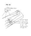
- FIG. 5panels A–C, depict selected steps in producing an x-ray image including a calibration device as described herein of a patient's hip.
- FIGS. 5A and Bdepict alignment of the bone, film and calibration device.
- FIG. 5Cdepicts positioning of the patient's leg and production of the x-ray image.
- the inventionincludes methods of obtaining and/or deriving information from an x-ray image in network environment. Additionally, the present invention relates to the provision of accurate calibration phantoms for X-ray systems and methods of using these calibration phantoms.
- the calibration phantomis formed of a material that simulates the properties of human bone tissue and is provided in an x-ray assembly such that improved accuracy and precision in the quantification of calcium, bone mass and bone density using conventional X-ray equipment is achieved.
- the imagesinclude one or more accurate reference markers, for example calibration phantoms for assessing density and geometry of any given structure in any given image.
- Geometryrefers broadly to any bone structure parameter, for example as described in U.S. Ser. No. 10/809,578, incorporated by reference herein in its entirety.
- the current inventionprovides for methods and devices that allow accurate quantitative assessment of information contained in an image such as density, geometry or morphology of an anatomic structure.
- the calibration phantom materials and methods of the present inventionare suitable for use in both conventional radiography systems and computed tomography (CT) systems.
- CTcomputed tomography
- a step-wedge phantomcan be fabricated from a matrix containing a desired concentration of reference material in varying thicknesses is used.
- the imageis an x-ray image, for example of hip, spine, knee, ankle, elbow, etc.
- X-ray imagescan be acquired using well-known techniques from any local site. For example, in certain aspects, 2D planar x-ray imaging techniques are used.
- 2D planar x-ray imagingis a method that generates an image by transmitting an x-ray beam through a body or structure or material and by measuring the x-ray attenuation on the other side of said body or said structure or said material.
- 2D planar x-ray imagingis distinguishable from cross-sectional imaging techniques such as computed tomography or magnetic resonance imaging.
- X-ray images that are captured using conventional x-ray filmcan be digitized using any suitable scanning device.
- the digitized x-ray imagemay then transmitted over the network, e.g. the Internet, into a remote computer or server.
- the networke.g. the Internet
- x-ray imagescan also be acquired using digital acquisition techniques, e.g. using phosphorus plate systems or selenium or silicon detector systems, the x-ray image information is already available in digital format.
- the imagecan be transmitted directly over the network, e.g. the Internet, or alternatively, it can be compressed prior to transmission.
- one or more calibration devicesare included in the field of view.
- Any suitable calibration phantomcan be used, for example, one that comprises aluminum, copper and/or other radiopaque materials.
- U.S. Pat. No. 5,335,260describes other calibration phantoms suitable for use in assessing bone mineral density in x-ray images.
- Examples of other suitable calibration reference materialscan be fluid or fluid-like materials, for example, one or more chambers filled with varying concentrations of calcium chloride or the like.
- a calibration phantomcan contain several different areas of different radiopacity.
- the calibration phantomcan have a step-like design, whereby changes in local thickness of the wedge result in differences in radiopacity.
- Stepwedges using material of varying thicknessare frequently used in radiology for quality control testing of x-ray beam properties. By varying the thickness of the steps, the intensity and spectral content of the x-ray beam in the projection image can be varied.
- Stepwedgesare commonly made of aluminum, copper and other convenient and homogeneous materials of known x-ray attenuation properties.
- Stepwedge-like phantomscan also contain calcium phosphate powder or calcium phosphate powder in molten paraffin.
- FIG. 1panels A–C, shows an exemplary step-wedge calibration phantom according to the present invention. Dimensions are shown in inches unless other specified. Panel (A) shows a top view and the overall dimensions as well as the distance between wedges (6 wedges shown in FIG. 1 ). Each rectangle of the step-wedge is approximately 0.5 inch long. Panel (B) shows a perspective view and Panel (C) shows a side view and depicts dimensions of the stepped nature of the phantom. The dimensions (e.g., height) of each component of the step-wedge is shown between the arrows, in a 2:1 scale.
- One of skill in the artwill recognize that the shape and specific dimensions of the phantom shown in FIG.
- the devicewill be smaller.
- the step-wedge calibration phantomcomprises copper.
- FIGS. 2A–Cshows lamination of an exemplary copper step-wedge as described herein between to plastic layers.
- the thickness of the plasticis preferably such that it does not significantly effect imaging of the wedge.
- each plastic sheetmay be approximately 0.012 inches thick.
- FIG. 3shows another exemplary embodiment of the present invention comprising a rod-shaped calibration device.
- the dimensions and characteristics of the exemplary deviceare shown in FIG. 3 and described in Example 1 below.
- the calibration devices described hereinmay be reused and do not require sterilization prior to use.
- the calibration devicesare typically re-useable for at least 6 months.
- a step-wedgecan be permanently attached to a rod-shaped calibration device.
- FIG. 4depicts the device of FIG. 2C (laminated step-wedge) in combination with the device depicted in FIG. 3 .
- the rod-shaped calibration deviceis affixed to the edge of the plastic laminate of the step-wedge such that the long axis of the rod is parallel to the long axis of the rectangles making up the step-wedge.
- the calibration phantomSince the density and attenuation of the calibration phantom are both known, the calibration phantom provides an external reference for measuring the density of the anatomic structure or non-living object to be measured.
- the calibration phantomcan be imaged at the same time as the x-ray image.
- the calibration phantomcan be physically connected to an x-ray film and/or film holder.
- Such physical connectioncan be achieved using any suitable mechanical or other attachment mechanism, including but not limited to adhesive, a chemical bond, use of screws or nails, welding, a VelcroTM strap or VelcroTM material and the like.
- a calibration phantomcan be physically connected to a detector system or a storage plate for digital x-ray imaging using one or more attachment mechanisms (e.g., a mechanical connection device, a VelcroTM strap or other VelcroTM material, a chemical bond, use of screws or nails, welding and an adhesive).
- the attachmentmay be permanent or temporary and the calibration phantom can be integral (e.g., built-in) to the film, film holder and/or detector system or can be attached or positioned permanently or temporarily appropriately after the film and/or film holder is produced, whether integrally formed or not.
- the calibration phantomcan be designed for single-use (e.g., disposable) or for multiple uses with different x-ray images.
- the calibration phantomis reusable and, additionally, can be sterilized between uses. Integration of a calibration phantom can be achieved by including a material of known x-ray density between two of the physical layers of the x-ray film.
- Integrationcan also be achieved by including a material of known x-ray density within one of the physical layers of the x-ray film. Additionally, the calibration phantom can be integrated into the film cover. A calibration phantom or an external standard can also be integrated into a detector system or a storage plate for digital x-ray imaging. For example, integration can be achieved by including a material of known x-ray density between two of the physical layers of the detector system or the storage plate. Integration can also be achieved by including a material of know x-ray density within one of the physical layers of the detector system or the storage plate.
- cross-hairs, lines or other markersmay be placed on the apparatus as indicators for positioning of the calibration phantom. These indicators can help to ensure that the calibration phantom is positioned such that it doesn't project on materials that will alter the apparent density in the resulting image.
- any of the calibration phantom-containing assemblies described hereincan be used in methods of analyzing and/or quantifying density and/or geometry of a structure in an x-ray image.
- the methodsgenerally involve simultaneously imaging or scanning the calibration phantom and another material (e.g., bone tissue from a subject) for the purpose of quantifying the density or geometry of the imaged material (e.g., bone mass).
- another materiale.g., bone tissue from a subject
- a calibration phantomis preferably imaged or scanned simultaneously with the individual subject, although the invention allows for non-simultaneous scanning of the phantom and the subject.
- Methods of scanning and imaging structures by radiographic techniqueare well known.
- reference calibration samplesallow corrections and calibration of the absorption properties of bone.
- FIGS. 5A–Cdepict an exemplary method of using a calibration phantom as described herein to produce an x-ray image.
- FIG. 5Ashows alignment of the central ray with the femoral neck (top arrow) as well as alignment of the center of the calibration device along the transverse line of aiming cross (bottom arrow).
- FIG. 5Bdepicts placement of a film cassette in a bucky tray and the film centered to central ray and adjusted width of collimator to include all of calibration device in exposure field.
- the x-rayis then taken after the patient's leg has been rotated 15–20 degrees internally and held in the rotated position.
- Exemplary x-ray conditions and parametersare also shown in FIG. 5C and described in Example 2 below.
- the data obtained from the calibration device(s)can be further manipulated.
- one or more calibration curvescan be constructed as described for example in International Publications WO 02/30283; WO 03/071934 and WO 04/019256, which disclosures are incorporated herein in their entireties.
- kits for obtaining information from x-ray imagesfor example for obtaining information regarding bone mineral density from an x-ray.
- the kitcomprises one or more computer (e.g., software) programs, for example for receiving, analyzing and generating reports based on x-ray images.
- the kitscan include calibration phantoms, for example calibration phantoms integrated or attachable-to x-ray film and/or x-ray film holders.
- a step-wedge comprising copperwas made as shown in FIG. 1 .
- Dimensions of the step-wedgeare shown in FIGS. 1 and 2 .
- the devicehad equivalent area bone density coverage range of 0.02 g/cm 2 to 1.5 g/cm 2 and this bone density measurement had a variation of less than 10%.
- the step-wedgewas laminated in plastic.
- a calibration device made of aluminumwas constructed as follows. Aluminum was selected because it has an x-ray attenuation coefficient similar to that of human cortical bone. A rod-shaped aluminum calibration device having the dimensions shown in FIG. 3 was constructed using standard techniques.
- the aluminum devicehad an optical density between about 30% to 70% when imaged with energy between 70 kVp to 80 kVp. This optical density measurement had a coefficient of variation of less than 10%.
- the copper step-wedge and aluminum rod described abovewere combined into a single calibration device as follows. Using clear plastic sheets the aluminum rod was attached to the previously-laminated step-wedge such that the long axis of the aluminum rod was essentially parallel to the rectangles making up the step-wedge. FIG. 4 .
- the x-ray energy used to capture an imagecan be estimated by comparing the optical density of the aluminum device to that of the copper device.
- a more accurate attenuation coefficient of the copper calibration deviceis used to generate a more accurate standard calibration curve adjusted for the x-ray energy.
- a calibration device as described in Example 1(copper step-wedge or combination copper step-wedge-aluminum calibration device) was placed on the x-ray table, lengthwise, lateral, and adjacent to the patient with center of device aligned with the transverse aiming cross of collimator light.
- the central raywas centered with respect to the femoral neck and the calibration device was placed with the center is aligned with the transverse line of aiming cross. ( FIG. 5A ).
- the film cassettewas placed in the busky tray and centered to the central ray.
- the width of the collimatorwas adjusted to include all of the calibration device in the exposure field. ( FIG. 5B ).
- the patient's legwas then rotated internally 15–20 degrees using foam block and Velcro to maintain positioning. ( FIG. 5C )
- the x-ray energy used to capture an imagewas estimated by comparing the optical density of the aluminum device to that of the copper device.
Landscapes
- Health & Medical Sciences (AREA)
- Life Sciences & Earth Sciences (AREA)
- Engineering & Computer Science (AREA)
- Medical Informatics (AREA)
- Physics & Mathematics (AREA)
- Pathology (AREA)
- Molecular Biology (AREA)
- Biophysics (AREA)
- High Energy & Nuclear Physics (AREA)
- Veterinary Medicine (AREA)
- Nuclear Medicine, Radiotherapy & Molecular Imaging (AREA)
- Optics & Photonics (AREA)
- Public Health (AREA)
- Radiology & Medical Imaging (AREA)
- Biomedical Technology (AREA)
- Heart & Thoracic Surgery (AREA)
- General Health & Medical Sciences (AREA)
- Surgery (AREA)
- Animal Behavior & Ethology (AREA)
- Oral & Maxillofacial Surgery (AREA)
- Orthopedic Medicine & Surgery (AREA)
- Dentistry (AREA)
- Computer Networks & Wireless Communication (AREA)
- Computer Vision & Pattern Recognition (AREA)
- General Physics & Mathematics (AREA)
- Theoretical Computer Science (AREA)
- Apparatus For Radiation Diagnosis (AREA)
Abstract
Description
| Current | 150 mA | ||
| Energy | 70 kV–80 kV | ||
| Exposure time | |||
| Film size | |||
| 14″ × 11″ (36 cm × 28 cm) | |||
| Screen | 400 speed | ||
| Source height | 40″ (102 cm) | ||
Claims (10)
Priority Applications (9)
| Application Number | Priority Date | Filing Date | Title |
|---|---|---|---|
| US10/917,848US7467892B2 (en) | 2000-08-29 | 2004-08-12 | Calibration devices and methods of use thereof |
| PCT/US2005/028004WO2006033712A1 (en) | 2004-08-12 | 2005-08-04 | Calibration devices and methods of use thereof |
| JP2007525690AJP2008509737A (en) | 2004-08-12 | 2005-08-04 | Calibration device and method of using the same |
| AU2005287416AAU2005287416A1 (en) | 2004-08-12 | 2005-08-04 | Calibration devices and methods of use thereof |
| EP05783847AEP1776045A1 (en) | 2004-08-12 | 2005-08-04 | Calibration devices and methods of use thereof |
| CA002572404ACA2572404A1 (en) | 2004-08-12 | 2005-08-04 | Calibration devices and methods of use thereof |
| US12/055,491US7580504B2 (en) | 2000-08-29 | 2008-03-26 | Calibration devices and methods of use thereof |
| US12/505,908US8000441B2 (en) | 2000-08-29 | 2009-07-20 | Calibration devices and methods of use thereof |
| US13/209,966US8588365B2 (en) | 2000-08-29 | 2011-08-15 | Calibration devices and methods of use thereof |
Applications Claiming Priority (5)
| Application Number | Priority Date | Filing Date | Title |
|---|---|---|---|
| US22859100P | 2000-08-29 | 2000-08-29 | |
| US09/942,528US7545964B2 (en) | 2000-08-29 | 2001-08-29 | Methods and devices for quantitative analysis of x-ray images |
| US10/086,653US6904123B2 (en) | 2000-08-29 | 2002-02-27 | Methods and devices for quantitative analysis of x-ray images |
| US10/225,363US7050534B2 (en) | 2000-08-29 | 2002-08-20 | Methods and devices for quantitative analysis of x-ray images |
| US10/917,848US7467892B2 (en) | 2000-08-29 | 2004-08-12 | Calibration devices and methods of use thereof |
Related Parent Applications (1)
| Application Number | Title | Priority Date | Filing Date |
|---|---|---|---|
| US10/225,363Continuation-In-PartUS7050534B2 (en) | 2000-08-29 | 2002-08-20 | Methods and devices for quantitative analysis of x-ray images |
Related Child Applications (1)
| Application Number | Title | Priority Date | Filing Date |
|---|---|---|---|
| US12/055,491ContinuationUS7580504B2 (en) | 2000-08-29 | 2008-03-26 | Calibration devices and methods of use thereof |
Publications (2)
| Publication Number | Publication Date |
|---|---|
| US20050078802A1 US20050078802A1 (en) | 2005-04-14 |
| US7467892B2true US7467892B2 (en) | 2008-12-23 |
Family
ID=35427846
Family Applications (4)
| Application Number | Title | Priority Date | Filing Date |
|---|---|---|---|
| US10/917,848Expired - LifetimeUS7467892B2 (en) | 2000-08-29 | 2004-08-12 | Calibration devices and methods of use thereof |
| US12/055,491Expired - LifetimeUS7580504B2 (en) | 2000-08-29 | 2008-03-26 | Calibration devices and methods of use thereof |
| US12/505,908Expired - LifetimeUS8000441B2 (en) | 2000-08-29 | 2009-07-20 | Calibration devices and methods of use thereof |
| US13/209,966Expired - LifetimeUS8588365B2 (en) | 2000-08-29 | 2011-08-15 | Calibration devices and methods of use thereof |
Family Applications After (3)
| Application Number | Title | Priority Date | Filing Date |
|---|---|---|---|
| US12/055,491Expired - LifetimeUS7580504B2 (en) | 2000-08-29 | 2008-03-26 | Calibration devices and methods of use thereof |
| US12/505,908Expired - LifetimeUS8000441B2 (en) | 2000-08-29 | 2009-07-20 | Calibration devices and methods of use thereof |
| US13/209,966Expired - LifetimeUS8588365B2 (en) | 2000-08-29 | 2011-08-15 | Calibration devices and methods of use thereof |
Country Status (6)
| Country | Link |
|---|---|
| US (4) | US7467892B2 (en) |
| EP (1) | EP1776045A1 (en) |
| JP (1) | JP2008509737A (en) |
| AU (1) | AU2005287416A1 (en) |
| CA (1) | CA2572404A1 (en) |
| WO (1) | WO2006033712A1 (en) |
Cited By (59)
| Publication number | Priority date | Publication date | Assignee | Title |
|---|---|---|---|---|
| US20090268953A1 (en)* | 2008-04-24 | 2009-10-29 | Apteryx, Inc. | Method for the automatic adjustment of image parameter settings in an imaging system |
| US20090268876A1 (en)* | 2008-04-24 | 2009-10-29 | Crucs Holdings, Llc | Methods of assessing performance in an imaging system |
| US20100080350A1 (en)* | 2008-09-29 | 2010-04-01 | Mir Medical Imaging Research Holding Gmbh | Method and Device for Thermal Breast Tumor Treatment with 3D Monitoring Function |
| US7796791B2 (en) | 2002-11-07 | 2010-09-14 | Conformis, Inc. | Methods for determining meniscal size and shape and for devising treatment |
| US7840247B2 (en) | 2002-09-16 | 2010-11-23 | Imatx, Inc. | Methods of predicting musculoskeletal disease |
| US7881768B2 (en) | 1998-09-14 | 2011-02-01 | The Board Of Trustees Of The Leland Stanford Junior University | Assessing the condition of a joint and devising treatment |
| US20110096911A1 (en)* | 2009-10-27 | 2011-04-28 | Dental Imaging Consultants, LLC | Quality Assurance Phantom for Digital Dental Imaging and Related Method |
| US7981158B2 (en) | 2001-05-25 | 2011-07-19 | Conformis, Inc. | Patient selectable joint arthroplasty devices and surgical tools |
| US7995822B2 (en) | 2003-03-25 | 2011-08-09 | Imatx, Inc. | Methods for the compensation of imaging technique in the processing of radiographic images |
| US8000441B2 (en) | 2000-08-29 | 2011-08-16 | Imatx, Inc. | Calibration devices and methods of use thereof |
| US8000766B2 (en) | 2001-05-25 | 2011-08-16 | Imatx, Inc. | Methods to diagnose treat and prevent bone loss |
| US8031836B2 (en) | 2000-08-29 | 2011-10-04 | Imatx, Inc. | Methods and devices for quantitative analysis of x-ray images |
| US8036729B2 (en) | 1998-09-14 | 2011-10-11 | The Board Of Trustees Of The Leland Stanford Junior University | Assessing the condition of a joint and devising treatment |
| US8068580B2 (en) | 2000-08-29 | 2011-11-29 | Imatx, Inc. | Methods and devices for quantitative analysis of x-ray images |
| US8066708B2 (en) | 2001-05-25 | 2011-11-29 | Conformis, Inc. | Patient selectable joint arthroplasty devices and surgical tools |
| US8073521B2 (en) | 2003-09-19 | 2011-12-06 | Imatx, Inc. | Method for bone structure prognosis and simulated bone remodeling |
| US8122582B2 (en) | 2001-05-25 | 2012-02-28 | Conformis, Inc. | Surgical tools facilitating increased accuracy, speed and simplicity in performing joint arthroplasty |
| US8234097B2 (en) | 2001-05-25 | 2012-07-31 | Conformis, Inc. | Automated systems for manufacturing patient-specific orthopedic implants and instrumentation |
| US8265730B2 (en) | 1998-09-14 | 2012-09-11 | The Board Of Trustees Of The Leland Stanford Junior University | Assessing the condition of a joint and preventing damage |
| US8290564B2 (en) | 2003-09-19 | 2012-10-16 | Imatx, Inc. | Method for bone structure prognosis and simulated bone remodeling |
| US8337507B2 (en) | 2001-05-25 | 2012-12-25 | Conformis, Inc. | Methods and compositions for articular repair |
| US8377129B2 (en) | 2001-05-25 | 2013-02-19 | Conformis, Inc. | Joint arthroplasty devices and surgical tools |
| US8439926B2 (en) | 2001-05-25 | 2013-05-14 | Conformis, Inc. | Patient selectable joint arthroplasty devices and surgical tools |
| US8480754B2 (en) | 2001-05-25 | 2013-07-09 | Conformis, Inc. | Patient-adapted and improved articular implants, designs and related guide tools |
| US8500740B2 (en) | 2006-02-06 | 2013-08-06 | Conformis, Inc. | Patient-specific joint arthroplasty devices for ligament repair |
| US8545569B2 (en) | 2001-05-25 | 2013-10-01 | Conformis, Inc. | Patient selectable knee arthroplasty devices |
| US8556983B2 (en) | 2001-05-25 | 2013-10-15 | Conformis, Inc. | Patient-adapted and improved orthopedic implants, designs and related tools |
| US8600124B2 (en) | 2004-09-16 | 2013-12-03 | Imatx, Inc. | System and method of predicting future fractures |
| US8617242B2 (en) | 2001-05-25 | 2013-12-31 | Conformis, Inc. | Implant device and method for manufacture |
| US8625874B2 (en) | 2000-10-11 | 2014-01-07 | Imatx, Inc. | Methods and devices for analysis of x-ray images |
| US8623026B2 (en) | 2006-02-06 | 2014-01-07 | Conformis, Inc. | Patient selectable joint arthroplasty devices and surgical tools incorporating anatomical relief |
| US8639009B2 (en) | 2000-10-11 | 2014-01-28 | Imatx, Inc. | Methods and devices for evaluating and treating a bone condition based on x-ray image analysis |
| US8682052B2 (en) | 2008-03-05 | 2014-03-25 | Conformis, Inc. | Implants for altering wear patterns of articular surfaces |
| US8709089B2 (en) | 2002-10-07 | 2014-04-29 | Conformis, Inc. | Minimally invasive joint implant with 3-dimensional geometry matching the articular surfaces |
| US8735773B2 (en) | 2007-02-14 | 2014-05-27 | Conformis, Inc. | Implant device and method for manufacture |
| US8771365B2 (en) | 2009-02-25 | 2014-07-08 | Conformis, Inc. | Patient-adapted and improved orthopedic implants, designs, and related tools |
| US8808303B2 (en) | 2009-02-24 | 2014-08-19 | Microport Orthopedics Holdings Inc. | Orthopedic surgical guide |
| US8882847B2 (en) | 2001-05-25 | 2014-11-11 | Conformis, Inc. | Patient selectable knee joint arthroplasty devices |
| US8939917B2 (en) | 2009-02-13 | 2015-01-27 | Imatx, Inc. | Methods and devices for quantitative analysis of bone and cartilage |
| US8951260B2 (en) | 2001-05-25 | 2015-02-10 | Conformis, Inc. | Surgical cutting guide |
| US8965075B2 (en) | 2002-09-16 | 2015-02-24 | Imatx, Inc. | System and method for predicting future fractures |
| US9017334B2 (en) | 2009-02-24 | 2015-04-28 | Microport Orthopedics Holdings Inc. | Patient specific surgical guide locator and mount |
| US9020788B2 (en) | 1997-01-08 | 2015-04-28 | Conformis, Inc. | Patient-adapted and improved articular implants, designs and related guide tools |
| US9286686B2 (en) | 1998-09-14 | 2016-03-15 | The Board Of Trustees Of The Leland Stanford Junior University | Assessing the condition of a joint and assessing cartilage loss |
| US9308091B2 (en) | 2001-05-25 | 2016-04-12 | Conformis, Inc. | Devices and methods for treatment of facet and other joints |
| US9486226B2 (en) | 2012-04-18 | 2016-11-08 | Conformis, Inc. | Tibial guides, tools, and techniques for resecting the tibial plateau |
| US9603711B2 (en) | 2001-05-25 | 2017-03-28 | Conformis, Inc. | Patient-adapted and improved articular implants, designs and related guide tools |
| US9649117B2 (en) | 2009-02-24 | 2017-05-16 | Microport Orthopedics Holdings, Inc. | Orthopedic surgical guide |
| US9675471B2 (en) | 2012-06-11 | 2017-06-13 | Conformis, Inc. | Devices, techniques and methods for assessing joint spacing, balancing soft tissues and obtaining desired kinematics for joint implant components |
| US9737406B2 (en) | 2013-08-21 | 2017-08-22 | Laboratories Bodycad Inc. | Anatomically adapted orthopedic implant and method of manufacturing same |
| USD808524S1 (en) | 2016-11-29 | 2018-01-23 | Laboratoires Bodycad Inc. | Femoral implant |
| US10085839B2 (en) | 2004-01-05 | 2018-10-02 | Conformis, Inc. | Patient-specific and patient-engineered orthopedic implants |
| US10667829B2 (en) | 2013-08-21 | 2020-06-02 | Laboratoires Bodycad Inc. | Bone resection guide and method |
| US20230136930A1 (en)* | 2020-04-16 | 2023-05-04 | Hamamatsu Photonics K.K. | Radiographic image processing method, trained model, radiographic image processing module, radiographic image processing program, and radiographic image processing system |
| US12108959B2 (en) | 2019-05-29 | 2024-10-08 | Wright Medical Technology, Inc. | Preparing a tibia for receiving tibial implant component of a replacement ankle |
| US12178515B2 (en) | 2021-04-26 | 2024-12-31 | Arthrex, Inc. | Systems and methods for density calibration |
| US12383287B2 (en) | 2009-02-24 | 2025-08-12 | Microport Orthopedics Holdings, Inc. | Systems and methods for installing an orthopedic implant |
| US12396739B2 (en) | 2020-01-17 | 2025-08-26 | Wright Medical Technology, Inc. | Guidance tools, systems, and methods |
| US12440227B2 (en) | 2022-01-05 | 2025-10-14 | Wright Medical Technology, Inc. | Preparing a tibia for receiving tibial implant component of a replacement ankle |
Families Citing this family (34)
| Publication number | Priority date | Publication date | Assignee | Title |
|---|---|---|---|---|
| US9289153B2 (en)* | 1998-09-14 | 2016-03-22 | The Board Of Trustees Of The Leland Stanford Junior University | Joint and cartilage diagnosis, assessment and modeling |
| US20020186818A1 (en)* | 2000-08-29 | 2002-12-12 | Osteonet, Inc. | System and method for building and manipulating a centralized measurement value database |
| US7050534B2 (en)* | 2000-08-29 | 2006-05-23 | Imaging Therapeutics, Inc. | Methods and devices for quantitative analysis of x-ray images |
| US20070047794A1 (en)* | 2000-10-11 | 2007-03-01 | Philipp Lang | Methods and devices for analysis of x-ray images |
| EP1546982A1 (en)* | 2002-09-16 | 2005-06-29 | Imaging Therapeutics, Inc. | Imaging markers in musculoskeletal disease |
| US20080058613A1 (en)* | 2003-09-19 | 2008-03-06 | Imaging Therapeutics, Inc. | Method and System for Providing Fracture/No Fracture Classification |
| US20050259793A1 (en)* | 2004-05-19 | 2005-11-24 | Yeo In H | Medical phantom, holder and method of use thereof |
| US7471761B2 (en)* | 2005-09-15 | 2008-12-30 | Schick Technologies, Inc. | System and method for computing oral bone mineral density with a panoramic x-ray system |
| US8417010B1 (en) | 2006-01-12 | 2013-04-09 | Diagnoscan, LLC | Digital x-ray diagnosis and evaluation of dental disease |
| US7959742B2 (en)* | 2007-07-11 | 2011-06-14 | Whirlpool Corporation | Outer support body for a drawer-type dishwasher |
| AU2009221773B2 (en)* | 2008-03-05 | 2015-03-05 | Conformis, Inc. | Edge-matched articular implant |
| KR101213303B1 (en) | 2010-07-08 | 2012-12-18 | 서울대학교산학협력단 | Micro-patterned device and method for compensating distortion of optical coherent tomography image using the same |
| WO2012090148A1 (en)* | 2010-12-30 | 2012-07-05 | Mediguide Ltd | System and method for registration of fluoroscopic images in a coordinate system of a medical system |
| US8541740B2 (en) | 2011-02-28 | 2013-09-24 | Ethicon, Inc. | Device and method for electron beam energy verification |
| US9235892B2 (en) | 2011-03-31 | 2016-01-12 | Denise De Andrade Castro | Method and device for comparing radiographic images |
| US9008264B2 (en) | 2011-07-15 | 2015-04-14 | The Regents Of The University Of California | Apparatus and methods for determination of the half value layer of X-ray beams |
| US9693748B2 (en)* | 2011-07-23 | 2017-07-04 | Broncus Medical Inc. | System and method for automatically determining calibration parameters of a fluoroscope |
| US9820709B2 (en) | 2012-11-29 | 2017-11-21 | Controlrad Systems, Inc. | X-ray reduction system |
| WO2014084231A1 (en) | 2012-11-30 | 2014-06-05 | 株式会社トプコン | Fundus photographing device |
| US8708562B1 (en) | 2013-03-05 | 2014-04-29 | Nosil DSC Innovations, Inc. | Phantom systems and methods for diagnostic x-ray equipment |
| WO2015050941A1 (en) | 2013-10-04 | 2015-04-09 | Battelle Memorial Institute | Contrast phantom for passive millimeter wave imaging systems |
| RU2585403C2 (en)* | 2013-11-22 | 2016-05-27 | Дмитрий Юрьевич Анохин | Method for assessing success of treatment of osteoporosis |
| US9936935B1 (en) | 2014-02-14 | 2018-04-10 | Nosil DSC Innovations, Inc. | Phantom systems and methods for diagnostic radiographic and fluoroscopic X-ray equipment |
| US10034651B2 (en) | 2014-07-18 | 2018-07-31 | Gammex, Inc. | Brain tissue equivalent material and phantom device comprising the same |
| US9669116B2 (en) | 2014-07-18 | 2017-06-06 | Gammex, Inc. | Water-equivalent phantom |
| US20170039735A1 (en)* | 2015-08-06 | 2017-02-09 | General Electric Company | Computed tomography self-calibration without calibration targets |
| CN106913383B (en)* | 2015-12-25 | 2020-04-21 | 先健科技(深圳)有限公司 | Development structure and implantable medical device with development structure |
| KR101894178B1 (en)* | 2016-11-29 | 2018-08-31 | 주식회사 메디코어스 | Calibration method for fan-beam dual energy X-ray absorptiometry |
| US10634797B2 (en)* | 2017-07-07 | 2020-04-28 | International Business Machines Corporation | Real time X-ray dosimeter using diodes with variable thickness degrader |
| WO2019099544A1 (en)* | 2017-11-14 | 2019-05-23 | Imatrex, Inc. | Geometric calibration of x-ray imaging systems |
| US11357645B2 (en) | 2020-04-17 | 2022-06-14 | Warsaw Orthopedic, Inc. | Implant with graded radiopacity calibration feature |
| JP7467389B2 (en)* | 2021-06-14 | 2024-04-15 | 富士フイルムヘルスケア株式会社 | Phantom, radiation imaging device, and method for calibrating photon counting detector |
| WO2023223757A1 (en)* | 2022-05-17 | 2023-11-23 | オリンパステルモバイオマテリアル株式会社 | Attachment set and bone cutting system |
| US20240024703A1 (en)* | 2022-07-21 | 2024-01-25 | Radformation, Inc. | Modular Jig System |
Citations (18)
| Publication number | Priority date | Publication date | Assignee | Title |
|---|---|---|---|---|
| US4126789A (en)* | 1977-06-06 | 1978-11-21 | Vogl Thomas M | X-ray phantom |
| US4233507A (en)* | 1979-05-07 | 1980-11-11 | General Electric Company | Computer tomography table containing calibration and correlation samples |
| US4649561A (en) | 1983-11-28 | 1987-03-10 | Ben Arnold | Test phantom and method of use of same |
| US5235628A (en) | 1990-11-26 | 1993-08-10 | Wisconsin Alumni Research Foundation | Calibration phantom for bone mineral measurement on the lumbar spine |
| US5335260A (en)* | 1992-11-25 | 1994-08-02 | Arnold Ben A | Calibration phantom and improved method of quantifying calcium and bone density using same |
| US5493601A (en) | 1993-12-24 | 1996-02-20 | Agfa-Gevaert | Radiographic calibration phantom |
| US6064716A (en) | 1997-09-05 | 2000-05-16 | Cyberlogic, Inc. | Plain x-ray bone densitometry apparatus and method |
| US6178225B1 (en) | 1999-06-04 | 2001-01-23 | Edge Medical Devices Ltd. | System and method for management of X-ray imaging facilities |
| WO2001063488A2 (en) | 2000-02-25 | 2001-08-30 | Healthscreen International, Inc. | Method for centralized health data management |
| WO2001065449A1 (en) | 2000-03-01 | 2001-09-07 | Medeview.Com, Inc. | A medical diagnosis and prescription communications delivery system, method and apparatus |
| US6289115B1 (en) | 1998-02-20 | 2001-09-11 | Fuji Photo Film Co., Ltd. | Medical network system |
| US6320931B1 (en) | 1998-03-02 | 2001-11-20 | Image Analysis, Inc. | Automated x-ray bone densitometer |
| WO2002030283A2 (en) | 2000-10-11 | 2002-04-18 | Imaging Therapeutics | Methods and devices for analysis of x-ray images |
| WO2003071934A2 (en) | 2002-02-27 | 2003-09-04 | Imaging Therapeutics, Inc. | Methods and devices for quantitative analysis of x-ray images |
| US6694047B1 (en) | 1999-07-15 | 2004-02-17 | General Electric Company | Method and apparatus for automated image quality evaluation of X-ray systems using any of multiple phantoms |
| WO2004019256A2 (en) | 2002-08-20 | 2004-03-04 | Imaging Therapeutics, Inc. | Measurement of bone density and bone structure from x-ray images for diagnosis of osteoporosis |
| US6824309B2 (en)* | 2001-02-16 | 2004-11-30 | Commissariat A L'energie Atomique | Double energy radiography method, and calibration device for this method |
| US20050010106A1 (en) | 2003-03-25 | 2005-01-13 | Imaging Therapeutics, Inc. | Methods for the compensation of imaging technique in the processing of radiographic images |
Family Cites Families (156)
| Publication number | Priority date | Publication date | Assignee | Title |
|---|---|---|---|---|
| US2274808A (en) | 1941-01-07 | 1942-03-03 | Irwin C Rinn | Bite wing for dental film packs and the like |
| US7366676B2 (en) | 2001-05-29 | 2008-04-29 | Mevis Breastcare Gmbh & Co. Kg | Method and system for in-service monitoring and training for a radiologic workstation |
| DE2042009C3 (en) | 1970-08-25 | 1975-02-27 | Siemens Ag, 1000 Berlin U. 8000 Muenchen | Arrangement for the non-destructive density measurement of substances of living objects by means of penetrating rays |
| US4012638A (en) | 1976-03-09 | 1977-03-15 | Altschuler Bruce R | Dental X-ray alignment system |
| US4298800A (en) | 1978-02-27 | 1981-11-03 | Computome Corporation | Tomographic apparatus and method for obtaining three-dimensional information by radiation scanning |
| GB2023920A (en) | 1978-06-19 | 1980-01-03 | Thoro Ray Inc | Dental X-ray apparatus |
| US4686695A (en) | 1979-02-05 | 1987-08-11 | Board Of Trustees Of The Leland Stanford Junior University | Scanned x-ray selective imaging system |
| US4251732A (en) | 1979-08-20 | 1981-02-17 | Fried Alan J | Dental x-ray film holders |
| US4356400A (en) | 1980-08-04 | 1982-10-26 | General Electric Company | X-Ray apparatus alignment method and device |
| US4400827A (en) | 1981-11-13 | 1983-08-23 | Spears James R | Method and apparatus for calibrating rapid sequence radiography |
| FR2547495B1 (en) | 1983-06-16 | 1986-10-24 | Mouyen Francis | APPARATUS FOR OBTAINING A DENTAL RADIOLOGICAL IMAGE |
| JPS61109557A (en) | 1984-11-02 | 1986-05-28 | 帝人株式会社 | Evaluation of bone |
| JPH07102210B2 (en) | 1986-05-14 | 1995-11-08 | 帝人株式会社 | Evaluation method of bone atrophy of alveolar bone |
| US4782502A (en) | 1986-10-01 | 1988-11-01 | Schulz Eloy E | Flexible calibration phantom for computer tomography system |
| US4985906A (en) | 1987-02-17 | 1991-01-15 | Arnold Ben A | Calibration phantom for computer tomography system |
| CA1288176C (en) | 1987-10-29 | 1991-08-27 | David C. Hatcher | Method and apparatus for improving the alignment of radiographic images |
| US4922915A (en) | 1987-11-27 | 1990-05-08 | Ben A. Arnold | Automated image detail localization method |
| US5127032A (en) | 1987-12-03 | 1992-06-30 | Johns Hopkins University | Multi-directional x-ray imager |
| US4956859A (en) | 1989-03-10 | 1990-09-11 | Expert Image Systems, Inc. | Source filter for radiographic imaging |
| US5090040A (en) | 1989-03-10 | 1992-02-18 | Expert Image Systems, Inc. | Data acquisition system for radiographic imaging |
| US5001738A (en) | 1989-04-07 | 1991-03-19 | Brooks Jack D | Dental X-ray film holding tab and alignment method |
| FR2649883B1 (en) | 1989-07-20 | 1991-10-11 | Gen Electric Cgr | METHOD FOR CORRECTING THE MEASUREMENT OF BONE DENSITY IN A SCANNER |
| US5537483A (en) | 1989-10-03 | 1996-07-16 | Staplevision, Inc. | Automated quality assurance image processing system |
| US6031892A (en)* | 1989-12-05 | 2000-02-29 | University Of Massachusetts Medical Center | System for quantitative radiographic imaging |
| US5150394A (en) | 1989-12-05 | 1992-09-22 | University Of Massachusetts Medical School | Dual-energy system for quantitative radiographic imaging |
| US5864146A (en)* | 1996-11-13 | 1999-01-26 | University Of Massachusetts Medical Center | System for quantitative radiographic imaging |
| US5562448A (en) | 1990-04-10 | 1996-10-08 | Mushabac; David R. | Method for facilitating dental diagnosis and treatment |
| US5122664A (en) | 1990-04-27 | 1992-06-16 | Fuji Photo Film Co., Ltd. | Method and apparatus for quantitatively analyzing bone calcium |
| US5228445A (en) | 1990-06-18 | 1993-07-20 | Board Of Regents, The University Of Texas System | Demonstration by in vivo measurement of reflection ultrasound analysis of improved bone quality following slow-release fluoride treatment in osteoporosis patients |
| US5172695A (en) | 1990-09-10 | 1992-12-22 | Cann Christopher E | Method for improved prediction of bone fracture risk using bone mineral density in structural analysis |
| US5577089A (en) | 1991-02-13 | 1996-11-19 | Lunar Corporation | Device and method for analysis of bone morphology |
| JP2641078B2 (en) | 1991-03-28 | 1997-08-13 | 富士写真フイルム株式会社 | Bone mineral analysis |
| US5200993A (en) | 1991-05-10 | 1993-04-06 | Bell Atlantic Network Services, Inc. | Public telephone network including a distributed imaging system |
| US5270651A (en) | 1991-05-21 | 1993-12-14 | The Trustees Of The University Of Pennsylvania | Method and apparatus for diagnosing osteoporosis |
| US5247934A (en) | 1991-08-09 | 1993-09-28 | Trustees Of The University Of Pennsylvania | Method and apparatus for diagnosing osteoporosis with MR imaging |
| JP2607782B2 (en) | 1991-10-11 | 1997-05-07 | 眞希 末光 | Heated thermal desorption method |
| US5271401A (en) | 1992-01-15 | 1993-12-21 | Praxair Technology, Inc. | Radiological imaging method |
| DE69329168T2 (en) | 1992-05-20 | 2001-04-05 | Aloka Co. Ltd., Mitaka | Device for determining the properties of bones |
| WO1993024055A1 (en)* | 1992-05-29 | 1993-12-09 | Ge Yokogawa Medical Systems, Ltd. | Method of quantitative determination of bone salt with ct equipment |
| US5321520A (en) | 1992-07-20 | 1994-06-14 | Automated Medical Access Corporation | Automated high definition/resolution image storage, retrieval and transmission system |
| US5320102A (en) | 1992-11-18 | 1994-06-14 | Ciba-Geigy Corporation | Method for diagnosing proteoglycan deficiency in cartilage based on magnetic resonance image (MRI) |
| US5592943A (en) | 1993-04-07 | 1997-01-14 | Osteo Sciences Corporation | Apparatus and method for acoustic analysis of bone using optimized functions of spectral and temporal signal components |
| US5513240A (en)* | 1993-05-18 | 1996-04-30 | The Research Foundation Of Suny | Intraoral radiograph alignment device |
| FR2705785B1 (en) | 1993-05-28 | 1995-08-25 | Schlumberger Ind Sa | Method for determining the attenuation function of an object with respect to the transmission of a reference thickness of a reference material and device for implementing the method. |
| US5657369A (en) | 1993-11-22 | 1997-08-12 | Hologic, Inc. | X-ray bone densitometry system having forearm positioning assembly |
| US5931780A (en) | 1993-11-29 | 1999-08-03 | Arch Development Corporation | Method and system for the computerized radiographic analysis of bone |
| EP0745850B1 (en)* | 1994-02-19 | 2005-11-16 | Seikagaku Corporation | Method of assaying normal aglycan, assaying kit, and method of judging joint-related information |
| US5600574A (en) | 1994-05-13 | 1997-02-04 | Minnesota Mining And Manufacturing Company | Automated image quality control |
| US5476865A (en) | 1994-07-06 | 1995-12-19 | Eli Lilly And Company | Methods of inhibiting bone loss |
| DE69513300T2 (en) | 1994-08-29 | 2000-03-23 | Torsana A/S, Skodsborg | DETERMINATION PROCEDURE |
| US5493593A (en) | 1994-09-27 | 1996-02-20 | University Of Delaware | Tilted detector microscopy in computerized tomography |
| AU1837495A (en) | 1994-10-13 | 1996-05-06 | Horus Therapeutics, Inc. | Computer assisted methods for diagnosing diseases |
| JPH08186762A (en) | 1994-12-27 | 1996-07-16 | Toshiba Medical Eng Co Ltd | Mammography equipment |
| SE9601065L (en) | 1996-03-20 | 1997-03-03 | Siemens Elema Ab | Anesthesia System |
| US5886353A (en)* | 1995-04-21 | 1999-03-23 | Thermotrex Corporation | Imaging device |
| US5565678A (en) | 1995-06-06 | 1996-10-15 | Lumisys, Inc. | Radiographic image quality assessment utilizing a stepped calibration target |
| US6038287A (en) | 1995-10-10 | 2000-03-14 | Miles; Dale A. | Portable X-ray device |
| US5772592A (en) | 1996-01-08 | 1998-06-30 | Cheng; Shu Lin | Method for diagnosing and monitoring osteoporosis |
| US6215846B1 (en) | 1996-02-21 | 2001-04-10 | Lunar Corporation | Densitometry adapter for compact x-ray fluoroscopy machine |
| US5785041A (en) | 1996-03-26 | 1998-07-28 | Hologic Inc. | System for assessing bone characteristics |
| AU2763597A (en) | 1996-05-06 | 1997-11-26 | Torsana A/S | A method of estimating skeletal status |
| US6108635A (en) | 1996-05-22 | 2000-08-22 | Interleukin Genetics, Inc. | Integrated disease information system |
| DE19625835A1 (en) | 1996-06-27 | 1998-01-02 | Siemens Ag | Medical system architecture |
| US5837674A (en) | 1996-07-03 | 1998-11-17 | Big Bear Bio, Inc. | Phosphopeptides and methods of treating bone diseases |
| US5945412A (en)* | 1996-12-09 | 1999-08-31 | Merck & Co., Inc. | Methods and compositions for preventing and treating bone loss |
| US8545569B2 (en) | 2001-05-25 | 2013-10-01 | Conformis, Inc. | Patient selectable knee arthroplasty devices |
| JP3863963B2 (en) | 1997-03-27 | 2006-12-27 | 大日本印刷株式会社 | Digital data correction and storage method and apparatus for X-ray image |
| EP0993643A1 (en) | 1997-07-04 | 2000-04-19 | Torsana Osteoporosis Diagnostics A/S | A method for estimating the bone quality or skeletal status of a vertebrate |
| EP1014858A4 (en) | 1997-08-19 | 2005-07-13 | John D Mendlein | Ultrasonic transmission films and devices, particularly for hygienic transducer surfaces |
| JP3706719B2 (en) | 1997-08-19 | 2005-10-19 | キヤノン株式会社 | Image processing apparatus and method, and storage medium |
| US5917877A (en) | 1997-09-05 | 1999-06-29 | Cyberlogic, Inc. | Plain x-ray bone densitometry apparatus and method |
| CA2303800A1 (en) | 1997-09-09 | 1999-03-18 | Mark Walden Lundy | Method of increasing bone volume using non-naturally-occurring fp selective agonists |
| US5852647A (en) | 1997-09-24 | 1998-12-22 | Schick Technologies | Method and apparatus for measuring bone density |
| JP3656695B2 (en) | 1997-09-30 | 2005-06-08 | 富士写真フイルム株式会社 | Bone measuring method and apparatus |
| US6252928B1 (en)* | 1998-01-23 | 2001-06-26 | Guard Inc. | Method and device for estimating bone mineral content of the calcaneus |
| US6077224A (en) | 1998-03-23 | 2000-06-20 | Lang; Philipp | Methods and device for improving broadband ultrasonic attenuation and speed of sound measurements using anatomical landmarks |
| EP1061854A4 (en) | 1998-03-09 | 2005-07-13 | Philipp Lang | Methods and devices for improving broadband ultrasonic attenuation and speed of sound measurements |
| JP3592927B2 (en) | 1998-03-20 | 2004-11-24 | 株式会社日立ユニシアオートモティブ | Gas injection valve |
| DE69815814T2 (en) | 1998-04-24 | 2004-05-06 | Eastman Kodak Co. | Method and system for assigning exposed X-ray films to associated patient information |
| US6835377B2 (en) | 1998-05-13 | 2004-12-28 | Osiris Therapeutics, Inc. | Osteoarthritis cartilage regeneration |
| US6442287B1 (en) | 1998-08-28 | 2002-08-27 | Arch Development Corporation | Method and system for the computerized analysis of bone mass and structure |
| US6714623B2 (en) | 1998-08-31 | 2004-03-30 | Canon Kabushiki Kaisha | Image collecting system |
| JP3639750B2 (en) | 1998-08-31 | 2005-04-20 | キヤノン株式会社 | Image acquisition device |
| US7239908B1 (en) | 1998-09-14 | 2007-07-03 | The Board Of Trustees Of The Leland Stanford Junior University | Assessing the condition of a joint and devising treatment |
| WO2000035346A2 (en) | 1998-09-14 | 2000-06-22 | Stanford University | Assessing the condition of a joint and preventing damage |
| US6368326B1 (en) | 1998-09-28 | 2002-04-09 | Daos Limited | Internal cord fixation device |
| US6501827B1 (en) | 1998-09-29 | 2002-12-31 | Canon Kabushiki Kaisha | Examination system, image processing apparatus and method, medium, and x-ray photographic system |
| JP3499761B2 (en) | 1998-10-22 | 2004-02-23 | 帝人株式会社 | Bone image processing method and bone strength evaluation method |
| DE19853965A1 (en) | 1998-11-23 | 2000-05-31 | Siemens Ag | Bone contour and bone structure determination |
| US7283857B1 (en)* | 1998-11-30 | 2007-10-16 | Hologic, Inc. | DICOM compliant file communication including quantitative and image data |
| US6302582B1 (en)* | 1998-12-22 | 2001-10-16 | Bio-Imaging Technologies, Inc. | Spine phantom simulating cortical and trabecular bone for calibration of dual energy x-ray bone densitometers |
| US6430427B1 (en) | 1999-02-25 | 2002-08-06 | Electronics And Telecommunications Research Institute | Method for obtaining trabecular index using trabecular pattern and method for estimating bone mineral density using trabecular indices |
| JP4067220B2 (en) | 1999-03-25 | 2008-03-26 | 富士フイルム株式会社 | Quality control system for medical diagnostic equipment |
| AU4744500A (en) | 1999-05-20 | 2000-12-12 | Torsana Osteoporosis Diagnostics A/S | Method and apparatus for selection and evaluation of substances in treatment of bone disorders |
| US6356621B1 (en) | 1999-07-14 | 2002-03-12 | Nitto Denko Corporation | Pressure-sensitive adhesive sheet for radiography |
| US6285901B1 (en) | 1999-08-25 | 2001-09-04 | Echo Medical Systems, L.L.C. | Quantitative magnetic resonance method and apparatus for bone analysis |
| KR20010026690A (en) | 1999-09-08 | 2001-04-06 | 서평원 | Method for backup of user data in mobile terminal |
| US6246745B1 (en) | 1999-10-29 | 2001-06-12 | Compumed, Inc. | Method and apparatus for determining bone mineral density |
| US6385283B1 (en) | 1999-11-24 | 2002-05-07 | Hologic, Inc. | Device and method for determining future fracture risk |
| US6315553B1 (en) | 1999-11-30 | 2001-11-13 | Orametrix, Inc. | Method and apparatus for site treatment of an orthodontic patient |
| FR2801776B1 (en) | 1999-12-03 | 2002-04-26 | Commissariat Energie Atomique | METHOD OF USING AN OSTEODENSITOMETRY SYSTEM, BY BI-ENERGY X-RADIATION, WITH A CONICAL BEAM |
| KR100343777B1 (en) | 1999-12-10 | 2002-07-20 | 한국전자통신연구원 | Method for calibrating trabecular index using sawtooth-shaped rack |
| US6463344B1 (en) | 2000-02-17 | 2002-10-08 | Align Technology, Inc. | Efficient data representation of teeth model |
| US6775401B2 (en) | 2000-03-29 | 2004-08-10 | The Trustees Of The University Of Pennsylvania | Subvoxel processing: a method for reducing partial volume blurring |
| US7088847B2 (en) | 2000-07-19 | 2006-08-08 | Craig Monique F | Method and system for analyzing animal digit conformation |
| JP2002045722A (en) | 2000-08-03 | 2002-02-12 | Inax Corp | Garbage crusher |
| US6249692B1 (en) | 2000-08-17 | 2001-06-19 | The Research Foundation Of City University Of New York | Method for diagnosis and management of osteoporosis |
| US6633772B2 (en) | 2000-08-18 | 2003-10-14 | Cygnus, Inc. | Formulation and manipulation of databases of analyte and associated values |
| US6904123B2 (en) | 2000-08-29 | 2005-06-07 | Imaging Therapeutics, Inc. | Methods and devices for quantitative analysis of x-ray images |
| US20020186818A1 (en) | 2000-08-29 | 2002-12-12 | Osteonet, Inc. | System and method for building and manipulating a centralized measurement value database |
| US7467892B2 (en)* | 2000-08-29 | 2008-12-23 | Imaging Therapeutics, Inc. | Calibration devices and methods of use thereof |
| US7545964B2 (en) | 2000-08-29 | 2009-06-09 | Imaging Therapeutics, Inc. | Methods and devices for quantitative analysis of x-ray images |
| ATE426357T1 (en) | 2000-09-14 | 2009-04-15 | Univ Leland Stanford Junior | ASSESSING THE CONDITION OF A JOINT AND PLANNING TREATMENT |
| AU2001296873A1 (en) | 2000-09-14 | 2002-03-26 | Leland Stanford Junior University | Technique for manipulating medical images |
| DE60136474D1 (en) | 2000-09-14 | 2008-12-18 | Univ R | ASSESSMENT OF THE CONDITION OF A JOINT AND LOSS OF CARTEL TISSUE |
| US8639009B2 (en) | 2000-10-11 | 2014-01-28 | Imatx, Inc. | Methods and devices for evaluating and treating a bone condition based on x-ray image analysis |
| US20070047794A1 (en) | 2000-10-11 | 2007-03-01 | Philipp Lang | Methods and devices for analysis of x-ray images |
| AU2002230789A1 (en)* | 2000-10-27 | 2002-05-06 | Fluidmaster, Inc. | Bayonet quick coupler |
| AU2002214869A1 (en) | 2000-10-31 | 2002-05-15 | Centre National De La Recherche Scientifique (C.N.R.S.) | High precision modeling of a body part using a 3d imaging system |
| DE20100641U1 (en) | 2001-01-27 | 2001-07-26 | Herzog, Rainer, Dipl.-Ing.Univ., 90537 Feucht | Universally adjustable holder system for easy positioning of a recording medium for X-rays |
| US6975894B2 (en) | 2001-04-12 | 2005-12-13 | Trustees Of The University Of Pennsylvania | Digital topological analysis of trabecular bone MR images and prediction of osteoporosis fractures |
| US20050037515A1 (en) | 2001-04-23 | 2005-02-17 | Nicholson Jeremy Kirk | Methods for analysis of spectral data and their applications osteoporosis |
| US20020159657A1 (en)* | 2001-04-27 | 2002-10-31 | Delorme Publishing Company | Folding holder for maps and related travel information printouts |
| US6829378B2 (en) | 2001-05-04 | 2004-12-07 | Biomec, Inc. | Remote medical image analysis |
| CA2446296A1 (en) | 2001-05-25 | 2002-12-05 | Imaging Therapeutics, Inc. | Methods to diagnose treat and prevent bone loss |
| GB0124947D0 (en) | 2001-10-17 | 2001-12-05 | Mccue Plc | Bone simulation analysis |
| US6895077B2 (en)* | 2001-11-21 | 2005-05-17 | University Of Massachusetts Medical Center | System and method for x-ray fluoroscopic imaging |
| JP3799603B2 (en) | 2002-02-13 | 2006-07-19 | 勇 鹿島 | Trabecular structure analysis method and trabecular structure improvement effect judgment support method |
| EP1923809A3 (en) | 2002-03-27 | 2010-12-29 | Agfa HealthCare NV | Method of performing geometric measurements on digital radiological images |
| US20050168460A1 (en) | 2002-04-04 | 2005-08-04 | Anshuman Razdan | Three-dimensional digital library system |
| EP1357480A1 (en) | 2002-04-17 | 2003-10-29 | Agfa-Gevaert | Osteoporosis screening method |
| US20030198316A1 (en)* | 2002-04-17 | 2003-10-23 | Piet Dewaele | Osteoporosis screening method |
| AU2003232063A1 (en) | 2002-05-06 | 2003-11-11 | Institute For Infocomm Research | Simulation system for medical procedures |
| AU2003229328A1 (en) | 2002-05-17 | 2003-12-02 | Jerome L. Ackerman | Method and apparatus for quantitative bone matrix imaging by magnetic resonance imaging |
| KR100442503B1 (en) | 2002-05-18 | 2004-07-30 | 엘지.필립스 엘시디 주식회사 | Image quality analysis method and system for display device by using the fractal dimension |
| WO2004001569A2 (en) | 2002-06-21 | 2003-12-31 | Cedara Software Corp. | Computer assisted system and method for minimal invasive hip, uni knee and total knee replacement |
| EP1546982A1 (en) | 2002-09-16 | 2005-06-29 | Imaging Therapeutics, Inc. | Imaging markers in musculoskeletal disease |
| US7840247B2 (en) | 2002-09-16 | 2010-11-23 | Imatx, Inc. | Methods of predicting musculoskeletal disease |
| US8965075B2 (en) | 2002-09-16 | 2015-02-24 | Imatx, Inc. | System and method for predicting future fractures |
| US6836557B2 (en) | 2002-10-02 | 2004-12-28 | VirtualS{tilde over (c)}opics, LLC | Method and system for assessment of biomarkers by measurement of response to stimulus |
| US20040101186A1 (en) | 2002-11-27 | 2004-05-27 | Xin Tong | Initializing model-based interpretations of digital radiographs |
| US7769214B2 (en) | 2002-12-05 | 2010-08-03 | The Trustees Of The University Of Pennsylvania | Method for measuring structural thickness from low-resolution digital images |
| JP2006517433A (en) | 2003-01-07 | 2006-07-27 | イメージング セラピューティクス,インコーポレーテッド | How to predict musculoskeletal diseases |
| US7848558B2 (en) | 2003-02-14 | 2010-12-07 | The University Of Chicago | Method and system for fractal-based analysis of medical image texture |
| WO2004096048A1 (en) | 2003-03-27 | 2004-11-11 | Wright State University | Osteoporosis screening using radiographic absorptiometry of the mandible |
| US20050015002A1 (en)* | 2003-07-18 | 2005-01-20 | Dixon Gary S. | Integrated protocol for diagnosis, treatment, and prevention of bone mass degradation |
| US20050059887A1 (en) | 2003-09-16 | 2005-03-17 | Hassan Mostafavi | Localization of a target using in vivo markers |
| US8073521B2 (en) | 2003-09-19 | 2011-12-06 | Imatx, Inc. | Method for bone structure prognosis and simulated bone remodeling |
| US20080058613A1 (en) | 2003-09-19 | 2008-03-06 | Imaging Therapeutics, Inc. | Method and System for Providing Fracture/No Fracture Classification |
| US8290564B2 (en) | 2003-09-19 | 2012-10-16 | Imatx, Inc. | Method for bone structure prognosis and simulated bone remodeling |
| GB0325523D0 (en)* | 2003-10-31 | 2003-12-03 | Univ Aberdeen | Apparatus for predicting bone fracture risk |
| DE602004015739D1 (en) | 2004-05-18 | 2008-09-25 | Agfa Healthcare Nv | Method for the automatic positioning of geometric objects in medical images |
| CA2580726A1 (en) | 2004-09-16 | 2006-03-30 | Imaging Therapeutics, Inc. | System and method of predicting future fractures |
| JP5116947B2 (en) | 2005-03-02 | 2013-01-09 | 株式会社沖データ | Transfer device and image forming apparatus |
| US20070156066A1 (en) | 2006-01-03 | 2007-07-05 | Zimmer Technology, Inc. | Device for determining the shape of an anatomic surface |
| US8939917B2 (en) | 2009-02-13 | 2015-01-27 | Imatx, Inc. | Methods and devices for quantitative analysis of bone and cartilage |
- 2004
- 2004-08-12USUS10/917,848patent/US7467892B2/ennot_activeExpired - Lifetime
- 2005
- 2005-08-04CACA002572404Apatent/CA2572404A1/ennot_activeAbandoned
- 2005-08-04AUAU2005287416Apatent/AU2005287416A1/ennot_activeAbandoned
- 2005-08-04EPEP05783847Apatent/EP1776045A1/ennot_activeWithdrawn
- 2005-08-04JPJP2007525690Apatent/JP2008509737A/enactivePending
- 2005-08-04WOPCT/US2005/028004patent/WO2006033712A1/enactiveApplication Filing
- 2008
- 2008-03-26USUS12/055,491patent/US7580504B2/ennot_activeExpired - Lifetime
- 2009
- 2009-07-20USUS12/505,908patent/US8000441B2/ennot_activeExpired - Lifetime
- 2011
- 2011-08-15USUS13/209,966patent/US8588365B2/ennot_activeExpired - Lifetime
Patent Citations (18)
| Publication number | Priority date | Publication date | Assignee | Title |
|---|---|---|---|---|
| US4126789A (en)* | 1977-06-06 | 1978-11-21 | Vogl Thomas M | X-ray phantom |
| US4233507A (en)* | 1979-05-07 | 1980-11-11 | General Electric Company | Computer tomography table containing calibration and correlation samples |
| US4649561A (en) | 1983-11-28 | 1987-03-10 | Ben Arnold | Test phantom and method of use of same |
| US5235628A (en) | 1990-11-26 | 1993-08-10 | Wisconsin Alumni Research Foundation | Calibration phantom for bone mineral measurement on the lumbar spine |
| US5335260A (en)* | 1992-11-25 | 1994-08-02 | Arnold Ben A | Calibration phantom and improved method of quantifying calcium and bone density using same |
| US5493601A (en) | 1993-12-24 | 1996-02-20 | Agfa-Gevaert | Radiographic calibration phantom |
| US6064716A (en) | 1997-09-05 | 2000-05-16 | Cyberlogic, Inc. | Plain x-ray bone densitometry apparatus and method |
| US6289115B1 (en) | 1998-02-20 | 2001-09-11 | Fuji Photo Film Co., Ltd. | Medical network system |
| US6320931B1 (en) | 1998-03-02 | 2001-11-20 | Image Analysis, Inc. | Automated x-ray bone densitometer |
| US6178225B1 (en) | 1999-06-04 | 2001-01-23 | Edge Medical Devices Ltd. | System and method for management of X-ray imaging facilities |
| US6694047B1 (en) | 1999-07-15 | 2004-02-17 | General Electric Company | Method and apparatus for automated image quality evaluation of X-ray systems using any of multiple phantoms |
| WO2001063488A2 (en) | 2000-02-25 | 2001-08-30 | Healthscreen International, Inc. | Method for centralized health data management |
| WO2001065449A1 (en) | 2000-03-01 | 2001-09-07 | Medeview.Com, Inc. | A medical diagnosis and prescription communications delivery system, method and apparatus |
| WO2002030283A2 (en) | 2000-10-11 | 2002-04-18 | Imaging Therapeutics | Methods and devices for analysis of x-ray images |
| US6824309B2 (en)* | 2001-02-16 | 2004-11-30 | Commissariat A L'energie Atomique | Double energy radiography method, and calibration device for this method |
| WO2003071934A2 (en) | 2002-02-27 | 2003-09-04 | Imaging Therapeutics, Inc. | Methods and devices for quantitative analysis of x-ray images |
| WO2004019256A2 (en) | 2002-08-20 | 2004-03-04 | Imaging Therapeutics, Inc. | Measurement of bone density and bone structure from x-ray images for diagnosis of osteoporosis |
| US20050010106A1 (en) | 2003-03-25 | 2005-01-13 | Imaging Therapeutics, Inc. | Methods for the compensation of imaging technique in the processing of radiographic images |
Non-Patent Citations (9)
| Title |
|---|
| Barker, R., Case Method: Entity Relationship Modeling(Computer Aided Systems Engineering), 1990, Addison-Wesley Pub. Co., publisher. |
| JP 05099829 - English Abstract (1 page) Apr. 23, 1993 (Matsushita Electric Ind. Co., Ltd., Oct. 4, 1991). |
| JP 08186762 - English Abstract (1 page) Jul. 16, 1996 (Toshiba Medical Eng. Co. Ltd., Dec. 12, 1994). |
| JP 10145396 - English Abstract (Patent Abstracts in Japan, 1 page) May 29, 1998 (Siemens AG, Jun. 26, 1997). |
| JP 11069136 - English Abstract (1 page) Mar. 9, 1999 (Canon Inc., Aug. 18, 1997). |
| JP 11112877 - English Abstract (1 page) Apr. 23, 1999 (Fuji Photo Film Co., Ltd., Sep. 30, 1997). |
| JP 2000126168 - English Abstract (1 page) May 9, 2000 (Teijin Ltd., Oct. 22, 1998). |
| JP 2000139889 - English Abstract (1 page) May 23, 2000 (Canon, Inc., Aug. 24, 1999). |
| Supplemental Partial European Search Report, Feb. 9, 2007. |
Cited By (178)
| Publication number | Priority date | Publication date | Assignee | Title |
|---|---|---|---|---|
| US9020788B2 (en) | 1997-01-08 | 2015-04-28 | Conformis, Inc. | Patient-adapted and improved articular implants, designs and related guide tools |
| US8369926B2 (en) | 1998-09-14 | 2013-02-05 | The Board Of Trustees Of The Leland Stanford Junior University | Assessing the condition of a joint and devising treatment |
| US8036729B2 (en) | 1998-09-14 | 2011-10-11 | The Board Of Trustees Of The Leland Stanford Junior University | Assessing the condition of a joint and devising treatment |
| US9286686B2 (en) | 1998-09-14 | 2016-03-15 | The Board Of Trustees Of The Leland Stanford Junior University | Assessing the condition of a joint and assessing cartilage loss |
| US8112142B2 (en) | 1998-09-14 | 2012-02-07 | The Board Of Trustees Of The Leland Stanford Junior University | Assessing the condition of a joint and devising treatment |
| USRE43282E1 (en) | 1998-09-14 | 2012-03-27 | The Board Of Trustees Of The Leland Stanford Junior University | Assessing the condition of a joint and devising treatment |
| US8862202B2 (en) | 1998-09-14 | 2014-10-14 | The Board Of Trustees Of The Leland Stanford Junior University | Assessing the condition of a joint and preventing damage |
| US7881768B2 (en) | 1998-09-14 | 2011-02-01 | The Board Of Trustees Of The Leland Stanford Junior University | Assessing the condition of a joint and devising treatment |
| US8265730B2 (en) | 1998-09-14 | 2012-09-11 | The Board Of Trustees Of The Leland Stanford Junior University | Assessing the condition of a joint and preventing damage |
| US8306601B2 (en) | 1998-09-14 | 2012-11-06 | The Board Of Trustees Of The Leland Stanford Junior University | Assessing the condition of a joint and devising treatment |
| US8588365B2 (en)* | 2000-08-29 | 2013-11-19 | Imatx, Inc. | Calibration devices and methods of use thereof |
| US8000441B2 (en) | 2000-08-29 | 2011-08-16 | Imatx, Inc. | Calibration devices and methods of use thereof |
| US8649481B2 (en) | 2000-08-29 | 2014-02-11 | Imatx, Inc. | Methods and devices for quantitative analysis of X-ray images |
| US20120063568A1 (en)* | 2000-08-29 | 2012-03-15 | Imatx, Inc. | Calibration Devices and Methods of Use Thereof |
| US8068580B2 (en) | 2000-08-29 | 2011-11-29 | Imatx, Inc. | Methods and devices for quantitative analysis of x-ray images |
| US8031836B2 (en) | 2000-08-29 | 2011-10-04 | Imatx, Inc. | Methods and devices for quantitative analysis of x-ray images |
| US8625874B2 (en) | 2000-10-11 | 2014-01-07 | Imatx, Inc. | Methods and devices for analysis of x-ray images |
| US8639009B2 (en) | 2000-10-11 | 2014-01-28 | Imatx, Inc. | Methods and devices for evaluating and treating a bone condition based on x-ray image analysis |
| US8913818B2 (en) | 2000-10-11 | 2014-12-16 | Imatx, Inc. | Methods and devices for evaluating and treating a bone condition based on X-ray image analysis |
| US9275469B2 (en) | 2000-10-11 | 2016-03-01 | Imatx, Inc. | Methods and devices for evaluating and treating a bone condition on x-ray image analysis |
| US9767551B2 (en) | 2000-10-11 | 2017-09-19 | Imatx, Inc. | Methods and devices for analysis of x-ray images |
| US8882847B2 (en) | 2001-05-25 | 2014-11-11 | Conformis, Inc. | Patient selectable knee joint arthroplasty devices |
| US9603711B2 (en) | 2001-05-25 | 2017-03-28 | Conformis, Inc. | Patient-adapted and improved articular implants, designs and related guide tools |
| US9877790B2 (en) | 2001-05-25 | 2018-01-30 | Conformis, Inc. | Tibial implant and systems with variable slope |
| US7981158B2 (en) | 2001-05-25 | 2011-07-19 | Conformis, Inc. | Patient selectable joint arthroplasty devices and surgical tools |
| US8062302B2 (en) | 2001-05-25 | 2011-11-22 | Conformis, Inc. | Surgical tools for arthroplasty |
| US9775680B2 (en) | 2001-05-25 | 2017-10-03 | Conformis, Inc. | Patient-adapted and improved articular implants, designs and related guide tools |
| US8066708B2 (en) | 2001-05-25 | 2011-11-29 | Conformis, Inc. | Patient selectable joint arthroplasty devices and surgical tools |
| US9700971B2 (en) | 2001-05-25 | 2017-07-11 | Conformis, Inc. | Implant device and method for manufacture |
| US8000766B2 (en) | 2001-05-25 | 2011-08-16 | Imatx, Inc. | Methods to diagnose treat and prevent bone loss |
| US8083745B2 (en) | 2001-05-25 | 2011-12-27 | Conformis, Inc. | Surgical tools for arthroplasty |
| US8974539B2 (en) | 2001-05-25 | 2015-03-10 | Conformis, Inc. | Patient-adapted and improved articular implants, designs and related guide tools |
| US8105330B2 (en) | 2001-05-25 | 2012-01-31 | Conformis, Inc. | Patient selectable joint arthroplasty devices and surgical tools |
| US9495483B2 (en) | 2001-05-25 | 2016-11-15 | Conformis, Inc. | Automated Systems for manufacturing patient-specific orthopedic implants and instrumentation |
| US8122582B2 (en) | 2001-05-25 | 2012-02-28 | Conformis, Inc. | Surgical tools facilitating increased accuracy, speed and simplicity in performing joint arthroplasty |
| US9439767B2 (en) | 2001-05-25 | 2016-09-13 | Conformis, Inc. | Patient-adapted and improved articular implants, designs and related guide tools |
| US9387079B2 (en) | 2001-05-25 | 2016-07-12 | Conformis, Inc. | Patient-adapted and improved articular implants, designs and related guide tools |
| US9358018B2 (en) | 2001-05-25 | 2016-06-07 | Conformis, Inc. | Joint arthroplasty devices and surgical tools |
| US8234097B2 (en) | 2001-05-25 | 2012-07-31 | Conformis, Inc. | Automated systems for manufacturing patient-specific orthopedic implants and instrumentation |
| US9333085B2 (en) | 2001-05-25 | 2016-05-10 | Conformis, Inc. | Patient selectable knee arthroplasty devices |
| US9308091B2 (en) | 2001-05-25 | 2016-04-12 | Conformis, Inc. | Devices and methods for treatment of facet and other joints |
| US9295482B2 (en) | 2001-05-25 | 2016-03-29 | Conformis, Inc. | Patient selectable joint arthroplasty devices and surgical tools |
| US9267955B2 (en) | 2001-05-25 | 2016-02-23 | Imatx, Inc. | Methods to diagnose treat and prevent bone loss |
| US8337507B2 (en) | 2001-05-25 | 2012-12-25 | Conformis, Inc. | Methods and compositions for articular repair |
| US8337501B2 (en) | 2001-05-25 | 2012-12-25 | Conformis, Inc. | Patient selectable joint arthroplasty devices and surgical tools |
| US8343218B2 (en) | 2001-05-25 | 2013-01-01 | Conformis, Inc. | Methods and compositions for articular repair |
| US8366771B2 (en) | 2001-05-25 | 2013-02-05 | Conformis, Inc. | Surgical tools facilitating increased accuracy, speed and simplicity in performing joint arthroplasty |
| US9216025B2 (en) | 2001-05-25 | 2015-12-22 | Conformis, Inc. | Joint arthroplasty devices and surgical tools |
| US8377129B2 (en) | 2001-05-25 | 2013-02-19 | Conformis, Inc. | Joint arthroplasty devices and surgical tools |
| US8439926B2 (en) | 2001-05-25 | 2013-05-14 | Conformis, Inc. | Patient selectable joint arthroplasty devices and surgical tools |
| US8460304B2 (en) | 2001-05-25 | 2013-06-11 | Conformis, Inc. | Joint arthroplasty devices and surgical tools |
| US8480754B2 (en) | 2001-05-25 | 2013-07-09 | Conformis, Inc. | Patient-adapted and improved articular implants, designs and related guide tools |
| US9186254B2 (en) | 2001-05-25 | 2015-11-17 | Conformis, Inc. | Patient selectable knee arthroplasty devices |
| US8529630B2 (en) | 2001-05-25 | 2013-09-10 | Conformis, Inc. | Patient selectable joint arthroplasty devices and surgical tools |
| US8545569B2 (en) | 2001-05-25 | 2013-10-01 | Conformis, Inc. | Patient selectable knee arthroplasty devices |
| US8551103B2 (en) | 2001-05-25 | 2013-10-08 | Conformis, Inc. | Joint arthroplasty devices and surgical tools |
| US8551169B2 (en) | 2001-05-25 | 2013-10-08 | Conformis, Inc. | Joint arthroplasty devices and surgical tools |
| US8551102B2 (en) | 2001-05-25 | 2013-10-08 | Conformis, Inc. | Joint arthroplasty devices and surgical tools |
| US8551099B2 (en) | 2001-05-25 | 2013-10-08 | Conformis, Inc. | Surgical tools for arthroplasty |
| US8556906B2 (en) | 2001-05-25 | 2013-10-15 | Conformis, Inc. | Joint arthroplasty devices and surgical tools |
| US8556907B2 (en) | 2001-05-25 | 2013-10-15 | Conformis, Inc. | Joint arthroplasty devices and surgical tools |
| US8556983B2 (en) | 2001-05-25 | 2013-10-15 | Conformis, Inc. | Patient-adapted and improved orthopedic implants, designs and related tools |
| US8562611B2 (en) | 2001-05-25 | 2013-10-22 | Conformis, Inc. | Joint arthroplasty devices and surgical tools |
| US8562618B2 (en) | 2001-05-25 | 2013-10-22 | Conformis, Inc. | Joint arthroplasty devices and surgical tools |
| US8561278B2 (en) | 2001-05-25 | 2013-10-22 | Conformis, Inc. | Joint arthroplasty devices and surgical tools |
| US8568480B2 (en) | 2001-05-25 | 2013-10-29 | Conformis, Inc. | Joint arthroplasty devices and surgical tools |
| US8568479B2 (en) | 2001-05-25 | 2013-10-29 | Conformis, Inc. | Joint arthroplasty devices and surgical tools |
| US8585708B2 (en) | 2001-05-25 | 2013-11-19 | Conformis, Inc. | Patient selectable joint arthroplasty devices and surgical tools |
| US9186161B2 (en) | 2001-05-25 | 2015-11-17 | Conformis, Inc. | Surgical tools for arthroplasty |
| US9125672B2 (en) | 2001-05-25 | 2015-09-08 | Conformis, Inc. | Joint arthroplasty devices and surgical tools |
| US8617172B2 (en) | 2001-05-25 | 2013-12-31 | Conformis, Inc. | Joint arthroplasty devices and surgical tools |
| US8617242B2 (en) | 2001-05-25 | 2013-12-31 | Conformis, Inc. | Implant device and method for manufacture |
| US9125673B2 (en) | 2001-05-25 | 2015-09-08 | Conformis, Inc. | Joint arthroplasty devices and surgical tools |
| US9107679B2 (en) | 2001-05-25 | 2015-08-18 | Conformis, Inc. | Patient selectable joint arthroplasty devices and surgical tools |
| US9107680B2 (en) | 2001-05-25 | 2015-08-18 | Conformis, Inc. | Patient selectable joint arthroplasty devices and surgical tools |
| US9084617B2 (en) | 2001-05-25 | 2015-07-21 | Conformis, Inc. | Patient selectable joint arthroplasty devices and surgical tools |
| US8641716B2 (en) | 2001-05-25 | 2014-02-04 | Conformis, Inc. | Joint arthroplasty devices and surgical tools |
| US9072531B2 (en) | 2001-05-25 | 2015-07-07 | Conformis, Inc. | Patient selectable joint arthroplasty devices and surgical tools |
| US8657827B2 (en) | 2001-05-25 | 2014-02-25 | Conformis, Inc. | Surgical tools for arthroplasty |
| US9066728B2 (en) | 2001-05-25 | 2015-06-30 | Conformis, Inc. | Surgical tools facilitating increased accuracy, speed and simplicity in performing joint arthroplasty |
| US8690945B2 (en) | 2001-05-25 | 2014-04-08 | Conformis, Inc. | Patient selectable knee arthroplasty devices |
| US9055953B2 (en) | 2001-05-25 | 2015-06-16 | Conformis, Inc. | Methods and compositions for articular repair |
| US9023050B2 (en) | 2001-05-25 | 2015-05-05 | Conformis, Inc. | Surgical tools for arthroplasty |
| US8768028B2 (en) | 2001-05-25 | 2014-07-01 | Conformis, Inc. | Methods and compositions for articular repair |
| US8998915B2 (en) | 2001-05-25 | 2015-04-07 | Conformis, Inc. | Joint arthroplasty devices and surgical tools |
| US9579110B2 (en) | 2001-05-25 | 2017-02-28 | Conformis, Inc. | Patient selectable joint arthroplasty devices and surgical tools |
| US8951260B2 (en) | 2001-05-25 | 2015-02-10 | Conformis, Inc. | Surgical cutting guide |
| US8951259B2 (en) | 2001-05-25 | 2015-02-10 | Conformis, Inc. | Patient selectable joint arthroplasty devices and surgical tools |
| US8945230B2 (en) | 2001-05-25 | 2015-02-03 | Conformis, Inc. | Patient selectable knee joint arthroplasty devices |
| US8926706B2 (en) | 2001-05-25 | 2015-01-06 | Conformis, Inc. | Patient-adapted and improved articular implants, designs and related guide tools |
| US8906107B2 (en) | 2001-05-25 | 2014-12-09 | Conformis, Inc. | Patient-adapted and improved orthopedic implants, designs and related tools |
| US8818484B2 (en) | 2002-09-16 | 2014-08-26 | Imatx, Inc. | Methods of predicting musculoskeletal disease |
| US7840247B2 (en) | 2002-09-16 | 2010-11-23 | Imatx, Inc. | Methods of predicting musculoskeletal disease |
| US8965075B2 (en) | 2002-09-16 | 2015-02-24 | Imatx, Inc. | System and method for predicting future fractures |
| US9460506B2 (en) | 2002-09-16 | 2016-10-04 | Imatx, Inc. | System and method for predicting future fractures |
| US8709089B2 (en) | 2002-10-07 | 2014-04-29 | Conformis, Inc. | Minimally invasive joint implant with 3-dimensional geometry matching the articular surfaces |
| US7796791B2 (en) | 2002-11-07 | 2010-09-14 | Conformis, Inc. | Methods for determining meniscal size and shape and for devising treatment |
| US8077950B2 (en) | 2002-11-07 | 2011-12-13 | Conformis, Inc. | Methods for determining meniscal size and shape and for devising treatment |
| US8932363B2 (en) | 2002-11-07 | 2015-01-13 | Conformis, Inc. | Methods for determining meniscal size and shape and for devising treatment |
| US8965088B2 (en) | 2002-11-07 | 2015-02-24 | Conformis, Inc. | Methods for determining meniscal size and shape and for devising treatment |
| US8634617B2 (en) | 2002-11-07 | 2014-01-21 | Conformis, Inc. | Methods for determining meniscal size and shape and for devising treatment |
| US7995822B2 (en) | 2003-03-25 | 2011-08-09 | Imatx, Inc. | Methods for the compensation of imaging technique in the processing of radiographic images |
| US8781191B2 (en) | 2003-03-25 | 2014-07-15 | Imatx, Inc. | Methods for the compensation of imaging technique in the processing of radiographic images |
| US9155501B2 (en) | 2003-03-25 | 2015-10-13 | Imatx, Inc. | Methods for the compensation of imaging technique in the processing of radiographic images |
| US8073521B2 (en) | 2003-09-19 | 2011-12-06 | Imatx, Inc. | Method for bone structure prognosis and simulated bone remodeling |
| US8290564B2 (en) | 2003-09-19 | 2012-10-16 | Imatx, Inc. | Method for bone structure prognosis and simulated bone remodeling |
| US10085839B2 (en) | 2004-01-05 | 2018-10-02 | Conformis, Inc. | Patient-specific and patient-engineered orthopedic implants |
| US8965087B2 (en) | 2004-09-16 | 2015-02-24 | Imatx, Inc. | System and method of predicting future fractures |
| US8600124B2 (en) | 2004-09-16 | 2013-12-03 | Imatx, Inc. | System and method of predicting future fractures |
| US8623026B2 (en) | 2006-02-06 | 2014-01-07 | Conformis, Inc. | Patient selectable joint arthroplasty devices and surgical tools incorporating anatomical relief |
| US9220516B2 (en) | 2006-02-06 | 2015-12-29 | Conformis, Inc. | Patient selectable joint arthroplasty devices and surgical tools |
| US9308053B2 (en) | 2006-02-06 | 2016-04-12 | Conformis, Inc. | Patient-specific joint arthroplasty devices for ligament repair |
| US8500740B2 (en) | 2006-02-06 | 2013-08-06 | Conformis, Inc. | Patient-specific joint arthroplasty devices for ligament repair |
| US9220517B2 (en) | 2006-02-06 | 2015-12-29 | Conformis, Inc. | Patient selectable joint arthroplasty devices and surgical tools |
| US9326780B2 (en) | 2006-02-06 | 2016-05-03 | Conformis, Inc. | Patient selectable joint arthroplasty devices and surgical tools incorporating anatomical relief |
| US8735773B2 (en) | 2007-02-14 | 2014-05-27 | Conformis, Inc. | Implant device and method for manufacture |
| US8682052B2 (en) | 2008-03-05 | 2014-03-25 | Conformis, Inc. | Implants for altering wear patterns of articular surfaces |
| US9180015B2 (en) | 2008-03-05 | 2015-11-10 | Conformis, Inc. | Implants for altering wear patterns of articular surfaces |
| US9700420B2 (en) | 2008-03-05 | 2017-07-11 | Conformis, Inc. | Implants for altering wear patterns of articular surfaces |
| US7775714B2 (en)* | 2008-04-24 | 2010-08-17 | Crucs Holdings, Llc | Methods of assessing performance in an imaging system |
| US20090268953A1 (en)* | 2008-04-24 | 2009-10-29 | Apteryx, Inc. | Method for the automatic adjustment of image parameter settings in an imaging system |
| US20090268876A1 (en)* | 2008-04-24 | 2009-10-29 | Crucs Holdings, Llc | Methods of assessing performance in an imaging system |
| US20100080345A1 (en)* | 2008-09-29 | 2010-04-01 | Mir Medical Imaging Research Holding Gmbh | Breast Locating Means for a Diagnostic Instrument for Examining a Female Breast |
| US7881427B2 (en) | 2008-09-29 | 2011-02-01 | Mir Medical Imaging Research Holding Gmbh | Breast locating means with sample container for an instrument for examining a female breast |
| US20100080344A1 (en)* | 2008-09-29 | 2010-04-01 | Mir Medical Imaging Research Holding Gmbh | X-Ray Machine for Breast Examination Having a Gantry Incorporated in a Patient Table |
| US20100080350A1 (en)* | 2008-09-29 | 2010-04-01 | Mir Medical Imaging Research Holding Gmbh | Method and Device for Thermal Breast Tumor Treatment with 3D Monitoring Function |
| US7864918B2 (en) | 2008-09-29 | 2011-01-04 | Mir Medical Imaging Research Holding Gmbh | X-ray machine for breast examination having a gantry incorporated in a patient table |
| US20100080346A1 (en)* | 2008-09-29 | 2010-04-01 | Mir Medical Imaging Research Holding Gmbh | Breast Locating Means with Sample Container for an Instrument for Examining a Female Breast |
| US8102964B2 (en) | 2008-09-29 | 2012-01-24 | Mir Medical Imaging Research Holding Gmbh | Breast locating device including an RFID transponder for a diagnostic instrument for examining a female breast |
| US20100080347A1 (en)* | 2008-09-29 | 2010-04-01 | Mir Medical Imaging Research Holding Gmbh | Method for Defining an Individual Coordination System for a Breast of a Female Patient |
| US20100080348A1 (en)* | 2008-09-29 | 2010-04-01 | Mir Medical Imaging Research Holding Gmbh | X-Ray Machine for Breast Examination Having a Beam Configuration for High Resolution Images |
| US7869564B2 (en) | 2008-09-29 | 2011-01-11 | Mir Medical Imaging Research Holding Gmbh | X-ray machine for breast examination having a beam configuration for high resolution images |
| US20100080343A1 (en)* | 2008-09-29 | 2010-04-01 | Mir Medical Imaging Research Holding Gmbh | X-Ray Machine for Breast Examination in a Standing Position |
| US7945019B2 (en)* | 2008-09-29 | 2011-05-17 | Mir Medical Imaging Research Holding Gmbh | Method and device for thermal breast tumor treatment with 3D monitoring function |
| US8199993B2 (en) | 2008-09-29 | 2012-06-12 | Mir Medical Imaging Research Holding Gmbh | Method for defining an individual coordination system for a breast of a female patient |
| US7924974B2 (en) | 2008-09-29 | 2011-04-12 | Mir Medical Imaging Research Holding Gmbh | X-ray machine for breast examination in a standing position |
| US8939917B2 (en) | 2009-02-13 | 2015-01-27 | Imatx, Inc. | Methods and devices for quantitative analysis of bone and cartilage |
| US11911046B2 (en) | 2009-02-24 | 2024-02-27 | Microport Orthopedics Holdings, Inc. | Patient specific surgical guide locator and mount |
| US9089342B2 (en) | 2009-02-24 | 2015-07-28 | Microport Orthopedics Holdings Inc. | Patient specific surgical guide locator and mount |
| US12383287B2 (en) | 2009-02-24 | 2025-08-12 | Microport Orthopedics Holdings, Inc. | Systems and methods for installing an orthopedic implant |
| US11779347B2 (en) | 2009-02-24 | 2023-10-10 | Microport Orthopedics Holdings Inc. | System for forming a patient specific surgical guide mount |
| US9642632B2 (en) | 2009-02-24 | 2017-05-09 | Microport Orthopedics Holdings Inc. | Orthopedic surgical guide |
| US9649117B2 (en) | 2009-02-24 | 2017-05-16 | Microport Orthopedics Holdings, Inc. | Orthopedic surgical guide |
| US12256944B2 (en) | 2009-02-24 | 2025-03-25 | MicroPort Orthopedic Holdings, Inc. | Patient specific surgical guide locator and mount |
| US9675365B2 (en) | 2009-02-24 | 2017-06-13 | Microport Orthopedics Holdings Inc. | System and method for anterior approach for installing tibial stem |
| US8808303B2 (en) | 2009-02-24 | 2014-08-19 | Microport Orthopedics Holdings Inc. | Orthopedic surgical guide |
| US12220134B2 (en) | 2009-02-24 | 2025-02-11 | Microport Orthopedics Holdings Inc. | System for forming a patient specific surgical guide mount |
| US9113914B2 (en) | 2009-02-24 | 2015-08-25 | Microport Orthopedics Holdings Inc. | Method for forming a patient specific surgical guide mount |
| US11779356B2 (en) | 2009-02-24 | 2023-10-10 | Microport Orthopedics Holdings, Inc. | Orthopedic surgical guide |
| US9017334B2 (en) | 2009-02-24 | 2015-04-28 | Microport Orthopedics Holdings Inc. | Patient specific surgical guide locator and mount |
| US9566075B2 (en) | 2009-02-24 | 2017-02-14 | Microport Orthopedics Holdings Inc. | Patient specific surgical guide locator and mount |
| US9320620B2 (en) | 2009-02-24 | 2016-04-26 | Conformis, Inc. | Patient-adapted and improved articular implants, designs and related guide tools |
| US9883870B2 (en) | 2009-02-24 | 2018-02-06 | Microport Orthopedics Holdings Inc. | Method for forming a patient specific surgical guide mount |
| US9901353B2 (en) | 2009-02-24 | 2018-02-27 | Microport Holdings Inc. | Patient specific surgical guide locator and mount |
| US9949747B2 (en) | 2009-02-24 | 2018-04-24 | Microport Orthopedics Holdings, Inc. | Systems and methods for installing an orthopedic implant |
| US10039557B2 (en) | 2009-02-24 | 2018-08-07 | Micorport Orthopedics Holdings, Inc. | Orthopedic surgical guide |
| US11534186B2 (en) | 2009-02-24 | 2022-12-27 | Microport Orthopedics Holdings Inc. | Orthopedic surgical guide |
| US10512476B2 (en) | 2009-02-24 | 2019-12-24 | Microport Orthopedics Holdings, Inc. | Orthopedic surgical guide |
| US10646238B2 (en) | 2009-02-24 | 2020-05-12 | Microport Orthopedics Holdings, Inc. | Systems and methods for installing an orthopedic implant |
| US10660654B2 (en) | 2009-02-24 | 2020-05-26 | Microport Orthopedics Holdings Inc. | Method for forming a patient specific surgical guide mount |
| US11464527B2 (en) | 2009-02-24 | 2022-10-11 | Microport Orthopedics Holdings Inc. | Systems and methods for installing an orthopedic implant |
| US10973536B2 (en) | 2009-02-24 | 2021-04-13 | Microport Orthopedics Holdings, Inc. | Orthopedic surgical guide |
| US11154305B2 (en) | 2009-02-24 | 2021-10-26 | Microport Orthopedics Holdings Inc. | Patient specific surgical guide locator and mount |
| US11389177B2 (en) | 2009-02-24 | 2022-07-19 | Microport Orthopedics Holdings Inc. | Method for forming a patient specific surgical guide mount |
| US8771365B2 (en) | 2009-02-25 | 2014-07-08 | Conformis, Inc. | Patient-adapted and improved orthopedic implants, designs, and related tools |
| US8308362B2 (en) | 2009-10-27 | 2012-11-13 | Dental Imaging Consultants, LLC | Quality assurance phantom for digital dental imaging and related method |
| US20110096911A1 (en)* | 2009-10-27 | 2011-04-28 | Dental Imaging Consultants, LLC | Quality Assurance Phantom for Digital Dental Imaging and Related Method |
| US9486226B2 (en) | 2012-04-18 | 2016-11-08 | Conformis, Inc. | Tibial guides, tools, and techniques for resecting the tibial plateau |
| US9675471B2 (en) | 2012-06-11 | 2017-06-13 | Conformis, Inc. | Devices, techniques and methods for assessing joint spacing, balancing soft tissues and obtaining desired kinematics for joint implant components |
| US9737406B2 (en) | 2013-08-21 | 2017-08-22 | Laboratories Bodycad Inc. | Anatomically adapted orthopedic implant and method of manufacturing same |
| US10667829B2 (en) | 2013-08-21 | 2020-06-02 | Laboratoires Bodycad Inc. | Bone resection guide and method |
| US11583298B2 (en) | 2013-08-21 | 2023-02-21 | Laboratoires Bodycad Inc. | Bone resection guide and method |
| USD808524S1 (en) | 2016-11-29 | 2018-01-23 | Laboratoires Bodycad Inc. | Femoral implant |
| US12108959B2 (en) | 2019-05-29 | 2024-10-08 | Wright Medical Technology, Inc. | Preparing a tibia for receiving tibial implant component of a replacement ankle |
| US12396739B2 (en) | 2020-01-17 | 2025-08-26 | Wright Medical Technology, Inc. | Guidance tools, systems, and methods |
| US20230136930A1 (en)* | 2020-04-16 | 2023-05-04 | Hamamatsu Photonics K.K. | Radiographic image processing method, trained model, radiographic image processing module, radiographic image processing program, and radiographic image processing system |
| US12178515B2 (en) | 2021-04-26 | 2024-12-31 | Arthrex, Inc. | Systems and methods for density calibration |
| US12440227B2 (en) | 2022-01-05 | 2025-10-14 | Wright Medical Technology, Inc. | Preparing a tibia for receiving tibial implant component of a replacement ankle |
Also Published As
| Publication number | Publication date |
|---|---|
| US20080170659A1 (en) | 2008-07-17 |
| US20120063568A1 (en) | 2012-03-15 |
| WO2006033712A1 (en) | 2006-03-30 |
| EP1776045A1 (en) | 2007-04-25 |
| JP2008509737A (en) | 2008-04-03 |
| US7580504B2 (en) | 2009-08-25 |
| US20050078802A1 (en) | 2005-04-14 |
| US8000441B2 (en) | 2011-08-16 |
| US8588365B2 (en) | 2013-11-19 |
| CA2572404A1 (en) | 2006-03-03 |
| US20100014636A1 (en) | 2010-01-21 |
| AU2005287416A1 (en) | 2006-03-30 |
Similar Documents
| Publication | Publication Date | Title |
|---|---|---|
| US7467892B2 (en) | Calibration devices and methods of use thereof | |
| Samei et al. | Performance evaluation of computed radiography systems | |
| Kaufhold et al. | A calibration approach to glandular tissue composition estimation in digital mammography | |
| Williams et al. | Measurement of dose in panoramic dental radiology. | |
| Geijer et al. | Digital radiography of scoliosis with a scanning method: initial evaluation | |
| Ali et al. | A method for calculating effective lifetime risk of radiation-induced cancer from screening mammography | |
| Marshall et al. | A comparison of radiation dose in examination of the abdomen using different radiological imaging techniques | |
| Moey et al. | Optimization of dose and image quality in full-fiand computed radiography systems for common digital radiographic examinations | |
| Neitzel | Management of pediatric radiation dose using Philips digital radiography | |
| Pina et al. | Development of phantom for radiographic image optimization of standard patient in the lateral view of chest and skull examination | |
| Ng et al. | Imaging phantoms: conventional X-ray imaging applications | |
| Sanfridsson et al. | Radiation dose and image information in computed radiography: A phantom study of angle measurements in the weight-bearing knee | |
| Moey et al. | Evaluation of the influence of exposure index on image quality and radiation dose | |
| Rill et al. | Evaluating radiographic parameters for mobile chest computed radiography: phantoms, image quality and effective dose | |
| Lochschmidt et al. | Exposure control at the first dark-field chest radiography demonstrator system | |
| Mangset et al. | Radiographic Reject Film Analysis in Radiology Department of a Teaching Hospital in Jos, Plateau State, Nigeria | |
| Breitenstein et al. | Comparison of three tissue composition measurement techniques using digital mammograms—A signal-to-noise study | |
| Lopez et al. | A regional dose and image quality survey for chest, abdomen and pelvis radiographs in paediatrics | |
| Susanto et al. | Effect of Silicon Rubber (SR) Sheets as an Alternative Filter on high and low Contrast of the TDR Phantom | |
| Servomaa et al. | Quality control and patient dose from x ray examinations in some hospitals in Estonia | |
| Schaefer-Prokop et al. | Computed radiography/digital radiography: radiologist perspective on controlling dose and study quality | |
| Alrehily | Investigating the ability to use the CT scan projection radiograph to monitor adolescent idiopathic scoliosis | |
| Damilakis et al. | Radiation protection and quality assurance in bone densitometry | |
| Tongkum | THE DETERMINATION OF OPTIMAL PROTOCOL FOR DIGITAL CHEST TOMOSYNTHESIS | |
| Newell et al. | Production of the radiographic image |
Legal Events
| Date | Code | Title | Description |
|---|---|---|---|
| AS | Assignment | Owner name:IMAGING THERAPEUTICS, INC., CALIFORNIA Free format text:ASSIGNMENT OF ASSIGNORS INTEREST;ASSIGNORS:LANG, PHILIPP;LIEW, SIAU-WAY;REEL/FRAME:016857/0990;SIGNING DATES FROM 20041111 TO 20051129 | |
| AS | Assignment | Owner name:IMAGING THERAPEUTICS, INC., CALIFORNIA Free format text:ASSIGNMENT OF ASSIGNORS INTEREST;ASSIGNORS:LANG, PHILIPP;LIEW, SIAU-WAY;REEL/FRAME:018980/0892 Effective date:20070302 | |
| AS | Assignment | Owner name:IMAGING THERAPEUTICS, INC., CALIFORNIA Free format text:ASSIGNMENT OF ASSIGNORS INTEREST;ASSIGNOR:VARGAS-VORACEK, RENE;REEL/FRAME:020504/0827 Effective date:20080208 | |
| STCF | Information on status: patent grant | Free format text:PATENTED CASE | |
| CC | Certificate of correction | ||
| AS | Assignment | Owner name:IMATX, INC., MASSACHUSETTS Free format text:MERGER AND CHANGE OF NAME;ASSIGNOR:IMAGING THERAPEUTICS, INC.;REEL/FRAME:027087/0156 Effective date:20091230 | |
| FPAY | Fee payment | Year of fee payment:4 | |
| FPAY | Fee payment | Year of fee payment:8 | |
| AS | Assignment | Owner name:OXFORD FINANCE LLC, AS COLLATERAL AGENT, VIRGINIA Free format text:SECURITY INTEREST;ASSIGNOR:IMATX, INC.;REEL/FRAME:046517/0636 Effective date:20180731 | |
| AS | Assignment | Owner name:INNOVATUS LIFE SCIENCES LENDING FUND I, LP, AS COL Free format text:SECURITY INTEREST;ASSIGNORS:CONFORMIS, INC.;IMATX, INC.;CONFORMIS CARES LLC;REEL/FRAME:049588/0288 Effective date:20190625 Owner name:INNOVATUS LIFE SCIENCES LENDING FUND I, LP, AS COLLATERAL AGENT, NEW YORK Free format text:SECURITY INTEREST;ASSIGNORS:CONFORMIS, INC.;IMATX, INC.;CONFORMIS CARES LLC;REEL/FRAME:049588/0288 Effective date:20190625 | |
| AS | Assignment | Owner name:IMATX, INC., MASSACHUSETTS Free format text:RELEASE BY SECURED PARTY;ASSIGNOR:OXFORD FINANCE LLC, AS COLLATERAL AGENT;REEL/FRAME:049592/0351 Effective date:20190625 | |
| MAFP | Maintenance fee payment | Free format text:PAYMENT OF MAINTENANCE FEE, 12TH YR, SMALL ENTITY (ORIGINAL EVENT CODE: M2553); ENTITY STATUS OF PATENT OWNER: SMALL ENTITY Year of fee payment:12 | |
| AS | Assignment | Owner name:MIDCAP FINANCIAL TRUST, AS AGENT, MARYLAND Free format text:SECURITY INTEREST;ASSIGNORS:CONFORMIS, INC.;IMATX, INC.;REEL/FRAME:058219/0346 Effective date:20211122 | |
| AS | Assignment | Owner name:INNOVATUS LIFE SCIENCES LENDING FUND I, LP, NEW YORK Free format text:RELEASE BY SECURED PARTY;ASSIGNORS:CONFORMIS, INC.;IMATX, INC.;CONFORMIS CARES LLC;REEL/FRAME:058234/0292 Effective date:20211122 | |
| AS | Assignment | Owner name:IMATX, INC., NORTH CAROLINA Free format text:RELEASE BY SECURED PARTY;ASSIGNOR:MIDCAP FINANCIAL TRUST, AS AGENT FOR LENDERS;REEL/FRAME:064818/0713 Effective date:20230905 Owner name:CONFORMIS, INC., NORTH CAROLINA Free format text:RELEASE BY SECURED PARTY;ASSIGNOR:MIDCAP FINANCIAL TRUST, AS AGENT FOR LENDERS;REEL/FRAME:064818/0713 Effective date:20230905 |