US7465279B2 - Marker device and method of deploying a cavity marker using a surgical biopsy device - Google Patents
Marker device and method of deploying a cavity marker using a surgical biopsy deviceDownload PDFInfo
- Publication number
- US7465279B2 US7465279B2US10/815,004US81500404AUS7465279B2US 7465279 B2US7465279 B2US 7465279B2US 81500404 AUS81500404 AUS 81500404AUS 7465279 B2US7465279 B2US 7465279B2
- Authority
- US
- United States
- Prior art keywords
- marker
- cutter
- biopsy
- tube
- probe
- Prior art date
- Legal status (The legal status is an assumption and is not a legal conclusion. Google has not performed a legal analysis and makes no representation as to the accuracy of the status listed.)
- Expired - Fee Related, expires
Links
- 238000001574biopsyMethods0.000titleclaimsabstractdescription134
- 239000003550markerSubstances0.000titleclaimsabstractdescription134
- 238000000034methodMethods0.000titleclaimsdescription29
- 239000000523sampleSubstances0.000claimsabstractdescription80
- 238000013519translationMethods0.000claimsdescription7
- 238000007789sealingMethods0.000claimsdescription4
- 238000013459approachMethods0.000claimsdescription2
- 238000013188needle biopsyMethods0.000claimsdescription2
- 239000012858resilient materialSubstances0.000claims1
- 210000000481breastAnatomy0.000abstractdescription17
- 238000003384imaging methodMethods0.000abstractdescription9
- 230000000694effectsEffects0.000abstractdescription2
- 230000008901benefitEffects0.000description7
- 230000003902lesionEffects0.000description6
- 238000002595magnetic resonance imagingMethods0.000description5
- QLHULAHOXSSASE-UHFFFAOYSA-Nbutan-2-yl 2-(2-hydroxyethyl)piperidine-1-carboxylateChemical compoundCCC(C)OC(=O)N1CCCCC1CCOQLHULAHOXSSASE-UHFFFAOYSA-N0.000description4
- 208000037265diseases, disorders, signs and symptomsDiseases0.000description4
- 238000005070samplingMethods0.000description4
- 206010006187Breast cancerDiseases0.000description3
- 208000026310Breast neoplasmDiseases0.000description3
- 201000010099diseaseDiseases0.000description3
- 230000008569processEffects0.000description3
- 238000011282treatmentMethods0.000description3
- 206010028980NeoplasmDiseases0.000description2
- 231100000504carcinogenesisToxicity0.000description2
- 238000012790confirmationMethods0.000description2
- 230000034994deathEffects0.000description2
- 231100000517deathToxicity0.000description2
- 239000012530fluidSubstances0.000description2
- 238000003780insertionMethods0.000description2
- 230000037431insertionEffects0.000description2
- 239000003589local anesthetic agentSubstances0.000description2
- 239000000463materialSubstances0.000description2
- 238000002360preparation methodMethods0.000description2
- 230000000007visual effectEffects0.000description2
- 229920002799BoPETPolymers0.000description1
- 241000282412HomoSpecies0.000description1
- 241001465754MetazoaSpecies0.000description1
- 239000005041Mylar™Substances0.000description1
- 208000006994Precancerous ConditionsDiseases0.000description1
- 230000005856abnormalityEffects0.000description1
- 201000011510cancerDiseases0.000description1
- 230000003247decreasing effectEffects0.000description1
- 230000007812deficiencyEffects0.000description1
- 230000000881depressing effectEffects0.000description1
- 238000003745diagnosisMethods0.000description1
- 238000002059diagnostic imagingMethods0.000description1
- 238000010586diagramMethods0.000description1
- 208000035475disorderDiseases0.000description1
- 238000007387excisional biopsyMethods0.000description1
- 238000000605extractionMethods0.000description1
- 239000012634fragmentSubstances0.000description1
- 230000002962histologic effectEffects0.000description1
- 238000002513implantationMethods0.000description1
- 238000007386incisional biopsyMethods0.000description1
- 230000000977initiatory effectEffects0.000description1
- 238000012986modificationMethods0.000description1
- 230000004048modificationEffects0.000description1
- 238000002559palpationMethods0.000description1
- 239000012188paraffin waxSubstances0.000description1
- 238000012216screeningMethods0.000description1
- 238000001356surgical procedureMethods0.000description1
- 238000012360testing methodMethods0.000description1
- 230000001225therapeutic effectEffects0.000description1
- 238000002604ultrasonographyMethods0.000description1
- 238000012285ultrasound imagingMethods0.000description1
Images


Classifications
- A—HUMAN NECESSITIES
- A61—MEDICAL OR VETERINARY SCIENCE; HYGIENE
- A61B—DIAGNOSIS; SURGERY; IDENTIFICATION
- A61B90/00—Instruments, implements or accessories specially adapted for surgery or diagnosis and not covered by any of the groups A61B1/00 - A61B50/00, e.g. for luxation treatment or for protecting wound edges
- A61B90/39—Markers, e.g. radio-opaque or breast lesions markers
- A—HUMAN NECESSITIES
- A61—MEDICAL OR VETERINARY SCIENCE; HYGIENE
- A61B—DIAGNOSIS; SURGERY; IDENTIFICATION
- A61B10/00—Instruments for taking body samples for diagnostic purposes; Other methods or instruments for diagnosis, e.g. for vaccination diagnosis, sex determination or ovulation-period determination; Throat striking implements
- A61B10/02—Instruments for taking cell samples or for biopsy
- A61B10/0233—Pointed or sharp biopsy instruments
- A61B10/0266—Pointed or sharp biopsy instruments means for severing sample
- A61B10/0275—Pointed or sharp biopsy instruments means for severing sample with sample notch, e.g. on the side of inner stylet
- A—HUMAN NECESSITIES
- A61—MEDICAL OR VETERINARY SCIENCE; HYGIENE
- A61B—DIAGNOSIS; SURGERY; IDENTIFICATION
- A61B90/00—Instruments, implements or accessories specially adapted for surgery or diagnosis and not covered by any of the groups A61B1/00 - A61B50/00, e.g. for luxation treatment or for protecting wound edges
- A61B90/39—Markers, e.g. radio-opaque or breast lesions markers
- A61B2090/3904—Markers, e.g. radio-opaque or breast lesions markers specially adapted for marking specified tissue
- A61B2090/3908—Soft tissue, e.g. breast tissue
- A—HUMAN NECESSITIES
- A61—MEDICAL OR VETERINARY SCIENCE; HYGIENE
- A61B—DIAGNOSIS; SURGERY; IDENTIFICATION
- A61B90/00—Instruments, implements or accessories specially adapted for surgery or diagnosis and not covered by any of the groups A61B1/00 - A61B50/00, e.g. for luxation treatment or for protecting wound edges
- A61B90/39—Markers, e.g. radio-opaque or breast lesions markers
- A61B2090/3987—Applicators for implanting markers
Definitions
- the present inventionrelates, in general, to an applier for delivering and deploying a marker for implantation in tissue of a surgical patient, and more particularly, to such an applier for delivering and deploying an implantable biopsy marker for defining specific locations in human tissue during a biopsy procedure, especially in a human breast.
- Biopsy requestsstem from a screening process generally performed via a physical examination (palpable) and/or mammogram (non-palpable). A biopsy is indicated if suspicious tissue is detected. Five out of six biopsies performed return benign indications.
- Biopsymay be an open or percutaneous technique. Open biopsy removes the entire mass (excisional biopsy) or a part of the mass (incisional biopsy).
- Percutaneous biopsyon the other hand is usually done with a needle-like instrument and may be either a fine needle aspiration (FNA) or a core biopsy.
- FNAfine needle aspiration
- core biopsyvery small needles are used to obtain individual cells or clusters of cells for cytologic examination.
- the cellsmay be prepared such as in a Papanicolaou (Pap) smear.
- PapPapanicolaou
- core biopsyas the term suggests, a core or fragment of tissue is obtained for histologic examination, which may be done via a frozen section or paraffin section.
- a probeneed only be inserted once into the patient's breast via a small incision.
- the needlelike probecan collect multiple samples by means of vacuum aspiration and an internal rotating cutter. The vacuum draws the sample into the probe aperture within reach of the cutter. From there, tissue samples can be obtained in and around the targeted area. Even though the incision is smaller, these samples can be eight times the weight of samples obtained with traditional spring-loaded biopsy equipment.
- a generally known vacuum assisted breast biopsy system described abovehas incorporated an onboard microprocessor to automate the sampling process.
- a positioning sensorallows the color touch-screen monitor to reflect the exact position of the cutting tip.
- An easy-to-follow graphical user interfacegives surgeons maximum control over the location from which the biopsy sample is taken.
- a two-motor cutting drive systemself-contained in the base unit and connected to a lightweight handpiece eliminates the need to table-mount the cutter assembly. Lightweight, flexible cables connect the base to the disposable handpiece. This handheld unit, which incorporates cutter position sensors, allows physicians to place the sampling probe accurately and obtain larger samples of suspect tissue.
- the cutting drive systemincludes direct feedback control of both cutter translational and rotational speeds.
- the control featuremodifies the power to the motor. This allows the speeds to remain near their desired levels, enabling maximum control throughout the procedure.
- the cuttercan sample both palpable and nonpalpable lesions, including spiculated masses, asymmetric densities, multifocal disease, and diffuse tissue.
- the handheld cutting probeincludes ergonomic features to allow for easy manipulation and procedure control such as a soft-touch finger-control keypad.
- Precise position controllets the cutter close the aperture through which the sample enters without bottoming out at the end of the probe.
- Good position controle.g., within 0.001 in.
- This hand-held biopsy deviceis capable of rapidly taking a number of biopsy samples, assisted by a vacuum capability that draws tissue into proximity with a cutter.
- a computer-controlled vacuum systemenables the vacuum to cycle on and off, and optimizes the vacuum in accord with the cutter activity.
- the vacuum retrieval systemallows the caregiver to take multiple samples of a lesion while the needle probe remains in the breast. Moreover, large enough samples are obtainable, avoiding dry taps, or the inability to obtain an adequately sized sample of the suspect tissue, a common problem associated with other breast biopsy systems.
- the hand-held biopsy devicemay further completely remove suspicious material as a therapeutic treatment for suspicious lesions.
- an applier for an implantable surgical markeris advantageously used in conjunction with a hand-held biopsy device, such as described in U.S. Pat. No. 6,261,302, the disclosure of which is hereby incorporated by reference in its entirety.
- marker placement devicessuch as the MICROMARK II device described above, relies upon the user to insert its distal end fully through the open needle of the biopsy device. Actuating the marker placement device in some instances may fail to deploy the marker from the biopsy probe, however.
- the inventionovercomes the above-noted and other deficiencies of the prior art by providing a marker introducing assembly that advantageously conforms to a biopsy probe and is actuated by a cutter of a biopsy handle to deploy a biopsy marker out of a distal end of the biopsy probe.
- a surgeonmay advantageously position the biopsy probe to a desired surgical site and actuate a cutter control to deploy the marker while having another hand free to perform other functions such as positioning an ultrasonic transceiver for imaging the biopsy probe.
- a biopsy introducing assemblyincludes an alignment feature that confirms a fully seated and oriented placement into the biopsy probe so that advancement of the cutter of the biopsy handle will achieve full travel without bottoming out to reliably deploy the marker.
- a biopsy system and methodalters a direct feedback control of the cutter of the biopsy handle to a desired translation distance appropriate for the marker introducing assembly to reliably deploy the marker without bottoming out a plunger therein that communicates between the marker and the cutter.
- rate of translation and disabling cutter revolutionmay be included.
- FIG. 1is a left side elevation view with a left half cut-away of a biopsy system with a cutter of a biopsy handle retracted allowing placement of a biopsy marker introduction assembly into a biopsy probe.
- FIG. 2is a left side elevation view in cross section of the biopsy marker introduction assembly of FIG. 1 .
- FIG. 3is a left side elevation view in cross section of the biopsy marker introduction assembly seated in the biopsy probe of FIG. 1 .
- FIG. 4is a left side elevation view in cross section with the cutter advanced to drive a plunger through an introducer tube to deploy the marker out of the biopsy probe of the biopsy handle of FIG. 1 .
- FIG. 5is a perspective view of a handheld breast biopsy device in phantom with a flexible marker introduction assembly being inserted.
- FIG. 6is a perspective view of the handheld breast biopsy device of FIG. 5 with the flexible marker introduction assembly further inserted.
- FIG. 7is a right-side elevation view of the handheld breast biopsy device taken in longitudinal cross section along lines 7 - 7 of FIG. 6 .
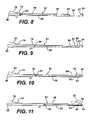
- FIGS. 8-11are right side elevation views of a cutter of the handheld breast biopsy device and the marker introduction assembly taken in longitudinal cross section along lines 7 - 7 of FIG. 6 depicting a sequence respectively of the cutter engaging the pusher, the cutter advanced to expose a marker in a specimen opening of the probe, the cutter advanced to ramp a distal end of the marker against an angled surface of the assembly, and the cutter advanced to fully deploy the marker by underrunning a driver tip of the pusher.
- FIG. 12is a sequence of operations or procedures for controlling a biopsy system to deploy a biopsy marker at a surgical biopsy site.
- FIG. 1a breast biopsy handle 10 of a biopsy system 12 , a minimally invasive device, is used under local anesthetic and ultrasound guidance to collect multiple biopsy samples with a single insertion of a probe 14 into the breast of a patient.
- a cutter 16is retracted proximally in a housing 18 of the biopsy handle 10 to expose a distally opening entry cone 20 of a cutter lumen 22 of the probe 12 .
- the surgeonmay then insert and seat a biopsy marker introduction assembly 24 , which is shown separately in FIG. 2 .
- FIG. 2Also shown in FIG. 2 is a Mylar sealing cap 26 that has been removed just prior to use from a distal end 28 of the introduction assembly 24 to expose a laterally disposed deployment opening 30 in an introducer tube 32 .
- a marker 34is positioned inside of the introducer tube 32 proximal to the deployment opening 30 that has sufficient length to allow the marker 34 to be laterally expelled when a distal end 36 of the marker 34 ramps against an angled surface 38 at the distal end 28 of the introduction assembly 24 .
- a proximal end 40 of the marker 34resides approximately twice the length (2 ⁇ ) of the marker 34 from the angled surface 38 when in its initial state as depicted.
- a marker pusherdepicted as a plunger tip 42 of a marker deployment rod 44 that longitudinally translates within the introducer tube 32 .
- the plunger tip 42in some applications dynamically seals to the interior of the introducer tube 32 to form a sterile environment for the marker 34 and to pneumatically assist in deploying the marker 34 .
- the plunger tip 42may push air toward the marker 40 as a syringe, which may advantageously reduce the amount of travel required for the cutter 16 .
- a vacuum lumen 45 in the probe 14may provide vacuum assist to draw the plunger tip 42 and bleed any excess pressure from the surgical site.
- a proximal end 46 of the marker deployment rod 44terminates in a cutter seat 48 that extends proximally beyond the entry cone 20 of the cutter lumen 22 a sufficient distance to provide full travel of the marker deployment rod 44 yet fit within a specimen retrieval recess 50 formed within the housing 18 of the biopsy handle 10 .
- the cutter seat 48has a greater lateral diameter than the tube-shaped cutter 16 to ensure contact. Since the cutter 16 closely fits the inner diameter of the cutter lumen 22 , the cutter seat 48 reaches full travel at the entry cone 20 .
- the funnel 50advantageously includes an alignment feature, such as a transversely extending key 54 , that mates with a corresponding alignment feature in the biopsy probe 14 , such as a proximally opened recess 56 .
- These alignment features 54 , 56ensure that the marker introduction assembly 30 is correctly rotated to present its deployment opening 30 to a specimen opening 58 of the probe 14 .
- visual and tactile confirmationis given that the introduction assembly 24 is fully inserted into the probe 14 so that reliable deployment of the marker 34 may be achieved.
- the cutter 16has been advanced, distally translating the deployment rod 44 to deploy the marker 34 . Then the probe 14 may be withdrawn leaving the marker 34 behind.
- the existing controls of the biopsy system 12may be used to achieve deployment as depicted in FIG. 4 .
- the cutter seat 48may advantageously resist damage from a cutter 16 that is rotating (e.g., hardened surface, low friction surface, or configured to be rotated by the cutter). The surgeon would command forward translation of the cutter until visually confirming full travel or the cutter is prevented from further travel by the cutter seat 48 engaging introducer tube 32 .
- the breast biopsy handle 10 depictedis similar to sterotactic devices attached to a tabletop.
- a handheld MRI-compatible biopsy device 60is depicted in phantom in FIGS. 5-6 for use with a marker introduction assembly 62 .
- access to an entry cone 64 of a cutter lumen 66 of a probe 68may be constrained by the shape of a specimen retrieval recess 70 formed in a handle 72 of the handheld breast biopsy device 60 . Consequently, portions of the marker introduction assembly 62 are formed of a sufficiently flexible material, as depicted in FIG. 5 , to be bent during insertion into the biopsy handle 70 .
- FIGS. 1the breast biopsy handle 10 depicted is similar to sterotactic devices attached to a tabletop.
- a handheld MRI-compatible biopsy device 60is depicted in phantom in FIGS. 5-6 for use with a marker introduction assembly 62 .
- access to an entry cone 64 of a cutter lumen 66 of a probe 68may be constrained by
- an alignment key 74 along the top of a funnel 76rotationally and longitudinally orients an attached introducer tube 78 within the cutter lumen 66 of the probe 68 .
- the introducer tube 78is sufficiently thin to allow advancement of a cutter 80 (depicted in FIGS. 8-11 ) as desired within the cutter lumen 66 without binding. This has an added advantage of thus being usable in specimen retrieval recesses 70 that are not at least twice the length of a marker 82 or when it is otherwise desirable to advance the cutter 80 more fully during deployment of the marker 82 .
- the marker introducing assembly 62has been inserted into the probe 68 in preparation for the surgeon initiating deployment of the marker 82 .
- the cutter 80has been advanced into contact with a cutter seat 84 .
- the cutter 80has further distally advanced, driving a pusher rod 86 and its driving tip 88 , and thus the distally placed marker 82 to expose the marker in a distal opening 90 of the introducer tube 78 .
- the cutter 80has been further advanced so that the distal end of the marker 82 ramps outward against an angled surface 92 of the distal opening 90 .
- the cutter 80has further advanced so that the driving tip 88 has underrun the fully deployed marker 82 and has been received within a tip slot 94 of the introducer tube 78 (shown also in FIG. 6 ).
- the pusher rod 86advantageously closes the distal opening 90 of the introduction assembly 62 and thus a specimen opening 96 of the probe 68 .
- Thismay be an advantageous feature that allows retracting the probe 68 without inadvertently dragging out the marker 82 .
- a marker deployment modeinto the controls of the biopsy system 12 to optimize this function. For instance, if two motors are used, one for longitudinal translation of the cutter and one for rotating the cutter 16 , then dedicated control logic would allow disabling cutter rotation during deployment. Also, the amount of longitudinal travel and the rate of travel may be optimized for marker deployment.
- FIG. 5an illustrative sequence of operations or procedure 100 for biopsy device control of marker deployment is depicted as a flow diagram.
- a surgical cite within the patient's breastis imaged (e.g., ultrasonic imaging) (block 102 ) to guide the surgeon to insert and position the probe (block 104 ).
- Vacuumis applied through the probe to assist in drawing tissue into a specimen bowl of the probe (block 106 ) while the cutter is rotated and distally translated to cut the tissue.
- the probemay be repositioned, such as by rotating the probe to present the specimen bowl at a different angle with the specimen bowl blocked by the extended cutter. Thereafter, the cutter and sample are proximally retracted (block 110 ).
- the surgeonengages the marker introduction assembly (“introducer”) into the biopsy probe (block 112 ) and enables deployment mode of the marker (block 114 ).
- the interface between the introducer and the biopsy handlemay be such that its presence is sensed by a sensor (block 116 ) and automatically prepares the biopsy system for deployment mode, preventing inadvertent rotation of the cutter or an inappropriate application of the vacuum assist system and/or adjusting cutter travel and/or rate of travel.
- Enabling deployment modemay be manual (block 118 ) wherein the surgeon uses a control, such as pushing a deployment soft button, to initiate deployment mode.
- a remote control unitthat is spaced away from the biopsy handle may initiate deployment mode (block 120 ).
- a control unitmay be elsewhere in the surgical suite, for instance in an MRI or CTI facility wherein the biopsy system is remotely actuated.
- cutter rotationmay advantageously be disabled in a two-motor biopsy system (block 122 ).
- Distal translation speedis set (block 124 ). For instance, a greater rate of travel may be desired to better propel the marker from the probe.
- Maximum cutter travelmay be set (block 126 ) to avoid binding and damage, especially if the mechanical advantage of the motor and mechanization is such as to cause damage before full travel may be sensed. It may be desirable to use a fluid drainage capability of the biopsy system at this point, such as using the vacuum assist system to remove fluid from the surgical site and/or to insufflate the surgical site to provide a cavity to receive the biopsy marker (block 128 ).
- the cutteris advanced to deploy the marker (block 130 ), which may be a programmed mode that is initiated by the surgeon (block 132 ) or a manual process (block 134 ) with a deployment command maintained by the surgeon. Imaging may be performed contemporaneously or subsequently to confirm that the marker has been deployed at the surgical site (block 136 ). Advancement continues until a determination is made that the marker is deployed (block 138 ), which may be determined based on one or more conditions such as the biopsy system sensing full travel, sensing of cutter binding at full travel, visual confirmation of the marker deployment, etc.
- the introducer and/or biopsy probemay be determined to be insufficiently closed at this point (block 140 ), such as the introducer prevents closure.
- the cutteris retracted (block 142 )
- the marker introduceris removed with the probe still in place (block 144 )
- the probeis rotated (e.g., 180 degrees) so that the specimen bowl is moved away from the marker to prevent its inadvertent entry (block 146 ), and the cutter is advanced to close the port (block 148 ).
- the probeis removed (block 150 ).
- the illustrative embodimentdescribes a handheld biopsy procedure guided by ultrasonic imaging to accentuate advantages of incorporating marker placement into biopsy instrument controls.
- aspects of the present inventionpertain to tabletop fixed biopsy instruments, biopsy instruments used for tissue other than the breast, biopsy instruments positioned with other imaging modalities such as X-ray and Magnetic Resonance Imaging (MRI), and remotely actuated biopsy instruments.
- MRIMagnetic Resonance Imaging
- a vacuum-assisted biopsy systemhas been advantageously depicted herein as benefiting from a marker introduction device
- application of a marker introduction device to a core needle biopsy devicewould provide similar advantages, allowing a surgeon to place a marker while positioning the core needle device with reference to a diagnostic image.
- a detachable probemay be used in conjunction with a marker introduction device.
- the vacuum assist control system of a biopsy systemmay be used to draw a plunger of a biopsy marker introduction device toward the distal end of the probe. With the plunger fully translated distally, the marker would deploy and then the vacuum assist may be removed, leaving the marker in place as the biopsy probe is removed.
Landscapes
- Health & Medical Sciences (AREA)
- Surgery (AREA)
- Life Sciences & Earth Sciences (AREA)
- Heart & Thoracic Surgery (AREA)
- Molecular Biology (AREA)
- Oral & Maxillofacial Surgery (AREA)
- Engineering & Computer Science (AREA)
- Biomedical Technology (AREA)
- Nuclear Medicine, Radiotherapy & Molecular Imaging (AREA)
- Medical Informatics (AREA)
- Pathology (AREA)
- Animal Behavior & Ethology (AREA)
- General Health & Medical Sciences (AREA)
- Public Health (AREA)
- Veterinary Medicine (AREA)
- Surgical Instruments (AREA)
- Ultra Sonic Daignosis Equipment (AREA)
Abstract
Description
Claims (20)
Priority Applications (7)
| Application Number | Priority Date | Filing Date | Title |
|---|---|---|---|
| US10/815,004US7465279B2 (en) | 2004-03-31 | 2004-03-31 | Marker device and method of deploying a cavity marker using a surgical biopsy device |
| AU2005201304AAU2005201304B2 (en) | 2004-03-31 | 2005-03-24 | Marker device and method of deploying a cavity marker using a surgical biopsy device |
| JP2005099444AJP2005288175A (en) | 2004-03-31 | 2005-03-30 | Marker device and method for arranging marker using surgical biopsy device |
| CA002502997ACA2502997A1 (en) | 2004-03-31 | 2005-03-30 | Marker device and method of deploying a cavity marker using a surgical biopsy device |
| EP05251978AEP1582168A1 (en) | 2004-03-31 | 2005-03-30 | Marker device and method of deploying a cavity marker using a surgical biopsy device |
| CNA2005100598482ACN1676106A (en) | 2004-03-31 | 2005-03-31 | Marker device and method of deploying a cavity marker using a surgical biopsy device |
| US12/330,560US20090088665A1 (en) | 2004-03-31 | 2008-12-09 | Marker device and method of deploying a cavity marker using a surgical biopsy device |
Applications Claiming Priority (1)
| Application Number | Priority Date | Filing Date | Title |
|---|---|---|---|
| US10/815,004US7465279B2 (en) | 2004-03-31 | 2004-03-31 | Marker device and method of deploying a cavity marker using a surgical biopsy device |
Related Child Applications (1)
| Application Number | Title | Priority Date | Filing Date |
|---|---|---|---|
| US12/330,560ContinuationUS20090088665A1 (en) | 2004-03-31 | 2008-12-09 | Marker device and method of deploying a cavity marker using a surgical biopsy device |
Publications (2)
| Publication Number | Publication Date |
|---|---|
| US20050228311A1 US20050228311A1 (en) | 2005-10-13 |
| US7465279B2true US7465279B2 (en) | 2008-12-16 |
Family
ID=34887732
Family Applications (2)
| Application Number | Title | Priority Date | Filing Date |
|---|---|---|---|
| US10/815,004Expired - Fee RelatedUS7465279B2 (en) | 2004-03-31 | 2004-03-31 | Marker device and method of deploying a cavity marker using a surgical biopsy device |
| US12/330,560AbandonedUS20090088665A1 (en) | 2004-03-31 | 2008-12-09 | Marker device and method of deploying a cavity marker using a surgical biopsy device |
Family Applications After (1)
| Application Number | Title | Priority Date | Filing Date |
|---|---|---|---|
| US12/330,560AbandonedUS20090088665A1 (en) | 2004-03-31 | 2008-12-09 | Marker device and method of deploying a cavity marker using a surgical biopsy device |
Country Status (6)
| Country | Link |
|---|---|
| US (2) | US7465279B2 (en) |
| EP (1) | EP1582168A1 (en) |
| JP (1) | JP2005288175A (en) |
| CN (1) | CN1676106A (en) |
| AU (1) | AU2005201304B2 (en) |
| CA (1) | CA2502997A1 (en) |
Cited By (53)
| Publication number | Priority date | Publication date | Assignee | Title |
|---|---|---|---|---|
| US20060149162A1 (en)* | 2004-11-29 | 2006-07-06 | Derek Daw | Graphical user interface for tissue biopsy system |
| US20090216150A1 (en)* | 2008-02-25 | 2009-08-27 | Lee Reichel | Method and Apparatus For Inserting Biopsy Site Marker In Marker Body |
| US20090216181A1 (en)* | 2008-02-25 | 2009-08-27 | Speeg Trevor W V | Biopsy Site Marker Deployment Instrument |
| US20100049084A1 (en)* | 2008-08-22 | 2010-02-25 | Nock Andrew P | Biopsy marker delivery device |
| US20100049085A1 (en)* | 2008-08-22 | 2010-02-25 | Nock Andrew P | Method of making a biopsy marker delivery device |
| US20100160819A1 (en)* | 2008-12-18 | 2010-06-24 | Parihar Shailendra K | Biopsy Device with Central Thumbwheel |
| US20100160826A1 (en)* | 2008-12-18 | 2010-06-24 | Parihar Shailendra K | Tissue Biopsy Device with Rotatably Linked Thumbwheel and Tissue Sample Holder |
| US7806835B2 (en) | 2007-11-20 | 2010-10-05 | Devicor Medical Products, Inc. | Biopsy device with sharps reduction feature |
| US20100280367A1 (en)* | 2009-04-30 | 2010-11-04 | Ducharme Richard W | System and method for fiducial deployment |
| US7858038B2 (en) | 2007-11-20 | 2010-12-28 | Devicor Medical Products, Inc. | Biopsy device with illuminated tissue holder |
| US20110071431A1 (en)* | 2009-09-24 | 2011-03-24 | Speeg Trevor W V | Biopsy marker delivery devices and methods |
| US7938786B2 (en) | 2006-12-13 | 2011-05-10 | Devicor Medical Products, Inc. | Vacuum timing algorithm for biopsy device |
| US7981049B2 (en) | 2006-12-13 | 2011-07-19 | Devicor Medical Products, Inc. | Engagement interface for biopsy system vacuum module |
| US20110218433A1 (en)* | 2010-03-02 | 2011-09-08 | Speeg Trevor W V | Biopsy Marker Delivery Device |
| US8052616B2 (en) | 2007-11-20 | 2011-11-08 | Devicor Medical Products, Inc. | Biopsy device with fine pitch drive train |
| WO2012033796A2 (en) | 2010-09-10 | 2012-03-15 | Devicor Medical Products, Inc. | Biopsy device tissue sample holder with removable tray |
| US8251916B2 (en) | 2006-12-13 | 2012-08-28 | Devicor Medical Products, Inc. | Revolving tissue sample holder for biopsy device |
| US20130041256A1 (en)* | 2011-08-08 | 2013-02-14 | Kevin M. Fiebig | Access chamber and markers for biopsy device |
| US8454531B2 (en) | 2007-11-20 | 2013-06-04 | Devicor Medical Products, Inc. | Icon-based user interface on biopsy system control module |
| US8480595B2 (en) | 2006-12-13 | 2013-07-09 | Devicor Medical Products, Inc. | Biopsy device with motorized needle cocking |
| USD695404S1 (en) | 2011-09-07 | 2013-12-10 | Devicor Medical Products, Inc. | Biopsy device |
| US20140024945A1 (en)* | 2012-07-23 | 2014-01-23 | ClariTrac Inc. | Ultrasound device for needle procedures |
| WO2014028366A1 (en) | 2012-08-13 | 2014-02-20 | Devicor Medical Products, Inc. | Biopsy system |
| US8702623B2 (en) | 2008-12-18 | 2014-04-22 | Devicor Medical Products, Inc. | Biopsy device with discrete tissue chambers |
| WO2014150882A2 (en) | 2013-03-15 | 2014-09-25 | Devicor Medical Products, Inc. | Biopsy site marker applier |
| WO2015031498A1 (en) | 2013-08-28 | 2015-03-05 | Devicor Medical Products, Inc. | Tissue collection assembly for biopsy device |
| US9039634B2 (en) | 2007-11-20 | 2015-05-26 | Devicor Medical Products, Inc. | Biopsy device tissue sample holder rotation control |
| EP2989994A1 (en) | 2011-06-23 | 2016-03-02 | Devicor Medical Products, Inc. | Introducer for biopsy device |
| WO2016073912A1 (en) | 2014-11-06 | 2016-05-12 | Devicor Medical Products, Inc. | Spring-ejected biopsy marker |
| US9345457B2 (en) | 2006-12-13 | 2016-05-24 | Devicor Medical Products, Inc. | Presentation of biopsy sample by biopsy device |
| US9522264B2 (en) | 2013-02-26 | 2016-12-20 | Cook Medical Technologies Llc | Ratchet-slide handle and system for fiducial deployment |
| WO2017083417A1 (en) | 2015-11-12 | 2017-05-18 | Devicor Medical Products, Inc. | Marker delivery device and method of deploying a marker |
| WO2017083412A1 (en) | 2015-11-11 | 2017-05-18 | Devicor Medical Products, Inc. | Marker delivery device and method of deploying a marker |
| US9770262B2 (en) | 2014-06-09 | 2017-09-26 | Cook Medical Technologies Llc | Screw-driven handles and systems for fiducial deployment |
| US9788819B2 (en) | 2014-05-01 | 2017-10-17 | Devicor Medical Products, Inc. | Introducer for biopsy device |
| US9877706B2 (en) | 2013-03-15 | 2018-01-30 | Devicor Medical Products, Inc. | Biopsy device |
| US9993232B2 (en) | 2014-05-22 | 2018-06-12 | Andrew N. Ellingson | Biopsy with marker device and method |
| US10123848B2 (en) | 2014-12-03 | 2018-11-13 | Cook Medical Technologies Llc | EUS fiducial needle stylet handle assembly |
| US10149700B2 (en) | 2013-08-12 | 2018-12-11 | Jan R. Lau | 3 dimensional simultaneous multiple core biopsy or fiducial marker placement device and methods |
| US10258373B2 (en) | 2011-06-28 | 2019-04-16 | Cook Medical Technologies Llc | Fiducial deployment needle system |
| US10285671B2 (en) | 2017-02-06 | 2019-05-14 | Device And Design, Llc | System, method and apparatus for integrated tissue sampling and tissue marker placement |
| US10314563B2 (en) | 2014-11-26 | 2019-06-11 | Devicor Medical Products, Inc. | Graphical user interface for biopsy device |
| US10335124B1 (en) | 2016-02-29 | 2019-07-02 | Devicor Medical Products, Inc. | Marker delivery device with adaptor for biopsy site marking and method of use thereof |
| US10363407B2 (en) | 2014-06-16 | 2019-07-30 | Cook Medical Technologies Llc | Plunger-driven collet handle and system for fiducial deployment |
| US10595831B2 (en) | 2012-05-30 | 2020-03-24 | Devicor Medical Products, Inc. | Control for biopsy device |
| US10610205B2 (en) | 2014-05-15 | 2020-04-07 | Devicor Medical Products, Inc. | Biopsy device |
| US10683119B2 (en) | 2014-05-23 | 2020-06-16 | Merit Medical Systems, Inc. | Marker element, device for making a marker element, and method for making a marker element |
| US10869653B2 (en) | 2015-10-30 | 2020-12-22 | Devicor Medical Products, Inc. | Tissue sample holder with bulk tissue collection feature |
| US11160541B2 (en)* | 2016-05-10 | 2021-11-02 | Koninklijke Philips N.V. | Biopsy container |
| US11179141B2 (en) | 2006-12-13 | 2021-11-23 | Devicor Medical Products, Inc. | Biopsy system |
| WO2024163634A3 (en)* | 2023-02-01 | 2025-01-23 | Singular Genomics Systems, Inc. | Methods and devices for manipulating biological samples |
| US12298211B2 (en) | 2021-10-25 | 2025-05-13 | Singular Genomics Systems, Inc. | Device for tissue section immobilization and retention |
| US12426860B2 (en) | 2018-04-18 | 2025-09-30 | C.R. Bard, Inc. | Dual lumen coaxial introducer having integrated tissue marker delivery |
Families Citing this family (32)
| Publication number | Priority date | Publication date | Assignee | Title |
|---|---|---|---|---|
| US8062230B1 (en)* | 2004-10-14 | 2011-11-22 | Suros Surgical Systems, Inc. | Surgical site marker delivery system |
| US7867173B2 (en)* | 2005-08-05 | 2011-01-11 | Devicor Medical Products, Inc. | Biopsy device with replaceable probe and incorporating vibration insertion assist and static vacuum source sample stacking retrieval |
| US20120283563A1 (en)* | 2011-05-03 | 2012-11-08 | Moore Kyle P | Biopsy device with manifold alignment feature and tissue sensor |
| US7575556B2 (en) | 2007-11-20 | 2009-08-18 | Ethicon Endo-Surgery, Inc. | Deployment device interface for biopsy device |
| US20090131819A1 (en) | 2007-11-20 | 2009-05-21 | Ritchie Paul G | User Interface On Biopsy Device |
| US20090209853A1 (en)* | 2008-02-19 | 2009-08-20 | Parihar Shailendra K | Biopsy site marker applier |
| AU2009201610A1 (en) | 2008-04-23 | 2009-11-19 | Devicor Medical Products, Inc. | PEM and BSGI biopsy devices and methods |
| US8532748B2 (en) | 2008-04-23 | 2013-09-10 | Devicor Medical Products, Inc. | Devices useful in imaging |
| US20090270726A1 (en)* | 2008-04-23 | 2009-10-29 | Leimbach Jessica P | Methods For Imaging |
| US8864681B2 (en)* | 2008-04-23 | 2014-10-21 | Devicor Medical Products, Inc. | Biopsy devices |
| US20100331677A1 (en)* | 2008-04-25 | 2010-12-30 | The Johns Hopkins University | Marker delivery system |
| US8050742B2 (en)* | 2008-07-30 | 2011-11-01 | Devicor Medical Products, Inc. | Biopsy device |
| US20100081925A1 (en)* | 2008-09-29 | 2010-04-01 | Searete Llc, A Limited Liability Corporation Of The State Of Delaware | Histological facilitation systems and methods |
| US20100081919A1 (en)* | 2008-09-29 | 2010-04-01 | Searete Llc, A Limited Liability Corporation Of The State Of Delaware | Histological facilitation systems and methods |
| US11298113B2 (en) | 2008-10-01 | 2022-04-12 | Covidien Lp | Device for needle biopsy with integrated needle protection |
| US9186128B2 (en) | 2008-10-01 | 2015-11-17 | Covidien Lp | Needle biopsy device |
| US9332973B2 (en) | 2008-10-01 | 2016-05-10 | Covidien Lp | Needle biopsy device with exchangeable needle and integrated needle protection |
| US9782565B2 (en) | 2008-10-01 | 2017-10-10 | Covidien Lp | Endoscopic ultrasound-guided biliary access system |
| US8968210B2 (en) | 2008-10-01 | 2015-03-03 | Covidien LLP | Device for needle biopsy with integrated needle protection |
| US8371443B2 (en)* | 2009-09-22 | 2013-02-12 | Devicor Medical Products, Inc. | Biopsy marker delivery device |
| US20110071391A1 (en)* | 2009-09-24 | 2011-03-24 | Speeg Trevor W V | Biopsy marker delivery device with positioning component |
| US8241299B2 (en)* | 2009-10-22 | 2012-08-14 | Devicor Medical Products, Inc. | Biopsy marker delivery configured to retain marker prior to intended deployment |
| US8597201B2 (en) | 2010-03-30 | 2013-12-03 | Siteselect Medical Technologies, Inc. | Tissue excision device with a flexible transection blade |
| EP3203925B1 (en)* | 2014-10-08 | 2024-03-13 | Devicor Medical Products, Inc. | Biopsy marker |
| WO2016201115A1 (en)* | 2015-06-11 | 2016-12-15 | Radvation, Llc | Device and method for marking a location of a tissue biopsy |
| EP3871586B1 (en)* | 2016-12-07 | 2023-03-22 | Boston Scientific Scimed, Inc. | Systems for eccentric nodule tissue acquisition |
| CN109009344B (en)* | 2018-07-24 | 2020-10-30 | 西安卓恰医疗器械有限公司 | Puncture locator with replaceable needle |
| EP3893799B1 (en)* | 2019-02-15 | 2024-09-04 | Devicor Medical Products, Inc. | Marker delivery device with sterile guide |
| CN117958999A (en)* | 2019-05-30 | 2024-05-03 | Devicor医疗产业收购公司 | Method and apparatus for direct marking |
| CN112617905A (en)* | 2020-12-29 | 2021-04-09 | 深圳开立生物医疗科技股份有限公司 | Ultrasonic microprobe, ultrasonic endoscopy system and biopsy sampling system |
| CN118648853B (en)* | 2024-08-19 | 2024-11-22 | 湖南省华芯医疗器械有限公司 | Front end housing assembly, insertion part and endoscope |
| CN120131136A (en)* | 2025-04-29 | 2025-06-13 | 东莞市道滘医院 | A minimally invasive treatment device for patients with tophi on the surface of limbs |
Citations (51)
| Publication number | Priority date | Publication date | Assignee | Title |
|---|---|---|---|---|
| US3744493A (en)* | 1972-01-10 | 1973-07-10 | Syntex Corp | Implanter having an improved cartridge ejector |
| US4007732A (en)* | 1975-09-02 | 1977-02-15 | Robert Carl Kvavle | Method for location and removal of soft tissue in human biopsy operations |
| US4774948A (en)* | 1986-11-24 | 1988-10-04 | Markham Charles W | Marking and retraction needle having retrievable stylet |
| US4900304A (en)* | 1986-07-30 | 1990-02-13 | Sumitomo Pharmaceuticals Company, Limited | Solid preparation administering instrument |
| US5286257A (en)* | 1992-11-18 | 1994-02-15 | Ultradent Products, Inc. | Syringe apparatus with detachable mixing and delivery tip |
| US5526822A (en) | 1994-03-24 | 1996-06-18 | Biopsys Medical, Inc. | Method and apparatus for automated biopsy and collection of soft tissue |
| US5649547A (en) | 1994-03-24 | 1997-07-22 | Biopsys Medical, Inc. | Methods and devices for automated biopsy and collection of soft tissue |
| US5718237A (en)* | 1993-11-15 | 1998-02-17 | Haaga; John R. | Biopsy needle |
| US5769086A (en) | 1995-12-06 | 1998-06-23 | Biopsys Medical, Inc. | Control system and method for automated biopsy device |
| US5810806A (en) | 1996-08-29 | 1998-09-22 | Ethicon Endo-Surgery | Methods and devices for collection of soft tissue |
| US5902310A (en) | 1996-08-12 | 1999-05-11 | Ethicon Endo-Surgery, Inc. | Apparatus and method for marking tissue |
| US5913857A (en) | 1996-08-29 | 1999-06-22 | Ethicon End0-Surgery, Inc. | Methods and devices for collection of soft tissue |
| US5922002A (en) | 1989-12-05 | 1999-07-13 | Yoon; Inbae | Surgical instrument with jaws and movable internal biopsy device and method for use thereof |
| US5941890A (en) | 1998-06-26 | 1999-08-24 | Ethicon Endo-Surgery, Inc. | Implantable surgical marker |
| US5944673A (en) | 1998-05-14 | 1999-08-31 | Ethicon Endo-Surgery, Inc. | Biopsy instrument with multi-port needle |
| US5964716A (en) | 1998-05-14 | 1999-10-12 | Ethicon Endo-Surgery, Inc. | Method of use for a multi-port biopsy instrument |
| US6007497A (en) | 1998-06-30 | 1999-12-28 | Ethicon Endo-Surgery, Inc. | Surgical biopsy device |
| US6017316A (en) | 1997-06-18 | 2000-01-25 | Biopsys Medical | Vacuum control system and method for automated biopsy device |
| US6077230A (en) | 1998-05-14 | 2000-06-20 | Ethicon Endo-Surgery, Inc. | Biopsy instrument with removable extractor |
| US6083237A (en) | 1998-10-23 | 2000-07-04 | Ethico Endo-Surgery, Inc. | Biopsy instrument with tissue penetrating spiral |
| WO2000038579A2 (en) | 1998-12-24 | 2000-07-06 | Vivant Medical, Inc. | Device and method for safe location and marking of a cavity and sentinel lymph nodes |
| US6086544A (en) | 1999-03-31 | 2000-07-11 | Ethicon Endo-Surgery, Inc. | Control apparatus for an automated surgical biopsy device |
| US6085749A (en) | 1996-02-26 | 2000-07-11 | Ethicon Endo-Surgery, Inc. | Articulating guide arm for medical applications |
| US6120462A (en) | 1999-03-31 | 2000-09-19 | Ethicon Endo-Surgery, Inc. | Control method for an automated surgical biopsy device |
| US6162187A (en) | 1999-08-02 | 2000-12-19 | Ethicon Endo-Surgery, Inc. | Fluid collection apparatus for a surgical device |
| US6220248B1 (en) | 1998-10-21 | 2001-04-24 | Ethicon Endo-Surgery, Inc. | Method for implanting a biopsy marker |
| US6228055B1 (en) | 1994-09-16 | 2001-05-08 | Ethicon Endo-Surgery, Inc. | Devices for marking and defining particular locations in body tissue |
| US6231522B1 (en) | 2000-02-18 | 2001-05-15 | Ethicon Endo-Surgery, Inc. | Biopsy instrument with breakable sample segments |
| US6234177B1 (en)* | 1999-08-12 | 2001-05-22 | Thomas Barsch | Apparatus and method for deploying an expandable biopsy marker |
| US6241687B1 (en) | 2000-02-18 | 2001-06-05 | Ethicon Endo-Surgery, Inc. | Method of use for a biopsy instrument with breakable sample segments |
| US6261302B1 (en) | 1998-06-26 | 2001-07-17 | Ethicon Endo-Surgery, Inc. | Applier for implantable surgical marker |
| US6273862B1 (en) | 1998-10-23 | 2001-08-14 | Ethicon Endo-Surgery, Inc | Surgical device for the collection of soft tissue |
| WO2002013876A2 (en) | 2000-08-15 | 2002-02-21 | Carbon Medical Technologies | Tissue marking using biocompatible microparticles |
| US6375634B1 (en)* | 1997-11-19 | 2002-04-23 | Oncology Innovations, Inc. | Apparatus and method to encapsulate, kill and remove malignancies, including selectively increasing absorption of x-rays and increasing free-radical damage to residual tumors targeted by ionizing and non-ionizing radiation therapy |
| US6428487B1 (en) | 1999-12-17 | 2002-08-06 | Ethicon Endo-Surgery, Inc. | Surgical biopsy system with remote control for selecting an operational mode |
| US6432065B1 (en) | 1999-12-17 | 2002-08-13 | Ethicon Endo-Surgery, Inc. | Method for using a surgical biopsy system with remote control for selecting and operational mode |
| US6432064B1 (en) | 2001-04-09 | 2002-08-13 | Ethicon Endo-Surgery, Inc. | Biopsy instrument with tissue marking element |
| US6436068B1 (en)* | 2000-08-24 | 2002-08-20 | Gust H. Bardy | Instrument for implanting sensors and solid materials in a subcutaneous location and method thereof |
| WO2002078560A1 (en) | 2001-03-30 | 2002-10-10 | H.S. Hospital Service S.P.A. | Method and means for locating and signalling a non-palpable lesion in soft tissues |
| US20020161298A1 (en) | 1999-02-02 | 2002-10-31 | Senorx, Inc. | Methods and chemical preparations for time-limited marking of biopsy sites |
| US20030050571A1 (en) | 2001-09-10 | 2003-03-13 | Vivant Medical, Inc. | Biopsy marker delivery system |
| US6554760B2 (en) | 2000-10-25 | 2003-04-29 | Gary A. Lamoureux | Pre-loaded needle assembly |
| WO2003034894A2 (en) | 2001-10-23 | 2003-05-01 | Valentino Montegrande | Ultrasound imaging marker and method of use |
| US6557196B2 (en) | 2000-07-07 | 2003-05-06 | Medical Positioning, Inc. | Patient support apparatus and method for performing decubitus breast biopsy |
| US6560310B2 (en) | 2000-11-16 | 2003-05-06 | Is2 Research Inc. | Apparatus for mammography |
| US6558407B1 (en) | 2000-10-24 | 2003-05-06 | Tyco Healthcare Group Lp | Breast stabilizer with instrument guide |
| US6602204B2 (en)* | 1998-02-10 | 2003-08-05 | Artemis Medical, Inc | Intraoperative tissue treatment methods |
| US6638235B2 (en)* | 2000-11-06 | 2003-10-28 | Suros Surgical Systems, Inc. | Biopsy apparatus |
| US6648811B2 (en)* | 1999-07-26 | 2003-11-18 | C.R. Bard, Inc. | Brachytherapy cartridge including absorbable and autoclaveable spacer |
| US6662041B2 (en) | 1999-02-02 | 2003-12-09 | Senorx, Inc. | Imageable biopsy site marker |
| US20040030262A1 (en)* | 2002-05-03 | 2004-02-12 | Fisher John S. | Biodegradable polymer for marking tissue and sealing tracts |
Family Cites Families (4)
| Publication number | Priority date | Publication date | Assignee | Title |
|---|---|---|---|---|
| US5567689A (en)* | 1993-08-13 | 1996-10-22 | The Uab Research Foundation | Methods for increasing uridine levels with L-nucleosides |
| US5593420A (en)* | 1995-02-17 | 1997-01-14 | Mist, Inc. | Miniature endoscopic surgical instrument assembly and method of use |
| US6056700A (en)* | 1998-10-13 | 2000-05-02 | Emx, Inc. | Biopsy marker assembly and method of use |
| US6725083B1 (en)* | 1999-02-02 | 2004-04-20 | Senorx, Inc. | Tissue site markers for in VIVO imaging |
- 2004
- 2004-03-31USUS10/815,004patent/US7465279B2/ennot_activeExpired - Fee Related
- 2005
- 2005-03-24AUAU2005201304Apatent/AU2005201304B2/ennot_activeCeased
- 2005-03-30JPJP2005099444Apatent/JP2005288175A/enactivePending
- 2005-03-30CACA002502997Apatent/CA2502997A1/ennot_activeAbandoned
- 2005-03-30EPEP05251978Apatent/EP1582168A1/ennot_activeWithdrawn
- 2005-03-31CNCNA2005100598482Apatent/CN1676106A/enactivePending
- 2008
- 2008-12-09USUS12/330,560patent/US20090088665A1/ennot_activeAbandoned
Patent Citations (62)
| Publication number | Priority date | Publication date | Assignee | Title |
|---|---|---|---|---|
| US3744493A (en)* | 1972-01-10 | 1973-07-10 | Syntex Corp | Implanter having an improved cartridge ejector |
| US4007732A (en)* | 1975-09-02 | 1977-02-15 | Robert Carl Kvavle | Method for location and removal of soft tissue in human biopsy operations |
| US4900304A (en)* | 1986-07-30 | 1990-02-13 | Sumitomo Pharmaceuticals Company, Limited | Solid preparation administering instrument |
| US4774948A (en)* | 1986-11-24 | 1988-10-04 | Markham Charles W | Marking and retraction needle having retrievable stylet |
| US5922002A (en) | 1989-12-05 | 1999-07-13 | Yoon; Inbae | Surgical instrument with jaws and movable internal biopsy device and method for use thereof |
| US5286257A (en)* | 1992-11-18 | 1994-02-15 | Ultradent Products, Inc. | Syringe apparatus with detachable mixing and delivery tip |
| US5718237A (en)* | 1993-11-15 | 1998-02-17 | Haaga; John R. | Biopsy needle |
| US6428486B2 (en) | 1994-03-24 | 2002-08-06 | Ethicon Endo-Surgery, Inc. | Methods and devices for automated biopsy and collection of soft tissue |
| US5775333A (en) | 1994-03-24 | 1998-07-07 | Ethicon Endo-Surgery, Inc. | Apparatus for automated biopsy and collection of soft tissue |
| US5649547A (en) | 1994-03-24 | 1997-07-22 | Biopsys Medical, Inc. | Methods and devices for automated biopsy and collection of soft tissue |
| US5928164A (en) | 1994-03-24 | 1999-07-27 | Ethicon Endo-Surgery, Inc. | Apparatus for automated biopsy and collection of soft tissue |
| US5526822A (en) | 1994-03-24 | 1996-06-18 | Biopsys Medical, Inc. | Method and apparatus for automated biopsy and collection of soft tissue |
| US5980469A (en) | 1994-03-24 | 1999-11-09 | Ethicon Endo-Surgery, Inc. | Method and apparatus for automated biopsy and collection of soft tissue |
| US6228055B1 (en) | 1994-09-16 | 2001-05-08 | Ethicon Endo-Surgery, Inc. | Devices for marking and defining particular locations in body tissue |
| US5769086A (en) | 1995-12-06 | 1998-06-23 | Biopsys Medical, Inc. | Control system and method for automated biopsy device |
| US6085749A (en) | 1996-02-26 | 2000-07-11 | Ethicon Endo-Surgery, Inc. | Articulating guide arm for medical applications |
| US6488030B1 (en) | 1996-02-26 | 2002-12-03 | Ethicon Endo-Surgery, Inc. | Articulating guide arm for medical applications |
| US5902310A (en) | 1996-08-12 | 1999-05-11 | Ethicon Endo-Surgery, Inc. | Apparatus and method for marking tissue |
| US5810806A (en) | 1996-08-29 | 1998-09-22 | Ethicon Endo-Surgery | Methods and devices for collection of soft tissue |
| US5913857A (en) | 1996-08-29 | 1999-06-22 | Ethicon End0-Surgery, Inc. | Methods and devices for collection of soft tissue |
| US6017316A (en) | 1997-06-18 | 2000-01-25 | Biopsys Medical | Vacuum control system and method for automated biopsy device |
| US6375634B1 (en)* | 1997-11-19 | 2002-04-23 | Oncology Innovations, Inc. | Apparatus and method to encapsulate, kill and remove malignancies, including selectively increasing absorption of x-rays and increasing free-radical damage to residual tumors targeted by ionizing and non-ionizing radiation therapy |
| US6602204B2 (en)* | 1998-02-10 | 2003-08-05 | Artemis Medical, Inc | Intraoperative tissue treatment methods |
| US5944673A (en) | 1998-05-14 | 1999-08-31 | Ethicon Endo-Surgery, Inc. | Biopsy instrument with multi-port needle |
| US6077230A (en) | 1998-05-14 | 2000-06-20 | Ethicon Endo-Surgery, Inc. | Biopsy instrument with removable extractor |
| US5964716A (en) | 1998-05-14 | 1999-10-12 | Ethicon Endo-Surgery, Inc. | Method of use for a multi-port biopsy instrument |
| US6261302B1 (en) | 1998-06-26 | 2001-07-17 | Ethicon Endo-Surgery, Inc. | Applier for implantable surgical marker |
| US5941890A (en) | 1998-06-26 | 1999-08-24 | Ethicon Endo-Surgery, Inc. | Implantable surgical marker |
| US6007497A (en) | 1998-06-30 | 1999-12-28 | Ethicon Endo-Surgery, Inc. | Surgical biopsy device |
| US6220248B1 (en) | 1998-10-21 | 2001-04-24 | Ethicon Endo-Surgery, Inc. | Method for implanting a biopsy marker |
| US6083237A (en) | 1998-10-23 | 2000-07-04 | Ethico Endo-Surgery, Inc. | Biopsy instrument with tissue penetrating spiral |
| US6273862B1 (en) | 1998-10-23 | 2001-08-14 | Ethicon Endo-Surgery, Inc | Surgical device for the collection of soft tissue |
| US6371904B1 (en)* | 1998-12-24 | 2002-04-16 | Vivant Medical, Inc. | Subcutaneous cavity marking device and method |
| WO2000038579A2 (en) | 1998-12-24 | 2000-07-06 | Vivant Medical, Inc. | Device and method for safe location and marking of a cavity and sentinel lymph nodes |
| US6662041B2 (en) | 1999-02-02 | 2003-12-09 | Senorx, Inc. | Imageable biopsy site marker |
| US20020161298A1 (en) | 1999-02-02 | 2002-10-31 | Senorx, Inc. | Methods and chemical preparations for time-limited marking of biopsy sites |
| US6567689B2 (en) | 1999-02-02 | 2003-05-20 | Senorx, Inc. | Methods and chemical preparations for time-limited marking of biopsy sites |
| US6120462A (en) | 1999-03-31 | 2000-09-19 | Ethicon Endo-Surgery, Inc. | Control method for an automated surgical biopsy device |
| US6086544A (en) | 1999-03-31 | 2000-07-11 | Ethicon Endo-Surgery, Inc. | Control apparatus for an automated surgical biopsy device |
| US6648811B2 (en)* | 1999-07-26 | 2003-11-18 | C.R. Bard, Inc. | Brachytherapy cartridge including absorbable and autoclaveable spacer |
| US6162187A (en) | 1999-08-02 | 2000-12-19 | Ethicon Endo-Surgery, Inc. | Fluid collection apparatus for a surgical device |
| US6234177B1 (en)* | 1999-08-12 | 2001-05-22 | Thomas Barsch | Apparatus and method for deploying an expandable biopsy marker |
| US6428487B1 (en) | 1999-12-17 | 2002-08-06 | Ethicon Endo-Surgery, Inc. | Surgical biopsy system with remote control for selecting an operational mode |
| US6432065B1 (en) | 1999-12-17 | 2002-08-13 | Ethicon Endo-Surgery, Inc. | Method for using a surgical biopsy system with remote control for selecting and operational mode |
| US6231522B1 (en) | 2000-02-18 | 2001-05-15 | Ethicon Endo-Surgery, Inc. | Biopsy instrument with breakable sample segments |
| US6241687B1 (en) | 2000-02-18 | 2001-06-05 | Ethicon Endo-Surgery, Inc. | Method of use for a biopsy instrument with breakable sample segments |
| US6557196B2 (en) | 2000-07-07 | 2003-05-06 | Medical Positioning, Inc. | Patient support apparatus and method for performing decubitus breast biopsy |
| WO2002013876A2 (en) | 2000-08-15 | 2002-02-21 | Carbon Medical Technologies | Tissue marking using biocompatible microparticles |
| US20020082517A1 (en) | 2000-08-15 | 2002-06-27 | Carbon Medical Technologies, Inc. | Tissue marking using biocompatible microparticles |
| US6394965B1 (en) | 2000-08-15 | 2002-05-28 | Carbon Medical Technologies, Inc. | Tissue marking using biocompatible microparticles |
| US6436068B1 (en)* | 2000-08-24 | 2002-08-20 | Gust H. Bardy | Instrument for implanting sensors and solid materials in a subcutaneous location and method thereof |
| US6558407B1 (en) | 2000-10-24 | 2003-05-06 | Tyco Healthcare Group Lp | Breast stabilizer with instrument guide |
| US6554760B2 (en) | 2000-10-25 | 2003-04-29 | Gary A. Lamoureux | Pre-loaded needle assembly |
| US6638235B2 (en)* | 2000-11-06 | 2003-10-28 | Suros Surgical Systems, Inc. | Biopsy apparatus |
| US6560310B2 (en) | 2000-11-16 | 2003-05-06 | Is2 Research Inc. | Apparatus for mammography |
| WO2002078560A1 (en) | 2001-03-30 | 2002-10-10 | H.S. Hospital Service S.P.A. | Method and means for locating and signalling a non-palpable lesion in soft tissues |
| EP1249209A1 (en) | 2001-04-09 | 2002-10-16 | Ethicon Endo-Surgery, Inc. | Biopsy instrument with tissue marking element |
| US6432064B1 (en) | 2001-04-09 | 2002-08-13 | Ethicon Endo-Surgery, Inc. | Biopsy instrument with tissue marking element |
| WO2003022133A2 (en) | 2001-09-10 | 2003-03-20 | Vivant Medical, Inc. | Biopsy marker delivery system |
| US20030050571A1 (en) | 2001-09-10 | 2003-03-13 | Vivant Medical, Inc. | Biopsy marker delivery system |
| WO2003034894A2 (en) | 2001-10-23 | 2003-05-01 | Valentino Montegrande | Ultrasound imaging marker and method of use |
| US20040030262A1 (en)* | 2002-05-03 | 2004-02-12 | Fisher John S. | Biodegradable polymer for marking tissue and sealing tracts |
Non-Patent Citations (4)
| Title |
|---|
| Arternis Medical, Inc., MammoMark(TM) Biopsy Site Marker (2001), pp. 1-6. |
| Ethicon Endo-Surgery, Inc., Mammotome(R) HH (2000), Johnson & Johnson, pp. 1-7. |
| Ethicon Endo-Surgery, Inc., MicroMark(TM) II (1999), Johnson & Johnson, pp. 1-7. |
| SenoRx Inc., SenoRx Gel Mark(TM) Biopsy Site Marker ((2001), pp. 1-4. |
Cited By (85)
| Publication number | Priority date | Publication date | Assignee | Title |
|---|---|---|---|---|
| US8795195B2 (en)* | 2004-11-29 | 2014-08-05 | Senorx, Inc. | Graphical user interface for tissue biopsy system |
| US20060149162A1 (en)* | 2004-11-29 | 2006-07-06 | Derek Daw | Graphical user interface for tissue biopsy system |
| US10687733B2 (en) | 2004-11-29 | 2020-06-23 | Senorx, Inc. | Graphical user interface for tissue biopsy system |
| US10517577B2 (en) | 2006-12-13 | 2019-12-31 | Devicor Medical Products, Inc. | Presentation of biopsy sample by biopsy device |
| US8480595B2 (en) | 2006-12-13 | 2013-07-09 | Devicor Medical Products, Inc. | Biopsy device with motorized needle cocking |
| US9345457B2 (en) | 2006-12-13 | 2016-05-24 | Devicor Medical Products, Inc. | Presentation of biopsy sample by biopsy device |
| US11179141B2 (en) | 2006-12-13 | 2021-11-23 | Devicor Medical Products, Inc. | Biopsy system |
| US8251916B2 (en) | 2006-12-13 | 2012-08-28 | Devicor Medical Products, Inc. | Revolving tissue sample holder for biopsy device |
| US7981049B2 (en) | 2006-12-13 | 2011-07-19 | Devicor Medical Products, Inc. | Engagement interface for biopsy system vacuum module |
| US10905403B2 (en) | 2006-12-13 | 2021-02-02 | Devicor Medical Products, Inc. | Presentation of biopsy sample by biopsy device |
| US8968212B2 (en) | 2006-12-13 | 2015-03-03 | Devicor Medical Products, Inc. | Biopsy device with motorized needle cocking |
| US7938786B2 (en) | 2006-12-13 | 2011-05-10 | Devicor Medical Products, Inc. | Vacuum timing algorithm for biopsy device |
| US9039634B2 (en) | 2007-11-20 | 2015-05-26 | Devicor Medical Products, Inc. | Biopsy device tissue sample holder rotation control |
| US8454531B2 (en) | 2007-11-20 | 2013-06-04 | Devicor Medical Products, Inc. | Icon-based user interface on biopsy system control module |
| US8052616B2 (en) | 2007-11-20 | 2011-11-08 | Devicor Medical Products, Inc. | Biopsy device with fine pitch drive train |
| US7858038B2 (en) | 2007-11-20 | 2010-12-28 | Devicor Medical Products, Inc. | Biopsy device with illuminated tissue holder |
| US7806835B2 (en) | 2007-11-20 | 2010-10-05 | Devicor Medical Products, Inc. | Biopsy device with sharps reduction feature |
| US9433403B2 (en) | 2007-11-20 | 2016-09-06 | Devicor Medical Products, Inc. | Icon-based user interface on biopsy system control module |
| US8068895B2 (en) | 2008-02-25 | 2011-11-29 | Devicor Medical Products, Inc. | Biopsy site marker deployment instrument |
| US8079964B2 (en) | 2008-02-25 | 2011-12-20 | Devicor Medical Products, Inc. | Method and apparatus for inserting biopsy site marker in marker body |
| US20090216181A1 (en)* | 2008-02-25 | 2009-08-27 | Speeg Trevor W V | Biopsy Site Marker Deployment Instrument |
| US20090216150A1 (en)* | 2008-02-25 | 2009-08-27 | Lee Reichel | Method and Apparatus For Inserting Biopsy Site Marker In Marker Body |
| US20100049084A1 (en)* | 2008-08-22 | 2010-02-25 | Nock Andrew P | Biopsy marker delivery device |
| US8532747B2 (en) | 2008-08-22 | 2013-09-10 | Devicor Medical Products, Inc. | Biopsy marker delivery device |
| US20100049085A1 (en)* | 2008-08-22 | 2010-02-25 | Nock Andrew P | Method of making a biopsy marker delivery device |
| US20100160819A1 (en)* | 2008-12-18 | 2010-06-24 | Parihar Shailendra K | Biopsy Device with Central Thumbwheel |
| US8083687B2 (en) | 2008-12-18 | 2011-12-27 | Devicor Medical Products, Inc. | Tissue biopsy device with rotatably linked thumbwheel and tissue sample holder |
| US8702623B2 (en) | 2008-12-18 | 2014-04-22 | Devicor Medical Products, Inc. | Biopsy device with discrete tissue chambers |
| US20100160826A1 (en)* | 2008-12-18 | 2010-06-24 | Parihar Shailendra K | Tissue Biopsy Device with Rotatably Linked Thumbwheel and Tissue Sample Holder |
| US20100280367A1 (en)* | 2009-04-30 | 2010-11-04 | Ducharme Richard W | System and method for fiducial deployment |
| US9042964B2 (en) | 2009-04-30 | 2015-05-26 | Cook Medical Technologies Llc | System and method for fiducial deployment via slotted needle |
| US20110071431A1 (en)* | 2009-09-24 | 2011-03-24 | Speeg Trevor W V | Biopsy marker delivery devices and methods |
| US8529465B2 (en) | 2009-09-24 | 2013-09-10 | Devicor Medical Products, Inc. | Biopsy marker delivery devices and methods |
| US20110218433A1 (en)* | 2010-03-02 | 2011-09-08 | Speeg Trevor W V | Biopsy Marker Delivery Device |
| US11324490B2 (en) | 2010-09-10 | 2022-05-10 | Devicor Medical Products, Inc. | Biopsy device tissue sample holder with removable tray |
| US9999406B2 (en) | 2010-09-10 | 2018-06-19 | Devicor Medical Products, Inc. | Biopsy device tissue sample holder with removable tray |
| WO2012033796A2 (en) | 2010-09-10 | 2012-03-15 | Devicor Medical Products, Inc. | Biopsy device tissue sample holder with removable tray |
| US10258316B2 (en) | 2011-06-23 | 2019-04-16 | Devicor Medical Products, Inc. | Introducer for biopsy device |
| EP2989994A1 (en) | 2011-06-23 | 2016-03-02 | Devicor Medical Products, Inc. | Introducer for biopsy device |
| US9414816B2 (en) | 2011-06-23 | 2016-08-16 | Devicor Medical Products, Inc. | Introducer for biopsy device |
| US10258373B2 (en) | 2011-06-28 | 2019-04-16 | Cook Medical Technologies Llc | Fiducial deployment needle system |
| US20130041256A1 (en)* | 2011-08-08 | 2013-02-14 | Kevin M. Fiebig | Access chamber and markers for biopsy device |
| US9717483B2 (en) | 2011-08-08 | 2017-08-01 | Devicor Medical Products, Inc. | Access chamber and markers for biopsy device |
| US9370402B2 (en) | 2011-08-08 | 2016-06-21 | Devicor Medical Products, Inc. | Access chamber and markers for biopsy device |
| US8938285B2 (en)* | 2011-08-08 | 2015-01-20 | Devicor Medical Products, Inc. | Access chamber and markers for biopsy device |
| US9737285B2 (en) | 2011-08-08 | 2017-08-22 | Devicor Medical Products Inc. | Access chamber and markers for biopsy device |
| WO2013022614A2 (en) | 2011-08-08 | 2013-02-14 | Devicor Medical Products, Inc. | Access chamber and markers for biopsy device |
| US9717482B2 (en) | 2011-08-08 | 2017-08-01 | Devicor Medical Products, Inc. | Access chamber and markers for biopsy device |
| USD695404S1 (en) | 2011-09-07 | 2013-12-10 | Devicor Medical Products, Inc. | Biopsy device |
| US10595831B2 (en) | 2012-05-30 | 2020-03-24 | Devicor Medical Products, Inc. | Control for biopsy device |
| US9289185B2 (en)* | 2012-07-23 | 2016-03-22 | ClariTrac, Inc. | Ultrasound device for needle procedures |
| US20140024945A1 (en)* | 2012-07-23 | 2014-01-23 | ClariTrac Inc. | Ultrasound device for needle procedures |
| WO2014028366A1 (en) | 2012-08-13 | 2014-02-20 | Devicor Medical Products, Inc. | Biopsy system |
| US9522264B2 (en) | 2013-02-26 | 2016-12-20 | Cook Medical Technologies Llc | Ratchet-slide handle and system for fiducial deployment |
| US10292786B2 (en) | 2013-02-26 | 2019-05-21 | Cook Medical Technologies Llc | Ratchet-slide handle and system for fiducial deployment |
| WO2014150882A2 (en) | 2013-03-15 | 2014-09-25 | Devicor Medical Products, Inc. | Biopsy site marker applier |
| US9877706B2 (en) | 2013-03-15 | 2018-01-30 | Devicor Medical Products, Inc. | Biopsy device |
| EP3581115A1 (en) | 2013-03-15 | 2019-12-18 | Devicor Medical Products, Inc. | Tissue management system |
| US10413280B2 (en) | 2013-03-15 | 2019-09-17 | Devicor Medical Products, Inc. | Biopsy device |
| US10874841B2 (en) | 2013-03-15 | 2020-12-29 | Devicor Medical Products, Inc. | Biopsy site marker applier |
| US10149700B2 (en) | 2013-08-12 | 2018-12-11 | Jan R. Lau | 3 dimensional simultaneous multiple core biopsy or fiducial marker placement device and methods |
| WO2015031498A1 (en) | 2013-08-28 | 2015-03-05 | Devicor Medical Products, Inc. | Tissue collection assembly for biopsy device |
| US10064607B2 (en) | 2013-08-28 | 2018-09-04 | Devicor Medical Products, Inc. | Tissue collection assembly for biopsy device |
| US9788819B2 (en) | 2014-05-01 | 2017-10-17 | Devicor Medical Products, Inc. | Introducer for biopsy device |
| US11564668B2 (en) | 2014-05-15 | 2023-01-31 | Devicor Medical Products, Inc. | Biopsy device |
| US10610205B2 (en) | 2014-05-15 | 2020-04-07 | Devicor Medical Products, Inc. | Biopsy device |
| US9993232B2 (en) | 2014-05-22 | 2018-06-12 | Andrew N. Ellingson | Biopsy with marker device and method |
| US10683119B2 (en) | 2014-05-23 | 2020-06-16 | Merit Medical Systems, Inc. | Marker element, device for making a marker element, and method for making a marker element |
| US9770262B2 (en) | 2014-06-09 | 2017-09-26 | Cook Medical Technologies Llc | Screw-driven handles and systems for fiducial deployment |
| US10363407B2 (en) | 2014-06-16 | 2019-07-30 | Cook Medical Technologies Llc | Plunger-driven collet handle and system for fiducial deployment |
| WO2016073912A1 (en) | 2014-11-06 | 2016-05-12 | Devicor Medical Products, Inc. | Spring-ejected biopsy marker |
| US10314563B2 (en) | 2014-11-26 | 2019-06-11 | Devicor Medical Products, Inc. | Graphical user interface for biopsy device |
| US10123848B2 (en) | 2014-12-03 | 2018-11-13 | Cook Medical Technologies Llc | EUS fiducial needle stylet handle assembly |
| US10869653B2 (en) | 2015-10-30 | 2020-12-22 | Devicor Medical Products, Inc. | Tissue sample holder with bulk tissue collection feature |
| US11571273B2 (en) | 2015-11-11 | 2023-02-07 | Devicor Medical Products, Inc. | Marker delivery device and method of deploying a marker |
| WO2017083412A1 (en) | 2015-11-11 | 2017-05-18 | Devicor Medical Products, Inc. | Marker delivery device and method of deploying a marker |
| WO2017083417A1 (en) | 2015-11-12 | 2017-05-18 | Devicor Medical Products, Inc. | Marker delivery device and method of deploying a marker |
| US11364089B2 (en) | 2015-11-12 | 2022-06-21 | Devicor Medical Products, Inc. | Marker delivery device and method of deploying a marker |
| US10335124B1 (en) | 2016-02-29 | 2019-07-02 | Devicor Medical Products, Inc. | Marker delivery device with adaptor for biopsy site marking and method of use thereof |
| US11160541B2 (en)* | 2016-05-10 | 2021-11-02 | Koninklijke Philips N.V. | Biopsy container |
| US10285671B2 (en) | 2017-02-06 | 2019-05-14 | Device And Design, Llc | System, method and apparatus for integrated tissue sampling and tissue marker placement |
| US12426860B2 (en) | 2018-04-18 | 2025-09-30 | C.R. Bard, Inc. | Dual lumen coaxial introducer having integrated tissue marker delivery |
| US12298211B2 (en) | 2021-10-25 | 2025-05-13 | Singular Genomics Systems, Inc. | Device for tissue section immobilization and retention |
| WO2024163634A3 (en)* | 2023-02-01 | 2025-01-23 | Singular Genomics Systems, Inc. | Methods and devices for manipulating biological samples |
| US12416552B2 (en) | 2023-02-01 | 2025-09-16 | Singular Genomics Systems, Inc. | Methods and devices for manipulating biological samples |
Also Published As
| Publication number | Publication date |
|---|---|
| US20090088665A1 (en) | 2009-04-02 |
| JP2005288175A (en) | 2005-10-20 |
| AU2005201304B2 (en) | 2010-07-29 |
| CA2502997A1 (en) | 2005-09-30 |
| CN1676106A (en) | 2005-10-05 |
| US20050228311A1 (en) | 2005-10-13 |
| EP1582168A1 (en) | 2005-10-05 |
| AU2005201304A1 (en) | 2005-10-20 |
Similar Documents
| Publication | Publication Date | Title |
|---|---|---|
| US7465279B2 (en) | Marker device and method of deploying a cavity marker using a surgical biopsy device | |
| US10813716B2 (en) | Self-contained, self-piercing, side-expelling marking apparatus | |
| EP1093757B1 (en) | Device for collection of soft tissue | |
| CA2262552C (en) | Methods and devices for collection of soft tissue | |
| JP3769288B2 (en) | Biopsy instrument for obtaining tissue samples | |
| EP1545316B1 (en) | Biopsy devices | |
| US8262585B2 (en) | Single-insertion, multiple sampling biopsy device with linear drive | |
| US6017316A (en) | Vacuum control system and method for automated biopsy device | |
| US20050113716A1 (en) | Biopsy device having endoscope | |
| JP2002360581A (en) | Biopsy device having tissue marker element | |
| JP2003525064A (en) | Biopsy system | |
| JP2008510596A (en) | APPARATUS AND METHOD FOR PERFORMING TREATMENTS ON THE BREAST (Related Application) This application is a co-pending application of application number 10 / 732,670 filed on December 9, 2003, Application No. 10 / 272,448, partly pending application, application No. 10 / 796,328, partly pending application, filed March 8, 2004, No. 10 / 796,328 is a partially pending application of application number 09 / 417,520 filed on Oct. 13, 1999 which is now US Pat. No. 6,423,081. This application number 09 / 417,520 is a divisional application of application number 09 / 146,743 filed on September 3, 1998, currently US Pat. No. 6,022,362. . The entirety of all the above related applications is hereby incorporated by reference. | |
| WO2002062231A2 (en) | Biopsy apparatus and method | |
| WO2002062228A1 (en) | Biopsy apparatus and method | |
| WO2002062229A2 (en) | Biopsy apparatus and method | |
| EP0919192A2 (en) | Biopsy instrument including tip for tissue dilation | |
| WO2002062230A1 (en) | Biopsy apparatus and method |
Legal Events
| Date | Code | Title | Description |
|---|---|---|---|
| AS | Assignment | Owner name:ETHICON-ENDO SURGERY, INC., OHIO Free format text:ASSIGNMENT OF ASSIGNORS INTEREST;ASSIGNORS:BECKMAN, ANDREW T.;APPLEGATE, RICK D.;BOEHM, MICHAEL E.;AND OTHERS;REEL/FRAME:016005/0034;SIGNING DATES FROM 20041004 TO 20041106 | |
| STCF | Information on status: patent grant | Free format text:PATENTED CASE | |
| AS | Assignment | Owner name:DEVICOR MEDICAL PRODUCTS, INC., WISCONSIN Free format text:ASSIGNMENT OF ASSIGNORS INTEREST;ASSIGNOR:ETHICON ENDO-SURGERY, INC.;REEL/FRAME:024656/0606 Effective date:20100709 | |
| AS | Assignment | Owner name:GENERAL ELECTRIC CAPITAL CORPORATION, AS AGENT, MA Free format text:SECURITY AGREEMENT;ASSIGNOR:DEVICOR MEDICAL PRODUCTS, INC.;REEL/FRAME:024672/0088 Effective date:20100709 | |
| FEPP | Fee payment procedure | Free format text:PAT HOLDER CLAIMS SMALL ENTITY STATUS, ENTITY STATUS SET TO SMALL (ORIGINAL EVENT CODE: LTOS); ENTITY STATUS OF PATENT OWNER: LARGE ENTITY | |
| FPAY | Fee payment | Year of fee payment:4 | |
| FEPP | Fee payment procedure | Free format text:PAYOR NUMBER ASSIGNED (ORIGINAL EVENT CODE: ASPN); ENTITY STATUS OF PATENT OWNER: LARGE ENTITY | |
| FEPP | Fee payment procedure | Free format text:PAT HOLDER NO LONGER CLAIMS SMALL ENTITY STATUS, ENTITY STATUS SET TO UNDISCOUNTED (ORIGINAL EVENT CODE: STOL); ENTITY STATUS OF PATENT OWNER: LARGE ENTITY | |
| FPAY | Fee payment | Year of fee payment:8 | |
| FEPP | Fee payment procedure | Free format text:MAINTENANCE FEE REMINDER MAILED (ORIGINAL EVENT CODE: REM.); ENTITY STATUS OF PATENT OWNER: LARGE ENTITY | |
| LAPS | Lapse for failure to pay maintenance fees | Free format text:PATENT EXPIRED FOR FAILURE TO PAY MAINTENANCE FEES (ORIGINAL EVENT CODE: EXP.); ENTITY STATUS OF PATENT OWNER: LARGE ENTITY | |
| STCH | Information on status: patent discontinuation | Free format text:PATENT EXPIRED DUE TO NONPAYMENT OF MAINTENANCE FEES UNDER 37 CFR 1.362 | |
| FP | Lapsed due to failure to pay maintenance fee | Effective date:20201216 |