US7462162B2 - Antiproliferative devices for maintaining patency of surgically created channels in a body organ - Google Patents
Antiproliferative devices for maintaining patency of surgically created channels in a body organDownload PDFInfo
- Publication number
- US7462162B2 US7462162B2US10/895,010US89501004AUS7462162B2US 7462162 B2US7462162 B2US 7462162B2US 89501004 AUS89501004 AUS 89501004AUS 7462162 B2US7462162 B2US 7462162B2
- Authority
- US
- United States
- Prior art keywords
- implant
- support member
- airway
- tissue
- polymer
- Prior art date
- Legal status (The legal status is an assumption and is not a legal conclusion. Google has not performed a legal analysis and makes no representation as to the accuracy of the status listed.)
- Expired - Fee Related, expires
Links
- 230000001028anti-proliverative effectEffects0.000titleclaimsdescription16
- 210000000056organAnatomy0.000titleabstractdescription10
- 210000004072lungAnatomy0.000claimsabstractdescription107
- 239000007943implantSubstances0.000claimsdescription295
- 210000001519tissueAnatomy0.000claimsdescription89
- 239000000203mixtureSubstances0.000claimsdescription55
- 239000000126substanceSubstances0.000claimsdescription49
- 229920000642polymerPolymers0.000claimsdescription43
- 230000035876healingEffects0.000claimsdescription37
- -1polyethylenePolymers0.000claimsdescription33
- 230000004044responseEffects0.000claimsdescription33
- 238000012800visualizationMethods0.000claimsdescription30
- 239000003795chemical substances by applicationSubstances0.000claimsdescription25
- RCINICONZNJXQF-MZXODVADSA-NtaxolChemical compoundO([C@@H]1[C@@]2(C[C@@H](C(C)=C(C2(C)C)[C@H](C([C@]2(C)[C@@H](O)C[C@H]3OC[C@]3([C@H]21)OC(C)=O)=O)OC(=O)C)OC(=O)[C@H](O)[C@@H](NC(=O)C=1C=CC=CC=1)C=1C=CC=CC=1)O)C(=O)C1=CC=CC=C1RCINICONZNJXQF-MZXODVADSA-N0.000claimsdescription21
- 229930012538PaclitaxelNatural products0.000claimsdescription20
- 239000000463materialSubstances0.000claimsdescription20
- 229960001592paclitaxelDrugs0.000claimsdescription20
- 230000004888barrier functionEffects0.000claimsdescription11
- 102000029749MicrotubuleHuman genes0.000claimsdescription10
- 108091022875MicrotubuleProteins0.000claimsdescription10
- 210000004688microtubuleAnatomy0.000claimsdescription10
- BWGVNKXGVNDBDI-UHFFFAOYSA-NFibrin monomerChemical compoundCNC(=O)CNC(=O)CNBWGVNKXGVNDBDI-UHFFFAOYSA-N0.000claimsdescription9
- 108010073385FibrinProteins0.000claimsdescription8
- 102000009123FibrinHuman genes0.000claimsdescription8
- 229920001577copolymerPolymers0.000claimsdescription8
- 229950003499fibrinDrugs0.000claimsdescription8
- ZAHRKKWIAAJSAO-UHFFFAOYSA-NrapamycinNatural productsCOCC(O)C(=C/C(C)C(=O)CC(OC(=O)C1CCCCN1C(=O)C(=O)C2(O)OC(CC(OC)C(=CC=CC=CC(C)CC(C)C(=O)C)C)CCC2C)C(C)CC3CCC(O)C(C3)OC)CZAHRKKWIAAJSAO-UHFFFAOYSA-N0.000claimsdescription8
- QFJCIRLUMZQUOT-HPLJOQBZSA-NsirolimusChemical compoundC1C[C@@H](O)[C@H](OC)C[C@@H]1C[C@@H](C)[C@H]1OC(=O)[C@@H]2CCCCN2C(=O)C(=O)[C@](O)(O2)[C@H](C)CC[C@H]2C[C@H](OC)/C(C)=C/C=C/C=C/[C@@H](C)C[C@@H](C)C(=O)[C@H](OC)[C@H](O)/C(C)=C/[C@@H](C)C(=O)C1QFJCIRLUMZQUOT-HPLJOQBZSA-N0.000claimsdescription8
- 229960002930sirolimusDrugs0.000claimsdescription8
- 210000003097mucusAnatomy0.000claimsdescription7
- 230000000368destabilizing effectEffects0.000claimsdescription6
- 239000012530fluidSubstances0.000claimsdescription6
- RJURFGZVJUQBHK-UHFFFAOYSA-Nactinomycin DChemical compoundCC1OC(=O)C(C(C)C)N(C)C(=O)CN(C)C(=O)C2CCCN2C(=O)C(C(C)C)NC(=O)C1NC(=O)C1=C(N)C(=O)C(C)=C2OC(C(C)=CC=C3C(=O)NC4C(=O)NC(C(N5CCCC5C(=O)N(C)CC(=O)N(C)C(C(C)C)C(=O)OC4C)=O)C(C)C)=C3N=C21RJURFGZVJUQBHK-UHFFFAOYSA-N0.000claimsdescription5
- 239000003381stabilizerSubstances0.000claimsdescription4
- 150000003431steroidsChemical class0.000claimsdescription4
- 229960004528vincristineDrugs0.000claimsdescription4
- OGWKCGZFUXNPDA-XQKSVPLYSA-NvincristineChemical compoundC([N@]1C[C@@H](C[C@]2(C(=O)OC)C=3C(=CC4=C([C@]56[C@H]([C@@]([C@H](OC(C)=O)[C@]7(CC)C=CCN([C@H]67)CC5)(O)C(=O)OC)N4C=O)C=3)OC)C[C@@](C1)(O)CC)CC1=C2NC2=CC=CC=C12OGWKCGZFUXNPDA-XQKSVPLYSA-N0.000claimsdescription4
- OGWKCGZFUXNPDA-UHFFFAOYSA-NvincristineNatural productsC1C(CC)(O)CC(CC2(C(=O)OC)C=3C(=CC4=C(C56C(C(C(OC(C)=O)C7(CC)C=CCN(C67)CC5)(O)C(=O)OC)N4C=O)C=3)OC)CN1CCC1=C2NC2=CC=CC=C12OGWKCGZFUXNPDA-UHFFFAOYSA-N0.000claimsdescription4
- AKNNEGZIBPJZJG-MSOLQXFVSA-N(-)-noscapineChemical compoundCN1CCC2=CC=3OCOC=3C(OC)=C2[C@@H]1[C@@H]1C2=CC=C(OC)C(OC)=C2C(=O)O1AKNNEGZIBPJZJG-MSOLQXFVSA-N0.000claimsdescription3
- IIUZTXTZRGLYTI-UHFFFAOYSA-NDihydrogriseofulvinNatural productsCOC1CC(=O)CC(C)C11C(=O)C(C(OC)=CC(OC)=C2Cl)=C2O1IIUZTXTZRGLYTI-UHFFFAOYSA-N0.000claimsdescription3
- HKVAMNSJSFKALM-GKUWKFKPSA-NEverolimusChemical compoundC1C[C@@H](OCCO)[C@H](OC)C[C@@H]1C[C@@H](C)[C@H]1OC(=O)[C@@H]2CCCCN2C(=O)C(=O)[C@](O)(O2)[C@H](C)CC[C@H]2C[C@H](OC)/C(C)=C/C=C/C=C/[C@@H](C)C[C@@H](C)C(=O)[C@H](OC)[C@H](O)/C(C)=C/[C@@H](C)C(=O)C1HKVAMNSJSFKALM-GKUWKFKPSA-N0.000claimsdescription3
- UXWOXTQWVMFRSE-UHFFFAOYSA-NGriseoviridinNatural productsO=C1OC(C)CC=C(C(NCC=CC=CC(O)CC(O)C2)=O)SCC1NC(=O)C1=COC2=N1UXWOXTQWVMFRSE-UHFFFAOYSA-N0.000claimsdescription3
- DDUHZTYCFQRHIY-UHFFFAOYSA-NNegwer: 6874Natural productsCOC1=CC(=O)CC(C)C11C(=O)C(C(OC)=CC(OC)=C2Cl)=C2O1DDUHZTYCFQRHIY-UHFFFAOYSA-N0.000claimsdescription3
- 239000004793PolystyreneSubstances0.000claimsdescription3
- QJJXYPPXXYFBGM-LFZNUXCKSA-NTacrolimusChemical compoundC1C[C@@H](O)[C@H](OC)C[C@@H]1\C=C(/C)[C@@H]1[C@H](C)[C@@H](O)CC(=O)[C@H](CC=C)/C=C(C)/C[C@H](C)C[C@H](OC)[C@H]([C@H](C[C@H]2C)OC)O[C@@]2(O)C(=O)C(=O)N2CCCC[C@H]2C(=O)O1QJJXYPPXXYFBGM-LFZNUXCKSA-N0.000claimsdescription3
- JXLYSJRDGCGARV-WWYNWVTFSA-NVinblastineNatural productsO=C(O[C@H]1[C@](O)(C(=O)OC)[C@@H]2N(C)c3c(cc(c(OC)c3)[C@]3(C(=O)OC)c4[nH]c5c(c4CCN4C[C@](O)(CC)C[C@H](C3)C4)cccc5)[C@@]32[C@H]2[C@@]1(CC)C=CCN2CC3)CJXLYSJRDGCGARV-WWYNWVTFSA-N0.000claimsdescription3
- 229940122803Vinca alkaloidDrugs0.000claimsdescription3
- AKNNEGZIBPJZJG-UHFFFAOYSA-Nalpha-noscapineNatural productsCN1CCC2=CC=3OCOC=3C(OC)=C2C1C1C2=CC=C(OC)C(OC)=C2C(=O)O1AKNNEGZIBPJZJG-UHFFFAOYSA-N0.000claimsdescription3
- FRPJXPJMRWBBIH-RBRWEJTLSA-NestramustineChemical compoundClCCN(CCCl)C(=O)OC1=CC=C2[C@H]3CC[C@](C)([C@H](CC4)O)[C@@H]4[C@@H]3CCC2=C1FRPJXPJMRWBBIH-RBRWEJTLSA-N0.000claimsdescription3
- 229960005167everolimusDrugs0.000claimsdescription3
- DDUHZTYCFQRHIY-RBHXEPJQSA-NgriseofulvinChemical compoundCOC1=CC(=O)C[C@@H](C)[C@@]11C(=O)C(C(OC)=CC(OC)=C2Cl)=C2O1DDUHZTYCFQRHIY-RBHXEPJQSA-N0.000claimsdescription3
- 229960002867griseofulvinDrugs0.000claimsdescription3
- 239000000017hydrogelSubstances0.000claimsdescription3
- 206010020718hyperplasiaDiseases0.000claimsdescription3
- PLPRGLOFPNJOTN-UHFFFAOYSA-NnarcotineNatural productsCOc1ccc2C(OC(=O)c2c1OC)C3Cc4c(CN3C)cc5OCOc5c4OCPLPRGLOFPNJOTN-UHFFFAOYSA-N0.000claimsdescription3
- 239000000041non-steroidal anti-inflammatory agentSubstances0.000claimsdescription3
- 229960004708noscapineDrugs0.000claimsdescription3
- YJGVMLPVUAXIQN-XVVDYKMHSA-NpodophyllotoxinChemical compoundCOC1=C(OC)C(OC)=CC([C@@H]2C3=CC=4OCOC=4C=C3[C@H](O)[C@@H]3[C@@H]2C(OC3)=O)=C1YJGVMLPVUAXIQN-XVVDYKMHSA-N0.000claimsdescription3
- 229920001200poly(ethylene-vinyl acetate)Polymers0.000claimsdescription3
- 229920000747poly(lactic acid)Polymers0.000claimsdescription3
- 239000002745poly(ortho ester)Substances0.000claimsdescription3
- 229920000058polyacrylatePolymers0.000claimsdescription3
- 229920000728polyesterPolymers0.000claimsdescription3
- 229920002223polystyrenePolymers0.000claimsdescription3
- 229960001967tacrolimusDrugs0.000claimsdescription3
- QJJXYPPXXYFBGM-SHYZHZOCSA-NtacrolimusNatural productsCO[C@H]1C[C@H](CC[C@@H]1O)C=C(C)[C@H]2OC(=O)[C@H]3CCCCN3C(=O)C(=O)[C@@]4(O)O[C@@H]([C@H](C[C@H]4C)OC)[C@@H](C[C@H](C)CC(=C[C@@H](CC=C)C(=O)C[C@H](O)[C@H]2C)C)OCQJJXYPPXXYFBGM-SHYZHZOCSA-N0.000claimsdescription3
- 229960003048vinblastineDrugs0.000claimsdescription3
- JXLYSJRDGCGARV-XQKSVPLYSA-NvincaleukoblastineChemical compoundC([C@@H](C[C@]1(C(=O)OC)C=2C(=CC3=C([C@]45[C@H]([C@@]([C@H](OC(C)=O)[C@]6(CC)C=CCN([C@H]56)CC4)(O)C(=O)OC)N3C)C=2)OC)C[C@@](C2)(O)CC)N2CCC2=C1NC1=CC=CC=C21JXLYSJRDGCGARV-XQKSVPLYSA-N0.000claimsdescription3
- CGTADGCBEXYWNE-JUKNQOCSSA-NzotarolimusChemical compoundN1([C@H]2CC[C@@H](C[C@@H](C)[C@H]3OC(=O)[C@@H]4CCCCN4C(=O)C(=O)[C@@]4(O)[C@H](C)CC[C@H](O4)C[C@@H](/C(C)=C/C=C/C=C/[C@@H](C)C[C@@H](C)C(=O)[C@H](OC)[C@H](O)/C(C)=C/[C@@H](C)C(=O)C3)OC)C[C@H]2OC)C=NN=N1CGTADGCBEXYWNE-JUKNQOCSSA-N0.000claimsdescription3
- 229950009819zotarolimusDrugs0.000claimsdescription3
- UHKPXKGJFOKCGG-UHFFFAOYSA-N2-methylprop-1-ene;styreneChemical compoundCC(C)=C.C=CC1=CC=CC=C1.C=CC1=CC=CC=C1UHKPXKGJFOKCGG-UHFFFAOYSA-N0.000claimsdescription2
- MUZDXNQOSGWMJJ-UHFFFAOYSA-N2-methylprop-2-enoic acid;prop-2-enoic acidChemical compoundOC(=O)C=C.CC(=C)C(O)=OMUZDXNQOSGWMJJ-UHFFFAOYSA-N0.000claimsdescription2
- 239000004698PolyethyleneSubstances0.000claimsdescription2
- 239000004743PolypropyleneSubstances0.000claimsdescription2
- 108010023197StreptokinaseProteins0.000claimsdescription2
- 229920006311Urethane elastomerPolymers0.000claimsdescription2
- 108090000435Urokinase-type plasminogen activatorProteins0.000claimsdescription2
- 102000003990Urokinase-type plasminogen activatorHuman genes0.000claimsdescription2
- 239000003242anti bacterial agentSubstances0.000claimsdescription2
- DQXBYHZEEUGOBF-UHFFFAOYSA-Nbut-3-enoic acid;etheneChemical compoundC=C.OC(=O)CC=CDQXBYHZEEUGOBF-UHFFFAOYSA-N0.000claimsdescription2
- 238000004891communicationMethods0.000claimsdescription2
- DOBMPNYZJYQDGZ-UHFFFAOYSA-NdicoumarolChemical compoundC1=CC=CC2=C1OC(=O)C(CC=1C(OC3=CC=CC=C3C=1O)=O)=C2ODOBMPNYZJYQDGZ-UHFFFAOYSA-N0.000claimsdescription2
- 229960001912dicoumarolDrugs0.000claimsdescription2
- 229960001842estramustineDrugs0.000claimsdescription2
- 239000005038ethylene vinyl acetateSubstances0.000claimsdescription2
- 229920000573polyethylenePolymers0.000claimsdescription2
- 229920001155polypropylenePolymers0.000claimsdescription2
- 229920002379silicone rubberPolymers0.000claimsdescription2
- 229960005202streptokinaseDrugs0.000claimsdescription2
- 229920001169thermoplasticPolymers0.000claimsdescription2
- 229920001187thermosetting polymerPolymers0.000claimsdescription2
- 229960005356urokinaseDrugs0.000claimsdescription2
- 239000003638chemical reducing agentSubstances0.000claims2
- 239000000824cytostatic agentSubstances0.000claims2
- 108090000373Tissue Plasminogen ActivatorProteins0.000claims1
- 102000003978Tissue Plasminogen ActivatorHuman genes0.000claims1
- 230000003115biocidal effectEffects0.000claims1
- 230000003013cytotoxicityEffects0.000claims1
- 231100000135cytotoxicityToxicity0.000claims1
- 239000003172expectorant agentSubstances0.000claims1
- 239000007769metal materialSubstances0.000claims1
- 230000000510mucolytic effectEffects0.000claims1
- 229940066491mucolyticsDrugs0.000claims1
- 229960000187tissue plasminogen activatorDrugs0.000claims1
- YYSFXUWWPNHNAZ-PKJQJFMNSA-NumirolimusChemical compoundC1[C@@H](OC)[C@H](OCCOCC)CC[C@H]1C[C@@H](C)[C@H]1OC(=O)[C@@H]2CCCCN2C(=O)C(=O)[C@](O)(O2)[C@H](C)CC[C@H]2C[C@H](OC)/C(C)=C/C=C/C=C/[C@@H](C)C[C@@H](C)C(=O)[C@H](OC)[C@H](O)/C(C)=C/[C@@H](C)C(=O)C1YYSFXUWWPNHNAZ-PKJQJFMNSA-N0.000claims1
- 238000000034methodMethods0.000abstractdescription28
- 208000006545Chronic Obstructive Pulmonary DiseaseDiseases0.000abstractdescription15
- 108091006146ChannelsProteins0.000description63
- 230000000975bioactive effectEffects0.000description39
- 239000003814drugSubstances0.000description30
- 229940079593drugDrugs0.000description28
- 239000007789gasSubstances0.000description21
- 210000004027cellAnatomy0.000description17
- 239000013543active substanceSubstances0.000description15
- 239000010410layerSubstances0.000description15
- 239000013047polymeric layerSubstances0.000description15
- 239000008280bloodSubstances0.000description14
- 210000004369bloodAnatomy0.000description13
- BASFCYQUMIYNBI-UHFFFAOYSA-NplatinumChemical compound[Pt]BASFCYQUMIYNBI-UHFFFAOYSA-N0.000description12
- 210000000621bronchiAnatomy0.000description11
- QVGXLLKOCUKJST-UHFFFAOYSA-Natomic oxygenChemical compound[O]QVGXLLKOCUKJST-UHFFFAOYSA-N0.000description10
- 230000006378damageEffects0.000description10
- 235000013870dimethyl polysiloxaneNutrition0.000description10
- 239000001301oxygenSubstances0.000description10
- 229910052760oxygenInorganic materials0.000description10
- 229920000435poly(dimethylsiloxane)Polymers0.000description10
- XUIMIQQOPSSXEZ-UHFFFAOYSA-NSiliconChemical compound[Si]XUIMIQQOPSSXEZ-UHFFFAOYSA-N0.000description9
- 230000008901benefitEffects0.000description9
- 239000004205dimethyl polysiloxaneSubstances0.000description9
- 208000014674injuryDiseases0.000description9
- 229910052710siliconInorganic materials0.000description9
- 239000010703siliconSubstances0.000description9
- 238000001356surgical procedureMethods0.000description9
- 238000000576coating methodMethods0.000description8
- 229910052697platinumInorganic materials0.000description8
- 206010014561EmphysemaDiseases0.000description7
- 206010016654FibrosisDiseases0.000description7
- 210000000845cartilageAnatomy0.000description7
- 230000000694effectsEffects0.000description7
- 230000004761fibrosisEffects0.000description7
- 230000001965increasing effectEffects0.000description7
- 230000002829reductive effectEffects0.000description7
- 208000027418Wounds and injuryDiseases0.000description6
- 239000012867bioactive agentSubstances0.000description6
- 210000003123bronchioleAnatomy0.000description6
- 229940124630bronchodilatorDrugs0.000description6
- 239000011248coating agentSubstances0.000description6
- 230000009467reductionEffects0.000description6
- 230000002792vascularEffects0.000description6
- 125000000391vinyl groupChemical group[H]C([*])=C([H])[H]0.000description6
- 229920002554vinyl polymerPolymers0.000description6
- CURLTUGMZLYLDI-UHFFFAOYSA-NCarbon dioxideChemical compoundO=C=OCURLTUGMZLYLDI-UHFFFAOYSA-N0.000description5
- 230000001684chronic effectEffects0.000description5
- 201000010099diseaseDiseases0.000description5
- 208000037265diseases, disorders, signs and symptomsDiseases0.000description5
- 230000006870functionEffects0.000description5
- 239000003102growth factorSubstances0.000description5
- 230000008569processEffects0.000description5
- 210000000115thoracic cavityAnatomy0.000description5
- 230000029663wound healingEffects0.000description5
- 206010020880HypertrophyDiseases0.000description4
- 238000010171animal modelMethods0.000description4
- 230000004323axial lengthEffects0.000description4
- 208000037976chronic inflammationDiseases0.000description4
- 230000006020chronic inflammationEffects0.000description4
- 230000007423decreaseEffects0.000description4
- 238000009792diffusion processMethods0.000description4
- 238000011038discontinuous diafiltration by volume reductionMethods0.000description4
- 231100000614poisonToxicity0.000description4
- 229910001285shape-memory alloyInorganic materials0.000description4
- 210000000329smooth muscle myocyteAnatomy0.000description4
- 229910001220stainless steelInorganic materials0.000description4
- 239000010935stainless steelSubstances0.000description4
- 230000008467tissue growthEffects0.000description4
- 230000008733traumaEffects0.000description4
- 238000009423ventilationMethods0.000description4
- 230000000007visual effectEffects0.000description4
- OVMSOCFBDVBLFW-VHLOTGQHSA-N5beta,20-epoxy-1,7beta,13alpha-trihydroxy-9-oxotax-11-ene-2alpha,4alpha,10beta-triyl 4,10-diacetate 2-benzoateChemical compoundO([C@@H]1[C@@]2(C[C@H](O)C(C)=C(C2(C)C)[C@H](C([C@]2(C)[C@@H](O)C[C@H]3OC[C@]3([C@H]21)OC(C)=O)=O)OC(=O)C)O)C(=O)C1=CC=CC=C1OVMSOCFBDVBLFW-VHLOTGQHSA-N0.000description3
- 241000894006BacteriaSpecies0.000description3
- 206010006458Bronchitis chronicDiseases0.000description3
- 108010035532CollagenProteins0.000description3
- 102000008186CollagenHuman genes0.000description3
- 239000004971Cross linkerSubstances0.000description3
- 229920000954PolyglycolidePolymers0.000description3
- 229940123237TaxaneDrugs0.000description3
- 230000015572biosynthetic processEffects0.000description3
- 206010006451bronchitisDiseases0.000description3
- 229910002092carbon dioxideInorganic materials0.000description3
- 230000021164cell adhesionEffects0.000description3
- 208000007451chronic bronchitisDiseases0.000description3
- 229920001436collagenPolymers0.000description3
- 239000003086colorantSubstances0.000description3
- 150000001875compoundsChemical class0.000description3
- 210000004177elastic tissueAnatomy0.000description3
- 238000010828elutionMethods0.000description3
- 210000000981epitheliumAnatomy0.000description3
- 238000002474experimental methodMethods0.000description3
- 230000003480fibrinolytic effectEffects0.000description3
- 238000003384imaging methodMethods0.000description3
- 238000002513implantationMethods0.000description3
- 239000003112inhibitorSubstances0.000description3
- 210000003205muscleAnatomy0.000description3
- 229920001296polysiloxanePolymers0.000description3
- 239000011148porous materialSubstances0.000description3
- 230000000241respiratory effectEffects0.000description3
- 208000037803restenosisDiseases0.000description3
- 231100000241scarToxicity0.000description3
- TYLVGQKNNUHXIP-MHHARFCSSA-N10-deacetyltaxolChemical compoundO([C@H]1[C@H]2[C@@](C([C@H](O)C3=C(C)[C@@H](OC(=O)[C@H](O)[C@@H](NC(=O)C=4C=CC=CC=4)C=4C=CC=CC=4)C[C@]1(O)C3(C)C)=O)(C)[C@@H](O)C[C@H]1OC[C@]12OC(=O)C)C(=O)C1=CC=CC=C1TYLVGQKNNUHXIP-MHHARFCSSA-N0.000description2
- YWLXLRUDGLRYDR-ZHPRIASZSA-N5beta,20-epoxy-1,7beta,10beta,13alpha-tetrahydroxy-9-oxotax-11-ene-2alpha,4alpha-diyl 4-acetate 2-benzoateChemical compoundO([C@H]1[C@H]2[C@@](C([C@H](O)C3=C(C)[C@@H](O)C[C@]1(O)C3(C)C)=O)(C)[C@@H](O)C[C@H]1OC[C@]12OC(=O)C)C(=O)C1=CC=CC=C1YWLXLRUDGLRYDR-ZHPRIASZSA-N0.000description2
- 206010067484Adverse reactionDiseases0.000description2
- 108010092160DactinomycinProteins0.000description2
- 108010049003FibrinogenProteins0.000description2
- 102000008946FibrinogenHuman genes0.000description2
- NTYJJOPFIAHURM-UHFFFAOYSA-NHistamineChemical compoundNCCC1=CN=CN1NTYJJOPFIAHURM-UHFFFAOYSA-N0.000description2
- 206010054949MetaplasiaDiseases0.000description2
- 229930192392MitomycinNatural products0.000description2
- NWIBSHFKIJFRCO-WUDYKRTCSA-NMytomycinChemical compoundC1N2C(C(C(C)=C(N)C3=O)=O)=C3[C@@H](COC(N)=O)[C@@]2(OC)[C@@H]2[C@H]1N2NWIBSHFKIJFRCO-WUDYKRTCSA-N0.000description2
- 208000012868OvergrowthDiseases0.000description2
- 229920000297RayonPolymers0.000description2
- 208000007536ThrombosisDiseases0.000description2
- RTAQQCXQSZGOHL-UHFFFAOYSA-NTitaniumChemical compound[Ti]RTAQQCXQSZGOHL-UHFFFAOYSA-N0.000description2
- 238000009825accumulationMethods0.000description2
- RJURFGZVJUQBHK-IIXSONLDSA-Nactinomycin DChemical compoundC[C@H]1OC(=O)[C@H](C(C)C)N(C)C(=O)CN(C)C(=O)[C@@H]2CCCN2C(=O)[C@@H](C(C)C)NC(=O)[C@H]1NC(=O)C1=C(N)C(=O)C(C)=C2OC(C(C)=CC=C3C(=O)N[C@@H]4C(=O)N[C@@H](C(N5CCC[C@H]5C(=O)N(C)CC(=O)N(C)[C@@H](C(C)C)C(=O)O[C@@H]4C)=O)C(C)C)=C3N=C21RJURFGZVJUQBHK-IIXSONLDSA-N0.000description2
- 230000001154acute effectEffects0.000description2
- 208000038016acute inflammationDiseases0.000description2
- 230000006022acute inflammationEffects0.000description2
- 230000006838adverse reactionEffects0.000description2
- 210000004712air sacAnatomy0.000description2
- 229910045601alloyInorganic materials0.000description2
- 239000000956alloySubstances0.000description2
- 239000004037angiogenesis inhibitorSubstances0.000description2
- 229910052785arsenicInorganic materials0.000description2
- RQNWIZPPADIBDY-UHFFFAOYSA-Narsenic atomChemical compound[As]RQNWIZPPADIBDY-UHFFFAOYSA-N0.000description2
- DSVRVHYFPPQFTI-UHFFFAOYSA-Nbis(ethenyl)-methyl-trimethylsilyloxysilane;platinumChemical compound[Pt].C[Si](C)(C)O[Si](C)(C=C)C=CDSVRVHYFPPQFTI-UHFFFAOYSA-N0.000description2
- 210000004204blood vesselAnatomy0.000description2
- 239000001569carbon dioxideSubstances0.000description2
- 239000003054catalystSubstances0.000description2
- 229920002678cellulosePolymers0.000description2
- 239000001913celluloseSubstances0.000description2
- 229960000640dactinomycinDrugs0.000description2
- 230000034994deathEffects0.000description2
- 238000013461designMethods0.000description2
- 238000011833dog modelMethods0.000description2
- 229940012952fibrinogenDrugs0.000description2
- 239000011521glassSubstances0.000description2
- 230000012010growthEffects0.000description2
- 150000002430hydrocarbonsChemical class0.000description2
- 208000015181infectious diseaseDiseases0.000description2
- 230000036512infertilityEffects0.000description2
- 230000002757inflammatory effectEffects0.000description2
- 230000002401inhibitory effectEffects0.000description2
- 238000007689inspectionMethods0.000description2
- 230000003434inspiratory effectEffects0.000description2
- 230000002427irreversible effectEffects0.000description2
- 238000004519manufacturing processMethods0.000description2
- 239000011159matrix materialSubstances0.000description2
- 229910052751metalInorganic materials0.000description2
- 239000002184metalSubstances0.000description2
- 230000015689metaplastic ossificationEffects0.000description2
- 229960004857mitomycinDrugs0.000description2
- 230000003843mucus productionEffects0.000description2
- 229910001000nickel titaniumInorganic materials0.000description2
- 230000008520organizationEffects0.000description2
- 230000002085persistent effectEffects0.000description2
- 201000003144pneumothoraxDiseases0.000description2
- 239000002574poisonSubstances0.000description2
- 230000007096poisonous effectEffects0.000description2
- 229920002635polyurethanePolymers0.000description2
- 239000004814polyurethaneSubstances0.000description2
- 230000035755proliferationEffects0.000description2
- 210000003456pulmonary alveoliAnatomy0.000description2
- 239000002964rayonSubstances0.000description2
- 238000011084recoveryMethods0.000description2
- 230000010076replicationEffects0.000description2
- 230000029058respiratory gaseous exchangeEffects0.000description2
- 229920006395saturated elastomerPolymers0.000description2
- SQGYOTSLMSWVJD-UHFFFAOYSA-Nsilver(1+) nitrateChemical compound[Ag+].[O-]N(=O)=OSQGYOTSLMSWVJD-UHFFFAOYSA-N0.000description2
- 230000006641stabilisationEffects0.000description2
- 238000011105stabilizationMethods0.000description2
- 230000000087stabilizing effectEffects0.000description2
- 230000000153supplemental effectEffects0.000description2
- 230000008961swellingEffects0.000description2
- 230000008719thickeningEffects0.000description2
- 230000009772tissue formationEffects0.000description2
- 229910052719titaniumInorganic materials0.000description2
- 239000010936titaniumSubstances0.000description2
- 210000003437tracheaAnatomy0.000description2
- KIUKXJAPPMFGSW-DNGZLQJQSA-N(2S,3S,4S,5R,6R)-6-[(2S,3R,4R,5S,6R)-3-Acetamido-2-[(2S,3S,4R,5R,6R)-6-[(2R,3R,4R,5S,6R)-3-acetamido-2,5-dihydroxy-6-(hydroxymethyl)oxan-4-yl]oxy-2-carboxy-4,5-dihydroxyoxan-3-yl]oxy-5-hydroxy-6-(hydroxymethyl)oxan-4-yl]oxy-3,4,5-trihydroxyoxane-2-carboxylic acidChemical compoundCC(=O)N[C@H]1[C@H](O)O[C@H](CO)[C@@H](O)[C@@H]1O[C@H]1[C@H](O)[C@@H](O)[C@H](O[C@H]2[C@@H]([C@@H](O[C@H]3[C@@H]([C@@H](O)[C@H](O)[C@H](O3)C(O)=O)O)[C@H](O)[C@@H](CO)O2)NC(C)=O)[C@@H](C(O)=O)O1KIUKXJAPPMFGSW-DNGZLQJQSA-N0.000description1
- TYLVGQKNNUHXIP-IDZUEFMLSA-N10-Deacetyl-7-epi-taxolNatural productsO=C(O[C@@H]1C(C)=C2[C@@H](O)C(=O)[C@@]3(C)[C@H](O)C[C@@H]4[C@@](OC(=O)C)([C@H]3[C@H](OC(=O)c3ccccc3)[C@](O)(C2(C)C)C1)CO4)[C@H](O)[C@@H](NC(=O)c1ccccc1)c1ccccc1TYLVGQKNNUHXIP-IDZUEFMLSA-N0.000description1
- 22993018298610-DeacetyltaxolNatural products0.000description1
- TYLVGQKNNUHXIP-DIYBZAJCSA-N7-epi 10-desacetyl paclitaxelChemical compoundO([C@H]1[C@H]2[C@@](C([C@H](O)C3=C(C)[C@@H](OC(=O)[C@H](O)[C@@H](NC(=O)C=4C=CC=CC=4)C=4C=CC=CC=4)C[C@]1(O)C3(C)C)=O)(C)[C@H](O)C[C@H]1OC[C@]12OC(=O)C)C(=O)C1=CC=CC=C1TYLVGQKNNUHXIP-DIYBZAJCSA-N0.000description1
- YWLXLRUDGLRYDR-LUPIKGFISA-N7-epi-10-deacetylbaccatin iiiChemical compoundO([C@H]1[C@H]2[C@@](C([C@H](O)C3=C(C)[C@@H](O)C[C@]1(O)C3(C)C)=O)(C)[C@H](O)C[C@H]1OC[C@]12OC(=O)C)C(=O)C1=CC=CC=C1YWLXLRUDGLRYDR-LUPIKGFISA-N0.000description1
- 239000005541ACE inhibitorSubstances0.000description1
- 208000000884Airway ObstructionDiseases0.000description1
- 108010079709AngiostatinsProteins0.000description1
- 102000012936AngiostatinsHuman genes0.000description1
- 229940127291Calcium channel antagonistDrugs0.000description1
- KLWPJMFMVPTNCC-UHFFFAOYSA-NCamptothecinNatural productsCCC1(O)C(=O)OCC2=C1C=C3C4Nc5ccccc5C=C4CN3C2=OKLWPJMFMVPTNCC-UHFFFAOYSA-N0.000description1
- OKTJSMMVPCPJKN-UHFFFAOYSA-NCarbonChemical compound[C]OKTJSMMVPCPJKN-UHFFFAOYSA-N0.000description1
- 239000004215Carbon black (E152)Substances0.000description1
- 229920002134Carboxymethyl cellulosePolymers0.000description1
- 229920000298CellophanePolymers0.000description1
- DQEFEBPAPFSJLV-UHFFFAOYSA-NCellulose propionateChemical compoundCCC(=O)OCC1OC(OC(=O)CC)C(OC(=O)CC)C(OC(=O)CC)C1OC1C(OC(=O)CC)C(OC(=O)CC)C(OC(=O)CC)C(COC(=O)CC)O1DQEFEBPAPFSJLV-UHFFFAOYSA-N0.000description1
- DBXFAPJCZABTDR-KUEXGRMWSA-NCephalomannineNatural productsO=C(O[C@@H]1C(C)=C2[C@@H](OC(=O)C)C(=O)[C@]3(C)[C@@H](O)C[C@@H]4[C@](OC(=O)C)([C@H]3[C@H](OC(=O)c3ccccc3)[C@@](O)(C2(C)C)C1)CO4)[C@@H](O)[C@H](NC(=O)/C(=C\C)/C)c1ccccc1DBXFAPJCZABTDR-KUEXGRMWSA-N0.000description1
- 229920001651CyanoacrylatePolymers0.000description1
- 229920004934Dacron®Polymers0.000description1
- 102000016942ElastinHuman genes0.000description1
- 108010014258ElastinProteins0.000description1
- 108010079505EndostatinsProteins0.000description1
- 108090000790EnzymesProteins0.000description1
- 102000004190EnzymesHuman genes0.000description1
- JOYRKODLDBILNP-UHFFFAOYSA-NEthyl urethaneChemical compoundCCOC(N)=OJOYRKODLDBILNP-UHFFFAOYSA-N0.000description1
- GHASVSINZRGABV-UHFFFAOYSA-NFluorouracilChemical compoundFC1=CNC(=O)NC1=OGHASVSINZRGABV-UHFFFAOYSA-N0.000description1
- 208000032843HemorrhageDiseases0.000description1
- HTTJABKRGRZYRN-UHFFFAOYSA-NHeparinChemical compoundOC1C(NC(=O)C)C(O)OC(COS(O)(=O)=O)C1OC1C(OS(O)(=O)=O)C(O)C(OC2C(C(OS(O)(=O)=O)C(OC3C(C(O)C(O)C(O3)C(O)=O)OS(O)(=O)=O)C(CO)O2)NS(O)(=O)=O)C(C(O)=O)O1HTTJABKRGRZYRN-UHFFFAOYSA-N0.000description1
- 229940122957Histamine H2 receptor antagonistDrugs0.000description1
- 206010061218InflammationDiseases0.000description1
- 108010070716Intercellular Signaling Peptides and ProteinsProteins0.000description1
- 102000005755Intercellular Signaling Peptides and ProteinsHuman genes0.000description1
- PWKSKIMOESPYIA-BYPYZUCNSA-NL-N-acetyl-CysteineChemical compoundCC(=O)N[C@@H](CS)C(O)=OPWKSKIMOESPYIA-BYPYZUCNSA-N0.000description1
- FBOZXECLQNJBKD-ZDUSSCGKSA-NL-methotrexateChemical compoundC=1N=C2N=C(N)N=C(N)C2=NC=1CN(C)C1=CC=C(C(=O)N[C@@H](CCC(O)=O)C(O)=O)C=C1FBOZXECLQNJBKD-ZDUSSCGKSA-N0.000description1
- 208000003898Mediastinal EmphysemaDiseases0.000description1
- 229940119336Microtubule stabilizerDrugs0.000description1
- ZDZOTLJHXYCWBA-VCVYQWHSSA-NN-debenzoyl-N-(tert-butoxycarbonyl)-10-deacetyltaxolChemical compoundO([C@H]1[C@H]2[C@@](C([C@H](O)C3=C(C)[C@@H](OC(=O)[C@H](O)[C@@H](NC(=O)OC(C)(C)C)C=4C=CC=CC=4)C[C@]1(O)C3(C)C)=O)(C)[C@@H](O)C[C@H]1OC[C@]12OC(=O)C)C(=O)C1=CC=CC=C1ZDZOTLJHXYCWBA-VCVYQWHSSA-N0.000description1
- 229910002651NO3Inorganic materials0.000description1
- 208000034827NeointimaDiseases0.000description1
- 108010025020Nerve Growth FactorProteins0.000description1
- 102000007072Nerve Growth FactorsHuman genes0.000description1
- NHNBFGGVMKEFGY-UHFFFAOYSA-NNitrateChemical compound[O-][N+]([O-])=ONHNBFGGVMKEFGY-UHFFFAOYSA-N0.000description1
- 239000000020NitrocelluloseSubstances0.000description1
- 229920002292Nylon 6Polymers0.000description1
- 229920002302Nylon 6,6Polymers0.000description1
- ORKLEZFXASNLFJ-DYLQFHMVSA-NO([C@H]1C[C@H]2OC[C@]2([C@@H]2[C@]1(C)C([C@H](O)C1=C(C)[C@@H](OC(=O)[C@H](O)[C@@H](NC(=O)C=3C=CC=CC=3)C=3C=CC=CC=3)C[C@@](C1(C)C)(O)[C@H]2OC(=O)C=1C=CC=CC=1)=O)OC(=O)C)[C@@H]1OC[C@@H](O)[C@H](O)[C@H]1OChemical compoundO([C@H]1C[C@H]2OC[C@]2([C@@H]2[C@]1(C)C([C@H](O)C1=C(C)[C@@H](OC(=O)[C@H](O)[C@@H](NC(=O)C=3C=CC=CC=3)C=3C=CC=CC=3)C[C@@](C1(C)C)(O)[C@H]2OC(=O)C=1C=CC=CC=1)=O)OC(=O)C)[C@@H]1OC[C@@H](O)[C@H](O)[C@H]1OORKLEZFXASNLFJ-DYLQFHMVSA-N0.000description1
- CTQNGGLPUBDAKN-UHFFFAOYSA-NO-XyleneChemical compoundCC1=CC=CC=C1CCTQNGGLPUBDAKN-UHFFFAOYSA-N0.000description1
- 206010030113OedemaDiseases0.000description1
- 239000002033PVDF binderSubstances0.000description1
- 206010050184PneumomediastinumDiseases0.000description1
- 229920001244Poly(D,L-lactide)Polymers0.000description1
- 239000004952PolyamideSubstances0.000description1
- 229920002732PolyanhydridePolymers0.000description1
- 229920000331PolyhydroxybutyratePolymers0.000description1
- 239000004642PolyimideSubstances0.000description1
- 229920002367PolyisobutenePolymers0.000description1
- 229920001710PolyorthoesterPolymers0.000description1
- 229920001328Polyvinylidene chloridePolymers0.000description1
- BQCADISMDOOEFD-UHFFFAOYSA-NSilverChemical compound[Ag]BQCADISMDOOEFD-UHFFFAOYSA-N0.000description1
- 229920002472StarchPolymers0.000description1
- 241001116500TaxusSpecies0.000description1
- 229920006362Teflon®Polymers0.000description1
- 108090000190ThrombinProteins0.000description1
- 229910001069Ti alloyInorganic materials0.000description1
- 229910052770UraniumInorganic materials0.000description1
- 206010052428WoundDiseases0.000description1
- HCHKCACWOHOZIP-UHFFFAOYSA-NZincChemical compound[Zn]HCHKCACWOHOZIP-UHFFFAOYSA-N0.000description1
- FJWGYAHXMCUOOM-QHOUIDNNSA-N[(2s,3r,4s,5r,6r)-2-[(2r,3r,4s,5r,6s)-4,5-dinitrooxy-2-(nitrooxymethyl)-6-[(2r,3r,4s,5r,6s)-4,5,6-trinitrooxy-2-(nitrooxymethyl)oxan-3-yl]oxyoxan-3-yl]oxy-3,5-dinitrooxy-6-(nitrooxymethyl)oxan-4-yl] nitrateChemical compoundO([C@@H]1O[C@@H]([C@H]([C@H](O[N+]([O-])=O)[C@H]1O[N+]([O-])=O)O[C@H]1[C@@H]([C@@H](O[N+]([O-])=O)[C@H](O[N+]([O-])=O)[C@@H](CO[N+]([O-])=O)O1)O[N+]([O-])=O)CO[N+](=O)[O-])[C@@H]1[C@@H](CO[N+]([O-])=O)O[C@@H](O[N+]([O-])=O)[C@H](O[N+]([O-])=O)[C@H]1O[N+]([O-])=OFJWGYAHXMCUOOM-QHOUIDNNSA-N0.000description1
- 210000003815abdominal wallAnatomy0.000description1
- 229960004308acetylcysteineDrugs0.000description1
- 229920006243acrylic copolymerPolymers0.000description1
- 229920000122acrylonitrile butadiene styrenePolymers0.000description1
- 229920001893acrylonitrile styrenePolymers0.000description1
- 239000000654additiveSubstances0.000description1
- 239000002318adhesion promoterSubstances0.000description1
- 239000000853adhesiveSubstances0.000description1
- 230000001070adhesive effectEffects0.000description1
- 230000002411adverseEffects0.000description1
- 229940057282albuterol sulfateDrugs0.000description1
- BNPSSFBOAGDEEL-UHFFFAOYSA-Nalbuterol sulfateChemical compoundOS(O)(=O)=O.CC(C)(C)NCC(O)C1=CC=C(O)C(CO)=C1.CC(C)(C)NCC(O)C1=CC=C(O)C(CO)=C1BNPSSFBOAGDEEL-UHFFFAOYSA-N0.000description1
- 150000001336alkenesChemical class0.000description1
- 229920000180alkydPolymers0.000description1
- 229910052782aluminiumInorganic materials0.000description1
- XAGFODPZIPBFFR-UHFFFAOYSA-NaluminiumChemical compound[Al]XAGFODPZIPBFFR-UHFFFAOYSA-N0.000description1
- 229940035676analgesicsDrugs0.000description1
- 238000004873anchoringMethods0.000description1
- 229940121369angiogenesis inhibitorDrugs0.000description1
- 229940044094angiotensin-converting-enzyme inhibitorDrugs0.000description1
- 239000005557antagonistSubstances0.000description1
- 239000000730antalgic agentSubstances0.000description1
- 230000000844anti-bacterial effectEffects0.000description1
- 230000002082anti-convulsionEffects0.000description1
- 230000002924anti-infective effectEffects0.000description1
- 230000003110anti-inflammatory effectEffects0.000description1
- 230000000340anti-metaboliteEffects0.000description1
- 230000000118anti-neoplastic effectEffects0.000description1
- 229940088710antibiotic agentDrugs0.000description1
- 239000003146anticoagulant agentSubstances0.000description1
- 229940127219anticoagulant drugDrugs0.000description1
- 239000000739antihistaminic agentSubstances0.000description1
- 229940125715antihistaminic agentDrugs0.000description1
- 239000002220antihypertensive agentSubstances0.000description1
- 229940030600antihypertensive agentDrugs0.000description1
- 229960005475antiinfective agentDrugs0.000description1
- 229940100197antimetaboliteDrugs0.000description1
- 239000002256antimetaboliteSubstances0.000description1
- 239000004599antimicrobialSubstances0.000description1
- 229940034982antineoplastic agentDrugs0.000description1
- 239000002246antineoplastic agentSubstances0.000description1
- 229940127218antiplatelet drugDrugs0.000description1
- 238000013459approachMethods0.000description1
- 208000006673asthmaDiseases0.000description1
- 229930014667baccatin IIINatural products0.000description1
- 230000001580bacterial effectEffects0.000description1
- 229920000249biocompatible polymerPolymers0.000description1
- 230000000903blocking effectEffects0.000description1
- 210000000988bone and boneAnatomy0.000description1
- 239000000480calcium channel blockerSubstances0.000description1
- 229940127093camptothecinDrugs0.000description1
- VSJKWCGYPAHWDS-FQEVSTJZSA-NcamptothecinChemical compoundC1=CC=C2C=C(CN3C4=CC5=C(C3=O)COC(=O)[C@]5(O)CC)C4=NC2=C1VSJKWCGYPAHWDS-FQEVSTJZSA-N0.000description1
- 229910052799carbonInorganic materials0.000description1
- 239000001768carboxy methyl celluloseSubstances0.000description1
- 235000010948carboxy methyl celluloseNutrition0.000description1
- 239000008112carboxymethyl-celluloseSubstances0.000description1
- 230000015556catabolic processEffects0.000description1
- 230000030833cell deathEffects0.000description1
- 230000032823cell divisionEffects0.000description1
- 230000010261cell growthEffects0.000description1
- 230000012292cell migrationEffects0.000description1
- 230000004663cell proliferationEffects0.000description1
- 229920002301cellulose acetatePolymers0.000description1
- 229920006217cellulose acetate butyratePolymers0.000description1
- 229920001727cellulose butyratePolymers0.000description1
- 229920003086cellulose etherPolymers0.000description1
- 229920006218cellulose propionatePolymers0.000description1
- DBXFAPJCZABTDR-WBYYIXQISA-NcephalomannineChemical compoundO([C@@H]1[C@]2(O)C[C@@H](C(=C([C@@H](OC(C)=O)C(=O)[C@]3(C)[C@@H](O)C[C@H]4OC[C@]4([C@H]31)OC(C)=O)C2(C)C)C)OC(=O)[C@H](O)[C@@H](NC(=O)C(/C)=C/C)C=1C=CC=CC=1)C(=O)C1=CC=CC=C1DBXFAPJCZABTDR-WBYYIXQISA-N0.000description1
- 239000000919ceramicSubstances0.000description1
- 238000003486chemical etchingMethods0.000description1
- 238000006243chemical reactionMethods0.000description1
- 210000000038chestAnatomy0.000description1
- PRQRQKBNBXPISG-UHFFFAOYSA-Nchromium cobalt molybdenum nickelChemical compound[Cr].[Co].[Ni].[Mo]PRQRQKBNBXPISG-UHFFFAOYSA-N0.000description1
- 210000004081ciliaAnatomy0.000description1
- 229960005188collagenDrugs0.000description1
- 238000004040coloringMethods0.000description1
- 239000002131composite materialSubstances0.000description1
- 210000002808connective tissueAnatomy0.000description1
- 239000003246corticosteroidSubstances0.000description1
- 229960001334corticosteroidsDrugs0.000description1
- NLCKLZIHJQEMCU-UHFFFAOYSA-Ncyano prop-2-enoateChemical classC=CC(=O)OC#NNLCKLZIHJQEMCU-UHFFFAOYSA-N0.000description1
- 229940127089cytotoxic agentDrugs0.000description1
- 231100000599cytotoxic agentToxicity0.000description1
- 239000002254cytotoxic agentSubstances0.000description1
- 230000002354daily effectEffects0.000description1
- 238000000151depositionMethods0.000description1
- 230000008021depositionEffects0.000description1
- 230000001627detrimental effectEffects0.000description1
- 125000000118dimethyl groupChemical group[H]C([H])([H])*0.000description1
- 238000003618dip coatingMethods0.000description1
- VSJKWCGYPAHWDS-UHFFFAOYSA-Ndl-camptothecinNatural productsC1=CC=C2C=C(CN3C4=CC5=C(C3=O)COC(=O)C5(O)CC)C4=NC2=C1VSJKWCGYPAHWDS-UHFFFAOYSA-N0.000description1
- 229960003668docetaxelDrugs0.000description1
- 229960000533dornase alfaDrugs0.000description1
- 108010067396dornase alfaProteins0.000description1
- 230000009977dual effectEffects0.000description1
- 229920002549elastinPolymers0.000description1
- 239000013536elastomeric materialSubstances0.000description1
- 210000002889endothelial cellAnatomy0.000description1
- 210000003038endotheliumAnatomy0.000description1
- 230000002708enhancing effectEffects0.000description1
- 229940088598enzymeDrugs0.000description1
- 230000008472epithelial growthEffects0.000description1
- 239000003822epoxy resinSubstances0.000description1
- 238000005530etchingMethods0.000description1
- 229920006213ethylene-alphaolefin copolymerPolymers0.000description1
- 229920005680ethylene-methyl methacrylate copolymerPolymers0.000description1
- 229960005420etoposideDrugs0.000description1
- VJJPUSNTGOMMGY-MRVIYFEKSA-NetoposideChemical compoundCOC1=C(O)C(OC)=CC([C@@H]2C3=CC=4OCOC=4C=C3[C@@H](O[C@H]3[C@@H]([C@@H](O)[C@@H]4O[C@H](C)OC[C@H]4O3)O)[C@@H]3[C@@H]2C(OC3)=O)=C1VJJPUSNTGOMMGY-MRVIYFEKSA-N0.000description1
- 230000003203everyday effectEffects0.000description1
- 238000001125extrusionMethods0.000description1
- 210000002950fibroblastAnatomy0.000description1
- 230000003176fibrotic effectEffects0.000description1
- DBEPLOCGEIEOCV-WSBQPABSSA-NfinasterideChemical compoundN([C@@H]1CC2)C(=O)C=C[C@]1(C)[C@@H]1[C@@H]2[C@@H]2CC[C@H](C(=O)NC(C)(C)C)[C@@]2(C)CC1DBEPLOCGEIEOCV-WSBQPABSSA-N0.000description1
- 229960002949fluorouracilDrugs0.000description1
- MKXKFYHWDHIYRV-UHFFFAOYSA-NflutamideChemical compoundCC(C)C(=O)NC1=CC=C([N+]([O-])=O)C(C(F)(F)F)=C1MKXKFYHWDHIYRV-UHFFFAOYSA-N0.000description1
- 230000000762glandularEffects0.000description1
- 210000002175goblet cellAnatomy0.000description1
- PCHJSUWPFVWCPO-UHFFFAOYSA-NgoldChemical compound[Au]PCHJSUWPFVWCPO-UHFFFAOYSA-N0.000description1
- 229910052737goldInorganic materials0.000description1
- 239000010931goldSubstances0.000description1
- 230000003179granulationEffects0.000description1
- 238000005469granulationMethods0.000description1
- LNEPOXFFQSENCJ-UHFFFAOYSA-NhaloperidolChemical compoundC1CC(O)(C=2C=CC(Cl)=CC=2)CCN1CCCC(=O)C1=CC=C(F)C=C1LNEPOXFFQSENCJ-UHFFFAOYSA-N0.000description1
- 229960002897heparinDrugs0.000description1
- 229920000669heparinPolymers0.000description1
- 229960001340histamineDrugs0.000description1
- 210000003630histaminocyteAnatomy0.000description1
- 239000005556hormoneSubstances0.000description1
- 229940088597hormoneDrugs0.000description1
- 229920002674hyaluronanPolymers0.000description1
- 229960003160hyaluronic acidDrugs0.000description1
- 229930195733hydrocarbonNatural products0.000description1
- 239000002955immunomodulating agentSubstances0.000description1
- 229940121354immunomodulatorDrugs0.000description1
- 230000001976improved effectEffects0.000description1
- 238000010348incorporationMethods0.000description1
- 238000007373indentationMethods0.000description1
- 230000004054inflammatory processEffects0.000description1
- 230000028709inflammatory responseEffects0.000description1
- 238000002347injectionMethods0.000description1
- 239000007924injectionSubstances0.000description1
- 238000001746injection mouldingMethods0.000description1
- 230000010354integrationEffects0.000description1
- 229960001361ipratropium bromideDrugs0.000description1
- KEWHKYJURDBRMN-ZEODDXGYSA-Mipratropium bromide hydrateChemical compoundO.[Br-].O([C@H]1C[C@H]2CC[C@@H](C1)[N@@+]2(C)C(C)C)C(=O)C(CO)C1=CC=CC=C1KEWHKYJURDBRMN-ZEODDXGYSA-M0.000description1
- 231100000021irritantToxicity0.000description1
- 239000002085irritantSubstances0.000description1
- 230000007794irritationEffects0.000description1
- 238000003698laser cuttingMethods0.000description1
- 230000000670limiting effectEffects0.000description1
- 239000007788liquidSubstances0.000description1
- 230000033001locomotionEffects0.000description1
- 230000007774longtermEffects0.000description1
- 238000012423maintenanceMethods0.000description1
- 230000014759maintenance of locationEffects0.000description1
- 230000003211malignant effectEffects0.000description1
- 239000003550markerSubstances0.000description1
- 238000005259measurementMethods0.000description1
- 230000007246mechanismEffects0.000description1
- 229910001092metal group alloyInorganic materials0.000description1
- 229910044991metal oxideInorganic materials0.000description1
- 150000004706metal oxidesChemical class0.000description1
- 150000002739metalsChemical class0.000description1
- 229960000485methotrexateDrugs0.000description1
- 125000002496methyl groupChemical group[H]C([H])([H])*0.000description1
- 230000025090microtubule depolymerizationEffects0.000description1
- 230000029115microtubule polymerizationEffects0.000description1
- 239000000178monomerSubstances0.000description1
- 238000000465mouldingMethods0.000description1
- 210000004400mucous membraneAnatomy0.000description1
- HLXZNVUGXRDIFK-UHFFFAOYSA-Nnickel titaniumChemical compound[Ti].[Ti].[Ti].[Ti].[Ti].[Ti].[Ti].[Ti].[Ti].[Ti].[Ti].[Ni].[Ni].[Ni].[Ni].[Ni].[Ni].[Ni].[Ni].[Ni].[Ni].[Ni].[Ni].[Ni].[Ni]HLXZNVUGXRDIFK-UHFFFAOYSA-N0.000description1
- 229920001220nitrocellulosPolymers0.000description1
- 229940127073nucleoside analogueDrugs0.000description1
- 238000002355open surgical procedureMethods0.000description1
- 238000012261overproductionMethods0.000description1
- 150000003891oxalate saltsChemical class0.000description1
- 238000010422paintingMethods0.000description1
- 230000036961partial effectEffects0.000description1
- 229950004354phosphorylcholineDrugs0.000description1
- PYJNAPOPMIJKJZ-UHFFFAOYSA-Nphosphorylcholine chlorideChemical compound[Cl-].C[N+](C)(C)CCOP(O)(O)=OPYJNAPOPMIJKJZ-UHFFFAOYSA-N0.000description1
- 239000000106platelet aggregation inhibitorSubstances0.000description1
- 210000003281pleural cavityAnatomy0.000description1
- 229920001308poly(aminoacid)Polymers0.000description1
- 229920006211poly(glycolic acid-co-trimethylene carbonate)Polymers0.000description1
- 239000005015poly(hydroxybutyrate)Substances0.000description1
- 229920001849poly(hydroxybutyrate-co-valerate)Polymers0.000description1
- 229920001606poly(lactic acid-co-glycolic acid)Polymers0.000description1
- 229920002463poly(p-dioxanone) polymerPolymers0.000description1
- 229920002627poly(phosphazenes)Polymers0.000description1
- 229920002432poly(vinyl methyl ether) polymerPolymers0.000description1
- 229920002239polyacrylonitrilePolymers0.000description1
- 229920001281polyalkylenePolymers0.000description1
- 229920002647polyamidePolymers0.000description1
- 229920001610polycaprolactonePolymers0.000description1
- 239000004632polycaprolactoneSubstances0.000description1
- 239000004417polycarbonateSubstances0.000description1
- 229920000515polycarbonatePolymers0.000description1
- 239000000622polydioxanoneSubstances0.000description1
- 229920000647polyepoxidePolymers0.000description1
- 229920000570polyetherPolymers0.000description1
- 229920001721polyimidePolymers0.000description1
- 238000006116polymerization reactionMethods0.000description1
- 229920001843polymethylhydrosiloxanePolymers0.000description1
- 229920000098polyolefinPolymers0.000description1
- 229920006324polyoxymethylenePolymers0.000description1
- 229920000166polytrimethylene carbonatePolymers0.000description1
- 229920002689polyvinyl acetatePolymers0.000description1
- 239000011118polyvinyl acetateSubstances0.000description1
- 229920006216polyvinyl aromaticPolymers0.000description1
- 239000004800polyvinyl chlorideSubstances0.000description1
- 229920000915polyvinyl chloridePolymers0.000description1
- 229920001290polyvinyl esterPolymers0.000description1
- 229920001289polyvinyl etherPolymers0.000description1
- 229920006215polyvinyl ketonePolymers0.000description1
- 239000005033polyvinylidene chlorideSubstances0.000description1
- 229920002981polyvinylidene fluoridePolymers0.000description1
- 229920006214polyvinylidene halidePolymers0.000description1
- 230000008092positive effectEffects0.000description1
- 108090000765processed proteins & peptidesProteins0.000description1
- 102000004196processed proteins & peptidesHuman genes0.000description1
- SCUZVMOVTVSBLE-UHFFFAOYSA-Nprop-2-enenitrile;styreneChemical compoundC=CC#N.C=CC1=CC=CC=C1SCUZVMOVTVSBLE-UHFFFAOYSA-N0.000description1
- 229940072254proscarDrugs0.000description1
- 230000001681protective effectEffects0.000description1
- 230000009325pulmonary functionEffects0.000description1
- 238000004080punchingMethods0.000description1
- 238000011555rabbit modelMethods0.000description1
- 230000002285radioactive effectEffects0.000description1
- 230000008929regenerationEffects0.000description1
- 238000011069regeneration methodMethods0.000description1
- 238000007634remodelingMethods0.000description1
- 210000001533respiratory mucosaAnatomy0.000description1
- 210000003019respiratory muscleAnatomy0.000description1
- 230000002441reversible effectEffects0.000description1
- 150000003839saltsChemical group0.000description1
- 230000003248secreting effectEffects0.000description1
- 239000012781shape memory materialSubstances0.000description1
- 229920000431shape-memory polymerPolymers0.000description1
- 229910052709silverInorganic materials0.000description1
- 239000004332silverSubstances0.000description1
- 229910001961silver nitrateInorganic materials0.000description1
- 229960001516silver nitrateDrugs0.000description1
- 210000002460smooth muscleAnatomy0.000description1
- 239000000243solutionSubstances0.000description1
- 239000002904solventSubstances0.000description1
- 238000004528spin coatingMethods0.000description1
- 239000008107starchSubstances0.000description1
- 235000019698starchNutrition0.000description1
- 230000004936stimulating effectEffects0.000description1
- 230000008093supporting effectEffects0.000description1
- 230000003319supportive effectEffects0.000description1
- 239000003356suture materialSubstances0.000description1
- 208000024891symptomDiseases0.000description1
- 230000009897systematic effectEffects0.000description1
- 239000000454talcSubstances0.000description1
- 229910052623talcInorganic materials0.000description1
- FQZYTYWMLGAPFJ-OQKDUQJOSA-Ntamoxifen citrateChemical compound[H+].[H+].[H+].[O-]C(=O)CC(O)(CC([O-])=O)C([O-])=O.C=1C=CC=CC=1C(/CC)=C(C=1C=CC(OCCN(C)C)=CC=1)/C1=CC=CC=C1FQZYTYWMLGAPFJ-OQKDUQJOSA-N0.000description1
- 229960003454tamoxifen citrateDrugs0.000description1
- 229910052715tantalumInorganic materials0.000description1
- GUVRBAGPIYLISA-UHFFFAOYSA-Ntantalum atomChemical compound[Ta]GUVRBAGPIYLISA-UHFFFAOYSA-N0.000description1
- DKPFODGZWDEEBT-QFIAKTPHSA-NtaxaneChemical classC([C@]1(C)CCC[C@@H](C)[C@H]1C1)C[C@H]2[C@H](C)CC[C@@H]1C2(C)CDKPFODGZWDEEBT-QFIAKTPHSA-N0.000description1
- RCINICONZNJXQF-XAZOAEDWSA-Ntaxol®Chemical compoundO([C@@H]1[C@@]2(CC(C(C)=C(C2(C)C)[C@H](C([C@]2(C)[C@@H](O)C[C@H]3OC[C@]3(C21)OC(C)=O)=O)OC(=O)C)OC(=O)[C@H](O)[C@@H](NC(=O)C=1C=CC=CC=1)C=1C=CC=CC=1)O)C(=O)C1=CC=CC=C1RCINICONZNJXQF-XAZOAEDWSA-N0.000description1
- VCKUSRYTPJJLNI-UHFFFAOYSA-NterazosinChemical compoundN=1C(N)=C2C=C(OC)C(OC)=CC2=NC=1N(CC1)CCN1C(=O)C1CCCO1VCKUSRYTPJJLNI-UHFFFAOYSA-N0.000description1
- 229920002725thermoplastic elastomerPolymers0.000description1
- 229960004072thrombinDrugs0.000description1
- 230000000451tissue damageEffects0.000description1
- 231100000827tissue damageToxicity0.000description1
- 231100000167toxic agentToxicity0.000description1
- 238000001721transfer mouldingMethods0.000description1
- 230000000472traumatic effectEffects0.000description1
- ILJSQTXMGCGYMG-UHFFFAOYSA-Ntriacetic acidChemical compoundCC(=O)CC(=O)CC(O)=OILJSQTXMGCGYMG-UHFFFAOYSA-N0.000description1
- 230000001960triggered effectEffects0.000description1
- 125000000026trimethylsilyl groupChemical group[H]C([H])([H])[Si]([*])(C([H])([H])[H])C([H])([H])[H]0.000description1
- 229910052720vanadiumInorganic materials0.000description1
- 230000006444vascular growthEffects0.000description1
- 210000005166vasculatureAnatomy0.000description1
- 229940124549vasodilatorDrugs0.000description1
- 239000003071vasodilator agentSubstances0.000description1
- 239000000602vitalliumSubstances0.000description1
- 238000003466weldingMethods0.000description1
- 230000003245working effectEffects0.000description1
- 239000008096xyleneSubstances0.000description1
- 229910052725zincInorganic materials0.000description1
- 239000011701zincSubstances0.000description1
Images


Classifications
- A—HUMAN NECESSITIES
- A61—MEDICAL OR VETERINARY SCIENCE; HYGIENE
- A61B—DIAGNOSIS; SURGERY; IDENTIFICATION
- A61B17/00—Surgical instruments, devices or methods
- A61B17/064—Surgical staples, i.e. penetrating the tissue
- A—HUMAN NECESSITIES
- A61—MEDICAL OR VETERINARY SCIENCE; HYGIENE
- A61B—DIAGNOSIS; SURGERY; IDENTIFICATION
- A61B17/00—Surgical instruments, devices or methods
- A61B17/08—Wound clamps or clips, i.e. not or only partly penetrating the tissue ; Devices for bringing together the edges of a wound
- A61B17/083—Clips, e.g. resilient
- A—HUMAN NECESSITIES
- A61—MEDICAL OR VETERINARY SCIENCE; HYGIENE
- A61B—DIAGNOSIS; SURGERY; IDENTIFICATION
- A61B17/00—Surgical instruments, devices or methods
- A61B17/10—Surgical instruments, devices or methods for applying or removing wound clamps, e.g. containing only one clamp or staple; Wound clamp magazines
- A—HUMAN NECESSITIES
- A61—MEDICAL OR VETERINARY SCIENCE; HYGIENE
- A61B—DIAGNOSIS; SURGERY; IDENTIFICATION
- A61B17/00—Surgical instruments, devices or methods
- A61B17/12—Surgical instruments, devices or methods for ligaturing or otherwise compressing tubular parts of the body, e.g. blood vessels or umbilical cord
- A61B17/12022—Occluding by internal devices, e.g. balloons or releasable wires
- A61B17/12099—Occluding by internal devices, e.g. balloons or releasable wires characterised by the location of the occluder
- A61B17/12104—Occluding by internal devices, e.g. balloons or releasable wires characterised by the location of the occluder in an air passage
- A—HUMAN NECESSITIES
- A61—MEDICAL OR VETERINARY SCIENCE; HYGIENE
- A61B—DIAGNOSIS; SURGERY; IDENTIFICATION
- A61B17/00—Surgical instruments, devices or methods
- A61B17/00491—Surgical glue applicators
- A—HUMAN NECESSITIES
- A61—MEDICAL OR VETERINARY SCIENCE; HYGIENE
- A61B—DIAGNOSIS; SURGERY; IDENTIFICATION
- A61B17/00—Surgical instruments, devices or methods
- A61B17/00234—Surgical instruments, devices or methods for minimally invasive surgery
- A61B2017/00238—Type of minimally invasive operation
- A61B2017/00243—Type of minimally invasive operation cardiac
- A61B2017/00247—Making holes in the wall of the heart, e.g. laser Myocardial revascularization
- A61B2017/00252—Making holes in the wall of the heart, e.g. laser Myocardial revascularization for by-pass connections, i.e. connections from heart chamber to blood vessel or from blood vessel to blood vessel
- A—HUMAN NECESSITIES
- A61—MEDICAL OR VETERINARY SCIENCE; HYGIENE
- A61B—DIAGNOSIS; SURGERY; IDENTIFICATION
- A61B17/00—Surgical instruments, devices or methods
- A61B2017/00743—Type of operation; Specification of treatment sites
- A61B2017/00809—Lung operations
- A—HUMAN NECESSITIES
- A61—MEDICAL OR VETERINARY SCIENCE; HYGIENE
- A61B—DIAGNOSIS; SURGERY; IDENTIFICATION
- A61B18/00—Surgical instruments, devices or methods for transferring non-mechanical forms of energy to or from the body
- A61B2018/00315—Surgical instruments, devices or methods for transferring non-mechanical forms of energy to or from the body for treatment of particular body parts
- A61B2018/00541—Lung or bronchi
- A—HUMAN NECESSITIES
- A61—MEDICAL OR VETERINARY SCIENCE; HYGIENE
- A61F—FILTERS IMPLANTABLE INTO BLOOD VESSELS; PROSTHESES; DEVICES PROVIDING PATENCY TO, OR PREVENTING COLLAPSING OF, TUBULAR STRUCTURES OF THE BODY, e.g. STENTS; ORTHOPAEDIC, NURSING OR CONTRACEPTIVE DEVICES; FOMENTATION; TREATMENT OR PROTECTION OF EYES OR EARS; BANDAGES, DRESSINGS OR ABSORBENT PADS; FIRST-AID KITS
- A61F2/00—Filters implantable into blood vessels; Prostheses, i.e. artificial substitutes or replacements for parts of the body; Appliances for connecting them with the body; Devices providing patency to, or preventing collapsing of, tubular structures of the body, e.g. stents
- A61F2/02—Prostheses implantable into the body
- A61F2/04—Hollow or tubular parts of organs, e.g. bladders, tracheae, bronchi or bile ducts
- A61F2/06—Blood vessels
- A61F2002/068—Modifying the blood flow model, e.g. by diffuser or deflector
Definitions
- the American Lung Association(ALA) estimates that nearly 16 million Americans suffer from chronic obstructive pulmonary disease (COPD) which includes diseases such as chronic bronchitis, emphysema, and some types of asthma.
- COPDchronic obstructive pulmonary disease
- the ALAestimated that COPD was the fourth-ranking cause of death in the U.S.
- the ALAestimates that about 14 million and 2 million Americans suffer from emphysema and chronic bronchitis respectively.
- the primary function of the lungsis to permit the exchange of two gasses by removing carbon dioxide from arterial blood and replacing it with oxygen.
- the lungsprovide a blood gas interface.
- the oxygen and carbon dioxidemove between the gas (air) and blood by diffusion. This diffusion is possible since the blood is delivered to one side of the blood-gas interface via small blood vessels (capillaries).
- the capillariesare wrapped around numerous air sacs called alveoli which function as the blood-gas interface.
- a typical human lungcontains about 300 million alveoli.
- a natural respiratory airwayhereafter referred to as a natural airway or airway, consisting of branching tubes which become narrower, shorter, and more numerous as they penetrate deeper into the lung.
- the airwaybegins with the trachea which branches into the left and right bronchi which divide into lobar, then segmental bronchi.
- the branchingcontinues down to the terminal bronchioles which lead to the alveoli. Plates of cartilage may be found as part of the walls throughout most of the airway from the trachea to the bronchi. The cartilage plates become less prevalent as the airways branch. Eventually, in the last generations of the bronchi, the cartilage plates are found only at the branching points.
- the bronchi and bronchiolesmay be distinguished as the bronchi lie proximal to the last plate of cartilage found along the airway, while the bronchiole lies distal to the last plate of cartilage.
- the bronchiolesare the smallest airways that do not contain alveoli.
- the function of the bronchi and bronchiolesis to provide conducting airways that lead air to and from the gas-blood interface. However, these conducting airways do not take part in gas exchange because they do not contain alveoli. Rather, the gas exchange takes place in the alveoli which are found in the distal most end of the airways.
- the mechanics of breathinginclude the lungs, the rib cage, the diaphragm and abdominal wall.
- inspiratory musclescontract increasing the volume of the chest cavity.
- the pleural pressurethe pressure within the chest cavity, becomes sub-atmospheric. Consequently, air flows into the lungs and the lungs expand.
- the inspiratory musclesrelax and the lungs begin to recoil and reduce in size.
- the lungs recoilbecause they contain elastic fibers that allow for expansion, as the lungs inflate, and relaxation, as the lungs deflate, with each breath. This characteristic is called elastic recoil.
- the recoil of the lungscauses alveolar pressure to exceed atmospheric pressure causing air to flow out of the lungs and deflate the lungs. If the lungs' ability to recoil is damaged, the lungs cannot contract and reduce in size from their inflated state. As a result, the lungs cannot evacuate all of the inspired air.
- the lung's elastic fibersalso assist in keeping small airways open during the exhalation cycle. This effect is also known as “tethering” of the airways. Tethering is desirable since small airways do not contain cartilage that would otherwise provide structural rigidity for these airways. Without tethering, and in the absence of structural rigidity, the small airways collapse during exhalation and prevent air from exiting thereby trapping air within the lung.
- Emphysemais characterized by irreversible biochemical destruction of the alveolar walls that contain the elastic fibers, called elastin, described above.
- the destruction of the alveolar wallsresults in a dual problem of reduction of elastic recoil and the loss of tethering of the airways.
- these two problemscombine to result in extreme hyperinflation (air trapping) of the lung and an inability of the person to exhale. In this situation, the individual will be debilitated since the lungs are unable to perform gas exchange at a satisfactory rate.
- alveolar wall destructionis that the airflow between neighboring air sacs, known as collateral ventilation or collateral air flow, is markedly increased as when compared to a healthy lung. While alveolar wall destruction decreases resistance to collateral ventilation, the resulting increased collateral ventilation does not benefit the individual since air is still unable to flow into and out of the lungs. Hence, because this trapped air is rich in CO 2 , it is of little or no benefit to the individual.
- Chronic bronchitisis characterized by excessive mucus production in the bronchial tree. Usually there is a general increase in bulk (hypertrophy) of the large bronchi and chronic inflammatory changes in the small airways. Excessive amounts of mucus are found in the airways and semisolid plugs of this mucus may occlude some small bronchi. Also, the small airways are usually narrowed and show inflammatory changes.
- bronchodilator drugsrelax and widen the air passages thereby reducing the residual volume and increasing gas flow permitting more oxygen to enter the lungs.
- bronchodilator drugsare only effective for a short period of time and require repeated application.
- the bronchodilator drugsare only effective in a certain percentage of the population of those diagnosed with COPD.
- patients suffering from COPDare given supplemental oxygen to assist in breathing.
- the oxygenis only partially functional and does not eliminate the effects of the COPD.
- patients requiring a supplemental source of oxygenare usually never able to return to functioning without the oxygen.
- Lung volume reduction surgeryis a procedure which removes portions of the lung that are over-inflated.
- the portion of the lung that remainshas relatively better elastic recoil, providing reduced airway obstruction.
- the reduced lung volumealso improves the efficiency of the respiratory muscles.
- lung reduction surgeryis an extremely traumatic procedure which involves opening the chest and thoracic cavity to remove a portion of the lung. As such, the procedure involves an extended recovery period. Hence, the long term benefits of this surgery are still being evaluated. In any case, it is thought that lung reduction surgery is sought in those cases of emphysema where only a portion of the lung is emphysematous as opposed to the case where the entire lung is emphysematous.
- tissue proliferationthat can occlude or otherwise close the surgically created opening. Additionally, in the event an implant is deployed in the surgically created opening to maintain the patency of the opening, the implant may become encapsulated or filled with tissue thereby occluding the channel.
- Drug eluting coronary-type stentsare not known to overcome the above mentioned events because these stents are often substantially cylindrical (or otherwise have a shape that conforms to the shape of a tubular blood vessel). Hence, they may slide and eject from surgically created openings in an airway wall leading to rapid closure of any channel. Additionally, the design and structure of the coronary-type stents reflect the fact that these stents operate in an environment that contains different tissues when compared to the airways not to mention an environment where there is a constant flow of blood against the stent.
- the design of coronary stentsalso acknowledges the need to place the stent within a tubular vessel and avoid partial restenosis of the vessel after stent placement so that blood may continue to flow.
- implants suited for placement in the coronaryare often designed to account for factors that may be insignificant when considering a device for the airways.
- the healing response in both the coronary and the lungsmay be divided into approximately four stages as measured relative to the time of the injury: 1) acute phase; 2) sub-chronic phase; 3) chronic phase; and 4) late phase.
- the healing processbegins in the acute phase with thrombus and acute inflammation.
- thrombusDuring the sub-chronic phase, there is an organization of the thrombus, an acute/chronic inflammation and early neointima hyperplasia.
- chronic phasethere is a proliferation of smooth muscle cells along with chronic inflammation and adventitial thickening.
- chronic inflammationIn the late stage of the healing process there is chronic inflammation, neointimal remodeling, medial hypertrophy and adventitial thickening.
- the healing response in the airwaybegins with a fibrinous clot, edema hemorrhage, and fibrin deposition.
- the sub-chronic phasethere is re-epithelialization, mucosal hypertrophy, squamous metaplasia, fibroplasias and fibrosis.
- the chronic phasewhile the epithelium is intact and there is less mucosal hypertrophy, there is still fibroplasia and fibrosis.
- the respiratory epitheliumis intact and there is evidence of a scar.
- the unique requirements of the airways and collateral channelscalls for specific features for any implant used in collateral channels.
- these implants/conduitsare often placed across three different tissue zones; namely the parenchyma, the newly sectioned airway wall, and the interior of the airway surface.
- Each different zonemay have a different reaction to the presence of the implant/conduit.
- the parenchymamay build up a layer of scar tissue around the conduit, which may eventually eject the implant or block the air path on the parenchyma side of the conduit.
- the airway wallmay undergo a healing response as a result of the trauma of the procedure. This healing response and associated tissue growth may restrict air-flow through the implant.
- mucus from the airwaysmay deposit in to the conduit thereby further occluding the conduit.
- placement of an implant or conduit within the collateral channelmay present additional structure requirements for the devices.
- surgeonsoften use radiological imaging to place coronary stents within the vasculature. In most cases, placement of coronary stents is critical so that the ends of the coronary stent straddle the vascular obstruction.
- a surgeon placing an implant in collateral channelsis often using a remote access device such as a bronchoscope or endoscope that allows for direct observation of the device during placement.
- a remote access devicesuch as a bronchoscope or endoscope that allows for direct observation of the device during placement.
- the devices and methods described hereinserve to maintain the patency of a channel surgically created in an organ such as an airway wall.
- the devices and methodsare suited for placement within a channel created within the airway wall and prevent closure of the channel such that air may flow through the channel and into the airway.
- the devices and methods described hereinhave particular use for individuals having emphysema and COPD. However, the devices and methods could also benefit any individuals having hyperinflation of the lungs.
- Implants of the present inventionmay include a support member having a structure that is adapted for placement within a wall of a body organ, especially an airway wall.
- implants of the present inventionmodify the healing response of the lung tissue (e.g., at the site of newly created hole/channel) for a sufficient time until the healing response of the lung tissue subsides or reduces such that the hole/channel becomes a persistent air path.
- the implant and bioactive substancewill modify the healing response for a sufficient time until the healing response is reduced and, from a visual observation, the body treats the opening essentially as a natural airway passage rather than as an injury to the airway wall.
- composition or implantmay also include additional substance as required by the location of the implant.
- additional substancemay affect/suppress mucus production, provide protection against bacteria, or maintain sterility of the implant site or surrounding tissue.
- bio-active substances listed hereinincludes all forms of the substances (e.g., analogs, derivatives, salt forms and crystalline forms.)
- Variations of the inventionalso may include visualization features which provide assistance when attempting to place the implant from within an organ and having no or little direct visibility outside of the organ.
- the inventionmay also include additional features such as valves within the implant to regulate flow or provide a protective barrier.
- the inventionincludes a combination of support member and bioactive substance
- the structural configurations of several, if not all, of the support membersprovide unique advantages that lend themselves to use in securing the implant about a wall of an organ. Therefore, it is further contemplated that the structural configurations may also provide inventive embodiments without the bioactive substance.
- FIGS. 1A-1Cillustrate various states of the natural airways and the blood-gas interface.
- FIG. 1Dillustrates a schematic of a lung demonstrating a principle of the invention described herein.
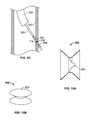
- FIGS. 2A-2Billustrates deployment of an implant of the present invention.
- FIGS. 4A-4Care views of an additional variation of the invention.
- FIGS. 5A-5C and 6 A- 6 Billustrate a variation of the invention having control members in an alternating fashion about the implant and additional control members at an end of the implant.
- FIGS. 7A-7Cillustrate a variation of the invention where the proximal portion and the distal portion are of differing sizes.
- FIGS. 8A-8Billustrate additional variations of delivering an bioactive agent with the present invention.
- FIGS. 9A-9Cillustrate variations of the present invention having visualization marks or features.
- FIG. 11A-11Billustrate histology samples comparing conventional devices and an implant of the present invention.
- FIG. 12illustrates pre-clinical data of an animal model comparing conventional devices, coronary drug eluting stents, and implants of the present invention.
- Described hereinare devices (and methods) for improving the gas exchange in the lung.
- methods and devicesare described that serve to maintain and extend the patency of collateral openings or channels through an airway wall so that air is able to pass directly out of the lung tissue and into the airways. This facilitates exchange of oxygen into the blood and decompresses hyper inflated lungs.
- channelit is meant to include, but not be limited to, any opening, hole, slit, channel or passage created in the tissue wall (e.g., airway wall).
- the channelmay be created in tissue having a discrete wall thickness and the channel may extend all the way through the wall. Also, a channel may extend through lung tissue which does not have well defined boundaries such as, for example, parenchymal tissue.
- FIGS. 1A-1Care simplified illustrations of various states of a natural airway and a blood gas interface found at a distal end of those airways.
- FIG. 1Ashows a natural airway 100 which eventually branches to a blood gas interface 102 .
- the airwaycomprises an internal layer of epithelial pseudostratified columnar or cuboidal cells. Mucous secreting goblet cells are also found in this layer and cilia may be present on the free surface of the epithelial lining of the upper respiratory airways. Supporting the epithelium is a loose fibrous, glandular, vascular lamina basement including mobile fibroblasts. Deep in this connective tissue layer is supportive cartilage for the bronchi and smooth muscle for the bronchi and bronchioles.
- FIG. 1Billustrates an airway 100 and blood gas interface 102 in an individual having COPD.
- the obstructions 104impair the passage of gas between the airways 100 and the interface 102 .
- FIG. 1Cillustrates a portion of an emphysematous lung where the blood gas interface 102 expands due to the loss of the interface walls 106 which have deteriorated due to a bio-chemical breakdown of the walls 106 .
- a constriction 108 of the airway 100It is generally understood that there is usually a combination of the phenomena depicted in FIGS. 1 A- 1 C. Often, the states of the lung depicted in FIGS. 1B and 1C may be found in the same lung.
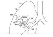
- FIG. 1Dillustrates airflow in a lung 118 when implants 200 are placed in collateral channels 112 .
- collateral channels 112located in an airway wall
- the inventionis not limited to the number of collateral channels which may be created, it is to be understood that 1 or 2 channels may be placed per lobe of the lung and perhaps, 2-12 channels per individual patient.
- the inventionincludes the creation of any number of collateral channels in the lung. This number may vary on a case by case basis. For instance, in some cases in an emphysematous lung, it may be desirable to place 3 or more collateral channels in one or more lobes of the lung.
- FIGS. 2A-2Billustrate deployment of a variation of an implant 200 of the present invention.
- the implant 200is well suited for maintaining an opening in a wall of a body organ.
- the illustrationdepicts the implant 200 as deployed into a collateral channel 112 formed in a wall of an airway 100 .
- a delivery device 300 carrying the implant 200is advanced to the site and inserted into the channel 112 .
- the delivery device 300may optionally be constructed to also form the channel 112 .
- the delivery device 300may extend from an access device such as an endoscope or bronchoscope 302 , or it may be directly advanced to the site.
- FIG. 2Billustrates deployment of the implant 200 in the airway wall 100 .
- an expandable membersuch as a balloon 304 , expands the implant 200 into a non-cylindrical shape that is able to sandwich or capture the tissue 100 between the expanded portions of the implant 200 .
- the implant 200forms a non-cylindrical (e.g., a “grommet” or “hour-glass”) shape that is suited, when used in the airways, for limiting movement of the implant 200 within the tissue opening and securing the implant 200 about the perimeter of the tissue opening in the airway wall.
- the implant 200expands in the mid portion and flares at the ends to retain itself within the opening in the airway wall.
- the grommet shape of the implant 200extends only minimally into the airway.
- the implantis suited for placement about an opening in the wall of an organ.
- the implantis suited to placement in an organ having a thin wall.
- airway wall thicknessis fairly proportional to the diameter of the airway lumen by approximately a factor of 1 ⁇ 6.
- the inventionis not limited to use in any particular sized airway, on average the implant is placed in airways ranging from 3 mm to 15 mm in diameter with respective airway wall thicknesses of 0.5 mm to 2.5 mm. Therefore, in many variations of the invention, the grommet or hour-glass shape will be suitable to retain itself on the relatively thin airway wall tissue.
- a variation of the implant 200shrinks in axial length as it secures itself within the channel.
- Shrinking in axial lengthmay also provide additional benefit as it reduces the length of the implant 200 that extends into the airway. This reduction in length may prevent unwanted tissue damage to the airway wall and/or occlusion of the airway.
- the implant 200must not only capture relatively thin tissue, but must also maintain a minimum internal diameter to allow sufficient air flow. For example, a fewer number of implants may be used given a sufficiently large diameter. In such cases it is undesirable for the implant 200 to constrict in internal diameter as it forms the non-cylindrical shape.
- the entire implantis expandable, but a portion of the implant 200 expands to a greater amount as compared to a remainder of the implant. Such a configuration allows for the entire implant 200 to expand while still forming a non-cylindrical shape.
- the implants of the present inventioninclude a support member and a composition that maintain patency of the channel.
- Variations of the inventioninclude support members selected from a mesh or woven structure either of which are comprised of a metal alloy(e.g., stainless steel, titanium, a shape-memory alloy, etc.), a polymer, a ceramic, or a combination thereof.
- the support memberprovides a structure that mechanically maintains patency of the channel as well as provides a delivery means for the composition or other substances as described herein. It is specifically noted that while the variations of the present invention are suited for use in the airways, the invention is not limited to such applications. Rather, the variations of the present invention may be used in various applications as appropriate.
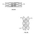
- FIG. 3Aillustrates a planar view of a variation of an implant 200 where the support member 202 is in the unexpanded shape.
- the support member 202comprises a plurality of struts or members and has a proximal portion 204 , a distal portion 206 , and a mid-portion 208 therebetween.
- a composition 212is located on the implant.
- the composition 212may encapsulate the support member 202 , or it may be located on an exterior or interior surface. Alternatively, it may be located between or within the intensities of the support member 202 .
- FIG. 3Aalso illustrates the struts or members (i.e., the extension member) on the proximal and distal portions 204 , 206 as being tapered. Because the proximal and distal portions 204 , 206 expand significantly, there is a propensity for the composition to tear at these locations. The tapering configuration is helpful to prevent tearing of the composition 212 during expansion as it allows for more material between adjacent struts.
- the variation of the support member 202 illustrated in FIG. 3Aincludes control segments 210 which permit the support member 200 to assume a desired shape upon deployment. As will be described herein, the control segments 210 limit expansion of a portion of the implant (in this case the mid portion 208 ) as well as enable the implant to expand in a uniform manner. Although FIG. 3A illustrates the entire implant 200 as being covered by the composition 212 , it is noted that the composition 212 may alternatively extend over portions of the support member 202 .
- FIG. 3Billustrates a side view of the implant 200 after expansion.
- the control segments 210restrain expansion at the mid portion 208 . Because the proximal and distal portions 204 , 206 are not restrained, upon expansion, the implant 200 forms a grommet shape as the control segments 210 unfold.
- FIG. 3Cillustrates a front view of an expanded implant 200 .
- FIG. 3Cshows the passageway having a hexagonal cross section.
- the cross-sectionis not limited to such a shape.
- the cross sectionmay be circular, oval, rectangular, elliptical, or any other multi-faceted or curved shape. Because of its shape, the implant 200 will have a variable diameter.
- the inner diameter (D 1 ) of the center sectionwill be a minimum expanded diameter and the diameter of the implant at the expanded ends (D 2 ) will be a maximum expanded diameter.
- the inner diameter (D 1 ) when deployed,may range from 1 to 10 mm and perhaps, from 2 to 5 mm.
- implants 200have a sufficiently small delivery state diameter so that they are delivered to the channel having a sufficiently small diameter profile but a relatively large axial length.
- the implant's 200 minimum (internal) diameteris greater than or equal to its axial expanded length.
- This particular configurationprovides several benefits. During deployment having a sufficient axial length permits proper centering of the implant 200 when inserted into the collateral channel, where improper centering could result in a inadequate placement about the airway walls. Upon expansion, as the implant 200 decreases in length it is able to grommet about the airway walls, thereby minimizing the amount of the structure that extends into the airway lumen. Simultaneously, maximizing the minimum internal expanded diameter (e.g., the diameter of the implant at the mid portion 208 ) allows for an implant that permits a sufficient amount of airflow.
- FIGS. 4A-4Cillustrate additional variations of implants 200 of the present invention. It is noted that in FIG. 4A , as in many additional figures below, the composition is not illustrated for sake of clarity.
- FIG. 4Ashows a side view of a implant 200 in an un-deployed state. The variation shown in FIG. 4A is similar to that shown in FIG. 3 with the exception of that the proximal and distal portions 204 , 206 are not tapered.
- FIG. 4Billustrates a side view of the implant 200 of FIG. 4A when expanded.
- the opposing ends of the implant 200may have a V, U, or similar shape.
- the angles A 1 , A 2may vary and may range from, for example, 30 to 150 degrees, 45 to 135 degrees and perhaps from 30 to 90 degrees.
- the angle A 1may be different than angle A 2 .
- the angle corresponding to each proximal extension membermay be different or identical to that of another proximal extension member.
- the angle corresponding to each distal extension membermay be different or identical to that of another distal extension member.
- FIG. 4Balso illustrates the implant 200 having a length L that decreases upon expansion of the implant 200 .
- the length of the implants of the present inventionwill depend upon their intended site of implantation. Variations of implants may have lengths ranging from between 2-20 mm. Furthermore, although the figures illustrate the proximal and distal portions of the implant as being symmetric about its center, the implant is not limited to such a configuration.
- the implant of the present inventionmay have any number of extension members on each end device.
- the number of extension members on each endmay range from 2-10.
- the number of proximal extension membersmay differ from the number of distal extension members for a particular implant.
- the extension membersmay be symmetrical or non-symmetrical about the center section.
- the proximal and distal extension membersmay also be arranged in an in-line pattern or an alternating pattern.
- the extension members or the center sectionmay also contain barbs or other similar configurations to increase adhesion between the implant and the tissue.
- the extension membersmay also have openings to permit tissue in-growth for improved retention.
- Variations of the implant 200also includes diametric-control segments, tethers, or leashes 210 to control and limit the expansion of the a portion of the implant 200 when expanded.
- the shape of the center-control segment 210typically bends, when the implant radially expands, until it is substantially straight or unfolded.
- Such a center-control segment 210may be circular or annular shaped in its folded or unexpanded shape. However, its shape may vary widely and it may have, for example, an arcuate, semi-circular, v-shape, u-shape, s-shape, sinusoidal shape, or other type of shape which limits the expansion of the implant upon unfolding.
- the control members 210assist the implant 200 in assuming a uniform non-cylindrical expanded shape.
- the implant 200there will be variation in the amounts of expansion of various cells (i.e., where a cell is typically defined by an area surrounded by a number of joined struts—as an example refer to FIG. 4C , the shaded portion representing the cell 216 ) of the implant 200 . If one cell expands at an increased amount relative to the remaining cells, once the control member 210 fully unfolds, the cell will be unable to further expand. Thus, the expansion force, as applied by the balloon, is re-directed to a remaining part of the implant 200 . It should be noted that while the control members substantially straighten, there may be a residual bend or “kink” in the control member when expanded.
- the center-control segments 210may bend, unfold, straighten, or otherwise deform until they maximize their length (i.e., unfold to become substantially straight) such as the center-control segments 210 shown in FIG. 3B .
- the inventionis not so limited and other types of center-control segments may be employed.
- FIG. 6Aillustrates an implant 200 similar to that of FIG. 3 with additional control segments 214 located at both ends of the implant 200 .
- FIG. 6Billustrates the implant 200 of 6 A in an expanded state.
- the control segmentsare illustrated to have equal lengths, any length may be selected. For example, adjacent control segments may have different lengths, or opposing control segments (e.g., those located on opposing ends) may have different lengths.
- FIGS. 7A and 7Bdiffer from previously described implants as the proximal 204 and distal 206 portions are of different sizes.
- the larger sized portion 206may be useful in separating parenchymal tissue or providing a larger anchoring structure when implanted (as shown in FIG. 7B .)
- control segmentsmay be added or mounted to the implant or alternatively, they may be integral with the implant. That is, the control segments may be part of the implant rather than separately joined to the implant with adhesives or welding, for example.
- the control segmentsmay also be mounted exteriorly or interiorly to the members to be linked. Additionally, sections of the implant may be removed to allow areas of the implant to deform more readily. These weakened areas provide another approach to control the final shape of the deployed implant. Details for creating and utilizing weakened sections to control the final shape of the deployed implant may be found in U.S. Pat. Ser. No. 09/947,144 filed on Sep. 4, 2001 which is hereby incorporated by reference in its entirety.
- the implant described hereinmay be manufactured by a variety of manufacturing processes including but not limited to laser cutting, chemical etching, punching, stamping, etc.
- the implantmay be formed from a tube that is slit to form extension members and a center section between the members.
- One variation of the implantmay be constructed from a metal tube, such as stainless steel, 316L stainless steel, titanium, tantalum, titanium alloy, nitinol, MP35N (a nickel-cobalt-chromium-molybdenum alloy), etc.
- the implantmay be formed from a rigid or elastomeric material that is formable into the configurations described herein.
- the implantmay be formed from a cylinder with the passageway being formed through the implant.
- the implantmay also be formed from a sheet of material in which a specific pattern is cut. The cut sheet may then be rolled and formed into a tube.
- the materials used for the implantcan be those described above as well as a polymeric material, a biostable or implantable material, a material with rigid properties, a material with elastomeric properties, or a combination thereof. If the implant is a polymeric elastic tube (e.g. a thermoplastic elastomer), the implant may be extruded and cut to size, injection molded, or otherwise formed.
- the implants described hereinmay be comprised of a shape memory alloy, a super-elastic alloy (e.g., a NiTi alloy), a shape memory polymer, or a shape memory composite material.
- the implantmay be constructed to have a natural self-assuming deployed configuration, but is restrained in a pre-deployed configuration. As such, removal of the restraints (e.g., a sheath) causes the implant to assume the deployed configuration.
- a implant of this typecould be, but is not limited to being, comprised from an elastic polymeric material, or shape memory material such as a shape memory alloy. It is also contemplated that the implant could comprise a shape memory alloy such that, upon reaching a particular temperature (e.g., 98.5° F.), it assumes a deployed configuration.
- the implant described hereinmay be formed of a plastically deformable material such that the implant is expanded and plastically deforms into a deployed configuration.
- the implantmay be expanded into its expanded state by a variety of devices such as, for example, a balloon catheter.
- the implant's surfacemay be modified to affect tissue growth or adhesion.
- an implantmay comprise a smooth surface finish in the range of 0.1 micrometer to 0.01 micrometer. Such a finish may serve to prevent the implant from being ejected or occluded by tissue overgrowth.
- the surfacemay be roughened or porous.
- the implantmay also comprise various coatings and polymeric layers as discussed below.
- the implants of the present inventionmay include a composition or polymeric layer that includes a bio-active substance or combination of bioactive substances.
- a composition or polymeric layerthat includes a bio-active substance or combination of bioactive substances.
- One purpose of the compositionis to assist in modifying the healing response as a result of the trauma to lung tissue resulting from creation of the collateral channel.
- the compositionmay also serve other purposes as well.
- the compositionmay assist in controlling of bacteria, prevent irritation of the tissue near the implant, or may carry additional bio-active substances.
- lung tissueis intended to include the tissue lining the airway, the tissue beneath the lining, and the tissue within the lung but exterior to the airway (e.g., lung parenchyma.)
- tissue lining the airwaye.g., the tissue beneath the lining
- tissue within the lung but exterior to the airwaye.g., lung parenchyma.
- FIGS. 3A and 3Billustrate an example an implant 200 having a composition 212 .
- the compositionmay comprise a polymeric layer which acts as a carrier for various bioactive or other agents as described herein.
- the polymeric layermay function as a tissue barrier to inhibit growth of tissue into the conduit/implant.
- the support membermay be fabricated from a polymeric material having the bio-active substance incorporated directly therein.
- the composition 212prevents tissue in-growth from occluding the collateral channel or passage of the implant 200 .
- the polymeric layer 212may coaxially cover the center section from one end to the other or it may only cover one or more regions of the implant 200 .
- the composition 212may completely or partially cover the implant 200 .
- the composition 212may be located about an exterior of the implant's surface, about an interior of the implant's surface.
- the composition 212may be located within an opening or pocket 220 in the support structure 202 of the implant.
- the pocket 220will have a barrier (e.g., polymeric or other porous material) that either degrades to allow the composition or bioactive substance to be delivered from the implant, or acts as a diffusible barrier to deliver the composition or bioactive substance.
- a barriere.g., polymeric or other porous material
- compositionshould be selected to accommodate the significant expansion of the implant.
- polymersinclude, but are not limited to, thermoplastic polymers, thermoset polymers, acrylate polymers, a blend of acrylate-methacrylate polymers, silicone elastomers, urethane elastomers, ethylene vinyl acetate polymers, polyethylene, polypropylene, PLA-PGA, PLA, PGA, polyortho-ester, polycapralactone, polyester, hydrogels, polystyrene, co-polymers of styrene-isobutylene-styrene, and combinations or blends thereof.
- bioabsorbable polymersinclude but are not limited to poly(L-lactic acid), polycaprolactone, poly(lactide-co-glycolide), poly(hydroxybutyrate), poly(hydroxybutyrate-co-valerate), polydioxanone, polyorthoester, polyanhydride, poly(glycolic acid), poly(D,L-lactic acid), poly(glycolic acid-co-trimethylene carbonate), polyphosphoester, polyphosphoester urethane, poly(amino acids), cyanoacrylates, poly(trimethylene carbonate), poly(iminocarbonate), copoly(ether-esters) (e.g., PEO/PLA), polyalkylene oxalates, polyphosphazenes and biomolecules such as fibrin, fibrinogen, cellulose, starch, collagen and hyaluronic acid. Also, biostable polymers with a relatively low chronic tissue response such as polyurethanes, silicones, fluorosilicon
- polystyrene resinsexamples include but are not limited to polyolefins, polyisobutylene and ethylene-alphaolefin copolymers; acrylic polymers and copolymers, vinyl halide polymers and copolymers, such as polyvinyl chloride; polyvinyl ethers, such as polyvinyl methyl ether; polyvinylidene halides, such as polyvinylidene fluoride and polyvinylidene chloride; polyacrylonitrile, polyvinyl ketones; polyvinyl aromatics, such as polystyrene, polyvinyl esters, such as polyvinyl acetate; copolymers of vinyl monomers with each other and olefins, such as ethylene-methyl methacrylate copolymers, acrylonitrile-styrene copolymers, ABS resins, and ethylene-vinyl acetate copolymers; polyamides, such as Nylon 66 and polycaprolact
- the coatingsmay be applied, for example, by either painting, dip coating, molding, spin-coating, transfer molding or liquid injection molding.
- the polymeric layermay be a tube of a material and the tube is placed either over and/or within the implant. The polymeric layer may then be bonded, crimped, heated, melted, shrink fitted or fused to the implant.
- the polymeric layermay also be tied to the implant with a filament of, for example, a suture material.
- Still other techniques for attaching the polymeric layerinclude: solvent swelling applications and extrusion processes; wrapping a sheet of material about the implant, or placing a tube of the material about the implant and securing the tube to the implant.
- the polymeric layermay be secured on the interior of the implant by positioning a sheet or tube of material on the inside of the center section and securing the material therein.
- the compositionmay also be formed of a fine mesh with a porosity or treatment such that tissue may not penetrate the pores.
- a ChronoFlexTM DACRON® or TEFLON® meshhaving a pore size of 100-300 microns may be saturated with collagen or another biocompatible substance.
- This constructmay form a suitable polymeric layer.
- the meshmay be coaxially attached to a frame such as the open frame structures disclosed above. Still other suitable frames include a continuous spiral metallic or polymeric element.
- the bio-active substance or combination of bioactive substancesis selected to assists in modifying the healing response as a result of the trauma to the lung tissue resulting from creation of the collateral channel.
- lung tissueis intended to include the tissue lining the airway, the tissue beneath the lining, and the tissue within the lung but exterior to the airway (e.g., lung parenchyma.)
- the purpose of modifying the healing responseis to further extend the patency of the channel or implant to increase the duration which trapped gasses may exit through the implant into the airways.
- antiproliferative agentis intended to include those bioactive substances that directly modify the healing response described herein.
- bioactive substancesmay be used alone or in combination with the devices described herein.
- bioactive substancesinclude, but are not limited to, antimetabolites, antithrobotics, anticoagulants, antiplatelet agents, thorombolytics, antiproliferatives, antinflammatories, agents that inhibit hyperplasia and in particular restenosis, smooth muscle cell inhibitors, growth factors, growth factor inhibitors, cell adhesion inhibitors, cell adhesion promoters and drugs that may enhance the formation of healthy neointimal tissue, including endothelial cell regeneration.
- bioactive agentsinclude but are not limited to analgesics, anticonvulsives, anti-infectives (e.g., antibiotics, antimicrobials), antineoplastics, H2 antagonists (Histamine 2 antagonists), steroids, non-steroidal anti-inflammatories, hormones, immunomodulators, mast cell stabilizers, nucleoside analogues, respiratory agents, antihypertensives, antihistamines, ACE inhibitors, cell growth factors, nerve growth factors, anti-angiogenic agents or angiogenesis inhibitors (e.g., endostatins or angiostatins), tissue irritants (e.g., a compound comprising talc), poisons (e.g., arsenic), cytotoxic agents (e.g., a compound that can cause cell death), various metals (silver, aluminum, zinc, platinum, arsenic, etc.), epithelial growth factors or a combination of any of the agents disclosed herein.
- analgesicse
- agentsinclude pyrolitic carbon, titanium-nitride-oxide, taxanes, fibrinogen, collagen, thrombin, phosphorylcholine, heparin, rapamycin, radioactive 188Re and 32P, silver nitrate, dactinomycin, sirolimus, everolimus, Abt-578, tacrolimus, camptothecin, etoposide, vincristine, mitomycin, fluorouracil, or cell adhesion peptides.
- Taxanesinclude, for example, paclitaxel, 10-deacetyltaxol, 7-epi-10-deacetyltaxol, 7-xylosyl-10-deacetyltaxol, 7-epi-taxol, cephalomannine, baccatin III, baccatin V, 10-deacetylbaccatin III, 7-epi-10-deacetylbaccatin III,docetaxel.
- bioactive materials having other functionscan also be successfully delivered in accordance with the present invention.
- an antiproliferative agentsuch as methotrexate will inhibit over-proliferation of smooth muscle cells and thus inhibit restenosis.
- the antiproliferativeis desirably supplied for this purpose until the tissue has properly healed.
- localized delivery of an antiproliferative agentis also useful for the treatment of a variety of malignant conditions characterized by highly vascular growth.
- an implantsuch as a implant could be placed in the surgically created channel to provide a means of delivering a relatively high dose of the antiproliferative agent directly to the target area.
- a vasodilatorsuch as a calcium channel blocker or a nitrate may also be delivered to the target site.
- the agentmay further be a curative, a pre-operative debulker reducing the size of the growth, or a palliative which eases the symptoms of the disease.
- a curativefor example, tamoxifen citrate, Taxol® or derivatives thereof.
- Proscar®, Hytrin®, or Eulexin®may be applied to the target site as described herein.
- Variations of the inventionmay also include fibrinolytics such as tPA, streptokinase, or urokinase, etc.
- fibrinolyticsprevent or reduce the accumulation of fibrin within the opening. Accumulation of fibrin in the opening may result from inflammation of the tissue.
- the fibrinmay form a structure which makes it easier for tissue to grow into the opening using the fibrin structure as a framework.
- Use of fibrinolyticseither topically, locally, or on the implant, serves to remove or hinder the network of fibrin from forming within the opening (or implant) and therefore aids in modifying the healing response.
- the poisonous agentshould be delivered locally or only be effective locally.
- One method for delivering the bioactive agent locallyis to associate the bioactive agent with an implant.
- the implants described hereinmay include a bioactive substance or medicine deposited onto the interior, the exterior, or both the interior and exterior surfaces of the implant. The bioactive substance may remain on the implant so that it does not leach. Cells that grow into the surgically created channel contact the poison and die.
- the bioactive agentmay be configured to gradually elute as discussed below.
- the implantWhen used in the lungs, the implant modifies the healing response of the lung tissue (e.g., at the site of newly created hole/channel) for a sufficient time until the healing response of the lung tissue subsides or reduces such that the hole/channel becomes a persistent air path.
- the implant and bioactive substancewill modify the healing response for a sufficient time until the healing response is reduced and, from a visual observation, the body treats the opening essentially as a natural airway passage rather than as an injury to the airway wall.
- FIGS. 11A-11Bshow histology from animal models.
- the histologyis a cross sectional slice of the airway wall 110 and lung parenchyma 116 .
- the collateral channel 112was created in the airway wall 110 and extended into the lung parenchyma 116 .
- the implant(which was removed for histology and is not shown) was placed in the channel 112 so as to create an airflow path (as demonstrated by the arrows 114 ) from the lung parenchyma 116 through the airway wall 110 .
- FIG. 11Aillustrates a histology sample from a site two weeks subsequent to the creation of a channel and implantation with a device.
- the deviceincluded a polymeric coating but no bio-active substance.
- This sitewas also given a single local treatment of a bioactive substance (mitomycin) subsequent to creation of the channel 112 .
- a bioactive substancemitomycin
- the healing process of the lung tissuealready caused a considerable amount of fibrosis 120 between the channel 112 and lung parenchyma 116 . From the figure, the fibrosis appears as a darker tissue that is adjacent to the lung parenchyma 116 . The presence of this fibrosis 120 strongly suggests that air would not be able to flow from the lung parenchyma 116 through the channel 112 .
- FIG. 11Billustrates a histology sample from a site 18 weeks subsequent to the creation of a channel and implantation with an implant of the present invention (an example of which is discussed below.) As evident from the figure, the channel 112 remained significantly unobstructed with only a minimal discontinuous layer of fibrosis 120 .
- the implantprovides a steady release rate of bio-active substance as well as has a sufficient amount of available bio-active substance to modify the healing response of the lung tissue.
- lung tissueis intended to include the tissue lining the airway, the tissue beneath the lining, and the tissue within the lung but exterior to the airway (e.g., lung parenchyma.) Such a delivery profile allows for a concentration gradient of drug to build in these tissues adjacent to the delivery site of the implant.
- the concentration gradientaffects the healing response of the lung tissue so that the implant does not become occluded as a result of the healing response. Because the implant is often placed in the airway wall it is exposed to the healing process of the multiple tissues. Providing a sufficient amount of bio-active substance allows for the formation of a concentration of the bio-active substance across these various tissues. In one variation of the invention it is believed that the fluids from these tissues enter into the composition layer of the device. The fluids then combine with the bio-active substances and migrate out of the composition layer to settle into the lung tissue. A concentration gradient forms when the drug ‘saturates’ local tissue and migrates beyond the saturated tissues. Furthermore, by providing a sufficient delivery rate, the healing response may be affected or suppressed during the critical time immediately after the wounding caused by creation of the collateral channel when the healing response is greatest.
- the solubility parameter of the polymermust be matched with the bio-active substance to provide an acceptable slow elution rate from the polymer.
- the polymer itselfmust be selected to have the proper attributes, such as a proper diffusion coefficient (to slow fluid entering and departing from the implant), and proper mechanical expansion properties (to allow for the significant expansion of the polymer to accommodate formation of the grommet shape.)
- the solubility parameteris defined as the square root of the cohesive energy of the molecules in a compound.
- the level of control that a polymer has over the elution of a drugis the difference between the solubility parameters of the polymer and the solubility parameter of the drug.
- paclitaxelis a taxane that is regarded as a microtubule stabilizer.
- the benefits of a microtubule stabilizing substance for use in vascular drug eluting stentsis discussed, for examples, in U.S. Pat. No. 5,616,608 to Kinsella et al. This type of drug operates to enhance microtubule polymerization which inhibits cell replication by stabilizing microtubules in spindles which block cell division.
- the implant for use in the present inventionmay use microtubule destabilizing substances such as taxenes (e.g., paclitaxel) as well as those microtubule destabilizing substances that are believed to promote microtubule disassembly in preventing cell replication.
- destabilizing substancesinclude, but are not limited to vincristine, vinblastine, podophylotoxin, estramustin, noscapine, griseofulvin, dicolmarol, a vinca alkaloid, and a combination thereof.
- the exterior surface of the implantmay be treated via etching processes or with electrical charge to encourage binding of the bioactive substance of the implant.
- the exterior surfacemay also be roughened to enhance binding of the medicine to the surface as discussed in U.S. Patent Application Publication No. 2002/0098278. See also U.S. Patent Application Publication Nos. 2002/0071902, 2002/0127327 and U.S. Pat. No. 5,824,048 which discuss various techniques for coating medical implants.
- the implantmay comprise a frame or body with a bioactive matrix disposed or otherwise associated therewith, the invention is not so limited.
- the support memberis formed from a polymer and the composition is joined to the polymeric support member.
- the bioactive substancesmay be placed directly onto the polymeric support member.
- Adverse reactionsinclude, but are not limited to, granulation, swelling, and mucus overproduction. These substance may also be inhaled, injected, orally applied, topically applied, or carried by the implant. These substances may include anti-imflammatory, infection-fighting substances, steroids, mucalytics enzymes, and wound healing-accelarating substances. Examples of these substances include but are not limited to, acetylcysteine, albuterol sulfate, ipratropium bromide, dornase alfa, and corticosteroids.
- FIG. 12illustrates data from a pre-clinical animal model evaluating the wound healing response, under pre-clinical protocol (QT-305), using an implant w/o any antiprliferative substance, a paclitaxel coronary Stent (manufactured by Boston Scientific under the name Taxus®), and a sirolimus coronary stent (manufactured by Johnson & Johnson under the name of Cypher®)
- QT-345 and QT-362were conducted.
- the implant w/o intiproliferative substance, the paclitaxel coronary stent, and the sirolimus coronary stentreduced to at least 50% patency without stabilization (i.e., the determination was made that 100% closure would occur.)
- the chartindicates closure of these devices given a criteria that at least half of the implanted devices closed with tissue and the trend indicated that full closure of the devices would occur.
- the implants according to the present inventionmaintained 88% patency of the openings @ 12 weeks (QT-362) and 69% patency @ 18 weeks (QT-345). In both of these latter cases, repeated inspection determined that the healing response (as evidenced by the closure rate) of the implants stabilized.
- QT-3622 specimens maintained 100% patency while 1 specimen maintained 75% patency. For QT-345, no decline in patency occurred for the last 6 weeks of the trial.
- the implant of the present inventionwhen placed into an airway wall, is usually placed using a bronchoscope under direct visualization.
- the direct visualizationonly permits viewing of the interior of the airway and the care must be taken to place the implant such that during expansion, the implant properly deploys about the airway wall.
- caremust be taken not to advance the implant/balloon catheter too far into the opening into the airway wall. Improper advancing of the implant/balloon could potentially result in a pneumothorax or pneumomediastinum.
- the implant 200may also include a visualization mark 218 .
- the visualization marker 218is visually apparent during a procedure and gives the medical practitioner an indication when the implant/balloon is advanced to the proper location. In this manner, the visualization mark 218 facilitates alignment and deployment of the implants into collateral channels.
- the visualization mark 218may be a ring of biocompatible polymer and may be selected to provide contrast so that it may be identified as the medical practitioner views the device through a endoscope or bronchoscope.
- the bronchoscopewill usually contain a light-source that illuminates the target area. Therefore, the visualization mark may be something that reflects or refracts the light in a different manner from the remainder of the implant.
- the visualization markmay be the same color as the remainder of the device, or partially transparent, or entirely transparent, but is identifiable because the mark reflects or refracts light differently than the remainder of the device.
- the visualization featuremay protrude from the center section or it may be an indentation(s).
- the visualization markmay also be a ring, groove or other physical feature on the implant.
- the visualization featuremay be continuous or comprise discrete segments (e.g., dots of segments).
- the visualization featuremay be made using a number of techniques.
- the markis a ring formed of silicon and is white.
- the polymeric ringmay be spun onto the polymeric layer.
- a clear silicon barriermay be coated onto the implant such that it coaxially covers the implant.
- a thin ring of white materialsuch as a metal oxide suspended in clear silicon may be spun onto the silicon coating.
- another coating of clear siliconmay be applied to coat the white layer.
- the implantthus may include upwards of 1-3 layers including a polymeric layer, a visualization mark layer, and a clear outer covering.
- the markis a ring formed by of silicon and is black.
- the markis a ring formed by suspending gold particulates in the polymer as shown in FIG. 9A .
- the visualization markis made to stand out when viewed with, for example, an endoscope.
- the implantsmay also have additional imaging enhancing additives to increase non-direct imaging, such as flouroscope or radioscope viewing. It is also contemplated that other elements of the implant can include visualization features such as but not limited to the extension members, polymeric layer, control segments, etc.
- composition layere.g., the polymer turns white .
- This colorationobscures the support member structure in the layer making it difficult to identify the edges and center of the support member or implant.
- placement of the implantmay depend upon positioning the center of the implant within the opening in tissue. If the support member structure is identifiable, then one is able to visually identify the center of the implant.
- the composition colorsobscures the support member or renders the implant otherwise opaque, it may become difficult to properly place the device. This may be especially true when the composition layer extends continuously over the support member.
- a variation of the inventionincludes e delivery device for delivering an expandable implant (such as those described herein and in the cases referenced herein), where the delivery device includes an expandable member having an expandable implant located about the expandable member. Where the implant and the expandable member are of different visually identifiable colors or shades such that the distinction is easy to identify under endoscopic or bronchoscopic viewing.
- a balloon catheterhas a colored sleeve 306 located about the ballon.
- the sleeve 306comprises a visually identifiable color where selection of the colors should ease identification of the implant in an endoscopic visualization system (e.g., blue or a similar color that is not naturally occurring within the body.)
- the implantis placed about the sleeve 306 where the proximal and distal areas of the implant would be identifiable by the difference in color.
- the sleeve 306may be fashioned from any expandable material, such as a polymer.
- the sleeve 306may also provide an elastic force to return the balloon to a reduced profile after expansion of the balloon. Such a system allows for identification without affecting the properties of the implant.
- variations of the inventioninclude coloring the balloon itself, or other expandable member, a color that meets the above criteria.
- the visualization markmay comprise providing a contrast between the implant and a delivery catheter.
- the implantappears mostly white and while mounted on a contrasting color inflation balloon.
- the implantwould be placed over a blue deflated balloon catheter.
- the proximal and distal areas of the implantwould be flanked by the deflated blue balloon, thus giving the appearance of a distinct distal and proximal end of the implant.
- Thiswould allow a physician to place the implant properly by using the blue flanks as a guide for placing the center white portion in the tissue wall.
- a colored flexible sheath covering the balloonwould also suffice.
- inventive featuresare not limited as such.
- the featuresmay be incorporated into any system where placement of an implant under direct visualization requires clear identification of the implant regardless of the whether the implant is opaque or colored.
- an implantmay include a valve 224 .
- the valve 224may be positioned such that it permits expiration of gas from lung tissue but prevents gas from entering the tissue.
- the valve 224may be placed anywhere within the passageway of the implant.
- the valve 224may also be used as bacterial in-flow protection for the lungs.
- the valve 224may also be used in combination with a bioactive or biostable polymeric layer/matrix and the polymeric layer may be disposed coaxially about the implant.
- Various types of one way valvesmay be used as is known to those of skill in art.
- Implantscomprising stainless steel mesh frame fully encapsulated with a composition comprising silicon (as described below) and paclitaxel were implanted in several canine models.
- the compositioncomprised approximately a 9% paclitaxel to silicone ratio with approximately 400 micrograms of paclitaxel per implant. Measurements found that approximately 30% of the paclitaxel released after 60 days. In general, for implants with the paclitaxel/silicon composition, observations of chronic inflammation, epithelial metaplasia and fibrosis were very mild.
- polymers with solubility parameter between 5-25 (Mpa) ⁇ 1 ⁇ 2were believed to provide sufficient elution rates.
- the polymer used in the example devicehas good diffusivity for lipophilic drug (such as paclitaxel) because the side methyl group on the silicone may be substituted with more lipophilic hydrocarbon molecules containing vinyl group or groups for polymerization by platinum catalyst.
- composition for the examplemay be as follow: polymer part: polydimethylsiloxane, vinyldimethyl terminated, any viscosity; and/or polydimethylsiloxane, vinyldimethyl terminated, any viscosity.
- the cross-linker partpolydimethylsiloxane, any viscosity; and/or polydimethylsiloxane, any viscosity.
- Platinum catalyst part and/or cross-linker partplatinum; and/or platinum-divinyltetramethyldisiloxane complex in xylene, 2-3% Pt; and/or platinum-divinyltetramethylsiloxane complex in vinyl terminated polydimethylsiloxane, 2-3% Pt; and/or platinum-divinyltetramethyldisiloxane complex in vinyl terminated polydimethylsiloxane, ⁇ 1% Pt; platinum-Cyclovinylmethylsiloxane complex, 2-3% Pt in cyclic methyl siloxane.
- the hydrocarbon side chain off the silicon back bonemakes this polymer system unique and may result in a “zero-order”—like release profile.
- the amount of vinyl siloxane cross-linkermay determine the rate of the drug release and diffusivity of the polymer to the drug.
- paclitaxela release profile was found to be acceptable with a polymer system consisting of polydimethylsiloxane vinyl terminated at various viscosity and a range of platinum-mono, di, tri and/or tetrametramethyldisiloxane complex.
Landscapes
- Health & Medical Sciences (AREA)
- Life Sciences & Earth Sciences (AREA)
- Surgery (AREA)
- Molecular Biology (AREA)
- Engineering & Computer Science (AREA)
- Biomedical Technology (AREA)
- Heart & Thoracic Surgery (AREA)
- Medical Informatics (AREA)
- Nuclear Medicine, Radiotherapy & Molecular Imaging (AREA)
- Animal Behavior & Ethology (AREA)
- General Health & Medical Sciences (AREA)
- Public Health (AREA)
- Veterinary Medicine (AREA)
- Reproductive Health (AREA)
- Vascular Medicine (AREA)
- Prostheses (AREA)
Abstract
Description
Claims (32)
Priority Applications (8)
| Application Number | Priority Date | Filing Date | Title |
|---|---|---|---|
| US10/895,010US7462162B2 (en) | 2001-09-04 | 2004-07-19 | Antiproliferative devices for maintaining patency of surgically created channels in a body organ |
| US11/000,626US20050137715A1 (en) | 1999-08-05 | 2004-12-01 | Methods and devices for maintaining patency of surgically created channels in a body organ |
| JP2007522700AJP2008506507A (en) | 2004-07-19 | 2005-07-19 | Method and instrument for maintaining a surgically created channel in a body organ |
| AU2005269718AAU2005269718A1 (en) | 2004-07-19 | 2005-07-19 | Methods and devices for maintaining surgically created channels in a body organ |
| PCT/US2005/025738WO2006014731A2 (en) | 2001-09-04 | 2005-07-19 | Methods and devices for maintaining surgically created channels in a body organ |
| CA002577792ACA2577792A1 (en) | 2004-07-19 | 2005-07-19 | Methods and devices for maintaining surgically created channels in a body organ |
| EP05791123AEP1802365A4 (en) | 2004-07-19 | 2005-07-19 | Methods and devices for maintaining surgically created channels in a body organ |
| US11/421,741US20060280772A1 (en) | 2001-09-04 | 2006-06-01 | Methods and devices for maintaining surgically created channels in a body organ |
Applications Claiming Priority (4)
| Application Number | Priority Date | Filing Date | Title |
|---|---|---|---|
| US31733801P | 2001-09-04 | 2001-09-04 | |
| US10/235,240US20030070676A1 (en) | 1999-08-05 | 2002-09-04 | Conduits having distal cage structure for maintaining collateral channels in tissue and related methods |
| US10/458,085US20040073155A1 (en) | 2000-01-14 | 2003-06-09 | Methods and devices for maintaining patency of surgically created channels in tissue |
| US10/895,010US7462162B2 (en) | 2001-09-04 | 2004-07-19 | Antiproliferative devices for maintaining patency of surgically created channels in a body organ |
Related Parent Applications (2)
| Application Number | Title | Priority Date | Filing Date |
|---|---|---|---|
| US10/235,240Continuation-In-PartUS20030070676A1 (en) | 1999-08-05 | 2002-09-04 | Conduits having distal cage structure for maintaining collateral channels in tissue and related methods |
| US10/458,085Continuation-In-PartUS20040073155A1 (en) | 1999-08-05 | 2003-06-09 | Methods and devices for maintaining patency of surgically created channels in tissue |
Related Child Applications (2)
| Application Number | Title | Priority Date | Filing Date |
|---|---|---|---|
| US11/000,626Continuation-In-PartUS20050137715A1 (en) | 1999-08-05 | 2004-12-01 | Methods and devices for maintaining patency of surgically created channels in a body organ |
| US11/421,741Continuation-In-PartUS20060280772A1 (en) | 2001-09-04 | 2006-06-01 | Methods and devices for maintaining surgically created channels in a body organ |
Publications (2)
| Publication Number | Publication Date |
|---|---|
| US20050043752A1 US20050043752A1 (en) | 2005-02-24 |
| US7462162B2true US7462162B2 (en) | 2008-12-09 |
Family
ID=56290580
Family Applications (1)
| Application Number | Title | Priority Date | Filing Date |
|---|---|---|---|
| US10/895,010Expired - Fee RelatedUS7462162B2 (en) | 1999-08-05 | 2004-07-19 | Antiproliferative devices for maintaining patency of surgically created channels in a body organ |
Country Status (1)
| Country | Link |
|---|---|
| US (1) | US7462162B2 (en) |
Cited By (59)
| Publication number | Priority date | Publication date | Assignee | Title |
|---|---|---|---|---|
| US20060111704A1 (en)* | 2004-11-22 | 2006-05-25 | Rox Medical, Inc. | Devices, systems, and methods for energy assisted arterio-venous fistula creation |
| US20060206123A1 (en)* | 2004-08-27 | 2006-09-14 | Rox Medical, Inc. | Device and method for establishing an artificial arterio-venous fistula |
| US20070249985A1 (en)* | 2004-08-27 | 2007-10-25 | Rox Medical, Inc. | Device and method for establishing an artificial arterio-venous fistula |
| WO2011017440A3 (en)* | 2009-08-06 | 2011-06-30 | Mayo Foundation For Medical Education And Research | Implanting organ ports |
| US8088127B2 (en) | 2008-05-09 | 2012-01-03 | Innovative Pulmonary Solutions, Inc. | Systems, assemblies, and methods for treating a bronchial tree |
| US8137285B1 (en) | 2008-08-26 | 2012-03-20 | Rhythmlink International, Llc | Monopolar stimulation probe system |
| US8172827B2 (en) | 2003-05-13 | 2012-05-08 | Innovative Pulmonary Solutions, Inc. | Apparatus for treating asthma using neurotoxin |
| US8483831B1 (en) | 2008-02-15 | 2013-07-09 | Holaira, Inc. | System and method for bronchial dilation |
| US8734380B2 (en) | 2007-10-22 | 2014-05-27 | Uptake Medical Corp. | Determining patient-specific vapor treatment and delivery parameters |
| US8740895B2 (en) | 2009-10-27 | 2014-06-03 | Holaira, Inc. | Delivery devices with coolable energy emitting assemblies |
| US8852272B2 (en)* | 2011-08-05 | 2014-10-07 | Mitraltech Ltd. | Techniques for percutaneous mitral valve replacement and sealing |
| US8870950B2 (en) | 2009-12-08 | 2014-10-28 | Mitral Tech Ltd. | Rotation-based anchoring of an implant |
| US8911439B2 (en) | 2009-11-11 | 2014-12-16 | Holaira, Inc. | Non-invasive and minimally invasive denervation methods and systems for performing the same |
| US8932341B2 (en) | 2004-08-27 | 2015-01-13 | Rox Medical, Inc. | Method for establishing an artificial arterio-venous fistula |
| US8992604B2 (en) | 2010-07-21 | 2015-03-31 | Mitraltech Ltd. | Techniques for percutaneous mitral valve replacement and sealing |
| US8998985B2 (en) | 2011-07-25 | 2015-04-07 | Rainbow Medical Ltd. | Sinus stent |
| US9017399B2 (en) | 2010-07-21 | 2015-04-28 | Mitraltech Ltd. | Techniques for percutaneous mitral valve replacement and sealing |
| US9050076B2 (en) | 2004-11-16 | 2015-06-09 | Uptake Medical Corp. | Device and method for lung treatment |
| US9113858B2 (en) | 2006-11-13 | 2015-08-25 | Uptake Medical Corp. | High pressure and high temperature vapor catheters and systems |
| US9149328B2 (en) | 2009-11-11 | 2015-10-06 | Holaira, Inc. | Systems, apparatuses, and methods for treating tissue and controlling stenosis |
| US9398933B2 (en) | 2012-12-27 | 2016-07-26 | Holaira, Inc. | Methods for improving drug efficacy including a combination of drug administration and nerve modulation |
| US9592138B1 (en) | 2015-09-13 | 2017-03-14 | Martin Mayse | Pulmonary airflow |
| US9681952B2 (en) | 2013-01-24 | 2017-06-20 | Mitraltech Ltd. | Anchoring of prosthetic valve supports |
| US9713703B2 (en) | 2013-08-09 | 2017-07-25 | Boston Scientific Scimed, Inc. | Devices and methods for treating a lung |
| US9763657B2 (en) | 2010-07-21 | 2017-09-19 | Mitraltech Ltd. | Techniques for percutaneous mitral valve replacement and sealing |
| US9782211B2 (en) | 2013-10-01 | 2017-10-10 | Uptake Medical Technology Inc. | Preferential volume reduction of diseased segments of a heterogeneous lobe |
| USD800908S1 (en) | 2016-08-10 | 2017-10-24 | Mitraltech Ltd. | Prosthetic valve element |
| US9974651B2 (en) | 2015-02-05 | 2018-05-22 | Mitral Tech Ltd. | Prosthetic valve with axially-sliding frames |
| USD841812S1 (en) | 2017-08-03 | 2019-02-26 | Cardiovalve Ltd. | Prosthetic heart valve element |
| US10245143B2 (en) | 2011-08-05 | 2019-04-02 | Cardiovalve Ltd. | Techniques for percutaneous mitral valve replacement and sealing |
| USD845467S1 (en) | 2017-09-17 | 2019-04-09 | Uptake Medical Technology Inc. | Hand-piece for medical ablation catheter |
| US10376361B2 (en) | 2011-08-05 | 2019-08-13 | Cardiovalve Ltd. | Techniques for percutaneous mitral valve replacement and sealing |
| US10390952B2 (en) | 2015-02-05 | 2019-08-27 | Cardiovalve Ltd. | Prosthetic valve with flexible tissue anchor portions |
| US10485604B2 (en) | 2014-12-02 | 2019-11-26 | Uptake Medical Technology Inc. | Vapor treatment of lung nodules and tumors |
| US10492908B2 (en) | 2014-07-30 | 2019-12-03 | Cardiovalve Ltd. | Anchoring of a prosthetic valve |
| US10531866B2 (en) | 2016-02-16 | 2020-01-14 | Cardiovalve Ltd. | Techniques for providing a replacement valve and transseptal communication |
| US10531906B2 (en) | 2015-02-02 | 2020-01-14 | Uptake Medical Technology Inc. | Medical vapor generator |
| US10575948B2 (en) | 2017-08-03 | 2020-03-03 | Cardiovalve Ltd. | Prosthetic heart valve |
| US10856975B2 (en) | 2016-08-10 | 2020-12-08 | Cardiovalve Ltd. | Prosthetic valve with concentric frames |
| US10888421B2 (en) | 2017-09-19 | 2021-01-12 | Cardiovalve Ltd. | Prosthetic heart valve with pouch |
| US11109964B2 (en) | 2010-03-10 | 2021-09-07 | Cardiovalve Ltd. | Axially-shortening prosthetic valve |
| US11129673B2 (en) | 2017-05-05 | 2021-09-28 | Uptake Medical Technology Inc. | Extra-airway vapor ablation for treating airway constriction in patients with asthma and COPD |
| US11246704B2 (en) | 2017-08-03 | 2022-02-15 | Cardiovalve Ltd. | Prosthetic heart valve |
| US11291547B2 (en) | 2011-08-05 | 2022-04-05 | Cardiovalve Ltd. | Leaflet clip with collars |
| US11344364B2 (en) | 2017-09-07 | 2022-05-31 | Uptake Medical Technology Inc. | Screening method for a target nerve to ablate for the treatment of inflammatory lung disease |
| US11350988B2 (en) | 2017-09-11 | 2022-06-07 | Uptake Medical Technology Inc. | Bronchoscopic multimodality lung tumor treatment |
| US11382746B2 (en) | 2017-12-13 | 2022-07-12 | Cardiovalve Ltd. | Prosthetic valve and delivery tool therefor |
| US11419658B2 (en) | 2017-11-06 | 2022-08-23 | Uptake Medical Technology Inc. | Method for treating emphysema with condensable thermal vapor |
| US11490946B2 (en) | 2017-12-13 | 2022-11-08 | Uptake Medical Technology Inc. | Vapor ablation handpiece |
| US11633277B2 (en) | 2018-01-10 | 2023-04-25 | Cardiovalve Ltd. | Temperature-control during crimping of an implant |
| US11653910B2 (en) | 2010-07-21 | 2023-05-23 | Cardiovalve Ltd. | Helical anchor implantation |
| US11653927B2 (en) | 2019-02-18 | 2023-05-23 | Uptake Medical Technology Inc. | Vapor ablation treatment of obstructive lung disease |
| US11793633B2 (en) | 2017-08-03 | 2023-10-24 | Cardiovalve Ltd. | Prosthetic heart valve |
| US12029646B2 (en) | 2017-08-03 | 2024-07-09 | Cardiovalve Ltd. | Prosthetic heart valve |
| US12053379B2 (en) | 2016-08-01 | 2024-08-06 | Cardiovalve Ltd. | Minimally-invasive delivery systems |
| US12285345B2 (en) | 2021-07-20 | 2025-04-29 | Apreo Health, Inc. | Endobronchial implants and related technology |
| US12357459B2 (en) | 2020-12-03 | 2025-07-15 | Cardiovalve Ltd. | Transluminal delivery system |
| US12408907B1 (en) | 2019-11-14 | 2025-09-09 | Edwards Lifesciences Corporation | Method of reducing left atrial pressure |
| US12414797B2 (en) | 2019-08-22 | 2025-09-16 | Edwards Lifesciences Corporation | Puncture needles |
Families Citing this family (81)
| Publication number | Priority date | Publication date | Assignee | Title |
|---|---|---|---|---|
| CA2178541C (en)* | 1995-06-07 | 2009-11-24 | Neal E. Fearnot | Implantable medical device |
| US7892246B2 (en)* | 1999-07-28 | 2011-02-22 | Bioconnect Systems, Inc. | Devices and methods for interconnecting conduits and closing openings in tissue |
| US20030130657A1 (en)* | 1999-08-05 | 2003-07-10 | Tom Curtis P. | Devices for applying energy to tissue |
| US7422563B2 (en)* | 1999-08-05 | 2008-09-09 | Broncus Technologies, Inc. | Multifunctional tip catheter for applying energy to tissue and detecting the presence of blood flow |
| US20030070676A1 (en)* | 1999-08-05 | 2003-04-17 | Cooper Joel D. | Conduits having distal cage structure for maintaining collateral channels in tissue and related methods |
| US20040073155A1 (en)* | 2000-01-14 | 2004-04-15 | Broncus Technologies, Inc. | Methods and devices for maintaining patency of surgically created channels in tissue |
| EP1400204A1 (en)* | 1999-08-05 | 2004-03-24 | Broncus Technologies, Inc. | Methods and devices for creating collateral channels in the lungs |
| US7462162B2 (en) | 2001-09-04 | 2008-12-09 | Broncus Technologies, Inc. | Antiproliferative devices for maintaining patency of surgically created channels in a body organ |
| US7022088B2 (en)* | 1999-08-05 | 2006-04-04 | Broncus Technologies, Inc. | Devices for applying energy to tissue |
| US20050060044A1 (en)* | 1999-08-05 | 2005-03-17 | Ed Roschak | Methods and devices for maintaining patency of surgically created channels in a body organ |
| US20050137715A1 (en)* | 1999-08-05 | 2005-06-23 | Broncus Technologies, Inc. | Methods and devices for maintaining patency of surgically created channels in a body organ |
| US7708712B2 (en) | 2001-09-04 | 2010-05-04 | Broncus Technologies, Inc. | Methods and devices for maintaining patency of surgically created channels in a body organ |
| US20050060042A1 (en)* | 2001-09-04 | 2005-03-17 | Broncus Technologies, Inc. | Methods and devices for maintaining surgically created channels in a body organ |
| US7892247B2 (en)* | 2001-10-03 | 2011-02-22 | Bioconnect Systems, Inc. | Devices and methods for interconnecting vessels |
| WO2003088820A2 (en)* | 2002-04-19 | 2003-10-30 | Broncus Technologies, Inc. | Devices for maintaining surgically created openings |
| US7351247B2 (en) | 2002-09-04 | 2008-04-01 | Bioconnect Systems, Inc. | Devices and methods for interconnecting body conduits |
| US7811274B2 (en)* | 2003-05-07 | 2010-10-12 | Portaero, Inc. | Method for treating chronic obstructive pulmonary disease |
| US7426929B2 (en) | 2003-05-20 | 2008-09-23 | Portaero, Inc. | Intra/extra-thoracic collateral ventilation bypass system and method |
| US7533667B2 (en)* | 2003-05-29 | 2009-05-19 | Portaero, Inc. | Methods and devices to assist pulmonary decompression |
| US7252086B2 (en)* | 2003-06-03 | 2007-08-07 | Cordis Corporation | Lung reduction system |
| US7377278B2 (en)* | 2003-06-05 | 2008-05-27 | Portaero, Inc. | Intra-thoracic collateral ventilation bypass system and method |
| US7682332B2 (en)* | 2003-07-15 | 2010-03-23 | Portaero, Inc. | Methods to accelerate wound healing in thoracic anastomosis applications |
| US8308682B2 (en) | 2003-07-18 | 2012-11-13 | Broncus Medical Inc. | Devices for maintaining patency of surgically created channels in tissue |
| GB0329654D0 (en)* | 2003-12-23 | 2004-01-28 | Smith & Nephew | Tunable segmented polyacetal |
| US7670282B2 (en) | 2004-06-14 | 2010-03-02 | Pneumrx, Inc. | Lung access device |
| EP3542736A1 (en) | 2004-06-16 | 2019-09-25 | PneumRx, Inc. | Intra-bronchial lung volume reduction system |
| US7766891B2 (en)* | 2004-07-08 | 2010-08-03 | Pneumrx, Inc. | Lung device with sealing features |
| CA2570261C (en)* | 2004-07-08 | 2014-06-10 | Pneumrx, Inc. | Pleural effusion treatment device, method and material |
| CA2591543A1 (en)* | 2004-07-19 | 2006-02-09 | Broncus Technologies, Inc. | Methods and devices for maintaining patency of surgically created channels in a body organ |
| US8409167B2 (en)* | 2004-07-19 | 2013-04-02 | Broncus Medical Inc | Devices for delivering substances through an extra-anatomic opening created in an airway |
| US8220460B2 (en)* | 2004-11-19 | 2012-07-17 | Portaero, Inc. | Evacuation device and method for creating a localized pleurodesis |
| US7398782B2 (en)* | 2004-11-19 | 2008-07-15 | Portaero, Inc. | Method for pulmonary drug delivery |
| US20060118126A1 (en)* | 2004-11-19 | 2006-06-08 | Don Tanaka | Methods and devices for controlling collateral ventilation |
| WO2006058195A2 (en) | 2004-11-23 | 2006-06-01 | Pneumrx, Inc. | Steerable device for accessing a target site and methods |
| US7824366B2 (en)* | 2004-12-10 | 2010-11-02 | Portaero, Inc. | Collateral ventilation device with chest tube/evacuation features and method |
| WO2006130873A2 (en)* | 2005-06-01 | 2006-12-07 | Broncus Technologies, Inc. | Methods and devices for maintaining surgically created channels in a body organ |
| GB0515140D0 (en)* | 2005-07-22 | 2005-08-31 | Ark Therapeutics Ltd | Therapeutic device |
| US8104474B2 (en)* | 2005-08-23 | 2012-01-31 | Portaero, Inc. | Collateral ventilation bypass system with retention features |
| US20070196423A1 (en)* | 2005-11-21 | 2007-08-23 | Med Institute, Inc. | Implantable medical device coatings with biodegradable elastomer and releasable therapeutic agent |
| US7406963B2 (en) | 2006-01-17 | 2008-08-05 | Portaero, Inc. | Variable resistance pulmonary ventilation bypass valve and method |
| US8888800B2 (en) | 2006-03-13 | 2014-11-18 | Pneumrx, Inc. | Lung volume reduction devices, methods, and systems |
| US8157837B2 (en)* | 2006-03-13 | 2012-04-17 | Pneumrx, Inc. | Minimally invasive lung volume reduction device and method |
| US9402633B2 (en) | 2006-03-13 | 2016-08-02 | Pneumrx, Inc. | Torque alleviating intra-airway lung volume reduction compressive implant structures |
| US20070233233A1 (en)* | 2006-03-31 | 2007-10-04 | Boston Scientific Scimed, Inc | Tethered expansion columns for controlled stent expansion |
| JP5457178B2 (en)* | 2006-06-30 | 2014-04-02 | ブロンカス テクノロジーズ, インコーポレイテッド | Airway bypass site selection and treatment planning |
| US20100119833A1 (en)* | 2006-07-25 | 2010-05-13 | Niels Jorgen Madsen | Photo-curing of thermoplastic coatings |
| EP2091439B1 (en)* | 2006-11-22 | 2012-10-24 | Broncus Technologies, Inc. | Devices for creating passages and sensing for blood vessels |
| US8722783B2 (en)* | 2006-11-30 | 2014-05-13 | Smith & Nephew, Inc. | Fiber reinforced composite material |
| ATE472987T1 (en)* | 2006-12-07 | 2010-07-15 | Mallinckrodt Inc | MEDICAL DEVICES FOR LOCALIZED DRUG DELIVERY |
| EP2142353A1 (en) | 2007-04-18 | 2010-01-13 | Smith & Nephew PLC | Expansion moulding of shape memory polymers |
| US9000066B2 (en) | 2007-04-19 | 2015-04-07 | Smith & Nephew, Inc. | Multi-modal shape memory polymers |
| EP2150288B1 (en) | 2007-04-19 | 2011-04-13 | Smith & Nephew, Inc. | Graft fixation |
| US20080281151A1 (en)* | 2007-05-11 | 2008-11-13 | Portaero, Inc. | Pulmonary pleural stabilizer |
| US8163034B2 (en)* | 2007-05-11 | 2012-04-24 | Portaero, Inc. | Methods and devices to create a chemically and/or mechanically localized pleurodesis |
| US7931641B2 (en) | 2007-05-11 | 2011-04-26 | Portaero, Inc. | Visceral pleura ring connector |
| US20080283065A1 (en)* | 2007-05-15 | 2008-11-20 | Portaero, Inc. | Methods and devices to maintain patency of a lumen in parenchymal tissue of the lung |
| US8062315B2 (en) | 2007-05-17 | 2011-11-22 | Portaero, Inc. | Variable parietal/visceral pleural coupling |
| US20080295829A1 (en)* | 2007-05-30 | 2008-12-04 | Portaero, Inc. | Bridge element for lung implant |
| US8540752B2 (en)* | 2007-07-03 | 2013-09-24 | Spine Tek, Inc. | Interspinous mesh |
| US20130197546A1 (en) | 2007-08-02 | 2013-08-01 | Bioconnect Systems, Inc. | Implantable flow connector |
| EP2173259A4 (en) | 2007-08-02 | 2015-07-08 | Bio Connect Systems | Implantable flow connector |
| US8475389B2 (en) | 2008-02-19 | 2013-07-02 | Portaero, Inc. | Methods and devices for assessment of pneumostoma function |
| WO2009105432A2 (en)* | 2008-02-19 | 2009-08-27 | Portaero, Inc. | Devices and methods for delivery of a therapeutic agent through a pneumostoma |
| US8336540B2 (en)* | 2008-02-19 | 2012-12-25 | Portaero, Inc. | Pneumostoma management device and method for treatment of chronic obstructive pulmonary disease |
| US8642063B2 (en)* | 2008-08-22 | 2014-02-04 | Cook Medical Technologies Llc | Implantable medical device coatings with biodegradable elastomer and releasable taxane agent |
| US9173669B2 (en) | 2008-09-12 | 2015-11-03 | Pneumrx, Inc. | Enhanced efficacy lung volume reduction devices, methods, and systems |
| WO2010080427A1 (en)* | 2008-12-18 | 2010-07-15 | Med Institute, Inc. | Stents and stent grafts |
| US8347881B2 (en)* | 2009-01-08 | 2013-01-08 | Portaero, Inc. | Pneumostoma management device with integrated patency sensor and method |
| US8518053B2 (en)* | 2009-02-11 | 2013-08-27 | Portaero, Inc. | Surgical instruments for creating a pneumostoma and treating chronic obstructive pulmonary disease |
| WO2010135352A1 (en) | 2009-05-18 | 2010-11-25 | Pneumrx, Inc. | Cross-sectional modification during deployment of an elongate lung volume reduction device |
| US20100305688A1 (en)* | 2009-05-26 | 2010-12-02 | Mallinckrodt Inc. | Medical Devices for Localized Drug Delivery |
| US9345532B2 (en) | 2011-05-13 | 2016-05-24 | Broncus Medical Inc. | Methods and devices for ablation of tissue |
| US8709034B2 (en) | 2011-05-13 | 2014-04-29 | Broncus Medical Inc. | Methods and devices for diagnosing, monitoring, or treating medical conditions through an opening through an airway wall |
| WO2013078235A1 (en) | 2011-11-23 | 2013-05-30 | Broncus Medical Inc | Methods and devices for diagnosing, monitoring, or treating medical conditions through an opening through an airway wall |
| US9314600B2 (en) | 2012-04-15 | 2016-04-19 | Bioconnect Systems, Inc. | Delivery system for implantable flow connector |
| US10434293B2 (en) | 2012-04-15 | 2019-10-08 | Tva Medical, Inc. | Implantable flow connector |
| US10842969B2 (en)* | 2013-10-25 | 2020-11-24 | Mercator Medsystems, Inc. | Systems and methods of treating malacia by local delivery of hydrogel to augment tissue |
| US9687240B2 (en)* | 2014-03-14 | 2017-06-27 | Cook Medical Technologies Llc | Implant for facilitating sutureless side-to-side arteriovenous fistula creation and maintaining patency |
| AU2015253052A1 (en)* | 2014-04-30 | 2016-08-25 | Soffio Medical Inc. | Methods and devices for treating a hyper-inflated lung |
| US10390838B1 (en) | 2014-08-20 | 2019-08-27 | Pneumrx, Inc. | Tuned strength chronic obstructive pulmonary disease treatment |
| EP3606457A4 (en) | 2017-04-03 | 2021-04-21 | Broncus Medical Inc. | ELECTROSURGICAL ACCESS SHEATH |
Citations (400)
| Publication number | Priority date | Publication date | Assignee | Title |
|---|---|---|---|---|
| US2127903A (en) | 1936-05-05 | 1938-08-23 | Davis & Geck Inc | Tube for surgical purposes and method of preparing and using the same |
| US3174851A (en) | 1961-12-01 | 1965-03-23 | William J Buehler | Nickel-base alloys |
| US3351463A (en) | 1965-08-20 | 1967-11-07 | Alexander G Rozner | High strength nickel-base alloys |
| US3433226A (en) | 1965-07-21 | 1969-03-18 | Aeroprojects Inc | Vibratory catheterization apparatus and method of using |
| US3556079A (en) | 1967-05-16 | 1971-01-19 | Haruo Omizo | Method of puncturing a medical instrument under guidance of ultrasound |
| US3565062A (en) | 1968-06-13 | 1971-02-23 | Ultrasonic Systems | Ultrasonic method and apparatus for removing cholesterol and other deposits from blood vessels and the like |
| US3617060A (en) | 1967-04-10 | 1971-11-02 | Warren Iezzi | Pneumothorox-inducing hunting arrow |
| US3707151A (en) | 1971-02-16 | 1972-12-26 | Richard Robert Jackson | Self-inflating endotracheal tube |
| US3753700A (en) | 1970-07-02 | 1973-08-21 | Raychem Corp | Heat recoverable alloy |
| US3779234A (en) | 1971-06-30 | 1973-12-18 | Intersc Res Inst | Ultrasonic catheter with rotating transducers |
| US3823717A (en) | 1972-04-22 | 1974-07-16 | R Pohlman | Apparatus for disintegrating concretions in body cavities of living organisms by means of an ultrasonic probe |
| US3874388A (en) | 1973-02-12 | 1975-04-01 | Ochsner Med Found Alton | Shunt defect closure system |
| US3889688A (en) | 1973-12-19 | 1975-06-17 | Precha Eamkaow | Tracheostomy tube with novel retaining means |
| US3942530A (en) | 1973-09-03 | 1976-03-09 | Akademiet For De Tekniske Videnskaber, Svejsecentralen | Prostate resectoscope having ultrasonic scanning |
| US4249539A (en) | 1979-02-09 | 1981-02-10 | Technicare Corporation | Ultrasound needle tip localization system |
| US4249541A (en) | 1979-04-26 | 1981-02-10 | David S. Pratt | Biopsy device |
| US4319580A (en) | 1979-08-28 | 1982-03-16 | The Board Of Regents Of The University Of Washington | Method for detecting air emboli in the blood in an intracorporeal blood vessel |
| US4324235A (en) | 1980-03-24 | 1982-04-13 | Beran Anthony V | Endotracheal tube |
| US4355426A (en) | 1975-05-09 | 1982-10-26 | Macgregor David C | Porous flexible vascular graft |
| US4407294A (en) | 1982-01-07 | 1983-10-04 | Technicare Corporation | Ultrasound tissue probe localization system |
| US4431006A (en) | 1982-01-07 | 1984-02-14 | Technicare Corporation | Passive ultrasound needle probe locator |
| US4503569A (en) | 1983-03-03 | 1985-03-12 | Dotter Charles T | Transluminally placed expandable graft prosthesis |
| US4534761A (en) | 1981-08-14 | 1985-08-13 | Bentley Laboratories, Inc. | Implant device |
| US4538606A (en) | 1982-12-10 | 1985-09-03 | Whited Robert E | Endotracheal tube |
| US4538618A (en) | 1983-03-14 | 1985-09-03 | Lior Rosenberg | Fluid flow detector particularly useful for microvascular monitoring |
| US4582067A (en) | 1983-02-14 | 1986-04-15 | Washington Research Foundation | Method for endoscopic blood flow detection by the use of ultrasonic energy |
| US4583969A (en) | 1984-06-26 | 1986-04-22 | Mortensen J D | Apparatus and method for in vivo extrapulmonary blood gas exchange |
| US4658817A (en) | 1985-04-01 | 1987-04-21 | Children's Hospital Medical Center | Method and apparatus for transmyocardial revascularization using a laser |
| US4674498A (en) | 1983-07-06 | 1987-06-23 | Everest Medical Corporation | Electro cautery surgical blade |
| US4676782A (en) | 1984-09-21 | 1987-06-30 | Vitaphore Corporation | Positionable tissue interfacing device for the management of percutaneous conduits |
| US4682596A (en) | 1984-05-22 | 1987-07-28 | Cordis Corporation | Electrosurgical catheter and method for vascular applications |
| US4687482A (en) | 1984-04-27 | 1987-08-18 | Scripps Clinic And Research Foundation | Vascular prosthesis |
| US4750902A (en) | 1985-08-28 | 1988-06-14 | Sonomed Technology, Inc. | Endoscopic ultrasonic aspirators |
| US4753236A (en) | 1986-04-08 | 1988-06-28 | Healey Maureen A | Temporary anastomotic device |
| US4757821A (en) | 1986-11-12 | 1988-07-19 | Corazonix Corporation | Omnidirectional ultrasonic probe |
| US4757822A (en) | 1985-02-07 | 1988-07-19 | Biotronix S.R.L. | Instrument to detect and represent the cross-sectional variations of a blood vessel |
| US4767627A (en) | 1985-05-29 | 1988-08-30 | Merck & Co., Inc. | Drug delivery device which can be retained in the stomach for a controlled period of time |
| US4769031A (en) | 1986-06-25 | 1988-09-06 | Mcgough Edwin C | Ventricular access device and method |
| US4770185A (en) | 1983-02-14 | 1988-09-13 | The Board Of Regents Of The University Of Washington | Method and apparatus for endoscopic blood flow detection by the use of ultrasonic energy |
| US4771788A (en) | 1986-07-18 | 1988-09-20 | Pfizer Hospital Products Group, Inc. | Doppler tip wire guide |
| US4773413A (en) | 1983-06-13 | 1988-09-27 | Trimedyne Laser Systems, Inc. | Localized heat applying medical device |
| US4785402A (en) | 1985-11-02 | 1988-11-15 | Kabushiki Kaisha Toshiba | Ultrasonic imaging apparatus for color display of flow velocity |
| US4795465A (en) | 1987-05-14 | 1989-01-03 | Hood Laboratories | Tracheobronchial stent |
| US4802476A (en) | 1987-06-01 | 1989-02-07 | Everest Medical Corporation | Electro-surgical instrument |
| US4808153A (en) | 1986-11-17 | 1989-02-28 | Ultramed Corporation | Device for removing plaque from arteries |
| US4807634A (en) | 1986-02-04 | 1989-02-28 | Kabushiki Kaisha Toshiba | Mechanical type ultrasonic scanner |
| US4834102A (en) | 1988-02-25 | 1989-05-30 | Jack Schwarzchild | Endoscope for transesophageal echocardiography |
| US4869268A (en) | 1987-05-14 | 1989-09-26 | Inbae Yoon | Multi-functional instruments and stretchable ligating and occluding devices |
| US4870953A (en) | 1987-11-13 | 1989-10-03 | Donmicheal T Anthony | Intravascular ultrasonic catheter/probe and method for treating intravascular blockage |
| US4887606A (en) | 1986-09-18 | 1989-12-19 | Yock Paul G | Apparatus for use in cannulation of blood vessels |
| US4892099A (en) | 1984-07-18 | 1990-01-09 | Sumitomo Electric Industries, Ltd. | Catheter |
| US4892098A (en) | 1985-06-26 | 1990-01-09 | Sauer Jude S | Tubular tissue welding device without moving parts |
| US4899757A (en) | 1988-02-22 | 1990-02-13 | Intertherapy, Inc. | Ultrasound imaging probe with zero dead space |
| US4917097A (en) | 1987-10-27 | 1990-04-17 | Endosonics Corporation | Apparatus and method for imaging small cavities |
| US4920954A (en) | 1988-08-05 | 1990-05-01 | Sonic Needle Corporation | Ultrasonic device for applying cavitation forces |
| US4924863A (en) | 1988-05-04 | 1990-05-15 | Mmtc, Inc. | Angioplastic method for removing plaque from a vas |
| US4936281A (en) | 1989-04-13 | 1990-06-26 | Everest Medical Corporation | Ultrasonically enhanced RF ablation catheter |
| US4955377A (en) | 1988-10-28 | 1990-09-11 | Lennox Charles D | Device and method for heating tissue in a patient's body |
| US4957508A (en) | 1986-10-31 | 1990-09-18 | Ube Industries, Ltd. | Medical tubes |
| US4967753A (en) | 1987-04-10 | 1990-11-06 | Cardiometrics, Inc. | Apparatus, system and method for measuring spatial average velocity and/or volumetric flow of blood in a vessel |
| US4973301A (en) | 1989-07-11 | 1990-11-27 | Israel Nissenkorn | Catheter and method of using same |
| US4977898A (en) | 1988-02-25 | 1990-12-18 | Hoffrel Instruments, Inc. | Miniaturized encapsulated ultrasonic transducer |
| US5002058A (en) | 1986-04-25 | 1991-03-26 | Intra-Sonix, Inc. | Ultrasonic transducer |
| US5030201A (en) | 1989-11-24 | 1991-07-09 | Aubrey Palestrant | Expandable atherectomy catheter device |
| US5054483A (en) | 1989-03-06 | 1991-10-08 | Hood Laboratories | Tracheal cannulas and stents |
| US5061275A (en) | 1986-04-21 | 1991-10-29 | Medinvent S.A. | Self-expanding prosthesis |
| US5064435A (en) | 1990-06-28 | 1991-11-12 | Schneider (Usa) Inc. | Self-expanding prosthesis having stable axial length |
| US5069664A (en) | 1990-01-25 | 1991-12-03 | Inter Therapy, Inc. | Intravascular ultrasonic angioplasty probe |
| US5081993A (en) | 1987-11-11 | 1992-01-21 | Circulation Research Limited | Methods and apparatus for the examination and treatment of internal organs |
| US5100423A (en) | 1990-08-21 | 1992-03-31 | Medical Engineering & Development Institute, Inc. | Ablation catheter |
| US5105816A (en) | 1989-05-20 | 1992-04-21 | Fujitsu Limited | Method and system for making blood flow visible |
| US5105817A (en) | 1988-06-08 | 1992-04-21 | Kabushiki Kaisha Toshiba | Ultrasonic bloodstream imaging apparatus |
| US5123917A (en) | 1990-04-27 | 1992-06-23 | Lee Peter Y | Expandable intraluminal vascular graft |
| US5125926A (en) | 1990-09-24 | 1992-06-30 | Laser Engineering, Inc. | Heart-synchronized pulsed laser system |
| US5127917A (en) | 1989-06-01 | 1992-07-07 | Schneider (Europe) A.G. | Probe, especially for the recanalization of occlusions, and catheter arrangement with such a probe |
| US5131394A (en) | 1990-03-28 | 1992-07-21 | Gehlbach Steve M | Ultrasonic guided needle |
| US5148809A (en) | 1990-02-28 | 1992-09-22 | Asgard Medical Systems, Inc. | Method and apparatus for detecting blood vessels and displaying an enhanced video image from an ultrasound scan |
| US5178635A (en) | 1992-05-04 | 1993-01-12 | Allergan, Inc. | Method for determining amount of medication in an implantable device |
| US5190528A (en) | 1990-10-19 | 1993-03-02 | Boston University | Percutaneous transseptal left atrial cannulation system |
| US5201316A (en) | 1991-03-18 | 1993-04-13 | Cardiovascular Imaging Systems, Inc. | Guide wire receptacle for catheters having rigid housings |
| US5209721A (en) | 1992-01-31 | 1993-05-11 | Wilk Peter J | Laparoscopic surgical device and related method |
| US5220924A (en) | 1989-09-28 | 1993-06-22 | Frazin Leon J | Doppler-guided retrograde catheterization using transducer equipped guide wire |
| US5226421A (en) | 1992-03-06 | 1993-07-13 | Cardiometrics, Inc. | Doppler elongate flexible member having an inflatable balloon mounted thereon |
| US5254112A (en) | 1990-10-29 | 1993-10-19 | C. R. Bard, Inc. | Device for use in laser angioplasty |
| US5257990A (en) | 1992-02-24 | 1993-11-02 | Kensey Nash Corporation | Electrosurgical catheter instrument with impacting working head and method of use |
| US5259385A (en) | 1991-12-23 | 1993-11-09 | Advanced Cardiovascular Systems, Inc. | Apparatus for the cannulation of blood vessels |
| US5261409A (en) | 1991-05-27 | 1993-11-16 | Sulzer Brothers Limited | Puncturing device for blood vessels |
| US5263992A (en)* | 1986-10-17 | 1993-11-23 | Bio-Metric Systems, Inc. | Biocompatible device with covalently bonded biocompatible agent |
| US5269326A (en) | 1991-10-24 | 1993-12-14 | Georgetown University | Method for transvenously accessing the pericardial space via the right auricle for medical procedures |
| US5273529A (en) | 1992-12-04 | 1993-12-28 | Olajire Idowu | Gastrostomy tube with expandable insertion tip |
| US5275166A (en) | 1992-11-16 | 1994-01-04 | Ethicon, Inc. | Method and apparatus for performing ultrasonic assisted surgical procedures |
| US5282824A (en) | 1990-10-09 | 1994-02-01 | Cook, Incorporated | Percutaneous stent assembly |
| US5287861A (en) | 1992-10-30 | 1994-02-22 | Wilk Peter J | Coronary artery by-pass method and associated catheter |
| US5295484A (en) | 1992-05-19 | 1994-03-22 | Arizona Board Of Regents For And On Behalf Of The University Of Arizona | Apparatus and method for intra-cardiac ablation of arrhythmias |
| US5299578A (en) | 1990-08-02 | 1994-04-05 | B.V. Optische Industrie "De Oude Delft" | Endoscopic probe |
| US5309915A (en) | 1993-06-07 | 1994-05-10 | Mte, Inc. | Apparatus for locating veins and arteries |
| US5311871A (en) | 1993-01-12 | 1994-05-17 | Yock Paul G | Syringe with ultrasound emitting transducer for flow-directed cannulation of arteries and veins |
| US5313950A (en) | 1992-02-25 | 1994-05-24 | Fujitsu Limited | Ultrasonic probe |
| US5316001A (en) | 1990-09-11 | 1994-05-31 | Ferek Petric Bozidar | Cardiac measurement system for measuring blood flow velocity by use of a sensor implanted inside the heart |
| US5320106A (en) | 1992-02-20 | 1994-06-14 | Fuji Photo Optical Co., Ltd. | Intracavitary diagnosing apparatus employing ultrasound |
| US5330500A (en) | 1990-10-18 | 1994-07-19 | Song Ho Y | Self-expanding endovascular stent with silicone coating |
| US5334210A (en) | 1993-04-09 | 1994-08-02 | Cook Incorporated | Vascular occlusion assembly |
| US5334183A (en) | 1985-08-28 | 1994-08-02 | Valleylab, Inc. | Endoscopic electrosurgical apparatus |
| US5339289A (en) | 1992-06-08 | 1994-08-16 | Erickson Jon W | Acoustic and ultrasound sensor with optical amplification |
| US5344420A (en) | 1991-02-13 | 1994-09-06 | Applied Medical Resources Corporation | Surgical trocar |
| US5351693A (en) | 1991-11-08 | 1994-10-04 | Baxter International Inc. | Ultrasound probe for use with transport catheter and method of making same |
| US5363852A (en) | 1992-06-19 | 1994-11-15 | Advanced Cardiovascular Systems, Inc. | Flow monitor and vascular access system with continuously variable frequency control |
| US5366504A (en) | 1992-05-20 | 1994-11-22 | Boston Scientific Corporation | Tubular medical prosthesis |
| US5368035A (en) | 1988-03-21 | 1994-11-29 | Boston Scientific Corporation | Ultrasound imaging guidewire |
| US5372138A (en) | 1988-03-21 | 1994-12-13 | Boston Scientific Corporation | Acousting imaging catheters and the like |
| US5375602A (en) | 1990-10-02 | 1994-12-27 | Du-Med, B.V. | Ultrasonic instrument with a micro motor |
| US5377682A (en) | 1991-09-05 | 1995-01-03 | Matsushita Electric Industrial Co., Ltd. | Ultrasonic probe for transmission and reception of ultrasonic wave and ultrasonic diagnostic apparatus including ultrasonic probe |
| US5380316A (en) | 1990-12-18 | 1995-01-10 | Advanced Cardiovascular Systems, Inc. | Method for intra-operative myocardial device revascularization |
| US5381795A (en) | 1993-11-19 | 1995-01-17 | Advanced Technology Laboratories, Inc. | Intraoperative ultrasound probe |
| US5383887A (en) | 1992-12-28 | 1995-01-24 | Celsa Lg | Device for selectively forming a temporary blood filter |
| US5383460A (en) | 1992-10-05 | 1995-01-24 | Cardiovascular Imaging Systems, Inc. | Method and apparatus for ultrasound imaging and atherectomy |
| US5385148A (en) | 1993-07-30 | 1995-01-31 | The Regents Of The University Of California | Cardiac imaging and ablation catheter |
| US5389096A (en) | 1990-12-18 | 1995-02-14 | Advanced Cardiovascular Systems | System and method for percutaneous myocardial revascularization |
| US5402792A (en) | 1993-03-30 | 1995-04-04 | Shimadzu Corporation | Ultrasonic medical apparatus |
| US5409012A (en) | 1993-12-30 | 1995-04-25 | Boston Scientific Corporation | Sample collection using catheter with expandable member |
| US5409019A (en) | 1992-10-30 | 1995-04-25 | Wilk; Peter J. | Coronary artery by-pass method |
| US5411466A (en) | 1991-09-05 | 1995-05-02 | Robert L. Hess | Apparatus for restenosis treatment |
| US5413601A (en) | 1990-03-26 | 1995-05-09 | Keshelava; Viktor V. | Tubular organ prosthesis |
| US5425739A (en) | 1989-03-09 | 1995-06-20 | Avatar Design And Development, Inc. | Anastomosis stent and stent selection system |
| US5427107A (en) | 1993-12-07 | 1995-06-27 | Devices For Vascular Intervention, Inc. | Optical encoder for catheter device |
| US5429144A (en) | 1992-10-30 | 1995-07-04 | Wilk; Peter J. | Coronary artery by-pass method |
| US5435314A (en) | 1994-03-25 | 1995-07-25 | Hewlett Packard Company | Intravascular imaging catheter tip having a dynamic radius |
| US5443843A (en) | 1988-05-31 | 1995-08-22 | Pfizer Inc. | Gastric retention system for controlled drug release |
| US5443498A (en) | 1991-10-01 | 1995-08-22 | Cook Incorporated | Vascular stent and method of making and implanting a vacsular stent |
| US5452733A (en) | 1993-02-22 | 1995-09-26 | Stanford Surgical Technologies, Inc. | Methods for performing thoracoscopic coronary artery bypass |
| US5454373A (en) | 1994-07-20 | 1995-10-03 | Boston Scientific Corporation | Medical acoustic imaging |
| US5454809A (en) | 1989-01-06 | 1995-10-03 | Angioplasty Systems, Inc. | Electrosurgical catheter and method for resolving atherosclerotic plaque by radio frequency sparking |
| US5456258A (en) | 1993-12-20 | 1995-10-10 | Fuji Photo Optical Co., Ltd. | Catheter type ultrasound probe |
| US5458120A (en) | 1993-12-08 | 1995-10-17 | General Electric Company | Ultrasonic transducer with magnetostrictive lens for dynamically focussing and steering a beam of ultrasound energy |
| US5464016A (en) | 1993-05-24 | 1995-11-07 | Boston Scientific Corporation | Medical acoustic imaging catheter and guidewire |
| US5466242A (en) | 1994-02-02 | 1995-11-14 | Mori; Katsushi | Stent for biliary, urinary or vascular system |
| US5465726A (en) | 1992-01-30 | 1995-11-14 | Intravascular Research Limited | Ultrasound imaging and catheters for use therein |
| US5470308A (en) | 1992-08-12 | 1995-11-28 | Vidamed, Inc. | Medical probe with biopsy stylet |
| US5474075A (en) | 1993-11-24 | 1995-12-12 | Thomas Jefferson University | Brush-tipped catheter for ultrasound imaging |
| US5484416A (en) | 1993-08-05 | 1996-01-16 | Advanced Cardiovascular Systems, Inc. | Coaxial cable vascular access system for use in various needles |
| US5485841A (en) | 1995-02-14 | 1996-01-23 | Univ Mcgill | Ultrasonic lung tissue assessment |
| US5500012A (en) | 1992-07-15 | 1996-03-19 | Angeion Corporation | Ablation catheter system |
| US5505088A (en) | 1993-08-27 | 1996-04-09 | Stellartech Research Corp. | Ultrasound microscope for imaging living tissues |
| US5509900A (en)* | 1992-03-02 | 1996-04-23 | Kirkman; Thomas R. | Apparatus and method for retaining a catheter in a blood vessel in a fixed position |
| US5514154A (en) | 1991-10-28 | 1996-05-07 | Advanced Cardiovascular Systems, Inc. | Expandable stents |
| US5520684A (en) | 1993-06-10 | 1996-05-28 | Imran; Mir A. | Transurethral radio frequency apparatus for ablation of the prostate gland and method |
| US5522822A (en) | 1992-10-26 | 1996-06-04 | Target Therapeutics, Inc. | Vasoocclusion coil with attached tubular woven or braided fibrous covering |
| US5527324A (en) | 1994-09-07 | 1996-06-18 | Krantz; Kermit E. | Surgical stent |
| US5527292A (en) | 1990-10-29 | 1996-06-18 | Scimed Life Systems, Inc. | Intravascular device for coronary heart treatment |
| US5540713A (en) | 1991-10-11 | 1996-07-30 | Angiomed Ag | Apparatus for widening a stenosis in a body cavity |
| US5545195A (en) | 1994-08-01 | 1996-08-13 | Boston Scientific Corporation | Interstitial heating of tissue |
| US5545210A (en) | 1994-09-22 | 1996-08-13 | Advanced Coronary Technology, Inc. | Method of implanting a permanent shape memory alloy stent |
| US5554118A (en) | 1991-05-24 | 1996-09-10 | Jang; G. David | Universal mode vascular catheter system |
| US5555886A (en) | 1995-09-28 | 1996-09-17 | Siemens Medical Systems, Inc. | Apparatus and method for detecting blood vessel size and direction for doppler flow measurement system |
| US5562922A (en)* | 1993-03-18 | 1996-10-08 | Cedars-Sinai Medical Center | Drug incorporating and release polymeric coating for bioprosthesis |
| US5564434A (en) | 1995-02-27 | 1996-10-15 | Medtronic, Inc. | Implantable capacitive absolute pressure and temperature sensor |
| US5571180A (en) | 1992-01-10 | 1996-11-05 | Hansa Medical Products, Inc. | Tool for loading flexible flange into retainer |
| US5571086A (en) | 1992-11-02 | 1996-11-05 | Localmed, Inc. | Method and apparatus for sequentially performing multiple intraluminal procedures |
| US5573531A (en) | 1994-06-20 | 1996-11-12 | Gregory; Kenton W. | Fluid core laser angioscope |
| US5575815A (en) | 1988-08-24 | 1996-11-19 | Endoluminal Therapeutics, Inc. | Local polymeric gel therapy |
| US5575818A (en) | 1995-02-14 | 1996-11-19 | Corvita Corporation | Endovascular stent with locking ring |
| US5588432A (en) | 1988-03-21 | 1996-12-31 | Boston Scientific Corporation | Catheters for imaging, sensing electrical potentials, and ablating tissue |
| US5593417A (en) | 1995-11-27 | 1997-01-14 | Rhodes; Valentine J. | Intravascular stent with secure mounting means |
| US5593442A (en) | 1995-06-05 | 1997-01-14 | Localmed, Inc. | Radially expansible and articulated vessel scaffold |
| US5596989A (en) | 1993-12-28 | 1997-01-28 | Olympus Optical Co., Ltd. | Ultrasonic probe |
| US5607444A (en) | 1993-12-02 | 1997-03-04 | Advanced Cardiovascular Systems, Inc. | Ostial stent for bifurcations |
| US5615679A (en) | 1995-02-06 | 1997-04-01 | Ge Yokogawa Medical Systems, Limited | Method of displaying ultrasonic images and apparatus for ultrasonic diagnosis |
| US5616608A (en) | 1993-07-29 | 1997-04-01 | The United States Of America As Represented By The Department Of Health And Human Services | Method of treating atherosclerosis or restenosis using microtubule stabilizing agent |
| US5618301A (en) | 1993-10-07 | 1997-04-08 | Angiomed Ag | Reducing stent, device with reducing stent and use of a reducing stent |
| US5630837A (en) | 1993-07-01 | 1997-05-20 | Boston Scientific Corporation | Acoustic ablation |
| USD380266S (en) | 1994-12-30 | 1997-06-24 | Cook Incorporated | Implantable, actively expandable stent |
| US5645559A (en) | 1992-05-08 | 1997-07-08 | Schneider (Usa) Inc | Multiple layer stent |
| US5647871A (en) | 1995-03-10 | 1997-07-15 | Microsurge, Inc. | Electrosurgery with cooled electrodes |
| US5653746A (en) | 1994-03-08 | 1997-08-05 | Meadox Medicals, Inc. | Radially expandable tubular prosthesis |
| US5655548A (en) | 1996-09-16 | 1997-08-12 | Circulation, Inc. | Method for treatment of ischemic heart disease by providing transvenous myocardial perfusion |
| US5658280A (en) | 1995-05-22 | 1997-08-19 | Issa; Muta M. | Resectoscope electrode assembly with simultaneous cutting and coagulation |
| US5672172A (en) | 1994-06-23 | 1997-09-30 | Vros Corporation | Surgical instrument with ultrasound pulse generator |
| US5674298A (en) | 1994-10-21 | 1997-10-07 | The Board Of Regents Of The University Of Michigan | Calcification-resistant bioprosthetic tissue and methods of making same |
| US5674277A (en) | 1994-12-23 | 1997-10-07 | Willy Rusch Ag | Stent for placement in a body tube |
| US5674242A (en)* | 1995-06-06 | 1997-10-07 | Quanam Medical Corporation | Endoprosthetic device with therapeutic compound |
| US5678555A (en) | 1996-04-08 | 1997-10-21 | O'connell; Peter | Method of locating and marking veins |
| US5693085A (en) | 1994-04-29 | 1997-12-02 | Scimed Life Systems, Inc. | Stent with collagen |
| US5704361A (en) | 1991-11-08 | 1998-01-06 | Mayo Foundation For Medical Education And Research | Volumetric image ultrasound transducer underfluid catheter system |
| US5713949A (en) | 1996-08-06 | 1998-02-03 | Jayaraman; Swaminathan | Microporous covered stents and method of coating |
| US5716393A (en) | 1994-05-26 | 1998-02-10 | Angiomed Gmbh & Co. Medizintechnik Kg | Stent with an end of greater diameter than its main body |
| US5718701A (en) | 1993-08-11 | 1998-02-17 | Electro-Catheter Corporation | Ablation electrode |
| US5720735A (en) | 1997-02-12 | 1998-02-24 | Dorros; Gerald | Bifurcated endovascular catheter |
| US5725547A (en) | 1996-01-04 | 1998-03-10 | Chuter; Timothy A. M. | Corrugated stent |
| US5725572A (en) | 1994-04-25 | 1998-03-10 | Advanced Cardiovascular Systems, Inc. | Radiopaque stent |
| US5736642A (en) | 1997-01-08 | 1998-04-07 | The United States Of America As Represented By The Administrator Of The National Aeronautics And Space Administration | Nonlinear ultrasonic scanning to detect material defects |
| US5741333A (en) | 1995-04-12 | 1998-04-21 | Corvita Corporation | Self-expanding stent for a medical device to be introduced into a cavity of a body |
| US5741234A (en) | 1996-07-16 | 1998-04-21 | Aboul-Hosn; Walid Nagib | Anatomical cavity access sealing condit |
| US5746767A (en) | 1994-10-25 | 1998-05-05 | Scimed Life Systems, Inc. | Removable thrombus filter |
| US5752518A (en) | 1996-10-28 | 1998-05-19 | Ep Technologies, Inc. | Systems and methods for visualizing interior regions of the body |
| US5755778A (en) | 1996-10-16 | 1998-05-26 | Nitinol Medical Technologies, Inc. | Anastomosis device |
| US5755769A (en) | 1992-03-12 | 1998-05-26 | Laboratoire Perouse Implant | Expansible endoprosthesis for a human or animal tubular organ, and fitting tool for use thereof |
| US5759769A (en) | 1993-06-09 | 1998-06-02 | Connaught Laboratories Limited | Tandem synthetic HIV-1 peptides |
| US5762638A (en) | 1991-02-27 | 1998-06-09 | Shikani; Alain H. | Anti-infective and anti-inflammatory releasing systems for medical devices |
| US5779642A (en) | 1996-01-16 | 1998-07-14 | Nightengale; Christopher | Interrogation device and method |
| US5792119A (en) | 1992-10-27 | 1998-08-11 | Marx; Karl-Heinz | Tubular implant to be used for percutaneously feeding a patient |
| US5795325A (en) | 1991-07-16 | 1998-08-18 | Heartport, Inc. | Methods and apparatus for anchoring an occluding member |
| US5797920A (en) | 1996-06-14 | 1998-08-25 | Beth Israel Deaconess Medical Center | Catheter apparatus and method using a shape-memory alloy cuff for creating a bypass graft in-vivo |
| US5810836A (en) | 1996-03-04 | 1998-09-22 | Myocardial Stents, Inc. | Device and method for trans myocardial revascularization (TMR) |
| US5810008A (en) | 1996-12-03 | 1998-09-22 | Isg Technologies Inc. | Apparatus and method for visualizing ultrasonic images |
| US5824046A (en) | 1996-09-27 | 1998-10-20 | Scimed Life Systems, Inc. | Covered stent |
| US5824048A (en) | 1993-04-26 | 1998-10-20 | Medtronic, Inc. | Method for delivering a therapeutic substance to a body lumen |
| US5830191A (en) | 1992-06-30 | 1998-11-03 | Ethicon, Inc. | Flexible endoscopic surgical port |
| US5830222A (en) | 1995-10-13 | 1998-11-03 | Transvascular, Inc. | Device, system and method for intersititial transvascular intervention |
| US5840431A (en) | 1994-04-14 | 1998-11-24 | Fritz Borsi Kg | Method for preferably region-by-region coating of a transparent carrier plate |
| US5843158A (en) | 1996-01-05 | 1998-12-01 | Medtronic, Inc. | Limited expansion endoluminal prostheses and methods for their use |
| US5843175A (en) | 1997-06-13 | 1998-12-01 | Global Therapeutics, Inc. | Enhanced flexibility surgical stent |
| US5849037A (en) | 1995-04-12 | 1998-12-15 | Corvita Corporation | Self-expanding stent for a medical device to be introduced into a cavity of a body, and method for its preparation |
| US5855597A (en) | 1997-05-07 | 1999-01-05 | Iowa-India Investments Co. Limited | Stent valve and stent graft for percutaneous surgery |
| US5855598A (en)* | 1993-10-21 | 1999-01-05 | Corvita Corporation | Expandable supportive branched endoluminal grafts |
| US5860951A (en) | 1992-01-07 | 1999-01-19 | Arthrocare Corporation | Systems and methods for electrosurgical myocardial revascularization |
| US5868763A (en) | 1996-09-16 | 1999-02-09 | Guidant Corporation | Means and methods for performing an anastomosis |
| US5873904A (en) | 1995-06-07 | 1999-02-23 | Cook Incorporated | Silver implantable medical device |
| US5876445A (en) | 1991-10-09 | 1999-03-02 | Boston Scientific Corporation | Medical stents for body lumens exhibiting peristaltic motion |
| US5876434A (en) | 1997-07-13 | 1999-03-02 | Litana Ltd. | Implantable medical devices of shape memory alloy |
| US5876345A (en) | 1997-02-27 | 1999-03-02 | Acuson Corporation | Ultrasonic catheter, system and method for two dimensional imaging or three-dimensional reconstruction |
| US5916158A (en) | 1995-10-06 | 1999-06-29 | Cordis Webster, Inc. | Split tip electrode catheter |
| US5922019A (en) | 1995-11-27 | 1999-07-13 | Schneider (Europe) A.G. | Conical stent |
| US5935135A (en) | 1995-09-29 | 1999-08-10 | United States Surgical Corporation | Balloon delivery system for deploying stents |
| US5938697A (en) | 1998-03-04 | 1999-08-17 | Scimed Life Systems, Inc. | Stent having variable properties |
| US5951567A (en) | 1997-07-24 | 1999-09-14 | Cardiogenesis Corporation | Introducer for channel forming device |
| US5954649A (en) | 1997-10-20 | 1999-09-21 | Irvine Biomedical, Inc. | Catheter system having ultrasound locating capabilities |
| US5957974A (en) | 1997-01-23 | 1999-09-28 | Schneider (Usa) Inc | Stent graft with braided polymeric sleeve |
| US5957849A (en) | 1997-06-30 | 1999-09-28 | The Regents Of The University Of California | Endoluminal ultrasound-guided resectoscope |
| US5957919A (en) | 1997-07-02 | 1999-09-28 | Laufer; Michael D. | Bleb reducer |
| US5957949A (en) | 1997-05-01 | 1999-09-28 | World Medical Manufacturing Corp. | Percutaneous placement valve stent |
| US5968053A (en) | 1997-01-31 | 1999-10-19 | Cardiac Assist Technologies, Inc. | Method and apparatus for implanting a graft in a vessel of a patient |
| US5968070A (en) | 1995-02-22 | 1999-10-19 | Cordis Corporation | Covered expanding mesh stent |
| US5967990A (en) | 1998-08-13 | 1999-10-19 | President And Fellows Of Harvard College | Surgical probe comprising visible markings on an elastic membrane |
| US5972017A (en) | 1997-04-23 | 1999-10-26 | Vascular Science Inc. | Method of installing tubular medical graft connectors |
| US5971980A (en) | 1995-05-02 | 1999-10-26 | Heart Rhythm Technologies, Inc. | System for controlling the energy delivered to a patient for ablation |
| US5976178A (en) | 1996-11-07 | 1999-11-02 | Vascular Science Inc. | Medical grafting methods |
| US5984871A (en) | 1997-08-12 | 1999-11-16 | Boston Scientific Technologies, Inc. | Ultrasound transducer with extended focus |
| US5989276A (en) | 1996-11-08 | 1999-11-23 | Advanced Bypass Technologies, Inc. | Percutaneous bypass graft and securing system |
| US5993484A (en) | 1996-10-23 | 1999-11-30 | United States Surgical | Apparatus and method for dilatation of a body lumen and delivery of a prosthesis therein |
| US6001124A (en) | 1997-10-09 | 1999-12-14 | Vascular Science, Inc. | Oblique-angle graft connectors |
| US6002955A (en) | 1996-11-08 | 1999-12-14 | Medtronic, Inc. | Stabilized electrophysiology catheter and method for use |
| US6004269A (en) | 1993-07-01 | 1999-12-21 | Boston Scientific Corporation | Catheters for imaging, sensing electrical potentials, and ablating tissue |
| US6004319A (en) | 1995-06-23 | 1999-12-21 | Gyrus Medical Limited | Electrosurgical instrument |
| US6004273A (en) | 1997-09-22 | 1999-12-21 | Fuji Photo Optical Co., Ltd. | Ultrasound transmission medium feed device for endoscopically inserting ultrasound probe |
| US6003517A (en) | 1998-04-30 | 1999-12-21 | Ethicon Endo-Surgery, Inc. | Method for using an electrosurgical device on lung tissue |
| US6007574A (en) | 1993-12-28 | 1999-12-28 | Pulnev; Sergei Appolonovich | Stent |
| US6007544A (en) | 1996-06-14 | 1999-12-28 | Beth Israel Deaconess Medical Center | Catheter apparatus having an improved shape-memory alloy cuff and inflatable on-demand balloon for creating a bypass graft in-vivo |
| US6011995A (en) | 1997-12-29 | 2000-01-04 | The Regents Of The University Of California | Endovascular device for hyperthermia and angioplasty and method for using the same |
| US6010529A (en) | 1996-12-03 | 2000-01-04 | Atrium Medical Corporation | Expandable shielded vessel support |
| US6013033A (en) | 1995-02-01 | 2000-01-11 | Centre National De La Recherche Scientifique | Intracavitary echographic imaging catheter |
| US6013854A (en) | 1994-06-17 | 2000-01-11 | Terumo Kabushiki Kaisha | Indwelling stent and the method for manufacturing the same |
| US6013093A (en) | 1995-11-28 | 2000-01-11 | Boston Scientific Corporation | Blood clot filtering |
| US6015405A (en) | 1998-01-20 | 2000-01-18 | Tricardia, L.L.C. | Device for forming holes in tissue |
| US6019789A (en) | 1998-04-01 | 2000-02-01 | Quanam Medical Corporation | Expandable unit cell and intraluminal stent |
| US6022371A (en) | 1996-10-22 | 2000-02-08 | Scimed Life Systems, Inc. | Locking stent |
| US6024703A (en) | 1997-05-07 | 2000-02-15 | Eclipse Surgical Technologies, Inc. | Ultrasound device for axial ranging |
| US6024756A (en) | 1996-03-22 | 2000-02-15 | Scimed Life Systems, Inc. | Method of reversibly closing a septal defect |
| US6030392A (en) | 1995-01-18 | 2000-02-29 | Motorola, Inc. | Connector for hollow anatomical structures and methods of use |
| US6036702A (en) | 1997-04-23 | 2000-03-14 | Vascular Science Inc. | Medical grafting connectors and fasteners |
| US6045511A (en) | 1995-02-24 | 2000-04-04 | Dipl-Ing. Lutz Ott | Device and evaluation procedure for the depth-selective, noninvasive detection of the blood flow and/or intra and/or extra-corporeally flowing liquids in biological tissue |
| US6045532A (en) | 1998-02-20 | 2000-04-04 | Arthrocare Corporation | Systems and methods for electrosurgical treatment of tissue in the brain and spinal cord |
| US6048362A (en) | 1998-01-12 | 2000-04-11 | St. Jude Medical Cardiovascular Group, Inc. | Fluoroscopically-visible flexible graft structures |
| US6059731A (en) | 1998-08-19 | 2000-05-09 | Mayo Foundation For Medical Education And Research | Simultaneous side-and-end viewing underfluid catheter |
| US6059811A (en) | 1994-03-17 | 2000-05-09 | Medinol Ltd. | Articulated stent |
| US6064902A (en) | 1998-04-16 | 2000-05-16 | C.R. Bard, Inc. | Pulmonary vein ablation catheter |
| US6063111A (en) | 1998-03-31 | 2000-05-16 | Cordis Corporation | Stent aneurysm treatment system and method |
| US6066169A (en) | 1998-06-02 | 2000-05-23 | Ave Connaught | Expandable stent having articulated connecting rods |
| US6070094A (en) | 1994-10-11 | 2000-05-30 | Ep Technologies, Inc. | Systems and methods for guiding movable electrode elements within multiple-electrode structures |
| US6074349A (en) | 1994-11-30 | 2000-06-13 | Boston Scientific Corporation | Acoustic imaging and doppler catheters and guidewires |
| US6074416A (en) | 1997-10-09 | 2000-06-13 | St. Jude Medical Cardiovascular Group, Inc. | Wire connector structures for tubular grafts |
| US6080109A (en) | 1997-12-09 | 2000-06-27 | Endosonics Corporation | Modular imaging/treatment catheter assembly |
| US6096053A (en) | 1996-05-03 | 2000-08-01 | Scimed Life Systems, Inc. | Medical retrieval basket |
| US6099563A (en)* | 1995-02-22 | 2000-08-08 | Boston Scientific Corporation | Substrates, particularly medical devices, provided with bio-active/biocompatible coatings |
| US6112123A (en) | 1998-07-28 | 2000-08-29 | Endonetics, Inc. | Device and method for ablation of tissue |
| US6113612A (en) | 1998-11-06 | 2000-09-05 | St. Jude Medical Cardiovascular Group, Inc. | Medical anastomosis apparatus |
| US6117101A (en) | 1997-07-08 | 2000-09-12 | The Regents Of The University Of California | Circumferential ablation device assembly |
| US6120432A (en) | 1997-04-23 | 2000-09-19 | Vascular Science Inc. | Medical grafting methods and apparatus |
| US6120534A (en) | 1997-10-29 | 2000-09-19 | Ruiz; Carlos E. | Endoluminal prosthesis having adjustable constriction |
| US6129726A (en) | 1992-08-12 | 2000-10-10 | Vidamed, Inc. | Medical probe device and method |
| US6143019A (en) | 1995-08-22 | 2000-11-07 | Board Of Regents, The University Of Texas System | Method for emitting therapeutic energy within tissue |
| US6152937A (en) | 1998-11-06 | 2000-11-28 | St. Jude Medical Cardiovascular Group, Inc. | Medical graft connector and methods of making and installing same |
| US6162245A (en) | 1997-05-07 | 2000-12-19 | Iowa-India Investments Company Limited | Stent valve and stent graft |
| US6174323B1 (en) | 1998-06-05 | 2001-01-16 | Broncus Technologies, Inc. | Method and assembly for lung volume reduction |
| US6176872B1 (en) | 1995-08-15 | 2001-01-23 | Ethicon, Inc. | Radial strength stent |
| US6183444B1 (en) | 1998-05-16 | 2001-02-06 | Microheart, Inc. | Drug delivery module |
| US6190353B1 (en) | 1995-10-13 | 2001-02-20 | Transvascular, Inc. | Methods and apparatus for bypassing arterial obstructions and/or performing other transvascular procedures |
| US6200313B1 (en) | 1994-03-31 | 2001-03-13 | Fuji Photo Optical Co., Ltd. | Puncture instrument for punctured high frequency treatments |
| US6200564B1 (en) | 1997-04-11 | 2001-03-13 | J. Thomas Lamont | Pharmaceutical compositions containing chondroitinase and uses therefor |
| US6206831B1 (en) | 1999-01-06 | 2001-03-27 | Scimed Life Systems, Inc. | Ultrasound-guided ablation catheter and methods of use |
| US6235054B1 (en) | 1998-02-27 | 2001-05-22 | St. Jude Medical Cardiovascular Group, Inc. | Grafts with suture connectors |
| US6235024B1 (en) | 1999-06-21 | 2001-05-22 | Hosheng Tu | Catheters system having dual ablation capability |
| US6241746B1 (en) | 1998-06-29 | 2001-06-05 | Cordis Corporation | Vascular filter convertible to a stent and method |
| US6245020B1 (en) | 1998-01-26 | 2001-06-12 | Scimed Life System, Inc. | Catheter assembly with distal end inductive coupler and embedded transmission line |
| US6245102B1 (en) | 1997-05-07 | 2001-06-12 | Iowa-India Investments Company Ltd. | Stent, stent graft and stent valve |
| US6245057B1 (en) | 1997-04-23 | 2001-06-12 | Micronas Intermetall Gmbh | Device for treating malignant, tumorous tissue areas |
| US6254632B1 (en) | 2000-09-28 | 2001-07-03 | Advanced Cardiovascular Systems, Inc. | Implantable medical device having protruding surface structures for drug delivery and cover attachment |
| US6258100B1 (en) | 1999-08-24 | 2001-07-10 | Spiration, Inc. | Method of reducing lung size |
| US6258115B1 (en) | 1997-04-23 | 2001-07-10 | Artemis Medical, Inc. | Bifurcated stent and distal protection system |
| US20010007940A1 (en) | 1999-06-21 | 2001-07-12 | Hosheng Tu | Medical device having ultrasound imaging and therapeutic means |
| US6261601B1 (en) | 1997-09-19 | 2001-07-17 | Ranbaxy Laboratories Limited | Orally administered controlled drug delivery system providing temporal and spatial control |
| US6264690B1 (en) | 1998-08-31 | 2001-07-24 | Jomed Implantate Gmbh | Stent having varying thickness along its length |
| US6270524B1 (en) | 1996-11-12 | 2001-08-07 | Medtronic, Inc. | Flexible, radially expansible luminal prostheses |
| US6270515B1 (en) | 1995-02-06 | 2001-08-07 | Scimed Life Systems, Inc. | Device for closing a septal defect |
| US6280457B1 (en) | 1999-06-04 | 2001-08-28 | Scimed Life Systems, Inc. | Polymer covered vaso-occlusive devices and methods of producing such devices |
| US6283951B1 (en) | 1996-10-11 | 2001-09-04 | Transvascular, Inc. | Systems and methods for delivering drugs to selected locations within the body |
| US6283983B1 (en) | 1995-10-13 | 2001-09-04 | Transvascular, Inc. | Percutaneous in-situ coronary bypass method and apparatus |
| US6287290B1 (en) | 1999-07-02 | 2001-09-11 | Pulmonx | Methods, systems, and kits for lung volume reduction |
| US20010021872A1 (en) | 1999-12-31 | 2001-09-13 | Bailey Steven R. | Endoluminal cardiac and venous valve prostheses and methods of manufacture and delivery thereof |
| US6290728B1 (en) | 1998-09-10 | 2001-09-18 | Percardia, Inc. | Designs for left ventricular conduit |
| US6299604B1 (en)* | 1998-08-20 | 2001-10-09 | Cook Incorporated | Coated implantable medical device |
| US6299635B1 (en) | 1997-09-29 | 2001-10-09 | Cook Incorporated | Radially expandable non-axially contracting surgical stent |
| US20010044650A1 (en) | 2001-01-12 | 2001-11-22 | Simso Eric J. | Stent for in-stent restenosis |
| US20010047180A1 (en) | 1999-03-09 | 2001-11-29 | St. Jude Medical Cardiovascular Group, Inc. | Medical grafting methods and apparatus |
| US6325825B1 (en) | 1999-04-08 | 2001-12-04 | Cordis Corporation | Stent with variable wall thickness |
| US6328689B1 (en) | 2000-03-23 | 2001-12-11 | Spiration, Inc., | Lung constriction apparatus and method |
| US20010052344A1 (en) | 2000-06-16 | 2001-12-20 | Rajiv Doshi | Methods and devices for improving breathing in patients with pulmonary disease |
| US6334869B1 (en) | 1995-10-30 | 2002-01-01 | World Medical Manufacturing Corporation | Endoluminal prosthesis |
| US6335029B1 (en)* | 1998-08-28 | 2002-01-01 | Scimed Life Systems, Inc. | Polymeric coatings for controlled delivery of active agents |
| US20020002401A1 (en) | 2000-06-26 | 2002-01-03 | Mcguckin James F. | Vascular device for valve leaflet apposition |
| US6336933B1 (en) | 1998-03-13 | 2002-01-08 | Juan C. Parodi | Endovascular device for application of prosthesis with sutures |
| US6342591B1 (en)* | 1998-09-22 | 2002-01-29 | Biosurface Engineering Technologies, Inc. | Amphipathic coating for modulating cellular adhesion composition and methods |
| US6344053B1 (en) | 1993-12-22 | 2002-02-05 | Medtronic Ave, Inc. | Endovascular support device and method |
| US6355057B1 (en) | 1999-01-14 | 2002-03-12 | Medtronic, Inc. | Staggered endoluminal stent |
| US20020032477A1 (en) | 1995-04-19 | 2002-03-14 | Michael N. Helmus | Drug release coated stent |
| US20020042564A1 (en) | 1999-08-05 | 2002-04-11 | Cooper Joel D. | Devices for creating collateral channels in the lungs |
| US6371964B1 (en) | 1999-05-18 | 2002-04-16 | Vascular Innovations, Inc. | Trocar for use in deploying an anastomosis device and method of performing anastomosis |
| US6379382B1 (en)* | 2000-03-13 | 2002-04-30 | Jun Yang | Stent having cover with drug delivery capability |
| US20020055772A1 (en) | 2000-06-26 | 2002-05-09 | Rex Medical | Vascular device with valve for approximating vessel wall |
| US6391036B1 (en) | 1998-01-30 | 2002-05-21 | St. Jude Medical Atg Inc. | Medical graft connector or plug structures, and methods of making and installing same |
| US6394956B1 (en) | 2000-02-29 | 2002-05-28 | Scimed Life Systems, Inc. | RF ablation and ultrasound catheter for crossing chronic total occlusions |
| US6394093B1 (en) | 1999-05-13 | 2002-05-28 | Scott Lethi | Nasopharyngeal airway with inflatable cuff |
| US20020071902A1 (en) | 1995-04-19 | 2002-06-13 | Ni Ding | Drug release stent coating |
| US20020091433A1 (en) | 1995-04-19 | 2002-07-11 | Ni Ding | Drug release coated stent |
| US20020111619A1 (en) | 1999-08-05 | 2002-08-15 | Broncus Technologies, Inc. | Devices for creating collateral channels |
| US20020111620A1 (en) | 2001-02-14 | 2002-08-15 | Broncus Technologies, Inc. | Devices and methods for maintaining collateral channels in tissue |
| US20020119178A1 (en) | 2001-02-23 | 2002-08-29 | Luc Levesque | Drug eluting device for treating vascular diseases |
| US20020128647A1 (en) | 1999-08-05 | 2002-09-12 | Ed Roschak | Devices for applying energy to tissue |
| US6488673B1 (en) | 1997-04-07 | 2002-12-03 | Broncus Technologies, Inc. | Method of increasing gas exchange of a lung |
| US6490474B1 (en) | 1997-08-01 | 2002-12-03 | Cardiac Pathways Corporation | System and method for electrode localization using ultrasound |
| US6506408B1 (en) | 2000-07-13 | 2003-01-14 | Scimed Life Systems, Inc. | Implantable or insertable therapeutic agent delivery device |
| US20030017150A1 (en) | 2001-03-14 | 2003-01-23 | Torphy Theodore J. | Chronic obstructive pulmonary disease-related immunglobulin derived proteins, compositions, methods and uses |
| US6514249B1 (en) | 1997-07-08 | 2003-02-04 | Atrionix, Inc. | Positioning system and method for orienting an ablation element within a pulmonary vein ostium |
| US6514290B1 (en) | 2000-03-31 | 2003-02-04 | Broncus Technologies, Inc. | Lung elastic recoil restoring or tissue compressing device and method |
| US6528301B1 (en) | 1998-02-20 | 2003-03-04 | Pharmacia & Upjohn Spa | Paclitaxel production by actinomycetes |
| US20030070676A1 (en) | 1999-08-05 | 2003-04-17 | Cooper Joel D. | Conduits having distal cage structure for maintaining collateral channels in tissue and related methods |
| US20030130657A1 (en) | 1999-08-05 | 2003-07-10 | Tom Curtis P. | Devices for applying energy to tissue |
| US6599311B1 (en) | 1998-06-05 | 2003-07-29 | Broncus Technologies, Inc. | Method and assembly for lung volume reduction |
| US6602263B1 (en) | 1999-11-30 | 2003-08-05 | St. Jude Medical Atg, Inc. | Medical grafting methods and apparatus |
| US20030153971A1 (en) | 2002-02-14 | 2003-08-14 | Chandru Chandrasekaran | Metal reinforced biodegradable intraluminal stents |
| US6616675B1 (en) | 1996-02-02 | 2003-09-09 | Transvascular, Inc. | Methods and apparatus for connecting openings formed in adjacent blood vessels or other anatomical structures |
| US6634363B1 (en) | 1997-04-07 | 2003-10-21 | Broncus Technologies, Inc. | Methods of treating lungs having reversible obstructive pulmonary disease |
| US6635279B2 (en) | 2000-03-27 | 2003-10-21 | Basf Aktiengesellschaft | Active ingredient-containing floating forms comprising polyvinyl acetate and polyvinylpyrrolidone, their use and production |
| US20030216806A1 (en) | 2002-05-14 | 2003-11-20 | Terumo Kabushiki Kaisha | Stent |
| US6652582B1 (en) | 1997-08-01 | 2003-11-25 | Boston Scientific Scimed, Inc. | Bioabsorbable endoprosthesis having porosity for by-product collection |
| US6652577B2 (en) | 1997-07-17 | 2003-11-25 | Boston Scientific Scimed, Inc. | Stents with elevations at selected crossing points |
| US6663662B2 (en) | 2000-12-28 | 2003-12-16 | Advanced Cardiovascular Systems, Inc. | Diffusion barrier layer for implantable devices |
| US6689803B2 (en) | 1996-12-02 | 2004-02-10 | Angiotech Pharmaceuticals, Inc. | Compositions and methods for treating surgical adhesions |
| US6699256B1 (en) | 1999-06-04 | 2004-03-02 | St. Jude Medical Atg, Inc. | Medical grafting apparatus and methods |
| US20040055606A1 (en) | 2001-03-02 | 2004-03-25 | Emphasys Medical, Inc. | Bronchial flow control devices with membrane seal |
| US6712812B2 (en) | 1999-08-05 | 2004-03-30 | Broncus Technologies, Inc. | Devices for creating collateral channels |
| US6712804B2 (en) | 1999-09-20 | 2004-03-30 | Ev3 Sunnyvale, Inc. | Method of closing an opening in a wall of the heart |
| US6712845B2 (en) | 2001-04-24 | 2004-03-30 | Advanced Cardiovascular Systems, Inc. | Coating for a stent and a method of forming the same |
| US20040060563A1 (en) | 2001-03-02 | 2004-04-01 | Alan Rapacki | Bronchial flow control devices and methods of use |
| US20040073284A1 (en) | 2002-07-12 | 2004-04-15 | Cook Incorporated | Coated medical device |
| US20040073155A1 (en) | 2000-01-14 | 2004-04-15 | Broncus Technologies, Inc. | Methods and devices for maintaining patency of surgically created channels in tissue |
| US6730349B2 (en) | 1999-04-19 | 2004-05-04 | Scimed Life Systems, Inc. | Mechanical and acoustical suspension coating of medical implants |
| US20040093070A1 (en) | 2002-05-10 | 2004-05-13 | Hikmat Hojeibane | Frame based unidirectional flow prosthetic implant |
| US6770070B1 (en) | 2000-03-17 | 2004-08-03 | Rita Medical Systems, Inc. | Lung treatment apparatus and method |
| US6790228B2 (en) | 1999-12-23 | 2004-09-14 | Advanced Cardiovascular Systems, Inc. | Coating for implantable devices and a method of forming the same |
| US20040211434A1 (en) | 2002-07-05 | 2004-10-28 | Broncus Technologies, Inc. | Extrapleural airway device and method |
| US20040261203A1 (en) | 2003-06-24 | 2004-12-30 | Dworzan William S. | Toothbrush with tuned vibrating head |
| US20050043751A1 (en) | 2001-09-04 | 2005-02-24 | Broncus Technologies, Inc. | Methods and devices for maintaining patency of surgically created channels in a body organ |
| US20050043752A1 (en) | 2001-09-04 | 2005-02-24 | Broncus Technologies, Inc. | Methods and devices for maintaining patency of surgically created channels in a body organ |
| US20050060042A1 (en) | 2001-09-04 | 2005-03-17 | Broncus Technologies, Inc. | Methods and devices for maintaining surgically created channels in a body organ |
| US20050056292A1 (en) | 1999-08-05 | 2005-03-17 | Cooper Joel D. | Devices for maintaining patency of surgically created channels in tissue |
| US20050060041A1 (en) | 2001-09-04 | 2005-03-17 | Broncus Technologies, Inc. | Methods and devices for maintaining surgically created channels in a body organ |
| US20050060044A1 (en) | 1999-08-05 | 2005-03-17 | Ed Roschak | Methods and devices for maintaining patency of surgically created channels in a body organ |
| US6869443B2 (en) | 1991-10-04 | 2005-03-22 | Scimed Life Systems, Inc. | Biodegradable drug delivery vascular stent |
| US6872183B2 (en) | 1999-11-10 | 2005-03-29 | Cytyc Surgical Product | System and method for detecting perforations in a body cavity |
| US6890583B2 (en) | 1998-04-27 | 2005-05-10 | Surmodics, Inc. | Bioactive agent release coating |
| US6899731B2 (en) | 1999-12-30 | 2005-05-31 | Boston Scientific Scimed, Inc. | Controlled delivery of therapeutic agents by insertable medical devices |
| US20050137715A1 (en) | 1999-08-05 | 2005-06-23 | Broncus Technologies, Inc. | Methods and devices for maintaining patency of surgically created channels in a body organ |
| US20050137611A1 (en) | 2001-09-04 | 2005-06-23 | Broncus Technologies, Inc. | Methods and devices for maintaining surgically created channels in a body organ |
| US20050137712A1 (en) | 2002-04-19 | 2005-06-23 | Michael Biggs | Devices for maintaining surgically created openings |
| US6918927B2 (en) | 2000-10-31 | 2005-07-19 | Cook Incorporated | Coated implantable medical device |
| US6918869B2 (en) | 2002-12-02 | 2005-07-19 | Scimed Life Systems | System for administering a combination of therapies to a body lumen |
| US6920882B2 (en) | 1998-11-06 | 2005-07-26 | St. Jude Medical Atg, Inc. | Medical grafting methods and apparatus |
| US20050177144A1 (en)* | 1999-08-05 | 2005-08-11 | Broncus Technologies, Inc. | Methods and devices for maintaining patency of surgically created channels in a body organ |
| US6941950B2 (en)* | 2001-10-11 | 2005-09-13 | Emphasys Medical, Inc. | Bronchial flow control devices and methods of use |
| US20050228268A1 (en) | 2003-07-18 | 2005-10-13 | Cary Cole | Ingestible tracking and locating device |
| US6994713B2 (en) | 1998-01-30 | 2006-02-07 | St. Jude Medical Atg, Inc. | Medical graft connector or plug structures, and methods of making and installing same |
| US6997189B2 (en) | 1998-06-05 | 2006-02-14 | Broncus Technologies, Inc. | Method for lung volume reduction |
| US7014654B2 (en) | 2001-11-30 | 2006-03-21 | Scimed Life Systems, Inc. | Stent designed for the delivery of therapeutic substance or other agents |
| US7022088B2 (en) | 1999-08-05 | 2006-04-04 | Broncus Technologies, Inc. | Devices for applying energy to tissue |
| US20060116749A1 (en) | 2003-07-18 | 2006-06-01 | Broncus Technologies, Inc. | Devices for maintaining patency of surgically created channels in tissue |
| US20060135984A1 (en) | 2003-07-18 | 2006-06-22 | Broncus Technologies, Inc. | Devices for maintaining patency of surgically created channels in tissue |
| US7086398B2 (en) | 2002-07-31 | 2006-08-08 | Cordis Corporation | Long term oxygen therapy system |
| US20060280773A1 (en) | 1999-08-05 | 2006-12-14 | Broncus Technologies, Inc. | Methods and devices for maintaining patency of surgically created channels in a body organ |
| US20060280772A1 (en) | 2001-09-04 | 2006-12-14 | Broncus Technologies, Inc. | Methods and devices for maintaining surgically created channels in a body organ |
| US7226442B2 (en) | 2000-10-10 | 2007-06-05 | Microchips, Inc. | Microchip reservoir devices using wireless transmission of power and data |
- 2004
- 2004-07-19USUS10/895,010patent/US7462162B2/ennot_activeExpired - Fee Related
Patent Citations (457)
| Publication number | Priority date | Publication date | Assignee | Title |
|---|---|---|---|---|
| US2127903A (en) | 1936-05-05 | 1938-08-23 | Davis & Geck Inc | Tube for surgical purposes and method of preparing and using the same |
| US3174851A (en) | 1961-12-01 | 1965-03-23 | William J Buehler | Nickel-base alloys |
| US3433226A (en) | 1965-07-21 | 1969-03-18 | Aeroprojects Inc | Vibratory catheterization apparatus and method of using |
| US3351463A (en) | 1965-08-20 | 1967-11-07 | Alexander G Rozner | High strength nickel-base alloys |
| US3617060A (en) | 1967-04-10 | 1971-11-02 | Warren Iezzi | Pneumothorox-inducing hunting arrow |
| US3556079A (en) | 1967-05-16 | 1971-01-19 | Haruo Omizo | Method of puncturing a medical instrument under guidance of ultrasound |
| US3565062A (en) | 1968-06-13 | 1971-02-23 | Ultrasonic Systems | Ultrasonic method and apparatus for removing cholesterol and other deposits from blood vessels and the like |
| US3753700A (en) | 1970-07-02 | 1973-08-21 | Raychem Corp | Heat recoverable alloy |
| US3707151A (en) | 1971-02-16 | 1972-12-26 | Richard Robert Jackson | Self-inflating endotracheal tube |
| US3779234A (en) | 1971-06-30 | 1973-12-18 | Intersc Res Inst | Ultrasonic catheter with rotating transducers |
| US3823717A (en) | 1972-04-22 | 1974-07-16 | R Pohlman | Apparatus for disintegrating concretions in body cavities of living organisms by means of an ultrasonic probe |
| US3874388A (en) | 1973-02-12 | 1975-04-01 | Ochsner Med Found Alton | Shunt defect closure system |
| US3942530A (en) | 1973-09-03 | 1976-03-09 | Akademiet For De Tekniske Videnskaber, Svejsecentralen | Prostate resectoscope having ultrasonic scanning |
| US3889688A (en) | 1973-12-19 | 1975-06-17 | Precha Eamkaow | Tracheostomy tube with novel retaining means |
| US4355426A (en) | 1975-05-09 | 1982-10-26 | Macgregor David C | Porous flexible vascular graft |
| US4249539A (en) | 1979-02-09 | 1981-02-10 | Technicare Corporation | Ultrasound needle tip localization system |
| US4249541A (en) | 1979-04-26 | 1981-02-10 | David S. Pratt | Biopsy device |
| US4319580A (en) | 1979-08-28 | 1982-03-16 | The Board Of Regents Of The University Of Washington | Method for detecting air emboli in the blood in an intracorporeal blood vessel |
| US4324235A (en) | 1980-03-24 | 1982-04-13 | Beran Anthony V | Endotracheal tube |
| US4534761A (en) | 1981-08-14 | 1985-08-13 | Bentley Laboratories, Inc. | Implant device |
| US4407294A (en) | 1982-01-07 | 1983-10-04 | Technicare Corporation | Ultrasound tissue probe localization system |
| US4431006A (en) | 1982-01-07 | 1984-02-14 | Technicare Corporation | Passive ultrasound needle probe locator |
| US4538606A (en) | 1982-12-10 | 1985-09-03 | Whited Robert E | Endotracheal tube |
| US4770185A (en) | 1983-02-14 | 1988-09-13 | The Board Of Regents Of The University Of Washington | Method and apparatus for endoscopic blood flow detection by the use of ultrasonic energy |
| US4582067A (en) | 1983-02-14 | 1986-04-15 | Washington Research Foundation | Method for endoscopic blood flow detection by the use of ultrasonic energy |
| US4503569A (en) | 1983-03-03 | 1985-03-12 | Dotter Charles T | Transluminally placed expandable graft prosthesis |
| US4538618A (en) | 1983-03-14 | 1985-09-03 | Lior Rosenberg | Fluid flow detector particularly useful for microvascular monitoring |
| US4773413A (en) | 1983-06-13 | 1988-09-27 | Trimedyne Laser Systems, Inc. | Localized heat applying medical device |
| US4674498A (en) | 1983-07-06 | 1987-06-23 | Everest Medical Corporation | Electro cautery surgical blade |
| US4687482A (en) | 1984-04-27 | 1987-08-18 | Scripps Clinic And Research Foundation | Vascular prosthesis |
| US4682596A (en) | 1984-05-22 | 1987-07-28 | Cordis Corporation | Electrosurgical catheter and method for vascular applications |
| US4583969A (en) | 1984-06-26 | 1986-04-22 | Mortensen J D | Apparatus and method for in vivo extrapulmonary blood gas exchange |
| US4892099A (en) | 1984-07-18 | 1990-01-09 | Sumitomo Electric Industries, Ltd. | Catheter |
| US4676782A (en) | 1984-09-21 | 1987-06-30 | Vitaphore Corporation | Positionable tissue interfacing device for the management of percutaneous conduits |
| US4757822A (en) | 1985-02-07 | 1988-07-19 | Biotronix S.R.L. | Instrument to detect and represent the cross-sectional variations of a blood vessel |
| US4658817A (en) | 1985-04-01 | 1987-04-21 | Children's Hospital Medical Center | Method and apparatus for transmyocardial revascularization using a laser |
| US4767627A (en) | 1985-05-29 | 1988-08-30 | Merck & Co., Inc. | Drug delivery device which can be retained in the stomach for a controlled period of time |
| US4892098A (en) | 1985-06-26 | 1990-01-09 | Sauer Jude S | Tubular tissue welding device without moving parts |
| US5334183A (en) | 1985-08-28 | 1994-08-02 | Valleylab, Inc. | Endoscopic electrosurgical apparatus |
| US4750902A (en) | 1985-08-28 | 1988-06-14 | Sonomed Technology, Inc. | Endoscopic ultrasonic aspirators |
| US4785402A (en) | 1985-11-02 | 1988-11-15 | Kabushiki Kaisha Toshiba | Ultrasonic imaging apparatus for color display of flow velocity |
| US4807634A (en) | 1986-02-04 | 1989-02-28 | Kabushiki Kaisha Toshiba | Mechanical type ultrasonic scanner |
| US4753236A (en) | 1986-04-08 | 1988-06-28 | Healey Maureen A | Temporary anastomotic device |
| US5061275A (en) | 1986-04-21 | 1991-10-29 | Medinvent S.A. | Self-expanding prosthesis |
| US5002058A (en) | 1986-04-25 | 1991-03-26 | Intra-Sonix, Inc. | Ultrasonic transducer |
| US4769031A (en) | 1986-06-25 | 1988-09-06 | Mcgough Edwin C | Ventricular access device and method |
| US4771788A (en) | 1986-07-18 | 1988-09-20 | Pfizer Hospital Products Group, Inc. | Doppler tip wire guide |
| US4887606A (en) | 1986-09-18 | 1989-12-19 | Yock Paul G | Apparatus for use in cannulation of blood vessels |
| US5263992A (en)* | 1986-10-17 | 1993-11-23 | Bio-Metric Systems, Inc. | Biocompatible device with covalently bonded biocompatible agent |
| US4957508A (en) | 1986-10-31 | 1990-09-18 | Ube Industries, Ltd. | Medical tubes |
| US4757821A (en) | 1986-11-12 | 1988-07-19 | Corazonix Corporation | Omnidirectional ultrasonic probe |
| US4808153A (en) | 1986-11-17 | 1989-02-28 | Ultramed Corporation | Device for removing plaque from arteries |
| US4967753A (en) | 1987-04-10 | 1990-11-06 | Cardiometrics, Inc. | Apparatus, system and method for measuring spatial average velocity and/or volumetric flow of blood in a vessel |
| US4869268A (en) | 1987-05-14 | 1989-09-26 | Inbae Yoon | Multi-functional instruments and stretchable ligating and occluding devices |
| US4795465A (en) | 1987-05-14 | 1989-01-03 | Hood Laboratories | Tracheobronchial stent |
| US4802476A (en) | 1987-06-01 | 1989-02-07 | Everest Medical Corporation | Electro-surgical instrument |
| US4917097A (en) | 1987-10-27 | 1990-04-17 | Endosonics Corporation | Apparatus and method for imaging small cavities |
| US5081993A (en) | 1987-11-11 | 1992-01-21 | Circulation Research Limited | Methods and apparatus for the examination and treatment of internal organs |
| US4870953A (en) | 1987-11-13 | 1989-10-03 | Donmicheal T Anthony | Intravascular ultrasonic catheter/probe and method for treating intravascular blockage |
| US4899757A (en) | 1988-02-22 | 1990-02-13 | Intertherapy, Inc. | Ultrasound imaging probe with zero dead space |
| US4834102A (en) | 1988-02-25 | 1989-05-30 | Jack Schwarzchild | Endoscope for transesophageal echocardiography |
| US4977898A (en) | 1988-02-25 | 1990-12-18 | Hoffrel Instruments, Inc. | Miniaturized encapsulated ultrasonic transducer |
| US5524630A (en) | 1988-03-21 | 1996-06-11 | Crowley; Robert J. | Acoustic imaging catheter and the like |
| US5368035A (en) | 1988-03-21 | 1994-11-29 | Boston Scientific Corporation | Ultrasound imaging guidewire |
| US5372138A (en) | 1988-03-21 | 1994-12-13 | Boston Scientific Corporation | Acousting imaging catheters and the like |
| US6165127A (en) | 1988-03-21 | 2000-12-26 | Boston Scientific Corporation | Acoustic imaging catheter and the like |
| US6585655B2 (en) | 1988-03-21 | 2003-07-01 | Scimed Life Systems, Inc. | Medical imaging device |
| US5588432A (en) | 1988-03-21 | 1996-12-31 | Boston Scientific Corporation | Catheters for imaging, sensing electrical potentials, and ablating tissue |
| US4924863A (en) | 1988-05-04 | 1990-05-15 | Mmtc, Inc. | Angioplastic method for removing plaque from a vas |
| US5443843A (en) | 1988-05-31 | 1995-08-22 | Pfizer Inc. | Gastric retention system for controlled drug release |
| US5105817A (en) | 1988-06-08 | 1992-04-21 | Kabushiki Kaisha Toshiba | Ultrasonic bloodstream imaging apparatus |
| US4920954A (en) | 1988-08-05 | 1990-05-01 | Sonic Needle Corporation | Ultrasonic device for applying cavitation forces |
| US5575815A (en) | 1988-08-24 | 1996-11-19 | Endoluminal Therapeutics, Inc. | Local polymeric gel therapy |
| US4955377A (en) | 1988-10-28 | 1990-09-11 | Lennox Charles D | Device and method for heating tissue in a patient's body |
| US5454809A (en) | 1989-01-06 | 1995-10-03 | Angioplasty Systems, Inc. | Electrosurgical catheter and method for resolving atherosclerotic plaque by radio frequency sparking |
| US5054483A (en) | 1989-03-06 | 1991-10-08 | Hood Laboratories | Tracheal cannulas and stents |
| US5425739A (en) | 1989-03-09 | 1995-06-20 | Avatar Design And Development, Inc. | Anastomosis stent and stent selection system |
| US4936281A (en) | 1989-04-13 | 1990-06-26 | Everest Medical Corporation | Ultrasonically enhanced RF ablation catheter |
| US5105816A (en) | 1989-05-20 | 1992-04-21 | Fujitsu Limited | Method and system for making blood flow visible |
| US5127917A (en) | 1989-06-01 | 1992-07-07 | Schneider (Europe) A.G. | Probe, especially for the recanalization of occlusions, and catheter arrangement with such a probe |
| US4973301A (en) | 1989-07-11 | 1990-11-27 | Israel Nissenkorn | Catheter and method of using same |
| US5220924A (en) | 1989-09-28 | 1993-06-22 | Frazin Leon J | Doppler-guided retrograde catheterization using transducer equipped guide wire |
| US5030201A (en) | 1989-11-24 | 1991-07-09 | Aubrey Palestrant | Expandable atherectomy catheter device |
| US5069664A (en) | 1990-01-25 | 1991-12-03 | Inter Therapy, Inc. | Intravascular ultrasonic angioplasty probe |
| US5148809A (en) | 1990-02-28 | 1992-09-22 | Asgard Medical Systems, Inc. | Method and apparatus for detecting blood vessels and displaying an enhanced video image from an ultrasound scan |
| US5413601A (en) | 1990-03-26 | 1995-05-09 | Keshelava; Viktor V. | Tubular organ prosthesis |
| US5131394A (en) | 1990-03-28 | 1992-07-21 | Gehlbach Steve M | Ultrasonic guided needle |
| US5123917A (en) | 1990-04-27 | 1992-06-23 | Lee Peter Y | Expandable intraluminal vascular graft |
| US5064435A (en) | 1990-06-28 | 1991-11-12 | Schneider (Usa) Inc. | Self-expanding prosthesis having stable axial length |
| US5299578A (en) | 1990-08-02 | 1994-04-05 | B.V. Optische Industrie "De Oude Delft" | Endoscopic probe |
| US5100423A (en) | 1990-08-21 | 1992-03-31 | Medical Engineering & Development Institute, Inc. | Ablation catheter |
| US5316001A (en) | 1990-09-11 | 1994-05-31 | Ferek Petric Bozidar | Cardiac measurement system for measuring blood flow velocity by use of a sensor implanted inside the heart |
| US5125926A (en) | 1990-09-24 | 1992-06-30 | Laser Engineering, Inc. | Heart-synchronized pulsed laser system |
| US5375602A (en) | 1990-10-02 | 1994-12-27 | Du-Med, B.V. | Ultrasonic instrument with a micro motor |
| US5282824A (en) | 1990-10-09 | 1994-02-01 | Cook, Incorporated | Percutaneous stent assembly |
| US5330500A (en) | 1990-10-18 | 1994-07-19 | Song Ho Y | Self-expanding endovascular stent with silicone coating |
| US5190528A (en) | 1990-10-19 | 1993-03-02 | Boston University | Percutaneous transseptal left atrial cannulation system |
| US5254112A (en) | 1990-10-29 | 1993-10-19 | C. R. Bard, Inc. | Device for use in laser angioplasty |
| US5527292A (en) | 1990-10-29 | 1996-06-18 | Scimed Life Systems, Inc. | Intravascular device for coronary heart treatment |
| US5380316A (en) | 1990-12-18 | 1995-01-10 | Advanced Cardiovascular Systems, Inc. | Method for intra-operative myocardial device revascularization |
| US5389096A (en) | 1990-12-18 | 1995-02-14 | Advanced Cardiovascular Systems | System and method for percutaneous myocardial revascularization |
| US5554152A (en) | 1990-12-18 | 1996-09-10 | Cardiogenesis Corporation | Method for intra-operative myocardial revascularization |
| US5344420A (en) | 1991-02-13 | 1994-09-06 | Applied Medical Resources Corporation | Surgical trocar |
| US5762638A (en) | 1991-02-27 | 1998-06-09 | Shikani; Alain H. | Anti-infective and anti-inflammatory releasing systems for medical devices |
| US5201316A (en) | 1991-03-18 | 1993-04-13 | Cardiovascular Imaging Systems, Inc. | Guide wire receptacle for catheters having rigid housings |
| US5554118A (en) | 1991-05-24 | 1996-09-10 | Jang; G. David | Universal mode vascular catheter system |
| US5261409A (en) | 1991-05-27 | 1993-11-16 | Sulzer Brothers Limited | Puncturing device for blood vessels |
| US5795325A (en) | 1991-07-16 | 1998-08-18 | Heartport, Inc. | Methods and apparatus for anchoring an occluding member |
| US5377682A (en) | 1991-09-05 | 1995-01-03 | Matsushita Electric Industrial Co., Ltd. | Ultrasonic probe for transmission and reception of ultrasonic wave and ultrasonic diagnostic apparatus including ultrasonic probe |
| US5411466A (en) | 1991-09-05 | 1995-05-02 | Robert L. Hess | Apparatus for restenosis treatment |
| US5443498A (en) | 1991-10-01 | 1995-08-22 | Cook Incorporated | Vascular stent and method of making and implanting a vacsular stent |
| US6869443B2 (en) | 1991-10-04 | 2005-03-22 | Scimed Life Systems, Inc. | Biodegradable drug delivery vascular stent |
| US5876445A (en) | 1991-10-09 | 1999-03-02 | Boston Scientific Corporation | Medical stents for body lumens exhibiting peristaltic motion |
| US5540713A (en) | 1991-10-11 | 1996-07-30 | Angiomed Ag | Apparatus for widening a stenosis in a body cavity |
| US5269326A (en) | 1991-10-24 | 1993-12-14 | Georgetown University | Method for transvenously accessing the pericardial space via the right auricle for medical procedures |
| US5514154A (en) | 1991-10-28 | 1996-05-07 | Advanced Cardiovascular Systems, Inc. | Expandable stents |
| US5704361A (en) | 1991-11-08 | 1998-01-06 | Mayo Foundation For Medical Education And Research | Volumetric image ultrasound transducer underfluid catheter system |
| US5363853A (en) | 1991-11-08 | 1994-11-15 | Baxter International Inc. | Ultrasound probe for use with transport catheter and method of making same |
| US5351693A (en) | 1991-11-08 | 1994-10-04 | Baxter International Inc. | Ultrasound probe for use with transport catheter and method of making same |
| US5259385A (en) | 1991-12-23 | 1993-11-09 | Advanced Cardiovascular Systems, Inc. | Apparatus for the cannulation of blood vessels |
| US6032674A (en) | 1992-01-07 | 2000-03-07 | Arthrocare Corporation | Systems and methods for myocardial revascularization |
| US5860951A (en) | 1992-01-07 | 1999-01-19 | Arthrocare Corporation | Systems and methods for electrosurgical myocardial revascularization |
| US5571180A (en) | 1992-01-10 | 1996-11-05 | Hansa Medical Products, Inc. | Tool for loading flexible flange into retainer |
| US5465726A (en) | 1992-01-30 | 1995-11-14 | Intravascular Research Limited | Ultrasound imaging and catheters for use therein |
| US5209721A (en) | 1992-01-31 | 1993-05-11 | Wilk Peter J | Laparoscopic surgical device and related method |
| US5320106A (en) | 1992-02-20 | 1994-06-14 | Fuji Photo Optical Co., Ltd. | Intracavitary diagnosing apparatus employing ultrasound |
| US5257990A (en) | 1992-02-24 | 1993-11-02 | Kensey Nash Corporation | Electrosurgical catheter instrument with impacting working head and method of use |
| US5313950A (en) | 1992-02-25 | 1994-05-24 | Fujitsu Limited | Ultrasonic probe |
| US5509900A (en)* | 1992-03-02 | 1996-04-23 | Kirkman; Thomas R. | Apparatus and method for retaining a catheter in a blood vessel in a fixed position |
| US5226421A (en) | 1992-03-06 | 1993-07-13 | Cardiometrics, Inc. | Doppler elongate flexible member having an inflatable balloon mounted thereon |
| US6019787A (en) | 1992-03-12 | 2000-02-01 | Laboratoire Perouse Implant | Fitting tool for use of an expansible endoprosthesis for a human or animal tubular organ |
| US5755769A (en) | 1992-03-12 | 1998-05-26 | Laboratoire Perouse Implant | Expansible endoprosthesis for a human or animal tubular organ, and fitting tool for use thereof |
| US5178635A (en) | 1992-05-04 | 1993-01-12 | Allergan, Inc. | Method for determining amount of medication in an implantable device |
| US5645559A (en) | 1992-05-08 | 1997-07-08 | Schneider (Usa) Inc | Multiple layer stent |
| US5876448A (en) | 1992-05-08 | 1999-03-02 | Schneider (Usa) Inc. | Esophageal stent |
| US5295484A (en) | 1992-05-19 | 1994-03-22 | Arizona Board Of Regents For And On Behalf Of The University Of Arizona | Apparatus and method for intra-cardiac ablation of arrhythmias |
| US5366504A (en) | 1992-05-20 | 1994-11-22 | Boston Scientific Corporation | Tubular medical prosthesis |
| US5339289A (en) | 1992-06-08 | 1994-08-16 | Erickson Jon W | Acoustic and ultrasound sensor with optical amplification |
| US5363852A (en) | 1992-06-19 | 1994-11-15 | Advanced Cardiovascular Systems, Inc. | Flow monitor and vascular access system with continuously variable frequency control |
| US5830191A (en) | 1992-06-30 | 1998-11-03 | Ethicon, Inc. | Flexible endoscopic surgical port |
| US5500012A (en) | 1992-07-15 | 1996-03-19 | Angeion Corporation | Ablation catheter system |
| US5470308A (en) | 1992-08-12 | 1995-11-28 | Vidamed, Inc. | Medical probe with biopsy stylet |
| US6129726A (en) | 1992-08-12 | 2000-10-10 | Vidamed, Inc. | Medical probe device and method |
| US5383460A (en) | 1992-10-05 | 1995-01-24 | Cardiovascular Imaging Systems, Inc. | Method and apparatus for ultrasound imaging and atherectomy |
| US5522822A (en) | 1992-10-26 | 1996-06-04 | Target Therapeutics, Inc. | Vasoocclusion coil with attached tubular woven or braided fibrous covering |
| US5792119A (en) | 1992-10-27 | 1998-08-11 | Marx; Karl-Heinz | Tubular implant to be used for percutaneously feeding a patient |
| US5409019A (en) | 1992-10-30 | 1995-04-25 | Wilk; Peter J. | Coronary artery by-pass method |
| US5429144A (en) | 1992-10-30 | 1995-07-04 | Wilk; Peter J. | Coronary artery by-pass method |
| US5287861A (en) | 1992-10-30 | 1994-02-22 | Wilk Peter J | Coronary artery by-pass method and associated catheter |
| US5571086A (en) | 1992-11-02 | 1996-11-05 | Localmed, Inc. | Method and apparatus for sequentially performing multiple intraluminal procedures |
| US5275166A (en) | 1992-11-16 | 1994-01-04 | Ethicon, Inc. | Method and apparatus for performing ultrasonic assisted surgical procedures |
| US5273529A (en) | 1992-12-04 | 1993-12-28 | Olajire Idowu | Gastrostomy tube with expandable insertion tip |
| US5383887A (en) | 1992-12-28 | 1995-01-24 | Celsa Lg | Device for selectively forming a temporary blood filter |
| US5311871A (en) | 1993-01-12 | 1994-05-17 | Yock Paul G | Syringe with ultrasound emitting transducer for flow-directed cannulation of arteries and veins |
| US5452733A (en) | 1993-02-22 | 1995-09-26 | Stanford Surgical Technologies, Inc. | Methods for performing thoracoscopic coronary artery bypass |
| US5562922A (en)* | 1993-03-18 | 1996-10-08 | Cedars-Sinai Medical Center | Drug incorporating and release polymeric coating for bioprosthesis |
| US5402792A (en) | 1993-03-30 | 1995-04-04 | Shimadzu Corporation | Ultrasonic medical apparatus |
| US5334210A (en) | 1993-04-09 | 1994-08-02 | Cook Incorporated | Vascular occlusion assembly |
| US5824048A (en) | 1993-04-26 | 1998-10-20 | Medtronic, Inc. | Method for delivering a therapeutic substance to a body lumen |
| US5464016A (en) | 1993-05-24 | 1995-11-07 | Boston Scientific Corporation | Medical acoustic imaging catheter and guidewire |
| US5309915A (en) | 1993-06-07 | 1994-05-10 | Mte, Inc. | Apparatus for locating veins and arteries |
| US5759769A (en) | 1993-06-09 | 1998-06-02 | Connaught Laboratories Limited | Tandem synthetic HIV-1 peptides |
| US5520684A (en) | 1993-06-10 | 1996-05-28 | Imran; Mir A. | Transurethral radio frequency apparatus for ablation of the prostate gland and method |
| US6004269A (en) | 1993-07-01 | 1999-12-21 | Boston Scientific Corporation | Catheters for imaging, sensing electrical potentials, and ablating tissue |
| US5630837A (en) | 1993-07-01 | 1997-05-20 | Boston Scientific Corporation | Acoustic ablation |
| US5616608A (en) | 1993-07-29 | 1997-04-01 | The United States Of America As Represented By The Department Of Health And Human Services | Method of treating atherosclerosis or restenosis using microtubule stabilizing agent |
| US5385148A (en) | 1993-07-30 | 1995-01-31 | The Regents Of The University Of California | Cardiac imaging and ablation catheter |
| US5484416A (en) | 1993-08-05 | 1996-01-16 | Advanced Cardiovascular Systems, Inc. | Coaxial cable vascular access system for use in various needles |
| US5718701A (en) | 1993-08-11 | 1998-02-17 | Electro-Catheter Corporation | Ablation electrode |
| US5505088A (en) | 1993-08-27 | 1996-04-09 | Stellartech Research Corp. | Ultrasound microscope for imaging living tissues |
| US5618301A (en) | 1993-10-07 | 1997-04-08 | Angiomed Ag | Reducing stent, device with reducing stent and use of a reducing stent |
| US5855598A (en)* | 1993-10-21 | 1999-01-05 | Corvita Corporation | Expandable supportive branched endoluminal grafts |
| US5381795A (en) | 1993-11-19 | 1995-01-17 | Advanced Technology Laboratories, Inc. | Intraoperative ultrasound probe |
| US5474075A (en) | 1993-11-24 | 1995-12-12 | Thomas Jefferson University | Brush-tipped catheter for ultrasound imaging |
| US5607444A (en) | 1993-12-02 | 1997-03-04 | Advanced Cardiovascular Systems, Inc. | Ostial stent for bifurcations |
| US5868777A (en) | 1993-12-02 | 1999-02-09 | Advanced Cardiovascular Systems, Inc. | Method for repairing a bifurcated vessel |
| US5427107A (en) | 1993-12-07 | 1995-06-27 | Devices For Vascular Intervention, Inc. | Optical encoder for catheter device |
| US5458120A (en) | 1993-12-08 | 1995-10-17 | General Electric Company | Ultrasonic transducer with magnetostrictive lens for dynamically focussing and steering a beam of ultrasound energy |
| US5456258A (en) | 1993-12-20 | 1995-10-10 | Fuji Photo Optical Co., Ltd. | Catheter type ultrasound probe |
| US6344053B1 (en) | 1993-12-22 | 2002-02-05 | Medtronic Ave, Inc. | Endovascular support device and method |
| US5596989A (en) | 1993-12-28 | 1997-01-28 | Olympus Optical Co., Ltd. | Ultrasonic probe |
| US6309415B1 (en) | 1993-12-28 | 2001-10-30 | Endovascular Technologies, Inc. | Stent |
| US6007574A (en) | 1993-12-28 | 1999-12-28 | Pulnev; Sergei Appolonovich | Stent |
| US5409012A (en) | 1993-12-30 | 1995-04-25 | Boston Scientific Corporation | Sample collection using catheter with expandable member |
| US5466242A (en) | 1994-02-02 | 1995-11-14 | Mori; Katsushi | Stent for biliary, urinary or vascular system |
| US5653746A (en) | 1994-03-08 | 1997-08-05 | Meadox Medicals, Inc. | Radially expandable tubular prosthesis |
| US6059811A (en) | 1994-03-17 | 2000-05-09 | Medinol Ltd. | Articulated stent |
| US5435314A (en) | 1994-03-25 | 1995-07-25 | Hewlett Packard Company | Intravascular imaging catheter tip having a dynamic radius |
| US6200313B1 (en) | 1994-03-31 | 2001-03-13 | Fuji Photo Optical Co., Ltd. | Puncture instrument for punctured high frequency treatments |
| US5840431A (en) | 1994-04-14 | 1998-11-24 | Fritz Borsi Kg | Method for preferably region-by-region coating of a transparent carrier plate |
| US5725572A (en) | 1994-04-25 | 1998-03-10 | Advanced Cardiovascular Systems, Inc. | Radiopaque stent |
| US5693085A (en) | 1994-04-29 | 1997-12-02 | Scimed Life Systems, Inc. | Stent with collagen |
| US5716393A (en) | 1994-05-26 | 1998-02-10 | Angiomed Gmbh & Co. Medizintechnik Kg | Stent with an end of greater diameter than its main body |
| US6053941A (en) | 1994-05-26 | 2000-04-25 | Angiomed Gmbh & Co. Medizintechnik Kg | Stent with an end of greater diameter than its main body |
| US6013854A (en) | 1994-06-17 | 2000-01-11 | Terumo Kabushiki Kaisha | Indwelling stent and the method for manufacturing the same |
| US5573531A (en) | 1994-06-20 | 1996-11-12 | Gregory; Kenton W. | Fluid core laser angioscope |
| US5672172A (en) | 1994-06-23 | 1997-09-30 | Vros Corporation | Surgical instrument with ultrasound pulse generator |
| US5454373A (en) | 1994-07-20 | 1995-10-03 | Boston Scientific Corporation | Medical acoustic imaging |
| US5545195A (en) | 1994-08-01 | 1996-08-13 | Boston Scientific Corporation | Interstitial heating of tissue |
| US5527324A (en) | 1994-09-07 | 1996-06-18 | Krantz; Kermit E. | Surgical stent |
| US5545210A (en) | 1994-09-22 | 1996-08-13 | Advanced Coronary Technology, Inc. | Method of implanting a permanent shape memory alloy stent |
| US6070094A (en) | 1994-10-11 | 2000-05-30 | Ep Technologies, Inc. | Systems and methods for guiding movable electrode elements within multiple-electrode structures |
| US5674298A (en) | 1994-10-21 | 1997-10-07 | The Board Of Regents Of The University Of Michigan | Calcification-resistant bioprosthetic tissue and methods of making same |
| US5746767A (en) | 1994-10-25 | 1998-05-05 | Scimed Life Systems, Inc. | Removable thrombus filter |
| US6074349A (en) | 1994-11-30 | 2000-06-13 | Boston Scientific Corporation | Acoustic imaging and doppler catheters and guidewires |
| US5674277A (en) | 1994-12-23 | 1997-10-07 | Willy Rusch Ag | Stent for placement in a body tube |
| USD380266S (en) | 1994-12-30 | 1997-06-24 | Cook Incorporated | Implantable, actively expandable stent |
| US6030392A (en) | 1995-01-18 | 2000-02-29 | Motorola, Inc. | Connector for hollow anatomical structures and methods of use |
| US6013033A (en) | 1995-02-01 | 2000-01-11 | Centre National De La Recherche Scientifique | Intracavitary echographic imaging catheter |
| US6270515B1 (en) | 1995-02-06 | 2001-08-07 | Scimed Life Systems, Inc. | Device for closing a septal defect |
| US5615679A (en) | 1995-02-06 | 1997-04-01 | Ge Yokogawa Medical Systems, Limited | Method of displaying ultrasonic images and apparatus for ultrasonic diagnosis |
| US5575818A (en) | 1995-02-14 | 1996-11-19 | Corvita Corporation | Endovascular stent with locking ring |
| US5485841A (en) | 1995-02-14 | 1996-01-23 | Univ Mcgill | Ultrasonic lung tissue assessment |
| US5968070A (en) | 1995-02-22 | 1999-10-19 | Cordis Corporation | Covered expanding mesh stent |
| US6099563A (en)* | 1995-02-22 | 2000-08-08 | Boston Scientific Corporation | Substrates, particularly medical devices, provided with bio-active/biocompatible coatings |
| US6045511A (en) | 1995-02-24 | 2000-04-04 | Dipl-Ing. Lutz Ott | Device and evaluation procedure for the depth-selective, noninvasive detection of the blood flow and/or intra and/or extra-corporeally flowing liquids in biological tissue |
| US5564434A (en) | 1995-02-27 | 1996-10-15 | Medtronic, Inc. | Implantable capacitive absolute pressure and temperature sensor |
| US5647871A (en) | 1995-03-10 | 1997-07-15 | Microsurge, Inc. | Electrosurgery with cooled electrodes |
| US5849037A (en) | 1995-04-12 | 1998-12-15 | Corvita Corporation | Self-expanding stent for a medical device to be introduced into a cavity of a body, and method for its preparation |
| US5741333A (en) | 1995-04-12 | 1998-04-21 | Corvita Corporation | Self-expanding stent for a medical device to be introduced into a cavity of a body |
| US20020091433A1 (en) | 1995-04-19 | 2002-07-11 | Ni Ding | Drug release coated stent |
| US20020032477A1 (en) | 1995-04-19 | 2002-03-14 | Michael N. Helmus | Drug release coated stent |
| US20020071902A1 (en) | 1995-04-19 | 2002-06-13 | Ni Ding | Drug release stent coating |
| US5971980A (en) | 1995-05-02 | 1999-10-26 | Heart Rhythm Technologies, Inc. | System for controlling the energy delivered to a patient for ablation |
| US5658280A (en) | 1995-05-22 | 1997-08-19 | Issa; Muta M. | Resectoscope electrode assembly with simultaneous cutting and coagulation |
| US5593442A (en) | 1995-06-05 | 1997-01-14 | Localmed, Inc. | Radially expansible and articulated vessel scaffold |
| US5674242A (en)* | 1995-06-06 | 1997-10-07 | Quanam Medical Corporation | Endoprosthetic device with therapeutic compound |
| US5873904A (en) | 1995-06-07 | 1999-02-23 | Cook Incorporated | Silver implantable medical device |
| US6004319A (en) | 1995-06-23 | 1999-12-21 | Gyrus Medical Limited | Electrosurgical instrument |
| US6176872B1 (en) | 1995-08-15 | 2001-01-23 | Ethicon, Inc. | Radial strength stent |
| US6143019A (en) | 1995-08-22 | 2000-11-07 | Board Of Regents, The University Of Texas System | Method for emitting therapeutic energy within tissue |
| US5555886A (en) | 1995-09-28 | 1996-09-17 | Siemens Medical Systems, Inc. | Apparatus and method for detecting blood vessel size and direction for doppler flow measurement system |
| US5935135A (en) | 1995-09-29 | 1999-08-10 | United States Surgical Corporation | Balloon delivery system for deploying stents |
| US5916158A (en) | 1995-10-06 | 1999-06-29 | Cordis Webster, Inc. | Split tip electrode catheter |
| US6190353B1 (en) | 1995-10-13 | 2001-02-20 | Transvascular, Inc. | Methods and apparatus for bypassing arterial obstructions and/or performing other transvascular procedures |
| US5830222A (en) | 1995-10-13 | 1998-11-03 | Transvascular, Inc. | Device, system and method for intersititial transvascular intervention |
| US6159225A (en) | 1995-10-13 | 2000-12-12 | Transvascular, Inc. | Device for interstitial transvascular intervention and revascularization |
| US6231587B1 (en) | 1995-10-13 | 2001-05-15 | Transvascular, Inc. | Devices for connecting anatomical conduits such as vascular structures |
| US6068638A (en) | 1995-10-13 | 2000-05-30 | Transvascular, Inc. | Device, system and method for interstitial transvascular intervention |
| US6283983B1 (en) | 1995-10-13 | 2001-09-04 | Transvascular, Inc. | Percutaneous in-situ coronary bypass method and apparatus |
| US6334869B1 (en) | 1995-10-30 | 2002-01-01 | World Medical Manufacturing Corporation | Endoluminal prosthesis |
| US5922019A (en) | 1995-11-27 | 1999-07-13 | Schneider (Europe) A.G. | Conical stent |
| US5593417A (en) | 1995-11-27 | 1997-01-14 | Rhodes; Valentine J. | Intravascular stent with secure mounting means |
| US6013093A (en) | 1995-11-28 | 2000-01-11 | Boston Scientific Corporation | Blood clot filtering |
| US5725547A (en) | 1996-01-04 | 1998-03-10 | Chuter; Timothy A. M. | Corrugated stent |
| US5843158A (en) | 1996-01-05 | 1998-12-01 | Medtronic, Inc. | Limited expansion endoluminal prostheses and methods for their use |
| US5885219A (en) | 1996-01-16 | 1999-03-23 | Nightengale; Christopher | Interrogation device and method |
| US5779642A (en) | 1996-01-16 | 1998-07-14 | Nightengale; Christopher | Interrogation device and method |
| US6616675B1 (en) | 1996-02-02 | 2003-09-09 | Transvascular, Inc. | Methods and apparatus for connecting openings formed in adjacent blood vessels or other anatomical structures |
| US5810836A (en) | 1996-03-04 | 1998-09-22 | Myocardial Stents, Inc. | Device and method for trans myocardial revascularization (TMR) |
| US6024756A (en) | 1996-03-22 | 2000-02-15 | Scimed Life Systems, Inc. | Method of reversibly closing a septal defect |
| US5678555A (en) | 1996-04-08 | 1997-10-21 | O'connell; Peter | Method of locating and marking veins |
| US6096053A (en) | 1996-05-03 | 2000-08-01 | Scimed Life Systems, Inc. | Medical retrieval basket |
| US5797920A (en) | 1996-06-14 | 1998-08-25 | Beth Israel Deaconess Medical Center | Catheter apparatus and method using a shape-memory alloy cuff for creating a bypass graft in-vivo |
| US6007544A (en) | 1996-06-14 | 1999-12-28 | Beth Israel Deaconess Medical Center | Catheter apparatus having an improved shape-memory alloy cuff and inflatable on-demand balloon for creating a bypass graft in-vivo |
| US6719781B1 (en)* | 1996-06-14 | 2004-04-13 | Aptus Medical Inc. | Catheter apparatus having an improved shape-memory alloy cuff and inflatable on-demand balloon for creating a bypass graft in-vivo |
| US5741234A (en) | 1996-07-16 | 1998-04-21 | Aboul-Hosn; Walid Nagib | Anatomical cavity access sealing condit |
| US5713949A (en) | 1996-08-06 | 1998-02-03 | Jayaraman; Swaminathan | Microporous covered stents and method of coating |
| US5655548A (en) | 1996-09-16 | 1997-08-12 | Circulation, Inc. | Method for treatment of ischemic heart disease by providing transvenous myocardial perfusion |
| US5868763A (en) | 1996-09-16 | 1999-02-09 | Guidant Corporation | Means and methods for performing an anastomosis |
| US6241742B1 (en) | 1996-09-16 | 2001-06-05 | Origin Medsystems, Inc. | Means and method for performing an anastomosis |
| US5824046A (en) | 1996-09-27 | 1998-10-20 | Scimed Life Systems, Inc. | Covered stent |
| US6283951B1 (en) | 1996-10-11 | 2001-09-04 | Transvascular, Inc. | Systems and methods for delivering drugs to selected locations within the body |
| US5755778A (en) | 1996-10-16 | 1998-05-26 | Nitinol Medical Technologies, Inc. | Anastomosis device |
| US5921995A (en) | 1996-10-16 | 1999-07-13 | Nitinol Medical Technologies, Inc. | Anastomosis Device |
| US6022371A (en) | 1996-10-22 | 2000-02-08 | Scimed Life Systems, Inc. | Locking stent |
| US5993484A (en) | 1996-10-23 | 1999-11-30 | United States Surgical | Apparatus and method for dilatation of a body lumen and delivery of a prosthesis therein |
| US5752518A (en) | 1996-10-28 | 1998-05-19 | Ep Technologies, Inc. | Systems and methods for visualizing interior regions of the body |
| US5976178A (en) | 1996-11-07 | 1999-11-02 | Vascular Science Inc. | Medical grafting methods |
| US6002955A (en) | 1996-11-08 | 1999-12-14 | Medtronic, Inc. | Stabilized electrophysiology catheter and method for use |
| US5989276A (en) | 1996-11-08 | 1999-11-23 | Advanced Bypass Technologies, Inc. | Percutaneous bypass graft and securing system |
| US6270524B1 (en) | 1996-11-12 | 2001-08-07 | Medtronic, Inc. | Flexible, radially expansible luminal prostheses |
| US6689803B2 (en) | 1996-12-02 | 2004-02-10 | Angiotech Pharmaceuticals, Inc. | Compositions and methods for treating surgical adhesions |
| US5810008A (en) | 1996-12-03 | 1998-09-22 | Isg Technologies Inc. | Apparatus and method for visualizing ultrasonic images |
| US6010529A (en) | 1996-12-03 | 2000-01-04 | Atrium Medical Corporation | Expandable shielded vessel support |
| US5736642A (en) | 1997-01-08 | 1998-04-07 | The United States Of America As Represented By The Administrator Of The National Aeronautics And Space Administration | Nonlinear ultrasonic scanning to detect material defects |
| US5957974A (en) | 1997-01-23 | 1999-09-28 | Schneider (Usa) Inc | Stent graft with braided polymeric sleeve |
| US5968053A (en) | 1997-01-31 | 1999-10-19 | Cardiac Assist Technologies, Inc. | Method and apparatus for implanting a graft in a vessel of a patient |
| US5720735A (en) | 1997-02-12 | 1998-02-24 | Dorros; Gerald | Bifurcated endovascular catheter |
| US5876345A (en) | 1997-02-27 | 1999-03-02 | Acuson Corporation | Ultrasonic catheter, system and method for two dimensional imaging or three-dimensional reconstruction |
| US6488673B1 (en) | 1997-04-07 | 2002-12-03 | Broncus Technologies, Inc. | Method of increasing gas exchange of a lung |
| US6634363B1 (en) | 1997-04-07 | 2003-10-21 | Broncus Technologies, Inc. | Methods of treating lungs having reversible obstructive pulmonary disease |
| US6200564B1 (en) | 1997-04-11 | 2001-03-13 | J. Thomas Lamont | Pharmaceutical compositions containing chondroitinase and uses therefor |
| US6120432A (en) | 1997-04-23 | 2000-09-19 | Vascular Science Inc. | Medical grafting methods and apparatus |
| US6036702A (en) | 1997-04-23 | 2000-03-14 | Vascular Science Inc. | Medical grafting connectors and fasteners |
| US6702829B2 (en) | 1997-04-23 | 2004-03-09 | St. Jude Medical Atg, Inc. | Medical grafting connectors and fasteners |
| US5972017A (en) | 1997-04-23 | 1999-10-26 | Vascular Science Inc. | Method of installing tubular medical graft connectors |
| US6245057B1 (en) | 1997-04-23 | 2001-06-12 | Micronas Intermetall Gmbh | Device for treating malignant, tumorous tissue areas |
| US6186942B1 (en) | 1997-04-23 | 2001-02-13 | St. Jude Medical Cardiovascular Group, Inc. | Medical grafting methods and apparatus |
| US6152945A (en) | 1997-04-23 | 2000-11-28 | St. Jude Medical Cardiovascular Group, Inc. | Tubular medical graft connectors |
| US6258115B1 (en) | 1997-04-23 | 2001-07-10 | Artemis Medical, Inc. | Bifurcated stent and distal protection system |
| US5957949A (en) | 1997-05-01 | 1999-09-28 | World Medical Manufacturing Corp. | Percutaneous placement valve stent |
| US5855597A (en) | 1997-05-07 | 1999-01-05 | Iowa-India Investments Co. Limited | Stent valve and stent graft for percutaneous surgery |
| US6162245A (en) | 1997-05-07 | 2000-12-19 | Iowa-India Investments Company Limited | Stent valve and stent graft |
| US6024703A (en) | 1997-05-07 | 2000-02-15 | Eclipse Surgical Technologies, Inc. | Ultrasound device for axial ranging |
| US6245102B1 (en) | 1997-05-07 | 2001-06-12 | Iowa-India Investments Company Ltd. | Stent, stent graft and stent valve |
| US5843175A (en) | 1997-06-13 | 1998-12-01 | Global Therapeutics, Inc. | Enhanced flexibility surgical stent |
| US5957849A (en) | 1997-06-30 | 1999-09-28 | The Regents Of The University Of California | Endoluminal ultrasound-guided resectoscope |
| US5957919A (en) | 1997-07-02 | 1999-09-28 | Laufer; Michael D. | Bleb reducer |
| US6514249B1 (en) | 1997-07-08 | 2003-02-04 | Atrionix, Inc. | Positioning system and method for orienting an ablation element within a pulmonary vein ostium |
| US6117101A (en) | 1997-07-08 | 2000-09-12 | The Regents Of The University Of California | Circumferential ablation device assembly |
| US5876434A (en) | 1997-07-13 | 1999-03-02 | Litana Ltd. | Implantable medical devices of shape memory alloy |
| US6652577B2 (en) | 1997-07-17 | 2003-11-25 | Boston Scientific Scimed, Inc. | Stents with elevations at selected crossing points |
| US5951567A (en) | 1997-07-24 | 1999-09-14 | Cardiogenesis Corporation | Introducer for channel forming device |
| US6652582B1 (en) | 1997-08-01 | 2003-11-25 | Boston Scientific Scimed, Inc. | Bioabsorbable endoprosthesis having porosity for by-product collection |
| US6970733B2 (en) | 1997-08-01 | 2005-11-29 | Scimed Life Systems, Inc. | System and method for electrode localization using ultrasound |
| US6490474B1 (en) | 1997-08-01 | 2002-12-03 | Cardiac Pathways Corporation | System and method for electrode localization using ultrasound |
| US5984871A (en) | 1997-08-12 | 1999-11-16 | Boston Scientific Technologies, Inc. | Ultrasound transducer with extended focus |
| US6261601B1 (en) | 1997-09-19 | 2001-07-17 | Ranbaxy Laboratories Limited | Orally administered controlled drug delivery system providing temporal and spatial control |
| US6004273A (en) | 1997-09-22 | 1999-12-21 | Fuji Photo Optical Co., Ltd. | Ultrasound transmission medium feed device for endoscopically inserting ultrasound probe |
| US6299635B1 (en) | 1997-09-29 | 2001-10-09 | Cook Incorporated | Radially expandable non-axially contracting surgical stent |
| US6074416A (en) | 1997-10-09 | 2000-06-13 | St. Jude Medical Cardiovascular Group, Inc. | Wire connector structures for tubular grafts |
| US6451048B1 (en) | 1997-10-09 | 2002-09-17 | St. Jude Medical Atg, Inc. | Wire connector structures for tubular grafts |
| US6001124A (en) | 1997-10-09 | 1999-12-14 | Vascular Science, Inc. | Oblique-angle graft connectors |
| US5954649A (en) | 1997-10-20 | 1999-09-21 | Irvine Biomedical, Inc. | Catheter system having ultrasound locating capabilities |
| US6120534A (en) | 1997-10-29 | 2000-09-19 | Ruiz; Carlos E. | Endoluminal prosthesis having adjustable constriction |
| US6080109A (en) | 1997-12-09 | 2000-06-27 | Endosonics Corporation | Modular imaging/treatment catheter assembly |
| US6011995A (en) | 1997-12-29 | 2000-01-04 | The Regents Of The University Of California | Endovascular device for hyperthermia and angioplasty and method for using the same |
| US6048362A (en) | 1998-01-12 | 2000-04-11 | St. Jude Medical Cardiovascular Group, Inc. | Fluoroscopically-visible flexible graft structures |
| US6015405A (en) | 1998-01-20 | 2000-01-18 | Tricardia, L.L.C. | Device for forming holes in tissue |
| US6245020B1 (en) | 1998-01-26 | 2001-06-12 | Scimed Life System, Inc. | Catheter assembly with distal end inductive coupler and embedded transmission line |
| US6994713B2 (en) | 1998-01-30 | 2006-02-07 | St. Jude Medical Atg, Inc. | Medical graft connector or plug structures, and methods of making and installing same |
| US6660015B1 (en) | 1998-01-30 | 2003-12-09 | St. Jude Medical Atg, Inc. | Medical graft connector or plug structures, and methods of making and installing same |
| US6391036B1 (en) | 1998-01-30 | 2002-05-21 | St. Jude Medical Atg Inc. | Medical graft connector or plug structures, and methods of making and installing same |
| US6045532A (en) | 1998-02-20 | 2000-04-04 | Arthrocare Corporation | Systems and methods for electrosurgical treatment of tissue in the brain and spinal cord |
| US6528301B1 (en) | 1998-02-20 | 2003-03-04 | Pharmacia & Upjohn Spa | Paclitaxel production by actinomycetes |
| US6235054B1 (en) | 1998-02-27 | 2001-05-22 | St. Jude Medical Cardiovascular Group, Inc. | Grafts with suture connectors |
| US5938697A (en) | 1998-03-04 | 1999-08-17 | Scimed Life Systems, Inc. | Stent having variable properties |
| US6336933B1 (en) | 1998-03-13 | 2002-01-08 | Juan C. Parodi | Endovascular device for application of prosthesis with sutures |
| US6063111A (en) | 1998-03-31 | 2000-05-16 | Cordis Corporation | Stent aneurysm treatment system and method |
| US6019789A (en) | 1998-04-01 | 2000-02-01 | Quanam Medical Corporation | Expandable unit cell and intraluminal stent |
| US6064902A (en) | 1998-04-16 | 2000-05-16 | C.R. Bard, Inc. | Pulmonary vein ablation catheter |
| US6890583B2 (en) | 1998-04-27 | 2005-05-10 | Surmodics, Inc. | Bioactive agent release coating |
| US6003517A (en) | 1998-04-30 | 1999-12-21 | Ethicon Endo-Surgery, Inc. | Method for using an electrosurgical device on lung tissue |
| US6309375B1 (en) | 1998-05-16 | 2001-10-30 | Microheart, Inc. | Drug delivery module |
| US6183444B1 (en) | 1998-05-16 | 2001-02-06 | Microheart, Inc. | Drug delivery module |
| US6066169A (en) | 1998-06-02 | 2000-05-23 | Ave Connaught | Expandable stent having articulated connecting rods |
| US6997189B2 (en) | 1998-06-05 | 2006-02-14 | Broncus Technologies, Inc. | Method for lung volume reduction |
| US6599311B1 (en) | 1998-06-05 | 2003-07-29 | Broncus Technologies, Inc. | Method and assembly for lung volume reduction |
| US6174323B1 (en) | 1998-06-05 | 2001-01-16 | Broncus Technologies, Inc. | Method and assembly for lung volume reduction |
| US6241746B1 (en) | 1998-06-29 | 2001-06-05 | Cordis Corporation | Vascular filter convertible to a stent and method |
| US6112123A (en) | 1998-07-28 | 2000-08-29 | Endonetics, Inc. | Device and method for ablation of tissue |
| US5967990A (en) | 1998-08-13 | 1999-10-19 | President And Fellows Of Harvard College | Surgical probe comprising visible markings on an elastic membrane |
| US6059731A (en) | 1998-08-19 | 2000-05-09 | Mayo Foundation For Medical Education And Research | Simultaneous side-and-end viewing underfluid catheter |
| US6730064B2 (en) | 1998-08-20 | 2004-05-04 | Cook Incorporated | Coated implantable medical device |
| US6299604B1 (en)* | 1998-08-20 | 2001-10-09 | Cook Incorporated | Coated implantable medical device |
| US6335029B1 (en)* | 1998-08-28 | 2002-01-01 | Scimed Life Systems, Inc. | Polymeric coatings for controlled delivery of active agents |
| US6264690B1 (en) | 1998-08-31 | 2001-07-24 | Jomed Implantate Gmbh | Stent having varying thickness along its length |
| US6290728B1 (en) | 1998-09-10 | 2001-09-18 | Percardia, Inc. | Designs for left ventricular conduit |
| US6342591B1 (en)* | 1998-09-22 | 2002-01-29 | Biosurface Engineering Technologies, Inc. | Amphipathic coating for modulating cellular adhesion composition and methods |
| US6440163B1 (en) | 1998-11-06 | 2002-08-27 | St. Jude Medical Atg, Inc. | Medical anastomosis apparatus |
| US6599303B1 (en) | 1998-11-06 | 2003-07-29 | St. Jude Medical Atg, Inc. | Medical graft connector and methods of making and installing same |
| US6508822B1 (en) | 1998-11-06 | 2003-01-21 | St. Jude Medical Atg, Inc. | Medical graft assembly |
| US6309416B1 (en) | 1998-11-06 | 2001-10-30 | St. Jude Medical Cardiovascular Group, Inc. | Medical anastomosis apparatus |
| US6533812B2 (en) | 1998-11-06 | 2003-03-18 | St. Jude Medical Atg, Inc. | Medical anastomosis apparatus |
| US6920882B2 (en) | 1998-11-06 | 2005-07-26 | St. Jude Medical Atg, Inc. | Medical grafting methods and apparatus |
| US6673084B1 (en) | 1998-11-06 | 2004-01-06 | St. Jude Medical Atg, Inc. | Medical graft connector |
| US6152937A (en) | 1998-11-06 | 2000-11-28 | St. Jude Medical Cardiovascular Group, Inc. | Medical graft connector and methods of making and installing same |
| US6620176B1 (en) | 1998-11-06 | 2003-09-16 | St. Jude Medical Atg, Inc. | Medical graft connector and methods of making and installing same |
| US6113612A (en) | 1998-11-06 | 2000-09-05 | St. Jude Medical Cardiovascular Group, Inc. | Medical anastomosis apparatus |
| US6206831B1 (en) | 1999-01-06 | 2001-03-27 | Scimed Life Systems, Inc. | Ultrasound-guided ablation catheter and methods of use |
| US6355057B1 (en) | 1999-01-14 | 2002-03-12 | Medtronic, Inc. | Staggered endoluminal stent |
| US6511491B2 (en) | 1999-03-09 | 2003-01-28 | St. Jude Medical Atg, Inc. | Medical grafting methods and apparatus |
| US6960219B2 (en) | 1999-03-09 | 2005-11-01 | St. Jude Medical Atg, Inc. | Medical grafting methods and apparatus |
| US20010047180A1 (en) | 1999-03-09 | 2001-11-29 | St. Jude Medical Cardiovascular Group, Inc. | Medical grafting methods and apparatus |
| US6325825B1 (en) | 1999-04-08 | 2001-12-04 | Cordis Corporation | Stent with variable wall thickness |
| US6730349B2 (en) | 1999-04-19 | 2004-05-04 | Scimed Life Systems, Inc. | Mechanical and acoustical suspension coating of medical implants |
| US6394093B1 (en) | 1999-05-13 | 2002-05-28 | Scott Lethi | Nasopharyngeal airway with inflatable cuff |
| US6428550B1 (en) | 1999-05-18 | 2002-08-06 | Cardica, Inc. | Sutureless closure and deployment system for connecting blood vessels |
| US6371964B1 (en) | 1999-05-18 | 2002-04-16 | Vascular Innovations, Inc. | Trocar for use in deploying an anastomosis device and method of performing anastomosis |
| US6280457B1 (en) | 1999-06-04 | 2001-08-28 | Scimed Life Systems, Inc. | Polymer covered vaso-occlusive devices and methods of producing such devices |
| US6699256B1 (en) | 1999-06-04 | 2004-03-02 | St. Jude Medical Atg, Inc. | Medical grafting apparatus and methods |
| US6235024B1 (en) | 1999-06-21 | 2001-05-22 | Hosheng Tu | Catheters system having dual ablation capability |
| US20010007940A1 (en) | 1999-06-21 | 2001-07-12 | Hosheng Tu | Medical device having ultrasound imaging and therapeutic means |
| US6287290B1 (en) | 1999-07-02 | 2001-09-11 | Pulmonx | Methods, systems, and kits for lung volume reduction |
| US20020049370A1 (en) | 1999-08-05 | 2002-04-25 | Laufer Michael D. | Devices for creating collateral channels in the lungs |
| US20020042564A1 (en) | 1999-08-05 | 2002-04-11 | Cooper Joel D. | Devices for creating collateral channels in the lungs |
| US20030070676A1 (en) | 1999-08-05 | 2003-04-17 | Cooper Joel D. | Conduits having distal cage structure for maintaining collateral channels in tissue and related methods |
| US20060280773A1 (en) | 1999-08-05 | 2006-12-14 | Broncus Technologies, Inc. | Methods and devices for maintaining patency of surgically created channels in a body organ |
| US20050049615A1 (en) | 1999-08-05 | 2005-03-03 | Broncus Technologies, Inc. | Methods for treating chronic obstructive pulmonary disease |
| US20060276807A1 (en) | 1999-08-05 | 2006-12-07 | Broncus Technologies, Inc. | Methods for treating chronic obstructive pulmonary disease |
| US20060142672A1 (en) | 1999-08-05 | 2006-06-29 | Broncus Technologies, Inc. | Devices for applying energy to tissue |
| US7022088B2 (en) | 1999-08-05 | 2006-04-04 | Broncus Technologies, Inc. | Devices for applying energy to tissue |
| US6629951B2 (en) | 1999-08-05 | 2003-10-07 | Broncus Technologies, Inc. | Devices for creating collateral in the lungs |
| US6749606B2 (en) | 1999-08-05 | 2004-06-15 | Thomas Keast | Devices for creating collateral channels |
| US20040073201A1 (en) | 1999-08-05 | 2004-04-15 | Broncus Technologies, Inc. | Methods for treating chronic obstructive pulmonary disease |
| US20050056292A1 (en) | 1999-08-05 | 2005-03-17 | Cooper Joel D. | Devices for maintaining patency of surgically created channels in tissue |
| US20020128647A1 (en) | 1999-08-05 | 2002-09-12 | Ed Roschak | Devices for applying energy to tissue |
| US20020042565A1 (en) | 1999-08-05 | 2002-04-11 | Cooper Joel D. | Conduits for maintaining openings in tissue |
| US20050177144A1 (en)* | 1999-08-05 | 2005-08-11 | Broncus Technologies, Inc. | Methods and devices for maintaining patency of surgically created channels in a body organ |
| US20050137715A1 (en) | 1999-08-05 | 2005-06-23 | Broncus Technologies, Inc. | Methods and devices for maintaining patency of surgically created channels in a body organ |
| US20020111619A1 (en) | 1999-08-05 | 2002-08-15 | Broncus Technologies, Inc. | Devices for creating collateral channels |
| US20050060044A1 (en) | 1999-08-05 | 2005-03-17 | Ed Roschak | Methods and devices for maintaining patency of surgically created channels in a body organ |
| US6692494B1 (en) | 1999-08-05 | 2004-02-17 | Broncus Technologies, Inc. | Methods and devices for creating collateral channels in the lungs |
| US20040220556A1 (en) | 1999-08-05 | 2004-11-04 | Broncus Technologies, Inc. | Devices and methods for maintaining collateral channels in tissue |
| US20030130657A1 (en) | 1999-08-05 | 2003-07-10 | Tom Curtis P. | Devices for applying energy to tissue |
| US20050107783A1 (en) | 1999-08-05 | 2005-05-19 | Broncus Technologies, Inc. | Devices for applying energy to tissue |
| US6712812B2 (en) | 1999-08-05 | 2004-03-30 | Broncus Technologies, Inc. | Devices for creating collateral channels |
| US20050085801A1 (en) | 1999-08-05 | 2005-04-21 | Broncus Technologies, Inc. | Methods of treating chronic obstructive pulmonary disease |
| US20050096529A1 (en) | 1999-08-05 | 2005-05-05 | Broncus Technologies, Inc. | Methods for treating chronic obstructive pulmonary disease |
| US6293951B1 (en) | 1999-08-24 | 2001-09-25 | Spiration, Inc. | Lung reduction device, system, and method |
| US6258100B1 (en) | 1999-08-24 | 2001-07-10 | Spiration, Inc. | Method of reducing lung size |
| US6712804B2 (en) | 1999-09-20 | 2004-03-30 | Ev3 Sunnyvale, Inc. | Method of closing an opening in a wall of the heart |
| US6872183B2 (en) | 1999-11-10 | 2005-03-29 | Cytyc Surgical Product | System and method for detecting perforations in a body cavity |
| US6866674B2 (en) | 1999-11-30 | 2005-03-15 | St. Jude Medical Atg, Inc. | Medical grafting methods and apparatus |
| US6602263B1 (en) | 1999-11-30 | 2003-08-05 | St. Jude Medical Atg, Inc. | Medical grafting methods and apparatus |
| US6790228B2 (en) | 1999-12-23 | 2004-09-14 | Advanced Cardiovascular Systems, Inc. | Coating for implantable devices and a method of forming the same |
| US6899731B2 (en) | 1999-12-30 | 2005-05-31 | Boston Scientific Scimed, Inc. | Controlled delivery of therapeutic agents by insertable medical devices |
| US20010021872A1 (en) | 1999-12-31 | 2001-09-13 | Bailey Steven R. | Endoluminal cardiac and venous valve prostheses and methods of manufacture and delivery thereof |
| US6458153B1 (en)* | 1999-12-31 | 2002-10-01 | Abps Venture One, Ltd. | Endoluminal cardiac and venous valve prostheses and methods of manufacture and delivery thereof |
| US20040073155A1 (en) | 2000-01-14 | 2004-04-15 | Broncus Technologies, Inc. | Methods and devices for maintaining patency of surgically created channels in tissue |
| US6394956B1 (en) | 2000-02-29 | 2002-05-28 | Scimed Life Systems, Inc. | RF ablation and ultrasound catheter for crossing chronic total occlusions |
| US6379382B1 (en)* | 2000-03-13 | 2002-04-30 | Jun Yang | Stent having cover with drug delivery capability |
| US6770070B1 (en) | 2000-03-17 | 2004-08-03 | Rita Medical Systems, Inc. | Lung treatment apparatus and method |
| US6328689B1 (en) | 2000-03-23 | 2001-12-11 | Spiration, Inc., | Lung constriction apparatus and method |
| US6635279B2 (en) | 2000-03-27 | 2003-10-21 | Basf Aktiengesellschaft | Active ingredient-containing floating forms comprising polyvinyl acetate and polyvinylpyrrolidone, their use and production |
| US6514290B1 (en) | 2000-03-31 | 2003-02-04 | Broncus Technologies, Inc. | Lung elastic recoil restoring or tissue compressing device and method |
| US20010052344A1 (en) | 2000-06-16 | 2001-12-20 | Rajiv Doshi | Methods and devices for improving breathing in patients with pulmonary disease |
| US20020002401A1 (en) | 2000-06-26 | 2002-01-03 | Mcguckin James F. | Vascular device for valve leaflet apposition |
| US20020055772A1 (en) | 2000-06-26 | 2002-05-09 | Rex Medical | Vascular device with valve for approximating vessel wall |
| US6506408B1 (en) | 2000-07-13 | 2003-01-14 | Scimed Life Systems, Inc. | Implantable or insertable therapeutic agent delivery device |
| US6254632B1 (en) | 2000-09-28 | 2001-07-03 | Advanced Cardiovascular Systems, Inc. | Implantable medical device having protruding surface structures for drug delivery and cover attachment |
| US7226442B2 (en) | 2000-10-10 | 2007-06-05 | Microchips, Inc. | Microchip reservoir devices using wireless transmission of power and data |
| US6918927B2 (en) | 2000-10-31 | 2005-07-19 | Cook Incorporated | Coated implantable medical device |
| US6663662B2 (en) | 2000-12-28 | 2003-12-16 | Advanced Cardiovascular Systems, Inc. | Diffusion barrier layer for implantable devices |
| US20010044650A1 (en) | 2001-01-12 | 2001-11-22 | Simso Eric J. | Stent for in-stent restenosis |
| US20020111620A1 (en) | 2001-02-14 | 2002-08-15 | Broncus Technologies, Inc. | Devices and methods for maintaining collateral channels in tissue |
| US20070123922A1 (en) | 2001-02-14 | 2007-05-31 | Broncus Technologies, Inc. | Devices and methods for maintaining collateral channels in tissue |
| US7175644B2 (en) | 2001-02-14 | 2007-02-13 | Broncus Technologies, Inc. | Devices and methods for maintaining collateral channels in tissue |
| US20020119178A1 (en) | 2001-02-23 | 2002-08-29 | Luc Levesque | Drug eluting device for treating vascular diseases |
| US20040055606A1 (en) | 2001-03-02 | 2004-03-25 | Emphasys Medical, Inc. | Bronchial flow control devices with membrane seal |
| US20040060563A1 (en) | 2001-03-02 | 2004-04-01 | Alan Rapacki | Bronchial flow control devices and methods of use |
| US7011094B2 (en) | 2001-03-02 | 2006-03-14 | Emphasys Medical, Inc. | Bronchial flow control devices and methods of use |
| US20030017150A1 (en) | 2001-03-14 | 2003-01-23 | Torphy Theodore J. | Chronic obstructive pulmonary disease-related immunglobulin derived proteins, compositions, methods and uses |
| US6712845B2 (en) | 2001-04-24 | 2004-03-30 | Advanced Cardiovascular Systems, Inc. | Coating for a stent and a method of forming the same |
| US20050060041A1 (en) | 2001-09-04 | 2005-03-17 | Broncus Technologies, Inc. | Methods and devices for maintaining surgically created channels in a body organ |
| US20050043751A1 (en) | 2001-09-04 | 2005-02-24 | Broncus Technologies, Inc. | Methods and devices for maintaining patency of surgically created channels in a body organ |
| US20050137611A1 (en) | 2001-09-04 | 2005-06-23 | Broncus Technologies, Inc. | Methods and devices for maintaining surgically created channels in a body organ |
| US20060280772A1 (en) | 2001-09-04 | 2006-12-14 | Broncus Technologies, Inc. | Methods and devices for maintaining surgically created channels in a body organ |
| US20050060042A1 (en) | 2001-09-04 | 2005-03-17 | Broncus Technologies, Inc. | Methods and devices for maintaining surgically created channels in a body organ |
| US20050043752A1 (en) | 2001-09-04 | 2005-02-24 | Broncus Technologies, Inc. | Methods and devices for maintaining patency of surgically created channels in a body organ |
| US6941950B2 (en)* | 2001-10-11 | 2005-09-13 | Emphasys Medical, Inc. | Bronchial flow control devices and methods of use |
| US7014654B2 (en) | 2001-11-30 | 2006-03-21 | Scimed Life Systems, Inc. | Stent designed for the delivery of therapeutic substance or other agents |
| US20030153971A1 (en) | 2002-02-14 | 2003-08-14 | Chandru Chandrasekaran | Metal reinforced biodegradable intraluminal stents |
| US20050137712A1 (en) | 2002-04-19 | 2005-06-23 | Michael Biggs | Devices for maintaining surgically created openings |
| US20050192526A1 (en) | 2002-04-19 | 2005-09-01 | Michael Biggs | Devices for maintaining surgically created openings |
| US20050137518A1 (en) | 2002-04-19 | 2005-06-23 | Broncus Technologies, Inc. | Devices for maintaining surgically created openings |
| US20040093070A1 (en) | 2002-05-10 | 2004-05-13 | Hikmat Hojeibane | Frame based unidirectional flow prosthetic implant |
| US20030216806A1 (en) | 2002-05-14 | 2003-11-20 | Terumo Kabushiki Kaisha | Stent |
| US20040211434A1 (en) | 2002-07-05 | 2004-10-28 | Broncus Technologies, Inc. | Extrapleural airway device and method |
| US20040073284A1 (en) | 2002-07-12 | 2004-04-15 | Cook Incorporated | Coated medical device |
| US7086398B2 (en) | 2002-07-31 | 2006-08-08 | Cordis Corporation | Long term oxygen therapy system |
| US6918869B2 (en) | 2002-12-02 | 2005-07-19 | Scimed Life Systems | System for administering a combination of therapies to a body lumen |
| US20040261203A1 (en) | 2003-06-24 | 2004-12-30 | Dworzan William S. | Toothbrush with tuned vibrating head |
| US20060135984A1 (en) | 2003-07-18 | 2006-06-22 | Broncus Technologies, Inc. | Devices for maintaining patency of surgically created channels in tissue |
| US20060116749A1 (en) | 2003-07-18 | 2006-06-01 | Broncus Technologies, Inc. | Devices for maintaining patency of surgically created channels in tissue |
| US20050228268A1 (en) | 2003-07-18 | 2005-10-13 | Cary Cole | Ingestible tracking and locating device |
Non-Patent Citations (3)
| Title |
|---|
| Grippi, M. et al., "Emphysema," Lippincotts' Pathophysiology Series: Pulmonary. |
| Paterra, TM, (Version 1.5) Machine Translation of JP 2000-107178, 1-31. |
| White, R., et al., "Peripheral Endovascular Interventions," Mosby & Co, 166-169, 1996. |
Cited By (174)
| Publication number | Priority date | Publication date | Assignee | Title |
|---|---|---|---|---|
| US8172827B2 (en) | 2003-05-13 | 2012-05-08 | Innovative Pulmonary Solutions, Inc. | Apparatus for treating asthma using neurotoxin |
| US10953170B2 (en) | 2003-05-13 | 2021-03-23 | Nuvaira, Inc. | Apparatus for treating asthma using neurotoxin |
| US9339618B2 (en) | 2003-05-13 | 2016-05-17 | Holaira, Inc. | Method and apparatus for controlling narrowing of at least one airway |
| US9468441B2 (en) | 2004-08-27 | 2016-10-18 | Rox Medical, Inc. | Device and method for establishing an artificial arterio-venous fistula |
| US9510832B2 (en) | 2004-08-27 | 2016-12-06 | Rox Medical, Inc. | Device and method for establishing an artificial arterio-venous fistula |
| US10098643B2 (en) | 2004-08-27 | 2018-10-16 | Rox Medical, Inc. | Device and method for establishing an artificial arterio-venous fistula |
| US9706997B2 (en) | 2004-08-27 | 2017-07-18 | Rox Medical, Inc. | Device and method for establishing an artificial arterio-venous fistula |
| US8088171B2 (en) | 2004-08-27 | 2012-01-03 | Rox Medical, Inc. | Device and method for establishing an artificial arterio-venous fistula |
| US10751057B2 (en) | 2004-08-27 | 2020-08-25 | Edwards Lifesciences Corporation | Device and method for establishing an artificial arterio-venous fistula |
| US20070249985A1 (en)* | 2004-08-27 | 2007-10-25 | Rox Medical, Inc. | Device and method for establishing an artificial arterio-venous fistula |
| US9820745B2 (en) | 2004-08-27 | 2017-11-21 | Rox Medical, Inc. | Device and method for establishing an artificial arterio-venous fistula |
| US20060206123A1 (en)* | 2004-08-27 | 2006-09-14 | Rox Medical, Inc. | Device and method for establishing an artificial arterio-venous fistula |
| US20070173867A1 (en)* | 2004-08-27 | 2007-07-26 | Brenneman Rodney A | Device and method for establishing an artificial arterio-venous fistula |
| US8523800B2 (en) | 2004-08-27 | 2013-09-03 | Rox Medical, Inc. | Device and method for establishing an artificial arterio-venous fistula |
| US9023097B2 (en) | 2004-08-27 | 2015-05-05 | Rox Medical, Inc. | Device and method for establishing an artificial arterio-venous fistula |
| US8734472B2 (en) | 2004-08-27 | 2014-05-27 | Rox Medical, Inc. | Device and method for establishing an artificial arterio-venous fistula |
| US8932341B2 (en) | 2004-08-27 | 2015-01-13 | Rox Medical, Inc. | Method for establishing an artificial arterio-venous fistula |
| US7828814B2 (en)* | 2004-08-27 | 2010-11-09 | Rox Medical, Inc. | Device and method for establishing an artificial arterio-venous fistula |
| US9642668B2 (en) | 2004-11-16 | 2017-05-09 | Uptake Medical Technology Inc. | Device and method for lung treatment |
| US9050076B2 (en) | 2004-11-16 | 2015-06-09 | Uptake Medical Corp. | Device and method for lung treatment |
| US20060111704A1 (en)* | 2004-11-22 | 2006-05-25 | Rox Medical, Inc. | Devices, systems, and methods for energy assisted arterio-venous fistula creation |
| US9113858B2 (en) | 2006-11-13 | 2015-08-25 | Uptake Medical Corp. | High pressure and high temperature vapor catheters and systems |
| US8734380B2 (en) | 2007-10-22 | 2014-05-27 | Uptake Medical Corp. | Determining patient-specific vapor treatment and delivery parameters |
| US8731672B2 (en) | 2008-02-15 | 2014-05-20 | Holaira, Inc. | System and method for bronchial dilation |
| US9125643B2 (en) | 2008-02-15 | 2015-09-08 | Holaira, Inc. | System and method for bronchial dilation |
| US8489192B1 (en) | 2008-02-15 | 2013-07-16 | Holaira, Inc. | System and method for bronchial dilation |
| US11058879B2 (en) | 2008-02-15 | 2021-07-13 | Nuvaira, Inc. | System and method for bronchial dilation |
| US12357827B2 (en) | 2008-02-15 | 2025-07-15 | Nuvaira, Inc. | System and method for bronchial dilation |
| US8483831B1 (en) | 2008-02-15 | 2013-07-09 | Holaira, Inc. | System and method for bronchial dilation |
| US10149714B2 (en) | 2008-05-09 | 2018-12-11 | Nuvaira, Inc. | Systems, assemblies, and methods for treating a bronchial tree |
| US9668809B2 (en) | 2008-05-09 | 2017-06-06 | Holaira, Inc. | Systems, assemblies, and methods for treating a bronchial tree |
| US8808280B2 (en) | 2008-05-09 | 2014-08-19 | Holaira, Inc. | Systems, assemblies, and methods for treating a bronchial tree |
| US8821489B2 (en) | 2008-05-09 | 2014-09-02 | Holaira, Inc. | Systems, assemblies, and methods for treating a bronchial tree |
| US8088127B2 (en) | 2008-05-09 | 2012-01-03 | Innovative Pulmonary Solutions, Inc. | Systems, assemblies, and methods for treating a bronchial tree |
| US11937868B2 (en) | 2008-05-09 | 2024-03-26 | Nuvaira, Inc. | Systems, assemblies, and methods for treating a bronchial tree |
| US8961507B2 (en) | 2008-05-09 | 2015-02-24 | Holaira, Inc. | Systems, assemblies, and methods for treating a bronchial tree |
| US8961508B2 (en) | 2008-05-09 | 2015-02-24 | Holaira, Inc. | Systems, assemblies, and methods for treating a bronchial tree |
| US8226638B2 (en) | 2008-05-09 | 2012-07-24 | Innovative Pulmonary Solutions, Inc. | Systems, assemblies, and methods for treating a bronchial tree |
| US8137285B1 (en) | 2008-08-26 | 2012-03-20 | Rhythmlink International, Llc | Monopolar stimulation probe system |
| WO2011017440A3 (en)* | 2009-08-06 | 2011-06-30 | Mayo Foundation For Medical Education And Research | Implanting organ ports |
| US9649153B2 (en) | 2009-10-27 | 2017-05-16 | Holaira, Inc. | Delivery devices with coolable energy emitting assemblies |
| US9675412B2 (en) | 2009-10-27 | 2017-06-13 | Holaira, Inc. | Delivery devices with coolable energy emitting assemblies |
| US9931162B2 (en) | 2009-10-27 | 2018-04-03 | Nuvaira, Inc. | Delivery devices with coolable energy emitting assemblies |
| US8777943B2 (en) | 2009-10-27 | 2014-07-15 | Holaira, Inc. | Delivery devices with coolable energy emitting assemblies |
| US8932289B2 (en) | 2009-10-27 | 2015-01-13 | Holaira, Inc. | Delivery devices with coolable energy emitting assemblies |
| US9017324B2 (en) | 2009-10-27 | 2015-04-28 | Holaira, Inc. | Delivery devices with coolable energy emitting assemblies |
| US8740895B2 (en) | 2009-10-27 | 2014-06-03 | Holaira, Inc. | Delivery devices with coolable energy emitting assemblies |
| US9005195B2 (en) | 2009-10-27 | 2015-04-14 | Holaira, Inc. | Delivery devices with coolable energy emitting assemblies |
| US12343060B2 (en) | 2009-11-11 | 2025-07-01 | Nuvaira, Inc. | Non-invasive and minimally invasive denervation methods and systems for performing the same |
| US11712283B2 (en) | 2009-11-11 | 2023-08-01 | Nuvaira, Inc. | Non-invasive and minimally invasive denervation methods and systems for performing the same |
| US9649154B2 (en) | 2009-11-11 | 2017-05-16 | Holaira, Inc. | Non-invasive and minimally invasive denervation methods and systems for performing the same |
| US10610283B2 (en) | 2009-11-11 | 2020-04-07 | Nuvaira, Inc. | Non-invasive and minimally invasive denervation methods and systems for performing the same |
| US8911439B2 (en) | 2009-11-11 | 2014-12-16 | Holaira, Inc. | Non-invasive and minimally invasive denervation methods and systems for performing the same |
| US12290309B2 (en) | 2009-11-11 | 2025-05-06 | Nuvaira, Inc. | Systems, apparatuses, and methods for treating tissue and controlling stenosis |
| US11389233B2 (en) | 2009-11-11 | 2022-07-19 | Nuvaira, Inc. | Systems, apparatuses, and methods for treating tissue and controlling stenosis |
| US9149328B2 (en) | 2009-11-11 | 2015-10-06 | Holaira, Inc. | Systems, apparatuses, and methods for treating tissue and controlling stenosis |
| US10610359B2 (en) | 2009-12-08 | 2020-04-07 | Cardiovalve Ltd. | Folding ring prosthetic heart valve |
| US11141268B2 (en) | 2009-12-08 | 2021-10-12 | Cardiovalve Ltd. | Prosthetic heart valve with upper and lower skirts |
| US11839541B2 (en) | 2009-12-08 | 2023-12-12 | Cardiovalve Ltd. | Prosthetic heart valve with upper skirt |
| US8870950B2 (en) | 2009-12-08 | 2014-10-28 | Mitral Tech Ltd. | Rotation-based anchoring of an implant |
| US10548726B2 (en) | 2009-12-08 | 2020-02-04 | Cardiovalve Ltd. | Rotation-based anchoring of an implant |
| US11351026B2 (en) | 2009-12-08 | 2022-06-07 | Cardiovalve Ltd. | Rotation-based anchoring of an implant |
| US10660751B2 (en) | 2009-12-08 | 2020-05-26 | Cardiovalve Ltd. | Prosthetic heart valve with upper skirt |
| US10231831B2 (en) | 2009-12-08 | 2019-03-19 | Cardiovalve Ltd. | Folding ring implant for heart valve |
| US12167963B2 (en) | 2010-03-10 | 2024-12-17 | Cardiovalve Ltd. | Method for use at a heart valve |
| US11109964B2 (en) | 2010-03-10 | 2021-09-07 | Cardiovalve Ltd. | Axially-shortening prosthetic valve |
| US12310575B2 (en) | 2010-07-21 | 2025-05-27 | Cardiovalve Ltd. | Helical anchor implantation |
| US10925595B2 (en) | 2010-07-21 | 2021-02-23 | Cardiovalve Ltd. | Valve prosthesis configured for deployment in annular spacer |
| US11969163B2 (en) | 2010-07-21 | 2024-04-30 | Cardiovalve Ltd. | Valve prosthesis configured for deployment in annular spacer |
| US8992604B2 (en) | 2010-07-21 | 2015-03-31 | Mitraltech Ltd. | Techniques for percutaneous mitral valve replacement and sealing |
| US10512456B2 (en) | 2010-07-21 | 2019-12-24 | Cardiovalve Ltd. | Techniques for percutaneous mitral valve replacement and sealing |
| US10531872B2 (en) | 2010-07-21 | 2020-01-14 | Cardiovalve Ltd. | Valve prosthesis configured for deployment in annular spacer |
| US9132009B2 (en) | 2010-07-21 | 2015-09-15 | Mitraltech Ltd. | Guide wires with commissural anchors to advance a prosthetic valve |
| US9763657B2 (en) | 2010-07-21 | 2017-09-19 | Mitraltech Ltd. | Techniques for percutaneous mitral valve replacement and sealing |
| US9017399B2 (en) | 2010-07-21 | 2015-04-28 | Mitraltech Ltd. | Techniques for percutaneous mitral valve replacement and sealing |
| US11426155B2 (en) | 2010-07-21 | 2022-08-30 | Cardiovalve Ltd. | Helical anchor implantation |
| US11653910B2 (en) | 2010-07-21 | 2023-05-23 | Cardiovalve Ltd. | Helical anchor implantation |
| US9597179B2 (en) | 2011-07-25 | 2017-03-21 | Rainbow Medical Ltd. | Sinus stent |
| US8998985B2 (en) | 2011-07-25 | 2015-04-07 | Rainbow Medical Ltd. | Sinus stent |
| US11291546B2 (en) | 2011-08-05 | 2022-04-05 | Cardiovalve Ltd. | Leaflet clip with collars |
| US10226341B2 (en) | 2011-08-05 | 2019-03-12 | Cardiovalve Ltd. | Implant for heart valve |
| US11864995B2 (en) | 2011-08-05 | 2024-01-09 | Cardiovalve Ltd. | Implant for heart valve |
| US11517429B2 (en) | 2011-08-05 | 2022-12-06 | Cardiovalve Ltd. | Apparatus for use at a heart valve |
| US11369469B2 (en) | 2011-08-05 | 2022-06-28 | Cardiovalve Ltd. | Method for use at a heart valve |
| US11951005B2 (en) | 2011-08-05 | 2024-04-09 | Cardiovalve Ltd. | Implant for heart valve |
| US10376361B2 (en) | 2011-08-05 | 2019-08-13 | Cardiovalve Ltd. | Techniques for percutaneous mitral valve replacement and sealing |
| US11344410B2 (en) | 2011-08-05 | 2022-05-31 | Cardiovalve Ltd. | Implant for heart valve |
| US11690712B2 (en) | 2011-08-05 | 2023-07-04 | Cardiovalve Ltd. | Clip-secured implant for heart valve |
| US10245143B2 (en) | 2011-08-05 | 2019-04-02 | Cardiovalve Ltd. | Techniques for percutaneous mitral valve replacement and sealing |
| US11517436B2 (en) | 2011-08-05 | 2022-12-06 | Cardiovalve Ltd. | Implant for heart valve |
| US11291545B2 (en) | 2011-08-05 | 2022-04-05 | Cardiovalve Ltd. | Implant for heart valve |
| US11291547B2 (en) | 2011-08-05 | 2022-04-05 | Cardiovalve Ltd. | Leaflet clip with collars |
| US8852272B2 (en)* | 2011-08-05 | 2014-10-07 | Mitraltech Ltd. | Techniques for percutaneous mitral valve replacement and sealing |
| US12396848B2 (en) | 2011-08-05 | 2025-08-26 | Cardiovalve Ltd. | Method for use at a heart valve |
| US10695173B2 (en) | 2011-08-05 | 2020-06-30 | Cardiovalve Ltd. | Techniques for percutaneous mitral valve replacement and sealing |
| US10702385B2 (en) | 2011-08-05 | 2020-07-07 | Cardiovalve Ltd. | Implant for heart valve |
| US9387078B2 (en) | 2011-08-05 | 2016-07-12 | Mitraltech Ltd. | Percutaneous mitral valve replacement and sealing |
| US9398933B2 (en) | 2012-12-27 | 2016-07-26 | Holaira, Inc. | Methods for improving drug efficacy including a combination of drug administration and nerve modulation |
| US11135059B2 (en) | 2013-01-24 | 2021-10-05 | Cardiovalve Ltd. | Prosthetic valve and upstream support therefor |
| US10835377B2 (en) | 2013-01-24 | 2020-11-17 | Cardiovalve Ltd. | Rolled prosthetic valve support |
| US11844691B2 (en) | 2013-01-24 | 2023-12-19 | Cardiovalve Ltd. | Partially-covered prosthetic valves |
| US9681952B2 (en) | 2013-01-24 | 2017-06-20 | Mitraltech Ltd. | Anchoring of prosthetic valve supports |
| US10631982B2 (en) | 2013-01-24 | 2020-04-28 | Cardiovale Ltd. | Prosthetic valve and upstream support therefor |
| US9713703B2 (en) | 2013-08-09 | 2017-07-25 | Boston Scientific Scimed, Inc. | Devices and methods for treating a lung |
| US11090102B2 (en) | 2013-10-01 | 2021-08-17 | Uptake Medical Technology Inc. | Preferential volume reduction of diseased segments of a heterogeneous lobe |
| US9782211B2 (en) | 2013-10-01 | 2017-10-10 | Uptake Medical Technology Inc. | Preferential volume reduction of diseased segments of a heterogeneous lobe |
| US10492908B2 (en) | 2014-07-30 | 2019-12-03 | Cardiovalve Ltd. | Anchoring of a prosthetic valve |
| US12053380B2 (en) | 2014-07-30 | 2024-08-06 | Cardiovalve Ltd. | Anchoring of a prosthetic valve |
| US10485604B2 (en) | 2014-12-02 | 2019-11-26 | Uptake Medical Technology Inc. | Vapor treatment of lung nodules and tumors |
| US10531906B2 (en) | 2015-02-02 | 2020-01-14 | Uptake Medical Technology Inc. | Medical vapor generator |
| US10973636B2 (en) | 2015-02-05 | 2021-04-13 | Cardiovalve Ltd. | Prosthetic valve with tissue anchors free from lateral interconnections |
| US10449047B2 (en) | 2015-02-05 | 2019-10-22 | Cardiovalve Ltd. | Prosthetic heart valve with compressible frames |
| US10695177B2 (en) | 2015-02-05 | 2020-06-30 | Cardiovalve Ltd. | Prosthetic valve with aligned inner and outer frames |
| US9974651B2 (en) | 2015-02-05 | 2018-05-22 | Mitral Tech Ltd. | Prosthetic valve with axially-sliding frames |
| US10736742B2 (en) | 2015-02-05 | 2020-08-11 | Cardiovalve Ltd. | Prosthetic valve with atrial arms |
| US11672658B2 (en) | 2015-02-05 | 2023-06-13 | Cardiovalve Ltd. | Prosthetic valve with aligned inner and outer frames |
| US10463488B2 (en) | 2015-02-05 | 2019-11-05 | Cardiovalve Ltd. | Prosthetic valve with separably-deployable valve body and tissue anchors |
| US10682227B2 (en) | 2015-02-05 | 2020-06-16 | Cardiovalve Ltd. | Prosthetic valve with pivoting tissue anchor portions |
| US10667908B2 (en) | 2015-02-05 | 2020-06-02 | Cardiovalve Ltd. | Prosthetic valve with S-shaped tissue anchors |
| US10918481B2 (en) | 2015-02-05 | 2021-02-16 | Cardiovalve Ltd. | Techniques for deployment of a prosthetic valve |
| US10357360B2 (en) | 2015-02-05 | 2019-07-23 | Cardiovalve Ltd. | Prosthetic valve with aligned inner and outer frames |
| US10888422B2 (en) | 2015-02-05 | 2021-01-12 | Cardiovalve Ltd. | Prosthetic valve with flexible tissue anchor portions |
| US10390952B2 (en) | 2015-02-05 | 2019-08-27 | Cardiovalve Ltd. | Prosthetic valve with flexible tissue anchor portions |
| US10426610B2 (en) | 2015-02-05 | 2019-10-01 | Cardiovalve Ltd. | Prosthetic valve with radially-deflectable tissue anchors |
| US10758344B2 (en) | 2015-02-05 | 2020-09-01 | Cardiovalve Ltd. | Prosthetic valve with angularly offset frames |
| US10722360B2 (en) | 2015-02-05 | 2020-07-28 | Cardiovalve Ltd. | Prosthetic valve with radially-deflectable tissue anchors |
| US10864078B2 (en) | 2015-02-05 | 2020-12-15 | Cardiovalve Ltd. | Prosthetic valve with separably-deployable valve body and tissue anchors |
| US12396851B2 (en) | 2015-02-05 | 2025-08-26 | Cardiovalve Ltd. | Prosthetic valve with arms and flanges |
| US10463487B2 (en) | 2015-02-05 | 2019-11-05 | Cardiovalve Ltd. | Prosthetic valve delivery system with independently-movable capsule portions |
| US11801135B2 (en) | 2015-02-05 | 2023-10-31 | Cardiovalve Ltd. | Techniques for deployment of a prosthetic valve |
| US11793638B2 (en) | 2015-02-05 | 2023-10-24 | Cardiovalve Ltd. | Prosthetic valve with pivoting tissue anchor portions |
| US10524903B2 (en) | 2015-02-05 | 2020-01-07 | Cardiovalve Ltd. | Prosthetic valve with aligned inner and outer frames |
| US10507105B2 (en) | 2015-02-05 | 2019-12-17 | Cardiovalve Ltd. | Prosthetic valve with tissue anchors free from lateral interconnections |
| US11534298B2 (en) | 2015-02-05 | 2022-12-27 | Cardiovalve Ltd. | Prosthetic valve with s-shaped tissue anchors |
| US11793635B2 (en) | 2015-02-05 | 2023-10-24 | Cardiovalve Ltd. | Prosthetic valve with angularly offset frames |
| US10849748B2 (en) | 2015-02-05 | 2020-12-01 | Cardiovalve Ltd. | Prosthetic valve delivery system with independently-movable capsule portions |
| US11759305B2 (en) | 2015-09-13 | 2023-09-19 | Apreo Health, Inc. | Devices, systems, and methods for treating pulmonary disease |
| US11607303B2 (en) | 2015-09-13 | 2023-03-21 | Apreo Health, Inc. | Devices, systems, and methods for treating pulmonary disease |
| US9592138B1 (en) | 2015-09-13 | 2017-03-14 | Martin Mayse | Pulmonary airflow |
| US10682218B2 (en) | 2015-09-13 | 2020-06-16 | Martin Mayse | Pulmonary airflow |
| US11937795B2 (en) | 2016-02-16 | 2024-03-26 | Cardiovalve Ltd. | Techniques for providing a replacement valve and transseptal communication |
| US11298117B2 (en) | 2016-02-16 | 2022-04-12 | Cardiovalve Ltd. | Techniques for providing a replacement valve and transseptal communication |
| US10531866B2 (en) | 2016-02-16 | 2020-01-14 | Cardiovalve Ltd. | Techniques for providing a replacement valve and transseptal communication |
| US12053379B2 (en) | 2016-08-01 | 2024-08-06 | Cardiovalve Ltd. | Minimally-invasive delivery systems |
| US10856975B2 (en) | 2016-08-10 | 2020-12-08 | Cardiovalve Ltd. | Prosthetic valve with concentric frames |
| US11779458B2 (en) | 2016-08-10 | 2023-10-10 | Cardiovalve Ltd. | Prosthetic valve with leaflet connectors |
| USD800908S1 (en) | 2016-08-10 | 2017-10-24 | Mitraltech Ltd. | Prosthetic valve element |
| US11129673B2 (en) | 2017-05-05 | 2021-09-28 | Uptake Medical Technology Inc. | Extra-airway vapor ablation for treating airway constriction in patients with asthma and COPD |
| USD841813S1 (en) | 2017-08-03 | 2019-02-26 | Cardiovalve Ltd. | Prosthetic heart valve element |
| US11571298B2 (en) | 2017-08-03 | 2023-02-07 | Cardiovalve Ltd. | Prosthetic valve with appendages |
| US10537426B2 (en) | 2017-08-03 | 2020-01-21 | Cardiovalve Ltd. | Prosthetic heart valve |
| US11793633B2 (en) | 2017-08-03 | 2023-10-24 | Cardiovalve Ltd. | Prosthetic heart valve |
| USD841812S1 (en) | 2017-08-03 | 2019-02-26 | Cardiovalve Ltd. | Prosthetic heart valve element |
| US10575948B2 (en) | 2017-08-03 | 2020-03-03 | Cardiovalve Ltd. | Prosthetic heart valve |
| US12232958B2 (en) | 2017-08-03 | 2025-02-25 | Cardiovalve Ltd. | Prosthetic heart valve |
| US12090048B2 (en) | 2017-08-03 | 2024-09-17 | Cardiovalve Ltd. | Prosthetic heart valve |
| US12064347B2 (en) | 2017-08-03 | 2024-08-20 | Cardiovalve Ltd. | Prosthetic heart valve |
| US12029646B2 (en) | 2017-08-03 | 2024-07-09 | Cardiovalve Ltd. | Prosthetic heart valve |
| US11246704B2 (en) | 2017-08-03 | 2022-02-15 | Cardiovalve Ltd. | Prosthetic heart valve |
| US11344364B2 (en) | 2017-09-07 | 2022-05-31 | Uptake Medical Technology Inc. | Screening method for a target nerve to ablate for the treatment of inflammatory lung disease |
| US11350988B2 (en) | 2017-09-11 | 2022-06-07 | Uptake Medical Technology Inc. | Bronchoscopic multimodality lung tumor treatment |
| USD845467S1 (en) | 2017-09-17 | 2019-04-09 | Uptake Medical Technology Inc. | Hand-piece for medical ablation catheter |
| US10888421B2 (en) | 2017-09-19 | 2021-01-12 | Cardiovalve Ltd. | Prosthetic heart valve with pouch |
| US11419658B2 (en) | 2017-11-06 | 2022-08-23 | Uptake Medical Technology Inc. | Method for treating emphysema with condensable thermal vapor |
| US11872131B2 (en) | 2017-12-13 | 2024-01-16 | Cardiovalve Ltd. | Prosthetic valve and delivery tool therefor |
| US11382746B2 (en) | 2017-12-13 | 2022-07-12 | Cardiovalve Ltd. | Prosthetic valve and delivery tool therefor |
| US11490946B2 (en) | 2017-12-13 | 2022-11-08 | Uptake Medical Technology Inc. | Vapor ablation handpiece |
| US11872124B2 (en) | 2018-01-10 | 2024-01-16 | Cardiovalve Ltd. | Temperature-control during crimping of an implant |
| US11633277B2 (en) | 2018-01-10 | 2023-04-25 | Cardiovalve Ltd. | Temperature-control during crimping of an implant |
| US11653927B2 (en) | 2019-02-18 | 2023-05-23 | Uptake Medical Technology Inc. | Vapor ablation treatment of obstructive lung disease |
| US12414797B2 (en) | 2019-08-22 | 2025-09-16 | Edwards Lifesciences Corporation | Puncture needles |
| US12408907B1 (en) | 2019-11-14 | 2025-09-09 | Edwards Lifesciences Corporation | Method of reducing left atrial pressure |
| US12357459B2 (en) | 2020-12-03 | 2025-07-15 | Cardiovalve Ltd. | Transluminal delivery system |
| US12285345B2 (en) | 2021-07-20 | 2025-04-29 | Apreo Health, Inc. | Endobronchial implants and related technology |
Also Published As
| Publication number | Publication date |
|---|---|
| US20050043752A1 (en) | 2005-02-24 |
Similar Documents
| Publication | Publication Date | Title |
|---|---|---|
| US7462162B2 (en) | Antiproliferative devices for maintaining patency of surgically created channels in a body organ | |
| US7708712B2 (en) | Methods and devices for maintaining patency of surgically created channels in a body organ | |
| US20050060041A1 (en) | Methods and devices for maintaining surgically created channels in a body organ | |
| US20050060042A1 (en) | Methods and devices for maintaining surgically created channels in a body organ | |
| US20050137611A1 (en) | Methods and devices for maintaining surgically created channels in a body organ | |
| US20060280772A1 (en) | Methods and devices for maintaining surgically created channels in a body organ | |
| US20050137715A1 (en) | Methods and devices for maintaining patency of surgically created channels in a body organ | |
| US20050060044A1 (en) | Methods and devices for maintaining patency of surgically created channels in a body organ | |
| US20220265976A1 (en) | Devices for delivering substances through an extra-anatomic opening created in an airway | |
| US20060280773A1 (en) | Methods and devices for maintaining patency of surgically created channels in a body organ | |
| US20050177144A1 (en) | Methods and devices for maintaining patency of surgically created channels in a body organ | |
| US7815590B2 (en) | Devices for maintaining patency of surgically created channels in tissue | |
| US9533128B2 (en) | Devices for maintaining patency of surgically created channels in tissue | |
| US8002740B2 (en) | Devices for maintaining patency of surgically created channels in tissue | |
| EP1648283A2 (en) | Devices for maintaining patency of surgically created channels in tissue | |
| CA2577792A1 (en) | Methods and devices for maintaining surgically created channels in a body organ | |
| WO2006014731A2 (en) | Methods and devices for maintaining surgically created channels in a body organ |
Legal Events
| Date | Code | Title | Description |
|---|---|---|---|
| AS | Assignment | Owner name:BRONCUS TECHNOLOGIES, INC., CALIFORNIA Free format text:ASSIGNMENT OF ASSIGNORS INTEREST;ASSIGNORS:PHAN, LOC;ROSCHAK, ED;REEL/FRAME:016322/0374 Effective date:20041013 | |
| FPAY | Fee payment | Year of fee payment:4 | |
| AS | Assignment | Owner name:BRONCUS MEDICAL INC., CALIFORNIA Free format text:ASSIGNMENT OF ASSIGNORS INTEREST;ASSIGNOR:BRONCUS TECHNOLOGIES, INC.;REEL/FRAME:028942/0821 Effective date:20120608 | |
| AS | Assignment | Owner name:LIFETECH SCIENTIFIC (HONG KONG) CO., LTD., CHINA Free format text:SECURITY AGREEMENT;ASSIGNOR:BRONCUS MEDICAL INC.;REEL/FRAME:031218/0227 Effective date:20130916 Owner name:TIP-BRONCUS LIMITED, HONG KONG Free format text:SECURITY AGREEMENT;ASSIGNOR:BRONCUS MEDICAL INC.;REEL/FRAME:031218/0227 Effective date:20130916 Owner name:DINOVA VENTURE PARTNERS LP II, L.P., CHINA Free format text:SECURITY AGREEMENT;ASSIGNOR:BRONCUS MEDICAL INC.;REEL/FRAME:031218/0227 Effective date:20130916 | |
| AS | Assignment | Owner name:AETHER CORPORATE LIMITED, CHINA Free format text:SECURITY AGREEMENT;ASSIGNOR:BRONCUS MEDICAL INC.;REEL/FRAME:031960/0567 Effective date:20140109 Owner name:LIFETECH SCIENTIFIC (HONG KONG) CO., LTD., CHINA Free format text:SECURITY AGREEMENT;ASSIGNOR:BRONCUS MEDICAL INC.;REEL/FRAME:031960/0567 Effective date:20140109 Owner name:TIP-BRONCUS LIMITED, HONG KONG Free format text:SECURITY AGREEMENT;ASSIGNOR:BRONCUS MEDICAL INC.;REEL/FRAME:031960/0567 Effective date:20140109 Owner name:DINOVA VENTURE PARTNERS LP II, L.P., CHINA Free format text:SECURITY AGREEMENT;ASSIGNOR:BRONCUS MEDICAL INC.;REEL/FRAME:031960/0567 Effective date:20140109 | |
| AS | Assignment | Owner name:BRONCUS MEDICAL INC., CALIFORNIA Free format text:RELEASE OF SECURITY INTEREST;ASSIGNORS:LIFETECH SCIENTIFIC (HONG KONG) CO., LTD.;DINOVA VENTURE PARTNERS LP II, L.P.;TIP-BRONCUS LIMITED;AND OTHERS;REEL/FRAME:033012/0784 Effective date:20140523 | |
| AS | Assignment | Owner name:SRONCUB TECHNOLOGIES, INC., CALIFORNIA Free format text:SECURITY INTEREST;ASSIGNORS:BRONCUS MEDICAL, INC.;BRONCUS HOLDING CORPORATION;REEL/FRAME:033085/0827 Effective date:20140523 | |
| REMI | Maintenance fee reminder mailed | ||
| LAPS | Lapse for failure to pay maintenance fees | ||
| STCH | Information on status: patent discontinuation | Free format text:PATENT EXPIRED DUE TO NONPAYMENT OF MAINTENANCE FEES UNDER 37 CFR 1.362 | |
| FP | Lapsed due to failure to pay maintenance fee | Effective date:20161209 | |
| AS | Assignment | Owner name:SRONCUB, INC., CALIFORNIA Free format text:CORRECTIVE ASSIGNMENT TO CORRECT THE ASSIGNEE NAME/RECEIVING PARTY NAME PREVIOUSLY RECORDED ON REEL 033085 FRAME 0827. ASSIGNOR(S) HEREBY CONFIRMS THE SECURITY INTEREST;ASSIGNORS:BRONCUS MEDICAL, INC.;BRONCUS HOLDING CORPORATION;REEL/FRAME:042242/0408 Effective date:20170405 | |
| AS | Assignment | Owner name:BRONCUS MEDICAL INC., CALIFORNIA Free format text:RELEASE BY SECURED PARTY;ASSIGNOR:SRONCUB, INC.;REEL/FRAME:043949/0765 Effective date:20170911 |