US7458963B2 - Methods, systems, and kits for lung volume reduction - Google Patents
Methods, systems, and kits for lung volume reductionDownload PDFInfo
- Publication number
- US7458963B2 US7458963B2US10/923,109US92310904AUS7458963B2US 7458963 B2US7458963 B2US 7458963B2US 92310904 AUS92310904 AUS 92310904AUS 7458963 B2US7458963 B2US 7458963B2
- Authority
- US
- United States
- Prior art keywords
- lung
- segment
- catheter
- isolation
- target
- Prior art date
- Legal status (The legal status is an assumption and is not a legal conclusion. Google has not performed a legal analysis and makes no representation as to the accuracy of the status listed.)
- Expired - Lifetime, expires
Links
- 210000004072lungAnatomy0.000titleclaimsabstractdescription138
- 238000011038discontinuous diafiltration by volume reductionMethods0.000titleclaimsabstractdescription10
- 238000000034methodMethods0.000titleclaimsdescription45
- 210000000621bronchiAnatomy0.000claimsdescription16
- 238000007789sealingMethods0.000claimsdescription12
- 230000002829reductive effectEffects0.000claimsdescription6
- 210000003437tracheaAnatomy0.000claimsdescription4
- 238000002955isolationMethods0.000abstractdescription56
- 238000010521absorption reactionMethods0.000abstractdescription9
- 206010003598AtelectasisDiseases0.000abstractdescription8
- 208000007123Pulmonary AtelectasisDiseases0.000abstractdescription8
- 230000009467reductionEffects0.000abstractdescription4
- 238000002405diagnostic procedureMethods0.000abstractdescription3
- 230000000510mucolytic effectEffects0.000abstractdescription2
- 230000010339dilationEffects0.000abstract1
- 230000006698inductionEffects0.000abstract1
- 239000007789gasSubstances0.000description24
- QVGXLLKOCUKJST-UHFFFAOYSA-Natomic oxygenChemical compound[O]QVGXLLKOCUKJST-UHFFFAOYSA-N0.000description12
- 238000003384imaging methodMethods0.000description12
- 229910052760oxygenInorganic materials0.000description12
- 239000001301oxygenSubstances0.000description12
- 239000000523sampleSubstances0.000description10
- 239000000835fiberSubstances0.000description8
- 210000000038chestAnatomy0.000description7
- 210000003281pleural cavityAnatomy0.000description7
- CURLTUGMZLYLDI-UHFFFAOYSA-NCarbon dioxideChemical compoundO=C=OCURLTUGMZLYLDI-UHFFFAOYSA-N0.000description6
- 239000003795chemical substances by applicationSubstances0.000description6
- 239000000463materialSubstances0.000description5
- 208000006545Chronic Obstructive Pulmonary DiseaseDiseases0.000description4
- 102000008186CollagenHuman genes0.000description4
- 108010035532CollagenProteins0.000description4
- 229920001436collagenPolymers0.000description4
- 239000003172expectorant agentSubstances0.000description4
- 238000011065in-situ storageMethods0.000description4
- 229940066491mucolyticsDrugs0.000description4
- 230000003287optical effectEffects0.000description4
- 238000004806packaging method and processMethods0.000description4
- 239000004094surface-active agentSubstances0.000description4
- 238000011282treatmentMethods0.000description4
- 208000002352blisterDiseases0.000description3
- 239000008280bloodSubstances0.000description3
- 210000004369bloodAnatomy0.000description3
- 229910002092carbon dioxideInorganic materials0.000description3
- 239000001569carbon dioxideSubstances0.000description3
- 239000003153chemical reaction reagentSubstances0.000description3
- 230000000916dilatatory effectEffects0.000description3
- 238000001125extrusionMethods0.000description3
- 239000012530fluidSubstances0.000description3
- 238000012634optical imagingMethods0.000description3
- 230000000241respiratory effectEffects0.000description3
- MYMOFIZGZYHOMD-UHFFFAOYSA-NDioxygenChemical compoundO=OMYMOFIZGZYHOMD-UHFFFAOYSA-N0.000description2
- 108010080379Fibrin Tissue AdhesiveProteins0.000description2
- 230000008901benefitEffects0.000description2
- 230000005540biological transmissionEffects0.000description2
- 206010006451bronchitisDiseases0.000description2
- 238000010276constructionMethods0.000description2
- 238000003745diagnosisMethods0.000description2
- 230000002708enhancing effectEffects0.000description2
- 238000005286illuminationMethods0.000description2
- 230000001939inductive effectEffects0.000description2
- 239000007788liquidSubstances0.000description2
- 229920001684low density polyethylenePolymers0.000description2
- 230000010412perfusionEffects0.000description2
- 238000003825pressingMethods0.000description2
- 230000029058respiratory gaseous exchangeEffects0.000description2
- 230000002269spontaneous effectEffects0.000description2
- 230000000638stimulationEffects0.000description2
- 238000001356surgical procedureMethods0.000description2
- 210000000115thoracic cavityAnatomy0.000description2
- 238000012546transferMethods0.000description2
- 238000009423ventilationMethods0.000description2
- 238000012800visualizationMethods0.000description2
- 206010001052Acute respiratory distress syndromeDiseases0.000description1
- 229920001651CyanoacrylatePolymers0.000description1
- 206010014561EmphysemaDiseases0.000description1
- 108010073385FibrinProteins0.000description1
- 102000009123FibrinHuman genes0.000description1
- BWGVNKXGVNDBDI-UHFFFAOYSA-NFibrin monomerChemical compoundCNC(=O)CNC(=O)CNBWGVNKXGVNDBDI-UHFFFAOYSA-N0.000description1
- MWCLLHOVUTZFKS-UHFFFAOYSA-NMethyl cyanoacrylateChemical compoundCOC(=O)C(=C)C#NMWCLLHOVUTZFKS-UHFFFAOYSA-N0.000description1
- 206010035664PneumoniaDiseases0.000description1
- 239000004696Poly ether ether ketoneSubstances0.000description1
- 239000004952PolyamideSubstances0.000description1
- 208000013616Respiratory Distress SyndromeDiseases0.000description1
- 208000004756Respiratory InsufficiencyDiseases0.000description1
- FAPWRFPIFSIZLT-UHFFFAOYSA-MSodium chlorideChemical compound[Na+].[Cl-]FAPWRFPIFSIZLT-UHFFFAOYSA-M0.000description1
- 208000031737Tissue AdhesionsDiseases0.000description1
- 239000002250absorbentSubstances0.000description1
- 230000002745absorbentEffects0.000description1
- 239000000853adhesiveSubstances0.000description1
- 238000004026adhesive bondingMethods0.000description1
- 230000001070adhesive effectEffects0.000description1
- 238000013459approachMethods0.000description1
- 208000006673asthmaDiseases0.000description1
- JUPQTSLXMOCDHR-UHFFFAOYSA-Nbenzene-1,4-diol;bis(4-fluorophenyl)methanoneChemical compoundOC1=CC=C(O)C=C1.C1=CC(F)=CC=C1C(=O)C1=CC=C(F)C=C1JUPQTSLXMOCDHR-UHFFFAOYSA-N0.000description1
- 229940124630bronchodilatorDrugs0.000description1
- 239000000168bronchodilator agentSubstances0.000description1
- 230000015556catabolic processEffects0.000description1
- 230000008859changeEffects0.000description1
- 230000001684chronic effectEffects0.000description1
- 230000001010compromised effectEffects0.000description1
- 238000002591computed tomographyMethods0.000description1
- 239000002872contrast mediaSubstances0.000description1
- 238000007796conventional methodMethods0.000description1
- 230000007423decreaseEffects0.000description1
- 239000002274desiccantSubstances0.000description1
- 201000010099diseaseDiseases0.000description1
- 208000037265diseases, disorders, signs and symptomsDiseases0.000description1
- 230000000694effectsEffects0.000description1
- 229950003499fibrinDrugs0.000description1
- 238000002594fluoroscopyMethods0.000description1
- 230000004907fluxEffects0.000description1
- 230000004927fusionEffects0.000description1
- 238000002695general anesthesiaMethods0.000description1
- 239000003292glueSubstances0.000description1
- 238000010438heat treatmentMethods0.000description1
- 229920001903high density polyethylenePolymers0.000description1
- 239000000017hydrogelSubstances0.000description1
- 230000001771impaired effectEffects0.000description1
- 230000003434inspiratory effectEffects0.000description1
- 230000002262irrigationEffects0.000description1
- 238000003973irrigationMethods0.000description1
- 229920000126latexPolymers0.000description1
- 239000004816latexSubstances0.000description1
- 230000000670limiting effectEffects0.000description1
- 238000002690local anesthesiaMethods0.000description1
- 239000004702low-density polyethyleneSubstances0.000description1
- 230000004199lung functionEffects0.000description1
- 239000003580lung surfactantSubstances0.000description1
- 229940066294lung surfactantDrugs0.000description1
- 238000002595magnetic resonance imagingMethods0.000description1
- 230000001404mediated effectEffects0.000description1
- 238000000968medical method and processMethods0.000description1
- 239000000203mixtureSubstances0.000description1
- 238000012986modificationMethods0.000description1
- 230000004048modificationEffects0.000description1
- 210000003097mucusAnatomy0.000description1
- 230000002956necrotizing effectEffects0.000description1
- 229920001778nylonPolymers0.000description1
- 230000000414obstructive effectEffects0.000description1
- 239000013307optical fiberSubstances0.000description1
- 230000001936parietal effectEffects0.000description1
- 230000036961partial effectEffects0.000description1
- 229920002647polyamidePolymers0.000description1
- 229920002530polyetherether ketonePolymers0.000description1
- 229920001343polytetrafluoroethylenePolymers0.000description1
- 239000004810polytetrafluoroethyleneSubstances0.000description1
- 229920002635polyurethanePolymers0.000description1
- 239000004814polyurethaneSubstances0.000description1
- 229920000915polyvinyl chloridePolymers0.000description1
- 239000004800polyvinyl chlorideSubstances0.000description1
- 230000008569processEffects0.000description1
- 230000000541pulsatile effectEffects0.000description1
- 238000002271resectionMethods0.000description1
- 201000004193respiratory failureDiseases0.000description1
- 238000005070samplingMethods0.000description1
- 239000000565sealantSubstances0.000description1
- 229920002379silicone rubberPolymers0.000description1
- 239000004945silicone rubberSubstances0.000description1
- 239000011780sodium chlorideSubstances0.000description1
- 239000002904solventSubstances0.000description1
- 239000013589supplementSubstances0.000description1
- 230000001225therapeutic effectEffects0.000description1
- 238000002560therapeutic procedureMethods0.000description1
- 239000003106tissue adhesiveSubstances0.000description1
- 229940075469tissue adhesivesDrugs0.000description1
- 230000000472traumatic effectEffects0.000description1
- 238000012285ultrasound imagingMethods0.000description1
- 230000009278visceral effectEffects0.000description1
- 239000011800void materialSubstances0.000description1
- XLYOFNOQVPJJNP-UHFFFAOYSA-NwaterSubstancesOXLYOFNOQVPJJNP-UHFFFAOYSA-N0.000description1
Images


Classifications
- A—HUMAN NECESSITIES
- A61—MEDICAL OR VETERINARY SCIENCE; HYGIENE
- A61B—DIAGNOSIS; SURGERY; IDENTIFICATION
- A61B17/00—Surgical instruments, devices or methods
- A61B17/12—Surgical instruments, devices or methods for ligaturing or otherwise compressing tubular parts of the body, e.g. blood vessels or umbilical cord
- A61B17/12022—Occluding by internal devices, e.g. balloons or releasable wires
- A61B17/12131—Occluding by internal devices, e.g. balloons or releasable wires characterised by the type of occluding device
- A61B17/12181—Occluding by internal devices, e.g. balloons or releasable wires characterised by the type of occluding device formed by fluidized, gelatinous or cellular remodelable materials, e.g. embolic liquids, foams or extracellular matrices
- A61B17/1219—Occluding by internal devices, e.g. balloons or releasable wires characterised by the type of occluding device formed by fluidized, gelatinous or cellular remodelable materials, e.g. embolic liquids, foams or extracellular matrices expandable in contact with liquids
- A—HUMAN NECESSITIES
- A61—MEDICAL OR VETERINARY SCIENCE; HYGIENE
- A61B—DIAGNOSIS; SURGERY; IDENTIFICATION
- A61B17/00—Surgical instruments, devices or methods
- A61B17/12—Surgical instruments, devices or methods for ligaturing or otherwise compressing tubular parts of the body, e.g. blood vessels or umbilical cord
- A61B17/12022—Occluding by internal devices, e.g. balloons or releasable wires
- A61B17/12027—Type of occlusion
- A61B17/1204—Type of occlusion temporary occlusion
- A61B17/12045—Type of occlusion temporary occlusion double occlusion, e.g. during anastomosis
- A—HUMAN NECESSITIES
- A61—MEDICAL OR VETERINARY SCIENCE; HYGIENE
- A61B—DIAGNOSIS; SURGERY; IDENTIFICATION
- A61B17/00—Surgical instruments, devices or methods
- A61B17/12—Surgical instruments, devices or methods for ligaturing or otherwise compressing tubular parts of the body, e.g. blood vessels or umbilical cord
- A61B17/12022—Occluding by internal devices, e.g. balloons or releasable wires
- A61B17/12099—Occluding by internal devices, e.g. balloons or releasable wires characterised by the location of the occluder
- A61B17/12104—Occluding by internal devices, e.g. balloons or releasable wires characterised by the location of the occluder in an air passage
- A—HUMAN NECESSITIES
- A61—MEDICAL OR VETERINARY SCIENCE; HYGIENE
- A61B—DIAGNOSIS; SURGERY; IDENTIFICATION
- A61B17/00—Surgical instruments, devices or methods
- A61B17/12—Surgical instruments, devices or methods for ligaturing or otherwise compressing tubular parts of the body, e.g. blood vessels or umbilical cord
- A61B17/12022—Occluding by internal devices, e.g. balloons or releasable wires
- A61B17/12131—Occluding by internal devices, e.g. balloons or releasable wires characterised by the type of occluding device
- A61B17/12136—Balloons
- A—HUMAN NECESSITIES
- A61—MEDICAL OR VETERINARY SCIENCE; HYGIENE
- A61B—DIAGNOSIS; SURGERY; IDENTIFICATION
- A61B17/00—Surgical instruments, devices or methods
- A61B17/12—Surgical instruments, devices or methods for ligaturing or otherwise compressing tubular parts of the body, e.g. blood vessels or umbilical cord
- A61B17/12022—Occluding by internal devices, e.g. balloons or releasable wires
- A61B17/12131—Occluding by internal devices, e.g. balloons or releasable wires characterised by the type of occluding device
- A61B17/12159—Solid plugs; being solid before insertion
- A—HUMAN NECESSITIES
- A61—MEDICAL OR VETERINARY SCIENCE; HYGIENE
- A61B—DIAGNOSIS; SURGERY; IDENTIFICATION
- A61B17/00—Surgical instruments, devices or methods
- A61B17/12—Surgical instruments, devices or methods for ligaturing or otherwise compressing tubular parts of the body, e.g. blood vessels or umbilical cord
- A61B17/12022—Occluding by internal devices, e.g. balloons or releasable wires
- A61B17/12131—Occluding by internal devices, e.g. balloons or releasable wires characterised by the type of occluding device
- A61B17/12181—Occluding by internal devices, e.g. balloons or releasable wires characterised by the type of occluding device formed by fluidized, gelatinous or cellular remodelable materials, e.g. embolic liquids, foams or extracellular matrices
- A61B17/12186—Occluding by internal devices, e.g. balloons or releasable wires characterised by the type of occluding device formed by fluidized, gelatinous or cellular remodelable materials, e.g. embolic liquids, foams or extracellular matrices liquid materials adapted to be injected
- A—HUMAN NECESSITIES
- A61—MEDICAL OR VETERINARY SCIENCE; HYGIENE
- A61B—DIAGNOSIS; SURGERY; IDENTIFICATION
- A61B50/00—Containers, covers, furniture or holders specially adapted for surgical or diagnostic appliances or instruments, e.g. sterile covers
- A61B50/30—Containers specially adapted for packaging, protecting, dispensing, collecting or disposing of surgical or diagnostic appliances or instruments
- A—HUMAN NECESSITIES
- A61—MEDICAL OR VETERINARY SCIENCE; HYGIENE
- A61M—DEVICES FOR INTRODUCING MEDIA INTO, OR ONTO, THE BODY; DEVICES FOR TRANSDUCING BODY MEDIA OR FOR TAKING MEDIA FROM THE BODY; DEVICES FOR PRODUCING OR ENDING SLEEP OR STUPOR
- A61M16/00—Devices for influencing the respiratory system of patients by gas treatment, e.g. ventilators; Tracheal tubes
- A61M16/04—Tracheal tubes
- A61M16/0402—Special features for tracheal tubes not otherwise provided for
- A61M16/0404—Special features for tracheal tubes not otherwise provided for with means for selective or partial lung respiration
- A—HUMAN NECESSITIES
- A61—MEDICAL OR VETERINARY SCIENCE; HYGIENE
- A61M—DEVICES FOR INTRODUCING MEDIA INTO, OR ONTO, THE BODY; DEVICES FOR TRANSDUCING BODY MEDIA OR FOR TAKING MEDIA FROM THE BODY; DEVICES FOR PRODUCING OR ENDING SLEEP OR STUPOR
- A61M16/00—Devices for influencing the respiratory system of patients by gas treatment, e.g. ventilators; Tracheal tubes
- A61M16/04—Tracheal tubes
- A61M16/0402—Special features for tracheal tubes not otherwise provided for
- A61M16/0418—Special features for tracheal tubes not otherwise provided for with integrated means for changing the degree of curvature, e.g. for easy intubation
- A—HUMAN NECESSITIES
- A61—MEDICAL OR VETERINARY SCIENCE; HYGIENE
- A61M—DEVICES FOR INTRODUCING MEDIA INTO, OR ONTO, THE BODY; DEVICES FOR TRANSDUCING BODY MEDIA OR FOR TAKING MEDIA FROM THE BODY; DEVICES FOR PRODUCING OR ENDING SLEEP OR STUPOR
- A61M16/00—Devices for influencing the respiratory system of patients by gas treatment, e.g. ventilators; Tracheal tubes
- A61M16/04—Tracheal tubes
- A61M16/0434—Cuffs
- A61M16/0454—Redundant cuffs
- A61M16/0459—Redundant cuffs one cuff behind another
- A—HUMAN NECESSITIES
- A61—MEDICAL OR VETERINARY SCIENCE; HYGIENE
- A61M—DEVICES FOR INTRODUCING MEDIA INTO, OR ONTO, THE BODY; DEVICES FOR TRANSDUCING BODY MEDIA OR FOR TAKING MEDIA FROM THE BODY; DEVICES FOR PRODUCING OR ENDING SLEEP OR STUPOR
- A61M16/00—Devices for influencing the respiratory system of patients by gas treatment, e.g. ventilators; Tracheal tubes
- A61M16/04—Tracheal tubes
- A61M16/0486—Multi-lumen tracheal tubes
- A—HUMAN NECESSITIES
- A61—MEDICAL OR VETERINARY SCIENCE; HYGIENE
- A61B—DIAGNOSIS; SURGERY; IDENTIFICATION
- A61B1/00—Instruments for performing medical examinations of the interior of cavities or tubes of the body by visual or photographical inspection, e.g. endoscopes; Illuminating arrangements therefor
- A61B1/00163—Optical arrangements
- A61B1/00165—Optical arrangements with light-conductive means, e.g. fibre optics
- A—HUMAN NECESSITIES
- A61—MEDICAL OR VETERINARY SCIENCE; HYGIENE
- A61B—DIAGNOSIS; SURGERY; IDENTIFICATION
- A61B1/00—Instruments for performing medical examinations of the interior of cavities or tubes of the body by visual or photographical inspection, e.g. endoscopes; Illuminating arrangements therefor
- A61B1/005—Flexible endoscopes
- A61B1/0051—Flexible endoscopes with controlled bending of insertion part
- A—HUMAN NECESSITIES
- A61—MEDICAL OR VETERINARY SCIENCE; HYGIENE
- A61B—DIAGNOSIS; SURGERY; IDENTIFICATION
- A61B1/00—Instruments for performing medical examinations of the interior of cavities or tubes of the body by visual or photographical inspection, e.g. endoscopes; Illuminating arrangements therefor
- A61B1/267—Instruments for performing medical examinations of the interior of cavities or tubes of the body by visual or photographical inspection, e.g. endoscopes; Illuminating arrangements therefor for the respiratory tract, e.g. laryngoscopes, bronchoscopes
- A61B1/2676—Bronchoscopes
- A—HUMAN NECESSITIES
- A61—MEDICAL OR VETERINARY SCIENCE; HYGIENE
- A61B—DIAGNOSIS; SURGERY; IDENTIFICATION
- A61B17/00—Surgical instruments, devices or methods
- A61B17/00491—Surgical glue applicators
- A—HUMAN NECESSITIES
- A61—MEDICAL OR VETERINARY SCIENCE; HYGIENE
- A61B—DIAGNOSIS; SURGERY; IDENTIFICATION
- A61B17/00—Surgical instruments, devices or methods
- A61B2017/00535—Surgical instruments, devices or methods pneumatically or hydraulically operated
- A—HUMAN NECESSITIES
- A61—MEDICAL OR VETERINARY SCIENCE; HYGIENE
- A61B—DIAGNOSIS; SURGERY; IDENTIFICATION
- A61B17/00—Surgical instruments, devices or methods
- A61B17/12—Surgical instruments, devices or methods for ligaturing or otherwise compressing tubular parts of the body, e.g. blood vessels or umbilical cord
- A61B17/12022—Occluding by internal devices, e.g. balloons or releasable wires
- A61B2017/1205—Introduction devices
- A—HUMAN NECESSITIES
- A61—MEDICAL OR VETERINARY SCIENCE; HYGIENE
- A61B—DIAGNOSIS; SURGERY; IDENTIFICATION
- A61B17/00—Surgical instruments, devices or methods
- A61B17/28—Surgical forceps
- A61B17/29—Forceps for use in minimally invasive surgery
- A61B2017/2901—Details of shaft
- A61B2017/2905—Details of shaft flexible
- A—HUMAN NECESSITIES
- A61—MEDICAL OR VETERINARY SCIENCE; HYGIENE
- A61B—DIAGNOSIS; SURGERY; IDENTIFICATION
- A61B17/00—Surgical instruments, devices or methods
- A61B17/28—Surgical forceps
- A61B17/29—Forceps for use in minimally invasive surgery
- A61B2017/2926—Details of heads or jaws
- A61B2017/2927—Details of heads or jaws the angular position of the head being adjustable with respect to the shaft
- A—HUMAN NECESSITIES
- A61—MEDICAL OR VETERINARY SCIENCE; HYGIENE
- A61M—DEVICES FOR INTRODUCING MEDIA INTO, OR ONTO, THE BODY; DEVICES FOR TRANSDUCING BODY MEDIA OR FOR TAKING MEDIA FROM THE BODY; DEVICES FOR PRODUCING OR ENDING SLEEP OR STUPOR
- A61M16/00—Devices for influencing the respiratory system of patients by gas treatment, e.g. ventilators; Tracheal tubes
- A61M16/04—Tracheal tubes
- A—HUMAN NECESSITIES
- A61—MEDICAL OR VETERINARY SCIENCE; HYGIENE
- A61M—DEVICES FOR INTRODUCING MEDIA INTO, OR ONTO, THE BODY; DEVICES FOR TRANSDUCING BODY MEDIA OR FOR TAKING MEDIA FROM THE BODY; DEVICES FOR PRODUCING OR ENDING SLEEP OR STUPOR
- A61M16/00—Devices for influencing the respiratory system of patients by gas treatment, e.g. ventilators; Tracheal tubes
- A61M16/0003—Accessories therefor, e.g. sensors, vibrators, negative pressure
- A61M2016/0027—Accessories therefor, e.g. sensors, vibrators, negative pressure pressure meter
- A—HUMAN NECESSITIES
- A61—MEDICAL OR VETERINARY SCIENCE; HYGIENE
- A61M—DEVICES FOR INTRODUCING MEDIA INTO, OR ONTO, THE BODY; DEVICES FOR TRANSDUCING BODY MEDIA OR FOR TAKING MEDIA FROM THE BODY; DEVICES FOR PRODUCING OR ENDING SLEEP OR STUPOR
- A61M16/00—Devices for influencing the respiratory system of patients by gas treatment, e.g. ventilators; Tracheal tubes
- A61M16/04—Tracheal tubes
- A61M16/0402—Special features for tracheal tubes not otherwise provided for
- A61M16/0411—Special features for tracheal tubes not otherwise provided for with means for differentiating between oesophageal and tracheal intubation
- A61M2016/0413—Special features for tracheal tubes not otherwise provided for with means for differentiating between oesophageal and tracheal intubation with detectors of CO2 in exhaled gases
- A—HUMAN NECESSITIES
- A61—MEDICAL OR VETERINARY SCIENCE; HYGIENE
- A61M—DEVICES FOR INTRODUCING MEDIA INTO, OR ONTO, THE BODY; DEVICES FOR TRANSDUCING BODY MEDIA OR FOR TAKING MEDIA FROM THE BODY; DEVICES FOR PRODUCING OR ENDING SLEEP OR STUPOR
- A61M25/00—Catheters; Hollow probes
- A61M25/10—Balloon catheters
- A61M2025/1043—Balloon catheters with special features or adapted for special applications
- A61M2025/1052—Balloon catheters with special features or adapted for special applications for temporarily occluding a vessel for isolating a sector
- A—HUMAN NECESSITIES
- A61—MEDICAL OR VETERINARY SCIENCE; HYGIENE
- A61M—DEVICES FOR INTRODUCING MEDIA INTO, OR ONTO, THE BODY; DEVICES FOR TRANSDUCING BODY MEDIA OR FOR TAKING MEDIA FROM THE BODY; DEVICES FOR PRODUCING OR ENDING SLEEP OR STUPOR
- A61M2205/00—General characteristics of the apparatus
- A61M2205/05—General characteristics of the apparatus combined with other kinds of therapy
- A61M2205/058—General characteristics of the apparatus combined with other kinds of therapy with ultrasound therapy
- A—HUMAN NECESSITIES
- A61—MEDICAL OR VETERINARY SCIENCE; HYGIENE
- A61M—DEVICES FOR INTRODUCING MEDIA INTO, OR ONTO, THE BODY; DEVICES FOR TRANSDUCING BODY MEDIA OR FOR TAKING MEDIA FROM THE BODY; DEVICES FOR PRODUCING OR ENDING SLEEP OR STUPOR
- A61M2209/00—Ancillary equipment
- A61M2209/06—Packaging for specific medical equipment
- A—HUMAN NECESSITIES
- A61—MEDICAL OR VETERINARY SCIENCE; HYGIENE
- A61M—DEVICES FOR INTRODUCING MEDIA INTO, OR ONTO, THE BODY; DEVICES FOR TRANSDUCING BODY MEDIA OR FOR TAKING MEDIA FROM THE BODY; DEVICES FOR PRODUCING OR ENDING SLEEP OR STUPOR
- A61M2210/00—Anatomical parts of the body
- A61M2210/10—Trunk
- A61M2210/1025—Respiratory system
- A61M2210/1039—Lungs
Definitions
- the present inventionrelates generally to medical methods, systems, and kits. More particularly, the present invention relates to methods and apparatus for effecting lung volume reduction by aspirating isolated segments of lung tissue.
- Chronic obstructive pulmonary diseaseis a significant medical problem affecting 16 million people or about 6% of the U.S. population. Specific diseases in this group include chronic obstructive bronchitis, asthmatic (without bronchitis), and emphysema. While a number of therapeutic interventions are used and have been proposed, none are completely effective, and chronic obstructive pulmonary disease remains the fourth most common cause of death in the United States. Thus, improved and alternative treatments and therapies would be of significant benefit.
- lung function in patients suffering from chronic obstructive pulmonary diseasecan be improved by reducing the effective lung volume, typically by resecting diseased portions of the lung.
- Resection of diseased portions of the lungsboth promotes expansion of the non-diseased regions of the lung and decreases the portion of inhaled air which goes into the lungs but is unable to transfer oxygen to the blood.
- Lung reductionis conventionally performed in open chest or thoracoscopic procedures where the lung is resected, typically using stapling devices having integral cutting blades.
- WO 99/01076describes devices and methods for reducing the size of lung tissue by applying heat energy to shrink collagen in the tissue.
- airmay be removed from a bleb in the lung to reduce its size. Air passages to the bleb may then be sealed, e.g., by heating, to fix the size of the bleb.
- WO 98/49191describes a plug-like device for placement in a lung air passage to isolate a region of lung tissue, where air is not removed from the tissue prior to plugging.
- WO 98/48706describes the use of surfactants in lung lavage for treating respiratory distress syndrome.
- Patents and applications relating to lung access, diagnosis, and treatmentinclude U.S. Pat. Nos. 5,752,921; 5,707,352; 5,682,880; 5,660,175; 5,653,231; 5,645,519; 5,642,730; 5,598,840; 5,499,625; 5,477,851; 5,361,753; 5,331,947; 5,309,903; 5,285,778; 5,146,916; 5,143,062; 5,056,529; 4,976,710; 4,955,375; 4,961,738; 4,958,932; 4,949,716; 4,896,941; 4,862,874; 4,850,371; 4,846,153; 4,819,664; 4,784,133; 4,742,819; 4,716,896; 4,567,882; 4,453,545; 4,468,216; 4,327,721; 4,327,720; 4,041,936; 3,913,568 3,866,599;
- mucolytic agentsfor clearing lung obstructions is described in Sclafani (1999) AARC Times, January, 69-97.
- Use of a balloon-cuffed bronchofiberscope to reinflate a lung segment suffering from refractory atelectasisis described in Harada et al. (1983) Chest 84:725-728.
- the present inventionprovides improved methods, systems, and kits for performing lung volume reduction in patients suffering from chronic obstructive pulmonary disease or other conditions where isolation of a lung segment or reduction of lung volume is desired.
- the methodsare minimally invasive with instruments being introduced through the mouth (endotracheally) and/or in some cases through the chest, (e.g., thoracoscopically), and rely on isolating the target lung tissue segment from other regions of the lung. Isolation is usually achieved by introducing an isolation/access catheter endotracheally to the air passages of a lung.
- the segmentBy positioning a distal end of an isolation/access catheter within an air passage which opens into a target lung tissue segment, the segment may be isolated by occluding the air passage, typically by inflating an occlusion balloon or other structure on the catheter within the air passage.
- the target lung tissue segmentmay then be collapsed by aspirating air (and any other gases or liquids that may have been introduced) from the segment, typically through a lumen in the isolation/access catheter.
- the air passagemay then be sealed, for example by deploying a plug within the air passage. Suitable plugs include swellable collagen matrices which hydrate and expand within the air passage so that they fully occlude the passage.
- tissue adhesivessuch as fibrin glues, cyanoacrylate, etc.
- occlusive balloonsthe use of self-expanding meshes, coils, and other occlusive structures
- energy-induced tissue fusionsuch as radiofrequency tissue closure
- air flow through and from the target lung tissue segmentwill be enhanced prior to aspiration of the segment. It is an objective of the present invention to aspirate and reduce the volume of the lung tissue segment as completely as possible. In one instance, obstructions to gas flow within the target tissue segment are reduced prior to or during aspiration of the segment. Mucus and other obstructions within the target lung tissue segment (which is diseased and frequently subject to blockages) will interfere with substantially complete aspiration of the segment unless removed, disrupted, or otherwise addressed. In a second instance, where a lack of lung surfactant is a cause of the impeded air flow, the present invention will provide for administering a suitable surfactant prior to or during aspiration of the target lung tissue segment.
- the present inventionreduces gas flow obstructions by inflating the lung tissue segment to a pressure higher than normal respiratory inflation pressures.
- portions or segments of the lung adjacent to the target lung segmentsmay be partially deflated or under-ventilated at the same time that the target segment is being inflated at a higher than normal pressure.
- airflow into adjacent lung segmentscan be partially blocked to lower pressure in those segments, causing those segments to partially collapse.
- a ballooncan be used to partially block the bronchus of the lung with the target lung tissue segment.
- the isolated lung tissue segmentwill be inflated to a pressure in the range from 60 cm H 2 O to 200 cm H 2 O, preferably in the range from 100 cm-H 2 O to 150 cm H 2 O, usually during the administration of general anesthesia (positive pressure ventilation). If a local anesthesia is being used, the pressure will usually be in the range from 10 cm H 2 O to 100 cm H 2 O, preferably from 30 cm H 2 O to 60 cm H 2 O. The duration of such “over inflation” will typically be in the range from one second to 600 seconds, preferably being in the range from 5 seconds to 60 seconds. Such lung inflation may be repeated more than one time. For example, the lung inflation may be carried out by inflating the isolated lung tissue segment in a pulsatile fashion. Over inflation will usually be performed using the isolation/access catheter which was used to isolate the lung tissue segment. Optionally, it would be possible to inflate regions of the lung percutaneously using a needle introduced through the chest, typically under thoracoscopic observation.
- gas flow obstructions within the target lung tissue segmentmay be reduced by introducing an agent which clears the obstructions and/or dilates the air passages to permit gas flow around any blockages.
- agentsinclude mucolytic agents, bronchodilators, surfactants, desiccants, solvents, necrosing agents, absorbents, and the like.
- agentsmay be introduced through a catheter, typically through the isolation/access catheter which has been used to isolate the target lung tissue segment.
- such agentsmay be heated, typically to a temperature in the range from 38° C. to 90° C. to enhance activity.
- gas flow obstructionsare reduced by delivering mechanical energy to the lung segment, typically vibratory energy which will break down at least some of the obstructions.
- the vibratory energywill be ultrasonic energy, more typically being ultrasonic energy having a frequency in the range from 20 kHz to 20 MHz, usually from 20 kHz to 5 MHz.
- the mechanical energywill usually be delivered to the target lung tissue segment through a non-compressible fluid introduced to the segment, usually through the isolation/access catheter. It will be appreciated that air is a poor transmission and absorption material for ultrasonic and other vibratory energy.
- a non-compressible fluidsuch as saline, contrast medium, treatment solution (e.g., mucolytic solution, surfactant solution, etc.), or the like, will enhance transmission and absorption of the energy throughout the target lung tissue segment.
- the vibratory energymay then be applied either through a catheter which has been introduced endotracheally and then into the target lung tissue segment, or externally using a hand-held or other ultrasonic probe intended to deliver ultrasonic energy transcutaneously.
- the vibrational treatmentwill last for time in the range from 5 seconds to 60 minutes, usually from 30 seconds to 30 minutes.
- collapse of the target isolated lung tissue segmentis enhanced by applying external pressure to the isolated segment.
- the external pressurewill usually be applied through the chest, e.g., thoracoscopically.
- a needlecan be introduced to a pleural space over the lung, typically intracostally (between adjacent ribs).
- the pleural spacecan then be insufflated, e.g., carbon dioxide or other gas inflation medium introduced to the pleural space, in order to increase pressure on the lung and enhance collapse of the target segment.
- the target segmentwill be aspirated so that the combined lowering of the internal pressure and raising of the external pressure work to substantially completely collapse the segment.
- the external pressuremay be applied by inflating a balloon in the pleural space over the target lung tissue segment.
- the external pressuremay be applied by a probe which is engaged and pushed against at least a portion of the external surface of the lung overlying the target segment.
- a thoracoscopically or other percutaneously placed needlecould be used to puncture and aspirate a portion of the lung, typically in conjunction with a catheter-based aspiration as described elsewhere herein.
- portions of the lung which could not be collapsed using an internal cathetercould be targeted with an external needle by thoracoscopic visualization. Any puncture holes left in the lung could then be sealed with a suitable adhesive, such as a fibrin glue.
- methods for reducing lung volume by isolating the lung tissue segment and aspirating the isolated segmentare combined with diagnostic methods which permit, for example, determination of whether the segment which has been accessed and isolated is in fact diseased and should be collapsed.
- the diagnostic methods and stepsmay take a wide variety of forms.
- the isolation/access catheter or other endotracheally introduced cathetermay be used to measure air flow to and from the lung tissue segment to determine whether the air flow capabilities of that segment are impaired.
- the isolation/access cathetermay be used to measure carbon dioxide concentrations within the target lung tissue segment.
- Other parameters which may be measuredinclude forced expiratory volume, pressure, pressure/volume PN curves, segment compliance curves, work of breathing data, perfusion scans, bronchograms, or the like.
- a target lung tissue segmentis isolated and aspirated, where the segment is collapsed to a volume which is no greater than 40% of its inflated size prior to aspiration, usually being no greater than 30%, and preferably being no greater than 20%.
- the inflated sizeis its maximum size at normal spontaneous respiratory pressures, assumed to be 40 cm H 2 O for patients undergoing positive pressure ventilation, the spontaneous respiratory pressure is assumed to be 90 cm H 2 O.
- the change in volumemay be determined by conventional techniques, such as thoracoscopy (X-ray), CT scans, MRI, ultrasound imaging, bronchograms, and the like.
- Such efficient collapsing of the target lung tissue segmentmay be achieved in any of the ways discussed above. Additionally, it may be achieved by inducing absorption atelectasis prior to aspiration. Most simply, absorption atelectasis can be induced by insufflating the isolated lung tissue segment with high oxygen concentrations prior to aspiration.
- the oxygen concentrations in the insufflation gasshould be at least 50% by volume, preferably 75% by volume, and more preferably being substantially pure oxygen.
- the present inventionfurther provides systems for performing intraluminal lung volume reduction procedures according to the methods of the present invention.
- the systemscomprise at least an isolation or access catheter having a proximal end, a distal end, an occlusion element near the distal end, and at least one lumen therethrough.
- the isolation/access cathetersare used for establishing access and isolation of a target lung tissue segment, typically by endotracheal introduction into the air passages of the lung.
- the isolation/access catheteris combined with a sealing catheter which carries a closure element.
- a sealing catheteris adapted to be introduced through the lumen of the isolation/access catheter, and the closure element is adapted to be deployed from the isolation/access catheter within an air passage leading to the target tissue segment.
- the closure elementtypically comprises a swellable plug, such as a partially hydrated collagen plug. Deployment within the air passage thus permits the plug to swell in situ and completely block the air passage leading into the target tissue segment so that, once the segment is collapsed, air will not enter to reinflate the segment. Surprisingly, it has been found that such occlusion will substantially inhibit reinflation of the lung, and that there is little significant collateral air flow into the collapsed region.
- the systems of the present inventionwill combine the isolation/access catheter with a reagent capable of either clearing, dilating, or widening the air passages in order to facilitate substantially complete aspiration of the target tissue segments.
- a reagentcapable of either clearing, dilating, or widening the air passages in order to facilitate substantially complete aspiration of the target tissue segments.
- Exemplary reagentshave been set forth above.
- the systems of the present inventionwill combine the isolation/access catheter with probes intended for percutaneous introduction to apply external pressure over the lung.
- the probesmay be in the form of a needle, a balloon, or a simple engagement element intended for pressing inwardly against the lung.
- kitswhich include at least an isolation/access catheter as described above.
- the kitswill further comprise instructions for use according to any of the methods set forth above.
- the instructions for usemay set forth that the isolated lung tissue segment is to be over inflated in order to reduce blockages therein.
- the instructions for usemay set forth that certain agents (as described above) are to be introduced to the segment in order to breakdown obstructive materials prior to aspiration.
- the kit instructionsmay set forth that the lung is to be externally collapsed by applying pressure or other external force to a target tissue segment prior to or simultaneous with aspiration of that segment.
- the instructionsmay set forth that the volume of the target lung tissue segment is to be reduced by at least the percentages set forth above.
- kitswill usually further comprise packaging, such as a pouch, tray, tube, box, or the like for holding the kit components together with the instructions for use.
- the instructions for usemay be printed on a separate sheet (commonly referred to as a package insert) and/or may be printed on the packaging itself.
- the kit components which will be introduced to the patientwill be sterilized and packaged in a sterile manner within the kit.
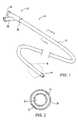
- FIG. 1is a perspective illustration of an isolation/access catheter useful in the methods, systems, and kits of the present invention.
- FIG. 2is a cross-sectional view taken along line 2 to a FIG. 1 .
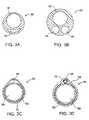
- FIGS. 3A-3Fillustrate alternative cross-sectional views of the isolation/access catheter of FIG. 1 .
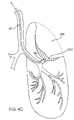
- FIGS. 4A-4Cillustrate use of the isolation/access catheter of FIG. 1 for isolating and collapsing a target lung tissue segment according the to the methods of the present invention.
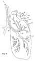
- FIG. 4Dillustrates one protocol for over inflating a target lung tissue segment prior to aspiration according to the present invention.
- FIG. 5illustrates an optional aspect of the present invention where an insufflation gas is introduced to aid in the collapse of the target segment from the pleural space.
- FIG. 6illustrates an alternative optional aspect of the present invention where an inflatable balloon is used to externally collapse a portion of a target lung tissue segment.
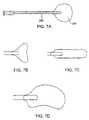
- FIGS. 7A-7Dillustrate alternative balloon designs for use in external collapse of the target lung tissue segment.
- FIG. 8illustrates yet another alternative optional aspect of the methods of the present invention where a probe is used to engage and collapse a portion of a target lung tissue segment.
- FIGS. 9A-9Cillustrate alternative probe designs.
- FIGS. 10A-10Cillustrate a sealing catheter carrying a swellable closure element which may be used in the methods, systems, and kits of the present invention.
- FIG. 11illustrates use of the sealing catheter of FIGS. 10A-10C for selectively occluding an air passage leading to a target lung tissue segment according to the methods of the present invention.
- FIGS. 12A-12Cillustrate a steerable imaging guidewire which may be used to facilitate positioning of the isolation/access catheter used in the methods of the present invention.
- FIG. 13illustrates a kit constructed in accordance with the principles of the present invention.
- Lung volume reductionis performed by collapsing a target lung tissue segment, usually within sub-lobular regions of the lung which receive air through a single air passage, i.e., segment of the branching bronchus which deliver to and receive air from the alveolar regions of the lung.
- Such isolated lung tissue segmentsare first isolated and then collapsed by aspiration of the air (or other gases or liquids which may have been introduced, as discussed below) from the target lung tissue segment.
- Lung tissuehas a very high percentage of void volume, so removal of internal gases can reduce the lung tissue to a small percentage of the volume which it has when fully inflated, i.e. inflated at normal inspiratory pressures.
- the exemplary and preferred percentages for the volume reductionare set forth above.
- the present inventionprovides methods and apparatus for enhancing the aspiration and collapse of the target lung tissue segment.
- Such methods and apparatusmay involve one or more of the following improvements.
- First, various approachesmay be taken to remove or lessen obstructions to gas flow within the target tissue region.
- Second, methods and apparatusmay be employed to apply external pressure over the lung to enhance the collapse achieved by internal aspiration.
- Third, aspiration of the gases within the target tissue segmentmay be enhanced by inducing absorption atelectasis prior to aspiration.
- Absorption atelectasismay be induced, for example, by introducing an oxygen-rich gas to the lung tissue segment, usually at least 50% oxygen by weight, more usually at least 75% oxygen by weight, and preferably substantially pure oxygen.
- Absorption atelectasisis a phenomena which occurs when an enriched oxygen mixture is inspired.
- the high oxygen concentrationcauses an increase in the partial oxygen pressure which in turn causes the rate of oxygen transfer into the capillary blood within the alveolar regions to increase greatly.
- the increased oxygen fluxmay increase so much that the net flow of gas into the blood exceeds the inspired flow of gas, causing the lung unit to become progressively smaller.
- the access methods and apparatusmay be used for performing in situ diagnosis, usually as part of the collapse procedure. Any one of a number of lung performance characteristics may be measured, typically by sampling using the isolation/access catheter.
- FIGS. 1 and 2An exemplary isolation/access catheter 10 is illustrated in FIGS. 1 and 2 and comprises a catheter body 12 having a distal end 14 , a proximal end 16 , an inflatable occlusion balloon 18 near its distal end, and at least one lumen therethrough.
- the catheterwill have at least two lumens, and catheter 10 includes both a central lumen 20 and an annular lumen 22 defined by inner body member 24 and outer body member 26 which is coaxially disposed about the inner body member.
- the annular lumen 22opens to port 30 on a proximal hub 32 and provides for inflation of balloon 18 .
- the central lumen 20opens to port 36 on hub 32 and provides for multiple functions, including optional introduction over a guidewire, aspiration, introduction of secondary catheters, such as sealing catheters described below, and the like.
- isolation/access catheter 10The dimensions and materials of isolation/access catheter 10 are selected to permit endotracheal introduction and intraluminal advancement through the lung bronchus, optionally over a guidewire and/or through a primary tracheal tube structure (as illustrated in FIG. 4B below). Suitable materials include low and high density polyethylenes, polyamides, nylons, PTFE, PEEK, and the like, particularly for the inner tubular member 24 .
- the outer member, including the occlusion ballooncan be made from elastomeric materials, such as polyurethane, low density polyethylene, polyvinylchloride, silicone rubber, latex, and the like.
- portions of the outer tubular member 26 proximal to the inflatable ballooncan be made thicker and/or reinforced so that they do not dilate upon pressurization of the balloon. Exemplary dimensions for the isolation/access catheter 10 are set forth in the table below.
- the isolation/access catheter 10may be modified in a number of ways, some of which are illustrated in FIGS. 3A-3F .
- the catheterinstead of a inner and outer coaxial tube construction, can be a single extrusion having a catheter body 30 with a circular main lumen 32 and a crescent-shaped inflation lumen 34 , as illustrated in FIG. 3A .
- catheter body 40may be formed as a single extrusion having three lumens, i.e., a primary lumen 42 for receiving a guidewire, applying aspiration, and/or delivering secondary catheters.
- a second lumen 44can be provided for inflating the occlusion balloon, and a third lumen 46 can be provided as an alternative guidewire or aspiration lumen.
- Catheter body 50comprising a main tubular body 52 having an outer layer 54 fused thereover to define a lumen 56 suitable for balloon inflation as shown in FIG. 3C .
- a primary lumen 58is formed within the main tubular member 52 .
- catheter body 60can be formed from a primary tubular member 62 , and a secondary tubular member 64 , where the tubular members are held together by an outer member 66 , such as a layer which is applied by heat shrinking.
- the primary tubular member 62provides the main lumen 68 while secondary tube 64 provides a secondary lumen 70 .
- the secondary lumen 70will typically be used for balloon inflation, while the primary lumen 68 can be used for all other functions of the isolation/access catheter.
- catheter body 80can be formed to include four lumens, typically by conventional extrusion processes.
- Lumen 82is suitable for passage over a guidewire.
- Lumens 84 and 86both contain light fibers 88 for illumination.
- Lumen 90carries an optical wave guide or image fiber 92 .
- Lumen 82can be used for irrigation and aspiration, typically after the guidewire is withdrawn. Balloon inflation can be effected through the space remaining and lumens 84 and 86 surrounding the light fibers 88 .
- a second catheter body 100is formed as a coaxial arrangement of a number separate tubes.
- Outer tube 102contains a separate guidewire tube 104 defining lumen 106 which permits introduction over a guidewire as well as perfusion and aspiration after the guidewire is removed.
- Second inner tubular member 110will carry an optical image fiber 112 and a plurality of light fibers 112 are passed within the remaining space 114 within the outer tubular member.
- forward imagingcan be effected by illuminating through the light fibers and detecting an image through a lens at the distal end of the catheter. The image can be displayed on conventional cathode-ray or other types of imaging screens. In particular, as described below, forward imaging permits a user to selectively place the guidewire for advancing the catheters through a desired route through the branching bronchus.
- a catheter 10can be advanced to a diseased region DR within a lung L through a patient's trachea T. Advancement through the trachea T is relatively simple and will optionally employ a guidewire to select the advancement route through the branching bronchus. As described above, steering can be effected under real time imaging using the imaging isolation/access catheters illustrated in FIGS. 3E and 3F . Optionally, the isolation/access catheter 10 may be introduced through a visualizing tracheal tube, such as that described in U.S. Pat. No. 5,285,778, licensed to the assignee of the present application.
- the visualizing endotracheal tube 120includes an occlusion cuff 122 which may be inflated within the trachea just above the branch of the left bronchus and right bronchus LB and RB, respectively.
- the visualizing endotracheal tube 120includes a forward-viewing optical system, typically including both illumination fibers and an image fiber to permit direct viewing of the main branch between the left bronchus LB and right bronchus RB.
- initial placement of isolation/access cathetercan be made under visualization of the visualizing endotracheal tube 120 and optionally the isolation/access catheter 10 itself. Referring again in particular to FIG.
- the isolation/access catheter 10is advanced until its distal end 14 reaches a region in the bronchus which leads directly into the diseased region DR.
- the balloon 18can be inflated and the lung tissue segment which includes the diseased region isolated from the remainder of the lung.
- isolatedit is meant that air or other gases will not pass between the isolated region and the remaining portions of the lung to any significant extent.
- FIG. 4Cit is the object of the present invention to apply a vacuum to a lumen within the isolation/access catheter 10 to aspirate the internal regions within the isolated lung tissue segment in order to collapse the tissue. This results in a collapsed lung tissue region CLT, as shown as a shaded region in FIG. 4C .
- a variety of steps and protocolsmay be performed prior to aspirating the isolated lung tissue region in order to enhance gas removal from the region.
- the regionmay be over inflated, subjected to vibrations, subjected to a dilating or mucolytic agent, or otherwise treated in order to remove gas flow obstructions within the region.
- over inflationcan be effected simply by introducing an inflation gas through the isolation/access catheter to a desired pressure. Pressure may be measured using a transducer at the distal tip of the catheter 10 , but will usually be measured statically at a location proximal of the catheter.
- an oxygen-rich gascan be introduced through the isolation/access catheter in order to induce absorption atelectasis.
- incompressible fluidmay be introduced through the isolation/access catheter. Stimulation may be imparted using an external probe and/or a vibratory catheter which is introduced through an access lumen of the isolation/access catheter.
- an entire one-half lungcan be selectively controlled by an isolation or shunting catheter having a balloon 132 near its distal end.
- the balloonis inflated to occlude a portion of the selected bronchus, typically about 60% of the area.
- pressure within the lungcan be reduced and the lung partly collapsed other than in the isolated region.
- inflation of the target lung tissue segmentcan be enhanced which can assist in breaking up occlusions within the lung which would otherwise interfere with subsequent aspiration of the segment.
- a needle or other cannula 200can be percutaneously introduced into a peritoneal space PS between the parietal pleural PP and visceral pleural VP.
- Insufflation gassuch as carbon dioxide
- the gaswill typically be introduced to a pressure in the range from 30 cm H 2 O to 200 cm H 2 O in spontaneously breathing patients and 70 cm H 2 O to 250 cm H 2 O in positive pressure ventilated patients.
- a balloon 210can be introduced to the pleural space, typically through a thoracic trocar 212 .
- the ballooncan be placed based on fluoroscopic observation.
- FIGS. 7A-7Da variety of specific balloon configurations can be employed, as illustrated in FIGS. 7A-7D .
- a generally spherical balloon 220is shown attached to shaft 220 in FIG. 7A .
- Other configurationsinclude a winged profile ( FIG. 7B ), a cylindrical or spatula profile ( FIG. 7C ), and a convex profile ( FIG. 7D ). Each of these will be attached to a shaft which permits inflation after introduction into the pleural space.
- a target lung tissue segmentcan be externally collapsed using a simple probe 250 , usually introduced through a thoracic trocar 252 , as shown in FIG. 8 .
- a simple probe 250usually introduced through a thoracic trocar 252 , as shown in FIG. 8 .
- a variety of probes for mechanically engaging and compressing the outer lung surfaceare illustrated in FIGS. 9A-9C .
- a needlecan be used to puncture at a desired point in the target tissue lung segment in order to release and/or aspirate air, usually as a supplement to a primary catheter-based aspiration. The puncture can then be sealed with fibrin glue or other suitable sealant.
- the methods of the present inventionwill optionally comprise sealing or occluding the air passage leading to the collapsed tissue region CLT.
- sealingcan be performed in a variety of ways, including suturing, gluing, energy-mediated tissue adhesion, and the like.
- a sealing catheter 280can be used to deliver a plug 282 , typically at partially hydrated collagen hydrogel, as illustrated in FIGS. 10A-10C .
- the catheterwill have dimensions which permit it to be introduced through the main access lumen of isolation/access catheter 10 .
- the plug 282will be contained in the distal tip of a lumen in the catheter, and a push rod 284 extends the length of the catheter to permit the treating physician to deploy the plug 282 after the tip of the catheter is properly located, as illustrated in FIG. 11 , usually while the balloon on the isolation/access catheter remains inflated and the target lung tissue remains sealed and in an aspirated, collapsed configuration.
- the plug 282Once deployed within the moist environment of the lung bronchus, the plug 282 will absorb water and will swell substantially, typically from 100% to 1000% in order to fully occupy and plug the air passage into the collapsed lung tissue region CLT.
- Positioning of the isolation/access catheter 10 within the lungcan be performed using on-board optical imaging capability, as discussed above. Usually, positioning of a guidewire through the branching bronchus will be manipulated while viewing through the imaging components of the isolation/access catheter. In this way, the isolation/access catheter can be “inched” along by alternately advancing the guidewire and the isolation/access catheter. As an alternative to providing the isolation/access catheter with imaging, positioning could be done solely by fluoroscopy. As a further alternative, a steerable, imaging guidewire 300 ( FIGS. 12A-12C ) could be used. The guidewire 300 includes a deflectable tip 302 which can be deflected in a single plane using push/pull ribbon 304 .
- the tipwill comprise a spring 306 to facilitate deflection.
- the guidewire 300will include an optical imaging wave guide 310 and illuminating optical fibers 312 , as best seen in cross-sectional view of FIG. 12C .
- the guidewire 300can be steered through the branching bronchus to reach the target tissue segment using its own in situ imaging capability.
- an isolation/access cathetercan be introduced to the target lung tissue segment as well. Since the guidewire has imaging capability, the isolation/access catheter need not incorporate such imaging. This can be an advantage since it permits the access lumen to be made larger since the catheter need not carry any optical wave guides.
- kits 400comprise at least an isolation/access catheter 10 and instructions for use IFU.
- the kitsmay further include any of the other system components described above, such as a balloon probe 210 , a sealing catheter 280 , a reagent container 420 (optionally including any of the dilating or mucolytic agents described above), or other components.
- the instructions for use IFUwill set forth any of the methods as described above, and all kit components will usually be packaged together in a pouch 450 or other conventional medical device packaging.
- those kit components, such as isolation/access catheter 10which will be used in performing the procedure on the patient will be sterilized and maintained sterilely within the kit.
- separate pouches, bags, trays, or other packagingmay be provided within a larger package, where the smaller packs may be opened separately and separately maintain the components in a sterile fashion.
Landscapes
- Health & Medical Sciences (AREA)
- Life Sciences & Earth Sciences (AREA)
- Surgery (AREA)
- Veterinary Medicine (AREA)
- Public Health (AREA)
- General Health & Medical Sciences (AREA)
- Engineering & Computer Science (AREA)
- Biomedical Technology (AREA)
- Heart & Thoracic Surgery (AREA)
- Animal Behavior & Ethology (AREA)
- Molecular Biology (AREA)
- Medical Informatics (AREA)
- Nuclear Medicine, Radiotherapy & Molecular Imaging (AREA)
- Pulmonology (AREA)
- Vascular Medicine (AREA)
- Reproductive Health (AREA)
- Emergency Medicine (AREA)
- Anesthesiology (AREA)
- Hematology (AREA)
- Surgical Instruments (AREA)
- Endoscopes (AREA)
- Materials For Medical Uses (AREA)
- External Artificial Organs (AREA)
Abstract
Description
| ISOLATION/ACCESS CATHETER DIMENSIONS |
| Exemplary | Preferred |
| Inner | Outer | Inner | Outer | ||
| Tubular | Tubular | Tubular | Tubular | ||
| Member | Member | Member | Member | ||
| Outer Diameter (mm) | 0.4-4 | 0.6-4.5 | 1-1.5 | 2-4 |
| Wall Thickness (mm) | 0.05-0.25 | 0.5-0.25 | 0.1-0.2 | 0.15-0.25 |
| Length (cm) | 50-150 | same | 50-80 | same |
| Balloon Length (mm) | 5-50 | 10-20 |
| Balloon Diameter (mm) | 2-15 | 6-12 |
| (inflated) | ||
Claims (5)
Priority Applications (1)
| Application Number | Priority Date | Filing Date | Title |
|---|---|---|---|
| US10/923,109US7458963B2 (en) | 1999-07-02 | 2004-08-20 | Methods, systems, and kits for lung volume reduction |
Applications Claiming Priority (4)
| Application Number | Priority Date | Filing Date | Title |
|---|---|---|---|
| US09/347,032US6287290B1 (en) | 1999-07-02 | 1999-07-02 | Methods, systems, and kits for lung volume reduction |
| US09/606,320US6878141B1 (en) | 1999-07-02 | 2000-06-28 | Methods systems and kits for lung volume reduction |
| US10/017,068US7141046B2 (en) | 1999-07-02 | 2001-12-13 | Methods, systems, and kits for lung volume reduction |
| US10/923,109US7458963B2 (en) | 1999-07-02 | 2004-08-20 | Methods, systems, and kits for lung volume reduction |
Related Parent Applications (1)
| Application Number | Title | Priority Date | Filing Date |
|---|---|---|---|
| US10/017,068ContinuationUS7141046B2 (en) | 1999-07-02 | 2001-12-13 | Methods, systems, and kits for lung volume reduction |
Publications (2)
| Publication Number | Publication Date |
|---|---|
| US20050015106A1 US20050015106A1 (en) | 2005-01-20 |
| US7458963B2true US7458963B2 (en) | 2008-12-02 |
Family
ID=23362046
Family Applications (8)
| Application Number | Title | Priority Date | Filing Date |
|---|---|---|---|
| US09/347,032Expired - LifetimeUS6287290B1 (en) | 1999-07-02 | 1999-07-02 | Methods, systems, and kits for lung volume reduction |
| US09/606,320Expired - LifetimeUS6878141B1 (en) | 1999-07-02 | 2000-06-28 | Methods systems and kits for lung volume reduction |
| US09/898,703Expired - LifetimeUS6709401B2 (en) | 1999-07-02 | 2001-07-02 | Methods, systems, and kits for lung volume reduction |
| US10/017,068Expired - LifetimeUS7141046B2 (en) | 1999-07-02 | 2001-12-13 | Methods, systems, and kits for lung volume reduction |
| US10/765,380Expired - LifetimeUS7186259B2 (en) | 1999-07-02 | 2004-01-26 | Methods, systems, and kits for lung volume reduction |
| US10/923,109Expired - LifetimeUS7458963B2 (en) | 1999-07-02 | 2004-08-20 | Methods, systems, and kits for lung volume reduction |
| US11/104,717Expired - LifetimeUS7766895B2 (en) | 1999-07-02 | 2005-04-12 | Methods, systems, and kits for lung volume reduction |
| US12/820,402Expired - Fee RelatedUS8177769B2 (en) | 1999-07-02 | 2010-06-22 | Methods, systems, and kits for lung volume reduction |
Family Applications Before (5)
| Application Number | Title | Priority Date | Filing Date |
|---|---|---|---|
| US09/347,032Expired - LifetimeUS6287290B1 (en) | 1999-07-02 | 1999-07-02 | Methods, systems, and kits for lung volume reduction |
| US09/606,320Expired - LifetimeUS6878141B1 (en) | 1999-07-02 | 2000-06-28 | Methods systems and kits for lung volume reduction |
| US09/898,703Expired - LifetimeUS6709401B2 (en) | 1999-07-02 | 2001-07-02 | Methods, systems, and kits for lung volume reduction |
| US10/017,068Expired - LifetimeUS7141046B2 (en) | 1999-07-02 | 2001-12-13 | Methods, systems, and kits for lung volume reduction |
| US10/765,380Expired - LifetimeUS7186259B2 (en) | 1999-07-02 | 2004-01-26 | Methods, systems, and kits for lung volume reduction |
Family Applications After (2)
| Application Number | Title | Priority Date | Filing Date |
|---|---|---|---|
| US11/104,717Expired - LifetimeUS7766895B2 (en) | 1999-07-02 | 2005-04-12 | Methods, systems, and kits for lung volume reduction |
| US12/820,402Expired - Fee RelatedUS8177769B2 (en) | 1999-07-02 | 2010-06-22 | Methods, systems, and kits for lung volume reduction |
Country Status (6)
| Country | Link |
|---|---|
| US (8) | US6287290B1 (en) |
| EP (1) | EP1198269A4 (en) |
| JP (1) | JP4185283B2 (en) |
| AU (1) | AU781579B2 (en) |
| CA (1) | CA2375752A1 (en) |
| WO (1) | WO2001002042A1 (en) |
Cited By (6)
| Publication number | Priority date | Publication date | Assignee | Title |
|---|---|---|---|---|
| US20100280538A1 (en)* | 1999-07-02 | 2010-11-04 | Pulmonx Corporation | Methods, systems, and kits for lung volume reduction |
| US20110087122A1 (en)* | 2001-09-10 | 2011-04-14 | Pulmonx Corporation | Minimally invasive determination of collateral ventilation in lungs |
| US20120157967A1 (en)* | 2002-10-07 | 2012-06-21 | Suros Surgical Systems, Inc. | System and method for minimally invasive disease therapy |
| US8523782B2 (en) | 2005-12-07 | 2013-09-03 | Pulmonx Corporation | Minimally invasive determination of collateral ventilation in lungs |
| US10555736B2 (en) | 2016-09-30 | 2020-02-11 | Pneumrx, Inc. | Guidewire |
| US12004707B2 (en) | 2018-04-17 | 2024-06-11 | The Board Of Trustees Of The Leland Stanford Junior University | Airway visualization system |
Families Citing this family (260)
| Publication number | Priority date | Publication date | Assignee | Title |
|---|---|---|---|---|
| US5868704A (en)* | 1995-09-18 | 1999-02-09 | W. L. Gore & Associates, Inc. | Balloon catheter device |
| US20060271091A1 (en)* | 1995-09-18 | 2006-11-30 | Campbell Carey V | Balloon catheter device |
| GB2324729B (en) | 1997-04-30 | 2002-01-02 | Bradford Hospitals Nhs Trust | Lung treatment device |
| US5954766A (en)* | 1997-09-16 | 1999-09-21 | Zadno-Azizi; Gholam-Reza | Body fluid flow control device |
| US6997189B2 (en)* | 1998-06-05 | 2006-02-14 | Broncus Technologies, Inc. | Method for lung volume reduction |
| US7422563B2 (en) | 1999-08-05 | 2008-09-09 | Broncus Technologies, Inc. | Multifunctional tip catheter for applying energy to tissue and detecting the presence of blood flow |
| US7022088B2 (en) | 1999-08-05 | 2006-04-04 | Broncus Technologies, Inc. | Devices for applying energy to tissue |
| US7175644B2 (en) | 2001-02-14 | 2007-02-13 | Broncus Technologies, Inc. | Devices and methods for maintaining collateral channels in tissue |
| US7462162B2 (en) | 2001-09-04 | 2008-12-09 | Broncus Technologies, Inc. | Antiproliferative devices for maintaining patency of surgically created channels in a body organ |
| US6749606B2 (en) | 1999-08-05 | 2004-06-15 | Thomas Keast | Devices for creating collateral channels |
| EP1400204A1 (en)* | 1999-08-05 | 2004-03-24 | Broncus Technologies, Inc. | Methods and devices for creating collateral channels in the lungs |
| US6712812B2 (en) | 1999-08-05 | 2004-03-30 | Broncus Technologies, Inc. | Devices for creating collateral channels |
| US6610043B1 (en)* | 1999-08-23 | 2003-08-26 | Bistech, Inc. | Tissue volume reduction |
| US7654998B1 (en)* | 1999-08-23 | 2010-02-02 | Aeris Therapeutics, Inc. | Tissue volume reduction |
| US6328689B1 (en) | 2000-03-23 | 2001-12-11 | Spiration, Inc., | Lung constriction apparatus and method |
| JP2003517870A (en) | 1999-12-22 | 2003-06-03 | ボストン サイエンティフィック リミテッド | Endovascular occlusion irrigation catheter and its use |
| US20030070683A1 (en)* | 2000-03-04 | 2003-04-17 | Deem Mark E. | Methods and devices for use in performing pulmonary procedures |
| US6679264B1 (en) | 2000-03-04 | 2004-01-20 | Emphasys Medical, Inc. | Methods and devices for use in performing pulmonary procedures |
| US8474460B2 (en) | 2000-03-04 | 2013-07-02 | Pulmonx Corporation | Implanted bronchial isolation devices and methods |
| US8251070B2 (en)* | 2000-03-27 | 2012-08-28 | Asthmatx, Inc. | Methods for treating airways |
| US6514290B1 (en) | 2000-03-31 | 2003-02-04 | Broncus Technologies, Inc. | Lung elastic recoil restoring or tissue compressing device and method |
| US7374532B2 (en) | 2000-04-14 | 2008-05-20 | Attenuex Technologies, Inc. | High vapor pressure attenuation device |
| US6682473B1 (en) | 2000-04-14 | 2004-01-27 | Solace Therapeutics, Inc. | Devices and methods for attenuation of pressure waves in the body |
| US10327880B2 (en) | 2000-04-14 | 2019-06-25 | Attenuex Technologies, Inc. | Attenuation device for use in an anatomical structure |
| EP1284663A4 (en)* | 2000-05-18 | 2007-04-18 | Emphasys Medical Inc | Bronchiopulmonary occlusion devices and lung volume reduction methods |
| US6722360B2 (en) | 2000-06-16 | 2004-04-20 | Rajiv Doshi | Methods and devices for improving breathing in patients with pulmonary disease |
| AU2001292609A1 (en)* | 2000-09-11 | 2002-03-26 | Closure Medical Corporation | Bronchial occlusion method and apparatus |
| US6527761B1 (en)* | 2000-10-27 | 2003-03-04 | Pulmonx, Inc. | Methods and devices for obstructing and aspirating lung tissue segments |
| US6585639B1 (en)* | 2000-10-27 | 2003-07-01 | Pulmonx | Sheath and method for reconfiguring lung viewing scope |
| US20060135947A1 (en) | 2000-10-27 | 2006-06-22 | Pulmonx | Occlusal stent and methods for its use |
| US6890343B2 (en) | 2000-12-14 | 2005-05-10 | Ensure Medical, Inc. | Plug with detachable guidewire element and methods for use |
| US6623509B2 (en) | 2000-12-14 | 2003-09-23 | Core Medical, Inc. | Apparatus and methods for sealing vascular punctures |
| US8083768B2 (en) | 2000-12-14 | 2011-12-27 | Ensure Medical, Inc. | Vascular plug having composite construction |
| US6846319B2 (en) | 2000-12-14 | 2005-01-25 | Core Medical, Inc. | Devices for sealing openings through tissue and apparatus and methods for delivering them |
| US6896692B2 (en) | 2000-12-14 | 2005-05-24 | Ensure Medical, Inc. | Plug with collet and apparatus and method for delivering such plugs |
| US20020112729A1 (en)* | 2001-02-21 | 2002-08-22 | Spiration, Inc. | Intra-bronchial obstructing device that controls biological interaction with the patient |
| US7011094B2 (en) | 2001-03-02 | 2006-03-14 | Emphasys Medical, Inc. | Bronchial flow control devices and methods of use |
| US7798147B2 (en) | 2001-03-02 | 2010-09-21 | Pulmonx Corporation | Bronchial flow control devices with membrane seal |
| US20040074491A1 (en)* | 2001-03-02 | 2004-04-22 | Michael Hendricksen | Delivery methods and devices for implantable bronchial isolation devices |
| US6609521B1 (en)* | 2001-04-09 | 2003-08-26 | Regents Of The University Of Minnesota | Endotracheal tube |
| US6577752B2 (en)* | 2001-06-15 | 2003-06-10 | Arch Development Corporation | Automated method and system for the delineation of the chest wall in computed tomography scans for the assessment of pleural disease |
| JP4602602B2 (en)* | 2001-07-19 | 2010-12-22 | オリンパス株式会社 | Medical instruments |
| JP4409123B2 (en)* | 2001-07-19 | 2010-02-03 | オリンパス株式会社 | Medical obturator |
| US6645205B2 (en)* | 2001-08-15 | 2003-11-11 | Core Medical, Inc. | Apparatus and methods for reducing lung volume |
| US7708712B2 (en) | 2001-09-04 | 2010-05-04 | Broncus Technologies, Inc. | Methods and devices for maintaining patency of surgically created channels in a body organ |
| JP4831389B2 (en)* | 2001-09-07 | 2011-12-07 | まゆみ 岡田 | Endotracheal tube |
| US20030051733A1 (en)* | 2001-09-10 | 2003-03-20 | Pulmonx | Method and apparatus for endobronchial diagnosis |
| US20030050648A1 (en) | 2001-09-11 | 2003-03-13 | Spiration, Inc. | Removable lung reduction devices, systems, and methods |
| CA2458595C (en)* | 2001-10-11 | 2007-12-04 | Peter M. Wilson | Bronchial flow control devices and methods of use |
| US6620100B2 (en)* | 2001-10-17 | 2003-09-16 | Natus Medical Inc. | Hearing evaluation device with noise-weighting capabilities |
| US6592594B2 (en) | 2001-10-25 | 2003-07-15 | Spiration, Inc. | Bronchial obstruction device deployment system and method |
| US20030154988A1 (en)* | 2002-02-21 | 2003-08-21 | Spiration, Inc. | Intra-bronchial device that provides a medicant intra-bronchially to the patient |
| US6929637B2 (en)* | 2002-02-21 | 2005-08-16 | Spiration, Inc. | Device and method for intra-bronchial provision of a therapeutic agent |
| US20060235432A1 (en)* | 2002-02-21 | 2006-10-19 | Devore Lauri J | Intra-bronchial obstructing device that controls biological interaction with the patient |
| AU2003220124A1 (en)* | 2002-03-08 | 2003-09-22 | Emphasys Medical, Inc. | Methods and devices for inducing collapse in lung regions fed by collateral pathways |
| US20030216769A1 (en) | 2002-05-17 | 2003-11-20 | Dillard David H. | Removable anchored lung volume reduction devices and methods |
| EP2353557B1 (en)* | 2002-03-20 | 2020-05-27 | Spiration, Inc. | Removable anchored lung volume reduction devices |
| US20030181922A1 (en) | 2002-03-20 | 2003-09-25 | Spiration, Inc. | Removable anchored lung volume reduction devices and methods |
| US20030195385A1 (en)* | 2002-04-16 | 2003-10-16 | Spiration, Inc. | Removable anchored lung volume reduction devices and methods |
| US20030212412A1 (en)* | 2002-05-09 | 2003-11-13 | Spiration, Inc. | Intra-bronchial obstructing device that permits mucus transport |
| US20040089306A1 (en)* | 2002-05-28 | 2004-05-13 | Ronald Hundertmark | Devices and methods for removing bronchial isolation devices implanted in the lung |
| FR2840796B1 (en)* | 2002-06-13 | 2004-09-10 | Novatech Inc | BRONCHICAL SHUTTER |
| WO2003105676A2 (en)* | 2002-06-17 | 2003-12-24 | Bistech, Inc. | Compositions and methods for reducing lung volume |
| US7819908B2 (en)* | 2002-06-17 | 2010-10-26 | Aeris Therapeutics, Inc. | Compositions and methods for reducing lung volume |
| US20040039252A1 (en)* | 2002-06-27 | 2004-02-26 | Koch Kenneth Elmon | Self-navigating endotracheal tube |
| EP1524942B1 (en) | 2002-07-26 | 2008-09-10 | Emphasys Medical, Inc. | Bronchial flow control devices with membrane seal |
| US7814912B2 (en)* | 2002-11-27 | 2010-10-19 | Pulmonx Corporation | Delivery methods and devices for implantable bronchial isolation devices |
| US7717115B2 (en)* | 2002-11-27 | 2010-05-18 | Pulmonx Corporation | Delivery methods and devices for implantable bronchial isolation devices |
| DE10302310A1 (en)* | 2003-01-20 | 2004-07-29 | Freitag, Lutz, Dr. | Patient lung reduction method, e.g. for treating pulmonary emphysema, whereby a bronchial catheter is inserted into an over-swollen lung area and the supplying bronchopulmonary closed in synchronism with patient breathing |
| US20040210248A1 (en)* | 2003-03-12 | 2004-10-21 | Spiration, Inc. | Apparatus, method and assembly for delivery of intra-bronchial devices |
| US7100616B2 (en)* | 2003-04-08 | 2006-09-05 | Spiration, Inc. | Bronchoscopic lung volume reduction method |
| US8082921B2 (en)* | 2003-04-25 | 2011-12-27 | Anthony David Wondka | Methods, systems and devices for desufflating a lung area |
| US7811274B2 (en) | 2003-05-07 | 2010-10-12 | Portaero, Inc. | Method for treating chronic obstructive pulmonary disease |
| US20040221853A1 (en)* | 2003-05-08 | 2004-11-11 | Plasiatek, Llc | Ultrasonic placement and monitoring of a tube within the body |
| DE10321990B4 (en)* | 2003-05-15 | 2005-10-13 | Microcuff Gmbh | Trachealbeatmungungsvorrichtung |
| US7426929B2 (en) | 2003-05-20 | 2008-09-23 | Portaero, Inc. | Intra/extra-thoracic collateral ventilation bypass system and method |
| US7200559B2 (en)* | 2003-05-29 | 2007-04-03 | Microsoft Corporation | Semantic object synchronous understanding implemented with speech application language tags |
| US7533667B2 (en)* | 2003-05-29 | 2009-05-19 | Portaero, Inc. | Methods and devices to assist pulmonary decompression |
| US7252086B2 (en) | 2003-06-03 | 2007-08-07 | Cordis Corporation | Lung reduction system |
| US7377278B2 (en)* | 2003-06-05 | 2008-05-27 | Portaero, Inc. | Intra-thoracic collateral ventilation bypass system and method |
| US7682332B2 (en) | 2003-07-15 | 2010-03-23 | Portaero, Inc. | Methods to accelerate wound healing in thoracic anastomosis applications |
| US8308682B2 (en) | 2003-07-18 | 2012-11-13 | Broncus Medical Inc. | Devices for maintaining patency of surgically created channels in tissue |
| US7533671B2 (en)* | 2003-08-08 | 2009-05-19 | Spiration, Inc. | Bronchoscopic repair of air leaks in a lung |
| US20050103340A1 (en)* | 2003-08-20 | 2005-05-19 | Wondka Anthony D. | Methods, systems & devices for endobronchial ventilation and drug delivery |
| US8852229B2 (en) | 2003-10-17 | 2014-10-07 | Cordis Corporation | Locator and closure device and method of use |
| US7361183B2 (en) | 2003-10-17 | 2008-04-22 | Ensure Medical, Inc. | Locator and delivery device and method of use |
| WO2005070489A1 (en)* | 2004-01-23 | 2005-08-04 | Cook Critical Care Incorporated | Endobronchial blocking device |
| US20050178389A1 (en)* | 2004-01-27 | 2005-08-18 | Shaw David P. | Disease indications for selective endobronchial lung region isolation |
| US8206684B2 (en) | 2004-02-27 | 2012-06-26 | Pulmonx Corporation | Methods and devices for blocking flow through collateral pathways in the lung |
| US7650886B1 (en)* | 2004-03-04 | 2010-01-26 | Christian Keller | Esophageal airway management device guides |
| EP2368525B1 (en)* | 2004-03-08 | 2019-09-18 | Pulmonx, Inc | Implanted bronchial isolation devices |
| US20060081255A1 (en)* | 2004-04-02 | 2006-04-20 | Michael Miller | Ultrasonic placement and monitoring of an endotracheal tube |
| US20060037617A1 (en)* | 2004-04-19 | 2006-02-23 | Walke Amrish J | Airway implant devices and methods of use |
| NL1026190C2 (en)* | 2004-05-13 | 2005-11-15 | Peter Tjong Joe Wai | Bronchus blocker and ventilation system with bronchus blocker and ventilation tube. |
| US7530947B2 (en)* | 2004-05-28 | 2009-05-12 | Olympus Corporation | Lesion portion determining method of infrared observing system |
| US7670282B2 (en) | 2004-06-14 | 2010-03-02 | Pneumrx, Inc. | Lung access device |
| US20050281739A1 (en)* | 2004-06-16 | 2005-12-22 | Glen Gong | Imaging damaged lung tissue using compositions |
| US20050281800A1 (en)* | 2004-06-16 | 2005-12-22 | Glen Gong | Targeting sites of damaged lung tissue |
| US20050281740A1 (en)* | 2004-06-16 | 2005-12-22 | Glen Gong | Imaging damaged lung tissue |
| US20050281798A1 (en)* | 2004-06-16 | 2005-12-22 | Glen Gong | Targeting sites of damaged lung tissue using composition |
| US7678767B2 (en) | 2004-06-16 | 2010-03-16 | Pneumrx, Inc. | Glue compositions for lung volume reduction |
| US7608579B2 (en)* | 2004-06-16 | 2009-10-27 | Pneumrx, Inc. | Lung volume reduction using glue compositions |
| EP3542736A1 (en)* | 2004-06-16 | 2019-09-25 | PneumRx, Inc. | Intra-bronchial lung volume reduction system |
| US7553810B2 (en)* | 2004-06-16 | 2009-06-30 | Pneumrx, Inc. | Lung volume reduction using glue composition |
| CA2570261C (en) | 2004-07-08 | 2014-06-10 | Pneumrx, Inc. | Pleural effusion treatment device, method and material |
| US7766891B2 (en)* | 2004-07-08 | 2010-08-03 | Pneumrx, Inc. | Lung device with sealing features |
| US8409167B2 (en) | 2004-07-19 | 2013-04-02 | Broncus Medical Inc | Devices for delivering substances through an extra-anatomic opening created in an airway |
| US20060030863A1 (en)* | 2004-07-21 | 2006-02-09 | Fields Antony J | Implanted bronchial isolation device delivery devices and methods |
| US7824384B2 (en) | 2004-08-10 | 2010-11-02 | Kci Licensing, Inc. | Chest tube drainage system |
| US20060047291A1 (en)* | 2004-08-20 | 2006-03-02 | Uptake Medical Corporation | Non-foreign occlusion of an airway and lung collapse |
| US7949407B2 (en) | 2004-11-05 | 2011-05-24 | Asthmatx, Inc. | Energy delivery devices and methods |
| US20060162731A1 (en)* | 2004-11-16 | 2006-07-27 | Pulmonx | Pulmonary occlusal stent delivery catheter, loading system and methods of use |
| MX2007005937A (en) | 2004-11-16 | 2007-09-11 | Robert L Barry | Device and method for lung treatment. |
| US7771472B2 (en) | 2004-11-19 | 2010-08-10 | Pulmonx Corporation | Bronchial flow control devices and methods of use |
| US20060118126A1 (en)* | 2004-11-19 | 2006-06-08 | Don Tanaka | Methods and devices for controlling collateral ventilation |
| US7398782B2 (en) | 2004-11-19 | 2008-07-15 | Portaero, Inc. | Method for pulmonary drug delivery |
| US8220460B2 (en) | 2004-11-19 | 2012-07-17 | Portaero, Inc. | Evacuation device and method for creating a localized pleurodesis |
| WO2006058195A2 (en) | 2004-11-23 | 2006-06-01 | Pneumrx, Inc. | Steerable device for accessing a target site and methods |
| WO2006062862A1 (en)* | 2004-12-06 | 2006-06-15 | Cook Incorporated | Inflatable occlusion devices, methods, and systems |
| US8061357B2 (en) | 2004-12-08 | 2011-11-22 | Ventus Medical, Inc. | Adhesive nasal respiratory devices |
| US10610228B2 (en) | 2004-12-08 | 2020-04-07 | Theravent, Inc. | Passive nasal peep devices |
| US9833354B2 (en) | 2004-12-08 | 2017-12-05 | Theravent, Inc. | Nasal respiratory devices |
| EP1824544B1 (en) | 2004-12-08 | 2013-06-05 | Ventus Medical, Inc. | Respiratory devices and methods of use |
| US7824366B2 (en) | 2004-12-10 | 2010-11-02 | Portaero, Inc. | Collateral ventilation device with chest tube/evacuation features and method |
| US11883029B2 (en) | 2005-01-20 | 2024-01-30 | Pulmonx Corporation | Methods and devices for passive residual lung volume reduction and functional lung volume expansion |
| US8496006B2 (en) | 2005-01-20 | 2013-07-30 | Pulmonx Corporation | Methods and devices for passive residual lung volume reduction and functional lung volume expansion |
| EP1838217B1 (en)* | 2005-01-20 | 2014-06-25 | Pulmonx | Minimally invasive determination of collateral ventilation in lungs |
| US20080228137A1 (en) | 2007-03-12 | 2008-09-18 | Pulmonx | Methods and devices for passive residual lung volume reduction and functional lung volume expansion |
| US20080091062A1 (en)* | 2005-02-07 | 2008-04-17 | Smart Medical Systems, Ltd. | Endoscope assembly |
| US20070186932A1 (en)* | 2006-01-06 | 2007-08-16 | Pulmonx | Collateral pathway treatment using agent entrained by aspiration flow current |
| US8876791B2 (en) | 2005-02-25 | 2014-11-04 | Pulmonx Corporation | Collateral pathway treatment using agent entrained by aspiration flow current |
| US8153435B1 (en) | 2005-03-30 | 2012-04-10 | Tracer Detection Technology Corp. | Methods and articles for identifying objects using encapsulated perfluorocarbon tracers |
| US8088144B2 (en) | 2005-05-04 | 2012-01-03 | Ensure Medical, Inc. | Locator and closure device and method of use |
| US8926654B2 (en) | 2005-05-04 | 2015-01-06 | Cordis Corporation | Locator and closure device and method of use |
| US8287535B2 (en) | 2005-05-11 | 2012-10-16 | Mayo Foundation For Medical Education And Research | Apparatus and methods for internal surgical procedures |
| US20070142742A1 (en)* | 2005-07-13 | 2007-06-21 | Pulmonx | Methods and systems for segmental lung diagnostics |
| EP1901653A4 (en)* | 2005-07-13 | 2009-12-30 | Pulmonx | Methods and systems for segmental lung diagnostics |
| US7904144B2 (en)* | 2005-08-02 | 2011-03-08 | Brainscope Company, Inc. | Method for assessing brain function and portable automatic brain function assessment apparatus |
| US7628789B2 (en)* | 2005-08-17 | 2009-12-08 | Pulmonx Corporation | Selective lung tissue ablation |
| US8104474B2 (en) | 2005-08-23 | 2012-01-31 | Portaero, Inc. | Collateral ventilation bypass system with retention features |
| WO2007038476A2 (en) | 2005-09-26 | 2007-04-05 | Atteneux Technologies, Inc. | Pressure attenuation device |
| US7406963B2 (en) | 2006-01-17 | 2008-08-05 | Portaero, Inc. | Variable resistance pulmonary ventilation bypass valve and method |
| US8235939B2 (en) | 2006-02-06 | 2012-08-07 | Kci Licensing, Inc. | System and method for purging a reduced pressure apparatus during the administration of reduced pressure treatment |
| WO2007100846A2 (en)* | 2006-02-28 | 2007-09-07 | Emphasys Medical, Inc. | Endoscopic tool |
| US9402633B2 (en) | 2006-03-13 | 2016-08-02 | Pneumrx, Inc. | Torque alleviating intra-airway lung volume reduction compressive implant structures |
| US8888800B2 (en) | 2006-03-13 | 2014-11-18 | Pneumrx, Inc. | Lung volume reduction devices, methods, and systems |
| US8157837B2 (en) | 2006-03-13 | 2012-04-17 | Pneumrx, Inc. | Minimally invasive lung volume reduction device and method |
| US20070225809A1 (en)* | 2006-03-27 | 2007-09-27 | Ray Charles D | System and device for filling a human implantable container with a filler material |
| US7691151B2 (en) | 2006-03-31 | 2010-04-06 | Spiration, Inc. | Articulable Anchor |
| US7829986B2 (en)* | 2006-04-01 | 2010-11-09 | Stats Chippac Ltd. | Integrated circuit package system with net spacer |
| CA2955328C (en)* | 2006-05-18 | 2018-04-10 | Smart Medical Systems Ltd. | Flexible endoscope system and functionality |
| GB0610171D0 (en) | 2006-05-23 | 2006-06-28 | Robitaille Jean Pierre | Valved nasal canula |
| CA2653139C (en) | 2006-05-23 | 2016-01-05 | Ventus Medical, Inc. | Nasal respiratory devices |
| US20090145441A1 (en)* | 2007-12-06 | 2009-06-11 | Rajiv Doshi | Delayed resistance nasal devices and methods of use |
| CN101489630B (en) | 2006-06-07 | 2013-10-23 | 温吐斯医学公司 | Layered nasal devices |
| EP2032213A4 (en) | 2006-06-07 | 2014-02-19 | Theravent Inc | DEVICES FOR THE NOSE |
| US20080097139A1 (en)* | 2006-07-14 | 2008-04-24 | Boston Scientific Scimed, Inc. | Systems and methods for treating lung tissue |
| US20080140173A1 (en) | 2006-08-07 | 2008-06-12 | Sherif Eskaros | Non-shortening wrapped balloon |
| US9180279B2 (en) | 2006-08-07 | 2015-11-10 | W. L. Gore & Associates, Inc. | Inflatable imbibed polymer devices |
| US8460240B2 (en) | 2006-08-07 | 2013-06-11 | W. L. Gore & Associates, Inc. | Inflatable toroidal-shaped balloons |
| US20080125711A1 (en) | 2006-08-07 | 2008-05-29 | Alpini Alfred A | Catheter balloons with integrated non-distensible seals |
| US7785290B2 (en) | 2006-08-07 | 2010-08-31 | Gore Enterprise Holdings, Inc. | Non-shortening high angle wrapped balloons |
| DE102006039236A1 (en)* | 2006-08-21 | 2008-02-28 | Variomed Ag | Apparatus and method for reducing or removing stenoses |
| US20080072914A1 (en)* | 2006-08-25 | 2008-03-27 | Hendricksen Michael J | Bronchial Isolation Devices for Placement in Short Lumens |
| US8342182B2 (en) | 2006-08-28 | 2013-01-01 | Pulmonx Corporation | Functional assessment and treatment catheters and methods for their use in the lung |
| US8585645B2 (en)* | 2006-11-13 | 2013-11-19 | Uptake Medical Corp. | Treatment with high temperature vapor |
| US7993323B2 (en)* | 2006-11-13 | 2011-08-09 | Uptake Medical Corp. | High pressure and high temperature vapor catheters and systems |
| WO2008061252A2 (en)* | 2006-11-16 | 2008-05-22 | Ventus Medical, Inc. | Nasal devices applicators |
| US8267908B2 (en) | 2007-02-09 | 2012-09-18 | Kci Licensing, Inc. | Delivery tube, system, and method for storing liquid from a tissue site |
| WO2008100446A2 (en) | 2007-02-09 | 2008-08-21 | Kci Licensing Inc. | Apparatus and method for administering reduced pressure treatment to a tissue site |
| US20080221582A1 (en)* | 2007-03-05 | 2008-09-11 | Pulmonx | Pulmonary stent removal device |
| US8100959B2 (en)* | 2007-03-09 | 2012-01-24 | Pulmonx Corporation | Loading device for a pulmonary implant |
| US8137302B2 (en)* | 2007-03-12 | 2012-03-20 | Pulmonx Corporation | Methods and systems for occluding collateral flow channels in the lung |
| US8821376B2 (en)* | 2007-03-12 | 2014-09-02 | David Tolkowsky | Devices and methods for performing medical procedures in tree-like luminal structures |
| DE102009013205A1 (en) | 2009-03-17 | 2010-09-23 | Dolphys Technologies B.V. | Jet ventilation catheter, in particular for the ventilation of a patient |
| US8911750B2 (en)* | 2007-04-03 | 2014-12-16 | Larry W. Tsai | Lung volume reduction therapy using crosslinked biopolymers |
| US20080281151A1 (en)* | 2007-05-11 | 2008-11-13 | Portaero, Inc. | Pulmonary pleural stabilizer |
| US8163034B2 (en) | 2007-05-11 | 2012-04-24 | Portaero, Inc. | Methods and devices to create a chemically and/or mechanically localized pleurodesis |
| US7931641B2 (en) | 2007-05-11 | 2011-04-26 | Portaero, Inc. | Visceral pleura ring connector |
| US20080283065A1 (en)* | 2007-05-15 | 2008-11-20 | Portaero, Inc. | Methods and devices to maintain patency of a lumen in parenchymal tissue of the lung |
| US20080287878A1 (en)* | 2007-05-15 | 2008-11-20 | Portaero, Inc. | Pulmonary visceral pleura anastomosis reinforcement |
| US8062315B2 (en) | 2007-05-17 | 2011-11-22 | Portaero, Inc. | Variable parietal/visceral pleural coupling |
| US20080295829A1 (en)* | 2007-05-30 | 2008-12-04 | Portaero, Inc. | Bridge element for lung implant |
| US8375952B2 (en)* | 2007-08-09 | 2013-02-19 | The Nemours Foundation | Device and method for unilateral lung ventilation |
| US8607795B2 (en)* | 2007-09-20 | 2013-12-17 | Kimberly-Clark Worldwide, Inc. | Balloon cuff tracheostomy tube |
| US8043301B2 (en) | 2007-10-12 | 2011-10-25 | Spiration, Inc. | Valve loader method, system, and apparatus |
| EP2641572B1 (en) | 2007-10-12 | 2019-07-24 | Spiration Inc. | Valve loader method, system, and apparatus |
| US8322335B2 (en) | 2007-10-22 | 2012-12-04 | Uptake Medical Corp. | Determining patient-specific vapor treatment and delivery parameters |
| US8147532B2 (en) | 2007-10-22 | 2012-04-03 | Uptake Medical Corp. | Determining patient-specific vapor treatment and delivery parameters |
| JP5263765B2 (en)* | 2007-11-22 | 2013-08-14 | 国立大学法人 長崎大学 | Airway securing device |
| US8020700B2 (en) | 2007-12-05 | 2011-09-20 | Ventus Medical, Inc. | Packaging and dispensing nasal devices |
| US8882756B2 (en)* | 2007-12-28 | 2014-11-11 | Medtronic Advanced Energy Llc | Fluid-assisted electrosurgical devices, methods and systems |
| US8481845B2 (en)* | 2008-02-05 | 2013-07-09 | Gtat Corporation | Method to form a photovoltaic cell comprising a thin lamina |
| US8336540B2 (en) | 2008-02-19 | 2012-12-25 | Portaero, Inc. | Pneumostoma management device and method for treatment of chronic obstructive pulmonary disease |
| WO2009105432A2 (en) | 2008-02-19 | 2009-08-27 | Portaero, Inc. | Devices and methods for delivery of a therapeutic agent through a pneumostoma |
| US8475389B2 (en) | 2008-02-19 | 2013-07-02 | Portaero, Inc. | Methods and devices for assessment of pneumostoma function |
| JP5346922B2 (en)* | 2008-03-31 | 2013-11-20 | テルモ株式会社 | Medical obturator |
| US20100036361A1 (en)* | 2008-06-20 | 2010-02-11 | Pulmonx | System and method for delivering multiple implants into lung passageways |
| US9173669B2 (en) | 2008-09-12 | 2015-11-03 | Pneumrx, Inc. | Enhanced efficacy lung volume reduction devices, methods, and systems |
| US8444635B2 (en)* | 2008-11-19 | 2013-05-21 | Samuel Victor Lichtenstein | Methods for selectively heating tissue |
| WO2010068467A1 (en) | 2008-11-25 | 2010-06-17 | Attenuex Technologies, Inc. | Implant with high vapor pressure medium |
| CA2746525C (en) | 2008-12-31 | 2017-12-12 | Kci Licensing, Inc. | Manifolds, systems, and methods for administering reduced pressure to a subcutaneous tissue site |
| US8347881B2 (en) | 2009-01-08 | 2013-01-08 | Portaero, Inc. | Pneumostoma management device with integrated patency sensor and method |
| US8518053B2 (en) | 2009-02-11 | 2013-08-27 | Portaero, Inc. | Surgical instruments for creating a pneumostoma and treating chronic obstructive pulmonary disease |
| WO2010113305A1 (en)* | 2009-04-01 | 2010-10-07 | 株式会社アイ.エス.テイ | Tube for resolving obstructive sleep apnea syndrome |
| WO2010135352A1 (en) | 2009-05-18 | 2010-11-25 | Pneumrx, Inc. | Cross-sectional modification during deployment of an elongate lung volume reduction device |
| US8529526B2 (en) | 2009-10-20 | 2013-09-10 | Kci Licensing, Inc. | Dressing reduced-pressure indicators, systems, and methods |
| US10512761B2 (en) | 2009-12-02 | 2019-12-24 | Renovorx, Inc. | Methods for delivery of therapeutic materials to treat pancreatic cancer |
| EP2506913B8 (en) | 2009-12-02 | 2020-11-18 | RenovoRX, Inc. | Devices and kits for delivery of therapeutic materials to a pancreas |
| US9457171B2 (en) | 2009-12-02 | 2016-10-04 | Renovorx, Inc. | Devices, methods and kits for delivery of therapeutic materials to a target artery |
| US8875711B2 (en) | 2010-05-27 | 2014-11-04 | Theravent, Inc. | Layered nasal respiratory devices |
| US9592008B2 (en) | 2010-07-01 | 2017-03-14 | Pulmonx Corporation | Devices and systems for lung treatment |
| US20120053566A1 (en)* | 2010-08-25 | 2012-03-01 | Terumo Kabushiki Kaisha | Method for treatment of emphysema |
| JP2012045358A (en) | 2010-08-25 | 2012-03-08 | Terumo Corp | Therapeutic agent for pulmonary emphysema |
| US9345532B2 (en) | 2011-05-13 | 2016-05-24 | Broncus Medical Inc. | Methods and devices for ablation of tissue |
| US8795241B2 (en) | 2011-05-13 | 2014-08-05 | Spiration, Inc. | Deployment catheter |
| US8709034B2 (en) | 2011-05-13 | 2014-04-29 | Broncus Medical Inc. | Methods and devices for diagnosing, monitoring, or treating medical conditions through an opening through an airway wall |
| WO2013044267A1 (en) | 2011-09-23 | 2013-03-28 | Pulmonx, Inc. | Implant loading device and system |
| US9730830B2 (en) | 2011-09-29 | 2017-08-15 | Trudell Medical International | Nasal insert and cannula and methods for the use thereof |
| WO2013078235A1 (en) | 2011-11-23 | 2013-05-30 | Broncus Medical Inc | Methods and devices for diagnosing, monitoring, or treating medical conditions through an opening through an airway wall |
| US9821130B2 (en) | 2011-12-08 | 2017-11-21 | Avent, Inc. | Multi-diameter pediatric tracheal cuff |
| US9186053B2 (en)* | 2012-05-03 | 2015-11-17 | Covidien Lp | Methods of using light to repair hernia defects |
| WO2013188845A1 (en) | 2012-06-15 | 2013-12-19 | The Regents Of The University Of California | System and methods for lung isolation and one lung ventilation |
| US8894563B2 (en) | 2012-08-10 | 2014-11-25 | Attenuex Technologies, Inc. | Methods and systems for performing a medical procedure |
| US20140187863A1 (en)* | 2012-12-28 | 2014-07-03 | Metal Industries Research & Development Centre | Endoscope Controlling Device |
| US20140261407A1 (en)* | 2013-03-14 | 2014-09-18 | Patient Centered Medical Incorporated | Aspiration catheters, systems, and methods |
| WO2014144294A1 (en)* | 2013-03-15 | 2014-09-18 | Cook Biotech Incorporated | Methods and devices for lung volume reduction with extracellular matrix material |
| WO2014197362A1 (en) | 2013-06-03 | 2014-12-11 | Ramtin Agah | Devices, methods and kits for delivery of therapeutic materials to a pancreas |
| EP3030162B1 (en)* | 2013-08-09 | 2017-05-31 | Boston Scientific Scimed, Inc. | Devices for treating a lung |
| AU2014240225A1 (en) | 2013-10-01 | 2015-04-16 | Uptake Medical Technology Inc. | Preferential volume reduction of diseased segments of a heterogeneous lobe |
| US9968758B2 (en) | 2014-03-21 | 2018-05-15 | Boston Scientific Scimed, Inc. | Devices and methods for treating a lung |
| US10206686B2 (en) | 2014-06-10 | 2019-02-19 | Ethicon Llc | Bronchus sealants and methods of sealing bronchial tubes |
| US10390838B1 (en) | 2014-08-20 | 2019-08-27 | Pneumrx, Inc. | Tuned strength chronic obstructive pulmonary disease treatment |
| WO2016054480A2 (en)* | 2014-10-03 | 2016-04-07 | The Regent Of The University Of Colorado, A Body Corporate | Venous access catheters and methods for portal venous system catheterization |
| US10485604B2 (en) | 2014-12-02 | 2019-11-26 | Uptake Medical Technology Inc. | Vapor treatment of lung nodules and tumors |
| US10531906B2 (en) | 2015-02-02 | 2020-01-14 | Uptake Medical Technology Inc. | Medical vapor generator |
| WO2016153635A1 (en) | 2015-03-24 | 2016-09-29 | Spiration, Inc.D/B/A Olympus Respiratory America | Airway stent |
| US20160287223A1 (en)* | 2015-04-01 | 2016-10-06 | Boston Scientific Scimed, Inc. | Pulmonary biopsy devices |
| US10179008B2 (en)* | 2016-04-19 | 2019-01-15 | Lsi Solutions, Inc. | Needle assembly for pleural space insufflation and methods thereof |
| JP6761692B2 (en) | 2016-08-02 | 2020-09-30 | テルモ株式会社 | Medical device positioning method and medical device system |
| US11419490B2 (en)* | 2016-08-02 | 2022-08-23 | Covidien Lp | System and method of using an endoscopic catheter as a port in laparoscopic surgery |
| WO2018027145A1 (en)* | 2016-08-04 | 2018-02-08 | Shifamed Holdings, Llc | Devices and methods for lung volume reduction |
| US10448886B2 (en)* | 2016-08-17 | 2019-10-22 | Covidien Lp | Induced atelectasis and pulmonary consolidation systems and methods |
| GB2558920A (en)* | 2017-01-20 | 2018-07-25 | Cook Medical Technologies Llc | Aneurysm treatment apparatus |
| EP3606457A4 (en) | 2017-04-03 | 2021-04-21 | Broncus Medical Inc. | ELECTROSURGICAL ACCESS SHEATH |
| US11129673B2 (en) | 2017-05-05 | 2021-09-28 | Uptake Medical Technology Inc. | Extra-airway vapor ablation for treating airway constriction in patients with asthma and COPD |
| US11052224B2 (en) | 2017-05-18 | 2021-07-06 | Renovorx, Inc. | Methods for treating cancerous tumors |
| US10695543B2 (en) | 2017-05-18 | 2020-06-30 | Renovorx, Inc. | Methods for treating cancerous tumors |
| US12290564B2 (en) | 2017-05-18 | 2025-05-06 | Renovorx, Inc. | Methods and apparatuses for treating tumors |
| US11344364B2 (en) | 2017-09-07 | 2022-05-31 | Uptake Medical Technology Inc. | Screening method for a target nerve to ablate for the treatment of inflammatory lung disease |
| US11350988B2 (en) | 2017-09-11 | 2022-06-07 | Uptake Medical Technology Inc. | Bronchoscopic multimodality lung tumor treatment |
| USD845467S1 (en) | 2017-09-17 | 2019-04-09 | Uptake Medical Technology Inc. | Hand-piece for medical ablation catheter |
| US11419658B2 (en) | 2017-11-06 | 2022-08-23 | Uptake Medical Technology Inc. | Method for treating emphysema with condensable thermal vapor |
| US20210169632A1 (en)* | 2017-12-01 | 2021-06-10 | Eolo Medical Inc. | Methods and devices for trans-bronchial airway bypass |
| US11490946B2 (en) | 2017-12-13 | 2022-11-08 | Uptake Medical Technology Inc. | Vapor ablation handpiece |
| WO2020163625A1 (en) | 2019-02-07 | 2020-08-13 | Solace Therapeutics, Inc. | Pressure attenuation device |
| US11653927B2 (en) | 2019-02-18 | 2023-05-23 | Uptake Medical Technology Inc. | Vapor ablation treatment of obstructive lung disease |
| EP3917426B1 (en)* | 2019-06-20 | 2023-09-06 | Axon Therapies, Inc. | Devices for endovascular ablation of a splanchnic nerve |
| CN115003231B (en)* | 2019-12-03 | 2025-09-19 | 波士顿科学国际有限公司 | Medical delivery device and method of use |
| US20220404055A1 (en)* | 2021-06-16 | 2022-12-22 | United States Of America As Represented By The Secretary Of The Army | Bioprotection of transportion and facilities using lumped element model |
Citations (59)
| Publication number | Priority date | Publication date | Assignee | Title |
|---|---|---|---|---|
| US3322126A (en) | 1963-04-19 | 1967-05-30 | Willy Rusch Fa | Endotracheal catheter |
| US3498286A (en) | 1966-09-21 | 1970-03-03 | American Optical Corp | Catheters |
| US3669098A (en) | 1968-10-05 | 1972-06-13 | Olympus Optical Co | Endotracheal tube |
| US3677262A (en) | 1970-07-23 | 1972-07-18 | Henry J Zukowski | Surgical instrument illuminating endotracheal tube inserter |
| US3776222A (en) | 1971-12-23 | 1973-12-04 | Lurosso A | Fiber optic entubator and method of entubation of the trachea through the nasopharynx |
| US3866599A (en) | 1972-01-21 | 1975-02-18 | Univ Washington | Fiberoptic catheter |
| US3913568A (en) | 1973-01-22 | 1975-10-21 | American Optical Corp | Nasopharyngoscope |
| US4041936A (en) | 1975-04-23 | 1977-08-16 | Medical Engineering Corporation | Bronchoscopy tube |
| US4086919A (en) | 1976-07-09 | 1978-05-02 | Bullard James R | Laryngoscope |
| US4327720A (en) | 1979-01-22 | 1982-05-04 | Bronson Paul A | Esophageal-endotracheal airway |
| US4327721A (en) | 1978-07-07 | 1982-05-04 | George Hanover | Endotracheal tube with topical agent delivery system and method of using the same |
| US4453545A (en) | 1981-05-07 | 1984-06-12 | Hiroshi Inoue | Endotracheal tube with movable endobronchial blocker for one-lung anesthesia |
| US4468216A (en) | 1982-05-20 | 1984-08-28 | Rudolph Muto | Irrigation suction catheter |
| US4567882A (en) | 1982-12-06 | 1986-02-04 | Vanderbilt University | Method for locating the illuminated tip of an endotracheal tube |
| US4716896A (en) | 1986-08-01 | 1988-01-05 | Ackrad Laboratories | Bronchial catheter |
| US4742819A (en) | 1987-03-23 | 1988-05-10 | George Gordon P | Intubating scope with camera and screen |
| US4784133A (en) | 1987-01-28 | 1988-11-15 | Mackin Robert A | Working well balloon angioscope and method |
| US4819664A (en) | 1984-11-15 | 1989-04-11 | Stefano Nazari | Device for selective bronchial intubation and separate lung ventilation, particularly during anesthesia, intensive therapy and reanimation |
| US4846153A (en) | 1988-06-10 | 1989-07-11 | George Berci | Intubating video endoscope |
| US4850371A (en) | 1988-06-13 | 1989-07-25 | Broadhurst John H | Novel endotracheal tube and mass spectrometer |
| US4862874A (en) | 1987-06-10 | 1989-09-05 | Kellner Hans Joerg | Endoscope for removal of thrombi from pulmonary arterial vessels |
| US4896941A (en) | 1985-04-27 | 1990-01-30 | Doryokuro Kakunenryo Kaihatsu Jigyodan | Image-transmitting fiber |
| US4949716A (en) | 1988-10-31 | 1990-08-21 | Medical Devices, Inc. | Nasal intubation adjunct |
| US4955375A (en) | 1989-01-23 | 1990-09-11 | Ricardo Martinez | Endotracheal tube with channel for delivering drugs |
| US4958932A (en) | 1988-08-18 | 1990-09-25 | Mcdonnell Douglas Corporation | Optical measuring apparatus |
| US4961738A (en) | 1987-01-28 | 1990-10-09 | Mackin Robert A | Angioplasty catheter with illumination and visualization within angioplasty balloon |
| US4976710A (en) | 1987-01-28 | 1990-12-11 | Mackin Robert A | Working well balloon method |
| US5056529A (en) | 1990-04-03 | 1991-10-15 | Groot William J De | Apparatus and method for performing a transbroncheal biopsy |
| WO1992010971A1 (en) | 1990-12-21 | 1992-07-09 | Ballard Medical Products | Bronchoalveolar lavage catheter |
| US5143062A (en) | 1990-10-26 | 1992-09-01 | Mallinckrodt Medical, Inc. | Endotracheal tube having irrigation means |
| US5146916A (en) | 1990-01-05 | 1992-09-15 | Catalani Angelo S | Endotracheal tube incorporating a drug-irrigation device |
| US5165420A (en) | 1990-12-21 | 1992-11-24 | Ballard Medical Products | Bronchoalveolar lavage catheter |
| US5285778A (en) | 1991-04-19 | 1994-02-15 | Mackin Robert A | Endotracheal tube wih fibers optic illumination and viewing and auxiliary tube |
| US5309903A (en) | 1989-12-12 | 1994-05-10 | Burroughs Wellcome Co. | Method for administering surfactant to the lungs while concurrently providing one-lung ventilation |
| US5331947A (en) | 1992-05-01 | 1994-07-26 | Shturman Cardiology Systems, Inc. | Inflatable sheath for introduction of ultrasonic catheter through the lumen of a fiber optic endoscope |
| US5361753A (en) | 1992-07-07 | 1994-11-08 | Deutsche Aerospace Ag | Method of measuring and regulating the pressure in the sealing cuff of a tracheal tube and apparatus for implementing the method |
| US5400771A (en) | 1993-01-21 | 1995-03-28 | Pirak; Leon | Endotracheal intubation assembly and related method |
| WO1995033506A1 (en) | 1994-06-04 | 1995-12-14 | Archibald Ian Jeremy Brain | A fibreoptic intubating laryngeal mask airway |
| US5477851A (en) | 1995-01-26 | 1995-12-26 | Callaghan; Eric B. | Laryngeal mask assembly and method for removing same |
| US5499625A (en) | 1994-01-27 | 1996-03-19 | The Kendall Company | Esophageal-tracheal double lumen airway |
| US5598840A (en) | 1995-03-17 | 1997-02-04 | Sorenson Critical Care, Inc. | Apparatus and method for ventilation and aspiration |
| US5642730A (en) | 1994-06-17 | 1997-07-01 | Trudell Medical Limited | Catheter system for delivery of aerosolized medicine for use with pressurized propellant canister |
| US5645519A (en) | 1994-03-18 | 1997-07-08 | Jai S. Lee | Endoscopic instrument for controlled introduction of tubular members in the body and methods therefor |
| US5653231A (en) | 1995-11-28 | 1997-08-05 | Medcare Medical Group, Inc. | Tracheostomy length single use suction catheter |
| US5660175A (en) | 1995-08-21 | 1997-08-26 | Dayal; Bimal | Endotracheal device |
| US5682880A (en) | 1996-07-26 | 1997-11-04 | Brain; Archibald Ian Jeremy | Laryngeal-mask airway with guide element, stiffener, and fiberoptic access |
| US5707352A (en) | 1989-08-28 | 1998-01-13 | Alliance Pharmaceutical Corp. | Pulmonary delivery of therapeutic agent |
| US5752921A (en) | 1996-01-11 | 1998-05-19 | Korr Medical Technologies, Inc. | Method and apparatus for determining tracheal pressure |
| WO1998048706A1 (en) | 1997-04-30 | 1998-11-05 | Bradford Hospitals Nhs Trust | Occlusion device |
| WO1998049191A1 (en) | 1997-04-28 | 1998-11-05 | The Scripps Research Institute | Novel pulmonary surfactants and therapeutic uses, including pulmonary lavage |
| WO1999001076A1 (en) | 1997-07-02 | 1999-01-14 | Broncus Technologies, Inc. | Bleb reducer |
| US5893841A (en) | 1996-08-30 | 1999-04-13 | Delcath Systems, Inc. | Balloon catheter with occluded segment bypass |
| WO1999017827A2 (en) | 1997-10-03 | 1999-04-15 | Scimed Life Systems, Inc. | Braided angiography catheter having full length radiopacity and controlled flexibility |
| US5897528A (en) | 1998-04-30 | 1999-04-27 | Medtronic, Inc. | Filtered intracerebroventricular or intraspinal access port with direct cerebrospinal fluid access |
| US5928264A (en) | 1995-11-08 | 1999-07-27 | Sugar Surgical Technologies, Inc. | Tissue grasping device |
| US6174323B1 (en) | 1998-06-05 | 2001-01-16 | Broncus Technologies, Inc. | Method and assembly for lung volume reduction |
| US6258100B1 (en) | 1999-08-24 | 2001-07-10 | Spiration, Inc. | Method of reducing lung size |
| US6287290B1 (en)* | 1999-07-02 | 2001-09-11 | Pulmonx | Methods, systems, and kits for lung volume reduction |
| US6493589B1 (en) | 1998-05-07 | 2002-12-10 | Medtronic, Inc. | Methods and apparatus for treatment of pulmonary conditions |
Family Cites Families (14)
| Publication number | Priority date | Publication date | Assignee | Title |
|---|---|---|---|---|
| US1906678A (en)* | 1931-06-09 | 1933-05-02 | Reinhold H Wappler | Plugging device for catheters |
| US4445892A (en)* | 1982-05-06 | 1984-05-01 | Laserscope, Inc. | Dual balloon catheter device |
| IT8629545V0 (en)* | 1986-06-12 | 1986-06-12 | Fina Ernesto | SET BALLOON URETERAL CATHETER BALLOON FOR EXTRACTION OF URETERAL STONES |
| US5019039A (en)* | 1989-11-15 | 1991-05-28 | Anderson Ronald W | Fluid drainage needle method of use |
| US5370134A (en)* | 1991-05-29 | 1994-12-06 | Orgin Medsystems, Inc. | Method and apparatus for body structure manipulation and dissection |
| US5258042A (en)* | 1991-12-16 | 1993-11-02 | Henry Ford Health System | Intravascular hydrogel implant |
| US5174323A (en)* | 1992-03-02 | 1992-12-29 | Kent Haselden | Liquid material reservoir |
| US5471851A (en) | 1994-03-15 | 1995-12-05 | Zakryk; John M. | Self-regulating swimming pool heater unit |
| US5583114A (en)* | 1994-07-27 | 1996-12-10 | Minnesota Mining And Manufacturing Company | Adhesive sealant composition |
| US5588424A (en)* | 1995-06-28 | 1996-12-31 | The Cleveland Clinic Foundation | Bronchial blocker endotracheal apparatus |
| DE19547538C2 (en)* | 1995-12-20 | 1999-09-23 | Ruesch Willy Ag | Instrument for use in interventional flexible tracheoscopy / bronchoscopy |
| US5904648A (en)* | 1996-06-18 | 1999-05-18 | Cook Incorporated | Guided endobronchial blocker catheter |
| US5954636A (en)* | 1997-07-15 | 1999-09-21 | Schwartz; Roy E. | Pediatric endotracheal tube with bronchial blocker and method for selectively blocking respiratory airflow to a pediatric patient's lung |
| US6099481A (en)* | 1997-11-03 | 2000-08-08 | Ntc Technology, Inc. | Respiratory profile parameter determination method and apparatus |
- 1999
- 1999-07-02USUS09/347,032patent/US6287290B1/ennot_activeExpired - Lifetime
- 2000
- 2000-06-28USUS09/606,320patent/US6878141B1/ennot_activeExpired - Lifetime
- 2000-06-29JPJP2001507530Apatent/JP4185283B2/ennot_activeExpired - Lifetime
- 2000-06-29AUAU59109/00Apatent/AU781579B2/ennot_activeExpired
- 2000-06-29WOPCT/US2000/018286patent/WO2001002042A1/enactiveIP Right Grant
- 2000-06-29CACA002375752Apatent/CA2375752A1/ennot_activeAbandoned
- 2000-06-29EPEP00945122Apatent/EP1198269A4/ennot_activeWithdrawn
- 2001
- 2001-07-02USUS09/898,703patent/US6709401B2/ennot_activeExpired - Lifetime
- 2001-12-13USUS10/017,068patent/US7141046B2/ennot_activeExpired - Lifetime
- 2004
- 2004-01-26USUS10/765,380patent/US7186259B2/ennot_activeExpired - Lifetime
- 2004-08-20USUS10/923,109patent/US7458963B2/ennot_activeExpired - Lifetime
- 2005
- 2005-04-12USUS11/104,717patent/US7766895B2/ennot_activeExpired - Lifetime
- 2010
- 2010-06-22USUS12/820,402patent/US8177769B2/ennot_activeExpired - Fee Related
Patent Citations (60)
| Publication number | Priority date | Publication date | Assignee | Title |
|---|---|---|---|---|
| US3322126A (en) | 1963-04-19 | 1967-05-30 | Willy Rusch Fa | Endotracheal catheter |
| US3498286A (en) | 1966-09-21 | 1970-03-03 | American Optical Corp | Catheters |
| US3669098A (en) | 1968-10-05 | 1972-06-13 | Olympus Optical Co | Endotracheal tube |
| US3677262A (en) | 1970-07-23 | 1972-07-18 | Henry J Zukowski | Surgical instrument illuminating endotracheal tube inserter |
| US3776222A (en) | 1971-12-23 | 1973-12-04 | Lurosso A | Fiber optic entubator and method of entubation of the trachea through the nasopharynx |
| US3866599A (en) | 1972-01-21 | 1975-02-18 | Univ Washington | Fiberoptic catheter |
| US3913568A (en) | 1973-01-22 | 1975-10-21 | American Optical Corp | Nasopharyngoscope |
| US4041936A (en) | 1975-04-23 | 1977-08-16 | Medical Engineering Corporation | Bronchoscopy tube |
| US4086919A (en) | 1976-07-09 | 1978-05-02 | Bullard James R | Laryngoscope |
| US4327721A (en) | 1978-07-07 | 1982-05-04 | George Hanover | Endotracheal tube with topical agent delivery system and method of using the same |
| US4327720A (en) | 1979-01-22 | 1982-05-04 | Bronson Paul A | Esophageal-endotracheal airway |
| US4453545A (en) | 1981-05-07 | 1984-06-12 | Hiroshi Inoue | Endotracheal tube with movable endobronchial blocker for one-lung anesthesia |
| US4468216A (en) | 1982-05-20 | 1984-08-28 | Rudolph Muto | Irrigation suction catheter |
| US4567882A (en) | 1982-12-06 | 1986-02-04 | Vanderbilt University | Method for locating the illuminated tip of an endotracheal tube |
| US4819664A (en) | 1984-11-15 | 1989-04-11 | Stefano Nazari | Device for selective bronchial intubation and separate lung ventilation, particularly during anesthesia, intensive therapy and reanimation |
| US4896941A (en) | 1985-04-27 | 1990-01-30 | Doryokuro Kakunenryo Kaihatsu Jigyodan | Image-transmitting fiber |
| US4716896A (en) | 1986-08-01 | 1988-01-05 | Ackrad Laboratories | Bronchial catheter |
| US4961738A (en) | 1987-01-28 | 1990-10-09 | Mackin Robert A | Angioplasty catheter with illumination and visualization within angioplasty balloon |
| US4784133A (en) | 1987-01-28 | 1988-11-15 | Mackin Robert A | Working well balloon angioscope and method |
| US4976710A (en) | 1987-01-28 | 1990-12-11 | Mackin Robert A | Working well balloon method |
| US4742819A (en) | 1987-03-23 | 1988-05-10 | George Gordon P | Intubating scope with camera and screen |
| US4862874A (en) | 1987-06-10 | 1989-09-05 | Kellner Hans Joerg | Endoscope for removal of thrombi from pulmonary arterial vessels |
| US4846153A (en) | 1988-06-10 | 1989-07-11 | George Berci | Intubating video endoscope |
| US4850371A (en) | 1988-06-13 | 1989-07-25 | Broadhurst John H | Novel endotracheal tube and mass spectrometer |
| US4958932A (en) | 1988-08-18 | 1990-09-25 | Mcdonnell Douglas Corporation | Optical measuring apparatus |
| US4949716A (en) | 1988-10-31 | 1990-08-21 | Medical Devices, Inc. | Nasal intubation adjunct |
| US4955375A (en) | 1989-01-23 | 1990-09-11 | Ricardo Martinez | Endotracheal tube with channel for delivering drugs |
| US5707352A (en) | 1989-08-28 | 1998-01-13 | Alliance Pharmaceutical Corp. | Pulmonary delivery of therapeutic agent |
| US5309903A (en) | 1989-12-12 | 1994-05-10 | Burroughs Wellcome Co. | Method for administering surfactant to the lungs while concurrently providing one-lung ventilation |
| US5146916A (en) | 1990-01-05 | 1992-09-15 | Catalani Angelo S | Endotracheal tube incorporating a drug-irrigation device |
| US5056529A (en) | 1990-04-03 | 1991-10-15 | Groot William J De | Apparatus and method for performing a transbroncheal biopsy |
| US5143062A (en) | 1990-10-26 | 1992-09-01 | Mallinckrodt Medical, Inc. | Endotracheal tube having irrigation means |
| US5165420A (en) | 1990-12-21 | 1992-11-24 | Ballard Medical Products | Bronchoalveolar lavage catheter |
| WO1992010971A1 (en) | 1990-12-21 | 1992-07-09 | Ballard Medical Products | Bronchoalveolar lavage catheter |
| US5285778A (en) | 1991-04-19 | 1994-02-15 | Mackin Robert A | Endotracheal tube wih fibers optic illumination and viewing and auxiliary tube |
| US5331947A (en) | 1992-05-01 | 1994-07-26 | Shturman Cardiology Systems, Inc. | Inflatable sheath for introduction of ultrasonic catheter through the lumen of a fiber optic endoscope |
| US5361753A (en) | 1992-07-07 | 1994-11-08 | Deutsche Aerospace Ag | Method of measuring and regulating the pressure in the sealing cuff of a tracheal tube and apparatus for implementing the method |
| US5400771A (en) | 1993-01-21 | 1995-03-28 | Pirak; Leon | Endotracheal intubation assembly and related method |
| US5499625A (en) | 1994-01-27 | 1996-03-19 | The Kendall Company | Esophageal-tracheal double lumen airway |
| US5645519A (en) | 1994-03-18 | 1997-07-08 | Jai S. Lee | Endoscopic instrument for controlled introduction of tubular members in the body and methods therefor |
| WO1995033506A1 (en) | 1994-06-04 | 1995-12-14 | Archibald Ian Jeremy Brain | A fibreoptic intubating laryngeal mask airway |
| US5642730A (en) | 1994-06-17 | 1997-07-01 | Trudell Medical Limited | Catheter system for delivery of aerosolized medicine for use with pressurized propellant canister |
| US5477851A (en) | 1995-01-26 | 1995-12-26 | Callaghan; Eric B. | Laryngeal mask assembly and method for removing same |
| US5598840A (en) | 1995-03-17 | 1997-02-04 | Sorenson Critical Care, Inc. | Apparatus and method for ventilation and aspiration |
| US5660175A (en) | 1995-08-21 | 1997-08-26 | Dayal; Bimal | Endotracheal device |
| US5928264A (en) | 1995-11-08 | 1999-07-27 | Sugar Surgical Technologies, Inc. | Tissue grasping device |
| US5653231A (en) | 1995-11-28 | 1997-08-05 | Medcare Medical Group, Inc. | Tracheostomy length single use suction catheter |
| US5752921A (en) | 1996-01-11 | 1998-05-19 | Korr Medical Technologies, Inc. | Method and apparatus for determining tracheal pressure |
| US5682880A (en) | 1996-07-26 | 1997-11-04 | Brain; Archibald Ian Jeremy | Laryngeal-mask airway with guide element, stiffener, and fiberoptic access |
| US5893841A (en) | 1996-08-30 | 1999-04-13 | Delcath Systems, Inc. | Balloon catheter with occluded segment bypass |
| WO1998049191A1 (en) | 1997-04-28 | 1998-11-05 | The Scripps Research Institute | Novel pulmonary surfactants and therapeutic uses, including pulmonary lavage |
| WO1998048706A1 (en) | 1997-04-30 | 1998-11-05 | Bradford Hospitals Nhs Trust | Occlusion device |
| US5957919A (en) | 1997-07-02 | 1999-09-28 | Laufer; Michael D. | Bleb reducer |
| WO1999001076A1 (en) | 1997-07-02 | 1999-01-14 | Broncus Technologies, Inc. | Bleb reducer |
| WO1999017827A2 (en) | 1997-10-03 | 1999-04-15 | Scimed Life Systems, Inc. | Braided angiography catheter having full length radiopacity and controlled flexibility |
| US5897528A (en) | 1998-04-30 | 1999-04-27 | Medtronic, Inc. | Filtered intracerebroventricular or intraspinal access port with direct cerebrospinal fluid access |
| US6493589B1 (en) | 1998-05-07 | 2002-12-10 | Medtronic, Inc. | Methods and apparatus for treatment of pulmonary conditions |
| US6174323B1 (en) | 1998-06-05 | 2001-01-16 | Broncus Technologies, Inc. | Method and assembly for lung volume reduction |
| US6287290B1 (en)* | 1999-07-02 | 2001-09-11 | Pulmonx | Methods, systems, and kits for lung volume reduction |
| US6258100B1 (en) | 1999-08-24 | 2001-07-10 | Spiration, Inc. | Method of reducing lung size |
Non-Patent Citations (12)
Cited By (8)
| Publication number | Priority date | Publication date | Assignee | Title |
|---|---|---|---|---|
| US20100280538A1 (en)* | 1999-07-02 | 2010-11-04 | Pulmonx Corporation | Methods, systems, and kits for lung volume reduction |
| US8177769B2 (en) | 1999-07-02 | 2012-05-15 | Pulmonx Corporation | Methods, systems, and kits for lung volume reduction |
| US20110087122A1 (en)* | 2001-09-10 | 2011-04-14 | Pulmonx Corporation | Minimally invasive determination of collateral ventilation in lungs |
| US8454527B2 (en)* | 2001-09-10 | 2013-06-04 | Pulmonx Corporation | Minimally invasive determination of collateral ventilation in lungs |
| US20120157967A1 (en)* | 2002-10-07 | 2012-06-21 | Suros Surgical Systems, Inc. | System and method for minimally invasive disease therapy |
| US8523782B2 (en) | 2005-12-07 | 2013-09-03 | Pulmonx Corporation | Minimally invasive determination of collateral ventilation in lungs |
| US10555736B2 (en) | 2016-09-30 | 2020-02-11 | Pneumrx, Inc. | Guidewire |
| US12004707B2 (en) | 2018-04-17 | 2024-06-11 | The Board Of Trustees Of The Leland Stanford Junior University | Airway visualization system |
Also Published As
| Publication number | Publication date |
|---|---|
| EP1198269A1 (en) | 2002-04-24 |
| US7186259B2 (en) | 2007-03-06 |
| US20020062120A1 (en) | 2002-05-23 |
| US6709401B2 (en) | 2004-03-23 |
| US6287290B1 (en) | 2001-09-11 |
| CA2375752A1 (en) | 2001-01-11 |
| JP2003503162A (en) | 2003-01-28 |
| JP4185283B2 (en) | 2008-11-26 |
| US20040158228A1 (en) | 2004-08-12 |
| AU5910900A (en) | 2001-01-22 |
| US7141046B2 (en) | 2006-11-28 |
| US8177769B2 (en) | 2012-05-15 |
| US20010056274A1 (en) | 2001-12-27 |
| WO2001002042A1 (en) | 2001-01-11 |
| US20050015106A1 (en) | 2005-01-20 |
| US20100280538A1 (en) | 2010-11-04 |
| EP1198269A4 (en) | 2009-10-21 |
| AU781579B2 (en) | 2005-06-02 |
| US7766895B2 (en) | 2010-08-03 |
| US20050203483A1 (en) | 2005-09-15 |
| US6878141B1 (en) | 2005-04-12 |
Similar Documents
| Publication | Publication Date | Title |
|---|---|---|
| US7458963B2 (en) | Methods, systems, and kits for lung volume reduction | |
| WO2001002042A9 (en) | Methods, systems, and kits for lung volume reduction | |
| US12350428B2 (en) | Methods and devices for passive residual lung volume reduction and functional lung volume expansion | |
| US20240358229A1 (en) | Endoscope for tissue dissection and visualization | |
| US6997918B2 (en) | Methods and devices for obstructing and aspirating lung tissue segments | |
| US6609521B1 (en) | Endotracheal tube | |
| US8079368B2 (en) | Bronchoscopic lung volume reduction method | |
| US5669380A (en) | Laryngeal bypass | |
| JP2005522242A (en) | Airway assembly | |
| CN218685624U (en) | Safe type double-cavity trachea cannula for respiratory endoscope interventional therapy | |
| Dumon | Technique of safe laser surgery | |
| CN114468966A (en) | Cuff blocking device fixed on bronchofiberscope and operation method thereof | |
| CN217430090U (en) | A kind of cuff used for fixing on bronchofiberscope blocks up the device | |
| Blessing et al. | The development of thoracic anesthesia and surgery |
Legal Events
| Date | Code | Title | Description |
|---|---|---|---|
| STCF | Information on status: patent grant | Free format text:PATENTED CASE | |
| AS | Assignment | Owner name:PULMONX,CALIFORNIA Free format text:ASSIGNMENT OF ASSIGNORS INTEREST;ASSIGNORS:PERKINS, RODNEY C.;SOLTESZ, PETER P.;KOTMEL, ROBERT;SIGNING DATES FROM 19990630 TO 19990701;REEL/FRAME:023882/0092 | |
| FPAY | Fee payment | Year of fee payment:4 | |
| FPAY | Fee payment | Year of fee payment:8 | |
| AS | Assignment | Owner name:OXFORD FINANCE LLC, AS COLLATERAL AGENT, VIRGINIA Free format text:SECURITY INTEREST;ASSIGNOR:PULMONX CORPORATION;REEL/FRAME:042466/0349 Effective date:20170515 | |
| AS | Assignment | Owner name:BOSTON SCIENTIFIC CORPORATION, MASSACHUSETTS Free format text:SECURITY INTEREST;ASSIGNOR:PULMONX CORPORATION;REEL/FRAME:043349/0725 Effective date:20170515 | |
| AS | Assignment | Owner name:PULMONX CORPORATION, DELAWARE Free format text:MERGER;ASSIGNOR:PULMONX;REEL/FRAME:048964/0563 Effective date:20131230 | |
| AS | Assignment | Owner name:CANADIAN IMPERIAL BANK OF COMMERCE, CANADA Free format text:SECURITY INTEREST;ASSIGNOR:PULMONX CORPORATION;REEL/FRAME:052916/0213 Effective date:20200220 | |
| MAFP | Maintenance fee payment | Free format text:PAYMENT OF MAINTENANCE FEE, 12TH YR, SMALL ENTITY (ORIGINAL EVENT CODE: M2553); ENTITY STATUS OF PATENT OWNER: SMALL ENTITY Year of fee payment:12 | |
| AS | Assignment | Owner name:PULMONX CORPORATION, CALIFORNIA Free format text:RELEASE BY SECURED PARTY;ASSIGNOR:OXFORD FINANCE LLC, AS COLLATERAL AGENT;REEL/FRAME:053952/0044 Effective date:20200930 Owner name:PULMONX CORPORATION, CALIFORNIA Free format text:RELEASE BY SECURED PARTY;ASSIGNOR:BOSTON SCIENTIFIC CORPORATION;REEL/FRAME:053953/0548 Effective date:20200930 |