US7449020B2 - Method for treating a sphincter - Google Patents
Method for treating a sphincterDownload PDFInfo
- Publication number
- US7449020B2 US7449020B2US11/079,697US7969705AUS7449020B2US 7449020 B2US7449020 B2US 7449020B2US 7969705 AUS7969705 AUS 7969705AUS 7449020 B2US7449020 B2US 7449020B2
- Authority
- US
- United States
- Prior art keywords
- sphincter
- tissue
- polymer material
- energy
- band
- Prior art date
- Legal status (The legal status is an assumption and is not a legal conclusion. Google has not performed a legal analysis and makes no representation as to the accuracy of the status listed.)
- Expired - Fee Related, expires
Links
Images


Classifications
- A—HUMAN NECESSITIES
- A61—MEDICAL OR VETERINARY SCIENCE; HYGIENE
- A61B—DIAGNOSIS; SURGERY; IDENTIFICATION
- A61B18/00—Surgical instruments, devices or methods for transferring non-mechanical forms of energy to or from the body
- A61B18/04—Surgical instruments, devices or methods for transferring non-mechanical forms of energy to or from the body by heating
- A61B18/12—Surgical instruments, devices or methods for transferring non-mechanical forms of energy to or from the body by heating by passing a current through the tissue to be heated, e.g. high-frequency current
- A61B18/14—Probes or electrodes therefor
- A61B18/1492—Probes or electrodes therefor having a flexible, catheter-like structure, e.g. for heart ablation
- A—HUMAN NECESSITIES
- A61—MEDICAL OR VETERINARY SCIENCE; HYGIENE
- A61B—DIAGNOSIS; SURGERY; IDENTIFICATION
- A61B18/00—Surgical instruments, devices or methods for transferring non-mechanical forms of energy to or from the body
- A61B18/04—Surgical instruments, devices or methods for transferring non-mechanical forms of energy to or from the body by heating
- A61B18/12—Surgical instruments, devices or methods for transferring non-mechanical forms of energy to or from the body by heating by passing a current through the tissue to be heated, e.g. high-frequency current
- A61B18/14—Probes or electrodes therefor
- A61B18/1477—Needle-like probes
- A—HUMAN NECESSITIES
- A61—MEDICAL OR VETERINARY SCIENCE; HYGIENE
- A61B—DIAGNOSIS; SURGERY; IDENTIFICATION
- A61B18/00—Surgical instruments, devices or methods for transferring non-mechanical forms of energy to or from the body
- A61B18/04—Surgical instruments, devices or methods for transferring non-mechanical forms of energy to or from the body by heating
- A61B18/12—Surgical instruments, devices or methods for transferring non-mechanical forms of energy to or from the body by heating by passing a current through the tissue to be heated, e.g. high-frequency current
- A61B18/1206—Generators therefor
- A—HUMAN NECESSITIES
- A61—MEDICAL OR VETERINARY SCIENCE; HYGIENE
- A61B—DIAGNOSIS; SURGERY; IDENTIFICATION
- A61B18/00—Surgical instruments, devices or methods for transferring non-mechanical forms of energy to or from the body
- A61B18/18—Surgical instruments, devices or methods for transferring non-mechanical forms of energy to or from the body by applying electromagnetic radiation, e.g. microwaves
- A—HUMAN NECESSITIES
- A61—MEDICAL OR VETERINARY SCIENCE; HYGIENE
- A61B—DIAGNOSIS; SURGERY; IDENTIFICATION
- A61B18/00—Surgical instruments, devices or methods for transferring non-mechanical forms of energy to or from the body
- A61B2018/00005—Cooling or heating of the probe or tissue immediately surrounding the probe
- A61B2018/00011—Cooling or heating of the probe or tissue immediately surrounding the probe with fluids
- A61B2018/00029—Cooling or heating of the probe or tissue immediately surrounding the probe with fluids open
- A—HUMAN NECESSITIES
- A61—MEDICAL OR VETERINARY SCIENCE; HYGIENE
- A61B—DIAGNOSIS; SURGERY; IDENTIFICATION
- A61B18/00—Surgical instruments, devices or methods for transferring non-mechanical forms of energy to or from the body
- A61B2018/00005—Cooling or heating of the probe or tissue immediately surrounding the probe
- A61B2018/00047—Cooling or heating of the probe or tissue immediately surrounding the probe using Peltier effect
- A—HUMAN NECESSITIES
- A61—MEDICAL OR VETERINARY SCIENCE; HYGIENE
- A61B—DIAGNOSIS; SURGERY; IDENTIFICATION
- A61B18/00—Surgical instruments, devices or methods for transferring non-mechanical forms of energy to or from the body
- A61B2018/00053—Mechanical features of the instrument of device
- A61B2018/00214—Expandable means emitting energy, e.g. by elements carried thereon
- A61B2018/0022—Balloons
- A—HUMAN NECESSITIES
- A61—MEDICAL OR VETERINARY SCIENCE; HYGIENE
- A61B—DIAGNOSIS; SURGERY; IDENTIFICATION
- A61B18/00—Surgical instruments, devices or methods for transferring non-mechanical forms of energy to or from the body
- A61B2018/00315—Surgical instruments, devices or methods for transferring non-mechanical forms of energy to or from the body for treatment of particular body parts
- A61B2018/00482—Digestive system
- A61B2018/00494—Stomach, intestines or bowel
- A—HUMAN NECESSITIES
- A61—MEDICAL OR VETERINARY SCIENCE; HYGIENE
- A61B—DIAGNOSIS; SURGERY; IDENTIFICATION
- A61B18/00—Surgical instruments, devices or methods for transferring non-mechanical forms of energy to or from the body
- A61B2018/00315—Surgical instruments, devices or methods for transferring non-mechanical forms of energy to or from the body for treatment of particular body parts
- A61B2018/00553—Sphincter
- A—HUMAN NECESSITIES
- A61—MEDICAL OR VETERINARY SCIENCE; HYGIENE
- A61B—DIAGNOSIS; SURGERY; IDENTIFICATION
- A61B18/00—Surgical instruments, devices or methods for transferring non-mechanical forms of energy to or from the body
- A61B2018/00636—Sensing and controlling the application of energy
- A61B2018/00666—Sensing and controlling the application of energy using a threshold value
- A61B2018/00678—Sensing and controlling the application of energy using a threshold value upper
- A—HUMAN NECESSITIES
- A61—MEDICAL OR VETERINARY SCIENCE; HYGIENE
- A61B—DIAGNOSIS; SURGERY; IDENTIFICATION
- A61B18/00—Surgical instruments, devices or methods for transferring non-mechanical forms of energy to or from the body
- A61B2018/00636—Sensing and controlling the application of energy
- A61B2018/00684—Sensing and controlling the application of energy using lookup tables
- A—HUMAN NECESSITIES
- A61—MEDICAL OR VETERINARY SCIENCE; HYGIENE
- A61B—DIAGNOSIS; SURGERY; IDENTIFICATION
- A61B18/00—Surgical instruments, devices or methods for transferring non-mechanical forms of energy to or from the body
- A61B2018/00636—Sensing and controlling the application of energy
- A61B2018/00696—Controlled or regulated parameters
- A61B2018/00702—Power or energy
- A61B2018/00708—Power or energy switching the power on or off
- A—HUMAN NECESSITIES
- A61—MEDICAL OR VETERINARY SCIENCE; HYGIENE
- A61B—DIAGNOSIS; SURGERY; IDENTIFICATION
- A61B18/00—Surgical instruments, devices or methods for transferring non-mechanical forms of energy to or from the body
- A61B2018/00636—Sensing and controlling the application of energy
- A61B2018/00696—Controlled or regulated parameters
- A61B2018/00726—Duty cycle
- A—HUMAN NECESSITIES
- A61—MEDICAL OR VETERINARY SCIENCE; HYGIENE
- A61B—DIAGNOSIS; SURGERY; IDENTIFICATION
- A61B18/00—Surgical instruments, devices or methods for transferring non-mechanical forms of energy to or from the body
- A61B2018/00636—Sensing and controlling the application of energy
- A61B2018/00696—Controlled or regulated parameters
- A61B2018/00744—Fluid flow
- A—HUMAN NECESSITIES
- A61—MEDICAL OR VETERINARY SCIENCE; HYGIENE
- A61B—DIAGNOSIS; SURGERY; IDENTIFICATION
- A61B18/00—Surgical instruments, devices or methods for transferring non-mechanical forms of energy to or from the body
- A61B2018/00636—Sensing and controlling the application of energy
- A61B2018/00773—Sensed parameters
- A61B2018/00791—Temperature
- A—HUMAN NECESSITIES
- A61—MEDICAL OR VETERINARY SCIENCE; HYGIENE
- A61B—DIAGNOSIS; SURGERY; IDENTIFICATION
- A61B18/00—Surgical instruments, devices or methods for transferring non-mechanical forms of energy to or from the body
- A61B2018/00636—Sensing and controlling the application of energy
- A61B2018/00773—Sensed parameters
- A61B2018/00875—Resistance or impedance
- A—HUMAN NECESSITIES
- A61—MEDICAL OR VETERINARY SCIENCE; HYGIENE
- A61B—DIAGNOSIS; SURGERY; IDENTIFICATION
- A61B18/00—Surgical instruments, devices or methods for transferring non-mechanical forms of energy to or from the body
- A61B18/04—Surgical instruments, devices or methods for transferring non-mechanical forms of energy to or from the body by heating
- A61B18/12—Surgical instruments, devices or methods for transferring non-mechanical forms of energy to or from the body by heating by passing a current through the tissue to be heated, e.g. high-frequency current
- A61B18/1206—Generators therefor
- A61B2018/124—Generators therefor switching the output to different electrodes, e.g. sequentially
- A—HUMAN NECESSITIES
- A61—MEDICAL OR VETERINARY SCIENCE; HYGIENE
- A61B—DIAGNOSIS; SURGERY; IDENTIFICATION
- A61B18/00—Surgical instruments, devices or methods for transferring non-mechanical forms of energy to or from the body
- A61B18/04—Surgical instruments, devices or methods for transferring non-mechanical forms of energy to or from the body by heating
- A61B18/12—Surgical instruments, devices or methods for transferring non-mechanical forms of energy to or from the body by heating by passing a current through the tissue to be heated, e.g. high-frequency current
- A61B18/1206—Generators therefor
- A61B2018/1246—Generators therefor characterised by the output polarity
- A61B2018/1253—Generators therefor characterised by the output polarity monopolar
- A—HUMAN NECESSITIES
- A61—MEDICAL OR VETERINARY SCIENCE; HYGIENE
- A61B—DIAGNOSIS; SURGERY; IDENTIFICATION
- A61B18/00—Surgical instruments, devices or methods for transferring non-mechanical forms of energy to or from the body
- A61B18/04—Surgical instruments, devices or methods for transferring non-mechanical forms of energy to or from the body by heating
- A61B18/12—Surgical instruments, devices or methods for transferring non-mechanical forms of energy to or from the body by heating by passing a current through the tissue to be heated, e.g. high-frequency current
- A61B18/1206—Generators therefor
- A61B2018/1246—Generators therefor characterised by the output polarity
- A61B2018/126—Generators therefor characterised by the output polarity bipolar
- A—HUMAN NECESSITIES
- A61—MEDICAL OR VETERINARY SCIENCE; HYGIENE
- A61B—DIAGNOSIS; SURGERY; IDENTIFICATION
- A61B18/00—Surgical instruments, devices or methods for transferring non-mechanical forms of energy to or from the body
- A61B18/04—Surgical instruments, devices or methods for transferring non-mechanical forms of energy to or from the body by heating
- A61B18/12—Surgical instruments, devices or methods for transferring non-mechanical forms of energy to or from the body by heating by passing a current through the tissue to be heated, e.g. high-frequency current
- A61B18/14—Probes or electrodes therefor
- A61B2018/1405—Electrodes having a specific shape
- A61B2018/1425—Needle
- A—HUMAN NECESSITIES
- A61—MEDICAL OR VETERINARY SCIENCE; HYGIENE
- A61B—DIAGNOSIS; SURGERY; IDENTIFICATION
- A61B2218/00—Details of surgical instruments, devices or methods for transferring non-mechanical forms of energy to or from the body
- A61B2218/001—Details of surgical instruments, devices or methods for transferring non-mechanical forms of energy to or from the body having means for irrigation and/or aspiration of substances to and/or from the surgical site
- A61B2218/002—Irrigation
Definitions
- This inventionrelates generally to an apparatus to treat sphincters, and more particularly to an apparatus to treat esophageal sphincters.
- Gastroesophageal reflux diseaseis a common gastroesophageal disorder in which the stomach contents are ejected into the lower esophagus due to a dysfunction of the lower esophageal sphincter (LES). These contents are highly acidic and potentially injurious to the esophagus resulting in a number of possible complications of varying medical severity.
- the reported incidence of GERD in the U.S.is as high as 10% of the population (Castell D O; Johnston B T: Gastroesophageal Reflux Disease: Current Strategies For Patient Management. Arch Fam Med, 5(4): 221-7; (1996 April)).
- GERDGERD causes adenocarcinoma, or cancer of the esophagus, which is increasing in incidence faster than any other cancer (Reynolds J C: Influence Of Pathophysiology, Severity, And Cost On The Medical Management Of Gastroesophageal Reflux Disease. Am J Health Syst Pharm, 53(22 Suppl 3):S5-12 (1996 Nov. 15)).
- Nissen fundoplicationinvolves constructing a new “valve” to support the LES by wrapping the gastric fundus around the lower esophagus.
- abdominal surgeryincluding: postoperative infection, herniation at the operative site, internal hemorrhage and perforation of the esophagus or of the cardia.
- Laparoscopic Nissen fundoplicationreported by Dallemagne et al. Surgical Laparoscopy and Endoscopy, Vol. 1, No. 3, (1991), pp. 138-43 and by Hindler et al. Surgical Laparoscopy and Endoscopy, Vol. 2, No. 3, (1992), pp. 265-272, involves essentially the same steps as Nissen fundoplication with the exception that surgical manipulation is performed through a plurality of surgical cannula introduced using trocars inserted at various positions in the abdomen.
- One aspect of the inventionprovides a method for treating a sphincter.
- the methodprovides a polymer material having a liquid state.
- the methodalso provides a catheter having a distal end, a tissue piercing device carried by the distal end, and an energy delivery device coupled to the tissue piercing device.
- the tissue piercing devicehas a lumen.
- the methodintroduces the catheter into an esophagus and pierces an exterior sphincter tissue surface within with the tissue piercing device.
- the methodadvances the tissue piercing device into an interior sphincter tissue site and conveys the polymer material while in a liquid state through the lumen into the interior sphincter tissue site.
- the methoddelivers energy to the tissue piercing device to transform the polymer material into a less liquid state within the interior sphincter tissue site, to thereby remodel the sphincter.
- the methoddelivers energy to the tissue piercing device to create controlled cell necrosis in the sphincter.
- the methodprovides a cooling medium and conveys the cooling medium into contact with the exterior sphincter tissue surface pierced by the tissue piercing device.
- the polymer materialcan comprise, e.g., collagen or silicone.
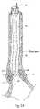
- FIG. 1Ais an illustrated lateral view of the upper GI tract illustrating the positioning of the sphincter treatment apparatus of the present invention in the lower esophageal sphincter.
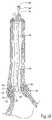
- FIG. 1Bis an illustrated lateral view of the upper GI tract illustrating the delivery of a polymer material into a treatment site in the sphincter wall.
- FIG. 2is a lateral view of the present invention illustrating the catheter lumen, catheter end energy delivery device, cable and power supply.
- FIG. 3depicts a cross sectional view of sphincter anatomy illustrating the layers of the sphincter wall.
- FIG. 4Ais a lateral view of the RF electrode and sphincter wall, illustrating insulated and exposed electrode segments and the creation of a protected site.
- FIG. 4Bis a lateral view of the RF electrode and sphincter wall, illustrating apertures in the catheter which are used to control the penetration angle of the tissue piercing device in a sphincter wall.
- FIG. 5is an enlarged lateral view illustrating the placement of sensors on/adjacent the energy delivery device/RF electrode.
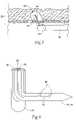
- FIG. 6is a cross sectional view illustrating the use of a fluid introduction lumen and aperture in the energy delivery device/RF electrode for delivery of a cooling medium.
- FIG. 7is a cross sectional view illustrating a visualization device coupled to an embodiment of the invention.
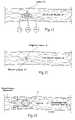
- FIG. 8is a lateral view of the sphincter wall illustrating the use of cooling medium to create cooled zones at the electrode-tissue interface.
- FIG. 9depicts the flow path and fluid connections employed to deliver cooling medium to the energy delivery device/RF electrode and/or electrode-tissue interface.
- FIG. 10is a flow chart illustrating a sphincter treatment method.
- FIG. 11is a lateral view of sphincter smooth muscle tissue illustrating electrical foci and electrically conductive pathways for the origination and conduction of aberrant electrical signals in the smooth muscle of the lower esophageal sphincter or other tissue.
- FIG. 12is a lateral view of a sphincter wall illustrating the infiltration of tissue healing cells into a lesion in the smooth tissue of a sphincter following treatment with the sphincter treatment apparatus of the present invention.
- FIG. 13is a view similar to that of FIG. 12 illustrating shrinkage of the lesion site caused by cell infiltration.
- FIG. 14is a lateral view of the esophageal wall illustrating the preferred placement of lesions in the smooth muscle layer of an esophageal sphincter.
- FIG. 15is a lateral view illustrating a radial distribution of cured polymer particles used to increase sphincter wall thickness and decrease sphincter inner diameter.
- FIG. 16is a lateral view illustrating the use of a band of shrunk collagen surrounding and mechanically supporting a radial distribution of cured polymer particles.
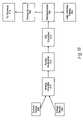
- FIG. 17depicts a block diagram of the feed back control system that can be used with the sphincter treatment apparatus.
- FIG. 18depicts a block diagram of an analog amplifier, analog multiplexer and microprocessor used with the feedback control system of FIG. 17 .
- FIG. 19depicts a block diagram of the operations performed in the feedback control system depicted in FIG. 17 .
- a sphincter treatment apparatus 10delivers energy to a target tissue site 12 , also called treatment site 12 , to produce cell necrosis 14 in a sphincter 16 , such as the lower esophageal sphincter (LES).
- sphincter treatment apparatus 10comprises a flexible elongate shaft 18 , also called introducer 18 , or catheter 18 , with a distal extremity 20 , also called catheter end 20 , in turn coupled with one or more energy delivery devices 22 .
- Energy delivery devices 22are coupled to a guide wire 24 also called cable 24 and are also configured to be coupled to a power source.
- Energy delivery device 22is coupled to a tissue piercing device 26 , which can also be the distal end 26 of energy delivery device 22 .
- Energy delivery device 22 and tissue piercing device 26may both have a continuous internal lumen 23 that is fluidically coupled to a fluid lumen 24 ′ in guide wire 24 .
- Energy delivery device 22 and tissue piercing device 26are configured to penetrate a fixed depth into a sphincter wall 28 and deliver energy to a portion thereof.
- tissue piercing device 26is a hollow hypodermic needle 26 well known to those skilled in the art.
- tissue piercing device 26is configured to penetrate a fixed depth into a sphincter wall 28 and deliver a polymer material 15 (also called polymer 15 ) via lumen 23 to a treatment site 12 . Delivery of polymer 15 can be accomplished using an infusion pump or syringe (neither shown but both well known to those skilled in the art) fluidically coupled to tissue piercing device 26 .
- the delivered polymer material 15 ′undergoes a curing and/or polymerization reaction well known to those skilled in the art whereby one or more of the following occurs: (i) crosslinks form between adjacent molecular chains of polymer 15 , (ii) the molecular chains of polymer 15 contract along their linear/longitudinal axis resulting in a shortening or shrinkage of polymer 15 in one or more axises, (iii) the molecular chains of polymer 15 increase in length (iv) a viscoelastic property of delivered polymer 15 is altered (v) the viscosity of delivered polymer 15 ′ is increased, and (vi) the stiffness of delivered polymer 15 is increased.
- cured polymer 15 ′′also called polymer particle 15 ′′.
- Suitable materials for polymer 15include polysiloxanes (e.g. silicones), polyurethanes and collagen, all well known to those skilled in the art.
- Suitable geometries for polymer particle 15 ′′include, but are not limited to, the following shapes: spherical, semispherical, oval and cylindrical. Suitable diameters for polymer particles 15 ′′ include a range from 0.01 to 0.5 inches.
- catheter end 20is configured to be positionable in a sphincter 16 such as the LES or adjacent anatomical structure, such as the cardia of the stomach.
- Catheter 18has sufficient length to position catheter end 20 in the LES and/or stomach using a trans-oral approach.
- Typical lengths for catheter 18include, but are not limited to, a range of 40-180 cms.
- Suitable materials for catheter 18include, but are not limited to, polyethylenes, polyurethanes, silicones other biocompatible polymers known to those skilled in the art.
- Energy delivery devices 22can be in the form of needle electrodes, both solid or hollow, as is well known to those skilled in the art.
- energy delivery device 22can be conical, cylindrical, rectangular or any polyhedron; each of said shapes having a flat, rounded, beveled, or pointed tip.
- Suitable materials for energy delivery device 22include a combination of one or more of the following: i) stainless and other steels suitable for electrode applications known to those skilled in the art, ii) alloys of gold, silver and platinum, iii) nickel-titanium alloys, or iv) other conductors known to those skilled in the art.
- Catheter 18may have one or more lumens 30 , that extend the frill length of catheter 18 , or only a portion thereof.
- Lumens 30may be used as paths for cables, catheters, guide wires, pull wires, insulated wires, fluid and optical fibers.
- Lumens 30may have one or more apertures 30 ′ at or near distal catheter end 20 .
- lumens 30 (along with aperture 30 ′) in catheter 18are used as a guiding pathway for guidewire 24 to facilitate the positioning of tissue piercing device 26 at treatment site 12 .
- Guide wire 24is configured to facilitate the positioning of energy delivery device 22 a selectable distance (1-4 mms) into the sphincter wall 28 .
- Suitable materials and components for guide wire 24include an insulated wire, an insulated guide wire, a plastic-coated stainless steel hypotube with internal wiring, or a catheter with internal wiring, all of which are known to those skilled in the art.
- Guide wire 24may also have one or more lumens 24 ′ which can be used to deliver fluid or gas.
- guide wire 24may have one or more proximal fittings 24 ′′ (such as a luer fitting or lemo connector) for facilitating connection to fluid lines and electronic cabling.
- energy flowing through sphincter or other tissuecauses heating of the tissue due to absorption of the energy by the tissue. This heating can cause injury to the affected cells and can be substantial enough to cause cell death, a phenomenon also known as cell necrosis.
- the controlled delivery of energy by energy delivery device 22results in controlled cell necrosis 14 , also called lesions 14 , at target tissue site 12 .
- Suitable energy devices and power sources for energy delivery device 22include the following: (i) a radio-frequency (RF) source coupled to an RF electrode, (ii) a coherent source of light coupled to an optical fiber, (iii) an incoherent light source coupled to an optical fiber, (iv) a heated fluid coupled to a catheter with a closed channel configured to receive the heated fluid, (v) a heated fluid coupled to a catheter with an open channel configured to receive the heated fluid, (vi) a cooled fluid coupled to a catheter with a closed channel configured to receive the cooled fluid, (vii) a cooled fluid coupled to a catheter with an open channel configured to receive the cooled fluid, (viii) a cryogenic fluid, (ix) a resistive heating source, (x) a microwave source providing energy from 915 MHz to 2.45 GHz and coupled to a microwave antenna, (xi) an ultrasound power source coupled to an ultrasound emitter, wherein the ultrasound power source produces energy in the range of 300 KHZ to 3 GHz, or (x
- the power source utilizedis an RF source and energy delivery device 22 is one or more RF electrodes 22 , also described as electrodes 22 .
- all of the other herein mentioned power sources and energy delivery devicesare equally applicable to sphincter treatment apparatus 10 .
- RF electrode 22is configured to produce controlled cell necrosis or lesions 14 in smooth muscle collagen tissue layer 34 underlying mucosal and submucosal layers 32 and 33 . More specifically, RF electrode 22 is configured to produce controlled cell necrosis 14 in the portion of smooth muscle collagen tissue 34 ′ that lies approximately 1-4 nuns from the surface of mucosal layer 32 .
- RF electrode 22has an insulator 36 , covering the exterior of an insulated segment 38 except for an exposed segment 40 .
- an insulatoris a barrier to either thermal or electromagnetic energy flow.
- insulated segment 38is of sufficient length to extend into sphincter wall 28 and minimize the transmission of energy and subsequent injury to a protected site 42 near or adjacent to insulated segment 38 .
- Typical lengths for insulated segment 38include, but are not limited to, 1-4 mms.
- Suitable materials for insulator 36include, but are not limited to, polytetrafluoroethylene (Teflon®, polyimides, polyamides and other insulating polymers known to those skilled in the art.
- lumens 30 and apertures 30 ′are of sufficient diameter to allow the free movement of guidewire 24 in catheter 18 so as to be able to controllably position tissue piercing device 26 to a selected depth into sphincter wall 28 .
- Apertures 30 ′can be configured so as to control the angle of penetration 48 (also called penetration angle 48 emergence angle 48 ) that tissue piercing device 26 makes with sphincter wall 28 .
- Apertures 30 ′can be further configured so as to maintain penetration angle 48 constant (or near constant) during the insertion of tissue piercing device 26 into sphincter wall 28 so as to minimize tearing or unnecessary trauma to sphincter wall tissue.
- the emergence angle 48 of apertures 30 ′which can vary from 1 to 90°.
- one or more sensors 44can be coupled to RF electrode 22 for sensing the temperature of sphincter tissue at target tissue site 12 . More specifically, sensors 44 permit accurate determination of the surface temperature of sphincter wall 28 at an electrode tissue interface 46 . This information can be used to regulate both the delivery of energy and cooling medium to the interior surface of sphincter wall 28 as will be discussed herein. Sensors 44 can be positioned on or adjacent to RF electrode 22 . Suitable sensors that may be used for sensor 44 include: thermocouples, fiber optics, resistive wires, thermocouple IR detectors, and the like. Suitable thermocouples for sensor 44 include: T type with copper constantene, J type, E type and K types as are well known those skilled in the art.
- RF electrode 22includes a fluid introduction lumen 23 , that may be coupled with catheter lumen 30 . These coupled lumens provide a path for the delivery of a fluid, such as a cooling or electrolytic fluid (which will be discussed herein), to electrode tissue interface 46 or another site. As shown in FIG. 6 , fluid introduction lumen 23 may include an aperture 50 on the distal portion of RF electrode 22 .
- sphincter treatment apparatus 10includes a visualization device 52 which can include a combination of one or more of the following: a viewing scope, an expanded eyepiece, fiber optics (both imaging and illuminating fibers), video imaging and the like.
- cooling system 54coupled to energy delivery device 22 to cool all or a portion of energy-delivery device 22 and the area near electrode tissue interface 46 before, during or after the delivery of energy in order to reduce the degree and area of cell injury in the tissue adjacent electrode tissue interface 46 .
- the use of coolingprotects against, or otherwise reduces the degree of, cell damage to cooled zone 56 in the vicinity of aperture 50 and/or electrode tissue interface 46 which will preferably include mucosal and submucosal layers 32 and 33 .
- FIG. 8the use of cooling protects against, or otherwise reduces the degree of, cell damage to cooled zone 56 in the vicinity of aperture 50 and/or electrode tissue interface 46 which will preferably include mucosal and submucosal layers 32 and 33 .
- cooling system 54can include one or more of the following: i) a cooling medium 55 (which can be a liquid or a gas) that is delivered to RF electrode 22 via aperture 50 and flow-controlled via a feedback control system 60 discussed herein, ii) a cooling medium reservoir 58 coupled to aperture 50 , and iii) a cooling device 59 (which may be integral to fluid reservoir 58 ) coupled to cooling medium 55 and controlled via a feedback control system 60 discussed herein.
- cooling medium 55can be introduced via apertures 50 or semipermeable membranes located in one or more locations on sphincter treatment apparatus 10 in communication with reservoir 58 and thermal communication with cooling device 59 .
- cooling medium 55can be introduced externally to RF electrode 22 .
- cooling medium 55is thermally coupled to RF electrode 22 and/or electrode tissue interface 46 .
- cooling device 59(such as a Peltier effect device or heat pipe) is thermally coupled to RF electrode 22 and/or electrode tissue interface 46 .
- FIG. 10is a flow chart illustrating a method for using sphincter treatment apparatus 10 .
- sphincter treatment apparatus 10is first introduced into the esophagus under local anesthesia and positioned at target tissue site 12 .
- Sphincter treatment apparatus 10can be introduced into the esophagus by itself or through a lumen in an endoscope (not shown), such as disclosed in U.S. Pat. Nos. 5,448,990 and 5,275,608, incorporated herein by reference, or a similar esophageal access device known to those skilled in the art.
- the diagnostic phase of the procedurethen begins and can be performed using a variety of diagnostic methods, including, but not limited to, the following: (i) visualization of the interior surface of the esophagus via an endoscope or other viewing apparatus inserted into the esophagus, (ii) visualization of the interior morphology of the esophageal wall using ultrasonography to establish a baseline for the tissue to be treated, (iii) impedance measurement to determine the electrical conductivity between the esophageal mucosal layers and sphincter treatment apparatus 10 , and (iv) measurement and surface mapping of the electropotential of the LES during varying time periods which may include such events as depolarization, contraction and repolarization of LES smooth muscle tissue.
- This latter techniqueis done to determine target tissue sites 12 in the LES or adjoining anatomical structures that are acting as electrical foci 107 or electrically conductive pathways 109 for abnormal or inappropriate polarization and relaxation of the smooth muscle of the LES (Refer to FIG. 11 ).
- the treatment phase of the procedurethen begins.
- the delivery of energy to target tissue site 12can be conducted under feedback control (described herein), manually or by a combination of both.
- Feedback controlenables sphincter treatment apparatus 10 to be positioned and retained in the esophagus during treatment with minimal attention by the physician.
- Feedbackcan be included and is achieved by the use of one or more of the following methods: (i) visualization, (ii) impedance measurement, (iii) ultrasonography, (iv) temperature measurement and, (v) sphincter contractile force measurement via manometry.
- a second diagnostic phasemay be included after the treatment is completed.
- Thisprovides an indication of LES tightening treatment success, and whether or not a second phase of treatment, to all or only a portion of the esophagus, now or at some later time, should be conducted.
- the second diagnostic phaseis accomplished through, (i) visualization, (ii) measuring impedance, (iii) ultrasonography, (iv) temperature measurement, or (v) measurement of LES tension and contractile force via manometry. It will be appreciated that the above procedure is applicable in whole or part to the treatment of other sphincters in the body.
- the area and magnitude of cell injury in the LES or sphincter 16can vary. However, it is desirable to deliver sufficient energy to the targeted tissue site 12 to be able to achieve tissue temperatures in the range of 55-95° C. and produce lesions 14 at depths ranging from 1-4 mms from the interior surface of the LES or sphincter wall 28 . It is also desirable to deliver sufficient energy such that the resulting lesions 14 have a sufficient magnitude and area of cell injury to cause an infiltration and/or proliferation of lesion 14 by fibroblasts 110 , myofibroblasts 112 , macrophages 114 and other cells involved in the tissue healing process (refer to FIG. 12 ). As shown in FIG.
- these cellscause a contraction of tissue around lesion 14 , decreasing its volume and/or altering the biomechanical properties at lesion 14 so as to result in a tightening of LES or sphincter 16 .
- These changesare reflected in transformed lesion 14 ′ shown in FIG. 13 .
- lesions 14are predominantly located in the smooth muscle collagen layer of selected sphincter 16 at the depths ranging from 1 to 4 mms from the interior surface of sphincter wall 28 .
- the diameter of lesions 14can vary between 0.1 to 4 mms. It is preferable that lesions 14 are less than 4 mms in diameter in order to reduce the risk of thermal damage to the mucosal layer.
- a 2 mm diameter lesion 14 centered in the wall of the smooth muscle collagen layerprovides a 1 mm buffer zone to prevent damage to the mucosa, submucosa and adventitia, while still allowing for cell infiltration and subsequent sphincter tightening on approximately 50% of the thickness of the wall of the smooth muscle collagen layer (refer to FIG. 14 ).
- lesions 14can vary both in number and position within sphincter wall 28 .
- polymer particles 15 ′′can be distributed in a variety of patterns in sphincter wall 28 including a radial distribution at even depths along a radial axis of sphincter 16 .
- Other distributions not showninclude: (i) a wavy or folded circle of polymer particles 15 ′′ at varying depths in sphincter wall 28 evenly spaced along the radial axis of sphincter 16 , (ii) polymer particles 15 ′′ randomly distributed at varying depths, but evenly spaced in a radial direction; and, (iii) an eccentric pattern of polymer particles 15 ′′ in one or more radial locations in sphincter wall 28 .
- the pattern of and diameter of polymer particles 15 ′′can be selected to controllably increase the thickness 28 ′ of sphincter wall 28 and/or decrease the inner diameter 16 ′ of sphincter 16 .
- RF energycan be delivered to sphincter wall 28 to shrink native collagen 51 within the smooth muscle collagen tissue layer 34 of sphincter wall 28 so as to create a supporting band 57 of tightened collagen in contact with one or more of polymer particles 15 ′′ distributed along a radial axis of sphincter 16 .
- Band 57serves to both mechanically link and mechanically support polymer particles 15 ′′. This serves one or more of the following functions: (i) distribution of the stresses within sphincter wall 28 , (ii) retention of the desired placement of polymer particles 15 ′′ within sphincter wall 28 ; and, (iii) maintenance of improvements in the tension and inner diameter of sphincter wall 28 .
- band 57can be selectively shrunk so as to selectively tighten sphincter 16 and decrease inner sphincter diameter 16 ′.
- band 57can be composed of fibroblasts 110 , myofibroblasts 112 and other tissue healing cells.
- elements of sphincter treatment apparatus 10are coupled to an open or closed loop feedback control system 60 .
- an open or closed loop feedback system 60couples sensor 346 to energy source 392 .
- electrode 314is one or more RF electrodes 314 .
- the temperature of the tissue, or of RF electrode 314is monitored, and the output power of energy source 392 adjusted accordingly.
- the physiciancan, if desired, override the closed or open loop system 60 .
- a microprocessor 394can be included and incorporated in the closed or open loop system to switch power on and off, as well as modulate the power.
- the closed loop system 60utilizes microprocessor 394 to serve as a controller, monitor the temperature, adjust the RF power, analyze the result, refeed the result, and then modulate the power.
- tissue adjacent to RF electrode 314can be maintained at a desired temperature for a selected period of time without causing a shut down of the power circuit to electrode 314 due to the development of excessive electrical impedance at electrode 314 or adjacent tissue as is discussed herein.
- Each RF electrode 314is connected to resources which generate an independent output. The output maintains a selected energy at RF electrode 314 for a selected length of time.
- a control signalis generated by controller 404 that is proportional to the difference between an actual measured value, and a desired value.
- the control signalis used by power circuits 406 to adjust the power output an appropriate amount in order to maintain the desired power delivered at respective RF electrodes 314 .
- temperatures detected at sensor 346provide feedback for maintaining a selected power. Temperature at sensor 346 is used as a safety means to interrupt the delivery of power when maximum pre-set temperatures are exceeded.
- the actual temperaturesare measured at temperature measurement device 408 , and the temperatures are displayed at user interface and display 402 .
- a control signalis generated by controller 404 that is proportional to the difference between an actual measured temperature and a desired temperature.
- the control signalis used by power circuits 406 to adjust the power output an appropriate amount in order to maintain the desired temperature delivered at the sensor 346 .
- a multiplexercan be included to measure current, voltage and temperature, at the sensor 346 , and energy can be delivered to RF electrode 314 in monopolar or bipolar fashion.
- Controller 404can be a digital or analog controller, or a computer with software.
- controller 404is a computer it can include a CPU coupled through a system bus.
- This systemcan include a keyboard, a disk drive, or other non-volatile memory systems, a display, and other peripherals, as are known in the art.
- Also coupled to the busis a program memory and a data memory.
- User interface and display 402includes operator controls and a display.
- Controller 404can be coupled to imaging systems including, but not limited to, ultrasound, CT scanners, X-ray, MRI, mammographic X-ray and the like. Further, direct visualization and tactile imaging can be utilized.
- the output of current sensor 396 and voltage sensor 398are used by controller 404 to maintain a selected power level at RF electrode 314 .
- the amount of RF energy deliveredcontrols the amount of power.
- a profile of the power delivered to electrode 314can be incorporated in controller 404 and a preset amount of energy to be delivered may also be profiled.
- Circuitry, software and feedback to controller 404result in process control, the maintenance of the selected power setting which is independent of changes in voltage or current, and is used to change the following process variables: (i) the selected power setting, (ii) the duty cycle (e.g., on-off time), (iii) bipolar or monopolar energy delivery; and, (iv) fluid delivery, including flow rate and pressure.
- process variablesare controlled and varied, while maintaining the desired delivery of power independent of changes in voltage or current, based on temperatures monitored at sensor 346 .
- Analog amplifier 410can be a conventional differential amplifier circuit for use with sensor 346 .
- the output of analog amplifier 410is sequentially connected by an analog multiplexer 412 to the input of A/D converter 414 .
- the output of analog amplifier 410is a voltage which represents the respective sensed temperatures.
- Digitized amplifier output voltagesare supplied by A/D converter 414 to microprocessor 394 .
- Microprocessor 394may be a type 68HCII available from Motorola. However, it will be appreciated that any suitable microprocessor or general purpose digital or analog computer can be used to calculate impedance or temperature.
- Microprocessor 394sequentially receives and stores digital representations of impedance and temperature. Each digital value received by microprocessor 394 corresponds to different temperatures and impedances.
- Calculated power and impedance valuescan be indicated on user interface and display 402 .
- calculated impedance and power valuescan be compared by microprocessor 394 to power and impedance limits. When the values exceed predetermined power or impedance values, a warning can be given on user interface and display 402 , and additionally, the delivery of RF energy can be reduced, modified or interrupted.
- a control signal from microprocessor 394can modify the power level supplied by energy source 392 .
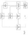
- FIG. 19illustrates a block diagram of a temperature and impedance feedback system that can be used to control the delivery of energy to tissue site 416 by energy source 392 and the delivery of cooling medium 55 to electrode 314 and/or tissue site 416 by flow regulator 418 .
- Energyis delivered to RF electrode 314 by energy source 392 , and applied to tissue site 416 .
- a monitor 420ascertains tissue impedance, based on the energy delivered to tissue, and compares the measured impedance value to a set value. If measured impedance is within acceptable limits, energy continues to be applied to the tissue. However if the measured impedance exceeds the set value, a disabling signal 422 is transmitted to energy source 392 , ceasing further delivery of energy to RF electrode 314 .
- temperature measurement device 408measures the temperature of tissue site 416 and/or RF electrode 314 .
- a comparator 424receives a signal representative of the measured temperature and compares this value to a pre-set signal representative of the desired temperature. If the measured temperature has not exceeded the desired temperature, comparator 424 sends a signal to flow regulator 418 to maintain the cooling solution flow rate at its existing level. However if the tissue temperature is too high, comparator 424 sends a signal to a flow regulator 418 (connected to an electronically controlled micropump, not shown) representing a need for an increased cooling solution flow rate.
Landscapes
- Health & Medical Sciences (AREA)
- Surgery (AREA)
- Engineering & Computer Science (AREA)
- Life Sciences & Earth Sciences (AREA)
- Biomedical Technology (AREA)
- Molecular Biology (AREA)
- Nuclear Medicine, Radiotherapy & Molecular Imaging (AREA)
- Plasma & Fusion (AREA)
- Physics & Mathematics (AREA)
- Heart & Thoracic Surgery (AREA)
- Medical Informatics (AREA)
- Otolaryngology (AREA)
- Animal Behavior & Ethology (AREA)
- General Health & Medical Sciences (AREA)
- Public Health (AREA)
- Veterinary Medicine (AREA)
- Cardiology (AREA)
- Surgical Instruments (AREA)
Abstract
Description
Claims (17)
Priority Applications (2)
| Application Number | Priority Date | Filing Date | Title |
|---|---|---|---|
| US11/079,697US7449020B2 (en) | 1998-02-27 | 2005-03-14 | Method for treating a sphincter |
| US12/291,626US8518032B2 (en) | 1998-02-27 | 2008-11-12 | Method for treating a sphincter |
Applications Claiming Priority (6)
| Application Number | Priority Date | Filing Date | Title |
|---|---|---|---|
| US3236698A | 1998-02-27 | 1998-02-27 | |
| US09/032,367US6044846A (en) | 1994-06-24 | 1998-02-27 | Method to treat esophageal sphincters |
| US9079498A | 1998-06-04 | 1998-06-04 | |
| US09/921,356US6613047B2 (en) | 1994-06-24 | 2001-08-02 | Apparatus to treat esophageal sphincters |
| US10/254,471US6866663B2 (en) | 1998-02-27 | 2002-09-25 | Method for treating a sphincter |
| US11/079,697US7449020B2 (en) | 1998-02-27 | 2005-03-14 | Method for treating a sphincter |
Related Parent Applications (2)
| Application Number | Title | Priority Date | Filing Date |
|---|---|---|---|
| US09/921,356Continuation-In-PartUS6613047B2 (en) | 1994-06-24 | 2001-08-02 | Apparatus to treat esophageal sphincters |
| US10/254,471DivisionUS6866663B2 (en) | 1998-02-27 | 2002-09-25 | Method for treating a sphincter |
Related Child Applications (2)
| Application Number | Title | Priority Date | Filing Date |
|---|---|---|---|
| US9079498AContinuation-In-Part | 1998-02-19 | 1998-06-04 | |
| US12/291,626DivisionUS8518032B2 (en) | 1998-02-27 | 2008-11-12 | Method for treating a sphincter |
Publications (2)
| Publication Number | Publication Date |
|---|---|
| US20050159743A1 US20050159743A1 (en) | 2005-07-21 |
| US7449020B2true US7449020B2 (en) | 2008-11-11 |
Family
ID=27487990
Family Applications (4)
| Application Number | Title | Priority Date | Filing Date |
|---|---|---|---|
| US10/207,520AbandonedUS20030135206A1 (en) | 1998-02-27 | 2002-07-29 | Method for treating a sphincter |
| US10/254,471Expired - Fee RelatedUS6866663B2 (en) | 1998-02-27 | 2002-09-25 | Method for treating a sphincter |
| US11/079,697Expired - Fee RelatedUS7449020B2 (en) | 1998-02-27 | 2005-03-14 | Method for treating a sphincter |
| US12/291,626Expired - Fee RelatedUS8518032B2 (en) | 1998-02-27 | 2008-11-12 | Method for treating a sphincter |
Family Applications Before (2)
| Application Number | Title | Priority Date | Filing Date |
|---|---|---|---|
| US10/207,520AbandonedUS20030135206A1 (en) | 1998-02-27 | 2002-07-29 | Method for treating a sphincter |
| US10/254,471Expired - Fee RelatedUS6866663B2 (en) | 1998-02-27 | 2002-09-25 | Method for treating a sphincter |
Family Applications After (1)
| Application Number | Title | Priority Date | Filing Date |
|---|---|---|---|
| US12/291,626Expired - Fee RelatedUS8518032B2 (en) | 1998-02-27 | 2008-11-12 | Method for treating a sphincter |
Country Status (1)
| Country | Link |
|---|---|
| US (4) | US20030135206A1 (en) |
Cited By (6)
| Publication number | Priority date | Publication date | Assignee | Title |
|---|---|---|---|---|
| US20060070631A1 (en)* | 2004-09-27 | 2006-04-06 | Boston Scientific Scimed, Inc. | Devices and methods for agent-assisted medical procedures |
| US20100160995A1 (en)* | 2008-12-18 | 2010-06-24 | Jerome Dargent | Method for treating obesity |
| US8226730B2 (en)* | 2003-08-11 | 2012-07-24 | Cook Medical Technologies Llc | Surgical implant |
| US20130096552A1 (en)* | 2011-10-14 | 2013-04-18 | Christopher L. Brace | Hydrodissection Material with Reduced Migration |
| US10716618B2 (en) | 2010-05-21 | 2020-07-21 | Stratus Medical, LLC | Systems and methods for tissue ablation |
| US10736688B2 (en) | 2009-11-05 | 2020-08-11 | Stratus Medical, LLC | Methods and systems for spinal radio frequency neurotomy |
Families Citing this family (80)
| Publication number | Priority date | Publication date | Assignee | Title |
|---|---|---|---|---|
| US9023031B2 (en) | 1997-08-13 | 2015-05-05 | Verathon Inc. | Noninvasive devices, methods, and systems for modifying tissues |
| US20030135206A1 (en)* | 1998-02-27 | 2003-07-17 | Curon Medical, Inc. | Method for treating a sphincter |
| US6740082B2 (en)* | 1998-12-29 | 2004-05-25 | John H. Shadduck | Surgical instruments for treating gastro-esophageal reflux |
| US6238335B1 (en)* | 1998-12-11 | 2001-05-29 | Enteric Medical Technologies, Inc. | Method for treating gastroesophageal reflux disease and apparatus for use therewith |
| US6821285B2 (en) | 1999-06-22 | 2004-11-23 | Ndo Surgical, Inc. | Tissue reconfiguration |
| US6663639B1 (en)* | 1999-06-22 | 2003-12-16 | Ndo Surgical, Inc. | Methods and devices for tissue reconfiguration |
| US6835200B2 (en) | 1999-06-22 | 2004-12-28 | Ndo Surgical. Inc. | Method and devices for tissue reconfiguration |
| US6494888B1 (en)* | 1999-06-22 | 2002-12-17 | Ndo Surgical, Inc. | Tissue reconfiguration |
| US8287554B2 (en) | 1999-06-22 | 2012-10-16 | Ethicon Endo-Surgery, Inc. | Method and devices for tissue reconfiguration |
| US7846180B2 (en) | 1999-06-22 | 2010-12-07 | Ethicon Endo-Surgery, Inc. | Tissue fixation devices and methods of fixing tissue |
| US20040215235A1 (en)* | 1999-11-16 | 2004-10-28 | Barrx, Inc. | Methods and systems for determining physiologic characteristics for treatment of the esophagus |
| WO2001035846A1 (en)* | 1999-11-16 | 2001-05-25 | Ganz Robert A | System and method of treating abnormal tissue in the human esophagus |
| US20060095032A1 (en) | 1999-11-16 | 2006-05-04 | Jerome Jackson | Methods and systems for determining physiologic characteristics for treatment of the esophagus |
| ES2435094T3 (en) | 2000-05-19 | 2013-12-18 | C.R. Bard, Inc. | Device and method of tissue capture and suturing |
| SE516673C2 (en)* | 2000-07-27 | 2002-02-12 | Q Med Ab | Arrangement and method of cooperation with an internal body organ |
| US7737109B2 (en) | 2000-08-11 | 2010-06-15 | Temple University Of The Commonwealth System Of Higher Education | Obesity controlling method |
| CA2417129A1 (en) | 2000-08-11 | 2002-02-21 | Temple University Of The Commonwealth System Of Higher Education | Obesity controlling method |
| US7306591B2 (en) | 2000-10-02 | 2007-12-11 | Novasys Medical, Inc. | Apparatus and methods for treating female urinary incontinence |
| DE10129912A1 (en)* | 2001-06-21 | 2003-01-02 | Efmt Entwicklungs Und Forschun | needle electrode |
| US7047980B2 (en)* | 2002-04-09 | 2006-05-23 | Promethean Surgical Devices Llc | Treatment for Gastroesophageal disease |
| US20040019388A1 (en)* | 2002-07-24 | 2004-01-29 | Starkebaum Warren L. | Methods and implants for retarding stomach emptying to treat eating disorders |
| US8021359B2 (en)* | 2003-02-13 | 2011-09-20 | Coaptus Medical Corporation | Transseptal closure of a patent foramen ovale and other cardiac defects |
| US7257450B2 (en) | 2003-02-13 | 2007-08-14 | Coaptus Medical Corporation | Systems and methods for securing cardiovascular tissue |
| US7655048B2 (en)* | 2003-04-02 | 2010-02-02 | Furlow Jr Leonard T | Materials and methods for soft tissue augmentation |
| US7097650B2 (en)* | 2003-10-14 | 2006-08-29 | Satiety, Inc. | System for tissue approximation and fixation |
| US7914543B2 (en) | 2003-10-14 | 2011-03-29 | Satiety, Inc. | Single fold device for tissue fixation |
| US7150745B2 (en) | 2004-01-09 | 2006-12-19 | Barrx Medical, Inc. | Devices and methods for treatment of luminal tissue |
| US7467015B2 (en) | 2004-04-29 | 2008-12-16 | Neuwave Medical, Inc. | Segmented catheter for tissue ablation |
| US20070016181A1 (en) | 2004-04-29 | 2007-01-18 | Van Der Weide Daniel W | Microwave tissue resection tool |
| US7244254B2 (en)* | 2004-04-29 | 2007-07-17 | Micrablate | Air-core microwave ablation antennas |
| US20070016180A1 (en)* | 2004-04-29 | 2007-01-18 | Lee Fred T Jr | Microwave surgical device |
| US7473252B2 (en) | 2004-10-07 | 2009-01-06 | Coaptus Medical Corporation | Systems and methods for shrinking and/or securing cardiovascular tissue |
| EP1827555A4 (en) | 2004-11-18 | 2010-03-10 | David W Chang | Endoluminal delivery of anesthesia |
| AU2005319399B2 (en)* | 2004-12-21 | 2011-05-12 | Davol Inc. | Anastomotic outlet revision |
| US20080221597A1 (en)* | 2005-10-12 | 2008-09-11 | Wallace Jeffrey M | Methods and devices for intragastrointestinal fixation |
| US20070093804A1 (en)* | 2005-10-17 | 2007-04-26 | Coaptus Medical Corporation | Control systems for patient devices, including devices for securing cardiovascular tissue, and associated methods |
| US8702694B2 (en) | 2005-11-23 | 2014-04-22 | Covidien Lp | Auto-aligning ablating device and method of use |
| US7959627B2 (en) | 2005-11-23 | 2011-06-14 | Barrx Medical, Inc. | Precision ablating device |
| US7997278B2 (en) | 2005-11-23 | 2011-08-16 | Barrx Medical, Inc. | Precision ablating method |
| US20070142699A1 (en)* | 2005-12-16 | 2007-06-21 | Acoustx Corporation | Methods and implantable apparatuses for treating an esophageal disorder such as gastroesophageal reflux disease |
| US20070142884A1 (en)* | 2005-12-16 | 2007-06-21 | Acoustx Corporation | Methods and apparatuses for treating an esophageal disorder such as gastroesophageal reflux disease |
| US20080086200A1 (en)* | 2006-01-03 | 2008-04-10 | Heartcor | Injectable implants for tissue augmentation and restoration |
| EP1998699A1 (en)* | 2006-03-24 | 2008-12-10 | Neuwave Medical, Inc. | Energy delivery system |
| US8672932B2 (en) | 2006-03-24 | 2014-03-18 | Neuwave Medical, Inc. | Center fed dipole for use with tissue ablation systems, devices and methods |
| US10363092B2 (en) | 2006-03-24 | 2019-07-30 | Neuwave Medical, Inc. | Transmission line with heat transfer ability |
| US7942809B2 (en)* | 2006-05-26 | 2011-05-17 | Leban Stanley G | Flexible ultrasonic wire in an endoscope delivery system |
| US11389235B2 (en) | 2006-07-14 | 2022-07-19 | Neuwave Medical, Inc. | Energy delivery systems and uses thereof |
| US10376314B2 (en) | 2006-07-14 | 2019-08-13 | Neuwave Medical, Inc. | Energy delivery systems and uses thereof |
| US8136532B2 (en)* | 2006-08-30 | 2012-03-20 | Med-El Elektromedizinische Geraete Gmbh | System, apparatus, and method for facilitating interface with laryngeal structures |
| EP1897506B1 (en)* | 2006-09-08 | 2010-03-03 | Ethicon Endo-Surgery, Inc. | A surgical instrument for performing controlled myotomies |
| US8852216B2 (en) | 2007-03-23 | 2014-10-07 | Ethicon Endo-Surgery, Inc. | Tissue approximation methods |
| US8641711B2 (en) | 2007-05-04 | 2014-02-04 | Covidien Lp | Method and apparatus for gastrointestinal tract ablation for treatment of obesity |
| US8784338B2 (en) | 2007-06-22 | 2014-07-22 | Covidien Lp | Electrical means to normalize ablational energy transmission to a luminal tissue surface of varying size |
| WO2009009443A1 (en) | 2007-07-06 | 2009-01-15 | Barrx Medical, Inc. | Method and apparatus for gastrointestinal tract ablation to achieve loss of persistent and/or recurrent excess body weight following a weight-loss operation |
| US20090012518A1 (en)* | 2007-07-06 | 2009-01-08 | Utley David S | Method and Apparatus for Ablation of Benign, Pre-Cancerous and Early Cancerous Lesions That Originate Within the Epithelium and are Limited to the Mucosal Layer of the Gastrointestinal Tract |
| CN102688092B (en) | 2007-07-06 | 2015-04-22 | 柯惠有限合伙公司 | Ablation in the gastrointestinal tract to achieve hemostasis and eradicate lesions with a propensity for bleeding |
| EP2497520B1 (en) | 2007-07-18 | 2022-04-13 | Silk Road Medical, Inc. | Systems for establishing retrograde carotid arterial blood flow |
| US8858490B2 (en) | 2007-07-18 | 2014-10-14 | Silk Road Medical, Inc. | Systems and methods for treating a carotid artery |
| US8273012B2 (en) | 2007-07-30 | 2012-09-25 | Tyco Healthcare Group, Lp | Cleaning device and methods |
| US8646460B2 (en)* | 2007-07-30 | 2014-02-11 | Covidien Lp | Cleaning device and methods |
| US20090118726A1 (en)* | 2007-10-05 | 2009-05-07 | Coaptus Medical Corporation | Systems and Methods for Transeptal Cardiac Procedures, Including Tissue Sealing Members Associated Methods |
| WO2010063024A1 (en)* | 2008-11-30 | 2010-06-03 | Purdue Research Foundation | Flexible neural probe for magnetic insertion |
| EP3549544B1 (en) | 2009-07-28 | 2021-01-06 | Neuwave Medical, Inc. | DEVICE FOR ABLATION |
| US8568398B2 (en) | 2009-09-29 | 2013-10-29 | Covidien Lp | Flow rate monitor for fluid cooled microwave ablation probe |
| ES2856026T3 (en) | 2010-05-03 | 2021-09-27 | Neuwave Medical Inc | Power supply systems |
| US10278774B2 (en) | 2011-03-18 | 2019-05-07 | Covidien Lp | Selectively expandable operative element support structure and methods of use |
| US9192438B2 (en) | 2011-12-21 | 2015-11-24 | Neuwave Medical, Inc. | Energy delivery systems and uses thereof |
| US8403927B1 (en) | 2012-04-05 | 2013-03-26 | William Bruce Shingleton | Vasectomy devices and methods |
| US9409014B2 (en) | 2013-06-12 | 2016-08-09 | Med-El Elektromedizinische Geraete Gmbh | Method for modifying larynx position by trans-positioning muscle and electrode stimulation |
| US9108054B2 (en) | 2013-06-12 | 2015-08-18 | Med-El Elektromedizinische Geraete Gmbh | Method for modifying larynx position by trans-positioning muscle and electrode stimulation |
| CN113367788B (en) | 2015-10-26 | 2024-09-06 | 纽韦弗医疗设备公司 | Energy delivery systems and uses thereof |
| US10065018B2 (en)* | 2016-03-16 | 2018-09-04 | Krishna Rocha-Singh | Apparatus and method for promoting angiogenesis in ischemic tissue |
| US10531917B2 (en) | 2016-04-15 | 2020-01-14 | Neuwave Medical, Inc. | Systems and methods for energy delivery |
| EP3522807B1 (en) | 2016-10-04 | 2025-07-09 | Avent, Inc. | Cooled rf probes |
| US11896823B2 (en) | 2017-04-04 | 2024-02-13 | Btl Healthcare Technologies A.S. | Method and device for pelvic floor tissue treatment |
| US10993759B2 (en)* | 2017-10-20 | 2021-05-04 | Ethicon, Inc. | Hypothermic linear surgical staplers and methods of use |
| US10888320B2 (en)* | 2017-10-20 | 2021-01-12 | Ethicon, Inc. | Hypothermic circular surgical staplers and methods of use |
| US11672596B2 (en) | 2018-02-26 | 2023-06-13 | Neuwave Medical, Inc. | Energy delivery devices with flexible and adjustable tips |
| US20200114041A1 (en)* | 2018-10-15 | 2020-04-16 | Avent, Inc. | Systems and methods for delivering a polymeric material to a treatment site during a radio frequency ablation procedure |
| US11832879B2 (en) | 2019-03-08 | 2023-12-05 | Neuwave Medical, Inc. | Systems and methods for energy delivery |
Citations (161)
| Publication number | Priority date | Publication date | Assignee | Title |
|---|---|---|---|---|
| US1798902A (en) | 1928-11-05 | 1931-03-31 | Edwin M Raney | Surgical instrument |
| US3517128A (en) | 1968-02-08 | 1970-06-23 | James R Hines | Surgical expanding arm dilator |
| US3901241A (en) | 1973-05-31 | 1975-08-26 | Al Corp Du | Disposable cryosurgical instrument |
| US4011872A (en) | 1974-04-01 | 1977-03-15 | Olympus Optical Co., Ltd. | Electrical apparatus for treating affected part in a coeloma |
| US4196724A (en) | 1978-01-31 | 1980-04-08 | Frecker William H | Tongue locking device |
| US4411266A (en) | 1980-09-24 | 1983-10-25 | Cosman Eric R | Thermocouple radio frequency lesion electrode |
| US4423812A (en) | 1980-09-18 | 1984-01-03 | Olympus Optical Company Limited | Cassette receptacle device |
| EP0139607A1 (en) | 1983-10-07 | 1985-05-02 | Yeda Research And Development Company, Ltd. | Hyperthermia apparatus |
| US4532924A (en) | 1980-05-13 | 1985-08-06 | American Hospital Supply Corporation | Multipolar electrosurgical device and method |
| US4565200A (en) | 1980-09-24 | 1986-01-21 | Cosman Eric R | Universal lesion and recording electrode system |
| US4705041A (en) | 1984-07-06 | 1987-11-10 | Kim Il G | Dilator for Sphincter of Oddi |
| US4901737A (en) | 1987-04-13 | 1990-02-20 | Toone Kent J | Method and therapeutic apparatus for reducing snoring |
| US4906203A (en) | 1988-10-24 | 1990-03-06 | General Motors Corporation | Electrical connector with shorting clip |
| US4907589A (en) | 1988-04-29 | 1990-03-13 | Cosman Eric R | Automatic over-temperature control apparatus for a therapeutic heating device |
| US4943290A (en) | 1987-06-23 | 1990-07-24 | Concept Inc. | Electrolyte purging electrode tip |
| US4947842A (en) | 1988-09-22 | 1990-08-14 | Medical Engineering And Development Institute, Inc. | Method and apparatus for treating tissue with first and second modalities |
| US4955377A (en) | 1988-10-28 | 1990-09-11 | Lennox Charles D | Device and method for heating tissue in a patient's body |
| US4966597A (en) | 1988-11-04 | 1990-10-30 | Cosman Eric R | Thermometric cardiac tissue ablation electrode with ultra-sensitive temperature detection |
| US4976711A (en) | 1989-04-13 | 1990-12-11 | Everest Medical Corporation | Ablation catheter with selectively deployable electrodes |
| WO1991001773A1 (en) | 1989-08-01 | 1991-02-21 | Enrico Mangieri | Percutaneous mechanical dilating catheter for cardiac valves and blood vessels |
| US5019075A (en) | 1984-10-24 | 1991-05-28 | The Beth Israel Hospital | Method and apparatus for angioplasty |
| US5035696A (en) | 1990-02-02 | 1991-07-30 | Everest Medical Corporation | Electrosurgical instrument for conducting endoscopic retrograde sphincterotomy |
| US5046512A (en) | 1989-03-10 | 1991-09-10 | Murchie John A | Method and apparatus for treatment of snoring |
| US5047028A (en) | 1989-05-12 | 1991-09-10 | Quinghua Qian | Method for inducing thrombosis in blood vessels |
| US5057107A (en) | 1989-04-13 | 1991-10-15 | Everest Medical Corporation | Ablation catheter with selectively deployable electrodes |
| US5078717A (en) | 1989-04-13 | 1992-01-07 | Everest Medical Corporation | Ablation catheter with selectively deployable electrodes |
| US5083565A (en) | 1990-08-03 | 1992-01-28 | Everest Medical Corporation | Electrosurgical instrument for ablating endocardial tissue |
| US5084044A (en) | 1989-07-14 | 1992-01-28 | Ciron Corporation | Apparatus for endometrial ablation and method of using same |
| US5088979A (en) | 1990-10-11 | 1992-02-18 | Wilson-Cook Medical Inc. | Method for esophageal invagination and devices useful therein |
| US5094233A (en) | 1991-01-11 | 1992-03-10 | Brennan Louis G | Turbinate sheath device |
| US5100423A (en) | 1990-08-21 | 1992-03-31 | Medical Engineering & Development Institute, Inc. | Ablation catheter |
| US5106360A (en) | 1987-09-17 | 1992-04-21 | Olympus Optical Co., Ltd. | Thermotherapeutic apparatus |
| US5122137A (en) | 1990-04-27 | 1992-06-16 | Boston Scientific Corporation | Temperature controlled rf coagulation |
| WO1992010142A1 (en) | 1990-12-10 | 1992-06-25 | Howmedica Inc. | A device and method for interstitial laser energy delivery |
| US5125928A (en) | 1989-04-13 | 1992-06-30 | Everest Medical Corporation | Ablation catheter with selectively deployable electrodes |
| US5156151A (en) | 1991-02-15 | 1992-10-20 | Cardiac Pathways Corporation | Endocardial mapping and ablation system and catheter probe |
| US5190541A (en) | 1990-10-17 | 1993-03-02 | Boston Scientific Corporation | Surgical instrument and method |
| US5197964A (en) | 1991-11-12 | 1993-03-30 | Everest Medical Corporation | Bipolar instrument utilizing one stationary electrode and one movable electrode |
| US5197963A (en) | 1991-12-02 | 1993-03-30 | Everest Medical Corporation | Electrosurgical instrument with extendable sheath for irrigation and aspiration |
| US5205287A (en) | 1990-04-26 | 1993-04-27 | Hoechst Aktiengesellschaft | Ultrasonic contrast agents, processes for their preparation and the use thereof as diagnostic and therapeutic agents |
| WO1993008755A1 (en) | 1991-11-08 | 1993-05-13 | Ep Technologies, Inc. | Ablation electrode with insulated temperature sensing elements |
| US5215103A (en) | 1986-11-14 | 1993-06-01 | Desai Jawahar M | Catheter for mapping and ablation and method therefor |
| US5232444A (en) | 1988-06-25 | 1993-08-03 | Just Hansjoerg | Dilatation catheter |
| US5236413A (en) | 1990-05-07 | 1993-08-17 | Feiring Andrew J | Method and apparatus for inducing the permeation of medication into internal tissue |
| US5242441A (en) | 1992-02-24 | 1993-09-07 | Boaz Avitall | Deflectable catheter with rotatable tip electrode |
| US5254126A (en) | 1992-06-24 | 1993-10-19 | Ethicon, Inc. | Endoscopic suture punch |
| US5256138A (en) | 1990-10-04 | 1993-10-26 | The Birtcher Corporation | Electrosurgical handpiece incorporating blade and conductive gas functionality |
| US5257451A (en) | 1991-11-08 | 1993-11-02 | Ep Technologies, Inc. | Method of making durable sleeve for enclosing a bendable electrode tip assembly |
| US5263493A (en) | 1992-02-24 | 1993-11-23 | Boaz Avitall | Deflectable loop electrode array mapping and ablation catheter for cardiac chambers |
| US5275162A (en) | 1991-11-08 | 1994-01-04 | Ep Technologies, Inc. | Valve mapping catheter |
| US5275610A (en) | 1991-05-13 | 1994-01-04 | Cook Incorporated | Surgical retractors and method of use |
| US5275608A (en) | 1991-10-16 | 1994-01-04 | Implemed, Inc. | Generic endoscopic instrument |
| US5277201A (en) | 1992-05-01 | 1994-01-11 | Vesta Medical, Inc. | Endometrial ablation apparatus and method |
| US5281218A (en) | 1992-06-05 | 1994-01-25 | Cardiac Pathways Corporation | Catheter having needle electrode for radiofrequency ablation |
| US5281217A (en) | 1992-04-13 | 1994-01-25 | Ep Technologies, Inc. | Steerable antenna systems for cardiac ablation that minimize tissue damage and blood coagulation due to conductive heating patterns |
| US5281216A (en) | 1992-03-31 | 1994-01-25 | Valleylab, Inc. | Electrosurgical bipolar treating apparatus |
| US5292321A (en) | 1990-06-08 | 1994-03-08 | Lee Benjamin I | Thermal balloon angioplasty with thermoplastic stent |
| US5293869A (en) | 1992-09-25 | 1994-03-15 | Ep Technologies, Inc. | Cardiac probe with dynamic support for maintaining constant surface contact during heart systole and diastole |
| US5309910A (en) | 1992-09-25 | 1994-05-10 | Ep Technologies, Inc. | Cardiac mapping and ablation systems |
| US5314466A (en) | 1992-04-13 | 1994-05-24 | Ep Technologies, Inc. | Articulated unidirectional microwave antenna systems for cardiac ablation |
| US5313943A (en) | 1992-09-25 | 1994-05-24 | Ep Technologies, Inc. | Catheters and methods for performing cardiac diagnosis and treatment |
| WO1994010925A1 (en) | 1992-11-13 | 1994-05-26 | American Cardiac Ablation Co., Inc. | Fluid cooled electrosurgical cauterization system |
| US5316020A (en) | 1990-10-03 | 1994-05-31 | Ernest Truffer | Snoring prevention device |
| US5324284A (en) | 1992-06-05 | 1994-06-28 | Cardiac Pathways, Inc. | Endocardial mapping and ablation system utilizing a separately controlled ablation catheter and method |
| US5328467A (en) | 1991-11-08 | 1994-07-12 | Ep Technologies, Inc. | Catheter having a torque transmitting sleeve |
| US5334196A (en) | 1992-10-05 | 1994-08-02 | United States Surgical Corporation | Endoscopic fastener remover |
| EP0608609A2 (en) | 1992-12-01 | 1994-08-03 | Cardiac Pathways Corporation | Catheter for RF ablation with cooled electrode and method |
| US5336222A (en) | 1993-03-29 | 1994-08-09 | Boston Scientific Corporation | Integrated catheter for diverse in situ tissue therapy |
| US5345936A (en) | 1991-02-15 | 1994-09-13 | Cardiac Pathways Corporation | Apparatus with basket assembly for endocardial mapping |
| WO1994021178A1 (en) | 1993-03-23 | 1994-09-29 | Goldrath Milton H | Verres needle suturing device |
| WO1994021165A1 (en) | 1993-03-16 | 1994-09-29 | Ep Technologies, Inc. | Guide sheaths for cardiac mapping and ablation |
| WO1994022366A1 (en) | 1993-04-07 | 1994-10-13 | Cardiac Pathways Corporation | Apparatus and method for ventricular mapping and ablation |
| US5363861A (en) | 1991-11-08 | 1994-11-15 | Ep Technologies, Inc. | Electrode tip assembly with variable resistance to bending |
| US5365945A (en) | 1993-04-13 | 1994-11-22 | Halstrom Leonard W | Adjustable dental applicance for treatment of snoring and obstructive sleep apnea |
| US5366490A (en) | 1992-08-12 | 1994-11-22 | Vidamed, Inc. | Medical probe device and method |
| US5365926A (en) | 1986-11-14 | 1994-11-22 | Desai Jawahar M | Catheter for mapping and ablation and method therefor |
| WO1994026178A1 (en) | 1993-05-14 | 1994-11-24 | Vidamed, Inc. | Bph ablation method and apparatus |
| US5368557A (en) | 1991-01-11 | 1994-11-29 | Baxter International Inc. | Ultrasonic ablation catheter device having multiple ultrasound transmission members |
| US5368592A (en) | 1992-04-13 | 1994-11-29 | Ep Technologies, Inc. | Articulated systems for cardiac ablation |
| US5370678A (en) | 1992-04-13 | 1994-12-06 | Ep Technologies, Inc. | Steerable microwave antenna systems for cardiac ablation that minimize tissue damage and blood coagulation due to conductive heating patterns |
| US5383876A (en) | 1992-11-13 | 1995-01-24 | American Cardiac Ablation Co., Inc. | Fluid cooled electrosurgical probe for cutting and cauterizing tissue |
| US5383917A (en) | 1991-07-05 | 1995-01-24 | Jawahar M. Desai | Device and method for multi-phase radio-frequency ablation |
| US5397339A (en) | 1986-11-14 | 1995-03-14 | Desai; Jawahar M. | Catheter for mapping and ablation and method therefor |
| US5398683A (en) | 1991-05-24 | 1995-03-21 | Ep Technologies, Inc. | Combination monophasic action potential/ablation catheter and high-performance filter system |
| US5401272A (en) | 1992-09-25 | 1995-03-28 | Envision Surgical Systems, Inc. | Multimodality probe with extendable bipolar electrodes |
| US5403311A (en) | 1993-03-29 | 1995-04-04 | Boston Scientific Corporation | Electro-coagulation and ablation and other electrotherapeutic treatments of body tissue |
| US5409483A (en) | 1993-01-22 | 1995-04-25 | Jeffrey H. Reese | Direct visualization surgical probe |
| US5409453A (en) | 1992-08-12 | 1995-04-25 | Vidamed, Inc. | Steerable medical probe with stylets |
| US5415657A (en) | 1992-10-13 | 1995-05-16 | Taymor-Luria; Howard | Percutaneous vascular sealing method |
| US5421819A (en) | 1992-08-12 | 1995-06-06 | Vidamed, Inc. | Medical probe device |
| US5423808A (en) | 1991-11-08 | 1995-06-13 | Ep Technologies, Inc. | Systems and methods for radiofrequency ablation with phase sensitive power detection |
| US5423812A (en) | 1994-01-31 | 1995-06-13 | Ellman; Alan G. | Electrosurgical stripping electrode for palatopharynx tissue |
| WO1995018575A1 (en) | 1994-01-06 | 1995-07-13 | Vidamed, Inc. | Medical probe apparatus with enhanced rf, resistance heating, and microwave ablation capabilities |
| US5433739A (en) | 1993-11-02 | 1995-07-18 | Sluijter; Menno E. | Method and apparatus for heating an intervertebral disc for relief of back pain |
| WO1995019142A1 (en) | 1994-01-12 | 1995-07-20 | Vidamed, Inc. | Thermal mapping catheter with ultrasound probe |
| US5435805A (en) | 1992-08-12 | 1995-07-25 | Vidamed, Inc. | Medical probe device with optical viewing capability |
| US5441499A (en) | 1993-07-14 | 1995-08-15 | Dekna Elektro-U. Medizinische Apparatebau Gesellschaft Mbh | Bipolar radio-frequency surgical instrument |
| US5443470A (en) | 1992-05-01 | 1995-08-22 | Vesta Medical, Inc. | Method and apparatus for endometrial ablation |
| US5451406A (en)* | 1994-07-14 | 1995-09-19 | Advanced Uroscience, Inc. | Tissue injectable composition and method of use |
| WO1995025472A1 (en) | 1994-03-23 | 1995-09-28 | Vidamed, Inc. | Dual-channel rf power delivery system |
| US5454782A (en) | 1994-08-11 | 1995-10-03 | Perkins; Rodney C. | Translumenal circumferential energy delivery device |
| US5456662A (en) | 1993-02-02 | 1995-10-10 | Edwards; Stuart D. | Method for reducing snoring by RF ablation of the uvula |
| US5458597A (en) | 1993-11-08 | 1995-10-17 | Zomed International | Device for treating cancer and non-malignant tumors and methods |
| US5458596A (en) | 1994-05-06 | 1995-10-17 | Dorsal Orthopedic Corporation | Method and apparatus for controlled contraction of soft tissue |
| US5465717A (en) | 1991-02-15 | 1995-11-14 | Cardiac Pathways Corporation | Apparatus and Method for ventricular mapping and ablation |
| US5470308A (en) | 1992-08-12 | 1995-11-28 | Vidamed, Inc. | Medical probe with biopsy stylet |
| US5471982A (en) | 1992-09-29 | 1995-12-05 | Ep Technologies, Inc. | Cardiac mapping and ablation systems |
| US5472441A (en) | 1993-11-08 | 1995-12-05 | Zomed International | Device for treating cancer and non-malignant tumors and methods |
| WO1996000042A1 (en) | 1994-06-24 | 1996-01-04 | Vidacare International | Thin layer ablation apparatus |
| US5484400A (en) | 1992-08-12 | 1996-01-16 | Vidamed, Inc. | Dual channel RF delivery system |
| US5486161A (en) | 1993-02-02 | 1996-01-23 | Zomed International | Medical probe device and method |
| US5490984A (en) | 1992-02-28 | 1996-02-13 | Jsf Consulants Ltd. | Use of injectable biomaterials for the repair and augmentation of the anal sphincters |
| US5496271A (en) | 1990-09-14 | 1996-03-05 | American Medical Systems, Inc. | Combined hyperthermia and dilation catheter |
| US5496311A (en) | 1988-10-28 | 1996-03-05 | Boston Scientific Corporation | Physiologic low stress angioplasty |
| US5500012A (en) | 1992-07-15 | 1996-03-19 | Angeion Corporation | Ablation catheter system |
| US5507743A (en) | 1993-11-08 | 1996-04-16 | Zomed International | Coiled RF electrode treatment apparatus |
| US5514131A (en) | 1992-08-12 | 1996-05-07 | Stuart D. Edwards | Method for the ablation treatment of the uvula |
| US5514130A (en) | 1994-10-11 | 1996-05-07 | Dorsal Med International | RF apparatus for controlled depth ablation of soft tissue |
| US5520684A (en) | 1993-06-10 | 1996-05-28 | Imran; Mir A. | Transurethral radio frequency apparatus for ablation of the prostate gland and method |
| WO1996016606A1 (en) | 1994-12-01 | 1996-06-06 | Vidamed, Inc. | Transurethral needle delivery device with cystoscope and method for treatment of urinary incontinence |
| US5536267A (en) | 1993-11-08 | 1996-07-16 | Zomed International | Multiple electrode ablation apparatus |
| US5545161A (en) | 1992-12-01 | 1996-08-13 | Cardiac Pathways Corporation | Catheter for RF ablation having cooled electrode with electrically insulated sleeve |
| US5545171A (en) | 1994-09-22 | 1996-08-13 | Vidamed, Inc. | Anastomosis catheter |
| US5545434A (en) | 1994-04-01 | 1996-08-13 | Huarng; Hermes | Method of making irregularly porous cloth |
| US5545193A (en) | 1993-10-15 | 1996-08-13 | Ep Technologies, Inc. | Helically wound radio-frequency emitting electrodes for creating lesions in body tissue |
| US5549644A (en) | 1992-08-12 | 1996-08-27 | Vidamed, Inc. | Transurethral needle ablation device with cystoscope and method for treatment of the prostate |
| US5549108A (en) | 1992-09-25 | 1996-08-27 | Ep Technologies, Inc. | Cardiac mapping and ablation systems |
| US5556377A (en) | 1992-08-12 | 1996-09-17 | Vidamed, Inc. | Medical probe apparatus with laser and/or microwave monolithic integrated circuit probe |
| US5558673A (en) | 1994-09-30 | 1996-09-24 | Vidamed, Inc. | Medical probe device and method having a flexible resilient tape stylet |
| WO1996029946A1 (en) | 1995-03-24 | 1996-10-03 | Board Of Regents Of The University Of Nebraska | Apparatus for ablation of tissue masses |
| US5562720A (en) | 1992-05-01 | 1996-10-08 | Vesta Medical, Inc. | Bipolar/monopolar endometrial ablation device and method |
| US5571116A (en) | 1994-10-02 | 1996-11-05 | United States Surgical Corporation | Non-invasive treatment of gastroesophageal reflux disease |
| US5588432A (en) | 1988-03-21 | 1996-12-31 | Boston Scientific Corporation | Catheters for imaging, sensing electrical potentials, and ablating tissue |
| US5599345A (en) | 1993-11-08 | 1997-02-04 | Zomed International, Inc. | RF treatment apparatus |
| US5609151A (en) | 1994-09-08 | 1997-03-11 | Medtronic, Inc. | Method for R-F ablation |
| US5624439A (en) | 1995-08-18 | 1997-04-29 | Somnus Medical Technologies, Inc. | Method and apparatus for treatment of air way obstructions |
| WO1997032532A1 (en) | 1996-03-05 | 1997-09-12 | Vnus Medical Technologies, Inc. | Vascular catheter-based system for heating tissue |
| US5672153A (en) | 1992-08-12 | 1997-09-30 | Vidamed, Inc. | Medical probe device and method |
| US5686425A (en) | 1991-07-10 | 1997-11-11 | C.R. Bard | Composition and method for revitalizing scar tissue |
| US5688490A (en) | 1991-02-15 | 1997-11-18 | Bracco International B.V. | Mucoadhesive compositions for increasing the ultrasonic image contrast of the digestive tract |
| US5702438A (en) | 1995-06-08 | 1997-12-30 | Avitall; Boaz | Expandable recording and ablation catheter system |
| US5709224A (en) | 1995-06-07 | 1998-01-20 | Radiotherapeutics Corporation | Method and device for permanent vessel occlusion |
| US5732698A (en) | 1994-01-28 | 1998-03-31 | Ep Technologies, Inc. | Systems for examining the electrical characteristics of cardiac tissue while minimizing contact with blood within the heart |
| WO1997043971A3 (en) | 1996-05-22 | 1998-04-02 | Somnus Medical Tech Inc | Method and apparatus for ablating turbinates |
| US5738096A (en) | 1993-07-20 | 1998-04-14 | Biosense, Inc. | Cardiac electromechanics |
| WO1997006857A3 (en) | 1995-08-15 | 1998-10-15 | Rita Medical Systems Inc | Apparatus for ablation of a selected mass |
| US5830210A (en) | 1996-10-21 | 1998-11-03 | Plc Medical Systems, Inc. | Catheter navigation apparatus |
| US5830213A (en) | 1996-04-12 | 1998-11-03 | Ep Technologies, Inc. | Systems for heating and ablating tissue using multifunctional electrode structures |
| US5836874A (en) | 1996-04-08 | 1998-11-17 | Ep Technologies, Inc. | Multi-function electrode structures for electrically analyzing and heating body tissue |
| US5860974A (en) | 1993-07-01 | 1999-01-19 | Boston Scientific Corporation | Heart ablation catheter with expandable electrode and method of coupling energy to an electrode on a catheter shaft |
| US5871483A (en) | 1996-01-19 | 1999-02-16 | Ep Technologies, Inc. | Folding electrode structures |
| US6033401A (en) | 1997-03-12 | 2000-03-07 | Advanced Closure Systems, Inc. | Vascular sealing device with microwave antenna |
| US6044846A (en) | 1994-06-24 | 2000-04-04 | Edwards; Stuart D. | Method to treat esophageal sphincters |
| US6073052A (en) | 1996-11-15 | 2000-06-06 | Zelickson; Brian D. | Device and method for treatment of gastroesophageal reflux disease |
| US6098629A (en)* | 1999-04-07 | 2000-08-08 | Endonetics, Inc. | Submucosal esophageal bulking device |
| US6238335B1 (en) | 1998-12-11 | 2001-05-29 | Enteric Medical Technologies, Inc. | Method for treating gastroesophageal reflux disease and apparatus for use therewith |
| US6251064B1 (en) | 1998-12-11 | 2001-06-26 | Enteric Medical Technologies, Inc. | Method for creating valve-like mechanism in natural body passageway |
| US6358197B1 (en) | 1999-08-13 | 2002-03-19 | Enteric Medical Technologies, Inc. | Apparatus for forming implants in gastrointestinal tract and kit for use therewith |
| US20020082610A1 (en) | 2000-11-13 | 2002-06-27 | Iulian Cioanta | Methods for treating the prostate and inhibiting obstruction of the prostatic urethra using biodegradable stents |
| US6613047B2 (en) | 1994-06-24 | 2003-09-02 | Curon Medical, Inc. | Apparatus to treat esophageal sphincters |
| US6666848B2 (en)* | 2001-06-14 | 2003-12-23 | Artes Medical Usa, Inc. | Medical injection apparatus |
Family Cites Families (3)
| Publication number | Priority date | Publication date | Assignee | Title |
|---|---|---|---|---|
| US555637A (en)* | 1896-03-03 | Machine for stringing tags | ||
| US5807395A (en)* | 1993-08-27 | 1998-09-15 | Medtronic, Inc. | Method and apparatus for RF ablation and hyperthermia |
| US20030135206A1 (en)* | 1998-02-27 | 2003-07-17 | Curon Medical, Inc. | Method for treating a sphincter |
- 2002
- 2002-07-29USUS10/207,520patent/US20030135206A1/ennot_activeAbandoned
- 2002-09-25USUS10/254,471patent/US6866663B2/ennot_activeExpired - Fee Related
- 2005
- 2005-03-14USUS11/079,697patent/US7449020B2/ennot_activeExpired - Fee Related
- 2008
- 2008-11-12USUS12/291,626patent/US8518032B2/ennot_activeExpired - Fee Related
Patent Citations (184)
| Publication number | Priority date | Publication date | Assignee | Title |
|---|---|---|---|---|
| US1798902A (en) | 1928-11-05 | 1931-03-31 | Edwin M Raney | Surgical instrument |
| US3517128A (en) | 1968-02-08 | 1970-06-23 | James R Hines | Surgical expanding arm dilator |
| US3901241A (en) | 1973-05-31 | 1975-08-26 | Al Corp Du | Disposable cryosurgical instrument |
| US4011872A (en) | 1974-04-01 | 1977-03-15 | Olympus Optical Co., Ltd. | Electrical apparatus for treating affected part in a coeloma |
| US4196724A (en) | 1978-01-31 | 1980-04-08 | Frecker William H | Tongue locking device |
| US4532924A (en) | 1980-05-13 | 1985-08-06 | American Hospital Supply Corporation | Multipolar electrosurgical device and method |
| US4423812A (en) | 1980-09-18 | 1984-01-03 | Olympus Optical Company Limited | Cassette receptacle device |
| US4411266A (en) | 1980-09-24 | 1983-10-25 | Cosman Eric R | Thermocouple radio frequency lesion electrode |
| US4565200A (en) | 1980-09-24 | 1986-01-21 | Cosman Eric R | Universal lesion and recording electrode system |
| EP0139607A1 (en) | 1983-10-07 | 1985-05-02 | Yeda Research And Development Company, Ltd. | Hyperthermia apparatus |
| US4705041A (en) | 1984-07-06 | 1987-11-10 | Kim Il G | Dilator for Sphincter of Oddi |
| US5019075A (en) | 1984-10-24 | 1991-05-28 | The Beth Israel Hospital | Method and apparatus for angioplasty |
| US5215103A (en) | 1986-11-14 | 1993-06-01 | Desai Jawahar M | Catheter for mapping and ablation and method therefor |
| US5397339A (en) | 1986-11-14 | 1995-03-14 | Desai; Jawahar M. | Catheter for mapping and ablation and method therefor |
| US5365926A (en) | 1986-11-14 | 1994-11-22 | Desai Jawahar M | Catheter for mapping and ablation and method therefor |
| US4901737A (en) | 1987-04-13 | 1990-02-20 | Toone Kent J | Method and therapeutic apparatus for reducing snoring |
| US4943290A (en) | 1987-06-23 | 1990-07-24 | Concept Inc. | Electrolyte purging electrode tip |
| US5106360A (en) | 1987-09-17 | 1992-04-21 | Olympus Optical Co., Ltd. | Thermotherapeutic apparatus |
| US5588432A (en) | 1988-03-21 | 1996-12-31 | Boston Scientific Corporation | Catheters for imaging, sensing electrical potentials, and ablating tissue |
| US4907589A (en) | 1988-04-29 | 1990-03-13 | Cosman Eric R | Automatic over-temperature control apparatus for a therapeutic heating device |
| US5232444A (en) | 1988-06-25 | 1993-08-03 | Just Hansjoerg | Dilatation catheter |
| US4947842A (en) | 1988-09-22 | 1990-08-14 | Medical Engineering And Development Institute, Inc. | Method and apparatus for treating tissue with first and second modalities |
| US4906203A (en) | 1988-10-24 | 1990-03-06 | General Motors Corporation | Electrical connector with shorting clip |
| US4955377A (en) | 1988-10-28 | 1990-09-11 | Lennox Charles D | Device and method for heating tissue in a patient's body |
| US5496311A (en) | 1988-10-28 | 1996-03-05 | Boston Scientific Corporation | Physiologic low stress angioplasty |
| US4966597A (en) | 1988-11-04 | 1990-10-30 | Cosman Eric R | Thermometric cardiac tissue ablation electrode with ultra-sensitive temperature detection |
| US5046512A (en) | 1989-03-10 | 1991-09-10 | Murchie John A | Method and apparatus for treatment of snoring |
| US5057107A (en) | 1989-04-13 | 1991-10-15 | Everest Medical Corporation | Ablation catheter with selectively deployable electrodes |
| US5078717A (en) | 1989-04-13 | 1992-01-07 | Everest Medical Corporation | Ablation catheter with selectively deployable electrodes |
| US5125928A (en) | 1989-04-13 | 1992-06-30 | Everest Medical Corporation | Ablation catheter with selectively deployable electrodes |
| US4976711A (en) | 1989-04-13 | 1990-12-11 | Everest Medical Corporation | Ablation catheter with selectively deployable electrodes |
| US5047028A (en) | 1989-05-12 | 1991-09-10 | Quinghua Qian | Method for inducing thrombosis in blood vessels |
| US5084044A (en) | 1989-07-14 | 1992-01-28 | Ciron Corporation | Apparatus for endometrial ablation and method of using same |
| WO1991001773A1 (en) | 1989-08-01 | 1991-02-21 | Enrico Mangieri | Percutaneous mechanical dilating catheter for cardiac valves and blood vessels |
| US5035696A (en) | 1990-02-02 | 1991-07-30 | Everest Medical Corporation | Electrosurgical instrument for conducting endoscopic retrograde sphincterotomy |
| US5205287A (en) | 1990-04-26 | 1993-04-27 | Hoechst Aktiengesellschaft | Ultrasonic contrast agents, processes for their preparation and the use thereof as diagnostic and therapeutic agents |
| US5122137A (en) | 1990-04-27 | 1992-06-16 | Boston Scientific Corporation | Temperature controlled rf coagulation |
| US5236413B1 (en) | 1990-05-07 | 1996-06-18 | Andrew J Feiring | Method and apparatus for inducing the permeation of medication into internal tissue |
| US5236413A (en) | 1990-05-07 | 1993-08-17 | Feiring Andrew J | Method and apparatus for inducing the permeation of medication into internal tissue |
| US5292321A (en) | 1990-06-08 | 1994-03-08 | Lee Benjamin I | Thermal balloon angioplasty with thermoplastic stent |
| US5083565A (en) | 1990-08-03 | 1992-01-28 | Everest Medical Corporation | Electrosurgical instrument for ablating endocardial tissue |
| US5100423A (en) | 1990-08-21 | 1992-03-31 | Medical Engineering & Development Institute, Inc. | Ablation catheter |
| US5496271A (en) | 1990-09-14 | 1996-03-05 | American Medical Systems, Inc. | Combined hyperthermia and dilation catheter |
| US5316020A (en) | 1990-10-03 | 1994-05-31 | Ernest Truffer | Snoring prevention device |
| US5256138A (en) | 1990-10-04 | 1993-10-26 | The Birtcher Corporation | Electrosurgical handpiece incorporating blade and conductive gas functionality |
| US5088979A (en) | 1990-10-11 | 1992-02-18 | Wilson-Cook Medical Inc. | Method for esophageal invagination and devices useful therein |
| US5190541A (en) | 1990-10-17 | 1993-03-02 | Boston Scientific Corporation | Surgical instrument and method |
| WO1992010142A1 (en) | 1990-12-10 | 1992-06-25 | Howmedica Inc. | A device and method for interstitial laser energy delivery |
| US5368557A (en) | 1991-01-11 | 1994-11-29 | Baxter International Inc. | Ultrasonic ablation catheter device having multiple ultrasound transmission members |
| US5094233A (en) | 1991-01-11 | 1992-03-10 | Brennan Louis G | Turbinate sheath device |
| US5345936A (en) | 1991-02-15 | 1994-09-13 | Cardiac Pathways Corporation | Apparatus with basket assembly for endocardial mapping |
| US5156151A (en) | 1991-02-15 | 1992-10-20 | Cardiac Pathways Corporation | Endocardial mapping and ablation system and catheter probe |
| US5465717A (en) | 1991-02-15 | 1995-11-14 | Cardiac Pathways Corporation | Apparatus and Method for ventricular mapping and ablation |
| US5688490A (en) | 1991-02-15 | 1997-11-18 | Bracco International B.V. | Mucoadhesive compositions for increasing the ultrasonic image contrast of the digestive tract |
| US5275610A (en) | 1991-05-13 | 1994-01-04 | Cook Incorporated | Surgical retractors and method of use |
| US5398683A (en) | 1991-05-24 | 1995-03-21 | Ep Technologies, Inc. | Combination monophasic action potential/ablation catheter and high-performance filter system |
| US5383917A (en) | 1991-07-05 | 1995-01-24 | Jawahar M. Desai | Device and method for multi-phase radio-frequency ablation |
| US5686425A (en) | 1991-07-10 | 1997-11-11 | C.R. Bard | Composition and method for revitalizing scar tissue |
| US5275608A (en) | 1991-10-16 | 1994-01-04 | Implemed, Inc. | Generic endoscopic instrument |
| US5688266A (en) | 1991-11-08 | 1997-11-18 | Ep Technologies, Inc. | Electrode and associated systems using thermally insulated temperature sensing elements |
| WO1993008755A1 (en) | 1991-11-08 | 1993-05-13 | Ep Technologies, Inc. | Ablation electrode with insulated temperature sensing elements |
| US5456682A (en) | 1991-11-08 | 1995-10-10 | Ep Technologies, Inc. | Electrode and associated systems using thermally insulated temperature sensing elements |
| US5275162A (en) | 1991-11-08 | 1994-01-04 | Ep Technologies, Inc. | Valve mapping catheter |
| US5363861A (en) | 1991-11-08 | 1994-11-15 | Ep Technologies, Inc. | Electrode tip assembly with variable resistance to bending |
| US5257451A (en) | 1991-11-08 | 1993-11-02 | Ep Technologies, Inc. | Method of making durable sleeve for enclosing a bendable electrode tip assembly |
| US5328467A (en) | 1991-11-08 | 1994-07-12 | Ep Technologies, Inc. | Catheter having a torque transmitting sleeve |
| US5423808A (en) | 1991-11-08 | 1995-06-13 | Ep Technologies, Inc. | Systems and methods for radiofrequency ablation with phase sensitive power detection |
| US5197964A (en) | 1991-11-12 | 1993-03-30 | Everest Medical Corporation | Bipolar instrument utilizing one stationary electrode and one movable electrode |
| US5290286A (en) | 1991-11-12 | 1994-03-01 | Everest Medical Corporation | Bipolar instrument utilizing one stationary electrode and one movable electrode |
| US5197963A (en) | 1991-12-02 | 1993-03-30 | Everest Medical Corporation | Electrosurgical instrument with extendable sheath for irrigation and aspiration |
| US5242441A (en) | 1992-02-24 | 1993-09-07 | Boaz Avitall | Deflectable catheter with rotatable tip electrode |
| US5263493A (en) | 1992-02-24 | 1993-11-23 | Boaz Avitall | Deflectable loop electrode array mapping and ablation catheter for cardiac chambers |
| US5490984A (en) | 1992-02-28 | 1996-02-13 | Jsf Consulants Ltd. | Use of injectable biomaterials for the repair and augmentation of the anal sphincters |
| US5281216A (en) | 1992-03-31 | 1994-01-25 | Valleylab, Inc. | Electrosurgical bipolar treating apparatus |
| US5370678A (en) | 1992-04-13 | 1994-12-06 | Ep Technologies, Inc. | Steerable microwave antenna systems for cardiac ablation that minimize tissue damage and blood coagulation due to conductive heating patterns |
| US5281217A (en) | 1992-04-13 | 1994-01-25 | Ep Technologies, Inc. | Steerable antenna systems for cardiac ablation that minimize tissue damage and blood coagulation due to conductive heating patterns |
| US5368592A (en) | 1992-04-13 | 1994-11-29 | Ep Technologies, Inc. | Articulated systems for cardiac ablation |
| US5314466A (en) | 1992-04-13 | 1994-05-24 | Ep Technologies, Inc. | Articulated unidirectional microwave antenna systems for cardiac ablation |
| US5443470A (en) | 1992-05-01 | 1995-08-22 | Vesta Medical, Inc. | Method and apparatus for endometrial ablation |
| US5562720A (en) | 1992-05-01 | 1996-10-08 | Vesta Medical, Inc. | Bipolar/monopolar endometrial ablation device and method |
| US5277201A (en) | 1992-05-01 | 1994-01-11 | Vesta Medical, Inc. | Endometrial ablation apparatus and method |
| US5578007A (en) | 1992-06-05 | 1996-11-26 | Cardiac Pathways Corporation | Endocardial mapping and ablation system utilizing a separately controlled ablation catheter and method |
| US5324284A (en) | 1992-06-05 | 1994-06-28 | Cardiac Pathways, Inc. | Endocardial mapping and ablation system utilizing a separately controlled ablation catheter and method |
| US5281218A (en) | 1992-06-05 | 1994-01-25 | Cardiac Pathways Corporation | Catheter having needle electrode for radiofrequency ablation |
| US5254126A (en) | 1992-06-24 | 1993-10-19 | Ethicon, Inc. | Endoscopic suture punch |
| US5500012A (en) | 1992-07-15 | 1996-03-19 | Angeion Corporation | Ablation catheter system |
| US5421819A (en) | 1992-08-12 | 1995-06-06 | Vidamed, Inc. | Medical probe device |
| US5366490A (en) | 1992-08-12 | 1994-11-22 | Vidamed, Inc. | Medical probe device and method |
| US5385544A (en) | 1992-08-12 | 1995-01-31 | Vidamed, Inc. | BPH ablation method and apparatus |
| US5672153A (en) | 1992-08-12 | 1997-09-30 | Vidamed, Inc. | Medical probe device and method |
| US5556377A (en) | 1992-08-12 | 1996-09-17 | Vidamed, Inc. | Medical probe apparatus with laser and/or microwave monolithic integrated circuit probe |
| US5409453A (en) | 1992-08-12 | 1995-04-25 | Vidamed, Inc. | Steerable medical probe with stylets |
| US5554110A (en) | 1992-08-12 | 1996-09-10 | Vidamed, Inc. | Medical ablation apparatus |
| US5549644A (en) | 1992-08-12 | 1996-08-27 | Vidamed, Inc. | Transurethral needle ablation device with cystoscope and method for treatment of the prostate |
| US5370675A (en) | 1992-08-12 | 1994-12-06 | Vidamed, Inc. | Medical probe device and method |
| US5542916A (en) | 1992-08-12 | 1996-08-06 | Vidamed, Inc. | Dual-channel RF power delivery system |
| US5542915A (en) | 1992-08-12 | 1996-08-06 | Vidamed, Inc. | Thermal mapping catheter with ultrasound probe |
| US5540655A (en) | 1992-08-12 | 1996-07-30 | Vidamed, Inc. | PBH ablation method and apparatus |
| US5536240A (en) | 1992-08-12 | 1996-07-16 | Vidamed, Inc. | Medical probe device and method |
| US5531677A (en) | 1992-08-12 | 1996-07-02 | Vidamed, Inc. | Steerable medical probe with stylets |
| US5435805A (en) | 1992-08-12 | 1995-07-25 | Vidamed, Inc. | Medical probe device with optical viewing capability |
| US5531676A (en) | 1992-08-12 | 1996-07-02 | Vidamed, Inc. | Medical probe device and method |
| US5514131A (en) | 1992-08-12 | 1996-05-07 | Stuart D. Edwards | Method for the ablation treatment of the uvula |
| US5484400A (en) | 1992-08-12 | 1996-01-16 | Vidamed, Inc. | Dual channel RF delivery system |
| US5470308A (en) | 1992-08-12 | 1995-11-28 | Vidamed, Inc. | Medical probe with biopsy stylet |
| US5309910A (en) | 1992-09-25 | 1994-05-10 | Ep Technologies, Inc. | Cardiac mapping and ablation systems |
| US5509419A (en) | 1992-09-25 | 1996-04-23 | Ep Technologies, Inc. | Cardiac mapping and ablation systems |
| US5293869A (en) | 1992-09-25 | 1994-03-15 | Ep Technologies, Inc. | Cardiac probe with dynamic support for maintaining constant surface contact during heart systole and diastole |
| US5401272A (en) | 1992-09-25 | 1995-03-28 | Envision Surgical Systems, Inc. | Multimodality probe with extendable bipolar electrodes |
| US5313943A (en) | 1992-09-25 | 1994-05-24 | Ep Technologies, Inc. | Catheters and methods for performing cardiac diagnosis and treatment |
| US5549108A (en) | 1992-09-25 | 1996-08-27 | Ep Technologies, Inc. | Cardiac mapping and ablation systems |
| US5471982A (en) | 1992-09-29 | 1995-12-05 | Ep Technologies, Inc. | Cardiac mapping and ablation systems |
| US5334196A (en) | 1992-10-05 | 1994-08-02 | United States Surgical Corporation | Endoscopic fastener remover |
| US5415657A (en) | 1992-10-13 | 1995-05-16 | Taymor-Luria; Howard | Percutaneous vascular sealing method |
| WO1994010925A1 (en) | 1992-11-13 | 1994-05-26 | American Cardiac Ablation Co., Inc. | Fluid cooled electrosurgical cauterization system |
| US5383876A (en) | 1992-11-13 | 1995-01-24 | American Cardiac Ablation Co., Inc. | Fluid cooled electrosurgical probe for cutting and cauterizing tissue |
| EP0608609A2 (en) | 1992-12-01 | 1994-08-03 | Cardiac Pathways Corporation | Catheter for RF ablation with cooled electrode and method |
| US5545161A (en) | 1992-12-01 | 1996-08-13 | Cardiac Pathways Corporation | Catheter for RF ablation having cooled electrode with electrically insulated sleeve |
| US5423811A (en) | 1992-12-01 | 1995-06-13 | Cardiac Pathways Corporation | Method for RF ablation using cooled electrode |
| US5348554A (en) | 1992-12-01 | 1994-09-20 | Cardiac Pathways Corporation | Catheter for RF ablation with cooled electrode |
| US5409483A (en) | 1993-01-22 | 1995-04-25 | Jeffrey H. Reese | Direct visualization surgical probe |
| US5486161A (en) | 1993-02-02 | 1996-01-23 | Zomed International | Medical probe device and method |
| US5456662A (en) | 1993-02-02 | 1995-10-10 | Edwards; Stuart D. | Method for reducing snoring by RF ablation of the uvula |
| WO1994021165A1 (en) | 1993-03-16 | 1994-09-29 | Ep Technologies, Inc. | Guide sheaths for cardiac mapping and ablation |
| WO1994021178A1 (en) | 1993-03-23 | 1994-09-29 | Goldrath Milton H | Verres needle suturing device |
| US5336222A (en) | 1993-03-29 | 1994-08-09 | Boston Scientific Corporation | Integrated catheter for diverse in situ tissue therapy |
| US5403311A (en) | 1993-03-29 | 1995-04-04 | Boston Scientific Corporation | Electro-coagulation and ablation and other electrotherapeutic treatments of body tissue |
| WO1994022366A1 (en) | 1993-04-07 | 1994-10-13 | Cardiac Pathways Corporation | Apparatus and method for ventricular mapping and ablation |
| US5365945A (en) | 1993-04-13 | 1994-11-22 | Halstrom Leonard W | Adjustable dental applicance for treatment of snoring and obstructive sleep apnea |
| WO1994026178A1 (en) | 1993-05-14 | 1994-11-24 | Vidamed, Inc. | Bph ablation method and apparatus |
| US5520684A (en) | 1993-06-10 | 1996-05-28 | Imran; Mir A. | Transurethral radio frequency apparatus for ablation of the prostate gland and method |
| US5860974A (en) | 1993-07-01 | 1999-01-19 | Boston Scientific Corporation | Heart ablation catheter with expandable electrode and method of coupling energy to an electrode on a catheter shaft |
| US5441499A (en) | 1993-07-14 | 1995-08-15 | Dekna Elektro-U. Medizinische Apparatebau Gesellschaft Mbh | Bipolar radio-frequency surgical instrument |
| US5738096A (en) | 1993-07-20 | 1998-04-14 | Biosense, Inc. | Cardiac electromechanics |
| US5545193A (en) | 1993-10-15 | 1996-08-13 | Ep Technologies, Inc. | Helically wound radio-frequency emitting electrodes for creating lesions in body tissue |
| US5433739A (en) | 1993-11-02 | 1995-07-18 | Sluijter; Menno E. | Method and apparatus for heating an intervertebral disc for relief of back pain |
| US5458597A (en) | 1993-11-08 | 1995-10-17 | Zomed International | Device for treating cancer and non-malignant tumors and methods |
| US5599345A (en) | 1993-11-08 | 1997-02-04 | Zomed International, Inc. | RF treatment apparatus |
| US5536267A (en) | 1993-11-08 | 1996-07-16 | Zomed International | Multiple electrode ablation apparatus |
| US5472441A (en) | 1993-11-08 | 1995-12-05 | Zomed International | Device for treating cancer and non-malignant tumors and methods |
| US5507743A (en) | 1993-11-08 | 1996-04-16 | Zomed International | Coiled RF electrode treatment apparatus |
| WO1995018575A1 (en) | 1994-01-06 | 1995-07-13 | Vidamed, Inc. | Medical probe apparatus with enhanced rf, resistance heating, and microwave ablation capabilities |
| WO1995019142A1 (en) | 1994-01-12 | 1995-07-20 | Vidamed, Inc. | Thermal mapping catheter with ultrasound probe |
| US5732698A (en) | 1994-01-28 | 1998-03-31 | Ep Technologies, Inc. | Systems for examining the electrical characteristics of cardiac tissue while minimizing contact with blood within the heart |
| US5505728A (en) | 1994-01-31 | 1996-04-09 | Ellman; Alan G. | Electrosurgical stripping electrode for palatopharynx tissue |
| US5423812A (en) | 1994-01-31 | 1995-06-13 | Ellman; Alan G. | Electrosurgical stripping electrode for palatopharynx tissue |
| WO1995025472A1 (en) | 1994-03-23 | 1995-09-28 | Vidamed, Inc. | Dual-channel rf power delivery system |
| US5545434A (en) | 1994-04-01 | 1996-08-13 | Huarng; Hermes | Method of making irregularly porous cloth |
| US5458596A (en) | 1994-05-06 | 1995-10-17 | Dorsal Orthopedic Corporation | Method and apparatus for controlled contraction of soft tissue |
| US6044846A (en) | 1994-06-24 | 2000-04-04 | Edwards; Stuart D. | Method to treat esophageal sphincters |
| WO1996000042A1 (en) | 1994-06-24 | 1996-01-04 | Vidacare International | Thin layer ablation apparatus |
| US5558672A (en) | 1994-06-24 | 1996-09-24 | Vidacare, Inc. | Thin layer ablation apparatus |
| US6613047B2 (en) | 1994-06-24 | 2003-09-02 | Curon Medical, Inc. | Apparatus to treat esophageal sphincters |
| US5505730A (en) | 1994-06-24 | 1996-04-09 | Stuart D. Edwards | Thin layer ablation apparatus |
| US5451406A (en)* | 1994-07-14 | 1995-09-19 | Advanced Uroscience, Inc. | Tissue injectable composition and method of use |
| US5454782A (en) | 1994-08-11 | 1995-10-03 | Perkins; Rodney C. | Translumenal circumferential energy delivery device |
| US5609151A (en) | 1994-09-08 | 1997-03-11 | Medtronic, Inc. | Method for R-F ablation |
| US5545171A (en) | 1994-09-22 | 1996-08-13 | Vidamed, Inc. | Anastomosis catheter |
| US5558673A (en) | 1994-09-30 | 1996-09-24 | Vidamed, Inc. | Medical probe device and method having a flexible resilient tape stylet |
| US5676674A (en) | 1994-10-02 | 1997-10-14 | Bolanos; Henry | Non-invasive treatment of gastroesophageal reflux disease |
| US5571116A (en) | 1994-10-02 | 1996-11-05 | United States Surgical Corporation | Non-invasive treatment of gastroesophageal reflux disease |
| US5514130A (en) | 1994-10-11 | 1996-05-07 | Dorsal Med International | RF apparatus for controlled depth ablation of soft tissue |
| US5588960A (en)* | 1994-12-01 | 1996-12-31 | Vidamed, Inc. | Transurethral needle delivery device with cystoscope and method for treatment of urinary incontinence |
| WO1996016606A1 (en) | 1994-12-01 | 1996-06-06 | Vidamed, Inc. | Transurethral needle delivery device with cystoscope and method for treatment of urinary incontinence |
| WO1996029946A1 (en) | 1995-03-24 | 1996-10-03 | Board Of Regents Of The University Of Nebraska | Apparatus for ablation of tissue masses |
| US5709224A (en) | 1995-06-07 | 1998-01-20 | Radiotherapeutics Corporation | Method and device for permanent vessel occlusion |
| US5702438A (en) | 1995-06-08 | 1997-12-30 | Avitall; Boaz | Expandable recording and ablation catheter system |
| WO1997006857A3 (en) | 1995-08-15 | 1998-10-15 | Rita Medical Systems Inc | Apparatus for ablation of a selected mass |
| US5624439A (en) | 1995-08-18 | 1997-04-29 | Somnus Medical Technologies, Inc. | Method and apparatus for treatment of air way obstructions |
| US5871483A (en) | 1996-01-19 | 1999-02-16 | Ep Technologies, Inc. | Folding electrode structures |
| WO1997032532A1 (en) | 1996-03-05 | 1997-09-12 | Vnus Medical Technologies, Inc. | Vascular catheter-based system for heating tissue |
| US5836874A (en) | 1996-04-08 | 1998-11-17 | Ep Technologies, Inc. | Multi-function electrode structures for electrically analyzing and heating body tissue |
| US5830213A (en) | 1996-04-12 | 1998-11-03 | Ep Technologies, Inc. | Systems for heating and ablating tissue using multifunctional electrode structures |
| WO1997043971A3 (en) | 1996-05-22 | 1998-04-02 | Somnus Medical Tech Inc | Method and apparatus for ablating turbinates |
| US5830210A (en) | 1996-10-21 | 1998-11-03 | Plc Medical Systems, Inc. | Catheter navigation apparatus |
| US6073052A (en) | 1996-11-15 | 2000-06-06 | Zelickson; Brian D. | Device and method for treatment of gastroesophageal reflux disease |
| US6033401A (en) | 1997-03-12 | 2000-03-07 | Advanced Closure Systems, Inc. | Vascular sealing device with microwave antenna |
| US6238335B1 (en) | 1998-12-11 | 2001-05-29 | Enteric Medical Technologies, Inc. | Method for treating gastroesophageal reflux disease and apparatus for use therewith |
| US6251063B1 (en) | 1998-12-11 | 2001-06-26 | Enteric Medical Technologies, Inc. | Method for treating wall forming gastrointestinal tract |
| US6251064B1 (en) | 1998-12-11 | 2001-06-26 | Enteric Medical Technologies, Inc. | Method for creating valve-like mechanism in natural body passageway |
| US6098629A (en)* | 1999-04-07 | 2000-08-08 | Endonetics, Inc. | Submucosal esophageal bulking device |
| US6358197B1 (en) | 1999-08-13 | 2002-03-19 | Enteric Medical Technologies, Inc. | Apparatus for forming implants in gastrointestinal tract and kit for use therewith |
| US20020082610A1 (en) | 2000-11-13 | 2002-06-27 | Iulian Cioanta | Methods for treating the prostate and inhibiting obstruction of the prostatic urethra using biodegradable stents |
| US6666848B2 (en)* | 2001-06-14 | 2003-12-23 | Artes Medical Usa, Inc. | Medical injection apparatus |
Non-Patent Citations (13)
| Title |
|---|
| Dallemagne, B., et al. "Laparoscopic Nissen Fundoplication: Preliminary Report." Surgical Laparoscopy & Endoscopy. 1991. 1(3): 138-43. |
| Hinder, R. A., et al. "The Technique of Laparoscopic Nissen Fundoplication." Surgical Laparoscopy & Endoscopy. 1992. 1992. 2(3): 265-272. |
| Kaneko, et al., Physiological Laryngeal Pacemaker, May 1985, Trans Am Soc Artif Intern Organs, vol. XXXI, pp. 293-296. |
| Karlstrom, L. H. et al. "Ectopic jejunal pacemakers and enterogastric reflux after Roux gastrectomy: Effect of intestinal pacing." Surgery. 1989. 106(3): 486-495. |
| Kelly, K. A., et al. "Duodenal-gastric reflux and slowed gastric emptying by electrical pacing of the canine duodenal pacesetter potential." Gastroenterology. 1977. 72(3):429-33. |
| Kelly, K. A., et al. "Duodenal-gastric reflux and slowed gastric emptying by electrical pacing of the canine duodenal pacesetter potential." Gastroenterology. 1977. 72(3):429-33. |
| Mugica, et al., Direct Diaphragm Stimulation, Jan. 1987, PACE, vol. 10, pp. 252-256. |
| Mugica, et al., Neurostimulation: An Overview, Chapter 21, Preliminary Test of a Muscular Diaphragm Pacing System on Human Patients, 1985, pp. 263-279. |
| Nochomovitz, et al., Electrical Activation of the Diaphragm, Jun. 1988, Clinics in Chest Medicine, vol. 9, No. 2, pp. 349-358. |
| Prior, et al., Treatment of Menorrhagia by Radiofrequency Heating, 1991, Int. J. Hyperthermia, vol. 7, pp. 213-220. |
| Rice, et al., Endoscopic Paranasal Sinus Surgery, Chapters 5, Functional Endoscopic Paranasal Sinus Surgery, The Technique of Messerklinger, Raven Press, 1988, pp. 75-104. |
| Rice, et al., Endoscopic Paranasal Sinus Surgery, Chapters 6, Total Endoscopic Sphenoethmoidectomy, The Technique of Wigand, Raven Press, 1988, pp. 105-125. |
| Urschel, J. D. "Complications of Antireflux Surgery." Am J Surg. 1993. 166(1): 68-70. |
Cited By (17)
| Publication number | Priority date | Publication date | Assignee | Title |
|---|---|---|---|---|
| US8226730B2 (en)* | 2003-08-11 | 2012-07-24 | Cook Medical Technologies Llc | Surgical implant |
| US9956286B2 (en) | 2004-09-27 | 2018-05-01 | Boston Scientific Scimed, Inc. | Devices and methods and agent-assisted medical procedures |
| US10617762B2 (en)* | 2004-09-27 | 2020-04-14 | Boston Scientific Scimed, Inc. | Devices and methods and agent-assisted medical procedures |
| US20110196335A1 (en)* | 2004-09-27 | 2011-08-11 | Boston Scientific Scimed, Inc. | Devices and methods for agent-assisted medical procedures |
| US11484594B2 (en) | 2004-09-27 | 2022-11-01 | Boston Scientific Scimed, Inc. | Devices and methods and agent-assisted medical procedures |
| US8282621B2 (en) | 2004-09-27 | 2012-10-09 | Boston Scientific Scimed Inc. | Devices and methods for agent-assisted medical procedures |
| US20060070631A1 (en)* | 2004-09-27 | 2006-04-06 | Boston Scientific Scimed, Inc. | Devices and methods for agent-assisted medical procedures |
| US7909809B2 (en) | 2004-09-27 | 2011-03-22 | Boston Scientific Scimed, Inc. | Devices and methods for agent-assisted medical procedures |
| US8864738B2 (en) | 2004-09-27 | 2014-10-21 | Boston Scientific Scimed, Inc. | Devices and methods for agent-assisted medical procedures |
| US20180214555A1 (en)* | 2004-09-27 | 2018-08-02 | Boston Scientific Scimed, Inc. | Devices and methods and agent-assisted medical procedures |
| US20100160995A1 (en)* | 2008-12-18 | 2010-06-24 | Jerome Dargent | Method for treating obesity |
| US10736688B2 (en) | 2009-11-05 | 2020-08-11 | Stratus Medical, LLC | Methods and systems for spinal radio frequency neurotomy |
| US10925664B2 (en) | 2009-11-05 | 2021-02-23 | Stratus Medical, LLC | Methods for radio frequency neurotomy |
| US11806070B2 (en) | 2009-11-05 | 2023-11-07 | Stratus Medical, LLC | Methods and systems for spinal radio frequency neurotomy |
| US10716618B2 (en) | 2010-05-21 | 2020-07-21 | Stratus Medical, LLC | Systems and methods for tissue ablation |
| US10966782B2 (en) | 2010-05-21 | 2021-04-06 | Stratus Medical, LLC | Needles and systems for radiofrequency neurotomy |
| US20130096552A1 (en)* | 2011-10-14 | 2013-04-18 | Christopher L. Brace | Hydrodissection Material with Reduced Migration |
Also Published As
| Publication number | Publication date |
|---|---|
| US8518032B2 (en) | 2013-08-27 |
| US20030024538A1 (en) | 2003-02-06 |
| US6866663B2 (en) | 2005-03-15 |
| US20050159743A1 (en) | 2005-07-21 |
| US20090076438A1 (en) | 2009-03-19 |
| US20030135206A1 (en) | 2003-07-17 |
Similar Documents
| Publication | Publication Date | Title |
|---|---|---|
| US7449020B2 (en) | Method for treating a sphincter | |
| AU754424B2 (en) | Apparatus to electrosurgically treat esophageal sphincters | |
| US6044846A (en) | Method to treat esophageal sphincters | |
| US9351787B2 (en) | Sphincter treatment apparatus | |
| US6092528A (en) | Method to treat esophageal sphincters | |
| US6009877A (en) | Method for treating a sphincter | |
| US6749607B2 (en) | Apparatus to treat esophageal sphincters | |
| US6056744A (en) | Sphincter treatment apparatus | |
| EP1056403B1 (en) | Electrosurgical sphincter treatment apparatus |
Legal Events
| Date | Code | Title | Description |
|---|---|---|---|
| STCF | Information on status: patent grant | Free format text:PATENTED CASE | |
| AS | Assignment | Owner name:RESPIRATORY DIAGNOSTIC, INC., WASHINGTON Free format text:ASSIGNMENT OF ASSIGNORS INTEREST;ASSIGNOR:CURON MEDICAL, INC. BY JOHN T. KENDALL, TRUSTEE;REEL/FRAME:021849/0881 Effective date:20070413 | |
| AS | Assignment | Owner name:MEDERI THERAPEUTICS, INC., CONNECTICUT Free format text:ASSIGNMENT OF ASSIGNORS INTEREST;ASSIGNOR:RESPIRATORY DIAGNOSTIC, INC.;REEL/FRAME:022597/0080 Effective date:20080912 | |
| REMI | Maintenance fee reminder mailed | ||
| FPAY | Fee payment | Year of fee payment:4 | |
| SULP | Surcharge for late payment | ||
| AS | Assignment | Owner name:HORIZON CREDIT II LLC, CONNECTICUT Free format text:SECURITY INTEREST;ASSIGNOR:MEDERI THERAPEUTICS INC.;REEL/FRAME:036252/0728 Effective date:20150730 | |
| FPAY | Fee payment | Year of fee payment:8 | |
| AS | Assignment | Owner name:MEDERI RF, LLC, TEXAS Free format text:ASSIGNMENT OF ASSIGNORS INTEREST;ASSIGNOR:HORIZON CREDIT II LLC;REEL/FRAME:046069/0567 Effective date:20180424 Owner name:HORIZON CREDIT II LLC, CONNECTICUT Free format text:ASSIGNMENT OF ASSIGNORS INTEREST;ASSIGNOR:MEDERI THERAPEUTICS INC.;REEL/FRAME:046076/0317 Effective date:20180424 | |
| FEPP | Fee payment procedure | Free format text:MAINTENANCE FEE REMINDER MAILED (ORIGINAL EVENT CODE: REM.); ENTITY STATUS OF PATENT OWNER: SMALL ENTITY | |
| LAPS | Lapse for failure to pay maintenance fees | Free format text:PATENT EXPIRED FOR FAILURE TO PAY MAINTENANCE FEES (ORIGINAL EVENT CODE: EXP.); ENTITY STATUS OF PATENT OWNER: SMALL ENTITY | |
| STCH | Information on status: patent discontinuation | Free format text:PATENT EXPIRED DUE TO NONPAYMENT OF MAINTENANCE FEES UNDER 37 CFR 1.362 | |
| FP | Lapsed due to failure to pay maintenance fee | Effective date:20201111 |