US7438685B2 - Apparatus and method for registration, guidance and targeting of external beam radiation therapy - Google Patents
Apparatus and method for registration, guidance and targeting of external beam radiation therapyDownload PDFInfo
- Publication number
- US7438685B2 US7438685B2US10/286,368US28636802AUS7438685B2US 7438685 B2US7438685 B2US 7438685B2US 28636802 AUS28636802 AUS 28636802AUS 7438685 B2US7438685 B2US 7438685B2
- Authority
- US
- United States
- Prior art keywords
- treatment
- target volume
- dimensional
- image
- images
- Prior art date
- Legal status (The legal status is an assumption and is not a legal conclusion. Google has not performed a legal analysis and makes no representation as to the accuracy of the status listed.)
- Expired - Lifetime, expires
Links
Images


Classifications
- A—HUMAN NECESSITIES
- A61—MEDICAL OR VETERINARY SCIENCE; HYGIENE
- A61B—DIAGNOSIS; SURGERY; IDENTIFICATION
- A61B8/00—Diagnosis using ultrasonic, sonic or infrasonic waves
- A61B8/46—Ultrasonic, sonic or infrasonic diagnostic devices with special arrangements for interfacing with the operator or the patient
- A61B8/461—Displaying means of special interest
- A61B8/463—Displaying means of special interest characterised by displaying multiple images or images and diagnostic data on one display
- A—HUMAN NECESSITIES
- A61—MEDICAL OR VETERINARY SCIENCE; HYGIENE
- A61B—DIAGNOSIS; SURGERY; IDENTIFICATION
- A61B34/00—Computer-aided surgery; Manipulators or robots specially adapted for use in surgery
- A61B34/20—Surgical navigation systems; Devices for tracking or guiding surgical instruments, e.g. for frameless stereotaxis
- A—HUMAN NECESSITIES
- A61—MEDICAL OR VETERINARY SCIENCE; HYGIENE
- A61B—DIAGNOSIS; SURGERY; IDENTIFICATION
- A61B6/00—Apparatus or devices for radiation diagnosis; Apparatus or devices for radiation diagnosis combined with radiation therapy equipment
- A61B6/52—Devices using data or image processing specially adapted for radiation diagnosis
- A61B6/5211—Devices using data or image processing specially adapted for radiation diagnosis involving processing of medical diagnostic data
- A61B6/5229—Devices using data or image processing specially adapted for radiation diagnosis involving processing of medical diagnostic data combining image data of a patient, e.g. combining a functional image with an anatomical image
- A61B6/5247—Devices using data or image processing specially adapted for radiation diagnosis involving processing of medical diagnostic data combining image data of a patient, e.g. combining a functional image with an anatomical image combining images from an ionising-radiation diagnostic technique and a non-ionising radiation diagnostic technique, e.g. X-ray and ultrasound
- A—HUMAN NECESSITIES
- A61—MEDICAL OR VETERINARY SCIENCE; HYGIENE
- A61B—DIAGNOSIS; SURGERY; IDENTIFICATION
- A61B8/00—Diagnosis using ultrasonic, sonic or infrasonic waves
- A61B8/08—Clinical applications
- A—HUMAN NECESSITIES
- A61—MEDICAL OR VETERINARY SCIENCE; HYGIENE
- A61B—DIAGNOSIS; SURGERY; IDENTIFICATION
- A61B8/00—Diagnosis using ultrasonic, sonic or infrasonic waves
- A61B8/42—Details of probe positioning or probe attachment to the patient
- A61B8/4245—Details of probe positioning or probe attachment to the patient involving determining the position of the probe, e.g. with respect to an external reference frame or to the patient
- A—HUMAN NECESSITIES
- A61—MEDICAL OR VETERINARY SCIENCE; HYGIENE
- A61B—DIAGNOSIS; SURGERY; IDENTIFICATION
- A61B8/00—Diagnosis using ultrasonic, sonic or infrasonic waves
- A61B8/52—Devices using data or image processing specially adapted for diagnosis using ultrasonic, sonic or infrasonic waves
- A61B8/5215—Devices using data or image processing specially adapted for diagnosis using ultrasonic, sonic or infrasonic waves involving processing of medical diagnostic data
- A61B8/5238—Devices using data or image processing specially adapted for diagnosis using ultrasonic, sonic or infrasonic waves involving processing of medical diagnostic data for combining image data of patient, e.g. merging several images from different acquisition modes into one image
- A—HUMAN NECESSITIES
- A61—MEDICAL OR VETERINARY SCIENCE; HYGIENE
- A61N—ELECTROTHERAPY; MAGNETOTHERAPY; RADIATION THERAPY; ULTRASOUND THERAPY
- A61N5/00—Radiation therapy
- A61N5/10—X-ray therapy; Gamma-ray therapy; Particle-irradiation therapy
- A61N5/103—Treatment planning systems
- A—HUMAN NECESSITIES
- A61—MEDICAL OR VETERINARY SCIENCE; HYGIENE
- A61B—DIAGNOSIS; SURGERY; IDENTIFICATION
- A61B17/00—Surgical instruments, devices or methods
- A61B17/00234—Surgical instruments, devices or methods for minimally invasive surgery
- A61B2017/00238—Type of minimally invasive operation
- A61B2017/00274—Prostate operation, e.g. prostatectomy, turp, bhp treatment
- A—HUMAN NECESSITIES
- A61—MEDICAL OR VETERINARY SCIENCE; HYGIENE
- A61B—DIAGNOSIS; SURGERY; IDENTIFICATION
- A61B17/00—Surgical instruments, devices or methods
- A61B17/34—Trocars; Puncturing needles
- A61B17/3403—Needle locating or guiding means
- A61B2017/3413—Needle locating or guiding means guided by ultrasound
- A—HUMAN NECESSITIES
- A61—MEDICAL OR VETERINARY SCIENCE; HYGIENE
- A61B—DIAGNOSIS; SURGERY; IDENTIFICATION
- A61B18/00—Surgical instruments, devices or methods for transferring non-mechanical forms of energy to or from the body
- A61B2018/00315—Surgical instruments, devices or methods for transferring non-mechanical forms of energy to or from the body for treatment of particular body parts
- A61B2018/00547—Prostate
- A—HUMAN NECESSITIES
- A61—MEDICAL OR VETERINARY SCIENCE; HYGIENE
- A61B—DIAGNOSIS; SURGERY; IDENTIFICATION
- A61B34/00—Computer-aided surgery; Manipulators or robots specially adapted for use in surgery
- A61B34/20—Surgical navigation systems; Devices for tracking or guiding surgical instruments, e.g. for frameless stereotaxis
- A61B2034/2046—Tracking techniques
- A61B2034/2055—Optical tracking systems
- A—HUMAN NECESSITIES
- A61—MEDICAL OR VETERINARY SCIENCE; HYGIENE
- A61B—DIAGNOSIS; SURGERY; IDENTIFICATION
- A61B90/00—Instruments, implements or accessories specially adapted for surgery or diagnosis and not covered by any of the groups A61B1/00 - A61B50/00, e.g. for luxation treatment or for protecting wound edges
- A61B90/10—Instruments, implements or accessories specially adapted for surgery or diagnosis and not covered by any of the groups A61B1/00 - A61B50/00, e.g. for luxation treatment or for protecting wound edges for stereotaxic surgery, e.g. frame-based stereotaxis
- A61B2090/101—Instruments, implements or accessories specially adapted for surgery or diagnosis and not covered by any of the groups A61B1/00 - A61B50/00, e.g. for luxation treatment or for protecting wound edges for stereotaxic surgery, e.g. frame-based stereotaxis for stereotaxic radiosurgery
- A—HUMAN NECESSITIES
- A61—MEDICAL OR VETERINARY SCIENCE; HYGIENE
- A61B—DIAGNOSIS; SURGERY; IDENTIFICATION
- A61B90/00—Instruments, implements or accessories specially adapted for surgery or diagnosis and not covered by any of the groups A61B1/00 - A61B50/00, e.g. for luxation treatment or for protecting wound edges
- A61B90/36—Image-producing devices or illumination devices not otherwise provided for
- A61B2090/364—Correlation of different images or relation of image positions in respect to the body
- A61B2090/365—Correlation of different images or relation of image positions in respect to the body augmented reality, i.e. correlating a live optical image with another image
- A—HUMAN NECESSITIES
- A61—MEDICAL OR VETERINARY SCIENCE; HYGIENE
- A61B—DIAGNOSIS; SURGERY; IDENTIFICATION
- A61B90/00—Instruments, implements or accessories specially adapted for surgery or diagnosis and not covered by any of the groups A61B1/00 - A61B50/00, e.g. for luxation treatment or for protecting wound edges
- A61B90/36—Image-producing devices or illumination devices not otherwise provided for
- A61B2090/364—Correlation of different images or relation of image positions in respect to the body
- A61B2090/367—Correlation of different images or relation of image positions in respect to the body creating a 3D dataset from 2D images using position information
- A—HUMAN NECESSITIES
- A61—MEDICAL OR VETERINARY SCIENCE; HYGIENE
- A61B—DIAGNOSIS; SURGERY; IDENTIFICATION
- A61B90/00—Instruments, implements or accessories specially adapted for surgery or diagnosis and not covered by any of the groups A61B1/00 - A61B50/00, e.g. for luxation treatment or for protecting wound edges
- A61B90/36—Image-producing devices or illumination devices not otherwise provided for
- A61B90/37—Surgical systems with images on a monitor during operation
- A61B2090/374—NMR or MRI
- A—HUMAN NECESSITIES
- A61—MEDICAL OR VETERINARY SCIENCE; HYGIENE
- A61B—DIAGNOSIS; SURGERY; IDENTIFICATION
- A61B90/00—Instruments, implements or accessories specially adapted for surgery or diagnosis and not covered by any of the groups A61B1/00 - A61B50/00, e.g. for luxation treatment or for protecting wound edges
- A61B90/36—Image-producing devices or illumination devices not otherwise provided for
- A61B90/37—Surgical systems with images on a monitor during operation
- A61B2090/376—Surgical systems with images on a monitor during operation using X-rays, e.g. fluoroscopy
- A—HUMAN NECESSITIES
- A61—MEDICAL OR VETERINARY SCIENCE; HYGIENE
- A61B—DIAGNOSIS; SURGERY; IDENTIFICATION
- A61B90/00—Instruments, implements or accessories specially adapted for surgery or diagnosis and not covered by any of the groups A61B1/00 - A61B50/00, e.g. for luxation treatment or for protecting wound edges
- A61B90/36—Image-producing devices or illumination devices not otherwise provided for
- A61B90/37—Surgical systems with images on a monitor during operation
- A61B2090/378—Surgical systems with images on a monitor during operation using ultrasound
- A—HUMAN NECESSITIES
- A61—MEDICAL OR VETERINARY SCIENCE; HYGIENE
- A61B—DIAGNOSIS; SURGERY; IDENTIFICATION
- A61B90/00—Instruments, implements or accessories specially adapted for surgery or diagnosis and not covered by any of the groups A61B1/00 - A61B50/00, e.g. for luxation treatment or for protecting wound edges
- A61B90/39—Markers, e.g. radio-opaque or breast lesions markers
- A61B2090/3983—Reference marker arrangements for use with image guided surgery
- A—HUMAN NECESSITIES
- A61—MEDICAL OR VETERINARY SCIENCE; HYGIENE
- A61B—DIAGNOSIS; SURGERY; IDENTIFICATION
- A61B34/00—Computer-aided surgery; Manipulators or robots specially adapted for use in surgery
- A61B34/10—Computer-aided planning, simulation or modelling of surgical operations
- A—HUMAN NECESSITIES
- A61—MEDICAL OR VETERINARY SCIENCE; HYGIENE
- A61B—DIAGNOSIS; SURGERY; IDENTIFICATION
- A61B6/00—Apparatus or devices for radiation diagnosis; Apparatus or devices for radiation diagnosis combined with radiation therapy equipment
- A—HUMAN NECESSITIES
- A61—MEDICAL OR VETERINARY SCIENCE; HYGIENE
- A61B—DIAGNOSIS; SURGERY; IDENTIFICATION
- A61B6/00—Apparatus or devices for radiation diagnosis; Apparatus or devices for radiation diagnosis combined with radiation therapy equipment
- A61B6/02—Arrangements for diagnosis sequentially in different planes; Stereoscopic radiation diagnosis
- A61B6/03—Computed tomography [CT]
- A—HUMAN NECESSITIES
- A61—MEDICAL OR VETERINARY SCIENCE; HYGIENE
- A61B—DIAGNOSIS; SURGERY; IDENTIFICATION
- A61B8/00—Diagnosis using ultrasonic, sonic or infrasonic waves
- A61B8/12—Diagnosis using ultrasonic, sonic or infrasonic waves in body cavities or body tracts, e.g. by using catheters
- A—HUMAN NECESSITIES
- A61—MEDICAL OR VETERINARY SCIENCE; HYGIENE
- A61B—DIAGNOSIS; SURGERY; IDENTIFICATION
- A61B8/00—Diagnosis using ultrasonic, sonic or infrasonic waves
- A61B8/42—Details of probe positioning or probe attachment to the patient
- A61B8/4245—Details of probe positioning or probe attachment to the patient involving determining the position of the probe, e.g. with respect to an external reference frame or to the patient
- A61B8/4263—Details of probe positioning or probe attachment to the patient involving determining the position of the probe, e.g. with respect to an external reference frame or to the patient using sensors not mounted on the probe, e.g. mounted on an external reference frame
- A—HUMAN NECESSITIES
- A61—MEDICAL OR VETERINARY SCIENCE; HYGIENE
- A61N—ELECTROTHERAPY; MAGNETOTHERAPY; RADIATION THERAPY; ULTRASOUND THERAPY
- A61N5/00—Radiation therapy
- A61N5/10—X-ray therapy; Gamma-ray therapy; Particle-irradiation therapy
- A61N5/1048—Monitoring, verifying, controlling systems and methods
- A61N5/1049—Monitoring, verifying, controlling systems and methods for verifying the position of the patient with respect to the radiation beam
- A61N2005/1058—Monitoring, verifying, controlling systems and methods for verifying the position of the patient with respect to the radiation beam using ultrasound imaging
Definitions
- the present inventionrelates generally to systems for radiation therapy. More particularly, the present invention relates to a system for targeting soft tissue for external beam radiation therapy.
- Prostate adenocarcinomais the most commonly diagnosed cancer in the U.S. male population (excluding skin cancer). Over 20% of these cases are locally-advanced non-metastatic cancers. Treatment for this stage is problematic with significantly low control rates using traditional doses of radiation, which is the main-line therapy. Treatment of prostate cancer is difficult because of the extreme proximal position of tissues that are sensitive to radiation, such as the bladder and rectum. Radiation treatment, which is typically delivered in daily fractionated doses over the course of several weeks, is further complicated by prostate motion relative to the radiation field on a daily basis. More aggressive radiation treatment techniques utilizing conformal fields and higher doses have been used with improved therapeutic results.
- a linear acceleratoris a treatment device which generates a beam of therapeutic high-energy X rays or electrons.
- the treatment focus of the beamis the isocenter, which exists at a fixed location with respect to a movable gantry. Moving the gantry allows the angular orientation of the beam (but not the location of the isocenter) to be adjusted.
- a movable treatment tableallows the position and orientation of the isocenter to be adjusted with respect to the patient.
- the cross-sectional size and shape of the beamcan be modified by adjusting the rectangular aperture and by obscuring portions of the resulting rectangular beam, using either custom-cut lead blocks or an automatic multileaf beam collimator.
- the position of the isocenter for a specific LINAC installationis indicated by orthogonal laser beams. This positional information aids the treatment technician in positioning the patient, treatment table and gantry correctly prior to each treatment.
- the lasersare aligned with ink marks made on the patient's skin.
- An X-ray simulatoris a treatment planning device which uses low-energy diagnostic X-rays to simulate an external-beam LINAC treatment.
- the simulatoris a low-energy X-ray unit with a movable gantry and treatment table similar to that of the LINAC. Low-energy beams are directed through the patient at the same angles of incidence which will be used during treatment.
- the resulting “beams-eye” X-ray imagesare captured on film and imported into a treatment planning system, where beams are defined, sized, and blocked, and the resulting dose distribution is predicted.
- a CT simulatoris a treatment planning device which captures transverse CT images referenced to a simulated isocenter.
- the resulting CT view volumeis typically imported directly into a treatment planning system, where beams are defined, sized and blocked, and the resulting dose distribution is predicted.
- the CT Simulatorprovides more information than the X-Ray Simulator, because additional anatomical information, including the density of intervening tissue, is visible.
- Treatment planning systemsinclude third-party software applications that enable an operator to graphically specify beam apertures conformal to the prostate, based on externally-obtained image data.
- the radiotherapeutic dose resulting from the specified beamsis then computed, and decisions are made with respect to beam energy, number of planned treatments, etc.
- the first step in radiation treatmentinvolves simulation, during which an X-ray simulator or CT simulator is used to capture anatomical information about the patient referenced to a simulated treatment isocenter. Using indelible ink, marks are made on the patient's skin indicating the location of the simulated isocenter. Later, these marks will be used to align the patient during treatment.
- the input to this processis the number of beams to be used and the angles of incidence of each beam, which correspond to the positions of the LINAC gantry to be used at treatment time. Typically, four or six beams are defined.
- the output of this processis either X-ray images or a CT view volume, spatially referenced to the simulated isocenter.
- the second phaseinvolves treatment planning, during which a radiation physicist and radiation oncologist design a multi-beam treatment plan for the patient using a Treatment Planning System (TPS).
- TPSTreatment Planning System
- the input to this processconsists of the isocenter-referenced X-ray images or CT view volume resulting from the simulation process, as well as information on the specific LINAC system to be used during treatment.
- a urologist or radiation oncologistdetermines the presumed location of the prostate with respect to the isocenter and “contours” or delineates its outline to the TPS.
- the oncologistdefines the apertures and blocking for each beam, thereby defining the cross sectional geometry of each beam.
- Beamsare defined so that the volumetric intersection of the beams conforms as nearly as possible to the presumed location and extent of the prostate.
- the output of this processis configuration information for the LINAC, including beam apertures, block geometry, beam energy, and beam orientation and also treatment methodology, including the number and frequency of treatments.
- the third stage of the LINAC processis the actual treatment delivery, during which a radiologist aligns the patient with respect to the isocenter, using the guidance lasers associated with the LINAC and the ink marks made on the patient's skin during simulation. This is accomplished by moving the patient and/or treatment table as necessary.
- the LINACis set up with the appropriate gantry angle and beam configuration (field size and blocking), and the specified radiation dosage is delivered.
- One of the primary problems associated with radiation treatment of prostate canceris the location of the prostate during treatment planning.
- the prostateis not visible on simulation X-rays and is difficult to define in simulation CT data.
- the oncologistmust make a judgment determination as to the location of the prostate by reference to nearby structures (e.g. pelvic girdle, bladder, etc.) Variations between patients, especially in prostate size, make this an imperfect process.
- the resulting beam definitionsare not optimally conformal with respect to the prostate, resulting in potential under-dosage of the cancerous tissue and/or overdosage of nearby healthy tissue.
- the ability to accurately determine the location and extent of the prostate during the treatment planning processwould result in better beam/prostate conformance and allow more accurate treatment delivery.
- Another significant problem during radiation therapyis caused by prostatic movement between treatment sessions.
- the patientis positioned at treatment time by aligning the LINAC guiding lasers (indicating the position of the isocenter) with the ink marks on the patient's skin indicating the location of the simulated isocenter.
- Normal migration of the prostate within the body due to bladder contents, rectal contents, prostatic edema, hormonal therapy, and other factorscannot be accounted for at treatment time. Since numerous treatments are delivered over a period of weeks or months, this migration can result in significant loss of targeting accuracy due to prostatic movement with respect to the isocenter.
- the BATconsists of a transabdominal ultrasound probe attached to a table-mounted localizer arm, and a two-dimensional ultrasound imaging system, which is used to display the prostate during the process of positioning a patient with respect to the isocenter at treatment time. BAT does not offer a treatment planning component.
- the BAT systemuses a transabdominal TA probe, which can be used by a radiation technician with minimal increase in treatment time, instead of a transrectal (TR) probe.
- TRtransrectal
- the TR probeprovides more reliable imaging of the prostate than the TA probe, since the amount of intervening tissue between the rectum and prostate is small and patient size has little effect on the relevant geometry. Patient size can have a significant effect on the ability of a TA probe to view the prostate.
- the BATprovides two-dimensional imaging and must be moved by an operator to offer different spatial views of the prostate.
- the BATcannot be used during treatment, because it would interfere with the therapeutic beams and because it would be difficult to ensure continued ultrasound-transparent contact with the patient throughout the treatment. Consequently the BAT is used only during patient set-up.
- the BATis not integrated with the treatment plan and is only used to visually position the center of the prostate with respect to the isocenter.
- the present inventionaccurately and definitively localizes and fixates the position of a prostate gland (and other portions of the human body) during all phases of the radiation therapy, thus permitting accurate targeting and delivery of escalated dose to the patient without increased morbidity and damage to the surrounding healthy tissues.
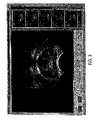
- FIG. 1is a standard treatment planning simulation image for radiation therapy of the prostate, displaying the bones, bladder, and rectum for inferring the position of the prostate, wherein the radiation field zone is superimposed to indicate the region to receive radiation therapy;
- FIG. 2is an image showing the fusion of the ultrasound image to the X-ray simulator image, together with the new superimposed radiation field zone, wherein the interior outline displays the size of the treatment field necessary to completely treat the prostate as determined by the ultrasound guided technique and the exterior outline displays the size of the treatment field as determined by the conventional technique, and wherein the prostate and other structures are visible in the fused image which were not visible in the X-ray simulation alone;
- FIG. 3is an image of a panel displaying on the right hand side a set of ultrasound images that have been read into the software planning program, while also displaying in the main window a selected image with markings removed;
- FIG. 4is an image of a panel displaying the X-ray images read into the software program
- FIG. 5is an image of a panel for investigating how digitally recomputed ultrasound images (DRU) can be used to obtain information about the prostate, wherein the window on the left-hand side displays an anterior-posterior DRU image calculated using all the data in the ultrasound image set displayed on the panel in the first figure, and the window on the right-hand side displays an anterior-posterior DRU image calculated using only the data points between horizontal planes at 2.5 cm and 4.0 cm away from the axis of the ultrasound probe, in which the prostate can be clearly identified;
- DRUdigitally recomputed ultrasound images
- FIG. 6is an image showing a panel combining an anterior-posterior X-ray image with an anterior-posterior DRU image in the main window, wherein the X-ray image data are displayed using the usual gray-scale method, while the DRU image data have been assigned color cyan (equal green and blue components), and wherein the upper and lower windows on the right-hand side display the original X-ray image and DRU image, respectively;
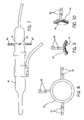
- FIG. 7is a representation of a transrectal ultrasound probe mounted with a ring collar containing LED emitters or lights on the surface to allow tracking the probe position with an optical registration system;
- FIG. 8is an end view of the ring collar shown in FIG. 7 , containing multiple LED emitters or lights for tracking with the optical registration system, wherein the ring collar mounts onto the ultrasound transrectal probe;
- FIG. 9is a representation of an alternate configuration for mounting the LEDs or light sources onto the surface of a ring collar
- FIG. 10is yet another representation of an alternate configuration for mounting the LEDs or light sources onto the surface of a ring collar
- FIG. 11Ais a representation of a ring collar with multiple LED emitters or lights according to an alternate embodiment of the invention
- FIG. 11Bis a base upon which the ring collar of FIG. 11A is mounted
- FIG. 11Cis a side view of the ring collar of FIG. 11A ;
- FIG. 12is a representation of a room setup of one example of the system of the present invention for the treatment of prostate cancer, including an optical registration system with two cameras mounted adjacent to a patient table for tracking the three-dimensional spatial position of the LEDs mounted on the transrectal ultrasound imaging probe;
- FIG. 13is another representation of a room setup of another example of the system of the present invention for the treatment of prostate cancer, including an optical registration system with two cameras mounted adjacent to a patient table for tracking the three-dimensional spatial position of the LEDs mounted on the transrectal ultrasound imaging probe; and
- FIG. 14Ais a representation of a plurality of ultrasound transducers within the head of a transrectal imaging probe
- FIG. 14Bis a representation of another configuration of a plurality of ultrasound transducers within the head of a transrectal imaging probe.
- the present inventioncomprises a technique and integrated hardware and software system to provide improved planning, registration, targeting, and delivery of conformal, external beam radiation therapy of prostate cancer and other soft-tissue diseases.
- Real time ultrasound imaging during planning and treatmentis used for localization of soft tissue treatment targets and fused with radiographic or CT data for conformal treatment optimization.
- the fusion technique of the present inventionprovides accurate localization of the prostate (or other tissue) volume in real time.
- visualization of the prostate glandis achieved using transrectal ultrasonography and the fusion of that image in the precise location of the prostate within the pelvic region. This makes possible accurate determination of the location of the prostate target by transformation of the ultrasound image data on both the ultrasound and x-ray/CT images.
- the radiation fieldcan be optimized to significantly reduce the volume of irradiated normal tissue, thereby minimizing the exposure of the surrounding healthy tissues and increasing dosage delivered to the prostate treatment target.
- the present inventionin general comprises the design and use of an integrated system for providing accurate targeting of soft tissues for conformal external beam radiation therapy.
- Real-time ultrasound imaging during planning and treatmentis used for localization of soft tissue targets and fused with radiographic or CT data for conformal treatment optimization.
- imaging of the prostate glandis achieved using transrectal ultrasonography, and these images are automatically registered to the exact location within the pelvic region.
- FIG. 1is a standard treatment planning simulation image for radiation therapy of the prostate, displaying the bones, bladder, and rectum for inferring the position of the prostate.
- the radiation field zoneis superimposed to indicate the region to receive radiation therapy
- Some of the key features of the invention relating to the planning stagesinclude the use of an integrated ultrasound system and software, the frameless stereotactic spatial registration of soft tissue images, the generation of anatomy segmentation, the generation of beam's eye views and control of field size, the seamless integration with standard and IMRT treatment planning systems, and the ability to export to treatment planning systems for fusion with either CT or standard simulation images for localizing internal soft tissue treatment targets.
- the present inventionprovides for rapid patient alignment using ultrasound imaging with a treatment plan, the accurate localization and targeting of therapy to soft tissue treatment volume, the ability to have confirmation with the treatment plan, and continuous monitoring during each and every treatment.
- the preferred form of integrated hardware and software system of the present inventioncan be separated into five primary components: (1) the ultrasound imaging hardware, (2) the treatment guidance and registration system, (3) the image fusion algorithm and software for treatment planning, (4) the real-time image fusion and localization software for use during the radiation treatment delivery phase, and (5) the computer system used to integrate these components.
- the following sectionsprovide more details regarding the specific embodiments of this proposed invention.
- the ultrasound imaging probeis use to provide sufficient diagnostic imaging of soft tissue while still maintaining a cost-effective system.
- the ultrasound imaging hardware systemcan comprise a standard transrectal ultrasound imaging probe, preferably providing sagittal and transverse views of the prostate.
- the ultrasound transducer/probeis swept or scanned, either mechanically or electronically, to provide a plurality of two dimensional image slices through the target tissue.
- the spatial position of each image sliceis registered in space, and these two dimensional images are then reconstructed to form a three dimensional image of the target volume used for treatment planning and guidance of the radiation therapy.
- the ultrasound probecan be held in place by a mechanical stepper or holder device, or the probe can be positioned “freehand” and retained by a support strap to allow the patient to move freely during set-up but prevent the probe from falling out.
- the ultrasound unitis also preferably integrated with the computer workstation to provide digital control and integration of the ultrasound images with the software applications as well as other imaging sources.
- the spatial registration and guidance system of the present inventionis used to record the 3-D spatial position of the ultrasound imaging probe at all times, relative to a known reference point.
- One method to accomplish the registrationis through the use of a spatially encoded localizer arm, which tracks the position of the ultrasound probe.
- the localizer armholds the ultrasound probe at one end, and is mechanically stabilized or fixed to the treatment table at the other end.
- the armcan be articulated at several joints, allowing at least three degrees of freedom, and employs encoding devices at these joints to accurately determine the position and orientation of the ultrasound probe relative to the table mount.
- the localizer armalso allows free movement and positioning of the ultrasound probe, facilitating patient movement during set-up.
- the localizer armis used to accurately and reproducibly position the ultrasound probe relative to the center of the radiation field.
- the positional information of the probeis then conveyed to the image localization software for registration of the images with respect to the treatment iso-center.
- Another method to accomplish the spatial registration and guidance of the ultrasound probeis through optical or electromagnetic sensor detection.
- cameras or other concentrated detectorsare mounted in the treatment room, and are used to track and register the position of the ultrasound probe or its holder.
- multiple LED emitters or light sources 40can be attached to the ultrasound imaging probe or onto a ring collar 42 which is mounted onto the probe 41 .
- the cameras or detectorsare then able to determine and record the 3-D position of these light sources 40 in real time, and therefore the position of the ultrasound probe 41 , as it is moved freely about near the treatment region of the patient.
- a simple calibration processcan be used to determine the relative spatial position of the ultrasound probe 41 to a known reference frame, as well the registration of the ultrasound images. This calibration information can be stored in a reference file on the computer and used by the software program.
- the image fusion and localization softwarequantifies the ultrasound imaging plane and combines these ultrasound images with X-ray simulation films to accurately display the location of the prostate relative to the treatment iso-center.
- This computer softwarecommunicates anatomy contours, radiation fields, shielding blocks, ultrasound images, X-ray images, and fused images in electronic form to an external treatment planning system, which the operator can use to perform dose calculations. All structures and images are referenced to the isocenter.
- One technique for fusing the ultrasound images with the simulation planning imageis accomplished in the software program using unambiguous identification of two identical point fiducials on each image.
- the ultrasound deviceis directly connected to the computer system running this image fusion software, so that the resultant images may be computed and displayed in real time.
- beam-targeting softwareis used to display a real-time ultrasound image of the prostate, either in 2-D image slices or a 3-D image volume, which is referenced to the radiation isocenter and superimposed with the beam profiles defined during the treatment planning phase.
- the image registration and visualizationallows the operator to determine whether the patient is optimally positioned to conform with the treatment plan and to make any appropriate adjustments to the patient or the radiation beam to produce optimal targeting.
- the software programis ideally integrated into the main computer system, but can also be implemented via a network connection.
- An integrated computer systembased on a standard personal computer architecture, is preferably used to execute the software programs, as well as to control and coordinate the activities of the other hardware components of the system.
- the computerprovides a user interface, high-quality graphic and viewing capabilities, and a standard network interface for communication with other computers, including DICOM servers.
- the computeralso uses appropriate processors, memory storage, and a video/imaging card to provide advanced image processing, computation and real time display capabilities.
- the system and software of the present inventioncan also include additional capabilities relating to the importation and measurement of ultrasound images. For example, the operator can manipulate the system to capture ultrasound images in real time from the ultrasound system via the video capture hardware in the host computer.
- the systemcan also maintain at least one “stack” of ultrasound images in a patient file.
- the systemcan provide thumbnail views of the ultrasound images, while also allowing the operator to delete images and/or add new images in real time.
- the systemalso allows the user to view the X-ray images, one at a time, and will include tools for windowing and leveling, histogram equalization, for example.
- the usercan calibrate the video image by designating specific points within the image (e.g. top center of the probe, and points five cm above and to the right, etc.) and designating which portion of the video image contains valid ultrasound data.
- the calibration datacan then be saved in a configuration file.
- the usermay also adjust brightness, contrast, and/or other basic video parameters, and also specify that multiple frames should be averaged together for noise reduction.
- the usercan calibrate the software for the specific probe. This involves measuring the position and orientation of the image plane relative to the position and orientation of the arm-probe attachment.
- the usercan, in one embodiment of the invention, calibrate the arm and probe together by moving the probe (or a jig attached to the probe) to specific known points referenced to the isocenter.
- the calibration datais then saved in a configuration file.
- the softwaresaves captured images in their entirety, as well as the position and orientation of the image plane relative to the isocenter.
- the softwareincludes a three dimensional view which shows the image slice as it is acquired, within a bounding box with superimposed guide lines through the isocenter; the slice would move in three dimensional space as the ultrasound probe is moved.
- An ultrasound volume, localized with respect to the isocentercan be exported in a format suitable for import into the TPS.
- the present inventionalso provides a number of improved features relating to the importation and management of X-ray images.
- the present inventionallows the operator to import multiple X-ray simulation images. These images are taken from x-ray films which have been scanned into bitmap image files (i.e. jpeg, tiff, etc.).
- the operatorcan move these files via a network connection to a file server, or via recordable media.
- the operatorFor each imported image, the operator must specify a gantry angle to identify the projection of the isocenter in the image the scale of the image (probably by identifying the intersection of fiducial wires in the simulator head, which project into the X-ray image).
- the operatorspecifies the geometry of the simulator, including the source-to-isocenter distance and the source-to-film distance.
- the calibration datais then saved in a configuration file.
- the systemmaintains one “set” of X-ray images in a patient file, in addition to provide thumbnail views of the X-ray images, and allows the operator to name, delete and/or add new images to the current set.
- the systemalso allows the user to view the x-ray images, one at a time, and will include tools for windowing and leveling, histogram equalization, etc.
- FIG. 4Shows a panel for inspecting individual X-ray images that are read into the software program.
- FIG. 5shows a panel for investigating how digitally recomputed ultrasound images (DRU) can be used to obtain information about the prostate.
- the window on the left-hand sidedisplays an anterior-posterior DRU image calculated using all the data in the ultrasound image set displayed on the panel in the first figure.
- the window on the right-hand sidedisplays an anterior-posterior DRU image calculated using only the data points between horizontal planes at 2.5 cm and 4.0 cm away from the axis of the ultrasound probe. In the latter image, the prostate can be clearly identified.
- the DRUis divergent, meaning that the individual rays projected through the ultrasound view volume diverge along a line from the radiation source, which is assumed to be a point source.
- the operatorcan “blend” the DRU and X-ray image to varying degrees, allowing the user to clearly view the location of the prostate within the X-ray image.
- Fused imagesmay be exported in a format suitable for import into the treatment planning software.
- FIG. 6shows a panel combining an anterior-posterior x-ray image with an anterior-posterior DRU image in the main window, wherein the x-ray image data are displayed using the usual gray-scale method, while the DRU image data have been assigned color cyan (equal green and blue components).
- the upper and lower windows on the right-hand sidedisplay the original X-ray image and DRU image, respectively
- the present inventionprovides the user with the ability to draw anatomical contours, using the ultrasound data as a guide.
- the useralso has the ability to contour in either 2-D or 3-D modes.
- Contourscan be accepted for multiple anatomies and identified by name.
- the usercan also designate new structures to be contoured and assign names to the new structures.
- Contourscan also be exported in a format suitable for import into the TPS.
- the present inventionpermits the operator to resize proposed treatment fields, using the fused image (X-ray film with DRU and/or contours superimposed) as a guide.
- One fieldis saved for each X-ray film.
- the operatorcan specify the size constraints on the field dimensions and whether or not the field must remain symmetric about the isocenter (“symmetric jaws” constraint) based on the capabilities of the linear accelerator.
- the calibration datais then saved in a configuration file.
- the beam profilesmay be exported in a format suitable for import into the TPS.
- the present inventionalso allows the operator to draw “blocks” on the proposed treatment fields.
- the blocksare thick Cerrobend (Pb—Cd) alloy blocks which are used to collimate or shield the corners of the treatment field from radiation and to conform the radiation to the beam's-eye cross-section of the prostate.
- the operatorcan specify a margin, which is the extent to which the block extends beyond the edge of the field.
- the marginis required to allow the blocks, which may be exported in a format suitable for import into the TPS, to be physically attached to the carrier which is placed into the head of the linear accelerator.
- the calibration datawill be saved in a configuration file.
- the operatorcan print the blocks, scaled by an operator-specified scale factor, as “blueprints” to be used by personnel who fabricate the blocks for a specific patient.
- the scale factoris saved in a configuration file.
- the system and softwareallows the operator to view the ultrasound image data in real time, as it is acquired.
- the location of the ultrasound imageis known based on the table position and the localizer arm.
- the operatorcan superimpose the original prostate contours, and/or the geometry of any one of the treatment beams (field, blocks, etc.) on the image slice view, allowing the operator to determine whether or not the prostate is positioned as planned.
- This featuremay be used at treatment time to assist in aligning the patient with the isocenter and making sure that the prostate remains in its expected location for the duration of the treatment. Involuntary patient movement, gas passing through the bowel, etc. can also cause the prostate to move during treatment.
- the system of the present inventionalso has the ability to store all data, images, settings, etc. associated with a particular patient in a “patient file,” including administrative information (patient name, physician's name, institution, etc.).
- a configuration filecontains the calibration settings for the application.
- the operatorcan also name, save, and load configurations.
- a technicianmounts the probe and registration system to the simulation table. Using a combination of probe movement and the system's user interface, the technician calibrates the registration system relative to the simulated isocenter. The ultrasound probe is then inserted during the simulation process. Using the system's user interface, the user acquires a three-dimensional ultrasound view volume including the prostate. The user may need to manipulate the ultrasound probe in order to ensure that the prostate is enclosed within the extent of the probe's three-dimensional view. The probe remains in place while the simulation data (CT or X-ray images) are captured in the normal manner.
- CTcomputed tomography
- the image fusion softwareis used to combine the image data acquired from the simulator with the isocenter-referenced ultrasound view volume provided by the registration system. The result is a fused set of images or a fused view volume in which the prostate is clearly visible.
- FIG. 2shows the fusion of the ultrasound to the X-ray simulator image, together with the new superimposed radiation field zone.
- the interior outlinedisplays the size of the treatment field necessary to completely treat the prostate as determined by the ultrasound guided technique and the exterior outline displays the size of the treatment field as determined by the conventional technique.
- the prostate and other structuresare visible in the fused image which were not visible in the X-ray simulation alone. It is important to note that the high-dose radiation field is confined to the prostate with minimal involvement of nearby critical structures such as the bladder and rectum.
- Treatment planningis conducted in a manner similar to the conventional model described herein using an existing Treatment Planning System.
- the fused image data used during treatment planningallows the radiation oncologist to easily identify the exact location and extent of the prostate.
- a technicianmounts the probe and registration localizer arm to the treatment table. Using a combination of probe movement and the system's user interface, the technician then calibrates the probe localizer relative to the LINAC isocenter. The ultrasound probe is then inserted. At that point, the system can provide real-time three-dimensional ultrasound data, spatially referenced to the treatment isocenter.
- FIG. 3shows an image of a panel displaying on the right hand side a set of ultrasound images that have been read into the software planning program. A selected image with markings removed is shown in the primary window.
- the beam-targeting and guidance softwareuses real-time images from the image-registration system and information from the treatment plan, allows a technician to view the ultrasound image of the prostate with the beam geometry and original prostate contours superimposed.
- the technicianadjusts the position of the patient and/or the treatment table to align the prostate as closely as possible with the original prostate contours and verify that the beam is conformal with the prostate.
- the usermonitors the real-time display of the prostate and treatment geometry, ensuring that the radiation beams remain conformal to the prostate throughout the treatment.
- the image-registration systemis mounted to the simulation table and calibrated by an operator with respect to the simulated isocenter.
- the transrectal probeis then inserted into the patient during simulation.
- the image fusion softwareis used to fuse the ultrasound data (on which the prostate is clearly visible) into the simulation data.
- the enhanced datais then imported into the TPS and allows the oncologist to clearly identify the location and extent of the prostate with respect to the treatment isocenter, allowing highly conformal beams to be defined.
- the image-registration systemis mounted to the treatment table and calibrated by an operator with respect to the treatment isocenter.
- the transrectal probeis then inserted into the patient during treatment.
- the beam geometry information provided by the TPSis imported into the beam-targeting software, which provides the operator with an image of the prostate referenced to the treatment isocenter.
- the operatorcan adjust the position of the patient.
- the high degree of beam/prostate conformance assumed during treatment planningcan thus be maintained throughout the duration of the treatment.
- the presence of the transrectal probehelps to ensure consistency in prostate position for each treatment.
- the image-registration systemprovides a real-time image of the prostate during treatment.
- the beam-targeting softwareis used to view this real-time image. Monitoring of position during treatment will permit the operator to stop treatment if the patient position changes.
- transrectal probeduring treatment displaces the posterior side of the rectum away from the prostate.
- the use of the transrectal probe during simulation and treatmenthelps maintain a consistent spatial relationship between the rectum, bladder neck, and prostate throughout the simulation and treatment process. This allows excessive exposure of the rectal wall and bladder neck to be avoid, and the conformal treatment plan margins to be ensured.
- the probe or imaging device of the present inventionis maneuverable in space but does not have to be tied to an encoded arm or a similar structure.
- the probe of the present inventioncan be moved in several directions.
- the probeis an ultrasound imaging probe that creates images. Unlike conventional imaging and treatment systems, the created images do not have to be combined from a stack of parallel images or from a sweep of multiple images through an angular sweep. It can be in any arbitrary position in space. And, the user knows where it is in space at all times by the use of points on this imaging device.
- the devicecollects the data, and a combination of optical and electromagnetic devices are positioned around the room that work together and spatially localize where this the imaging device is grounded via points on the imaging device or probe and.
- the image's location in spaceis already known and is produced by the imaging probe relative go the points so that is already a given because that is fixed since they are all on the same device. This provides the special relationship to the user; and because the user knows that the points are relative to the device and from the accommodation of the optical electromagnetic system (which can also be a simple optical system alone or an electromagnetic system alone depending on the embodiment), the position of the device is determined.
- the optical electromagnetic systemwhich can also be a simple optical system alone or an electromagnetic system alone depending on the embodiment
- FIGS. 12 and 13show a generic setup of the system according to one embodiment of the present invention.
- a pair of cameras 30are mounted on stands 32 on opposite sides of a patient table 34 .
- the information provided aboveis taken by the related software and is converted into a three dimensional image volume. Not only does this system render the volume, but it also permits the user to manipulate the volume. For example, a user could literally use a computer mouse to click on points on the surface of that volume. The system will then generate a surface rendering that matches that exact volume shape of the actual intrinsic image data set that is underneath it and create volume information or other information. Individual locations on the surface can be mapped, and a complete volume contour can be shown of the surface using as few as 8 or 9 or 10 points or as many as 20 or 30 or 40 points. For example, this system is very useful in treatment of prostrate disease because the user can identify an exact position in space in the body without having to have all of several fixtures, encoded arms and a stepping device that have positional encoding on the stepping.
Landscapes
- Health & Medical Sciences (AREA)
- Life Sciences & Earth Sciences (AREA)
- Engineering & Computer Science (AREA)
- Biomedical Technology (AREA)
- Public Health (AREA)
- Surgery (AREA)
- Veterinary Medicine (AREA)
- Nuclear Medicine, Radiotherapy & Molecular Imaging (AREA)
- General Health & Medical Sciences (AREA)
- Animal Behavior & Ethology (AREA)
- Medical Informatics (AREA)
- Heart & Thoracic Surgery (AREA)
- Molecular Biology (AREA)
- Pathology (AREA)
- Radiology & Medical Imaging (AREA)
- Biophysics (AREA)
- Physics & Mathematics (AREA)
- Computer Vision & Pattern Recognition (AREA)
- High Energy & Nuclear Physics (AREA)
- Optics & Photonics (AREA)
- Robotics (AREA)
- Radiation-Therapy Devices (AREA)
- Surgical Instruments (AREA)
Abstract
Description
Claims (59)
Priority Applications (1)
| Application Number | Priority Date | Filing Date | Title |
|---|---|---|---|
| US10/286,368US7438685B2 (en) | 2001-11-05 | 2002-11-01 | Apparatus and method for registration, guidance and targeting of external beam radiation therapy |
Applications Claiming Priority (2)
| Application Number | Priority Date | Filing Date | Title |
|---|---|---|---|
| US33744901P | 2001-11-05 | 2001-11-05 | |
| US10/286,368US7438685B2 (en) | 2001-11-05 | 2002-11-01 | Apparatus and method for registration, guidance and targeting of external beam radiation therapy |
Publications (2)
| Publication Number | Publication Date |
|---|---|
| US20030112922A1 US20030112922A1 (en) | 2003-06-19 |
| US7438685B2true US7438685B2 (en) | 2008-10-21 |
Family
ID=23320582
Family Applications (1)
| Application Number | Title | Priority Date | Filing Date |
|---|---|---|---|
| US10/286,368Expired - LifetimeUS7438685B2 (en) | 2001-11-05 | 2002-11-01 | Apparatus and method for registration, guidance and targeting of external beam radiation therapy |
Country Status (4)
| Country | Link |
|---|---|
| US (1) | US7438685B2 (en) |
| EP (1) | EP1460938A4 (en) |
| CN (1) | CN1612713A (en) |
| WO (1) | WO2003039370A1 (en) |
Cited By (55)
| Publication number | Priority date | Publication date | Assignee | Title |
|---|---|---|---|---|
| US20050259116A1 (en)* | 2004-05-24 | 2005-11-24 | Kabushiki Kaisha Toshiba | Medical image display apparatus |
| US20070280521A1 (en)* | 2006-06-05 | 2007-12-06 | Hui-Min Chao | Methods for Volumetric Contouring with Expert Guidance |
| US20070284545A1 (en)* | 2006-05-31 | 2007-12-13 | Ulf Isacsson | Method and system for radiotherapy treatment |
| US20080130825A1 (en)* | 2006-11-02 | 2008-06-05 | Accuray Incorporated | Target tracking using direct target registration |
| US20090080721A1 (en)* | 2007-09-25 | 2009-03-26 | Yulong Yan | Method and system for image pumping |
| US20100056908A1 (en)* | 2008-03-14 | 2010-03-04 | Baylor Research Institute | System and method for pre-planning a radiation treatment |
| US20100054572A1 (en)* | 2002-12-04 | 2010-03-04 | Conformis, Inc. | Fusion of Multiple Imaging Planes for Isotropic Imaging in MRI and Quantitative Image Analysis using Isotropic or Near-isotropic Imaging |
| US20100218140A1 (en)* | 2005-09-08 | 2010-08-26 | Feke Gilbert D | Graphical user interface for multi-modal images at incremental angular displacements |
| US7796791B2 (en)* | 2002-11-07 | 2010-09-14 | Conformis, Inc. | Methods for determining meniscal size and shape and for devising treatment |
| US7799077B2 (en) | 2002-10-07 | 2010-09-21 | Conformis, Inc. | Minimally invasive joint implant with 3-dimensional geometry matching the articular surfaces |
| US20100268073A1 (en)* | 2004-07-20 | 2010-10-21 | Tony Falco | Verifying Lesion Characteristics Using Beam Shapes |
| US20110006212A1 (en)* | 2007-12-05 | 2011-01-13 | Tal Shchory | Detecting photons in the presence of a pulsed radiation beam |
| US7881768B2 (en) | 1998-09-14 | 2011-02-01 | The Board Of Trustees Of The Leland Stanford Junior University | Assessing the condition of a joint and devising treatment |
| US8031838B2 (en) | 2009-01-29 | 2011-10-04 | The Invention Science Fund I, Llc | Diagnostic delivery service |
| US8036729B2 (en) | 1998-09-14 | 2011-10-11 | The Board Of Trustees Of The Leland Stanford Junior University | Assessing the condition of a joint and devising treatment |
| US20110263963A1 (en)* | 2010-04-26 | 2011-10-27 | Canon Kabushiki Kaisha | Acoustic-wave measuring apparatus and method |
| US8130904B2 (en) | 2009-01-29 | 2012-03-06 | The Invention Science Fund I, Llc | Diagnostic delivery service |
| US8135198B2 (en) | 2007-08-08 | 2012-03-13 | Resonant Medical, Inc. | Systems and methods for constructing images |
| US8189738B2 (en) | 2008-06-02 | 2012-05-29 | Elekta Ltd. | Methods and systems for guiding clinical radiotherapy setups |
| US8234097B2 (en) | 2001-05-25 | 2012-07-31 | Conformis, Inc. | Automated systems for manufacturing patient-specific orthopedic implants and instrumentation |
| US8249317B2 (en) | 2007-07-20 | 2012-08-21 | Eleckta Ltd. | Methods and systems for compensating for changes in anatomy of radiotherapy patients |
| US8265730B2 (en) | 1998-09-14 | 2012-09-11 | The Board Of Trustees Of The Leland Stanford Junior University | Assessing the condition of a joint and preventing damage |
| US8337507B2 (en) | 2001-05-25 | 2012-12-25 | Conformis, Inc. | Methods and compositions for articular repair |
| US8406844B2 (en) | 2002-03-06 | 2013-03-26 | Tomotherapy Incorporated | Method for modification of radiotherapy treatment delivery |
| US8480754B2 (en) | 2001-05-25 | 2013-07-09 | Conformis, Inc. | Patient-adapted and improved articular implants, designs and related guide tools |
| US8545569B2 (en) | 2001-05-25 | 2013-10-01 | Conformis, Inc. | Patient selectable knee arthroplasty devices |
| US8556983B2 (en) | 2001-05-25 | 2013-10-15 | Conformis, Inc. | Patient-adapted and improved orthopedic implants, designs and related tools |
| US8617242B2 (en) | 2001-05-25 | 2013-12-31 | Conformis, Inc. | Implant device and method for manufacture |
| US8682052B2 (en) | 2008-03-05 | 2014-03-25 | Conformis, Inc. | Implants for altering wear patterns of articular surfaces |
| US8690814B2 (en) | 2011-07-22 | 2014-04-08 | Elizabeth Chabner Thompson | Radiation treatment garment-II |
| US8735773B2 (en) | 2007-02-14 | 2014-05-27 | Conformis, Inc. | Implant device and method for manufacture |
| US8771365B2 (en) | 2009-02-25 | 2014-07-08 | Conformis, Inc. | Patient-adapted and improved orthopedic implants, designs, and related tools |
| US8810640B2 (en) | 2011-05-16 | 2014-08-19 | Ut-Battelle, Llc | Intrinsic feature-based pose measurement for imaging motion compensation |
| US8882847B2 (en) | 2001-05-25 | 2014-11-11 | Conformis, Inc. | Patient selectable knee joint arthroplasty devices |
| US9020788B2 (en) | 1997-01-08 | 2015-04-28 | Conformis, Inc. | Patient-adapted and improved articular implants, designs and related guide tools |
| US9248316B2 (en) | 2010-01-12 | 2016-02-02 | Elekta Ltd. | Feature tracking using ultrasound |
| US9286686B2 (en) | 1998-09-14 | 2016-03-15 | The Board Of Trustees Of The Leland Stanford Junior University | Assessing the condition of a joint and assessing cartilage loss |
| US9308091B2 (en) | 2001-05-25 | 2016-04-12 | Conformis, Inc. | Devices and methods for treatment of facet and other joints |
| US9421399B2 (en) | 2002-12-18 | 2016-08-23 | Varian Medical Systems, Inc. | Multi-mode cone beam CT radiotherapy simulator and treatment machine with a flat panel imager |
| US9443633B2 (en) | 2013-02-26 | 2016-09-13 | Accuray Incorporated | Electromagnetically actuated multi-leaf collimator |
| US9451928B2 (en) | 2006-09-13 | 2016-09-27 | Elekta Ltd. | Incorporating internal anatomy in clinical radiotherapy setups |
| US9492685B2 (en) | 2014-06-13 | 2016-11-15 | Infinitt Healthcare Co., Ltd. | Method and apparatus for controlling and monitoring position of radiation treatment system |
| US9603711B2 (en) | 2001-05-25 | 2017-03-28 | Conformis, Inc. | Patient-adapted and improved articular implants, designs and related guide tools |
| US9630025B2 (en) | 2005-07-25 | 2017-04-25 | Varian Medical Systems International Ag | Methods and apparatus for the planning and delivery of radiation treatments |
| US9950194B2 (en) | 2014-09-09 | 2018-04-24 | Mevion Medical Systems, Inc. | Patient positioning system |
| USRE46953E1 (en) | 2007-04-20 | 2018-07-17 | University Of Maryland, Baltimore | Single-arc dose painting for precision radiation therapy |
| US10085839B2 (en) | 2004-01-05 | 2018-10-02 | Conformis, Inc. | Patient-specific and patient-engineered orthopedic implants |
| US10426424B2 (en) | 2017-11-21 | 2019-10-01 | General Electric Company | System and method for generating and performing imaging protocol simulations |
| US10449390B2 (en) | 2010-01-12 | 2019-10-22 | Elekta ltd | Feature tracking using ultrasound |
| US10531858B2 (en) | 2007-07-20 | 2020-01-14 | Elekta, LTD | Methods and systems for guiding the acquisition of ultrasound images |
| US10542962B2 (en) | 2009-07-10 | 2020-01-28 | Elekta, LTD | Adaptive radiotherapy treatment using ultrasound |
| US10773101B2 (en) | 2010-06-22 | 2020-09-15 | Varian Medical Systems International Ag | System and method for estimating and manipulating estimated radiation dose |
| US11278742B2 (en) | 2016-04-28 | 2022-03-22 | Koninklijke Philips N.V. | Image guided treatment delivery |
| US20220087638A1 (en)* | 2020-09-18 | 2022-03-24 | B-K Medical Aps | Image Fusion-Based Tracking without a Tracking Sensor |
| US11301198B2 (en)* | 2019-12-25 | 2022-04-12 | Industrial Technology Research Institute | Method for information display, processing device, and display system |
Families Citing this family (97)
| Publication number | Priority date | Publication date | Assignee | Title |
|---|---|---|---|---|
| CA2314794A1 (en)* | 2000-08-01 | 2002-02-01 | Dimitre Hristov | Apparatus for lesion or organ localization |
| IL143255A (en)* | 2001-05-20 | 2015-09-24 | Simbionix Ltd | Endoscopic ultrasonography simulation |
| US7016522B2 (en)* | 2002-01-15 | 2006-03-21 | Siemens Medical Solutions Usa, Inc. | Patient positioning by video imaging |
| US20030187349A1 (en)* | 2002-03-29 | 2003-10-02 | Olympus Optical Co., Ltd. | Sentinel lymph node detecting method |
| DE10328765B4 (en)* | 2003-06-25 | 2005-11-24 | aviCOM Gesellschaft für angewandte visuelle Systeme mbH | Device and method for connecting the representation of the electric cardiac field with the representation of the associated heart |
| ATE503419T1 (en) | 2004-02-20 | 2011-04-15 | Univ Florida | SYSTEM FOR ADMINISTERING CONFORMAL RADIATION THERAPY WHILE IMAGING SOFT TISSUE |
| US7657298B2 (en)* | 2004-03-11 | 2010-02-02 | Stryker Leibinger Gmbh & Co. Kg | System, device, and method for determining a position of an object |
| WO2005107601A2 (en)* | 2004-05-06 | 2005-11-17 | Focus Surgery, Inc. | Method and apparatus for the selective treatment of tissue |
| US7672705B2 (en)* | 2004-07-19 | 2010-03-02 | Resonant Medical, Inc. | Weighted surface-to-surface mapping |
| EP1774312B1 (en)* | 2004-07-20 | 2017-04-12 | Elekta Ltd. | Calibrating ultrasound imaging devices |
| JP5107709B2 (en)* | 2004-08-13 | 2012-12-26 | コーニンクレッカ フィリップス エレクトロニクス エヌ ヴィ | Adapting radiotherapy treatment plans |
| JP4690683B2 (en)* | 2004-09-13 | 2011-06-01 | 株式会社東芝 | Ultrasonic diagnostic apparatus and medical image browsing method |
| WO2006032134A2 (en)* | 2004-09-20 | 2006-03-30 | Resonant Medical Inc. | Radiotherapy treatment monitoring using ultrasound |
| US8702580B2 (en)* | 2004-10-06 | 2014-04-22 | Brainlab Ag | Method and device for assisting in a tissue treatment |
| US9002432B2 (en)* | 2004-11-15 | 2015-04-07 | Brainlab Ag | Method and device for calibrating a medical instrument |
| WO2006057911A2 (en)* | 2004-11-22 | 2006-06-01 | Civco Medical Instruments Co., Inc. | Real time ultrasound monitoring of the motion of internal structures during respiration for control of therapy delivery |
| GB2421572A (en)* | 2004-12-22 | 2006-06-28 | Elekta Ab | Radiotherapeutic apparatus |
| WO2006086223A2 (en)* | 2005-02-08 | 2006-08-17 | Blue Belt Technologies, Inc. | Augmented reality device and method |
| DE102005016256B3 (en)* | 2005-04-08 | 2006-06-08 | Siemens Ag | Displaying preoperative three-dimensional images with two-dimensional x-ray image acquisition involves repeatedly generating two-dimensional representations with varying parameters and displaying them on a screen |
| US8232535B2 (en)* | 2005-05-10 | 2012-07-31 | Tomotherapy Incorporated | System and method of treating a patient with radiation therapy |
| JP4999012B2 (en) | 2005-06-06 | 2012-08-15 | インチュイティブ サージカル,インコーポレイテッド | Laparoscopic ultrasonic robotic surgical system |
| JP5060476B2 (en) | 2005-07-22 | 2012-10-31 | トモセラピー・インコーポレーテッド | System and method for detecting respiratory phase of a patient undergoing radiation therapy |
| EP1940515A4 (en)* | 2005-09-06 | 2010-05-26 | Resonant Medical Inc | System and method for patient setup for radiotherapy treatment |
| US8057408B2 (en) | 2005-09-22 | 2011-11-15 | The Regents Of The University Of Michigan | Pulsed cavitational ultrasound therapy |
| US10219815B2 (en) | 2005-09-22 | 2019-03-05 | The Regents Of The University Of Michigan | Histotripsy for thrombolysis |
| US7912258B2 (en) | 2005-09-27 | 2011-03-22 | Vanderbilt University | Method and apparatus for standardizing ultrasonography training using image to physical space registration of tomographic volumes from tracked ultrasound |
| DE102005058871B3 (en)* | 2005-12-09 | 2007-07-26 | Siemens Ag | Medical irradiation device for treating tumor of patient, has data processing device that is data technically equipped in such a manner that characteristics of radiation are visualizable in common representation |
| US8929621B2 (en)* | 2005-12-20 | 2015-01-06 | Elekta, Ltd. | Methods and systems for segmentation and surface matching |
| US7453984B2 (en)* | 2006-01-19 | 2008-11-18 | Carestream Health, Inc. | Real-time target confirmation for radiation therapy |
| US7848592B2 (en)* | 2006-07-31 | 2010-12-07 | Carestream Health, Inc. | Image fusion for radiation therapy |
| US20080033748A1 (en)* | 2006-08-02 | 2008-02-07 | Erik Busch | System to measure and analyze the usage of dose reduction features |
| US7620147B2 (en) | 2006-12-13 | 2009-11-17 | Oraya Therapeutics, Inc. | Orthovoltage radiotherapy |
| AU2012261645B2 (en)* | 2006-10-16 | 2016-01-14 | Carl Zeiss Meditec, Inc. | Ocular radiosurgery |
| US7535991B2 (en)* | 2006-10-16 | 2009-05-19 | Oraya Therapeutics, Inc. | Portable orthovoltage radiotherapy |
| CN100551465C (en)* | 2006-12-25 | 2009-10-21 | 深圳市海博科技有限公司 | A method for automatic positioning of patient's target area in radiotherapy |
| US20080177280A1 (en)* | 2007-01-09 | 2008-07-24 | Cyberheart, Inc. | Method for Depositing Radiation in Heart Muscle |
| WO2008086434A2 (en)* | 2007-01-09 | 2008-07-17 | Cyberheart, Inc. | Depositing radiation in heart muscle under ultrasound guidance |
| CN101678210B (en)* | 2007-04-18 | 2012-05-23 | 伊利克塔股份有限公司 | radiotherapy device |
| US8506558B2 (en)* | 2008-01-11 | 2013-08-13 | Oraya Therapeutics, Inc. | System and method for performing an ocular irradiation procedure |
| US8363783B2 (en) | 2007-06-04 | 2013-01-29 | Oraya Therapeutics, Inc. | Method and device for ocular alignment and coupling of ocular structures |
| US7847380B2 (en)* | 2007-09-20 | 2010-12-07 | Samsung Electronics Co., Ltd. | Tape substrate and semiconductor module for smart card, method of fabricating the same, and smart card |
| US8467497B2 (en)* | 2007-10-25 | 2013-06-18 | Tomotherapy Incorporated | System and method for motion adaptive optimization for radiation therapy delivery |
| CN101820948A (en)* | 2007-10-25 | 2010-09-01 | 断层放疗公司 | System and method for motion adaptive optimization of radiotherapy delivery |
| US7801271B2 (en) | 2007-12-23 | 2010-09-21 | Oraya Therapeutics, Inc. | Methods and devices for orthovoltage ocular radiotherapy and treatment planning |
| EP3272395B1 (en)* | 2007-12-23 | 2019-07-17 | Carl Zeiss Meditec, Inc. | Devices for detecting, controlling, and predicting radiation delivery |
| US8803910B2 (en)* | 2008-08-28 | 2014-08-12 | Tomotherapy Incorporated | System and method of contouring a target area |
| US9521994B2 (en) | 2009-05-11 | 2016-12-20 | Siemens Healthcare Gmbh | System and method for image guided prostate cancer needle biopsy |
| CN102510735A (en) | 2009-07-17 | 2012-06-20 | 计算机心脏股份有限公司 | Heart treatment kit, system, and method for radiosurgically alleviating arrhythmia |
| WO2011022411A2 (en)* | 2009-08-17 | 2011-02-24 | Histosonics, Inc. | Disposable acoustic coupling medium container |
| JP5726191B2 (en) | 2009-08-26 | 2015-05-27 | リージェンツ オブ ザ ユニバーシティー オブ ミシガン | Apparatus and method using control of bubble turbidity cavitation phenomenon during fracture of ureteral stones |
| JP5863654B2 (en)* | 2009-08-26 | 2016-02-16 | リージェンツ オブ ザ ユニバーシティー オブ ミシガン | Micromanipulator control arm for therapeutic and image processing ultrasonic transducers |
| US8539813B2 (en)* | 2009-09-22 | 2013-09-24 | The Regents Of The University Of Michigan | Gel phantoms for testing cavitational ultrasound (histotripsy) transducers |
| WO2012001551A1 (en)* | 2010-06-30 | 2012-01-05 | Koninklijke Philips Electronics N.V. | System and method for guided adaptive brachytherapy |
| US11612377B2 (en)* | 2010-12-16 | 2023-03-28 | Best Medical International, Inc. | Image guided surgical methodology and system employing patient movement detection and correction |
| US9430831B2 (en)* | 2011-03-15 | 2016-08-30 | Koninklijke Philips N.V. | Correlated image mapping pointer |
| CN102204846B (en)* | 2011-04-22 | 2012-10-31 | 重庆伟渡医疗设备股份有限公司 | Method for quickly and accurately calibrating medical imaging component after changing of position thereof |
| US9144694B2 (en) | 2011-08-10 | 2015-09-29 | The Regents Of The University Of Michigan | Lesion generation through bone using histotripsy therapy without aberration correction |
| JP6068503B2 (en)* | 2012-01-06 | 2017-01-25 | ヒストソニックス,インコーポレーテッド | Histotripsy treatment transducer |
| US11027149B2 (en)* | 2012-01-12 | 2021-06-08 | Sensus Healthcare, Inc. | Hybrid ultrasound-guided superficial radiotherapy system and method |
| EP2822472B1 (en)* | 2012-03-07 | 2022-09-28 | Ziteo, Inc. | Systems for tracking and guiding sensors and instruments |
| US9049783B2 (en) | 2012-04-13 | 2015-06-02 | Histosonics, Inc. | Systems and methods for obtaining large creepage isolation on printed circuit boards |
| WO2013166019A1 (en) | 2012-04-30 | 2013-11-07 | The Regents Of The University Of Michigan | Ultrasound transducer manufacturing using rapid-prototyping method |
| US10561861B2 (en) | 2012-05-02 | 2020-02-18 | Viewray Technologies, Inc. | Videographic display of real-time medical treatment |
| US20130342577A1 (en)* | 2012-06-20 | 2013-12-26 | Carestream Health, Inc. | Image synthesis for diagnostic review |
| US20140100459A1 (en) | 2012-10-05 | 2014-04-10 | The Regents Of The University Of Michigan | Bubble-induced color doppler feedback during histotripsy |
| EP3628370A1 (en) | 2012-10-26 | 2020-04-01 | ViewRay Technologies, Inc. | Assessment and improvement of treatment using imaging of physiological responses to radiation therapy |
| WO2014096993A1 (en) | 2012-12-17 | 2014-06-26 | Koninklijke Philips N.V. | Real-time adaptive dose computation radiation therapy |
| EP3016594B1 (en) | 2013-07-03 | 2023-01-25 | Histosonics, Inc. | Histotripsy excitation sequences optimized for bubble cloud formation using shock scattering |
| WO2015003154A1 (en) | 2013-07-03 | 2015-01-08 | Histosonics, Inc. | Articulating arm limiter for cavitational ultrasound therapy system |
| US10780298B2 (en) | 2013-08-22 | 2020-09-22 | The Regents Of The University Of Michigan | Histotripsy using very short monopolar ultrasound pulses |
| CN104574329B (en)* | 2013-10-09 | 2018-03-09 | 深圳迈瑞生物医疗电子股份有限公司 | Ultrasonic fusion of imaging method, ultrasonic fusion of imaging navigation system |
| WO2015092604A1 (en)* | 2013-12-18 | 2015-06-25 | Koninklijke Philips N.V. | System and method for ultrasound and computed tomography image registration for sonothrombolysis treatment |
| CN106132484B (en)* | 2014-05-28 | 2019-09-06 | 核通业务有限公司 | Method and system for the plesioradiotherapy plan based on imaging data |
| JP6263447B2 (en)* | 2014-06-30 | 2018-01-17 | ジーイー・メディカル・システムズ・グローバル・テクノロジー・カンパニー・エルエルシー | Ultrasonic diagnostic apparatus and program |
| EP3171807A1 (en)* | 2014-07-25 | 2017-05-31 | Analogic Corporation | Instrument annular location sensor |
| CN113069691B (en)* | 2014-10-27 | 2023-08-15 | 医科达有限公司 | Image guidance for radiation therapy |
| US10617401B2 (en) | 2014-11-14 | 2020-04-14 | Ziteo, Inc. | Systems for localization of targets inside a body |
| JP6979882B2 (en) | 2015-06-24 | 2021-12-15 | ザ リージェンツ オブ ザ ユニヴァシティ オブ ミシガン | Tissue disruption therapy systems and methods for the treatment of brain tissue |
| JP6164662B2 (en)* | 2015-11-18 | 2017-07-19 | みずほ情報総研株式会社 | Treatment support system, operation method of treatment support system, and treatment support program |
| CN109310879A (en) | 2016-03-02 | 2019-02-05 | 优瑞技术公司 | Particle Therapy Using Magnetic Resonance Imaging |
| CA3046091A1 (en) | 2016-12-13 | 2018-06-21 | Viewray Technologies, Inc. | Radiation therapy systems and methods |
| WO2018170592A1 (en)* | 2017-03-20 | 2018-09-27 | Exact Imaging Inc. | Method and system for visually assisting an operator of an ultrasound system |
| CN107174340A (en)* | 2017-07-21 | 2017-09-19 | 李刚 | A kind of device that prostate is removed by control with computational intelligence laser Full-automatic cutting |
| TW201923776A (en)* | 2017-10-27 | 2019-06-16 | 美商蝴蝶網路公司 | Automated measurements on ultrasound images and collected quality indicators for automated measurements |
| WO2019100212A1 (en)* | 2017-11-21 | 2019-05-31 | 深圳迈瑞生物医疗电子股份有限公司 | Ultrasonic system and method for planning ablation |
| CN107909624B (en)* | 2017-12-05 | 2019-12-24 | 南京大学 | A method for extracting and fusing two-dimensional images from three-dimensional tomography |
| US11033758B2 (en) | 2017-12-06 | 2021-06-15 | Viewray Technologies, Inc. | Radiotherapy systems, methods and software |
| US11209509B2 (en) | 2018-05-16 | 2021-12-28 | Viewray Technologies, Inc. | Resistive electromagnet systems and methods |
| CN109523867B (en)* | 2018-11-21 | 2021-03-09 | 杨天烈 | Bracelet-type cow rectum positioning examination teaching device and teaching method |
| AU2019389001B2 (en) | 2018-11-28 | 2025-08-14 | Histosonics, Inc. | Histotripsy systems and methods |
| CN109701168B (en)* | 2018-12-27 | 2022-03-18 | 成植温 | Gamma radiation tumor treatment system |
| JP7603608B2 (en) | 2019-04-09 | 2024-12-20 | ジティオ, インコーポレイテッド | Method and system for high performance and versatile molecular imaging - Patents.com |
| US11813485B2 (en) | 2020-01-28 | 2023-11-14 | The Regents Of The University Of Michigan | Systems and methods for histotripsy immunosensitization |
| US11446522B2 (en) | 2020-04-24 | 2022-09-20 | Shanghai United Imaging Healthcare Co., Ltd. | Systems and methods for scintillation camera-based motion tracking in radiotherapy |
| JP2023540482A (en) | 2020-08-27 | 2023-09-25 | ザ リージェンツ オブ ザ ユニバーシティー オブ ミシガン | Ultrasonic transducer with transmitting and receiving functions for histotripsy |
| EP4608504A1 (en) | 2022-10-28 | 2025-09-03 | Histosonics, Inc. | Histotripsy systems and methods |
| CN116492049B (en)* | 2023-06-29 | 2023-10-03 | 北京智愈医疗科技有限公司 | Method and device for determining conformal ablation range of prostate |
Citations (176)
| Publication number | Priority date | Publication date | Assignee | Title |
|---|---|---|---|---|
| US4125858A (en) | 1976-03-27 | 1978-11-14 | Emi Limited | Video display arrangements |
| US4567896A (en) | 1984-01-20 | 1986-02-04 | Elscint, Inc. | Method and apparatus for calibrating a biopsy attachment for ultrasonic imaging apparatus |
| US4586512A (en) | 1981-06-26 | 1986-05-06 | Thomson-Csf | Device for localized heating of biological tissues |
| US4751643A (en) | 1986-08-04 | 1988-06-14 | General Electric Company | Method and apparatus for determining connected substructures within a body |
| US4764971A (en) | 1985-11-25 | 1988-08-16 | Eastman Kodak Company | Image processing method including image segmentation |
| US4791567A (en) | 1986-09-15 | 1988-12-13 | General Electric Company | Three dimensional connectivity system employing an equivalence schema for determining connected substructures within a body |
| US4856074A (en) | 1987-03-20 | 1989-08-08 | Fuji Xerox Co., Ltd. | Region recognizing device |
| US4896673A (en) | 1988-07-15 | 1990-01-30 | Medstone International, Inc. | Method and apparatus for stone localization using ultrasound imaging |
| US4961425A (en) | 1987-08-14 | 1990-10-09 | Massachusetts Institute Of Technology | Morphometric analysis of anatomical tomographic data |
| US4991579A (en) | 1987-11-10 | 1991-02-12 | Allen George S | Method and apparatus for providing related images over time of a portion of the anatomy using fiducial implants |
| US4994013A (en) | 1988-07-28 | 1991-02-19 | Best Industries, Inc. | Pellet for a radioactive seed |
| WO1991007726A1 (en) | 1989-11-21 | 1991-05-30 | I.S.G. Technologies Inc. | Probe-correlated viewing of anatomical image data |
| EP0455439A2 (en) | 1990-05-02 | 1991-11-06 | International Business Machines Corporation | Computer method and system for analysing real or simulated physical systems |
| US5072384A (en) | 1988-11-23 | 1991-12-10 | Arch Development Corp. | Method and system for automated computerized analysis of sizes of hearts and lungs in digital chest radiographs |
| US5133020A (en) | 1989-07-21 | 1992-07-21 | Arch Development Corporation | Automated method and system for the detection and classification of abnormal lesions and parenchymal distortions in digital medical images |
| EP0424912A3 (en) | 1989-10-27 | 1992-09-23 | Hitachi, Ltd. | Region extracting method and three-dimensional display method |
| US5166876A (en) | 1991-01-16 | 1992-11-24 | General Electric Company | System and method for detecting internal structures contained within the interior region of a solid object |
| US5170790A (en) | 1990-04-06 | 1992-12-15 | Technomed International | Arm having an end movable in translation, and therapeutic treatment apparatus constituting an application thereof |
| US5178148A (en) | 1990-04-06 | 1993-01-12 | Technomed International | Method of automatically measuring the volume of a tumor or of a gland, in particular the prostate, a measuring device, and a method and apparatus constituting and application thereof |
| US5185809A (en) | 1987-08-14 | 1993-02-09 | The General Hospital Corporation | Morphometric analysis of anatomical tomographic data |
| US5187658A (en) | 1990-01-17 | 1993-02-16 | General Electric Company | System and method for segmenting internal structures contained within the interior region of a solid object |
| US5204625A (en) | 1990-12-20 | 1993-04-20 | General Electric Company | Segmentation of stationary and vascular surfaces in magnetic resonance imaging |
| US5222499A (en) | 1989-11-15 | 1993-06-29 | Allen George S | Method and apparatus for imaging the anatomy |
| US5227969A (en) | 1988-08-01 | 1993-07-13 | W. L. Systems, Inc. | Manipulable three-dimensional projection imaging method |
| US5234004A (en) | 1988-11-21 | 1993-08-10 | Technomed International | Method and apparatus for the surgical treatment of tissues by thermal effect, and in particular the prostate, using a urethral microwave-emitting probe means |
| US5239591A (en) | 1991-07-03 | 1993-08-24 | U.S. Philips Corp. | Contour extraction in multi-phase, multi-slice cardiac mri studies by propagation of seed contours between images |
| US5242373A (en) | 1991-09-17 | 1993-09-07 | Scott Walter P | Medical seed implantation instrument |
| US5260871A (en) | 1991-07-31 | 1993-11-09 | Mayo Foundation For Medical Education And Research | Method and apparatus for diagnosis of breast tumors |
| US5289374A (en) | 1992-02-28 | 1994-02-22 | Arch Development Corporation | Method and system for analysis of false positives produced by an automated scheme for the detection of lung nodules in digital chest radiographs |
| US5299253A (en) | 1992-04-10 | 1994-03-29 | Akzo N.V. | Alignment system to overlay abdominal computer aided tomography and magnetic resonance anatomy with single photon emission tomography |
| US5319549A (en) | 1992-11-25 | 1994-06-07 | Arch Development Corporation | Method and system for determining geometric pattern features of interstitial infiltrates in chest images |
| US5339812A (en) | 1988-12-23 | 1994-08-23 | Medical Instrumentation And Diagnostic Corporation | Three-dimensional computer graphics simulation and computerized numerical optimization for dose delivery and treatment planning |
| US5371810A (en) | 1991-09-27 | 1994-12-06 | E. I. Du Pont De Nemours And Company | Method of determining the interior points of an object in a background |
| US5389101A (en) | 1992-04-21 | 1995-02-14 | University Of Utah | Apparatus and method for photogrammetric surgical localization |
| US5391139A (en)* | 1992-09-03 | 1995-02-21 | William Beaumont Hospital | Real time radiation treatment planning system |
| US5391199A (en) | 1993-07-20 | 1995-02-21 | Biosense, Inc. | Apparatus and method for treating cardiac arrhythmias |
| US5394457A (en) | 1992-10-08 | 1995-02-28 | Leibinger Gmbh | Device for marking body sites for medical examinations |
| US5398690A (en) | 1994-08-03 | 1995-03-21 | Batten; Bobby G. | Slaved biopsy device, analysis apparatus, and process |
| US5409006A (en) | 1992-12-03 | 1995-04-25 | Siemens Aktiengesellschaft | System for the treatment of pathological tissue having a catheter with a marker for avoiding damage to healthy tissue |
| US5410617A (en) | 1991-04-25 | 1995-04-25 | Unisys Corporation | Method for adaptively thresholding grayscale image data |
| US5412563A (en) | 1993-09-16 | 1995-05-02 | General Electric Company | Gradient image segmentation method |
| US5411026A (en) | 1993-10-08 | 1995-05-02 | Nomos Corporation | Method and apparatus for lesion position verification |
| US5433199A (en) | 1994-02-24 | 1995-07-18 | General Electric Company | Cardiac functional analysis method using gradient image segmentation |
| US5447154A (en)* | 1992-07-31 | 1995-09-05 | Universite Joseph Fourier | Method for determining the position of an organ |
| US5452367A (en) | 1993-11-29 | 1995-09-19 | Arch Development Corporation | Automated method and system for the segmentation of medical images |
| US5457754A (en) | 1990-08-02 | 1995-10-10 | University Of Cincinnati | Method for automatic contour extraction of a cardiac image |
| US5458126A (en) | 1994-02-24 | 1995-10-17 | General Electric Company | Cardiac functional analysis system employing gradient image segmentation |
| US5460592A (en) | 1994-01-24 | 1995-10-24 | Amersham Holdings, Inc. | Apparatus and method for making carrier assembly for radioactive seed carrier |
| US5491627A (en) | 1993-05-13 | 1996-02-13 | Arch Development Corporation | Method and system for the detection of microcalcifications in digital mammograms |
| US5494039A (en) | 1993-07-16 | 1996-02-27 | Cryomedical Sciences, Inc. | Biopsy needle insertion guide and method of use in prostate cryosurgery |
| WO1996010949A1 (en) | 1994-10-07 | 1996-04-18 | Medical Media Systems | Video-based surgical targeting system |
| US5517602A (en) | 1992-12-03 | 1996-05-14 | Hewlett-Packard Company | Method and apparatus for generating a topologically consistent visual representation of a three dimensional surface |
| US5526812A (en) | 1993-06-21 | 1996-06-18 | General Electric Company | Display system for enhancing visualization of body structures during medical procedures |
| US5531223A (en) | 1990-02-15 | 1996-07-02 | Kabushiki Kaisha Toshiba | Method and apparatus of nuclear magnetic resonance imaging with nonlinearly processed image display |
| US5531227A (en) | 1994-01-28 | 1996-07-02 | Schneider Medical Technologies, Inc. | Imaging device and method |
| US5537485A (en) | 1992-07-21 | 1996-07-16 | Arch Development Corporation | Method for computer-aided detection of clustered microcalcifications from digital mammograms |
| WO1996011624A3 (en) | 1994-10-07 | 1996-07-18 | Univ St Louis | Surgical navigation systems including reference and localization frames |
| US5553207A (en) | 1991-05-27 | 1996-09-03 | Hitachi, Ltd. | Method of and apparatus for region extraction in three-dimensional voxel data |
| US5562095A (en) | 1992-12-24 | 1996-10-08 | Victoria Hospital Corporation | Three dimensional ultrasound imaging system |
| US5566246A (en) | 1991-03-28 | 1996-10-15 | Texas Instruments Incorporated | System and method for ranking and extracting salient contours for target recognition |
| US5570430A (en) | 1994-05-31 | 1996-10-29 | University Of Washington | Method for determining the contour of an in vivo organ using multiple image frames of the organ |
| US5574799A (en) | 1992-06-12 | 1996-11-12 | The Johns Hopkins University | Method and system for automated detection of microcalcification clusters in mammograms |
| US5583659A (en) | 1994-11-10 | 1996-12-10 | Eastman Kodak Company | Multi-windowing technique for thresholding an image using local image properties |
| WO1996042070A1 (en) | 1995-06-13 | 1996-12-27 | British Technology Group Limited | Apparatus for image enhancement, and related method |
| US5588430A (en)* | 1995-02-14 | 1996-12-31 | University Of Florida Research Foundation, Inc. | Repeat fixation for frameless stereotactic procedure |
| US5590215A (en) | 1993-10-15 | 1996-12-31 | Allen; George S. | Method for providing medical images |
| US5603318A (en) | 1992-04-21 | 1997-02-18 | University Of Utah Research Foundation | Apparatus and method for photogrammetric surgical localization |
| US5622170A (en) | 1990-10-19 | 1997-04-22 | Image Guided Technologies, Inc. | Apparatus for determining the position and orientation of an invasive portion of a probe inside a three-dimensional body |
| US5624382A (en) | 1992-03-10 | 1997-04-29 | Siemens Aktiengesellschaft | Method and apparatus for ultrasound tissue therapy |
| US5640496A (en) | 1991-02-04 | 1997-06-17 | Medical Instrumentation And Diagnostics Corp. (Midco) | Method and apparatus for management of image data by linked lists of pixel values |
| US5638819A (en) | 1995-08-29 | 1997-06-17 | Manwaring; Kim H. | Method and apparatus for guiding an instrument to a target |
| US5660185A (en) | 1995-04-13 | 1997-08-26 | Neovision Corporation | Image-guided biopsy apparatus with enhanced imaging and methods |
| US5662111A (en) | 1991-01-28 | 1997-09-02 | Cosman; Eric R. | Process of stereotactic optical navigation |
| US5669382A (en) | 1996-11-19 | 1997-09-23 | General Electric Company | System for measuring myocardium in cardiac images |
| US5672172A (en) | 1994-06-23 | 1997-09-30 | Vros Corporation | Surgical instrument with ultrasound pulse generator |
| US5682886A (en) | 1995-12-26 | 1997-11-04 | Musculographics Inc | Computer-assisted surgical system |
| US5709206A (en) | 1995-11-27 | 1998-01-20 | Teboul; Michel | Imaging system for breast sonography |
| US5715836A (en) | 1993-02-16 | 1998-02-10 | Kliegis; Ulrich | Method and apparatus for planning and monitoring a surgical operation |
| US5727538A (en) | 1996-04-05 | 1998-03-17 | Shawn Ellis | Electronically actuated marking pellet projector |
| US5734739A (en) | 1994-05-31 | 1998-03-31 | University Of Washington | Method for determining the contour of an in vivo organ using multiple image frames of the organ |
| US5740225A (en) | 1995-12-07 | 1998-04-14 | Kabushiki Kaisha Toshiba | Radiation therapy planning method and its system and apparatus |
| US5742263A (en) | 1995-12-18 | 1998-04-21 | Telxon Corporation | Head tracking system for a head mounted display system |
| US5749362A (en) | 1992-05-27 | 1998-05-12 | International Business Machines Corporation | Method of creating an image of an anatomical feature where the feature is within a patient's body |
| US5769074A (en) | 1994-10-13 | 1998-06-23 | Horus Therapeutics, Inc. | Computer assisted methods for diagnosing diseases |
| US5776063A (en) | 1996-09-30 | 1998-07-07 | Molecular Biosystems, Inc. | Analysis of ultrasound images in the presence of contrast agent |
| US5778043A (en) | 1996-09-20 | 1998-07-07 | Cosman; Eric R. | Radiation beam control system |
| US5787886A (en) | 1993-03-19 | 1998-08-04 | Compass International Incorporated | Magnetic field digitizer for stereotatic surgery |
| US5797849A (en) | 1995-03-28 | 1998-08-25 | Sonometrics Corporation | Method for carrying out a medical procedure using a three-dimensional tracking and imaging system |
| WO1998039736A1 (en) | 1997-03-07 | 1998-09-11 | Computerized Medical Systems, Inc. | Autosegmentation/autocontouring system and method for use with three-dimensional radiation therapy treatment planning |
| US5810007A (en) | 1995-07-26 | 1998-09-22 | Associates Of The Joint Center For Radiation Therapy, Inc. | Ultrasound localization and image fusion for the treatment of prostate cancer |
| US5817022A (en) | 1995-03-28 | 1998-10-06 | Sonometrics Corporation | System for displaying a 2-D ultrasound image within a 3-D viewing environment |
| US5820623A (en) | 1995-06-20 | 1998-10-13 | Ng; Wan Sing | Articulated arm for medical procedures |
| US5830145A (en) | 1996-09-20 | 1998-11-03 | Cardiovascular Imaging Systems, Inc. | Enhanced accuracy of three-dimensional intraluminal ultrasound (ILUS) image reconstruction |
| US5833627A (en) | 1995-04-13 | 1998-11-10 | United States Surgical Corporation | Image-guided biopsy apparatus and methods of use |
| US5851183A (en) | 1990-10-19 | 1998-12-22 | St. Louis University | System for indicating the position of a surgical probe within a head on an image of the head |
| US5860909A (en) | 1996-10-18 | 1999-01-19 | Mick Radio Nuclear Instruments, Inc. | Seed applicator for use in radiation therapy |
| US5868757A (en) | 1994-11-16 | 1999-02-09 | Pgk, Enterprises, Inc. | Method and apparatus for interstitial radiation of the prostate gland |
| US5868673A (en) | 1995-03-28 | 1999-02-09 | Sonometrics Corporation | System for carrying out surgery, biopsy and ablation of a tumor or other physical anomaly |
| US5871448A (en) | 1997-10-14 | 1999-02-16 | Real World Design And Development Co. | Stepper apparatus for use in the imaging/treatment of internal organs using an ultrasound probe |
| US5891034A (en) | 1990-10-19 | 1999-04-06 | St. Louis University | System for indicating the position of a surgical probe within a head on an image of the head |
| US5906574A (en) | 1995-10-06 | 1999-05-25 | Kan; William C. | Apparatus for vacuum-assisted handling and loading of radioactive seeds and spacers into implant needles within an enclosed visible radiation shield for use in therapeutic radioactive seed implantation |
| US5928130A (en) | 1998-03-16 | 1999-07-27 | Schmidt; Bruno | Apparatus and method for implanting radioactive seeds in tissue |
| US5931786A (en) | 1997-06-13 | 1999-08-03 | Barzell Whitmore Maroon Bells, Inc. | Ultrasound probe support and stepping device |
| US5938583A (en) | 1997-12-29 | 1999-08-17 | Grimm; Peter D. | Precision implant needle and method of using same in seed implant treatment of prostate cancer |
| WO1999027839A3 (en) | 1997-12-01 | 1999-09-10 | Eric R Cosman | Surgical positioning system |
| US5951571A (en) | 1996-09-19 | 1999-09-14 | Surgical Navigation Specialist Inc. | Method and apparatus for correlating a body with an image of the body |
| US5961527A (en) | 1997-01-22 | 1999-10-05 | Barzell Whitmore Maroon Bells, Inc. | Omni-directional precision instrument platform |
| US5974165A (en) | 1993-11-30 | 1999-10-26 | Arch Development Corporation | Automated method and system for the alignment and correlation of images from two different modalities |
| US5984870A (en) | 1997-07-25 | 1999-11-16 | Arch Development Corporation | Method and system for the automated analysis of lesions in ultrasound images |
| US5989811A (en) | 1994-09-29 | 1999-11-23 | Urocor, Inc. | Sextant core biopsy predictive mechanism for non-organ confined disease status |
| US6006126A (en) | 1991-01-28 | 1999-12-21 | Cosman; Eric R. | System and method for stereotactic registration of image scan data |
| US6004267A (en) | 1997-03-07 | 1999-12-21 | University Of Florida | Method for diagnosing and staging prostate cancer |
| DE19825999A1 (en) | 1998-06-10 | 1999-12-23 | Siemens Ag | System for intracorporeal and intraluminal X-ray therapy |
| US6007474A (en) | 1997-10-20 | 1999-12-28 | Ablation Technologies, Inc. | Radioactive and/or thermal seed implantation device |
| US6025128A (en) | 1994-09-29 | 2000-02-15 | The University Of Tulsa | Prediction of prostate cancer progression by analysis of selected predictive parameters |
| US6027446A (en) | 1998-01-12 | 2000-02-22 | Washington Univ. Of Office Of Technology Transfer | Pubic arch detection and interference assessment in transrectal ultrasound guided prostate cancer therapy |
| US6033357A (en) | 1997-03-28 | 2000-03-07 | Navius Corporation | Intravascular radiation delivery device |
| US6036632A (en) | 1998-05-28 | 2000-03-14 | Barzell-Whitmore Maroon Bells, Inc. | Sterile disposable template grid system |
| US6038467A (en) | 1997-01-24 | 2000-03-14 | U.S. Philips Corporation | Image display system and image guided surgery system |
| WO2000014668A1 (en) | 1998-09-08 | 2000-03-16 | Catholic University Of America | Method and system for improved detection of prostate cancer |
| US6048312A (en) | 1998-04-23 | 2000-04-11 | Ishrak; Syed Omar | Method and apparatus for three-dimensional ultrasound imaging of biopsy needle |
| US6083167A (en) | 1998-02-10 | 2000-07-04 | Emory University | Systems and methods for providing radiation therapy and catheter guides |
| US6083166A (en) | 1997-12-02 | 2000-07-04 | Situs Corporation | Method and apparatus for determining a measure of tissue manipulation |
| US6095975A (en) | 1997-05-27 | 2000-08-01 | Silvern; David A. | Apparatus and method for determining optimal locations to place radioactive seeds at a cancerous site |
| US6102867A (en) | 1997-02-11 | 2000-08-15 | Tetrad Corporation | Sheath and methods of ultrasonic guidance of biopsy and catheter insertion |
| US6102844A (en) | 1995-12-18 | 2000-08-15 | Kerisma Medical Products, L.L.C. | Fiberoptic-guided interstitial seed manual applicator and seed cartridge |
| US6129670A (en) | 1997-11-24 | 2000-10-10 | Burdette Medical Systems | Real time brachytherapy spatial registration and visualization system |
| US6140065A (en) | 1997-09-05 | 2000-10-31 | Dianon Systems, Inc. | Methods for diagnosing benign prostatic diseases and prostatic adenocarcinoma using an algorithm |
| US6144875A (en)* | 1999-03-16 | 2000-11-07 | Accuray Incorporated | Apparatus and method for compensating for respiratory and patient motion during treatment |
| US6167296A (en) | 1996-06-28 | 2000-12-26 | The Board Of Trustees Of The Leland Stanford Junior University | Method for volumetric image navigation |
| WO2000063658A3 (en) | 1999-04-15 | 2001-01-04 | Ultraguide Ltd | Apparatus and method for detecting the bending of medical invasive tools in medical interventions |
| US6179768B1 (en) | 1996-07-08 | 2001-01-30 | Delft Instruments Intellectual Property B.V. | Capsule for use in brachytherapy and a combination of a capsule for brachytherapy and a guidewire |
| US6196963B1 (en) | 1999-03-02 | 2001-03-06 | Medtronic Ave, Inc. | Brachytherapy device assembly and method of use |
| US6196964B1 (en) | 1996-07-05 | 2001-03-06 | Delft Instruments Intellectual Property B.V. | Combination of a capsule for brachytherapy and a guidewire |
| US6206832B1 (en) | 1996-11-29 | 2001-03-27 | Life Imaging Systems | Apparatus for guiding medical instruments during ultrasonographic imaging |
| US6210315B1 (en) | 1998-07-20 | 2001-04-03 | Cook Urological Inc. | Brachytherapy device including an anti-static handle |
| US6213110B1 (en) | 1999-12-16 | 2001-04-10 | Odyssey Paintball Products, Inc. | Rapid feed paintball loader |
| US6213932B1 (en) | 1997-12-12 | 2001-04-10 | Bruno Schmidt | Interstitial brachytherapy device and method |
| US6219403B1 (en)* | 1999-02-17 | 2001-04-17 | Mitsubishi Denki Kabushiki Kaisha | Radiation therapy method and system |
| US6226543B1 (en) | 1998-09-24 | 2001-05-01 | Super Dimension Ltd. | System and method of recording and displaying in context of an image a location of at least one point-of-interest in a body during an intra-body medical procedure |
| US6238342B1 (en) | 1998-05-26 | 2001-05-29 | Riverside Research Institute | Ultrasonic tissue-type classification and imaging methods and apparatus |
| US6241670B1 (en) | 1997-07-02 | 2001-06-05 | Kabushiki Kaisha Toshiba | Radiotherapy system |
| US6256529B1 (en) | 1995-07-26 | 2001-07-03 | Burdette Medical Systems, Inc. | Virtual reality 3D visualization for surgical procedures |
| US6261219B1 (en) | 1998-05-04 | 2001-07-17 | Novoste Corporation | Intraluminal radiation treatment system |
| US6266453B1 (en) | 1999-07-26 | 2001-07-24 | Computerized Medical Systems, Inc. | Automated image fusion/alignment system and method |
| US6270472B1 (en) | 1998-12-29 | 2001-08-07 | University Of Pittsburgh Of The Commonwealth System Of Higher Education | Apparatus and a method for automatically introducing implants into soft tissue with adjustable spacing |
| US6301495B1 (en) | 1999-04-27 | 2001-10-09 | International Business Machines Corporation | System and method for intra-operative, image-based, interactive verification of a pre-operative surgical plan |
| US20010029334A1 (en) | 1999-12-28 | 2001-10-11 | Rainer Graumann | Method and system for visualizing an object |
| US6311084B1 (en) | 1998-05-04 | 2001-10-30 | Robert A. Cormack | Radiation seed implant method and apparatus |
| US6325758B1 (en)* | 1997-10-27 | 2001-12-04 | Nomos Corporation | Method and apparatus for target position verification |
| US6332888B1 (en) | 1998-02-12 | 2001-12-25 | Urogyn Ltd. | Finger-guided surgical instrument |
| US6354989B1 (en) | 1998-10-14 | 2002-03-12 | Terumo Kabushiki Kaisha | Radiation source delivery wire and catheter assembly for radiation therapy provided with the same |
| US6358195B1 (en) | 2000-03-09 | 2002-03-19 | Neoseed Technology Llc | Method and apparatus for loading radioactive seeds into brachytherapy needles |
| WO2001087164A3 (en) | 2000-05-18 | 2002-03-21 | Integrated Implant Systems L L | Targeting fixture for a grid template |
| US6361487B1 (en) | 2000-03-09 | 2002-03-26 | Neoseed Technology Llc | Method and apparatus for brachytherapy treatment of prostate disease |
| US6366796B1 (en) | 1998-10-23 | 2002-04-02 | Philips Medical Systems (Cleveland), Inc. | Method and apparatus for planning brachytherapy surgical procedures |
| US6379302B1 (en) | 1999-10-28 | 2002-04-30 | Surgical Navigation Technologies Inc. | Navigation information overlay onto ultrasound imagery |
| US6387034B1 (en) | 1998-08-17 | 2002-05-14 | Georia Tech Research Corporation | Brachytherapy treatment planning method and apparatus |
| US6387035B1 (en) | 1997-03-28 | 2002-05-14 | Jomed, Inc. | Catheter with swivel tip |
| WO2002044749A1 (en) | 2000-11-28 | 2002-06-06 | Roke Manor Research Limited | Optical tracking systems |
| WO2001095795A3 (en) | 2000-06-15 | 2002-06-20 | Spectros Corp | Optical imaging of induced signals in vivo under ambient light conditions |
| US20020077546A1 (en) | 2000-10-17 | 2002-06-20 | Bernd Aldefeld | Method for intravascular localization and imaging without X-rays |
| US20020087080A1 (en) | 2000-12-28 | 2002-07-04 | Slayton Michael H. | Visual imaging system for ultrasonic probe |
| US6416492B1 (en) | 2000-09-28 | 2002-07-09 | Scimed Life Systems, Inc. | Radiation delivery system utilizing intravascular ultrasound |
| WO2001006924A9 (en) | 1999-07-23 | 2002-07-11 | Univ Florida | Ultrasonic guidance of target structures for medical procedures |
| US6423009B1 (en) | 1996-11-29 | 2002-07-23 | Life Imaging Systems, Inc. | System, employing three-dimensional ultrasonographic imaging, for assisting in guiding and placing medical instruments |
| US6425865B1 (en) | 1998-06-12 | 2002-07-30 | The University Of British Columbia | Robotically assisted medical ultrasound |
| US6454696B1 (en) | 1999-07-23 | 2002-09-24 | Nucletron B. V. | Device and method for implanting radioactive seeds |
| US6490475B1 (en) | 2000-04-28 | 2002-12-03 | Ge Medical Systems Global Technology Company, Llc | Fluoroscopic tracking and visualization system |
| US6500109B2 (en) | 2000-07-21 | 2002-12-31 | Tayman Medical, Inc. | Prostate treatment template |
| US20030074011A1 (en) | 1998-09-24 | 2003-04-17 | Super Dimension Ltd. | System and method of recording and displaying in context of an image a location of at least one point-of-interest in a body during an intra-body medical procedure |
| US6572525B1 (en) | 2000-05-26 | 2003-06-03 | Lisa Yoshizumi | Needle having an aperture for detecting seeds or spacers loaded therein and colored seeds or spacers |
| US6612991B2 (en) | 2001-08-16 | 2003-09-02 | Siemens Corporate Research, Inc. | Video-assistance for ultrasound guided needle biopsy |
| US6766036B1 (en) | 1999-07-08 | 2004-07-20 | Timothy R. Pryor | Camera based man machine interfaces |
| US6775404B1 (en) | 1999-03-18 | 2004-08-10 | University Of Washington | Apparatus and method for interactive 3D registration of ultrasound and magnetic resonance images based on a magnetic position sensor |
| US6796943B2 (en)* | 2002-03-27 | 2004-09-28 | Aloka Co., Ltd. | Ultrasonic medical system |
- 2002
- 2002-11-01EPEP02776410Apatent/EP1460938A4/ennot_activeWithdrawn
- 2002-11-01USUS10/286,368patent/US7438685B2/ennot_activeExpired - Lifetime
- 2002-11-01CNCNA028268032Apatent/CN1612713A/enactivePending
- 2002-11-01WOPCT/US2002/035041patent/WO2003039370A1/ennot_activeApplication Discontinuation
Patent Citations (187)
| Publication number | Priority date | Publication date | Assignee | Title |
|---|---|---|---|---|
| US4125858A (en) | 1976-03-27 | 1978-11-14 | Emi Limited | Video display arrangements |
| US4586512A (en) | 1981-06-26 | 1986-05-06 | Thomson-Csf | Device for localized heating of biological tissues |
| US4567896A (en) | 1984-01-20 | 1986-02-04 | Elscint, Inc. | Method and apparatus for calibrating a biopsy attachment for ultrasonic imaging apparatus |
| US4764971A (en) | 1985-11-25 | 1988-08-16 | Eastman Kodak Company | Image processing method including image segmentation |
| US4751643A (en) | 1986-08-04 | 1988-06-14 | General Electric Company | Method and apparatus for determining connected substructures within a body |
| US4791567A (en) | 1986-09-15 | 1988-12-13 | General Electric Company | Three dimensional connectivity system employing an equivalence schema for determining connected substructures within a body |
| US4856074A (en) | 1987-03-20 | 1989-08-08 | Fuji Xerox Co., Ltd. | Region recognizing device |
| US4961425A (en) | 1987-08-14 | 1990-10-09 | Massachusetts Institute Of Technology | Morphometric analysis of anatomical tomographic data |
| US5185809A (en) | 1987-08-14 | 1993-02-09 | The General Hospital Corporation | Morphometric analysis of anatomical tomographic data |
| US4991579A (en) | 1987-11-10 | 1991-02-12 | Allen George S | Method and apparatus for providing related images over time of a portion of the anatomy using fiducial implants |
| US4896673A (en) | 1988-07-15 | 1990-01-30 | Medstone International, Inc. | Method and apparatus for stone localization using ultrasound imaging |
| US4994013A (en) | 1988-07-28 | 1991-02-19 | Best Industries, Inc. | Pellet for a radioactive seed |
| US5227969A (en) | 1988-08-01 | 1993-07-13 | W. L. Systems, Inc. | Manipulable three-dimensional projection imaging method |
| US5234004A (en) | 1988-11-21 | 1993-08-10 | Technomed International | Method and apparatus for the surgical treatment of tissues by thermal effect, and in particular the prostate, using a urethral microwave-emitting probe means |
| US5072384A (en) | 1988-11-23 | 1991-12-10 | Arch Development Corp. | Method and system for automated computerized analysis of sizes of hearts and lungs in digital chest radiographs |
| US5339812A (en) | 1988-12-23 | 1994-08-23 | Medical Instrumentation And Diagnostic Corporation | Three-dimensional computer graphics simulation and computerized numerical optimization for dose delivery and treatment planning |
| US5133020A (en) | 1989-07-21 | 1992-07-21 | Arch Development Corporation | Automated method and system for the detection and classification of abnormal lesions and parenchymal distortions in digital medical images |
| EP0424912A3 (en) | 1989-10-27 | 1992-09-23 | Hitachi, Ltd. | Region extracting method and three-dimensional display method |
| US5319551A (en) | 1989-10-27 | 1994-06-07 | Hitachi, Ltd. | Region extracting method and three-dimensional display method |
| US5222499A (en) | 1989-11-15 | 1993-06-29 | Allen George S | Method and apparatus for imaging the anatomy |
| WO1991007726A1 (en) | 1989-11-21 | 1991-05-30 | I.S.G. Technologies Inc. | Probe-correlated viewing of anatomical image data |
| US5187658A (en) | 1990-01-17 | 1993-02-16 | General Electric Company | System and method for segmenting internal structures contained within the interior region of a solid object |
| US5531223A (en) | 1990-02-15 | 1996-07-02 | Kabushiki Kaisha Toshiba | Method and apparatus of nuclear magnetic resonance imaging with nonlinearly processed image display |
| US5178148A (en) | 1990-04-06 | 1993-01-12 | Technomed International | Method of automatically measuring the volume of a tumor or of a gland, in particular the prostate, a measuring device, and a method and apparatus constituting and application thereof |
| US5170790A (en) | 1990-04-06 | 1992-12-15 | Technomed International | Arm having an end movable in translation, and therapeutic treatment apparatus constituting an application thereof |
| EP0455439A2 (en) | 1990-05-02 | 1991-11-06 | International Business Machines Corporation | Computer method and system for analysing real or simulated physical systems |
| US5457754A (en) | 1990-08-02 | 1995-10-10 | University Of Cincinnati | Method for automatic contour extraction of a cardiac image |
| US5851183A (en) | 1990-10-19 | 1998-12-22 | St. Louis University | System for indicating the position of a surgical probe within a head on an image of the head |
| US5891034A (en) | 1990-10-19 | 1999-04-06 | St. Louis University | System for indicating the position of a surgical probe within a head on an image of the head |
| US5622170A (en) | 1990-10-19 | 1997-04-22 | Image Guided Technologies, Inc. | Apparatus for determining the position and orientation of an invasive portion of a probe inside a three-dimensional body |
| US5204625A (en) | 1990-12-20 | 1993-04-20 | General Electric Company | Segmentation of stationary and vascular surfaces in magnetic resonance imaging |
| US5166876A (en) | 1991-01-16 | 1992-11-24 | General Electric Company | System and method for detecting internal structures contained within the interior region of a solid object |
| US5662111A (en) | 1991-01-28 | 1997-09-02 | Cosman; Eric R. | Process of stereotactic optical navigation |
| US6662036B2 (en)* | 1991-01-28 | 2003-12-09 | Sherwood Services Ag | Surgical positioning system |
| US6006126A (en) | 1991-01-28 | 1999-12-21 | Cosman; Eric R. | System and method for stereotactic registration of image scan data |
| US5640496A (en) | 1991-02-04 | 1997-06-17 | Medical Instrumentation And Diagnostics Corp. (Midco) | Method and apparatus for management of image data by linked lists of pixel values |
| US5566246A (en) | 1991-03-28 | 1996-10-15 | Texas Instruments Incorporated | System and method for ranking and extracting salient contours for target recognition |
| US5410617A (en) | 1991-04-25 | 1995-04-25 | Unisys Corporation | Method for adaptively thresholding grayscale image data |
| US5553207A (en) | 1991-05-27 | 1996-09-03 | Hitachi, Ltd. | Method of and apparatus for region extraction in three-dimensional voxel data |
| US5239591A (en) | 1991-07-03 | 1993-08-24 | U.S. Philips Corp. | Contour extraction in multi-phase, multi-slice cardiac mri studies by propagation of seed contours between images |
| US5260871A (en) | 1991-07-31 | 1993-11-09 | Mayo Foundation For Medical Education And Research | Method and apparatus for diagnosis of breast tumors |
| US5242373A (en) | 1991-09-17 | 1993-09-07 | Scott Walter P | Medical seed implantation instrument |
| US5371810A (en) | 1991-09-27 | 1994-12-06 | E. I. Du Pont De Nemours And Company | Method of determining the interior points of an object in a background |
| US5289374A (en) | 1992-02-28 | 1994-02-22 | Arch Development Corporation | Method and system for analysis of false positives produced by an automated scheme for the detection of lung nodules in digital chest radiographs |
| US5624382A (en) | 1992-03-10 | 1997-04-29 | Siemens Aktiengesellschaft | Method and apparatus for ultrasound tissue therapy |
| US5299253A (en) | 1992-04-10 | 1994-03-29 | Akzo N.V. | Alignment system to overlay abdominal computer aided tomography and magnetic resonance anatomy with single photon emission tomography |
| US5389101A (en) | 1992-04-21 | 1995-02-14 | University Of Utah | Apparatus and method for photogrammetric surgical localization |
| US5603318A (en) | 1992-04-21 | 1997-02-18 | University Of Utah Research Foundation | Apparatus and method for photogrammetric surgical localization |
| US6165181A (en) | 1992-04-21 | 2000-12-26 | Sofamor Danek Holdings, Inc. | Apparatus and method for photogrammetric surgical localization |
| US6146390A (en) | 1992-04-21 | 2000-11-14 | Sofamor Danek Holdings, Inc. | Apparatus and method for photogrammetric surgical localization |
| US5836954A (en) | 1992-04-21 | 1998-11-17 | University Of Utah Research Foundation | Apparatus and method for photogrammetric surgical localization |
| US5749362A (en) | 1992-05-27 | 1998-05-12 | International Business Machines Corporation | Method of creating an image of an anatomical feature where the feature is within a patient's body |
| US5574799A (en) | 1992-06-12 | 1996-11-12 | The Johns Hopkins University | Method and system for automated detection of microcalcification clusters in mammograms |
| US5537485A (en) | 1992-07-21 | 1996-07-16 | Arch Development Corporation | Method for computer-aided detection of clustered microcalcifications from digital mammograms |
| US5447154A (en)* | 1992-07-31 | 1995-09-05 | Universite Joseph Fourier | Method for determining the position of an organ |
| US5391139A (en)* | 1992-09-03 | 1995-02-21 | William Beaumont Hospital | Real time radiation treatment planning system |
| US5394457A (en) | 1992-10-08 | 1995-02-28 | Leibinger Gmbh | Device for marking body sites for medical examinations |
| US5319549A (en) | 1992-11-25 | 1994-06-07 | Arch Development Corporation | Method and system for determining geometric pattern features of interstitial infiltrates in chest images |
| US5517602A (en) | 1992-12-03 | 1996-05-14 | Hewlett-Packard Company | Method and apparatus for generating a topologically consistent visual representation of a three dimensional surface |
| US5409006A (en) | 1992-12-03 | 1995-04-25 | Siemens Aktiengesellschaft | System for the treatment of pathological tissue having a catheter with a marker for avoiding damage to healthy tissue |
| US5562095A (en) | 1992-12-24 | 1996-10-08 | Victoria Hospital Corporation | Three dimensional ultrasound imaging system |
| US5715836A (en) | 1993-02-16 | 1998-02-10 | Kliegis; Ulrich | Method and apparatus for planning and monitoring a surgical operation |
| US5787886A (en) | 1993-03-19 | 1998-08-04 | Compass International Incorporated | Magnetic field digitizer for stereotatic surgery |
| US5491627A (en) | 1993-05-13 | 1996-02-13 | Arch Development Corporation | Method and system for the detection of microcalcifications in digital mammograms |
| US5526812A (en) | 1993-06-21 | 1996-06-18 | General Electric Company | Display system for enhancing visualization of body structures during medical procedures |
| US5494039A (en) | 1993-07-16 | 1996-02-27 | Cryomedical Sciences, Inc. | Biopsy needle insertion guide and method of use in prostate cryosurgery |
| US5391199A (en) | 1993-07-20 | 1995-02-21 | Biosense, Inc. | Apparatus and method for treating cardiac arrhythmias |
| US5412563A (en) | 1993-09-16 | 1995-05-02 | General Electric Company | Gradient image segmentation method |
| US5411026A (en) | 1993-10-08 | 1995-05-02 | Nomos Corporation | Method and apparatus for lesion position verification |
| US5590215A (en) | 1993-10-15 | 1996-12-31 | Allen; George S. | Method for providing medical images |
| US5452367A (en) | 1993-11-29 | 1995-09-19 | Arch Development Corporation | Automated method and system for the segmentation of medical images |
| US5974165A (en) | 1993-11-30 | 1999-10-26 | Arch Development Corporation | Automated method and system for the alignment and correlation of images from two different modalities |
| US5460592A (en) | 1994-01-24 | 1995-10-24 | Amersham Holdings, Inc. | Apparatus and method for making carrier assembly for radioactive seed carrier |
| US5531227A (en) | 1994-01-28 | 1996-07-02 | Schneider Medical Technologies, Inc. | Imaging device and method |
| US5433199A (en) | 1994-02-24 | 1995-07-18 | General Electric Company | Cardiac functional analysis method using gradient image segmentation |
| US5458126A (en) | 1994-02-24 | 1995-10-17 | General Electric Company | Cardiac functional analysis system employing gradient image segmentation |
| US5734739A (en) | 1994-05-31 | 1998-03-31 | University Of Washington | Method for determining the contour of an in vivo organ using multiple image frames of the organ |
| US5570430A (en) | 1994-05-31 | 1996-10-29 | University Of Washington | Method for determining the contour of an in vivo organ using multiple image frames of the organ |
| US5672172A (en) | 1994-06-23 | 1997-09-30 | Vros Corporation | Surgical instrument with ultrasound pulse generator |
| US5398690A (en) | 1994-08-03 | 1995-03-21 | Batten; Bobby G. | Slaved biopsy device, analysis apparatus, and process |
| US5989811A (en) | 1994-09-29 | 1999-11-23 | Urocor, Inc. | Sextant core biopsy predictive mechanism for non-organ confined disease status |
| US6025128A (en) | 1994-09-29 | 2000-02-15 | The University Of Tulsa | Prediction of prostate cancer progression by analysis of selected predictive parameters |
| WO1996011624A3 (en) | 1994-10-07 | 1996-07-18 | Univ St Louis | Surgical navigation systems including reference and localization frames |
| US5765561A (en) | 1994-10-07 | 1998-06-16 | Medical Media Systems | Video-based surgical targeting system |
| WO1996010949A1 (en) | 1994-10-07 | 1996-04-18 | Medical Media Systems | Video-based surgical targeting system |
| US6236875B1 (en) | 1994-10-07 | 2001-05-22 | Surgical Navigation Technologies | Surgical navigation systems including reference and localization frames |
| US5769074A (en) | 1994-10-13 | 1998-06-23 | Horus Therapeutics, Inc. | Computer assisted methods for diagnosing diseases |
| US5583659A (en) | 1994-11-10 | 1996-12-10 | Eastman Kodak Company | Multi-windowing technique for thresholding an image using local image properties |
| US5868757A (en) | 1994-11-16 | 1999-02-09 | Pgk, Enterprises, Inc. | Method and apparatus for interstitial radiation of the prostate gland |
| US5588430A (en)* | 1995-02-14 | 1996-12-31 | University Of Florida Research Foundation, Inc. | Repeat fixation for frameless stereotactic procedure |
| US5797849A (en) | 1995-03-28 | 1998-08-25 | Sonometrics Corporation | Method for carrying out a medical procedure using a three-dimensional tracking and imaging system |
| US5868673A (en) | 1995-03-28 | 1999-02-09 | Sonometrics Corporation | System for carrying out surgery, biopsy and ablation of a tumor or other physical anomaly |
| US5817022A (en) | 1995-03-28 | 1998-10-06 | Sonometrics Corporation | System for displaying a 2-D ultrasound image within a 3-D viewing environment |
| US5660185A (en) | 1995-04-13 | 1997-08-26 | Neovision Corporation | Image-guided biopsy apparatus with enhanced imaging and methods |
| US5833627A (en) | 1995-04-13 | 1998-11-10 | United States Surgical Corporation | Image-guided biopsy apparatus and methods of use |
| WO1996042070A1 (en) | 1995-06-13 | 1996-12-27 | British Technology Group Limited | Apparatus for image enhancement, and related method |
| US5820623A (en) | 1995-06-20 | 1998-10-13 | Ng; Wan Sing | Articulated arm for medical procedures |
| US5810007A (en) | 1995-07-26 | 1998-09-22 | Associates Of The Joint Center For Radiation Therapy, Inc. | Ultrasound localization and image fusion for the treatment of prostate cancer |
| US6208883B1 (en)* | 1995-07-26 | 2001-03-27 | Associates Of The Joint Center For Radiation Therapy, Inc. | Ultrasound localization and image fusion for the treatment of prostate cancer |
| US6256529B1 (en) | 1995-07-26 | 2001-07-03 | Burdette Medical Systems, Inc. | Virtual reality 3D visualization for surgical procedures |
| US5638819A (en) | 1995-08-29 | 1997-06-17 | Manwaring; Kim H. | Method and apparatus for guiding an instrument to a target |
| US5906574A (en) | 1995-10-06 | 1999-05-25 | Kan; William C. | Apparatus for vacuum-assisted handling and loading of radioactive seeds and spacers into implant needles within an enclosed visible radiation shield for use in therapeutic radioactive seed implantation |
| US5709206A (en) | 1995-11-27 | 1998-01-20 | Teboul; Michel | Imaging system for breast sonography |
| US5740225A (en) | 1995-12-07 | 1998-04-14 | Kabushiki Kaisha Toshiba | Radiation therapy planning method and its system and apparatus |
| US5742263A (en) | 1995-12-18 | 1998-04-21 | Telxon Corporation | Head tracking system for a head mounted display system |
| US6102844A (en) | 1995-12-18 | 2000-08-15 | Kerisma Medical Products, L.L.C. | Fiberoptic-guided interstitial seed manual applicator and seed cartridge |
| US5682886A (en) | 1995-12-26 | 1997-11-04 | Musculographics Inc | Computer-assisted surgical system |
| US5727538A (en) | 1996-04-05 | 1998-03-17 | Shawn Ellis | Electronically actuated marking pellet projector |
| US6167296A (en) | 1996-06-28 | 2000-12-26 | The Board Of Trustees Of The Leland Stanford Junior University | Method for volumetric image navigation |
| US6196964B1 (en) | 1996-07-05 | 2001-03-06 | Delft Instruments Intellectual Property B.V. | Combination of a capsule for brachytherapy and a guidewire |
| US6179768B1 (en) | 1996-07-08 | 2001-01-30 | Delft Instruments Intellectual Property B.V. | Capsule for use in brachytherapy and a combination of a capsule for brachytherapy and a guidewire |
| US5951571A (en) | 1996-09-19 | 1999-09-14 | Surgical Navigation Specialist Inc. | Method and apparatus for correlating a body with an image of the body |
| US5830145A (en) | 1996-09-20 | 1998-11-03 | Cardiovascular Imaging Systems, Inc. | Enhanced accuracy of three-dimensional intraluminal ultrasound (ILUS) image reconstruction |
| US5778043A (en) | 1996-09-20 | 1998-07-07 | Cosman; Eric R. | Radiation beam control system |
| US5776063A (en) | 1996-09-30 | 1998-07-07 | Molecular Biosystems, Inc. | Analysis of ultrasound images in the presence of contrast agent |
| US5860909A (en) | 1996-10-18 | 1999-01-19 | Mick Radio Nuclear Instruments, Inc. | Seed applicator for use in radiation therapy |
| US5669382A (en) | 1996-11-19 | 1997-09-23 | General Electric Company | System for measuring myocardium in cardiac images |
| US6423009B1 (en) | 1996-11-29 | 2002-07-23 | Life Imaging Systems, Inc. | System, employing three-dimensional ultrasonographic imaging, for assisting in guiding and placing medical instruments |
| US6206832B1 (en) | 1996-11-29 | 2001-03-27 | Life Imaging Systems | Apparatus for guiding medical instruments during ultrasonographic imaging |
| US5961527A (en) | 1997-01-22 | 1999-10-05 | Barzell Whitmore Maroon Bells, Inc. | Omni-directional precision instrument platform |
| US6038467A (en) | 1997-01-24 | 2000-03-14 | U.S. Philips Corporation | Image display system and image guided surgery system |
| US6102867A (en) | 1997-02-11 | 2000-08-15 | Tetrad Corporation | Sheath and methods of ultrasonic guidance of biopsy and catheter insertion |
| US6004267A (en) | 1997-03-07 | 1999-12-21 | University Of Florida | Method for diagnosing and staging prostate cancer |
| US6249594B1 (en) | 1997-03-07 | 2001-06-19 | Computerized Medical Systems, Inc. | Autosegmentation/autocontouring system and method |
| WO1998039736A1 (en) | 1997-03-07 | 1998-09-11 | Computerized Medical Systems, Inc. | Autosegmentation/autocontouring system and method for use with three-dimensional radiation therapy treatment planning |
| US5859891A (en) | 1997-03-07 | 1999-01-12 | Hibbard; Lyn | Autosegmentation/autocontouring system and method for use with three-dimensional radiation therapy treatment planning |
| US6033357A (en) | 1997-03-28 | 2000-03-07 | Navius Corporation | Intravascular radiation delivery device |
| US6387035B1 (en) | 1997-03-28 | 2002-05-14 | Jomed, Inc. | Catheter with swivel tip |
| US6095975A (en) | 1997-05-27 | 2000-08-01 | Silvern; David A. | Apparatus and method for determining optimal locations to place radioactive seeds at a cancerous site |
| US5931786A (en) | 1997-06-13 | 1999-08-03 | Barzell Whitmore Maroon Bells, Inc. | Ultrasound probe support and stepping device |
| US6241670B1 (en) | 1997-07-02 | 2001-06-05 | Kabushiki Kaisha Toshiba | Radiotherapy system |
| US5984870A (en) | 1997-07-25 | 1999-11-16 | Arch Development Corporation | Method and system for the automated analysis of lesions in ultrasound images |
| US6140065A (en) | 1997-09-05 | 2000-10-31 | Dianon Systems, Inc. | Methods for diagnosing benign prostatic diseases and prostatic adenocarcinoma using an algorithm |
| US5871448A (en) | 1997-10-14 | 1999-02-16 | Real World Design And Development Co. | Stepper apparatus for use in the imaging/treatment of internal organs using an ultrasound probe |
| US6007474A (en) | 1997-10-20 | 1999-12-28 | Ablation Technologies, Inc. | Radioactive and/or thermal seed implantation device |
| US6325758B1 (en)* | 1997-10-27 | 2001-12-04 | Nomos Corporation | Method and apparatus for target position verification |
| US6512942B1 (en) | 1997-11-24 | 2003-01-28 | Computerized Medical Systems, Inc. | Radiation therapy and real time imaging of a patient treatment region |
| US6129670A (en) | 1997-11-24 | 2000-10-10 | Burdette Medical Systems | Real time brachytherapy spatial registration and visualization system |
| WO1999027839A3 (en) | 1997-12-01 | 1999-09-10 | Eric R Cosman | Surgical positioning system |
| US6083166A (en) | 1997-12-02 | 2000-07-04 | Situs Corporation | Method and apparatus for determining a measure of tissue manipulation |
| US6213932B1 (en) | 1997-12-12 | 2001-04-10 | Bruno Schmidt | Interstitial brachytherapy device and method |
| US5938583A (en) | 1997-12-29 | 1999-08-17 | Grimm; Peter D. | Precision implant needle and method of using same in seed implant treatment of prostate cancer |
| US6027446A (en) | 1998-01-12 | 2000-02-22 | Washington Univ. Of Office Of Technology Transfer | Pubic arch detection and interference assessment in transrectal ultrasound guided prostate cancer therapy |
| US6083167A (en) | 1998-02-10 | 2000-07-04 | Emory University | Systems and methods for providing radiation therapy and catheter guides |
| US6332888B1 (en) | 1998-02-12 | 2001-12-25 | Urogyn Ltd. | Finger-guided surgical instrument |
| US5928130A (en) | 1998-03-16 | 1999-07-27 | Schmidt; Bruno | Apparatus and method for implanting radioactive seeds in tissue |
| US6048312A (en) | 1998-04-23 | 2000-04-11 | Ishrak; Syed Omar | Method and apparatus for three-dimensional ultrasound imaging of biopsy needle |
| US6311084B1 (en) | 1998-05-04 | 2001-10-30 | Robert A. Cormack | Radiation seed implant method and apparatus |
| US6261219B1 (en) | 1998-05-04 | 2001-07-17 | Novoste Corporation | Intraluminal radiation treatment system |
| US6238342B1 (en) | 1998-05-26 | 2001-05-29 | Riverside Research Institute | Ultrasonic tissue-type classification and imaging methods and apparatus |
| US6036632A (en) | 1998-05-28 | 2000-03-14 | Barzell-Whitmore Maroon Bells, Inc. | Sterile disposable template grid system |
| DE19825999A1 (en) | 1998-06-10 | 1999-12-23 | Siemens Ag | System for intracorporeal and intraluminal X-ray therapy |
| US6425865B1 (en) | 1998-06-12 | 2002-07-30 | The University Of British Columbia | Robotically assisted medical ultrasound |
| US6210315B1 (en) | 1998-07-20 | 2001-04-03 | Cook Urological Inc. | Brachytherapy device including an anti-static handle |
| US6387034B1 (en) | 1998-08-17 | 2002-05-14 | Georia Tech Research Corporation | Brachytherapy treatment planning method and apparatus |
| WO2000014668A1 (en) | 1998-09-08 | 2000-03-16 | Catholic University Of America | Method and system for improved detection of prostate cancer |
| US6226543B1 (en) | 1998-09-24 | 2001-05-01 | Super Dimension Ltd. | System and method of recording and displaying in context of an image a location of at least one point-of-interest in a body during an intra-body medical procedure |
| US20030074011A1 (en) | 1998-09-24 | 2003-04-17 | Super Dimension Ltd. | System and method of recording and displaying in context of an image a location of at least one point-of-interest in a body during an intra-body medical procedure |
| US6354989B1 (en) | 1998-10-14 | 2002-03-12 | Terumo Kabushiki Kaisha | Radiation source delivery wire and catheter assembly for radiation therapy provided with the same |
| US6366796B1 (en) | 1998-10-23 | 2002-04-02 | Philips Medical Systems (Cleveland), Inc. | Method and apparatus for planning brachytherapy surgical procedures |
| US6270472B1 (en) | 1998-12-29 | 2001-08-07 | University Of Pittsburgh Of The Commonwealth System Of Higher Education | Apparatus and a method for automatically introducing implants into soft tissue with adjustable spacing |
| US6219403B1 (en)* | 1999-02-17 | 2001-04-17 | Mitsubishi Denki Kabushiki Kaisha | Radiation therapy method and system |
| US6196963B1 (en) | 1999-03-02 | 2001-03-06 | Medtronic Ave, Inc. | Brachytherapy device assembly and method of use |
| US6144875A (en)* | 1999-03-16 | 2000-11-07 | Accuray Incorporated | Apparatus and method for compensating for respiratory and patient motion during treatment |
| US6775404B1 (en) | 1999-03-18 | 2004-08-10 | University Of Washington | Apparatus and method for interactive 3D registration of ultrasound and magnetic resonance images based on a magnetic position sensor |
| WO2000063658A3 (en) | 1999-04-15 | 2001-01-04 | Ultraguide Ltd | Apparatus and method for detecting the bending of medical invasive tools in medical interventions |
| US6301495B1 (en) | 1999-04-27 | 2001-10-09 | International Business Machines Corporation | System and method for intra-operative, image-based, interactive verification of a pre-operative surgical plan |
| US6766036B1 (en) | 1999-07-08 | 2004-07-20 | Timothy R. Pryor | Camera based man machine interfaces |
| WO2001006924A9 (en) | 1999-07-23 | 2002-07-11 | Univ Florida | Ultrasonic guidance of target structures for medical procedures |
| US6454696B1 (en) | 1999-07-23 | 2002-09-24 | Nucletron B. V. | Device and method for implanting radioactive seeds |
| US6266453B1 (en) | 1999-07-26 | 2001-07-24 | Computerized Medical Systems, Inc. | Automated image fusion/alignment system and method |
| US6379302B1 (en) | 1999-10-28 | 2002-04-30 | Surgical Navigation Technologies Inc. | Navigation information overlay onto ultrasound imagery |
| US6213110B1 (en) | 1999-12-16 | 2001-04-10 | Odyssey Paintball Products, Inc. | Rapid feed paintball loader |
| US20010029334A1 (en) | 1999-12-28 | 2001-10-11 | Rainer Graumann | Method and system for visualizing an object |
| US6358195B1 (en) | 2000-03-09 | 2002-03-19 | Neoseed Technology Llc | Method and apparatus for loading radioactive seeds into brachytherapy needles |
| US6361487B1 (en) | 2000-03-09 | 2002-03-26 | Neoseed Technology Llc | Method and apparatus for brachytherapy treatment of prostate disease |
| US6490475B1 (en) | 2000-04-28 | 2002-12-03 | Ge Medical Systems Global Technology Company, Llc | Fluoroscopic tracking and visualization system |
| WO2001087164A3 (en) | 2000-05-18 | 2002-03-21 | Integrated Implant Systems L L | Targeting fixture for a grid template |
| US6572525B1 (en) | 2000-05-26 | 2003-06-03 | Lisa Yoshizumi | Needle having an aperture for detecting seeds or spacers loaded therein and colored seeds or spacers |
| WO2001095795A3 (en) | 2000-06-15 | 2002-06-20 | Spectros Corp | Optical imaging of induced signals in vivo under ambient light conditions |
| US6500109B2 (en) | 2000-07-21 | 2002-12-31 | Tayman Medical, Inc. | Prostate treatment template |
| US6416492B1 (en) | 2000-09-28 | 2002-07-09 | Scimed Life Systems, Inc. | Radiation delivery system utilizing intravascular ultrasound |
| US20020077546A1 (en) | 2000-10-17 | 2002-06-20 | Bernd Aldefeld | Method for intravascular localization and imaging without X-rays |
| WO2002044749A1 (en) | 2000-11-28 | 2002-06-06 | Roke Manor Research Limited | Optical tracking systems |
| US20020087080A1 (en) | 2000-12-28 | 2002-07-04 | Slayton Michael H. | Visual imaging system for ultrasonic probe |
| US6612991B2 (en) | 2001-08-16 | 2003-09-02 | Siemens Corporate Research, Inc. | Video-assistance for ultrasound guided needle biopsy |
| US6796943B2 (en)* | 2002-03-27 | 2004-09-28 | Aloka Co., Ltd. | Ultrasonic medical system |
Non-Patent Citations (72)
| Title |
|---|
| Adams and Bischof; "Seeded Region Growing"; IEEE Transactions on Pattern Analysis and Machine Intelligence; Jun. 1994; pp. 641-647; vol. 16, No. 6. |
| Anuta, P.E., Oct. 1970 IEEE Transactions on Geoscience Electronics, vol. GE-8, No. 4, "Spatial Registration of Multispectrical and Multitemporal Digital Imagery Using Fast Fourier Transform Techniques", pp. 353-368. |
| Ballard and Brown; Computer Vision; 1982; pp. 119-165; Chapters 4-5; Prentice-Hall, Inc., Englewood Cliffs, New Jersey. |
| Barnea and Silverman, Feb. 1972 IEEE Transactions on Computers, vol. C-21, No. 2, "A Class of Algorithms for Fast Digital Image Registration", pp. 179-186. |
| Besag; "On the Statistical Analysis of Dirty Pictures"; Journal of the Royal Statistical Society; 1986; pp. 259-279; vol. 48, No. 3. |
| Beveridge, Griffith, Kohler, Hanson and Riseman; "Segmenting Images Using Localized Histograms and Region Merging"; International Journal of Computer Vision; 1989; pp. 311-347; vol. 2; Kluwer Academic Publishers, Boston. |
| Bezdek et al.; "Review of MR image segmentation techniques using pattern recognition"; Medical Physics; vol. 20(4); pp. 1033-1048; 1993. |
| Brown, L.G., Dec. 1992 ACM Computing Surveys vol. 24 No. 4, "A Survey of Image Registration Techniques", pp. 325-378. |
| Chakraborty et al.; Deformable boundary finding in medical images by integrating gradient and region information; IEEE Transactions on Medical Imaging; vol. 15; pp. 859-870; 1996. |
| Cline et al. "3D reconstruction of the brain from magnetic resonance images using a connectivity algorithm"; Magnetic Resonance Imaging; vol. 5; pp. 345-352; 1987. |
| Cline et al.; "Three-dimensional segmentation of MR images of the head using probability and connectivity"; Journal of Computer Assisted Tomography; vol. 14(6); pp. 1037-1045; 1990. |
| Cline et al.; "Vascular morphology by three-dimensional magnetic resonance imaging"; Magnetic Resonance Imaging; vol. 7; pp. 45-54; 1989. |
| Cohen; "On Active Contour Models and Balloons"; CVGIP: Image Understanding; Mar. 1991; pp. 211-218; vol. 53, No. 2; Academic Press, Inc. |
| Collignon, A., et al., Apr. 1995 Proc. of the Xvth Int'l. Conf. On Computer Vision, Virtual Reality, and Robotics in Medicine (CVRMed '95), vol. 905, "Automated Multi-Modality Image Registration Based On Information Theory", pp. 263-274. |
| Cover and Thomas; Elements of Information Theory; 1991; pp. 12-42, 279-332; Chapters 2, 12; John Wiley & Sons, Inc., New York. |
| Decarli et al.; "Method for quantification of brain, ventricular, and subarachnoid CSF volumes from MR images"; Journal of Computer Assisted Tomography; vol. 16(2); pp. 274-284; 1992. |
| Delagnes et al.; "Active contours approach to object tracing in image sequences with complex background"; Pattern Recognition Letters; vol. 16(2); pp. 171-178; 1995. |
| Devroye, Györfi and Lugosi; A Probabilistic Theory of Pattern Recognition; 1996; pp. 9-20, 61-90; Chapters 2, 5; Springer-Verlag New York, Inc. |
| Diallo, B.; "Conception, realisation et exploitation d'une base de donnees en neuroimagerie cognitive"; Universite de Caen/Basse-Normandi; Mar. 1992; pp. 148-180; Caen, France. |
| Duda, Hart and Stork; Pattern Classification; 2001; pp. 20-83, 161-214; Second Edition, Chapters 2, 4; John Wiley & Sons, Inc., New York. |
| Duda, Hart and Stork; Pattern Classification; 2001; pp. 517-599; Second Edition, Chapter 10; John Wiley & Sons, Inc., New York. |
| European Patent Office; Supplementary Search Report dated Jun. 13, 2006. |
| Fletcher et al.; A multispectral analysis of brain tissues; Magnetic Resonance in Medicine; vol. 29; pp. 623-630; 1993. |
| Fukunaga; Introduction to Statistical Pattern Recognition; 1990; pp. 51-123; Second Edition, Chapter 3; Academic Press, Inc., San Diego, California. |
| Geman and Geman; "Stochastic Relaxation, Gibbs Distributions, and the Bayesian Restoration of Images"; IEEE Transactions on Pattern Analysis and Machine Intelligence; Nov. 1984, pp. 721-741; vol. PAMI-6, No. 6. |
| Giardina and Kuhl; "Accuracy of Curve Approximation by Harmonically Related Vectors with Elliptical Loci"; Computer Graphics and Image Processing; 1977; pp. 277-285; vol. 6; Academic Press, Inc. |
| Gomez-Lopera J F et al.: "An analysis of edge detection by using the Jensen-Shannon divergence" Journal of Mathematical Imaging and Vision, vol. 13, No. 1, Aug. 2000, pp. 35-56, XP008033153 Kluwer Academic Publishers, Netherlands ISSN: 0924-9907 the whole document. |
| Grimmett and Stirzaker; Probability and Random Processes; 1992; pp. 1-21; Second Edition, Chapter 1; Oxford University Press, Oxford. |
| Haralick and Shapiro; "Image Segmentation Techniques"; Computer Vision, Graphics, and Image Processing; 1985; pp. 100-132; vol. 29. |
| He, Hamza and Krim; "An Information Divergence Measure for ISAR Image Registration"; IEEE Workshop on Statistical Signal Processing; Aug. 2001; Singapore. |
| Hibbard, et al., Jun. 26, 1987 Science vol. 236, "Three-Dimensional Representation and Analysis of Brain Energy Metabolism", pp. 1641-1646. |
| Hibbard; "Maximum a posteriori Segmentation for Medical Visualization"; IEEE Workshop on Biomedical Image Analysis (WBIA98), Santa Barbara, California; Jun. 1998; pp. 93-102. |
| Höhne et al.: "Interactive 3D segmentation of MRI and CT volumes using morphological operations"; Journal of Computer Assisted Tomography; vol. 16(2); pp. 285-294; 1992. |
| Holupka et al.; "Ultrasound image fusions for external beam radiotherapy for prostate cancer"; Int. J. Radiation Oncology Biol. Phys.; vol. 35, No. 5; pp. 975-984; 1996; Elsevier Science Inc., USA. |
| Kass, Witkin and Terzopoulos; "Snakes: Active Contour Models"; International Journal of Computer Vision; 1988; pp. 321-331; vol. 1, No. 4; Kluwer Academic Publishers, Boston. |
| Kohn et al.; "Analysis of brain and cerebrospinal fluid volumes with MR imaging"; Radiology; vol. 178; pp. 115-122; 1991. |
| Kuhl and Giardina; "Elliptic Fourier Features of a Closed Contour"; Computer Graphics and Image Processing; 1982; pp. 236-258; vol. 18; Academic Press, Inc. |
| Kullback and Leibler; "On Information and Sufficiency"; Annals of Mathematical Statistics; 1951; pp. 79-86; vol. 22. |
| Kullback; Information Theory and Statistics; 1959; pp. 1-35; Chapters 1, 2; Dover Publications, Inc., Mineola, New York. |
| Lin; "Divergence Measures Based on the Shannon Entropy"; IEEE Transactions on Information Theory; Jan. 1991; pp. 145-151; vol. 37, No. 1. |
| Maes, F., et al., Jun. 1996 IEEE Proceedings of MMBIA, "Multi-Modality Image Registration by Maximization of Mutual Information", pp. 14-22. |
| Malladi, Sethian and Vemuri; "Shape Modeling with Front Propagation: A Level Set Approach"; IEEE Transactions on Pattern Analysis and Machine Intelligence; Feb. 1995; pp. 158-175; vol. 17, No. 2. |
| McInerney et al.; Deformable models in medical image analysis; Proceedings of Mathematical Methods Biomedical Image Analysis; pp. 171-180; 1996. |
| Miller et al.; Mathematical textbook of deformable neuroanatomies; Proceedings of the National Academy of Sciences USA; vol. 90; pp. 11944-11948; 1993. |
| Neal et al.; "Technical Note: Evaluation of a region growing algorithm for segmenting pelvic computed tomography images during radiotherapy planning"; The British Journal of Radiology; vol. 67; pp. 392-395; 1994. |
| Nocedal and Wright; Numerical Optimization; 1999; pp. 10-63; Chapters 2, 3; Springer-Verlag New York, Inc. |
| Pal and Pal; "A Review on Image Segmentation Techniques"; Pattern Recognition; 1993; pp. 1277-1294; vol. 26, No. 9; Great Britain. |
| Patent Cooperation Treaty; International Search Report; Jan. 29, 2003. |
| Pelizzari, C.A., et al., Jan./Feb. 1989 Journal of Computer Assisted Tomography, vol. 13, No. 1, "Accurate Three-Dimensional Registration of CT, PET, and/or MR Images of the Brain", pp. 20-26. |
| Pietrzuk, U. et al.; "An Interactive technique for three-dimensional image registration: validation for PET, SPECT, MRI and CT brain studies"; The Journal of Nuclear Medicine; vol. 35, No. 12; Dec. 1994; pp. 2011-2018. |
| Pietrzuk, U. et al.; "Three-dimensional alignment of functional and morphological tomograms"; Journal of Computer Assisted Tomography; Jan.-Feb. 1990; vol. 14, No. 1; pp. 51-59; USA. |
| Pratt, W.K., May 1974 IEEE Transactions on Aerospace and Electronic Systems, vol. AES-10, No. 3, "Correlation Techniques of Image Registration", pp. 353-358. |
| Press, Teukolsky, Vetterling and Flannery; Numerical Recipes in C++, The Art of Scientific Computing; 2002; pp. 398-429; Second Edition; Chapter 10; Cambridge University Press, Cambridge, UK. |
| Principe; Xu and Fisher; "Information-Theoretic Learning" in Unsupervised Adaptive Filtering; 1999; pp. 265-319; Wiley, New York. |
| Rényi; "On Measures of Entropy and Information"; Proceedings of the Fourth Berkley Symposium on Mathematical Statistics and Probability; Berkeley, California, Jun.-Jul. 1960; Published 1961; pp. 547-561; vol. I; University of California Press. |
| Rényi; "Some Fundamental Questions of Information Theory"; Selected Papers of Alfred Rényi; 1976; pp. 526-552; vol. 2; Akademia Kiado, Budapest. |
| Rosenfeld and Kak; Digital Picture Processing; 1982; pp. 55-190; Second Edition, vol. 2, Chapter 10; Academic Press, New York. |
| Rosenman, J.G., et al., 1998 Int. J. Radiation Oncology Biol. Phys., vol. 40, No. 1, "Image Registration: An Essential Part of Radiation Therapy Treatment Planning", pp. 197-205. |
| Shannon; "A Mathematical Theory of Communication"; The Bell System Technical Journal; Jul., Oct. 1948; pp. 379-423, 623-656; vol. 27. |
| Staib and Duncan; "Boundary Finding with Parametrically Deformable Models"; IEEE Transactions on Pattern Analysis and Machine Intelligence; Nov. 1992; pp. 1061-1075; vol. 14, No. 11. |
| Studholme, C., et al., 1996 Medical Image Analysis, vol. 1, No. 2, "Automated 3-D Registration of MR and CT Images of the Head", pp. 163-175. |
| Tang and Ma; "General Scheme of Region Competition Based on Scale Space"; IEEE Transactions on Pattern Analysis and Machine Intelligence; Dec. 2001; pp. 1366-1378; vol. 23, No. 12. |
| Toennies, K. D. et al.; "Registration of 3D objects and surfaces"; IEEE Computer Graphics and Applications; vol. 10, No. 3; May 1, 1990; pp. 52-62; New York. |
| Unal G et al.: "Active polygons for object tracking" proceedings First International Symposium on 30 Data Processing Visualization and Transmission, Padova, Italy, Jun. 19-21, 2002, Jun. 19, 2002, pp. 696-699, XP002290194 2002, Los Alamitos, CA, USA, IEEE Comput. Sox, USA ISBN: 0-7695-1521-5 sections 2,3. |
| Vaidyanathan et al.; "Comparison of supervised MRI segmentation methods for tumor volume determination during therapy"; Magnetic Resonance Imaging; vol. 13(5); pp. 719-728; 1995. |
| Van Den Elsen, P.A., et al., Mar. 1993 IEEE Engineering in Medicine and Biology, "Medical Image Matching-A Review with Classification", pp. 26-39. |
| Van Herk, M., et al., Jul. 1994 Medical Physics, vol. 21, No. 7, "Automatic Three-Dimensional Correlation of CT-CT, CT-MRI, and CT-SPECT Using Chamfer Matching", pp. 1163-1178. |
| Van Trees; Detection, Estimation, and Modulation Theory, Part I Detection, Estimation, and Linear Modulation Theory; 1968; pp. 19-165; Chapter 2; John Wiley & Sons, Inc., New York. |
| Viola, P., et al. Jun. 1995 Proc of the Vth Int'l Conf. On Computer Vision, "Alignment by Maximization of Mutual Information", pp. 16-23. |
| Wells et al.; "Adaptive segmentation of MRI data"; IEEE Transactions on Medical Imaging; vol. 15, No. 4; pp. 429-442; 1996. |
| Witten and Frank; Data Mining, Practical Machine Learning Tools and Techniques with Java Implementations; 2000; pp. 157-227; Chapter 6; Morgan Kaufmann Publishers, San Francisco, California. |
| Yin et al.; "Comparison of bilateral-subtraction and single-image processing techniques in the computerized detection of mammographic masses"; Investigative Radiology; vol. 28(6); pp. 473-481; 1993. |
Cited By (119)
| Publication number | Priority date | Publication date | Assignee | Title |
|---|---|---|---|---|
| US9020788B2 (en) | 1997-01-08 | 2015-04-28 | Conformis, Inc. | Patient-adapted and improved articular implants, designs and related guide tools |
| US8369926B2 (en) | 1998-09-14 | 2013-02-05 | The Board Of Trustees Of The Leland Stanford Junior University | Assessing the condition of a joint and devising treatment |
| US8306601B2 (en) | 1998-09-14 | 2012-11-06 | The Board Of Trustees Of The Leland Stanford Junior University | Assessing the condition of a joint and devising treatment |
| US8265730B2 (en) | 1998-09-14 | 2012-09-11 | The Board Of Trustees Of The Leland Stanford Junior University | Assessing the condition of a joint and preventing damage |
| US9286686B2 (en) | 1998-09-14 | 2016-03-15 | The Board Of Trustees Of The Leland Stanford Junior University | Assessing the condition of a joint and assessing cartilage loss |
| USRE43282E1 (en) | 1998-09-14 | 2012-03-27 | The Board Of Trustees Of The Leland Stanford Junior University | Assessing the condition of a joint and devising treatment |
| US8862202B2 (en) | 1998-09-14 | 2014-10-14 | The Board Of Trustees Of The Leland Stanford Junior University | Assessing the condition of a joint and preventing damage |
| US8112142B2 (en) | 1998-09-14 | 2012-02-07 | The Board Of Trustees Of The Leland Stanford Junior University | Assessing the condition of a joint and devising treatment |
| US8036729B2 (en) | 1998-09-14 | 2011-10-11 | The Board Of Trustees Of The Leland Stanford Junior University | Assessing the condition of a joint and devising treatment |
| US7881768B2 (en) | 1998-09-14 | 2011-02-01 | The Board Of Trustees Of The Leland Stanford Junior University | Assessing the condition of a joint and devising treatment |
| US8926706B2 (en) | 2001-05-25 | 2015-01-06 | Conformis, Inc. | Patient-adapted and improved articular implants, designs and related guide tools |
| US9308091B2 (en) | 2001-05-25 | 2016-04-12 | Conformis, Inc. | Devices and methods for treatment of facet and other joints |
| US9055953B2 (en) | 2001-05-25 | 2015-06-16 | Conformis, Inc. | Methods and compositions for articular repair |
| US8617242B2 (en) | 2001-05-25 | 2013-12-31 | Conformis, Inc. | Implant device and method for manufacture |
| US8556983B2 (en) | 2001-05-25 | 2013-10-15 | Conformis, Inc. | Patient-adapted and improved orthopedic implants, designs and related tools |
| US8974539B2 (en) | 2001-05-25 | 2015-03-10 | Conformis, Inc. | Patient-adapted and improved articular implants, designs and related guide tools |
| US8545569B2 (en) | 2001-05-25 | 2013-10-01 | Conformis, Inc. | Patient selectable knee arthroplasty devices |
| US8945230B2 (en) | 2001-05-25 | 2015-02-03 | Conformis, Inc. | Patient selectable knee joint arthroplasty devices |
| US9186254B2 (en) | 2001-05-25 | 2015-11-17 | Conformis, Inc. | Patient selectable knee arthroplasty devices |
| US9387079B2 (en) | 2001-05-25 | 2016-07-12 | Conformis, Inc. | Patient-adapted and improved articular implants, designs and related guide tools |
| US8480754B2 (en) | 2001-05-25 | 2013-07-09 | Conformis, Inc. | Patient-adapted and improved articular implants, designs and related guide tools |
| US8690945B2 (en) | 2001-05-25 | 2014-04-08 | Conformis, Inc. | Patient selectable knee arthroplasty devices |
| US9333085B2 (en) | 2001-05-25 | 2016-05-10 | Conformis, Inc. | Patient selectable knee arthroplasty devices |
| US9877790B2 (en) | 2001-05-25 | 2018-01-30 | Conformis, Inc. | Tibial implant and systems with variable slope |
| US8343218B2 (en) | 2001-05-25 | 2013-01-01 | Conformis, Inc. | Methods and compositions for articular repair |
| US8337507B2 (en) | 2001-05-25 | 2012-12-25 | Conformis, Inc. | Methods and compositions for articular repair |
| US9439767B2 (en) | 2001-05-25 | 2016-09-13 | Conformis, Inc. | Patient-adapted and improved articular implants, designs and related guide tools |
| US8906107B2 (en) | 2001-05-25 | 2014-12-09 | Conformis, Inc. | Patient-adapted and improved orthopedic implants, designs and related tools |
| US9775680B2 (en) | 2001-05-25 | 2017-10-03 | Conformis, Inc. | Patient-adapted and improved articular implants, designs and related guide tools |
| US8882847B2 (en) | 2001-05-25 | 2014-11-11 | Conformis, Inc. | Patient selectable knee joint arthroplasty devices |
| US9495483B2 (en) | 2001-05-25 | 2016-11-15 | Conformis, Inc. | Automated Systems for manufacturing patient-specific orthopedic implants and instrumentation |
| US8768028B2 (en) | 2001-05-25 | 2014-07-01 | Conformis, Inc. | Methods and compositions for articular repair |
| US9700971B2 (en) | 2001-05-25 | 2017-07-11 | Conformis, Inc. | Implant device and method for manufacture |
| US9603711B2 (en) | 2001-05-25 | 2017-03-28 | Conformis, Inc. | Patient-adapted and improved articular implants, designs and related guide tools |
| US8234097B2 (en) | 2001-05-25 | 2012-07-31 | Conformis, Inc. | Automated systems for manufacturing patient-specific orthopedic implants and instrumentation |
| US8406844B2 (en) | 2002-03-06 | 2013-03-26 | Tomotherapy Incorporated | Method for modification of radiotherapy treatment delivery |
| US8709089B2 (en) | 2002-10-07 | 2014-04-29 | Conformis, Inc. | Minimally invasive joint implant with 3-dimensional geometry matching the articular surfaces |
| US7799077B2 (en) | 2002-10-07 | 2010-09-21 | Conformis, Inc. | Minimally invasive joint implant with 3-dimensional geometry matching the articular surfaces |
| US8634617B2 (en) | 2002-11-07 | 2014-01-21 | Conformis, Inc. | Methods for determining meniscal size and shape and for devising treatment |
| US7796791B2 (en)* | 2002-11-07 | 2010-09-14 | Conformis, Inc. | Methods for determining meniscal size and shape and for devising treatment |
| US8965088B2 (en) | 2002-11-07 | 2015-02-24 | Conformis, Inc. | Methods for determining meniscal size and shape and for devising treatment |
| US8932363B2 (en) | 2002-11-07 | 2015-01-13 | Conformis, Inc. | Methods for determining meniscal size and shape and for devising treatment |
| US8077950B2 (en)* | 2002-11-07 | 2011-12-13 | Conformis, Inc. | Methods for determining meniscal size and shape and for devising treatment |
| US20100054572A1 (en)* | 2002-12-04 | 2010-03-04 | Conformis, Inc. | Fusion of Multiple Imaging Planes for Isotropic Imaging in MRI and Quantitative Image Analysis using Isotropic or Near-isotropic Imaging |
| US8638998B2 (en) | 2002-12-04 | 2014-01-28 | Conformis, Inc. | Fusion of multiple imaging planes for isotropic imaging in MRI and quantitative image analysis using isotropic or near-isotropic imaging |
| US8094900B2 (en)* | 2002-12-04 | 2012-01-10 | Conformis, Inc. | Fusion of multiple imaging planes for isotropic imaging in MRI and quantitative image analysis using isotropic or near-isotropic imaging |
| US9687945B2 (en) | 2002-12-04 | 2017-06-27 | Conformis, Inc. | Fusion of multiple imaging planes for isotropic imaging in MRI and quantitative image analysis using isotropic or near-isotropic imaging |
| US9901750B2 (en) | 2002-12-18 | 2018-02-27 | Varian Medical Systems, Inc. | Multi-mode cone beam CT radiotherapy simulator and treatment machine with a flat panel imager |
| US9421399B2 (en) | 2002-12-18 | 2016-08-23 | Varian Medical Systems, Inc. | Multi-mode cone beam CT radiotherapy simulator and treatment machine with a flat panel imager |
| US11344748B2 (en) | 2002-12-18 | 2022-05-31 | Varian Medical Systems, Inc. | Multi-mode cone beam CT radiotherapy simulator and treatment machine with a flat panel imager |
| US10085839B2 (en) | 2004-01-05 | 2018-10-02 | Conformis, Inc. | Patient-specific and patient-engineered orthopedic implants |
| US20050259116A1 (en)* | 2004-05-24 | 2005-11-24 | Kabushiki Kaisha Toshiba | Medical image display apparatus |
| US8397170B2 (en)* | 2004-05-24 | 2013-03-12 | Kabushiki Kaisha Toshiba | Medical image display apparatus |
| US20100268073A1 (en)* | 2004-07-20 | 2010-10-21 | Tony Falco | Verifying Lesion Characteristics Using Beam Shapes |
| US9687674B2 (en) | 2005-07-25 | 2017-06-27 | Varian Medical Systems International Ag | Methods and apparatus for the planning and delivery of radiation treatments |
| US9687675B2 (en) | 2005-07-25 | 2017-06-27 | Varian Medical Systems International Ag | Methods and apparatus for the planning and delivery of radiation treatments |
| US10595774B2 (en) | 2005-07-25 | 2020-03-24 | Varian Medical Systems International | Methods and apparatus for the planning and delivery of radiation treatments |
| US9687678B2 (en) | 2005-07-25 | 2017-06-27 | Varian Medical Systems International Ag | Methods and apparatus for the planning and delivery of radiation treatments |
| US9687676B2 (en) | 2005-07-25 | 2017-06-27 | Varian Medical Systems International Ag | Methods and apparatus for the planning and delivery of radiation treatments |
| US9630025B2 (en) | 2005-07-25 | 2017-04-25 | Varian Medical Systems International Ag | Methods and apparatus for the planning and delivery of radiation treatments |
| US9687677B2 (en) | 2005-07-25 | 2017-06-27 | Varian Medical Systems International Ag | Methods and apparatus for the planning and delivery of radiation treatments |
| US11642027B2 (en) | 2005-07-25 | 2023-05-09 | Siemens Healthineers International Ag | Methods and apparatus for the planning and delivery of radiation treatments |
| US9764159B2 (en) | 2005-07-25 | 2017-09-19 | Varian Medical Systems International Ag | Methods and apparatus for the planning and delivery of radiation treatments |
| US9788783B2 (en) | 2005-07-25 | 2017-10-17 | Varian Medical Systems International Ag | Methods and apparatus for the planning and delivery of radiation treatments |
| US9687673B2 (en) | 2005-07-25 | 2017-06-27 | Varian Medical Systems International Ag | Methods and apparatus for the planning and delivery of radiation treatments |
| US20100218140A1 (en)* | 2005-09-08 | 2010-08-26 | Feke Gilbert D | Graphical user interface for multi-modal images at incremental angular displacements |
| US7771339B2 (en)* | 2006-05-31 | 2010-08-10 | Ab Mimator | Method and system for radiotherapy treatment |
| US20070284545A1 (en)* | 2006-05-31 | 2007-12-13 | Ulf Isacsson | Method and system for radiotherapy treatment |
| US7817836B2 (en)* | 2006-06-05 | 2010-10-19 | Varian Medical Systems, Inc. | Methods for volumetric contouring with expert guidance |
| US20070280521A1 (en)* | 2006-06-05 | 2007-12-06 | Hui-Min Chao | Methods for Volumetric Contouring with Expert Guidance |
| US9451928B2 (en) | 2006-09-13 | 2016-09-27 | Elekta Ltd. | Incorporating internal anatomy in clinical radiotherapy setups |
| US8090175B2 (en)* | 2006-11-02 | 2012-01-03 | Accuray Incorporated | Target tracking using direct target registration |
| US7894649B2 (en)* | 2006-11-02 | 2011-02-22 | Accuray Incorporated | Target tracking using direct target registration |
| US20110116703A1 (en)* | 2006-11-02 | 2011-05-19 | Accuray Incorporated | Target tracking using direct target registration |
| US20080130825A1 (en)* | 2006-11-02 | 2008-06-05 | Accuray Incorporated | Target tracking using direct target registration |
| US8735773B2 (en) | 2007-02-14 | 2014-05-27 | Conformis, Inc. | Implant device and method for manufacture |
| USRE46953E1 (en) | 2007-04-20 | 2018-07-17 | University Of Maryland, Baltimore | Single-arc dose painting for precision radiation therapy |
| US10531858B2 (en) | 2007-07-20 | 2020-01-14 | Elekta, LTD | Methods and systems for guiding the acquisition of ultrasound images |
| US8249317B2 (en) | 2007-07-20 | 2012-08-21 | Eleckta Ltd. | Methods and systems for compensating for changes in anatomy of radiotherapy patients |
| US8135198B2 (en) | 2007-08-08 | 2012-03-13 | Resonant Medical, Inc. | Systems and methods for constructing images |
| US8775199B2 (en) | 2007-09-25 | 2014-07-08 | The Board Of Trustees Of The University Of Arkansas | Method and system for image pumping |
| US20090080721A1 (en)* | 2007-09-25 | 2009-03-26 | Yulong Yan | Method and system for image pumping |
| US20110006212A1 (en)* | 2007-12-05 | 2011-01-13 | Tal Shchory | Detecting photons in the presence of a pulsed radiation beam |
| US8193508B2 (en) | 2007-12-05 | 2012-06-05 | Navotek Medical Ltd. | Detecting photons in the presence of a pulsed radiation beam |
| US8682052B2 (en) | 2008-03-05 | 2014-03-25 | Conformis, Inc. | Implants for altering wear patterns of articular surfaces |
| US9180015B2 (en) | 2008-03-05 | 2015-11-10 | Conformis, Inc. | Implants for altering wear patterns of articular surfaces |
| US9700420B2 (en) | 2008-03-05 | 2017-07-11 | Conformis, Inc. | Implants for altering wear patterns of articular surfaces |
| US20100056908A1 (en)* | 2008-03-14 | 2010-03-04 | Baylor Research Institute | System and method for pre-planning a radiation treatment |
| US8825136B2 (en)* | 2008-03-14 | 2014-09-02 | Baylor Research Institute | System and method for pre-planning a radiation treatment |
| US8189738B2 (en) | 2008-06-02 | 2012-05-29 | Elekta Ltd. | Methods and systems for guiding clinical radiotherapy setups |
| US8031838B2 (en) | 2009-01-29 | 2011-10-04 | The Invention Science Fund I, Llc | Diagnostic delivery service |
| US8041008B2 (en) | 2009-01-29 | 2011-10-18 | The Invention Science Fund I, Llc | Diagnostic delivery service |
| US8047714B2 (en) | 2009-01-29 | 2011-11-01 | The Invention Science Fund I, Llc | Diagnostic delivery service |
| US8254524B2 (en) | 2009-01-29 | 2012-08-28 | The Invention Science Fund I, Llc | Diagnostic delivery service |
| US8249218B2 (en) | 2009-01-29 | 2012-08-21 | The Invention Science Fund I, Llc | Diagnostic delivery service |
| US8083406B2 (en) | 2009-01-29 | 2011-12-27 | The Invention Science Fund I, Llc | Diagnostic delivery service |
| US8111809B2 (en) | 2009-01-29 | 2012-02-07 | The Invention Science Fund I, Llc | Diagnostic delivery service |
| US8116429B2 (en) | 2009-01-29 | 2012-02-14 | The Invention Science Fund I, Llc | Diagnostic delivery service |
| US8130904B2 (en) | 2009-01-29 | 2012-03-06 | The Invention Science Fund I, Llc | Diagnostic delivery service |
| US9320620B2 (en) | 2009-02-24 | 2016-04-26 | Conformis, Inc. | Patient-adapted and improved articular implants, designs and related guide tools |
| US8771365B2 (en) | 2009-02-25 | 2014-07-08 | Conformis, Inc. | Patient-adapted and improved orthopedic implants, designs, and related tools |
| US10542962B2 (en) | 2009-07-10 | 2020-01-28 | Elekta, LTD | Adaptive radiotherapy treatment using ultrasound |
| US10449390B2 (en) | 2010-01-12 | 2019-10-22 | Elekta ltd | Feature tracking using ultrasound |
| US9248316B2 (en) | 2010-01-12 | 2016-02-02 | Elekta Ltd. | Feature tracking using ultrasound |
| US20110263963A1 (en)* | 2010-04-26 | 2011-10-27 | Canon Kabushiki Kaisha | Acoustic-wave measuring apparatus and method |
| US9125591B2 (en)* | 2010-04-26 | 2015-09-08 | Canon Kabushiki Kaisha | Acoustic-wave measuring apparatus and method |
| US11986671B2 (en) | 2010-06-22 | 2024-05-21 | Siemens Healthineers International Ag | System and method for estimating and manipulating estimated radiation dose |
| US10773101B2 (en) | 2010-06-22 | 2020-09-15 | Varian Medical Systems International Ag | System and method for estimating and manipulating estimated radiation dose |
| US8810640B2 (en) | 2011-05-16 | 2014-08-19 | Ut-Battelle, Llc | Intrinsic feature-based pose measurement for imaging motion compensation |
| US8690814B2 (en) | 2011-07-22 | 2014-04-08 | Elizabeth Chabner Thompson | Radiation treatment garment-II |
| US8690813B2 (en) | 2011-07-22 | 2014-04-08 | Elizabeth Chabner Thompson | Radiation treatment garment—I |
| US9443633B2 (en) | 2013-02-26 | 2016-09-13 | Accuray Incorporated | Electromagnetically actuated multi-leaf collimator |
| US9492685B2 (en) | 2014-06-13 | 2016-11-15 | Infinitt Healthcare Co., Ltd. | Method and apparatus for controlling and monitoring position of radiation treatment system |
| US9950194B2 (en) | 2014-09-09 | 2018-04-24 | Mevion Medical Systems, Inc. | Patient positioning system |
| US11278742B2 (en) | 2016-04-28 | 2022-03-22 | Koninklijke Philips N.V. | Image guided treatment delivery |
| US10426424B2 (en) | 2017-11-21 | 2019-10-01 | General Electric Company | System and method for generating and performing imaging protocol simulations |
| US11301198B2 (en)* | 2019-12-25 | 2022-04-12 | Industrial Technology Research Institute | Method for information display, processing device, and display system |
| US20220087638A1 (en)* | 2020-09-18 | 2022-03-24 | B-K Medical Aps | Image Fusion-Based Tracking without a Tracking Sensor |
| US12178637B2 (en)* | 2020-09-18 | 2024-12-31 | Bk Medical Aps | Image fusion-based tracking without a tracking sensor |
Also Published As
| Publication number | Publication date |
|---|---|
| EP1460938A1 (en) | 2004-09-29 |
| US20030112922A1 (en) | 2003-06-19 |
| CN1612713A (en) | 2005-05-04 |
| EP1460938A4 (en) | 2006-07-26 |
| WO2003039370A1 (en) | 2003-05-15 |
Similar Documents
| Publication | Publication Date | Title |
|---|---|---|
| US7438685B2 (en) | Apparatus and method for registration, guidance and targeting of external beam radiation therapy | |
| US11511132B2 (en) | Tumor tracking during radiation treatment using ultrasound imaging | |
| US9451928B2 (en) | Incorporating internal anatomy in clinical radiotherapy setups | |
| EP2175931B1 (en) | Systems for compensating for changes in anatomy of radiotherapy patients | |
| CN1672651B (en) | System and method for patient positioning for radiotherapy in the presence of respiratory motion | |
| EP0465590B1 (en) | Patient alignment procedure and system for radiation treatment | |
| US7634304B2 (en) | Method and apparatus for lesion localization, definition and verification | |
| CN101952837B (en) | Use of a single X-ray image in a quality-assured stereoscopic imaging pair for tracking | |
| US7729744B2 (en) | Verifying lesion characteristics using beam shapes | |
| WO1998011947A1 (en) | Radiation beam control system | |
| US11964171B2 (en) | Virtual beam's-eye view imaging in radiation therapy for patient setup | |
| JP2002165894A (en) | Radiation therapy system | |
| HK40012163B (en) | Virtual beam's-eye view imaging in radiation therapy for patient setup | |
| HK40012163A (en) | Virtual beam's-eye view imaging in radiation therapy for patient setup |
Legal Events
| Date | Code | Title | Description |
|---|---|---|---|
| AS | Assignment | Owner name:COMPUTERIZED MEDICAL SYSTEMS, INC., MISSOURI Free format text:ASSIGNMENT OF ASSIGNORS INTEREST;ASSIGNORS:BURDETTE, EVERETTE C.;DEARDORFF, DANA L.;REEL/FRAME:014437/0146;SIGNING DATES FROM 20030819 TO 20030821 | |
| AS | Assignment | Owner name:USB CAPITAL FUNDING CORP., MISSOURI Free format text:SECURITY AGREEMENT;ASSIGNOR:COMPUTERIZED MEDICAL SYSTEMS, INC.;REEL/FRAME:017400/0161 Effective date:20060328 Owner name:U.S. BANK NATIONAL ASSOCIATION, MISSOURI Free format text:SECURITY AGREEMENT;ASSIGNOR:COMPUTERIZED MEDICAL SYSTEMS, INC.;REEL/FRAME:017400/0082 Effective date:20060328 | |
| AS | Assignment | Owner name:COMPUTERIZED MEDICAL SYSTEMS, INC., MISSOURI Free format text:RELEASE BY SECURED PARTY;ASSIGNOR:USB CAPITAL RESOURCES, INC., AS SUCCESSOR IN INTEREST TO USB CAPITAL FUNDING CORP., F/K/A WISCONSIN CAPITAL CORPORATION;REEL/FRAME:020617/0139 Effective date:20080304 Owner name:COMPUTERIZED MEDICAL SYSTEMS, INC., MISSOURI Free format text:RELEASE BY SECURED PARTY;ASSIGNOR:U.S. BANK, N.A.;REEL/FRAME:020617/0273 Effective date:20080304 | |
| STCF | Information on status: patent grant | Free format text:PATENTED CASE | |
| FEPP | Fee payment procedure | Free format text:PAT HOLDER NO LONGER CLAIMS SMALL ENTITY STATUS, ENTITY STATUS SET TO UNDISCOUNTED (ORIGINAL EVENT CODE: STOL); ENTITY STATUS OF PATENT OWNER: LARGE ENTITY | |
| FPAY | Fee payment | Year of fee payment:4 | |
| AS | Assignment | Owner name:IMPAC MEDICAL SYSTEMS, INC., CALIFORNIA Free format text:MERGER;ASSIGNOR:CMSI HOLDINGS CORP.;REEL/FRAME:029977/0053 Effective date:20120430 Owner name:CMSI HOLDINGS CORP., MISSOURI Free format text:MERGER;ASSIGNOR:COMPUTERIZED MEDICAL SYSTEMS, INC.;REEL/FRAME:029974/0710 Effective date:20120430 | |
| FPAY | Fee payment | Year of fee payment:8 | |
| MAFP | Maintenance fee payment | Free format text:PAYMENT OF MAINTENANCE FEE, 12TH YEAR, LARGE ENTITY (ORIGINAL EVENT CODE: M1553); ENTITY STATUS OF PATENT OWNER: LARGE ENTITY Year of fee payment:12 |