US7435256B2 - Method and apparatus for controlled delivery of active substance - Google Patents
Method and apparatus for controlled delivery of active substanceDownload PDFInfo
- Publication number
- US7435256B2 US7435256B2US10/701,455US70145503AUS7435256B2US 7435256 B2US7435256 B2US 7435256B2US 70145503 AUS70145503 AUS 70145503AUS 7435256 B2US7435256 B2US 7435256B2
- Authority
- US
- United States
- Prior art keywords
- coated pellets
- coating
- pellets
- medical device
- therapeutic composition
- Prior art date
- Legal status (The legal status is an assumption and is not a legal conclusion. Google has not performed a legal analysis and makes no representation as to the accuracy of the status listed.)
- Expired - Fee Related, expires
Links
- 238000000034methodMethods0.000titleclaimsabstractdescription16
- 239000013543active substanceSubstances0.000titleclaimsdescription33
- 239000008188pelletSubstances0.000claimsabstractdescription149
- 238000000576coating methodMethods0.000claimsabstractdescription63
- 239000003814drugSubstances0.000claimsabstractdescription62
- 239000011248coating agentSubstances0.000claimsabstractdescription53
- 239000000203mixtureSubstances0.000claimsdescription63
- 230000001225therapeutic effectEffects0.000claimsdescription42
- 229920000642polymerPolymers0.000claimsdescription40
- 239000000853adhesiveSubstances0.000claimsdescription19
- 230000001070adhesive effectEffects0.000claimsdescription19
- 229940124597therapeutic agentDrugs0.000claimsdescription19
- 239000012790adhesive layerSubstances0.000claimsdescription15
- 239000011241protective layerSubstances0.000claimsdescription15
- 239000010410layerSubstances0.000claimsdescription12
- 238000002513implantationMethods0.000claimsdescription9
- 238000000354decomposition reactionMethods0.000claimsdescription8
- 238000000151depositionMethods0.000claimsdescription7
- 239000000126substanceSubstances0.000claimsdescription7
- 239000000463materialSubstances0.000claimsdescription6
- 238000001727in vivoMethods0.000claimsdescription2
- 238000013270controlled releaseMethods0.000claims1
- 238000011065in-situ storageMethods0.000claims1
- 229940079593drugDrugs0.000abstractdescription43
- 239000011247coating layerSubstances0.000description19
- 108090000623proteins and genesProteins0.000description17
- 108020004414DNAProteins0.000description14
- 235000018102proteinsNutrition0.000description14
- 102000004169proteins and genesHuman genes0.000description14
- 239000000902placeboSubstances0.000description11
- 229940068196placeboDrugs0.000description11
- 239000013598vectorSubstances0.000description10
- 108090000765processed proteins & peptidesProteins0.000description9
- 229920001184polypeptidePolymers0.000description8
- 102000004196processed proteins & peptidesHuman genes0.000description8
- 239000003795chemical substances by applicationSubstances0.000description7
- 150000001875compoundsChemical class0.000description7
- 238000010828elutionMethods0.000description7
- 239000007943implantSubstances0.000description7
- -1polyethylene terephthalatePolymers0.000description7
- 238000004090dissolutionMethods0.000description6
- MWUXSHHQAYIFBG-UHFFFAOYSA-NNitric oxideChemical compoundO=[N]MWUXSHHQAYIFBG-UHFFFAOYSA-N0.000description5
- 210000004027cellAnatomy0.000description5
- 230000002950deficientEffects0.000description5
- 239000003102growth factorSubstances0.000description5
- 108020004707nucleic acidsProteins0.000description5
- 102000039446nucleic acidsHuman genes0.000description5
- 150000007523nucleic acidsChemical class0.000description5
- 108091032973(ribonucleotides)n+mProteins0.000description4
- HTTJABKRGRZYRN-UHFFFAOYSA-NHeparinChemical compoundOC1C(NC(=O)C)C(O)OC(COS(O)(=O)=O)C1OC1C(OS(O)(=O)=O)C(O)C(OC2C(C(OS(O)(=O)=O)C(OC3C(C(O)C(O)C(O3)C(O)=O)OS(O)(=O)=O)C(CO)O2)NS(O)(=O)=O)C(C(O)=O)O1HTTJABKRGRZYRN-UHFFFAOYSA-N0.000description4
- 229920001577copolymerPolymers0.000description4
- 230000000694effectsEffects0.000description4
- 238000003780insertionMethods0.000description4
- 230000037431insertionEffects0.000description4
- 239000002861polymer materialSubstances0.000description4
- 108091033319polynucleotideProteins0.000description4
- 102000040430polynucleotideHuman genes0.000description4
- 239000002157polynucleotideSubstances0.000description4
- 230000001681protective effectEffects0.000description4
- 108010049870Bone Morphogenetic Protein 7Proteins0.000description3
- 102100022544Bone morphogenetic protein 7Human genes0.000description3
- 241000700605VirusesSpecies0.000description3
- 239000003146anticoagulant agentSubstances0.000description3
- 230000004663cell proliferationEffects0.000description3
- 239000012530fluidSubstances0.000description3
- 150000004676glycansChemical class0.000description3
- 238000004519manufacturing processMethods0.000description3
- 239000011159matrix materialSubstances0.000description3
- 229920001282polysaccharidePolymers0.000description3
- 239000005017polysaccharideSubstances0.000description3
- 238000005507sprayingMethods0.000description3
- 238000012546transferMethods0.000description3
- KIUKXJAPPMFGSW-DNGZLQJQSA-N(2S,3S,4S,5R,6R)-6-[(2S,3R,4R,5S,6R)-3-Acetamido-2-[(2S,3S,4R,5R,6R)-6-[(2R,3R,4R,5S,6R)-3-acetamido-2,5-dihydroxy-6-(hydroxymethyl)oxan-4-yl]oxy-2-carboxy-4,5-dihydroxyoxan-3-yl]oxy-5-hydroxy-6-(hydroxymethyl)oxan-4-yl]oxy-3,4,5-trihydroxyoxane-2-carboxylic acidChemical compoundCC(=O)N[C@H]1[C@H](O)O[C@H](CO)[C@@H](O)[C@@H]1O[C@H]1[C@H](O)[C@@H](O)[C@H](O[C@H]2[C@@H]([C@@H](O[C@H]3[C@@H]([C@@H](O)[C@H](O)[C@H](O3)C(O)=O)O)[C@H](O)[C@@H](CO)O2)NC(C)=O)[C@@H](C(O)=O)O1KIUKXJAPPMFGSW-DNGZLQJQSA-N0.000description2
- 108020005544Antisense RNAProteins0.000description2
- 108010049931Bone Morphogenetic Protein 2Proteins0.000description2
- 108010049951Bone Morphogenetic Protein 3Proteins0.000description2
- 108010049955Bone Morphogenetic Protein 4Proteins0.000description2
- 108010049976Bone Morphogenetic Protein 5Proteins0.000description2
- 108010049974Bone Morphogenetic Protein 6Proteins0.000description2
- 102100024506Bone morphogenetic protein 2Human genes0.000description2
- 102100024504Bone morphogenetic protein 3Human genes0.000description2
- 102100024505Bone morphogenetic protein 4Human genes0.000description2
- 102100022526Bone morphogenetic protein 5Human genes0.000description2
- 102100022525Bone morphogenetic protein 6Human genes0.000description2
- 101710112752CytotoxinProteins0.000description2
- AOJJSUZBOXZQNB-TZSSRYMLSA-NDoxorubicinChemical compoundO([C@H]1C[C@@](O)(CC=2C(O)=C3C(=O)C=4C=CC=C(C=4C(=O)C3=C(O)C=21)OC)C(=O)CO)[C@H]1C[C@H](N)[C@H](O)[C@H](C)O1AOJJSUZBOXZQNB-TZSSRYMLSA-N0.000description2
- 206010072064Exposure to body fluidDiseases0.000description2
- 108010073385FibrinProteins0.000description2
- 102000009123FibrinHuman genes0.000description2
- BWGVNKXGVNDBDI-UHFFFAOYSA-NFibrin monomerChemical compoundCNC(=O)CNC(=O)CNBWGVNKXGVNDBDI-UHFFFAOYSA-N0.000description2
- 229940123011Growth factor receptor antagonistDrugs0.000description2
- 241001465754MetazoaSpecies0.000description2
- 239000004480active ingredientSubstances0.000description2
- 230000001464adherent effectEffects0.000description2
- 230000000702anti-platelet effectEffects0.000description2
- 239000004019antithrombinChemical class0.000description2
- 230000001588bifunctional effectEffects0.000description2
- 230000000903blocking effectEffects0.000description2
- 239000002299complementary DNASubstances0.000description2
- 239000003184complementary RNASubstances0.000description2
- 231100000599cytotoxic agentToxicity0.000description2
- 239000002619cytotoxinSubstances0.000description2
- 238000007598dipping methodMethods0.000description2
- 201000010099diseaseDiseases0.000description2
- 208000037265diseases, disorders, signs and symptomsDiseases0.000description2
- 239000006185dispersionSubstances0.000description2
- 229950003499fibrinDrugs0.000description2
- 229960002897heparinDrugs0.000description2
- 229920000669heparinPolymers0.000description2
- 229920002674hyaluronanPolymers0.000description2
- 229960003160hyaluronic acidDrugs0.000description2
- 239000003112inhibitorSubstances0.000description2
- 239000000546pharmaceutical excipientSubstances0.000description2
- BASFCYQUMIYNBI-UHFFFAOYSA-NplatinumChemical compound[Pt]BASFCYQUMIYNBI-UHFFFAOYSA-N0.000description2
- 229920000747poly(lactic acid)Polymers0.000description2
- 229920001281polyalkylenePolymers0.000description2
- 229920001610polycaprolactonePolymers0.000description2
- 239000004632polycaprolactoneSubstances0.000description2
- 239000004626polylactic acidSubstances0.000description2
- 229920000036polyvinylpyrrolidonePolymers0.000description2
- 235000013855polyvinylpyrrolidoneNutrition0.000description2
- 239000001267polyvinylpyrrolidoneSubstances0.000description2
- 238000005096rolling processMethods0.000description2
- 210000000329smooth muscle myocyteAnatomy0.000description2
- 239000000758substrateSubstances0.000description2
- 230000008685targetingEffects0.000description2
- 210000001519tissueAnatomy0.000description2
- 210000005167vascular cellAnatomy0.000description2
- 230000003612virological effectEffects0.000description2
- KWPACVJPAFGBEQ-IKGGRYGDSA-N(2s)-1-[(2r)-2-amino-3-phenylpropanoyl]-n-[(3s)-1-chloro-6-(diaminomethylideneamino)-2-oxohexan-3-yl]pyrrolidine-2-carboxamideChemical compoundC([C@@H](N)C(=O)N1[C@@H](CCC1)C(=O)N[C@@H](CCCNC(N)=N)C(=O)CCl)C1=CC=CC=C1KWPACVJPAFGBEQ-IKGGRYGDSA-N0.000description1
- NMWKYTGJWUAZPZ-WWHBDHEGSA-N(4S)-4-[[(4R,7S,10S,16S,19S,25S,28S,31R)-31-[[(2S)-2-[[(1R,6R,9S,12S,18S,21S,24S,27S,30S,33S,36S,39S,42R,47R,53S,56S,59S,62S,65S,68S,71S,76S,79S,85S)-47-[[(2S)-2-[[(2S)-4-amino-2-[[(2S)-2-[[(2S)-2-[[(2S)-2-[[(2S)-2-[[(2S)-2-amino-3-methylbutanoyl]amino]-3-methylbutanoyl]amino]-3-hydroxypropanoyl]amino]-3-(1H-imidazol-4-yl)propanoyl]amino]-3-phenylpropanoyl]amino]-4-oxobutanoyl]amino]-3-carboxypropanoyl]amino]-18-(4-aminobutyl)-27,68-bis(3-amino-3-oxopropyl)-36,71,76-tribenzyl-39-(3-carbamimidamidopropyl)-24-(2-carboxyethyl)-21,56-bis(carboxymethyl)-65,85-bis[(1R)-1-hydroxyethyl]-59-(hydroxymethyl)-62,79-bis(1H-imidazol-4-ylmethyl)-9-methyl-33-(2-methylpropyl)-8,11,17,20,23,26,29,32,35,38,41,48,54,57,60,63,66,69,72,74,77,80,83,86-tetracosaoxo-30-propan-2-yl-3,4,44,45-tetrathia-7,10,16,19,22,25,28,31,34,37,40,49,55,58,61,64,67,70,73,75,78,81,84,87-tetracosazatetracyclo[40.31.14.012,16.049,53]heptaoctacontane-6-carbonyl]amino]-3-methylbutanoyl]amino]-7-(3-carbamimidamidopropyl)-25-(hydroxymethyl)-19-[(4-hydroxyphenyl)methyl]-28-(1H-imidazol-4-ylmethyl)-10-methyl-6,9,12,15,18,21,24,27,30-nonaoxo-16-propan-2-yl-1,2-dithia-5,8,11,14,17,20,23,26,29-nonazacyclodotriacontane-4-carbonyl]amino]-5-[[(2S)-1-[[(2S)-1-[[(2S)-3-carboxy-1-[[(2S)-1-[[(2S)-1-[[(1S)-1-carboxyethyl]amino]-4-methyl-1-oxopentan-2-yl]amino]-4-methyl-1-oxopentan-2-yl]amino]-1-oxopropan-2-yl]amino]-1-oxopropan-2-yl]amino]-3-(1H-imidazol-4-yl)-1-oxopropan-2-yl]amino]-5-oxopentanoic acidChemical compoundCC(C)C[C@H](NC(=O)[C@H](CC(C)C)NC(=O)[C@H](CC(O)=O)NC(=O)[C@H](C)NC(=O)[C@H](Cc1c[nH]cn1)NC(=O)[C@H](CCC(O)=O)NC(=O)[C@@H]1CSSC[C@H](NC(=O)[C@@H](NC(=O)[C@@H]2CSSC[C@@H]3NC(=O)[C@H](Cc4ccccc4)NC(=O)[C@H](CCC(N)=O)NC(=O)[C@@H](NC(=O)[C@H](Cc4c[nH]cn4)NC(=O)[C@H](CO)NC(=O)[C@H](CC(O)=O)NC(=O)[C@@H]4CCCN4C(=O)[C@H](CSSC[C@H](NC(=O)[C@@H](NC(=O)CNC(=O)[C@H](Cc4c[nH]cn4)NC(=O)[C@H](Cc4ccccc4)NC3=O)[C@@H](C)O)C(=O)N[C@@H](CCCNC(N)=N)C(=O)N[C@@H](Cc3ccccc3)C(=O)N[C@@H](CC(C)C)C(=O)N[C@@H](C(C)C)C(=O)N[C@@H](CCC(N)=O)C(=O)N[C@@H](CCC(O)=O)C(=O)N[C@@H](CC(O)=O)C(=O)N[C@@H](CCCCN)C(=O)N3CCC[C@H]3C(=O)N[C@@H](C)C(=O)N2)NC(=O)[C@H](CC(O)=O)NC(=O)[C@H](CC(N)=O)NC(=O)[C@H](Cc2ccccc2)NC(=O)[C@H](Cc2c[nH]cn2)NC(=O)[C@H](CO)NC(=O)[C@@H](NC(=O)[C@@H](N)C(C)C)C(C)C)[C@@H](C)O)C(C)C)C(=O)N[C@@H](Cc2c[nH]cn2)C(=O)N[C@@H](CO)C(=O)NCC(=O)N[C@@H](Cc2ccc(O)cc2)C(=O)N[C@@H](C(C)C)C(=O)NCC(=O)N[C@@H](C)C(=O)N[C@@H](CCCNC(N)=N)C(=O)N1)C(=O)N[C@@H](C)C(O)=ONMWKYTGJWUAZPZ-WWHBDHEGSA-N0.000description1
- PUDHBTGHUJUUFI-SCTWWAJVSA-N(4r,7s,10s,13r,16s,19r)-10-(4-aminobutyl)-n-[(2s,3r)-1-amino-3-hydroxy-1-oxobutan-2-yl]-19-[[(2r)-2-amino-3-naphthalen-2-ylpropanoyl]amino]-16-[(4-hydroxyphenyl)methyl]-13-(1h-indol-3-ylmethyl)-6,9,12,15,18-pentaoxo-7-propan-2-yl-1,2-dithia-5,8,11,14,17-pChemical compoundC([C@H]1C(=O)N[C@H](CC=2C3=CC=CC=C3NC=2)C(=O)N[C@@H](CCCCN)C(=O)N[C@H](C(N[C@@H](CSSC[C@@H](C(=O)N1)NC(=O)[C@H](N)CC=1C=C2C=CC=CC2=CC=1)C(=O)N[C@@H]([C@@H](C)O)C(N)=O)=O)C(C)C)C1=CC=C(O)C=C1PUDHBTGHUJUUFI-SCTWWAJVSA-N0.000description1
- ZKMNUMMKYBVTFN-HNNXBMFYSA-N(S)-ropivacaineChemical compoundCCCN1CCCC[C@H]1C(=O)NC1=C(C)C=CC=C1CZKMNUMMKYBVTFN-HNNXBMFYSA-N0.000description1
- ILBBNQMSDGAAPF-UHFFFAOYSA-N1-(6-hydroxy-6-methylcyclohexa-2,4-dien-1-yl)propan-1-oneChemical compoundCCC(=O)C1C=CC=CC1(C)OILBBNQMSDGAAPF-UHFFFAOYSA-N0.000description1
- LEBVLXFERQHONN-UHFFFAOYSA-N1-butyl-N-(2,6-dimethylphenyl)piperidine-2-carboxamideChemical compoundCCCCN1CCCCC1C(=O)NC1=C(C)C=CC=C1CLEBVLXFERQHONN-UHFFFAOYSA-N0.000description1
- SGTNSNPWRIOYBX-UHFFFAOYSA-N2-(3,4-dimethoxyphenyl)-5-{[2-(3,4-dimethoxyphenyl)ethyl](methyl)amino}-2-(propan-2-yl)pentanenitrileChemical compoundC1=C(OC)C(OC)=CC=C1CCN(C)CCCC(C#N)(C(C)C)C1=CC=C(OC)C(OC)=C1SGTNSNPWRIOYBX-UHFFFAOYSA-N0.000description1
- VNDNKFJKUBLYQB-UHFFFAOYSA-N2-(4-amino-6-chloro-5-oxohexyl)guanidineChemical compoundClCC(=O)C(N)CCCN=C(N)NVNDNKFJKUBLYQB-UHFFFAOYSA-N0.000description1
- BSYNRYMUTXBXSQ-FOQJRBATSA-N59096-14-9Chemical compoundCC(=O)OC1=CC=CC=C1[14C](O)=OBSYNRYMUTXBXSQ-FOQJRBATSA-N0.000description1
- STQGQHZAVUOBTE-UHFFFAOYSA-N7-Cyan-hept-2t-en-4,6-diinsaeureNatural productsC1=2C(O)=C3C(=O)C=4C(OC)=CC=CC=4C(=O)C3=C(O)C=2CC(O)(C(C)=O)CC1OC1CC(N)C(O)C(C)O1STQGQHZAVUOBTE-UHFFFAOYSA-N0.000description1
- 102400000068AngiostatinHuman genes0.000description1
- 108010079709AngiostatinsProteins0.000description1
- 108020004491Antisense DNAProteins0.000description1
- 108020000948Antisense OligonucleotidesProteins0.000description1
- IYMAXBFPHPZYIK-BQBZGAKWSA-NArg-Gly-AspChemical compoundNC(N)=NCCC[C@H](N)C(=O)NCC(=O)N[C@@H](CC(O)=O)C(O)=OIYMAXBFPHPZYIK-BQBZGAKWSA-N0.000description1
- BSYNRYMUTXBXSQ-UHFFFAOYSA-NAspirinChemical compoundCC(=O)OC1=CC=CC=C1C(O)=OBSYNRYMUTXBXSQ-UHFFFAOYSA-N0.000description1
- 102000051485Bcl-2 familyHuman genes0.000description1
- 108700038897Bcl-2 familyProteins0.000description1
- 102100028726Bone morphogenetic protein 10Human genes0.000description1
- 101710118482Bone morphogenetic protein 10Proteins0.000description1
- 102000003928Bone morphogenetic protein 15Human genes0.000description1
- 108090000349Bone morphogenetic protein 15Proteins0.000description1
- 102100022545Bone morphogenetic protein 8BHuman genes0.000description1
- VOVIALXJUBGFJZ-KWVAZRHASA-NBudesonideChemical compoundC1CC2=CC(=O)C=C[C@]2(C)[C@@H]2[C@@H]1[C@@H]1C[C@H]3OC(CCC)O[C@@]3(C(=O)CO)[C@@]1(C)C[C@@H]2OVOVIALXJUBGFJZ-KWVAZRHASA-N0.000description1
- 102100021943C-C motif chemokine 2Human genes0.000description1
- 101710155857C-C motif chemokine 2Proteins0.000description1
- 101100450705Caenorhabditis elegans hif-1 geneProteins0.000description1
- 108090000994Catalytic RNAProteins0.000description1
- 102000053642Catalytic RNAHuman genes0.000description1
- 229940123587Cell cycle inhibitorDrugs0.000description1
- 229930186147CephalosporinNatural products0.000description1
- OMFXVFTZEKFJBZ-UHFFFAOYSA-NCorticosteroneNatural productsO=C1CCC2(C)C3C(O)CC(C)(C(CC4)C(=O)CO)C4C3CCC2=C1OMFXVFTZEKFJBZ-UHFFFAOYSA-N0.000description1
- 229920001651CyanoacrylatePolymers0.000description1
- PMATZTZNYRCHOR-CGLBZJNRSA-NCyclosporin AChemical compoundCC[C@@H]1NC(=O)[C@H]([C@H](O)[C@H](C)C\C=C\C)N(C)C(=O)[C@H](C(C)C)N(C)C(=O)[C@H](CC(C)C)N(C)C(=O)[C@H](CC(C)C)N(C)C(=O)[C@@H](C)NC(=O)[C@H](C)NC(=O)[C@H](CC(C)C)N(C)C(=O)[C@H](C(C)C)NC(=O)[C@H](CC(C)C)N(C)C(=O)CN(C)C1=OPMATZTZNYRCHOR-CGLBZJNRSA-N0.000description1
- 108010036949CyclosporineProteins0.000description1
- FBPFZTCFMRRESA-KVTDHHQDSA-ND-MannitolChemical compoundOC[C@@H](O)[C@@H](O)[C@H](O)[C@H](O)COFBPFZTCFMRRESA-KVTDHHQDSA-N0.000description1
- 229920002307DextranPolymers0.000description1
- 208000005189EmbolismDiseases0.000description1
- 102400001047EndostatinHuman genes0.000description1
- 108010079505EndostatinsProteins0.000description1
- 108010041308Endothelial Growth FactorsProteins0.000description1
- 101800003838Epidermal growth factorProteins0.000description1
- 102400001368Epidermal growth factorHuman genes0.000description1
- 241000289669Erinaceus europaeusSpecies0.000description1
- 108010037362Extracellular Matrix ProteinsProteins0.000description1
- 102000010834Extracellular Matrix ProteinsHuman genes0.000description1
- 108010080379Fibrin Tissue AdhesiveProteins0.000description1
- 102000018233Fibroblast Growth FactorHuman genes0.000description1
- 108050007372Fibroblast Growth FactorProteins0.000description1
- GHASVSINZRGABV-UHFFFAOYSA-NFluorouracilChemical compoundFC1=CNC(=O)NC1=OGHASVSINZRGABV-UHFFFAOYSA-N0.000description1
- 108010010803GelatinProteins0.000description1
- 229920002683GlycosaminoglycanPolymers0.000description1
- 108010090290Growth Differentiation Factor 2Proteins0.000description1
- 102100040898Growth/differentiation factor 11Human genes0.000description1
- 101710194452Growth/differentiation factor 11Proteins0.000description1
- 102100040892Growth/differentiation factor 2Human genes0.000description1
- 102100035379Growth/differentiation factor 5Human genes0.000description1
- 101710204282Growth/differentiation factor 5Proteins0.000description1
- 102100035368Growth/differentiation factor 6Human genes0.000description1
- 101710204281Growth/differentiation factor 6Proteins0.000description1
- 108090000100Hepatocyte Growth FactorProteins0.000description1
- 102100021866Hepatocyte growth factorHuman genes0.000description1
- 208000009889Herpes SimplexDiseases0.000description1
- 239000004705High-molecular-weight polyethyleneSubstances0.000description1
- 102000007625HirudinsHuman genes0.000description1
- 108010007267HirudinsProteins0.000description1
- 102000006947HistonesHuman genes0.000description1
- 108010033040HistonesProteins0.000description1
- 101000899368Homo sapiens Bone morphogenetic protein 8BProteins0.000description1
- 241000700588Human alphaherpesvirus 1Species0.000description1
- DGAQECJNVWCQMB-PUAWFVPOSA-MIlexoside XXIXChemical compoundC[C@@H]1CC[C@@]2(CC[C@@]3(C(=CC[C@H]4[C@]3(CC[C@@H]5[C@@]4(CC[C@@H](C5(C)C)OS(=O)(=O)[O-])C)C)[C@@H]2[C@]1(C)O)C)C(=O)O[C@H]6[C@@H]([C@H]([C@@H]([C@H](O6)CO)O)O)O.[Na+]DGAQECJNVWCQMB-PUAWFVPOSA-M0.000description1
- 108090000723Insulin-Like Growth Factor IProteins0.000description1
- 102000014429Insulin-like growth factorHuman genes0.000description1
- 102000005755Intercellular Signaling Peptides and ProteinsHuman genes0.000description1
- 108010070716Intercellular Signaling Peptides and ProteinsProteins0.000description1
- UETNIIAIRMUTSM-UHFFFAOYSA-NJacareubinNatural productsCC1(C)OC2=CC3Oc4c(O)c(O)ccc4C(=O)C3C(=C2C=C1)OUETNIIAIRMUTSM-UHFFFAOYSA-N0.000description1
- ONIBWKKTOPOVIA-BYPYZUCNSA-NL-ProlineChemical compoundOC(=O)[C@@H]1CCCN1ONIBWKKTOPOVIA-BYPYZUCNSA-N0.000description1
- ODKSFYDXXFIFQN-BYPYZUCNSA-NL-arginineChemical compoundOC(=O)[C@@H](N)CCCN=C(N)NODKSFYDXXFIFQN-BYPYZUCNSA-N0.000description1
- 229930064664L-arginineNatural products0.000description1
- 235000014852L-arginineNutrition0.000description1
- FBOZXECLQNJBKD-ZDUSSCGKSA-NL-methotrexateChemical compoundC=1N=C2N=C(N)N=C(N)C2=NC=1CN(C)C1=CC=C(C(=O)N[C@@H](CCC(O)=O)C(O)=O)C=C1FBOZXECLQNJBKD-ZDUSSCGKSA-N0.000description1
- 241000713666LentivirusSpecies0.000description1
- NNJVILVZKWQKPM-UHFFFAOYSA-NLidocaineChemical compoundCCN(CC)CC(=O)NC1=C(C)C=CC=C1CNNJVILVZKWQKPM-UHFFFAOYSA-N0.000description1
- 229930195725MannitolNatural products0.000description1
- MWCLLHOVUTZFKS-UHFFFAOYSA-NMethyl cyanoacrylateChemical compoundCOC(=O)C(=C)C#NMWCLLHOVUTZFKS-UHFFFAOYSA-N0.000description1
- 102000014962Monocyte Chemoattractant ProteinsHuman genes0.000description1
- 108010064136Monocyte Chemoattractant ProteinsProteins0.000description1
- 108091061960Naked DNAProteins0.000description1
- 206010028980NeoplasmDiseases0.000description1
- 239000000020NitrocelluloseSubstances0.000description1
- 108091034117OligonucleotideProteins0.000description1
- 229930012538PaclitaxelNatural products0.000description1
- 108010038512Platelet-Derived Growth FactorProteins0.000description1
- 102000010780Platelet-Derived Growth FactorHuman genes0.000description1
- 229920003171Poly (ethylene oxide)Polymers0.000description1
- 239000004952PolyamideSubstances0.000description1
- 229920002732PolyanhydridePolymers0.000description1
- 239000004695Polyether sulfoneSubstances0.000description1
- 239000004698PolyethyleneSubstances0.000description1
- 229920000954PolyglycolidePolymers0.000description1
- 229920000331PolyhydroxybutyratePolymers0.000description1
- 229920001710PolyorthoesterPolymers0.000description1
- 239000004743PolypropyleneSubstances0.000description1
- ONIBWKKTOPOVIA-UHFFFAOYSA-NProlineNatural productsOC(=O)C1CCCN1ONIBWKKTOPOVIA-UHFFFAOYSA-N0.000description1
- 229920002125Sokalan®Polymers0.000description1
- 229920002472StarchPolymers0.000description1
- 101710192266Tegument protein VP22Proteins0.000description1
- 208000007536ThrombosisDiseases0.000description1
- 102000006601Thymidine KinaseHuman genes0.000description1
- 108020004440Thymidine kinaseProteins0.000description1
- 102000004887Transforming Growth Factor betaHuman genes0.000description1
- 108090001012Transforming Growth Factor betaProteins0.000description1
- 102400001320Transforming growth factor alphaHuman genes0.000description1
- 101800004564Transforming growth factor alphaProteins0.000description1
- XEFQLINVKFYRCS-UHFFFAOYSA-NTriclosanChemical compoundOC1=CC(Cl)=CC=C1OC1=CC=C(Cl)C=C1ClXEFQLINVKFYRCS-UHFFFAOYSA-N0.000description1
- 108060008682Tumor Necrosis FactorProteins0.000description1
- 102000000852Tumor Necrosis Factor-alphaHuman genes0.000description1
- 102000003990Urokinase-type plasminogen activatorHuman genes0.000description1
- 108090000435Urokinase-type plasminogen activatorProteins0.000description1
- 108010073929Vascular Endothelial Growth Factor AProteins0.000description1
- 102000005789Vascular Endothelial Growth FactorsHuman genes0.000description1
- 108010019530Vascular Endothelial Growth FactorsProteins0.000description1
- JXLYSJRDGCGARV-WWYNWVTFSA-NVinblastineNatural productsO=C(O[C@H]1[C@](O)(C(=O)OC)[C@@H]2N(C)c3c(cc(c(OC)c3)[C@]3(C(=O)OC)c4[nH]c5c(c4CCN4C[C@](O)(CC)C[C@H](C3)C4)cccc5)[C@@]32[C@H]2[C@@]1(CC)C=CCN2CC3)CJXLYSJRDGCGARV-WWYNWVTFSA-N0.000description1
- FJWGYAHXMCUOOM-QHOUIDNNSA-N[(2s,3r,4s,5r,6r)-2-[(2r,3r,4s,5r,6s)-4,5-dinitrooxy-2-(nitrooxymethyl)-6-[(2r,3r,4s,5r,6s)-4,5,6-trinitrooxy-2-(nitrooxymethyl)oxan-3-yl]oxyoxan-3-yl]oxy-3,5-dinitrooxy-6-(nitrooxymethyl)oxan-4-yl] nitrateChemical compoundO([C@@H]1O[C@@H]([C@H]([C@H](O[N+]([O-])=O)[C@H]1O[N+]([O-])=O)O[C@H]1[C@@H]([C@@H](O[N+]([O-])=O)[C@H](O[N+]([O-])=O)[C@@H](CO[N+]([O-])=O)O1)O[N+]([O-])=O)CO[N+](=O)[O-])[C@@H]1[C@@H](CO[N+]([O-])=O)O[C@@H](O[N+]([O-])=O)[C@H](O[N+]([O-])=O)[C@H]1O[N+]([O-])=OFJWGYAHXMCUOOM-QHOUIDNNSA-N0.000description1
- JLCPHMBAVCMARE-UHFFFAOYSA-N[3-[[3-[[3-[[3-[[3-[[3-[[3-[[3-[[3-[[3-[[3-[[5-(2-amino-6-oxo-1H-purin-9-yl)-3-[[3-[[3-[[3-[[3-[[3-[[5-(2-amino-6-oxo-1H-purin-9-yl)-3-[[5-(2-amino-6-oxo-1H-purin-9-yl)-3-hydroxyoxolan-2-yl]methoxy-hydroxyphosphoryl]oxyoxolan-2-yl]methoxy-hydroxyphosphoryl]oxy-5-(5-methyl-2,4-dioxopyrimidin-1-yl)oxolan-2-yl]methoxy-hydroxyphosphoryl]oxy-5-(6-aminopurin-9-yl)oxolan-2-yl]methoxy-hydroxyphosphoryl]oxy-5-(6-aminopurin-9-yl)oxolan-2-yl]methoxy-hydroxyphosphoryl]oxy-5-(6-aminopurin-9-yl)oxolan-2-yl]methoxy-hydroxyphosphoryl]oxy-5-(6-aminopurin-9-yl)oxolan-2-yl]methoxy-hydroxyphosphoryl]oxyoxolan-2-yl]methoxy-hydroxyphosphoryl]oxy-5-(5-methyl-2,4-dioxopyrimidin-1-yl)oxolan-2-yl]methoxy-hydroxyphosphoryl]oxy-5-(4-amino-2-oxopyrimidin-1-yl)oxolan-2-yl]methoxy-hydroxyphosphoryl]oxy-5-(5-methyl-2,4-dioxopyrimidin-1-yl)oxolan-2-yl]methoxy-hydroxyphosphoryl]oxy-5-(5-methyl-2,4-dioxopyrimidin-1-yl)oxolan-2-yl]methoxy-hydroxyphosphoryl]oxy-5-(6-aminopurin-9-yl)oxolan-2-yl]methoxy-hydroxyphosphoryl]oxy-5-(6-aminopurin-9-yl)oxolan-2-yl]methoxy-hydroxyphosphoryl]oxy-5-(4-amino-2-oxopyrimidin-1-yl)oxolan-2-yl]methoxy-hydroxyphosphoryl]oxy-5-(4-amino-2-oxopyrimidin-1-yl)oxolan-2-yl]methoxy-hydroxyphosphoryl]oxy-5-(4-amino-2-oxopyrimidin-1-yl)oxolan-2-yl]methoxy-hydroxyphosphoryl]oxy-5-(6-aminopurin-9-yl)oxolan-2-yl]methoxy-hydroxyphosphoryl]oxy-5-(4-amino-2-oxopyrimidin-1-yl)oxolan-2-yl]methyl [5-(6-aminopurin-9-yl)-2-(hydroxymethyl)oxolan-3-yl] hydrogen phosphatePolymersCc1cn(C2CC(OP(O)(=O)OCC3OC(CC3OP(O)(=O)OCC3OC(CC3O)n3cnc4c3nc(N)[nH]c4=O)n3cnc4c3nc(N)[nH]c4=O)C(COP(O)(=O)OC3CC(OC3COP(O)(=O)OC3CC(OC3COP(O)(=O)OC3CC(OC3COP(O)(=O)OC3CC(OC3COP(O)(=O)OC3CC(OC3COP(O)(=O)OC3CC(OC3COP(O)(=O)OC3CC(OC3COP(O)(=O)OC3CC(OC3COP(O)(=O)OC3CC(OC3COP(O)(=O)OC3CC(OC3COP(O)(=O)OC3CC(OC3COP(O)(=O)OC3CC(OC3COP(O)(=O)OC3CC(OC3COP(O)(=O)OC3CC(OC3COP(O)(=O)OC3CC(OC3COP(O)(=O)OC3CC(OC3COP(O)(=O)OC3CC(OC3CO)n3cnc4c(N)ncnc34)n3ccc(N)nc3=O)n3cnc4c(N)ncnc34)n3ccc(N)nc3=O)n3ccc(N)nc3=O)n3ccc(N)nc3=O)n3cnc4c(N)ncnc34)n3cnc4c(N)ncnc34)n3cc(C)c(=O)[nH]c3=O)n3cc(C)c(=O)[nH]c3=O)n3ccc(N)nc3=O)n3cc(C)c(=O)[nH]c3=O)n3cnc4c3nc(N)[nH]c4=O)n3cnc4c(N)ncnc34)n3cnc4c(N)ncnc34)n3cnc4c(N)ncnc34)n3cnc4c(N)ncnc34)O2)c(=O)[nH]c1=OJLCPHMBAVCMARE-UHFFFAOYSA-N0.000description1
- 229960001138acetylsalicylic acidDrugs0.000description1
- 239000002253acidSubstances0.000description1
- 230000002378acidificating effectEffects0.000description1
- 150000007513acidsChemical class0.000description1
- NIXOWILDQLNWCW-UHFFFAOYSA-Nacrylic acid groupChemical groupC(C=C)(=O)ONIXOWILDQLNWCW-UHFFFAOYSA-N0.000description1
- 229920000615alginic acidPolymers0.000description1
- 235000010443alginic acidNutrition0.000description1
- SHGAZHPCJJPHSC-YCNIQYBTSA-Nall-trans-retinoic acidChemical compoundOC(=O)\C=C(/C)\C=C\C=C(/C)\C=C\C1=C(C)CCCC1(C)CSHGAZHPCJJPHSC-YCNIQYBTSA-N0.000description1
- 229910045601alloyInorganic materials0.000description1
- 239000000956alloySubstances0.000description1
- 229940126575aminoglycosideDrugs0.000description1
- 230000033115angiogenesisEffects0.000description1
- 239000002870angiogenesis inducing agentSubstances0.000description1
- 239000004037angiogenesis inhibitorSubstances0.000description1
- 230000002491angiogenic effectEffects0.000description1
- 229920006318anionic polymerPolymers0.000description1
- 230000002424anti-apoptotic effectEffects0.000description1
- 229940121363anti-inflammatory agentDrugs0.000description1
- 239000002260anti-inflammatory agentSubstances0.000description1
- 230000000118anti-neoplastic effectEffects0.000description1
- 230000001028anti-proliverative effectEffects0.000description1
- 230000000692anti-sense effectEffects0.000description1
- 239000003529anticholesteremic agentSubstances0.000description1
- 229940127226anticholesterol agentDrugs0.000description1
- 229940127219anticoagulant drugDrugs0.000description1
- 239000004599antimicrobialSubstances0.000description1
- 239000003080antimitotic agentSubstances0.000description1
- 239000003963antioxidant agentSubstances0.000description1
- 239000003816antisense DNASubstances0.000description1
- 239000000074antisense oligonucleotideSubstances0.000description1
- 238000012230antisense oligonucleotidesMethods0.000description1
- 108010072041arginyl-glycyl-aspartic acidProteins0.000description1
- 210000001367arteryAnatomy0.000description1
- FZCSTZYAHCUGEM-UHFFFAOYSA-Naspergillomarasmine BNatural productsOC(=O)CNC(C(O)=O)CNC(C(O)=O)CC(O)=OFZCSTZYAHCUGEM-UHFFFAOYSA-N0.000description1
- QVGXLLKOCUKJST-UHFFFAOYSA-Natomic oxygenChemical compound[O]QVGXLLKOCUKJST-UHFFFAOYSA-N0.000description1
- 230000008901benefitEffects0.000description1
- 239000003124biologic agentSubstances0.000description1
- 210000004369bloodAnatomy0.000description1
- 239000008280bloodSubstances0.000description1
- 210000001124body fluidAnatomy0.000description1
- 239000010839body fluidSubstances0.000description1
- 210000000988bone and boneAnatomy0.000description1
- 230000001680brushing effectEffects0.000description1
- 229960004436budesonideDrugs0.000description1
- 229960003150bupivacaineDrugs0.000description1
- 230000003185calcium uptakeEffects0.000description1
- 125000002091cationic groupChemical group0.000description1
- 229920006317cationic polymerPolymers0.000description1
- 230000030833cell deathEffects0.000description1
- 230000010261cell growthEffects0.000description1
- 229920002678cellulosePolymers0.000description1
- 235000010980celluloseNutrition0.000description1
- 229920002301cellulose acetatePolymers0.000description1
- 229940124587cephalosporinDrugs0.000description1
- 150000001780cephalosporinsChemical class0.000description1
- 229960001265ciclosporinDrugs0.000description1
- DQLATGHUWYMOKM-UHFFFAOYSA-LcisplatinChemical compoundN[Pt](N)(Cl)ClDQLATGHUWYMOKM-UHFFFAOYSA-L0.000description1
- 229960004316cisplatinDrugs0.000description1
- 238000004140cleaningMethods0.000description1
- 229920001688coating polymerPolymers0.000description1
- 230000001010compromised effectEffects0.000description1
- 239000000599controlled substanceSubstances0.000description1
- 238000007796conventional methodMethods0.000description1
- OMFXVFTZEKFJBZ-HJTSIMOOSA-NcorticosteroneChemical compoundO=C1CC[C@]2(C)[C@H]3[C@@H](O)C[C@](C)([C@H](CC4)C(=O)CO)[C@@H]4[C@@H]3CCC2=C1OMFXVFTZEKFJBZ-HJTSIMOOSA-N0.000description1
- 238000005336crackingMethods0.000description1
- 229940043378cyclin-dependent kinase inhibitorDrugs0.000description1
- 229930182912cyclosporinNatural products0.000description1
- STQGQHZAVUOBTE-VGBVRHCVSA-NdaunorubicinChemical compoundO([C@H]1C[C@@](O)(CC=2C(O)=C3C(=O)C=4C=CC=C(C=4C(=O)C3=C(O)C=21)OC)C(C)=O)[C@H]1C[C@H](N)[C@H](O)[C@H](C)O1STQGQHZAVUOBTE-VGBVRHCVSA-N0.000description1
- 229960000975daunorubicinDrugs0.000description1
- 230000003111delayed effectEffects0.000description1
- 230000008021depositionEffects0.000description1
- 229960003957dexamethasoneDrugs0.000description1
- UREBDLICKHMUKA-CXSFZGCWSA-NdexamethasoneChemical compoundC1CC2=CC(=O)C=C[C@]2(C)[C@]2(F)[C@@H]1[C@@H]1C[C@@H](C)[C@@](C(=O)CO)(O)[C@@]1(C)C[C@@H]2OUREBDLICKHMUKA-CXSFZGCWSA-N0.000description1
- DOBMPNYZJYQDGZ-UHFFFAOYSA-NdicoumarolChemical compoundC1=CC=CC2=C1OC(=O)C(CC=1C(OC3=CC=CC=C3C=1O)=O)=C2ODOBMPNYZJYQDGZ-UHFFFAOYSA-N0.000description1
- 229960001912dicoumarolDrugs0.000description1
- HIZKPJUTKKJDGA-UHFFFAOYSA-NdicumarolNatural productsO=C1OC2=CC=CC=C2C(=O)C1CC1C(=O)C2=CC=CC=C2OC1=OHIZKPJUTKKJDGA-UHFFFAOYSA-N0.000description1
- HSUGRBWQSSZJOP-RTWAWAEBSA-NdiltiazemChemical compoundC1=CC(OC)=CC=C1[C@H]1[C@@H](OC(C)=O)C(=O)N(CCN(C)C)C2=CC=CC=C2S1HSUGRBWQSSZJOP-RTWAWAEBSA-N0.000description1
- 229960004166diltiazemDrugs0.000description1
- KPUWHANPEXNPJT-UHFFFAOYSA-NdisiloxaneChemical class[SiH3]O[SiH3]KPUWHANPEXNPJT-UHFFFAOYSA-N0.000description1
- 229960004679doxorubicinDrugs0.000description1
- 238000012377drug deliveryMethods0.000description1
- 230000005520electrodynamicsEffects0.000description1
- 238000005516engineering processMethods0.000description1
- 229960000610enoxaparinDrugs0.000description1
- 229940116977epidermal growth factorDrugs0.000description1
- 229930013356epothiloneNatural products0.000description1
- HESCAJZNRMSMJG-KKQRBIROSA-Nepothilone AChemical classC/C([C@@H]1C[C@@H]2O[C@@H]2CCC[C@@H]([C@@H]([C@@H](C)C(=O)C(C)(C)[C@@H](O)CC(=O)O1)O)C)=C\C1=CSC(C)=N1HESCAJZNRMSMJG-KKQRBIROSA-N0.000description1
- 229940011871estrogenDrugs0.000description1
- 239000000262estrogenSubstances0.000description1
- 210000002744extracellular matrixAnatomy0.000description1
- 239000003527fibrinolytic agentSubstances0.000description1
- 229960002949fluorouracilDrugs0.000description1
- 229920000159gelatinPolymers0.000description1
- 239000008273gelatinSubstances0.000description1
- 235000019322gelatineNutrition0.000description1
- 235000011852gelatine dessertsNutrition0.000description1
- 239000003193general anesthetic agentSubstances0.000description1
- 239000003966growth inhibitorSubstances0.000description1
- 238000010438heat treatmentMethods0.000description1
- 239000002628heparin derivativeSubstances0.000description1
- WQPDUTSPKFMPDP-OUMQNGNKSA-NhirudinChemical compoundC([C@@H](C(=O)N[C@@H](CCC(O)=O)C(=O)N[C@@H](CCC(O)=O)C(=O)N[C@@H]([C@@H](C)CC)C(=O)N1[C@@H](CCC1)C(=O)N[C@@H](CCC(O)=O)C(=O)N[C@@H](CCC(O)=O)C(=O)N[C@@H](CC=1C=CC(OS(O)(=O)=O)=CC=1)C(=O)N[C@@H](CC(C)C)C(=O)N[C@@H](CCC(N)=O)C(O)=O)NC(=O)[C@H](CC(O)=O)NC(=O)CNC(=O)[C@H](CC(O)=O)NC(=O)[C@H](CC(N)=O)NC(=O)[C@H](CC=1NC=NC=1)NC(=O)[C@H](CO)NC(=O)[C@H](CCC(N)=O)NC(=O)[C@H]1N(CCC1)C(=O)[C@H](CCCCN)NC(=O)[C@H]1N(CCC1)C(=O)[C@@H](NC(=O)CNC(=O)[C@H](CCC(O)=O)NC(=O)CNC(=O)[C@@H](NC(=O)[C@@H](NC(=O)[C@H]1NC(=O)[C@H](CCC(N)=O)NC(=O)[C@H](CC(N)=O)NC(=O)[C@H](CCCCN)NC(=O)[C@H](CCC(O)=O)NC(=O)CNC(=O)[C@H](CC(O)=O)NC(=O)[C@H](CO)NC(=O)CNC(=O)[C@H](CC(C)C)NC(=O)[C@H]([C@@H](C)CC)NC(=O)[C@@H]2CSSC[C@@H](C(=O)N[C@@H](CCC(O)=O)C(=O)NCC(=O)N[C@@H](CO)C(=O)N[C@@H](CC(N)=O)C(=O)N[C@H](C(=O)N[C@H](C(NCC(=O)N[C@@H](CCC(N)=O)C(=O)NCC(=O)N[C@@H](CC(N)=O)C(=O)N[C@@H](CCCCN)C(=O)N2)=O)CSSC1)C(C)C)NC(=O)[C@H](CC(C)C)NC(=O)[C@H]1NC(=O)[C@H](CC(C)C)NC(=O)[C@H](CC(N)=O)NC(=O)[C@H](CCC(N)=O)NC(=O)CNC(=O)[C@H](CO)NC(=O)[C@H](CCC(O)=O)NC(=O)[C@H]([C@@H](C)O)NC(=O)[C@@H](NC(=O)[C@H](CC(O)=O)NC(=O)[C@@H](NC(=O)[C@H](CC=2C=CC(O)=CC=2)NC(=O)[C@@H](NC(=O)[C@@H](N)C(C)C)C(C)C)[C@@H](C)O)CSSC1)C(C)C)[C@@H](C)O)[C@@H](C)O)C1=CC=CC=C1WQPDUTSPKFMPDP-OUMQNGNKSA-N0.000description1
- 229940006607hirudinDrugs0.000description1
- 230000002209hydrophobic effectEffects0.000description1
- 230000001939inductive effectEffects0.000description1
- 208000015181infectious diseaseDiseases0.000description1
- 230000002458infectious effectEffects0.000description1
- 230000002401inhibitory effectEffects0.000description1
- 230000002452interceptive effectEffects0.000description1
- 108010021336lanreotideProteins0.000description1
- 229960002437lanreotideDrugs0.000description1
- 239000004816latexSubstances0.000description1
- 229920000126latexPolymers0.000description1
- 229960004194lidocaineDrugs0.000description1
- 150000002632lipidsChemical class0.000description1
- 239000002502liposomeSubstances0.000description1
- FPYJFEHAWHCUMM-UHFFFAOYSA-Nmaleic anhydrideChemical compoundO=C1OC(=O)C=C1FPYJFEHAWHCUMM-UHFFFAOYSA-N0.000description1
- 230000036210malignancyEffects0.000description1
- 239000000594mannitolSubstances0.000description1
- 235000010355mannitolNutrition0.000description1
- 238000010297mechanical methods and processMethods0.000description1
- 230000005226mechanical processes and functionsEffects0.000description1
- 230000007246mechanismEffects0.000description1
- 239000000155meltSubstances0.000description1
- 239000012528membraneSubstances0.000description1
- KBOPZPXVLCULAV-UHFFFAOYSA-NmesalamineChemical compoundNC1=CC=C(O)C(C(O)=O)=C1KBOPZPXVLCULAV-UHFFFAOYSA-N0.000description1
- 229960004963mesalazineDrugs0.000description1
- 229960000485methotrexateDrugs0.000description1
- 238000012986modificationMethods0.000description1
- 230000004048modificationEffects0.000description1
- XLFWDASMENKTKL-UHFFFAOYSA-NmolsidomineChemical compoundO1C(N=C([O-])OCC)=C[N+](N2CCOCC2)=N1XLFWDASMENKTKL-UHFFFAOYSA-N0.000description1
- 229960004027molsidomineDrugs0.000description1
- 239000000178monomerSubstances0.000description1
- 230000000921morphogenic effectEffects0.000description1
- 230000007935neutral effectEffects0.000description1
- HLXZNVUGXRDIFK-UHFFFAOYSA-Nnickel titaniumChemical compound[Ti].[Ti].[Ti].[Ti].[Ti].[Ti].[Ti].[Ti].[Ti].[Ti].[Ti].[Ni].[Ni].[Ni].[Ni].[Ni].[Ni].[Ni].[Ni].[Ni].[Ni].[Ni].[Ni].[Ni].[Ni]HLXZNVUGXRDIFK-UHFFFAOYSA-N0.000description1
- 229910001000nickel titaniumInorganic materials0.000description1
- HYIMSNHJOBLJNT-UHFFFAOYSA-NnifedipineChemical compoundCOC(=O)C1=C(C)NC(C)=C(C(=O)OC)C1C1=CC=CC=C1[N+]([O-])=OHYIMSNHJOBLJNT-UHFFFAOYSA-N0.000description1
- 229960001597nifedipineDrugs0.000description1
- 229920001220nitrocellulosPolymers0.000description1
- 230000004768organ dysfunctionEffects0.000description1
- 229910052760oxygenInorganic materials0.000description1
- 239000001301oxygenSubstances0.000description1
- 229960001592paclitaxelDrugs0.000description1
- 208000003154papillomaDiseases0.000description1
- 239000002245particleSubstances0.000description1
- 238000001050pharmacotherapyMethods0.000description1
- 239000000106platelet aggregation inhibitorSubstances0.000description1
- 229910052697platinumInorganic materials0.000description1
- 229920001200poly(ethylene-vinyl acetate)Polymers0.000description1
- 239000005015poly(hydroxybutyrate)Substances0.000description1
- 229920002401polyacrylamidePolymers0.000description1
- 239000004584polyacrylic acidSubstances0.000description1
- 229920002647polyamidePolymers0.000description1
- 239000004417polycarbonateSubstances0.000description1
- 229920000515polycarbonatePolymers0.000description1
- 229920000728polyesterPolymers0.000description1
- 229920000570polyetherPolymers0.000description1
- 229920006393polyether sulfonePolymers0.000description1
- 229920000573polyethylenePolymers0.000description1
- 229920000139polyethylene terephthalatePolymers0.000description1
- 239000005020polyethylene terephthalateSubstances0.000description1
- 239000004633polyglycolic acidSubstances0.000description1
- 229920001228polyisocyanatePolymers0.000description1
- 239000005056polyisocyanateSubstances0.000description1
- 229920001155polypropylenePolymers0.000description1
- 229920001296polysiloxanePolymers0.000description1
- 229920001343polytetrafluoroethylenePolymers0.000description1
- 239000004810polytetrafluoroethyleneSubstances0.000description1
- 229920002635polyurethanePolymers0.000description1
- 239000004814polyurethaneSubstances0.000description1
- 229920003009polyurethane dispersionPolymers0.000description1
- 229920002451polyvinyl alcoholPolymers0.000description1
- 235000019422polyvinyl alcoholNutrition0.000description1
- 229920006216polyvinyl aromaticPolymers0.000description1
- 229920001289polyvinyl etherPolymers0.000description1
- 239000011148porous materialSubstances0.000description1
- 229960005205prednisoloneDrugs0.000description1
- OIGNJSKKLXVSLS-VWUMJDOOSA-NprednisoloneChemical compoundO=C1C=C[C@]2(C)[C@H]3[C@@H](O)C[C@](C)([C@@](CC4)(O)C(=O)CO)[C@@H]4[C@@H]3CCC2=C1OIGNJSKKLXVSLS-VWUMJDOOSA-N0.000description1
- FYPMFJGVHOHGLL-UHFFFAOYSA-NprobucolChemical compoundC=1C(C(C)(C)C)=C(O)C(C(C)(C)C)=CC=1SC(C)(C)SC1=CC(C(C)(C)C)=C(O)C(C(C)(C)C)=C1FYPMFJGVHOHGLL-UHFFFAOYSA-N0.000description1
- 229960003912probucolDrugs0.000description1
- 230000008569processEffects0.000description1
- 239000002089prostaglandin antagonistSubstances0.000description1
- ZAHRKKWIAAJSAO-UHFFFAOYSA-NrapamycinNatural productsCOCC(O)C(=C/C(C)C(=O)CC(OC(=O)C1CCCCN1C(=O)C(=O)C2(O)OC(CC(OC)C(=CC=CC=CC(C)CC(C)C(=O)C)C)CCC2C)C(C)CC3CCC(O)C(C3)OC)CZAHRKKWIAAJSAO-UHFFFAOYSA-N0.000description1
- 229940044551receptor antagonistDrugs0.000description1
- 239000002464receptor antagonistSubstances0.000description1
- 102000005962receptorsHuman genes0.000description1
- 108020003175receptorsProteins0.000description1
- 230000002787reinforcementEffects0.000description1
- 230000010076replicationEffects0.000description1
- 208000037803restenosisDiseases0.000description1
- 229930002330retinoic acidNatural products0.000description1
- 230000001177retroviral effectEffects0.000description1
- 108091092562ribozymeProteins0.000description1
- 229960001549ropivacaineDrugs0.000description1
- 239000000565sealantSubstances0.000description1
- QFJCIRLUMZQUOT-HPLJOQBZSA-NsirolimusChemical compoundC1C[C@@H](O)[C@H](OC)C[C@@H]1C[C@@H](C)[C@H]1OC(=O)[C@@H]2CCCCN2C(=O)C(=O)[C@](O)(O2)[C@H](C)CC[C@H]2C[C@H](OC)/C(C)=C/C=C/C=C/[C@@H](C)C[C@@H](C)C(=O)[C@H](OC)[C@H](O)/C(C)=C/[C@@H](C)C(=O)C1QFJCIRLUMZQUOT-HPLJOQBZSA-N0.000description1
- 229960002930sirolimusDrugs0.000description1
- 239000011734sodiumSubstances0.000description1
- 229910052708sodiumInorganic materials0.000description1
- 229940083542sodiumDrugs0.000description1
- 241000894007speciesSpecies0.000description1
- 238000009987spinningMethods0.000description1
- 239000010935stainless steelSubstances0.000description1
- 229910001220stainless steelInorganic materials0.000description1
- 235000019698starchNutrition0.000description1
- NCEXYHBECQHGNR-QZQOTICOSA-NsulfasalazineChemical compoundC1=C(O)C(C(=O)O)=CC(\N=N\C=2C=CC(=CC=2)S(=O)(=O)NC=2N=CC=CC=2)=C1NCEXYHBECQHGNR-QZQOTICOSA-N0.000description1
- 229960001940sulfasalazineDrugs0.000description1
- NCEXYHBECQHGNR-UHFFFAOYSA-NsulfasalazineNatural productsC1=C(O)C(C(=O)O)=CC(N=NC=2C=CC(=CC=2)S(=O)(=O)NC=2N=CC=CC=2)=C1NCEXYHBECQHGNR-UHFFFAOYSA-N0.000description1
- 230000004083survival effectEffects0.000description1
- 238000013268sustained releaseMethods0.000description1
- 239000012730sustained-release formSubstances0.000description1
- 230000009885systemic effectEffects0.000description1
- 229910052715tantalumInorganic materials0.000description1
- GUVRBAGPIYLISA-UHFFFAOYSA-Ntantalum atomChemical compound[Ta]GUVRBAGPIYLISA-UHFFFAOYSA-N0.000description1
- RCINICONZNJXQF-MZXODVADSA-NtaxolChemical compoundO([C@@H]1[C@@]2(C[C@@H](C(C)=C(C2(C)C)[C@H](C([C@]2(C)[C@@H](O)C[C@H]3OC[C@]3([C@H]21)OC(C)=O)=O)OC(=O)C)OC(=O)[C@H](O)[C@@H](NC(=O)C=1C=CC=CC=1)C=1C=CC=CC=1)O)C(=O)C1=CC=CC=C1RCINICONZNJXQF-MZXODVADSA-N0.000description1
- ZRKFYGHZFMAOKI-QMGMOQQFSA-NtgfbetaChemical compoundC([C@H](NC(=O)[C@H](C(C)C)NC(=O)CNC(=O)[C@H](CCC(O)=O)NC(=O)[C@H](CCCNC(N)=N)NC(=O)[C@H](CC(N)=O)NC(=O)[C@H](CC(C)C)NC(=O)[C@H]([C@@H](C)O)NC(=O)[C@H](CCC(O)=O)NC(=O)[C@H]([C@@H](C)O)NC(=O)[C@H](CC(C)C)NC(=O)CNC(=O)[C@H](C)NC(=O)[C@H](CO)NC(=O)[C@H](CCC(N)=O)NC(=O)[C@@H](NC(=O)[C@H](C)NC(=O)[C@H](C)NC(=O)[C@@H](NC(=O)[C@H](CC(C)C)NC(=O)[C@@H](N)CCSC)C(C)C)[C@@H](C)CC)C(=O)N[C@@H]([C@@H](C)O)C(=O)N[C@@H](C(C)C)C(=O)N[C@@H](CC=1C=CC=CC=1)C(=O)N[C@@H](C)C(=O)N1[C@@H](CCC1)C(=O)N[C@@H]([C@@H](C)O)C(=O)N[C@@H](CC(N)=O)C(=O)N[C@@H](CCC(O)=O)C(=O)N[C@@H](C)C(=O)N[C@@H](CC=1C=CC=CC=1)C(=O)N[C@@H](CCCNC(N)=N)C(=O)N[C@@H](C)C(=O)N[C@@H](CC(C)C)C(=O)N1[C@@H](CCC1)C(=O)N1[C@@H](CCC1)C(=O)N[C@@H](CCCNC(N)=N)C(=O)N[C@@H](CCC(O)=O)C(=O)N[C@@H](CCCNC(N)=N)C(=O)N[C@@H](CO)C(=O)N[C@@H](CCCNC(N)=N)C(=O)N[C@@H](CC(C)C)C(=O)N[C@@H](CC(C)C)C(O)=O)C1=CC=C(O)C=C1ZRKFYGHZFMAOKI-QMGMOQQFSA-N0.000description1
- 229940126585therapeutic drugDrugs0.000description1
- 239000003803thymidine kinase inhibitorSubstances0.000description1
- 229940033618tisseelDrugs0.000description1
- 230000009092tissue dysfunctionEffects0.000description1
- 231100000331toxicToxicity0.000description1
- 230000002588toxic effectEffects0.000description1
- 108091006106transcriptional activatorsProteins0.000description1
- 108091006107transcriptional repressorsProteins0.000description1
- 238000013519translationMethods0.000description1
- 229960001727tretinoinDrugs0.000description1
- 229960003500triclosanDrugs0.000description1
- 241000701161unidentified adenovirusSpecies0.000description1
- 241001430294unidentified retrovirusSpecies0.000description1
- 238000011144upstream manufacturingMethods0.000description1
- VBEQCZHXXJYVRD-GACYYNSASA-NuroantheloneChemical compoundC([C@@H](C(=O)N[C@H](C(=O)N[C@@H](CS)C(=O)N[C@@H](CC(N)=O)C(=O)N[C@@H](CS)C(=O)N[C@H](C(=O)N[C@@H]([C@@H](C)CC)C(=O)NCC(=O)N[C@@H](CC=1C=CC(O)=CC=1)C(=O)N[C@@H](CO)C(=O)NCC(=O)N[C@@H](CC(O)=O)C(=O)N[C@@H](CCCNC(N)=N)C(=O)N[C@@H](CS)C(=O)N[C@@H](CCC(N)=O)C(=O)N[C@@H]([C@@H](C)O)C(=O)N[C@@H](CCCNC(N)=N)C(=O)N[C@@H](CC(O)=O)C(=O)N[C@@H](CC(C)C)C(=O)N[C@@H](CCCNC(N)=N)C(=O)N[C@@H](CC=1C2=CC=CC=C2NC=1)C(=O)N[C@@H](CC=1C2=CC=CC=C2NC=1)C(=O)N[C@@H](CCC(O)=O)C(=O)N[C@@H](CC(C)C)C(=O)N[C@@H](CCCNC(N)=N)C(O)=O)C(C)C)[C@@H](C)O)NC(=O)[C@H](CO)NC(=O)[C@H](CC(O)=O)NC(=O)[C@H](CC(C)C)NC(=O)[C@H](CO)NC(=O)[C@H](CCC(O)=O)NC(=O)[C@@H](NC(=O)[C@H](CC=1NC=NC=1)NC(=O)[C@H](CCSC)NC(=O)[C@H](CS)NC(=O)[C@@H](NC(=O)CNC(=O)CNC(=O)[C@H](CC(N)=O)NC(=O)[C@H](CC(C)C)NC(=O)[C@H](CS)NC(=O)[C@H](CC=1C=CC(O)=CC=1)NC(=O)CNC(=O)[C@H](CC(O)=O)NC(=O)[C@H](CC=1C=CC(O)=CC=1)NC(=O)[C@H](CO)NC(=O)[C@H](CO)NC(=O)[C@H]1N(CCC1)C(=O)[C@H](CS)NC(=O)CNC(=O)[C@H]1N(CCC1)C(=O)[C@H](CC=1C=CC(O)=CC=1)NC(=O)[C@H](CO)NC(=O)[C@@H](N)CC(N)=O)C(C)C)[C@@H](C)CC)C1=CC=C(O)C=C1VBEQCZHXXJYVRD-GACYYNSASA-N0.000description1
- 229960005356urokinaseDrugs0.000description1
- 229940070710valerateDrugs0.000description1
- NQPDZGIKBAWPEJ-UHFFFAOYSA-Nvaleric acidChemical compoundCCCCC(O)=ONQPDZGIKBAWPEJ-UHFFFAOYSA-N0.000description1
- 208000019553vascular diseaseDiseases0.000description1
- 239000003071vasodilator agentSubstances0.000description1
- 239000003981vehicleSubstances0.000description1
- 229960001722verapamilDrugs0.000description1
- 229960003048vinblastineDrugs0.000description1
- JXLYSJRDGCGARV-XQKSVPLYSA-NvincaleukoblastineChemical compoundC([C@@H](C[C@]1(C(=O)OC)C=2C(=CC3=C([C@]45[C@H]([C@@]([C@H](OC(C)=O)[C@]6(CC)C=CCN([C@H]56)CC4)(O)C(=O)OC)N3C)C=2)OC)C[C@@](C2)(O)CC)N2CCC2=C1NC1=CC=CC=C21JXLYSJRDGCGARV-XQKSVPLYSA-N0.000description1
- OGWKCGZFUXNPDA-XQKSVPLYSA-NvincristineChemical compoundC([N@]1C[C@@H](C[C@]2(C(=O)OC)C=3C(=CC4=C([C@]56[C@H]([C@@]([C@H](OC(C)=O)[C@]7(CC)C=CCN([C@H]67)CC5)(O)C(=O)OC)N4C=O)C=3)OC)C[C@@](C1)(O)CC)CC1=C2NC2=CC=CC=C12OGWKCGZFUXNPDA-XQKSVPLYSA-N0.000description1
- 229960004528vincristineDrugs0.000description1
- OGWKCGZFUXNPDA-UHFFFAOYSA-NvincristineNatural productsC1C(CC)(O)CC(CC2(C(=O)OC)C=3C(=CC4=C(C56C(C(C(OC(C)=O)C7(CC)C=CCN(C67)CC5)(O)C(=O)OC)N4C=O)C=3)OC)CN1CCC1=C2NC2=CC=CC=C12OGWKCGZFUXNPDA-UHFFFAOYSA-N0.000description1
- 125000000391vinyl groupChemical group[H]C([*])=C([H])[H]0.000description1
- 229920002554vinyl polymerPolymers0.000description1
- 239000013603viral vectorSubstances0.000description1
Images

Classifications
- A—HUMAN NECESSITIES
- A61—MEDICAL OR VETERINARY SCIENCE; HYGIENE
- A61L—METHODS OR APPARATUS FOR STERILISING MATERIALS OR OBJECTS IN GENERAL; DISINFECTION, STERILISATION OR DEODORISATION OF AIR; CHEMICAL ASPECTS OF BANDAGES, DRESSINGS, ABSORBENT PADS OR SURGICAL ARTICLES; MATERIALS FOR BANDAGES, DRESSINGS, ABSORBENT PADS OR SURGICAL ARTICLES
- A61L31/00—Materials for other surgical articles, e.g. stents, stent-grafts, shunts, surgical drapes, guide wires, materials for adhesion prevention, occluding devices, surgical gloves, tissue fixation devices
- A61L31/14—Materials characterised by their function or physical properties, e.g. injectable or lubricating compositions, shape-memory materials, surface modified materials
- A61L31/148—Materials at least partially resorbable by the body
- A—HUMAN NECESSITIES
- A61—MEDICAL OR VETERINARY SCIENCE; HYGIENE
- A61L—METHODS OR APPARATUS FOR STERILISING MATERIALS OR OBJECTS IN GENERAL; DISINFECTION, STERILISATION OR DEODORISATION OF AIR; CHEMICAL ASPECTS OF BANDAGES, DRESSINGS, ABSORBENT PADS OR SURGICAL ARTICLES; MATERIALS FOR BANDAGES, DRESSINGS, ABSORBENT PADS OR SURGICAL ARTICLES
- A61L31/00—Materials for other surgical articles, e.g. stents, stent-grafts, shunts, surgical drapes, guide wires, materials for adhesion prevention, occluding devices, surgical gloves, tissue fixation devices
- A61L31/08—Materials for coatings
- A61L31/10—Macromolecular materials
- A—HUMAN NECESSITIES
- A61—MEDICAL OR VETERINARY SCIENCE; HYGIENE
- A61L—METHODS OR APPARATUS FOR STERILISING MATERIALS OR OBJECTS IN GENERAL; DISINFECTION, STERILISATION OR DEODORISATION OF AIR; CHEMICAL ASPECTS OF BANDAGES, DRESSINGS, ABSORBENT PADS OR SURGICAL ARTICLES; MATERIALS FOR BANDAGES, DRESSINGS, ABSORBENT PADS OR SURGICAL ARTICLES
- A61L31/00—Materials for other surgical articles, e.g. stents, stent-grafts, shunts, surgical drapes, guide wires, materials for adhesion prevention, occluding devices, surgical gloves, tissue fixation devices
- A61L31/14—Materials characterised by their function or physical properties, e.g. injectable or lubricating compositions, shape-memory materials, surface modified materials
- A61L31/16—Biologically active materials, e.g. therapeutic substances
- A—HUMAN NECESSITIES
- A61—MEDICAL OR VETERINARY SCIENCE; HYGIENE
- A61L—METHODS OR APPARATUS FOR STERILISING MATERIALS OR OBJECTS IN GENERAL; DISINFECTION, STERILISATION OR DEODORISATION OF AIR; CHEMICAL ASPECTS OF BANDAGES, DRESSINGS, ABSORBENT PADS OR SURGICAL ARTICLES; MATERIALS FOR BANDAGES, DRESSINGS, ABSORBENT PADS OR SURGICAL ARTICLES
- A61L2300/00—Biologically active materials used in bandages, wound dressings, absorbent pads or medical devices
- A61L2300/60—Biologically active materials used in bandages, wound dressings, absorbent pads or medical devices characterised by a special physical form
- A61L2300/606—Coatings
- A61L2300/608—Coatings having two or more layers
- A61L2300/61—Coatings having two or more layers containing two or more active agents in different layers
- A—HUMAN NECESSITIES
- A61—MEDICAL OR VETERINARY SCIENCE; HYGIENE
- A61L—METHODS OR APPARATUS FOR STERILISING MATERIALS OR OBJECTS IN GENERAL; DISINFECTION, STERILISATION OR DEODORISATION OF AIR; CHEMICAL ASPECTS OF BANDAGES, DRESSINGS, ABSORBENT PADS OR SURGICAL ARTICLES; MATERIALS FOR BANDAGES, DRESSINGS, ABSORBENT PADS OR SURGICAL ARTICLES
- A61L2300/00—Biologically active materials used in bandages, wound dressings, absorbent pads or medical devices
- A61L2300/60—Biologically active materials used in bandages, wound dressings, absorbent pads or medical devices characterised by a special physical form
- A61L2300/62—Encapsulated active agents, e.g. emulsified droplets
Definitions
- the present inventionrelates to the controlled delivery of therapeutic agents to a target site of an organic vessel.
- Medical implantsare used for a number of medical purposes, including the reinforcement of recently re-enlarged lumens, the replacement of ruptured vessels, and the treatment of disease such as vascular disease by local pharmacotherapy, i.e., delivering therapeutic drug doses to target tissues while minimizing systemic side effects.
- local pharmacotherapyi.e., delivering therapeutic drug doses to target tissues while minimizing systemic side effects.
- Such localized delivery of therapeutic agentshas been proposed or achieved using medical implants which both support a lumen within a patient's body and place appropriate coatings containing absorbable therapeutic agents at the implant location.
- Expandable stentsare tube-like medical devices, typically made from stainless steel, Tantalum, Platinum or Nitinol alloys, designed to be placed within the inner walls of a lumen within the body of a patient. These stents are typically maneuvered to a desired location within a lumen of the patient's body and then expanded to provide internal support for the lumen.
- the stentsmay be self-expanding or, alternatively, may require external forces to expand them, such as by inflating a balloon attached to the distal end of the stent delivery catheter.
- stentsBecause of the direct contact of the stent with the inner walls of the lumen, stents have been coated with various compounds and therapeutic agents to enhance their effectiveness. These coatings may, among other things, be designed to facilitate the acceptance of the stent into its applied surroundings. Such coatings may also be designed to facilitate the delivery of one or more therapeutic agents to the target site for treating, preventing, or otherwise affecting the course of a disease or tissue or organ dysfunction.
- the coatingsare typically on the order of 3 ⁇ m to 100 ⁇ m in thickness.
- a stentis to be coated, care must be taken during its manufacture to ensure the coating is properly applied and firmly adherent to the stent.
- the implant's effectivenessmay be compromised, and additional risks may be introduced.
- the coating of the implantincludes a therapeutic, if some of the coating were removed during deployment, the therapeutic may no longer be able to be administered to the target site the desired manner.
- the therapeuticif the therapeutic is ripped from the implant, it can reduce or slow down the blood flowing past it, thereby increasing the threat of thrombosis or, if it becomes dislodged, the risk of embolisms.
- the removal and reinsertion of the stent through a second medical proceduremay be required where the coatings have been damaged or are defective.
- the mechanical process of applying a coating onto a stentmay be accomplished in a variety of ways, including, for example, spraying the coating substance onto the stent, so-called spin-dipping, i.e., dipping a spinning stent into a coating solution to achieve the desired coating, and electrohydrodynamic fluid deposition, i.e., applying an electrical potential difference between a coating fluid and a target to cause the coating fluid to be discharged from the dispensing point and drawn toward the target.
- spin-dippingi.e., dipping a spinning stent into a coating solution to achieve the desired coating
- electrohydrodynamic fluid depositioni.e., applying an electrical potential difference between a coating fluid and a target to cause the coating fluid to be discharged from the dispensing point and drawn toward the target.
- the goalmay be the uniform delivery of the active substance while in other applications the desired effect would be a slow, sustained release of the active substance.
- Conventional methodsinclude applying the active substance(s) in combination with a polymer to the surface of an implantable device. The drug is released as it elutes through the polymer material when it is placed in the body.
- One disadvantage of this methodis that the polymer material and its composition control the drug's release rate.
- Another disadvantageis that the polymer provides only one release rate.
- the delivery of DNA or therapeutic agentcan be inefficient, requiring large amounts of DNA and long delivery times for the stent to be an effective delivery system.
- This in turncan require large amounts of polymer coating on the stent, adapted to hold and release the DNA over the required period of time.
- the coatingis too thick, expansion of the stent can cause cracking of the coating, thus reducing the effectiveness of the coating.
- excessive coatingmay also cover the open areas in the stent, which normally allow passage of oxygen into the walls of the artery.
- the coatingis too thin, the entire supply of DNA or the therapeutic agent can be released within a short frame of time.
- Controlled drug delivery from medical device coatingsis desirable.
- the conventional technologiesrely on bulk release of therapeutic agents from carrier coatings. In this manner, often a rapid burst of the therapeutic agent occurs.
- the medical device for insertion or implantation in a bodyincludes a structure and at least one therapeutic composition deposited on the structure.
- the structurecan include a first and a second site with therapeutic composition(s) deposited on each site.
- the therapeutic composition at the first sitecan be covered with a first protective layer and the therapeutic composition at the second site can be covered with a second protective layer such that the first protective layer provides a faster in vivo decomposition rate relative to the second protective layer, thus enabling the release of the therapeutic composition from the first site at a faster rate than the release rate of the therapeutic composition from the second site.
- each sitecan have the form of a micro coated pellet (or coated pellets) with each coated pellet including at least one active substance.
- the active substancecan include one or more of the therapeutic agents alone or in combination with excipients and/or placebo.
- the coated pelletscan also include a combination of an active substance with a polymeric composition that covers at least a portion of the active substance.
- each coated pelletcan include a micro coating layer substantially covering a core which contains the active substance.
- the active substancecan be entirely encapsulated by the coating layer.
- the coated pelletscan be similar or dissimilar in composition, size, release rate or decomposition rate.
- the inventioncomprises a stent having deposited thereon a plurality of pellets having one or more coating layers that act as a protective layer.
- a plurality of coated pellets having a similar release profiles or decomposition ratescan be arranged together to form a region.
- the regionscan be discontiguous and/or disconnected. Regions having different release profiles can be positioned linearly along the longitudinal axis of the stent, or as rings around the periphery of the stent, or in any suitable arrangement.
- the inventioncomprises a process for preparing a medical device with time-release properties including providing a bio-compatible structure; depositing a therapeutic composition and a protective layer on the structure at a first location and depositing a second therapeutic composition and a protective layer on the structure at a second location.
- the therapeutic composition and protective layer at the first and the second locationsare selected such that the therapeutic composition from the first location is released faster than the therapeutic composition from the second location.
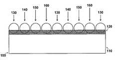
- FIG. 1is a schematic representation of one embodiment of the invention
- FIG. 2is a schematic representation of an exemplary micro coated pellet
- FIGS. 3A-Dare schematic representations of exemplary embodiments of micro coated pellets.
- FIG. 1is a schematic representation of one embodiment of the invention.
- an enlarged segment 100represents a portion of a medical device structure, e.g., a stent strut 110 , having deposited thereon an adhesive layer 120 .
- Micro coated pellets 130 , 140 , 150 and 160are embedded in adhesive layer 120 and are bonded to stent strut 110 .
- each of the micro coated pellets 130 , 140 , 150 and 160is represented as having a different composition and/or dissolution (decomposition) rate.
- FIG. 1While the underlying structure 110 represented in FIG. 1 is a stent, it is understood that the invention is not limited thereto. The principles of the invention can be applied equally to any structure adapted for insertion into a body whether the insertion is aimed at permanent or temporary placement of the structure in the body.
- Adhesive layer 120is adapted to receive the micro coated pellets.
- Adhesive layer 120can include one or a combination of several bio-compatible polymers (e.g., a polymer matrix).
- the polymer of the present inventionmay be hydrophilic or hydrophobic, and may be selected from the group consisting of polycarboxylic acids, cellulosic polymers, including cellulose acetate and cellulose nitrate, gelatin, polyvinylpyrrolidone, cross-linked polyvinylpyrrolidone, polyanhydrides including maleic anhydride polymers, polyamides, polyvinyl alcohols, copolymers of vinyl monomers such as EVA, polyvinyl ethers, polyvinyl aromatics, polyethylene oxides, glycosaminoglycans, polysaccharides, polyesters including polyethylene terephthalate, polyacrylamides, polyethers, polyether sulfone, polycarbonate, polyalkylenes including polypropylene, polyethylene
- Coatings from polymer dispersions such as polyurethane dispersions (BAYHDROL®, etc.) and acrylic latex dispersionsare also within the scope of the present invention.
- the polymermay be a protein polymer, fibrin, collage and derivatives thereof, polysaccharides such as celluloses, starches, dextrans, alginates and derivatives of these polysaccharides, an extracellular matrix component, hyaluronic acid, or another biologic agent or a suitable mixture of any of these, for example.
- the preferred polymeris polyacrylic acid, available as HYDROPLUS® (Boston Scientific Corporation, Natick, Mass.), and described in U.S. Pat. No.
- U.S. Pat. No. 5,091,205describes medical devices coated with one or more polyisocyanates such that the devices become instantly lubricious when exposed to body fluids.
- the polymeris a copolymer of polylactic acid and polycaprolactone.
- a preferred polymercan include a tri-block polymer.
- Exemplary adhesivesinclude cyanoacrylate or fibrin sealants such as TISSEEL® (Baxter Healthcare Corp., Deerfield, Ill.) or Hemaseel® (Haemacure Corp., Sarasota, Fla.).
- adhesive layer 120comprises a photo-cure polymer that can be activated through exposure to light of a particular wavelength.
- adhesive layer 120comprises a thermally-activated polymer. According to this embodiment, once the micro coated pellets are placed on adhesive layer 120 , they can be bonded to the stent strut 110 by heating the substrate and/or the adhesive layer to cure adhesive layer 120 .
- Adhesive layer 120can be applied by any conventional or known method including, for example, coating, spraying, brushing, contact transfer and electrodynamic coating.
- micro coated pellets 130 , 140 , 150 and 160are arranged side by side as discontiguous parts.
- the micro coated pelletscan be combined to form larger clusters or contiguous sections.
- micro coated pellets 130 , 140 , 150 and 160are represented as having a spherical shape, the principles of the invention are not limited to this shape and it is contemplated that the pellets, coated or uncoated, can have any shape, form or size.
- the micro coated pelletsmay be defined as a region conforming to the shape of the underlying structure.
- micro coated pelletscan be arranged according to their expected release profile or decomposition rate. For example, assuming that micro pellets 130 , 140 , 150 and 160 have different release profiles, they can be arranged on the structure 110 such that micro coated pellets having a substantially similar release profile are not immediately adjacent to each other.
- the surface of the structurecan be coated to have micro pellets 130 placed along the longitudinal axis of the stent and in a columnar arrangement. With this arrangement, repeating columns of micro coated pellets 130 are adjacent to, for example, columns of micro coated pellets 140 and 160 .
- a similar arrangementcan be implemented circumferentially around the periphery of the stent. In this embodiment, repeating rows of micro coated pellets 130 appear as rings around the circumference of the stent.
- An advantage of this and other similar arrangementsis that the coated device has a predictable, and similar release profile throughout its surface. In other words, such an arrangement can predictably release an active substance simultaneously throughout the body of the stent.
- the applicationmay demand arranging the micro coated pellets such that the composition(s) at one region, or one end, of the stent would dissolve faster than the composition(s) at other regions.
- the distal end of the stentwill begin releasing the active substance long after the proximal end of the stent is depleted.
- the release profile of a micro coated pelletcan be quantified according to the type and the amount of the polymer or biodegradable layer covering the active substance, designers can pre-define a release profile of the medical device by defining the polymer or biodegradable layer and its dissolution characteristics. The amount of the active substance can also be varied according to the desired effect.
- the material covering the active substancecan be a porous material that will allow the active substance to leach or elute through the porous openings.
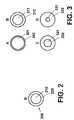
- FIG. 2is a schematic representation of an exemplary micro coated pellet.
- micro coated pellet 200is represented as having core 210 which contains a drug mixture such as an active substance or a mixture of different active substances.
- core 210is covered with micro coating layer 220 .
- micro coating layer 220can be any polymer or a combination of polymers or other biodegradable material having the desired characteristics.
- the release profile or decomposition rate of micro coated pellet 200can be quantified as a function of the physical and chemical characteristics of micro coating layer 220 .
- a micro coating layer 220 made of a polymer that has a very slow dissolution rate (or release rate) in the bodywould dictate a long release time.
- a micro coating layer 220 having a relatively fast dissolution ratecan be used if a more immediate drug release profile is desired.
- the thickness of the micro coating layer 220can affect the drug release profile. A thicker polymer coating layer would lead to a slower dissolution than a thinner polymer layer of the same composition.
- using the same physical characteristics of a micro coated pellet but having different solubility ratescan provide a similar drug release rate as that achieved with a thicker coating.
- core 210can be micro coated with a protective biodegradable material that dissolves at a rate of 50% per hour while another core can be micro coated with a protective biodegradable material that dissolves at a rate of 5% per hour. If these two pellets were placed adjacent to each other on a strut, the first pellet having the faster solubility rate will begin to release the drug mixture first. After some time, the polymer coating of the second pellet will dissolve away, and the drug mixture of the second pellet begins to enter the body. Thus, multiple micro coated pellets with different dissolution rates can be placed along a segment of a structure to provide a device with pre-defined time-release characteristics.
- core 210can include a polymer material mixed with an active ingredient that can elute the active ingredient over a period of time after the micro-coating 210 has partially or completely dissolved.
- core 210can be a mixture of 99% polymer compound and 1% drug that elutes over a period of 30 days when micro-coating 220 is partially or completely dissolved.
- Micro-coating 220 in this exampledissolves at a rate of 0.1 ⁇ m exposed thickness per day. By varying the thickness of micro-coating layer 220 , the beginning of the drug elution core 210 can be delayed as desired to create a micro-pellet with the needed drug release profile.
- FIGS. 3A-Dare schematic representations of exemplary embodiments of micro coated pellets.
- micro coated pellet 300 represented in FIG. 3Aincludes drug mixture 301 and micro coating layer 302 .
- the drug mixture corecan include, for example, an active substance, excipient, placebo or combinations thereof.
- active substance 311is substantially covered by micro coating layer 312 .
- FIG. 3Acan have 8 ⁇ g (25% drug and 75% placebo) of the drug mixture whereas the pellet represented in FIG. 3B can contain 4 ⁇ g (50% drug and 50% placebo) of the drug mixture.
- micro-coating polymer 302can be of thickness 0.2 ⁇ m and dissolve at a rate of 0.1 ⁇ m per day, thus allowing exposure to the therapeutic core 301 up to two days after implantation.
- micro-coating 312can have a thickness of 3.0 ⁇ m and dissolve at a rate of 0.1 ⁇ m per day, thus allowing exposure to the therapeutic core 311 up to thirty days after implantation.
- micro coated pellet Awould start releasing its content before pellet B starts. There may be a small overlap where both micro coated pellets A and B are releasing simultaneously. Finally, pellet B continues releasing its drug mixture long after pellet A has been completely dissolved.
- the wall thickness of pellet Ais thinner than the wall thickness of pellet B and will dissolve faster in the body, allowing pellet A start releasing the drug before pellet B.
- the supply of the active substance of pellet of FIG. 3Awill expire faster than that of FIG. 3B .
- the pellet represented in FIG. 3Awould have a release rate of 0.7 ⁇ g/day while the micro coated pellet represented in FIG. 3B would have a release rate of 0.15 ⁇ g/day. Since the internal volume of the micro coated pellet represented in FIG. 3A is larger than the micro coated pellet represented in FIG.
- the drug mixture of FIG. 3Acan contain more placebo in order to ensure that the drug content of the micro coated pellet of FIG. 3A would amount to 2 ⁇ g.
- the drug contentcan also be adjusted to control the release rate.
- micro coating 322is thicker than the wall thicknesses of micro coatings 302 and 312 .
- micro coating layer 322will dissolve slower than micro coating layers 302 and 312 .
- the exemplary micro coated pellet of FIG. 3Ccan contain 2.5 ⁇ g of a drug mixture (80% active substance and 20% placebo).
- the release profile for the micro coated pellet of FIG. 3Ccan start 10 days after exposure to body fluid and can expire 50 days later.
- this micro coated pelletholds the same relationship to the micro coated pellet of FIG. 3C as does the pellet of FIG. 3C to each of the micro coated pellets of FIGS. 3A and 3B .
- the pelletcontains 2 ⁇ g of drug mixture which is 100% active substance.
- the micro coated pellet in FIG. 3Dstarts releasing in 25 days and can expire in 75 days after exposure to body fluid.
- the pelletwould have a release rate of 0.05 ⁇ g/day and the micro coated pellet of FIG. 3D would have a release rate of 0.04 ⁇ g/day.
- the drug mixture of pellet of FIG. 3Cmay contain less placebo than each of the pellets 3 A or 3 B in order to have the drug content be 2 ⁇ g, as will the others.
- a micro coated pelletcan be provided with several alternating layers of drug mixture and polymer micro coating.
- a drug mixture layercan be covered, partially or completely, by a protective polymer layer, which itself is covered by alternating drug layers and protective polymer layers.
- the composition of each sublayer and its thicknesscan be varied to obtain the desired release rate.
- the pellet represent in FIG. 3Acan have a micro-coating 302 of 0.2 ⁇ m thickness that dissolves at a rate of 0.1 ⁇ m per day and a core 301 that contains 99% polymer, 0.25% drug and 0.75% placebo which can elute over a period of 30 days.
- the pellet represented in FIG. 3Bcan have a micro-coating 312 of 3.0 ⁇ m that dissolves at a rate of 0.1 ⁇ m per day and a core 311 that contains 99% polymer, 0.5% drug and 0.5% placebo which can elute over a period of 30 days.
- FIG. 3Acan begin drug elution two days after implantation and elute therapeutic agent for a thirty-day period where the micro-pellet represented by FIG. 3B would begin drug elution thirty days after implantation and elute therapeutic agent over a thirty-day period.
- the therapeutic/polymer/placebo mixcan be adjusted to control the dosage of elution rate as well as the varying of the micro-coating layer thickness can be adjusted to define the time period before therapeutic elution begins.
- the exemplary micro-coated pellet of FIG. 3Ccan have a micro-coating layer 322 of thickness of 6 ⁇ m that dissolves at a rate of 0.1 ⁇ m per day and a core 321 containing 99% polymer, 0.75% drug and 0.25% placebo that elutes over a period of 30 days.
- the exemplary micro-coated pellet of FIG. 3Dcan have a micro-coating layer 332 of thickness 9 ⁇ m that dissolves at a rate of 0.1 ⁇ m per day and a core 331 containing 99% polymer and 1% drug that elutes over a period of 30 days.
- the micro-pellet represented by FIG. 3Cwould begin drug elution 60 days after implantation and elutes therapeutic for a 30 day period where the micro-pellet represented by FIG. 3D would begin drug elution 30 days after implantation and elutes therapeutic over a 90 day period.
- the micro coated pelletscan be of the same size or can have different sizes depending on the desired release rate and/or the underlying drug composition.
- the pellets with the fastest release ratecan contain some inert chemical, such as mannitol. If the pellets having the faster release rate are supplied with a thinner micro coating, the addition of an inert compound would serve to increase the volume of the pellet while keeping the drug content constant.
- the pellet sizeis less than 50 ⁇ m.
- a mandrelis placed through an uncoated stent to provide structural support. Then the top surface of the stent (the surface not in contact with the mandrel) is coated with a bio-compatible adhesive layer.
- the coating processcan be done by any number of techniques, including rolling, spraying and roll-to-roll transfer. Once a sufficient amount of adhesive is coated on the stent, micro coated pellets can be imbedded on the stent until all or a portion of the stent is covered. The micro coated pellets can also be supplied to the extent necessary to deliver only the desired amount of the active substance.
- the micro coated pelletsare embedded on the surface of the stent circumferentially as rings with each adjacent ring, line or pellet having a different release rate.
- the adhesivecan be cured to maintain the pellets in place.
- the stentcan be air brushed to remove the loose pellets.
- the micro coated pelletsare secured to the stent with an adhesive, the principles of the invention are not limited thereto and it is understood that any method for securing the micro pellets to the surface of the stent is deemed to be well within the scope of the invention.
- the pelletscan be sprayed or coated with an adhesive and attached to the device.
- the pelletscan be coated with a material such as a biodegradable wax and placed on the device. Once the pellets and/or the device is heated, the wax melts and adheres the pellets to the surface of the device.
- the micro coated pelletsare imbedded on the stent structure by scribing lines (such as aperture, via, bore, groove, cavity, gap or notch) in a blank plate to a set depth and width, filling the scribed lines with micro coated pellets in the desired order, introducing an adhesive or other tacky substance over the stent structure, rolling the stent over the pellet-loaded scribed tray so that the pellets adhere to the stent, substrate and curing the adhesive.
- scribing linessuch as aperture, via, bore, groove, cavity, gap or notch
- the pelletsare arranged on a tacky sheet, the structure is coated with an adhesive tackier than the sheet, and the pellets are transferred to the structure from the sheet after the sheet is imposed and impressed on the structure. Once the tacky sheet is removed and the adhesive on the structure is cured, the pellets will remain embedded on the structure. In an alternative embodiment, the structure is cured with the tacky sheet in place and the sheet is removed after the adhesive on the structure is cured.
- that adhesive layercan be a polymer material that is mixed with the therapeutic agent that is on the device to form a matrix layer.
- the matrix layerwill elute therapeutic agent over a period of time.
- the micro coated pelletsare deposited on the end of a flat-tipped blade.
- the bladeis then rolled around the periphery of the stent to make contact around the stent (forming rings) or contacted along the length of the stent from end to end (forming lines).
- the stentcan be optionally coated with an adhesive to receive the micro coated pellets.
- pelletscan be placed and imbedded on the desired location of the stent strut. If the stent is pre-coated with adhesive, curing the adhesive would bond the micro coated pellets to its surface.
- thin strips of adhesivecan be coated with a range of pellets prior to placing the coated strip in a fixture. Once the fixture is occupied with sufficient number of strands, the stent can be rolled over the strands to effectively transfer the micro coated pellets to the surface of the stent. Alternatively, the strands can be wrapper around the stent. If the stent is provided with an adhesive coating, the coating can be cured to further secure the micro coated pellets thereto.
- active substanceand “therapeutic agent” as used herein includes one or more “therapeutic agents” or “drugs”.
- active substanceincludes pharmaceutically active compounds, nucleic acids with and without carrier vectors such as lipids, compacting agents (such as histones), virus (such as adenovirus, andenoassociated virus, retrovirus, lentivirus and ⁇ -virus), polymers, hyaluronic acid, proteins, cells and the like, with or without targeting sequences.
- therapeutic agents used in conjunction with the present inventioninclude, for example, pharmaceutically active compounds, proteins, cells, oligonucleotides, ribozymes, anti-sense oligonucleotides, DNA compacting agents, gene/vector systems (i.e., any vehicle that allows for the uptake and expression of nucleic acids), nucleic acids (including, for example, recombinant nucleic acids; naked DNA, cDNA, RNA; genomic DNA, cDNA or RNA in a non-infectious vector or in a viral vector and which further may have attached peptide targeting sequences; antisense nucleic acid (RNA or DNA); and DNA chimeras which include gene sequences and encoding for ferry proteins such as membrane translocating sequences (“MTS”) and herpes simplex virus-1 (“VP22”)), and viral, liposomes and cationic and anionic polymers and neutral polymers that are selected from a number of types depending on the desired application.
- gene/vector systemsi.e., any vehicle
- Non-limiting examples of virus vectors or vectors derived from viral sourcesinclude adenoviral vectors, herpes simplex vectors, papilloma vectors, adeno-associated vectors, retroviral vectors, and the like.
- Non-limiting examples of biologically active solutesinclude anti-thrombogenic agents such as heparin, heparin derivatives, urokinase, and PPACK (dextrophenylalanine proline arginine chloromethylketone); antioxidants such as probucol and retinoic acid; angiogenic and anti-angiogenic agents and factors; agents blocking smooth muscle cell proliferation such as rapamycin, angiopeptin, and monoclonal antibodies capable of blocking smooth muscle cell proliferation; anti-inflammatory agents such as dexamethasone, prednisolone, corticosterone, budesonide, estrogen, sulfasalazine, acetyl salicylic acid, and mesalamine; calcium entry blockers
- Polynucleotide sequences useful in practice of the inventioninclude DNA or RNA sequences having a therapeutic effect after being taken up by a cell.
- therapeutic polynucleotidesinclude anti-sense DNA and RNA; DNA coding for an anti-sense RNA; or DNA coding for tRNA or rRNA to replace defective or deficient endogenous molecules.
- the polynucleotidescan also code for therapeutic proteins or polypeptides.
- a polypeptideis understood to be any translation product of a polynucleotide regardless of size, and whether glycosylated or not.
- Therapeutic proteins and polypeptidesinclude as a primary example, those proteins or polypeptides that can compensate for defective or deficient species in an animal, or those that act through toxic effects to limit or remove harmful cells from the body.
- the polypeptides or proteins that can be injected, or whose DNA can be incorporatedinclude without limitation, angiogenic factors and other molecules competent to induce angiogenesis, including acidic and basic fibroblast growth factors, vascular endothelial growth factor, hif-1, epidermal growth factor, transforming growth factor ⁇ and ⁇ , platelet-derived endothelial growth factor, platelet-derived growth factor, tumor necrosis factor ⁇ , hepatocyte growth factor and insulin like growth factor; growth factors; cell cycle inhibitors including CDK inhibitors; anti-restenosis agents, including p15, p16, p18, p19, p21, p27, p53, p57, Rb, nFkB and E2F decoys, thymidine kina
- MCP-1monocyte chemoattractant protein
- BMP'sthe family of bone morphogenic proteins
- the known proteinsinclude BMP-2, BMP-3, BMP-4, BMP-5, BMP-6 (Vgr-1), BMP-7 (OP-1), BMP-8, BMP-9, BMP-10, BMP-11, BMP-12, BMP-13, BMP-14, BMP-15, and BMP-16.
- BMP'sare any of BMP-2, BMP-3, BMP-4, BMP-5, BMP-6 and BMP-7.
- dimeric proteinscan be provided as homodimers, heterodimers, or combinations thereof, alone or together with other molecules.
- molecules capable of inducing an upstream or downstream effect of a BMPcan be provided.
- Such moleculesinclude any of the “hedgehog” proteins, or the DNA's encoding them.
Landscapes
- Health & Medical Sciences (AREA)
- Life Sciences & Earth Sciences (AREA)
- Public Health (AREA)
- Veterinary Medicine (AREA)
- Epidemiology (AREA)
- Surgery (AREA)
- Animal Behavior & Ethology (AREA)
- General Health & Medical Sciences (AREA)
- Heart & Thoracic Surgery (AREA)
- Vascular Medicine (AREA)
- Chemical & Material Sciences (AREA)
- Engineering & Computer Science (AREA)
- Biomedical Technology (AREA)
- Medicinal Chemistry (AREA)
- Molecular Biology (AREA)
- Materials For Medical Uses (AREA)
- Medicinal Preparation (AREA)
Abstract
Description
Claims (20)
Priority Applications (2)
| Application Number | Priority Date | Filing Date | Title |
|---|---|---|---|
| US10/701,455US7435256B2 (en) | 2003-11-06 | 2003-11-06 | Method and apparatus for controlled delivery of active substance |
| PCT/US2004/036489WO2005046745A2 (en) | 2003-11-06 | 2004-11-03 | Method and device for controlled delivery of active substance |
Applications Claiming Priority (1)
| Application Number | Priority Date | Filing Date | Title |
|---|---|---|---|
| US10/701,455US7435256B2 (en) | 2003-11-06 | 2003-11-06 | Method and apparatus for controlled delivery of active substance |
Publications (2)
| Publication Number | Publication Date |
|---|---|
| US20050100582A1 US20050100582A1 (en) | 2005-05-12 |
| US7435256B2true US7435256B2 (en) | 2008-10-14 |
Family
ID=34551429
Family Applications (1)
| Application Number | Title | Priority Date | Filing Date |
|---|---|---|---|
| US10/701,455Expired - Fee RelatedUS7435256B2 (en) | 2003-11-06 | 2003-11-06 | Method and apparatus for controlled delivery of active substance |
Country Status (2)
| Country | Link |
|---|---|
| US (1) | US7435256B2 (en) |
| WO (1) | WO2005046745A2 (en) |
Cited By (32)
| Publication number | Priority date | Publication date | Assignee | Title |
|---|---|---|---|---|
| US20080249615A1 (en)* | 2007-04-05 | 2008-10-09 | Jan Weber | Stents with ceramic drug reservoir layer and methods of making and using the same |
| US20080294236A1 (en)* | 2007-05-23 | 2008-11-27 | Boston Scientific Scimed, Inc. | Endoprosthesis with Select Ceramic and Polymer Coatings |
| US20090088809A1 (en)* | 2007-09-28 | 2009-04-02 | Michael Alan Fisher | Anti-Microbial Implant |
| US20090118812A1 (en)* | 2007-11-02 | 2009-05-07 | Boston Scientific Scimed, Inc. | Endoprosthesis coating |
| US20090149942A1 (en)* | 2007-07-19 | 2009-06-11 | Boston Scientific Scimed, Inc. | Endoprosthesis having a non-fouling surface |
| US20090157172A1 (en)* | 2007-07-24 | 2009-06-18 | Boston Scientific Scrimed, Inc. | Stents with polymer-free coatings for delivering a therapeutic agent |
| US20100152777A1 (en)* | 2008-12-16 | 2010-06-17 | Fisher Michael A | Anti-Infective Spinal Rod with Surface Features |
| US7931683B2 (en) | 2007-07-27 | 2011-04-26 | Boston Scientific Scimed, Inc. | Articles having ceramic coated surfaces |
| US7938855B2 (en) | 2007-11-02 | 2011-05-10 | Boston Scientific Scimed, Inc. | Deformable underlayer for stent |
| US7942926B2 (en) | 2007-07-11 | 2011-05-17 | Boston Scientific Scimed, Inc. | Endoprosthesis coating |
| US7976915B2 (en) | 2007-05-23 | 2011-07-12 | Boston Scientific Scimed, Inc. | Endoprosthesis with select ceramic morphology |
| US7981150B2 (en) | 2006-11-09 | 2011-07-19 | Boston Scientific Scimed, Inc. | Endoprosthesis with coatings |
| US8002823B2 (en) | 2007-07-11 | 2011-08-23 | Boston Scientific Scimed, Inc. | Endoprosthesis coating |
| US8029554B2 (en) | 2007-11-02 | 2011-10-04 | Boston Scientific Scimed, Inc. | Stent with embedded material |
| US8066763B2 (en) | 1998-04-11 | 2011-11-29 | Boston Scientific Scimed, Inc. | Drug-releasing stent with ceramic-containing layer |
| US8071156B2 (en) | 2009-03-04 | 2011-12-06 | Boston Scientific Scimed, Inc. | Endoprostheses |
| US8070797B2 (en) | 2007-03-01 | 2011-12-06 | Boston Scientific Scimed, Inc. | Medical device with a porous surface for delivery of a therapeutic agent |
| US8187620B2 (en) | 2006-03-27 | 2012-05-29 | Boston Scientific Scimed, Inc. | Medical devices comprising a porous metal oxide or metal material and a polymer coating for delivering therapeutic agents |
| US8216632B2 (en) | 2007-11-02 | 2012-07-10 | Boston Scientific Scimed, Inc. | Endoprosthesis coating |
| US8221822B2 (en) | 2007-07-31 | 2012-07-17 | Boston Scientific Scimed, Inc. | Medical device coating by laser cladding |
| US8231980B2 (en) | 2008-12-03 | 2012-07-31 | Boston Scientific Scimed, Inc. | Medical implants including iridium oxide |
| US8287937B2 (en) | 2009-04-24 | 2012-10-16 | Boston Scientific Scimed, Inc. | Endoprosthese |
| US8353949B2 (en) | 2006-09-14 | 2013-01-15 | Boston Scientific Scimed, Inc. | Medical devices with drug-eluting coating |
| US8431149B2 (en) | 2007-03-01 | 2013-04-30 | Boston Scientific Scimed, Inc. | Coated medical devices for abluminal drug delivery |
| US8449603B2 (en) | 2008-06-18 | 2013-05-28 | Boston Scientific Scimed, Inc. | Endoprosthesis coating |
| US8574615B2 (en) | 2006-03-24 | 2013-11-05 | Boston Scientific Scimed, Inc. | Medical devices having nanoporous coatings for controlled therapeutic agent delivery |
| US8771343B2 (en) | 2006-06-29 | 2014-07-08 | Boston Scientific Scimed, Inc. | Medical devices with selective titanium oxide coatings |
| US8815273B2 (en) | 2007-07-27 | 2014-08-26 | Boston Scientific Scimed, Inc. | Drug eluting medical devices having porous layers |
| US8815275B2 (en) | 2006-06-28 | 2014-08-26 | Boston Scientific Scimed, Inc. | Coatings for medical devices comprising a therapeutic agent and a metallic material |
| US8900292B2 (en) | 2007-08-03 | 2014-12-02 | Boston Scientific Scimed, Inc. | Coating for medical device having increased surface area |
| US8920491B2 (en) | 2008-04-22 | 2014-12-30 | Boston Scientific Scimed, Inc. | Medical devices having a coating of inorganic material |
| US8932346B2 (en) | 2008-04-24 | 2015-01-13 | Boston Scientific Scimed, Inc. | Medical devices having inorganic particle layers |
Families Citing this family (10)
| Publication number | Priority date | Publication date | Assignee | Title |
|---|---|---|---|---|
| US8118864B1 (en)* | 2004-05-25 | 2012-02-21 | Endovascular Technologies, Inc. | Drug delivery endovascular graft |
| ES2432556T3 (en) | 2004-08-04 | 2013-12-04 | Evonik Corporation | Methods for manufacturing supply devices and their devices |
| US8057543B2 (en)* | 2005-01-28 | 2011-11-15 | Greatbatch Ltd. | Stent coating for eluting medication |
| EP1890648B1 (en)* | 2005-06-06 | 2017-08-09 | Innovational Holdings, LLC | Implantable medical device with openings for delivery of beneficial agents with combination release kinetics |
| US20080091262A1 (en)* | 2006-10-17 | 2008-04-17 | Gale David C | Drug delivery after biodegradation of the stent scaffolding |
| US8016879B2 (en)* | 2006-08-01 | 2011-09-13 | Abbott Cardiovascular Systems Inc. | Drug delivery after biodegradation of the stent scaffolding |
| US8124601B2 (en)* | 2007-11-21 | 2012-02-28 | Bristol-Myers Squibb Company | Compounds for the treatment of Hepatitis C |
| JP5502751B2 (en) | 2007-12-20 | 2014-05-28 | エボニック コーポレイション | Process for preparing microparticles with low residual solvent concentration |
| DE102008044316A1 (en)* | 2008-12-03 | 2010-06-10 | Biotronik Vi Patent Ag | Drug-coated medical device, process for its preparation and its uses |
| CA2827330A1 (en)* | 2011-02-23 | 2012-08-30 | Ams Research Corporation | Pelvic implant and therapeutic agent system and method |
Citations (26)
| Publication number | Priority date | Publication date | Assignee | Title |
|---|---|---|---|---|
| US5304121A (en) | 1990-12-28 | 1994-04-19 | Boston Scientific Corporation | Drug delivery system making use of a hydrogel polymer coating |
| EP0604022A1 (en) | 1992-12-22 | 1994-06-29 | Advanced Cardiovascular Systems, Inc. | Multilayered biodegradable stent and method for its manufacture |
| US5443458A (en) | 1992-12-22 | 1995-08-22 | Advanced Cardiovascular Systems, Inc. | Multilayered biodegradable stent and method of manufacture |
| EP0734721A2 (en) | 1995-03-30 | 1996-10-02 | Advanced Cardiovascular Systems, Inc. | Method of incorporating drugs into a polymer component of stents |
| US5873904A (en) | 1995-06-07 | 1999-02-23 | Cook Incorporated | Silver implantable medical device |
| US5972027A (en) | 1997-09-30 | 1999-10-26 | Scimed Life Systems, Inc | Porous stent drug delivery system |
| US6120536A (en) | 1995-04-19 | 2000-09-19 | Schneider (Usa) Inc. | Medical devices with long term non-thrombogenic coatings |
| WO2001015751A1 (en) | 1999-09-01 | 2001-03-08 | Bioxid Oy | Novel multilayered material bearing a biologically active agent and the preparation thereof |
| US6210703B1 (en) | 1997-12-19 | 2001-04-03 | Ppg Industries Ohio, Inc. | Glass fiber chemical delivery system |
| US20010027340A1 (en) | 1997-04-18 | 2001-10-04 | Carol Wright | Stent with therapeutically active dosage of rapamycin coated thereon |
| US6299604B1 (en) | 1998-08-20 | 2001-10-09 | Cook Incorporated | Coated implantable medical device |
| US6299905B1 (en)* | 1996-04-16 | 2001-10-09 | Depuy Orthopaedics, Inc. | Bioerodable polymeric adhesives for tissue repair |
| US6339130B1 (en)* | 1994-07-22 | 2002-01-15 | United States Surgical Corporation | Bioabsorbable branched polymers containing units derived from dioxanone and medical/surgical devices manufactured therefrom |
| US6368658B1 (en) | 1999-04-19 | 2002-04-09 | Scimed Life Systems, Inc. | Coating medical devices using air suspension |
| US6371979B1 (en) | 1993-01-27 | 2002-04-16 | Intratherapeutics, Inc. | Stent delivery system |
| US6395326B1 (en) | 2000-05-31 | 2002-05-28 | Advanced Cardiovascular Systems, Inc. | Apparatus and method for depositing a coating onto a surface of a prosthesis |
| US6468819B1 (en) | 1999-11-23 | 2002-10-22 | The Trustees Of Princeton University | Method for patterning organic thin film devices using a die |
| US20030068355A1 (en) | 2001-08-20 | 2003-04-10 | Shanley John F. | Therapeutic agent delivery device with protective separating layer |
| WO2003030879A1 (en) | 2001-10-05 | 2003-04-17 | Surmodics, Inc. | Particle immobilized coatings and uses thereof |
| US20030149479A1 (en) | 2002-01-18 | 2003-08-07 | Snyder Michael E. | Endocapsular tension ring sustained release device and method of making and using the same |
| WO2004016298A1 (en) | 2002-08-16 | 2004-02-26 | Boston Scientific Limited | Microarray drug delivery coatings |
| US20040127976A1 (en)* | 2002-09-20 | 2004-07-01 | Conor Medsystems, Inc. | Method and apparatus for loading a beneficial agent into an expandable medical device |
| WO2004087251A1 (en) | 2003-03-28 | 2004-10-14 | Conor Medsystems, Inc. | Implantable medical device and method for in situ selective modulation of agent delivery |
| US6805809B2 (en) | 2002-08-28 | 2004-10-19 | Board Of Trustees Of University Of Illinois | Decal transfer microfabrication |
| US20040254635A1 (en)* | 1998-03-30 | 2004-12-16 | Shanley John F. | Expandable medical device for delivery of beneficial agent |
| US6849089B2 (en)* | 2001-10-08 | 2005-02-01 | Biotronik Mess-Und Therapiegeraete Gmbh & Co Ingenieurbuero Berlin | Implant with proliferation-inhibiting substance |
- 2003
- 2003-11-06USUS10/701,455patent/US7435256B2/ennot_activeExpired - Fee Related
- 2004
- 2004-11-03WOPCT/US2004/036489patent/WO2005046745A2/enactiveApplication Filing
Patent Citations (26)
| Publication number | Priority date | Publication date | Assignee | Title |
|---|---|---|---|---|
| US5304121A (en) | 1990-12-28 | 1994-04-19 | Boston Scientific Corporation | Drug delivery system making use of a hydrogel polymer coating |
| EP0604022A1 (en) | 1992-12-22 | 1994-06-29 | Advanced Cardiovascular Systems, Inc. | Multilayered biodegradable stent and method for its manufacture |
| US5443458A (en) | 1992-12-22 | 1995-08-22 | Advanced Cardiovascular Systems, Inc. | Multilayered biodegradable stent and method of manufacture |
| US6371979B1 (en) | 1993-01-27 | 2002-04-16 | Intratherapeutics, Inc. | Stent delivery system |
| US6339130B1 (en)* | 1994-07-22 | 2002-01-15 | United States Surgical Corporation | Bioabsorbable branched polymers containing units derived from dioxanone and medical/surgical devices manufactured therefrom |
| EP0734721A2 (en) | 1995-03-30 | 1996-10-02 | Advanced Cardiovascular Systems, Inc. | Method of incorporating drugs into a polymer component of stents |
| US6120536A (en) | 1995-04-19 | 2000-09-19 | Schneider (Usa) Inc. | Medical devices with long term non-thrombogenic coatings |
| US5873904A (en) | 1995-06-07 | 1999-02-23 | Cook Incorporated | Silver implantable medical device |
| US6299905B1 (en)* | 1996-04-16 | 2001-10-09 | Depuy Orthopaedics, Inc. | Bioerodable polymeric adhesives for tissue repair |
| US20010027340A1 (en) | 1997-04-18 | 2001-10-04 | Carol Wright | Stent with therapeutically active dosage of rapamycin coated thereon |
| US5972027A (en) | 1997-09-30 | 1999-10-26 | Scimed Life Systems, Inc | Porous stent drug delivery system |
| US6210703B1 (en) | 1997-12-19 | 2001-04-03 | Ppg Industries Ohio, Inc. | Glass fiber chemical delivery system |
| US20040254635A1 (en)* | 1998-03-30 | 2004-12-16 | Shanley John F. | Expandable medical device for delivery of beneficial agent |
| US6299604B1 (en) | 1998-08-20 | 2001-10-09 | Cook Incorporated | Coated implantable medical device |
| US6368658B1 (en) | 1999-04-19 | 2002-04-09 | Scimed Life Systems, Inc. | Coating medical devices using air suspension |
| WO2001015751A1 (en) | 1999-09-01 | 2001-03-08 | Bioxid Oy | Novel multilayered material bearing a biologically active agent and the preparation thereof |
| US6468819B1 (en) | 1999-11-23 | 2002-10-22 | The Trustees Of Princeton University | Method for patterning organic thin film devices using a die |
| US6395326B1 (en) | 2000-05-31 | 2002-05-28 | Advanced Cardiovascular Systems, Inc. | Apparatus and method for depositing a coating onto a surface of a prosthesis |
| US20030068355A1 (en) | 2001-08-20 | 2003-04-10 | Shanley John F. | Therapeutic agent delivery device with protective separating layer |
| WO2003030879A1 (en) | 2001-10-05 | 2003-04-17 | Surmodics, Inc. | Particle immobilized coatings and uses thereof |
| US6849089B2 (en)* | 2001-10-08 | 2005-02-01 | Biotronik Mess-Und Therapiegeraete Gmbh & Co Ingenieurbuero Berlin | Implant with proliferation-inhibiting substance |
| US20030149479A1 (en) | 2002-01-18 | 2003-08-07 | Snyder Michael E. | Endocapsular tension ring sustained release device and method of making and using the same |
| WO2004016298A1 (en) | 2002-08-16 | 2004-02-26 | Boston Scientific Limited | Microarray drug delivery coatings |
| US6805809B2 (en) | 2002-08-28 | 2004-10-19 | Board Of Trustees Of University Of Illinois | Decal transfer microfabrication |
| US20040127976A1 (en)* | 2002-09-20 | 2004-07-01 | Conor Medsystems, Inc. | Method and apparatus for loading a beneficial agent into an expandable medical device |
| WO2004087251A1 (en) | 2003-03-28 | 2004-10-14 | Conor Medsystems, Inc. | Implantable medical device and method for in situ selective modulation of agent delivery |
Cited By (40)
| Publication number | Priority date | Publication date | Assignee | Title |
|---|---|---|---|---|
| US8066763B2 (en) | 1998-04-11 | 2011-11-29 | Boston Scientific Scimed, Inc. | Drug-releasing stent with ceramic-containing layer |
| US8574615B2 (en) | 2006-03-24 | 2013-11-05 | Boston Scientific Scimed, Inc. | Medical devices having nanoporous coatings for controlled therapeutic agent delivery |
| US8187620B2 (en) | 2006-03-27 | 2012-05-29 | Boston Scientific Scimed, Inc. | Medical devices comprising a porous metal oxide or metal material and a polymer coating for delivering therapeutic agents |
| US8815275B2 (en) | 2006-06-28 | 2014-08-26 | Boston Scientific Scimed, Inc. | Coatings for medical devices comprising a therapeutic agent and a metallic material |
| US8771343B2 (en) | 2006-06-29 | 2014-07-08 | Boston Scientific Scimed, Inc. | Medical devices with selective titanium oxide coatings |
| US8353949B2 (en) | 2006-09-14 | 2013-01-15 | Boston Scientific Scimed, Inc. | Medical devices with drug-eluting coating |
| US7981150B2 (en) | 2006-11-09 | 2011-07-19 | Boston Scientific Scimed, Inc. | Endoprosthesis with coatings |
| US8070797B2 (en) | 2007-03-01 | 2011-12-06 | Boston Scientific Scimed, Inc. | Medical device with a porous surface for delivery of a therapeutic agent |
| US8431149B2 (en) | 2007-03-01 | 2013-04-30 | Boston Scientific Scimed, Inc. | Coated medical devices for abluminal drug delivery |
| US8067054B2 (en) | 2007-04-05 | 2011-11-29 | Boston Scientific Scimed, Inc. | Stents with ceramic drug reservoir layer and methods of making and using the same |
| US20080249615A1 (en)* | 2007-04-05 | 2008-10-09 | Jan Weber | Stents with ceramic drug reservoir layer and methods of making and using the same |
| US7976915B2 (en) | 2007-05-23 | 2011-07-12 | Boston Scientific Scimed, Inc. | Endoprosthesis with select ceramic morphology |
| US20080294236A1 (en)* | 2007-05-23 | 2008-11-27 | Boston Scientific Scimed, Inc. | Endoprosthesis with Select Ceramic and Polymer Coatings |
| US8002823B2 (en) | 2007-07-11 | 2011-08-23 | Boston Scientific Scimed, Inc. | Endoprosthesis coating |
| US7942926B2 (en) | 2007-07-11 | 2011-05-17 | Boston Scientific Scimed, Inc. | Endoprosthesis coating |
| US20090149942A1 (en)* | 2007-07-19 | 2009-06-11 | Boston Scientific Scimed, Inc. | Endoprosthesis having a non-fouling surface |
| US9284409B2 (en) | 2007-07-19 | 2016-03-15 | Boston Scientific Scimed, Inc. | Endoprosthesis having a non-fouling surface |
| US20090157172A1 (en)* | 2007-07-24 | 2009-06-18 | Boston Scientific Scrimed, Inc. | Stents with polymer-free coatings for delivering a therapeutic agent |
| US7931683B2 (en) | 2007-07-27 | 2011-04-26 | Boston Scientific Scimed, Inc. | Articles having ceramic coated surfaces |
| US8815273B2 (en) | 2007-07-27 | 2014-08-26 | Boston Scientific Scimed, Inc. | Drug eluting medical devices having porous layers |
| US8221822B2 (en) | 2007-07-31 | 2012-07-17 | Boston Scientific Scimed, Inc. | Medical device coating by laser cladding |
| US8900292B2 (en) | 2007-08-03 | 2014-12-02 | Boston Scientific Scimed, Inc. | Coating for medical device having increased surface area |
| US11389202B2 (en) | 2007-09-28 | 2022-07-19 | De Puy Synthes Products. Inc. | Anti-microbial implant |
| US10786282B2 (en) | 2007-09-28 | 2020-09-29 | DePuy Synthes Products, Inc. | Anti-microbial implant |
| US10064655B2 (en) | 2007-09-28 | 2018-09-04 | DePuy Synthes Products, Inc. | Anti-microbial implant |
| US20090088809A1 (en)* | 2007-09-28 | 2009-04-02 | Michael Alan Fisher | Anti-Microbial Implant |
| US9247973B2 (en) | 2007-09-28 | 2016-02-02 | DePuy Synthes Products, Inc. | Anti-microbial implant |
| US20090118812A1 (en)* | 2007-11-02 | 2009-05-07 | Boston Scientific Scimed, Inc. | Endoprosthesis coating |
| US7938855B2 (en) | 2007-11-02 | 2011-05-10 | Boston Scientific Scimed, Inc. | Deformable underlayer for stent |
| US8029554B2 (en) | 2007-11-02 | 2011-10-04 | Boston Scientific Scimed, Inc. | Stent with embedded material |
| US8216632B2 (en) | 2007-11-02 | 2012-07-10 | Boston Scientific Scimed, Inc. | Endoprosthesis coating |
| US8920491B2 (en) | 2008-04-22 | 2014-12-30 | Boston Scientific Scimed, Inc. | Medical devices having a coating of inorganic material |
| US8932346B2 (en) | 2008-04-24 | 2015-01-13 | Boston Scientific Scimed, Inc. | Medical devices having inorganic particle layers |
| US8449603B2 (en) | 2008-06-18 | 2013-05-28 | Boston Scientific Scimed, Inc. | Endoprosthesis coating |
| US8231980B2 (en) | 2008-12-03 | 2012-07-31 | Boston Scientific Scimed, Inc. | Medical implants including iridium oxide |
| US20100152777A1 (en)* | 2008-12-16 | 2010-06-17 | Fisher Michael A | Anti-Infective Spinal Rod with Surface Features |
| US9763697B2 (en) | 2008-12-16 | 2017-09-19 | DePuy Synthes Products, Inc. | Anti-infective spinal rod with surface features |
| US11974783B2 (en) | 2008-12-16 | 2024-05-07 | DePuy Synthes Products, Inc. | Anti-infective orthopedic implant |
| US8071156B2 (en) | 2009-03-04 | 2011-12-06 | Boston Scientific Scimed, Inc. | Endoprostheses |
| US8287937B2 (en) | 2009-04-24 | 2012-10-16 | Boston Scientific Scimed, Inc. | Endoprosthese |
Also Published As
| Publication number | Publication date |
|---|---|
| WO2005046745A2 (en) | 2005-05-26 |
| WO2005046745A3 (en) | 2005-07-28 |
| US20050100582A1 (en) | 2005-05-12 |
Similar Documents
| Publication | Publication Date | Title |
|---|---|---|
| US7435256B2 (en) | Method and apparatus for controlled delivery of active substance | |
| US9272307B2 (en) | Contact coating of prostheses | |
| US7482034B2 (en) | Expandable mask stent coating method | |
| EP2173400B1 (en) | Implantable medical devices having adjustable pore volume and methods for making the same | |
| US8173200B2 (en) | Selective application of therapeutic agent to a medical device | |
| US7524527B2 (en) | Electrostatic coating of a device | |
| US8157854B2 (en) | Method and system for delivery of coated implants | |
| EP1536849B1 (en) | Microarray drug delivery coatings | |
| US20090123517A1 (en) | Medical devices for releasing therapeutic agent and methods of making the same | |
| US20080077218A1 (en) | Injection of therapeutic into porous regions of a medical device | |
| US20050147734A1 (en) | Method and system for coating tubular medical devices | |
| US7168605B2 (en) | Microtubes for therapeutic delivery | |
| US20070259116A1 (en) | Partially coated workpiece and method of making same | |
| US20080097569A1 (en) | Reduction of burst release from therapeutically treated medical devices | |
| US8263171B2 (en) | Methods for making drug-eluting medical devices |
Legal Events
| Date | Code | Title | Description |
|---|---|---|---|
| AS | Assignment | Owner name:SCIMED LIFE SYSTEMS, INC., MINNESOTA Free format text:ASSIGNMENT OF ASSIGNORS INTEREST;ASSIGNOR:STENZEL, ERIC B.;REEL/FRAME:014685/0427 Effective date:20031030 | |
| AS | Assignment | Owner name:BOSTON SCIENTIFIC SCIMED, INC., MINNESOTA Free format text:CHANGE OF NAME;ASSIGNOR:SCIMED LIFE SYSTEMS, INC.;REEL/FRAME:017798/0848 Effective date:20041222 | |
| AS | Assignment | Owner name:BOSTON SCIENTIFIC SCIMED, INC., MINNESOTA Free format text:CHANGE OF NAME;ASSIGNOR:SCIMED LIFE SYSTEMS, INC.;REEL/FRAME:018505/0868 Effective date:20050101 Owner name:BOSTON SCIENTIFIC SCIMED, INC.,MINNESOTA Free format text:CHANGE OF NAME;ASSIGNOR:SCIMED LIFE SYSTEMS, INC.;REEL/FRAME:018505/0868 Effective date:20050101 | |
| FEPP | Fee payment procedure | Free format text:PAYOR NUMBER ASSIGNED (ORIGINAL EVENT CODE: ASPN); ENTITY STATUS OF PATENT OWNER: LARGE ENTITY | |
| STCF | Information on status: patent grant | Free format text:PATENTED CASE | |
| CC | Certificate of correction | ||
| FPAY | Fee payment | Year of fee payment:4 | |
| FPAY | Fee payment | Year of fee payment:8 | |
| FEPP | Fee payment procedure | Free format text:MAINTENANCE FEE REMINDER MAILED (ORIGINAL EVENT CODE: REM.); ENTITY STATUS OF PATENT OWNER: LARGE ENTITY | |
| LAPS | Lapse for failure to pay maintenance fees | Free format text:PATENT EXPIRED FOR FAILURE TO PAY MAINTENANCE FEES (ORIGINAL EVENT CODE: EXP.); ENTITY STATUS OF PATENT OWNER: LARGE ENTITY | |
| STCH | Information on status: patent discontinuation | Free format text:PATENT EXPIRED DUE TO NONPAYMENT OF MAINTENANCE FEES UNDER 37 CFR 1.362 | |
| FP | Lapsed due to failure to pay maintenance fee | Effective date:20201014 |