US7407511B2 - Methods and materials for connective tissue repair - Google Patents
Methods and materials for connective tissue repairDownload PDFInfo
- Publication number
- US7407511B2 US7407511B2US10/845,402US84540204AUS7407511B2US 7407511 B2US7407511 B2US 7407511B2US 84540204 AUS84540204 AUS 84540204AUS 7407511 B2US7407511 B2US 7407511B2
- Authority
- US
- United States
- Prior art keywords
- scaffolding matrix
- scaffolding
- tendon
- matrix
- bone block
- Prior art date
- Legal status (The legal status is an assumption and is not a legal conclusion. Google has not performed a legal analysis and makes no representation as to the accuracy of the status listed.)
- Active, expires
Links
Images


Classifications
- A—HUMAN NECESSITIES
- A61—MEDICAL OR VETERINARY SCIENCE; HYGIENE
- A61F—FILTERS IMPLANTABLE INTO BLOOD VESSELS; PROSTHESES; DEVICES PROVIDING PATENCY TO, OR PREVENTING COLLAPSING OF, TUBULAR STRUCTURES OF THE BODY, e.g. STENTS; ORTHOPAEDIC, NURSING OR CONTRACEPTIVE DEVICES; FOMENTATION; TREATMENT OR PROTECTION OF EYES OR EARS; BANDAGES, DRESSINGS OR ABSORBENT PADS; FIRST-AID KITS
- A61F2/00—Filters implantable into blood vessels; Prostheses, i.e. artificial substitutes or replacements for parts of the body; Appliances for connecting them with the body; Devices providing patency to, or preventing collapsing of, tubular structures of the body, e.g. stents
- A61F2/02—Prostheses implantable into the body
- A61F2/08—Muscles; Tendons; Ligaments
- A61F2/0811—Fixation devices for tendons or ligaments
- A—HUMAN NECESSITIES
- A61—MEDICAL OR VETERINARY SCIENCE; HYGIENE
- A61B—DIAGNOSIS; SURGERY; IDENTIFICATION
- A61B17/00—Surgical instruments, devices or methods
- A61B17/11—Surgical instruments, devices or methods for performing anastomosis; Buttons for anastomosis
- A61B17/1146—Surgical instruments, devices or methods for performing anastomosis; Buttons for anastomosis of tendons
- A—HUMAN NECESSITIES
- A61—MEDICAL OR VETERINARY SCIENCE; HYGIENE
- A61F—FILTERS IMPLANTABLE INTO BLOOD VESSELS; PROSTHESES; DEVICES PROVIDING PATENCY TO, OR PREVENTING COLLAPSING OF, TUBULAR STRUCTURES OF THE BODY, e.g. STENTS; ORTHOPAEDIC, NURSING OR CONTRACEPTIVE DEVICES; FOMENTATION; TREATMENT OR PROTECTION OF EYES OR EARS; BANDAGES, DRESSINGS OR ABSORBENT PADS; FIRST-AID KITS
- A61F2/00—Filters implantable into blood vessels; Prostheses, i.e. artificial substitutes or replacements for parts of the body; Appliances for connecting them with the body; Devices providing patency to, or preventing collapsing of, tubular structures of the body, e.g. stents
- A61F2/02—Prostheses implantable into the body
- A61F2/08—Muscles; Tendons; Ligaments
- A61F2/0811—Fixation devices for tendons or ligaments
- A61F2002/0847—Mode of fixation of anchor to tendon or ligament
- A61F2002/087—Anchor integrated into tendons, e.g. bone blocks, integrated rings
Definitions
- This inventionis generally directed to methods and materials for connective tissue repair.
- the inventionis more specifically related to methods and materials for the repair of tendons and ligaments.
- Connective tissue in a personmay rupture or tear for various reasons, including trauma, and therefore need to be repaired. Numerous methods and materials for repairing soft tissue have been proposed. As used herein, “connective tissue”, generally refers to tendon and ligament.
- Adhesiveshave also been proposed for tendon repair. See e.g. US Pub 2002/0161400. Similarly, photochemical treatments have been proposed. See e.g. US Pub 2002/0187935.
- ACLanterior cruciate ligament
- PCLposterior cruciate ligament
- ACLanterior cruciate ligament
- PCLposterior cruciate ligament
- that repairinvolved the complete replacement of the remaining connective tissue with a graft of either autologous tendon harvested from patellar ligament or some other graft material.
- the graftmay have various forms of fixation at its ends, such as bone plugs, anchors, or eyelets for screws.
- a method for repairing a tendoncomprising the steps of: accessing an end of a first tendon; accessing an end of a second tendon; extra-operatively obtaining a scaffolding matrix; joining the ends of the first and second tendons using the scaffolding matrix.
- a material for use in connective tissue repaircomprising: an extra-operatively obtained scaffolding matrix, the matrix having a height, width, and length; the length being substantially greater than the width and the height; and the height and width being dimensioned such that said matrix may be sewn inside a tendon.
- a method of repairing a ligamentcomprising the steps of: accessing a first bone portion; accessing a second bone portion; accessing a ligament that should attach the first and second bone portions, but has come detached from one of the first and second bone portions; providing an extra-operatively obtained scaffolding matrix having first and second ends; attaching one end of the scaffolding matrix to the bone portions from which the ligament has become detached; attaching the scaffolding matrix to the ligament.
- a method of rotator cuff repaircomprising the steps of: accessing the proximal humerus; accessing the scapula; providing a scaffolding matrix having first and second ends and at least one bone block on an end thereof; attaching the end of the scaffolding matrix having the bone block to the proximal humerus; attaching the other end of the scaffolding matrix to the scapula.
- FIG. 1depicts a severed or ruptured tendon for repair with the various aspects of the method according to the invention.
- FIGS. 2A-Cdepict how a tunnel may be cored through the end of the ruptured or severed tendon for receipt of a scaffolding matrix according to the various aspects of the method according to the invention.
- FIG. 3depicts how the scaffolding matrix may be introduced and pulled through the tunnel cored through the ends of the ruptured or severed tendon according to the various aspects of the method according to the invention.
- FIG. 4depicts how a tunnel may be cored through the end of the other ruptured or severed tendon for receipt of a scaffolding matrix according to the various aspects of the method according to the invention.
- FIG. 5depicts how the scaffolding matrix may be introduced and pulled through the tunnel cored through the end of the other ruptured or severed tendon according to the various aspects of the method according to the invention.
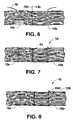
- FIG. 6depicts how the two ends of tendon are re-approximated and the scaffolding matrix pulled taught according to the various aspects of the method according to the invention.
- FIG. 7depicts a first step in securing the scaffolding matrix to the ends of tendon according to the various aspects of the method according to the invention.
- FIG. 8depicts a second step in securing the scaffolding matrix to the ends of tendon according to the various aspects of the method according to the invention.
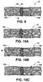
- FIG. 9depicts a third step in securing the scaffolding matrix to the ends of tendon according to the various aspects of the method according to the invention.
- FIGS. 10A , 10 B, and 10 Cdepict alternative embodiments for stitching the scaffolding matrix according to the various aspects of the method according to the invention.
- FIG. 11depicts yet another alternative embodiment for stitching the scaffolding matrix according to the various aspects of the method according to the invention.
- FIGS. 12A , 12 Bdepict how the method according to the invention provides benefits over conventional methods with respect to tendon tears that cannot be re-approximated.
- FIG. 13depicts how the method according to the invention provides benefits over conventional suturing methods.
- FIG. 14depicts a separated finger ligament that may be repaired using the method according to the various aspects of the invention.
- FIG. 15depicts the ligament of FIG. 14 after being repaired using the method according to the various aspects of the invention.
- FIGS. 16A-16Ddepict various aspects of an exemplary embodiment of the invention suitable for use in rotator cuff repair.
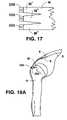
- FIG. 17depicts an exemplary embodiment of the invention suitable for use in rotator cuff repair through a closed arthroscopic approach.
- FIGS. 18A-18Cdepict various views of the embodiments of FIGS. 16A-16D being used to repair a rotator cuff.
- a tendon 10has been severed or ruptured into a first end portion 10 a and a second end portion 10 b of a tendon 10 and are in need of repair by re-connection of the two ends.
- These tendon end portions 10 a , 10 bmay be located anywhere in the body and accessed via conventional surgical techniques.
- Tendons, such as tendon 10are typically in the form of bundles of collagen fibers embedded in a connecting matrix, known as ground substance, provide the load carrying elements of natural tendons and ligaments. The arrangement of the collagen fibers is nearly parallel in tendons, equipping them to withstand high unidirectional loads.
- the ground substance in both tendons and ligamentsacts generally as a cementing matrix holding the collagen fibers together.
- the ground substanceretains large amounts of water essential to the non-compressive hydraulic function of the moving tissue.
- elastic fiberstenocytes, small blood vessels and nerves (none shown).
- the cellular materialfibroblasts
- the ground substance matrixaccounts for the remaining 62% to 80%.
- About 70% of the ground substance matrixconsists of water absorbed in an open polysaccharide matrix.
- the tendon endswill start to form frayed end portions 11 as the collagen fibers that make up tendon 10 start to separate from one another due to trauma and lack of blood.
- an extra-operatively obtained scaffolding matrixis used to connect the first and second end portions 10 a , 10 b .
- extra-operatively obtainedmeans not obtained by the surgical group currently performing the procedure or parties related to that group. Accordingly, extra-operatively is intended to exclude grafts obtained by such conventional procedures as autograft or allograft (where the graft is harvested at the same or near the same time as the surgery according to the invention—see below). Through the revascularization and repopulation of the scaffolding matrix the natural healing of the tendon 10 will be accelerated.
- the scaffolding matrixis threaded through the first and second tendon end portions 10 a , 10 b , of the ruptured or severed tendon 10 and, if there is enough tendon available, then manipulated to bring the first and second tendon end portions into contact with each other (this is known as “re-approximation”). Additional suturing, using conventional suture, may then be carried out to further secure the ends of the tendon together.
- Step 1As shown in FIGS. 2A-C , a ball probe 50 or tendon retriever 100 ( FIG. 3 ) is used to core a tunnel 20 a through the first tendon end portion 10 a towards the severed or ruptured end 13 a .
- An approach that enters the tendon end portion 10 a through its lateral walls 12 a and leads towards the severed or ruptured end 13 ais preferred so as to not further fray the fibrous tendon. Further fraying of the end of the tendon is why it is believed that splint methods where the splints are inserted into the frayed ends of the tendon, such as shown in U.S. Pat. No. 5,723,003 and US Pub 2002/0161400, have not found a wide following.
- Step 2A conventional tendon retriever 100 is used to grasp an end of the scaffolding matrix M and introduce it into and pull it back through the tunnel 20 a in the end of the first tendon portion 10 a ( FIG. 3 ).
- Step 3As with the first tendon end portion 10 a , a ball probe or tendon retriever is used to core a tunnel 20 b through the second tendon portion 10 b towards the severed or ruptured end 13 b . ( FIG. 4 ).
- Step 4A tendon retriever 100 is used to grasp the other end of the scaffolding matrix M and pull it back through the tunnel 20 b in the end of the second tendon portion 10 b . ( FIG. 5 ).
- Step 5The ends of the two tendon portions 13 a , 13 b are re-approximated and the scaffolding matrix M is pulled taught. ( FIG. 6 ).
- Step 6Suture 60 is used to anchor the scaffolding matrix M and temporarily hold the end portions 10 a , 10 b of tendon 10 together while the scaffolding matrix M assists and accelerates the natural healing of the tendon 10 .
- Kessler's suture methodhas been found to work well with the method of the invention. However, the invention is not limited to use of the Kessler method, as will be described.
- the first transverse stitch 60 a of the Kessler patternshould be passed through and penetrate the scaffolding matrix M, such as at point 60 b ( FIG. 7 ).
- Conventional suturesuch as 4.0 Ethibond® suture may be used for this purpose.
- the suture repairis made in a similar manner on both sides of the tendon tear (e.g. suture 60 passes through and penetrates scaffolding matrix M at point 60 c ).
- Step 7The final knot 60 d in the suture 60 is buried in the epitedineum of the tendon under lateral wall 11 b and the scaffolding matrix M is cut flush with the tendon 10 by cutting off the excess portions of the scaffolding matrix M before and after 60 a , 60 b , respectively ( FIG. 8 ).
- Step 8A final stitch is made circumferentially around the tendon tear.
- Conventional suture 61such as 6.0 BV Monofilament may be used for this purpose ( FIG. 9 ).
- the scaffolding matrixhas been in the form of Graftjacket® cellular matrix, sold by Wright Medical Technology, Inc., of Arlington, Tenn., and manufactured according to U.S. Pat. Nos. 4,865,871; 5,024,830; and 5,336,616.
- This productconsists of a selectively preserved extracellular protein matrix that is devoid of certain viable cells which normally express major histocompatibility complex antigenic determinants and other antigens which would be recognized as foreign by the recipient.
- This extracellular protein matrixis made up of collagen and other proteins and provides a structural template which may be repopulated with new viable cells that would not be rejected by the host. With this material complications following implantation (including but not limited to immunorejection, contracture, calcification, occlusion, and infection) are significantly reduced relative to current implant procedures and materials.
- the scaffolding matrixwhen made from this particular material, should have a width of about 2 mm and a height of about 2 mm. Length is variable and depends upon the amount of stitching required. But, in general the material will be in strip form where the length is substantially greater than both the width and height.
- a scaffolding matrixsuch as that described above (i.e., an acellular scaffolding matrix)
- an acellular scaffolding matrixnot only are the harvesting complications and morbidity concerns of autograft non-issues, so are the immediacy and tissue matching concerns of allograft.
- acellular biological materialssuch as that described above have shelf lives of two years, thereby alleviating immediacy concerns.
- unknown sourcemeans unknown source with respect to the parties implanting the scaffolding matrix as a graft and not necessarily the parties harvesting the original material and then processing it according to, e.g., the various accularization processes. The latter will, of course, rigorously monitor where the graft originates and certify its processing. Therefore, typically the scaffolding matrix will be an off-the-shelf material and not obtained as a step in the tendon repair process or in response to a request of the tendon repair process. Rather, the material will be sitting on the shelf waiting to be used and any package of material may be taken off the shelf with out the need to match with respect to the recipient's tissue type, etc.
- scaffolding matrixshould be interpreted as any non-autograft or non-allograft (i.e., fresh) material capable of revascularization and repopulation by cells, especially connective tissue.
- non-autograft or non-allografti.e., fresh
- connective tissueTypically the material with be off-the-shelf material rather than harvested or produced as needed. It would also be beneficial if the material was strong.
- revascularization and the ability to be repopulatedare more important to the fast healing of ruptured, severed, or torn tendons.
- the scaffolding matrixneed not even be from a human source. Graft materials from non-human sources, such as porcine, have been proposed. See e.g., U.S. Pat. No. 6,206,931.
- the scaffolding matrixneed not even be organic.
- synthetic porous materials capable of connective tissue in-growthmay be used. See e.g. U.S. Pat. No. 5,258,040.
- the method according to the inventionhas been described as applicable to any tendon, the method is especially beneficial to the flexor tendons of the hand. This is due to the fact that the method involves placing the scaffolding matrix inside the tendon end portions and therefore does not add any bulk to the outside of the repaired tendons. Accordingly, the method should not inhibit the movement of the repaired tendons through the pulleys of the hand. As mentioned above, this is a problem with many prior art methods. See e.g. U.S. Pat. Nos. 5,800,544; 6,080,192; 6,106,556.
- the scaffolding matrix Mmay be used by itself with: Kienert's ( FIG. 10A ), Bunuell's ( FIG. 10B ), or Kessler's ( FIG. 10C ) methods. Additionally, as experience with these scaffolding matrices and stitching and manipulating them grows, specific new methods methods of stitching them may be developed.
- FIGS. 12A , 12 B, and 13depict additional benefits of the various aspects of the method according to the invention.
- FIGS. 12A and 12Bdepict how the method according to the invention provides benefits over conventional methods with respect to tendon tears that cannot be re-approximated.
- a gap d 1will exist between them ( FIG. 12A ). If this gap d 1 is spanned by a material not capable of revascularization, repopulation and tissue ingrowth, such as suture, the two tendon end portions may be mechanically connected, but new connective tissue will never fill the gap d 1 and the tendon will never repair to anywhere near its full strength or functionality.
- FIG. 13depicts how the method according to the invention provides benefits over conventional suturing methods.
- suture connectionsmay come loose if not fixated properly.
- the two severed or ruptured end portions 10 a , 10 b of the tendon 10may start to move apart, such as in the direction of arrows A.
- a scaffolding matrix M capable of revascularization, repopulation and connective tissue ingrowth as discussed immediately aboveis used, even as a gap between the tendon end portions starts to form, natural healing can continue.
- the scaffolding matrixis one that provides for strength in excess to that of suture, the scaffolding matrix will act to keep the tendon end portions 10 a , 10 b together even as the suture may loosen.
- the graftmay have various forms of fixation at its ends, such as bone plugs, anchors, or eyelets for screws.
- FIG. 14shows a separated finger ligament.
- the ligament 200 that would normally connect bones 210 and 220has separated from its insertion point 215 , while remaining connected to bone at its origin 217 .
- the ligament 200will begin to atrophy and shrink due to a lack of tension thereon.
- a piece of extra-operatively obtained scaffolding matrix Mwill be attached to the bone 220 at the original ligament 200 insertion point 215 .
- the use of extra-operatively obtained scaffolding matrix Mwill provide for all the benefits described with respect to tendon repair above.
- the scaffolding matrix Mwill be ran parallel to the existing ligament and attached thereto such as by suture 60 , using any known method ( FIG. 15 ).
- the ligament 200 /scaffolding matrix M combinationwill now act as a complete natural ligament and give the finger back at least some of its natural movement. Furthermore, as the scaffolding matrix M becomes repopulated with connective tissue and begins to remodel itself to be more like connective tissue, the finger will begin to function even better.
- scaffolding matrix MWhen used for this application, scaffolding matrix M will typically be about 2 mm high by 3 mm wide. Again, length may vary depending upon the application and need.
- the doctormay chose to reinforce the ligament/scaffolding matrix combination by attaching it to both bones and not just the bone from which the ligament detatched (e.g. also attach to the origin 217 in FIG. 15 above).
- the scaffolding matrix M according to the inventionmay have various forms of fixation at its ends, such as bone plugs, anchors, or eyelets for screws.
- Scaffolding matrix Mmay be used for major tendon/ligament tears, such as in the rotator cuff of the shoulder.
- Materials useful for this purposeare shown in FIGS. 16A-16D . These materials comprise a sheet of scaffolding matrix M attached on one ( FIGS. 16A , B) or both ( FIGS. 16C , D) of its ends with a bone block.
- the bone blockwill be made from a bone material which has been thoroughly processed so as to eliminate tissue typing and pathogenic disease issues. Methods of producing such bone material and attaching it to materials similar to scaffolding matrix M are disclosed in, for example, U.S. Pat. Nos.
- one or both of the bone blocksmay have screw holes 210 drilled therethrough to provide for additional fixation of the bone block to its fixation point.
- FIG. 17shows an embodiment especially suitable for use in arthroscopic rotator cuff repairs.
- scaffolding matrix Mhas a main portion and then splits towards its end into a number of fingers M′.
- Each finger M′will have its own bone block 220 , rather than one bone block span the entire width of scaffolding matrix M. This allows the entire scaffolding matrix/bone block construct to be rolled up and fed through an arthroscope canula.
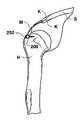
- FIGS. 18A-Cdepict different ways in which these various embodiments may be used in rotator cuff repair.
- bone block 200is seated within a groove made in the proximal humerus H.
- a press-fit connectionmay be used that is supplemented by screws 250 .
- Scaffolding matrix Mspans humeral head and is fixed to the scapula S via k-wires K.
- FIG. 18Cthere is also a bone block on the proximal end of scaffolding matrix M and that bone block is also attached to scapula S by K-wires K.
Landscapes
- Health & Medical Sciences (AREA)
- Orthopedic Medicine & Surgery (AREA)
- Rehabilitation Therapy (AREA)
- Rheumatology (AREA)
- Cardiology (AREA)
- Oral & Maxillofacial Surgery (AREA)
- Transplantation (AREA)
- Engineering & Computer Science (AREA)
- Biomedical Technology (AREA)
- Heart & Thoracic Surgery (AREA)
- Vascular Medicine (AREA)
- Life Sciences & Earth Sciences (AREA)
- Animal Behavior & Ethology (AREA)
- General Health & Medical Sciences (AREA)
- Public Health (AREA)
- Veterinary Medicine (AREA)
- Prostheses (AREA)
- Surgical Instruments (AREA)
Abstract
Description
Claims (19)
Priority Applications (2)
| Application Number | Priority Date | Filing Date | Title |
|---|---|---|---|
| US10/845,402US7407511B2 (en) | 2004-05-13 | 2004-05-13 | Methods and materials for connective tissue repair |
| US12/185,519US20080294256A1 (en) | 2004-05-13 | 2008-08-04 | Methods and Materials for Connective Tissue Repair |
Applications Claiming Priority (1)
| Application Number | Priority Date | Filing Date | Title |
|---|---|---|---|
| US10/845,402US7407511B2 (en) | 2004-05-13 | 2004-05-13 | Methods and materials for connective tissue repair |
Related Child Applications (1)
| Application Number | Title | Priority Date | Filing Date |
|---|---|---|---|
| US12/185,519ContinuationUS20080294256A1 (en) | 2004-05-13 | 2008-08-04 | Methods and Materials for Connective Tissue Repair |
Publications (2)
| Publication Number | Publication Date |
|---|---|
| US20050255140A1 US20050255140A1 (en) | 2005-11-17 |
| US7407511B2true US7407511B2 (en) | 2008-08-05 |
Family
ID=35309687
Family Applications (2)
| Application Number | Title | Priority Date | Filing Date |
|---|---|---|---|
| US10/845,402Active2026-10-13US7407511B2 (en) | 2004-05-13 | 2004-05-13 | Methods and materials for connective tissue repair |
| US12/185,519AbandonedUS20080294256A1 (en) | 2004-05-13 | 2008-08-04 | Methods and Materials for Connective Tissue Repair |
Family Applications After (1)
| Application Number | Title | Priority Date | Filing Date |
|---|---|---|---|
| US12/185,519AbandonedUS20080294256A1 (en) | 2004-05-13 | 2008-08-04 | Methods and Materials for Connective Tissue Repair |
Country Status (1)
| Country | Link |
|---|---|
| US (2) | US7407511B2 (en) |
Families Citing this family (33)
| Publication number | Priority date | Publication date | Assignee | Title |
|---|---|---|---|---|
| US9271766B2 (en)* | 2004-10-26 | 2016-03-01 | P Tech, Llc | Devices and methods for stabilizing tissue and implants |
| US9463012B2 (en) | 2004-10-26 | 2016-10-11 | P Tech, Llc | Apparatus for guiding and positioning an implant |
| US20060089646A1 (en) | 2004-10-26 | 2006-04-27 | Bonutti Peter M | Devices and methods for stabilizing tissue and implants |
| AU2007208243B2 (en)* | 2006-01-25 | 2013-09-19 | Children's Medical Center Corporation | Methods and procedures for ligament repair |
| WO2008097901A1 (en)* | 2007-02-02 | 2008-08-14 | Tornier, Inc. | System and method for repairing tendons and ligaments |
| US8911456B2 (en) | 2007-07-03 | 2014-12-16 | Ceterix Orthopaedics, Inc. | Methods and devices for preventing tissue bridging while suturing |
| US8465505B2 (en) | 2011-05-06 | 2013-06-18 | Ceterix Orthopaedics, Inc. | Suture passer devices and methods |
| US9861354B2 (en) | 2011-05-06 | 2018-01-09 | Ceterix Orthopaedics, Inc. | Meniscus repair |
| US8821518B2 (en) | 2007-11-05 | 2014-09-02 | Ceterix Orthopaedics, Inc. | Suture passing instrument and method |
| US9211119B2 (en) | 2007-07-03 | 2015-12-15 | Ceterix Orthopaedics, Inc. | Suture passers and methods of passing suture |
| US10441273B2 (en) | 2007-07-03 | 2019-10-15 | Ceterix Orthopaedics, Inc. | Pre-tied surgical knots for use with suture passers |
| US8500809B2 (en)* | 2011-01-10 | 2013-08-06 | Ceterix Orthopaedics, Inc. | Implant and method for repair of the anterior cruciate ligament |
| US9314234B2 (en) | 2007-07-03 | 2016-04-19 | Ceterix Orthopaedics, Inc. | Pre-tied surgical knots for use with suture passers |
| US8663253B2 (en) | 2007-07-03 | 2014-03-04 | Ceterix Orthopaedics, Inc. | Methods of meniscus repair |
| US8702731B2 (en) | 2007-07-03 | 2014-04-22 | Ceterix Orthopaedics, Inc. | Suturing and repairing tissue using in vivo suture loading |
| EP2206482A1 (en)* | 2009-01-13 | 2010-07-14 | Tornier, Inc. | Patch augmentation of achilles tendon repairs |
| US20100211174A1 (en)* | 2009-02-19 | 2010-08-19 | Tyco Healthcare Group Lp | Method For Repairing A Rotator Cuff |
| US9848868B2 (en) | 2011-01-10 | 2017-12-26 | Ceterix Orthopaedics, Inc. | Suture methods for forming locking loops stitches |
| US9011454B2 (en) | 2009-11-09 | 2015-04-21 | Ceterix Orthopaedics, Inc. | Suture passer with radiused upper jaw |
| JP5719374B2 (en) | 2009-11-09 | 2015-05-20 | セテリックス オーソピーディクス インコーポレイテッド | Device, system, and method for repairing a meniscus |
| US11744575B2 (en) | 2009-11-09 | 2023-09-05 | Ceterix Orthopaedics, Inc. | Suture passer devices and methods |
| US9913638B2 (en) | 2011-01-10 | 2018-03-13 | Ceterix Orthopaedics, Inc. | Transosteal anchoring methods for tissue repair |
| US10524778B2 (en) | 2011-09-28 | 2020-01-07 | Ceterix Orthopaedics | Suture passers adapted for use in constrained regions |
| US9492162B2 (en) | 2013-12-16 | 2016-11-15 | Ceterix Orthopaedics, Inc. | Automatically reloading suture passer devices and methods |
| US9247935B2 (en) | 2013-09-23 | 2016-02-02 | Ceterix Orthopaedics, Inc. | Arthroscopic knot pusher and suture cutter |
| CN204951031U (en) | 2014-04-08 | 2016-01-13 | 赛特里克斯整形公司 | Ware device is worn to draw by suture |
| US10226245B2 (en) | 2015-07-21 | 2019-03-12 | Ceterix Orthopaedics, Inc. | Automatically reloading suture passer devices that prevent entanglement |
| US10405853B2 (en) | 2015-10-02 | 2019-09-10 | Ceterix Orthpaedics, Inc. | Knot tying accessory |
| AU2016344019B2 (en) | 2015-10-30 | 2021-07-15 | New York Society For The Relief Of The Ruptured And Crippled, Maintaining The Hospital For Special Surgery | Suture sleeve patch and methods of delivery within an existing arthroscopic workflow |
| US11484401B2 (en) | 2016-02-01 | 2022-11-01 | Medos International Sarl | Tissue augmentation scaffolds for use in soft tissue fixation repair |
| US11523812B2 (en) | 2016-02-01 | 2022-12-13 | Medos International Sarl | Soft tissue fixation repair methods using tissue augmentation constructs |
| US20170273680A1 (en) | 2016-02-01 | 2017-09-28 | DePuy Synthes Products, Inc. | Tissue augmentation tacks for use with soft tissue fixation repair systems and methods |
| EP3423001A4 (en)* | 2016-03-02 | 2020-04-08 | The Regents of The University of Michigan | BREEDED STRING TRANSPLANT FOR REPAIRING THE ROTATOR CUFF |
Citations (52)
| Publication number | Priority date | Publication date | Assignee | Title |
|---|---|---|---|---|
| US4597766A (en) | 1984-10-26 | 1986-07-01 | American Hospital Supply Corporation | Implantable bioprosthetic tendons and ligaments |
| EP0278713A1 (en) | 1987-02-09 | 1988-08-17 | Pfizer Hospital Products Group, Inc. | Ligament prosthesis |
| US4772288A (en) | 1987-06-15 | 1988-09-20 | Borner William H | Method for producing implantable ligament and tendon prostheses and prostheses produced thereby |
| US4834752A (en)* | 1986-03-17 | 1989-05-30 | Minnesota Mining And Manufacturing | Tissue augmentation device and method of repairing a ligament or tendon |
| US4865871A (en)* | 1983-08-23 | 1989-09-12 | Board Of Regents The University Of Texas System | Method for cryopreparing biological tissue |
| FR2638349A1 (en) | 1988-10-28 | 1990-05-04 | Breard Francis | Tendon prosthesis and in particular a shoulder cuff prosthesis |
| US5024830A (en)* | 1983-08-23 | 1991-06-18 | The Board Of Regents, The University Of Texas | Method for cryopreparing biological tissue for ultrastructural analysis |
| US5061283A (en) | 1987-10-30 | 1991-10-29 | Pfizer Hospital Products Group, Inc. | Method for tendon and ligament repair |
| US5078744A (en) | 1987-09-04 | 1992-01-07 | Bio-Products, Inc. | Method of using tendon/ligament substitutes composed of long, parallel, non-antigenic tendon/ligament fibers |
| FR2690073A1 (en) | 1992-04-15 | 1993-10-22 | Deux C T | Shoulder muscle caul prosthesis - made from mesh layer of interwoven fibres, partially covered with friction surface of soft moulded material |
| US5258040A (en) | 1982-09-10 | 1993-11-02 | W. L. Gore & Associates | Prosthesis for tensile load-carrying tissue and method of manufacture |
| US5258014A (en)* | 1990-04-06 | 1993-11-02 | Sumitomo Electric Industries Ltd. | Surgical suture and process for producing same |
| US5336616A (en)* | 1990-09-12 | 1994-08-09 | Lifecell Corporation | Method for processing and preserving collagen-based tissues for transplantation |
| GB2282328A (en) | 1993-09-29 | 1995-04-05 | Johnson & Johnson Medical | Absorbable structure for ligament and tendon repair |
| US5527341A (en) | 1991-05-24 | 1996-06-18 | Synthes (U.S.A) | Resorbable tendon and bone augmentation device |
| US5575819A (en) | 1986-09-19 | 1996-11-19 | Imperial College Of Science And Technology | Artificial ligaments |
| FR2748652A1 (en) | 1996-05-15 | 1997-11-21 | Fontes Didier | Prosthetic ligament for shoulder joint |
| US5702422A (en)* | 1995-12-06 | 1997-12-30 | Stone; Kevin R. | Anterior cruciate ligament repair method |
| US5723008A (en) | 1995-07-20 | 1998-03-03 | Gordon; Leonard | Splint for repair of tendons or ligaments and method |
| US5800544A (en)* | 1994-12-02 | 1998-09-01 | Omeros Medical Systems, Inc. | Tendon and ligament repair system |
| US5855619A (en)* | 1994-06-06 | 1999-01-05 | Case Western Reserve University | Biomatrix for soft tissue regeneration |
| US5860978A (en)* | 1990-09-25 | 1999-01-19 | Innovasive Devices, Inc. | Methods and apparatus for preventing migration of sutures through transosseous tunnels |
| US6083244A (en) | 1996-09-13 | 2000-07-04 | Tendon Technology, Ltd. | Apparatus and method for tendon or ligament repair |
| US6106556A (en) | 1994-12-02 | 2000-08-22 | Omeros Medical Systems, Inc. | Tendon and ligament repair system |
| WO2001006933A2 (en) | 1999-07-26 | 2001-02-01 | Regeneration Technologies, Inc. | Suture anchor |
| WO2001028457A1 (en) | 1999-10-18 | 2001-04-26 | Tendon Technology, Ltd. | Apparatus and methods for tendon or ligament repair |
| WO2001067944A2 (en) | 2000-03-15 | 2001-09-20 | Esplin Medical Inventions, Llc | Soft tissue anchor |
| FR2810877A1 (en) | 2000-07-03 | 2002-01-04 | Vincent Travers | Shoulder ligament implant prosthesis comprises strip of supple material passed over sub-scapular muscle and fastened to bone |
| US20020022861A1 (en)* | 2000-05-19 | 2002-02-21 | Daniel Jacobs | Multi-point tissue tension distribution device, a combined orbital rim repair and suspension variation, and a method of tissue approximation using the device |
| US20020072806A1 (en) | 2000-01-11 | 2002-06-13 | Dayna Buskirk | Soft and calcified tissue implants |
| US20020077631A1 (en)* | 1996-09-13 | 2002-06-20 | Lubbers Lawrence M. | Apparatus and methods for tendon or ligament repair |
| US20020103542A1 (en) | 2000-09-18 | 2002-08-01 | Bilbo Patrick R. | Methods for treating a patient using a bioengineered flat sheet graft prostheses |
| US20020119177A1 (en)* | 2000-12-21 | 2002-08-29 | Bowman Steven M. | Reinforced foam implants with enhanced integrity for soft tissue repair and regeneration |
| US20020133229A1 (en) | 2000-03-24 | 2002-09-19 | Laurencin Cato T. | Ligament and tendon replacement constructs and methods for production and use thereof |
| US20020161400A1 (en) | 1999-12-23 | 2002-10-31 | Demopulos Gregory A. | Tendon repair using adhesive |
| US20020173558A1 (en) | 1999-03-25 | 2002-11-21 | Metabolix, Inc. | Medical devices and applications of polyhydroxyalkanoate polymers |
| US6497726B1 (en) | 2000-01-11 | 2002-12-24 | Regeneration Technologies, Inc. | Materials and methods for improved bone tendon bone transplantation |
| US20030023316A1 (en) | 2000-08-04 | 2003-01-30 | Brown Laura Jean | Hybrid biologic-synthetic bioabsorable scaffolds |
| US20030023304A1 (en) | 2000-01-11 | 2003-01-30 | Carter Kevin C. | Materials and methods for improved bone tendon bone transplantation |
| US6514515B1 (en) | 1999-03-04 | 2003-02-04 | Tepha, Inc. | Bioabsorbable, biocompatible polymers for tissue engineering |
| US6524317B1 (en)* | 1999-12-30 | 2003-02-25 | Opus Medical, Inc. | Method and apparatus for attaching connective tissues to bone using a knotless suture anchoring device |
| US6533821B1 (en) | 2000-06-22 | 2003-03-18 | Thomas Lally | Bio-adhesive composition, method for adhering objects to bone |
| US20030097179A1 (en) | 2000-01-11 | 2003-05-22 | Carter Kevin C. | Materials and methods for improved bone tendon bone transplantation |
| US6616694B1 (en) | 1996-11-21 | 2003-09-09 | Ethicon, Inc. | Apparatus for anchoring autologous or artificial tendon grafts in bone |
| US20030181889A1 (en) | 2002-03-21 | 2003-09-25 | Little Adrian Leigh | Healing accelerator |
| US6638312B2 (en) | 2000-08-04 | 2003-10-28 | Depuy Orthopaedics, Inc. | Reinforced small intestinal submucosa (SIS) |
| US20030212456A1 (en) | 2002-05-09 | 2003-11-13 | John Lipchitz | Implant for tissue repair |
| WO2003092761A1 (en) | 2002-05-03 | 2003-11-13 | Fite Holdings Limited | Surgical material comprising water glass fibres |
| US20040078077A1 (en) | 2002-10-18 | 2004-04-22 | Francois Binette | Biocompatible scaffold for ligament or tendon repair |
| US20050043805A1 (en)* | 2003-08-11 | 2005-02-24 | Chudik Steven C. | Devices and methods used for shoulder replacement |
| US20070162121A1 (en)* | 2006-01-12 | 2007-07-12 | Tarrant Laurence J | Method for repair and reconstruction of ruptured ligaments or tendons and for treatment of ligament and tendon injuries |
| US20070198087A1 (en)* | 2006-02-07 | 2007-08-23 | Tepha, Inc. | Methods and Devices for Rotator Cuff Repair |
Family Cites Families (5)
| Publication number | Priority date | Publication date | Assignee | Title |
|---|---|---|---|---|
| US3345306A (en)* | 1964-02-12 | 1967-10-03 | Ethicon Inc | Tubing fluid containing an amino acid |
| US4400833A (en)* | 1981-06-10 | 1983-08-30 | Kurland Kenneth Z | Means and method of implanting bioprosthetics |
| US5281422A (en)* | 1991-09-24 | 1994-01-25 | Purdue Research Foundation | Graft for promoting autogenous tissue growth |
| US6102947A (en)* | 1995-07-20 | 2000-08-15 | Gordon; Leonard | Splint with flexible body for repair of tendons or ligaments and method |
| CN1827766B (en)* | 2001-06-28 | 2010-08-25 | 徐荣祥 | In vitro cell cultivation method |
- 2004
- 2004-05-13USUS10/845,402patent/US7407511B2/enactiveActive
- 2008
- 2008-08-04USUS12/185,519patent/US20080294256A1/ennot_activeAbandoned
Patent Citations (59)
| Publication number | Priority date | Publication date | Assignee | Title |
|---|---|---|---|---|
| US5258040A (en) | 1982-09-10 | 1993-11-02 | W. L. Gore & Associates | Prosthesis for tensile load-carrying tissue and method of manufacture |
| US5024830A (en)* | 1983-08-23 | 1991-06-18 | The Board Of Regents, The University Of Texas | Method for cryopreparing biological tissue for ultrastructural analysis |
| US4865871A (en)* | 1983-08-23 | 1989-09-12 | Board Of Regents The University Of Texas System | Method for cryopreparing biological tissue |
| US4597766A (en) | 1984-10-26 | 1986-07-01 | American Hospital Supply Corporation | Implantable bioprosthetic tendons and ligaments |
| US4834752A (en)* | 1986-03-17 | 1989-05-30 | Minnesota Mining And Manufacturing | Tissue augmentation device and method of repairing a ligament or tendon |
| US5575819A (en) | 1986-09-19 | 1996-11-19 | Imperial College Of Science And Technology | Artificial ligaments |
| EP0278713A1 (en) | 1987-02-09 | 1988-08-17 | Pfizer Hospital Products Group, Inc. | Ligament prosthesis |
| US4772288A (en) | 1987-06-15 | 1988-09-20 | Borner William H | Method for producing implantable ligament and tendon prostheses and prostheses produced thereby |
| US5078744A (en) | 1987-09-04 | 1992-01-07 | Bio-Products, Inc. | Method of using tendon/ligament substitutes composed of long, parallel, non-antigenic tendon/ligament fibers |
| US5061283A (en) | 1987-10-30 | 1991-10-29 | Pfizer Hospital Products Group, Inc. | Method for tendon and ligament repair |
| FR2638349A1 (en) | 1988-10-28 | 1990-05-04 | Breard Francis | Tendon prosthesis and in particular a shoulder cuff prosthesis |
| US5258014A (en)* | 1990-04-06 | 1993-11-02 | Sumitomo Electric Industries Ltd. | Surgical suture and process for producing same |
| US5336616A (en)* | 1990-09-12 | 1994-08-09 | Lifecell Corporation | Method for processing and preserving collagen-based tissues for transplantation |
| US5860978A (en)* | 1990-09-25 | 1999-01-19 | Innovasive Devices, Inc. | Methods and apparatus for preventing migration of sutures through transosseous tunnels |
| US5527341A (en) | 1991-05-24 | 1996-06-18 | Synthes (U.S.A) | Resorbable tendon and bone augmentation device |
| FR2690073A1 (en) | 1992-04-15 | 1993-10-22 | Deux C T | Shoulder muscle caul prosthesis - made from mesh layer of interwoven fibres, partially covered with friction surface of soft moulded material |
| GB2282328A (en) | 1993-09-29 | 1995-04-05 | Johnson & Johnson Medical | Absorbable structure for ligament and tendon repair |
| US5855619A (en)* | 1994-06-06 | 1999-01-05 | Case Western Reserve University | Biomatrix for soft tissue regeneration |
| US6106556A (en) | 1994-12-02 | 2000-08-22 | Omeros Medical Systems, Inc. | Tendon and ligament repair system |
| US5800544A (en)* | 1994-12-02 | 1998-09-01 | Omeros Medical Systems, Inc. | Tendon and ligament repair system |
| US5723008A (en) | 1995-07-20 | 1998-03-03 | Gordon; Leonard | Splint for repair of tendons or ligaments and method |
| US5702422A (en)* | 1995-12-06 | 1997-12-30 | Stone; Kevin R. | Anterior cruciate ligament repair method |
| FR2748652A1 (en) | 1996-05-15 | 1997-11-21 | Fontes Didier | Prosthetic ligament for shoulder joint |
| US6083244A (en) | 1996-09-13 | 2000-07-04 | Tendon Technology, Ltd. | Apparatus and method for tendon or ligament repair |
| US20030088270A1 (en) | 1996-09-13 | 2003-05-08 | Tendon Technology, Ltd. | Implantable tissue fixation devices and methods of tissue approximation |
| US20020077631A1 (en)* | 1996-09-13 | 2002-06-20 | Lubbers Lawrence M. | Apparatus and methods for tendon or ligament repair |
| US6616694B1 (en) | 1996-11-21 | 2003-09-09 | Ethicon, Inc. | Apparatus for anchoring autologous or artificial tendon grafts in bone |
| US6514515B1 (en) | 1999-03-04 | 2003-02-04 | Tepha, Inc. | Bioabsorbable, biocompatible polymers for tissue engineering |
| US20020173558A1 (en) | 1999-03-25 | 2002-11-21 | Metabolix, Inc. | Medical devices and applications of polyhydroxyalkanoate polymers |
| WO2001006933A2 (en) | 1999-07-26 | 2001-02-01 | Regeneration Technologies, Inc. | Suture anchor |
| WO2001006933A3 (en) | 1999-07-26 | 2001-06-21 | Regeneration Tech Inc | Suture anchor |
| WO2001028457A1 (en) | 1999-10-18 | 2001-04-26 | Tendon Technology, Ltd. | Apparatus and methods for tendon or ligament repair |
| US20020161400A1 (en) | 1999-12-23 | 2002-10-31 | Demopulos Gregory A. | Tendon repair using adhesive |
| US6524317B1 (en)* | 1999-12-30 | 2003-02-25 | Opus Medical, Inc. | Method and apparatus for attaching connective tissues to bone using a knotless suture anchoring device |
| US20020072806A1 (en) | 2000-01-11 | 2002-06-13 | Dayna Buskirk | Soft and calcified tissue implants |
| US6497726B1 (en) | 2000-01-11 | 2002-12-24 | Regeneration Technologies, Inc. | Materials and methods for improved bone tendon bone transplantation |
| US20030097179A1 (en) | 2000-01-11 | 2003-05-22 | Carter Kevin C. | Materials and methods for improved bone tendon bone transplantation |
| US20030023304A1 (en) | 2000-01-11 | 2003-01-30 | Carter Kevin C. | Materials and methods for improved bone tendon bone transplantation |
| US20010051815A1 (en) | 2000-03-15 | 2001-12-13 | Esplin Vermon S. | Soft tissue anchor |
| WO2001067944A2 (en) | 2000-03-15 | 2001-09-20 | Esplin Medical Inventions, Llc | Soft tissue anchor |
| US20020133229A1 (en) | 2000-03-24 | 2002-09-19 | Laurencin Cato T. | Ligament and tendon replacement constructs and methods for production and use thereof |
| US6485503B2 (en)* | 2000-05-19 | 2002-11-26 | Coapt Systems, Inc. | Multi-point tissue tension distribution device, a brow and face lift variation, and a method of tissue approximation using the device |
| US20020022861A1 (en)* | 2000-05-19 | 2002-02-21 | Daniel Jacobs | Multi-point tissue tension distribution device, a combined orbital rim repair and suspension variation, and a method of tissue approximation using the device |
| US6533821B1 (en) | 2000-06-22 | 2003-03-18 | Thomas Lally | Bio-adhesive composition, method for adhering objects to bone |
| FR2810877A1 (en) | 2000-07-03 | 2002-01-04 | Vincent Travers | Shoulder ligament implant prosthesis comprises strip of supple material passed over sub-scapular muscle and fastened to bone |
| US20030023316A1 (en) | 2000-08-04 | 2003-01-30 | Brown Laura Jean | Hybrid biologic-synthetic bioabsorable scaffolds |
| US6638312B2 (en) | 2000-08-04 | 2003-10-28 | Depuy Orthopaedics, Inc. | Reinforced small intestinal submucosa (SIS) |
| US20020103542A1 (en) | 2000-09-18 | 2002-08-01 | Bilbo Patrick R. | Methods for treating a patient using a bioengineered flat sheet graft prostheses |
| US20020119177A1 (en)* | 2000-12-21 | 2002-08-29 | Bowman Steven M. | Reinforced foam implants with enhanced integrity for soft tissue repair and regeneration |
| WO2003034895A3 (en) | 2001-10-03 | 2003-08-21 | Tendon Technology Ltd | Apparatus and methods for tendon or ligament repair |
| WO2003034895A2 (en) | 2001-10-03 | 2003-05-01 | Tendon Technology, Ltd. | Apparatus and methods for tendon or ligament repair |
| US20030181889A1 (en) | 2002-03-21 | 2003-09-25 | Little Adrian Leigh | Healing accelerator |
| WO2003092761A1 (en) | 2002-05-03 | 2003-11-13 | Fite Holdings Limited | Surgical material comprising water glass fibres |
| US20030212456A1 (en) | 2002-05-09 | 2003-11-13 | John Lipchitz | Implant for tissue repair |
| WO2003095609A2 (en) | 2002-05-09 | 2003-11-20 | Smith & Nephew, Inc. | Implant for tissue repair |
| US20040078077A1 (en) | 2002-10-18 | 2004-04-22 | Francois Binette | Biocompatible scaffold for ligament or tendon repair |
| US20050043805A1 (en)* | 2003-08-11 | 2005-02-24 | Chudik Steven C. | Devices and methods used for shoulder replacement |
| US20070162121A1 (en)* | 2006-01-12 | 2007-07-12 | Tarrant Laurence J | Method for repair and reconstruction of ruptured ligaments or tendons and for treatment of ligament and tendon injuries |
| US20070198087A1 (en)* | 2006-02-07 | 2007-08-23 | Tepha, Inc. | Methods and Devices for Rotator Cuff Repair |
Also Published As
| Publication number | Publication date |
|---|---|
| US20050255140A1 (en) | 2005-11-17 |
| US20080294256A1 (en) | 2008-11-27 |
Similar Documents
| Publication | Publication Date | Title |
|---|---|---|
| US7407511B2 (en) | Methods and materials for connective tissue repair | |
| US8007533B2 (en) | Progressive grip assembled bone-tendon-bone grafts, methods of making, and methods of use | |
| AU2007227318B2 (en) | Devices, systems, and methods for material fixation | |
| US8663324B2 (en) | Double socket ACL reconstruction | |
| US8512411B2 (en) | Trapezoidal bone plugs and method of bone-tendon-bone ACL reconstruction | |
| AU2005206920B2 (en) | Bone-tendon-bone implant | |
| CA2896582C (en) | Double bundle acl repair | |
| US20080234819A1 (en) | All-inside double-bundle acl reconstruction | |
| US10568733B2 (en) | Anterior cable region superior capsule reconstructions | |
| WO2003075800A1 (en) | Improved bone-tendon-bone assembly with cancellous allograft bone block | |
| US20100211174A1 (en) | Method For Repairing A Rotator Cuff | |
| US20200171203A1 (en) | System and methods for connective tissue repair using scaffolds | |
| Gigante et al. | Semitendinosus and gracilis free muscle-tendon graft for repair of massive rotator cuff tears: surgical technique | |
| CN115944337A (en) | System and method for repairing damaged tissues and system and method for reconstructing cruciate ligament | |
| KR19990065287A (en) | LCI Screw for Reconstruction of the Cruciate Ligament of the Knee | |
| Sherief et al. | Use of Leeds–Keio Connective Tissue Prosthesis (L–K CTP) for reconstruction of deficient extensor mechanism with total knee replacement | |
| US20250072883A1 (en) | Adjustable tissue repair systems and surgical methods | |
| RU2424779C1 (en) | Method of forming cruciate ligaments of knee joint | |
| AU2013200756B2 (en) | Devices, systems, and methods for material fixation | |
| Wodziński et al. | Anterior cruciate ligament–to repair or not to repair? | |
| Denard et al. | Tricks of the Trade for Poor Tissue and Bone | |
| Aiyer et al. | Evolution of tendon transfer to allograft reconstruction in foot and ankle surgery | |
| Sakakeeny | Defining the physicomechanical benefit of TissueMend in tendon augmentation surgeries |
Legal Events
| Date | Code | Title | Description |
|---|---|---|---|
| AS | Assignment | Owner name:WRIGHT MEDICAL TECHNOLOGY, INC., TENNESSEE Free format text:ASSIGNMENT OF ASSIGNORS INTEREST;ASSIGNORS:HAGAN, CARY P.;SWAIM, RICK P.;SCHLACHTER, KELLY;AND OTHERS;REEL/FRAME:015916/0278 Effective date:20041013 | |
| STCF | Information on status: patent grant | Free format text:PATENTED CASE | |
| FPAY | Fee payment | Year of fee payment:4 | |
| FPAY | Fee payment | Year of fee payment:8 | |
| AS | Assignment | Owner name:MIDCAP FINANCIAL TRUST, AS AGENT, MARYLAND Free format text:SECURITY INTEREST;ASSIGNOR:WRIGHT MEDICAL TECHNOLOGY, INC.;REEL/FRAME:041257/0126 Effective date:20161223 | |
| MAFP | Maintenance fee payment | Free format text:PAYMENT OF MAINTENANCE FEE, 12TH YEAR, LARGE ENTITY (ORIGINAL EVENT CODE: M1553); ENTITY STATUS OF PATENT OWNER: LARGE ENTITY Year of fee payment:12 | |
| AS | Assignment | Owner name:WRIGHT MEDICAL GROUP INTELLECTUAL PROPERTY, INC., TENNESSEE Free format text:RELEASE BY SECURED PARTY;ASSIGNOR:MIDCAP FUNDING IV TRUST;REEL/FRAME:054480/0001 Effective date:20201112 Owner name:BIOMIMETIC THERAPEUTICS, LLC, TENNESSEE Free format text:RELEASE BY SECURED PARTY;ASSIGNOR:MIDCAP FUNDING IV TRUST;REEL/FRAME:054480/0001 Effective date:20201112 Owner name:SOLANA SURGICAL, LLC, TENNESSEE Free format text:RELEASE BY SECURED PARTY;ASSIGNOR:MIDCAP FUNDING IV TRUST;REEL/FRAME:054480/0001 Effective date:20201112 Owner name:WHITE BOX ORTHOPEDICS, LLC, TENNESSEE Free format text:RELEASE BY SECURED PARTY;ASSIGNOR:MIDCAP FUNDING IV TRUST;REEL/FRAME:054480/0001 Effective date:20201112 Owner name:BIOMIMETIC THERAPEUTICS CANADA, INC., TENNESSEE Free format text:RELEASE BY SECURED PARTY;ASSIGNOR:MIDCAP FUNDING IV TRUST;REEL/FRAME:054480/0001 Effective date:20201112 Owner name:WRIGHT MEDICAL GROUP, INC., TENNESSEE Free format text:RELEASE BY SECURED PARTY;ASSIGNOR:MIDCAP FUNDING IV TRUST;REEL/FRAME:054480/0001 Effective date:20201112 Owner name:TROOPER HOLDINGS INC., MINNESOTA Free format text:RELEASE BY SECURED PARTY;ASSIGNOR:MIDCAP FUNDING IV TRUST;REEL/FRAME:054480/0001 Effective date:20201112 Owner name:ORTHOHELIX SURGICAL DESIGNS, INC., MINNESOTA Free format text:RELEASE BY SECURED PARTY;ASSIGNOR:MIDCAP FUNDING IV TRUST;REEL/FRAME:054480/0001 Effective date:20201112 Owner name:TORNIER US HOLDINGS, INC., MINNESOTA Free format text:RELEASE BY SECURED PARTY;ASSIGNOR:MIDCAP FUNDING IV TRUST;REEL/FRAME:054480/0001 Effective date:20201112 Owner name:WRIGHT MEDICAL CAPITAL, INC., TENNESSEE Free format text:RELEASE BY SECURED PARTY;ASSIGNOR:MIDCAP FUNDING IV TRUST;REEL/FRAME:054480/0001 Effective date:20201112 Owner name:BIOMIMETIC THERAPEUTICS USA, INC., TENNESSEE Free format text:RELEASE BY SECURED PARTY;ASSIGNOR:MIDCAP FUNDING IV TRUST;REEL/FRAME:054480/0001 Effective date:20201112 Owner name:INBONE TECHNOLOGIES, INC., TENNESSEE Free format text:RELEASE BY SECURED PARTY;ASSIGNOR:MIDCAP FUNDING IV TRUST;REEL/FRAME:054480/0001 Effective date:20201112 Owner name:TORNIER, INC., MINNESOTA Free format text:RELEASE BY SECURED PARTY;ASSIGNOR:MIDCAP FUNDING IV TRUST;REEL/FRAME:054480/0001 Effective date:20201112 Owner name:WRIGHT MEDICAL GROUP N.V., NETHERLANDS Free format text:RELEASE BY SECURED PARTY;ASSIGNOR:MIDCAP FUNDING IV TRUST;REEL/FRAME:054480/0001 Effective date:20201112 Owner name:WRIGHT MEDICAL TECHNOLOGY, INC., TENNESSEE Free format text:RELEASE BY SECURED PARTY;ASSIGNOR:MIDCAP FUNDING IV TRUST;REEL/FRAME:054480/0001 Effective date:20201112 Owner name:ORTHOPRO, L.L.C., TENNESSEE Free format text:RELEASE BY SECURED PARTY;ASSIGNOR:MIDCAP FUNDING IV TRUST;REEL/FRAME:054480/0001 Effective date:20201112 |