US7379532B2 - ECG-based rotational angiography for cardiology - Google Patents
ECG-based rotational angiography for cardiologyDownload PDFInfo
- Publication number
- US7379532B2 US7379532B2US11/545,250US54525006AUS7379532B2US 7379532 B2US7379532 B2US 7379532B2US 54525006 AUS54525006 AUS 54525006AUS 7379532 B2US7379532 B2US 7379532B2
- Authority
- US
- United States
- Prior art keywords
- arm
- ray
- ecg
- patient
- set forth
- Prior art date
- Legal status (The legal status is an assumption and is not a legal conclusion. Google has not performed a legal analysis and makes no representation as to the accuracy of the status listed.)
- Expired - Fee Related, expires
Links
- 238000002583angiographyMethods0.000titleclaimsdescription7
- 230000000747cardiac effectEffects0.000claimsabstractdescription34
- 238000000034methodMethods0.000claimsabstractdescription29
- 238000003384imaging methodMethods0.000claimsabstractdescription20
- 230000005855radiationEffects0.000claimsabstractdescription13
- 210000004872soft tissueAnatomy0.000claimsabstractdescription9
- 230000001360synchronised effectEffects0.000claimsabstractdescription6
- 238000004891communicationMethods0.000claimsabstract4
- 210000005166vasculatureAnatomy0.000claimsdescription8
- 230000033001locomotionEffects0.000claimsdescription5
- 210000003484anatomyAnatomy0.000claimsdescription4
- 230000000737periodic effectEffects0.000claimsdescription4
- 238000012545processingMethods0.000claimsdescription3
- 238000011076safety testMethods0.000claimsdescription2
- 238000001514detection methodMethods0.000claims1
- 230000009977dual effectEffects0.000claims1
- 238000010859live-cell imagingMethods0.000claims1
- 238000005259measurementMethods0.000claims1
- 239000004065semiconductorSubstances0.000claims1
- 238000011156evaluationMethods0.000description6
- 238000010586diagramMethods0.000description4
- 238000002594fluoroscopyMethods0.000description4
- 210000004204blood vesselAnatomy0.000description3
- 210000001367arteryAnatomy0.000description2
- 210000005242cardiac chamberAnatomy0.000description2
- 239000002872contrast mediaSubstances0.000description2
- 210000004351coronary vesselAnatomy0.000description2
- 230000033764rhythmic processEffects0.000description2
- 208000031481Pathologic ConstrictionDiseases0.000description1
- 238000010276constructionMethods0.000description1
- 238000007887coronary angioplastyMethods0.000description1
- 230000006866deteriorationEffects0.000description1
- 238000000605extractionMethods0.000description1
- 230000000977initiatory effectEffects0.000description1
- 238000002347injectionMethods0.000description1
- 239000007924injectionSubstances0.000description1
- 238000013152interventional procedureMethods0.000description1
- 230000001678irradiating effectEffects0.000description1
- 230000003068static effectEffects0.000description1
- 230000036262stenosisEffects0.000description1
- 208000037804stenosisDiseases0.000description1
- 230000002792vascularEffects0.000description1
- 210000003462veinAnatomy0.000description1
Images


Classifications
- A—HUMAN NECESSITIES
- A61—MEDICAL OR VETERINARY SCIENCE; HYGIENE
- A61B—DIAGNOSIS; SURGERY; IDENTIFICATION
- A61B6/00—Apparatus or devices for radiation diagnosis; Apparatus or devices for radiation diagnosis combined with radiation therapy equipment
- A61B6/50—Apparatus or devices for radiation diagnosis; Apparatus or devices for radiation diagnosis combined with radiation therapy equipment specially adapted for specific body parts; specially adapted for specific clinical applications
- A61B6/504—Apparatus or devices for radiation diagnosis; Apparatus or devices for radiation diagnosis combined with radiation therapy equipment specially adapted for specific body parts; specially adapted for specific clinical applications for diagnosis of blood vessels, e.g. by angiography
- A—HUMAN NECESSITIES
- A61—MEDICAL OR VETERINARY SCIENCE; HYGIENE
- A61B—DIAGNOSIS; SURGERY; IDENTIFICATION
- A61B6/00—Apparatus or devices for radiation diagnosis; Apparatus or devices for radiation diagnosis combined with radiation therapy equipment
- A61B6/44—Constructional features of apparatus for radiation diagnosis
- A61B6/4429—Constructional features of apparatus for radiation diagnosis related to the mounting of source units and detector units
- A61B6/4435—Constructional features of apparatus for radiation diagnosis related to the mounting of source units and detector units the source unit and the detector unit being coupled by a rigid structure
- A61B6/4441—Constructional features of apparatus for radiation diagnosis related to the mounting of source units and detector units the source unit and the detector unit being coupled by a rigid structure the rigid structure being a C-arm or U-arm
- A—HUMAN NECESSITIES
- A61—MEDICAL OR VETERINARY SCIENCE; HYGIENE
- A61B—DIAGNOSIS; SURGERY; IDENTIFICATION
- A61B6/00—Apparatus or devices for radiation diagnosis; Apparatus or devices for radiation diagnosis combined with radiation therapy equipment
- A61B6/48—Diagnostic techniques
- A61B6/481—Diagnostic techniques involving the use of contrast agents
- A—HUMAN NECESSITIES
- A61—MEDICAL OR VETERINARY SCIENCE; HYGIENE
- A61B—DIAGNOSIS; SURGERY; IDENTIFICATION
- A61B6/00—Apparatus or devices for radiation diagnosis; Apparatus or devices for radiation diagnosis combined with radiation therapy equipment
- A61B6/50—Apparatus or devices for radiation diagnosis; Apparatus or devices for radiation diagnosis combined with radiation therapy equipment specially adapted for specific body parts; specially adapted for specific clinical applications
- A61B6/503—Apparatus or devices for radiation diagnosis; Apparatus or devices for radiation diagnosis combined with radiation therapy equipment specially adapted for specific body parts; specially adapted for specific clinical applications for diagnosis of the heart
- A—HUMAN NECESSITIES
- A61—MEDICAL OR VETERINARY SCIENCE; HYGIENE
- A61B—DIAGNOSIS; SURGERY; IDENTIFICATION
- A61B6/00—Apparatus or devices for radiation diagnosis; Apparatus or devices for radiation diagnosis combined with radiation therapy equipment
- A61B6/54—Control of apparatus or devices for radiation diagnosis
- A61B6/541—Control of apparatus or devices for radiation diagnosis involving acquisition triggered by a physiological signal
Definitions
- the present inventionrelates to 3D rotational angiography (3D RA), and more particularly relates to synchronized 3D rotational angiographic systems and processes for enhanced soft tissue imaging with optimized for X-ray dose reduction and improved patient throughput.
- 3D RA3D rotational angiography
- 3D rotational angiographyrefers generally to the capture and representation of blood vessels, in particular, the arteries and veins of the human body by means of X-ray imaging.
- 3D rotational angiographyincludes acquiring a series of 2D X-ray projection images (raw images) recorded at different projection angles, and using a sub-set of the series of raw images to generate a 3D RA image data record of the blood vessels to be examined.
- 3D RAmay be implemented on an X-ray system including a rotational C-arm to acquire the series of projection images along a circular orbit while a continuous injection of contrast agent (contrast bolus) is administered into the vasculature of the patient under examination.
- contrast agentcontrast agent
- the conventional C-arm X-ray systemincludes an X-ray source and X-ray sensor or detector (or image intensifier (XRII) camera) that is mounted on the C-arm in an opposing position with respect to the source, for acquiring the 2D projection images.
- a 3D reconstruction processorreceives the series of 2D projection (raw) images and implements a process such as cone beam reconstruction to generate a 3D reconstruction of the vasculature under study.
- the 3D reconstructed images or angiogramare studied by clinician(s) to support interventional procedures, e.g., an endovascular procedure such as percutaneous transluminal coronary angioplasty.
- an endovascular proceduresuch as percutaneous transluminal coronary angioplasty.
- 2D fluoroscopyis carried out with the same X-ray C-arm system used for the 3D angiographic procedure, preferably with the 3D reconstruction available for viewing on a split screen or a second monitor.
- the 2D fluoroscopyalso includes “roadmapping,” which is 2D fluoroscopic imaging and supports navigation and maneuvering of the catheters through the patient's vasculature.
- a contrast-enhanced fluoroscopic imageis captured and stored, and that image is subtracted from subsequent images.
- the resultis a static display of the vascular structures, typically displayed in white, while the catheter appears in black.
- the roadmappingmay display positional ambiguity. To remedy such positional ambiguity, the clinician must inject a contrast agent into the vasculature to opacify the vessels.
- ECG gatingis known for use in 3D RA imaging of the ventricles and coronary arteries, and arteries proximate the heart. For example, Onno Wink, et al., Coronary Intervention Planning Using Hybrid 3D Reconstruction, MICCAI 2002, LNCS 2488, pgs.
- 604-611discloses a 3D RA process where 2D raw images are synchronized with the cardiac rhythm using an ECG signal such that only the 2D projection (raw) images recorded during a low-movement phase of the cardiac cycle are used to reconstruct the 3D image data.
- U.S. Pat. No. 6,404,850discloses a cardioangiography apparatus that carries out 3D RA and provides compensation for cardiac motion with a cardiac motion compensation unit, narrowing the imaging to a small volume that includes a region of interest, e.g., a stenosis.
- the X-ray source or emittertypically exposes the patient to x-rays, continuously, or at least for all useable and non-useable 2D projection or raw images that are acquired. More particularly, during conventional ECG gating-based fluoroscopy, only a few raw 2D projection images may be used from the generally several hundred raw images recorded during a full rotation of the X-ray emitter (source) and detector unit (X-ray sensor).
- an inventive 3D C-arm X-ray system for 3D RAand processes for using the system are disclosed and described herein to provide for optimal dose reduction, accurate 3D reconstruction of the heart's chambers and/or coronary vasculature and faster patient throughput when utilized with an ECG triggering and corresponding acquisition of 2D raw projection images of same.
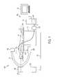
- FIG. 1is a schematic diagram of a C-arm X-ray system for 3D rotational angiography of the invention
- FIG. 2 aillustrates projection angles through a patient's heart using a conventional C-arm X-ray system and process
- FIG. 2 bis a plot of the patient's ECG signal upon which is superimposed rotation-based timing of 2D projections of FIG. 2 a;
- FIG. 3 aillustrates projection angles defined to image a patient's cardiac anatomy by an inventive C-arm X-ray system with ECG gating in accordance with an embodiment of the invention
- FIG. 3 bis plot of the patient's ECG signals upon which is superimposed the ECG gated signals which control the irradiation at each stop of a C-arm X-ray system in accordance with an embodiment of the invention.
- FIG. 4is a schematic flow diagram of an invention 3D RA method utilizing ECG gating in accordance with an embodiment of the invention.
- FIG. 1is a schematic diagram of an inventive C-arm system 100 for 3D rotational angiographic (3D RA) imaging of a chest area of a patient 105 , e.g., 3D imaging the cardiac vasculature and chambers.
- the C-arm X-ray system(“system”) is designed for optimal X-ray dose reduction during fluoroscopic acquisition of a series of 2D raw projection images, improved heart chamber imaging and faster patient throughput when implemented with the inventive ECG triggering.
- System 100includes a recording unit 110 , comprising an X-ray emitter 115 and X-ray detector 120 .
- the X-ray emitter 115 and detector 120are disposed opposite each other as mounted at the ends of a C-arm 125 .
- the C-arm 125is supported roughly in its center on a stand 130 , in order that the C-arm rotates about its isocentric axis 135 .
- the C-arm constructionprovides that a central beam 140 of X-ray radiation may be swung or directed at any projection angle ( ⁇ ) in relation to the surrounding space within a recording plane perpendicular to the isocentric axis 135 .
- System 100further includes a patient table 145 with tabletop 150 , which are together inserted into an opening in C-arm 125 between the X-ray emitter and detector.
- An ECG unit 155is shown in the figure with a number of ECG sensors 160 (attached to patient 105 ), and a control and evaluation unit 165 .
- the control and evaluation unit 165includes a data processing unit 170 in which an evaluation unit 175 is used to generate the 3D image data record (i.e., the 3D RA image).
- the control and evaluation unit 165includes input/output means 180 , e.g., a screen, keyboard, mouse, etc., for inputting control instructions for displaying status variables, examination results, etc.
- the recording unit 110provides raw 2D projection image data to the evaluation unit 240 at each angular rotation (or “stop”) of the C-arm only when the ECG unit provides a radiation control signal to the evaluation unit.
- the emitterirradiates the patient once at each angulation stop to realize a 2D projection image (at angle ⁇ ) at an exact desired point in the ECG detected cardiac cycle.
- a 2D projection imageat angle ⁇
- the radiation control signaltime-correlated to a fixed point in the patient's cardiac cycle is generated using the ECG data.
- the desired cardiac phaseis either user programmed, or it may be calculated automatically by the system.
- FIG. 2 aillustrates conventional C-arm operation for cardiac imaging a patient 105 positioned on a table 150 .
- the conventional C-arm X-ray system(not shown in FIG. 2 a ) steps through its angular rotations to generate a series of N raw 2D projection images.
- FIG. 2 bis a plot of the patient's ECG signals upon which the timing of each of the N stops, corresponding to the N raw 2D projection images upon which the timing is superimposed.
- the conventional pulsed irradiation and raw 2D projection images it generatesare not acquired synchronously with the patient's electrical characteristics; the irradiation is merely angle-based (angular rotation). As can be seen in FIG.
- images 2 and 6could be utilized from the scene or series of 2D raw projection images because these appear to correlate to the patient's R-wave.
- Image 17 of the acquired set or scenecould be of use with respect to the R-wave periodicity, but as should be apparent to the skilled artisan, image 17 does not correlate exactly, or substantially exactly to the R-wave peak.
- a clinician or radiologist who wishes to view or analyze the R-wave acquired cardiac images based on the ECG signalsmust manually extract the ECG-related images. Not only is such manual viewing and extraction of images associated with a fixed portion of the cardiac cycle lengthy and cumbersome, but also the patient (and possibly the clinician) may be exposed to unnecessary radiation.
- the non-R-wave related raw 2D projectionsare of little use when R-wave images are to be focused upon (in a particular study) using conventional hardware and methods.
- the C-arm system for ECG-gated 3D RA of the cardiac chambers and vasculature of this inventioncontrols the release of X-rays from the X-ray source, at each angular stop, until the same time or substantially the same time in the patient's cardiac cycle.
- the inventive C-arm X-ray system 100 of FIG. 1is designed so that the start and stop angles ( ⁇ ) for a 3D RA study are defined, or fixed so that the X-ray emitter does not irradiate until the patient's heart is at the same periodic portion of his/her cardiac cycle at each stop.
- the inventive system 100may be user-programmed to define the rotational step size between each acquired image, and to define the fixed phase at which the irradiation occurs to realize a desired resolution of the 3D reconstructed image for various 3D RA imaging applications and procedures.
- FIG. 3 ais an illustration of projection angles of X-ray irradiation irradiating a patient's ( 105 ) cardiac anatomy while positioned on a table 150 .
- FIG. 3 bis a plot of the patient's ECG signals superimposed on a scene or series of raw 2D projection images derived by the inventive system 100 . Operational parameters may be defined manually by patient input, or automatically calculated by the system 100 . In the example depicted in FIGS. 3 a and 3 b , the step angulation is set to 7.5 degrees and the radiation control signal is arranged so that a series of 64 images is acquired at 64 r-wave occurrences. In operation, the C-arm begins the image acquisition at the start position.
- the X-ray irradiationis released by the X-ray source to acquire the first R-wave 2D projection image.
- the C-armis rotated to the next angle or rotation step, where it “waits” for the next R-wave event before taking the next 2D raw projection image. X-ray exposure is accordingly reduced for both the patient and clinician, and the clinician is no longer required to manually select the images, improving workflow.
- FIG. 4is a schematic flow diagram, which defines one embodiment of an inventive method 400 of the invention.
- the inventive methodmay be included in a “dynavision” operation, wherein a dynavision exposure series is acquired from a patient at rest on a table, as shown in FIG. 1 .
- the methodis ECG-synchronized, and implemented for 3D rotational angiographic (3D RA) imaging of a patient's cardiac anatomy using a C-arm X-ray system.
- a step represented by block 410includes providing a start angle, a stop angle and a rotation step size for C-arm rotation about the patient.
- These inputsmay be automatically generated by a processor included in the C-arm X-ray system, or input by a user through a user interface.
- Block 420represents a step of generating an ECG-gating signal to control X-ray release at each step of C-arm rotation.
- the ECG gating signalmay be generated directly within an ECG unit, or generated by a processor automatically.
- the userchooses a phase of the patient's cardiac phase, for example, the R-wave.
- Block 430represents a step of acquiring a series of 2D projection images. Each or the acquired 2D projection images of the series is acquired at each step of C-arm rotation in the presence of the ECG-gating signal.
- a 2D projection image(raw image) is acquired synchronously with the ECG gating signal, whereby one image may be acquired at the same cardiac phase at each rotational step, to minimize x-ray exposure and movement-associated image artifacts when two or more of the acquired series are used to generate a real-time 3D RA image.
- Block 440represents a step of processing the series of 2D projection images to reconstruct a high-resolution 3D image of the cardiac soft-tissue.
- the inventive methodmay include preceding the image acquisition step by a safety test run.
- the methodmay further implement real-time subtraction imaging, roadmapping and interventional 2D fluoroscopy.
Landscapes
- Health & Medical Sciences (AREA)
- Life Sciences & Earth Sciences (AREA)
- Medical Informatics (AREA)
- Engineering & Computer Science (AREA)
- Heart & Thoracic Surgery (AREA)
- Animal Behavior & Ethology (AREA)
- Biophysics (AREA)
- Nuclear Medicine, Radiotherapy & Molecular Imaging (AREA)
- Optics & Photonics (AREA)
- Pathology (AREA)
- Radiology & Medical Imaging (AREA)
- Biomedical Technology (AREA)
- Physics & Mathematics (AREA)
- Molecular Biology (AREA)
- Surgery (AREA)
- High Energy & Nuclear Physics (AREA)
- General Health & Medical Sciences (AREA)
- Public Health (AREA)
- Veterinary Medicine (AREA)
- Dentistry (AREA)
- Oral & Maxillofacial Surgery (AREA)
- Cardiology (AREA)
- Physiology (AREA)
- Vascular Medicine (AREA)
- Apparatus For Radiation Diagnosis (AREA)
Abstract
Description
Claims (20)
Priority Applications (1)
| Application Number | Priority Date | Filing Date | Title |
|---|---|---|---|
| US11/545,250US7379532B2 (en) | 2006-08-01 | 2006-10-10 | ECG-based rotational angiography for cardiology |
Applications Claiming Priority (2)
| Application Number | Priority Date | Filing Date | Title |
|---|---|---|---|
| US83472506P | 2006-08-01 | 2006-08-01 | |
| US11/545,250US7379532B2 (en) | 2006-08-01 | 2006-10-10 | ECG-based rotational angiography for cardiology |
Publications (2)
| Publication Number | Publication Date |
|---|---|
| US20080031417A1 US20080031417A1 (en) | 2008-02-07 |
| US7379532B2true US7379532B2 (en) | 2008-05-27 |
Family
ID=39033792
Family Applications (1)
| Application Number | Title | Priority Date | Filing Date |
|---|---|---|---|
| US11/545,250Expired - Fee RelatedUS7379532B2 (en) | 2006-08-01 | 2006-10-10 | ECG-based rotational angiography for cardiology |
Country Status (1)
| Country | Link |
|---|---|
| US (1) | US7379532B2 (en) |
Cited By (6)
| Publication number | Priority date | Publication date | Assignee | Title |
|---|---|---|---|---|
| US20080240355A1 (en)* | 2007-03-30 | 2008-10-02 | Satoru Ohishi | X-ray diagnostic apparatus |
| US20090175515A1 (en)* | 2006-06-08 | 2009-07-09 | Tomtec Imaging Systems Gmbh | Method, device, and computer programme for evaluating images of a cavity |
| US8031838B2 (en) | 2009-01-29 | 2011-10-04 | The Invention Science Fund I, Llc | Diagnostic delivery service |
| US8130904B2 (en) | 2009-01-29 | 2012-03-06 | The Invention Science Fund I, Llc | Diagnostic delivery service |
| US8538505B2 (en) | 2009-08-13 | 2013-09-17 | Siemens Aktiengesellschaft | Method for 3-D data collection with a biplane C-arm system with biplane acquisition multiplexing |
| US10959697B2 (en)* | 2016-11-23 | 2021-03-30 | Carestream Health, Inc. | Synchronization for dynamic imaging |
Families Citing this family (3)
| Publication number | Priority date | Publication date | Assignee | Title |
|---|---|---|---|---|
| US7660382B2 (en)* | 2004-06-28 | 2010-02-09 | Koninklijke Philips Electronics N.V. | Examination apparatus for perfusion studies |
| FR2924255A1 (en)* | 2007-11-27 | 2009-05-29 | Gen Electric | METHOD FOR PROCESSING RADIOGRAPHIC CARDIAC IMAGES FOR OBTAINING A SUBTRACT AND RECALLED IMAGE |
| US8849388B2 (en)* | 2011-09-08 | 2014-09-30 | Apn Health, Llc | R-wave detection method |
Citations (2)
| Publication number | Priority date | Publication date | Assignee | Title |
|---|---|---|---|---|
| US6324254B1 (en)* | 1998-11-23 | 2001-11-27 | Siemens Aktiengesellschaft | Method and x-ray device for picking up x-ray images of a substantially rhythmically moving vessel or organ |
| US6507639B1 (en)* | 2001-08-30 | 2003-01-14 | Siemens Aktiengesellschaft | Method and apparatus for modulating the radiation dose from x-ray tube |
- 2006
- 2006-10-10USUS11/545,250patent/US7379532B2/ennot_activeExpired - Fee Related
Patent Citations (2)
| Publication number | Priority date | Publication date | Assignee | Title |
|---|---|---|---|---|
| US6324254B1 (en)* | 1998-11-23 | 2001-11-27 | Siemens Aktiengesellschaft | Method and x-ray device for picking up x-ray images of a substantially rhythmically moving vessel or organ |
| US6507639B1 (en)* | 2001-08-30 | 2003-01-14 | Siemens Aktiengesellschaft | Method and apparatus for modulating the radiation dose from x-ray tube |
Cited By (17)
| Publication number | Priority date | Publication date | Assignee | Title |
|---|---|---|---|---|
| US8077944B2 (en)* | 2006-06-08 | 2011-12-13 | Tomtec Imaging Systems Gmbh | Method, device, and computer programme for evaluating images of a cavity |
| US20090175515A1 (en)* | 2006-06-08 | 2009-07-09 | Tomtec Imaging Systems Gmbh | Method, device, and computer programme for evaluating images of a cavity |
| US7844029B2 (en) | 2007-03-30 | 2010-11-30 | Kabushiki Kaisha Toshiba | X-ray diagnostic apparatus |
| US20100046710A1 (en)* | 2007-03-30 | 2010-02-25 | Satoru Ohishi | X-ray diagnostic apparatus |
| US20080240355A1 (en)* | 2007-03-30 | 2008-10-02 | Satoru Ohishi | X-ray diagnostic apparatus |
| US7706504B2 (en)* | 2007-03-30 | 2010-04-27 | Kabushiki Kaisha Toshiba | X-ray diagnostic apparatus |
| US8083406B2 (en) | 2009-01-29 | 2011-12-27 | The Invention Science Fund I, Llc | Diagnostic delivery service |
| US8047714B2 (en) | 2009-01-29 | 2011-11-01 | The Invention Science Fund I, Llc | Diagnostic delivery service |
| US8041008B2 (en) | 2009-01-29 | 2011-10-18 | The Invention Science Fund I, Llc | Diagnostic delivery service |
| US8031838B2 (en) | 2009-01-29 | 2011-10-04 | The Invention Science Fund I, Llc | Diagnostic delivery service |
| US8111809B2 (en) | 2009-01-29 | 2012-02-07 | The Invention Science Fund I, Llc | Diagnostic delivery service |
| US8116429B2 (en) | 2009-01-29 | 2012-02-14 | The Invention Science Fund I, Llc | Diagnostic delivery service |
| US8130904B2 (en) | 2009-01-29 | 2012-03-06 | The Invention Science Fund I, Llc | Diagnostic delivery service |
| US8249218B2 (en) | 2009-01-29 | 2012-08-21 | The Invention Science Fund I, Llc | Diagnostic delivery service |
| US8254524B2 (en) | 2009-01-29 | 2012-08-28 | The Invention Science Fund I, Llc | Diagnostic delivery service |
| US8538505B2 (en) | 2009-08-13 | 2013-09-17 | Siemens Aktiengesellschaft | Method for 3-D data collection with a biplane C-arm system with biplane acquisition multiplexing |
| US10959697B2 (en)* | 2016-11-23 | 2021-03-30 | Carestream Health, Inc. | Synchronization for dynamic imaging |
Also Published As
| Publication number | Publication date |
|---|---|
| US20080031417A1 (en) | 2008-02-07 |
Similar Documents
| Publication | Publication Date | Title |
|---|---|---|
| US7379532B2 (en) | ECG-based rotational angiography for cardiology | |
| US20210128011A1 (en) | Method and System for 4D Radiological Intervention Guidance (4D-cath) | |
| US5960054A (en) | Angiographic system incorporating a computerized tomographic (CT) scanner | |
| US7180976B2 (en) | Rotational angiography based hybrid 3-D reconstruction of coronary arterial structure | |
| US8233688B2 (en) | Method of detection and compensation for respiratory motion in radiography cardiac images synchronized with an electrocardiogram signal | |
| Strobel et al. | 3D imaging with flat-detector C-arm systems | |
| CN100473344C (en) | X-ray angiography apparatus | |
| US7340033B2 (en) | X-ray unit having an automatically adjustable collimator | |
| EP1606770B1 (en) | Motion-corrected three-dimensional volume imaging method | |
| US20090198121A1 (en) | Method and apparatus for coordinating contrast agent injection and image acquisition in c-arm computed tomography | |
| US9936928B2 (en) | Medical image processing apparatus and X-ray diagnostic apparatus | |
| US20140003688A1 (en) | Multiple modality cardiac imaging | |
| US20140037049A1 (en) | Systems and methods for interventional imaging | |
| JP6419462B2 (en) | X-ray diagnostic equipment | |
| US9895119B2 (en) | Generation of mask and contrast image data in a continuous acquisition | |
| US8229066B2 (en) | X-ray image diagnosis apparatus and X-ray image processing method | |
| US7604404B2 (en) | X-ray imaging apparatus | |
| JP2016198262A (en) | X-ray diagnostic apparatus | |
| CN107307877A (en) | Radiographic apparatus | |
| US8855391B2 (en) | Operating method for an imaging system for the time-resolved mapping of an iteratively moving examination object | |
| WO2018156539A1 (en) | Systems and methods for intervention guidance using a combination of ultrasound and x-ray imaging | |
| JP2006296707A (en) | X-ray diagnostic imaging apparatus, and its three-dimensional blood flow image constituting/displaying method, and program | |
| JP6287719B2 (en) | X-ray equipment | |
| JP7139156B2 (en) | X-ray CT device | |
| US6404850B1 (en) | Cardioangiography apparatus |
Legal Events
| Date | Code | Title | Description |
|---|---|---|---|
| AS | Assignment | Owner name:SIEMENS MEDICAL SOLUTIONS USA, INC., PENNSYLVANIA Free format text:ASSIGNMENT OF ASSIGNORS INTEREST;ASSIGNOR:KRAMP, GEORGE;REEL/FRAME:018514/0457 Effective date:20061108 | |
| STCF | Information on status: patent grant | Free format text:PATENTED CASE | |
| FPAY | Fee payment | Year of fee payment:4 | |
| FPAY | Fee payment | Year of fee payment:8 | |
| AS | Assignment | Owner name:SIEMENS HEALTHCARE GMBH, GERMANY Free format text:ASSIGNMENT OF ASSIGNORS INTEREST;ASSIGNOR:SIEMENS MEDICAL SOLUTIONS USA, INC.;REEL/FRAME:043379/0673 Effective date:20170713 | |
| FEPP | Fee payment procedure | Free format text:MAINTENANCE FEE REMINDER MAILED (ORIGINAL EVENT CODE: REM.); ENTITY STATUS OF PATENT OWNER: LARGE ENTITY | |
| LAPS | Lapse for failure to pay maintenance fees | Free format text:PATENT EXPIRED FOR FAILURE TO PAY MAINTENANCE FEES (ORIGINAL EVENT CODE: EXP.); ENTITY STATUS OF PATENT OWNER: LARGE ENTITY | |
| STCH | Information on status: patent discontinuation | Free format text:PATENT EXPIRED DUE TO NONPAYMENT OF MAINTENANCE FEES UNDER 37 CFR 1.362 | |
| FP | Lapsed due to failure to pay maintenance fee | Effective date:20200527 | |
| AS | Assignment | Owner name:SIEMENS HEALTHCARE GMBH, GERMANY Free format text:CORRECTIVE ASSIGNMENT TO CORRECT THE EXECUTION DATE OF ASSIGNMENT 3, ASSIGNOR SIEMENS MEDICAL SOLUTIONS USA, INC. TO SIEMENS HEALTHCARE GMBH PREVIOUSLY RECORDED ON REEL 043379 FRAME 0673. ASSIGNOR(S) HEREBY CONFIRMS THE ASSIGNMENT OF INVENTOR RIGHTS.;ASSIGNOR:SIEMENS MEDICAL SOLUTIONS USA, INC.;REEL/FRAME:056112/0540 Effective date:20201120 |